Dräger Ventilation Mini Manual Brief explanation of ventilation ...
Upload
independentCategory
view
3download
0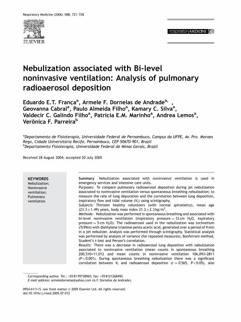
ARTICLE IN PRESS
Respiratory Medicine (2006) 100, 721–728
KEYWORDNebulizatiNoninvasivventilationPulmonaryventilation
0954-6111/$ - sdoi:10.1016/j.r
�CorrespondiE-mail addr
Nebulization associated with Bi-levelnoninvasive ventilation: Analysis of pulmonaryradioaerosol deposition
Eduardo E.T. Franc-aa, Armele F. Dornelas de Andradea,�,Geovanna Cabrala, Paulo Almeida Filhoa, Kamary C. Silvaa,Valdecir C. Galindo Filhoa, Patrıcia E.M. Marinhoa, Andrea Lemosa,Veronica F. Parreirab
aDepartamento de Fisioterapia, Universidade Federal de Pernambuco, Campus da UFPE, Av. Pro. MoraesRego, Cidade Universitaria Recife, Pernambuco, CEP 50670-901, BrazilbDepartamento Fisioterapia, Universidade Federal de Minas Gerais, Brazil
Received 28 August 2004; accepted 20 July 2005
Son;e;
ee front matter & 2005med.2005.07.012
ng author. Tel.: +55 81ess: armeledornelas@ya
Summary Nebulization associated with noninvasive ventilation is used inemergency services and intensive care units.Purposes: To compare pulmonary radioaerosol deposition during jet nebulizationassociated to noninvasive ventilation versus spontaneous breathing nebulization; tomeasure the rate of lung depuration and the correlation between lung deposition,inspiratory flow and tidal volume (Vt) using scintigraphy.Subjects: Thirteen healthy volunteers (with normal spirometry), mean age(23.371.49) years, body mass index 21.272.3 kg/m2.Methods: Nebulization was performed in spontaneous breathing and associated withbi-level noninvasive ventilation (inspiratory pressure ¼ 12 cm H2O, expiratorypressure ¼ 5 cm H2O). The radioaerosol used in the nebulization was technetium(Tc99m) with diethylene triamine penta acetic acid, generated over a period of 9minin a jet nebulizer. Analysis was performed through scintigraphy. Statistical analysiswas performed by analysis of variance (for repeated measures), Bonferroni method,Student’s t-test and Person’s correlation.Results: There was a decrease in radioaerosol lung deposition with nebulizationassociated to noninvasive ventilation (mean counts in spontaneous breathing200,510+11,012 and mean counts in noninvasive ventilation 106,093+2811(Po0:001). During spontaneous breathing nebulization there was a significantcorrelation between Vt and radioaerosol deposition (r ¼ 0:565, Po0:05), also
Elsevier Ltd. All rights reserved.
99738965; fax: +55 8121268490.hoo.com (A.F. Dornelas de Andrade).

ARTICLE IN PRESS
E.E.T. Franc-a et al.722
between inspiratory flow and radioaerosol deposition in the lungs (r ¼ 0:141,Po0:05). However, there was no correlation between Vt and pulmonary deposition ofradioaerosol in bi-level noninvasive ventilation nebulization (r ¼ 0:082).Conclusion: During nebulization with noninvasive ventilation in healthy volunteers,there was an increase in Vt associated to a higher inspiratory flow rate, withoutresulting in a significant increase in pulmonary radioaerosol deposition.& 2005 Elsevier Ltd. All rights reserved.
Introduction
Noninvasive ventilation has been used to treatacute and chronic respiratory failure due to severaldiseases.1–3 Despite the amount of therapies usedto treat respiratory diseases, the inhaling therapy isusually focused in our clinical practice usingmetered-dose inhaler (MDI),4,5 dry powder inhaler(DPI)6,7 or nebulizers.8–10
Nebulization associated with noninvasive venti-lation (NV) is used in emergency services andintensive care units, not only as a form of revertingbronchial obstruction and also to reduce work ofbreathing. The efficiency of nebulized drug duringnebulization by NV depends on the effectiveness ofthe drug deposition in the lungs.11,12
In practice, it is common to associate inhalatoryroute with NV in asthmatic and COPD patientsduring acute exacerbation. To date, there are fewstudies about inhalatory therapeutics associatedwith NV. This association was first studied byDolovich et al.13 In this study, scintigraphy wasused to analyze peripheral deposition of aerosolduring intermittent positive pressure breathingcomparing to spontaneous breathing in nine stableCOPD patients and showed that there was noincrease in peripheral deposition of aerosol duringintermittent positive pressure breathing. In an-other study, Pollack et al.12 analyzed the treatmentof acute bronchial spasm with b-adrenergic agonistdelivered as an aerosol, made by nasal masks in bi-level positive airway pressure (BiPAP), with in-spiratory positive airway pressure (IPAP) of 10 cmH2O and expiratory positive airway pressure (EPAP)of 5 cm H2O. One hundred patients in an emergencycenter showing clinical wheezing in chest werestudied and there was a significant increase of PEFin the group that used BiPAP2. Unfortunately onlythe PEF was analyzed.
Since nebulization is one of the most efficientway of administering drugs to asthmatic and COPDpatients, children and adults in emergency units, itcan be interesting to analyze the pulmonaryregional deposition of radioaerosol when adminis-tered by nebulization associated with NV.
The aims of the present study were: (1) toanalyze the pulmonary radioaerosol deposition
through scintigraphy, after using a jet nebulizer inspontaneous breathing (SB) and after nebulizationassociated to bi-level NV; (2) to analyze therate of depuration in the lungs and correlatepulmonary deposition with inspiratory flow andtidal volume (Vt).
Methods
Subjects
Thirteen healthy volunteers (nine females and fourmales) were studied. Their mean age was23.471.49 (SD) years, body mass index (BMI) was21.272.3 kg/m2. Exclusion criteria were: history ofsmoking or respiratory problems, pregnancy,breathing rate 420 bpm, heart rate o50 or4100 bpm, oxygen saturation (SpO2) p90%, max-imal inspiratory pressurep50 cm H2O, Vtp6ml/kg,or forced vital capacity (FVC) o81% and forcedexpiratory volume on the 1 s (FEV1) o82% ofpredicted values. The protocol was approved bythe Ethical Committee from all and the subjectsgave written informed consent.
Measurements and procedure
Volunteers were enquired about their age, historyof smoking and any previous pulmonary illness.Anthropometric data {weight, height and bodymass index (BMI)}, and cardiopulmonary assessmentwere then carried out: breathing rate (BR), heartrate (HR) and transcutaneous oxygen saturation(SpO2) were measured by pulse oximetry (ACTIVE;Ecaflex; Sao Paulo, Brazil). PEF (ASTECH; CenterLaboratories; New York, USA) was used to assessbronchial obstruction. Maximal inspiratory pressure(MIP) was obtained using a manometer (MV-150,Marshal-Town Instrument Industry, USA), accordingto the method of Black and Hyatt.14 Ventilometrywas performed with a respirometer (Wright Respi-rometer Mark 8; Ferraris Medical Ltd; England). FVCand FEV1 were measured using a spirometer(Satellite, Subminiature ad Jones Spirometer,Windsor, England), considering Knudson’s values.15

ARTICLE IN PRESS
(b)
(a)
Figure 1 Delimitation of the regions of interest (ROIs):(a) upper, middle and lower pulmonary regions; (b)central, intermediate and peripheralpulmonary regions.
Nebulization and noninvasive ventilation 723
After initial evaluation and following the exclu-sion criteria, the volunteers attended the MedicineNuclear Service, where they underwent nebuliza-tion in two phases, one in SB and another inassociation with bi-level NV, with a minimuminterval of 7 days between each phase. The choiceof the first phase was done by a random selection.In both phases, the radioaerosol used wasTc99m—DTPA (25mCi), generated in jet nebulizer(ST3; NS; Sao Paulo, Brazil) placed in a lead box(ULTRAVENT; Mallinckrodt Medical; St. Louis, USA),and then diluted in 0.9% physiologic serum to avolume of 4ml. Aerosol flow was maintained at 7 l/min and was adjusted by oxygen bursts. Radio-aerosol inhalation was carried out with subjectsusing a silicone facemask in a seated position. Allvolunteers were previously trained for mask adap-tation and deep breathing. At the 3, 6 and 9min ofinhalation, cardiorespiratory monitoring was ob-tained by measuring inspiratory flow, Vt and minuteventilation, using an inspiratory flow sensor(TRACE-5; Intermed; Sao Paulo, Brazil), and HRand SpO2, using pulse oximetry.
In the SB nebulization phase, inspiration andexpiration were realized through spontaneousunidirectional valves connected to a system of‘‘T-tubes’’. This system was interconnected to theradioaerosol inhalation generated by the jet nebu-lizer and a flow sensor attached to the siliconefacemask which was attached through straps on thevolunteers face. Exhalation was realized throughexpiration valves directly placed in the ‘‘Y’’ systemusing an appropiate collector.
During the nebulization associated with bi-levelNV, the same ‘‘T-Tube’’ system adapted for inhala-tion of the radioaerosol and the inspiratory flowsensor located in the nebulization during SB wereutilized. This was connected to the ventilator(BiPAP Synchrony Respironics Inc.; Murrysville,USA) used on spontaneous mode, with fixed levelsof IPAP (12 cm H2O) and EPAP (5 cm H2O). Duringthis phase, exhalation was done via expiratoryvalves of the ventilator.
Immediately after the radioaerosol radioactiveinhalation, the volunteers remained in time during300 s in the scintigraphy camera (Forte AdacLaboratories). The counts recorded were used toproduce static images of the pulmonary field with amatrix of 256� 256� 16 in the posterior position,which is considered the major part of lung volume.
Fig. 1 shows the analysis of pulmonary depositionof radioaerosol. The lung was divided into central,intermediate and peripheral regions and into upper,middle and lower thirds, as the regions of interest(ROIs). A comparison between pulmonary deposi-tion in each lung was taken considering the number
of counts captured by the scintigraphy camera ineach ROI. This was used to compare the SBnebulization phase with the nebulization asso-ciated with bi-level NV. The ROI levels wererecorded on computer (Pegasys ADAC) after thefirst phase and recaptured to be used in the analysisin the second phase to assure that the areas studiedwere the same. This was considered as the numberof pixels for each region of interest in SB and bi-level NV phases.
Statistical analysis
Analysis of variance for repeated measures wasused to analyze pulmonary deposition of radio-

ARTICLE IN PRESS
E.E.T. Franc-a et al.724
aerosol. Comparison between pairs of measureswas performed with Bonferroni method, used toanalyze pulmonary deposition of radioaerosol in thedifferent pulmonary regions, or with Student’st-test for paired samples, used to compare theparameters obtained from cardiorespiratory mon-itoring during inhalation in SB and associated tobi-level NV. Simple regression Person’s analysiswas used to obtain linear correlation betweeninspiratory flow and Vt and pulmonary depositionof radioaerosol. Level of significance was 5%(Po0:05) for all tests.
Results
All subjects were able to perform measurementswithout any kind of problem. Table 1 shows themean7SD of the initial evaluation obtained for thegroup of subjects. Table 2 shows the mean7SD ofcardiorespiratory monitoring during the radioaer-osol nebulization in SB and associated with NV bi-level.
The image was obtained at the end of theinhalation. Analysis of radioaerosol deposition inthe lungs showed that in SB nebulization the meancount in each lung was 200,510711,012 and counts
Table 1 Data of age, body mass index (BMI), respiratoryflow (PEF), maximal inspiratory pressure (MIP), tidal volumvolume on the 1 s (FEV1), for the group of subjects.
Initialevaluation
Aged(years)
BMI(kg/m2)
RR(ipm)
SpO2
(%)
Mean 23.30 21.3 14.4 97.6SD 1.49 2.1 1.9 0.6
Data are presented as mean7standard deviation (SD).
Table 2 Data of cardiorespiratory monitoring: respiratortidal volume (Vt), minute ventilation (MV), inspiratoryspontaneous breathing (SB) and associated with bi-level n
Cardiorespiratory monitoring Nebulization
Spontaneous breathi(mean7SD)
RR (ipm) 11.572.8HR (bpm) 71.8711.3SpO2 (%) 98.870.5Vt (ml) 740.07272.0MV (l) 8074.072685.0IF (l/min) 25.072.8
from bi-level NV were 106,09372811. We observedthat pulmonary deposition of radioaerosol whenassociated to bi-level NV was lower when comparedto SB (Po0:001). There was no significant differ-ence in deposition of radioaerosol between theright and left lungs. In SB nebulization the countsobtained were 208,297763,202 for the right and192,723757,796 for the left lung and in NV thecounts were 108,080747,168 for the right and104,106749,570 for the left lung.
Fig. 2 shows radioaerosol deposition in upper,middle and lower thirds of the lung. In all tests forcomparative analysis the radioaerosol deposition inupper, middle and lower thirds of the lung waslower when nebulization was carried out associatedto bi-level NV than when SB nebulization wasperformed (Po0:001). There was more depositionin middle and lower thirds than in the upper third inboth phases (Po0:05) and there was no significantdifference between middle and lower thirds in anyof the phases.
Fig. 3 shows radioaerosol deposition in central,intermediate and peripheral regions of the lungs.Since there was no significant difference betweenleft and right lungs, the radioaerosol depositionwas also compared in central, intermediate andperipheral regions of the lungs. Radioaerosol
rate (RR), oxygen saturation (SpO2), peak expiratorye (Vt), forced vital capacity (FVC), forced expiratory
PEF(l/s)
MIP(cm H2O)
Vt(ml)
FVC(ml)
FEV1(ml)
477.9 �87.9 631.2 3581 3057136.7 25.2 239.6 894 613
y rate (RR), oxygen saturation (SpO2), heart rate (HR),flow (IF) during the nebulization of radioaerosol inoninvasive ventilation (NV).
ng Associated the bi-level NV(mean7SD)
P-values
13.473.2 ¼ 0.01982.3714.7 ¼ 0.00599.970.7 ¼ 0.003
1726.07624.0 o0.00122096.078349.0 ¼ 0.001
42.0719.6 ¼ 0.005

ARTICLE IN PRESS
0
20000
40000
60000
80000
100000
1/3 upper 1/3 middle 1/3 lower
Pulmonary regions
Pul
mon
ary
radi
oaer
osol
dep
osit
ion
(C
ount
s )
Nebulization in SB
Nebulization associated the bi-level NV
*
**
Figure 2 Deposition of radioaerosol in the upper, middleand lower pulmonary regions: after nebulization inspontaneous breathing (SB) and associated with bi-levelnoninvasive ventilation (NV) (*Po0:05).
0
20000
40000
60000
80000
100000
central intermediate peripheralPulmonary regions
Pul
mon
ary
radi
oaer
osol
depo
siti
on (
Cou
nts)
Nebulization in SB
Nebulization associated the bi-level NV
*
*
*
*
***
Figure 3 Deposition of radioaerosol in pulmonary re-gions: central, intermediate and peripheral, after neb-ulization in spontaneous breathing (SB) and associatedwith bi-level noninvasive ventilation (NV) (*Po0:05).
0
100000
200000
300000
400000
500000
600000
200 400 600 800 1000 1200 1400 1600Tidal volume (ml)
Pul
mon
ary
radi
oaer
osol
dep
osit
ion
(Cou
nts)
r = 0.565
Figure 4 Correlation between tidal volume and pulmon-ary deposition of radioaerosol after nebulization inspontaneous breathing (SB). There was an increase in Vtand in deposition of radioaerosol during SB nebulization(r ¼ 0:565, Po0:05).
Nebulization and noninvasive ventilation 725
pulmonary deposition was lower when nebulizationassociated to bi-level NV was compared to SBnebulization in the regions analyzed (Po0:001).
Although there was more radioaerosol deposition inintermediate and peripheral than in central regionwith SB and bi-level NV nebulization, there were nosignificant differences in any of the phases.
Fig. 4 shows correlation between Vt and pulmon-ary deposition of radioaerosol. There was anincrease in Vt and in deposition of radioaerosolduring SB nebulization (r ¼ 0:565, Po0:05). Duringbi-level NV nebulization there was no correlationbetween Vt and pulmonary deposition of radio-aerosol in (r ¼ 0:082, P40:05). An increase in Vt didnot cause any increase in pulmonary deposition ofradioaerosol.
With respect to inspiratory flow and pulmonarydeposition of radioaerosol, there was a significantbut very low correlation in the SB nebulization(r ¼ 0:141, Po0:05) and there was no correlationbetween inspiratory flow and deposition of radio-aerosol in bi-level NV nebulization (r ¼ 0:048,P40:05).
Discussion
In cases of asthma and bronchitis, one of theconcerns about using nebulization is to guaranteethe deposition of drugs in the area where its actionis most required. The major failure in inhalatorytreatment of asthma is the inadequate depositionof drugs.10,16,17 Parkes and Bersten,18 studied thedynamics of aerosol in vitro and the bronchodilata-tion efficiency in vivo during continuous positiveairway pressure (CPAP) with facemasks in 10asthmatic patients. Although they reported anincrease in aerosol deposition in proximal airwaysbecause of the high gas flow in inhaling through aCPAP, there was no effect on bronchodilatatordeposition. Fauroux et al.,19 using ventilation lungscintigraphy studied the optimized aerosol deposi-tion associated to pressure support ventilation(PSV) and positive end expiratory pressure (PEEP),with PSV levels of 8–10 cm H2O, in 18 children withcystic fibrosis. They found an increase of 30% ofdeposition in the lungs with the use of PSV withoutany collection of particles in proximal airway.
Results from our study showed lower lungdeposition of radioaerosol in both lungs whennebulization associated to bi-level NV was com-pared to SB nebulization. These findings corrobo-rate to those of Dollovich et al.,13 who attributedan inspiratory flow of 40 l/min at the beginning ofinspiration and during respiration by intermittentpositive pressure to the drop of deposition inthe lung. The high flow of gas has a greatertendency to deposit particles by impaction.20–22

ARTICLE IN PRESS
E.E.T. Franc-a et al.726
High inspiratory flow values like these in respirationby intermittent positive pressure provide deposi-tion of particles in the orpharynx, trachea and largeairways due to inertia impact.13
The other studies that we did using PEEP wereperformed without any association with positivepressure ventilation. The first of them23 was madeusing an ultrasonic nebulizer and the second24 usinga device for expiratory positive pressure, butwithout a flow generation associated, as seen innoninvasive ventilation used in the present study.For that matter, it is very hard to make any kind ofcomparison between them.
Any comparison of the results obtained in thisstudy with those of Parkes and Bersten,18 andFauroux et al.,19 considering the efficiency ofnebulization associated with NV is not possiblebecause these studies were performed with distinctgroups of subjects. It is known that in normal lungaerosol distribution in the lower airway basicallydepends on gravity. However, in an asthmatic lung,it depends on the difference of inspiratory flowthrough the airway, which depends on the differentdegrees of obstruction. This fact creates a hetero-geneous deposition privileging the areas lesscompromised by the disease. In general, in patientswith intense bronchiole constriction, the deposi-tion predominates in the central airways during SBnebulization25 and when associated to NV theairway is kept open and consequently a betterperipheral aerosol deposition occurs.12,19
A small number of papers has been publishedabout the treatment of patients suffering frombronchospasm related to asthmatic or other pa-tients, associating NV and bronchodilators. Lookingfor clinical responses, Pollack et al.12 studyingwheezing patients, showed a significantly greaterincrease in percentage of peak expiratory flow rateafter NV delivered by a two-level positive pressureventilator compared to a simple nebulizer. How-ever, in another study, Fauroux et al.19 observed agreater deposition of aerosol in cystic fibrosischildren using PSV than in a control session.Recently, Soroksky et al.26 studying 30 patientswith a severe asthma attack randomly, assigned inthe two groups treating one with noninvasiveventilation only and the other with nebulizer. Animproved lung function was observed, as well as afaster alleviation of the attack and a significantreduction of the need for hospitalization. It ispossible that the differences observed betweenthese results and the one observed in the presentstudy are related to physiology. Indeed, in patientswith respiratory failure associated to a reduction inairway diameter and consequently reduced flow, itis possible that an increase in flow, verified when
noninvasive ventilation is used, might help thesepatients. Despite the fact that these results wereobserved in normal subjects we can postulate thatin clinical settings, with wheezing patients orpatients having a chronic obstructive respiratorydisease, it might be interesting to make a trial ofnoninvasive ventilation.
When the lungs were partially analyzed consider-ing upper, middle and lower thirds, deposition ofradioaerosol was greater in the middle and lowerthirds than in the upper third in both lungs with SBnebulization and associated to bi-level NV. Thisdifference is attributed to the vertical pressuregradient, which is present between base and apexof the lung. Which means that upper and lowerparts of the lung function in different positions onthe pressure/volume curve. Consequently thealveoli of the apex become more extended andless yielding and therefore showing little volumevariation during inspiration.24 On the other hand,the alveoli of the base show less expansion andmore yielding and thus have greater variation ofvolume during inspiration. This fact could justifythe presence of greater deposition of radioaerosolin the middle and lower third of both lungs sincethe base has a greater variation of volume andconsequently greater renovation of inspired aircontaining the radioaerosol.27–29
Analyzing the central region of the lung, moredeposition of radioaerosol was found in intermedi-ate and peripheral areas during SB nebulization andnebulization associated with bi-level NV. Thegradient of intrapleural pressure was relativelyuniform in the horizontal plane in which there wasno change in the horizontal gradient per unitalveolar volume in the erect position. This reduc-tion of radioaerosol deposition in the central regionof the lung could be attributed to some methodo-logical problem in the scintigraphy images or topossible radiation by the increase of the thoracicwalls resulting in a greater distance betweencentral region of the lung and the scintigraphycamera as compared to other regions of thehorizontal gradient.23,30
In this study, the correlation between Vt andinspiratory flow with radioaerosol deposition in thelung showed that when the Vt increases togetherwith an increase in inspiratory flow, up to approxi-mately 30 l/min, there is an increase in radio-aerosol deposition in the lung. Increase in Vtassociated to low inspiratory flow resulted in abetter deposition of radioaerosol in the lung duringSB nebulization. However, during nebulizationassociated to bi-level NV there was no correlationbetween Vt and inspiratory flow. Increase of Vtassociated to increase of inspiration flow did not

ARTICLE IN PRESS
Nebulization and noninvasive ventilation 727
produce any proportional increase of radioaerosoldeposition in the lung. This lack of correlationbetween the Vt and the inspiratory flow is probablydue to higher inspiratory flow rate (40 l/min) usedin bi-level NV and thus producing a greaterdeposition of particles measured by collection inthe upper airways.
In summary, the results of this study suggest thatin young volunteers from both sexes and withnormal spirometry, there was a greater depositionof radioaerosol after SB in both lungs whencompared to bi-level NV nebulization. This isprobably due to a greater inspiratory flow rate(40 l/min) during the application of NV. The resultsalso suggest that in SB nebulization any increase inVt also caused an increase in deposition in the lungsince during SB nebulization the inspiratory flowwas lower than 30 l/min but in bi-level NVnebulization the flow rate was greater. In SB andNV nebulization, radioaerosol deposition was great-er in the middle and lower regions in both lungs,due to the regional differences of ventilation, andin intermediate and peripheral regions, due tolower radiation attenuation compared to thecentral region.
In conclusion, during nebulization with noninva-sive ventilation in healthy volunteers, there was anincrease in Vt associated to a higher inspiratory flowrate, without resulting in a significant increase inpulmonary radioaerosol deposition. Further studiesanalyzing patients will be needed to clarify theeffects of nebulization associated to noninvasiveventilation.
Acknowledgments
We wish to thank Dra. Cristiana Almeida (Chief ofReal Nuclear), Dr. Ricardo Lima, Dra. Fabiana FariasLima, Dr. Walber Castro and Alberto Melo fromCRCN (Centro Regional de Ciencias Nucleares), Dr.Leopoldino dos Santos Neto (from Kesa Ltda.), Dra.Fatima Marinho and Dr. Flavio Maciel (from AGAHealth Care), Recife Brasil for their helpful withthis study. This work was supported by grants fromCNPq (Conselho Nacional de Desenvolvimentocientıfico e tecnologico Pesquisa) (Processo305127/2002-0), Brasil.
References
1. Mehta S, Hill NS. Noninvasive ventilation. Am J Respir CritCare Med 2001;163(2):540–77.
2. Clinical indications for noninvasive positive pressure venti-lation in chronic respiratory failure due to restrictive lung
disease, COPD, and nocturnal hypoventilation—a consensusconference report. Chest 1999;116(2):521–34.
3. Wijkstra PJ. Non-invasive positive pressure ventilation(NIPPV) in stable patients with chronic obstructive pulmon-ary disease (COPD). Respir Med 2003;97(10):1086–93.
4. Pauwels RA. National and international guidelines for COPD:the need for evidence. Chest 2000;117(2 Suppl):20S–2S.
5. Dahl R, Backer V, Ollgaard B, Gerken F, Kesten S. Assessmentof patient performance of the HandiHaler compared withthe metered dose inhaler four weeks after instruction.Respir Med 2003;97(10):1126–33.
6. Persson G, Olsson B, Soliman S. The impact of inspiratoryeffort on inspiratory flow through Turbuhaler(R) in asthmaticpatients. Eur Respir J 1997;10(3):681–4.
7. O’Connor BJ. The ideal inhaler: design and characteristics toimprove outcomes. Respir Med 2004;98(Suppl A):S10–6.
8. Eklund L, Sundblad BM, Malmberg P, Larsson K. The saltoutput of a nebulizer—a comparison between two nebulizertypes. Respir Med 2000;94(2):139–44.
9. Ward MJ. Nebulisers for asthma. Thorax 1997;52:S45–8.10. Dolovich MB. Assessing nebulizer performance. Respir Care
2002;47(11):1290–301.11. Chatmongkolchart S, Schettino GPP, Dillman C, Kacmarek
RM, Hess DR. In vitro evaluation of aerosol bronchodilatordelivery during noninvasive positive pressure ventilation:effect of ventilator settings and nebulizer position. CritCare Med 2002;30(11):2515–9.
12. Pollack CV, Fleisch KB, Dowsey K. Treatment of acutebronchospasm with beta-adrenergic agonist aerosols deliv-ered by a nasal bilevel positive airway pressure circuit. AnnEmergency Med 1995;26(5):552–7.
13. Dolovich MB, Killian D, Wolff RK, Obminski G, Newhouse MT.Pulmonary aerosol deposition in chronic bronchitis: inter-mittent positive pressure breathing versus quiet breathing.Am Rev Respir Dis 1977;115(3):397–402.
14. Black LF, Hyatt RE. Maximal respiratory pressures—normalvalues and relationship to age and sex. Am Rev Respir Dis1969;99(5):696–702.
15. Knudson RJ, Lebowitz MD, Holberg CJ, Burrows B. Changes inthe normal maximal expiratory flow-volume curve withgrowth and aging. Am Rev Respir Dis 1983;127(6):725–34.
16. Dolovich MB, Fink JB. Aerosols and devices. Respir Care ClinN Am 2001;7(2):131–73.
17. Everard ML, Devadason SG, Summers QA, Le Souef PN.Factors affecting total and ‘‘respirable’’ dose delivered by asalbutamol metered dose inhaler. Thorax 1995;50(7):746–9.
18. Parkes SN, Bersten AD. Aerosol kinetics and bronchodilatorefficacy during continuous positive airway pressure deliv-ered by face mask. Thorax 1997;52(2):171–5.
19. Fauroux B, Itti E, Pigeot J, Isabey D, Meignan M, et al.Optimization of aerosol deposition by pressure support inchildren with cystic fibrosis: an experimental and clinicalstudy. Am J Respir Crit Care Med 2000;162(6):2265–71.
20. Dolovich M, Rhem R, Kwong J. Simulating aerosol delivery:effect of particle size, tidal volume and inspiratory flowrate. Am J Respir Crit Care Med 1999;159(3):A615.
21. Dolovich M, Rhem R. Impact of oropharyngeal deposition oninhaled dose. J Aerosol Med-Deposit Clearance Eff Lung1998;11:S112–5.
22. Fink JB. Aerosol device selection: evidence to practice.Respir Care 2000;45(7):874–85.
23. Dornelas de Andrade AF, Lima JL, Gomes LGF, Marinho PEM,Oliveira EGCG, Gusmao AL, et al. Pulmonary deposition ofaerosol generated by an ultrasonic nebuliser associated toPEEP. Eur Resp J 2001;163(5):A445.

ARTICLE IN PRESS
E.E.T. Franc-a et al.728
24. Rodrigues Machado MG, Reis MAS, Barroso AA, Rezende MO,Zin WA. Respiratory function in normal and asthmaticindividuals: effects of the use of positive expiratorypressure. Am J Respir Crit Care Med 1999;159(3):A644.
25. Melchor R, Biddiscombe M, Mak V, Short M, Spiro S. Lungdeposition patterns of directly labeled salbutamol in normalsubjets and in patients with reverse airflow obstruction.Thorax 1993;48:506–11.
26. Soroksky A, Stav D, Shpirer I. A pilot prospective, rando-mized, placebo-controlled trial of bilevel positive airwaypressure in acute asthmatic attack. Chest 2003;123(4):1018–25.
27. Bryan AC, Bentivoglio LG, Beerel F, Macleish H, Zidulka A,Bates DV. Factors affecting regional distribution of ventila-tion and perfusion in the lung. J Appl Physiol 1964;19:395–402.
28. West JB, Hugh-Jones P. Experimental verification of thesingle breath tests of ventilatory and ventilation–perfusionratio inequality. Clin Sci 1959;18:553–9.
29. West JB, Hugh-Jones P. Patterns of gas flow in the upperbronchial tree. J Appl Physiol 1959;14:753–9.
30. Chamberlain MJ, Morgan WK, Vinitski S. Factors influencingthe regional deposition of inhaled particles in man. Clin Sci(London) 1983;64(1):69–78.
Copyright © 2022 FDOKUMEN