Nature and management of duplex abnormalities encountered during infrainguinal vein bypass grafting
9
Nature and management of duplex abnormalities encountered during infrainguinal vein bypass grafting Dennis E Bandyk, MD, Brad L. Johnson, MD, Ashish K. Gupta, MD, and Glenn E. Esses, MD, Tampa, Fla. Purpose: This study was undertaken to evaluate the application of duplex scanning during infrainguinal vein grafting procedures to verify vein conduit preparation, anastomotic patency, and graft hemodynamics. Methods: Between 1991 and 1995, 275 infrainguinal vein bypasses (in situ, 114; reversed, 82; noureversed translocated, 48; spliced alternative/arm vein, 31) to the popliteal (n = 116) or an infrageniculate artery (n = 159) were scanned during surgery for sites of color Doppler flow abnormality. Duplex-detected defects were graded with peak systolic velocity and velocity ratio criteria. Sites that demonstrated highly disturbed flow (peak systolic velocity >180 cm/sec, velocity ratio >2.4) were immediately revised by direct repair, patch angioplasty, or interposition grafting. Results: Intraoperative duplex scanning prompted revision of 50 abnormalities in 43 of the 275 grafts (16%), including 32 vein and seven anastomotic stenoses, nine vein segments with platelet thrombus, and two bypasses with low flow. The intraoperative revision rate was lowest (p < 0.02) for reversed saphenous vein bypasses (7%) compared with other grafting techniques (in situ, 20%; noureversed translocated, 15%; spliced alternative vein, 23%). The revision rates of popliteal and tibial bypasses were similar (14% vs 17%). A normal result shown by intraoperative scan (235 bypasses) was associated with a low 90-day thrombosis (0.4%) and revision (2%) rate, whereas six of 15 grafts (40%) with residual and 13 of 25 grafts (52%) with uurepaired duplex abnormalities required corrective procedures (p < 0.001). One graft failed within 3 months (secondary patency rate, 99%). Conclusions: Intraoperative duplex scanning accurately predicted the technical adequacy of infrainguinal vein grafts and was particularly useful in assessing bypasses constructed with valve lysis techniques or alternative veins. Early graft revisions indicated by duplex monitoring for thrombosis or stenosis were the result of a progression of residual defects and platelet thrombus formation rather than inadequate graft run-off flow. (J Vasc Surg 1996;24:430-8.) A favorable experience with intraoperative ultra- sound techniques gleaned over the past decade has resulted in the adoption of duplex scanning in the monitoring of infrainguinal vein bypass grafting pro- cedures) ,2 This decision was in part prompted by dissatisfaction with arteriography to accurately iden- tify problems with valve lysis when performing in situ From the Division of Vascular Surgery, Department of Surgery, Universityof South Florida Collegeof Medicine. Presented at the Twentieth Annual Meeting of The Southern Association for Vascular Surgery, Naples,Fla.,Jan. 24-27, 1996. Reprint requests: Dennis Bandyk, MD, Divisionof VascularSur- gery, HMT #730, 4 ColumbiaDr., Tampa, FL 33606. Copyright © 1996 by The Society for Vascular Surgery and InternationalSociety for Cardiovascular Surgery,North Ameri- can Chapter. 0741-5214/96/$5.00 + 0 24/6/74393 430 saphenous or nonreversed translocated vein bypass grafting. Initial application of intraoperative duplex scanning demonstrated that 18% ofinfrainguinal vein bypasses were revised on the basis of abnormal scan findings, whereas arteriography, which was per- formed in 81% of procedures, added no additional diagnostic information? Bypass grafts were scanned after restoration of blood flow and intragraft admin- istration of papaverine to augment graft flow. Color Doppler ultrasonography was used to evaluate the technical precision of vein graft preparation and anastomosis construction, and to verify adequate graft hemodynamics for patency. Based on the severity of lumen reduction and peak systolic velocity (PSV) changes associated with stenosis, graft segments were revised and rescanned to verify that the corrective
-
Upload
independent -
Category
Documents
-
view
2 -
download
0
Transcript of Nature and management of duplex abnormalities encountered during infrainguinal vein bypass grafting

Nature and management of duplex abnormalities encountered during infrainguinal vein bypass grafting Dennis E Bandyk, MD, Brad L. Johnson, MD, Ashish K. Gupta, MD, and Glenn E. Esses, MD, Tampa, Fla.
Purpose: This study was undertaken to evaluate the application of duplex scanning during infrainguinal vein grafting procedures to verify vein conduit preparation, anastomotic patency, and graft hemodynamics. Methods: Between 1991 and 1995, 275 infrainguinal vein bypasses (in situ, 114; reversed, 82; noureversed translocated, 48; spliced alternative/arm vein, 31) to the popliteal (n = 116) or an infrageniculate artery (n = 159) were scanned during surgery for sites of color Doppler flow abnormality. Duplex-detected defects were graded with peak systolic velocity and velocity ratio criteria. Sites that demonstrated highly disturbed flow (peak systolic velocity >180 cm/sec, velocity ratio >2.4) were immediately revised by direct repair, patch angioplasty, or interposition grafting. Results: Intraoperative duplex scanning prompted revision of 50 abnormalities in 43 of the 275 grafts (16%), including 32 vein and seven anastomotic stenoses, nine vein segments with platelet thrombus, and two bypasses with low flow. The intraoperative revision rate was lowest (p < 0.02) for reversed saphenous vein bypasses (7%) compared with other grafting techniques (in situ, 20%; noureversed translocated, 15%; spliced alternative vein, 23%). The revision rates of popliteal and tibial bypasses were similar (14% vs 17%). A normal result shown by intraoperative scan (235 bypasses) was associated with a low 90-day thrombosis (0.4%) and revision (2%) rate, whereas six of 15 grafts (40%) with residual and 13 of 25 grafts (52%) with uurepaired duplex abnormalities required corrective procedures (p < 0.001). One graft failed within 3 months (secondary patency rate, 99%). Conclusions: Intraoperative duplex scanning accurately predicted the technical adequacy of infrainguinal vein grafts and was particularly useful in assessing bypasses constructed with valve lysis techniques or alternative veins. Early graft revisions indicated by duplex monitoring for thrombosis or stenosis were the result of a progression of residual defects and platelet thrombus formation rather than inadequate graft run-off flow. (J Vasc Surg 1996;24:430-8.)
A favorable experience with intraoperative ultra- sound techniques gleaned over the past decade has resulted in the adoption of duplex scanning in the monitoring of infrainguinal vein bypass grafting pro- cedures) ,2 This decision was in part prompted by dissatisfaction with arteriography to accurately iden- tify problems with valve lysis when performing in situ
From the Division of Vascular Surgery, Department of Surgery, University of South Florida College of Medicine.
Presented at the Twentieth Annual Meeting of The Southern Association for Vascular Surgery, Naples, Fla., Jan. 24-27, 1996.
Reprint requests: Dennis Bandyk, MD, Division of Vascular Sur- gery, HMT #730, 4 Columbia Dr., Tampa, FL 33606.
Copyright © 1996 by The Society for Vascular Surgery and International Society for Cardiovascular Surgery, North Ameri- can Chapter.
0741-5214/96/$5.00 + 0 24/6/74393
430
saphenous or nonreversed translocated vein bypass grafting. Initial application of intraoperative duplex scanning demonstrated that 18% ofinfrainguinal vein bypasses were revised on the basis of abnormal scan findings, whereas arteriography, which was per- formed in 81% of procedures, added no additional diagnostic information? Bypass grafts were scanned after restoration of blood flow and intragraft admin- istration of papaverine to augment graft flow. Color Doppler ultrasonography was used to evaluate the technical precision of vein graft preparation and anastomosis construction, and to verify adequate graft hemodynamics for patency. Based on the severity of lumen reduction and peak systolic velocity (PSV) changes associated with stenosis, graft segments were revised and rescanned to verify that the corrective
JOURNAL OF VASCULAR SURGERY Volume 24, Number 3 Bandyk et al. 431
repair was successful in restoring normal hemody- namics. The purpose of this study was to prospectively evaluate previously derived threshold criteria for in- traoperative revision with respect to predicting early graft patency or the need for graft revision within 3 months of the procedure. We also sought to define further the fate of repaired and unrepaired sites identified by duplex scanning to harbor disturbed flOW.
Our long-held interest in vein graft surveillance was an additional motive for the adoption ofintraop- erative duplex scanning as the initial study in a surveillance protocol. We hypothesize that enhance- ment of the technical precision of bypass grafting may minimize both early graft thrombosis and the need to perform secondary procedures to correct residual stenosis. In a previous report dealing with in situ saphenous vein bypass grafting, we reported graft failure and revision rates of 3% and 7%, respectively, within 30 days of the procedure. 4 Other authors have cited 30-day inftalnguinal vein bypass thrombosis rates ranging from 3% to 14%. 5-s Because we use a variety of grafting techniques in patients who undergo infrainguinal bypass procedures on the basis of vein availability and quality, previous bypasses procedures, and sites of the proximal and distal anastomoses, the routine use ofintraoperative duplex scanning permit- ted the assessment of efficacy relative to the grafting technique as well as the development of a treatment algorithm when duplex abnormalities are identified.
MATERLA_L AND METHODS Patient population. From October 1991 to
November 1995, 266 patients (216 men, 50 women) underwent 275 infrainguinal vein bypass graft proce- dures, during which color-flow duplex ultrasonogra- play was used to assess technical adequacy. The opera- tive indications for leg revascularization were critical ischemia (tissue loss, 162; ischemic rest pain, 77) in 239 patients (87%), debilitating clandication in 28 (10%), and popliteal aneurysm in eight (3%). Ex- cluded from the study were vein grafts procedures performed for trauma and four emergency inftain- guinal vein bypasses performed in patients with acute limb ischemia for whom intraoperative duplex scan- ning was not available.
The vein grafting technique varied with the sur- geon's preference, vein availability, and the sites selected fbr the proximal and distal anastomosis. One hundred sixty-two of the inftalnguinal vein bypasses (59%) were constructed with in situ (n = 114) or nonreversed translocated (n = 48) saphenous vein grafting techniques. Valve lysis was performed with
Karmody scissors or a modified Mills valvulotome (American V. Mueller, Chicago, Ill.) with a "blind" retrograde technique in all but six cases, during which angioscopy-directed valvulotomy was performed. The remaining bypasses used reversed saphenous (n = 82) or reversed spliced alternative (lesser saphe- nous, greater saphenous remnants)/arm veins (n = 31). The location of the distal anastomosis was the popliteal artery in 116 limbs (above-knee, 29; below- knee, 87) or an infrageniculate artery in 159 limbs (peroneal, 58; posterior tibial, 47; anterior tibia], 41; pedal, 13).
Instrumentation and scanning technique. In- traoperative duplex scans were performed with one of three color duplex ultrasound systems (ATL HDI Ultramark 9, ATL HDI 3000, Advanced Technology Laboratories, Bothell, Wash.; Accuson 128 XP, Accu- son Corporation, Smyrna, Ga.). All studies were conducted with a linear array probe (7-10 MHz) with and without an acoustic standoff. A sterile plastic sleeve filled with acoustic gel was used to cover the transducer. Imaging of the vein graft, anastomoses, and adjacent native arteries was performed in a longitudinal plane and, if deemed necessary, trans- versely with a vascular laboratory technologist adjust- ing the ultrasound system for optimal color Doppler imaging and the recording of centerstream Doppler angle-corrected (60 degrees or less relative to vessel axis) velocity spectra. Previous experience has dem- onstrated that ultrasound instrument setup and com- plete graft imaging with selected-site velocity spectra recording can be performed in less than ] 5 minutes.
Vein bypasses were visually inspected and patency confirmed by pulse palpation or continuous-wave Doppler flow analysis before duplex scanning. Papav- erine (30 to 60 mg) was injected directly into the graft to augment blood flow and thus enhance the sensi- tivity of duplex scanning for detection and grading of residual stenosis. The entire bypass was imaged for anatomic and flow abnormalities. After in situ saphe- nous vein grafting, color Doppler imaging was used to locate remaining patent vein branches for ligation. In graft segments or anastomotic regions that had a color Doppler flow abnormality (lumen narrowing, in- creased velocity, aliasing, color-flow jet), velocity spectra were recorded proximal to and at the site of maximum flow disturbance, with measurements of PSV and calculation of the velocity ratio (Vr), where V r = PSVat lesion//PSVproximal . Defects were classified into four stenosis categories with previously devel- oped criteria (Table I). 3 Although 125 cm/sec was used a threshold for a vein graft flow abnormality, systolic velocity spectra in the range of 110 to 140
JOURNAL OF VASCULAR SURGERY 432 Bandyk et al. September 1996
Table I. Categories of duplex-identified residual lesions
Peak systolic velocity Velocity ratio Interpretation and intraoperative Stenosis category (cm/sec) (Vr) management
Normal/minimal <125 1.0 to 1.4
Moderate 125 to 180 1.5 to 2.4
Severe >180 with spectral broadening 2.5 to 4 High-grade >300 >4.0
Normal flow pattern; no further evalua- tion required
Residual flow abnormality; rescan after 5 rain with flow augmentation, arteriog- raphy
Significant abnormality; repair defect Critical lesion with pulse deficit, associ-
ated with low-flow graft flow; assess for platelet thrombus and repair defect
cm/sec can be recorded from small diameter (<3 mm) venous conduits. 9 I f duplex scanning demonstrated both a defect or narrowing in the vessel lumen and velocity spectra of a severe or high-grade stenosis, the site was immediately revised. Graft segments or anas- tomotic sites with moderate flow abnormalities or stenosis were further evaluated by transverse imaging to measure vessel diameter and to assess the lumen for evidence of thrombus formation. Segments with borderline stcnoses were typically rescanned after additional papaverine administration to assess hemo- dynamic response to flow augmentation. I f the PSV at the site increased to greater than 200 cm/sec in a normal-diameter vein or anastomosis, exploration and revision were performed. When increased veloci- ties were measured in outflow tibial arteries, an arteriographic scan was performed to differentiate between focal stenosis and smooth tapering caused by spasm. Sites with residual, unrepaircd, moderate stenoscs were noted in the patient's record and were evaluated in the early postoperative period for persis- tence, progression, or regression. Representative cen- terstream velocity spectra were recorded at anasto- motic sites and along the graft at various locations (high thigh, above knee, below knee, and distal graft segment) as an estimation of outflow resistance and graft flow.
Data analysis. Residual lesions that were identi- fied and corrected or that were left unrepaired were tabulated for each type of vein bypass performed. Adverse patient outcomes, including graft thrombosis or graft revision, were recorded for the 3 months after surgery and were analyzed relative to the duplex findings at surgery (i.e., normal results on scan, corrected duplex-identified defect with no residual flow abnormality, corrected duplex defect with re- sidual moderate stenosis, or unrepaired duplex flow abnormality). Postoperative scans were performed in all patients before discharge (approximately I week) and at 6 weeks and 3 months. No patient was lost to
follow-up during this time interval. Differences be- tween groups were compared by Z 2 analysis.
R E S U L T S
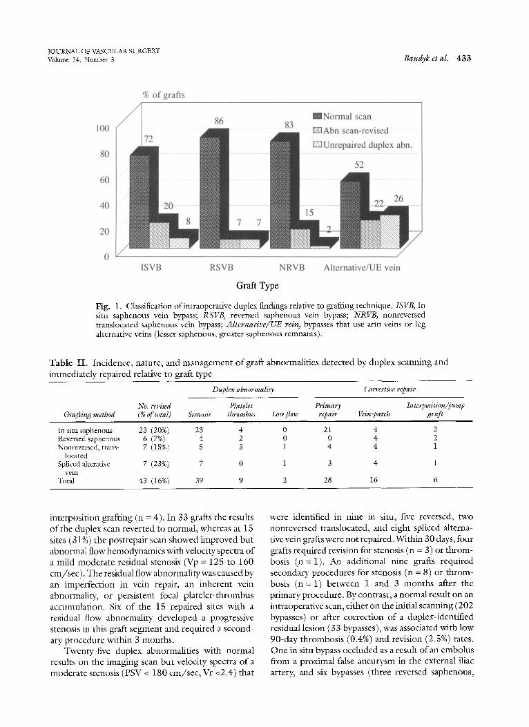
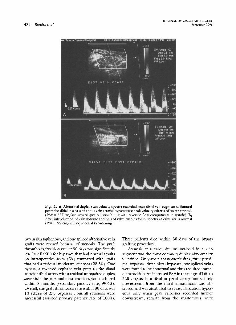
The incidence of duplex-identified flow abnor- malities varied with graft type, being highest for spliced alternative/arm vein bypass (48%) and lowest for reversed saphenous vein bypass (14%; Fig. 1). Color Doppler imaging typically demonstrated vessel lumen reduction and a focal region of disturbed flow, which was evident by aliasing and "flow jet" forma- tion. In most instances the cause of the flow abnor- mality (i.e., retained valve, residual atherosclerotic plaque in an inflow/outflow artery, thrombus forma- tion in a vein segment or anastomosis) could be determined. Velocity spectra of a severe or high grade stenosis was detected in 22% of alternative vein bypasses, 20% of in situ saphenous vein bypasses, 17% ofnonreversed translocated saphenous vein bypasses, and 7% of reversed saphenous vein bypasses. Intraop- erative duplex findings prompted the revision of 50 graft abnormalities in 43 of the 275 bypasses (16%), including 32 vein stenoses, seven anastomofic stenoses, nine vein segments with platelet thrombus formation, and two bypasses with low flow (Table II). Sixteen of 32 vein stenoses (50%) were caused by inadequate valve cusp incision and were corrected by reintroduction of a valvulotome with incision of the retained leaflet and correction of the duplex flow abnormality (Fig. 2). Only nine of 48 stenotic lesions (19%) had velocity spectra of a high-grade residual lesion with PSV > 300 cm/sec, and in six instances the flow abnormality was the result of platelet aggrega- tion in a vein segment or distal anastomotic region. The revision rate of grafts to the popliteal (15%, 17 bypasses) or a tibial/pedal (17%, 27 bypasses) artery was similar. All 48 stenotic lesions that had been judged to warrant corrective repair had an abnormal- ity found at exploration and were revised by primary repair (n = 21), vein-patch angioplasty (n = 16), or
JOURNAL OF VASCULAR SURGERY Volume 24, Number 3 Bandyk et aL 433
100
80
60
40
20
% of grafts
8 i 83 mNormal scan
.......... 72 .............................................................. ~ ............. ~A~ria e~ca~rrte~edviusp~x ab "n'[''''''"
......... 52 .............
i : i / / / / 2 2 26
....... 8 7 7 ~~.
ISVB RSVB NRVB Alternative/UE vein
Graft Type
Fig. 1. Classification ofintraoperative duplex findings relative to grafting technique. ISVB, In situ saphenous vein bypass; RSVB, reversed saphenous vein bypass; NRVB, nonreversed translocated saphenous vein bypass; Alternative/UE vein, bypasses that use arm veins or leg alternative veins (lesser saphenous, greater saphenous remnants).
Table IL Incidence, nature, and management of graft abnormalities detected by duplex scanning and immediately repaired relative to graft type
Duplex abnormality Corrective repair
No. revised Platelet Primary Interposition/jump Grafting method (% of total) Stenosis thrombus Low flow repair Vein-patch graft
In situ saphenous 23 (20%) 23 4 0 21 4 2 Reversed saphenous 6 (7%) 4 2 0 0 4 2 Nonreversed, trans- 7 (15%) 5 3 1 4 4 1
located Spliced alterafive 7 (23%) 7 0 1 3 4 1
vein Total 43 (16%) 39 9 2 28 16 6
interposition grafting (n = 4). In 33 grafts the results of the duplex scan reverted to normal, whereas at 15 sites (31%) the postrepair scan showed improved but abnormal flow hemodynamics with velocity spectra of a mild-moderate residual stenosis (Vp = 125 to 160 cm/sec). The residual flow abnormalitywas caused by an imperfection in vein repair, an inherent vein abnormality, or persistent focal platelet-thrombus accumulation. Six of the 15 repaired sites with a residual flow abnormality developed a progressive stenosis in this graft segment and required a second- ary procedure within 3 months.
Twenty-five duplex abnormalities with normal results on the imaging scan but velocity spectra of a moderate stenosis (PSV < 180 cm/sec, Vr <2.4) that
were identified in nine in situ, five reversed, two nonreversed translocated, and eight spliced alterna- tive vein grafts were not repaired. Within 30 days, four grafts required revision for stenosis (n = 3) or throm- bosis (n= 1). An additional nine grafts required secondary procedures for stenosis (n = 8) or throm- bosis (n= 1) between 1 and 3 months after the primary procedure. By contrast, a normal result on an intraoperative scan, either on the initial scanning (202 bypasses) or after correction of a duplex-identified residual lesion (33 bypasses), was associated with low 90-day thrombosis (0.4%) and revision (2.5%) rates. One in situ bypass occluded as a result of an embolus from a proximal false aneurysm in the external iliac artery, and six bypasses (three reversed saphenous,
JOURNAL OF VASCULAR. SURGERY 4 3 4 Bandyk et aI. September 1996
Fig. 2. A, Abnormal duplex scan velocity spectra recorded from distal vein segment of femoral posterior tibial in situ saphenous vein arterial bypass were peak velocity criteria of severe stenosis (PSV = 227 cm/sec, severe spectral broadening with reversed flow components in systole). B, After introduction ofvalvulotome and lysis of valve cusp, velocity spectra at valve site is normal (PSV = 92 cm/sec, no spectral broadening).
two in situ saphenous, and one spliced alternative vein graft) were revised because of stenosis. The graft thrombosis/revision rate at 90 days was significantly less (p < 0.001) for bypasses that had normal results on intraoperative scans (3%) compared with grafts that had a residual moderate stenoses (28.5%). One bypass, a reversed cephalic vein graft to the distal anterior tibial artery with a residual unrepaired duplex stenosis in the proximal anastomotic region, occluded within 3 months (secondary patency rate, 99.6%). Overall, the graft thrombosis rate within 30 days was 1% (three of 275 bypasses), but all revisions were successful (assisted primary patency rate of 100%).
Three patients died within 30 days of the bypass grafting procedure.
Stenosis at a valve site or localized in a vein segment was the most common duplex abnormality identified. Only seven anastomotic sites (three proxi- mal bypasses, three distal bypasses, one spliced vein) were found to be abnormal and thus required imme- diate revision. An increased PSVin the range of 160 to 220 cm/sec in a tibial or pedal artery immediately downstream from the distal anastomosis was ob- served and was attributed to revascularization hyper- emia only when peak velocities recorded further downstream, remote from the anastomosis, were
JOURNAL OF VASCULAR SURGERY Volume 24, Number 3 Bandyk et al. 4 3 5
similar and the Vr between the two recordings sites was less than 2.0. Sites ofplatelet thrombus formation were detected by findings of increased PSVs with color Doppler and spectrum analysis and of lumen filling defects on real-time B-mode imaging. The region of' flow abnormality typically extended over several centimeters, in contrast to the focal stenosis observed at valve sites. In the nine grafts in which platelet thrombus formation was identified at opera- tion, treatment consisted of uroldnase infusion (250,000 IU) proximal and downstream of the site and revision by interposition vein grafting (n = 5) or vein-patch angioplasty (n = 4). This thrombotic com- plication was missed in two grafts at sites when residual disturbed flow (Vp = 160 to 170 cm/sec) was detected but left uncorrected.
The finding of low graft flow (PSV <40 cm/sec in 3- to 4-mm diameter vein) and a high-outflow resis- tance flov¢ pattern (absence of diastolic flow in the distal graft after papaverine administration) was iden- tified in only four of the 275 bypasses. In two instances, surgical augmentation of graft flow was performed by creating a distal arteriovenous fistula (n = 1) or a jump graft to a second tibial artery (n = 1). In the remaining two instances, both of which involved patients with chronic renal failure on hemodialysis with low intraoperative cardiac output, treatment consisted of confirmatory normal results on an arteriogram of the distal graft and runoff, continued heparin anticoagulation, and medical treatment ofcardiogenic or hypovolemic shock states.
DISCUSSION
Our experience indicates that the routine use of color duplex scanning during infrainguinal vein by- pass procedures is a sensitive method to detect vascu- lar defects and thus upgrade technical precision re- gardless of the grafting technique used. This study documents the incidence, location, and nature of duplex-identified abnormalities, as well as the leash bility and efficacy ofintraoperative monitoring. Graft scanning was performed in lcss than 15 minutes in the majority of patients, and a spectrum of abnormalities were identified, including sites of platelet aggrega- tion, which are precursors of early graft thrombosis. The most common abnormality that was identified was valve-site stenosis and the result of incomplete valve lysis after "blind" retrograde valvulotomy. Overall, 16% of grafts were revised on the basis of the results of duplex scanning, which resulted in a 30-day graft thrombosis rate of only 1% (three bypasses) and revision rate of 2.2% (six bypasses). These results are superior to those from a previous report of only in situ
saphenous vein bypass (3% and 7%, respectively) despite the use of varied grafting techniques, includ- ing spliced alternative/arm vein bypasses. When ar- teriography or angioscopy have been used to monitor infrainguinal vein bypass procedures, early (30-day) graft failure rates in the range of 3% to 7% have been reported, l°,n The data from this study indicate that early graft failure is largely preventable and is not caused by inadequate graft runoff, but rather residual, unrepaired graft defects that progress in severity because ofplatelet thrombus formation at the site or result in vein wall sclerosis/myointimal hyperplasia. All three mechanisms were documented in the course of early graft revision procedures.
The clinical value of intraoperative duplex scan- ning varied with the grafting technique. Most inter- ventions were related to residual valve-site stenosis and occurred primarily in in sire and nonreversed translocated vein bypasses. A surgeon who performs only reversed saphenous vein bypass grafting would not derive as much benefit by using intraoperative duplex scanning as one who uses grafting techniques that require valve lysis or involve splicing alternative veins together or bypass with arm veins. The revision rate of in situ and nonreversed vein bypasses was higher (18%) than that of reversed saphenous veins (7%). Monitoring is particularly important when alternative veins (cephalic, basilic, lesser saphenous, greater saphenous remnants) are used. In this group, results ofintraoperative duplex scanning were abnor- mal in one half of the procedures, and one quarter of the bypasses were revised. Techniques to upgrade vein quality, such as prebypass angioscopy, have been recommended, but failure rates as high as I0% per- sist. 12 Although angioscopy can assist in vein prepa- ration (valve lysis) and selection of quality autogenous vein conduit, the assessment of graft hemodynamics remains an important concept because bypasses with normal results of duplex scans demonstrate a low (<3%) failure and revision rate.
Our study supports the principle that one goal of bypass grafting is to achieve normal results on the duplex scan. Unfortunately, this may not always be possible when veins of marginal quality must be used and anastomosis to diseased artery segments are the only option. In such instances, duplex inspection of the bypass for severe stenosis is important, and these sites should be corrected. When the abnormality found on the duplex scan is one of a moderate stenosis (PSV in the range of 125 to 180 cm/sec, Vr <2.4), careful early postoperative surveillance of the site is recommended. This approach was used in this study and resulted in the revision of I8 of 40 grafts (45%)
JOURNAL OF VASCULAR SURGERY 436 Bandyk et al. September 1996
with residual, unrepaired duplex abnormalities and a secondary graft patency rate of 99.6% at 3 months after the procedure. To date, we have resisted the temptation to be more aggressive in repairing mod- erate stenoses identified at operation until more predictive criteria are developed. When B-mode im- aging fails to shown a lumen abnormality, the focal velocity spectra changes of a moderate stenosis can be attributed to imperfection in anastomosis construc- tion, constricting valve site, or changes in vein caliber.
On the basis of the intraoperative duplex findings, vein conduit repair or replacement was required in approximately one fifth of the procedures. Primary repair of the abnormal vein segment was possible in 28 of 50 instances and involved reintroduction of a valvulotome, excision of an abnormal valve with primary anastomosis, or recreation of an anastomosis. In situ and nonreversed translocated vein segment abnormalities were usually able to be managed in this manner, whereas vein-path angioplasty and interpo- sition grafting were required when defects were detected in reversed vein grafts. The type of corrective procedure did not influence the outcome was long as the results of the postrepair duplex scan were normal. Only one of 38 repair sites that had normal results on a duplex scan after repair required revision within 3 months, compared with six (40%) of 15 grafts with a persistent moderate duplex stenosis after corrective repair. In two instances, the abnormal graft site had to be reexplored twice before duplex scanning con- firmed a normal flow pattern in the graft.
Reluctance to use duplex scanning to monitor bypass grafting procedures has been ascribed to examination difficulty, instrument expense and un- availability, and the erroneous assumption that arte- riography and audible analysis of continuous-wave Doppler signals are superior. With the development of transducers specifically designed for intraoperative use and the uniform availability of duplex scanners in hospitals where vascular surgery is performed, the validity of these concerns must be questioned. High- resolution vessel imaging is possible, which thereby permits the morphologic features of the vascular defect in a region of disturbed flow to be determined. By using an algorithm based on the severity of the flow disturbance and the findings with real-time B-mode imaging, the necessary diagnostic information to assess technical adequacy and implement revision can be provided? Intraoperative duplex surveillance has demonstrated a clear relationship between technical precision and the durability of arterial reconstruction. After both carotid endarterectomy and infrainguinal vein bypass, the incidence of adverse outcomes was
increased when defects were not repaired. 1'3'~a Our prospective study of the origins of vein graft stenosis also emphasized the association of residual defects with the subsequent development of a graft stenosis.14 The application of duplex scanning affords an oppor- tunity to optimize early graft patency and provide cost-effective care by minimizing the number of early graft revision procedures, particularly when grafting techniques that require valve lysis or use alternative veins are .performed.
REFERENCES
1. Bandyk DF, Jorgensen RA, Towne JB. Intraoperative assess- ment of in situ saphcnous vein arterial grafts using pulsed Doppler spectral analysis. Arch Surg 1986;121:292-9.
2. Bandyk DF, Govostis DM. Intraoperative color flow imaging of difficult arterial reconstructions. Video J Color Flow Imag- ing 1991;1:13-20.
3. Bandyk DF, Mills JL, Gahtan V, Esses GE. Intraopcrative duplex scanning of arterial reconstructions: fate of repaired and unrepaired defects. J Vasc Surg 1994;20:426-33.
4. Bergamini TM, Towne JB, Bandyk DF, Seabrook GR, Schmitt DD. Experience with in situ saphenous vein bypasses during 1981 to 1989: determinant factors of long-term patency. J Vase Surg 1991;13:137-49.
5. Mills JL, Fujitani RM, Taylor SM. The characteristics and anatomic distribution of lesions that cause reversed vein graft failure: a five-year prospective study. JVasc Surg 1993:17:195- 206.
6. Belldn M, Conte MS, Donaldson MC, Mannick JA, Whitte- more AD. Preferred strategies for secondary infrainguinal bypass: lessons learned from 300 consecutive reoperations. J Vase Surg 1995:21:282-95.
7. Chang BB, Darling RC III, Bock DEM, Shah DM, Leather RP. The use of spliced vein bypasses for infrainguinal arterial reconstruction. J Vase Surg 1995:21:403-12.
8. Schina MJ lr, Amip RG, Healy DA, Thiele BL. Relative risks of limb revascularization and amputation in the modern era. Cardiovasc Surg 1994;2:754-9.
9. Bandyk DF, Kaebnick HW, Bergamini TM, et al. Hemody- namics of in situ saphenous vein arterial bypass grafts. Arch Surg 1988;123:477-82.
10. MillerA'Marcacci° El'Tannenbaum GA'etal' Comparison of angioscopy and angiography for monitoring infrainguinal bypass vein grafts: results of a prospective randomized trial. J Vase Surg 1993;17:382-98.
11. Mills IL, Fujitani RM, Taylor SM. The contribution of routine intraoperative completion arteriography to early graft patency. Am J Surg 1992;164:506-11.
12. Marcaccio El, Miller A, Tannenbaum GA, et al. Angioscopi- cally, directed interventions improve arm vein bypass grafts. l Vase Surg 1993;17:994-1004.
13. Baker WH, Koustas G, Burke K, Littooy FN, Greisler HE Intraoperative duplex scanning mad late carotid artery stenosis. l Vase Surg 1994;19:829-33.
14. Mills yL, Bandyk DF, Gahtan V, Esses GE. The origin of infrainguinal vein graft stenosis: a prospective stud), based on duplex surveillance. J Vase Surg 1995;21:16-25.
Submitted lan. 31, 1996; accepted Apr. 12, 1996.
JOURNAL OF VASCULAR SURGERY Volume 24, Number 3 Bandyk et al. 437
D I S C U S S I O N
Dr. Jay Robison (Charleston, S.C.). The concept of duplex scanning for graft compromise is attractive: a straightforward technique that uses objective criteria to detect a problem that may lead to graft thrombosis. You are all aware, however, that the criteria are continually evoMng and present somewhat of a "moving target." Dr. Bandyk and his associates havc given us some further refinement of criteria that are applicable to intraoperative assessment, and I appreciate the opportunity to review the manuscript. I commend it to you for your consideration. I think this presentation will have an impact on how we evaluate and manage inffainguinal bypasses during surgery.
Sixteen percent of the grafts reported on in this series were revised on the basis of the duplex findings. The largcst number of revisions were a result of inadequate valve cusp incision with the in situ technique. Few anastomotic stcnoscs were idcntificd. Importantly, a normal result on the intraoperative scan was associated with an cnviable (<3%) combined thrombosis/revision rate at 90 days, a ratc nearly five times lower than that reported by others. The authors further observed that low graft velocities and high resistance patterns were rarely seen after papaverine administration, which imp]Lies that the causc of carly graft failure is nearly always an identifiable, technically correctable lcsion and is rarely caused by inadequate runoff--a suggestion that has implications for all of us. Our own pedal bypass experience, however, would suggest that we may be seeing a different population than the Tampa group is seeing. Furthermorc, Belkin et al. have suggested that grafts to different outflow levels differed significantly in PSV, graft diameter, and run-off resistance. Thus my first question is, in your bypasses to the pcdal vessels and other compromised short-segment peroneal grafts, does the location of the distal anastomosis at these levels have an impact on these newly derived criteria?
Other authors are just as convinced that angiography should remain the standard for intraoperativc assessment, whereas angioscopy has been recently described as being significantly more sensitive than duplex scanning for detect- ing residual valve cusps. Furthermore, the Dartmouth group found that duplex scanning had a i0% false-positive rate in detecting stenoses. Thus my second question is, in your experience, what is the predictive value of duplex scanning vis-fi-vis these other available methods, and how does duplex realistically stack up in terms of the expertise required, resource use, and the time required to perform?
Lastly, as Dr. Bandyk indicated, three times as many in situ grafts required revision as did reversed vein bypasses. He seems to be indirectly telling us that maybe our colleagues out on the West Coast are right about reversed vein being as effective as and less of a hassle than in situ grafts! The in situ technique appears to require extra work to achieve results comparable with reverse saphenous vein in the current series. Should yet another piece of expensive, somewhat cumbersome equipment such as the angioscope or the duplex scanner be introduced to help direct valve
lysis? Or in view of your findings, should we just take the vein out and turn it around?
Dr. Dennis F. Bandyk. We have observed lower flow velocities in vein bypasses to the pedal arteries, especially if the outflow vessel is a plantar artery. In one such case, graft blood flow velocity was not augmented by the administra- tion ofpapaverine, and a high outflow resistance waveform persisted. In this instance, an adjunctive arteriovenous fistula was constructed to augment flow, and sustained graft patency resulted.
Intraoperative graft hemodynamics is influenced by a number of factors, including blood pressure, cardiac out- put, and the presence of shock states. When low graft flow velocities are recorded and flow cannot be augmented, despite the presence of low outflow resistance waveform, the patient's cardiac status must be assessed and the anesthesiologist queried regarding measurements of the patient's hemodynamics and ongoing resuscitation. Over- all, the criteria used in this study are applicable to the majority of patients who undergo infrainguinal vein bypass, and not for an isolated case that a vascular surgeon may encounter.
I did not attempt to calculate the diagnostic predictive value ofintraoperative duplex scanning in this study. I have previously compared duplex scanning with arteriography and have found duplex ultrasound to be superior, particu- larly in recognition of the adequacy of vein vane lysis. In performing angioscopy-directed in situ saphenous vein bypass grafting, I have also verified inadequate valve lysis with completion duplex scanning. At present, I do not perform an arteriographic scan unless there is a question regarding the status of the outflow tract or when, after a difficult distal anastomosis, velocity spectra of a residual stenosis are recorded. On the basis of the graft patency rates observed in this review, I believe that duplex scanning is predictive of outcomes and that its continued use during vein bypass procedures is justified.
Whether a surgeon should be performing in situ or reversed vein bypass grafting is a more difficult question to address. The most important goal of infrainguinal vein bypass is to end up with a "hemodynamically" normal arterial reconstruction regardless of the grafting technique. In performing reversed vein bypass grafting, we observed that 86% of bypasses had a normal result on the intraopera- tive duplex scan--a percentage similar to that seen after in situ vein bypasses. But these reversed vein grafts exhibited a higher failure/revision rate during postoperative surveil- lance. Intraoperative duplex scanning was predictive of graft patency only for the early postoperative period. Although the results of the intraoperative scan were normal, reversed saphenous vein grafts were more prone to the development of stenosis than were in situ or nonreversed translocated saphenous vein bypasses that had normal results on an intraoperative duplex scan beyond 3 months.
Dr. Kimberly Hansen (Winston-Salem, N.C.). We have been enthusiastic users of completion duplex scanning,

JOURNAL OF VASCULAR SURGERY 438 Bandyk et aL September 1996
but it has been primarily for issues of renal artery recon- struction. We do not have much experience in the infrain- guinal bypass. Is the technical completion rate equivalent regardless of the position of the graft? Is a subsartorial graft as easily imaged and studied as an in situ graft?
Dr. Bandyk. It is more difficult to image the deep- placed grafts, such as a reversed vein graft placed in the subsartorial ttmnel. In this situation, I recommend chang- ing to a 5-MHz scan head rather than using a higher frequency probe. There is really no blind area of the bypass graft that could not be imaged.
Dr. P. Kevin Zirkle (Knoxville, Tenn.). Many of these lesions can be identified with a very cheap continuous wave Doppler scan. Certainly this is a more elegant way to do it,
and I am certain you pick up more of these patients, but it's a fairly costly addition. I was wondering what percentage you think you add with the more elegant technique.
Dr. Bandyk. It is my opinion that when using continu- ous wave Doppler it is not possible to apply the type of criteria we used for deciding which sites to repair versus which ones can be observed. This is particularly true when bypass grafting to diseased tibial arteries is performed. It is not uncommon to see velocities as high as 200 to 220 cm/sec in the outflow tibial artery. This sounds abnormal with the continuous wave Doppler, but by duplex scanning you can differentiate a focal lesion from one related to high flow or spasm.
Important Notice
Effective October 1, 1996, all new manuscript submissions should be sent to the new editorial office (Journal of Vascular Surgery, Editorial Office, Toronto Hospital, Eaton 5-312, Toronto, Ontario, Canada, M5G 2C4) to the attention of K. Wayne Johnston, MD, and Robert B. Rutherford, MD. Manuscripts received before October 1, 1996, and those currently in the process of review will remain the responsibility of Editors Calvin B. Ernst, MD, and James C. Stanley, MD.