n-3 LCPUFA improves cognition: The young, the old and the sick
20
Review n-3 LCPUFA improves cognition: The young, the old and the sick C. Joffre a,b,n,1 , A. Nadjar a,b,1 , M. Lebbadi c,2 , F. Calon c,2 , S. Laye a,b,1 a Université Bordeaux, Nutrition and Integrative Neurobiology, UMR 1286, F-33000 Bordeaux, France b INRA, Nutrition and Integrative Neurobiology, UMR 1286, F-33000 Bordeaux, France c Centre de Recherche du CHUL, Axe Neurosciences, T2-05, 2705, Boulevard Laurier, Québec, QC, Canada G1V 4G2 article info Article history: Received 21 November 2013 Received in revised form 29 April 2014 Accepted 1 May 2014 Keywords: Docosahexaenoic acid Memory Cognition Perinatal Adult Alzheimer's disease abstract Due to the implication of docosahexaenoic acid (DHA) in neurogenesis, synaptogenesis, neurite outgrowth and to its high incorporation into the brain, this n-3 long chain polyunsaturated fatty acid (LCPUFA) is considered as crucial in the development and maintenance of the learning memory performance throughout life. In the present chapter we aimed at reviewing data investigating the relation between DHA and cognition during the perinatal period, young adult- and adulthood and neurodegenerative diseases such as Alzheimer disease (AD). In Humans, dietary DHA supplementation from the perinatal period to adulthood does not reveal a clear and consistent memory improvement whereas it is the case in animal studies. The positive effects observed in animal models may have been enhanced by using n-3 PUFA deficient animal models as controls. In animal models of AD, a general consensus on the beneficial effects of n-3 LCPUFA in attenuating cognitive impairment was established. These studies make DHA a potential suitable micronutrient for the maintenance of cognitive perfor- mance at all periods of life. & 2014 Elsevier Ltd. All rights reserved. 1. Introduction Docosahexaenoic acid (DHA) is a 22-carbone polyunsaturated fatty acid (PUFA) from the n-3 series. It is synthesized from alpha- linolenic acid (ALA, 18:3 n-3), an essential PUFA, by desaturation and elongation steps. It is found mainly in marine products. Technically, humans can synthesize DHA from ALA that is found in vegetables but the conversion efficiency is very low ( o1%) even in healthy adults [1,2]. Several fatty fishes are rich in DHA and can be directly provided through the diet under preformed DHA. Hence, due to its numerous properties, DHA is considered as essential for humans. DHA is highly concentrated in the adult brain (15–20% of the lipids of the rodent brain) [3,4]. Most DHA accumulates in the brain during the perinatal period from the beginning of the third trimester of gestation to 2 years in humans and from prenatal day 7 to postnatal day 21 in rats at the same time that rapid neuronal maturation, synaptogenesis and gray matter expansion occurred [5–9]. Moreover, normal aging is characterized by a loss of memory and cognitive functions [10] and by a decrease in biomarkers of brain DHA level [11,12]. These two periods of life are then particularly sensitive to the n-3 PUFA intake. Nowadays, it is generally considered that human diet is typically unbalanced in n-3 PUFA [13]. This was reflected in the high n-6/n-3 ratio [14]. DHA is likely to affect brain by several possible mechanisms. It has been shown to cross the blood brain barrier and to play critical roles in neuroplasticity, the promotion of neurogenesis, neurite outgrowth and synaptogenesis, maintenance of membrane fluidity, the dowregulation and the resolution of inflammation but also pathological mechanisms of several neurodegenerative dis- eases such as Alzheimer's Disease (AD) [13,15]. All these Contents lists available at ScienceDirect journal homepage: www.elsevier.com/locate/plefa Prostaglandins, Leukotrienes and Essential Fatty Acids http://dx.doi.org/10.1016/j.plefa.2014.05.001 0952-3278/& 2014 Elsevier Ltd. All rights reserved. Abbreviations: AA, Arachidonic acid (20:4 n-6); AD, Alzheimer's disease; AHA, American Heart Association; ALA, Alpha-linolenic acid (18:3 n-3); ApoE, apolipo- protein E; APP, Amyloid precursor protein; BSID, Bayley Scales of Infant and Toddler Development; CHD, Coronary heart disease; CREB1, CAMP responsive element binding protein 1; DHA, Docosahexaenoic acid (22:6 n-3); E-DHA, Ethyl-DHA; DQ, Developmental Quotient; DPA, Docosapentaenoic acid (22:5 n-6); EPA, Eicosapen- taenoic acid (20:5 n-3); E-DHA, Ethyl-docosahexaenoic acid; E-EPA, Ethyl-eicosapentaenoic acid; FTII, Fagan test of Infant Intelligence; IQ, Intellectual quotient; K-ABC, Kaufman Assessment Battery for Children; KPSDSI, Knobloch Passamanik and Sherrards Developmental Creening Inventory; LA, Linoleic acid (18:2 n-6); LCPUFA, Long chain polyunsaturated fatty acids; LTP, Long term potentiation; MDI, Mental Development Index; MRI, Magnetic resonance imaging; PDI, Psychomotor Development Index; PSD 95, Post-synaptic density protein 95; PUFA, Polyunsaturated fatty acids; RCT, Randomized controlled trial n Corresponding author at: Université Bordeaux, Nutrition and Integrative Neurobiology, UMR 1286, F-33000 Bordeaux, France. Tel.: þ33 5 57 57 45 05; fax: 33 5 57 57 12 27. E-mail addresses: [email protected] (C. Joffre), [email protected] (A. Nadjar), [email protected] (M. Lebbadi), [email protected] (F. Calon), [email protected] (S. Laye). 1 Tel.: þ33 5 57 57 10 85; fax: þ33 5 57 57 12 27. 2 Tel.: þ1 418 656 4141; fax: þ1 418 654 2761. Please cite this article as: C. Joffre, et al., n-3 LCPUFA improves cognition: The young, the old and the sick, Prostaglandins Leukotrienes Essent. Fatty Acids (2014), http://dx.doi.org/10.1016/j.plefa.2014.05.001i Prostaglandins, Leukotrienes and Essential Fatty Acids ∎ (∎∎∎∎) ∎∎∎–∎∎∎
Transcript of n-3 LCPUFA improves cognition: The young, the old and the sick

Review
n-3 LCPUFA improves cognition: The young, the old and the sick
C. Joffre a,b,n,1, A. Nadjar a,b,1, M. Lebbadi c,2, F. Calon c,2, S. Laye a,b,1
a Université Bordeaux, Nutrition and Integrative Neurobiology, UMR 1286, F-33000 Bordeaux, Franceb INRA, Nutrition and Integrative Neurobiology, UMR 1286, F-33000 Bordeaux, Francec Centre de Recherche du CHUL, Axe Neurosciences, T2-05, 2705, Boulevard Laurier, Québec, QC, Canada G1V 4G2
a r t i c l e i n f o
Article history:Received 21 November 2013Received in revised form29 April 2014Accepted 1 May 2014
Keywords:Docosahexaenoic acidMemoryCognitionPerinatalAdultAlzheimer's disease
a b s t r a c t
Due to the implication of docosahexaenoic acid (DHA) in neurogenesis, synaptogenesis, neuriteoutgrowth and to its high incorporation into the brain, this n-3 long chain polyunsaturated fatty acid(LCPUFA) is considered as crucial in the development and maintenance of the learning memoryperformance throughout life. In the present chapter we aimed at reviewing data investigating therelation between DHA and cognition during the perinatal period, young adult- and adulthood andneurodegenerative diseases such as Alzheimer disease (AD). In Humans, dietary DHA supplementationfrom the perinatal period to adulthood does not reveal a clear and consistent memory improvementwhereas it is the case in animal studies. The positive effects observed in animal models may have beenenhanced by using n-3 PUFA deficient animal models as controls. In animal models of AD, a generalconsensus on the beneficial effects of n-3 LCPUFA in attenuating cognitive impairment was established.These studies make DHA a potential suitable micronutrient for the maintenance of cognitive perfor-mance at all periods of life.
& 2014 Elsevier Ltd. All rights reserved.
1. Introduction
Docosahexaenoic acid (DHA) is a 22-carbone polyunsaturatedfatty acid (PUFA) from the n-3 series. It is synthesized from alpha-linolenic acid (ALA, 18:3 n-3), an essential PUFA, by desaturation
and elongation steps. It is found mainly in marine products.Technically, humans can synthesize DHA from ALA that is foundin vegetables but the conversion efficiency is very low (o1%) evenin healthy adults [1,2]. Several fatty fishes are rich in DHA and canbe directly provided through the diet under preformed DHA.Hence, due to its numerous properties, DHA is considered asessential for humans.
DHA is highly concentrated in the adult brain (15–20% of thelipids of the rodent brain) [3,4]. Most DHA accumulates in thebrain during the perinatal period from the beginning of the thirdtrimester of gestation to 2 years in humans and from prenatal day7 to postnatal day 21 in rats at the same time that rapid neuronalmaturation, synaptogenesis and gray matter expansion occurred[5–9]. Moreover, normal aging is characterized by a loss ofmemory and cognitive functions [10] and by a decrease inbiomarkers of brain DHA level [11,12]. These two periods of lifeare then particularly sensitive to the n-3 PUFA intake. Nowadays, itis generally considered that human diet is typically unbalanced inn-3 PUFA [13]. This was reflected in the high n-6/n-3 ratio [14].
DHA is likely to affect brain by several possible mechanisms.It has been shown to cross the blood brain barrier and to playcritical roles in neuroplasticity, the promotion of neurogenesis,neurite outgrowth and synaptogenesis, maintenance of membranefluidity, the dowregulation and the resolution of inflammation butalso pathological mechanisms of several neurodegenerative dis-eases such as Alzheimer's Disease (AD) [13,15]. All these
Contents lists available at ScienceDirect
journal homepage: www.elsevier.com/locate/plefa
Prostaglandins, Leukotrienes and EssentialFatty Acids
http://dx.doi.org/10.1016/j.plefa.2014.05.0010952-3278/& 2014 Elsevier Ltd. All rights reserved.
Abbreviations: AA, Arachidonic acid (20:4 n-6); AD, Alzheimer's disease; AHA,American Heart Association; ALA, Alpha-linolenic acid (18:3 n-3); ApoE, apolipo-protein E; APP, Amyloid precursor protein; BSID, Bayley Scales of Infant and ToddlerDevelopment; CHD, Coronary heart disease; CREB1, CAMP responsive elementbinding protein 1; DHA, Docosahexaenoic acid (22:6 n-3); E-DHA, Ethyl-DHA; DQ,Developmental Quotient; DPA, Docosapentaenoic acid (22:5 n-6); EPA, Eicosapen-taenoic acid (20:5 n-3); E-DHA, Ethyl-docosahexaenoic acid;E-EPA, Ethyl-eicosapentaenoic acid; FTII, Fagan test of Infant Intelligence;IQ, Intellectual quotient; K-ABC, Kaufman Assessment Battery for Children; KPSDSI,Knobloch Passamanik and Sherrards Developmental Creening Inventory; LA,Linoleic acid (18:2 n-6); LCPUFA, Long chain polyunsaturated fatty acids; LTP, Longterm potentiation; MDI, Mental Development Index; MRI, Magnetic resonanceimaging; PDI, Psychomotor Development Index; PSD 95, Post-synaptic densityprotein 95; PUFA, Polyunsaturated fatty acids; RCT, Randomized controlled trial
n Corresponding author at: Université Bordeaux, Nutrition and IntegrativeNeurobiology, UMR 1286, F-33000 Bordeaux, France. Tel.: þ33 5 57 57 45 05;fax: 33 5 57 57 12 27.
E-mail addresses: [email protected] (C. Joffre),[email protected] (A. Nadjar),[email protected] (M. Lebbadi),[email protected] (F. Calon), [email protected] (S. Laye).
1 Tel.: þ33 5 57 57 10 85; fax: þ33 5 57 57 12 27.2 Tel.: þ1 418 656 4141; fax: þ1 418 654 2761.
Please cite this article as: C. Joffre, et al., n-3 LCPUFA improves cognition: The young, the old and the sick, Prostaglandins LeukotrienesEssent. Fatty Acids (2014), http://dx.doi.org/10.1016/j.plefa.2014.05.001i
Prostaglandins, Leukotrienes and Essential Fatty Acids ∎ (∎∎∎∎) ∎∎∎–∎∎∎

mechanisms may impact on learning memory performance. In thischapter, we will focus on the role of DHA in cognition from normalphysiological to pathological processes, in particular in AD.
2. DHA improves cognition during the perinatal period
During late gestation and early post-natal life, the neonate's brainexperiences a tremendous increase in growth and cellular prolifera-tion termed the “brain growth spurt”. For this rapidly growing infant,there is a high demand for complex lipids, such as DHA andarachidonic acid (AA), to form vital cell membrane structures [5].
Human fetuses and young infants have a limited ability tosynthesize n-3 LCPUFA de novo and are supplied via maternal(placental transfer, breast milk) or external (enriched formula) sources.The recent decline in n-3 LCPUFA consumption relative to n-6 LCPUFAin many Western countries has raised concern about its potentialdetrimental effects on the neurodevelopment of human infants [16].The question of whether a dietary supply of DHA and AA confersadvantages to cognitive development in infants has thus been thesubject of intense research for several decades (for review, [17,18]).
2.1. Evidence in humans
Lots of studies evaluated the effects of n-3 LCPUFA, like DHA,supplementation of pregnant and/or lactating women on theneurodevelopment of their children.
Levels of DHA in erythrocytes membrane is related to the DHAenrichment of brain phospholipids as demonstrated by Makrideset al. from necropsies [19]. Thus, most of the clinical studies madethis extrapolation, using the child DHA phospholipids fraction inplasma (or, alternatively, in erythrocytes) as a proxy for the brainDHA concentration, and correlated it to cognitive abilities.
2.1.1. Supplementation during pregnancy (Table 1)The studies vary according to the daily dose of n-3 LCPUFA
(135–2200 mg/d) and the onset of supplementation (from 20–27weeks of gestation). The source of the DHA is fish oil capsules [20–22], cereal-based bars enriched with DHA [23] or eggs [24]. Childneurodevelopment is assessed using different tests, such as theBayley Scales of Infant and Toddler Development (BSID), MentalDevelopment Index (MDI) and/or Psychomotor DevelopmentIndex (PDI) [21,22], single-object attention task and a distract-ibility task [24], the Griffiths Mental Development Scales; Peabodypicture Vocabulary test [20], the 2-step problem-solving test fromWillatts and the Fagan Test of Infant Intelligence, for recognitionmemory [23]. All these Randomized Controlled trials (RCTs) reportdata on the effects of n-3 LCPUFA supplementation during preg-nancy on the neurodevelopment of children at the ages of9 months [23], 10 months [22], 12–18 months [21,24] and 30months [20].
No harmful effect on the mother or the neonate of n-3 LCPUFAsupplementation during pregnancy was ever mentioned [25].While some of the studies revealed that n-3 LCPUFA supplementa-tion had a significant effect on the cognitive performances (pro-blem-solving tasks [23], attention measures and distractibilityperformances [24]), others could not find any differences betweensupplemented and control groups regarding motor, language, andcognitive development [20–22].
2.1.2. Supplementation during lactation (Table 2)For the most part, the researchers who carried out RCTs use
measures of (1) general intelligence with the BSID, MDI, Brunet–Lezine Developmental Quotient (DQ), problem solving, KnoblochPassamanik and Sherrards Developmental Screening Inventory(KPSDSI), Fagan Test of Infant Intelligence (FTII), (2) verbal ability Ta
ble
1Interven
tion
studiesof
theco
gnitivedev
elop
men
tin
term
infants
inrelation
toDHAsu
pplemen
tation
duringge
station.
Dose
ofDHA
SourceofDHA
Duration
Participan
tsCo
gnitivetest
Age
ofco
gnitive
assessmen
tOutcome
Study
Effect
on
cogn
ition
135mg/eg
g84
63mgtotal
DHA-enrich
edeg
gsLast
trim
ester
todelivery
77Singleob
ject
attention
Distractibility
12–18
mo
Moremature
dev
elop
men
talprofile
onsingleob
ject
attention
.Moreop
timal
perform
ance
ondistractibility
Colom
boet
al.[24
]þ
214mg/d
Cerea
l-ba
sedba
rsen
rich
edwithDHA(D
HA/EPA
8:1)
Wee
ks24
todelivery
302-step
problem
solvingFaga
ntest
ofInfantIntellige
nce
9mo
Ben
eficial
effectson
theperform
ance
ofproblem
-solving
tasks.
Nobe
nefi
cial
effectsin
Faga
ntest
Judge
etal.
[23]
þ
4g/d
Fish
oilcapsu
les(D
HAþEP
A)
Last
trim
ester
todelivery
249
BSID
10mo
Nobe
nefi
cial
effect
onmen
talor
psych
omotor
dev
elop
men
tTo
failet
al.
[22]
No
2.2g/d
Fish
oilcapsu
les(D
HA/EPA
2:1)
Wee
ks20
todelivery
72GriffithsMen
talDev
elop
men
tScales
Peab
odyPicture
Voc
abulary
test
30mo
Eye-han
dco
ordinationfavo
red.N
oclea
reffect
onco
gnitive
scores
Dunstan
etal.[20
]No
800mg/d
Fish
oilcapsu
les(D
HA/EPA
8:1)
Wee
ks19
todelivery
2399
BSID
12–18
mo
Noeffect
onmea
nco
mpositeco
gnitivean
dlangu
agescores
Mak
rides
etal.[21
]No
BSID,S
calesof
Infantan
dTo
ddlerDev
elop
men
t;d,d
ay;DHA,d
ocosah
exae
noicacid;mo,
mon
ths;
no:
noeffect;þ:positiveeffect.
C. Joffre et al. / Prostaglandins, Leukotrienes and Essential Fatty Acids ∎ (∎∎∎∎) ∎∎∎–∎∎∎2
Please cite this article as: C. Joffre, et al., n-3 LCPUFA improves cognition: The young, the old and the sick, Prostaglandins LeukotrienesEssent. Fatty Acids (2014), http://dx.doi.org/10.1016/j.plefa.2014.05.001i

Table 2Intervention studies of the cognitive development in term infants in relation to DHA supplementation during lactation.
Dose of DHA Source of DHA Duration Participants Cognitive test Age ofcognitiveassessment
Outcome Study Effect oncognition
17 mg DHA/100kcalþ34 mgAA/100 kcal
Algal or tuna oil (DHA or DHA/EPA 3:1)Fungal oil
3 or 12 mo 361 BSID 18 mo Improvement of mental and psychomotor development Clandininet al. [45]
þ
0.1-1.7% of totalFA
Breast-fed; oil with pure DHA tomothers (DHA-rich algal oil)
From birthto 12–24 mo
52 BSID 12–24 mo Early DHA may be positively related with BSID scores at12 mo. No long-term effects of infant DHA status onneurodevelopment
Gibsonet al. [31]
þ
Unknown LCPUFA enriched-formula from milkfat, vegetable oils and egg lipids(DHAþARA)
From birthto 4 mo
60 Brunet-Levine graded psychomotordevelopment test
4 mo DHA in erythrocytes correlated with DQ Agostoniet al. [26]
þ
0.35% of total FA LCPUFA enriched-formula from algaloil (DHAþARA)
Fromo5 d to17 weeks
56 BSID 18 mo Significant increase of the Mental Development Index Birchet al. [30]
þ
0.5% lipids (5%lipid diet)
Tuna fish oil (DHA/EPA 5:1) þGLA(0.9% lipids)
9 mo 238 BSID 18 mo No difference in neurocognition outcome þ5.7 pointsadvantage in boys on MDI
Fewtrellet al. [44]
þ
200 mg/d Algal TG DHA capsules From birthto 4 mo
230 BSID Gross motor development,language and visual-motor problemsolving abilities
30 mo12 and 30 mo
No effect on PDI. No effect on MDINo effect
Jensenet al. [38]
No
Unknown LCPUFA enriched-formula from milkfat, vegetable oils and egg lipids(DHAþARA)
From birthto 24 mo
81 Brunet-Levine graded psychomotordevelopment test
24 mo The diet/DQ association found at 4 mo was not predictiveof DQ scores at 24 mo
Agostoniet al. [27]
No
0.2% of toal FA LCPUFA enriched-formula; DHA fromfish oil (DHAþEPA)7ARA (from egg)
Fromo7 d to4 mo
274 BSID MaCArthur CommunicativeDevelopment Inventories
12 mo, 14 mo No effect on mental psychomotor neurodevelopment.Detrimental effects on language
Scott et al.[35]
No
0.32% of total FA LCPUFA enriched-formula from eggphospholipid and triglyceride fraction(DHAþARA)
Fromo7 d to6 mo
447 Knobloch, Passamanick and Sherrardstest BSID
9 mo, 18 mo No effect on cognitive or motor development at bothages.
Lucaset al. [32]
No
0.35% of total FA LCPUFA enriched-formula (DHA/DHAþARA)
Fromo7 d to12 mo
68 BSID 12 mo, 24 mo No effect on cognitive or motor development at both ages Makrideset al. [33]
No
0.14% of total FA LCPUFA enriched-formula from fish oilor fungal oil (DHAþARA)
Fromo9 d to12 mo
239 Fagan test of Infant Intelligence BSIDMaCArthur CommunicativeDevelopment Inventories
6–9 mo, 6–12 mo, 9–14 mo
No effect on global neurodevelopment at any age Auestadet al. [28]
No
2.7 g/d Fish oil capsules (60% DHA) From birthto 4 mo
175 Means-end problem-solving testMaCArthur CommunicativeDevelopment Inventories
9 mo, 1–2 y No effect on problem solving. Passive vocabulary at 1 ylower in the supplementation group. No differences werefound at 2 y.
Lauritzenet al. [39]
No
250-280 mg Fish oil capsules (DHA/EPA 4:1) From birthto 6 mo
420 MaCArthur CommunicativeDevelopment Inventories BSID
12–18 mo Benefits to early communicative development. No effecton global neurodevelopment at any age
Meldrumet al. [34]
No
ARA: arachidonic acid; BSID, Scales of Infant and Toddler Development; d, day; DHA, docosahexaenoic acid; LCPUFA, long chain polyunsaturated fatty acids; mo, months; no: no effect; y, year; þ: positive effect.
C.Joffreet
al./Prostaglandins,Leukotrienes
andEssential
FattyAcids
∎(∎∎∎∎)
∎∎∎–∎∎∎
3
Pleasecite
this
articleas:
C.Joffre,et
al.,n-3
LCPU
FAim
proves
cognition
:Th
eyou
ng,th
eold
andthesick,Prostaglan
dinsLeu
kotrienes
Essent.Fatty
Acid
s(2014),h
ttp://d
x.doi.org/10.1016/j.p
lefa.2014.05.001i

with the MacArthur Communicative Development Inventories and(3) motor skills with the BSID PDI.
Most of the RCTs studied formula-fed infants with a doseof DHA ranging from 0.1 to 1.7% of total fatty acids. Standardformula-fed and/or breastfed infants were used as controls.Supplementation started in the first days of life and lasted from6 weeks up to one year. The developmental impact of the diet wasassessed at various time points between 3 and 30 months of age[26–37]. Two RCTs exclusively studied breastfed children. Motherswere given DHA from fish oil or algal oil capsules (200 mg or4.5 g/day). Controls were given vegetable oil with no DHA [38,39].
These trials have yielded mixed results in term children. Relativeto infants fed control formulas, infants supplemented with DHAformulas showed either improved global cognitive function[26,30,36,40] or no significant differences [27–29,31–35,37–39]. Lan-guage production and comprehension in relation to DHA supplemen-tation have been assessed with mixed results [28,29,34,35,39]. In onestudy, supplemented infants who were born at term were notsignificantly different from those fed control formula [29,34]. How-ever, in other studies, the group of infants supplemented with onlyDHA (compared with supplementation with DHA and AA) [35] orwhen mothers were supplemented with fish oil [39] actually showedlower language production at 12 and 14months of age, as reported onthe MacArthur Communicative Development Inventory, than didthose fed the control formula. The effect was not evident when theseinfants were reaching 24 or 39 months of age [28,39]. Finally, in onestudy, the Bayley Psychomotor Development Index of the supple-mented group was higher at 30 months of age, though the MentalDevelopment Index was similar [38]. One prospective study corre-lated the DHA status of breast-fed term gestation infants, in theabsence of any supplementation, to developmental indices [41]. Theyshowed that the DHA status of children was significantly related tomeasures of language development, assessed as the ability todiscriminate non-native consonant contrasts. No statistically signifi-cant relations were found between the infant DHA status and testscores for novelty preference, Bayley's MDI or PDI, or the objectsearch task [41].
Interestingly, Cohen et al. (2005) aggregated results from eightRCTs representing ten of the publications cited above [26–28,30–33,35–37] comparing cognitive development in controls and inchildren who had received n-3 PUFA supplementation (sevenstudies of formula supplementation and one study of maternaldietary supplementation). They assigned study weights account-ing for statistical precision, relevance of three endpoint domains(general intelligence, verbal ability, and motor skills) to predictionof Intellectual Quotient (IQ), and age at evaluation. The studyestimated that increasing maternal DHA intake by 100 mg/dayincreases child IQ by 0.13 points.
Several studies specifically assessed beneficial effects of LCPUFAsupplementation in preterm infants' populations. High-DHA(0.26% to 1% of total fatty acids) enteral feeds were comparedwith standard DHA starting the first days of life. Supplementationstopped at the term corrected age [42,43], 9 months [44] or 21months [45]. The source of the DHAwas fish oil [42–45] or algal oil[45]. All infants were tested at 6, 9, 12, 18 or 27 months correctedage using the Bayley MDI and/or PDI. One study also used theFagan test of novelty preference [43]. Only one of these studiesshowed improved global cognitive function (Bayley scales) forinfants supplemented with DHA formulas [45]. Global analysesshowed no significant differences in the other studies [42–44].However, when going into details, some positive effects have beenshown either for girls [42], or only for boys [44] with the Bayleyscales. O'Connor et al. showed no positive effects of DHA supple-mentation on the Bayley score but performances were greater onthe Fagan test of novelty preference in LCPUFA supplementedinfants compared to controls. Ta
ble
3Interven
tion
studiesof
theco
gnitivedev
elop
men
tin
term
infants
inrelation
toDHAsu
pplemen
tation
duringge
stationan
dlactation.
Dose
ofDHA
SourceofDHA
Duration
Participan
tsCo
gnitivetest
Age
ofco
gnitive
assessmen
tOutcome
Study
Effect
on
cogn
ition
1.18
3g
Cod
liver
oil
tomothers
From
18w
ofge
station
to3moafterdelivery
84Kau
fman
Assessm
entBattery
4y
Higher
men
talprocessing
scores
Hellandet
al.[48
]þ
Asab
ove
Asab
ove
Asab
ove
341
Faga
ntest
6–9mo
Noeffect
onco
gnitivedev
elop
men
tHellandet
al.[46
]No
Asab
ove
Asab
ove
Asab
ove
143
Kau
fman
Assessm
entBattery
7y
Noeffect
onmen
tal
processing
Hellandet
al.[47
]No
DHA,d
ocosah
exae
noicacid;mo,
mon
ths;
no:
noeffect;y,
year;þ:positiveeffect.
C. Joffre et al. / Prostaglandins, Leukotrienes and Essential Fatty Acids ∎ (∎∎∎∎) ∎∎∎–∎∎∎4
Please cite this article as: C. Joffre, et al., n-3 LCPUFA improves cognition: The young, the old and the sick, Prostaglandins LeukotrienesEssent. Fatty Acids (2014), http://dx.doi.org/10.1016/j.plefa.2014.05.001i

2.1.3. Supplementation during both pregnancy and lactation(Table 3)
Three publications from the same group reported on theneurodevelopment of children born to mothers supplementedwith cod liver oil (containing 1183 mg of DHA and 803 mg ofeicosapentaenoic acid, EPA) or corn oil from the 18th weekof gestation until 3 months after delivery while breast-feeding[46–48]. All of the studies assessed the same population ofchildren but at different time points (6 months, 9 months, 4 yearsand 7 years). The Fagan test for novelty preference assessment wasused as an indicator of cognitive function at 6 and 9 months of ageand no difference were found between the two groups [46]. Aspart of the protocol, some of the children were invited forintelligence testing with the Kaufman Assessment Battery forChildren (K-ABC) at 4 years and 7 years of age as an indicator ofthe child's style of problem solving and information processing.DHA compared with corn oil supplementation also did not resultin significantly different attainment on all of the scales of the K-ABC at 4 years [48] and at 7 years of age [47]. However, childrenborn to mothers who received the DHA supplementation versuscorn oil scored higher on the Mental Processing Composite of theK-ABC at 4 years of age, yet the difference between groups was ofborderline statistical significance.
2.1.4. Conclusions from clinical studies and limitationsEvidence from RCTs does not demonstrate a clear and consistent
benefit of maternal n-3 LCPUFA supplementation on the neurodeve-lopment of the offspring. A few studies did show favorable effects ofn-3 LCPUFA supplementation on some domains of child develop-ment. However, these effects often referred to only one specificaspect of infant development without affecting the others or dis-appeared during subsequent assessments. Several possible explana-tions for the inconsistent results exist, the first one being that effectsdo not exist. Here are other possibilities:
(1) Lack of sensitivity of the tests. The inconsistent results could berelated to the use of inappropriate measures of cognition. Earlyglobal measures of development, such as those commonlyused in research of DHA supplementation (eg, the BSID orBrunet–Lezine's Scale), are a general rubric against which onecan judge overall development. These tests were originallydesigned to identify those who were developing non-typicallyas quantified by tests of age-normed milestones. As such, theglobal measures do not allow for the assessment of specificindependent cognitive processes (such as attention, memory,inhibition, or higher-order functions), and they may not besensitive to manipulations that produce specific effects. It ispossible that differences between infants supplemented withDHA and those fed control diet may not be evident whenglobal measures are used but might be detectable if outcomesfocus on specific abilities that underlie cognitive development(for review see [49]).
(2) Study design issues. Most of the cited studies used inadequatesample sizes. The LCPUFA composition of enriched and controldiets, the source and dose of DHA, as well as the time ofexposure to DHA were highly variable, leading to heteroge-neous paradigms, thus likely to heterogeneous results. Fish oilscontained EPA and DHA in concentrations varying with thesource and the process. They also contained other nutrientssuch as vitamins that are not deprived of effects per their own.
(3) No follow-up studies: Only one of the included studies (per-formed in the same group of patients) assessed the neurode-velopment of children after 3 years of age. These studiesshowed no benefit of n-3 LCPUFA supplementation up until
7 years of age [47,48]. There is, therefore, a need for morefollow-up studies in preschool and school-age children.
(4) Western countries only. Most of the trials were performed indeveloped countries in presumably well-nourished mothers.Therefore, the lack of an effect also could be explained by asufficient baseline n-3 LCPUFA supply to support adequateinfant neurodevelopment.
(5) Genetic background. In addition, it should be considered thatthe effects of n-3 LCPUFA supplementation might be offset bythe genetic background of the studied population. One studydemonstrated variations in fatty acid desaturase genes, whichare responsible for differences in plasma and erythrocyte lipidlevels in pregnant women and in breast milk. Genetic variantscan contribute to maternal-to-infant n-3 fatty acids transfer;thus, it is possible that to obtain the clinical effect of DHAsupplementation, the dose must be tailored to the subject'sgenetic background [50].
(6) Consider the developmental stage of children and not their actualage. It is the general view that girls with respect to most skillsmature faster than boys. The test has to be in accordance withthe maturational stage of the children and may better detectdifferences between infants at a specific stage of development.Moreover, the apparent sex difference noted in some of thepublications may be a coincidence not reflecting a differencein cognitive abilities, but rather small sample size or other sexdifferences, e.g. in accordance with the test.
2.2. Evidence in animals
Due to the urgency of the topic, effects of n-3 PUFA deficiencyand supplementation on cognitive development have been exten-sively researched in basic experimental studies with rodents[18,51]. A significant advantage of animal studies is that moreexperimental control over factors such as maternal or pup nutri-tional status or genetic background can be achieved, without thecomplicating aspects of demographic status. Furthermore, factorsthat potentially confound a valid measure of cognitive perfor-mance, such as anxiety, can be more easily controlled. On theother hand, animal studies are often biased towards the male sex,but studies suggest that there could be important sex difference ineffects of n-3 intake on cognition [14].
In general, brain DHA deficiency is induced by feeding an n-3fatty acid-deficient diet in utero (via the maternal intake) andthroughout life for two to three generations. Most DHA accumula-tion in the brain takes place during brain development fromprenatal d7 to postnatal d21 in rats. Dietary restriction of only n-3 fatty acids (ALA, DHA and metabolic intermediates) does notgrossly affect development or growth, but leads to a decrease ofthe brain and other organ levels of DHA, region specifically (frontalcortex and hippocampus being more susceptible), with a recipro-cal increase in the n-6 docosapentaenoic acid (DPA) in the adultnervous system [51–57].
2.2.1. Behavioral observations (Table 4)Cognitive assessments in rodents were performed with maze-
tests such as the Barnes circular maze or the Morris water mazebut also passive/active avoidance. All these tests depend on theactivity of the hippocampus.
In the Morris water maze, animals need to find a hidden platformin a circular tank filled with water and remember its position usingthe extramaze cues (place or spatial version) or intramaze cues (cuedversion). Because of the motivation of escaping from the water asquickly as possible, the Morris water maze is a good tool for assessinghippocampus-based spatial learning memory performance [58]. Thespatial learning ability test (place or spatial version) may address
C. Joffre et al. / Prostaglandins, Leukotrienes and Essential Fatty Acids ∎ (∎∎∎∎) ∎∎∎–∎∎∎ 5
Please cite this article as: C. Joffre, et al., n-3 LCPUFA improves cognition: The young, the old and the sick, Prostaglandins LeukotrienesEssent. Fatty Acids (2014), http://dx.doi.org/10.1016/j.plefa.2014.05.001i

Table 4Studies on the effect of developmental n-3 supplementation/deficiency on cognition in rodents.
Nutritionalintervention period
DHA concentration Form of DHA Duration Strain/species Cognitive test Age of testing Outcome Study Effect oncognition
GestationþPost-natalsupplementation
40 mg/100 g diet Pellets (DHAþARA 2:1or 1:1)
From G0 to adulthood Swiss OF1 mice Morris Water MazeActive avoidance
7–11 w, 9–11 mo,17–19 mo
Spatial memoryimproved in maturemice. Improvementin avoidance test injuvenile
Carrié et al. [3] þ
GestationþPost-nataldeficiency
1.3 g/100 g diet Pellets (DHA) Over 3 generations Long Evans rats Morris Water Maze 9–13 w n-3 adequate dietrestoredimpairment at bothages.
Moriguchi andSalem [67]
þ
Post-natalsupplementation
1% total FA Gavage (DHAþARA) From 8 d to weaning Wistar rats Passive avoidance Adults Repletion with DHArestored cognitiveperformances
Garcia-Catalayudet al. [63]
þ
Post-natal deficiency 0.36 g n-3 LCPUFA/100 gmilk (lactation)3.2 g n-3 LCPUFA/100 gdiet (at weaning)
Artificial rearing(lactation)Pellets (at weaning)(DHAþn-6 DPA)
From birth to 9 w Long Evans rats Morris Water Maze 9 w Repletion with DHArestoredcognitiveperformances
Lim et al. [64] þ
GestationþPost-natalsupplementation
7.7% total FA Pellets (DHA/ALA 1:3) From G0 to 60 d Sprague-Dawley rats Morris Water Maze 60 d Spatial and workingmemory best insupplemented rats
Fedorova et al. [60] þ
Post-natalsupplementation
2.5% total FA Artificial rearing(DHA7ARA)
From 45–18d to 9 w Long Evans rats Morris Water Maze 6–9 w Performance onspatial learning andmemory was notdifferent betweengroups
Wainwright et al.[66]
No
GestationþPost-natalsupplementation
1 g/kg body weight ofn-3 LCPUFA
Gavage with juice (DHA/EPA 1:2)
From G0 to 60 d Sprague-Dawley rats Passive avoidance 21–90 d No effect on learningand memoryperformances
Coluccia et al. [65] No
GestationþPost-nataldeficiency
1.28 g/100 g diet Pellets (DHA) From G0 to 8 w Long Evans rats Barnes Maze 8 w Slight differencesbetween groups inthe initial learningphase. Both groupsperformed equallywell by the last dayof training
Fedorova et al. [59] No
Post-natal deficiency 1% Total FA (lactation);2.5% n-3 LCPUFA of totalFA (at weaning)
Artificial rearing(lactation), Pellets (atweaning) (DHA/EPA40:60)
From birth to 7 w ICR mice Barnes Maze 7 w Impaired learning inthe reference-memory version. Nodifferences in thecued and workingmemory version.
Chung et al. [53] �
d, day; DHA, docosahexaenoic acid; DPA: docosapentaenoic acid; FA, fatty acids; LCPUFA, long chain polyunsaturated fatty acids; mo, months; no: no effect; w, week; þ: positive effect; �: negative effect.
C.Joffreet
al./Prostaglandins,Leukotrienes
andEssential
FattyAcids
∎(∎∎∎∎)
∎∎∎–∎∎∎
6
Pleasecite
this
articleas:
C.Joffre,et
al.,n-3
LCPU
FAim
proves
cognition
:Th
eyou
ng,th
eold
andthesick,Prostaglan
dinsLeu
kotrienes
Essent.Fatty
Acid
s(2014),h
ttp://d
x.doi.org/10.1016/j.p
lefa.2014.05.001i

working memory, i.e., memory for the specific details of a particularsession, or reference memory, which refers to learning the rulesassociated with a task. In the cued version of the water maze, thelocation of the platform is indicated by some prominent visual cueattached to the platform. A difference in the performance on this taskis used as an indication of alterations in the sensory, motor ormotivational attributes of the animal. The latter control is even moreimportant considering that clinical and animal studies have demon-strated that n-3 PUFA were essential for normal development ofvision [17]. A disadvantage of the Morris water maze is that thestress, evoked by the water, may interfere with memory perfor-mance. Thus, compounds that reduce stress, such as n-3 PUFA couldaffect memory performance indirectly, rather than having a directcognitive-enhancing effect. Conversely, deficiency of n-3 PUFA wouldhave a stronger effect in the water maze [59,60].
The Barnes circular maze is similar to the Morris maze in thatboth tests require an escape response [61]. The maze consists of acircular platform raised above the floor level with holes evenlyspaced around the circumference. Animals are trained to locate ablack escape tunnel beneath one of the holes in response to anaversive sound and light stimuli. The basic function of Barnesmaze is to measure the ability of a mouse to learn and rememberthe location of a target zone using a configuration of distal visualcues located around the testing area. This task is dependent on theintrinsic inclination of the subjects to escape from an aversiveenvironment and on hippocampal-dependent spatial referencememory.
Avoidance tasks are widely used to investigate the effects onlearning/acquisition or memory processes. They evaluate thememory of an aversive event. The stimulus is generally a mildfoot shock and the response is avoidance of the location in whichthe footshock was received. There are two types of avoidancetasks, active and passive avoidance. Passive avoidance usually usesa step-through-type apparatus, consisting of two compartments,one dark and one illuminated. An animal is placed in theilluminated compartment and allowed to enter the dark onewhere an electroshock is delivered through the floor. The nextday an animal is again placed in the illuminated compartment andthe step-through latency to enter the dark compartment ismeasured. Natural rodent behavior is to look for a dark hiddenplace but the negative experience associated with the darkcompartment should prevent the animal from entering it or atleast delay the entry. This task is called passive avoidance, becausethe animal is not required to make any active response, it mayremain in the same compartment in order to avoid electroshock.An active avoidance test also uses a two-compartment apparatusthe animal is expected to respond to the electroshock by activelyescaping to the other compartment.
As for clinical studies, regarding the mode of administration,the dose, the time of exposure or the form of LCPUFA, animalstudies are highly heterogeneous:
In supplementation experiments, the source of DHA variesbetween fish oil [53,62], high-DHA containing milk [63,64], high-DHA containing fruit juice [65] or single cell microbial oil [66].In n-3 deficiency experiments, LCPUFA are provided throughflaxseed oil [59,60,67] to get n-3 adequate diets.
In most studies, dams are fed during pregnancy and lactationwith diets under the form of pellets [59,62,64,67] or by gavage[53,65]. After weaning, pups are maintained under the same pelletdiet [59,60] or fruit juice diet [65] as their dams. In some cases, pupsare fed through an artificial rearing method during the lactatingperiod [60,64,66], employing a custom-designed bottle-nipple sys-tem to hand feed them. In supplementation experiments, DHA levelvaries between 1% and 27% of total fatty acids [53,62–66].In deficiency experiments, adequate diets present a n-6 LCPUFA/n-3 LCPUFA ratio between 4.1 and 6.2 [59,60,67]. Diets starts either in
the first days of gestation [59,62,65,67] or after birth [53,60,63,64,66].Animals are all tested for behavior at adulthood.
Overall, dietary depletion of n-3 PUFA causes detrimentaleffects on cognition in the Morris water maze [63,64,67] and inthe Barnes maze [59,60]. However, the learning deficits observedin the Barnes maze are less pronounced compared with that in theMorris water maze, suggesting that the increased stress reactivityof n-3 deficient animals is a contributing factor to their impairedperformance [59]. Moreover, DHA cannot be replaced by its closestlong-chain equivalent, DPA n-6, as pups fed a diet rich in LA andDPA but low in DHA had impaired spatial memory performancecompared to rats fed a diet rich in DHA [64].
On the other hand, supplementation with n-3 PUFA of a normalnon-depleted diet during the early stages of development hasdemonstrated benefits on cognitive performance [53,62–64],although lack of effect on cognition has been found as well[62,65,66]. Thus, in general, the strongest effects of n-3 PUFAsupplementation are observed in conditions of deficiency or inanimals displaying memory deficit. Whether increased dietaryDHA can improve memory of animals with normal memory hasbeen poorly addressed in rodent models [12]. This point constitu-tes a limitation in these studies as it remains unclear whether DHAsupplementation is protective or is a “normal” setting. Never-theless, a lesson learned from these investigations is that improve-ment in cognitive performance is clearly associated with higherbrain DHA concentrations [68]. Finally, it is of importance to note,that no evidence has been demonstrated that deficiency orsupplementation with EPA alone has effects on cognitive devel-opment. DHA appears to be the critical PUFA for cognitiveenhancement, at least during development.
In conclusion, we would like to emphasize the possible role ofnon-cognitive factors like emotionality and attention in the impairedperformance on different types of learning tasks of n-3 deficientanimals. This idea was first suggested by Wainwright who said that“it is important to realize that performance on cognitive measures(learning, memory) may be confounded by alterations in non-cognitive functions (emotionality, arousal) or by inadequate sensoryand motor skills” [66]. In any case, if brain DHA status affectsperformance, this is of importance since performance plays a crucialrole in adaptation and therefore survival.
2.2.2. Cellular and molecular substratesIndirect clues about the developmental effects of n-3 PUFA on
cognition can also be obtained from neurophysiological studies.
2.2.2.1. Neurogenesis, synaptogenesis, myelinogenesis. The spatio-temporal events of rat fetal brain neurogenesis and synaptogenesisare quite well characterized. Although minor differences exist, thebulk of neuronal “birth”, i.e., the period after neuron precursors havestopped dividing is between E14 and E17 for most brain regions.Synaptogenesis starts after neurons have undergone terminalmitosis, usually after E17. In 1999, Green et al. elegantly studied theaccretion of fatty acids in the developing brain [69]. They describedtwo surges in the accumulation of lipid, the first at E14–E17coinciding with peak neurogenesis and the second starting at thesecond postnatal week, representing synaptogenesis. At this secondtime point myelinogenesis also commences, giving rise to the well-known “growth spurt” [70]. Thus, depletion of DHA during thesecritical periods of brain growth could dramatically interfere withneurogenesis and synaptogenesis [71].
In light of these results, recent in vitro and in vivo experimentshave directly linked DHA deficiency or supplementation to neu-rogenesis. In 2006, Kawakita et al., showed on neural stem cellscultures that DHA application significantly increased the numberof cells and the newborn neurons were morphologically more
C. Joffre et al. / Prostaglandins, Leukotrienes and Essential Fatty Acids ∎ (∎∎∎∎) ∎∎∎–∎∎∎ 7
Please cite this article as: C. Joffre, et al., n-3 LCPUFA improves cognition: The young, the old and the sick, Prostaglandins LeukotrienesEssent. Fatty Acids (2014), http://dx.doi.org/10.1016/j.plefa.2014.05.001i
mature than in the control [72]. This was confirmed in vivo byCoti-Bertrand [71]. They used a model of dietary n-3 LCPUFAdeprivation in rats that they previously found to reduce embryonicbrain DHA accretion [41]. The diets were fed from 2 weeks prior tomating, and then throughout gestation until E19. Morphometricanalyses performed at E19 showed that the changes in theembryonic brain fatty acids were accompanied by increasedthickness of proliferative zones and decreased size of targetregions such as the cortex or the hippocampus [71]. The factorsthat regulate adult neurogenesis are highly conserved amongspecies, and Beltz et al. (2007) showed that short-term augmenta-tion of dietary n-3 relative to n-6 fatty acids in the lobster resultsin significant increase in the numbers of dividing cells as well [73].Experiments performed on hippocampal neurons in cultureshowed that application of 1 mM of DHA increased significantlythe total number of synapses compared to control cultures [74].This confirmed previous results from Cao et al. showing that 10days of in vitro supplementation with DHA at 1 mM significantlypromotes synaptogenesis whereas in vivo DHA-depletion in fetalhippocampi from E2 to E16 resulted in decrease in the number ofsynapses [75]. They also demonstrated that maternal dietarydepletion of DHA impairs long-term potentiation (LTP) in offspringmice [75]. Interestingly, in neonates, Salvati et al. injected a singledose of EPA or DHA into brains of rat pups, and myelin develop-ment was assessed 3 days later by evaluating myelin proteinexpression in different brain regions. They showed that EPA ratherthan DHA plays an important role in myelinogenesis [76].
2.2.2.2. Neurite outgrowth, neuron size. As for neurogenesis, severalexperiments showed the beneficial effects of DHA on neuronalgrowth. In 2004, Calderon and Kim performed fatty acid analyses andneurite measurements in hippocampal neurons after 6 days in vitrowith or without 1.5 mM DHA supplementation in the culture medium.DHA increased the total neurite length of hippocampal neurons. Thiseffect was specific to DHA since DPA, AA or oleic acid was unable toreproduce the same effect. They also studied the impact of n-3 LCPUFAdeficiency in vivo. They compared the effects of an n-3 fatty aciddeficient diet formulated with safflower oil because of its low contentof ALA and compared to an n-3 fatty acid adequate diet supplementedwith flaxseed oil as a source of linoleic acid (LA) to achieve the finalALA composition to be 2.6 mole % of total fatty acid. They found thatin vivo DHA deficiency from E2 to E18 decreases neurite growth ofhippocampal neurons [52]. These results were confirmed later on by areport from Cao et al. (2009) showing that after 10 days in vitrosupplementation with DHA at 1 mM increased neuronal length as wellas the arborization complexity [75]. On the other hand, in vivo DHA-depletion in fetal hippocampi from E2 to E16 resulted in inhibition ofhippocampal neuronal development in culture as shown by thedecrease of dendritic length and arborization complexity [75]. Arecent study showed that LCPUFA have significant neurotrophic
effects on rat primary sensory neurons at early stages ofdevelopment. On rat dorsal root ganglion primary cultures preparedfrom P3 and P9 pups, they showed that DHA and EPA, in aphysiological PUFA concentration range, increase neurite outgrowthduring the developmental period [77]. Finally, Ahmad et al. (2002)showed a decrease in neuron size in the CA1 region of thehippocampus in n-3 LCPUFA deficient rats compared tosupplemented rats [78].
2.2.2.3. Synaptic proteins expression. A recent study investigatedchanges in the synaptic plasma membrane proteome of DHA-adequate or -depleted mice cortices from E2 to adulthood. Theyfound that proteins involved in synaptic transmission such as munc18–1, PSD-95, synaptic vesicle protein, synapsin 1a/b, contactin 2,bassoon, GluA2 and GluN2B subunits were downregulated. Many ofthese down-regulated proteins belong to the CREB1 and caspase-3network pathways, suggesting that both transcription and degradationactivity is under the control of the DHA status in the brain. Noconcurrent up-regulation of proteins was apparent, possibly reflectingthe diminished synapses under DHA-depleted conditions, which isconsistent with the reduced synaptogenesis discussed above [79].
3. DHA improves cognition at adulthood
DHA is an essential fatty acid for the brain as already mentioned.DHA may play an essential role in brain functioning, due to the veryhigh level of DHA in the brain and to the difficulty to achieve a DHAdeficiency in this organ despite its limited capacity to synthesize DHA.DHA is involved in mood and emotional state, locomotor andexploratory activities and cognitive functions. Optimal cognitive func-tions are components of well-being quality of life.
The role of DHA in cognitive function has been extensivelystudied during the perinatal period, the most sensitive period interm of brain development and DHA accretion and aging, theperiod during which cognitive decline and dementia occured.However, few studies dealt with the effect of n-3 PUFA oncognition during the stage of life in between, during young oradvanced, healthy adulthood [18]. In this chapter we will focus onthe role of DHA in cognition at adulthood. We first describedstudies in humans and then in animals, mainly rodents. Thecognitive functions were assessed by neuropsychological test inhumans and by learning behaviors in rodents.
3.1. Evidence in humans
Observational and interventional studies have been conductedin healthy children, young adults and adults.
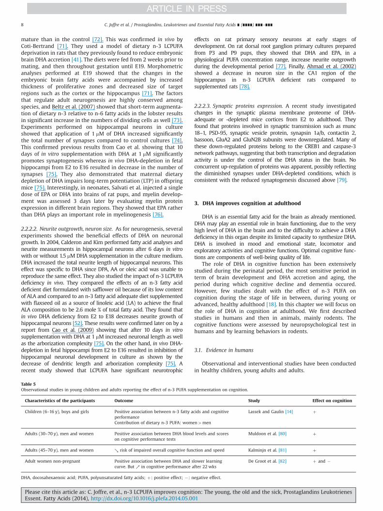
Table 5Observational studies in young children and adults reporting the effect of n-3 PUFA supplementation on cognition.
Characteristics of the participants Outcome Study Effect on cognition
Children (6–16 y), boys and girls Positive association between n-3 fatty acids and cognitiveperformanceContribution of dietary n-3 PUFA: women4men
Lassek and Gaulin [14] þ
Adults (30–70 y), men and women Positive association between DHA blood levels and scoreson cognitive performance tests
Muldoon et al. [80] þ
Adults (45–70 y), men and women ↘ risk of impaired overall cognitive function and speed Kalminjn et al. [81] þ
Adult women non-pregnant Positive association between DHA and slower learningcurve. But ↗ in cognitive performance after 22 wks
De Groot et al. [82] þ and �
DHA, docosahexaenoic acid; PUFA, polyunsaturated fatty acids; þ: positive effect; �: negative effect.
C. Joffre et al. / Prostaglandins, Leukotrienes and Essential Fatty Acids ∎ (∎∎∎∎) ∎∎∎–∎∎∎8
Please cite this article as: C. Joffre, et al., n-3 LCPUFA improves cognition: The young, the old and the sick, Prostaglandins LeukotrienesEssent. Fatty Acids (2014), http://dx.doi.org/10.1016/j.plefa.2014.05.001i

3.1.1. Observational studies (Table 5)Associations between cognitive performance and fatty acid
status were based on cognitive measures and either plasma fattyacid analysis or food frequency questionnaire associated to fooddatabase. Lassek and Gaulin (2011) found that dietary n-3 fattyacids are positively related to cognitive performance in children 6–16 year of age [14]. The contribution of dietary n-3 PUFA tocognitive performance is much greater in females than in males.In adults 30–70 years, DHA was related to cognitive functioning, withhigher blood levels associated with better scores on tests of nonverbalreasoning, mental flexibility, working memory and vocabulary [80]and with decreased risk of impaired overall cognitive function andspeed [81]. In contrast, de Groot et al. [82] reported a slower learningcurve on general speed information processing although there was asignificant increase in cognitive performance over 22 weeks in 54non-pregnant women [82].
These inconsistencies may be due to a different demographicbackground or to differences in evaluating the DHA status andrevealed the complexity of observational studies.
3.1.2. Interventional studies (Table 6)In most interventional studies undertaken in adults, DHA was
provided as capsules. Studies have provided mixed results regard-ing the effect of DHA intake on cognitive performance in adults.They showed either improved cognitive function or no beneficialeffects.
In a recent randomized clinical trial, Stonehouse et al. [15]showed that a DHA supplementation (1.16 g DHAþ0.17 g EPA/d)for 6 months improved memory and reaction time of memoryassessed with the Computerized Mental Performance AssessmentSystem in healthy young adults (mean age 33 years old) whosehabitual diet was low in DHA [15]. DHA improved the reactiontime of episodic memory and this response was modulated bygender. In women, it was the accuracy of episodic memory thatwas improved whereas in men it was the reaction time of workingmemory that was ameliorated. This study points out the impor-tance of considering gender differences in the relation between
DHA and cognition. In 2005, Fontani et al. used also a mixed n-3PUFA supplementation but with twice more EPA than DHA (1.6 gEPAþ0.8 g DHA) for 35 days in healthy subjects aged 22–51 years(males and females) [83]. They found an improvement of attentionand physiological functions (vigor, anger, anxiety, fatigue, depres-sion, and confusion). The possible mechanisms involved may berelated to the influence of the n-3 PUFA on neuronal excitability.Conducting a neuroimaging study, McNamara et al. (2010) aimedat determining the effects of DHA supplementation (400 or1200 mg/d) for 8 weeks on functional cortical activity duringsustained attention in healthy boys aged 8–10 years [84]. Thislow- or high-dose DHA significantly increased functional activa-tion in the prefrontal cortex during performance of an attentiontask (identical-pairs version of the continuous performance task)compared with placebo. Two years later, Jackson et al. [85] found thata supplementation with DHA-rich fish oil (1 g) for 12 weeks resultedin a significant increase in the concentrations of oxygenated hemo-globin, indicative of increased cerebral blood flow during the cognitivetasks [86].
In contrast to the beneficial effects of n-3 PUFA described, otherstudies have shown limited or no positive effects of DHA supple-mentation on cognitive performance, either in children aged 10–12years supplemented with 400 or 1000 mg/d DHA for 8 weeks [87], norcollege-aged adults (20 years old) supplemented with 480 mg DHAþ720mg EPA/d for 4 weeks [88], nor healthy young adults (18–35years old) supplemented with 1 g EPA-rich fish oil for 12 weeksalthough this supplementation may reduce subjective mental fatigueat times of high cognitive demand [85], nor in healthy adults aged 45–77 years supplemented with 252mg DHAþ60mg EPA/d for 90 days[89].
These differences may be due to the daily dose used, thelengths of intervention, the source of n-3 PUFA (mainly EPA ormainly DHA), the dietary n-3 PUFA history of the patients and thecognitive function assessed. As recently discussed by Dangour andAllen, the relevance of the cognitive tests used need to be betteraddressed [90]. In addition, larger studies with a clear definition ofprimary endpoints would help to decipher the beneficial effect ofn-3 LCPUFA on memory.
Table 6Interventional studies in young children and adults reporting the effect of n-3 PUFA supplementation on cognition.
Characteristics of the participants Experimental design Outcome Study Effect oncognition
Adults (33 y)Women and men
DHA supplementation (1.16 g DHAþ0.17 gEPA/d) for 6 months
Women: improvement of the accuracy ofepisodic memory
Stonehouseet al. [15]
þ
Low dietary intake of LC n-3PUFA (o200 mg EPAþDHA/wk)
Men: improvement of the reaction time ofepisodic memory
Adults (22–51 y)Women and men
EPA and DHA supplementation 1.6 g and0.8 g/d, respectively, for 35d
Improvement of attention and physiological functions(vigor, anxiety, fatigue, depression, confusion)
Fontaniet al. [83]
þ
Children (8–10 y), boys DHA supplementation (400 or 1200 mg/d)for 8 weeks (DHA)
Positive correlation between erythrocyte DHAcomposition and prefrontal cortex activation
McNamaraet al. [84]
�
Negative correlation between erythrocyte DHAcomposition and reaction time
Adults (18–35 y)Women and men
DHA-rich fish oil (1 g) for 12 weeks(DHA/EPA 5:1)
No positive effects on cognitive performance Jacksonet al. [85,86]
No
Children (10–12y), girls and boys DHA supplementation (400 or 1000 mg/d)for 8 weeks
No positive effects on cognitive performance Kennedyet al. [87]
No
College-aged adults (20 y),Women and men
DHA and EPA supplementation (480 mgand 720 mg/d, respectively, for 4 weeks)
No positive effects on cognitive performance Karret al. [88]
No
Adults (45–77 y) DHA and EPA supplementation 252 mgand 60 mg/d, respectively, for 90d
No positive effects on cognitive performance Stoughet al. [89]
No
DHA, docosahexaenoic acid; EPA, eicosapentaenoic acid; no: no effect; þ: positive effect; �: negative effect.
C. Joffre et al. / Prostaglandins, Leukotrienes and Essential Fatty Acids ∎ (∎∎∎∎) ∎∎∎–∎∎∎ 9
Please cite this article as: C. Joffre, et al., n-3 LCPUFA improves cognition: The young, the old and the sick, Prostaglandins LeukotrienesEssent. Fatty Acids (2014), http://dx.doi.org/10.1016/j.plefa.2014.05.001i

Table 7Effect of n-3 PUFA supplementation on cognition in adult rodents.
Species Test Experimental design Outcome Study Effect oncognition
Long Evansrats
Morris water maze Depletion of n-3 PUFA over 3 generations. Maleoffsprings resupplemented at birth or at weaning or at 7weeks with DHA (0.13%). Behavioral testing at 9 and 13 weeks (DHA).
- Impairment of the Morris water maze learning andretention with the n-3 PUFA deficient diet.
- Restored performance with n-3 adequate diet atbirth and weaning.
- When repletion occurred at 7 weeks, restoredperformance at 13 weeks only.
Moriguchi andSalem [67]
þ
Wistar rats Radial arm maze Depletion of n-3 PUFA over 3 generations. Male offspringsresupplemented at 5-weeks with DHA (300 mg/kg/day) for 10 weeks (DHA).
Improvement of the reference but not workingmemory errors in the maze
Gamoh et al. [92] þ
Sprague-Dawley
Morris water maze 2-month old male rats fed a diet with appropriate (150-300 mg/kg) or high doses(600 mg/kg) DHA for 1 month (DHA).
Improvement of the spatial learning performanceand retention with the appropriate dose. Impairment of theperformance with the higher dose
Pan et al. [93] þ
Crj:CD-1mice
Maze 3-week old male mice fed a diet supplemented with ethyl-DHA (2%) for 5 months Better performance Lim and Suzuki[99]
þ
Wistar rats Morris water maze Male adult rats fed a diet supplemented with ethyl-EPA (0.2 or 1%) for 8 weeks - No improvement of water maze performance- Improvement of an Il-1-induced impairment of memory
Song andHorrobin [94]
þ
Sprague-Dawleyrats
Morris water maze 2-month old rats fed a diet supplemented with EPA (1%) for 7 weeks. - No improvement of water maze performance- Improvement of an Il-1-induced impairment of memory
Song et al. [95] þ
Wistar rats Morris water maze 3-4 months rats fed a diet supplemented with ethyl-EPA or DPA (200 mg/kg/day)for 8 weeks
No improvement in memory performance Kelly et al. [97] no
C57Bl6 mice Morris water maze 6-week old male mice fed a diet supplemented with ethyl-EPA (1%) for 12 weeks. No improvement in spatial learning or retention compared tocontrol.
Luchtman et al.[98]
no
Swiss OF1mice
Morris water maze;passive avoidance
7-week female mice fed a diet supplemented with phospholipid-DHA (0.05%) for2 months
No improvement in memory or passive avoidance performance Carrié et al. [3] no
DHA, docosahexaenoic acid; DPA, docosapentaenoic acid; EPA, eicosapentaenoic acid; no: no effect; PUFA, polyunsaturated fatty acids; þ: positive effect.
C.Joffreet
al./Prostaglandins,Leukotrienes
andEssential
FattyAcids
∎(∎∎∎∎)
∎∎∎–∎∎∎
10Pleasecite
this
articleas:
C.Joffre,et
al.,n-3
LCPU
FAim
proves
cognition
:Th
eyou
ng,th
eold
andthesick,Prostaglan
dinsLeu
kotrienes
Essent.Fatty
Acid
s(2014),h
ttp://d
x.doi.org/10.1016/j.p
lefa.2014.05.001i

3.2. Evidence in animals (Table 7)
Lots of studies dealing with DHA and cognition conducted inanimals started the n-3 PUFA deficiency or supplementationduring the perinatal period and observed the effects at adulthood(see the previous paragraph). Few studies dealt with the relationbetween DHA and cognition with deficiency or supplementationstarted at adulthood. In this part, we considered only the studieswhose deficiency or supplementation started in young adult- and
adult- animals. Moreover we focused on animals, mainly rodents,without any pathology. Studies dealt with the effect of DHA eitheron n-3 PUFA deficient diet animals or on adequate n-3 dietanimals. In young and adult rodents submitted to an n-3 PUFAdeficient diet, DHA was decreased in the brain and associated withan increase in DPA n-6 [4,91]. This loss of DHA was independent ofage whereas the increase in DPA n-6 was faster in young animalsdue to higher desaturase activities [4]. Cognitive assessments wereperformed with the same tests than those described in the
Table 8n-3 long chain polyunsaturated fatty acids in animal models of aging.
Species of rodents and age Treatment and length Context Brainregions
Outcomes Study Effect oncognition
Mice, 3 months and 22 months 10% EPA and 7% DHA,2 months,Aging, inflammation and cognition
Hip andBrain
↑n-3 LCPUFA↓cytokines expression in old miceChanges in astrocytes morphology in old mice↑spatial memory in old mice
Labrousseet al. [12]
þ
Rats, 100 weeks Fish oil-deficient diet (3 generations),DHA-EE 300 mg/Kg/day,5 weeks,Aging and cognition
Hip ↓reference memory and workingmemory errors↓LPO
Gamoh et al.[121]
þ
Mice, young (7–11 weeks),Mature (9–10 months), old(17–19 months)
Fish oil diet (DHA 9.4 g/Kg diet).Palm oil diet (DHA 0 g/Kg diet),whole life,Aging and cognition
↑exploratory and locomotor activity inyoung miceNo change in mature and old mice↓locomotor activity in old mice and↑inyoug mice↑performance only on the probe trial of theMorris water maze in mature mice
Carrié et al.[3]
þ
Mice, 14 months DHA and EPA 1.14 mmol/Kg/ day vs ALA 1.5 mmol/Kg/day
ECneurons
↑DHA, ↓AA and ↑cellular capacitance withDHA diet,Membrane hyperpolarisation and ↑sEPSC,↑syntaxin-3 and translocation ratio of drebrin
Arsenaultet al. [147]
þ
Rat, birth, 3 weeks and7 weeks
n-3 adequate diet (n-3 Adq, 2.6% ALAþ1.3% DHA) or n-3 deficient diet (n-3 Def),3 generations of n-3 Def diet and n-3 Adq for 2, 6 or9 weeks, Aging
Halfbrain
↓DHA and ↑DPA n-6 with n-3 Def,Fatty acid profile of n-3 Adq diet at birth En-3 Adq reference group,↑DHA and ↓DPA n-6 with n-3 Adq,% DHA of 3 weeks of age4of 7 weeks of age,↑time to reach the platform in Morris watermaze with n-3 Def,↑time for 7 weeks of age to restore brain DHA,Nearly full recovery on the probe trial andspatial learning task after 6 weeks of n-3 Adqdiet of 7 weeks mice
Moriguchiand Salem[67]
þ
Rat, 3–4 months and 20–22months
E-EPA or DPA (200 mg/Kg/day),2 weeks,Aging, inflammation and cognition
Cx, Hip E-EPA ↑cortical DPA and DHA,↓microglial activation,↓coupled activation of sphingomyelinase andcaspase 3,↑spatial learning and long term potentiation
Kelly et al.[97]
þ
Mice, 4 months and 20 months DHA 0.9 g/Kg/day[14C]–DHA, [14C]–EPA,Aging and BBB
Wholebrain
DHA and EPA cross the BBB by diffusion,n-3 LC-PUFA deprivation increase brain DHAuptake in old mice
Ouellet et al.[131]
n.d.
Rats, 2 months and 2 years 11% DHA, 2.8% EPA,1 month,Aging
Wholebrain
↑DHA,↓AA,No difference in Morris water maze test
Barcelo-Coblijn Get al. [122]
no
AA, arachidonic acid; Adq, adequate; ALA, alpha-linolenic acid; BBB, blood-brain barrier; Cx, cortex; CxFr, cortex frontal; Def, deficient; DHA, docosahexaenoic acid; DHA-EE,DHA ethyl ester; DPA, docosapentaenoic acid; EC, enthorinal cortex; EPA, eicosapentaenoic acid; Hip, hippocampus; IL-6, interleukin-6; LCPUFA, long chain PUFA; LPO, lipidperoxide; n.d.: not determined; no: no effect; PUFA, polyunsaturated fatty acid; vs, versus; þ: positive effect; �: negative effect.
C. Joffre et al. / Prostaglandins, Leukotrienes and Essential Fatty Acids ∎ (∎∎∎∎) ∎∎∎–∎∎∎ 11
Please cite this article as: C. Joffre, et al., n-3 LCPUFA improves cognition: The young, the old and the sick, Prostaglandins LeukotrienesEssent. Fatty Acids (2014), http://dx.doi.org/10.1016/j.plefa.2014.05.001i

Table 9n-3 long chain polyunsaturated fatty acids in animal models of Alzheimer's disease.
Species of rodents andage
Treatment and length Context Brain regions Outcomes Study Effect oncognition
Rats, 20 weeks Ethyl-DHA 300 mg/Kg/day,7 weeks,Alzheimer's disease
Cx ↓Aβ,↓cholesterol,↓reference memory error
Hashimoto et al.[140]
þ
Rats, 25 weeks Ethyl-DHA 300 mg/Kg/day,12 weeks,Alzheimer's disease
Cx, Hip ↓avoidance learning ability,↑DHA/AA ratio,↓neuronal apoptotic products,↓oxidation
Hashimoto et al.[142]
þ
Rats, 20 weeks Ethyl-DHA 300 mg/Kg/day,12 weeks,Alzheimer's disease
Cx, Hip ↑DHA/AA ratio,↓reference and working memory errors
Hashimoto et al.[143]
þ
Mice, 8 months and 15months
DHA 3.5 g/Kg diet, 6 or13 months,Alzheimer's disease
CxFr, Cx, Hip, Acg No change in rCBV in 8 months old mice,↓Aβ in 15 months old mice,↑spatial memory in 15 months old mice,↑rCBV in 15 months old mice
Hooijmans et al.[145]
þ
Mice, 6 months DHA 0.4%, EPA 0.4%,3-4 months,Alzheimer's disease
Hip, CxFr, Cx andCer
↓Aβ,↓activated microglia,↑exploration activity,No change at spatial learning in Morris water maze
Oksman et al.[146]
þ
Mice, 12–14 months DHA 0.6 g/Kg/day (DHA/EPA 4:1),8–10 months,Alzheimer's disease
EC neurone, CxFr,Cx
↑DHA and ↓AA,↑object recognition,↓seizure-like akinetic episodes,↑cell capacitance,↓firing rate versus injected current
Arsenault et al.[147]
þ
Rats, 25 weeks Ethyl-DHA 300 mg/Kg/day,12 weeks,Alzheimer's disease
Cx ↑membrane lateral and rotational fluidity,↑DHA,↓cholesterol and lipid peroxidation
Hashimoto et al.[141]
n.d.
Mice, 17 months DHA 0.6 % ,10375 days,Alzheimer's disease
CxFr, Cx and hemibrain
↑drebrin,↓oxidation,↓caspase-cleaved actin,↑antiapoptotic BAD phosphorylation
Calon et al. [144] n.d.
Mice, 17 and 19 months DHA 0% or 0.09% or 0.6% Cx, Hip, parietal Cx ↓Aβ and Aβ42,↓Aβ plaques,↓α- and β-APP C-terminal fragment
Lim et al. [64] n.d.
Mice, 3 months DHA 0% or 0.6%,3 months,Alzheimer's disease
Cx, HipV, Str, Hip,liver
↑DHA and ↓AA,↓Aβ plaques,↑drebrin
Perez et al. [149] n.d.
Mice, 3 months DHA 1.3 g/100 g diet and DPA n-60.5 g/100 g diet,3, 6 or 9 months,Alzheimer's disease
Whole brain ↓intraneuronal Aβ and Tau,↓PS1,DPA n-6 ↓early-stage phospho-tau and↓of phosphorylated c-junN-terminal kinase
Green et al. [150] n.d.
C.Joffreet
al./Prostaglandins,Leukotrienes
andEssential
FattyAcids
∎(∎∎∎∎)
∎∎∎–∎∎∎
12Pleasecite
this
articleas:
C.Joffre,et
al.,n-3
LCPU
FAim
proves
cognition
:Th
eyou
ng,th
eold
andthesick,Prostaglan
dinsLeu
kotrienes
Essent.Fatty
Acid
s(2014),h
ttp://d
x.doi.org/10.1016/j.p
lefa.2014.05.001i

previous paragraph concerning the perinatal period. Moriguchiand Salem (2003) studied the reversibility of an n-3 PUFAdeficiency induced on 3 generations of rats with supplementationof DHA at different ages: birth, 3 and 7 weeks of age [67]. Theyfound that brain function can recover from a severe and extendedDHA deficiency by a dietary supply with DHA. The performance inspatial tasks was closely related to the level of brain DHA. Youngeranimals (3 weeks of age) restored brain DHA at a faster rate thanolder ones (7 weeks of age), when brain formation is ongoing. Thenormalization of brain DHA induced the total recovery of theperformance as compared to the n-3 adequate group (containing aLA/ALA ratio of 6.2 and 1.3% DHA) although the recovery in brainDHA was not total for the rats supplemented at 3 weeks (93%) andat 7 weeks (60%). No critical period in development was observedin this experiment during which DHA must be supplied. However,an earlier intervention appears to be more effective in restoringbrain DHA than a later one.
The same result was obtained in the study of Gamoh et al. [92]who worked on rats fed with a fish oil deficient diet (withoutDHA) instead of an n-3 PUFA deficient diet for 3 generations [92].The authors failed to mention the fatty acid composition of thedifferent diets they used. However, perorally administration ofDHA (300 mg/kg/day) to these rats caused a significant increase inDHA in the cerebral cortex and hippocampus but not in thecerebellum and brain stem. This increase is associated to theimprovement of the reference memory-related learning abilitywithout affecting working memory.
Only one study tested a supplementation of DHA on rats fed anadequate n-3 PUFA diet. Here again the authors did not disclosethe fatty acid composition of the adequate n-3 PUFA diet. Theyshowed that supplementation with DHA (150 and 300 mg/kg)significantly improved learning and memory in the Morriswater maze task but a higher dose of DHA (600 mg/kg) increasedthe risk of memory impairment [93]. Hence DHA deficiency orexcess (600 mg/kg) might impair spatial learning and memoryfunction.
The effect of a diet supplemented with pure EPA or DHA (asethyl-EPA, E-EPA or ethyl-DHA, E-DHA) was compared to the oneof an n-3 PUFA deficient diet on memory impairment. The chronicadministration of E-EPA, the precursor of DHA, for 5–7 weeks didnot significantly enhance memory in rats [94–96]. However, itattenuated the effects of intracerebroventricular injection ofinterleukin-1β (15 ng/10 mL/rat) on spatial memory by suppressinginflammation and reducing the decrease of acetylcholine release.Luchtman et al. (2012) found in mice that the E-EPA diet onlyincreased tissue EPA and DPA levels and not DHA whereas Kellyet al. (2011) found in rats that E-EPA administration increasedcortical DPA and DHA [97,98]. Thus, it is not clear whether EPA actsthrough a higher DHA incorporation in the brain or not. None-theless, E-EPA by itself can affect inflammation-induced cognitiveimpairment and normalize neurotransmitter release.
The beneficial effects of DHA on cognition were counterbalancedby few studies reporting no effect of DHA supplementation (started at3 weeks of age for 5 months) in young mice as shown by Lim andSuzuki [99]. They only found an enhancement in maze-learning abilityin old mice. The study of Carrie et al. (2000) examined the effects of asupplementation at 7 weeks of age with phospholipids rich in n-3PUFA for 2 months on behavior, learning, and phospholipid fatty acidcomposition of brain regions in n-3 PUFA deficient mice (deficiencyinduced in mums 3 weeks before mating) [3]. They did not find anydifferences between n-3 PUFA deficient diet, control diet and dietssupplemented phospholipids rich in DHA on Morris water maze orpassive avoidance although the brain DHA levels were different in thedeficient group and the others.
The discrepancies in the efficiency of DHA supplementation oncognitive deficit may be explained by the brain DHA levels before the
Mice,
12an
d20
mon
ths
n-6/n-3
¼25
(4.6
Kcalories/g
diet),fat-1
tran
sgen
e,W
holelife,
Alzhe
imer'sdisease
Cx,
CxF
r↑n
-3/n-6
ratioan
dDHAat
20mon
ths,
↓solubleAβ4
2at
20mon
ths,
↓solublean
dinsolublephosphorylated
tauat
20mon
ths,
↓CaM
KIIan
dGFA
Pat
20mon
ths
Lebb
adiet
al.
[151
]n.d.
Mice,
17mon
ths
DHA0.6%
,3-5
mon
ths,
Alzhe
imer'sdisease
Cx,
Hip
↑NMDAreceptor
subn
it(N
R2A
andNR2B
),↑C
aMKII,
↓caspase/calpainactivity
Calon
etal.[16
1]n.d.
Mice,
13mon
ths
DHA0g/Kgdietan
dn-3/n-6
¼0.01
,9mon
ths,
Alzhe
imer'sdisease
CxF
r,Cx
↓n-3/n-6
ratio,
↑insolubleAβ4
0an
dβ4
2,↑insolublean
dsolubletau,
↓drebrin
Julie
net
al.[16
2]n.d.
Mice,
4an
d12
-13
mon
ths
Youngvs
oldmice,
Alzhe
imer'sdisease
CxF
r,Cx,
Hip
DHAin
brainyo
ung4
oldmice,
NPD
1in
brainyo
ung4
oldmice,
↓inflam
matorysign
aling,
amyloidog
enic
APP
clea
vage
andap
optosis
Zhao
etal.[174]
n.d.
AA,a
rach
idon
icacid;Aβ,
β-am
yloid;Acg
,anterior
cingu
late
gyrus;
APP
,amyloidprotein
precu
rsor;CaM
KII,c
alcium/calmod
ulin
-dep
enden
tprotein
kinaseII;Cer,cerbe
llum;Cx,
cortex
;CxF
r,co
rtex
fron
tal;DHA,d
ocosah
exae
noic
acid;DPA
,doc
osap
entaen
oicacid;EC
,enthorinal
cortex
;Hip,h
ippoc
ampus;
HipV,v
entral
hippoc
ampus;
GFA
P,glialfibrillaryacidic
protein;n.d.:,not
determined
;NMDA,N
-methy
l-D-asp
artate;PS
1,presenilin1;
rCBV
,relative
cerebral
bloo
dvo
lume;
Str,striatum
;þþ:positiveeffect.
C. Joffre et al. / Prostaglandins, Leukotrienes and Essential Fatty Acids ∎ (∎∎∎∎) ∎∎∎–∎∎∎ 13
Please cite this article as: C. Joffre, et al., n-3 LCPUFA improves cognition: The young, the old and the sick, Prostaglandins LeukotrienesEssent. Fatty Acids (2014), http://dx.doi.org/10.1016/j.plefa.2014.05.001i

supplementation. Indeed, the “dietary history” of the animals understudy is very important: adequate diets, n-3 PUFA deficient orsupplemented diets, time at which the specific diet is started, theduration of the specific diet. The age of the animals when the cognitivetests were performed may also have an importance. We should alsokeep in mind that other factors like emotionality and attention,affected by brain DHA levels, could interfere with the cognitive testson memory and leaning.
4. DHA improves cognition: clues from animal models ofcognitive impairment
4.1. Cognitive decline in normal aging: effect of n-3 LCPUFA (Table 8)
Normal aging is associated with a decline in cognitive functionsincluding memory [10,100]. Memory loss is a prominent healthconcern in older individuals either due to “normal” aging orunderlying pathological processes such as vascular dementia orneurodegenerative diseases. The most important of such diseasesis Alzheimer's disease (AD), which affects over 24 millions peopleworldwide [101]. The undergoing pathological process in AD isdifferent from normal aging, in part because it is much faster andwidespread. Thus, much effort has been devoted to identify earlyAD stages, such as mild cognitive impairment, to make a cleardistinction with normal aging [102–104].
Epidemiological studies have pinpointed an associationbetween consumption of n-3 LCPUFA with slower cognitivedecline in elderly individuals without dementia [105–108]. Forexample, an association was found between red blood cell n-3LCPUFA and a decreased risk of white matter hyperintensities andsmaller brain volume in old study participants [109]. RCTs per-formed in cohort of old individuals were less conclusive. Tworelatively large studies with 302 and 867 healthy adults, respec-tively, detected no change in cognitive function scores aftersupplementation with supplements combining EPA and DHA[107,110]. In contrast, results from a RCT involving 485 healthysubjects showed that a treatment with pure DHA improvesepisodic memory and learning [108]. Memory enhancing effectof n-3 LCPUFA in non demented older subjects was also shown in2 smaller RCTs [111,112]. One of these smaller study (n¼30) alsoreports improved beneficial effects of n-3 LCPUFA on magneticresonance imaging (MRI)-determined brain structural endpointsin old subjects, such as white matter integrity and gray mattervolume [112]. Finally, independent RCTs performed in individualswith mild cognitive dysfunction adds support to the postulate thatn-3 LCPUFA exert a benefit on cognition [113–116]. However,another RCT detected effects of n-3 LCPUFA mostly limited toimprovement of depressive symptoms [117]. The apparent conflictbetween the results of these RCT may stem from variables such ascognitive reserve, dietary habits, pre-study n-3 LCPUFA status, thetype of LCPUFA supplement given, and population heterogeneity[68,106], and underscore the challenges of performing clinicaltrials in this population [104,118]. However, selection of the mostrelevant primary endpoints in cognitive studies remains a chal-lenge [90]. Larger studies with clearly defined a priori outcomesare needed before using the results of published study for healthrecommendation.
The effects of n-3 LCPUFA treatments were also assessed inanimal models [12,119]. The study of Labrousse et al. (2012)indicated that diet-induced accumulation of EPA and DHA in thebrain protects against neuroinflammation and cognitive impair-ment linked to aging in an animal model, further reinforcing theidea that increased EPA and DHA intake may provide protection tothe brain of aged subjects [12]. Consistent ameliorating effects oncognition or neuroinflammatory endpoints following n-3 LCPUFA
supplementation on age-related cognitive deficits are reported inold rodents [97,120,121], although not confirmed by other inves-tigators [122,123].
4.2. Alzheimer's disease n-3 LCPUFA: animal models
In the past 20 years, animal models have been generated topinpoint how the neuropathology of brain disorders like AD canlead to readily measurable cognitive deficits. The most widely usedstrategy is to transgenically express in the brain of mice mutantgenes known to cause AD in humans [124,125]. Although none ofthese mice develop the frank neuronal or synapse losses pathog-nomonic of AD, they exhibit various behavioral anomalies remi-niscent of AD symptoms, as well as an extensive AD-likeneuropathology. Among the 20 autosomal-dominant amyloidprecursor protein (APP) mutations, the Swedish mutation facil-itating APP cleavage near the β-secretase site was the first to beused to generate a transgenic model of AD (Tg2576) [126]. Suchstrategy was successful to generate series of transgenic micebearing APP mutation(s) and developing amyloid plaques andcognitive deficits [124,125]. In addition, transgenes have beenadded to recreate neurofibrillary tangles, another neuropathologi-cal hallmark of AD [127]. One example is the now widely used3xTg-AD mouse which develops both plaques and tangles [128].While these transgenic mice fail to replicate the full neurodegen-erative spectrum of human diseases, they are extensively used tomodel the preclinical phases of AD. They also offer an excellentopportunity to study the impact of nutraceutical interventions.
Indeed, in the past 10 years, preclinical trials in transgenic ADmice have been performed with controlled diets and designed toassess specific behavioral and molecular endpoints. It was longknown that fatty acids (especially DHA) ingested through the dietdirectly impact upon concentrations of corresponding fatty acids inthe cerebral tissue [4,68,129,130]. Indeed, circulating DHA readilycrosses the blood-brain barrier to enter the brain [131], where itdisplays a very slow turnover rate [132,133]. Therefore, dietarymanipulation provides a direct means of studying the effect of fattyacids on brain function, and n-3 LCPUFA have accordingly been thesubject of series of investigations on their impact on cognition andAD-like neuropathology in animal models [68,134–136].
There is a general consensus on beneficial effects of n-3 LCPUFAon cognition in animal models of AD (Table 9). The first series ofevidence come from the work of Hashimoto et al. using ratintracerebrally infused with Aβ40 to quickly recreate AD neuro-degeneration [137]. Although this model relies on a direct neuro-toxic effect of Aβ monomers, which is of debatable AD relevance[138,139], the data generated clearly support improving effects ofdietary DHA on memory and learning ability [140–143]. Then,using the Tg2576 animal model of AD in the Morris water mazeparadigm, DHA was shown to protect against cognitive impair-ment caused by an APP transgene bearing the Swedish mutation[144]. DHA intervention studies were also performed in APPswe/PS1dE9 mice, which in addition to the Swedish mutant APPexpress a presenilin variant known to potentiate Aβ plaquedeposition. Memory improvement after exposure to DHA-enriched diets was detected in old but not young APPswe/PS1dE9mice, again using the Morris water maze paradigm [145,146].More recently, it was found that long term DHA treatmentimproved object recognition and reduced akinetic periods in 12-month-old 3xTg-AD mice [147].
The main advantage of investigations with animal models is toprovide insights on the direct relationship between cognitivebehavior and underlying molecular mechanisms. Aβ pathologyremains the most studied neuropathological hallmark of AD and ithas been shown to be sensitive to brain DHA levels. Decreases inbrain Aβ load after DHA treatment have been reported in at least
C. Joffre et al. / Prostaglandins, Leukotrienes and Essential Fatty Acids ∎ (∎∎∎∎) ∎∎∎–∎∎∎14
Please cite this article as: C. Joffre, et al., n-3 LCPUFA improves cognition: The young, the old and the sick, Prostaglandins LeukotrienesEssent. Fatty Acids (2014), http://dx.doi.org/10.1016/j.plefa.2014.05.001i

Table 10n-3 long chain polyunsaturated fatty acids and miscellaneous brain and synaptic markers in normal animals.
Species of rodents andage
Treatment and length Context Brain regions Outcomes Study
Mice, 18 days DHA 0.9%,At 2 days of pregnancy to after lactation period,Molecular signaling
Hip ↑Neurite growth, synaptogenesis, synapsin, glutamate receptor expression and glutamatergicsynaptic function
Cao et al. [75]
Rats, 2 months DHA 1.25% (DHA/EPA 5:1),12 days,Cognition and molecular signaling
Hip, whole brain ↑syntaxine-3, ↑NR2B, ↑learning in Morris water maze Chytrova et al. [166]
Rats, 110 days DHA 0.3%,Whole life,Neurophysiology
Hip ↑neurons size Ahmad et al. [78]
Rats, 90 days DHA 18% and EPA 12%,Whole life,Neurotransmission
Hip, Cx ↑BDNF,↑5-HT of 90 day-old rats
Vines et al. [179]
Mice, 5-6 months DHA 0.7g/Kg/ day (DHA/EPA 4:1),3 months,Inflammation
Whole brain ↓microglial activation,↓ischemic lesion size,↑Bcl-2 and ↓Cox2 and IL-1β
Lalancette-Hébert et al.[176]
Rats, 6-7 weeks DHA 1.25% (DHA/EPA 4:1),12 days,Neurotransmission
Hip ↑spatial learning, ↑BDNF and synapsin 1, ↓oxydation, ↑Akt and CaMKII Wu et al. [180]
Mice, 8 weeks Diet rich in LA and ALA (n-3/n-6) or diet rich inLA (n-6),Gestation to 8 weeks, Inflammation
Cx (in PC and PE), Hip ↓DHA and↑DPA n-6 in PC and PE, No difference for AA, ↑social explorationwith n-6 diet, ↑IL-6 with n-6 diet in Hip, ↑expression of IL-6 mRNA with timewith n-6 diet in Hip, ↓LPS-induced STAT1, STAT3 and JAK2 activation with n-6 diet in Hip
Mingam et al. [91]
Rats, 5 weeks Fish oil deficient diet for 3 generations,DHA 300 mg/Kg/day or vehicule,Over 10 weeks
Cx, Hip, Cer and brainstem
↑DHA in Cx and Hip,↑reference memory-related learning ability
Gamoh et al. [92]
Rats, 2 months DHA 150 and 300 mg/Kg/day,DHA 600 mg/Kg/day,1 month,Cognition
Primary Hip, neuronalculture
↑learing and memory with 150 and 300 mg/Kg/d,↑ the risk of memory impairment with 600 mg/Kg/d,Upregulation of CB1 and TRPV1 with a dose-dependence
Pan et al. [93]
Rats, 5 weeks Coconut oil or soybean oil diet or coconut oil dietenriched with E-EPA (0, 0.2 and 1%),8 weeks,Inflammation and cognition
Hip E-EPA attenuates memory impairment induced by IL-1β Song and Horrobin [94]
Rats, 2 months 1% EPA,7 weeks,Inflammation and cognition
Hip, Amy, HipTha andPVN
E-EPA improved spatial memory,↓CRF expression and corticosterone secretion,↑NGF expression
Song et al. [95]
Rats, 5 weeks Pure E-EPA,Injection of IL-1β (15 ng/10μL/rat),7 weeks,Inflammation and cognition
Hip E-EPA improved memory,↑Ach release,↑NGF and ↓IL-1β expression
Taepavarapruk and Song[96]
5-HT, serotonin receptor; AA, arachidonic acid; Ach, acetylcholine; Akt, kinase; ALA, alpha-linolenic acid; Amyg, amygdale; Bcl-2, antiapoptotic molecule; BDNF, brain-derived neurotrophic factor; CaMKII, calcium/calmodulin-dependent protein kinase II; CB1, cannabinoid receptor 1; Cer, cerbellum; COX2, cyclo-oxygenase 2; CRF, corticotrophin-releasing hormone; Cx, cortex; DHA, docosahexaenoic acid; DPA, docosapentaenoic acid; EPA,eicosapentaenoic acid; E-EPA, ethyl EPA; Jak, Janus kinase; Hip, hippocampus; HipTha, hippocampe thalamus; IL-1β, interleukin-1β; LA, linoleic acid; LPS, lipopolysaccharides; NGF, nerve growth factor; PC, phosphatidylcholine; PE,phosphatidylethanolamine; PVN, paraventricular nucleus; STAT, Signal Transducers and Activators of Transcription; Str, striatum; TRPV, Transient Receptor Potential Vanilloid.
C.Joffreet
al./Prostaglandins,Leukotrienes
andEssential
FattyAcids
∎(∎∎∎∎)
∎∎∎–∎∎∎
15
Pleasecite
this
articleas:
C.Joffre,et
al.,n-3
LCPU
FAim
proves
cognition
:Th
eyou
ng,th
eold
andthesick,Prostaglan
dinsLeu
kotrienes
Essent.Fatty
Acid
s(2014),h
ttp://d
x.doi.org/10.1016/j.p
lefa.2014.05.001i

4 studies in APP transgenic mice [145,146,148,149], albeit to alesser extent in the 3xTg-AD model [147,150]. Tau-laden tanglesare equally important in AD but appear to respond less to DHAtreatment. Nonetheless, DHA-induced decreases in phosphory-lated soluble tau, with unchanged levels of insoluble tau, havebeen reported by 2 groups [147,150]. Finally, 3xTg-AD micegenetically altered to increase brain DHA levels using the fat-1transgene had less Aβ and tau pathologies [151], further under-scoring the positive impact of increasing brain DHA concentrationson AD neuropathology.
Preclinical intervention studies have perhaps shown even moreconvincingly a protective effect of n-3 LCPUFA on synaptic pro-teins. Postsynaptic dendrites and proteins located therein areaffected particularly early in the progression of AD. Losses ofpostsynaptic proteins such as drebrin (developmentally regulatedbrain protein), glutamate receptors and PSD95 have beendescribed in AD [144,152–160] and animal models [144,161,162].Deleterious effects of DHA deprivation on synaptic markers areremarkably striking in the Tg2576 mouse [144,161], but are alsodetected in the 3xTg-AD model when combined with a high-fatdiet [162], as well as in young mice [75] and old rats [163]. On theother hand, DHA exerts upregulating effects on various synapticproteins such as PSD-95, syntaxin-3 or drebrin in differenttransgenic models [144,149,161,164] and normal rodents[165,166]. Because accumulating data indicate that synapsedefects underlie clinical symptoms of AD, the enhancing effect ofDHA on these synaptic markers is likely to have clinical relevance.
Still, evidence of effects of LCPUFA on neuronal functionremains scarce. We recently reported that chronic consumptionof DHA alters basic electrophysiological properties of entorhinalcortex neurons, thereby correcting anomalies found in the 3xTg-AD mouse [147,164]. More specifically, treatment with DHA pre-vented the decrease in cellular capacitance observed in aged 3xTg-AD mice [147,164]. These electrophysiological effects cannot bereproduced with an equimolar dose of a precursor of DHA, ALA[164]. Since the capacitance of a given membrane surface area is aconstant value for all cells [167], cell capacitance directly reflectsthe total membrane surface [147,164]. DHA-induced enlargementof neurons is in agreement with previous reports of increasedhippocampal neuron size in DHA-treated animals, using cresylviolet staining and stereology [78]. Interestingly, both syntaxin-3and drebrin, two key synaptic markers, correlated well with cellcapacitance readouts [164]. Accordingly, DHA-induced neuriteoutgrowth and membrane expansion has been shown to dependon syntaxin-3 [168]. Such electrophysiological data combined withsynaptic marker assessment in mice altogether support thehypothesis that DHA incorporation in brain tissue can improvesynaptic integration and neuronal function.
Beside the aforementioned effects on AD markers, molecularstudies also reveal the pleiotropic action of n-3 LCPUFA in thebrain (Table 10). Indeed, DHA plays a critical role in regulatingmembrane fluidity [169], cell signaling [144,161,170], oxidativestress [144,171], gene expression [172], APP metabolism[148,173,174], and inflammatory cascades [12,175–177]. In addi-tion, we and others have recently shown that n-3 LCPUFA canstimulate the secretion of neurotrophic factors in the brain [178–180]. These last observations are of particular interest, as neurotrophicfactors remain the only compounds known to date to show neuror-estorative properties in vivo [181].
4.3. Alzheimer's disease and n-3 LCPUFA: clinical evidence
The clinical evidence supporting n-3 LCPUFA use as a supple-ment or in the food is generally considered stronger for cardio-vascular disease outcomes [182,183], including coronary heartdisease (CHD) [184], albeit still controversial [185]. The available
data has led to recommendation from the American Heart Asso-ciation (AHA) for higher intake of DHA and EPA. However, clinicalevidence for dementia is more difficult to gather, mainly due to theelusive nature and heterogeneity of neurodegenerative diseases,the lack of reliable surrogate markers, and rising evidence thatearly treatment is necessary [68,136]. On the positive side, it isestimated that an intervention delaying dementia onset by only afew years would result in a dramatic reduction in AD prevalence[186,187], suggesting that a treatment with a mild neuroprotectiveeffect can have a significant global health impact.
The question is whether n-3 LCPUFA can have such an impact.Numerous observational studies have highlighted a possible associa-tion between dietary intake of fish and n-3 LCPUFA and a lower risk ofdementia [188–191]. The results of these investigations also tell us thatthe origin (fish versus vegetable oil), dietary combination and ApoEstatus critically influence such an association [192–195]. Similarly, therelative impact of each n-3 LCPUFA subtype (ex EPA versus DHA)remains difficult to decipher [196,197]. Epidemiology and preclinicaldata have naturally provided an irresistible incentive to start clinicaltrials in patients with a diagnosis of AD, which are reviewed in detailselsewhere [68,136,188–191,198]. In short, RCTs do not lend muchsupport to a therapeutic role for n-3 LCPUFA supplementation in thetreatment of established AD [114,199]. In retrospect, however, it shouldhave been obvious that beneficial effects of n-3 LCPUFA could hardlybe expected when the neurodegenerative process is already wellengaged. Long and costly RCTs in preventive settings are clearlyrequired to determine whether DHA or other fatty acids can protectfrom age-related dementia or alter AD progression [68,200,201].
4.4. Conclusion
Despite drawbacks detailed above, preclinical studies in animalmodels of AD have taught us that DHA and other dietary fats canmodulate fatty acid concentration in cerebral tissue, and conse-quently key neuropathological markers of neurodegenerative dis-ease. Most importantly, such discoveries are raising the hope thatnutritional interventions can exert disease-modifying effects inneurodegenerative diseases. Two major research axes appear to beparticularly important. First, the pleiotropic effects of n-3 LCPUFAmakes it difficult to assess which therapeutic target is the morerelevant. Thus, the main mechanistic pathways by which n-3LCPUFA impact the brain cognitive function needs to be identified.Second, clinical and neuropathology research must go hand inhand aiming at identifying subgroups with a high chance ofbenefiting from n-3 LCPUFA treatment. Indeed, it is unlikely thatone single drug will benefit all elderly individuals or all ADpatients. The traditional “one size fits all” approach used inevidence-based medicine can hardly apply to a complex, elusivedisease such as AD. Nevertheless, data accumulated so far stronglysuggest that the optimization of brain lipid profile might translateinto a realistic strategy to enhance cognitive performance and/orto prevent neurodegenenerative diseases. Therefore, in the yearsto come, research effort has to be devoted to define the optimallipid dietary intake for the aging brain and who are the individualswho might benefit the most for it.
References
[1] P.M. Kidd, Omega-3 DHA and EPA for cognition, behavior, and mood: clinicalfindings and structural-functional synergies with cell membrane phospho-lipids, Altern. Med. Rev. 12 (2007) 207–227.
[2] M. Plourde, S.C. Cunnane, Extremely limited synthesis of long chain poly-unsaturates in adults: implications for their dietary essentiality and use assupplements, Appl. Physiol. Nutr. Metab. 32 (2007) 619–634.
[3] I. Carrie, M. Clement, D. de Javel, H. Frances, J.M. Bourre, Specific phospho-lipid fatty acid composition of brain regions in mice. Effects of n-3polyunsaturated fatty acid deficiency and phospholipid supplementation,J. Lipid Res. 41 (2000) 465–472.
C. Joffre et al. / Prostaglandins, Leukotrienes and Essential Fatty Acids ∎ (∎∎∎∎) ∎∎∎–∎∎∎16
Please cite this article as: C. Joffre, et al., n-3 LCPUFA improves cognition: The young, the old and the sick, Prostaglandins LeukotrienesEssent. Fatty Acids (2014), http://dx.doi.org/10.1016/j.plefa.2014.05.001i

[4] T. Moriguchi, A. Harauma, N. Salem Jr., Plasticity of mouse brain docosahex-aenoic acid: modulation by diet and age, Lipids 48 (2013) 343–355.
[5] M.T. Clandinin, J.E. Chappell, S. Leong, T. Heim, P.R. Swyer, G.W. Chance,Extrauterine fatty acid accretion in infant brain: implications for fatty acidrequirements, Early Hum. Dev. 4 (1980) 131–138.
[6] M.T. Clandinin, J.E. Chappell, S. Leong, T. Heim, P.R. Swyer, G.W. Chance,Intrauterine fatty acid accretion rates in human brain: implications for fattyacid requirements, Early Hum. Dev. 4 (1980) 121–129.
[7] J.N. Giedd, J. Blumenthal, N.O. Jeffries, F.X. Castellanos, H. Liu, A. Zijdenbos,T. Paus, A.C. Evans, J.L. Rapoport, Brain development during childhood andadolescence: a longitudinal MRI study, Nat. Neurosci. 2 (1999) 861–863.
[8] P.J. Morgane, R. Austin-LaFrance, J. Bronzino, J. Tonkiss, S. Diaz-Cintra,L. Cintra, T. Kemper, J.R. Galler, Prenatal malnutrition and development ofthe brain, Neurosci. Biobehav. Rev. 17 (1993) 91–128.
[9] H.M. Su, Mechanisms of n-3 fatty acid-mediated development and main-tenance of learning memory performance, J. Nutr. Biochem. 21 (2010)364–373.
[10] J.P. Lister, C.A. Barnes, Neurobiological changes in the hippocampus duringnormative aging, Arch. Neurol. 66 (2009) 829–833.
[11] B. Heude, P. Ducimetiere, C. Berr, Cognitive decline and fatty acid composi-tion of erythrocyte membranes—the EVA Study, Am. J. Clin. Nutr. 77 (2003)803–808.
[12] V.F. Labrousse, A. Nadjar, C. Joffre, L. Costes, A. Aubert, S. Gregoire,L. Bretillon, S. Laye, Short-term long chain omega3 diet protects fromneuroinflammatory processes and memory impairment in aged mice, PLoSOne 7 (2012) e36861.
[13] A.P. Simopoulos, Evolutionary aspects of diet: the omega-6/omega-3 ratioand the brain, Mol. Neurobiol. 44 (2011) 203–215.
[14] W.D. Lassek, S.J. Gaulin, Sex differences in the relationship of dietary Fattyacids to cognitive measures in american children, Front. Evol. Neurosci. 3(2011) 5.
[15] W. Stonehouse, C.A. Conlon, J. Podd, J.R. Hill, A.M. Minihane, C. Haskell,D. Kennedy, DHA supplementation improved both memory and reaction timein healthy young adults: a randomized controlled trial, Am. J. Clin. Nutr. 97(2013) 1134–1143.
[16] A.P. Simopoulos, The importance of the ratio of omega-6/omega-3 essentialfatty acids, Biomed. Pharmacother. 56 (2002) 365–379.
[17] D.R. Hoffman, J.A. Boettcher, D.A. Diersen-Schade, Toward optimizing visionand cognition in term infants by dietary docosahexaenoic and arachidonicacid supplementation: a review of randomized controlled trials, Prostaglan-dins Leukotrienes Essent. Fatty Acids 81 (2009) 151–158.
[18] D.W. Luchtman, C. Song, Cognitive enhancement by omega-3 fatty acids fromchild-hood to old age: findings from animal and clinical studies, Neurophar-macology 64 (2013) 550–565.
[19] M. Makrides, M.A. Neumann, R.W. Byard, K. Simmer, R.A. Gibson, Fatty acidcomposition of brain, retina, and erythrocytes in breast- and formula-fedinfants, Am. J. Clin. Nutr. 60 (1994) 189–194.
[20] J.A. Dunstan, K. Simmer, G. Dixon, S.L. Prescott, Cognitive assessment ofchildren at age 2(1/2) years after maternal fish oil supplementation inpregnancy: a randomised controlled trial, Arch. Dis. Child Fetal Neonatal Ed.93 (2008) F45–F50.
[21] M. Makrides, R.A. Gibson, A.J. McPhee, L. Yelland, J. Quinlivan, P. Ryan, Effectof DHA supplementation during pregnancy on maternal depression andneurodevelopment of young children: a randomized controlled trial, J. Am.Med. Assoc. 304 (2010) 1675–1683.
[22] F. Tofail, I. Kabir, J.D. Hamadani, F. Chowdhury, S. Yesmin, F. Mehreen, S.N. Huda, Supplementation of fish-oil and soy-oil during pregnancy andpsychomotor development of infants, J. Health Popul. Nutr. 24 (2006) 48–56.
[23] M.P. Judge, O. Harel, C.J. Lammi-Keefe, Maternal consumption of a docosa-hexaenoic acid-containing functional food during pregnancy: benefit forinfant performance on problem-solving but not on recognition memorytasks at age 9 mo, Am. J. Clin. Nutr. 85 (2007) 1572–1577.
[24] J. Colombo, K.N. Kannass, D.J. Shaddy, S. Kundurthi, J.M. Maikranz,C.J. Anderson, O.M. Blaga, S.E. Carlson, Maternal DHA and the developmentof attention in infancy and toddlerhood, Child Dev. 75 (2004) 1254–1267.
[25] E. Larque, A. Gil-Sanchez, M.T. Prieto-Sanchez, B. Koletzko, Omega 3 fatty acids,gestation and pregnancy outcomes, Br. J. Nutr. 107 (Suppl. 2) (2012) S77–S84.
[26] C. Agostoni, E. Riva, S. Trojan, R. Bellu, M. Giovannini, Docosahexaenoic acid statusand developmental quotient of healthy term infants, Lancet 346 (1995) 638.
[27] C. Agostoni, S. Trojan, R. Bellu, E. Riva, M.G. Bruzzese, M. Giovannini,Developmental quotient at 24 months and fatty acid composition of diet inearly infancy: a follow up study, Arch. Dis. Child. 76 (1997) 421–424.
[28] N. Auestad, R. Halter, R.T. Hall, M. Blatter, M.L. Bogle, W. Burks, J.R. Erickson,K.M. Fitzgerald, V. Dobson, S.M. Innis, L.T. Singer, M.B. Montalto, J.R. Jacobs,W. Qiu, M.H. Bornstein, Growth and development in term infants fed long-chain polyunsaturated fatty acids: a double-masked, randomized, parallel,prospective, multivariate study, Pediatrics, 108, 2001372–381.
[29] N. Auestad, M.B. Montalto, R.T. Hall, K.M. Fitzgerald, R.E. Wheeler, W.E. Connor, M. Neuringer, S.L. Connor, J.A. Taylor, E.E. Hartmann, Visualacuity, erythrocyte fatty acid composition, and growth in term infants fedformulas with long chain polyunsaturated fatty acids for one year. RossPediatric Lipid Study, Pediatr. Res. 41 (1997) 1–10.
[30] E.E. Birch, S. Garfield, D.R. Hoffman, R. Uauy, D.G. Birch, A randomizedcontrolled trial of early dietary supply of long-chain polyunsaturated fattyacids and mental development in term infants, Dev. Med. Child Neurol. 42(2000) 174–181.
[31] R.A. Gibson, M.A. Neumann, M. Makrides, Effect of increasing breast milkdocosahexaenoic acid on plasma and erythrocyte phospholipid fatty acidsand neural indices of exclusively breast fed infants, Eur. J. Clin. Nutr. 51(1997) 578–584.
[32] A. Lucas, M. Stafford, R. Morley, R. Abbott, T. Stephenson, U. MacFadyen,A. Elias-Jones, H. Clements, Efficacy and safety of long-chain polyunsaturatedfatty acid supplementation of infant-formula milk: a randomised trial,Lancet 354 (1999) 1948–1954.
[33] M. Makrides, M.A. Neumann, K. Simmer, R.A. Gibson, A critical appraisal ofthe role of dietary long-chain polyunsaturated fatty acids on neural indicesof term infants: a randomized, controlled trial, Pediatrics 105 (2000) 32–38.
[34] S.J. Meldrum, N. D'Vaz, K. Simmer, J.A. Dunstan, K. Hird, S.L. Prescott, Effects ofhigh-dose fish oil supplementation during early infancy on neurodevelop-ment and language: a randomised controlled trial, Br. J. Nutr. 108 (2012)1443–1454.
[35] D.T. Scott, J.S. Janowsky, R.E. Carroll, J.A. Taylor, N. Auestad, M.B. Montalto,Formula supplementation with long-chain polyunsaturated fatty acids: arethere developmental benefits? Pediatrics 102 (1998) E59.
[36] P. Willatts, J.S. Forsyth, M.K. DiModugno, S. Varma, M. Colvin, Effect of long-chain polyunsaturated fatty acids in infant formula on problem solving at 10months of age, Lancet 352 (1998) 688–691.
[37] P. Willatts, J.S. Forsyth, M.K. DiModugno, S. Varma, M. Colvin, Influence oflong-chain polyunsaturated fatty acids on infant cognitive function, Lipids 33(1998) 973–980.
[38] C.L. Jensen, R.G. Voigt, T.C. Prager, Y.L. Zou, J.K. Fraley, J.C. Rozelle,M.R. Turcich, A.M. Llorente, R.E. Anderson, W.C. Heird, Effects of maternaldocosahexaenoic acid intake on visual function and neurodevelopment inbreastfed term infants, Am. J. Clin. Nutr. 82 (2005) 125–132.
[39] L. Lauritzen, M.H. Jorgensen, S.F. Olsen, E.M. Straarup, K.F. Michaelsen,Maternal fish oil supplementation in lactation: effect on developmentaloutcome in breast-fed infants, Reprod. Nutr. Dev. 45 (2005) 535–547.
[40] J.T. Cohen, D.C. Bellinger, W.E. Connor, B.A. Shaywitz, A quantitative analysisof prenatal intake of n-3 polyunsaturated fatty acids and cognitive develop-ment, Am. J. Prev. Med. 29 (2005) 366–374.
[41] S.M. Innis, J. Gilley, J. Werker, Are human milk long-chain polyunsaturatedfatty acids related to visual and neural development in breast-fed terminfants? J. Pediatr. 139 (2001) 532–538.
[42] M. Makrides, R.A. Gibson, A.J. McPhee, C.T. Collins, P.G. Davis, L.W. Doyle,K. Simmer, P.B. Colditz, S. Morris, L.G. Smithers, K. Willson, P. Ryan,Neurodevelopmental outcomes of preterm infants fed high-dose docosahex-aenoic acid: a randomized controlled trial, J. Am. Med. Assoc. 301 (2009)175–182.
[43] D.L. O'Connor, R. Hall, D. Adamkin, N. Auestad, M. Castillo, W.E. Connor,S.L. Connor, K. Fitzgerald, S. Groh-Wargo, E.E. Hartmann, J. Jacobs,J. Janowsky, A. Lucas, D. Margeson, P. Mena, M. Neuringer, M. Nesin,L. Singer, T. Stephenson, J. Szabo, V. Zemon, Growth and development inpreterm infants fed long-chain polyunsaturated fatty acids: a prospective,randomized controlled trial, Pediatrics 108 (2001) 359–371.
[44] M.S. Fewtrell, R.A. Abbott, K. Kennedy, A. Singhal, R. Morley, E. Caine,C. Jamieson, F. Cockburn, A. Lucas, Randomized, double-blind trial of long-chain polyunsaturated fatty acid supplementation with fish oil and borageoil in preterm infants, J. Pediatr. 144 (2004) 471–479.
[45] M.T. Clandinin, J.E. Van Aerde, K.L. Merkel, C.L. Harris, M.A. Springer,J.W. Hansen, D.A. Diersen-Schade, Growth and development of preterminfants fed infant formulas containing docosahexaenoic acid and arachidonicacid, J. Pediatr. 146 (2005) 461–468.
[46] I.B. Helland, O.D. Saugstad, L. Smith, K. Saarem, K. Solvoll, T. Ganes,C.A. Drevon, Similar effects on infants of n-3 and n-6 fatty acids supple-mentation to pregnant and lactating women, Pediatrics 108 (2001) E82.
[47] I.B. Helland, L. Smith, B. Blomen, K. Saarem, O.D. Saugstad, C.A. Drevon, Effectof supplementing pregnant and lactating mothers with n-3 very-long-chainfatty acids on children's IQ and body mass index at 7 years of age, Pediatrics122 (2008) e472–e479.
[48] I.B. Helland, L. Smith, K. Saarem, O.D. Saugstad, C.A. Drevon, Maternalsupplementation with very-long-chain n-3 fatty acids during pregnancyand lactation augments children's IQ at 4 years of age, Pediatrics 111 (2003)e39–e44.
[49] C.L. Cheatham, J. Colombo, S.E. Carlson, N-3 fatty acids and cognitive andvisual acuity development: methodologic and conceptual considerations,Am. J. Clin. Nutr. 83 (2006) 1458S–1466S.
[50] L. Xie, S.M. Innis, Genetic variants of the FADS1 FADS2 gene cluster areassociated with altered (n-6) and (n-3) essential fatty acids in plasma anderythrocyte phospholipids in women during pregnancy and in breast milkduring lactation, J. Nutr. 138 (2008) 2222–2228.
[51] I. Fedorova, N. Salem Jr., Omega-3 fatty acids and rodent behavior, Prosta-glandins Leukotrienes Essent. Fatty Acids 75 (2006) 271–289.
[52] F. Calderon, H.Y. Kim, Docosahexaenoic acid promotes neurite growth inhippocampal neurons, J. Neurochem. 90 (2004) 979–988.
[53] W.L. Chung, J.J. Chen, H.M. Su, Fish oil supplementation of control and (n-3)fatty acid-deficient male rats enhances reference and working memoryperformance and increases brain regional docosahexaenoic acid levels,J. Nutr. 138 (2008) 1165–1171.
[54] S. Delion, S. Chalon, D. Guilloteau, B. Lejeune, J.C. Besnard, G. Durand, Age-related changes in phospholipid fatty acid composition and monoaminergicneurotransmission in the hippocampus of rats fed a balanced or an n-3polyunsaturated fatty acid-deficient diet, J. Lipid Res. 38 (1997) 680–689.
C. Joffre et al. / Prostaglandins, Leukotrienes and Essential Fatty Acids ∎ (∎∎∎∎) ∎∎∎–∎∎∎ 17
Please cite this article as: C. Joffre, et al., n-3 LCPUFA improves cognition: The young, the old and the sick, Prostaglandins LeukotrienesEssent. Fatty Acids (2014), http://dx.doi.org/10.1016/j.plefa.2014.05.001i

[55] C. Galli, H.B. White Jr., R. Paoletti, Lipid alterations and their reversion in thecentral nervous system of growing rats deficient in essential fatty acids,Lipids 6 (1971) 378–387.
[56] B. Levant, M.K. Ozias, S.E. Carlson, Specific brain regions of female rats aredifferentially depleted of docosahexaenoic acid by reproductive activity andan (n-3) fatty acid-deficient diet, J. Nutr. 137 (2007) 130–134.
[57] H. Mohrhauer, R.T. Holman, Alteration of the fatty acid composition of brainlipids by varying levels of dietary essential fatty acids, J. Neurochem. 10(1963) 523–530.
[58] R. Morris, Developments of a water-maze procedure for studying spatiallearning in the rat, J. Neurosci. Methods 11 (1984) 47–60.
[59] I. Fedorova, N. Hussein, M.H. Baumann, C. Di Martino, N. Salem Jr., An n-3fatty acid deficiency impairs rat spatial learning in the Barnes maze, Behav.Neurosci. 123 (2009) 196–205.
[60] I. Fedorova, N. Hussein, C. Di Martino, T. Moriguchi, J. Hoshiba, S. Majchrzak,N. Salem Jr., An n-3 fatty acid deficient diet affects mouse spatial learning inthe Barnes circular maze, Prostaglandins Leukotrienes Essent. Fatty Acids 77(2007) 269–277.
[61] C.A. Barnes, Memory deficits associated with senescence: a neurophysiologicaland behavioral study in the rat, J. Comp. Physiol. Psychol. 93 (1979) 74–104.
[62] I. Carrie, M. Clement, D. de Javel, H. Frances, J.M. Bourre, Phospholipid supple-mentation reverses behavioral and biochemical alterations induced by n-3polyunsaturated fatty acid deficiency in mice, J. Lipid Res. 41 (2000) 473–480.
[63] S. Garcia-Calatayud, C. Redondo, E. Martin, J.I. Ruiz, M. Garcia-Fuentes,P. Sanjurjo, Brain docosahexaenoic acid status and learning in young ratssubmitted to dietary long-chain polyunsaturated fatty acid deficiency andsupplementation limited to lactation, Pediatr. Res. 57 (2005) 719–723.
[64] S.Y. Lim, J. Hoshiba, N. Salem Jr., An extraordinary degree of structuralspecificity is required in neural phospholipids for optimal brain function: n-6docosapentaenoic acid substitution for docosahexaenoic acid leads to a lossin spatial task performance, J. Neurochem. 95 (2005) 848–857.
[65] A. Coluccia, P. Borracci, G. Renna, A. Giustino, T. Latronico, P. Riccio,M.R. Carratu, Developmental omega-3 supplementation improves motorskills in juvenile-adult rats, Int. J. Dev. Neurosci. 27 (2009) 599–605.
[66] P.E. Wainwright, H.C. Xing, G.R. Ward, Y.S. Huang, E. Bobik, N. Auestad,M. Montalto, Water maze performance is unaffected in artificially reared ratsfed diets supplemented with arachidonic acid and docosahexaenoic acid,J. Nutr. 129 (1999) 1079–1089.
[67] T. Moriguchi, N. Salem Jr., Recovery of brain docosahexaenoate leads torecovery of spatial task performance, J. Neurochem. 87 (2003) 297–309.
[68] F. Calon, Omega-3 polyunsaturated fatty acids in Alzheimer's disease: keyquestions and partial answers, Curr. Alzheimer Res. 8 (2011) 470–478.
[69] P. Green, S. Glozman, B. Kamensky, E. Yavin, Developmental changes in ratbrain membrane lipids and fatty acids. The preferential prenatal accumula-tion of docosahexaenoic acid, J. Lipid Res. 40 (1999) 960–966.
[70] J. Dobbing, Undernutrition and the developing brain. The relevance of animalmodels to the human problem, Bibl. Nutr. Dieta (1972) 35–46.
[71] P. Coti Bertrand, J.R. O'Kusky, S.M. Innis, Maternal dietary (n-3) fatty aciddeficiency alters neurogenesis in the embryonic rat brain, J. Nutr. 136 (2006)1570–1575.
[72] E. Kawakita, M. Hashimoto, O. Shido, Docosahexaenoic acid promotesneurogenesis in vitro and in vivo, Neuroscience 139 (2006) 991–997.
[73] B.S. Beltz, M.F. Tlusty, J.L. Benton, D.C. Sandeman, Omega-3 fatty acidsupregulate adult neurogenesis, Neurosci. Lett. 415 (2007) 154–158.
[74] H.Y. Kim, H.S. Moon, D. Cao, J. Lee, K. Kevala, S.B. Jun, D.M. Lovinger, M. Akbar,B.X. Huang, N-Docosahexaenoylethanolamide promotes development ofhippocampal neurons, Biochem. J. 435 (2011) 327–336.
[75] D. Cao, K. Kevala, J. Kim, H.S. Moon, S.B. Jun, D. Lovinger, H.Y. Kim,Docosahexaenoic acid promotes hippocampal neuronal development andsynaptic function, J. Neurochem. 111 (2009) 510–521.
[76] S. Salvati, F. Natali, L. Attorri, R. Di Benedetto, F. Leonardi, A. Di Biase, F. Ferri,S. Fortuna, P. Lorenzini, M. Sanchez, L. Ricceri, L. Vitelli, Eicosapentaenoic acidstimulates the expression of myelin proteins in rat brain, J. Neurosci. Res. 86(2008) 776–784.
[77] L.G. Robson, S. Dyall, D. Sidloff, A.T. Michael-Titus, Omega-3 polyunsaturatedfatty acids increase the neurite outgrowth of rat sensory neuronesthroughout development and in aged animals, Neurobiol. Aging 31 (2010)678–687.
[78] A. Ahmad, M. Murthy, R.S. Greiner, T. Moriguchi, N. Salem Jr., A decrease incell size accompanies a loss of docosahexaenoate in the rat hippocampus,Nutr. Neurosci. 5 (2002) 103–113.
[79] V.K. Sidhu, B.X. Huang, H.Y. Kim, Effects of docosahexaenoic acid on mousebrain synaptic plasma membrane proteome analyzed by mass spectrometryand (16)O/(18)O labeling, J. Proteome Res. 10 (2011) 5472–5480.
[80] M.F. Muldoon, C.M. Ryan, L. Sheu, J.K. Yao, S.M. Conklin, S.B. Manuck, Serumphospholipid docosahexaenonic acid is associated with cognitive function-ing during middle adulthood, J. Nutr. 140 (2010) 848–853.
[81] S. Kalmijn, M.P. van Boxtel, M. Ocke, W.M. Verschuren, D. Kromhout,L.J. Launer, Dietary intake of fatty acids and fish in relation to cognitiveperformance at middle age, Neurology 62 (2004) 275–280.
[82] R.H. de Groot, G. Hornstra, J. Jolles, Exploratory study into the relationbetween plasma phospholipid fatty acid status and cognitive performance,Prostaglandins Leukotrienes Essent. Fatty Acids 76 (2007) 165–172.
[83] G. Fontani, F. Corradeschi, A. Felici, F. Alfatti, S. Migliorini, L. Lodi, Cognitiveand physiological effects of Omega-3 polyunsaturated fatty acid supplemen-tation in healthy subjects, Eur. J. Clin. Invest. 35 (2005) 691–699.
[84] R.K. McNamara, J. Able, R. Jandacek, T. Rider, P. Tso, J.C. Eliassen, D. Alfieri,W. Weber, K. Jarvis, M.P. DelBello, S.M. Strakowski, C.M. Adler, Docosahexaenoicacid supplementation increases prefrontal cortex activation during sustainedattention in healthy boys: a placebo-controlled, dose-ranging, functional mag-netic resonance imaging study, Am. J. Clin. Nutr. 91 (2010) 1060–1067.
[85] P.A. Jackson, M.E. Deary, J.L. Reay, A.B. Scholey, D.O. Kennedy, No effect of 12weeks' supplementation with 1 g DHA-rich or EPA-rich fish oil on cognitivefunction or mood in healthy young adults aged 18–35 years, Br. J. Nutr. 107(2012) 1232–1243.
[86] P.A. Jackson, J.L. Reay, A.B. Scholey, D.O. Kennedy, DHA-rich oil modulates thecerebral haemodynamic response to cognitive tasks in healthy young adults:a near IR spectroscopy pilot study, Br. J. Nutr. 107 (2012) 1093–1098.
[87] D.O. Kennedy, P.A. Jackson, J.M. Elliott, A.B. Scholey, B.C. Robertson, J. Greer,B. Tiplady, T. Buchanan, C.F. Haskell, Cognitive and mood effects of 8 weeks'supplementation with 400 mg or 1000 mg of the omega-3 essential fattyacid docosahexaenoic acid (DHA) in healthy children aged 10–12 years, Nutr.Neurosci. 12 (2009) 48–56.
[88] J.E. Karr, T.R. Grindstaff, J.E. Alexander, Omega-3 polyunsaturated fatty acidsand cognition in a college-aged population, Exp. Clin. Psychopharmacol. 20(2012) 236–242.
[89] C. Stough, L. Downey, B. Silber, J. Lloyd, C. Kure, K. Wesnes, D. Camfield, Theeffects of 90-day supplementation with the omega-3 essential fatty aciddocosahexaenoic acid (DHA) on cognitive function and visual acuity in ahealthy aging population, Neurobiol. Aging 33 (2012) (824 e1-3).
[90] A.D. Dangour, E. Allen, Do omega-3 fats boost brain function in adults? Arewe any closer to an answer? Am. J. Clin. Nutr. 97 (2013) 909–910.
[91] R. Mingam, A. Moranis, R.M. Bluthe, V. De Smedt-Peyrusse, K.W. Kelley,P. Guesnet, M. Lavialle, R. Dantzer, S. Laye, Uncoupling of interleukin-6 fromits signalling pathway by dietary n-3-polyunsaturated fatty acid deprivationalters sickness behaviour in mice, Eur. J. Neurosci. 28 (2008) 1877–1886.
[92] S. Gamoh, M. Hashimoto, K. Sugioka, M. Shahdat Hossain, N. Hata, Y. Misawa,S. Masumura, Chronic administration of docosahexaenoic acid improves refer-ence memory-related learning ability in young rats, Neuroscience 93 (1999)237–241.
[93] J.P. Pan, H.Q. Zhang, W. Wei, Y.F. Guo, X. Na, X.H. Cao, L.J. Liu, Somesubtypes of endocannabinoid/endovanilloid receptors mediate docosahex-aenoic acid-induced enhanced spatial memory in rats, Brain Res. 1412 (2011)18–27.
[94] C. Song, D. Horrobin, Omega-3 fatty acid ethyl-eicosapentaenoate, but notsoybean oil, attenuates memory impairment induced by central IL-1betaadministration, J. Lipid Res. 45 (2004) 1112–1121.
[95] C. Song, X.Y. Zhang, M. Manku, Increased phospholipase A2 activity andinflammatory response but decreased nerve growth factor expression in theolfactory bulbectomized rat model of depression: effects of chronic ethyl-eicosapentaenoate treatment, J. Neurosci. 29 (2009) 14–22.
[96] P. Taepavarapruk, C. Song, Reductions of acetylcholine release and nervegrowth factor expression are correlated with memory impairment inducedby interleukin-1beta administrations: effects of omega-3 fatty acid EPAtreatment, J. Neurochem. 112 (2010) 1054–1064.
[97] L. Kelly, B. Grehan, A.D. Chiesa, S.M. O'Mara, E. Downer, G. Sahyoun, K.A. Massey, A. Nicolaou, M.A. Lynch, The polyunsaturated fatty acids, EPA andDPA exert a protective effect in the hippocampus of the aged rat, Neurobiol.Aging 32 (2011) (2318 e1-15).
[98] D.W. Luchtman, Q. Meng, C. Song, Ethyl-eicosapentaenoate (E-EPA) attenu-ates motor impairments and inflammation in the MPTP-probenecid mousemodel of Parkinson's disease, Behav. Brain Res. 226 (2012) 386–396.
[99] S.Y. Lim, H. Suzuki, Intakes of dietary docosahexaenoic acid ethyl ester andegg phosphatidylcholine improve maze-learning ability in young and oldmice, J. Nutr. 130 (2000) 1629–1632.
[100] E.L. Glisky, Changes in Cognitive Function in Human Aging, (2007).[101] C. Reitz, C. Brayne, R. Mayeux, Epidemiology of Alzheimer disease, Nat. Rev.
Neurol. 7 (2011) 137–152.[102] K.A. Gifford, D. Liu, Z. Lu, Y. Tripodis, N.G. Cantwell, J. Palmisano, N. Kowall,
A.L. Jefferson, The source of cognitive complaints predicts diagnostic conversiondifferentially among nondemented older adults, Alzheimers Dement 10 (2013)319–327.
[103] M. Grundman, R.C. Petersen, S.H. Ferris, R.G. Thomas, P.S. Aisen, D.A. Bennett,N.L. Foster, C.R. Jack Jr., D.R. Galasko, R. Doody, J. Kaye, M. Sano, R. Mohs,S. Gauthier, H.T. Kim, S. Jin, A.N. Schultz, K. Schafer, R. Mulnard, C.H. vanDyck, J. Mintzer, E.Y. Zamrini, D. Cahn-Weiner, L.J. Thal, Mild cognitiveimpairment can be distinguished from Alzheimer disease and normal agingfor clinical trials, Arch. Neurol. 61 (2004) 59–66.
[104] R.A. Sperling, P.S. Aisen, L.A. Beckett, D.A. Bennett, S. Craft, A.M. Fagan,T. Iwatsubo, C.R. Jack Jr., J. Kaye, T.J. Montine, D.C. Park, E.M. Reiman,C.C. Rowe, E. Siemers, Y. Stern, K. Yaffe, M.C. Carrillo, B. Thies, M. Morrison-Bogorad, M.V. Wagster, C.H. Phelps, Toward defining the preclinical stages ofAlzheimer's disease: recommendations from the National Institute on Aging-Alzheimer's Association workgroups on diagnostic guidelines for Alzheimer'sdisease, Alzheimers Dement. 7 (2011) 280–292.
[105] S.C. Cunnane, M. Plourde, F. Pifferi, M. Begin, C. Feart, P. Barberger-Gateau, Fish,docosahexaenoic acid and Alzheimer's disease, Prog. Lipid Res. 48 (2009)239–256.
[106] P.A. Dacks, D.W. Shineman, H.M. Fillit, Current evidence for the clinicaluse of long-chain polyunsaturated n-3 fatty acids to prevent age-relatedcognitive decline and Alzheimer's disease, J. Nutr. Health Aging 17 (2013)240–251.
C. Joffre et al. / Prostaglandins, Leukotrienes and Essential Fatty Acids ∎ (∎∎∎∎) ∎∎∎–∎∎∎18
Please cite this article as: C. Joffre, et al., n-3 LCPUFA improves cognition: The young, the old and the sick, Prostaglandins LeukotrienesEssent. Fatty Acids (2014), http://dx.doi.org/10.1016/j.plefa.2014.05.001i

[107] A.D. Dangour, E. Allen, D. Elbourne, N. Fasey, A.E. Fletcher, P. Hardy,G.E. Holder, R. Knight, L. Letley, M. Richards, R. Uauy, Effect of 2-y n-3long-chain polyunsaturated fatty acid supplementation on cognitive functionin older people: a randomized, double-blind, controlled trial, Am. J. Clin.Nutr. 91 (2010) 1725–1732.
[108] K. Yurko-Mauro, D. McCarthy, D. Rom, E.B. Nelson, A.S. Ryan, A. Blackwell,N. Salem Jr., M. Stedman, Beneficial effects of docosahexaenoic acid on cognitionin age-related cognitive decline, Alzheimers Dement. 6 (2010) 456–464.
[109] Z.S. Tan, W.S. Harris, A.S. Beiser, R. Au, J.J. Himali, S. Debette, A. Pikula,C. Decarli, P.A. Wolf, R.S. Vasan, S.J. Robins, S. Seshadri, Red blood cell omega-3 fatty acid levels and markers of accelerated brain aging, Neurology 78(2012) 658–664.
[110] O. van de Rest, J.M. Geleijnse, F.J. Kok, W.A. van Staveren, C. Dullemeijer, M.G. Olderikkert, A.T. Beekman, C.P. de Groot, Effect of fish oil on cognitive performancein older subjects: a randomized, controlled trial, Neurology 71 (2008) 430–438.
[111] V. Vakhapova, T. Cohen, Y. Richter, Y. Herzog, A.D. Korczyn, Phosphatidylser-ine containing omega-3 fatty acids may improve memory abilities in non-demented elderly with memory complaints: a double-blind placebo-con-trolled trial, Dement Geriatr. Cogn. Disord. 29 (2010) 467–474.
[112] A.V. Witte, L. Kerti, H.M. Hermannstadter, J.B. Fiebach, S.J. Schreiber,J.P. Schuchardt, A. Hahn, A. Floel, Long-chain omega-3 fatty acids improvebrain function and structure in older adults, Cereb. Cortex (2013).
[113] C.C. Chiu, K.P. Su, T.C. Cheng, H.C. Liu, C.J. Chang, M.E. Dewey, R. Stewart, S.Y. Huang, The effects of omega-3 fatty acids monotherapy in Alzheimer'sdisease and mild cognitive impairment: a preliminary randomized double-blind placebo-controlled study, Prog. Neuropsychopharmacol. Biol. Psychia-try 32 (2008) 1538–1544.
[114] Y. Freund-Levi, M. Eriksdotter-Jonhagen, T. Cederholm, H. Basun, G. Faxen-Irving,A. Garlind, I. Vedin, B. Vessby, L.O. Wahlund, J. Palmblad, Omega-3 fattyacid treatment in 174 patients with mild to moderate Alzheimer disease:OmegAD study: a randomized double-blind trial, Arch. Neurol. 63 (2006)1402–1408.
[115] S. Kotani, E. Sakaguchi, S. Warashina, N. Matsukawa, Y. Ishikura, Y. Kiso,M. Sakakibara, T. Yoshimoto, J. Guo, T. Yamashima, Dietary supplementationof arachidonic and docosahexaenoic acids improves cognitive dysfunction,Neurosci. Res. 56 (2006) 159–164.
[116] L.K. Lee, S. Shahar, A.V. Chin, N.A. Yusoff, Docosahexaenoic acid-concentratedfish oil supplementation in subjects with mild cognitive impairment (MCI): a12-month randomised, double-blind, placebo-controlled trial, Psychophar-macology (Berl.) 225 (2013) 605–612.
[117] N. Sinn, C.M. Milte, S.J. Street, J.D. Buckley, A.M. Coates, J. Petkov, P.R. Howe, Effectsof n-3 fatty acids, EPA v. DHA, on depressive symptoms, quality of life, memoryand executive function in older adults with mild cognitive impairment: a 6-monthrandomised controlled trial, Br. J. Nutr. 107 (2012) 1682–1693.
[118] E.M. Reiman, R.D. Brinton, R. Katz, R.C. Petersen, S. Negash, D. Mungas,P.S. Aisen, Considerations in the design of clinical trials for cognitive aging,J. Gerontol. A Biol. Sci. Med. Sci. 67 (2012) 766–772.
[119] A. Moranis, J.C. Delpech, V. De Smedt-Peyrusse, A. Aubert, P. Guesnet,M. Lavialle, C. Joffre, S. Laye, Long term adequate n-3 polyunsaturated fattyacid diet protects from depressive-like behavior but not from workingmemory disruption and brain cytokine expression in aged mice, Brain Behav.Immun. 26 (2012) 721–731.
[120] I. Carrie, M. Smirnova, M. Clement, J.D. De, H. Frances, J.M. Bourre, Docosa-hexaenoic acid-rich phospholipid supplementation: effect on behavior,learning ability, and retinal function in control and n-3 polyunsaturatedfatty acid deficient old mice, Nutr. Neurosci. 5 (2002) 43–52.
[121] S. Gamoh, M. Hashimoto, S. Hossain, S. Masumura, Chronic administration ofdocosahexaenoic acid improves the performance of radial arm maze task inaged rats, Clin. Exp. Pharmacol. Physiol. 28 (2001) 266–270.
[122] G. Barcelo-Coblijn, E. Hogyes, K. Kitajka, L.G. Puskas, A. Zvara, L. Hackler Jr.,C. Nyakas, Z. Penke, T. Farkas, Modification by docosahexaenoic acid of age-induced alterations in gene expression and molecular composition of ratbrain phospholipids, Proc. Natl. Acad. Sci. USA 100 (2003) 11321–11326.
[123] I. Carrie, P. Guesnet, J.M. Bourre, H. Frances, Diets containing long-chain n-3polyunsaturated fatty acids affect behaviour differently during developmentthan ageing in mice, Br. J. Nutr. 83 (2000) 439–447.
[124] K.H. Ashe, K.R. Zahs, Probing the biology of Alzheimer's disease in mice,Neuron 66 (2010) 631–645.
[125] D.A. Morrissette, A. Parachikova, K.N. Green, F.M. LaFerla, Relevance oftransgenic mouse models to human Alzheimer disease, J. Biol. Chem. 284(2009) 6033–6037.
[126] K. Hsiao, P. Chapman, S. Nilsen, C. Eckman, Y. Harigaya, S. Younkin, F. Yang,G. Cole, Correlative memory deficits, Abeta elevation, and amyloid plaques intransgenic mice, Science 274 (1996) 99–102.
[127] J. Gotz, N. Deters, A. Doldissen, L. Bokhari, Y. Ke, A. Wiesner, N. Schonrock, L.M. Ittner, A decade of tau transgenic animal models and beyond, BrainPathol. 17 (2007) 91–103.
[128] S. Oddo, A. Caccamo, J.D. Shepherd, M.P. Murphy, T.E. Golde, R. Kayed,R. Metherate, M.P. Mattson, Y. Akbari, F.M. LaFerla, Triple-transgenic model ofAlzheimer's disease with plaques and tangles: intracellular Abeta andsynaptic dysfunction, Neuron 39 (2003) 409–421.
[129] F. Calon, Julien C., Lebbadi M., Tremblay C., Emond, V., Manipulation ofdietary fatty acids: impact on neuropathological markers of AD in transgenicmodels, in Recent Advances on Nutrition and the prevention of Alzheimer'sdisease, C.R., B.S., (Eds.), 2010, Trivandrum, India: Research Signpost/Trans-world Research Network. pp. 97–108.
[130] N. Salem Jr., Omega-3 fatty acids: molecular and biochemical aspects, in:G.A. Spiller, J. Scala (Eds.), New Protective Roles for Selected Nutrients, AlanR. Liss, New York, 1989, pp. 109–228.
[131] M. Ouellet, V. Emond, C.T. Chen, C. Julien, F. Bourasset, S. Oddo, F. LaFerla,R.P. Bazinet, F. Calon, Diffusion of docosahexaenoic and eicosapentaenoicacids through the blood-brain barrier: an in situ cerebral perfusion study,Neurochem. Int. 55 (2009) 476–482.
[132] S.I. Rapoport, Translational studies on regulation of brain docosahexaenoicacid DHA metabolism in vivo, Prostaglandins Leukotrienes Essent. FattyAcids. 88 (2013) 79–85.
[133] J.C. Umhau, W. Zhou, R.E. Carson, S.I. Rapoport, A. Polozova, J. Demar,N. Hussein, A.K. Bhattacharjee, K. Ma, G. Esposito, S. Majchrzak,P. Herscovitch, W.C. Eckelman, K.A. Kurdziel, N. Salem Jr., Imaging incorpora-tion of circulating docosahexaenoic acid into the human brain using positronemission tomography, J. Lipid Res. 50 (2009) 1259–1268.
[134] C. Boudrault, R.P. Bazinet, D.W. Ma, Experimental models and mechanismsunderlying the protective effects of n-3 polyunsaturated fatty acids inAlzheimer's disease, J. Nutr. Biochem. 20 (2009) 1–10.
[135] F. Calon, G. Cole, Neuroprotective action of omega-3 polyunsaturated fattyacids against neurodegenerative diseases: evidence from animal studies,Prostaglandins Leukotrienes Essent Fatty Acids. 77 (2007) 287–293.
[136] G.M. Cole, Q.L. Ma, S.A. Frautschy, Dietary fatty acids and the aging brain,Nutr. Rev. 68 (Suppl 2) (2010) S102–S111.
[137] M. Hashimoto, S. Hossain, Neuroprotective and ameliorative actions ofpolyunsaturated fatty acids against neuronal diseases: beneficial effect ofdocosahexaenoic acid on cognitive decline in Alzheimer's disease, J. Pharma-col. Sci. 116 (2011) 150–162.
[138] J. Hardy, The amyloid hypothesis for Alzheimer's disease: a critical reapprai-sal, J. Neurochem. 110 (2009) 1129–1134.
[139] E. Karran, M. Mercken, B. De Strooper, The amyloid cascade hypothesis forAlzheimer's disease: an appraisal for the development of therapeutics, Nat.Rev. Drug Discov. 10 (2011) 698–712.
[140] M. Hashimoto, S. Hossain, H. Agdul, O. Shido, Docosahexaenoic acid-inducedamelioration on impairment of memory learning in amyloid beta-infusedrats relates to the decreases of amyloid beta and cholesterol levels indetergent-insoluble membrane fractions, Biochim. Biophys. Acta 1738(2005) 91–98.
[141] M. Hashimoto, S. Hossain, T. Shimada, O. Shido, Docosahexaenoic acid-induced protective effect against impaired learning in amyloid beta-infusedrats is associated with increased synaptosomal membrane fluidity, Clin. Exp.Pharmacol. Physiol. 33 (2006) 934–939.
[142] M. Hashimoto, S. Hossain, T. Shimada, K. Sugioka, H. Yamasaki, Y. Fujii,Y. Ishibashi, J. Oka, O. Shido, Docosahexaenoic acid provides protection fromimpairment of learning ability in Alzheimer's disease model rats, J. Neuro-chem. 81 (2002) 1084–1091.
[143] M. Hashimoto, Y. Tanabe, Y. Fujii, T. Kikuta, H. Shibata, O. Shido, Chronicadministration of docosahexaenoic acid ameliorates the impairment ofspatial cognition learning ability in amyloid beta-infused rats, J. Nutr. 135(2005) 549–555.
[144] F. Calon, G.P. Lim, F. Yang, T. Morihara, B. Teter, O. Ubeda, P. Rostaing,A. Triller, N. Salem Jr., K.H. Ashe, S.A. Frautschy, G.M. Cole, Docosahexaenoicacid protects from dendritic pathology in an Alzheimer's disease mousemodel, Neuron 43 (2004) 633–645.
[145] C.R. Hooijmans, C.E. Van der Zee, P.J. Dederen, K.M. Brouwer, Y.D. Reijmer,T. van Groen, L.M. Broersen, D. Lutjohann, A. Heerschap, A.J. Kiliaan, DHA andcholesterol containing diets influence Alzheimer-like pathology, cognitionand cerebral vasculature in APPswe/PS1dE9 mice, Neurobiol. Dis. 33 (2009)482–498.
[146] M. Oksman, H. Iivonen, E. Hogyes, Z. Amtul, B. Penke, I. Leenders, L. Broersen,D. Lutjohann, T. Hartmann, H. Tanila, Impact of different saturated fatty acid,polyunsaturated fatty acid and cholesterol containing diets on beta-amyloidaccumulation in APP/PS1 transgenic mice, Neurobiol. Dis. 23 (2006) 563–572.
[147] D. Arsenault, C. Julien, C. Tremblay, F. Calon, DHA improves cognition andprevents dysfunction of entorhinal cortex neurons in 3xTg-AD mice, PLoSOne 6 (2011) e17397.
[148] G.P. Lim, F. Calon, T. Morihara, F. Yang, B. Teter, O. Ubeda, N. Salem Jr.,S.A. Frautschy, G.M. Cole, A diet enriched with the omega-3 fatty aciddocosahexaenoic acid reduces amyloid burden in an aged Alzheimer mousemodel, J. Neurosci. 25 (2005) 3032–3040.
[149] S.E. Perez, B.M. Berg, K.A. Moore, B. He, S.E. Counts, J.J. Fritz, Y.S. Hu,O. Lazarov, J.J. Lah, E.J. Mufson, DHA diet reduces AD pathology in youngAPPswe/PS1 Delta E9 transgenic mice: possible gender effects, J. Neurosci.Res. 88 (2010) 1026–1040.
[150] K.N. Green, H. Martinez-Coria, H. Khashwji, E.B. Hall, K.A. Yurko-Mauro,L. Ellis, F.M. LaFerla, Dietary docosahexaenoic acid and docosapentaenoic acidameliorate amyloid-beta and tau pathology via a mechanism involvingpresenilin 1 levels, J. Neurosci. 27 (2007) 4385–4395.
[151] M. Lebbadi, C. Julien, A. Phivilay, C. Tremblay, V. Emond, J.X. Kang, F. Calon,Endogenous conversion of omega-6 into omega-3 fatty acids improvesneuropathology in an animal model of Alzheimer's disease, J. AlzheimersDis. 27 (2011) 853–869.
[152] S.E. Counts, M. Nadeem, S.P. Lad, J. Wuu, E.J. Mufson, Differential expression ofsynaptic proteins in the frontal and temporal cortex of elderly subjects with mildcognitive impairment, J. Neuropathol. Exp. Neurol. 65 (2006) 592–601.
[153] Y. Gong, C.F. Lippa, J. Zhu, Q. Lin, A.L. Rosso, Disruption of glutamate receptors atShank-postsynaptic platform in Alzheimer's disease, Brain Res. 1292 (2009) 191–198.
C. Joffre et al. / Prostaglandins, Leukotrienes and Essential Fatty Acids ∎ (∎∎∎∎) ∎∎∎–∎∎∎ 19
Please cite this article as: C. Joffre, et al., n-3 LCPUFA improves cognition: The young, the old and the sick, Prostaglandins LeukotrienesEssent. Fatty Acids (2014), http://dx.doi.org/10.1016/j.plefa.2014.05.001i

[154] K.H. Gylys, J.A. Fein, F. Yang, D.J. Wiley, C.A. Miller, G.M. Cole, Synapticchanges in Alzheimer's disease: increased amyloid-beta and gliosis insurviving terminals is accompanied by decreased PSD-95 fluorescence, Am.J. Pathol. 165 (2004) 1809–1817.
[155] Y. Harigaya, M. Shoji, T. Shirao, S. Hirai, Disappearance of actin-bindingprotein, drebrin, from hippocampal synapses in Alzheimer's disease,J. Neurosci. Res. 43 (1996) 87–92.
[156] K. Hatanpaa, K.R. Isaacs, T. Shirao, D.R. Brady, S.I. Rapoport, Loss of proteinsregulating synaptic plasticity in normal aging of the human brain and inAlzheimer disease, J. Neuropathol. Exp. Neurol. 58 (1999) 637–643.
[157] C. Julien, C. Tremblay, F. Bendjelloul, A. Phivilay, M.A. Coulombe, V. Emond,F. Calon, Decreased drebrin mRNA expression in Alzheimer disease: correla-tion with tau pathology, J. Neurosci. Res. 86 (2008) 2292–2302.
[158] A. Kashani, E. Lepicard, O. Poirel, C. Videau, J.P. David, C. Fallet-Bianco,A. Simon, A. Delacourte, B. Giros, J. Epelbaum, C. Betancur, S. El Mestikawy,Loss of VGLUT1 and VGLUT2 in the prefrontal cortex is correlated withcognitive decline in Alzheimer disease, Neurobiol. Aging 29 (2008)1619–1630.
[159] E. Pham, L. Crews, K. Ubhi, L. Hansen, A. Adame, A. Cartier, D. Salmon,D. Galasko, S. Michael, J.N. Savas, J.R. Yates, C. Glabe, E. Masliah, Progressiveaccumulation of amyloid-beta oligomers in Alzheimer's disease and inamyloid precursor protein transgenic mice is accompanied by selectivealterations in synaptic scaffold proteins, FEBS J. 277 (2010) 3051–3067.
[160] K.S. Shim, G. Lubec, Drebrin, a dendritic spine protein, is manifold decreasedin brains of patients with Alzheimer's disease and Down syndrome, Neurosci.Lett. 324 (2002) 209–212.
[161] F. Calon, G.P. Lim, T. Morihara, F. Yang, O. Ubeda, N. Salem Jr., S.A. Frautschy, G.M. Cole, Dietary n-3 polyunsaturated fatty acid depletion activates caspasesand decreases NMDA receptors in the brain of a transgenic mouse model ofAlzheimer's disease, Eur. J. Neurosci. 22 (2005) 617–626.
[162] C. Julien, C. Tremblay, A. Phivilay, L. Berthiaume, V. Emond, P. Julien, F. Calon,High-fat diet aggravates amyloid-beta and tau pathologies in the 3xTg-ADmouse model, Neurobiol. Aging 31 (2010) 1516–1531.
[163] A. Latour, B. Grintal, G. Champeil-Potokar, M. Hennebelle, M. Lavialle,P. Dutar, B. Potier, J.M. Billard, S. Vancassel, I. Denis, Omega-3 fatty acidsdeficiency aggravates glutamatergic synapse and astroglial aging in the rathippocampal CA1, Aging Cell 12 (2013) 76–84.
[164] D. Arsenault, C. Julien, F. Calon, Chronic dietary intake of alpha-linolenic aciddoes not replicate the effects of DHA on passive properties of entorhinalcortex neurons, Br. J. Nutr. 107 (2011) 1099–1111.
[165] M. Cansev, R.J. Wurtman, T. Sakamoto, I.H. Ulus, Oral administration ofcirculating precursors for membrane phosphatides can promote the synth-esis of new brain synapses, Alzheimers Dement. 4 (2008) S153–S168.
[166] G. Chytrova, Z. Ying, F. Gomez-Pinilla, Exercise contributes to the effects ofDHA dietary supplementation by acting on membrane-related synapticsystems, Brain Res. 1341 (2010) 32–40.
[167] L.J. Gentet, G.J. Stuart, J.D. Clements, Direct measurement of specific mem-brane capacitance in neurons, Biophys. J. 79 (2000) 314–320.
[168] F. Darios, B. Davletov, Omega-3 and omega-6 fatty acids stimulate cellmembrane expansion by acting on syntaxin 3, Nature 440 (2006) 813–817.
[169] N. Salem Jr., B. Litman, H.Y. Kim, K. Gawrisch, Mechanisms of action ofdocosahexaenoic acid in the nervous system, Lipids 36 (2001) 945–959.
[170] M. Bousquet, F. Calon, F. Cicchetti, Impact of omega-3 fatty acids inParkinson's disease, Ageing Res. Rev. 10 (2011) 453–463.
[171] N.G. Bazan, Neuroprotectin D1 (NPD1): a DHA-derived mediator that protectsbrain and retina against cell injury-induced oxidative stress, Brain Pathol. 15(2005) 159–166.
[172] D.B. Jump, Dietary polyunsaturated fatty acids and regulation of genetranscription, Curr. Opin. Lipidol. 13 (2002) 155–164.
[173] C. Sahlin, F.E. Pettersson, L.N. Nilsson, L. Lannfelt, A.S. Johansson, Docosahex-aenoic acid stimulates non-amyloidogenic APP processing resulting inreduced Abeta levels in cellular models of Alzheimer's disease, Eur. J.Neurosci. 26 (2007) 882–889.
[174] Y. Zhao, F. Calon, C. Julien, J.W. Winkler, N.A. Petasis, W.J. Lukiw, N.G. Bazan,Docosahexaenoic acid-derived neuroprotectin D1 induces neuronal survivalvia secretase- and PPARgamma-mediated mechanisms in Alzheimer's diseasemodels, PLoS One 6 (2011) e15816.
[175] P.C. Calder, Immunomodulation by omega-3 fatty acids, ProstaglandinsLeukotrienes Essent Fatty Acids. 77 (2007) 327–335.
[176] M. Lalancette-Hebert, C. Julien, P. Cordeau, I. Bohacek, Y.C. Weng, F. Calon,J. Kriz, Accumulation of dietary docosahexaenoic acid in the brain attenuatesacute immune response and development of postischemic neuronal damage,Stroke 42 (2011) 2903–2909.
[177] S.K. Orr, R.P. Bazinet, The emerging role of docosahexaenoic acid in neuroin-flammation, Curr. Opin. Invest. Drugs 9 (2008) 735–743.
[178] M. Bousquet, C. Gibrat, M. Saint-Pierre, C. Julien, F. Calon, F. Cicchetti,Modulation of brain-derived neurotrophic factor as a potential neuroprotec-tive mechanism of action of omega-3 fatty acids in a parkinsonian animalmodel, Prog. Neuropsychopharmacol. Biol. Psychiatry 33 (2009) 1401–1408.
[179] A. Vines, A.M. Delattre, M.M. Lima, L.S. Rodrigues, D. Suchecki, R.B. Machado,S. Tufik, S.I. Pereira, S.M. Zanata, A.C. Ferraz, The role of 5-HT(1)A receptors infish oil-mediated increased BDNF expression in the rat hippocampus andcortex: a possible antidepressant mechanism, Neuropharmacology 62 (2012)184–191.
[180] A. Wu, Z. Ying, F. Gomez-Pinilla, Docosahexaenoic acid dietary supplementa-tion enhances the effects of exercise on synaptic plasticity and cognition,Neuroscience 155 (2008) 751–759.
[181] S. Ramaswamy, K.E. Soderstrom, J.H. Kordower, Trophic factors therapy inParkinson's disease, Prog. Brain Res. 175 (2009) 201–216.
[182] R. Chowdhury, S. Stevens, D. Gorman, A. Pan, S. Warnakula, S. Chowdhury,H. Ward, L. Johnson, F. Crowe, F.B. Hu, O.H. Franco, Association between fishconsumption, long chain omega 3 fatty acids, and risk of cerebrovasculardisease: systematic review and meta-analysis, Br. Med. J. 345 (2012) e6698.
[183] R. De Caterina, n-3 fatty acids in cardiovascular disease, N. Engl. J. Med. 364(2011) 2439–2450.
[184] M. Yokoyama, H. Origasa, M. Matsuzaki, Y. Matsuzawa, Y. Saito, Y. Ishikawa,S. Oikawa, J. Sasaki, H. Hishida, H. Itakura, T. Kita, A. Kitabatake, N. Nakaya,T. Sakata, K. Shimada, K. Shirato, Effects of eicosapentaenoic acid on majorcoronary events in hypercholesterolaemic patients (JELIS): a randomisedopen-label, blinded endpoint analysis, Lancet 369 (2007) 1090–1098.
[185] E.C. Rizos, E.E. Ntzani, E. Bika, M.S. Kostapanos, M.S. Elisaf, Associationbetween omega-3 fatty acid supplementation and risk of major cardiovas-cular disease events: a systematic review and meta-analysis, J. Am. Med.Assoc. 308 (2012) 1024–1033.
[186] Alzheimer Society Canada, Rising Tide: The Impact of Dementia in Canada,2010.
[187] R. Brookmeyer, D.A. Evans, L. Hebert, K.M. Langa, S.G. Heeringa,B.L. Plassman, W.A. Kukull, National estimates of the prevalence of Alzhei-mer's disease in the United States, Alzheimers Dement. 7 (2011) 61–73.
[188] P. Barberger-Gateau, C. Samieri, C. Feart, M. Plourde, Dietary omega 3 poly-unsaturated fatty acids and Alzheimer's disease: interaction with apolipo-protein E genotype, Curr. Alzheimer Res. 8 (2011) 479–491.
[189] M. Fotuhi, P. Mohassel, K. Yaffe, Fish consumption, long-chain omega-3 fattyacids and risk of cognitive decline or Alzheimer disease: a complex associa-tion, Nat. Clin. Pract. Neurol. 5 (2009) 140–152.
[190] S. Gillette-Guyonnet, M. Secher, B. Vellas, Nutrition and neurodegeneration:epidemiological evidence and challenges for future research, Br. J. Clin.Pharmacol. 75 (2013) 738–755.
[191] M.C. Morris, Nutritional determinants of cognitive aging and dementia, Proc.Nutr. Soc. 71 (2012) 1–13.
[192] C. Feart, C. Samieri, V. Rondeau, H. Amieva, F. Portet, J.F. Dartigues,N. Scarmeas, P. Barberger-Gateau, Adherence to a Mediterranean diet,cognitive decline, and risk of dementia, J. Am. Med. Assoc. 302 (2009)638–648.
[193] S. Gardener, Y. Gu, S.R. Rainey-Smith, J.B. Keogh, P.M. Clifton, S.L. Mathieson,K. Taddei, A. Mondal, V.K. Ward, N. Scarmeas, M. Barnes, K.A. Ellis, R. Head,C.L. Masters, D. Ames, S.L. Macaulay, C.C. Rowe, C. Szoeke, R.N. Martins,Adherence to a Mediterranean diet and Alzheimer's disease risk in anAustralian population, Transl Psychiatry 2 (2012) e164.
[194] C. Samieri, C. Feart, C. Proust-Lima, E. Peuchant, J.F. Dartigues, H. Amieva,P. Barberger-Gateau, omega-3 fatty acids and cognitive decline: modulationby ApoEepsilon4 allele and depression, Neurobiol. Aging 32 (2011) (2317e13-22).
[195] C.C. Tangney, M.J. Kwasny, H. Li, R.S. Wilson, D.A. Evans, M.C. Morris,Adherence to a Mediterranean-type dietary pattern and cognitive declinein a community population, Am. J. Clin. Nutr. 93 (2011) 601–607.
[196] C. Samieri, C. Feart, L. Letenneur, J.F. Dartigues, K. Peres, S. Auriacombe,E. Peuchant, C. Delcourt, P. Barberger-Gateau, Low plasma eicosapentaenoicacid and depressive symptomatology are independent predictors of demen-tia risk, Am. J. Clin. Nutr. 88 (2008) 714–721.
[197] E.J. Schaefer, V. Bongard, A.S. Beiser, S. Lamon-Fava, S.J. Robins, R. Au,K.L. Tucker, D.J. Kyle, P.W. Wilson, P.A. Wolf, Plasma phosphatidylcholinedocosahexaenoic acid content and risk of dementia and Alzheimer disease:the Framingham Heart Study, Arch. Neurol. 63 (2006) 1545–1550.
[198] M.L. Daviglus, B.L. Plassman, A. Pirzada, C.C. Bell, P.E. Bowen, J.R. Burke,E.S. Connolly Jr., J.M. Dunbar-Jacob, E.C. Granieri, K. McGarry, D. Patel,M. Trevisan, J.W. Williams Jr., Risk factors and preventive interventions forAlzheimer disease: state of the science, Arch. Neurol. 68 (2011) 1185–1190.
[199] J.F. Quinn, R. Raman, R.G. Thomas, K. Ernstrom, K. Yurko-Mauro, E.B. Nelson,L. Shinto, A.K. Nair, P. Aisen, A clinical trial of docosahexanoic acid (DHA) forthe treatment of Alzheimer's disease, in Alzheimer's Association 2009International Conference on Alzheimer's Disease (ICAD 2009), Vienna, 2009.
[200] S.A. Frautschy, G.M. Cole, What was lost in translation in the DHA trial iswhom you should intend to treat, Alzheimers Res. Ther. 3 (2011) 2.
[201] M. Ganguli, W.A. Kukull, Lost in translation: epidemiology, risk, and Alzhei-mer disease, Arch. Neurol. 67 (2010) 107–111.
C. Joffre et al. / Prostaglandins, Leukotrienes and Essential Fatty Acids ∎ (∎∎∎∎) ∎∎∎–∎∎∎20
Please cite this article as: C. Joffre, et al., n-3 LCPUFA improves cognition: The young, the old and the sick, Prostaglandins LeukotrienesEssent. Fatty Acids (2014), http://dx.doi.org/10.1016/j.plefa.2014.05.001i