Mycorrhizal Associations in Ferns from Southern Ecuador
15
Mycorrhizal Associations in Ferns from Southern Ecuador MARCUS LEHNERT* Albrecht-von-Haller-Institut fu ¨ r Pflanzenwissenschaften, Abt. Systematische Botanik, Georg-August-Universita ¨t Go ¨ ttingen, Untere Karspu ¨ le 2, D-37073 Go ¨ ttingen, Germany INGRID KOTTKE and SABRINA SETARO Botanisches Institut, Spezielle Botanik, Mykologie und Botanischer Garten, Eberhard-Karls-Universita ¨t, Auf der Morgenstelle 1, D-72076 Tu ¨ bingen, Germany LINDA F. PAZMIN ˜ O and JUAN PABLO SUA ´ REZ Escuela de Ciencias Ambientales, Universidad Te ´cnica Particular de Loja, Ecuador MICHAEL KESSLER 1 Albrecht-von-Haller-Institut fu ¨ r Pflanzenwissenschaften, Abt. Systematische Botanik, Georg-August-Universita ¨t Go ¨ ttingen, Untere Karspu ¨ le 2, D-37073 Go ¨ ttingen, Germany ABSTRACT.—We conducted a survey on the mycorrhizal status of neotropical ferns, focusing on previously neglected taxa. These include the filmy ferns (Hymenophyllaceae), grammitid ferns (Polypodiaceae), and the genus Elaphoglossum (Dryopteridaceae). Samples were collected at different sites in southern Ecuador, Prov. Loja, Morona-Santiago, and Zamora-Chinchipe. Among the 85 investigated species (101 samples, 10 families), 19 were associated with arbuscular mycorrhizal fungi (AMF) and 36 were infected by dark septate endophytes (DSE), which are identified as ascomycetes and here considered as a kind of mycorrhiza similar to the ericoid type. The roots of 30 species (including all non-grammitid Polypodiaceae and half of the Elaphoglossum species) were free of evident fungal infection. AMF were frequent in terrestrial species (29.10% of species, or 48.49% of infected terrestrial samples). DSE prevailed in epiphytic species (58.62% of species, or 96.15% of infected epiphytic samples) and were also common in terrestrial samples of predominantly epiphytic species. KEY WORDS.—Andes, arbuscular mycorrhizal fungi (AMF), ascomycetes, dark septate endophytes (DSE), grammitid ferns, Hymenophyllaceae, vesicular arbuscular mycorrhizae (VAM) Mycorrhiza, the symbiosis between fungi and plant root, is known to enable plants to survive in the harshest environments by mediating nutrient and water fluxes (Allen et al., 2003; Cairney and Meharg, 2003; Cooke and Lefor, 1998). Despite the evident advantage, there are conditions under which plants may dispense of a fungal partner and thrive, especially if they are growing on substrates with easy nutrient availability. Since most plant groups have a preference for one type of substrate, it does not surprise that mycorrhizae are * Corresponding author new address: Staatliches Museum fu ¨r Naturkunde Stuttgart, Am Lo ¨wentor, Rosenstein 1, D-70191 Stuttgart, Germany; email: lehnert.smns@naturkundemuseum- bw.de 1 New address: Systematic Botany, University of Zu ¨ rich, Zollikerstrasse 107, CH-8008 Zu ¨ rich, Switzerland. American Fern Journal 99(4):292–306 (2009)
-
Upload
independent -
Category
Documents
-
view
1 -
download
0
Transcript of Mycorrhizal Associations in Ferns from Southern Ecuador
Mycorrhizal Associations in Ferns fromSouthern Ecuador
MARCUS LEHNERT*Albrecht-von-Haller-Institut fur Pflanzenwissenschaften, Abt. Systematische Botanik,Georg-August-Universitat Gottingen, Untere Karspule 2, D-37073 Gottingen, Germany
INGRID KOTTKE and SABRINA SETARO
Botanisches Institut, Spezielle Botanik, Mykologie und Botanischer Garten,Eberhard-Karls-Universitat, Auf der Morgenstelle 1, D-72076 Tubingen, Germany
LINDA F. PAZMINO and JUAN PABLO SUAREZ
Escuela de Ciencias Ambientales, Universidad Tecnica Particular de Loja, Ecuador
MICHAEL KESSLER1
Albrecht-von-Haller-Institut fur Pflanzenwissenschaften, Abt. Systematische Botanik,Georg-August-Universitat Gottingen, Untere Karspule 2, D-37073 Gottingen, Germany
ABSTRACT.—We conducted a survey on the mycorrhizal status of neotropical ferns, focusing onpreviously neglected taxa. These include the filmy ferns (Hymenophyllaceae), grammitid ferns(Polypodiaceae), and the genus Elaphoglossum (Dryopteridaceae). Samples were collected atdifferent sites in southern Ecuador, Prov. Loja, Morona-Santiago, and Zamora-Chinchipe. Amongthe 85 investigated species (101 samples, 10 families), 19 were associated with arbuscularmycorrhizal fungi (AMF) and 36 were infected by dark septate endophytes (DSE), which areidentified as ascomycetes and here considered as a kind of mycorrhiza similar to the ericoid type.The roots of 30 species (including all non-grammitid Polypodiaceae and half of the Elaphoglossumspecies) were free of evident fungal infection. AMF were frequent in terrestrial species (29.10% ofspecies, or 48.49% of infected terrestrial samples). DSE prevailed in epiphytic species (58.62% ofspecies, or 96.15% of infected epiphytic samples) and were also common in terrestrial samples ofpredominantly epiphytic species.
KEY WORDS.—Andes, arbuscular mycorrhizal fungi (AMF), ascomycetes, dark septate endophytes(DSE), grammitid ferns, Hymenophyllaceae, vesicular arbuscular mycorrhizae (VAM)
Mycorrhiza, the symbiosis between fungi and plant root, is known to enableplants to survive in the harshest environments by mediating nutrient andwater fluxes (Allen et al., 2003; Cairney and Meharg, 2003; Cooke and Lefor,1998). Despite the evident advantage, there are conditions under which plantsmay dispense of a fungal partner and thrive, especially if they are growing onsubstrates with easy nutrient availability. Since most plant groups have apreference for one type of substrate, it does not surprise that mycorrhizae are
* Corresponding author new address: Staatliches Museum fur Naturkunde Stuttgart, AmLowentor, Rosenstein 1, D-70191 Stuttgart, Germany; email: [email protected]
1 New address: Systematic Botany, University of Zurich, Zollikerstrasse 107, CH-8008 Zurich,Switzerland.
American Fern Journal 99(4):292–306 (2009)

unevenly distributed among the plant families (Newman and Reddell, 1987;Wang and Qui, 2006). Each new screening for fungal infections helps tounderstand the relationship between substrate type and mycorrhizae,especially if they include exceptions from the rule (e.g., Gemma et al., 1992;Moteetee et al., 1996).
Mycorrhization is common and diverse among landplants (Brundrett, 2002,2004; Allen et al., 2003) but only two types have been confirmed for ferns andlycophytes. The arbuscular mycorrhizal fungi (AMF) belong exclusively to theGlomeromycota (Schußler et al., 2001; Brundrett, 2004) and are the oldest formof the symbiosis (Pirozynski and Malloch, 1975; Blackwell, 2000; Brundrett,2002). They are prevailing among ferns, lycophytes, and most other groups ofvascular land plants (Brundrett, 2004). AMF are unable to grow without theassociation to a green plant (Brundrett, 2002), and are not easily dispersedfrom the soil to other habitats (Janos, 1993). The other group is the dark septateendophytes (DSE), which is a polyphyletic compound of several more derivedfungal lineages. Contrary to the AMF, their spores get airborne more easily andare thus more readily available in the epiphytic habitat. The symbioticcharacter of DSE associations is still discussed controversially because the taxainvolved are closely related to non-symbiotic endophytes, pathogens, andlitter decomposers (Jumpponen and Trappe, 1998). However, most DSE foundin ferns are apparently related to the ascomycetes (Schmid et al., 1995) thatform the well-studied Ericoid mycorrrhiza (Cairney and Meharg, 2003).Basidiomycetes (i.e., the known showy mushrooms) are commonly associatedwith northern temperate tree species and most orchids, including theepiphytic species (Brundrett 2004). Although they can also be found inliverworts (Kottke and Nebel, 2005), they are not confirmed as fungal partnersof ferns and lycophytes (Kottke et al., 2008).
Compared to the overwhelming diversity of green plants in the tropics, thestudies on tropical mycorrhizae are relatively few (Wang and Qiu, 2006). Onearea worthy of such investigations is the Reserva Biologica San Francisco insouthern Ecuador (Prov. Zamora-Chinchipe), where we conducted ecologicalstudies on ferns and lycophytes (Gradstein et al., 2007). The 1000 ha largereserve contains mature montane rain forest at 1800–3150 m and harbors 247species of ferns (incl. horsetails; Smith et al., 2006) and lycophytes (Lehnert etal., 2007). The rugged topography of the area creates a mosaic of differentsubstrate properties, with nutrient deficient soils on the ridges (Gradstein etal., 2008) and slopes that receive a downhill flow of nutrients (Wilcke et al.,2001). The divergent soil properties should also influence the mycorrhizationof the plant species, given the fact that mycorrhizae enable plants to prosper inharsh nutrient deficient environments (Cairney and Meharg, 2003). Surpris-ingly, many usually epiphytic species in the area also colonize the ground onthe ridges (Kessler and Lehnert, 2009), although epiphytic ferns are consideredto be less dependent on mycorrhizae than terrestrial ones. Highly abundantgroups with numerous epiphytic species in the area are the filmy ferns(Hymenophyllaceae), grammitid ferns (Polypodiaceae), and the genus Elapho-glossum (Dryopteridaceae).
LEHNERT ET AL.: MYCORRHIZAL FERNS FROM ECUADOR 293

Looking for a reference on the mycorrhizal status for these fern groups, wefound that most available reports are for smaller regions outside of SouthAmerica (e.g., Berch and Kendrick 1982; Cooper 1976; Gemma et al., 1992;Iqbal et al., 1981; Moteetee et al., 1996; Nadarajah and Nawawi, 1993), and thefew surveys cover only a fraction of the ferns and lycophytes worldwide(Boullard, 1958, 1979; Hepden, 1960; Newman and Reddell, 1987). Notreatment for tropical Andean ferns was found; the few studies in South andCentral America had either no overlap in the investigated species (Andrade etal., 2000; Fernandez 2005), or they had contradicting results for the samespecies (Lesica and Antibus, 1990; Schmid et al., 1995). Compared to thegeneral diversity, the number of investigated species from our three focalgroups (filmy ferns, grammitid ferns, and the genus Elaphoglossum) is ratherlow. The present account aims to increase the investigated species number ofthese groups in order to have a more representative basis for futurecomparative studies.
Boullard (1958) included several Neotropical species in his survey but thesewere sampled either from herbarium specimens or from cultivated material.Drying reduces the ability of the hyphae to take up the dye, so that themycorrhization of the plant may be rated too low or may go undetected. Incultivation, the kind or degree of mycorrhization may depend on thefertilization of the substrates (Entry et al., 2002). Species that otherwise aremycorrhizal may completely dispense of the symbiosis in cultivation.Therefore, root samples are best taken directly from nature and preservedspecifically for later dyeing. As far as we know, this is the first survey onmycorrhizae in tropical Andean ferns sampled in situ.
MATERIALS AND METHODS
Root samples were collected at different sites in SE Ecuador: A) along theGualaceo-Limon road (3100–3300 m, Prov. Azuay), B) the mountain pass ElTiro between the towns of Loja and Zamora (2600–2800 m, Prov. Loja/Zamora-Chinchipe), C) the area of Cerro Toledo, situated E of the town of Yantzatza(2900–3100 m, Prov. Loja), D) Reserva Biologica San Francisco (1800–2600 m,Prov. Zamora-Chinchipe), E) Reserva Cajanuma (2750 m, Prov. Loja), F)Reserva Tapichalaca (2450–2650 m, Prov. Zamora-Chinchipe), and G) theCampamento Indıgena Shaimi on the shores of Rıo Nangaritza (900–1200 m,Prov. Zamora-Chinchipe). The study sites span an elevational gradient of2400 m and range from lower montane forest to paramo vegetation. All sampleareas face east and receive heavy precipitation all year round (Richter, 2003).
Sampling was focused on previously rarely investigated taxa. The substratesof the ferns were categorized as terrestrial, epiphytic, and saxicolous(5 epilithic, rupicolous). Voucher specimens were deposited at PontificiaUniversidad Catolica del Ecuador, Quito (QCA). Duplicate collections of M.Lehnert were further distributed to Gottingen (GOET) and Berkeley (UC), and aset of specimens collected by L. Pazmino is deposited at the herbarium ofUniversidad Tecnica Particular de Loja (UTPL), Ecuador.
294 AMERICAN FERN JOURNAL: VOLUME 99 NUMBER 4 (2009)
Sample plants were carefully removed and cleaned mechanically from thesubstrate, then rinsed with water to remove smaller litter parts and mineralcompounds. At least 10 cm of roots from each specimen were preserved in70% ethanol; of plants which we suspected to harbor DSE, additional 5–10 cmof the roots were preserved in 10% aqueous glutardialdehyde for transmissionelectron microscopy (TEM) preparation and stored at 8–10uC.
Preparation of the ethanolic samples for light microscopy followed Graceand Stribley (1991) and Haug et al. (2004). The samples were cleared in 10%KOH for ca. 24 h at 60uC; if the roots were still dark, the KOH was changed andthe sample was kept at 60uC for another 12–24 h. Then the roots were rinsedtwice with water and acidified with 1 N HCl. Staining was done with 0.05%methyl blue in lactic acid for at least 3 h. The stained roots were examinedwith a dissecting microscope at 30–60 3; promising young roots were cut intoportions, mounted on slides in lactic acid and examined at 100–400 3. Ifmounted roots turned out to be insufficiently cleared, they were bleached with3 % H2O2 for 2–5 min, rinsed with water and acidified with 1 N HCl. Thenthey were covered with same staining solution as before and heated over asmall flame for 1–3 min. Excess staining solution was washed off with 90%lactic acid.
Preparation of the TEM samples followed Schmid et al. (1995). We opted forthe fixation with 1% osmiumtetroxid for 1 h at 20uC, then 1% uranylacetate for1 h at 20uC. Samples and slides are stored at the Georg-August-UniversitatGottingen, Germany.
AMF were screened in the light microscope for presence. AMF arerecognizable as relatively strong, aseptate hyphae with irregular diameter,forming terminal and lateral vesicles (Boullard, 1958). These infections werecounted as real mycorrhizae if arbuscules were visible in the cortex (Gemma etal., 2002).
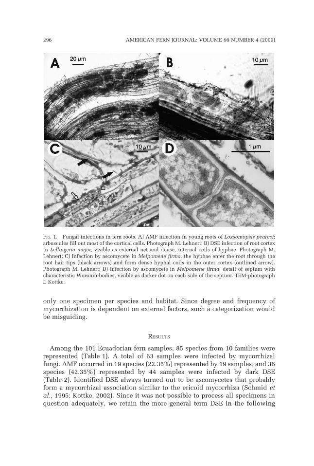
Dark septate endopyhtes (DSE) were assigned to ascomycetes (Schmid et al.,1995) if the characteristic Woronin bodies at the porate septa in the hyphaewere visible in the TEM (Fig. 1D; Haug et al., 2004). Fungal infection wasconsidered as mycorrhiza if hyphal coils were developed in host cells thatwere still intact and showed some response to the infection, i.e., thickening ofthe cell walls where the hyphae penetrated the cell and thickening of host cellcytoplasma as indicator of increased cytological activity (Fig. 1C).
The frequency of infections in the roots was quantified under the lightmicroscope, preferably on a single root with a minimum length of 10 cmmeasured from the root tips. In cases where the plants developed onlyconsiderably shorter roots, we combined several complete roots to reach theminimum length of 10 cm. The frequency of stained hyphae was categorized inthree classes (Gemma et al., 1992) to give an impression of the extent of theinfection: Present in 1) ,25%, 2) 25–75%, and 3) .75% of investigated rootlength. Presence of single hyphae or vesicles in the outer cortex as well asinfection rates below 5% were considered as erroneous infections and notcounted as mycorrhizal association. We did not distinguish between‘‘obligately’’ and ‘‘facultatively mycorrhizal’’ because we usually sampled
LEHNERT ET AL.: MYCORRHIZAL FERNS FROM ECUADOR 295
only one specimen per species and habitat. Since degree and frequency ofmycorrhization is dependent on external factors, such a categorization wouldbe misguiding.
RESULTS
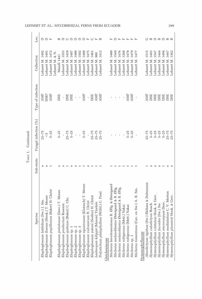
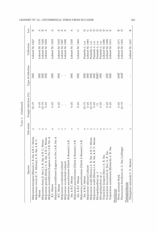
Among the 101 Ecuadorian fern samples, 85 species from 10 families wererepresented (Table 1). A total of 63 samples were infected by mycorrhizalfungi. AMF occurred in 19 species (22.35%) represented by 19 samples, and 36species (42.35%) represented by 44 samples were infected by dark DSE(Table 2). Identified DSE always turned out to be ascomycetes that probablyform a mycorrhizal association similar to the ericoid mycorrhiza (Schmid etal., 1995; Kottke, 2002). Since it was not possible to process all specimens inquestion adequately, we retain the more general term DSE in the following
FIG. 1. Fungal infections in fern roots. A) AMF infection in young roots of Loxsomopsis pearcei;arbuscules fill out most of the cortical cells. Photograph M. Lehnert; B) DSE infection of root cortexin Lellingeria major, visible as external net and dense, internal coils of hyphae. Photograph M.Lehnert; C) Infection by ascomycete in Melpomene firma; the hyphae enter the root through theroot hair tips (black arrows) and form dense hyphal coils in the outer cortex (outlined arrow).Photograph M. Lehnert; D) Infection by ascomycete in Melpomene firma; detail of septum withcharacteristic Woronin-bodies, visible as darker dot on each side of the septum. TEM-photographI. Kottke.
296 AMERICAN FERN JOURNAL: VOLUME 99 NUMBER 4 (2009)

passages. The roots of 35 samples were free of evident fungal infection. Threespecimens (Arachniodes denticulata, Elaphoglossum lloense, Micropolypo-dium sp.) had only a weak peripheral infection by DSE. They were regarded asdubious and are included in the non-mycorrhizal species (35.30% of thespecies). Mixed infections cannot be confidently reported.
AMF were found in 29.10% and 28.57% of the terrestrial and saxicolousspecies, respectively, but only in 3.45% of the epiphytes (Table 2). DSEshowed a similar presence in terrestrial and saxicolous species (30.91% and28.57%, respectively), but they dominated over AMF in the epiphytic specieswith 58.62%.
Hymenophyllaceae were represented with 18 species in our sample andshowed a high presence of mycorrhization (78%). The mainly epiphyticspecies of Hymenophyllum were colonized by DSE (80%), whereas thepredominantly terrestrial or saxicolous species of Trichomanes s.l. (Tricho-manes, Abrodyctium) had more cases of AMF infection (50%). Oneunidentified Trichomanes grew epiphytically and had DSE like the epiphyticHymenophyllum species. The only terrestrial Trichomanes s.l. with DSE wasTrichomanes dactylites Sodiro.
Grammitid ferns (Polypodiaceae; Schneider et al., 2004, Smith et al., 2006),represented by 24 species, had an infection rate of 75%. Only ascomycetes(i.e., DSE) were found as fungal partner, even in terrestrial and saxicolousspecies (L. Pazmino, unpubl. data). Non-grammitid Polypodiaceae werecompletely free of evident fungal infections.
Among the 23 species of Elaphoglossum, we found only 12 (52.20%) withfungal infection. DSE accounted for 75% of the infections.
The remainder of the investigated species showed mycorrhizal associationsas was more or less expected from previous accounts. All three species ofAsplenium (Aspleniaceae) were terrestrial and free of fungal infection. Of thetwo terrestrial species of Blechnum (Blechnaceae), only one had a low AMFinfection. The investigated Pteridaceae showed a medium to strong infectionby AMF (2 species, 100% infection).
Although they have been cited as examples for high infection rates(Boullard, 1958, 1979), only 50% of the species in the Cyatheaceae and 40%of the species in the Gleicheniaceae had mycorrhizal associations (Table 1).However, the exclusive colonization by AMF could be confirmed in bothfamilies.
Our sample size was not sufficient for a statistical analysis of changingmycorrhization along an elevational gradient. The localities of the samples areincluded in Table 1 for future studies focusing on this topic, which may wantto include the data presented here.
DISCUSSION
The overall infection by confirmed and putatively mycorrhizal fungi amongour samples was 62.38% (64.70% at the species level). These percentages arelower than those reported for angiosperms or land plants in general. Trappe
LEHNERT ET AL.: MYCORRHIZAL FERNS FROM ECUADOR 297

TA
BL
E1.
Inves
tiga
ted
sam
ple
s.A
bbre
via
tion
s:t
5te
rres
tria
l,e
5ep
iph
yte
,s
5sa
xic
olo
us;
AM
F5
arbu
scu
lar
myco
rrh
izal
fun
gi;
DS
E5
dar
kse
pta
teen
dop
hyte
s;?
5d
ubio
us
reco
rd.
Loca
liti
es:
A)
Gu
alac
eo-L
imon
road
(3100–3300
m),
B)
mou
nta
inp
ass
El
Tir
o(2
600–2800
m),
C)
Cer
roT
ole
do
(2900–
3100
m,
Pro
v.
Loja
),D
)R
eser
va
Bio
logi
caS
anF
ran
cisc
o(1
800–2600
m),
E)
Res
erva
Caj
anu
ma
(2750
m),
F)
Res
erva
Tap
ich
alac
a(2
450–2650
m),
G)
the
Cam
pam
ento
Ind
ıgen
aS
hai
mi
(900–1200
m).
Sp
ecie
sS
ub-s
trat
eF
un
gal
infe
ctio
n(%
)T
yp
eof
infe
ctio
nC
oll
ecti
on
Loc.
Asp
len
iace
ae
Asp
len
ium
au
ritu
mS
w.
t-
-L
ehn
ert
M.
1473
FA
sple
niu
mh
all
iiH
ook.
t-
-L
ehn
ert
M.
1395
BA
sple
niu
mse
rra
Lan
gsd
.&
Fis
ch.
t-
-L
ehn
ert
M.
1394
B
Ble
chn
acea
e
Ble
chn
um
sch
om
bu
rgkii
(Klo
tzsc
h)
C.
Ch
r.t
5–25
AM
FL
ehn
ert
M.
1484
DB
lech
nu
msp
.1
t-
-L
ehn
ert
M.
1440
D
Cyat
hea
ceae
Als
op
hil
aco
nan
tian
aL
ehn
ert
t5–25
AM
FL
ehn
ert
M.
1414
BC
yath
eabip
inn
ati
fid
a(B
aker
)D
om
int
--
Leh
ner
tM
.1438
DC
yath
ead
ud
leyi
R.
M.
Try
on
t5–25
AM
FL
ehn
ert
M.
1550
DC
yath
eah
ybri
dt
--
Leh
ner
tM
.1434
DC
yath
eaobn
oxia
Leh
ner
tt
--
Leh
ner
tM
.1470
FC
yath
eap
elad
ensi
s(H
iero
n.)
Dom
int
5–25
AM
FL
ehn
ert
M.
855
DD
ryop
teri
dac
eae
Ara
chn
iod
esd
enti
cula
ta(S
w.)
Ch
ing
t,
5D
SE
?L
ehn
ert
M.
996
FE
lap
hogl
oss
um
an
tisa
nae
(Sod
iro)
H.
Ch
rist
e-
-L
ehn
ert
M.
1485
DE
lap
hogl
oss
um
arg
yrop
hyl
lum
(Sod
iro)
R.
C.
Mora
n,
com
b.
ined
.e
--
Leh
ner
tM
.1490
DE
lap
hogl
oss
um
del
toid
eum
(Sod
iro)
H.
Ch
rist
t-
-L
ehn
ert
M.
1460
CE
lap
hogl
oss
um
del
toid
eum
(Sod
iro)
H.
Ch
rist
t-
-L
ehn
ert
M.
1463
CE
lap
hogl
oss
um
den
dri
cola
(Bak
er)
H.
Ch
rist
t-
-L
ehn
ert
M.
1458
CE
lap
hogl
oss
um
enge
lii
(H.
Kar
st.)
H.
Ch
rist
t25–75
DS
EL
ehn
ert
M.
1457
CE
lap
hogl
oss
um
erin
ace
um
(Fee
)T
.M
oore
e5–25
DS
EL
ehn
ert
M.
1387
BE
lap
hogl
oss
um
gloss
op
hyl
lum
Hie
ron
.e
--
Leh
ner
tM
.1551
DE
lap
hogl
oss
um
gloss
op
hyl
lum
Hie
ron
.e
5–25
DS
EL
ehn
ert
M.
1552
DE
lap
hogl
oss
um
guam
an
ian
um
(Sod
iro)
C.
Ch
r.e
25–75
DS
EL
ehn
ert
M.
1493
DE
lap
hogl
oss
um
het
erom
orp
hu
m(K
lotz
sch
)T
.M
oore
t5–25
AM
FL
ehn
ert
M.
1462
C
298 AMERICAN FERN JOURNAL: VOLUME 99 NUMBER 4 (2009)
TA
BL
E1.
Con
tin
ued
.
Sp
ecie
sS
ub-s
trat
eF
un
gal
infe
ctio
n(%
)T
yp
eof
infe
ctio
nC
oll
ecti
on
Loc.
Ela
ph
ogl
oss
um
lati
foli
um
(Sw
.)J.
Sm
.e
25–75
AM
FL
ehn
ert
M.
1492
DE
lap
hogl
oss
um
lloen
se(H
ook.)
T.
Moore
e,
5D
SE
?L
ehn
ert
M.
1491
DE
lap
hogl
oss
um
pap
illo
sum
(Bak
er)
H.
Ch
rist
t5–25
AM
FL
ehn
ert
M.
1472
F
Ela
ph
ogl
oss
um
pet
iolo
sum
(Des
v.)
T.
Moore
e5–25
DS
EL
ehn
ert
M.
&N
.M
and
l1441
BE
lap
hogl
oss
um
pro
du
ctu
mR
ose
nst
.e
--
Leh
ner
tM
.1553
DE
lap
hogl
oss
um
qu
iten
se(B
aker
)C
.C
hr.
t25–75
DS
EL
ehn
ert
M.
1459
CE
lap
hogl
oss
um
sp.
1e
5–25
DS
EL
ehn
ert
M.
1487
DE
lap
hogl
oss
um
sp.
2e
--
Leh
ner
tM
.1486
DE
lap
hogl
oss
um
sp.
3t
--
Leh
ner
tM
.1502
DE
lap
hogl
oss
um
squ
arr
osu
m(K
lotz
sch
)T
.M
oore
t5–25
AM
FL
ehn
ert
M.
1488
DE
lap
hogl
oss
um
vulc
an
icu
mH
.C
hri
ste
--
Leh
ner
tM
.1475
FE
lap
hogl
oss
um
yate
sii
(Sod
iro)
H.
Ch
rist
t25–75
DS
EL
ehn
ert
M.
1461
CLast
reop
sis
kil
ipp
ii(M
axon
)T
ind
ale
t25–75
AM
FL
ehn
ert
M.
980
FP
oly
stic
hu
mp
laty
ph
yllu
m(W
illd
.)C
.P
resl
t25–75
AM
FL
ehn
ert
M.
1412
B
Gle
ich
enia
ceae
Sti
cher
us
bre
vito
men
tosu
sB
.Ø
llg.
&Ø
ster
gaar
dt
--
Leh
ner
tM
.1480
FS
tich
eru
sm
elan
obla
stu
sØ
ster
gaar
d&
B.
Øll
g.t
--
Leh
ner
tM
.1549
DS
tich
eru
sm
elan
obla
stu
sØ
ster
gaar
d&
B.
Øll
g.t
--
Leh
ner
tM
.1476
FS
tich
eru
sru
big
nosu
s(M
ett.
)N
akai
t-
-L
ehn
ert
M.
1268
FS
tich
eru
sru
big
nosu
s(M
ett.
)N
akai
t5–25
AM
FL
ehn
ert
M.
1478
FS
tich
eru
ssp
.1
t5–25
AM
FL
ehn
ert
M.
1479
FS
tich
eru
sto
men
tosu
s(C
av.
exS
w.)
A.
R.
Sm
.t
--
Leh
ner
tM
.1477
F
Hym
enop
hyll
acea
e
Abro
dic
tyu
mri
gid
um
(Sw
.)E
bih
ara
&D
ubu
isso
nt
25–75
AM
FL
ehn
ert
M.
1515
GH
ymen
op
hyl
lum
calo
dic
tyon
Bosc
he
5–25
DS
EL
ehn
ert
M.
1443
BH
ymen
op
hyl
lum
cris
tatu
mH
ook.
&G
rev.
e5–25
DS
EL
ehn
ert
M.
1547
DH
ymen
op
hyl
lum
fuco
ides
(Sw
.)S
w.
e5–25
DS
EL
ehn
ert
M.
1444
BH
ymen
op
hyl
lum
mic
roca
rpu
mD
esv.
t5–25
DS
EL
ehn
ert
M.
1494
DH
ymen
op
hyl
lum
mu
ltia
latu
mC
.V
.M
ort
on
e25–75
DS
EL
ehn
ert
M.
1447
BH
ymen
op
hyl
lum
plu
mie
rii
Hook.
&G
rev.
e25–75
DS
EL
ehn
ert
M.
1362
B
LEHNERT ET AL.: MYCORRHIZAL FERNS FROM ECUADOR 299

TA
BL
E1.
Con
tin
ued
.
Sp
ecie
sS
ub-s
trat
eF
un
gal
infe
ctio
n(%
)T
yp
eof
infe
ctio
nC
oll
ecti
on
Loc.
Hym
enop
hyl
lum
poly
an
thos
(Sw
.)S
w.
e5–25
DS
EL
ehn
ert
M.
1445
BH
ymen
op
hyl
lum
sp.
1e
--
Leh
ner
tM
.1455
CH
ymen
op
hyl
lum
sp.
2s
--
Leh
ner
tM
.1566
AH
ymen
op
hyl
lum
tric
hom
an
oid
esB
osc
he
5–25
DS
EL
ehn
ert
M.
1446
BT
rich
om
an
esce
llu
losu
mK
lotz
sch
t5–25
AM
FL
ehn
ert
M.
1481
DT
rich
om
an
esd
act
ylit
esS
od
iro
t5–25
DS
EL
ehn
ert
M.
1501
DT
rich
om
an
esel
egan
sR
ich
.s
5–25
AM
FL
ehn
ert
M.
1516
GT
rich
om
an
esp
ellu
cen
sK
un
zet
5–25
AM
FL
ehn
ert
M.
1514
GT
rich
om
an
essp
.1
e5–25
DS
EL
ehn
ert
M.
1483
DT
rich
om
an
essp
.2
s-
-L
ehn
ert
M.
1546a
GT
rich
om
an
essp
.3
t-
-L
ehn
ert
M.
1482
D
Loxom
atac
eae
Loxso
mop
sis
pea
rcei
(Max
on
)B
aker
t.
75
AM
FL
ehn
ert
M.
1056
F
Poly
pod
iace
ae[n
on
-gra
mm
itid
s]
Cam
pyl
on
euru
mam
ph
ost
enon
Fee
t-
-P
azm
ino
L.
s.n
.D
Nip
hid
ium
alb
op
un
ctati
ssim
um
Lel
lin
ger
t-
-P
azm
ino
L.
s.n
.D
Ple
op
elti
sp
ercu
ssa
Hook.
&G
rev.
t-
-P
azm
ino
L.
s.n
.D
Poly
pod
iace
ae[g
ram
mit
ids]
Cer
ad
enia
fari
nosa
(Fors
sk.)
Kau
lf.
e25–75
DS
EP
azm
ino
L.
s.n
.D
Cer
ad
enia
fari
nosa
(Fors
sk.)
Kau
lf.
t5–25
DS
EP
azm
ino
L.
s.n
.D
Cer
ad
enia
glabra
A.
R.
Sm
ith
&M
.K
essl
ere
5–25
DS
EL
ehn
ert
M.
1495
DC
och
lid
ium
serr
ula
tum
(Sw
.)L
.E
.B
ish
op
t5–25
DS
EL
ehn
ert
M.
1467
CC
och
lid
ium
serr
ula
tum
(Sw
.)L
.E
.B
ish
op
e5–25
DS
EP
azm
ino
L.
s.n
.D
En
tero
sora
pari
etin
a(K
lotz
sch
)L
.E.
Bis
hop
e-
-L
ehn
ert
M.
1497
DG
ram
mit
isp
ara
mic
ola
L.
E.
Bis
hop
e25–75
DS
EP
azm
ino
L.
s.n
.D
Gra
mm
itis
para
mic
ola
L.
E.
Bis
hop
t25–75
DS
EP
azm
ino
L.
s.n
.D
Lel
lin
geri
am
ajo
r(C
op
el.)
A.
R.
Sm
.&
R.C
.M
ora
nt
25–75
DS
EL
ehn
ert
M.
1466
CLel
lin
geri
am
ajo
r(C
op
el.)
A.
R.
Sm
.&
R.C
.M
ora
ne
25–75
DS
EL
ehn
ert
M.
1498
DLel
lin
geri
asu
bse
ssil
is(B
aker
)A
.R
.S
m.&
R.C
.M
ora
ne
--
Leh
ner
tM
.1499
DLel
lin
geri
asu
bse
ssil
is(B
aker
)A
.R
.S
m.&
R.C
.M
ora
ne
25–75
DS
EP
azm
ino
L.
s.n
.D
Lel
lin
geri
asu
bse
ssil
is(B
aker
)A
.R
.S
m.&
R.C
.M
ora
nt
5–25
DS
EP
azm
ino
L.
s.n
.D
300 AMERICAN FERN JOURNAL: VOLUME 99 NUMBER 4 (2009)
TA
BL
E1.
Con
tin
ued
.
Sp
ecie
sS
ub-s
trat
eF
un
gal
infe
ctio
n(%
)T
yp
eof
infe
ctio
nC
oll
ecti
on
Loc.
Mel
pom
ene
assu
rgen
s(M
axon
)A
.R.S
m.&
R.C
.Mora
ne
25–75
DS
EL
ehn
ert
M.
1427
BM
elp
om
ene
erec
ta(C
.V
.M
ort
on
)A
.R
.S
m.
&R
.C
.M
ora
nt
5–25
DS
EL
ehn
ert
M.
1570
AM
elp
om
ene
firm
a(J
.S
m.)
A.
R.
Sm
.&
R.
C.
Mora
ne
25–75
DS
EL
ehn
ert
M.
1328
BM
elp
om
ene
graci
lis
(Hook.)
A.
R.
Sm
.&
R.
C.
Mora
ns
5–25
DS
EL
ehn
ert
M.
1569
AM
elp
om
ene
mon
ilif
orm
is(L
agas
caex
Sw
.)A
.R.S
m.&
R.C
.M
ora
ne
5–25
DS
EL
ehn
ert
M.
1559
AM
elp
om
ene
mon
ilif
orm
is(L
agas
caex
Sw
.)A
.R.S
m.&
R.C
.M
ora
nt
5–25
DS
EL
ehn
ert
M.
1510
EM
elp
om
ene
occ
iden
tali
sL
ehn
ert
t-
-L
ehn
ert
M.
1507
EM
elp
om
ene
occ
iden
tali
sL
ehn
ert
t-
-L
ehn
ert
M.
1508
EM
elp
om
ene
pse
ud
on
uta
ns
(Ch
rist
&R
ose
nst
.)A
.R.
Sm
.&
R.C
.M
ora
ns
5–25
DS
EL
ehn
ert
M.
1558
AM
elp
om
ene
pse
ud
on
uta
ns
(Ch
rist
&R
ose
nst
.)A
.R.
Sm
.&
R.C
.M
ora
nt
5–25
DS
EL
ehn
ert
M.
1464
CM
elp
om
ene
pse
ud
on
uta
ns
(Ch
rist
&R
ose
nst
.)A
.R.
Sm
.&
R.C
.M
ora
ne
25–75
DS
EP
azm
ino
L.
s.n
.D
Mel
pom
ene
skle
nari
iL
ehn
ert
t5–25
DS
EL
ehn
ert
M.
1465
CM
elp
om
ene
wolf
ii(H
iero
n.)
A.
R.
Sm
.&
R.
C.
Mora
ne
25–75
DS
EP
azm
ino
L.
s.n
.D
Mel
pom
ene
wolf
ii(H
iero
n.)
A.
R.
Sm
.&
R.
C.
Mora
nt
5–25
DS
EP
azm
ino
L.
s.n
.D
Mic
rop
oly
pod
ium
sp.
1t
,5
DS
E?
Paz
min
oL
.s.
n.
DM
icro
poly
pod
ium
sp.
1e
5–25
DS
EP
azm
ino
L.
s.n
.D
Ter
psi
chore
lan
iger
a(D
esv.)
A.
R.
Sm
.e
--
Leh
ner
tM
.1496
DT
erp
sich
ore
leu
cost
icta
(J.
Sm
.)A
.R
.S
m.
t5–25
DS
EL
ehn
ert
M.
1509
ET
erp
sich
ore
sem
ihir
suta
(Klo
tzsc
h)
A.
R.
Sm
.t
5–25
DS
EL
ehn
ert
M.
1511
E
Pte
rid
acea
e
Pte
ris
mu
rica
taH
ook.
t5–25
AM
FL
ehn
ert
M.
1571
BP
tero
zon
ium
bre
vifr
on
s(A
.C
.S
m.)
Lel
lin
ger
s25–75
AM
FL
ehn
ert
M.
1435
D
Th
elyp
teri
dac
eae
Th
elyp
teri
sm
inu
tula
C.
V.
Mort
on
s-
-L
ehn
ert
M.
1337
B
LEHNERT ET AL.: MYCORRHIZAL FERNS FROM ECUADOR 301
(1987) estimated that 82% of angiosperms host mycorrhizae; Wang and Qiu(2006) concluded that 80% of all land plants are mycorrhizal.
Studies focusing on ferns and lycophytes found similar results to ours.Values gathered from literature (Boullard, 1958; Cooper, 1976; Berch andKendrick, 1982, Iqbal et al., 1981, Gemma et al., 1992, Lesica and Antibus,1990, Moteetee et al., 1996; Ragupathy and Mahadevan, 1993, Schmid et al.,1995, Muthukumar and Udaiyan, 2000; Zhao, 2000; Zhang et al., 2003) sum upto 68% of general fungal colonization and to 53% of AMF in ferns andlycophytes (M. Lehnert, unpubl. data). Wang and Qiu (2006), considering onlyAMF, found a comparable 52% of the species of ferns and lycophytes to bemycorrhizal.
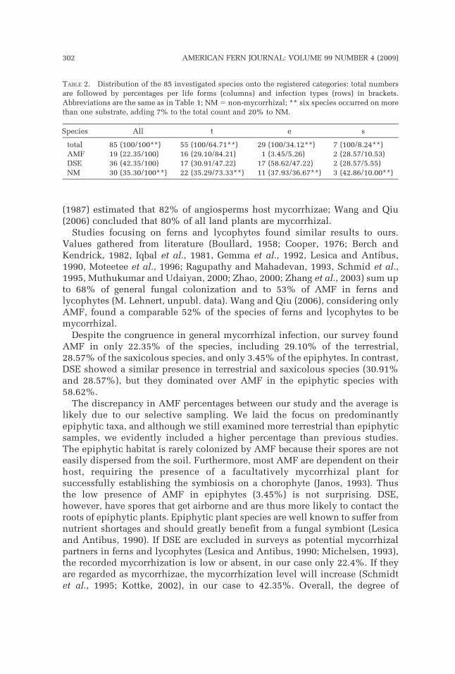
Despite the congruence in general mycorrhizal infection, our survey foundAMF in only 22.35% of the species, including 29.10% of the terrestrial,28.57% of the saxicolous species, and only 3.45% of the epiphytes. In contrast,DSE showed a similar presence in terrestrial and saxicolous species (30.91%and 28.57%), but they dominated over AMF in the epiphytic species with58.62%.
The discrepancy in AMF percentages between our study and the average islikely due to our selective sampling. We laid the focus on predominantlyepiphytic taxa, and although we still examined more terrestrial than epiphyticsamples, we evidently included a higher percentage than previous studies.The epiphytic habitat is rarely colonized by AMF because their spores are noteasily dispersed from the soil. Furthermore, most AMF are dependent on theirhost, requiring the presence of a facultatively mycorrhizal plant forsuccessfully establishing the symbiosis on a chorophyte (Janos, 1993). Thusthe low presence of AMF in epiphytes (3.45%) is not surprising. DSE,however, have spores that get airborne and are thus more likely to contact theroots of epiphytic plants. Epiphytic plant species are well known to suffer fromnutrient shortages and should greatly benefit from a fungal symbiont (Lesicaand Antibus, 1990). If DSE are excluded in surveys as potential mycorrhizalpartners in ferns and lycophytes (Lesica and Antibus, 1990; Michelsen, 1993),the recorded mycorrhization is low or absent, in our case only 22.4%. If theyare regarded as mycorrhizae, the mycorrhization level will increase (Schmidtet al., 1995; Kottke, 2002), in our case to 42.35%. Overall, the degree of
TABLE 2. Distribution of the 85 investigated species onto the registered categories: total numbersare followed by percentages per life forms (columns) and infection types (rows) in brackets.Abbreviations are the same as in Table 1; NM 5 non-mycorrhizal; ** six species occurred on morethan one substrate, adding 7% to the total count and 20% to NM.
Species All t e s
total 85 (100/100**) 55 (100/64.71**) 29 (100/34.12**) 7 (100/8.24**)AMF 19 (22.35/100) 16 (29.10/84.21) 1 (3.45/5.26) 2 (28.57/10.53)DSE 36 (42.35/100) 17 (30.91/47.22) 17 (58.62/47.22) 2 (28.57/5.55)NM 30 (35.30/100**) 22 (35.29/73.33**) 11 (37.93/36.67**) 3 (42.86/10.00**)
302 AMERICAN FERN JOURNAL: VOLUME 99 NUMBER 4 (2009)

infection by DSE was higher in our study than in any other previous study onferns and lycophytes.
Beyond these general patterns, it is worthwhile to focus on individual studygroups. The Hymenophyllaceae nicely mirror the general distribution patternof the fungal infections. Terrestrial and saxicolous species have predominantlyAMF, whereas DSE prevail in epiphytes. Gammitid ferns (Polypodiaceae),however, have almost exclusively DSE, no matter if they grew as epiphytes oras terrestrials. This apparent conflict with the general trend is due to themicrohabitats inhabited by the species. The investigated terrestrial grammitidferns usually grew in thick moss cushions like their epiphytic kin and by thismeans under very similar ecological conditions, which may lead tomaintaining the type of mycorrhiza. Furthermore, most of the species sampledas terrestrials are either potentially epiphytic or closely related to epiphyticspecies. Only the samples of Melpomene occidentalis Lehnert rooted directlyin mineral soil and showed no fungal infection. Opposed to this, the samplesof eleven terrestrial and epiphytic species of non-grammitid Polypodiaceaefrom the investigated area are free of fungal infections, which is congruentwith previous reports (Lesica and Antibus, 1990; Schmid et al., 1995). Sincegrammitid ferns represent a clade nested deeply within the Polypodiaceae, it islikely that the original condition in the family is a lack of mycorrhization andthat mycorrhization has been secondarily regained in grammitid ferns.Apparently, this symbiosis was developed with DSE rather than withglomeromycetes. A similar situation of loss of AMF mycorrhization andsecondary gain of DSE mycorrhization, also related with shifts between theterrestrial and epiphytic habitat, has been reported in liverworts (Kottke andNebel, 2005).
The genus Elaphoglossum showed no clear correlation between the types ofsubstrate and fungal infections. The genus Asplenium is not very diverse orabundant in the study sites and occurred only on the lower slopes wherenutrients are accumulated (Gradstein et al., 2008). The absence of mycorrhizaein our samples may be related to the improved availability of nutrients at theirmicrohabitats. Previous studies found generally low infection rates in theAspleniaceae (e.g., Boullard, 1958) and often varying results within a species,indicating that most species may be only facultatively mycorrhizal.
Gleicheniaceae are usually axiomatic for strong presence of mycorrhizae(100%; Boullard, 1958, 1979). It is assumed that this affects both their ability togrow on nutrient deficient soils and their inability to be transplanted andcultivated. Surprisingly, we found only 40% of our samples infected by AMF.Their root samples, however, were difficult to prepare because of a toughtexture and dark, persistent cortical colorants. The necessary clearing withhydrogen peroxide may have affected the colourability of fungal hyphae withdye. Possibly a higher percentage of fungal infections was present but notdetectable in our samples of Gleicheniaceae.
Our results for the Cyatheaceae are much lower (50% of specimens infected)than the results of previous surveys (100% of specimens infected; Boullard,1958; Hepden, 1960). The tree ferns (families Cyatheaceae and Dicksoniaceae)
LEHNERT ET AL.: MYCORRHIZAL FERNS FROM ECUADOR 303
bear the difficulty of acquiring fine roots from the compact subterranean rootsystem that many species develop. Aerial roots from the trunks are easier toharvest but are expected to lack mycorrhizae because they are less likely to getin contact with inoculum of soil fungi. In order to bypass this samplingartefact, the plants included in this study were either small species or youngplants of easily assignable larger species, which can be uprooted with most oftheir roots. One explanation for the low infection rate could be that thesejuvenile plants of Cyathea are less dependent on mycorrhizae than matureplants. The trunk-less tree ferns dwell in the shade where these often sun-loving species are under lesser drought stress but presumably achieve only apart of their potential photosynthetic rate. The profits of better supply withwater and micronutrients may not compensate the cost of sharing assimilateswith symbiotic fungi.
We are aware that negative results in any species here included do notexclude the potential occurrence of mycorrhiza in other individuals of thesame species. We aim to widen our sample size and want to includeconspecific samples from sites with different substrate chemistry. This shouldallow us not only to distinguish between facultative and obligatorymycorrhizae but also about the conditioning factors.
ACKNOWLEDGMENTS
We thank our colleagues of the Research Unit of the DFG 402 ‘‘Functionality in a TropicalMountain Rainforest: Diversity, Dynamic Processes and Utilization Potentials under EcosystemPerspectives’’ for various help and fruitful discussion, especially Nicki Mandl and Rob Gradstein;we are indebted to our Ecuadorian counterparts in Loja (Fundacion Cultura y Naturaleza; HerbarioLOJA/Universidad Nacional de Loja; Universidad Tecnica Particular de Loja [UTPL]) and Quito(Pontificia Universidad Catolica del Ecuador [PUCE]). The Ministerio del Ambiente, Ecuador,kindly issued permits for field research and collection of plant material. Special thanks go toRobbin C. Moran, New York Botanical Garden, for determinations of the Elaphoglossum samples.This study was financially supported by the German Research Foundation (DFG, grant GR 1588/7).
LITERATURE CITED
ALLEN, M. F., W. SWENSON, J. I. QUEREJETA, L. M. EGERTON-WARBURTON and K. K. TRESEDER. 2003.Ecology of mycorrhizae: A conceptual framework for complex interactions among plants andfungi. Annu. Rev. Phytopathol. 47:271–303.
ANDRADE, A. C. S., M. H. QUEIROZ, R. A. L. HERMES and V. L. OLIVEIRA. 2000. Mycorrhizal status ofsome plants of the Araucaria forest and the Atlantic rainforest in Santa Catarina, Brazil.Mycorrhiza 10:131–136.
BERCH, S. H. and B. KENDRICK. 1982. Vesicular-arbuscular mycorrhizae of southern Ontario ferns andfern allies. Mycologia 74:769–776.
BLACKWELL, M. 2000. Terrestrial life - Fungal from the start? Science 289:1884–1885.BOULLARD, B. 1958. La mycotrophie chez les pteridophytes. Sa frequence, ses caracteres, sa
signification. Doctor thesis, Universite de Caen. (Imprimerie E. Droulliard, Bordeaux)BOULLARD, B. 1979. Consideration sur la symbiose fongique chez les Pteridophytes. Syllogeous
19:1–59.BRUNDRETT, K. 2002. Coevolution of roots and mycorrhizas of land plants. New Phytol. 154:275–304.BRUNDRETT, M. C. 2004. Diversity and classification of mycorrhizal associations. Bot. Rev.
79:473–495.
304 AMERICAN FERN JOURNAL: VOLUME 99 NUMBER 4 (2009)

CAIRNEY, J. W. G. and A. A. MEHARG. 2003. Ericoid mycorrhiza: a partnership that exploits harshedaphic conditions. Eur. J. Soil Sci. 54:735–740.
COOKE, J. C. and M. W. LEFOR. 1998. The mycorrhizal status of selected plant species fromConneticut wetlands and transition zones. Restoration Ecology 6:214–222.
COOPER, K. M. 1976. A field survey of mycorrhizas in New Zealand ferns. N.Z. J. Bot. 14:169–181.ENTRY, J. A., P. T. RYGIEWICZ, L. S. WATRUD and P. K. DONNELLY. 2002. Influence of adverse soil
conditions on the formation and function of arbuscular mycorrhizas. Adv. Environ. Res.7:123–138.
FERNANDEZ, N., S. FONTENLA and M. I. MESSUTI. 2005. Micorrizas en pteridofitas de los bosquestemplado-lluviosos del Noroeste de Patagonia. II Convencion Ambiental UniversitariaPatagonica.
GEMMA, J. N., R. E. KOSKE and T. FLYNN. 1992. Mycorrhizae in Hawaiian pteridophytes: occurrenceand evolutionary significance. Amer. J. Bot. 79:843–852.
GRACE, C. and D. P. STRIBLEY. 1991. A safer procedure for routine staining of vesicular-arbuscularmycorrhizal fungi. Mycol. Res. 95:1160–1162.
GRADSTEIN, S. R., M. KESSLER, M. LEHNERT, M. ABIY, N. MANDL, F. MAKESCHIN and M. RICHTER. 2008.Vegetation, climate and soil of the unique Purdiaea forest of southern Ecuador. Ecotropica14:15–26.
HAUG, I., M. WEIß, J. HOMEIER, F. OBERWINKLER and I. KOTTKE. 2004. Russulaceae and Thelephoraceaeform ectomycorrhizas with members of the Nyctaginaceae (Caryophyllales) in the tropicalmountain rain forest of southern Ecuador. New Phytol. 165:923–936.
HEPDEN, P. M. 1960. Studies in vesicular-arbuscular endophytes. II. Endophytes in thePteridophyta, with special reference to leptosporangiate ferns. Trans. Br. Mycol. Soc.43:559–570.
IQBAL, S. H., M. YOUSAF and M. YOUNUS. 1981. A field survey of mycorrhizal associations in ferns ofPakistan. New Phytol 87:69–89.
JANOS, D. P. 1993. Vesicular-arbuscular mycorrhizae of epiphytes. Mycorrhiza 4:1–4.JUMPPONEN, A. and J. M. TRAPPE. 1998. Dark septate endophytes: a review of facultative biotrophic
root-colonizing fungi. New Phytol. 140:295–310.KESSLER, M. and M. LEHNERT. 2009. Do ridge habitats contribute to pteridophyte diversity in tropical
montane forests? A case study from southeastern Ecuador. J. Pl. Res. 122:421–428.KOTTKE, I. 2002. Mycorrhizae - Rhizosphere determinants of plant communities. Pp. 919–932, in:
WAISEL, Y., ESHEL, A. and U. KAFKAFI, (eds.), Plant Roots: The Hidden Half. 3rd ed. MarcelDekker, Inc.
KOTTKE, I. and M. NEBEL. 2005. The evolution of mycorrhiza-like associations in liverworts: Anupdate. New Phytol. 167:330–334.
KOTTKE, I., A. BECK, I. HAUG, S. SETARO, V. JESKE, J. P. SUAREZ, L. PAZMINO, M. PREUßING, M. NEBEL and F.OBERWINKLER. 2008. Mycorrhizal state and new and special features of mycorrhizae of trees,ericads, orchids, ferns and liverworts. Pp. 137–148, in: BECK, E., J. BENDIX, I. KOTTKE, F.MAKESCHIN and R. MOSANDL, (eds.), Gradients in a Tropical Mountain Ecosystem of Ecuador.Series Ecological Studies 198, Springer Verlag, Berlin, Heidelberg.
LEHNERT, M., M. KESSLER, L. I. SALAZAR, H. NAVARRETE, F. A. WERNER and S. R. GRADSTEIN. 2007.Pteridophyta. Pp. 59–68, in: LIEDE-SCHUMANN, S. and S.-W. BRECKLE, (eds.), ProvisionalChecklists of fauna and flora of the San Francisco valley and its surroundings (Reserva SanFrancisco/Prov. Zamora–Chinchipe, southern Ecuador). Ecotrop. Monogr. 4.
LESICA, P. and R. K. ANTIBUS. 1990. The occurrence of mycorrhizae in vascular epiphytes of twoCosta Rican rain forests. Biotropica 33:250–258.
MICHELSEN, A. 1993. The mycorrhizal status of vascular epiphytes in Bale Mountains National Park,Ethiopia. Mycorrhiza 4:11–15.
MOTEETEE, A., J. G. DUCKETT and A. J. RUSSELL. 1996. Mycorrhizas in the ferns of Lesotho. Pp. 621–631, CAMUS, J. M., M. GIBBY and R. J. JOHNS, (eds.), Pteridology in perspective. Royal BotanicGardens, Kew.
MUTHUKUMAR, T. and K. UDAIYAN. 2000. Arbuscular mycorrhizas of plants growing in the WesternGhats region, Southern India. Mycorrhiza 9:297–313.
LEHNERT ET AL.: MYCORRHIZAL FERNS FROM ECUADOR 305
NADARAJAH, P. and A. NAWAWI. 1993. Mycorrhizal status of epiphytes in Malaysian oil palmplantations. Mycorrhiza 4:21–24.
NEWMAN, E. I. and P. REDDELL. 1987. The distribution of mycorrhizas among families of vascularplants. New Phytol. 106:745–751.
PIROZYNSKI, K. A. and D. W. MALLOCH. 1975. The origin of land plants: A matter of mycotrophism.BioSystems 6:153–164.
RAGUPATHY, S. and A. MAHADEVAN. 1993. Distribution of vesicular-arbuscular mycorrhizae in theplants and rhizosphere soils of the tropical plains, Tamil Nadu, India. Mycorrhiza 3:123–136.
RICHTER, M. 2003. Using epiphytes and soil temperatures for eco-climatic interpretations insouthern Ecuador. Erdkunde 57:161–181.
SCHMID, E., F. OBERWINKLER and L. D. GOMEZ. 1995. Light and electron microscopy of a host-fungusinteraction in the roots of some epiphytic ferns from Costa Rica. Can. J. Bot. 73:991–996.
SCHNEIDER, H., A. R. SMITH, R. CRANFILL, T. J. HILDEBRAND, C. H. HAUFLER and T. A. RANKER. 2004.Unraveling the phylogeny of polygrammoid ferns (Polypodiaceae and Grammitidaceae):exploring aspects of diversification of epiphytic plants. Mol. Phylo. Evol. 31:1041–1063.
SCHUßLER, A., D. SCHWARZGOTT and C. WALKER. 2001. A new fungal phylum, the Glomeromycota:phylogeny and evolution. Mycol. Res. 105:1413–1421.
SMITH, A. R., K. M. PRYER, E. SCHUETTPELZ, P. KORALL, H. SCHNEIDER and P. G. WOLF. 2006. Aclassification for extant ferns. Taxon 55:705–731.
TRAPPE, J. M. 1987. Phylogenetic and ecologic aspects of mycotrophy in the angiosperms from anevolutionary standpoint. Pp. 5–25, in: SAFIR, G. R., (ed.), Ecophysiology of va mycorrhizalplants. Boca Raton, FL, USA.CRC Press.
WANG, B. and Y.-L. QIU. 2006. Phylogenetic distribution and evolution of mycorrhizas in landplants. Mycorrhiza 16:299–363.
WILCKE, W., S. YASIN, C. VALAREZO and W. ZECH. 2001. Nutrient budget of three microcatchmentsunder tropical montane forest in Ecuador. Die Erde 132:61–74.
ZHANG, Y., L.-D. GUO and R.-J. LIU. 2004. Arbuscular mycorrhizal fungi associated with commonpteridophytes in Dujiangyan, southwest China. Mycorrhiza 14:25–30.
ZHAO, Z. W. 2000. The arbuscular mycorrhizas of pteridophytes in Yunnan, southwest China:evolutionary interpretations. Mycorrhiza 10:145–149.
306 AMERICAN FERN JOURNAL: VOLUME 99 NUMBER 4 (2009)