Muscle activation profiles about the knee during Tai-Chi stepping movement compared to the normal...
9
Muscle activation profiles about the knee during Tai-Chi stepping movement compared to the normal gait step Shih-Chiao Tseng a, * , Wei Liu a , Margaret Finley a,b , Kevin McQuade c a Department of Physical Therapy and Rehabilitation Science, University of Maryland School of Medicine, Baltimore, MD, USA b Baltimore Veterans Medical Center, Rehabilitation Research and Development Service, Baltimore, MD, USA c Department of Rehabilitation Medicine, University of Washington, Seattle, WA, USA Received 20 August 2004; received in revised form 30 September 2005; accepted 6 December 2005 Abstract The purpose of this study was to investigate knee muscle activity patterns in experienced Tai-Chi (TC) practitioners during normal walking and TC stepping. The electromyographic (EMG) activity of vastus lateralis (VL), vastus medialis (VM), bicep femoris (BF), and gastrocnemius (GS) muscles of 11 subjects (five females and six males) during the stance phase of normal walking was compared to stance phase of a TC step. Knee joint motion was also monitored by using an Optotrakämotion analysis system. Raw EMG was pro- cessed by root-mean-square (RMS) technique using a time constant of 50 ms, and normalized to maximum of voluntary contraction for each muscle, referred to as normalized RMS (nRMS). Peak nRMS and co-contraction (quantified by co-contraction index) during stance phase of a gait cycle and a TC step were calculated. Paired t-tests were used to compare the difference for each muscle group peak and co- contraction pair between the tasks. The results showed that only peak values of nRMS in quadriceps and co-contraction were signifi- cantly greater in TC stepping compared to normal walking (Peak values of nRMS for VL were 26.93% for normal walking and 52.14% for TC step, p = 0.001; VM are 29.12% for normal walking and 51.93% for TC stepping, p = 0.028). Mean co-contraction index for VL–BF muscle pairs was 13.24 ± 11.02% during TC stepping and 9.47 ± 7.77% in stance phase of normal walking (p = 0.023). There was no significant difference in peak values of nRMS in the other two muscles during TC stepping compared to normal walking. Pre- liminary EMG profiles in this study demonstrated that experienced TC practitioners used relatively higher levels of knee muscle activa- tion patterns with greater co-contraction during TC exercise compared to normal walking. Ó 2006 Elsevier Ltd. All rights reserved. Keywords: Tai-Chi; Knee; Electromyography; Muscle activation 1. Introduction Tai-Chi (TC) is an ancient Chinese martial art consisting of a series of slow continuous movements of the trunk and limbs. The doctrine of Tai-Chi (TC) was derived from phi- losophy of Taoism, which is focused on mind tranquility and inside balance of the body. The principle of Tai-Chi exercise included meditation and a series of slow, fluent, and accurate body–limb movements while individual dee- ply breathed in and out during movement and rest [14]. The simplified TC Yang – style performed in a semi-squat position is frequently practiced in western society [19]. Since the components of TC involve sequential motions with cycling steps, its biomechanical characteristics have been well analyzed by applying gait analysis techniques and functional comparisons made to another cycling motion, normal walking [25]. It is believed that TC may benefit older people by maintaining good postural control, muscle strength, and physical fitness [2,7,15,24,26]. Several studies have shown that TC practitioners have good static and dynamic pos- tural control compared to age-matched community- dwelling elders [2,7,10,24]. This exercise intensity was estimated around four metabolic equivalents (METs) and approximate to 50–55% of maximal oxygen uptake for elderly groups, which was generally recommended 1050-6411/$ - see front matter Ó 2006 Elsevier Ltd. All rights reserved. doi:10.1016/j.jelekin.2005.12.005 * Corresponding author. Journal of Electromyography and Kinesiology 17 (2007) 372–380 www.elsevier.com/locate/jelekin
-
Upload
independent -
Category
Documents
-
view
1 -
download
0
Transcript of Muscle activation profiles about the knee during Tai-Chi stepping movement compared to the normal...

Journal of Electromyography and Kinesiology 17 (2007) 372–380
www.elsevier.com/locate/jelekin
Muscle activation profiles about the knee during Tai-Chistepping movement compared to the normal gait step
Shih-Chiao Tseng a,*, Wei Liu a, Margaret Finley a,b, Kevin McQuade c
a Department of Physical Therapy and Rehabilitation Science, University of Maryland School of Medicine, Baltimore, MD, USAb Baltimore Veterans Medical Center, Rehabilitation Research and Development Service, Baltimore, MD, USA
c Department of Rehabilitation Medicine, University of Washington, Seattle, WA, USA
Received 20 August 2004; received in revised form 30 September 2005; accepted 6 December 2005
Abstract
The purpose of this study was to investigate knee muscle activity patterns in experienced Tai-Chi (TC) practitioners during normalwalking and TC stepping. The electromyographic (EMG) activity of vastus lateralis (VL), vastus medialis (VM), bicep femoris (BF), andgastrocnemius (GS) muscles of 11 subjects (five females and six males) during the stance phase of normal walking was compared tostance phase of a TC step. Knee joint motion was also monitored by using an Optotrak�motion analysis system. Raw EMG was pro-cessed by root-mean-square (RMS) technique using a time constant of 50 ms, and normalized to maximum of voluntary contraction foreach muscle, referred to as normalized RMS (nRMS). Peak nRMS and co-contraction (quantified by co-contraction index) during stancephase of a gait cycle and a TC step were calculated. Paired t-tests were used to compare the difference for each muscle group peak and co-contraction pair between the tasks. The results showed that only peak values of nRMS in quadriceps and co-contraction were signifi-cantly greater in TC stepping compared to normal walking (Peak values of nRMS for VL were 26.93% for normal walking and52.14% for TC step, p = 0.001; VM are 29.12% for normal walking and 51.93% for TC stepping, p = 0.028). Mean co-contraction indexfor VL–BF muscle pairs was 13.24 ± 11.02% during TC stepping and 9.47 ± 7.77% in stance phase of normal walking (p = 0.023). Therewas no significant difference in peak values of nRMS in the other two muscles during TC stepping compared to normal walking. Pre-liminary EMG profiles in this study demonstrated that experienced TC practitioners used relatively higher levels of knee muscle activa-tion patterns with greater co-contraction during TC exercise compared to normal walking.� 2006 Elsevier Ltd. All rights reserved.
Keywords: Tai-Chi; Knee; Electromyography; Muscle activation
1. Introduction
Tai-Chi (TC) is an ancient Chinese martial art consistingof a series of slow continuous movements of the trunk andlimbs. The doctrine of Tai-Chi (TC) was derived from phi-losophy of Taoism, which is focused on mind tranquilityand inside balance of the body. The principle of Tai-Chiexercise included meditation and a series of slow, fluent,and accurate body–limb movements while individual dee-ply breathed in and out during movement and rest [14].The simplified TC Yang – style performed in a semi-squatposition is frequently practiced in western society [19].
1050-6411/$ - see front matter � 2006 Elsevier Ltd. All rights reserved.
doi:10.1016/j.jelekin.2005.12.005
* Corresponding author.
Since the components of TC involve sequential motionswith cycling steps, its biomechanical characteristics havebeen well analyzed by applying gait analysis techniquesand functional comparisons made to another cyclingmotion, normal walking [25].
It is believed that TC may benefit older people bymaintaining good postural control, muscle strength, andphysical fitness [2,7,15,24,26]. Several studies have shownthat TC practitioners have good static and dynamic pos-tural control compared to age-matched community-dwelling elders [2,7,10,24]. This exercise intensity wasestimated around four metabolic equivalents (METs)and approximate to 50–55% of maximal oxygen uptakefor elderly groups, which was generally recommended

S.-C. Tseng et al. / Journal of Electromyography and Kinesiology 17 (2007) 372–380 373
in physical fitness exercise for elderly groups [14,19]. Fur-thermore, Hong’s study showed that TC practitionerswith more than 10 years TC experience had better func-tional performance in terms of balance control, flexibil-ity, and cardiorespiratory fitness compared to sedentaryelders [7]. In addition, Wu and coworkers [26] demon-strated that TC practitioners with more than threeyears TC experience had significantly greater isokineticstrength of the knee extensor muscles compared to age-matched healthy elders. In addition, TC exercise alsohas been used as an alternative exercise intervention inreducing falls for elders [22–24]. Wolf et al. [22,23]showed elders receiving 15 weeks of TC training reducedthe risk of multiple falls by 47.5% and also had a trendtoward a reduced fear of falling. Kirsteins et al. [13]investigated the Yang style TC exercise therapy of peoplewith rheumatoid arthritis. After 10 weeks TC trainingsubjects demonstrated no deterioration in related symp-toms such as increased joint tenderness and swelling.Recently, one study investigated elders with osteoarthritisreceiving 12-weeks of TC exercise training and demon-strated significant improvement in their self-efficacy andquality of life [6].
The electromyographic (EMG) signal has been used tocompare different types of activities in order to make func-tional reference to differences in muscle activation levelsand muscle coactivation [11]. By applying EMG measuresfor same or different subjects during different activities suchas walking, stair-up and -down, it would provide an idea ofhow much demand in muscle activity was needed for eachactivity and the amount of differences in muscle activationlevels between different activities. The use of EMG profilesas a window into neuromuscular control strategies duringTC exercise has also been investigated in two studies. Wuand his colleagues [25] first investigated muscle activity pat-terns of the lower extremities during normal walking andTC gait in 10 young, normal subjects, all TC novices withonly one-week experience. The reasons for the functionalcomparison in muscle activity between TC and normalwalking are: First, the movement components of TCinvolve cycling steps, which are similar to gait cycles. Sec-ond, muscle activation level during normal walking couldbe a reference to TC exercise [25]. The results showed sig-nificantly greater peak values in root-mean-squares(RMS) of EMG (77–88%) during TC gait compared tonormal walking. In another study conducted by Chanet al. [1] studied EMG characteristics of the lower extrem-ity in a TC master who practiced TC for thirty minutesevery day for 22 years. The TC master demonstrated lessthan 20% of MVIC values for most muscle groups in hislower extremity. The lowest integrated EMG (IEMG)activity occurred in the medial hamstrings and the medialhead of gastrocnemius, while the greatest IEMG in the rec-tus femoris muscles reached as much as 68.3% of theMVIC. In fact, the greatest EMG in Chan’s study was stillless than any muscle activity in Wu’s study. The differencein muscle activation levels between two studies could be
due to different task demands on TC performance andTC practitioners’ experience. The relatively higher muscleactivity reported in Wu’s study might indicate higher mus-cle activity demand on an entire TC step than pushingmovement component of a TC step in Chan’s study. Inaddition, TC practice experience was also an importantfactor, which directly affects muscle activation levels. Wespeculate that the TC experience might be an importantfactor affecting muscle activity levels since TC exercisewas documented as a moderate-intensity exercise (about4 METs) for long-term elderly TC groups [14,19]. In aneffort to clarify and understand the uniqueness of TC exer-cise, one strategy is to assess muscle activation profiles oflong-term TC practitioners. Since previous two TC studies[1,25] investigated muscle activity of rectus femoris, whichis a bi-articular muscle serving as either a hip flexor or aknee extensor, the authors chose to investigate the contri-butions from single knee joint muscles in quadriceps, vas-tus lateralis and medialis muscles, for eccentrically andconcentrically knee flexion control during TC exercise.Furthermore, another two muscles (medial head of gas-trocnemius and long head of bicep femoris muscles) servingas antagonists were selected and compared results fromprevious studies [1,25]. The purpose of this pilot studywas to investigate the knee muscle activity patterns duringstance phase of normal walking and TC stepping in experi-enced TC practitioners. We hypothesized TC steppingwould result in relatively higher muscle activity at vastuslateralis and medialis muscle, and co-contraction of kneeextensors and flexor muscle pairs.
2. Methods
Prior to participation all subjects signed consent formsapproved by the University of Maryland School of Medi-cine Institutional Review Board. Eleven healthy subjects(five females and six males) with at least six months TCexperience participated in this study. The average age was53.0 ± 6.8 years with a mean TC exercise experience of8.3 ± 6.8 years (range from 6 months to 24 years).
The testing procedures included two parts: (1) subjectswere asked to walk normally across an 8-m walkway at aself-selected pace, and (2) subjects were asked to performthe beginning sequence of their TC exercises: stepping outwith the left foot. In this study, all subjects practiced theshort Yang form of TC, which included a regular repeateddouble support stepping move as a transition of center ofgravity (COG) between right and left limbs, called the TCstep. The major components of TC include three movementsequences: press movement with COG slowly moving for-ward and downward (Fig. 1C), rolling back movement withCOG slowly moving backward (Fig. 1D), and rolling overwith COG slowly moving forward continuously to propeland successively take the next step forward (Fig. 1E andF). During a TC step, subjects transferred their body weightfrom the left leg to the right leg requiring a longer phase ofsingle limb (left leg) support compared to a normal walking

FED
CBA
Fig. 1. A TC step illustrated from standing position (A), initial landing byleft limb (B, thick sticks with a black oval), press movement with COGslowly moving forward and downward (C), rolling backward movementwith COG slowly moving backward (D), and rolling over with COG slowlymoving forward again (E), lifting right limb and continuously movingforward until next TC step cycle (F). The muscle activity was recorded fromB to the end of F (toe-off of left limb defined by force plate data).
374 S.-C. Tseng et al. / Journal of Electromyography and Kinesiology 17 (2007) 372–380
step. Electromyography and knee joint angles for left leg instance phase of a gait cycle were measured during normalwalking and TC stepping.
2.1. Electromyography measurement
Surface electromyographic recordings of selected mus-cles surrounding the left knee: vastus lateralis (VL), vastusmedialis (VM), long head of bicep femoris (BF), and med-ial head of gastrocnemius (GS) were recorded. Followingpreparation of the area with shaving and alcohol, pairsof bipolar Ag–AgCl surface electrodes (3 cm inter-elec-trode distance, 1 cm active diameter, Blue Sensor, Medico-test, Denmark) were placed on the muscle bulk of eachmuscle aligned with directions of muscle fibers [20]. A ref-erence electrode was placed at left tibial tuberosity. AnEMG system with bandpass of 10–1000 Hz, CCRR 90 dB(Myopac Jr., Run Tech., Laguna Hills, CA) was used torecord (1000 Hz) surface activity.
Before normal walking and TC tests, subjects’ maximumvoluntary isometric contractions (MIVC) for each of fourmuscles (VL, VM, BF, and GS) were determined. EMGrecorded during the MVIC was used to normalize EMGvalues during TC and normal walking, expressing themas a percentage of each subject’s MVIC. For knee extensors(VL and VM) and flexor (BF) subjects sat with 90� of kneeflexion and performed three maximum isometric voluntarycontractions of knee extension or flexion, for each of themuscle groups respectively. For GS, subjects performedsingle leg toe standing with balance support provided andstrong resistance was given downward at the shoulders,for three trials. Five-seconds of MVIC for each musclewas recorded per trial and MVIC was defined as themaximum point of EMG amplitude within 5-s contraction
during three repeated trials. EMG was recorded in fourmuscles around left knee joint as each subject performedTC exercise and normal walking.
2.2. Knee joint motion measurement
Kinematic data were collected using a standard OptoT-rak�(Northern Digital, Inc., Waterloo, Ontario). The reli-ability and validity of the OptoTrak�system has beenestablished for motion analysis [9,18]. Four markers wereattached to bilateral mid-thighs and the upper third ofleg. Each marker consisted of a triad of infrared light emit-ting diodes (IREDs) and acted as a rigid body assembly fortracking and orienting positions and movements of bodysegments in space. These markers created the local coordi-nate system relative to each other to determine knee jointangle during normal walking and the TC stepping. Thesampling rate for OptoTrak�system was set at 100 Hz.Knee joint motions during both tasks were calculatedbased on the method reported by Grood and Suntay [4].An AMTI forceplate (Advanced Mechanical Technology,Inc. Newton, MA) was used to determine the stance phaseof gait for both the TC Step and a normal walking step.EMG and force plate data were collected using a 16-bitanalog to digital converter board at 1000 Hz sampling rateand synchronized to the kinematic data.
2.3. Data reduction
All EMG and kinematic data were analyzed in thestance phase of the gait cycle and normalized to 100% ofthe stance phase. Raw EMG was processed by RMS tech-nique using a time constant of 50 ms, and normalized tothe MVIC for each muscle (nRMS). Onset threshold ofmuscle activity during the stance phase of a gait cyclewas defined as 5% of peak nRMS with continuous activa-tion more than 30 ms. Means of peak nRMS over three tri-als under normal walking and TC stepping conditions werecomputed for each subject and means calculated across 11subjects. To assess co-contraction of muscles at the kneejoint, co-contraction index (CI) was used to quantify mus-cle co-activations at VL–BF and VM–BF muscle pairs. TheCI was calculated by finding the overlap between the nor-malized agonist and antagonist muscle curves (VL–BF andVM–BF muscle pairs) in stance phase of a gait cycle andTC step, and divided by the total number of data points[3,12,21]. Means of CI in normal walking and TC step werecomputed across 11 subjects. For reliability testing, threerepeated trials were recorded for each individual duringnormal walking and TC stepping, respectively. The peakvalues of EMG measures for each muscle group withineach individual were analyzed for EMG reliability.
2.3.1. Statistical analysisPaired t-tests were used to compare the difference in
nRMS for each muscle group and muscle co-contractionbetween the tasks (normal walking and TC step).

0
50
100
0
50
100
0
50
100
0
50
100
0 10 20 30 40 50 60 70 80 90 1000
50
100
100% stance phase
knee F/E(degree)
Flexion
VL (%)
VM (%)
BF (%)
GS (%)
Fig. 3. Ensemble averages of knee joint angle and muscle activity for eachmuscle during the stance phase of a Tai-Chi stepping. The stance phase ofa Tai-Chi stepping was normalized to 100%. Solid lines represent averagesof nRMS (as % of MVIC) at each time point (dashed line = ±standarddeviation). VL: vastus lateralis muscle, VM: vastus medialis muscle, BF:bicep femoris muscle, GS: gastrocnemius muscle. F/E: flexion/extension.
S.-C. Tseng et al. / Journal of Electromyography and Kinesiology 17 (2007) 372–380 375
Pearson’s correlation analyses were used to analyze therelationship between years of practice and peak nRMS val-ues of knee muscles during each task.
Intraclass correlation (ICC) coefficient was used to mea-sure the reliability of the EMG measures across threerepeated trials for each task (normal walking and TC step).EMG measures were defined as demonstrating high reli-ability with ICC P 0.7, moderate reliability ICC = 0.4–0.7, and low reliability ICC < 0.4. The significance levelwas set at 0.05.
3. Results
High reliability of EMG measures was shown except forVM during TC stepping by analyzing consistency of peaknRMS between three repeated trials for each individualduring normal walking and TC stepping. The intraclasscorrelation coefficients in normal walking were 0.94 forVL, 0.92 for VM, 0.97 for BF, and 0.81 for GS. In TC step-ping, intraclass correlation coefficients were 0.96 for VL,0.65 for VM, 0.95 for BF, and 0.93 for GS.
The average TC step stance time was 13.69 ± 0.34 scompared to 0.74 ± 0.08 s for normal walking. The velocityof normal walking was 0.91 m/s and the velocity of TCstepping was 0.07 m/s.
3.1. Range of motion of knee joint
The nRMS muscle activity for each muscle correspond-ing to knee joint angle during normalized stance phase of agait cycle of normal walking and TC is illustrated in Figs. 2and 3, respectively. Knee flexion/extension motion in thestance phase of normal walking was characterized by a pat-tern of small knee flexion at heel contact, graduallydecreased to mid-stance, and increased knee flexion prior
0
50
100
0
50
100
0
50
100
0
50
100
0 10 20 30 40 50 60 70 80 90 1000
50
100
100% stance phase
Knee F/E(degree)
Flexion
VL (%)
VM (%)
BF (%)
GS (%)
Fig. 2. Ensemble averages of knee joint angle and muscle activity for eachmuscle during the stance phase of normal walking. The stance phase ofnormal walking was normalized to 100%. Solid lines represent averages ofnRMS (as % of MVIC) at each time point (dashed line = ±standarddeviation). VL: vastus lateralis muscle, VM: vastus medialis muscle, BF:bicep femoris muscle, GS: gastrocnemius muscle. F/E: flexion/extension.
the swing phase of a gait cycle (Fig. 2). Knee joint motionduring the stance phase of a gait cycle ranged from 7.4� to46.0� for normal walking. During TC step, the knee jointalways stayed flexed with an average range of 13.1–46.5�with a repeated flexion and extension pattern of knee jointwith forward and backward transition of body position(Fig. 3).
3.2. EMG profiles
Small peak values (25.44–29.12% of MVIC) of muscleactivity in VL, VM and BF muscles were observed inthe early stance phase of normal walking and increasedmuscle activity in GS (50.95% of MVIC) was observedprior to swing phase (Table 1, Fig. 2). In comparison tonormal walking, the means of peak muscle activity levelsin quadriceps and hamstring muscles were relativelygreater in stance phase of TC stepping (37.23–52.14% ofMVIC; Table 1). The muscle activity patterns in quadri-ceps and BF maintained relatively greater activitiesthrough stance phase of a TC step, most likely relatedto control slowly repeated flexion and extension of kneejoint (Fig. 3). The GS muscle maintained below 44% ofMVIC since a TC step has no real push-off movementin the ankle joint (Fig. 3).
3.3. Peak muscle activities and co-contraction of knee joint
Averages of peak nRMS during normal walking and TCstep for each muscle group are shown in Table 1. The peaknRMS in VL (p = 0.001) and VM muscle (p = 0.028) dur-ing TC stepping was significantly higher compared to nor-mal walking. The EMG activity in the other two musclesshowed no significant difference between TC stepping andnormal walking.

Normal Walking50
100
of
MV
IC%
Table 1Mean of peak values and standard deviations (SD) of normalized RMS (nRMS)
Muscles Normal walking (n = 11) Tai-Chi step (n = 11) P-value
Mean (%) ± SD Mean (%) ± SD
Vastus lateralis 26.93 ± 15.14 52.14 ± 23.06 0.001a
Vastus medialis 29.12 ± 17.09 51.93 ± 16.17 0.028a
Bicep femoris 25.44 ± 15.29 37.23 ± 20.87 0.110Gastrocnemius 50.95 ± 14.32 43.76 ± 18.21 0.190
a There was a significant difference in peak value of nRMS between normal walking and Tai-Chi step.
376 S.-C. Tseng et al. / Journal of Electromyography and Kinesiology 17 (2007) 372–380
The mean CI for VL–BF muscle pairs was significantlyhigher during stance phase of TC step (13.24 ± 11.02%)than stance phase of normal walking (9.47 ± 7.77%;p = 0.023). The mean CI for VM–BF muscle pairs was bor-derline difference between TC step (11.42 ± 7.37%) andnormal walking (8.28 ± 4.31%; p = 0.056) (Fig. 4).
0 5 10 15 20 25 30
0 5 10 15 20 25 300
TC practice (years)
of
MV
IC%
TC stepping
0
50
100
TC practice (years)
Fig. 5. Correlations between Tai-Chi (TC) practice (years) and peakEMG values of vastus medialis (VM) during normal walking and TCstepping. Upper frame of the figure represents a decreased trend of muscleactivity in VM during normal walking with increased TC experience(r = �0.77, p = 0.006); Lower frame of the figure represents an trend of
3.4. Practicing effect and variation of EMG
The results showed TC practice experience was nega-tively correlated with peak values of VM during normalwalking (r = �0.77, p = 0.006; Fig. 5) and positively corre-lated with peak values of VM during TC stepping (r =0.62, p = 0.04; Fig. 5). As practice experience increased,TC practitioners decreased muscle activation in VM duringnormal walking and increased muscle activation in VMduring TC stepping. There was no correlation betweenTC experience and other three muscles’ EMG activity.Pearson’s correlation analysis showed a positive correla-tion between peak knee flexion angles and TC experience(r = 0.61; p = 0.047). As practice experience increased,
0
5
10
15
20
25
30
VL-BF VM-BF
muscle pairs
)CI
VM f
o %( xe
dni
noitcart
noc-
oc
NW
TC
Fig. 4. Muscle co-contraction during stance phase of normal walking(NW) and Tai-Chi stepping (TC). Means plus one standard deviation forvastus lateralis and bicep femoris muscle pairs (VL-BF), and vastusmedialis and bicep femoris muscle pairs (VM-BF). There is a significantlyhigher mean value of muscle co-contraction index (CI) index for VL-BFmuscle pairs in stance phase of TC stepping (13.24 ± 11.02%) than instance phase of normal walking (9.47 ± 7.77%; p = 0.02). There is aborderline difference in CI for VM-BF muscle pairs between TC stepping(11.42 ± 7.37%) and normal walking (8.28 ± 4.31%; p = 0.06).
increasing muscle activity in VM during TC stepping with increasing TCexperience (r = 0.62, p = 0.04).
TC practitioners tended to flex the knee more during TCstepping. However, there is no direct correlation betweenpeak knee flexion angle and peak values of knee musclesactivity during TC stepping (r = �0.5–0.4; p > 0.1).
4. Discussion
There were several factors that affected EMG activityaround knee joint in current study: activity demand, kneeangular position, velocity of the movement, and TC exer-cise experience. The TC activity required very slow, butwell coordinative movements between body and limbs.In comparison to normal walking, the knee joint motionfor TC exercise was slower and stayed in a more flexedposition throughout the whole stance phase. The inherentdifference in activity demands was directly reflected by sig-nificantly higher peak muscle activity in prime muscles(VL and VM) during TC step than normal walking.The greater peak muscle activity shown in prime muscleswas used to eccentrically and concentrically controlrepeated loading and unloading of the knee during TC

S.-C. Tseng et al. / Journal of Electromyography and Kinesiology 17 (2007) 372–380 377
stepping. The muscle co-contraction in VL–BF musclepair was also relatively higher in TC step than normalwalking in order to stabilize knee joint in a more flexedposition. Moreover, the current study also found thatTC experience was an important factor which influencedknee flexion angles and muscle activity levels. As TCpracticed experience increased a tendency of linearlyincreasing knee flexion angles during TC stepping,increasing peak muscle activity in VM muscle duringstance phase of TC step, and decreasing peak muscleactivity in VM muscle during stance phase of normalwalking were observed across TC practitioners.
4.1. Muscle activation demands during TC exercise: highermuscle activity levels of knee extensor muscles with relatively
lower muscle activity levels of hamstring and gastrocnemius
muscles for experienced TC practitioners
A consistent muscle activation pattern was observedacross our subject group. The muscle activity demandsobserved in experienced TC practitioners during stancephase of a TC step could be characterized by relativelyhigher activity levels at knee extensor muscles (VL andVM) with relatively lower activity levels at knee flexorsmuscles (BF and GS). The knee extensor muscles (VLand VM) acted as prime muscles or agonists to controlthe knee staying in a flexed position during TC perfor-mance. The knee flexors (BF and GS) served as antago-nists to stabilize knee position for slow, repeated kneeflexion/extension during a TC step. This muscle activationpattern was similar to an earlier single case report ofEMG profiles showing that TC exercise had relativelygreater muscle activity in the rectus femoris muscle(68.3% of MVIC) and relatively low muscle activitydemands (less than 20% of MVIC) in the GS and ham-string muscles [1].
Higher activity levels of agonists with relatively loweractivity levels of antagonists during TC stepping globallyobserved in experienced TC practitioners can be seen asthe most stable and efficient muscle activation pattern.The experienced TC practitioners only activated primemuscles at optimal levels, which were around minimalrequired activity demand of TC exercise, while agonistsstayed in relative lower activity levels. This efficient mus-cle activation pattern was not shown in novice-TC practi-tioners during TC exercise. The novices demonstratedexcessive and inclusive activity of all agonists and antag-onists around knee joints during TC exercise [25]. Wuet al. investigated young healthy adults received TC train-ing for 15 min per day for one week and showed greaterthan 77% of peak amplitude of all muscles around theknee joint [25]. The novice subjects also demonstratedgreater intra-subject variability in TC performance includ-ing muscle activity patterns and knee joint motions ascompared to normal walking. Wu suggested inconsistentTC performance might have been due to lack long-termpractice for TC practitioners.
This exclusive overactivation in agonists and antago-nists may be the result of TC experience. This phenome-non is commonly seen in gait. Okamoto et al. [17]investigated learning effect by looking at leg muscle activ-ity change in a single subject from newborn stepping tomature walking. They observed a progression from exces-sive gross activation (more co-activation in agonist–antag-onist pairs and involved more leg muscles) to moreefficient and economical production of muscle activitiesin lower limb in supported and independent walking.The increase in walking experience via learning and prac-tice continuously decreased co-contraction patterns anddeveloped efficient, adult-like muscle activation patternsof only activating prime muscles with less activation inantagonist muscles. The peak muscle activity levels werealso moved into stable and optimal levels, which werearound minimal requirements of activity demands. In linewith this point of view, the results of this study suggestedthat the average of optimal muscle activity level for expe-rienced TC practitioners was around 50% of MVIC atprime muscles (VL and VM). In addition, Horak et al.[8] investigated the immediate effect of practice and foundthat when people were repeatedly exposed to a particularpostural perturbation for 100 times, the EMG of lowerextremities and trunk gradually reduced in magnitudesand with further reduction in last 10 trials (91–100th tri-als) compared to first 10 trials (1–10th trials). She sug-gested neuromuscular control became more efficient interms of less muscle activation patterns with an increasein practice and experience. Results of this study supportedthe work of others that show experienced TC practitio-ners are better able to perform the exercise of TC withrelatively less muscle activation compared to inexperi-enced TC practitioners who tend to use inefficient andgreater muscle activation patterns.
4.2. The positive correlation between TC experience, peak
knee flexion angle, and knee extensor muscle activation
This study showed that peak knee flexion angle alsoinfluenced the peak level of knee muscle activity. The aver-ages of peak muscle activity levels of knee extensor musclesshown in the current study were lower than previous stud-ies [1,25]. The difference in EMG performance could resultfrom different movement velocities and knee flexion angles.Comparison of mean movement velocity during TC exer-cise found a slower movement velocity (0.04 m/s) reportedin Chan’s study than our study (0.07 m/s), while Wureported a faster mean moving velocity (0.09 m/s). Thepeak muscle activity level in the knee extensor reached68.3% of MVIC in the TC master with 22-years TC experi-ence while performing very slow movements with largerknee flexion angles (around 60� knee flexion). This requiredmore activation in prime muscles (knee extensors) to con-trol and stabilize knee motions. In comparison to Chan’sstudy, the participants in the current study with mean TCexperience of 8-years showed lower peak muscle activity

378 S.-C. Tseng et al. / Journal of Electromyography and Kinesiology 17 (2007) 372–380
level in knee extensors and smaller mean peak knee flexionangle (46.5�). The Pearson’s correlation analysis also con-firmed that TC practitioners tend to flex the knee moreduring TC stepping as practice experience increased (posi-tive correlation between TC experience and peak knee flex-ion angles). In addition, the correlation analysis alsoshowed positive correlation between TC experience andpeak values of the VM for TC stepping and a negative cor-relation for normal walking. There was a tendency for sub-jects with more TC experience to have greater muscleactivity in the VM during TC stepping and less VM muscleactivity during stance phase of normal walking comparedto subjects with less TC experience. The findings suggestedlong-term TC practice would facilitate more knee extensoractivity since experienced TC practitioners tended to flexknee more as increasing practiced experience. The reasonthat only VM correlated to TC experience could beexplained by natural TC stepping motion. The TC stepmotion was a diagonal motion, which was observed in thisstudy involved higher knee flexion combined with kneeexternal rotation. The function of VM was eccentricallyand concentrically controlled forward/backward and lat-eral/medial movement of body and limbs. Wu et al. [25]showed that lateral shift of the body during TC steppingmotion was greater than 25% of body height and occurredat all joints in lower extremities. However, there were nodirect correlation between peak muscle activity levels andpeak knee flexion angles. The results of correlation analysissuggested that TC practice experience was an importantfactor, which affected both peak muscle activity levelsand knee flexion angles.
4.3. Compared to normal walking: increased peak knee
extensor activity and knee muscle co-contraction during TC
The experienced TC practitioners showed significantlyhigher peak nRMS at knee extensors (VL and VM) instance phase compared to stance phase of normal waking.The differences in activity demands on TC exercise interms of slower movement speed and a more flexed kneeposition during TC stepping resulted in significantlyincreased quadriceps muscle activity compared to normalwalking. The current study showed efficient muscle activa-tion patterns for experienced TC practitioners when theydemonstrated a TC step. Experienced TC practitionerssimply activated quadriceps muscles to relatively higherlevels whether the muscle activity levels at hamstrings(BF) and gastronomius were relatively low. Chan et al.[1] suggested that TC exercise demands may help toincrease the strength of the lower limbs. Other studieshave shown that long-term TC practitioners have strongerknee extensors than non-TC practitioners, but no differ-ences in knee flexor strength [10,26]. Jacobson and hiscoworkers [10] showed that people who received 12-weeksTC exercise training increased maximal knee extensorstrength. It was suggested that TC training may stimulate
an increase in levels of knee extensor muscle activity byusing body weight as resistance for controlling slowdownward/upward and forward/backward movement ofCOG and therefore, strengthen the knee extensor (quadri-ceps). The EMG findings in this study showed an increasein levels of knee extensor muscles used during TC andmay help to support that TC exercise helps elders tomaintain good muscle strength of lower limbs by long-term practice [1,24,26]. Subjects with more than threeyears TC experience showed significantly greater isoki-netic strength of knee extensors as well as better perfor-mances in balance and functional activities compared tonon-TC practitioners [26]. Long-term TC practice couldprovide benefits of strength of knee extensor that couldalso lead to increased postural stability and prevent fallsfor elderly adults in their daily of activities [16,26].
Greater muscle co-contractions in quadriceps and ham-string muscles during TC stepping than during normalwalking are consistent with previous studies [25]. Theincrease in knee agonist–antagonist muscle pairs was alsorelatively higher in TC step than normal walking in orderto stabilize knee joint at more flexed position. Prolongedstance phase of a TC step with very slow knee motionresulted in increased knee muscle co-contraction duringTC stepping as compared to normal walking. Well-coordi-nated trunk and limb movements were based on the con-cept of TC training consisting of slow, fluent movementsrequire TC practitioners to concentrate and accurately per-form at each point of time during TC [24].
4.3.1. TC exercise in rehabilitative perspective
A previous study had shown that maximal musclestrength and magnitude of EMG increased followingsix-months of low intensity resistance training in middleaged and older adults [5]. TC has been seen as a kindof resistance training by using body weight as resistanceto increase strength of knee extensors and knee extensormuscle activity [10]. The results in this study showedincreasing years of TC practice may associate with anincrease in the level of muscle activation in VM. Subjectswith more TC experience tended to flex knee more com-pared to subjects with less TC experience in currentstudy. Furthermore, Wu et al. [25] suggested that long-term TC practice may enforce motor control strategiesthat are important for postural control. Based on thisassumption, an indirect training effect of TC is reflectedon normal walking since people with more TC experienceshow more efficient neuromuscular control in terms of adecrease in the level of muscle activity used in VM.Additionally, TC practitioners with more than one-yearTC experience showed significantly better functional per-formances in terms of functional reach, gait speed, strikelength, and standing balance than age-matched subjects[15]. TC experience was also negatively correlated withmedial/lateral postural sway while subjects with moreTC experience were more stable in the medial/lateral
S.-C. Tseng et al. / Journal of Electromyography and Kinesiology 17 (2007) 372–380 379
plane [16]. Long-term TC practice may can enhance bal-ance and gait function in healthy elders [16]. The resultsin this study suggested that long-term TC training couldfacilitate more muscle activation in VM during TC exer-cise and have more efficient control in level walking.
Overall, the TC exercise has been associated withimprovement and maintenance of muscle strength, balance,and functional activities as well as cardiorespiratory fitness[6,10,15,19]. Therefore, TC exercise may be a good form ofexercise to use for health maintenance and fall preventionin elders since it is a lower intensity exercise [2].
4.3.2. Reliability
The reliability of peak muscle activities in the experi-enced practitioners showed high correlation (ICC from0.81 to 0.97) of muscle activities in three repeated testsduring normal walking and TC stepping except VM mus-cle in TC stepping (ICC = 0.65). The possible reasons thatcaused this: firstly, one subject showed inconsistent peakmuscle activity in his third trial comparing to his firsttwo trials during TC stepping tests; secondly, in clinicalobservation, some subjects had small muscle bellies ofVM and more subcutaneous tissues around medial sidesof their thighs.
5. Conclusion
The current study was the first study which investi-gated mean knee muscle activation patterns by averagingmuscle performances in experienced TC practitioners. Thecorrelation between TC practice experience, knee kine-matics, and EMG characteristics of TC stepping weredemonstrated in this study. EMG profile showed a higherpeak knee extensor activation pattern with higher kneemuscle co-contraction during TC exercise compared tonormal walking. The fundamental TC requirement ofslow, coordinated movement may help long-term TCpractitioners to strengthen the muscles of lower extremi-ties especially knee extensor muscles. Further studiesshould include more subjects and investigate the musclestrength corresponding to EMG performances.
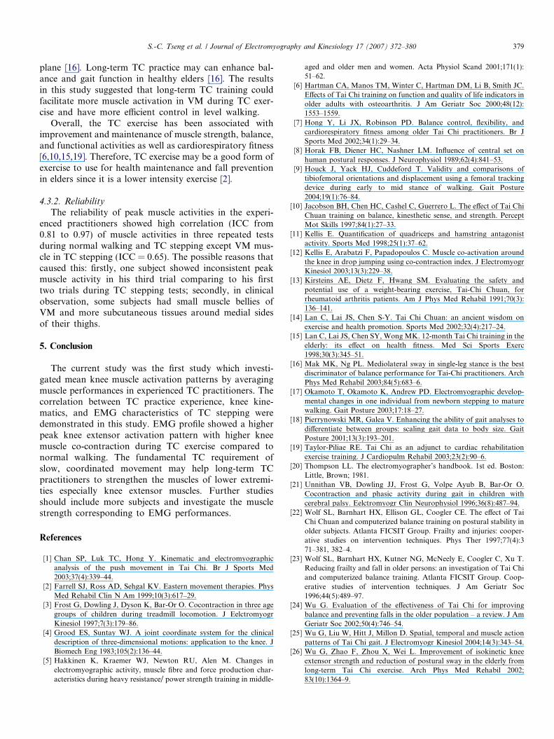
References
[1] Chan SP, Luk TC, Hong Y. Kinematic and electromyographicanalysis of the push movement in Tai Chi. Br J Sports Med2003;37(4):339–44.
[2] Farrell SJ, Ross AD, Sehgal KV. Eastern movement therapies. PhysMed Rehabil Clin N Am 1999;10(3):617–29.
[3] Frost G, Dowling J, Dyson K, Bar-Or O. Cocontraction in three agegroups of children during treadmill locomotion. J EelctromyogrKinesiol 1997;7(3):179–86.
[4] Grood ES, Suntay WJ. A joint coordinate system for the clinicaldescription of three-dimensional motions: application to the knee. JBiomech Eng 1983;105(2):136–44.
[5] Hakkinen K, Kraemer WJ, Newton RU, Alen M. Changes inelectromyographic activity, muscle fibre and force production char-acteristics during heavy resistance/ power strength training in middle-
aged and older men and women. Acta Physiol Scand 2001;171(1):51–62.
[6] Hartman CA, Manos TM, Winter C, Hartman DM, Li B, Smith JC.Effects of Tai Chi training on function and quality of life indicators inolder adults with osteoarthritis. J Am Geriatr Soc 2000;48(12):1553–1559.
[7] Hong Y, Li JX, Robinson PD. Balance control, flexibility, andcardiorespiratory fitness among older Tai Chi practitioners. Br JSports Med 2002;34(1):29–34.
[8] Horak FB, Diener HC, Nashner LM. Influence of central set onhuman postural responses. J Neurophysiol 1989;62(4):841–53.
[9] Houck J, Yack HJ, Cuddeford T. Validity and comparisons oftibiofemoral orientations and displacement using a femoral trackingdevice during early to mid stance of walking. Gait Posture2004;19(1):76–84.
[10] Jacobson BH, Chen HC, Cashel C, Guerrero L. The effect of Tai ChiChuan training on balance, kinesthetic sense, and strength. PerceptMot Skills 1997;84(1):27–33.
[11] Kellis E. Quantification of quadriceps and hamstring antagonistactivity. Sports Med 1998;25(1):37–62.
[12] Kellis E, Arabatzi F, Papadopoulos C. Muscle co-activation aroundthe knee in drop jumping using co-contraction index. J ElectromyogrKinesiol 2003;13(3):229–38.
[13] Kirsteins AE, Dietz F, Hwang SM. Evaluating the safety andpotential use of a weight-bearing exercise, Tai-Chi Chuan, forrheumatoid arthritis patients. Am J Phys Med Rehabil 1991;70(3):136–141.
[14] Lan C, Lai JS, Chen S-Y. Tai Chi Chuan: an ancient wisdom onexercise and health promotion. Sports Med 2002;32(4):217–24.
[15] Lan C, Lai JS, Chen SY, Wong MK. 12-month Tai Chi training in theelderly: its effect on health fitness. Med Sci Sports Exerc1998;30(3):345–51.
[16] Mak MK, Ng PL. Mediolateral sway in single-leg stance is the bestdiscriminator of balance performance for Tai-Chi practitioners. ArchPhys Med Rehabil 2003;84(5):683–6.
[17] Okamoto T, Okamoto K, Andrew PD. Electromyographic develop-mental changes in one individual from newborn stepping to maturewalking. Gait Posture 2003;17:18–27.
[18] Pierrynowski MR, Galea V. Enhancing the ability of gait analyses todifferentiate between groups: scaling gait data to body size. GaitPosture 2001;13(3):193–201.
[19] Taylor-Piliae RE. Tai Chi as an adjunct to cardiac rehabilitationexercise training. J Cardiopulm Rehabil 2003;23(2):90–6.
[20] Thompson LL. The electromyographer’s handbook. 1st ed. Boston:Little, Brown; 1981.
[21] Unnithan VB, Dowling JJ, Frost G, Volpe Ayub B, Bar-Or O.Cocontraction and phasic activity during gait in children withcerebral palsy. Eelctromyogr Clin Neurophysiol 1996;36(8):487–94.
[22] Wolf SL, Barnhart HX, Ellison GL, Coogler CE. The effect of TaiChi Chuan and computerized balance training on postural stability inolder subjects. Atlanta FICSIT Group. Frailty and injuries: cooper-ative studies on intervention techniques. Phys Ther 1997;77(4):371–381, 382–4.
[23] Wolf SL, Barnhart HX, Kutner NG, McNeely E, Coogler C, Xu T.Reducing frailty and fall in older persons: an investigation of Tai Chiand computerized balance training. Atlanta FICSIT Group. Coop-erative studies of intervention techniques. J Am Geriatr Soc1996;44(5):489–97.
[24] Wu G. Evaluation of the effectiveness of Tai Chi for improvingbalance and preventing falls in the older population – a review. J AmGeriatr Soc 2002;50(4):746–54.
[25] Wu G, Liu W, Hitt J, Millon D. Spatial, temporal and muscle actionpatterns of Tai Chi gait. J Electromyogr Kinesiol 2004;14(3):343–54.
[26] Wu G, Zhao F, Zhou X, Wei L. Improvement of isokinetic kneeextensor strength and reduction of postural sway in the elderly fromlong-term Tai Chi exercise. Arch Phys Med Rehabil 2002;83(10):1364–9.
380 S.-C. Tseng et al. / Journal of Electromyography and Kinesiology 17 (2007) 372–380
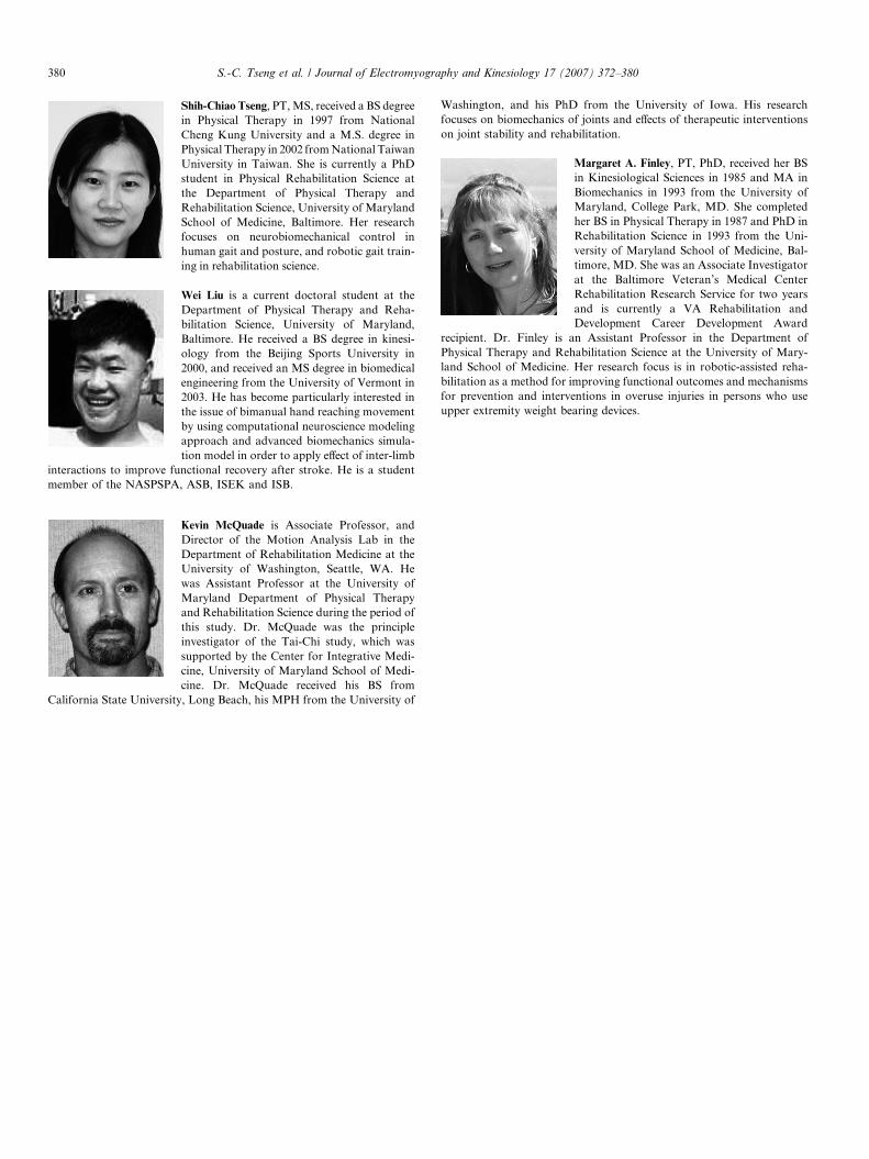
Shih-Chiao Tseng, PT, MS, received a BS degreein Physical Therapy in 1997 from NationalCheng Kung University and a M.S. degree inPhysical Therapy in 2002 from National TaiwanUniversity in Taiwan. She is currently a PhDstudent in Physical Rehabilitation Science atthe Department of Physical Therapy andRehabilitation Science, University of MarylandSchool of Medicine, Baltimore. Her researchfocuses on neurobiomechanical control inhuman gait and posture, and robotic gait train-ing in rehabilitation science.
Wei Liu is a current doctoral student at the
Department of Physical Therapy and Reha-bilitation Science, University of Maryland,Baltimore. He received a BS degree in kinesi-ology from the Beijing Sports University in2000, and received an MS degree in biomedicalengineering from the University of Vermont in 2003. He has become particularly interested inthe issue of bimanual hand reaching movementby using computational neuroscience modelingapproach and advanced biomechanics simula-tion model in order to apply effect of inter-limbinteractions to improve functional recovery after stroke. He is a studentmember of the NASPSPA, ASB, ISEK and ISB.
Kevin McQuade is Associate Professor, andDirector of the Motion Analysis Lab in theDepartment of Rehabilitation Medicine at theUniversity of Washington, Seattle, WA. Hewas Assistant Professor at the University ofMaryland Department of Physical Therapyand Rehabilitation Science during the period ofthis study. Dr. McQuade was the principleinvestigator of the Tai-Chi study, which wassupported by the Center for Integrative Medi-cine, University of Maryland School of Medi-cine. Dr. McQuade received his BS from
California State University, Long Beach, his MPH from the University of
Washington, and his PhD from the University of Iowa. His researchfocuses on biomechanics of joints and effects of therapeutic interventionson joint stability and rehabilitation.
Margaret A. Finley, PT, PhD, received her BSin Kinesiological Sciences in 1985 and MA inBiomechanics in 1993 from the University ofMaryland, College Park, MD. She completedher BS in Physical Therapy in 1987 and PhD inRehabilitation Science in 1993 from the Uni-versity of Maryland School of Medicine, Bal-timore, MD. She was an Associate Investigatorat the Baltimore Veteran’s Medical CenterRehabilitation Research Service for two yearsand is currently a VA Rehabilitation andDevelopment Career Development Award
recipient. Dr. Finley is an Assistant Professor in the Department ofPhysical Therapy and Rehabilitation Science at the University of Mary-
land School of Medicine. Her research focus is in robotic-assisted reha-bilitation as a method for improving functional outcomes and mechanismsfor prevention and interventions in overuse injuries in persons who useupper extremity weight bearing devices.