Munc18b is an essential gene in mice whose expression is limiting for secretion by airway epithelial...
15
Biochem. J. (2012) 446, 383–394 (Printed in Great Britain) doi:10.1042/BJ20120057 383 Munc18b is an essential gene in mice whose expression is limiting for secretion by airway epithelial and mast cells Kyubo KIM*†, Youlia M. PETROVA*, Brenton L. SCOTT*, Rupesh NIGAM*, Anurag AGRAWAL*‡, Christopher M. EVANS*, Zoulikha AZZEGAGH*, Alejandra GOMEZ§, Elsa M. RODARTE*§, Vesa M. OLKKONEN, Rustam BAGIRZADEH*, Lucia PICCOTTI*, Binhui REN*, Joo-Heon YOON†, James A. MCNEW¶, Roberto ADACHI*, Michael J. TUVIM* and Burton F. DICKEY* 1 *Department of Pulmonary Medicine, The University of Texas MD Anderson Cancer Center, Houston, TX 77030, U.S.A., †Department of Otorhinolaryngology, Yonsei University College of Medicine, Seoul 120-749, Korea, ‡Department of Molecular Medicine, Institute of Genomics and Integrative Biology, Delhi 110-007, India, §School of Medicine, Tecnol´ ogico de Monterrey, Monterrey, Nuevo Le´ on 64710, M´ exico, Minerva Foundation Institute for Medical Research, Helsinki 00290, Finland, and ¶Department of Biochemistry and Cell Biology, Rice University, Houston, TX 77005, U.S.A. Airway mucin secretion and MC (mast cell) degranulation must be tightly controlled for homoeostasis of the lungs and immune system respectively. We found the exocytic protein Munc18b to be highly expressed in mouse airway epithelial cells and MCs, and localized to the apical pole of airway secretory cells. To address its functions, we created a mouse with a severely hypomorphic Munc18b allele such that protein expression in heterozygotes was reduced by ∼ 50 %. Homozygous mutant mice were not viable, but heterozygotes showed a ∼ 50 % reduction in stimulated release of mucin from epithelial cells and granule contents from MCs. The defect in MCs affected only regulated secretion and not constitutive or transporter-mediated secretion. The severity of passive cutaneous anaphylaxis was also reduced by ∼ 50 %, showing that reduction of Munc18b expression results in an attenuation of physiological responses dependent on MC degranulation. The Munc18b promoter is controlled by INR (initiator), Sp1 (specificity protein 1), Ets, CRE (cAMP-response element), GRE (glucocorticoid-response element), GATA and E-box elements in airway epithelial cells; however, protein levels did not change during mucous metaplasia induced by allergic inflammation. Taken together, the results of the present study identify Munc18b as an essential gene that is a limiting component of the exocytic machinery of epithelial cells and MCs. Key words: exocytosis, mast cell, mucin, mucus, Munc18, secretion. INTRODUCTION SNARE [SNAP (soluble N-ethylmaleimide-sensitive factor- attachment protein) receptor] proteins and SM (Sec1/Munc18) proteins collaborate to mediate vesicular transport at every step of the exocytic and endocytic pathways of eukaryotic cells [1–5]. SNARE proteins form a four-helix bundle that is responsible for the specific pairing of donor and target membranes and for inducing membrane fusion. SM proteins provide a scaffold for the assembly of SNARE bundles, and in regulated exocytosis they can also hold the SNARE protein Syntaxin in a closed conformation to prevent its unregulated interaction with other SNARE proteins. Together, SM and SNARE proteins comprise the core vesicle trafficking machinery, whereas other trafficking proteins help to position the SM and SNARE proteins for productive interactions or regulate the timing of their function. There are seven SM proteins in mammals, and three of these function in regulated exocytosis: Munc18a, b and c. Munc18a is expressed in neurons [6–8], and its deletion in mice causes a complete failure of neurotransmitter release, death at birth and a gradual cell-autonomous loss of neuronal viability [9]. Munc18c is expressed ubiquitously [10], and in polarized epithelial cells is localized to the basolateral membrane [11,12]. Munc18c knockout mice die either in utero or shortly after birth, and heterozygous mice have impaired glucose tolerance due to partial defects in both insulin release from pancreatic islet cells and translocation of glucose transporters to the plasma membrane of adipocytes and skeletal muscle cells [13–15]. Munc18b is expressed in epithelial cells and leucocytes [10,16–20], but Munc18b-knockout mice have not been reported. In epithelial cells, Munc18b is localized to the apical membrane [11,21] and appears to participate in apical exocytosis based upon in vitro overexpression experiments [22–24]. In MCs (mast cells), Munc18b overexpression or knockdown results in impaired degranulation [25,26]. We targeted the murine Munc18b gene to study its role in airway epithelial and leucocyte secretion. Secretory epithelial cells of the conducting airways synthesize mucin glycoprotein polymers and secrete them into the airway lumen [27,28]. After secretion, mucins become hydrated to form a viscoelastic mucus gel that is swept from distal to proximal air- ways by the beating of ciliated epithelial cells intercalated among the secretory cells. Mucociliary clearance removes inhaled particles and pathogens from the lungs by transporting them to the pharynx to be swallowed. Insufficient mucus leaves the lungs vulnerable to injury, but excessive mucus obstructs the airway lumen and contributes to the pathophysiology of common lung Abbreviations used: AB-PAS, Alcian Blue/periodic acid/Schiff reagent; bHLH, basic helix–loop–helix; CCSP, Clara cell secretory protein; Clca3, chloride channel, calcium-activated, family member 3; CRE, cAMP-response element; DNP, 2,4-dinitrophenol; FBS, fetal bovine serum; FcεRIα, high-affinity IgE receptor, α subunit; FRT, flippase recognition target; GAPDH, glyceraldehyde-3-phosphate dehydrogenase; GRE, glucocorticoid-response element; HA, haemagglutinin; HSA, human serum albumin; HRP, horseradish peroxidase; IL-3, interleukin-3; INR, initiator; ISH, in situ hybridization; MC, mast cell; mBMMC, mouse bone-marrow-derived MC; mClca3, mouse Clca3; MFI, mean fluorescent intensity; mtCC, mouse transformed Clara cell; NK, natural killer; OCT, optimal cutting temperature compound; PAFS, periodic acid/fluorescent Schiff reagent; PBST, PBS containing 0.05% Tween 20; PGD 2 , prostaglandin D 2 ; PGK, phosphoglucokinase; SCF, stem cell factor; SM, Sec1/Munc18; SNAP, soluble N-ethylmaleimide-sensitive factor-attachment protein; SNARE, SNAP receptor; Stxbp2, syntaxin-binding protein 2; TK, thymidine kinase; TNFα, tumour necrosis factor α; WT, wild-type; YFP, yellow fluorescent protein. 1 To whom correspondence should be addressed (email [email protected]). c The Authors Journal compilation c 2012 Biochemical Society Biochemical Journal www.biochemj.org © 2012 The Author(s) The author(s) has paid for this article to be freely available under the terms of the Creative Commons Attribution Non-Commercial Licence (http://creativecommons.org/licenses/by-nc/2.5/) which permits unrestricted non-commercial use, distribution and reproduction in any medium, provided the original work is properly cited.
-
Upload
independent -
Category
Documents
-
view
2 -
download
0
Transcript of Munc18b is an essential gene in mice whose expression is limiting for secretion by airway epithelial...
Biochem. J. (2012) 446, 383–394 (Printed in Great Britain) doi:10.1042/BJ20120057 383
Munc18b is an essential gene in mice whose expression is limiting forsecretion by airway epithelial and mast cellsKyubo KIM*†, Youlia M. PETROVA*, Brenton L. SCOTT*, Rupesh NIGAM*, Anurag AGRAWAL*‡, Christopher M. EVANS*,Zoulikha AZZEGAGH*, Alejandra GOMEZ§, Elsa M. RODARTE*§, Vesa M. OLKKONEN‖, Rustam BAGIRZADEH*, Lucia PICCOTTI*,Binhui REN*, Joo-Heon YOON†, James A. MCNEW¶, Roberto ADACHI*, Michael J. TUVIM* and Burton F. DICKEY*1
*Department of Pulmonary Medicine, The University of Texas MD Anderson Cancer Center, Houston, TX 77030, U.S.A., †Department of Otorhinolaryngology, Yonsei University Collegeof Medicine, Seoul 120-749, Korea, ‡Department of Molecular Medicine, Institute of Genomics and Integrative Biology, Delhi 110-007, India, §School of Medicine, Tecnologico deMonterrey, Monterrey, Nuevo Leon 64710, Mexico, ‖Minerva Foundation Institute for Medical Research, Helsinki 00290, Finland, and ¶Department of Biochemistry and Cell Biology,Rice University, Houston, TX 77005, U.S.A.
Airway mucin secretion and MC (mast cell) degranulation mustbe tightly controlled for homoeostasis of the lungs and immunesystem respectively. We found the exocytic protein Munc18bto be highly expressed in mouse airway epithelial cells andMCs, and localized to the apical pole of airway secretory cells.To address its functions, we created a mouse with a severelyhypomorphic Munc18b allele such that protein expression inheterozygotes was reduced by ∼50%. Homozygous mutant micewere not viable, but heterozygotes showed a ∼50% reductionin stimulated release of mucin from epithelial cells and granulecontents from MCs. The defect in MCs affected only regulatedsecretion and not constitutive or transporter-mediated secretion.The severity of passive cutaneous anaphylaxis was also reduced by
∼50%, showing that reduction of Munc18b expression resultsin an attenuation of physiological responses dependent on MCdegranulation. The Munc18b promoter is controlled by INR(initiator), Sp1 (specificity protein 1), Ets, CRE (cAMP-responseelement), GRE (glucocorticoid-response element), GATA andE-box elements in airway epithelial cells; however, protein levelsdid not change during mucous metaplasia induced by allergicinflammation. Taken together, the results of the present studyidentify Munc18b as an essential gene that is a limiting componentof the exocytic machinery of epithelial cells and MCs.
Key words: exocytosis, mast cell, mucin, mucus, Munc18,secretion.
INTRODUCTION
SNARE [SNAP (soluble N-ethylmaleimide-sensitive factor-attachment protein) receptor] proteins and SM (Sec1/Munc18)proteins collaborate to mediate vesicular transport at every stepof the exocytic and endocytic pathways of eukaryotic cells[1–5]. SNARE proteins form a four-helix bundle that isresponsible for the specific pairing of donor and target membranesand for inducing membrane fusion. SM proteins provide a scaffoldfor the assembly of SNARE bundles, and in regulated exocytosisthey can also hold the SNARE protein Syntaxin in a closedconformation to prevent its unregulated interaction with otherSNARE proteins. Together, SM and SNARE proteins comprisethe core vesicle trafficking machinery, whereas other traffickingproteins help to position the SM and SNARE proteins forproductive interactions or regulate the timing of their function.
There are seven SM proteins in mammals, and three of thesefunction in regulated exocytosis: Munc18a, b and c. Munc18ais expressed in neurons [6–8], and its deletion in mice causes acomplete failure of neurotransmitter release, death at birth and agradual cell-autonomous loss of neuronal viability [9]. Munc18cis expressed ubiquitously [10], and in polarized epithelialcells is localized to the basolateral membrane [11,12]. Munc18c
knockout mice die either in utero or shortly after birth, andheterozygous mice have impaired glucose tolerance due to partialdefects in both insulin release from pancreatic islet cells andtranslocation of glucose transporters to the plasma membraneof adipocytes and skeletal muscle cells [13–15]. Munc18b isexpressed in epithelial cells and leucocytes [10,16–20], butMunc18b-knockout mice have not been reported. In epithelialcells, Munc18b is localized to the apical membrane [11,21]and appears to participate in apical exocytosis based uponin vitro overexpression experiments [22–24]. In MCs (mastcells), Munc18b overexpression or knockdown results in impaireddegranulation [25,26]. We targeted the murine Munc18b gene tostudy its role in airway epithelial and leucocyte secretion.
Secretory epithelial cells of the conducting airways synthesizemucin glycoprotein polymers and secrete them into the airwaylumen [27,28]. After secretion, mucins become hydrated to forma viscoelastic mucus gel that is swept from distal to proximal air-ways by the beating of ciliated epithelial cells intercalated amongthe secretory cells. Mucociliary clearance removes inhaledparticles and pathogens from the lungs by transporting them tothe pharynx to be swallowed. Insufficient mucus leaves the lungsvulnerable to injury, but excessive mucus obstructs the airwaylumen and contributes to the pathophysiology of common lung
Abbreviations used: AB-PAS, Alcian Blue/periodic acid/Schiff reagent; bHLH, basic helix–loop–helix; CCSP, Clara cell secretory protein; Clca3, chloridechannel, calcium-activated, family member 3; CRE, cAMP-response element; DNP, 2,4-dinitrophenol; FBS, fetal bovine serum; FcεRIα, high-affinity IgEreceptor, α subunit; FRT, flippase recognition target; GAPDH, glyceraldehyde-3-phosphate dehydrogenase; GRE, glucocorticoid-response element;HA, haemagglutinin; HSA, human serum albumin; HRP, horseradish peroxidase; IL-3, interleukin-3; INR, initiator; ISH, in situ hybridization; MC, mast cell;mBMMC, mouse bone-marrow-derived MC; mClca3, mouse Clca3; MFI, mean fluorescent intensity; mtCC, mouse transformed Clara cell; NK, natural killer;OCT, optimal cutting temperature compound; PAFS, periodic acid/fluorescent Schiff reagent; PBST, PBS containing 0.05% Tween 20; PGD2, prostaglandinD2; PGK, phosphoglucokinase; SCF, stem cell factor; SM, Sec1/Munc18; SNAP, soluble N-ethylmaleimide-sensitive factor-attachment protein; SNARE,SNAP receptor; Stxbp2, syntaxin-binding protein 2; TK, thymidine kinase; TNFα, tumour necrosis factor α; WT, wild-type; YFP, yellow fluorescent protein.
1 To whom correspondence should be addressed (email [email protected]).
c© The Authors Journal compilation c© 2012 Biochemical Society
Bio
chem
ical
Jo
urn
al
ww
w.b
ioch
emj.o
rg
© 2012 The Author(s)
The author(s) has paid for this article to be freely available under the terms of the Creative Commons Attribution Non-Commercial Licence (http://creativecommons.org/licenses/by-nc/2.5/)which permits unrestricted non-commercial use, distribution and reproduction in any medium, provided the original work is properly cited.
384 K. Kim and others
diseases such as asthma and cystic fibrosis. Thus tight control ofmucin synthesis and secretion is essential for lung health.
Similarly, regulated exocytosis is a key effector function ofleucocytes. For example, MCs store preformed inflammatorymediators in secretory granules whose contents can be rapidlyreleased upon activation of receptors for IgE, complementfragments or ATP [29]. This response helps to initiate an effectivedefence against pathogens, but can lead to anaphylaxis or allergiesif dysregulated [30]. As another example, cytotoxic T-cells andNK (natural killer) cells release the contents of secretory granulesto kill target host cells infected with viruses and to reduceleucocyte numbers after their expansion during an infection.Mutations in genes encoding the exocytic proteins Munc13-4,Syntaxin 11 and Munc18b or the lytic granule protein perforincause familial haemophagocytic lymphohistiocytosis [31–33].These examples indicate the importance of leucocyte secretoryfunction for homoeostasis.
EXPERIMENTAL
WT (wild-type) and transgenic mice
All mice were handled in accordance with the InstitutionalAnimal Care and Use Committee of The University of Texas MDAnderson Cancer Center. WT C57BL/6J mice were purchasedfrom the Jackson Laboratory. To generate HA (haemagglutinin)–YFP (yellow fluorescent protein)–Munc18b transgenic mice,the HA epitope tag and Venus YFP were cloned by PCRin tandem with rat Munc18b and inserted into a mammalianexpression vector containing the mouse CCSP (Clara cellsecretory protein) promoter, a rabbit β-globin intron, and thebovine growth hormone polyadenylation signal [34,35]. TheDNA was purified and microinjected into the pronucleus offertilized C57BL/6 oocytes, which were then implanted intopseudopregnant C57BL/6 females. Transgenic offspring wereidentified by PCR analysis of genomic DNA from tail biopsiesusing a 5′ primer specific to the CCSP promoter (5′-CT-GTGTCTTTGTCCTTCCCTGTG-3′) and 3′ primer specific toMunc18b (5′-TTACAACAGGATGACAAGATTCG-3′). Threetransgenic lines were established.
Munc18b hypomorphic mice
Homology arms and insertion fragments were generated byPCR using the BAC (bacterial artificial chromosome) cloneRP24-321J16 as a template. The backbone vector (a giftfrom Professor Thomas Sudhof, Stanford Institute for Neuro-Innovation and Translational Neurosciences, Stanford UniversitySchool of Medicine, Stanford, CA, U.S.A.) contains theneomycin-resistance gene under the control of the PGK(phosphoglucokinase) promoter (PGK-Neo) flanked by two FRT(flippase recognition target) sites situated between the two cloningsites for the recombination arms. The herpes virus TK (thymidinekinase) gene under the control of the same promoter (PGK-TK) is situated outside the homology region. The 5′ homologyarm was obtained with the primers 5′-GTCGACGGCTTT-CCTCCTCACAATT-3′ and 5′-GTCGACATGTTGATCACATT-CACCCT-3′, and included 2927 bp of the region upstream toexon 1 of the Stxbp2 (Syntaxin-binding protein 2) gene (encodingMunc18b; NCBI Gene ID 20911). This was cloned into theSalI site of the vector. The 3′ homology arm was obtainedwith primers 5′-CTCGAGGGCTGACGAGTTTCTGAA-3′ and5′-CTCGAGGGTAACTGAGGAAGACA-3′, included 3292 bpbetween introns 1 and 7, and was cloned into the XhoI site. Thereplacement insert was obtained with primers 5′-GGTACCCA-
TTATATGCATGTATGA-3′ and 5′-ATCGATCAGGGTTCAG-AAACTCGT-3′, included 1068 bp surrounding exon 1, and wascloned between the ClaI and KpnI sites. This targeting vectorwas electroporated into B6129SF1 ES cells that were then subjec-ted to positive selection of recombination events with neomycinand to negative selection of non-homologous recombinationevents with gancyclovir. ES cell clones were assessed by PCR withprimers inside the construct and outside the homology arms toconfirm homologous recombination: 5′-TTCCATTTGTCACG-TCCTGCAC-3′ and 5′-TTCCAGCACCCACATCCAGAG-3′ onthe 5′ side, and 5′-CCCTGGAACTCCTGCTGTCAT-3′ and5′-CCGCTTCCTCGTGCTTTACG-3′ on the 3′ side. Confirmedclones were expanded and ES cells were injected into C57BL/6J-Tyrc − 2J/J blastocysts, which were then implanted into pseudopreg-nant females of the same strain. Chimaeric males were crossedwith C57BL/6J-Tyrc − 2J/J females, and non-albino pups werescreened for the desired mutation by PCR with three primers: 5′-AAGGCGGTGGTAGGGGAAAGT-3′, 5′-CCGCTTCCTCGT-GCTTTACG-3′, and 5′-CTCGGGTGATCCATGATGAGG-3′.The lengths of the PCR products were 1337 bp from the WT alleleand 997 bp from the mutant allele, and the same strategy wasused for all subsequent genotyping. The expected homologousrecombination event would flank exon 1 with two loxP sites andinsert FRT-PGK-Neo-FRT into intron 1. The original goal was toremove exon 1 with Cre recombinase, eliminating the start codonfor translation of Munc18b, and any alternative ATG codon inexons 2 and 3 would induce frameshift mutations with early stopcodons. Sequencing the genomic region surrounding exon 1 inheterozygous mice revealed that the loxP site 5′ of exon 1 wasnot introduced during recombination. Nonetheless, we analysedwhether the presence of PGK-Neo in intron 1 disrupted expressionof the Munc18b gene. The hypomorphic mice used in thesestudies were on a 75–93 % C57BL6 background. Experimentaland control animals were obtained by crossings of heterozygousmutants with C57BL/6 mice.
Cell harvesting and culture
Peritoneal MCs were isolated as described previously [36]. Inbrief, after killing, the peritoneal cavity was lavaged with 10 mlof PBS. The resulting lavage fluid was centrifuged at 450 gfor 5 min at room temperature (22 ◦C), and the obtained cellpellet was resuspended in 0.5 ml of PBS. Cells were countedusing a Neubauer chamber, and differential cell counts wereperformed in cytospin samples stained with Wright–Giemsa.mBMMCs (mouse bone-marrow-derived MCs) were obtainedas described previously [29]. In brief, harvested bone marrowcells from mouse femora and tibiae were cultured for ∼6 weeksin medium supplemented with 5 ng/ml recombinant mouse IL-3(interleukin-3) and 100 ng/ml SCF (stem cell factor) (both fromR&D Systems). To follow their development, aliquots (100 μl) of5×104 non-adherent cells in the culture were analysed at differenttime intervals by flow cytometry as described below.
Antibodies
Three different rabbit polyclonal antibodies to Munc18b wereused, designated M18b-O, M18b-A and M18b-D. M18b-O wasraised against a recombinant polypeptide and supplied by VesaOlkkonen [18]; M18b-A was raised against a synthetic peptide andpurchased from Abcam (catalogue number ab3451); M18b-D wasraised by us against the isoform-specific peptide QSYSSLI-RNLEQLGGTVTNSAGSGTSS (amino acids 432–458). Theperformance of M18b-O has been described previously [18].The selectivity of M18b-A and M18b-D was tested with
c© The Authors Journal compilation c© 2012 Biochemical Society© 2012 The Author(s)
The author(s) has paid for this article to be freely available under the terms of the Creative Commons Attribution Non-Commercial Licence (http://creativecommons.org/licenses/by-nc/2.5/)which permits unrestricted non-commercial use, distribution and reproduction in any medium, provided the original work is properly cited.
Munc18b is limiting for epithelial and mast cell secretion 385
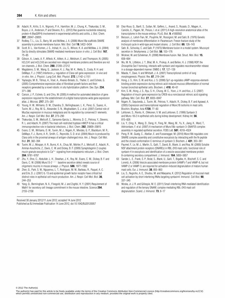
immunoblots using Munc18 proteins expressed in bacteriaand purified as described previously [37,38]. M18b-A reactedwith Munc18a ∼3-fold more intensely than Munc18b, but didnot detect Munc18c (results not shown); M18b-D detectedMunc18b, but not Munc18a or Munc18c in 100-fold greateramounts (Supplementary Figure S1 at http://www.BiochemJ.org/bj/446/bj4460383add.htm). Rabbit polyclonal antibodies againstSyntaxin 1 (#S1172) and Syntaxin 4 (#S9924) were purchasedfrom Sigma–Aldrich, to Syntaxin 2 were purchased from SynapticSystems (#110-022), and to Syntaxin 3 were supplied by VesaOlkkonen [21]. Rabbit monoclonal antibodies against GAPDH(glyceraldehyde-3-phosphate dehydrogenase) were purchasedfrom Cell Signaling Technology (#14C10), and rabbit polyclonalantibodies against mClca3 [mouse Clca3 (chloride channel,calcium-activated, family member 3)] [39] were a gift fromDr Karen Affleck (GlaxoSmithKline, Stevenage, U.K.).
Immunoblots
Tissues from mice were placed in iced 0.5% PBS containinga protease inhibitor cocktail (Sigma, #P8340, diluted 1:100),and manually homogenized using a ground glass tissue grinder(Kontes). The human lung cancer cell lines A549, Calu-3,Mes-1, H-2170 and H-292 were acquired from the A.T.C.C.(Manassas, VA, U.S.A.) and cultured according to the supplier’srecommendations. mtCCs (mouse transformed Clara cells) [40]were provided by Professor Francesco DeMayo (Program inDevelopmental Biology, Baylor College of Medicine, Houston,TX, U.S.A.) and cultured in high-glucose Dulbecco’s modifiedEagle’s medium with 5% (v/v) FBS (fetal bovine serum) (Gibco).Cell pellets from a confluent 10 cm plate were washed andresuspended in 10 ml of PBS containing 1 % Triton X-100and protease inhibitor cocktail as above, then homogenized bysonication at the maximal setting for 30 s (Sonic DismembratorF50, Fisher Scientific). Tissue and cell homogenates werecentrifuged to remove particulates, protein concentrations inthe supernatants were measured by a bicinchoninic acid assay(Pierce), and homogenates were diluted in PBS to 2 mg/ml,then boiled in SDS sample buffer [0.1 M Tris, 24% (w/v)glycerol, 8% (w/v) SDS, 0.2 M DTT (dithiothreitol) and 0.02%Coomassie Blue G-250]. Samples were resolved by SDS/PAGEusing 11% Tris-glycine or 4–15% Tris/HCl linear gradient gels,transferred to PVDF membranes (Millipore) and blocked with5% (w/v) non-fat milk in PBST (PBS containing 0.05% Tween20). Blots were incubated with primary antibodies in 5 % (w/v)non-fat milk in PBST for 1 h, washed sequentially with PBSTand PBS, then detected using HRP (horseradish peroxidase)-conjugated secondary antibodies (Jackson ImmunoResearch) andchemiluminescence reagents (Pierce). GAPDH, detected with aspecific antibody (Abcam), was used as a loading control.
ISH (in situ hybridization)
ISH was performed in collaboration with the Gene ExpressionCore Service of Baylor College of Medicine [41,42], but thepublished protocols using frozen tissue were modified for fixationand embedding to obtain better resolution as follows. Thelungs of an adult C57BL/6 mouse were inflated intratracheallywith 3.7% formaldehyde in 0.1 M phosphate buffer (pH 7.0) at15 cmH2O of pressure (1 cmH2O = 98.0665 Pa), fixed in situfor 10 min, then removed from the thoracic cavity, immersed inthe same solution overnight at 4 ◦C, embedded in paraffin andsliced into 3 μm sections. After deparaffination, acetylationand rehydration, slides were assembled in flow-throughhybridization chambers and placed into a Tecan Genesis 200
liquid-handling robot, which executed a script for non-radioactiveISH using 120 nM digoxygenin-labelled RNA antisense probespurchased from Exiqon. The probe sequences were ‘Munc18b-antisense’ 5′-AGCCCCAAGTTTGCTGTATTTT-3′, and ‘DD-scrambled’ 5′-GTGTAACACGTCTATACGCCCA-3′ that has nohomology to known RNAs. Hybridized probes were detectedby catalysed reporter deposition using biotinylated tyramidefollowed by avidin-coupled alkaline phosphatase, resulting in adark blue precipitate.
Immunohistochemistry
Lungs were inflated, fixed and embedded as describedpreviously [43]. Deparaffinated sections were washed with PBS,permeabilized with 0.05% Triton X-100 for 15 min, washed withPBST, exposed for 30 min to 6 M guanidinium chloride in 50 mMTris buffer (pH 7.5) for antigen retrieval, washed with PBST,exposed to 3 % H2O2 in 90% methanol for 30 min to quenchendogenous peroxidases, then washed with PBST. Specimenswere blocked with 5% (v/v) goat serum and 5% (v/v) FBS in PBSfor 2 h, washed with PBST, labelled overnight at 4 ◦C with M18b-O antibody diluted 1:500 in 5% (v/v) FBS and 0.5% saponinin PBS, then incubated for 1 h at 21 ◦C with HRP-labelled goatanti-(rabbit Fab) (Jackson ImmunoResearch) diluted 1:1000 in5% (v/v) FBS and 0.5% saponin in PBS, washed in PBST,and developed with 3-3′ diaminobenzidene (Vector Laboratories).Nuclei were counterstained with Methyl Green.
Fluorescence microscopy
For fluorescence microscopy of YFP–Munc18b, the lungsof transgenic mice were inflated and fixed with 4% (w/v)paraformaldehyde as above, then serially dehydrated at 4 ◦Cin 20% sucrose for 4 h, 50% sucrose for 2 h, 50% sucrose and50% OCT (optimal cutting temperature compound) overnight,then instilled with OCT intratracheally and frozen. Sections 5 μmthick were washed twice for 10 min with PBS at 21 ◦C, thenviewed through an upright Olympus BX60 microscope with aChroma Technology 41017 filter set.
For fluorescence microscopy of MCs, ears were excised,fixed overnight at 4 ◦C in 4% (w/v) paraformaldehyde (pH 7.0),dehydrated and embedded in paraffin. Avidin binds strongly tothe heparin-containing serglycin proteoglycans in the secretorygranules of MCs. Thus 5 μm cross-sections were deparaffinized,rehydrated, incubated with FITC–avidin and Hoechst dye 33342(Molecular Probes®, Life Technologies) for 1 h at 25 ◦C, and thenmounted with Fluoromount (Diagnostic BioSystems). Imageswere acquired using a fluorescent microscope with a triplefluorescent filter [DAPI (4′,6-diamidino-2-phenylindole), GFP(green fluorescent protein) and Texas Red]. Taking advantageof the autofluorescence of the cartilage and muscle observed inthe red channel, we used the Image Pro Plus software packageto delineate the dermis of the ears as the tissue between theepidermal layers, excluding all muscle and cartilage. All dermalFITC–avidin-positive cells with a Hoechst-positive nucleus werecounted. Results were expressed as the number of matureMCs/mm2 of dermis [44].
Electron microscopy
Cells were processed as described previously [29,44]. In brief,5×104 peritoneal cells or mBMMCs were fixed in 2.5% (v/v)glutaraldehyde/0.1 M sodium cacodylate (pH 7.2) for 2 h and thenincubated in 1 % (w/v) OsO4 for 1 h. After washing with double-distilled water, the cells were pelleted and embedded in Araldite
c© The Authors Journal compilation c© 2012 Biochemical Society© 2012 The Author(s)
The author(s) has paid for this article to be freely available under the terms of the Creative Commons Attribution Non-Commercial Licence (http://creativecommons.org/licenses/by-nc/2.5/)which permits unrestricted non-commercial use, distribution and reproduction in any medium, provided the original work is properly cited.
386 K. Kim and others
(Huntsman Advanced Materials), sectioned at 100 nm with anultramicrotome, stained with uranyl acetate and lead citrate,and then examined with a JEOL 200CX electron microscope.Cell profiles from each section were photographed using anunbiased random sampling technique. The fraction of cell profilesidentified morphologically as MCs (e.g. by the presence of theircharacteristic electron-dense granules, non-segmented nucleusand surface microplicae) was assessed by unbiased stereology.The volume fraction (which represents the fraction of the totalvolume of the cell occupied by granules) and the surface density(which is directly proportional to the membrane surface of thegranules per unit of volume) were calculated with the point-counting method using a cycloid grid. The area of the cell profileswas measured using a point grid.
Flow cytometry
Aliquots of 5×10⁴ mBMMCs were incubated with 200 ngof anti-mouse Kit/CD117 PE-Cy7 and 200 ng of anti-mouseFcεRIα (high-affinity IgE receptor, α subunit) Alexa Fluor® 647(eBioscience) in 100 μl of PBS for 30 min at 4 ◦C. The labelledcells were washed twice with PBS, resuspended in 0.5 ml ofPBS and analysed in a four-laser LSRII flow cytometer (Becton-Dickinson). The number of CD117+ /FcεRIα + cells and theirMFI (mean fluorescent intensity) at the emission ranges of thefluorophores were recorded, since the obtained data should beproportional to the amount of the two receptors expressed on thesurfaces of the MCs [36].
Mucin secretion
Mucous metaplasia was induced in the airways of miceby intraperitoneal immunization and aerosol challenge withovalbumin [45,46]. At 3 days after ovalbumin aerosol exposure,half of the mice were exposed for 5 min to an aerosol of100 mM ATP in 0.9% NaCl solution to induce mucin secretion,then killed after 20 min. Lungs were inflated and fixed withparaformaldehyde as above, then fixed, embedded, sectioned andstained with AB-PAS (Alcian Blue/periodic acid/Schiff reagent)or PAFS (periodic acid/fluorescent Schiff reagent) as describedpreviously [45]. Littermates were used as controls for the secretionexperiments because of the mixed genetic background. For thequantification of intracellular mucin, data are presented as theepithelial mucin volume density, derived from analysis of imagesof airways stained with PAFS as described previously [45,46].Images were acquired and analysed by investigators blinded tomouse genotype and treatment.
MC secretion assays
To test secretory responses, 106 mBMMCs were resuspendedin 1 ml of culture medium and sensitized with anti-DNP (2,4-dinitrophenol) IgE (5 μg/ml) for 3 h. After repeated washes,the cells were stimulated with 100 ng/ml DNP-HSA (humanserum albumin). Secreted products were measured in supernatantsbefore and after stimulation. Histamine was also measured incell lysates (106 mBMMCs treated with 1 ml of 0.2% Triton X-100). After stimulation, PGD2 (prostaglandin D2) was measuredat 30 min, histamine at 1 h and TNFα (tumour necrosis factorα) at 6 h. All products were measured using ELISA (OxfordBiomedical Research) [29]. Other samples of mBMMCs werecollected for electron microscopy before and after stimulation. Tostop degranulation at specified times, the cells were placed on iceand 1 ml of 5% (v/v) glutaraldehyde/0.2 M sodium cacodylate
(pH 7.2) was added to the aliquots. To test secretory responsesfrom peritoneal MCs, peritoneal lavages obtained as describedabove were enriched for MCs as follows. Cells suspended in 1 mlof modified Tyrode’s buffer (10 mM Hepes, pH 7.3, 130 mMNaCl, 5 mM KCl, 1.8 mM CaCl2, 1 mM MgCl2, 5.6 mM glucoseand 1 mg/ml BSA) were layered over a 2 ml solution of 22.5 %metrizamide (Nycomed/Accurate Chemical), then centrifuged at400 g for 15 min at 4 ◦C, and MCs were recovered at the bottom.Purification was confirmed by Toluidine Blue staining or by flowcytometry [44], then 105 peritoneal MCs were sensitized andstimulated, and histamine in cell lysates and supernatants wasdetermined as described for mBMMCs above.
Passive cutaneous anaphylaxis
Under anaesthesia with isoflurane, 100 ng of both anti-DNP IgEand anti-dansyl IgE (Pharmingen) in 20 μl of PBS were injectedintradermally into the right and left ear pinnae respectively. Twodays later, the treated mice were challenged intravenously with100 μg of DNP-HSA in 200 μl of PBS containing 0.5% EvansBlue. The mice were killed 30 min later, and their ears wereexcised and incubated in 150 μl of formamide at 55 ◦C for 24 h.The absorbance of Evans Blue in each supernatant was measuredat 610 nm using a μQuant universal microplate spectrophotometer(Bio-Tek Instruments). MC-deficient KitW − sh/KitW − sh mice werea gift from Professor Stephen Galli (Stanford University Schoolof Medicine, Stanford, CA, U.S.A.) and used as a negative controlas described previously [29].
Statistical analysis
Quantitative data are presented as means +− S.E.M., and statisticalanalysis was performed using Student’s t test (SPSS Statistics,version 16.0). P < 0.05 was considered significant.
RESULTS
Munc18b is strongly expressed in airway epithelial cells andleucocytes
The expression of Munc18b protein was analysed in mouse tissuesby immunoblotting with isoform-specific M18b-D antibodies.Bands were observed in multiple tissues at 72 kDa (Figure 1A),consistent with the mobility of post-translationally modifiedMunc18b (the unmodified protein is 67 kDa; SupplementaryFigure S1). Munc18b was most strongly expressed in lung andspleen, followed by kidney and colon, and weakly in liver.Munc18b was not detected in brain, heart or duodenum. Similarresults were obtained using M18b-O and M18b-A antibodies,along with strong expression in MCs (Supplementary Figure S2at http://www.BiochemJ.org/bj/446/bj4460383add.htm). Takentogether, these results suggest that Munc18b protein is moststrongly expressed in secretory epithelia and leucocytes.
To assess Munc18b expression in mouse conducting airwaysthat contain mucin-secreting goblet cells separately from lungalveoli that contain surfactant-secreting Type 2 cells, trachealtissue was analysed and also showed staining (Figure 1A, lane 1).To assess Munc18b expression in isolated lung epithelial secretorycells, human and mouse lung adenocarcinoma cell lines wereanalysed, and bands of the appropriate size were observed inall of these (shown in Figure 1A for A549 and in SupplementaryFigure S3 at http://www.BiochemJ.org/bj/446/bj4460383add.htmfor Calu-3 and mtCC).
c© The Authors Journal compilation c© 2012 Biochemical Society© 2012 The Author(s)
The author(s) has paid for this article to be freely available under the terms of the Creative Commons Attribution Non-Commercial Licence (http://creativecommons.org/licenses/by-nc/2.5/)which permits unrestricted non-commercial use, distribution and reproduction in any medium, provided the original work is properly cited.
Munc18b is limiting for epithelial and mast cell secretion 387
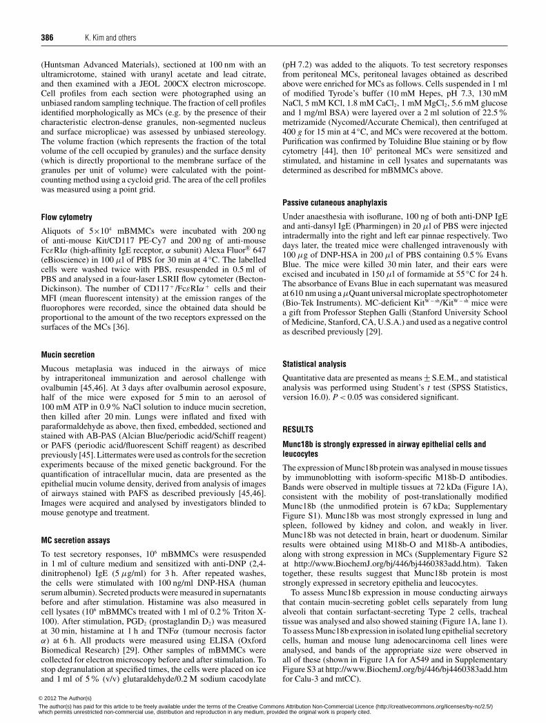
Figure 1 Munc18b is strongly expressed in lung epithelial cells and spleen
(A) Immunoblots of C57BL/6 mouse tissues and the A549 human lung adenocarcinoma cellline were probed with an isoform-specific polyclonal anti-Munc18b antibody (M18b-D). Darkbands consistent with post-translationally modified Munc18b are observed at 72 kDa. (B) ISH.An antisense RNA probe specific for Munc18b was used to analyse mRNA distribution in asection of mouse lung (upper panel). There is strong linear staining in airway epithelium (solidarrowhead), but not in vascular endothelium (open arrowhead), and scattered staining on thelung surface (lower left) and in alveoli. Scale bar = 200 μm for the low-magnification imageand 20 μm for the inset. As a negative control, a section of mouse lung was incubated with ascrambled sequence probe (lower panel).
ISH using a short synthetic isoform-specific probe showedstrongly expression of Munc18b in airway epithelial cells(Figure 1B, upper panel, solid arrowhead). In contrast, therewas little or no staining in endothelial cells of neighbouringblood vessels within the bronchovascular bundle (Figure 1B,upper panel, open arrowhead), and no staining anywhere witha control probe (Figure 1B, lower panel). In addition to airwayepithelial cells, there was linear staining of mesothelial cells onthe lung surface (Figure 1B, upper panel, lower left), and scatteredpunctate staining of alveolar epithelial Type 2 cells and alveolarmacrophages (Figure 1B, upper panel). At higher magnification ofthe airway epithelium (Figure 1B, inset), staining appeared to bepresent in alternating cells, consistent with the alternating ciliatedand secretory phenotypes of airway epithelium [28,45], although
cilia or other distinguishing features of either cell type could notbe identified with certainty.
Munc18b localizes to the apical pole of airway secretory cells
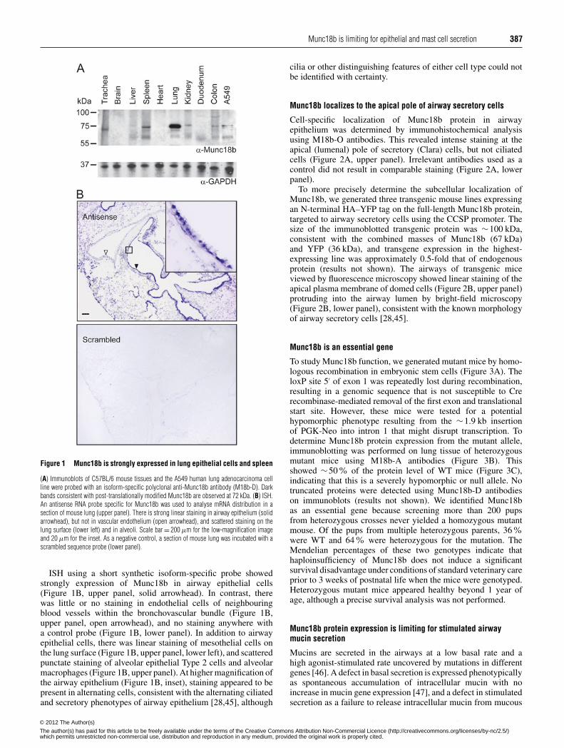
Cell-specific localization of Munc18b protein in airwayepithelium was determined by immunohistochemical analysisusing M18b-O antibodies. This revealed intense staining at theapical (lumenal) pole of secretory (Clara) cells, but not ciliatedcells (Figure 2A, upper panel). Irrelevant antibodies used as acontrol did not result in comparable staining (Figure 2A, lowerpanel).
To more precisely determine the subcellular localization ofMunc18b, we generated three transgenic mouse lines expressingan N-terminal HA–YFP tag on the full-length Munc18b protein,targeted to airway secretory cells using the CCSP promoter. Thesize of the immunoblotted transgenic protein was ∼100 kDa,consistent with the combined masses of Munc18b (67 kDa)and YFP (36 kDa), and transgene expression in the highest-expressing line was approximately 0.5-fold that of endogenousprotein (results not shown). The airways of transgenic miceviewed by fluorescence microscopy showed linear staining of theapical plasma membrane of domed cells (Figure 2B, upper panel)protruding into the airway lumen by bright-field microscopy(Figure 2B, lower panel), consistent with the known morphologyof airway secretory cells [28,45].
Munc18b is an essential gene
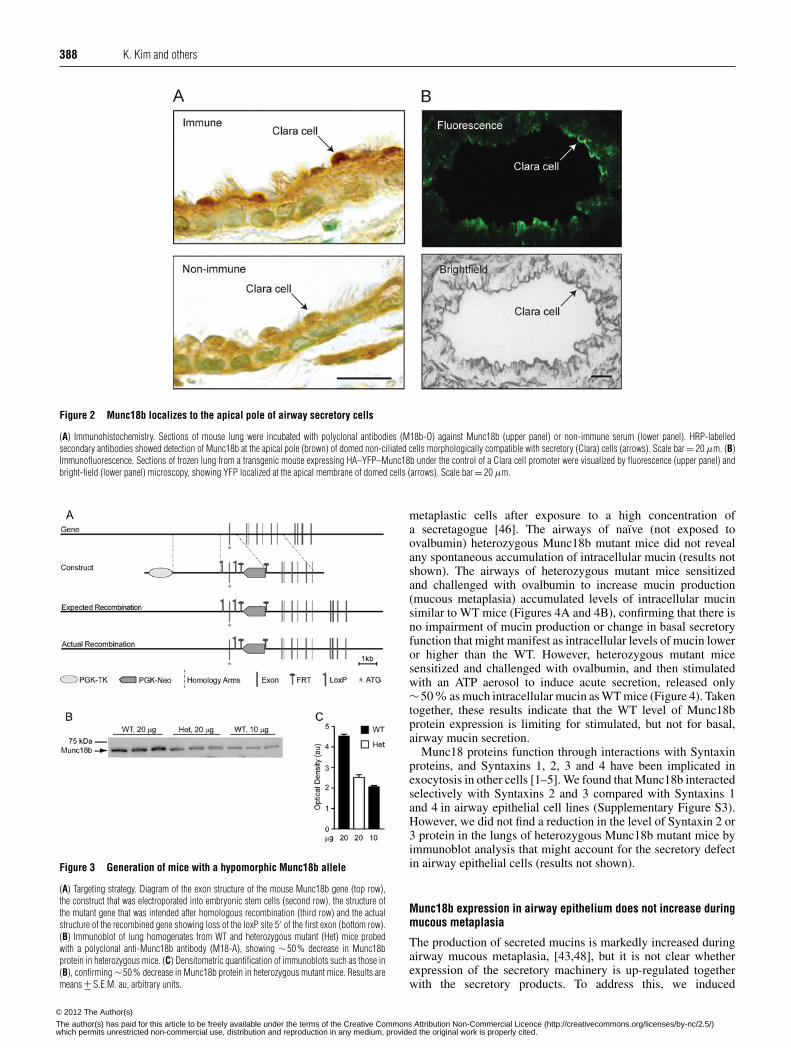
To study Munc18b function, we generated mutant mice by homo-logous recombination in embryonic stem cells (Figure 3A). TheloxP site 5′ of exon 1 was repeatedly lost during recombination,resulting in a genomic sequence that is not susceptible to Crerecombinase-mediated removal of the first exon and translationalstart site. However, these mice were tested for a potentialhypomorphic phenotype resulting from the ∼1.9 kb insertionof PGK-Neo into intron 1 that might disrupt transcription. Todetermine Munc18b protein expression from the mutant allele,immunoblotting was performed on lung tissue of heterozygousmutant mice using M18b-A antibodies (Figure 3B). Thisshowed ∼50% of the protein level of WT mice (Figure 3C),indicating that this is a severely hypomorphic or null allele. Notruncated proteins were detected using Munc18b-D antibodieson immunoblots (results not shown). We identified Munc18bas an essential gene because screening more than 200 pupsfrom heterozygous crosses never yielded a homozygous mutantmouse. Of the pups from multiple heterozygous parents, 36 %were WT and 64% were heterozygous for the mutation. TheMendelian percentages of these two genotypes indicate thathaploinsufficiency of Munc18b does not induce a significantsurvival disadvantage under conditions of standard veterinary careprior to 3 weeks of postnatal life when the mice were genotyped.Heterozygous mutant mice appeared healthy beyond 1 year ofage, although a precise survival analysis was not performed.
Munc18b protein expression is limiting for stimulated airwaymucin secretion
Mucins are secreted in the airways at a low basal rate and ahigh agonist-stimulated rate uncovered by mutations in differentgenes [46]. A defect in basal secretion is expressed phenotypicallyas spontaneous accumulation of intracellular mucin with noincrease in mucin gene expression [47], and a defect in stimulatedsecretion as a failure to release intracellular mucin from mucous
c© The Authors Journal compilation c© 2012 Biochemical Society© 2012 The Author(s)
The author(s) has paid for this article to be freely available under the terms of the Creative Commons Attribution Non-Commercial Licence (http://creativecommons.org/licenses/by-nc/2.5/)which permits unrestricted non-commercial use, distribution and reproduction in any medium, provided the original work is properly cited.
388 K. Kim and others
Figure 2 Munc18b localizes to the apical pole of airway secretory cells
(A) Immunohistochemistry. Sections of mouse lung were incubated with polyclonal antibodies (M18b-O) against Munc18b (upper panel) or non-immune serum (lower panel). HRP-labelledsecondary antibodies showed detection of Munc18b at the apical pole (brown) of domed non-ciliated cells morphologically compatible with secretory (Clara) cells (arrows). Scale bar = 20 μm. (B)Immunofluorescence. Sections of frozen lung from a transgenic mouse expressing HA–YFP–Munc18b under the control of a Clara cell promoter were visualized by fluorescence (upper panel) andbright-field (lower panel) microscopy, showing YFP localized at the apical membrane of domed cells (arrows). Scale bar = 20 μm.
Figure 3 Generation of mice with a hypomorphic Munc18b allele
(A) Targeting strategy. Diagram of the exon structure of the mouse Munc18b gene (top row),the construct that was electroporated into embryonic stem cells (second row), the structure ofthe mutant gene that was intended after homologous recombination (third row) and the actualstructure of the recombined gene showing loss of the loxP site 5′ of the first exon (bottom row).(B) Immunoblot of lung homogenates from WT and heterozygous mutant (Het) mice probedwith a polyclonal anti-Munc18b antibody (M18-A), showing ∼50 % decrease in Munc18bprotein in heterozygous mice. (C) Densitometric quantification of immunoblots such as those in(B), confirming ∼50 % decrease in Munc18b protein in heterozygous mutant mice. Results aremeans +− S.E.M. au, arbitrary units.
metaplastic cells after exposure to a high concentration ofa secretagogue [46]. The airways of naıve (not exposed toovalbumin) heterozygous Munc18b mutant mice did not revealany spontaneous accumulation of intracellular mucin (results notshown). The airways of heterozygous mutant mice sensitizedand challenged with ovalbumin to increase mucin production(mucous metaplasia) accumulated levels of intracellular mucinsimilar to WT mice (Figures 4A and 4B), confirming that there isno impairment of mucin production or change in basal secretoryfunction that might manifest as intracellular levels of mucin loweror higher than the WT. However, heterozygous mutant micesensitized and challenged with ovalbumin, and then stimulatedwith an ATP aerosol to induce acute secretion, released only∼50% as much intracellular mucin as WT mice (Figure 4). Takentogether, these results indicate that the WT level of Munc18bprotein expression is limiting for stimulated, but not for basal,airway mucin secretion.
Munc18 proteins function through interactions with Syntaxinproteins, and Syntaxins 1, 2, 3 and 4 have been implicated inexocytosis in other cells [1–5]. We found that Munc18b interactedselectively with Syntaxins 2 and 3 compared with Syntaxins 1and 4 in airway epithelial cell lines (Supplementary Figure S3).However, we did not find a reduction in the level of Syntaxin 2 or3 protein in the lungs of heterozygous Munc18b mutant mice byimmunoblot analysis that might account for the secretory defectin airway epithelial cells (results not shown).
Munc18b expression in airway epithelium does not increase duringmucous metaplasia
The production of secreted mucins is markedly increased duringairway mucous metaplasia, [43,48], but it is not clear whetherexpression of the secretory machinery is up-regulated togetherwith the secretory products. To address this, we induced
c© The Authors Journal compilation c© 2012 Biochemical Society© 2012 The Author(s)
The author(s) has paid for this article to be freely available under the terms of the Creative Commons Attribution Non-Commercial Licence (http://creativecommons.org/licenses/by-nc/2.5/)which permits unrestricted non-commercial use, distribution and reproduction in any medium, provided the original work is properly cited.
Munc18b is limiting for epithelial and mast cell secretion 389
Figure 4 Defect in stimulated mucin secretion in heterozygous mutantMunc18b mice
(A) Lung sections stained with PAFS from mice sensitized and challenged with ovalbumin toincrease mucin expression, then exposed to aerosolized ATP to induce mucin secretion. BothWT and heterozygous (Het) mutant mice accumulate abundant intracellular mucin (orange)after ovalbumin sensitization and challenge (upper panels). In response to ATP, heterozygousmutant mice release only a small fraction of mucin compared with WT (lower panels). Scalebar = 20 μm. (B) The volume density of retained intracellular mucin with or without ATPexposure as in (A) was quantified using fluorescence microscopy (n = 17–23 mice per groupfor four separate experiments). (C) The percentage of mucin released for each genotype inresponse to ATP was calculated from the data in (B), and results are weighted means +− S.E.M.for four separate experiments. *P < 0.05.
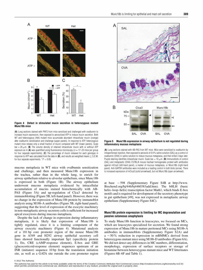
mucous metaplasia in WT mice with ovalbumin sensitizationand challenge, and then measured Munc18b expression inthe trachea, rather than in the whole lung, to enrich forairway epithelium relative to alveolar epithelium, since Munc18bis expressed in both (Figure 1B). The airway epitheliumunderwent mucous metaplasia evidenced by intracellularaccumulation of mucins stained histochemically with AB-PAS (Figure 5A) and up-regulation of Clca3 detected byimmunoblotting (Figure 5B, left-hand panel). However, there wasno change in the expression of Munc18b protein by immunoblotanalysis using M18b-A antibodies (Figure 5B, right-hand panel),suggesting that the level of expression of the exocytic machineryin non-metaplastic airway secretory cells is sufficient for increasedapical exocytosis during mucous metaplasia.
Despite the lack of change in expression during inflammatorymetaplasia, it is likely that expression of Munc18b istightly regulated, since it is a limiting component of theairway exocytic machinery (Figure 4). Mutational analysisof a 181 bp core promoter region of the mouse Munc18bgene in A549 and H292 airway epithelial secretory celllines showed functionally important Sp1 (specificity protein1), Ets, CRE (cAMP-response element), E-box and GRE(glucocorticoid-response element) sequences upstream of anINR (initiator) sequence 10 bp from the translation initiationsite, as well as a GATA site outside the core promoter region
Figure 5 Munc18b expression in airway epithelium is not regulated duringinflammatory mucous metaplasia
(A) Lung sections stained with AB-PAS from WT mice. Mice were sensitized to ovalbumin byintraperitoneal injection, then exposed to aerosols of 0.9 % saline solution (SAL) as a control orovalbumin (OVA) in saline solution to induce mucous metaplasia, and then killed 3 days later.Purple staining identifies intracellular mucin. Scale bar = 10 μm. (B) Immunoblots of control(SAL) and metaplastic (OVA) C57BL/6 mouse tracheal homogenates probed with antibodiesagainst mClca3 (left-hand panel), a marker of mucous metaplasia, or Munc18b (right-handpanel). Anti-GAPDH antibodies were included as a loading control in both blots (arrow). Thereis increased expression of mClca3 (solid arrowhead), but not Munc18b (open arrowhead).
at base − 598 (Supplementary Figure S4B at http://www.BiochemJ.org/bj/446/bj4460383add.htm). The bHLH (basichelix–loop–helix) transcription factor Math1, which binds E-boxmotifs and is required for development of the secretory phenotypein gut epithelium [49], was not expressed in metaplastic airwayepithelium (Supplementary Figure S4C).
Munc18b protein expression is limiting for MC degranulation andpassive cutaneous anaphylaxis
To study Munc18b function in leucocytes, we focused on MCs,which are leucocytes specialized for secretion. We found strongexpression of Munc18b in mature peritoneal MCs using M18b-Aantibodies in immunoblots (Supplementary Figure S2A) anda ∼50% reduction in expression in mBMMCs derived fromheterozygous mutant mice using M18b-D antibodies (Figure 6A).We did not detect any differences in MC numbers, differentiation,morphology, expression of surface receptors or storage ofhistamine between heterozygous mutant mice and WT littermates(Figures 6B–6F and Table 1).
c© The Authors Journal compilation c© 2012 Biochemical Society© 2012 The Author(s)
The author(s) has paid for this article to be freely available under the terms of the Creative Commons Attribution Non-Commercial Licence (http://creativecommons.org/licenses/by-nc/2.5/)which permits unrestricted non-commercial use, distribution and reproduction in any medium, provided the original work is properly cited.
390 K. Kim and others
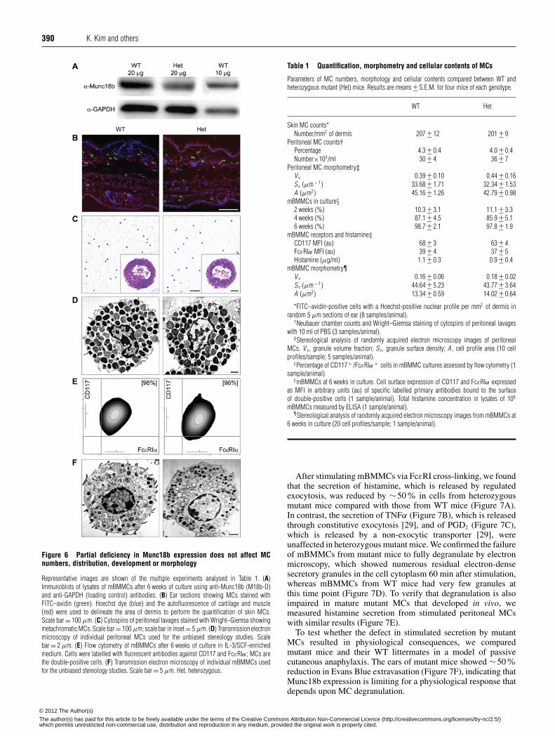
Figure 6 Partial deficiency in Munc18b expression does not affect MCnumbers, distribution, development or morphology
Representative images are shown of the multiple experiments analysed in Table 1. (A)Immunoblots of lysates of mBMMCs after 6 weeks of culture using anti-Munc18b (M18b-D)and anti-GAPDH (loading control) antibodies. (B) Ear sections showing MCs stained withFITC–avidin (green). Hoechst dye (blue) and the autofluorescence of cartilage and muscle(red) were used to delineate the area of dermis to perform the quantification of skin MCs.Scale bar = 100 μm. (C) Cytospins of peritoneal lavages stained with Wright–Giemsa showingmetachromatic MCs. Scale bar = 100 μm; scale bar in inset = 5 μm. (D) Transmission electronmicroscopy of individual peritoneal MCs used for the unbiased stereology studies. Scalebar = 2 μm. (E) Flow cytometry of mBMMCs after 6 weeks of culture in IL-3/SCF-enrichedmedium. Cells were labelled with fluorescent antibodies against CD117 and FcεRIα; MCs arethe double-positive cells. (F) Transmission electron microscopy of individual mBMMCs usedfor the unbiased stereology studies. Scale bar = 5 μm. Het, heterozygous.
Table 1 Quantification, morphometry and cellular contents of MCs
Parameters of MC numbers, morphology and cellular contents compared between WT andheterozygous mutant (Het) mice. Results are means +− S.E.M. for four mice of each genotype.
WT Het
Skin MC counts*Number/mm2 of dermis 207 +− 12 201 +− 9
Peritoneal MC counts†Percentage 4.3 +− 0.4 4.0 +− 0.4Number×103/ml 30 +− 4 36 +− 7
Peritoneal MC morphometry‡V v 0.39 +− 0.10 0.44 +− 0.16Sv (μm − 1) 33.68 +− 1.71 32.34 +− 1.53A (μm2) 45.16 +− 1.26 42.79 +− 0.98
mBMMCs in culture§2 weeks (%) 10.3 +− 3.1 11.1 +− 3.34 weeks (%) 87.1 +− 4.5 85.9 +− 5.16 weeks (%) 98.7 +− 2.1 97.8 +− 1.9
mBMMC receptors and histamine‖CD117 MFI (au) 68 +− 3 63 +− 4FcεRIα MFI (au) 39 +− 4 37 +− 5Histamine (μg/ml) 1.1 +− 0.3 0.9 +− 0.4
mBMMC morphometry¶V v 0.16 +− 0.06 0.18 +− 0.02Sv (μm − 1) 44.64 +− 5.23 43.77 +− 3.64A (μm2) 13.34 +− 0.59 14.02 +− 0.64
*FITC–avidin-positive cells with a Hoechst-positive nuclear profile per mm2 of dermis inrandom 5 μm sections of ear (8 samples/animal).
†Neubauer chamber counts and Wright–Giemsa staining of cytospins of peritoneal lavageswith 10 ml of PBS (3 samples/animal).
‡Stereological analysis of randomly acquired electron microscopy images of peritonealMCs. V v, granule volume fraction; Sv, granule surface density; A , cell profile area (10 cellprofiles/sample; 5 samples/animal).
§Percentage of CD117+ /FcεRIα + cells in mBMMC cultures assessed by flow cytometry (1sample/animal).
‖mBMMCs at 6 weeks in culture. Cell surface expression of CD117 and FcεRIα expressedas MFI in arbitrary units (au) of specific labelled primary antibodies bound to the surfaceof double-positive cells (1 sample/animal). Total histamine concentration in lysates of 106
mBMMCs measured by ELISA (1 sample/animal).¶Stereological analysis of randomly acquired electron microscopy images from mBMMCs at
6 weeks in culture (20 cell profiles/sample; 1 sample/animal).
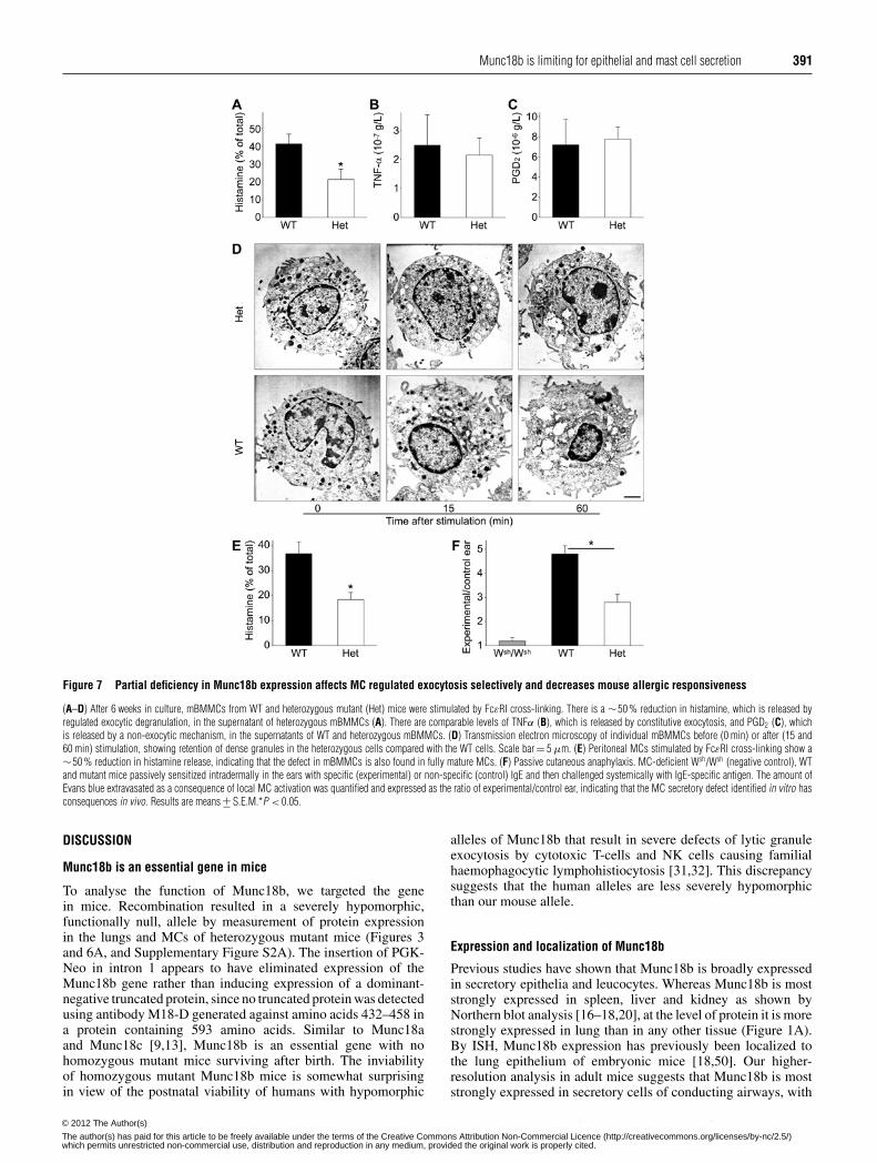
After stimulating mBMMCs via FcεRI cross-linking, we foundthat the secretion of histamine, which is released by regulatedexocytosis, was reduced by ∼50% in cells from heterozygousmutant mice compared with those from WT mice (Figure 7A).In contrast, the secretion of TNFα (Figure 7B), which is releasedthrough constitutive exocytosis [29], and of PGD2 (Figure 7C),which is released by a non-exocytic transporter [29], wereunaffected in heterozygous mutant mice. We confirmed the failureof mBMMCs from mutant mice to fully degranulate by electronmicroscopy, which showed numerous residual electron-densesecretory granules in the cell cytoplasm 60 min after stimulation,whereas mBMMCs from WT mice had very few granules atthis time point (Figure 7D). To verify that degranulation is alsoimpaired in mature mutant MCs that developed in vivo, wemeasured histamine secretion from stimulated peritoneal MCswith similar results (Figure 7E).
To test whether the defect in stimulated secretion by mutantMCs resulted in physiological consequences, we comparedmutant mice and their WT littermates in a model of passivecutaneous anaphylaxis. The ears of mutant mice showed ∼50%reduction in Evans Blue extravasation (Figure 7F), indicating thatMunc18b expression is limiting for a physiological response thatdepends upon MC degranulation.
c© The Authors Journal compilation c© 2012 Biochemical Society© 2012 The Author(s)
The author(s) has paid for this article to be freely available under the terms of the Creative Commons Attribution Non-Commercial Licence (http://creativecommons.org/licenses/by-nc/2.5/)which permits unrestricted non-commercial use, distribution and reproduction in any medium, provided the original work is properly cited.
Munc18b is limiting for epithelial and mast cell secretion 391
Figure 7 Partial deficiency in Munc18b expression affects MC regulated exocytosis selectively and decreases mouse allergic responsiveness
(A–D) After 6 weeks in culture, mBMMCs from WT and heterozygous mutant (Het) mice were stimulated by FcεRI cross-linking. There is a ∼50 % reduction in histamine, which is released byregulated exocytic degranulation, in the supernatant of heterozygous mBMMCs (A). There are comparable levels of TNFα (B), which is released by constitutive exocytosis, and PGD2 (C), whichis released by a non-exocytic mechanism, in the supernatants of WT and heterozygous mBMMCs. (D) Transmission electron microscopy of individual mBMMCs before (0 min) or after (15 and60 min) stimulation, showing retention of dense granules in the heterozygous cells compared with the WT cells. Scale bar = 5 μm. (E) Peritoneal MCs stimulated by FcεRI cross-linking show a∼50 % reduction in histamine release, indicating that the defect in mBMMCs is also found in fully mature MCs. (F) Passive cutaneous anaphylaxis. MC-deficient Wsh/Wsh (negative control), WTand mutant mice passively sensitized intradermally in the ears with specific (experimental) or non-specific (control) IgE and then challenged systemically with IgE-specific antigen. The amount ofEvans blue extravasated as a consequence of local MC activation was quantified and expressed as the ratio of experimental/control ear, indicating that the MC secretory defect identified in vitro hasconsequences in vivo. Results are means +− S.E.M.*P < 0.05.
DISCUSSION
Munc18b is an essential gene in mice
To analyse the function of Munc18b, we targeted the genein mice. Recombination resulted in a severely hypomorphic,functionally null, allele by measurement of protein expressionin the lungs and MCs of heterozygous mutant mice (Figures 3and 6A, and Supplementary Figure S2A). The insertion of PGK-Neo in intron 1 appears to have eliminated expression of theMunc18b gene rather than inducing expression of a dominant-negative truncated protein, since no truncated protein was detectedusing antibody M18-D generated against amino acids 432–458 ina protein containing 593 amino acids. Similar to Munc18aand Munc18c [9,13], Munc18b is an essential gene with nohomozygous mutant mice surviving after birth. The inviabilityof homozygous mutant Munc18b mice is somewhat surprisingin view of the postnatal viability of humans with hypomorphic
alleles of Munc18b that result in severe defects of lytic granuleexocytosis by cytotoxic T-cells and NK cells causing familialhaemophagocytic lymphohistiocytosis [31,32]. This discrepancysuggests that the human alleles are less severely hypomorphicthan our mouse allele.
Expression and localization of Munc18b
Previous studies have shown that Munc18b is broadly expressedin secretory epithelia and leucocytes. Whereas Munc18b is moststrongly expressed in spleen, liver and kidney as shown byNorthern blot analysis [16–18,20], at the level of protein it is morestrongly expressed in lung than in any other tissue (Figure 1A).By ISH, Munc18b expression has previously been localized tothe lung epithelium of embryonic mice [18,50]. Our higher-resolution analysis in adult mice suggests that Munc18b is moststrongly expressed in secretory cells of conducting airways, with
c© The Authors Journal compilation c© 2012 Biochemical Society© 2012 The Author(s)
The author(s) has paid for this article to be freely available under the terms of the Creative Commons Attribution Non-Commercial Licence (http://creativecommons.org/licenses/by-nc/2.5/)which permits unrestricted non-commercial use, distribution and reproduction in any medium, provided the original work is properly cited.
392 K. Kim and others
lower levels of expression in alveolar secretory cells, alveolarmacrophages and mesothelial cells (Figure 1B). At a subcellularlevel, Munc18b has been localized to the apical membrane ofpolarized epithelial cells of the intestine and kidney [11,21].We similarly find Munc18b localized to the apical membraneof airway secretory cells by immunohistochemistry and bytransgenic expression of Munc18b labelled with a fluorescenttag (Figure 2). After lung, Munc18b is most strongly expressed inspleen (Figure 1A), which is rich in leucocytes, and Munc18b isstrongly expressed in MCs (Figure 5 and Supplementary FigureS2), which are a leucocyte lineage specialized for secretion.
Function of Munc18b in airway mucin secretion
It is of historic interest that mucin secretion in protozoa wasamong the earliest exocytic processes studied genetically andby high-resolution electron microscopy [51–53]. To analyse therole of Munc18b in mammalian airway mucin secretion, wetargeted the gene in mice. Since homozygous mutant mice werenot viable postnatally, function was studied in heterozygousmutant mice. Heterozygous Munc18b mutant mice did notshow spontaneous accumulation of intracellular mucin, whichwould indicate a defect in basal secretion, but did show a∼50% decrease of stimulated mucin secretion (Figure 4). Thissuggests that Munc18b is a limiting component of regulatedexocytosis at high secretory rates, and a similar dose-dependencyof stimulated mucin secretion on synaptotagmin-2 expression hasbeen observed [46]. Fruitflies heterozygous for a null mutationof Rop, the Munc18 homologue in Drosophila, also showed amarked reduction in synaptic vesicle release [54], suggesting thatMunc18 protein expression is limiting for stimulated secretionbroadly across isoforms, cell types and organisms.
Stable post-developmental expression of Munc18b in airwayepithelium
In view of its limiting role in stimulated mucin secretion, animportant question is whether Munc18b expression is increasedalong with that of mucins and other secreted gene products duringmucous metaplasia. For example, Clca3 transcripts increase morethan 1000-fold during mucous metaplasia [48], and Muc5acmucin transcripts increase more than 40-fold [43]. Despite themarked increase in secreted products, the level of Munc18bprotein did not change detectably during mucous metaplasia(Figure 5) in an assay sensitive to a 2-fold change (Figure 3B).The ability of WT epithelial cells to nonetheless acutely releasemost accumulated mucins in response to a strong stimulus [46](Figure 4) indicates that airway secretory cells constitutivelypossess substantial exocytic reserve capacity. The strong andstable expression of Munc18b protein in airway secretory cellsshould make it a useful marker of the airway epithelial secretorylineage.
Transcriptional control of Munc18b expression
Despite the lack of post-developmental change in Munc18bexpression during allergic mucous metaplasia (Figure 5),it is likely that Munc18b expression is tightly regulatedduring development of the airway epithelium because ofits limiting role in secretion (Figure 3). Supporting thisinference, the Munc18b promoter contains binding sites fortranscription factors known to be important in airway epithelialdevelopment (Supplementary Figure S3). These include GRE,CRE and GATA sites, consistent with the well-known role of
glucocorticosteroids in lung epithelial secretory cell development[55], the importance of CREB (cAMP-response-element-bindingprotein) in mucociliary differentiation of airway epithelium inresponse to retinoids [56,57] and the requirement for GATA-6 in development of the bronchiolar epithelium [55–57]. Thepromoter also contains an E-box that binds bHLH transcriptionfactors required for development of a secretory phenotypein gut epithelium [49], and an Ets motif that could interactwith SPDEF (SAM-pointed-domain-containing Ets factor),which co-activates the expression of multiple lung epithelialgenes [55]. Munc18b expression also increases during MCdevelopment [58] and the same transcription-factor-binding sitesare functional in MCs [20], so comparison of the transcriptionfactors controlling Munc18b expression during development ofMCs and airway secretory cells would be informative.
Function of Munc18b in MC secretion
The commensurate reductions in Munc18b expression(Figure 5A) and stimulated degranulation (Figures 6A and 6D)of MCs from heterozygous mutant mice indicates that Munc18bcan be a limiting component of the regulated exocytic machineryfor leucocytes as well as epithelial cells. The fact that histamine,but not TNF-α, secretion was affected (Figures 6A and 6B) pointsto a selective role of Munc18b in regulated exocytosis, but not inconstituve exocytosis, in MCs. Our control experiments supportthat the functional differences we found between heterozygoteand WT MCs and mice could not be attributed to an intrinsicdefect in MC granulation, development, distribution or retentionin different tissues (Figures 6B–6F and Table 1). In addition, ourfindings that the cell surface densities of receptors for IgE andSCF (Figure 6E), and that cytokine and prostaglandin secretion(Figures 7B and 7C), were almost identical between mutant andWT MCs indicate that there was no deficiency in the signaltransduction pathways required for MC activation.
The correlation that we found between protein expression andcellular secretory function in MCs was further extended by thefinding of a matching reduction in physiological function inheterozygous mice in passive cutaneous anaphylaxis (Figure 6F),a response highly dependent on MC degranulation. Takentogether, these results suggest that variation in the expressionof Munc18b or other limiting components of the MC exocyticmachinery could alter the release of preformed inflammatorymediators by this cell and be a cause of variation in resistanceto infection or susceptibility to inflammatory diseases [29,44].For the same reason, targeting the expression or function of theMC exocytic machinery could be a therapeutic strategy.
Interaction of Munc18b with exocytic Syntaxins
The Syntaxin that interacts with Munc18b to mediate regulatedsecretion in epithelial cells and MCs is not known with certainty.Munc18b has been found to interact with Syntaxins 1, 2 and 3, butnot Syntaxin 4, in other cells [11,16,18,59,60], and we obtainedsimilar results in airway secretory cells (Supplementary FigureS3). Overexpression of Munc18b in epithelial cell lines preventedthe association of Syntaxin 3 with SNAP-23 and VAMP (vesicle-associated membrane protein) 3 and 8 [21,61] and inhibitedthe apical delivery of marker proteins [22–24], suggesting thatMunc18b interacts with Syntaxin 3 in apical epithelial exocytosis.However, Syntaxin 3 protein levels were not reduced in ourheterozygous mutant mice, indicating that the dependence ofstimulated airway mucin secretion on Munc18b is not exclusivelymediated by its stoichiometric interactions with Syntaxin 3 or that
c© The Authors Journal compilation c© 2012 Biochemical Society© 2012 The Author(s)
The author(s) has paid for this article to be freely available under the terms of the Creative Commons Attribution Non-Commercial Licence (http://creativecommons.org/licenses/by-nc/2.5/)which permits unrestricted non-commercial use, distribution and reproduction in any medium, provided the original work is properly cited.
Munc18b is limiting for epithelial and mast cell secretion 393
Syntaxin 3 does not in fact mediate airway mucin secretion. InMCs, Syntaxin 4 has been implicated in degranulation by over-expression [62], blocking antibodies [63] and small interferingRNA [64,65], even though Munc18b did not physically interactwith Syntaxin 4 [25,26]. In cytolytic T-cells and NK cells,mutations in Syntaxin 11 phenocopy mutations in Munc18b incausing defects in lytic granule exocytosis that result in familialhaemophagocytic lymphohistiocytosis [31,32], suggesting thatMunc18b and Syntaxin 11 collaborate in mediating exocytosisin these cells. We did not assess the expression of Syntaxin 11or its interactions with Munc18b in airway epithelial cell lines orMCs, but it should also be considered a candidate SNARE infuture studies of airway epithelial and MC exocytosis.
AUTHOR CONTRIBUTION
Burton Dickey, Roberto Adachi and Michael Tuvim designed the experiments. KyuboKim, Youlia Petrova, Brenton Scott, Rupesh Nigam, Anurag Agrawal, Christopher Evans,Zoulikha Azzegagh, Alejandra Gomez, Elsa Rodarte, Rustam Bagirzadeh, Lucia Piccottiand Binhui Ren performed the experiments. James McNew, Vesa Olkkonen and Joo-HeonYoon provided key reagents. Burton Dickey, Roberto Adachi and Kyubo Kim wrote thepaper.
ACKNOWLEDGEMENTS
We thank Professor Huda Zoghbi (Program in Developmental Biology, Baylor College ofMedicine, Houston, TX, U.S.A.) for the LacZ knockin/Math1 knockout mice and ChristinaThaller for assistance with the ISH studies.
FUNDING
This work was supported by the Cystic Fibrosis Foundation [grant number 08GO] andthe National Institutes of Health [grant numbers HL094848, HL097000, HL072984 andCA16672 (to B.F.D.) and AI093533 (to R.A.)]. B.S. was supported by an Odyssey Fellowshipfrom the MD Anderson Cancer Center.
REFERENCES
1 Carr, C. M. and Rizo, J. (2010) At the junction of SNARE and SM protein function. Curr.Opin. Cell Biol. 22, 488–495
2 McNew, J. A. (2008) Regulation of SNARE-mediated membrane fusion during exocytosis.Chem. Rev. 108, 1669–1686
3 Sudhof, T. C. and Rothman, J. E. (2009) Membrane fusion: grappling with SNARE and SMproteins. Science 323, 474–477
4 Burgoyne, R. D. and Morgan, A. (2007) Membrane trafficking: three steps to fusion. Curr.Biol. 17, R255–R258
5 Verhage, M. and Toonen, R. F. (2007) Regulated exocytosis: merging ideas on fusingmembranes. Curr. Opin. Cell Biol. 19, 402–408
6 Hata, Y., Slaughter, C. A. and Sudhof, T. C. (1993) Synaptic vesicle fusion complexcontains unc-18 homologue bound to syntaxin. Nature 366, 347–351
7 Garcia, E. P., Gatti, E., Butler, M., Burton, J. and De, C. P. (1994) A rat brain Sec1homologue related to Rop and UNC18 interacts with syntaxin. Proc. Natl. Acad. Sci.U.S.A. 91, 2003–2007
8 Pevsner, J., Hsu, S. C. and Scheller, R. H. (1994) n-Sec1: a neural-specific syntaxin-binding protein. Proc. Natl. Acad. Sci. U.S.A. 91, 1445–1449
9 Verhage, M., Maia, A. S., Plomp, J. J., Brussaard, A. B., Heeroma, J. H., Vermeer, H.,Toonen, R. F., Hammer, R. E., van den Berg, T. K., Missler, M. et al. (2000) Synapticassembly of the brain in the absence of neurotransmitter secretion. Science 287, 864–869
10 Tellam, J. T., McIntosh, S. and James, D. E. (1995) Molecular identification of two novelMunc-18 isoforms expressed in non-neuronal tissues. J. Biol. Chem. 270, 5857–5863
11 ter Beest, M. B., Chapin, S. J., Avrahami, D. and Mostov, K. E. (2005) The role ofsyntaxins in the specificity of vesicle targeting in polarized epithelial cells. Mol. Biol. Cell16, 5784–5792
12 Torres, J., Funk, H. M., Zegers, M. M. and ter Beest, M. B. (2011) The syntaxin 4 Nterminus regulates its basolateral targeting by munc18c-dependent and -independentmechanisms. J. Biol Chem. 286, 10834–10846
13 Oh, E., Spurlin, B. A., Pessin, J. E. and Thurmond, D. C. (2005) Munc18c heterozygousknockout mice display increased susceptibility for severe glucose intolerance. Diabetes54, 638–647
14 Oh, E. and Thurmond, D. C. (2009) Munc18c depletion selectively impairs the sustainedphase of insulin release. Diabetes 58, 1165–1174
15 Kanda, H., Tamori, Y., Shinoda, H., Yoshikawa, M., Sakaue, M., Udagawa, J., Otani, H.,Tashiro, F., Miyazaki, J. and Kasuga, M. (2005) Adipocytes from Munc18c-null miceshow increased sensitivity to insulin-stimulated GLUT4 externalization. J. Clin. Invest.115, 291–301
16 Hata, Y. and Sudhof, T. C. (1995) A novel ubiquitous form of Munc-18 interacts withmultiple syntaxins. Use of the yeast two-hybrid system to study interactions betweenproteins involved in membrane traffic. J. Biol. Chem. 270, 13022–13028
17 Katagiri, H., Terasaki, J., Murata, T., Ishihara, H., Ogihara, T., Inukai, K., Fukushima, Y.,Anai, M., Kikuchi, M. and Miyazaki, J. (1995) A novel isoform of syntaxin-binding proteinhomologous to yeast Sec1 expressed ubiquitously in mammalian cells. J. Biol. Chem.270, 4963–4966
18 Riento, K., Jantti, J., Jansson, S., Hielm, S., Lehtonen, E., Ehnholm, C., Keranen, S. andOlkkonen, V. M. (1996) A sec1-related vesicle-transport protein that is expressedpredominantly in epithelial cells. Eur. J. Biochem. 239, 638–646
19 Ziegler, S. F., Mortrud, M. T., Swartz, A. R., Baker, E., Sutherland, G. R., Burmeister, M.and Mulligan, J. T. (1996) Molecular characterization of a nonneuronal human UNC18homolog. Genomics 37, 19–23
20 Agrawal, A., Adachi, R., Tuvim, M., Yan, X. T., Teich, A. H. and Dickey, B. F. (2000) Genestructure and promoter function of murine Munc18-2, a nonneuronal exocytic Sec1homolog. Biochem. Biophys. Res. Commun. 276, 817–822
21 Riento, K., Galli, T., Jansson, S., Ehnholm, C., Lehtonen, E. and Olkkonen, V. M. (1998)Interaction of Munc-18-2 with syntaxin 3 controls the association of apical SNAREs inepithelial cells. J. Cell Sci. 111, 2681–2688
22 Riento, K., Kauppi, M., Keranen, S. and Olkkonen, V. M. (2000) Munc18-2, a functionalpartner of syntaxin 3, controls apical membrane trafficking in epithelial cells. J. Biol.Chem. 275, 13476–13483
23 Kauppi, M., Wohlfahrt, G. and Olkkonen, V. M. (2002) Analysis of the Munc18b-syntaxinbinding interface. Use of a mutant Munc18b to dissect the functions of syntaxins 2 and 3.J. Biol. Chem. 277, 43973–43979
24 Nicoletta, J. A., Ross, J. J., Li, G., Cheng, Q., Schwartz, J., Alexander, E. A. and Schwartz,J. H. (2004) Munc-18–2 regulates exocytosis of H+ -ATPase in rat inner medullarycollecting duct cells. Am. J. Physiol.: Cell Physiol. 287, C1366–C1374
25 Tadokoro, S., Kurimoto, T., Nakanishi, M. and Hirashima, N. (2007) Munc18-2 regulatesexocytotic membrane fusion positively interacting with syntaxin-3 in RBL-2H3 cells. Mol.Immunol. 44, 3427–3433
26 Martin-Verdeaux, S., Pombo, I., Iannascoli, B., Roa, M., Varin-Blank, N., Rivera, J. andBlank, U. (2003) Evidence of a role for Munc18-2 and microtubules in mast cell granuleexocytosis. J. Cell Sci. 116, 325–334
27 Davis, C. W. and Dickey, B. F. (2008) Regulated airway goblet cell mucin secretion. Annu.Rev. Physiol. 70, 487–512
28 Fahy, J. V. and Dickey, B. F. (2010) Airway mucus function and dysfunction. N. Engl. J.Med. 363, 2233–2247
29 Melicoff, E., Sansores-Garcia, L., Gomez, A., Moreira, D. C., Datta, P., Thakur, P., Petrova,Y., Siddiqi, T., Murthy, J. N., Dickey, B. F. et al. (2009) Synaptotagmin-2 controlsregulated exocytosis but not other secretory responses of mast cells. J. Biol. Chem. 284,19445–19451
30 Galli, S. J., Tsai, M. and Piliponsky, A. M. (2008) The development of allergicinflammation. Nature 454, 445–454
31 Cote, M., Menager, M. M., Burgess, A., Mahlaoui, N., Picard, C., Schaffner, C.,Al-Manjomi, F., Al-Harbi, M., Alangari, A., Le, D. F. et al. (2009) Munc18-2 deficiencycauses familial hemophagocytic lymphohistiocytosis type 5 and impairs cytotoxic granuleexocytosis in patient NK cells. J. Clin. Invest. 119, 3765–3773
32 Zur, S. U., Rohr, J., Seifert, W., Koch, F., Grieve, S., Pagel, J., Strauss, J., Kasper, B.,Nurnberg, G., Becker, C. et al. (2009) Familial hemophagocytic lymphohistiocytosistype 5 (FHL-5) is caused by mutations in Munc18-2 and impaired binding to syntaxin 11.Am. J. Hum. Genet. 85, 482–492
33 Zur, S. U., Beutel, K., Kolberg, S., Schneppenheim, R., Kabisch, H., Janka, G. andHennies, H. C. (2006) Mutation spectrum in children with primary hemophagocyticlymphohistiocytosis: molecular and functional analyses of PRF1, UNC13D, STX11, andRAB27A. Hum. Mutat. 27, 62–68
34 Moghaddam, S. J., Clement, C. G., De la Garza, M. M., Zou, X., Travis, E. L., Young,H. W., Evans, C. M., Tuvim, M. J. and Dickey, B. F. (2008) Haemophilus influenzae lysateinduces aspects of the chronic obstructive pulmonary disease phenotype. Am. J. Respir.Cell Mol. Biol. 38, 629–638
35 Ramsay, P. L., Luo, Z., Magdaleno, S. M., Whitbourne, S. K., Cao, X., Park, M. S., Welty,S. E., Yu-Lee, L. Y. and DeMayo, F. J. (2003) Transcriptional regulation of CCSP byinterferon-γ in vitro and in vivo. Am. J. Physiol.: Lung Cell. Mol. Physiol. 284,L108–L118
c© The Authors Journal compilation c© 2012 Biochemical Society© 2012 The Author(s)
The author(s) has paid for this article to be freely available under the terms of the Creative Commons Attribution Non-Commercial Licence (http://creativecommons.org/licenses/by-nc/2.5/)which permits unrestricted non-commercial use, distribution and reproduction in any medium, provided the original work is properly cited.
394 K. Kim and others
36 Adachi, R., Krilis, S. A., Nigrovic, P. A., Hamilton, M. J., Chung, K., Thakurdas, S. M.,Boyce, J. A., Anderson, P. and Stevens, R. L. (2012) Ras guanine nucleotide releasingprotein-4 (RasGRP4) involvement in experimental arthritis and colitis. J. Biol. Chem.287, 20047–20055
37 Rodkey, T. L., Liu, S., Barry, M. and McNew, J. A. (2008) Munc18a scaffolds SNAREassembly to promote membrane fusion. Mol. Biol. Cell 19, 5422–5434
38 Scott, B. L., Van Komen, J. S., Irshad, H., Liu, S., Wilson, K. A. and McNew, J. A. (2004)Sec1p directly stimulates SNARE-mediated membrane fusion in vitro. J. Cell Biol. 167,75–85
39 Gibson, A., Lewis, A. P., Affleck, K., Aitken, A. J., Meldrum, E. and Thompson, N. (2005)hCLCA1 and mCLCA3 are secreted non-integral membrane proteins and therefore are notion channels. J. Biol. Chem. 280, 27205–27212
40 Magdaleno, S. M., Wang, G., Jackson, K. J., Ray, M. K., Welty, S., Costa, R. H. andDeMayo, F. J. (1997) Interferon-γ regulation of Clara cell gene expression: in vivo andin vitro. Am. J. Physiol.: Lung Cell. Mol. Physiol. 272, L1142–L1151
41 Yaylaoglu, M. B., Titmus, A., Visel, A., Alvarez-Bolado, G., Thaller, C. and Eichele, G.(2005) Comprehensive expression atlas of fibroblast growth factors and theirreceptors generated by a novel robotic in situ hybridization platform. Dev. Dyn. 234,371–386
42 Carson, J. P., Eichele, G. and Chiu, W. (2005) A method for automated detection of geneexpression required for the establishment of a digital transcriptome-wide gene expressionatlas. J. Microsc. 217, 275–281
43 Young, H. W., Williams, O. W., Chandra, D., Bellinghausen, L. K., Perez, G., Suarez, A.,Tuvim, M. J., Roy, M. G., Alexander, S. N., Moghaddam, S. J. et al. (2007) Central role ofMuc5ac expression in mucous metaplasia and its regulation by conserved 5′ elements.Am. J. Respir. Cell Mol. Biol. 37, 273–290
44 Thakurdas, S. M., Melicoff, E., Sansores-Garcia, L., Moreira, D. C., Petrova, Y., Stevens,R. L. and Adachi, R. (2007) The mast cell-restricted tryptase mMCP-6 has a criticalimmunoprotective role in bacterial infections. J. Biol. Chem. 282, 20809–20815
45 Evans, C. M., Williams, O. W., Tuvim, M. J., Nigam, R., Mixides, G. P., Blackburn, M. R.,DeMayo, F. J., Burns, A. R., Smith, C., Reynolds, S. D. et al. (2004) Mucin is produced byClara cells in the proximal airways of antigen-challenged mice. Am. J. Respir. Cell Mol.Biol. 31, 382–394
46 Tuvim, M. J., Mospan, A. R., Burns, K. A., Chua, M., Mohler, P. J., Melicoff, E., Adachi, R.,Ammar-Aouchiche, Z., Davis, C. W. and Dickey, B. F. (2009) Synaptotagmin 2 couplesmucin granule exocytosis to Ca2 + signaling from endoplasmic reticulum. J. Biol. Chem.284, 9781–9787
47 Zhu, Y., Ehre, C., Abdullah, L. H., Sheehan, J. K., Roy, M., Evans, C. M., Dickey, B. F. andDavis, C. W. (2008) Munc13-2− / − baseline secretion defect reveals source ofoligomeric mucins in mouse airways. J. Physiol. 586, 1977–1992
48 Zhen, G., Park, S. W., Nguyenvu, L. T., Rodriguez, M. W., Barbeau, R., Paquet, A. C.and Erle, D. J. (2007) IL-13 and epidermal growth factor receptor have critical butdistinct roles in epithelial cell mucin production. Am. J. Respir. Cell Mol. Biol. 36,244–253
49 Yang, Q., Bermingham, N. A., Finegold, M. J. and Zoghbi, H. Y. (2001) Requirement ofMath1 for secretory cell lineage commitment in the mouse intestine. Science 294,2155–2158
50 Diez-Roux, G., Banfi, S., Sultan, M., Geffers, L., Anand, S., Rozado, D., Magen, A.,Canidio, E., Pagani, M., Peluso, I. et al. (2011) A high-resolution anatomical atlas of thetranscriptome in the mouse embryo. PLoS. Biol. 9, e1000582
51 Beisson, J., Lefort-Tran, M., Pouphile, M., Rossignol, M. and Satir, B. (1976) Geneticanalysis of membrane differentiation in Paramecium. Freeze-fracture study of thetrichocyst cycle in wild-type and mutant strains. J. Cell Biol. 69, 126–143
52 Satir, B., Schooley, C. and Satir, P. (1973) Membrane fusion in a model system. Mucocystsecretion in Tetrahymena. J. Cell Biol. 56, 153–176
53 Wickner, W. and Schekman, R. (2008) Membrane fusion. Nat. Struct. Mol. Biol. 15,658–664
54 Wu, M. N., Littleton, J. T., Bhat, M. A., Prokop, A. and Bellen, H. J. (1998) ROP, theDrosophila Sec1 homolog, interacts with syntaxin and regulates neurotransmitter releasein a dosage-dependent manner. EMBO J. 17, 127–139
55 Maeda, Y., Dave, V. and Whitsett, J. A. (2007) Transcriptional control of lungmorphogenesis. Physiol. Rev. 87, 219–244
56 Hong, J. S., Kim, S. W. and Koo, J. S. (2008) Sp1 up-regulates cAMP-response-element-binding protein expression during retinoic acid-induced mucous differentiation of normalhuman bronchial epithelial cells. Biochem. J. 410, 49–61
57 Kim, S. W., Hong, J. S., Ryu, S. H., Chung, W. C., Yoon, J. H. and Koo, J. S. (2007)Regulation of mucin gene expression by CREB via a nonclassical retinoic acid signalingpathway. Mol. Cell. Biol. 27, 6933–6947
58 Nigam, R., Sepulveda, J., Tuvim, M., Petrova, Y., Adachi, R., Dickey, B. F. and Agrawal, A.(2005) Expression and transcriptional regulation of Munc18 isoforms in mast cells.Biochim. Biophys. Acta 1728, 77–83
59 Lehtonen, S., Riento, K., Olkkonen, V. M. and Lehtonen, E. (1999) Syntaxin 3and Munc-18-2 in epithelial cells during kidney development. Kidney Int. 56,815–826
60 Liu, Y., Ding, X., Wang, D., Deng, H., Feng, M., Wang, M., Yu, X., Jiang, K., Ward, T.,Aikhionbare, F. et al. (2007) A mechanism of Munc18b–syntaxin 3–SNAP25 complexassembly in regulated epithelial secretion. FEBS Lett. 581, 4318–4324
61 Peng, R. W., Guetg, C., Abellan, E. and Fussenegger, M. (2010) Munc18b regulates coreSNARE complex assembly and constitutive exocytosis by interacting with the N-peptideand the closed-conformation C-terminus of syntaxin 3. Biochem. J. 431, 353–361
62 Paumet, F., Le, M. J., Martin, S., Galli, T., David, B., Blank, U. and Roa, M. (2000) SolubleNSF attachment protein receptors (SNAREs) in RBL-2H3 mast cells: functional role ofsyntaxin 4 in exocytosis and identification of a vesicle-associated membrane protein8-containing secretory compartment. J. Immunol. 164, 5850–5857
63 Sander, L. E., Frank, S. P., Bolat, S., Blank, U., Galli, T., Bigalke, H., Bischoff, S. C. andLorentz, A. (2008) Vesicle associated membrane protein (VAMP)-7 and VAMP-8, but notVAMP-2 or VAMP-3, are required for activation-induced degranulation of mature humanmast cells. Eur. J. Immunol. 38, 855–863
64 Liu, S., Nugroho, A. E., Shudou, M. and Maeyama, K. (2012) Regulation of mucosal mastcell activation by short interfering RNAs targeting syntaxin4. Immunol. Cell Biol. 90,337–345
65 Woska, Jr, J. R. and Gillespie, M. E. (2011) Small-interfering RNA-mediated identificationand regulation of the ternary SNARE complex mediating RBL-2H3 mast celldegranulation. Scand. J. Immunol. 73, 8–17
Received 30 January 2012/11 June 2012; accepted 14 June 2012Published as BJ Immediate Publication 14 June 2012, doi:10.1042/BJ20120057
c© The Authors Journal compilation c© 2012 Biochemical Society© 2012 The Author(s)
The author(s) has paid for this article to be freely available under the terms of the Creative Commons Attribution Non-Commercial Licence (http://creativecommons.org/licenses/by-nc/2.5/)which permits unrestricted non-commercial use, distribution and reproduction in any medium, provided the original work is properly cited.
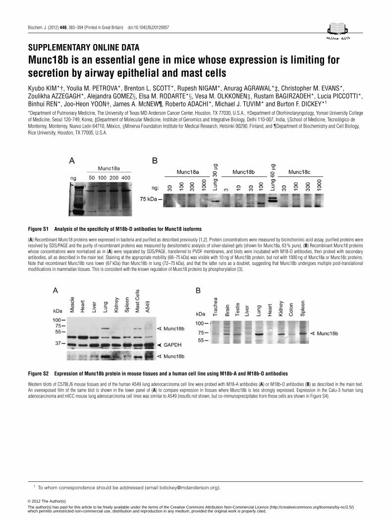
Biochem. J. (2012) 446, 383–394 (Printed in Great Britain) doi:10.1042/BJ20120057
SUPPLEMENTARY ONLINE DATAMunc18b is an essential gene in mice whose expression is limiting forsecretion by airway epithelial and mast cellsKyubo KIM*†, Youlia M. PETROVA*, Brenton L. SCOTT*, Rupesh NIGAM*, Anurag AGRAWAL*‡, Christopher M. EVANS*,Zoulikha AZZEGAGH*, Alejandra GOMEZ§, Elsa M. RODARTE*§, Vesa M. OLKKONEN‖, Rustam BAGIRZADEH*, Lucia PICCOTTI*,Binhui REN*, Joo-Heon YOON†, James A. MCNEW¶, Roberto ADACHI*, Michael J. TUVIM* and Burton F. DICKEY*1
*Department of Pulmonary Medicine, The University of Texas MD Anderson Cancer Center, Houston, TX 77030, U.S.A., †Department of Otorhinolaryngology, Yonsei University Collegeof Medicine, Seoul 120-749, Korea, ‡Department of Molecular Medicine, Institute of Genomics and Integrative Biology, Delhi 110-007, India, §School of Medicine, Tecnologico deMonterrey, Monterrey, Nuevo Leon 64710, Mexico, ‖Minerva Foundation Institute for Medical Research, Helsinki 00290, Finland, and ¶Department of Biochemistry and Cell Biology,Rice University, Houston, TX 77005, U.S.A.
Figure S1 Analysis of the specificity of M18b-D antibodies for Munc18 isoforms
(A) Recombinant Munc18 proteins were expressed in bacteria and purified as described previously [1,2]. Protein concentrations were measured by bicinchoninic acid assay, purified proteins wereresolved by SDS/PAGE and the purity of recombinant proteins was measured by densitometric analysis of silver-stained gels (shown for Munc18a, 63 % pure). (B) Recombinant Munc18 proteinswhose concentrations were normalized as in (A) were separated by SDS/PAGE, transferred to PVDF membranes, and blots were incubated with M18-D antibodies, then probed with secondaryantibodies, all as described in the main text. Staining at the appropriate mobility (68–75 kDa) was visible with 10 ng of Munc18b protein, but not with 1000 ng of Munc18a or Munc18c proteins.Note that recombinant Munc18b runs lower (67 kDa) than Munc18b in lung (72–75 kDa), and that the latter runs as a doublet, suggesting that Munc18b undergoes multiple post-translationalmodifications in mammalian tissues. This is consistent with the known regulation of Munc18 proteins by phosphorylation [3].
Figure S2 Expression of Munc18b protein in mouse tissues and a human cell line using M18b-A and M18b-O antibodies
Western blots of C57BL/6 mouse tissues and of the human A549 lung adenocarcinoma cell line were probed with M18-A antibodies (A) or M18b-O antibodies (B) as described in the main text.An overexposed film of the same blot is shown in the lower panel of (A) to compare expression in tissues where Munc18b is less strongly expressed. Expression in the Calu-3 human lungadenocarcinoma and mtCC mouse lung adenocarcinoma cell lines was similar to A549 (results not shown, but co-immunoprecipitates from those cells are shown in Figure S4).
1 To whom correspondence should be addressed (email [email protected]).
c© The Authors Journal compilation c© 2012 Biochemical Society© 2012 The Author(s)
The author(s) has paid for this article to be freely available under the terms of the Creative Commons Attribution Non-Commercial Licence (http://creativecommons.org/licenses/by-nc/2.5/)which permits unrestricted non-commercial use, distribution and reproduction in any medium, provided the original work is properly cited.
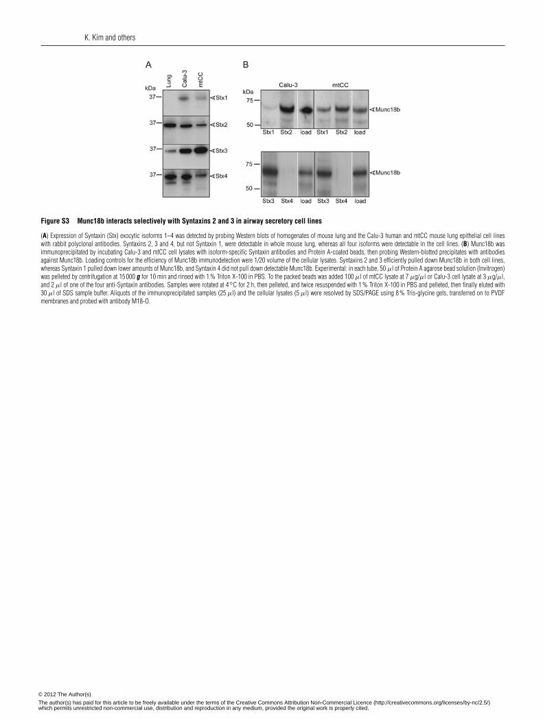
K. Kim and others
Figure S3 Munc18b interacts selectively with Syntaxins 2 and 3 in airway secretory cell lines
(A) Expression of Syntaxin (Stx) exocytic isoforms 1–4 was detected by probing Western blots of homogenates of mouse lung and the Calu-3 human and mtCC mouse lung epithelial cell lineswith rabbit polyclonal antibodies. Syntaxins 2, 3 and 4, but not Syntaxin 1, were detectable in whole mouse lung, whereas all four isoforms were detectable in the cell lines. (B) Munc18b wasimmunoprecipitated by incubating Calu-3 and mtCC cell lysates with isoform-specific Syntaxin antibodies and Protein A-coated beads, then probing Western-blotted precipitates with antibodiesagainst Munc18b. Loading controls for the efficiency of Munc18b immunodetection were 1/20 volume of the cellular lysates. Syntaxins 2 and 3 efficiently pulled down Munc18b in both cell lines,whereas Syntaxin 1 pulled down lower amounts of Munc18b, and Syntaxin 4 did not pull down detectable Munc18b. Experimental: in each tube, 50 μl of Protein A agarose bead solution (Invitrogen)was pelleted by centrifugation at 15 000 g for 10 min and rinsed with 1 % Triton X-100 in PBS. To the packed beads was added 100 μl of mtCC lysate at 7 μg/μl or Calu-3 cell lysate at 3 μg/μl,and 2 μl of one of the four anti-Syntaxin antibodies. Samples were rotated at 4◦C for 2 h, then pelleted, and twice resuspended with 1 % Triton X-100 in PBS and pelleted, then finally eluted with30 μl of SDS sample buffer. Aliquots of the immunoprecipitated samples (25 μl) and the cellular lysates (5 μl) were resolved by SDS/PAGE using 8 % Tris-glycine gels, transferred on to PVDFmembranes and probed with antibody M18-O.
c© The Authors Journal compilation c© 2012 Biochemical Society© 2012 The Author(s)
The author(s) has paid for this article to be freely available under the terms of the Creative Commons Attribution Non-Commercial Licence (http://creativecommons.org/licenses/by-nc/2.5/)which permits unrestricted non-commercial use, distribution and reproduction in any medium, provided the original work is properly cited.
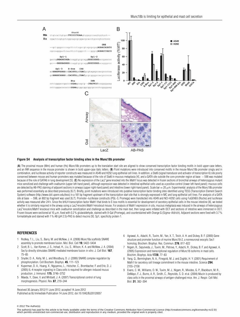
Munc18b is limiting for epithelial and mast cell secretion
Figure S4 Analysis of transcription factor binding sites in the Munc18b promoter
(A) The proximal mouse (Mm) and human (Hs) Munc18b promoters up to the translation start site are aligned to show conserved transcription factor binding motifs in bold upper-case letters,and an INR sequence in the mouse promoter is shown in bold upper-case italic letters. (B) Point mutations were introduced into conserved motifs in the mouse Munc18b promoter singly and incombination, and luciferase activity of reporter constructs was measured in A549 and H292 lung epithelial cell lines. In addition, a Stat6 (signal transducer and activator of transcription 6) site poorlyconserved between mouse and human promoters was mutated because of the role of Stat6 in mucous metaplasia [4], and a GATA site outside the core promoter region at base − 598 was mutatedbecause of the role of GATA6 in lung development [5]. (C) No expression of the LacZ gene knocked into the Math1 locus was detected in frozen sections of bronchial airways of heterozygous mutantmice sensitized and challenge with ovalbumin (upper left-hand panel), although expression was detected in intestinal epithelial cells used as a positive control (lower left-hand panel); mucous cellsare detected by AB-PAS staining of adjacent sections in airways (upper right-hand panel) and intestine (lower right-hand panel). Scale bar = 20 μm. Experimental: analysis of the Munc18b promoterwas performed essentially as described previously [6,7]. Briefly, point mutations were introduced into putative transcription factor binding sites identified using TESS (Transcription Element SearchSystem) software (http://www.cbil.upenn.edu/tess) in a 181 bp fragment upstream of the transcription start site that is strongly expressed in MC and lung epithelial cell lines. For analysis of a GATAsite at base − 598, an 802 bp fragment was used [6,7]. Promoter–luciferase constructs (PGL-3, Promega) were transfected into A549 and NCI-H292 cells using FuGENE6 (Roche) and luciferaseactivity was measured after 24 h. Since the bHLH transcription factor Math1 that binds to E-box motifs is essential for development of secretory epithelial cells in the mouse intestine [8], we testedwhether it is similarly required in the airway using a LacZ-knockin/Math1-knockout mouse. For analysis of Math1 expression in situ, mucous metaplasia was induced in the airways of heterozygousLacZ knockin/Math1 knockout mice with ovalbumin sensitization and challenge as described in the main text, then lungs were inflated with OCT and sections of intestine were immersed in OCT.Frozen tissues were sectioned at 10 μm, fixed with 0.2 % glutaraldehyde, stained with X-Gal (Promega), and counterstained with Orange G (Sigma–Aldrich). Adjacent sections were fixed with 3.7 %formaldehyde and stained with 1 % AB (pH 2.5)-PAS to detect mucins [9]. Sp1, specificity protein 1.
REFERENCES
1 Rodkey, T. L., Liu, S., Barry, M. and McNew, J. A. (2008) Munc18a scaffolds SNAREassembly to promote membrane fusion. Mol. Biol. Cell 19, 5422–5434
2 Scott, B. L., Van Komen, J. S., Irshad, H., Liu, S., Wilson, K. A. and McNew, J. A. (2004)Sec1p directly stimulates SNARE-mediated membrane fusion in vitro. J. Cell Biol. 167,75–85
3 Snyder, D. A., Kelly, M. L. and Woodbury, D. J. (2006) SNARE complex regulation byphosphorylation. Cell Biochem. Biophys. 45, 111–123
4 Kuperman, D. A., Huang, X., Nguyenvu, L., Holscher, C., Brombacher, F. and Erle, D. J.(2005) IL-4 receptor signaling in Clara cells is required for allergen-induced mucusproduction. J. Immunol. 175, 3746–3752
5 Maeda, Y., Dave, V. and Whitsett, J. A. (2007) Transcriptional control of lungmorphogenesis. Physiol. Rev. 87, 219–244
6 Agrawal, A., Adachi, R., Tuvim, M., Yan, X. T., Teich, A. H. and Dickey, B. F. (2000) Genestructure and promoter function of murine Munc18-2, a nonneuronal exocytic Sec1homolog. Biochem. Biophys. Res. Commun. 276, 817–822
7 Nigam, R., Sepulveda, J., Tuvim, M., Petrova, Y., Adachi, R., Dickey, B. F. and Agrawal, A.(2005) Expression and transcriptional regulation of Munc18 isoforms in mast cells.Biochim. Biophys. Acta 1728, 77–83
8 Yang, Q., Bermingham, N. A., Finegold, M. J. and Zoghbi, H. Y. (2001) Requirement ofMath1 for secretory cell lineage commitment in the mouse intestine. Science 294,2155–2158
9 Evans, C. M., Williams, O. W., Tuvim, M. J., Nigam, R., Mixides, G. P., Blackburn, M. R.,DeMayo, F. J., Burns, A. R., Smith, C., Reynolds, S. D. et al. (2004) Mucin is produced byclara cells in the proximal airways of antigen-challenged mice. Am. J. Respir. Cell Mol.Biol. 31, 382–394
Received 30 January 2012/11 June 2012; accepted 14 June 2012Published as BJ Immediate Publication 14 June 2012, doi:10.1042/BJ20120057
c© The Authors Journal compilation c© 2012 Biochemical Society© 2012 The Author(s)
The author(s) has paid for this article to be freely available under the terms of the Creative Commons Attribution Non-Commercial Licence (http://creativecommons.org/licenses/by-nc/2.5/)which permits unrestricted non-commercial use, distribution and reproduction in any medium, provided the original work is properly cited.