Multi-analytical non-invasive study of modern yellow paints from postwar Italian paintings from the...
9
Multi-analytical non-invasive study of modern yellow paints from postwar Italian paintings from the International Gallery of Modern Art Cà Pesaro, Venice † Francesca Caterina Izzo, a * Valentina Capogrosso, b Michele Gironda, c Roberto Alberti, c Chiara Mazzei, a Luca Nodari, e Arianna Gambirasi, e Elisabetta Zendri a and Austin Nevin d We present a multi-analytical non-invasive approach for the characterisation of yellow paints used in three paintings from the International Gallery of Modern Art Cà Pesaro, Venice. The paintings selected for this study were executed in oil-based media be- tween 1940 and 1960, they are unvarnished and have large monochromatic yellow areas. We focus on the identification of yellow pigments with a combined approach based on non-invasive portable X-ray Fluorescence spectrometry, complemented with VIS reflectance, remote-Fourier Transform Infrared and Raman spectroscopies. Different yellow pigments including Chrome yellow Naples Yellow and Cadmium-based yellow were identified. Their identification is of fundamental importance because several modern inorganic yellow pigments (in particular chrome and cadmium-based pigments) are known to exhibit peculiar degrada- tion patterns, leading to noteworthy aesthetic changes in paint such as darkening and fading. This work highlights the suitability of non-destructive techniques for the characterisation of modern paints and the utility of an integrated approach to obtain useful information for preventive conservation. Results demonstrate the need to complement elemental data acquired using portable X-ray Fluorescence with suitable complementary molecular spectroscopic techniques. Copyright © 2015 John Wiley & Sons, Ltd. Introduction During the middle of the 20th century, artists were strongly influenced by the introduction of new industrial materials into the art market, which can be appreciated from the wide range of new artistic mate- rials and techniques adopted in painting. [1–4] Commercial formulations of paint employed traditional pigments, which had been used for centuries, as well as new synthetic pigments and colorants; in addition stabilisers, extenders and fillers are generally present in manufactured paint recipes. Thanks to their availability and low cost, artists used, mixed and experimented with these new paints, even if the long-term stability of new colours were far from being totally understood. [5] The investigation of 20th century paintings is a challenging and demanding research field because the variety of materials, their potential interactions and their degradation have led to serious problems for the conservation of painting collections. [6] The identi- fication of modern paint constituents is essential to better under- stand materials and painting techniques used by modern artists and to detect potential degradation phenomena, which have been reported in modern and contemporary artworks. [7–9] The preventive conservation of modern and contemporary paintings thus requires a determination of their composition, using an analytical approach, which should limit sampling (which is a micro-destructive opera- tion), through the use and the development of non-destructive and non-invasive techniques. This approach should be considered as a first step in the whole conservation process, providing informa- tion which can inform conservation and prevent the long-term degradation of susceptible modern art materials. One of the main advantages of molecular non-invasive analysis is that it can be used for monitoring the chemical state of materials over time, leading to a method to assess the state of conservation of sensitive paint. In recent years, particular attention has focused on modern yellow pigments such as the Chrome yellow (PbCrO 4 ) and Cadmium-based yellows (containing Cadmium Sulphides or Cad- mium Sulphur Selenides, or even Cadmapones, coprecipitates of Cadmium Sulphide and Barium Sulphates). As recently reported for Van Gogh’s yellows based on Chrome yellow, or Cadmium- based yellows in paintings Matisse and Munch, for example, some yellow pigments easily undergo deterioration. [10–17] Furthermore, their peculiar degradation patterns lead to noteworthy visual * Correspondence to: Francesca Caterina Izzo, Department of Environmental Sciences, Informatics and Statistics, Ca’ Foscari University of Venice, Via Torino 155/b, Venice, Italy. E-mail: [email protected] † Presented at the European Conference on X-Ray Spectrometry, Bologna, Italy, 15–20 June 2014. a Department of Environmental Sciences, Informatics and Statistics, Ca’ Foscari University of Venice, Via Torino 155/b, Venice, Italy b Dipartimento di Fisica, Politecnico di Milano, Piazza Leonardo da Vinci 32, Milan, Italy c XGLab S.R.L., Spinoff del Politecnico di Milano, Via Francesco D’ Ovidio 3, Milan, Italy d Dipartimento di Fisica, Politecnico di Milano, Istituto di Fotonica e Nanotecnologie (IFN), National Research Council (CNR), Piazza Leonardo da Vinci 32, Milano, Italy e Istituto per l’Energetica e le Interfasi (IENI), National Research Council (CNR), Corso Stati Uniti 4, Padua, Italy X-Ray Spectrom. 2015 Copyright © 2015 John Wiley & Sons, Ltd. Research article Received: 29 September 2014 Revised: 22 March 2015 Accepted: 22 March 2015 Published online in Wiley Online Library (wileyonlinelibrary.com) DOI 10.1002/xrs.2623
Transcript of Multi-analytical non-invasive study of modern yellow paints from postwar Italian paintings from the...

Research article
Received: 29 September 2014 Revised: 22 March 2015 Accepted: 22 March 2015 Published online in Wiley Online Library
(wileyonlinelibrary.com) DOI 10.1002/xrs.2623
Multi-analytical non-invasive study of modernyellow paints from postwar Italian paintingsfrom the International Gallery of Modern ArtCà Pesaro, Venice†
Francesca Caterina Izzo,a* Valentina Capogrosso,b Michele Gironda,c
Roberto Alberti,c Chiara Mazzei,a Luca Nodari,e Arianna Gambirasi,e
Elisabetta Zendria and Austin Nevind
We present a multi-analytical non-invasive approach for the characterisation of yellow paints used in three paintings from theInternational Gallery of Modern Art Cà Pesaro, Venice. The paintings selected for this study were executed in oil-based media be-tween 1940 and 1960, they are unvarnished and have largemonochromatic yellow areas. We focus on the identification of yellowpigments with a combined approach based on non-invasive portable X-ray Fluorescence spectrometry, complemented with VISreflectance, remote-Fourier Transform Infrared and Raman spectroscopies. Different yellow pigments including Chrome yellowNaples Yellow and Cadmium-based yellow were identified. Their identification is of fundamental importance because severalmodern inorganic yellow pigments (in particular chrome and cadmium-based pigments) are known to exhibit peculiar degrada-tion patterns, leading to noteworthy aesthetic changes in paint such as darkening and fading. This work highlights the suitabilityof non-destructive techniques for the characterisation of modern paints and the utility of an integrated approach to obtain usefulinformation for preventive conservation. Results demonstrate the need to complement elemental data acquired using portableX-ray Fluorescence with suitable complementary molecular spectroscopic techniques. Copyright © 2015 John Wiley & Sons, Ltd.
* Correspondence to: Francesca Caterina Izzo, Department of EnvironmentalSciences, Informatics and Statistics, Ca’ Foscari University of Venice, Via Torino155/b, Venice, Italy. E-mail: [email protected]
† Presented at the European Conference on X-Ray Spectrometry, Bologna, Italy,15–20 June 2014.
a Department of Environmental Sciences, Informatics and Statistics, Ca’ FoscariUniversity of Venice, Via Torino 155/b, Venice, Italy
b Dipartimento di Fisica, Politecnico diMilano, Piazza Leonardo da Vinci 32,Milan, Italy
c XGLab S.R.L., Spinoff del Politecnico di Milano, Via Francesco D’Ovidio 3, Milan, Italy
d Dipartimento di Fisica, Politecnico di Milano, Istituto di Fotonica e Nanotecnologie(IFN), National Research Council (CNR), Piazza Leonardo da Vinci 32, Milano, Italy
e Istituto per l’Energetica e le Interfasi (IENI), National Research Council (CNR), CorsoStati Uniti 4, Padua, Italy
Introduction
During themiddle of the 20th century, artists were strongly influencedby the introduction of new industrial materials into the art market,which can be appreciated from the wide range of new artistic mate-rials and techniques adopted in painting.[1–4] Commercial formulationsof paint employed traditional pigments, which had been used forcenturies, as well as new synthetic pigments and colorants; in additionstabilisers, extenders and fillers are generally present in manufacturedpaint recipes. Thanks to their availability and low cost, artists used,mixed and experimented with these new paints, even if the long-termstability of new colours were far from being totally understood.[5]
The investigation of 20th century paintings is a challenging anddemanding research field because the variety of materials, theirpotential interactions and their degradation have led to seriousproblems for the conservation of painting collections.[6] The identi-fication of modern paint constituents is essential to better under-stand materials and painting techniques used by modern artistsand to detect potential degradation phenomena, which have beenreported inmodern and contemporary artworks.[7–9] The preventiveconservation of modern and contemporary paintings thus requiresa determination of their composition, using an analytical approach,which should limit sampling (which is a micro-destructive opera-tion), through the use and the development of non-destructiveand non-invasive techniques. This approach should be consideredas a first step in the whole conservation process, providing informa-tion which can inform conservation and prevent the long-termdegradation of susceptible modern art materials. One of the mainadvantages of molecular non-invasive analysis is that it can be used
X-Ray Spectrom. 2015
for monitoring the chemical state of materials over time, leading toa method to assess the state of conservation of sensitive paint.
In recent years, particular attention has focused on modernyellow pigments such as the Chrome yellow (PbCrO4) andCadmium-based yellows (containing Cadmium Sulphides or Cad-mium Sulphur Selenides, or even Cadmapones, coprecipitates ofCadmium Sulphide and Barium Sulphates). As recently reportedfor Van Gogh’s yellows based on Chrome yellow, or Cadmium-based yellows in paintings Matisse and Munch, for example, someyellow pigments easily undergo deterioration.[10–17] Furthermore,their peculiar degradation patterns lead to noteworthy visual
Copyright © 2015 John Wiley & Sons, Ltd.

F. C. Izzo et al.
changes such as darkening and fading, strongly compromising thelegibility and aesthetic impact of paintings.[10,18]
Large scale European infrastructures including mobile laboratorieshave been proposed, which offer an exhaustive range of imaging,molecular and elemental spectroscopic techniques for the non-invasive analysis of paintings and works of art.† There is still a signifi-cant need formethods for themonitoring and analysis of paintings inmuseums, which do not have access to such infrastructures, expertiseand instrumentation. In this collaborative work, we adopt a methodbased on the integration of portable instrumentation, combiningdata fromX-ray Fluorescence (XRF)with a reduced set of complemen-tary spectroscopic techniques for the analysis of three paintingsfrom the International Gallery of Modern Art Ca’ Pesaro in Venice.This work focuses on a multi-analytical non-invasive study of yel-
low paints in three paintings from Ca’ Pesaro collection. The paint-ings selected for this study were executed in oil-based media‡
between 1946 and 1960: Vibrazioni (Vibrations) by Simonetti Masi,La Passeggiata (Walking) by Massimo Campigli and Fiore Giallo (Yel-low Flower) by Guido Gomas (Table 1).The three paintingswere selected based on the following criteria:
the paintings are characterised by the presence of large monochro-matic yellow areas; the works of art are unvarnished, and they havenot been restored in the past. The lack of varnish on the surfacemeans that paint layer is unprotected from light but also meansthat we do not expect interactions with the varnish constituentsand/or the restoration materials in the analysis of yellow paints.Our non-invasive approach combines elemental analysis using
XRF, with molecular (Fourier Transform Infrared and Raman) andelectronic visible (VIS) spectroscopies.X-ray Fluorescence atomic spectroscopy has proved to be valuable
for in situ identification of pigments and a guide for further investiga-tions with complementary analytical techniques.[19–22] XRF is amatureanalytical technique for the determination of the elemental composi-tion of a material. This method is non-destructive, multi-elemental,fast and cost-effective. It can be applied in a non-vacuum environ-ment directly on the sample without any preparation. Over the lastdecade, advances in X-ray generation and detection have led to theevolution of instrumentation from laboratory-based standalone unitsto highly portable and lightweight handheld devices.[23,24] PortableXRF spectrometers have beenwidely adopted in conservation sciencefor the analysis of art.[25–28] However, the interpretation of data re-mains a challenge, and a good understanding of paint and the com-position ofmulti-layeredmaterials is necessary. Indeed, because of thepenetrating nature of X-rays, the interpretation of data from layeredstructures typical for paintings is more complex than other applica-tions such as, for example, the analysis of metal alloys, where it is as-sumed that elements are uniformly distributed and not stratified. Apainting’s structure, ground preparation, paint thickness and pigmentheterogeneity influence the data acquired with XRF. Because themethod identifies elements, pigments with similar elemental compo-sitions cannot be distinguished. In addition, light elements and tracesof pigments in mixtures may not be detectable. For these reasons, amulti-analytical approach is necessary for the analysis of paint.[29]
The result of XRF measurements on yellow paint layers has beencomplemented by other non-destructive and portable analytical
†An excellent example is Cultural Heritage Advanced Research Infrastructures -Synergy for a Multidisciplinary Approach to Conservation/Restoration(CHARISMA), an integrating activity project carried out in the FP7 CapacitiesSpecific Programme Research Infrastructures, www. charismaproject.eu.‡The paintings are described in the General Catalogue of Ca’ Pesaro Gallery as ‘oilon canvas’, although they have never been characterised before.
wileyonlinelibrary.com/journal/xrs Copyright © 2
techniques, such as VIS reflectance, External Reflection Fourier Trans-form Infrared (ER-FTIR) and Raman spectroscopy with the aim of identi-fying yellow pigments, when possible the class of paint binders and thepresence of inorganic fillers and extenders in the paint formulations.
Instrumental
X-ray Fluorescence spectrawere acquired by using the novel compact,portable and high performances XRF spectrometer ELIO produced byXGLab srl.§ The instrument is characterised by several novel features,which make it particularly well-suited for in situ analysis of works ofart. It is a fast system with Large area Silicon Drift Detector (25mm2)and good throughput capability, thanks to a new complementarymetal-oxide semiconductor (CMOS) Silicon Drift Detector readout(CUBE). ELIO is particularly efficient because of the X-ray transmissiongenerator that can reach up to 50kV and to a high solid detection an-gle geometry. ELIO has a high energy resolution (even below 130eV)and a high low detection limit, which can be coupled with several op-tions of anode tube, filters, collimators (usually 1mm diameter). Agreat advantage of this instrument is its compactness; the detectionhead (2 kg) can be mounted on any light-weight tripod, and it worksin contactless mode. Software provides complete control of the mea-surement head and all information, such as the elemental concentra-tions, the spectra and the images taken by the integrated microscopecamera and the external video camera. For this work, the excitationsource works with an Rh anode, and the beam is collimated to a spotdiameter on the sample surface of about 1.3mm. XRF measurementshave been carried out by fixing the tube voltage at 50 and 15kV ex-ploring a field of analysis from 1 to 40keV.
A CM-2600d Konica Minolta spectrophotometer was used to re-cord reflectance spectra, collected in the wavelength range from360 to 740nm with 10nm resolution. The measurements are in SCI(specular component included) modality. The instrument has eight-degree viewing angle geometry, with Xenon lamp diffusion lightand a high-resolutionmonolithic polychromator. Datawere collectedusing Spectramagic software and elaboratedwith Origin 10 software.
Raman spectroscopic analysis was performed using a handheldXantusTM-1 BaySpec instrument, equippedwith a 785nm laser (band-width from 0.2nm to 0.3nm) and operating from 70 to 500mW. Thespectrometer is customised to cover 500–1800cm�1 range withspectral resolution of 10cm�1. The detector is a 1024 elementcharge-coupled device (CCD) array thermoelectrically cooled to�20 °C. The instrument runs on BaySpec’s Micro 20/20V1038 softwareand has an integratedWindows-based Tablet personal computer (PC).
External Reflection Fourier Transform Infrared spectra were ac-quired by using an Alpha Bruker portable spectrometer operatingin external reflection geometry. Spectra were collected 40 scanwitha resolution of 4 cm�1. The analysed spot has an area of 20mm2.The wide spectral region investigated, from 7500 to 400 cm�1, pro-vides information on both combination and overtones absorptionbands, especially in 7500–4000 cm�1 and on the stretching andbending modes in the medium infrared (MIR) region. All the re-corded spectra show the distortions due to the derivative shape,Reststrahlen effect and intensity enhancement as a consequenceof the optical properties of the investigated substrates.[30]
All the data acquired duringmeasurements were compared witha database of reference artistic materials, both already available inliterature and ad hoc created by the authors.
§For further details: http://www.xglab.it/compact-portable-xrf-spectrometer-elio.shtml.
015 John Wiley & Sons, Ltd. X-Ray Spectrom. 2015

Table 1. Analysed paintings from the International Gallery of Modern Art Ca’ Pesaro, Venice: detail of yellow painted areas
Author, Title, year Image Detail of yellow analysed area
Simonetti Masi (1903–1969)
Vibrazioni, 1946
Oil on canvas
Massimo Campigli (1895–1971)
La passeggiata, 1955
Oil on canvas
Guido Gomas (1935–2005)
‘Fiore Giallo’, 1960
Oil on canvas
Multi-analytical non-invasive study of yellow paints in postwar Italian paintings
Results and discussion
Analyses were performed on the yellow painted layers of the threepaintings investigated, and close examination did not suggest dark-ening of the paint (see microscopic images in Table 1).
All the results are briefly reported in Table 2, and the discussion ispresented for each painting studied.
¶In the XRF spectra reported in Figs. 1(a)–(c), the labels indicate only K-linelinesand L-linelines (M-linelines are not reported). Characteristic of the presence ofair between the instrument and the sample is the K-lineline of Ar at 2.9 keV. Otherpeaks in the spectrum have been identified with Rh K-lines and L-lines, becauseof the Rhodium tube itself (Rh and Ar peaks are present in all the XRF spectra re-ported in this work, and their presence will not be explained further).
‘La passeggiata’ by Massimo Campigli
The XRF spectrum on a yellow area of ‘La passeggiata’ is reported inFig. 1(a). The most intense peaks are given by Zn K-lines and PbL-lines. Sb, Ca and Fe are also present. Furthermore, traces of Baand Cr have been detected. The presence of S can only be assumedbecause of the limitations of XRF in the detection of lightelements.[31] In fact, besides the inefficiency of the fluorescenceprocess and the weakness of the secondary radiation respect to
X-Ray Spectrom. 2015 Copyright © 2015 John Wiley &
the primary beam, the secondary radiation from lighter elementshas a low penetrating power and is severely attenuated if the beampasses through air for any distance.[26] The quantification of S in thisspectrum is compromised by the dominant presence of Pb, whoseM-line peak at 2.3 keV¶ is close to S K-line, and it is therefore not triv-ial to decouple these two contributions.
From XRF analysis, we deduce that the yellow paint maycontain a mixture of Naples yellow Pb3(SbO4)2 and Chrome yellow(lead chromate, PbCr1-xSxO4). The detection of a significant
Sons, Ltd. wileyonlinelibrary.com/journal/xrs

Table
2.XRF,vis-RS,Raman,ER-FTIR
results
from
theanalysed
yellowpaintedareas
Paintin
gXRF
(elemen
ts)*
Vis-reflectan
ceRaman
ER-FTIR
Com
men
ts
Zn
PbBa
Ca
FeSb
Cr
SCd
Sr
Cam
pigli
x*x*
xx
xx
xx
xSh
oulder
centred
atab
out400nm;slight
increase
between450an
d500nm
;sharp
increase
from
550nm
Nap
lesyellow
Chromeyellow
Mixture
ofNap
lesyellowandChromium
yellowin
dryingoil;lithop
onean
dlead
white
inthegrou
nd
layers;calcium
carbon
ateandbarium
sulpha
teas
extendersin
thepaintform
ulation
Bariu
msulphate
Nap
lesyellow
Calcium
Carbon
ate
Calcium
Carbon
ate
Lead
carbon
ate
Dryingoil
(Dryingoil)
Masi
x*x*
x*x
xx
xInitialincrease
from
360nm;lineartren
d
upto
abou
t450nm
;inflexion
centred
atab
out500nm
Cad
mium
yellow
Cad
mium
yellow
Cad
mium
Yellowor
Lemon
yellowin
dryingoil
calcium
carbon
atean
dbarium
sulphateas
extenders
Bariu
msulphate
Calcium
Carbon
ate
Calcium
carbon
ate
Bariu
msulphate
Dryingoil
Dryingoil
Gom
asx*
x*x
xx
xx
xStrongab
sorptio
nup
to500nm
;slight
increase
andainflexion
betweenab
out570
and600nm;lineartren
dfrom
600to
740nm
/Chromeyellow
Chromeyellowbased
paintspread
onapreparation
layerof
lithop
one
Gyp
sum
Binder
not
iden
tifiedsurely
Calcium
Carbon
ate
Dryingoil?
Protein?
*Th
easterisksymbol
indicates
that
theelem
entisdom
inantin
theXRF
spectrum
.
XRF,X
-ray
Fluo
rescen
ce;ER-FTIR,ExternalRe
flectionFourierTransform
Infrared
;vis-RS,vis-reflectan
cespectra.
F. C. Izzo et al.
wileyonlinelibrary.com/journal/xrs Copyright © 2
concentrations of Zn and Pb and of traces of Ba can be explainedwith the presence of a preparation layer of lithopone (copreciptateof ZnS+BaSO4) or the presence of ZnO and BaSO4 and possiblylead white (Pb(CO3)2*PbO2 used in the ground layer.
The vis-reflectance spectra spectrum of the yellow paint isdisplayed in Fig. 2. The reflectance spectrum shows the typicalsigmoidal trend of yellow pigments, with a first shoulder centredat about 400nm followed by a slight increase between 450and 500nm and a sharp increase in reflectance from 550nmapproximately.[32] According to the literature[33] and a database ofmodern pigments created by the authors, the use of both Naplesyellow and Chrome yellow is feasible. However, the informationobtained does not allow the distinction between the different na-ture of yellow pigments, likely because the technique is limitedby the compositional complexity, the non homogeneity of theyellow paint layer and the presence of an organic binder.[32]
External Reflection Fourier Transform Infrared analyses suggestthe presence of Naples yellow highlighted by Raman spectra, asdisplayed in Fig. 3(a) and (b). In fact, the absorption centred at668 cm�1 could be ascribed to Naples yellow, Pb2Sb2O7, in agree-ment with published data[34] and with the comparison with a stan-dard (Naples yellow from Lechler, first half of 20th century) asreported in Fig. 3(d). This attribution is supported by the XRF,Raman and vis-reflectance spectra investigations. Nevertheless,the presence of carbonate compounds cannot be excluded a priori.Among the various CO3
= compounds, only cerussite shows ν4 aclose to 668 cm�1 (i.e. 680 cm�1, reported in Fig. 3(d)). A carefulanalysis of the recorded spectra does not show any other bands at-tributable to PbCO3, supporting the presence only of Naples yellow.Vice versa, weak signals due to CO3
= overtones and combinationbands (ν1 + ν3, ν1 + ν4 and/or 2 ν2 + ν4) are evident (see fig. 3.e) inthe region between 2800 and 1750 cm�1.In fact, the clear shoulderin the C=O absorption, at 1796 cm�1, together with the weak bandat 2512 cm�1 (with a shoulder at 2594 cm�1) can be ascribed to thepresence of calcium carbonate, as suggested by Miliani et al.[30]
The presence of CaCO3 was also observed in Raman spectrawith a band at around 1082 cm�1 related to the stretching of�CO3
2�group and in Infra-red (IR) spectra with the doublet at1048 and 1052 cm�1 arising from the factor-group splitting ofthe ν1 symmetric stretching mode of the�CO3
2� ion. Calcium car-bonate is likely present in the paint formulation as an extender,which is commonly found in the analysis of modern paints.[35,36]
Despite the general difficulty of identifying Naples yellowthrough Raman analysis[37] and the fluorescence because of the or-ganic medium, the Raman spectrum shown in Fig. 4(a) suggests thepresence of lead-antimonate yellow through its characteristic Pb–Omodes at 519, 652 and 810 cm�1[38,39] (it is noted that the bandsvisible at 71, 143, 294, 335, 45 are not included in themeasurementrange with the handheld instrument). Furthermore, the Ramanspectrum highlights the presence of Chrome yellow, thanks tothree bands in the Cr–O stretching region at 825, 841 and856 cm�1; the most visible band is the one at 841 cm�1, which isassigned to the ν1 symmetric stretching vibration.[40] The bands at617, 647 and 988 cm�1 could indicate the presence of BaSO4, as re-ported in Burgio and Clark.[41] For the latter, it is not clear if there ispure barite or lithopone, because the bands related to ZnS, are gen-erally visible at around at 200–400 cm�1 are weak and not recordedby the instrumental range used in this study.
The binder of the yellow paint was identified as a drying oil byspectroscopic analyses. In Fig. 4(a), there are two intense Ramanbands at about 1050 and 1120 cm�1, which may be attributed toC–O–H bending and C–O stretching, respectively. There is also a
015 John Wiley & Sons, Ltd. X-Ray Spectrom. 2015

Figure 1. (a) X-ray Fluorescence (XRF) spectrum (reported in linear scale) acquired on the yellow area of ‘La passeggiata’ by Massimo Campigli; (b) XRFspectrum (reported in linear scale) acquired on the yellow area of ‘Fiore Giallo’ by Guido Gomas; (c) XRF spectrum (reported in linear scale) acquired onthe yellow area of ‘Le vibrazioni’ by Simonetti Masi.
Multi-analytical non-invasive study of yellow paints in postwar Italian paintings
X-Ray Spectrom. 2015 Copyright © 2015 John Wiley & Sons, Ltd. wileyonlinelibrary.com/journal/xrs
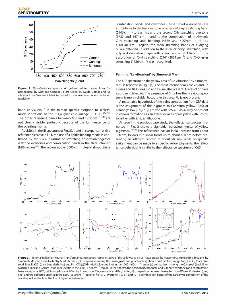
Figure 2. Vis-reflectance spectra of yellow painted areas from ‘Lapasseggiata’ by Massimo Campigli, ‘Fiore Giallo’ by Guido Gomas and ‘Levibrazioni’ by Simonetti Masi (acquired in specular component includedmodality).
F. C. Izzo et al.
band at 947 cm�1 in the Raman spectra assigned to skeletalmode vibrations of the α-1,4 glicosidic linkage (C–O–C).[42,43]
The other reference peaks between 800 and 1700 cm�1[44] arenot clearly visible, probably because of the luminescence ofthe painting matrix.As visible in the IR spectrum of Fig. 3(a), and in comparisonwith a
reference siccative oil 3 f, the use of a lipidic binding media is con-firmed by the C=O asymmetric stretching absorption togetherwith the overtones and combination bands in the Near Infra-red(NIR) region.[45] The region above 3000 cm�1 clearly shows these
Figure 3. External Reflection Fourier Transform Infrared spectra representativeSimonetti Masi; (c) ‘Fiore Giallo’ by Guido Gomas; (d) comparison among the Passolid line), PbCO3 (dark blue dash line) and Pb3(CO3)2(OH)2 (dark blue dot line) iMasi (red line) andGomas (blue line) spectra in the 3000–1700 cm�1 region. In thbans are reported (CC, calcium carbonate; hCer, hydrocerussite; Cer, cerussite; anline) and the collected spectra in the 6500–2500 cm�1 region (I: first δC-O overtonsiccative oil); in the box, the C =O region is enhanced.
wileyonlinelibrary.com/journal/xrs Copyright © 2
combination bands and overtones. These broad absorptions areattributable to the first overtone of ester carbonyl stretching band(5146 cm�1) to the first and the second CH2 stretching overtone(5787 and 5674 cm�1) and to the combination of methylenicC–H stretching and bending (4329 and 4259 cm�1). In the4000–400 cm�1 region, the main stretching bands of a dryingoil are detected. In addition to the ester carbonyl stretching, witha typical derivative shape with a flex centred at 1748 cm�1, theabsorption of C–H stretching (2961–2864 cm�1) and C–O esterstretching (1126 cm�1) was recognised.
Painting: ‘Le vibrazioni’ by Simonetti Masi
The XRF spectrum on the yellow area of ‘Le vibrazioni’ by SimonettiMasi is reported in Fig. 1(c). The most intense peaks are Zn and CaK-lines and Ba L-lines. Cd and Fe are also present. Traces of Sr havealso been detected. The presence of S, unlike the previous spec-trum, is more reliable, because in this area Pb is not present.
A reasonable hypothesis of the paint composition from XRF datais the assignment of this pigment to Cadmium yellow (CdS) orLemon yellow (CdxZn1-xS)mixedwith BaSO4. BaSO4may be presentin various formations: as an extender, as a coprecipitate with CdS or,together with ZnS, as lithopone.
As seen in the previous case study, the reflectance spectrum re-ported in Fig. 2 shows a sigmoidal behaviour typical of yellowpigments.[32,46] The reflectance has an initial increase from about360nm, follows in a linear trend up to about 450nm before pre-senting an inflexion centred at about 500nm. While no specificassignment can be made to a specific yellow pigments, the reflec-tance behaviour is similar to the reflectance spectrum of CdS.
of the yellow area in: (a) ‘Passeggiata’ byMassimo Campigli; (b) ‘Vibrazioni’ byseggiata and pure Naples yellow from Lechler (orange line), CaCO3 (dark bluen the 1000–400cm�1 range; (e) comparison among the Campigli (back line),e spectra, the position of carbonate and sulphate overtones and combinationd Bar, barite); (f) comparison between linseed oil fromWinsor & Newton (greye, II νs + δ and νa + δ combination bands of the carboxylic component of the
015 John Wiley & Sons, Ltd. X-Ray Spectrom. 2015
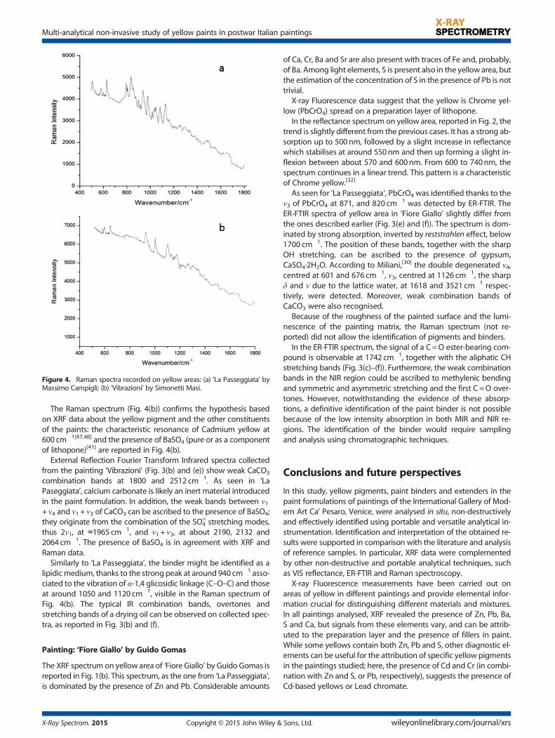
Figure 4. Raman spectra recorded on yellow areas: (a) ‘La Passeggiata’ byMassimo Campigli; (b) ‘Vibrazioni’ by Simonetti Masi.
Multi-analytical non-invasive study of yellow paints in postwar Italian paintings
The Raman spectrum (Fig. 4(b)) confirms the hypothesis basedon XRF data about the yellow pigment and the other constituentsof the paints: the characteristic resonance of Cadmium yellow at600 cm�1[47,48] and the presence of BaSO4 (pure or as a componentof lithopone)[41] are reported in Fig. 4(b).
External Reflection Fourier Transform Infrared spectra collectedfrom the painting ‘Vibrazioni’ (Fig. 3(b) and (e)) show weak CaCO3
combination bands at 1800 and 2512 cm�1. As seen in ‘LaPaseggiata’, calcium carbonate is likely an inert material introducedin the paint formulation. In addition, the weak bands between ν1+ ν4 and ν1 + ν3 of CaCO3 can be ascribed to the presence of BaSO4;they originate from the combination of the SO4
= stretching modes,thus 2ν1, at ≈1965 cm�1, and ν1 + ν3, at about 2190, 2132 and2064 cm�1. The presence of BaSO4 is in agreement with XRF andRaman data.
Similarly to ‘La Passeggiata’, the binder might be identified as alipidicmedium, thanks to the strong peak at around 940 cm�1 asso-ciated to the vibration of α-1,4 glicosidic linkage (C–O–C) and thoseat around 1050 and 1120 cm�1, visible in the Raman spectrum ofFig. 4(b). The typical IR combination bands, overtones andstretching bands of a drying oil can be observed on collected spec-tra, as reported in Fig. 3(b) and (f).
Painting: ‘Fiore Giallo’ by Guido Gomas
The XRF spectrum on yellow area of ‘Fiore Giallo’ by Guido Gomas isreported in Fig. 1(b). This spectrum, as the one from ‘La Passeggiata’,is dominated by the presence of Zn and Pb. Considerable amounts
X-Ray Spectrom. 2015 Copyright © 2015 John Wiley &
of Ca, Cr, Ba and Sr are also present with traces of Fe and, probably,of Ba. Among light elements, S is present also in the yellow area, butthe estimation of the concentration of S in the presence of Pb is nottrivial.
X-ray Fluorescence data suggest that the yellow is Chrome yel-low (PbCrO4) spread on a preparation layer of lithopone.
In the reflectance spectrum on yellow area, reported in Fig. 2, thetrend is slightly different from the previous cases. It has a strong ab-sorption up to 500nm, followed by a slight increase in reflectancewhich stabilises at around 550nm and then up forming a slight in-flexion between about 570 and 600nm. From 600 to 740nm, thespectrum continues in a linear trend. This pattern is a characteristicof Chrome yellow.[32]
As seen for ‘La Passeggiata’, PbCrO4 was identified thanks to theν3 of PbCrO4 at 871, and 820 cm�1 was detected by ER-FTIR. TheER-FTIR spectra of yellow area in ‘Fiore Giallo’ slightly differ fromthe ones described earlier (Fig. 3(e) and (f)). The spectrum is dom-inated by strong absorption, inverted by reststrahlen effect, below1700 cm�1. The position of these bands, together with the sharpOH stretching, can be ascribed to the presence of gypsum,CaSO4·2H2O. According to Miliani,[30] the double degenerated ν4,centred at 601 and 676 cm�1, ν3, centred at 1126 cm�1, the sharpδ and ν due to the lattice water, at 1618 and 3521 cm�1 respec-tively, were detected. Moreover, weak combination bands ofCaCO3 were also recognised.
Because of the roughness of the painted surface and the lumi-nescence of the painting matrix, the Raman spectrum (not re-ported) did not allow the identification of pigments and binders.
In the ER-FTIR spectrum, the signal of a C=O ester-bearing com-pound is observable at 1742 cm�1, together with the aliphatic CHstretching bands (Fig. 3(c)–(f)). Furthermore, the weak combinationbands in the NIR region could be ascribed to methylenic bendingand symmetric and asymmetric stretching and the first C =O over-tones. However, notwithstanding the evidence of these absorp-tions, a definitive identification of the paint binder is not possiblebecause of the low intensity absorption in both MIR and NIR re-gions. The identification of the binder would require samplingand analysis using chromatographic techniques.
Conclusions and future perspectives
In this study, yellow pigments, paint binders and extenders in thepaint formulations of paintings of the International Gallery of Mod-ern Art Ca’ Pesaro, Venice, were analysed in situ, non-destructivelyand effectively identified using portable and versatile analytical in-strumentation. Identification and interpretation of the obtained re-sults were supported in comparison with the literature and analysisof reference samples. In particular, XRF data were complementedby other non-destructive and portable analytical techniques, suchas VIS reflectance, ER-FTIR and Raman spectroscopy.
X-ray Fluorescence measurements have been carried out onareas of yellow in different paintings and provide elemental infor-mation crucial for distinguishing different materials and mixtures.In all paintings analysed, XRF revealed the presence of Zn, Pb, Ba,S and Ca, but signals from these elements vary, and can be attrib-uted to the preparation layer and the presence of fillers in paint.While some yellows contain both Zn, Pb and S, other diagnostic el-ements can be useful for the attribution of specific yellow pigmentsin the paintings studied; here, the presence of Cd and Cr (in combi-nation with Zn and S, or Pb, respectively), suggests the presence ofCd-based yellows or Lead chromate.
Sons, Ltd. wileyonlinelibrary.com/journal/xrs

F. C. Izzo et al.
The identification of Lead chromate, which is known to degradeand change colour in time is particularly important for the develop-ment of suitable exhibition, storage andmonitoring of paintings. Inaddition, the presence of Cadmium-based yellows calls for long-term monitoring of the paintings because of the recently reportedalteration of early 20th century paintings.While a non-invasive approach can answer many technical ques-
tions regarding paintings, the stratigraphy, composition of theground and the identification of specific binding media requirecareful sampling, the preparation of cross-sections and analysis ofsamples using suitable methods for the detection of organic mate-rials and trace impurities.[49] In the future, data from spectroscopicanalysis from specific areas of paintings will be integrated withimaging-based analysis and information acquired through the anal-ysis of paint samples.
Acknowledgements
The authors express their thanks to Dr. Matteo Piccolo of the Inter-national Gallery of Modern Art in Ca’ Pesaro (Venice) for the interestdemonstrated for the study and the interaction between art andconservation science.
References[1] L. Carlyle, The Artist’s Assistant: Oil Painting Instruction Manuals and
Handbooks in Britain 1800–1900 with Reference to Selected Eighteenth-Century Sources, Archetype Publications, London, 2001.
[2] S. Constantin. The Barbizon painters: a guide to their suppliers, Stud.Conserv. 2001, 46, 49–67.
[3] S. Rinaldi, in Colori e Pigmenti di Alcuni Dipinti Divisionisti AnalizzatiMediante XRF, (Ed.: S. Bordini), Dipingere con la luce. Teoria e tecnicadel colore nel Divisionismo italiano, Eue, Roma, 2003, pp. 37–53.
[4] M. Favaro, S. Bianchin, P. A. Vigato, M. Vervat. The palette of theMacchia Italian artist Giovanni Fattori in the second half of the xixthcentury, J. Cult. Herit. 2010, 11, 265–278.
[5] F. C. Izzo, K. J. van den Berg, H. van Keulen, B. Ferriani, E. Zendri. 20thcentury artists’ oil paints: the case of the Olii by Lucio Fontana, J. Cult.Herit. 2014, 15, 557–563.
[6] (Eds: K. J. van den Berg, A. Burnstock, M. de Keijzer Matthijs, J. Krueger,T. Learner, A. de Tagle, G. Heydenreich), Issues in Contemporary OilPaints, Springer International Publishing, Switzerland, 2014.
[7] M. J. Plater, B. De Silva, T. Gelbrich, M. B. Hursthouse, C. L. Higgitt,D. R. Saunders. The characterisation of lead fatty acid soaps in‘protrusions’ in aged traditional oil paint, Polyhedron 2003, 22,3171–3179.
[8] H. Kühn, M. Curran, in Chrome Yellow and Other Chromate Pigments. InArtists’ Pigments. A Handbook of their History and Characteristics.National Gallery of Art and Cambridge: Cambridge University Press:Washington, D.C., 1986, pp. 191.
[9] F. C. Izzo, K. J. van den Berg, H. van Keulen, E. Zendri, B. Ferriani, inModern Oil Paints – Formulations, Organic Additives and Degradation:Some Case Studies, (Eds: K. J. van den Berg, A. Burnstock, M. de KeijzerMatthijs, J. Krueger, T. Learner, A. de Tagle, G. Heydenreich), Issues inContemporary Oil paints, Springer International Publishing,Switzerland, 2014, pp.75–104.
[10] G. Van der Snickt, J. Dik, M. Cotte, K. Janssens, J. Jaroszewicz, W. De Nolf,J. Groenewegen, L. Van der Loeff. Characterization of a degradedcadmium yellow (CdS) pigment in an oil painting by means ofsynchrotron radiation based X-ray techniques, Anal. Chem. 2009, 81(7), 1600–10.
[11] G. Van der Snickt, K. Janssens, J. Dik, W. De Nolf, F. Vanmeert,J. Jaroszewicz, M. Cotte, G. Falkenberg, L. Van der Loeff. Combineduse of synchrotron radiation based micro-X-ray fluorescence, micro-X-ray diffraction, micro-X-ray absorption near-edge, and micro-fouriertransform infrared spectroscopies for revealing an alternativedegradation pathway of the pigment cadmium yellow in a paintingby Van Gogh, Anal. Chem. 2012, 84(23), 1221–8.
wileyonlinelibrary.com/journal/xrs Copyright © 2
[12] L. Monico, G. Van der Snickt, K. W. De Nolf, C. Miliani, J. Verbeeck,H. Tian, H. Tan, J. Dik, M. Radepont, M. Cotte. Degradation process oflead chromate in paintings by Vincent van Gogh studied by meansof synchrotron X-ray spectromicroscopy and related methods. 1.Artificially aged model samples, Anal. Chem. 2011, 83(4), 1214–23.
[13] L. Monico, G. Van der Snickt, K. Janssens, W. De Nolf, C. Miliani,J. Verbeeck, H. Tian, H. Tan, J. Dik, M. Radepont, M. Cotte.Degradation process of lead chromate in paintings by Vincent vanGogh studied by means of synchrotron X-ray spectromicroscopy andrelated methods. 2. Original paint layer samples, Anal. Chem. 2011,83(4), 1224–31.
[14] L. Monico, K. Janssens, C. Miliani, B. G. Brunetti, M. Vagnini, F. Vanmeert,G. Falkenberg, A. Abakumov, Y. Lu, H. Tian, J. Verbeeck, M. Radepont,M. Cotte, E. Hendriks, M. Geldof, L. van der Loeff, J. Salvant, M. Menu.By Vincent van Gogh studied by means of spectromicroscopicmethods. 3. Degradation process of lead chromate in paintingssynthesis, characterization, and detection of different crystal forms ofthe chrome yellow pigment, Anal. Chem. 2013, 85, 851–859.
[15] L. Monico, K. Janssens, C. Miliani, G. Van der Snickt, B. G. Brunetti,M. Cestelli Guidi, M. Radepont, M. Cotte. Degradation process of leadchromate in paintings by Vincent van Gogh studied by means ofspectromicroscopic methods. 4. Artificial aging of model samples ofco-precipitates of lead chromate and lead sulfate, Anal. Chem. 2013,85(2), 860–7.
[16] J. L. Mass, R. Opila, B. Buckley, M. Cotte, J. Church, A. Mehta. Thephotodegradation of cadmium yellow paints in Henri Matisse’s LeBonheur de vivre (1905–1906), Appl. Phys. A 2013, 111(1), 59–68.
[17] J. Mass, J. Sedlmair, C. Schmidt Patterson, D. Carson, B. Buckley,C. Hirschmugl. SR-FTIR imaging of the altered cadmium sulfide yellowpaints in Henri Matisse’s Le Bonheur de vivre (1905–6) – examinationof visually distinct degradation regions, Analyst 2013, 138, 6032–6043.
[18] C. Sandalinas, S. Ruiz-Moreno. Lead–tin–antimony yellow. Historicalmanufacture molecular characterization and identification inseventeenth-century Italian paintings, Stud. Conserv. 2004, 40, 41–53.
[19] F. Rosi, A. Burnstock, K. J. van den Berg, C. Miliani, B. G. Brunetti,A. Sgamellotti. A non invasive XRF study supported by multivariatestatistical analysis and reflectance FTIR to assess the composition ofmodern painting materials, Spectrochim. Acta A Mol. Biomol. Spectrosc.2009, 71(5), 1655–1662.
[20] N. Vornicu, C. Bibire, E. Murariu, D. Ivanov. Analysis of mural paintingsusing in situ non-invasive XRF, FTIR spectroscopy and opticalmicroscopy, X-Ray Spectrom. 2013, 42, 380–387.
[21] K. F. Gebremariam*, L. Kvittingen, F. G. Banica. Application of a portableXRF analyzer to investigate themedieval wall paintings of YemrehannaKrestos Church, Ethiopia, X-Ray Spectrom. 2013, 42(6), 462–469.
[22] M. F. Alberghina, R. Barraco, M. Brai, L. Pellegrino, F. Prestileo,S. Schiavone, L. Tranchina. Gilding and pigments of Renaissancemarble of Abatellis Palace: non-invasive investigation by XRFspectrometry, X-Ray Spectrom. 2013, 42(2), 68–78.
[23] S. Piorek, Coatings, Paint and Thin Film Deposits. In Portable X-rayFluorescence Spectrometry: Capabilities for in situ Analysis, (Eds: P. J. Potts,M. West), Royal society of Chemistry: Cambridge UK, 2008, pp. 56–82.
[24] C. Colombo, S. Bracci, C. Conti, M. Greco, M. Realini. Non-invasiveapproach in the study of polychrome terracotta sculptures:employment of the portable XRF to investigate complex stratigraphy,X-Ray Spectrom. 2011, 40, 273–279.
[25] A. N. Shugar, J. L. Mass, Studies in Archaeological Sciences, Handheld XRFfor Art and Archaeology, Leuven University Press, Belgium, 2012
[26] B. Beckhoff, B. Kanngießer, N. Langhoff, R.Wedell, H. Wolff,Handbook ofPractical X-Ray Fluorescence Analysis, Springer, Verlag Berlin Heidelberg,2006.
[27] H. Ida, T. Segawa, S. Tohyama, J. Kawai. Analysis of painted steel by ahand-held X-ray fluorescence spectrometer, Spectrochim. Acta B2005, 60, 249–252.
[28] G. Vittiglio, K. Janssens, B. Vekemans, F. Adams, A. Oost. A compactsmall-beam XRF instrument for in-situ analysis of objects of historicaland/or artistic value, Spectrochim. Acta B Atom.Spectrosc. 1999, 54(12),1967–1710.
[29] T. D. Chaplin, J. H. Clark, M. Martinòn-Torres. A combined Ramanmicroscopy, XRF and SEM-EDX study of three valuable objects, J. Mol.Struct. 2010, 973(1-3), 350–359.
[30] C. Miliani, F. Rosi, A. Daveri, B. G. Brunetti. Reflection infraredspectroscopy for the non-invasive in situ study of artists’ pigments,Appl. Phys. A 2012, 106, 295–307.
015 John Wiley & Sons, Ltd. X-Ray Spectrom. 2015

Multi-analytical non-invasive study of yellow paints in postwar Italian paintings
[31] L. D. Glinsman. The practical application of air-path X-ray fluorescencespectrometry in the analysis of museum objects, Rev. Conserv. 2005, 6,3–17.
[32] R. Johnston-Feller, Color science in the examination of museum objects,Nondestructive procedures, Getty Conservation Institute, 2001.
[33] Fiber optics reflectance spectra (FORS) of pictorial materials in the270–1700 nm range. Available at http://fors.ifac.cnr.it.
[34] K. Sakellariou, C. Miliani, A. Morresi, M. Ombelli. Spectroscopicinvestigation of yellow majolica glazes, J. Raman Spectros. 2004, 35,61–67.
[35] T. Rovetta, S. Bianchin, G. Salemi, M. Favaro, The case study of an Italiancontemporary art object: materials and state of conservation of thepainting ‘Ragazzo seduto’ by Remo Brindisi, J. Cult. Herit. 2013 articlein press.
[36] J. H. Townsend, L. Carlyle, N. Khandekar, S. Woodcock. Later nineteenthcentury pigments: evidence for addition and substitutions, Conservator1995, 19, 65–78.
[37] C. Pelosi, G. Agresti, U. Santamaria, E. Mattei. Artifical yellow pigments:production and characterization through spectroscopic methods ofanalysis, e-Preservation Science 2010, 7, 108–115.
[38] R. J. H. Clark, L. Cridland, B. M. Kariuki, K. D. M. Harris, R. Withnall.Synthesis, structural characterisation and raman spectroscopy of theinorganic pigments lead tin yellow types I and II and lead antimonateyellow: their identification on medieval paintings and manuscripts,J. Chem. Soc. Dalton Trans. 1995, 16, 2577–2582.
[39] F. Rosi, V. Manuali, T. Grygar, P. Bezdicka, B. G. Brunetti, A. Sgamellotti,L. Burgio, C. Seccaroni, C. Miliani. Raman scattering features of leadpyroantimonate compounds: implication for the non-invasiveidentification of yellow pigments on ancient ceramics. Part II. In situcharacterisation of Renaissance plates by portable micro-Raman andXRF studies, J. Raman Spectros. 2011, 42(3), 407–414.
[40] R. L. Frost. Raman microscopy of selected chromate minerals, J. RamanSpectros. 2004, 35(2), 153–158.
X-Ray Spectrom. 2015 Copyright © 2015 John Wiley &
[41] L. Burgio, R. J. H. Clark. Library of FT-Raman spectra of pigments,minerals, pigment media and varnishes, and supplement toexisting library of Raman spectra of pigments with visibleexcitation, Spectrochim. Acta A Mol. Biomol. Spectrosc. 2001,57(7), 1491–1521.
[42] J. Mallégol, J. Gardette, J. Lemaire. Long-term behavior of oil-basedvarnishes and paints I. Spectroscopic analysis of curing drying oils,JAOCS 1999, 76(8), 257–263.
[43] G. Smith. A Raman Spectroscopic Study of Paint and Dairy Samples,Master Thesis, University of Otago, Dunedin, New Zealand, 2012.
[44] P. Vandenabeele, B. Wehling, L. Moens, H. Edwards, M. De Reu,G. Van Hooydonk. Analysis with micro-Raman spectroscopy of naturalorganic binding media and varnishes used in art, Anal. Chim. Acta2000, 407, 261–274.
[45] M. Vagnini, C. Miliani, L. Cartechini, P. Rocchi, B. G. Brunetti,A. Sgamellotti. FT-NIR spectroscopy for non-invasive identification ofnatural polymers and resins in easel paintings, Anal. Bioanal. Chem.2009, 395, 2107–2118.
[46] T. Cavaleri, A. Giovagnoli, M. Nervo. Pigments and mixturesidentification by visible reflectance spectroscopy, Procedia Chemistry2013, 8, 45–54.
[47] M. Marcelino, V. S. F. Muralha. Synthetic organic pigments incontemporary Balinese painting: a Raman microscopy study,J. Raman Spectros. 2011, 43, 1281–1292.
[48] A. M. Correia, R. J. H. Clark. Pigments study by Ramanmicroscopy of 23paintings by the Portuguese artist Enrique Pousao (1859–1888),J. Raman Spectros. 2007, 38, 1390–1405.
[49] V. Capogrosso, F. Gabrieli, S. Bellei, L. Cartechini, A. Cesaratto, N. Trcera,F. Rosi, G. Valentini, D. Comelli, A. Nevin. An integrated approach basedonmicro-mapping analytical techniques for the detection of impuritiesin historical Zn-based white pigments, J. Analytical Atomic Spectrometry2015, 30, 828–838.
Sons, Ltd. wileyonlinelibrary.com/journal/xrs