Mouse brain expression patterns of Spg7, Afg3l1, and Afg3l2 transcripts, encoding for the...
9
Sacco et al. BMC Neuroscience 2010, 11:55 http://www.biomedcentral.com/1471-2202/11/55 Open Access RESEARCH ARTICLE BioMed Central © 2010 Sacco et al; licensee BioMed Central Ltd. This is an Open Access article distributed under the terms of the Creative Commons Attribution License (http://creativecommons.org/licenses/by/2.0), which permits unrestricted use, distribution, and reproduction in any medium, provided the original work is properly cited. Research article Mouse brain expression patterns of Spg7, Afg3l1, and Afg3l2 transcripts, encoding for the mitochondrial m-AAA protease Tiziana Sacco †1 , Enrica Boda †1 , Eriola Hoxha 1 , Riccardo Pizzo 1 , Claudia Cagnoli 2 , Alfredo Brusco 2 and Filippo Tempia* 1 Abstract Background: The m-AAA (ATPases Associated with a variety of cellular Activities) is an evolutionary conserved metalloprotease complex located in the internal mitochondrial membrane. In the mouse, it is a hetero-oligomer variably formed by the Spg7, Afg3l1, and Afg3l2 encoded proteins, or a homo-oligomer formed by either Afg3l1 or Afg3l2. In humans, AFG3L2 and SPG7 genes are conserved, whereas AFG3L1 became a pseudogene. Both AFG3L2 and SPG7 are involved in a neurodegenerative disease, namely the autosomal dominant spinocerebellar ataxia SCA28 and a recessive form of spastic paraplegia, respectively. Results: Using quantitative RT-PCR, we measured the expression levels of Spg7, Afg3l1, and Afg3l2 in the mouse brain. In all regions Afg3l2 is the most abundant transcript, followed by Spg7, and Afg3l1, with a ratio of approximately 5:3:1 in whole-brain mRNA. Using in-situ hybridization, we showed that Spg7, Afg3l1 and Afg3l2 have a similar cellular pattern of expression, with high levels in mitral cells, Purkinje cells, deep cerebellar nuclei cells, neocortical and hippocampal pyramidal neurons, and brainstem motor neurons. However, in some neuronal types, differences in the level of expression of these genes were present, suggesting distinct degrees of contribution of their proteins. Conclusions: Neurons involved in SCA28 and hereditary spastic paraplegia display high levels of expression, but similar or even higher expression is also present in other types of neurons, not involved in these diseases, suggesting that the selective cell sensitivity should be attributed to other, still unknown, mechanisms. Background Mitochondrial AAA-proteases (ATPases Associated with a variety of cellular Activities) are evolutionary conserved ATP-dependent metalloproteases that participate in the assembly of the respiratory chain complexes and ribo- somes, and in mitochondrial protein quality control [1-3]. In mice, two oligomeric AAA-protease complexes are present in the mitochondria inner membrane: the i-AAA protease, formed by the YME1L1 protein, oriented towards the intermembrane space, and the m-AAA pro- tease, composed by paraplegin (coded by Spg7), Afg3l1 and Afg3l2, which exposes its catalytic site to the matrix. It has been recently shown [4] that the m-AAA-protease formed by paraplegin has a hexameric structure like FtsH, a better studied bacterial homologue of eukaryotic m- AAA members [for review see [5]]. In humans, SPG7 and AFG3L2 only are active genes, whereas AFG3L1 has become a pseudogene. Both SPG7 and AFG3L2 have been associated with a human neurodegenerative disease: SPG7 loss-of-function mutations cause an autosomal recessive form of hereditary spastic paraplegia (HSP) [6], in which axons of corticospinal neurons and of soma- tosensory neurons degenerate [7]; AFG3L2 missense mutations have been implied in the autosomal dominant spinocerebellar ataxia SCA28 [8]. A mouse model for paraplegin deficiency has a motor phenotype that appears at 4 months, and later shows clear features of axonal swelling and degeneration of motor descending and sensory axons [9]. Recently two murine models for Afg3l2 have been described, one carrying a null mutation, and a second with a missense mutation in the protease domain. Afg3l2 defective mice display a marked impair- ment of axonal development leading to lethality at P16 * Correspondence: [email protected] 1 Section of Physiology of the Department of Neuroscience, University of T orino and National Institute of Neuroscience-Italy, Torino, Italy † Contributed equally Full list of author information is available at the end of the article
-
Upload
independent -
Category
Documents
-
view
0 -
download
0
Transcript of Mouse brain expression patterns of Spg7, Afg3l1, and Afg3l2 transcripts, encoding for the...
Sacco et al. BMC Neuroscience 2010, 11:55http://www.biomedcentral.com/1471-2202/11/55
Open AccessR E S E A R C H A R T I C L E
Research articleMouse brain expression patterns of Spg7, Afg3l1, and Afg3l2 transcripts, encoding for the mitochondrial m-AAA proteaseTiziana Sacco†1, Enrica Boda†1, Eriola Hoxha1, Riccardo Pizzo1, Claudia Cagnoli2, Alfredo Brusco2 and Filippo Tempia*1
AbstractBackground: The m-AAA (ATPases Associated with a variety of cellular Activities) is an evolutionary conserved metalloprotease complex located in the internal mitochondrial membrane. In the mouse, it is a hetero-oligomer variably formed by the Spg7, Afg3l1, and Afg3l2 encoded proteins, or a homo-oligomer formed by either Afg3l1 or Afg3l2. In humans, AFG3L2 and SPG7 genes are conserved, whereas AFG3L1 became a pseudogene. Both AFG3L2 and SPG7 are involved in a neurodegenerative disease, namely the autosomal dominant spinocerebellar ataxia SCA28 and a recessive form of spastic paraplegia, respectively.
Results: Using quantitative RT-PCR, we measured the expression levels of Spg7, Afg3l1, and Afg3l2 in the mouse brain. In all regions Afg3l2 is the most abundant transcript, followed by Spg7, and Afg3l1, with a ratio of approximately 5:3:1 in whole-brain mRNA. Using in-situ hybridization, we showed that Spg7, Afg3l1 and Afg3l2 have a similar cellular pattern of expression, with high levels in mitral cells, Purkinje cells, deep cerebellar nuclei cells, neocortical and hippocampal pyramidal neurons, and brainstem motor neurons. However, in some neuronal types, differences in the level of expression of these genes were present, suggesting distinct degrees of contribution of their proteins.
Conclusions: Neurons involved in SCA28 and hereditary spastic paraplegia display high levels of expression, but similar or even higher expression is also present in other types of neurons, not involved in these diseases, suggesting that the selective cell sensitivity should be attributed to other, still unknown, mechanisms.
BackgroundMitochondrial AAA-proteases (ATPases Associated witha variety of cellular Activities) are evolutionary conservedATP-dependent metalloproteases that participate in theassembly of the respiratory chain complexes and ribo-somes, and in mitochondrial protein quality control [1-3].
In mice, two oligomeric AAA-protease complexes arepresent in the mitochondria inner membrane: the i-AAAprotease, formed by the YME1L1 protein, orientedtowards the intermembrane space, and the m-AAA pro-tease, composed by paraplegin (coded by Spg7), Afg3l1and Afg3l2, which exposes its catalytic site to the matrix.It has been recently shown [4] that the m-AAA-proteaseformed by paraplegin has a hexameric structure like FtsH,
a better studied bacterial homologue of eukaryotic m-AAA members [for review see [5]]. In humans, SPG7 andAFG3L2 only are active genes, whereas AFG3L1 hasbecome a pseudogene. Both SPG7 and AFG3L2 havebeen associated with a human neurodegenerative disease:SPG7 loss-of-function mutations cause an autosomalrecessive form of hereditary spastic paraplegia (HSP) [6],in which axons of corticospinal neurons and of soma-tosensory neurons degenerate [7]; AFG3L2 missensemutations have been implied in the autosomal dominantspinocerebellar ataxia SCA28 [8]. A mouse model forparaplegin deficiency has a motor phenotype thatappears at 4 months, and later shows clear features ofaxonal swelling and degeneration of motor descendingand sensory axons [9]. Recently two murine models forAfg3l2 have been described, one carrying a null mutation,and a second with a missense mutation in the proteasedomain. Afg3l2 defective mice display a marked impair-ment of axonal development leading to lethality at P16
* Correspondence: [email protected] Section of Physiology of the Department of Neuroscience, University of Torino and National Institute of Neuroscience-Italy, Torino, Italy† Contributed equallyFull list of author information is available at the end of the article
BioMed Central© 2010 Sacco et al; licensee BioMed Central Ltd. This is an Open Access article distributed under the terms of the Creative CommonsAttribution License (http://creativecommons.org/licenses/by/2.0), which permits unrestricted use, distribution, and reproduction inany medium, provided the original work is properly cited.
Sacco et al. BMC Neuroscience 2010, 11:55http://www.biomedcentral.com/1471-2202/11/55
Page 2 of 9
[10]. Mice homozygous for Spg7 deletion, which in addi-tion also bear the loss of function Afg3l2Emv66 mutation inone allele, display a more severe phenotype with an ear-lier onset of symptoms when compared with Spg7 nullmice, and they also develop a marked cerebellar ataxia[11,12].
In yeast, Yta12 and Yta10 are the functional ortho-logues of paraplegin and AFG3L2, since respiratory com-petence defects induced by deficiency of both Yta12 andYta10 are compensated by coexpression of paraplegin andAFG3L2 [13]. Interestingly, the expression of Afg3l2 issufficient to rescue respiratory competence in this modelwhile paraplegin alone fails to restore m-AAA function,in line with the finding that Afg3l2 can form homo-oli-gomers while paraplegin does not [14].
Recently the assembly of paraplegin in m-AAA com-plexes has been studied in paraplegin deficient HSP fibro-blasts and in fibroblast from Spg7 -/- mouse model.Different amounts of paraplegin, Afg3l1, and Afg3l2 pro-teins were seen in liver and brain, with Afg3l1 being theless abundant in the latter. The subunit composition ofthe proteolytic complex seems therefore to vary in differ-ent tissues, and possibly within different subregions [14].
It is therefore interesting to evaluate the relative expres-sion levels of the three murine m-AAA components inthe brain to further correlate their abundance with theneurodegenerative pathologies associated with SPG7 andAFG3L2 genes in humans. To this aim, we measured therelative expression of m-AAA genes by quantitativereverse-transcriptase polymerase chain reaction (RT-PCR) in the whole brain and in specific regions (cerebel-lum, hippocampus, neocortex, olfactory bulb); further-more, a detailed pattern of expression was studied by in-situ hybridization under conditions which allowed eachprobe a sufficient resolution between neurons with dif-ferent degrees of expression. The combination of RT-PCRquantification with in-situ hybridization data allows us tomake a reliable comparison of the expression levels of them-AAA transcripts for the first time.
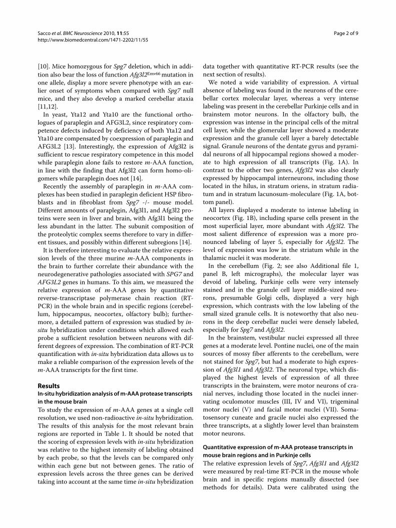
ResultsIn-situ hybridization analysis of m-AAA protease transcripts in the mouse brainTo study the expression of m-AAA genes at a single cellresolution, we used non-radioactive in-situ hybridization.The results of this analysis for the most relevant brainregions are reported in Table 1. It should be noted thatthe scoring of expression levels with in-situ hybridizationwas relative to the highest intensity of labeling obtainedby each probe, so that the levels can be compared onlywithin each gene but not between genes. The ratio ofexpression levels across the three genes can be derivedtaking into account at the same time in-situ hybridization
data together with quantitative RT-PCR results (see thenext section of results).
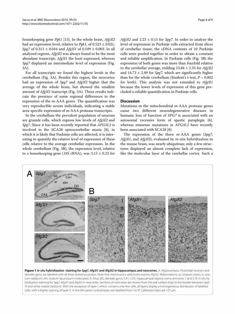
We noted a wide variability of expression. A virtualabsence of labeling was found in the neurons of the cere-bellar cortex molecular layer, whereas a very intenselabeling was present in the cerebellar Purkinje cells and inbrainstem motor neurons. In the olfactory bulb, theexpression was intense in the principal cells of the mitralcell layer, while the glomerular layer showed a moderateexpression and the granule cell layer a barely detectablesignal. Granule neurons of the dentate gyrus and pyrami-dal neurons of all hippocampal regions showed a moder-ate to high expression of all transcripts (Fig. 1A). Incontrast to the other two genes, Afg3l2 was also clearlyexpressed by hippocampal interneurons, including thoselocated in the hilus, in stratum oriens, in stratum radia-tum and in stratum lacunosum-moleculare (Fig. 1A, bot-tom panel).
All layers displayed a moderate to intense labeling inneocortex (Fig. 1B), including sparse cells present in themost superficial layer, more abundant with Afg3l2. Themost salient difference of expression was a more pro-nounced labeling of layer 5, especially for Afg3l2. Thelevel of expression was low in the striatum while in thethalamic nuclei it was moderate.
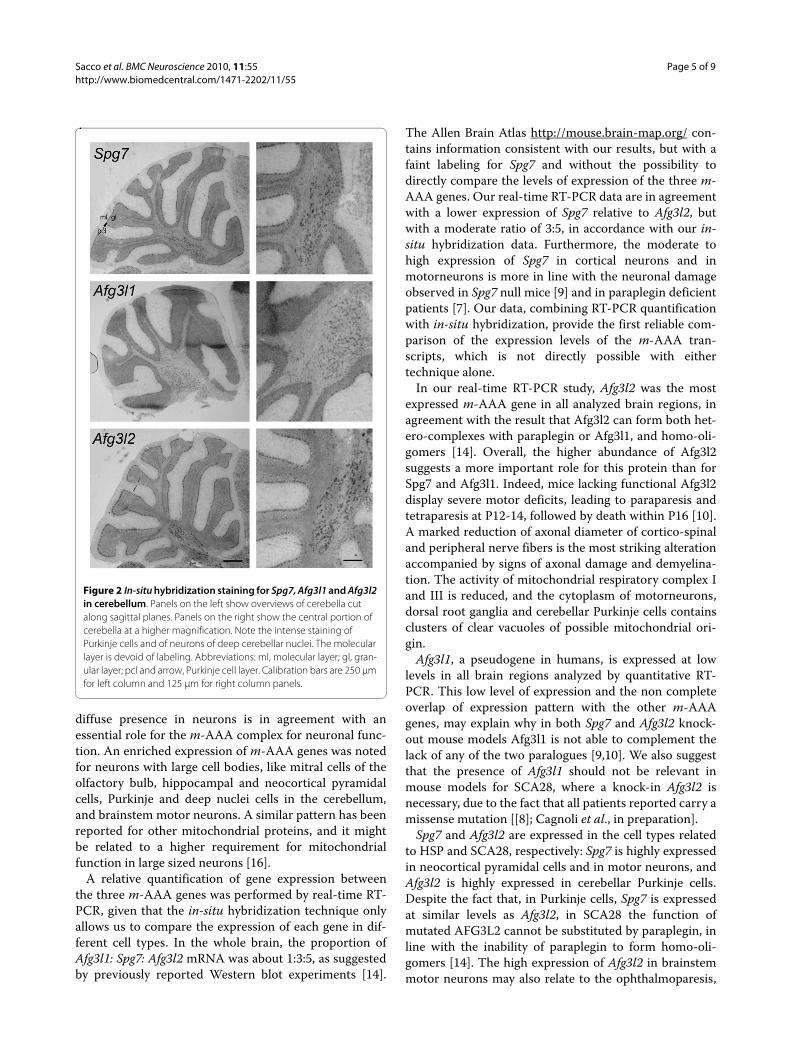
In the cerebellum (Fig. 2; see also Additional file 1,panel B, left micrographs), the molecular layer wasdevoid of labeling, Purkinje cells were very intenselystained and in the granule cell layer middle-sized neu-rons, presumable Golgi cells, displayed a very highexpression, which contrasts with the low labeling of thesmall sized granule cells. It is noteworthy that also neu-rons in the deep cerebellar nuclei were densely labeled,especially for Spg7 and Afg3l2.
In the brainstem, vestibular nuclei expressed all threegenes at a moderate level. Pontine nuclei, one of the mainsources of mossy fiber afferents to the cerebellum, werenot stained for Spg7, but had a moderate to high expres-sion of Afg3l1 and Afg3l2. The neuronal type, which dis-played the highest levels of expression of all threetranscripts in the brainstem, were motor neurons of cra-nial nerves, including those located in the nuclei inner-vating oculomotor muscles (III, IV and VI), trigeminalmotor nuclei (V) and facial motor nuclei (VII). Soma-tosensory cuneate and gracile nuclei also expressed thethree transcripts, at a slightly lower level than brainstemmotor neurons.
Quantitative expression of m-AAA protease transcripts in mouse brain regions and in Purkinje cellsThe relative expression levels of Spg7, Afg3l1 and Afg3l2were measured by real-time RT-PCR in the mouse wholebrain and in specific regions manually dissected (seemethods for details). Data were calibrated using the
Sacco et al. BMC Neuroscience 2010, 11:55http://www.biomedcentral.com/1471-2202/11/55
Page 3 of 9
Table 1: Expression patterns of m-AAA genes in mouse brain.
Spg7 Afg3l1 Afg3l2
Olfactory bulb
Glomerular layer + ++ +
Mitral cell layer ++ +++ +++
Granular cell layer +/- +/- +/-
Hippocampus
Dentate gyrus ++ ++ +++
Hilus interneurons + + +++
Pyramidal layer ++ ++ +++
Subiculum ++ ++ ++
Stratum radiatum interneurons + +/- +++
Stratum oriens interneurons + +/- +++
Stratum Lac-Mol interneurons + +/- ++
Neocortex
L 1 +/- +/- ++
L 2 ++ ++ ++
L 3 ++ ++ ++
L 4 ++ ++ ++
L 5 ++ ++/+++ +++
L 6 ++ ++ ++
Striatum + + +
Thalamus ++ ++ ++
Cerebellum
Purkinje cells +++ +++ +++
Molecular layer - - -
Granules + ++ +
Golgi cells +++ +++ +++
DCN +++ ++ +++
Brainstem
Vestibular n. ++ ++ ++
Pontine n. - ++ ++/+++
Motoneurons of III, V and VII n. +++ +++ +++
Cuneate n./Gracile n. ++ ++ +
Intensity of labeling of in-situ hybridization: - indicates absence of labeled structures; + mild labeling; ++ moderate labeling; +++ intense labeling. The scoring was relative to the highest intensity of each probe, so that the levels can be compared only within each gene but not between the three genes analyzed.
Sacco et al. BMC Neuroscience 2010, 11:55http://www.biomedcentral.com/1471-2202/11/55
Page 4 of 9
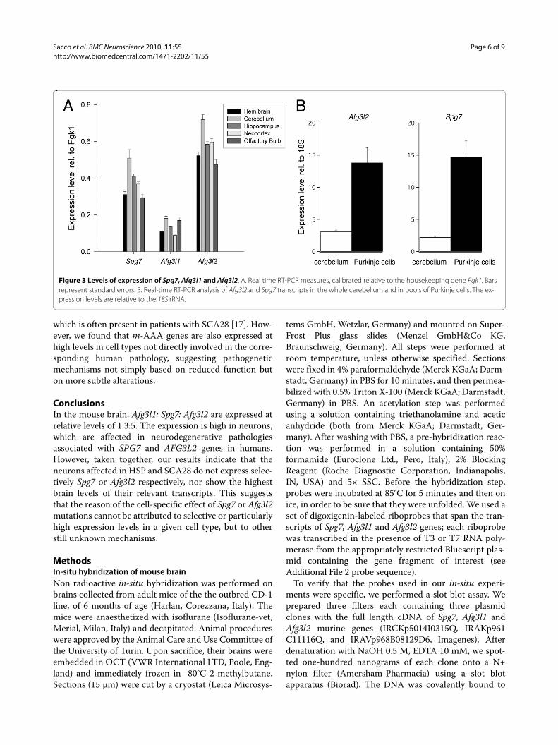
housekeeping gene Pgk1 [15]. In the whole brain, Afg3l2had an expression level, relative to Pgk1, of 0.523 ± 0.021,Spg7 of 0.311 ± 0.016 and Afg3l1 of 0.109 ± 0.003. In allanalyzed regions, Afg3l2 was always found to be the mostabundant transcript, Afg3l1 the least expressed, whereasSpg7 displayed an intermediate level of expression (Fig.3A).
For all transcripts we found the highest levels in thecerebellum (Fig. 3A). Besides this region, the neocortexhad an expression of Spg7 and Afg3l2 higher that theaverage of the whole brain, but showed the smallestamount of Afg3l1 transcript (Fig. 3A). These results indi-cate the presence of some regional differences in theexpression of the m-AAA genes. The quantification wasvery reproducible across individuals, indicating a stablearea-specific expression of m-AAA protease transcripts.
In the cerebellum the prevalent population of neuronsare granule cells, which express low levels of Afg3l2 andSpg7. Since it has been recently reported that AFG3L2 isinvolved in the SCA28 spinocerebellar ataxia [8], inwhich it is likely that Purkinje cells are affected, it is inter-esting to quantify the relative level of expression of thesecells relative to the average cerebellar expression. In thewhole cerebellum (Fig. 3B), the expression level, relativeto a housekeeping gene (18S rRNA), was 3.13 ± 0.23 for
Afg3l2 and 2.23 ± 0.15 for Spg7. In order to analyze thelevel of expression in Purkinje cells extracted from slicesof cerebellar tissue, the cDNA contents of 10 Purkinjecells were pooled together in order to obtain a constantand reliable amplification. In Purkinje cells (Fig. 3B) theexpression of both genes was more than fourfold relativeto the cerebellar average, yielding 13.84 ± 2.35 for Afg3l2and 14.73 ± 2.49 for Spg7, which are significantly higherthan for the whole cerebellum (Student's t-test, P = 0.002for both). This analysis was not extended to Afg3l1because the lower levels of expression of this gene pre-cluded a reliable quantification in Purkinje cells.
DiscussionMutations in the mitochondrial m-AAA protease genescause two different neurodegenerative diseases inhumans: loss of function of SPG7 is associated with anautosomal recessive form of spastic paraplegia [6],whereas missense mutations in AFG3L2 have recentlybeen associated with SCA28 [8].
The expression of the three m-AAA genes (Spg7,Afg3l1, and Afg3l2), evaluated by in-situ hybridization inthe mouse brain, was nearly ubiquitous; only a few struc-tures displayed an almost complete lack of expression,like the molecular layer of the cerebellar cortex. Such a
Figure 1 In-situ hybridization staining for Spg7, Afg3l1 and Afg3l2 in hippocampus and neocortex. A. Hippocampus. Pyramidal neurons and dentate gyrus are labelled with all three antisense probes. Note that interneurons selectively express Afg3l2. Abbreviations: so, stratum oriens; sr, stra-tum radiatum; slm, stratum lacunosum-moleculare; h, hilus; DG, dentate gyrus; CA1, CA3, hippocampal regions cornu ammonis 1 and 3. B. In-situ hy-bridization staining for Spg7, Afg3l1 and Afg3l2 in neocortex. Sections of neocortex are shown from the pial surface (top) to the border between layer VI and white matter (bottom). With the exception of layer I, which contains only few cells, all layers display a homogeneous distribution of labelled cells, with a higher staining of layer V. In the left panel, cortical layers are labelled from I to VI. Calibration bars are 125 μm.
Sacco et al. BMC Neuroscience 2010, 11:55http://www.biomedcentral.com/1471-2202/11/55
Page 5 of 9
diffuse presence in neurons is in agreement with anessential role for the m-AAA complex for neuronal func-tion. An enriched expression of m-AAA genes was notedfor neurons with large cell bodies, like mitral cells of theolfactory bulb, hippocampal and neocortical pyramidalcells, Purkinje and deep nuclei cells in the cerebellum,and brainstem motor neurons. A similar pattern has beenreported for other mitochondrial proteins, and it mightbe related to a higher requirement for mitochondrialfunction in large sized neurons [16].
A relative quantification of gene expression betweenthe three m-AAA genes was performed by real-time RT-PCR, given that the in-situ hybridization technique onlyallows us to compare the expression of each gene in dif-ferent cell types. In the whole brain, the proportion ofAfg3l1: Spg7: Afg3l2 mRNA was about 1:3:5, as suggestedby previously reported Western blot experiments [14].
The Allen Brain Atlas http://mouse.brain-map.org/ con-tains information consistent with our results, but with afaint labeling for Spg7 and without the possibility todirectly compare the levels of expression of the three m-AAA genes. Our real-time RT-PCR data are in agreementwith a lower expression of Spg7 relative to Afg3l2, butwith a moderate ratio of 3:5, in accordance with our in-situ hybridization data. Furthermore, the moderate tohigh expression of Spg7 in cortical neurons and inmotorneurons is more in line with the neuronal damageobserved in Spg7 null mice [9] and in paraplegin deficientpatients [7]. Our data, combining RT-PCR quantificationwith in-situ hybridization, provide the first reliable com-parison of the expression levels of the m-AAA tran-scripts, which is not directly possible with eithertechnique alone.
In our real-time RT-PCR study, Afg3l2 was the mostexpressed m-AAA gene in all analyzed brain regions, inagreement with the result that Afg3l2 can form both het-ero-complexes with paraplegin or Afg3l1, and homo-oli-gomers [14]. Overall, the higher abundance of Afg3l2suggests a more important role for this protein than forSpg7 and Afg3l1. Indeed, mice lacking functional Afg3l2display severe motor deficits, leading to paraparesis andtetraparesis at P12-14, followed by death within P16 [10].A marked reduction of axonal diameter of cortico-spinaland peripheral nerve fibers is the most striking alterationaccompanied by signs of axonal damage and demyelina-tion. The activity of mitochondrial respiratory complex Iand III is reduced, and the cytoplasm of motorneurons,dorsal root ganglia and cerebellar Purkinje cells containsclusters of clear vacuoles of possible mitochondrial ori-gin.
Afg3l1, a pseudogene in humans, is expressed at lowlevels in all brain regions analyzed by quantitative RT-PCR. This low level of expression and the non completeoverlap of expression pattern with the other m-AAAgenes, may explain why in both Spg7 and Afg3l2 knock-out mouse models Afg3l1 is not able to complement thelack of any of the two paralogues [9,10]. We also suggestthat the presence of Afg3l1 should not be relevant inmouse models for SCA28, where a knock-in Afg3l2 isnecessary, due to the fact that all patients reported carry amissense mutation [[8]; Cagnoli et al., in preparation].
Spg7 and Afg3l2 are expressed in the cell types relatedto HSP and SCA28, respectively: Spg7 is highly expressedin neocortical pyramidal cells and in motor neurons, andAfg3l2 is highly expressed in cerebellar Purkinje cells.Despite the fact that, in Purkinje cells, Spg7 is expressedat similar levels as Afg3l2, in SCA28 the function ofmutated AFG3L2 cannot be substituted by paraplegin, inline with the inability of paraplegin to form homo-oli-gomers [14]. The high expression of Afg3l2 in brainstemmotor neurons may also relate to the ophthalmoparesis,
Figure 2 In-situ hybridization staining for Spg7, Afg3l1 and Afg3l2 in cerebellum. Panels on the left show overviews of cerebella cut along sagittal planes. Panels on the right show the central portion of cerebella at a higher magnification. Note the intense staining of Purkinje cells and of neurons of deep cerebellar nuclei. The molecular layer is devoid of labeling. Abbreviations: ml, molecular layer; gl, gran-ular layer; pcl and arrow, Purkinje cell layer. Calibration bars are 250 μm for left column and 125 μm for right column panels.
Sacco et al. BMC Neuroscience 2010, 11:55http://www.biomedcentral.com/1471-2202/11/55
Page 6 of 9
which is often present in patients with SCA28 [17]. How-ever, we found that m-AAA genes are also expressed athigh levels in cell types not directly involved in the corre-sponding human pathology, suggesting pathogeneticmechanisms not simply based on reduced function buton more subtle alterations.
ConclusionsIn the mouse brain, Afg3l1: Spg7: Afg3l2 are expressed atrelative levels of 1:3:5. The expression is high in neurons,which are affected in neurodegenerative pathologiesassociated with SPG7 and AFG3L2 genes in humans.However, taken together, our results indicate that theneurons affected in HSP and SCA28 do not express selec-tively Spg7 or Afg3l2 respectively, nor show the highestbrain levels of their relevant transcripts. This suggeststhat the reason of the cell-specific effect of Spg7 or Afg3l2mutations cannot be attributed to selective or particularlyhigh expression levels in a given cell type, but to otherstill unknown mechanisms.
MethodsIn-situ hybridization of mouse brainNon radioactive in-situ hybridization was performed onbrains collected from adult mice of the the outbred CD-1line, of 6 months of age (Harlan, Corezzana, Italy). Themice were anaesthetized with isoflurane (Isoflurane-vet,Merial, Milan, Italy) and decapitated. Animal procedureswere approved by the Animal Care and Use Committee ofthe University of Turin. Upon sacrifice, their brains wereembedded in OCT (VWR International LTD, Poole, Eng-land) and immediately frozen in -80°C 2-methylbutane.Sections (15 μm) were cut by a cryostat (Leica Microsys-
tems GmbH, Wetzlar, Germany) and mounted on Super-Frost Plus glass slides (Menzel GmbH&Co KG,Braunschweig, Germany). All steps were performed atroom temperature, unless otherwise specified. Sectionswere fixed in 4% paraformaldehyde (Merck KGaA; Darm-stadt, Germany) in PBS for 10 minutes, and then permea-bilized with 0.5% Triton X-100 (Merck KGaA; Darmstadt,Germany) in PBS. An acetylation step was performedusing a solution containing triethanolamine and aceticanhydride (both from Merck KGaA; Darmstadt, Ger-many). After washing with PBS, a pre-hybridization reac-tion was performed in a solution containing 50%formamide (Euroclone Ltd., Pero, Italy), 2% BlockingReagent (Roche Diagnostic Corporation, Indianapolis,IN, USA) and 5× SSC. Before the hybridization step,probes were incubated at 85°C for 5 minutes and then onice, in order to be sure that they were unfolded. We used aset of digoxigenin-labeled riboprobes that span the tran-scripts of Spg7, Afg3l1 and Afg3l2 genes; each riboprobewas transcribed in the presence of T3 or T7 RNA poly-merase from the appropriately restricted Bluescript plas-mid containing the gene fragment of interest (seeAdditional File 2 probe sequence).
To verify that the probes used in our in-situ experi-ments were specific, we performed a slot blot assay. Weprepared three filters each containing three plasmidclones with the full length cDNA of Spg7, Afg3l1 andAfg3l2 murine genes (IRCKp5014I0315Q, IRAKp961C11116Q, and IRAVp968B08129D6, Imagenes). Afterdenaturation with NaOH 0.5 M, EDTA 10 mM, we spot-ted one-hundred nanograms of each clone onto a N+nylon filter (Amersham-Pharmacia) using a slot blotapparatus (Biorad). The DNA was covalently bound to
Figure 3 Levels of expression of Spg7, Afg3l1 and Afg3l2. A. Real time RT-PCR measures, calibrated relative to the housekeeping gene Pgk1. Bars represent standard errors. B. Real-time RT-PCR analysis of Afg3l2 and Spg7 transcripts in the whole cerebellum and in pools of Purkinje cells. The ex-pression levels are relative to the 18S rRNA.
Sacco et al. BMC Neuroscience 2010, 11:55http://www.biomedcentral.com/1471-2202/11/55
Page 7 of 9
the membrane using a UV stratalinker (Stratagene), andhybridised using the Quick-Hyb hybridisation solution(Roche Diagnostics) in the condition specified. Probeswere obtained from PCR amplification and gel-purifica-tion using QIAquick gel extraction kit (Qiagen), and cor-responded in sequence to the probes used for in-situexperiments. The probes were radiolabelled with 32-P-dCTP using High Prime labelling kit (Roche Diagnostics).The filters were exposed for 15 min at room temperature.The results [Additional file 1, panel A] show that allprobes are specific and only recognize the full lengthcDNA of the corresponding gene. A very faint cross-hybridisation with Spg7 is present for the Afg3l1 probe.
Each riboprobe was transcribed as antisense, whichspecifically labeled the target mRNA as expected, and assense, to control for unspecific binding or for backgroundstaining due to the endogenous alkaline phosphataseactivity of the tissue. Examples of sense and antisenselabeling are illustrated in panel B of the supplementaryfig. [Additional file 1, panel B].
Twenty nanograms of each probe were added to theslices and the hybridization was carried out at 65°C over-night. Washing steps included incubations in 5× SSC andin 0.2× SSC at 65°C for one hour, followed by 5 minutes in0.2× SSC and 5 minutes in MABS at room temperature.Sections were incubated in 1% Blocking Reagent (RocheDiagnostic Corporation, Indianapolis, IN, USA) in MABSbuffer for one hour and then in an AP-coniugated anti-digoxigenin Fab fragment (1:5000 dilution, Roche Diag-nostic Corporation, Indianapolis, IN, USA) for one hour.After two washing steps of 30 minutes in MABS bufferand 5 minutes in Buffer 3, the reaction was developedovernight with BCIP/NBT substrate (Sigma-Aldrich, St.Louis, MO, USA). When the color reaction was com-pleted, sections were washed in Buffer 4 and in PBS forsome minutes. The slides were examined by means of aZeiss Axiophot light microscope (Zeiss, Oberkochen,Germany). Micrographs were taken by means of a NikonCoolpix 950 digital camera (Nikon, Mellville, NY, USA)attached to the same microscope. Images were processedusing Adobe Photoshop CS2 9.0.2 (Adobe System, SanJosè, CA, USA).
Real Time RT-PCR analysis of the mouse brainThe quantitative RT-PCR analysis of the transcripts wasperformed on hemibrains and on dissected brain regionscollected from 5 female CD-1 mice (Harlan, Corezzana,Italy) at 6 months of age. The mice were anaesthetizedwith isoflurane (Isoflurane-vet, Merial, Milan, Italy) anddecapitated. Animal procedures were approved by theAnimal Care and Use Committee of the University ofTorino. The brains were quickly removed from the skull,placed in an ice-cold artificial liquor (containing, in mM:125 NaCl, 2.5 KCl, 2 CaCl2, 1 MgCl2, 1.25 NaH2PO4, 26
NaHCO3, 20 glucose, bubbled with 95% O2- 5% CO2). Forthe analysis performed on the dissected brain regions, thecerebellum and olfactory bulbs were manually dissectedimmediately after the whole brain collection. Then, coro-nal sections from neocortex and hippocampus were cutusing a vibratome (Leica Microsystems GmbH, Wetzlar,Germany), starting from Bregma 1.10 mm. First, two 400μm thick slices were cut in order to collect samples ofneocortex. The two hippocampal slices (400 μm thick)were cut starting from Bregma -1.50. White matter partswere manually separated and not included in the geneexpression analysis. Stereotaxic coordinates wereobtained from the mouse brain atlas "The mouse brain instereotaxic coordinates" [18]. All samples were rapidlyfrozen and stored at -80°C.
Total RNA was isolated by extraction with the commer-cially available TRIzol Reagent (Invitrogen Life Technolo-gies Inc., Grand Island, NY, USA), in accordance with themanufacturer's instructions. Genomic DNA was elimi-nated by treating the extracted RNA with DNase(Deoxiribonuclease I; Sigma-Aldrich). RNA concentra-tion and purity were evaluated prior and after the DNasetreatment by spectrophotometry. The absence of RNAdegradation was confirmed by agarose gel electrophore-sis. RNA samples were stored at -80°C.
One μg of total RNA was reverse-transcribed to cDNAusing the commercial High-Capacity cDNA Archive Kit(Applied Biosystems, Foster City, CA, USA), according tothe manufacturer's instructions. Negative controls of thereverse transcription were always performed. cDNA sam-ples were stored at -20°C.
Quantitative Real Time PCR was carried out using theABI Prism 7000 Sequence Detection System instrumen-tation (Applied Biosystems). Taqman Gene ExpressionAssays were purchased from Applied Biosystems todetermine the amount of the three target genes (Afg3l2cod. Mm01258204_m1, Afg3l1 cod. Mm00475312_m1,Spg7 cod. Mm00462651_m1) and the housekeeping genephosphoglycerate kinase 1 (Pgk1, cod. Mm00435617_m1). PCR amplifications were performed on cDNAsamples corresponding to a final RNA concentration 200pg. PCR was performed according to the following reac-tion conditions: 50°C for 2 minutes, 95°C for 10 minutes,followed by 50 cycles 95°C for 15 seconds alternating with60°C for 1 minute. Blank controls were performed oneach plate.
For the quantitative comparison of the genes, dataextracted from each real time RT-PCR run were analysedby means of the 7000 v1.1 SDS instrument software(Applied Biosystems). The CT (Cycle Threshold) wasautomatically calculated and used to quantify the startingcopy number of the target mRNA. The amounts of thetarget RNA copies were normalized to the endogenousreference Pgk1, because it shows a low variation among

Sacco et al. BMC Neuroscience 2010, 11:55http://www.biomedcentral.com/1471-2202/11/55
Page 8 of 9
mouse brain regions [15]. Data were analysed as in Bor-tone et al. [19] to compensate for small differences in theefficiency of amplification among target and housekeep-ing genes.
For a comparison of the level of expression of Afg3l2and Spg7 transcripts in the cerebellum relative toPurkinje cells, the analysis was performed on 8 cerebellaand 5 pools (10 cells each) of Purkinje cell (PCs), collectedfrom another set of 11 adult female CD-1 mice (Harlan,Corezzana, Italy) at 6 months of age. The cerebellum wasquickly removed under semi-sterile conditions and eitherrapidly frozen in -80°C 2-methylbutane and stored at -80°C (8 cerebella) or, for slice preparation, placed in theartificial liquor described above, bubbled with 95% O2-5% CO2. To collect PCs, parasagittal sections (200 μmthickness) of the vermis of 3 cerebella were preparedusing a vibratome. Single PCs were aspirated intomicropipettes under visual control; the tips of themicropipettes were broken into empty 0.2 ml Eppendorftubes. The entire RNA of each cell was retrotranscribedusing the High-Capacity cDNA Archive Kit (Applied Bio-systems). The cDNA was purified and concentrated usingthe QIAEX II Gel Extraction Kit (Qiagen, Hilden, Ger-many). Quantitative PCR and data analysis were per-formed as described above. For the comparison of theAfg3l2 and Spg7 transcripts in the cerebellum relative toPurkinje cells, each sample was normalized to the level ofthe 18S rRNA (Applied Biosystems, cod.4319413E).Transcript expression differences were statistically evalu-ated by means of a one way ANOVA test or Student's t-test. A P value < 0.05 was considered significant. Allresults were expressed as mean ± standard error (SE).
List of AbbreviationsCT: cycle threshold; HSP: hereditary spastic paraplegia; i-AAA: intermembrane space mitochondrial ATPaseAssociated with a variety of cellular Activities; m-AAAprotease: matrix mitochondrial ATPase Associated witha variety of cellular Activities; RT-PCR: reverse-tran-scriptase polymerase chain reaction; SCA28: spinocere-bellar ataxia, type 28
Additional material
Authors' contributionsTS and RP carried out the in-situ hybridization study. EB and EH carried out andanalysed the real time RT-PCR study. CC and AB designed the probes for in-situhybridization and constructed the plasmids containing the gene fragment ofinterest. AB conceived the study. FT designed and coordinated the study, anal-ysed the in-situ hybridization data and wrote the manuscript. CC and AB
helped to draft the manuscript. All authors read and approved the final manu-script.
AcknowledgementsThis work was supported by grants from: MIUR (PRIN-2007), Regione Piemonte (Ricerca Scientifica Applicata 2004 projects A183 and A74 and Ricerca Sanitaria Finalizzata 2006 and 2007) and Telethon research foundation grant GGP07110 (to A.B. and F.T.). E.B. is recipient of a CRT fellowship (Progetto Lagrange). The technical support of Dr. Federica Premoselli, Mr. Matteo Novello and Mrs. Lui-sella Milano is gratefully aknowledged. We thank Dr. A. Brussino for critical reading of the manuscript. We thank Dr. Julian Hoskins for the improvements to the final version of the text.
Author Details1Section of Physiology of the Department of Neuroscience, University of Torino and National Institute of Neuroscience-Italy, Torino, Italy and 2Department of Genetics, Biology and Biochemistry, University of Torino, and S.C.D.U. Medical Genetics, Az. Osp. Univ. San Giovanni Battista, Torino, Italy
References1. Arlt H, Tauer R, Feldmann H, Neupert W, Langer T: The YTA10-12
complex, an AAA protease with chaperone-like activity in the inner membrane of mitochondria. Cell 1996, 85:875-885.
2. Arlt H, Steglich G, Perryman R, Guiard B, Neupert W, Langer T: The formation of respiratory chain complexes in mitochondria is under the proteolytic control of the m-AAA protease. EMBO J 1998, 17:4837-4847.
3. Nolden M, Ehses S, Koppen M, Bernacchia A, Rugarli EI, Langer T: The m-AAA protease defective in hereditary spastic paraplegia controls ribosome assembly in mitochondria. Cell 2005, 123:277-289.
4. Karlberg T, Berg S van den, Hammarström M, Sagemark J, Johansson I, Holmberg-Schiavone L, Schüler H: Crystal structure of the ATPase domain of the human AAA+ protein paraplegin/SPG7. PLoS One 2009, 4:e6975.
5. Ito K, Akiyama Y: Cellular functions, mechanism of action, and regulation of FtsH protease. Annu Rev Microbiol 2005:211-231.
6. Casari G, De Fusco M, Ciarmatori S, Zeviani M, Mora M, Fernandez P, De Michele G, Filla A, Cocozza S, Marconi R, Dürr A, Fontaine B, Ballabio A: Spastic paraplegia and OXPHOS impairment caused by mutations in paraplegin, a nuclear-encoded mitochondrial metalloprotease. Cell 1998, 93:973-983.
7. McDermott C, White K, Bushby K, Shaw P: Hereditary spastic paraparesis: a review of new developments. J Neurol Neurosurg Psychiatry 2000, 69:150-160.
8. Di Bella D, Lazzaro F, Brusco A, Plumari M, Battaglia G, Pastore A, Finardi A, Cagnoli C, Tempia F, Frontali M, Veneziano L, Sacco T, Boda E, Brussino A, Bonn F, Castellotti B, Baratta S, Mariotti C, Gellera C, Fracasso V, Magri S, Langer T, Plevani P, Di Donato S, Muzi-Falconi M, Taroni F: Mutations in the mitochondrial protease gene AFG3L2 cause dominant hereditary ataxia SCA28. Nat Genet 2010, 42:313-331.
9. Ferreirinha F, Quattrini A, Pirozzi M, Valsecchi V, Dina G, Broccoli V, Auricchio A, Piemonte F, Tozzi G, Gaeta L, Casari G, Ballabio A, Rugarli EI: Axonal degeneration in paraplegin-deficient mice is associated with abnormal mitochondria and impairment of axonal transport. J Clin Invest 2004, 113:231-242.
10. Maltecca F, Aghaie A, Schroeder DG, Cassina L, Taylor BA, Phillips SJ, Malaguti M, Previtali S, Guénet JL, Quattrini A, Cox GA, Casari G: The mitochondrial protease AFG3L2 is essential for axonal development. J Neurosci 2008, 28:2827-2836.
11. Martinelli P, La Mattina V, Bernacchia A, Magnoni R, Cerri F, Cox G, Quattrini A, Casari G, Rugarli EI: Genetic interaction between the m-AAA protease isoenzymes reveals novel roles in cerebellar degeneration. Hum Mol Genet 2009, 18:2001-2013.
12. Maltecca F, Magnoni R, Cerri F, Cox GA, Quattrini A, Casari G: Haploinsufficiency of AFG3L2, the gene responsible for spinocerebellar ataxia type 28, causes mitochondria-mediated Purkinje cell dark degeneration. J Neurosci 2009, 29:9244-9254.
13. Atorino L, Silvestri L, Koppen M, Cassina L, Ballabio A, Marconi R, Langer T, Casari G: Loss of m-AAA protease in mitochondria causes complex I
Additional file 1 Assays for riboprobes specificity. A: Slot blot assay to exclude cross-hybridisation of the probes; B: Comparison of staining with antisense and sense riboprobes.Additional file 2 Riboprobe sequences. Description: sequence of the riboprobes for Spg7, Afg3l1 and Afg3l2, used for in-situ hybridazation.
Received: 8 July 2009 Accepted: 28 April 2010 Published: 28 April 2010This article is available from: http://www.biomedcentral.com/1471-2202/11/55© 2010 Sacco et al; licensee BioMed Central Ltd. This is an Open Access article distributed under the terms of the Creative Commons Attribution License (http://creativecommons.org/licenses/by/2.0), which permits unrestricted use, distribution, and reproduction in any medium, provided the original work is properly cited.BMC Neuroscience 2010, 11:55
http://www.ncbi.nlm.nih.gov/entrez/query.fcgi?cmd=Retrieve&db=PubMed&dopt=Abstract&list_uids=8681382
http://www.ncbi.nlm.nih.gov/entrez/query.fcgi?cmd=Retrieve&db=PubMed&dopt=Abstract&list_uids=9707443
Sacco et al. BMC Neuroscience 2010, 11:55http://www.biomedcentral.com/1471-2202/11/55
Page 9 of 9
deficiency and increased sensitivity to oxidative stress in hereditary spastic paraplegia. J Cell Biol 2003, 163:777-787.
14. Koppen M, Metodiev MD, Casari G, Rugarli EI, Langer T: Variable and tissue-specific subunit composition of mitochondrial m-AAA protease complexes linked to hereditary spastic paraplegia. Mol Cell Biol 2007, 27:758-767.
15. Boda E, Pini A, Hoxha E, Parolisi R, Tempia F: Selection of reference genes for quantitative real time RT-PCR studies in mouse brain. J Mol Neurosci 2009, 37:238-253.
16. Pettus EH, Betarbet R, Cottrell B, Wallace DC, Madyastha V, Greenamyre JT: Immunocytochemical characterization of the mitochondrially encoded ND1 subunit of complex I (NADH: ubiquinone oxidoreductase) in rat brain. J Neurochem 2000, 75:383-392.
17. Mariotti C, Brusco A, Di Bella D, Cagnoli C, Seri M, Gellera C, Di Donato S, Taroni F: Spinocerebellar ataxia type 28: A novel autosomal dominant cerebellar ataxia characterized by slow progression and ophthalmoparesis. Cerebellum 2008, 7:184-188.
18. Paxinos G, Franklin KBJ: The Mouse Brain in Stereotaxic Coordinates Academic Press; 2001.
19. Bortone DS, Mitchell K, Manis PB: Developmental time course of potassium channel expression in the rat cochlear nucleus. Hearing Research 2006, 211:114-125.
doi: 10.1186/1471-2202-11-55Cite this article as: Sacco et al., Mouse brain expression patterns of Spg7, Afg3l1, and Afg3l2 transcripts, encoding for the mitochondrial m-AAA pro-tease BMC Neuroscience 2010, 11:55


![Phenomenology in Contact Archaeology [AAA 2013]](https://static.fdokumen.com/doc/165x107/6319d63f77252cbc1a0ee287/phenomenology-in-contact-archaeology-aaa-2013.jpg)