Mood and spatial memory: emotion and right hemisphere contribution to spatial cognition
23
Biological Psychology 50 (1999) 103 – 125 Mood and spatial memory: emotion and right hemisphere contribution to spatial cognition Don M. Tucker a,b, *, Ann Hartry-Speiser a,b , Lynn McDougal a , Phan Luu a,b , Dan deGrandpre a a Psychology, Uni6ersity of Oregon, Eugene, OR 97403, USA b Electrical Geodesics, Inc., Eugene OR, USA Received 22 February 1998; accepted 10 February 1999 Abstract Depressed persons show an impairment of spatial cognition that may reflect the influence of affective arousal on right hemisphere cognition. We examined normal university students to determine whether individual differences in mood and arousal levels would be related to performance on a spatial memory task. Right-hemisphere specialization for this spatial memory task was confirmed by a left field advantage for the targets and this field asymmetry was enhanced as task difficulty was increased. Event-related brain potentials (ERPs), assessed with a 64-channel sensor array, showed a processing negativity contralateral to the target in the P300 interval (300–500 ms after the target appeared). This effect increased as task difficulty was increased. A stronger posterior negativity for good (rather than bad) targets may suggest that attention was allocated toward the good locations. A suggestion of right hemisphere sensitivity to mood in this normal sample was a tendency for the subjects high in Negative Arousal not to show the normal right hemisphere (left field) superiority for the spatial memory task. Interestingly, a medial frontal lobe negativity was elicited in the ERPs by the bad targets, perhaps paralleling the error-related negativity observed in other paradigms. This medial frontal negativity was also seen in response to the feedback stimulus for the bad targets. Motivation may be important to this frontal effect: It was enhanced for subjects describing themselves as high in either positive or negative affective arousal during the task. © 1999 Elsevier Science B.V. All rights reserved. Keywords: Depression; Anxiety; Mood; Affect; Attention; Cognition; Spatial memory; Brain mecha- nisms; Event-related potentials * Corresponding author. Tel.: +1-541-3464963; fax: +1-541-3464911. E-mail address: [email protected] (D.M. Tucker) 0301-0511/99/$ - see front matter © 1999 Elsevier Science B.V. All rights reserved. PII:S0301-0511(99)00005-8
-
Upload
independent -
Category
Documents
-
view
1 -
download
0
Transcript of Mood and spatial memory: emotion and right hemisphere contribution to spatial cognition
Biological Psychology 50 (1999) 103–125
Mood and spatial memory: emotion and righthemisphere contribution to spatial cognition
Don M. Tucker a,b,*, Ann Hartry-Speiser a,b,Lynn McDougal a, Phan Luu a,b, Dan deGrandpre a
a Psychology, Uni6ersity of Oregon, Eugene, OR 97403, USAb Electrical Geodesics, Inc., Eugene OR, USA
Received 22 February 1998; accepted 10 February 1999
Abstract
Depressed persons show an impairment of spatial cognition that may reflect the influenceof affective arousal on right hemisphere cognition. We examined normal university studentsto determine whether individual differences in mood and arousal levels would be related toperformance on a spatial memory task. Right-hemisphere specialization for this spatialmemory task was confirmed by a left field advantage for the targets and this field asymmetrywas enhanced as task difficulty was increased. Event-related brain potentials (ERPs),assessed with a 64-channel sensor array, showed a processing negativity contralateral to thetarget in the P300 interval (300–500 ms after the target appeared). This effect increased astask difficulty was increased. A stronger posterior negativity for good (rather than bad)targets may suggest that attention was allocated toward the good locations. A suggestion ofright hemisphere sensitivity to mood in this normal sample was a tendency for the subjectshigh in Negative Arousal not to show the normal right hemisphere (left field) superiority forthe spatial memory task. Interestingly, a medial frontal lobe negativity was elicited in theERPs by the bad targets, perhaps paralleling the error-related negativity observed in otherparadigms. This medial frontal negativity was also seen in response to the feedback stimulusfor the bad targets. Motivation may be important to this frontal effect: It was enhanced forsubjects describing themselves as high in either positive or negative affective arousal duringthe task. © 1999 Elsevier Science B.V. All rights reserved.
Keywords: Depression; Anxiety; Mood; Affect; Attention; Cognition; Spatial memory; Brain mecha-nisms; Event-related potentials
* Corresponding author. Tel.: +1-541-3464963; fax: +1-541-3464911.E-mail address: [email protected] (D.M. Tucker)
0301-0511/99/$ - see front matter © 1999 Elsevier Science B.V. All rights reserved.
PII: S0301 -0511 (99 )00005 -8

D.M. Tucker et al. / Biological Psychology 50 (1999) 103–125104
1. Introduction
Depression may impair a variety of cognitive functions, including attention,memory and reasoning. Because such deficits are often reversible, they may beattributed to a problem of motivation or arousal, rather than to any specificinfluence of the depressed state on the cognitive process. However, neuropsycho-logical studies have suggested that there may be a specificity to depression’sinfluence, with greater impairment of right than left hemisphere cognition(Tucker, 1981; Tucker and Frederick, 1989). In complex dichotic listening tasks,depressed patients perform poorly when stimuli are presented to the left ear (righthemisphere; Bruder et al., 1989). This degradation in right hemisphere perfor-mance can also be created in normal subjects through experimental induction of adepressed mood (Tucker, 1981; Liotti and Tucker, 1992).
An important issue in testing the ability of clinical populations is metricsensitivity. If one test is simply more sensitive to general impairment than an-other, it may appear to show specific impairment which is illusory. Miller et al.(1995) tested this argument by probing both verbal and visuospatial skills inpatients with affective disorders. The verbal task (naming words from their defini-tions) and the spatial task (remembering where dots were displayed in an array)had been carefully matched to have equal psychometric sensitivity in a separatestudy. Miller et al. (1995) found that subjects with major depression indeedperformed poorly on both tasks when compared to a normal standardizationsample. However, in these psychometrically-matched tasks, the patients with ma-jor depression demonstrated a significantly greater impairment for the spatiallocalization task than the verbal naming task. This finding supports a specificright hemisphere deficit in depressed persons.
In addition to its specialization for nonverbal emotional communication(Tucker and Frederick, 1989; Borod, 1992), the right hemisphere is specialized fororganizing spatial attention to both left and right sides of space (Mesulam, 1983).Lesions to the parietal lobe result in spatial neglect. It is commonly observed thatright parietal lesions produce a contralateral neglect that is more severe than thatseen with left parietal lesions. Consistent with these findings, depressed personsperform poorly at tasks designed to assess parietal lobe function. For example, inthe Liotti and Tucker study (1992), the task used was a cued spatial orienting taskand this task has been shown to be sensitive to parietal lobe damage (Posner etal., 1984). Recent PET studies of spatial attention with normal adults have shownmore extensive activation within the right parietal lobe than the left (Corbetta etal., 1993). ERP studies often demonstrate right parietal effects for attention toboth sides of space, but left hemisphere effects only for the right side of space(Hillyard et al., 1985).
In the present research, we developed an experimental paradigm that wouldextend the measurement of hemispheric function into the domain of memory.
We examined both performance and brain electrophysiological measures duringa video game that required remembering the hedonic significance (good or bad

D.M. Tucker et al. / Biological Psychology 50 (1999) 103–125 105
targets) and the spatial location of simple visual stimuli. The primary purpose ofthe research was to test whether this spatial memory task would show behavioraland ERP evidence of right-lateralization in this normal sample. In addition, wepredicted that the normal variations in a depressed mood in these universitysubjects would be associated with impaired function of spatial attention andmemory and that this impairment would reflect the contributions of the righthemisphere particularly.
The first hypothesis was that spatial memory would be associated with righthemisphere cognition, evidenced by a left visual field advantage and right-lateral-ization of ERP components related to performance. Because the emphasis wason memory rather than elementary attention, we focused the inferential statisticalanalyses on the later cognitive components (P300 or late positive complex).Descriptive analyses evaluated the full 3 s recording epoch. The second hypothe-sis was that the experimental manipulation, increasing task difficulty by increas-ing the memory load, would increase the measures of right-lateralization of thetask. The third hypothesis was that a depressed mood, indexed in normal sub-jects by low energy–vigor (low positive arousal), would be associated with im-paired performance on the task, particularly in the difficult condition andparticularly for the right hemisphere contribution to performance (as indexed bythe visual field and ERP asymmetries).
Although these hypotheses about decreased right hemisphere function in adepressed mood formed the primary theoretical basis for the study, we alsoexamined whether there would be any hemispheric biases toward rememberingeither good or bad locations. Differing interpretations of the literature on emo-tional asymmetry have suggested that high arousal of the right hemispherewould produce a positive emotional bias (Tucker, 1981) or a negative emotionalbias (Davidson, 1984). Following the speculaton of Tucker (1981), the fourthhypothesis of the present study was that there would be a bias to remembergood targets in the left visual field (right hemisphere) and bad targets in theright visual field (left hemisphere). Given previous observations of frontal lobealpha asymmetry in relation to mood and emotional states (Tucker et al., 1981;Davidson et al., 1987; Davidson, 1994), the fifth hypothesis was that greater leftfrontal alpha blocking (activation) will be observed in relation to PositiveArousal, compared to greater right frontal alpha blocking in relation to NegativeArousal. Both the Tucker et al. and the Davidson et al. models predict thefrontal asymmetry of this fifth hypothesis. However, Davidson and associateshave proposed that the frontal activation reflects increased hemispheric functionin that emotional state, whereas Tucker and associates have proposed that thefrontal activation reflects inhibitory control over the hemisphere’s affective re-sponse. For the present experiment, we focused the alpha analysis on the moti-vationally-significant interval of the trial: receiving feedback on whether theresponse was right or wrong.

D.M. Tucker et al. / Biological Psychology 50 (1999) 103–125106
2. Methods
2.1. Subjects
A total of 76 university subjects participated in this study, in partial fulfillment ofa course requirement. Of these 76 subjects, 23 were eliminated due to equipmentfailures or to the stringent EEG artifact criteria. One subject was missing psycho-metric data. The remaining 52 (27 female) were included in the analyses ofelectrophysiological data. These 52 subjects were between 18 and 31 years old, witha median age of 20 years. Prior to their participation, all subjects were screened toensure that they were right handed, had no history of head injury or epilepsy andwere not currently taking medications known to influence the EEG such asanti-depressants or antihistamines. All subjects reported normal hearing and 20/20(or corrected to 20/20) vision.
Most of the subjects were unscreened for mood level and simply signed up for theexperiment. However, to increase the sampling of depressed moods in this normalpopulation, nine of the 52 subjects were invited to participate in the study basedupon an initial screening of a larger sample with the Beck Depression Inventory(BDI). Students were contacted by phone if they scored in the moderately depressedto depressed range (13 and above). The phone interview and mailing procedureswere the same as for the random sign up students. All experimenters were blindregarding the subjects’ assessment scores and both the questionnaire and laboratorydata were coded in the computer to maintain complete confidentiality.
Prior to the experiment subjects received a brief mailing that described thepurpose of the experiment (to examine individual differences in mood in relation tobrain activity in attention), the EEG recording procedure and the video game thatthey would be asked to play.
2.2. Psychometric measures
Self-report of arousal state was assessed with the Activation–DeactivationChecklist, ADCL (Thayer, 1978, 1989). Thayer’s studies have suggested thatsubjects’ reports of arousal can be organized around four scales, energy, tiredness,tension and calmness of his ADCL. From our theoretical perspective, we argue thatarousal cannot be separated from its affective qualities (Tucker and Williamson,1984); therefore greater energy (and less tiredness) should be strongly associatedwith (if not identical to) positive affect and greater tension (and less calmness)should be associated with greater negative affect. To test this hypothesis, wecollected the ADCL, the Watson and Tellegen PA and NA scales (PANAS) and theBDI on the subjects of this experiment.
2.3. EEG recordings
Electroencephalographic data were recorded with the 64-channel Geodesic Sen-sor Net (Tucker, 1993). This device arranges 57 sponge sensors in an even
D.M. Tucker et al. / Biological Psychology 50 (1999) 103–125 107
(geodesic) distribution over the scalp. Additionally, seven Ag/AgCl disk sensorswere applied in the following locations: one at each external canthus; one at thecenter of each infraorbital region; one at the glabella; and one at the center of eachmastoid bone. The sensor on the right mastoid was used as the reference and a diskplaced in the area of the second cervical vertebrae served as the isolated ground.The signals were digitized at 250 samples per second, filtered with a 0.1–40 Hzbandpass filter and a 60 Hz notch filter and stored to disk during each inter-trialinterval.
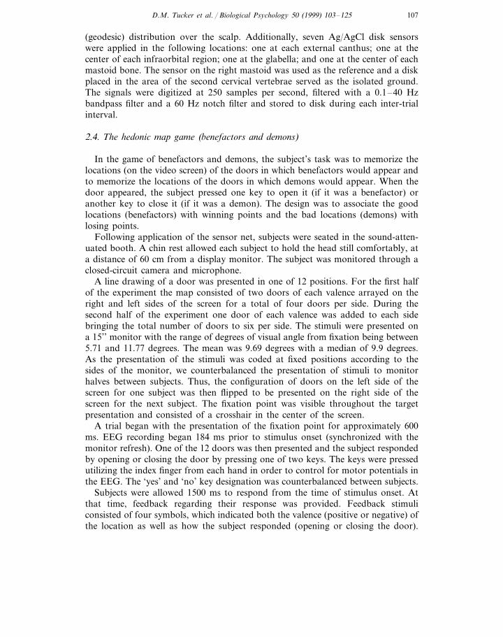
2.4. The hedonic map game (benefactors and demons)
In the game of benefactors and demons, the subject’s task was to memorize thelocations (on the video screen) of the doors in which benefactors would appear andto memorize the locations of the doors in which demons would appear. When thedoor appeared, the subject pressed one key to open it (if it was a benefactor) oranother key to close it (if it was a demon). The design was to associate the goodlocations (benefactors) with winning points and the bad locations (demons) withlosing points.
Following application of the sensor net, subjects were seated in the sound-atten-uated booth. A chin rest allowed each subject to hold the head still comfortably, ata distance of 60 cm from a display monitor. The subject was monitored through aclosed-circuit camera and microphone.
A line drawing of a door was presented in one of 12 positions. For the first halfof the experiment the map consisted of two doors of each valence arrayed on theright and left sides of the screen for a total of four doors per side. During thesecond half of the experiment one door of each valence was added to each sidebringing the total number of doors to six per side. The stimuli were presented ona 15’’ monitor with the range of degrees of visual angle from fixation being between5.71 and 11.77 degrees. The mean was 9.69 degrees with a median of 9.9 degrees.As the presentation of the stimuli was coded at fixed positions according to thesides of the monitor, we counterbalanced the presentation of stimuli to monitorhalves between subjects. Thus, the configuration of doors on the left side of thescreen for one subject was then flipped to be presented on the right side of thescreen for the next subject. The fixation point was visible throughout the targetpresentation and consisted of a crosshair in the center of the screen.
A trial began with the presentation of the fixation point for approximately 600ms. EEG recording began 184 ms prior to stimulus onset (synchronized with themonitor refresh). One of the 12 doors was then presented and the subject respondedby opening or closing the door by pressing one of two keys. The keys were pressedutilizing the index finger from each hand in order to control for motor potentials inthe EEG. The ‘yes’ and ‘no’ key designation was counterbalanced between subjects.
Subjects were allowed 1500 ms to respond from the time of stimulus onset. Atthat time, feedback regarding their response was provided. Feedback stimuliconsisted of four symbols, which indicated both the valence (positive or negative) ofthe location as well as how the subject responded (opening or closing the door).

D.M. Tucker et al. / Biological Psychology 50 (1999) 103–125108
There were two symbols to indicate correct performance. The first was a ‘happyface’ indicating that the subject correctly pressed the ‘open’ key for a benefactordoor. The other correct response was a ‘sad face’ with a line drawn through it (theinternational ‘No’ symbol), which indicated that the subject correctly pressed the‘close’ key for a demon door. The two symbols for incorrect responses were a‘happy face’ with a line through it (‘close’ key for a benefactor) and a ‘sad face’(‘open’ key for a demon). Feedback remained on the monitor, at fixation, for 500ms and EEG data gathered for 800 ms following feedback. If the subject failed torespond to the initial target prior to 1500 ms, the feedback was a small square toindicate to the subject to respond more quickly.
In order to assess performance, rather than acquisition, of the memory task (andthus to keep the psychological variables fairly constant across the ERP trialrepetitions), the subjects were given a learning block in which they were shown allof the benefactor doors, or the demon doors, whenever they requested. They wereallowed to practice until ready to begin the points blocks. The subject received 10points each time he or she responded opened a benefactor door (no penalty forclosing it) and lost 10 points for each opening of a demon door (with no reward forclosing it). This award scheme attempted to specify the relationship between thelocation and its valence; responses to negative locations could only lose points orleave the point total unchanged, whereas responses to locations with a positivevalence allowed the subject to win points or leave the total unchanged. In addition,the feedback stimulus was also associated with a consistent valence: good or neutralfor good trials and neutral or bad for bad trials.
Trials were then presented in six blocks of 56 trials. At the end of each block thesubject was invited to rest and was allowed full control of the length of each break.After the conclusion of the third block, the number of stimuli was increased inorder to increase task difficulty. In the first three blocks, subjects were required tolearn the identification of eight stimuli (two of each valence in each visual field),while in blocks four through six this load was increased to 12 stimuli (three of eachvalence in each visual field).
Halfway through and at the end of the experiment, the subjects were given eitherthe PANAS or the Thayer ADCL. The schedule of these two inventories wascounterbalanced across subjects.
2.5. Data analysis
The raw EEG epochs were first subjected to automated artifact rejection. Therejection algorithm detected blinks as rapid voltage changes between channelsabove and below the eye (supra- and infra-orbital). Lateral or vertical eye move-ments were detected as voltage excursions in the external canthus or supra-infra-orbital sensors. Channels were also rejected for amplitudes out of the EEGrange and for abrupt voltage transitions. To validate these algorithms, a systematiccomparison was made with artifacts examined by an experienced experimenter andgood agreement was obtained for each form of artifact.

D.M. Tucker et al. / Biological Psychology 50 (1999) 103–125 109
For statistical analysis the data were transformed to an average reference (Curranet al., 1993). This procedure then generates a 65th channel in the form of zerominus the average of all the other sensors. All analyses reported here use theresulting 65-channel data.
Descriptive data analysis began with study of the waveform plots and electricalfield animations. Condition differences were highlighted with animations of t-testresults for every channel and every sample. These descriptive analyses reviewed thefull 3-s ERP epoch, including the ERP to the target door, the response andanticipation of feedback and the ERP to the feedback.
Electrical field animations were created with spherical spline interpolation (Perrinet al., 1987) to generate a scalp map from the 65 data values for each time sample.This method allowed for better visualization of the spatial and temporal coherenceof the components of the ERP than traditional waveform plots. The t-tests wereconducted for each sample at each channel, resulting in a topography animation ofthe t values for each channel. This technique was usable for any two factor, twolevel analysis; deriving two t scores for the main effects and one for the interaction.This analysis was completed only for those effects which demonstrated significantpeaks in the sample by sample analyses of variance. The t-test results, because theyare present for each channel, can then be interpolated by the same method as thevoltage data. This results in a series of scalp maps which highlight the spatial andtemporal coherence of the effects. The t-waves are helpful in that they specify thedirection of the effect as well as the amplitude of effects and can be thought of asdifference waves calibrated by the standard error.
These descriptive analyses may be expected to yield many effects that requirereplication for confident interpretation. The specific hypotheses of the study weretested with inferential repeated-measures ANOVAs. These examined: (1) the accu-racy and reaction time performance data; (2) the late (P300) interval of the ERP tothe good and bad target doors; and (3) the frontal and posterior alpha blocking inresponse to the feedback stimulus.
3. Results
3.1. Psychometric analysis
Factor analysis was used to test the hypothesis that the dimensionality of arousalself-report would parallel the dimensionality of the PANAS, corresponding toelation and anxiety in the Tucker and Williamson (1984) framework. A higher-or-der factor analysis was performed on the energy (EN), tiredness (TI), tension (TE)and calmness (CA) scales of the ADCL. Fig. 1 shows that Factor 1 (labeled aspositive arousal) describes Thayer’s tiredness–energy dimension and Factor 2(negative arousal) describes the calmness–tension dimension.
The hypothesis that dimensions of arousal parallel dimensions of affect wastested by correlating the ADCL factors with the PANAS affect scales. The positivearousal factor correlated 0.63 with positive affect and −0.21 with negative affect,

D.M. Tucker et al. / Biological Psychology 50 (1999) 103–125110
showing both convergent and discriminant validity. The negative arousal scalecorrelated 0.55 with negative affect but 0.30 with positive affect, suggesting that thedominant affective quality of this scale is negative, but that some variance is sharedwith positive affect. The factor plot (Fig. 1) suggested that the PANAS factors maybe related rather than independent, as would be expected if these reflect neurophys-iological systems for regulating brain activity (i.e. separable but not necessarilyorthogonal). Therefore an oblique rotation was used in the subsequent regressionanalyses with the PANAS factors of the Thayer scales.
3.2. Beha6ioral analysis
The dependent variables of response time (RT) and percent accuracy wereexamined with a repeated-measures ANOVA in a three-within, two-between facto-rial design. Within factors were task difficulty (two versus three door locations perside), field of presentation (left or right) and valence of location (good or bad).Between factors were gender (male or female) and individual differences in arousalreport (high, medium and low). One analysis was conducted dividing subjects onthe positive arousal dimension and a second analysis dividing them on the negativearousal dimension. For brevity, the results are described for the positive arousalanalysis only, except for effects that differed when the negative arousal classificationwas used.
For response time, a main effect of task difficulty was observed (F [1,46]=81.95,pB0.0001), indicating that the more difficult task (three doors) took longer(mean=1030 ms) than the easy task (two doors; mean=964 ms). Task difficultyinteracted with gender (F [1,46]=8.068, p=0.0067): men were faster than women inthe easy task, but less so in the difficult task. These means are presented in Table1.
Fig. 1. Factor structure of Thayer’s ADCL (TE, tension; EN, energy; TI, tiredness; and CA, calmness).The factor pattern suggests two dimensions of tiredness–energy and calmness–tension.

D.M. Tucker et al. / Biological Psychology 50 (1999) 103–125 111
Table 1Mean reaction times for gender and task difficulty
Difficult taskEasy task
976 1042Females892 1018Males
Responses were also significantly faster for stimuli presented to the left visualfield (mean=964.43) than the right (mean=1001.99) (F [1,46]=20.10, pB0.0001).Responses were faster for good stimuli (mean=973.03) compared to bad stimuli(mean=993.39) (F [1,46]=4.14, p=0.0476). These main effects did not interact.
In the analysis of response time including the negative arousal dimension, therewas an added interaction between difficulty, field and negative arousal. The meansfor these groups are presented in Table 2. The high negative arousal subjects didnot show the expected right hemisphere (left visual field) superiority on the easytask.
For percent accuracy, there were no differences between the positive and negativearousal analyses, except for the gender factor (F [1,46]=4.63, p=0.0368), where,for negative arousal, men were more accurate (mean number correct=37.61 out of42 possible) than women (mean=35.65). Main effects of task difficulty (F [1,46]=41.63, pB0.0001) and field (F [1,46]=18.01, pB0.0001) paralleled the RT effects:the subjects were more accurate for the easy task (easy task mean=37.95; difficulttask mean=35.23) and they were more accurate for the left visual field (left fieldmean=37.24; right field mean=35.94).
An interaction was seen between task difficulty and field (F [1,46]=9.91, p=0.0029): post-hoc means comparisons revealed that subjects were significantly moreaccurate (F [1]=31.35, pB0.0001) for stimuli in the left visual field than the rightduring the difficult block. These means are presented in Table 3.
Paralleling the RT findings, a three-way interaction was seen between taskdifficulty, field and valence (F [1,46]=5.04, pB0.0296): the decrease in accuracy forthe difficult condition was particularly apparent for good stimuli in the right visualfield. These means are presented in Table 4.
Table 2Mean reaction times for negative arousal by difficulty and field of presentation
High NARLow NAR Mid NAR
EasyLeft 931921910
974 936942Right
Hard1016Left 1016991
Right 1060 10681031

D.M. Tucker et al. / Biological Psychology 50 (1999) 103–125112
Table 3Mean number of correct responses (of 42 possible) for field of presentation by difficulty
Easy task Difficult task
38.1 36.3Left field34.137.8Right field
3.3. Descripti6e analysis: ERP response to the target door
The visual ERP response to the target (door) showed a characteristic set of scalpelectrical waves, several of which were responsive not only to the laterality but alsoto the valence (benefactor or demon) of the target stimulus. The initial review ofthese data examined the voltage maps animated over time, with t-tests of the majorcontrasts (left versus right field, good versus bad valence) conducted for each map.Fig. 2 shows the 65-channel topographic waveform plot, the channels arranged asif looking down on the head with the nose at the top.
For many subjects a medial occipital effect, typically negative, was seen at 70–80ms. The first consistent scalp wave occurred at 88 ms, the contralateral P1. Fig. 3shows several time samples from the 53-subject grand average, collapsed over leftfield good (benefactor) targets for illustration. At 88 ms the frontopolar negativityof the CNV in anticipation of the target can still be seen, as the stimulus-contralat-eral positivity of the P1 appears over the inferior right parietal area. Just as thecontra P1 begins to wane, an ipsilateral P1 appears over a roughly homologousarea of the left hemisphere. This ipsi P1 becomes much stronger than the contra P1and overlaps with the development of the contralateral N1 (168 ms). A contralat-eral N1 develops at this time for both left and right visual field stimuli, but theposterior negativity then becomes fairly focal over the right parietal area at180–190 ms for both left and right field targets.
At about this time (200 ms), a right frontal positivity appears, regardless ofwhether the stimulus is left or right. The positivity shifts toward the vertex (208 ms)and then progresses posteriorly to create a second set of posterior positive foci,what we have termed the ‘P1 reprise’ (Curran et al., 1993; Tucker et al., 1994). For
Table 4Mean number of correct responses (of 42 possible) for difficulty by field of presentation by valence
Bad stimuliGood stimuli
EasyLeft 37.938.3Right 37.637.9
Hard37.4 35.3Left33.9Right 34.3

D.M
.T
uckeret
al./B
iologicalP
sychology50
(1999)103
–125
113
Fig. 2. A 65-channel (average reference) topographic waveform plot contrasting left and right field targets. The vertical line indicates stimulus onset and thepoststimulus epoch is 1 s.

D.M. Tucker et al. / Biological Psychology 50 (1999) 103–125114
Fig. 3. Selected events in the scalp topography of the ERP to a left field target door in the hedonic mapgame.
the lateral stimuli of the present experiment, the contra and ipsi P1 foci are not‘reprised’ exactly; the second posterior positive waves (296 ms) are more superiorthan the P1 foci. However, the contra-ipsi progression of the P1 is repeated at thistime, with the initial posterior positivity strongly contralateral (296 ms), thenprogressing to a sustained ipsilateral positivity (472 ms).
The maps for 208 and 296 ms in Fig. 3 show that the initial occipitotemporalnegativity of the N1 gives rise to a contralateral, inferior and anterior progressionof the late N1 negativity during this interval. After taking up the fairly focal,right-medial parietal location for the maximal negative peak of the N1, the negativewave appears to move inferiorly and anteriorly toward the temporal lobe (208 ms),similar to the late N1 effects with attention in a previous dense array dataset (Pottset al., 1996). The t-test animations for the visual field effect show a stronglycontralateral negativity in this interval, which progresses inferiorly and anteriorlyover the next 100 ms. As seen in the 296 ms frame, the contralateral negativitybecomes sustained over right anterior temporal and frontal areas.

D.M. Tucker et al. / Biological Psychology 50 (1999) 103–125 115
As this contralateral negativity moves anteriorly and inferiorly, the LPC or P300is seen initially as a contralateral, parietal positivity (296 ms, Fig. 3). Over the next100 ms or so, this contralateral parietal positivity appears to be replaced by anegative contralateral parietal wave and the positivity moves ipsilaterally (472 ms,Fig. 4). Examining the valence (good/bad) difference during this interval of timewith the t-test animations showed that, at about the same time as the contra-ipsiprogression of the posterior positivities, a medial frontal effect began to differenti-ate between good and bad targets. The t-test map for 472 ms shows a strong focusof greater negativity for the bad trials over medial frontal areas, coincident withgreater positivity over lateral frontal inferior sites.
This ‘evaluative negativity’ seen in the t-map comparison was superposed on thegeneral topography of this time interval (472 ms), which involves the parietal P3and a frontal negativity. Over the next 400 ms, as the subject completed a responseto the target (open or close the door) and waited to see the feedback stimulus
Fig. 4. Additional features of the scalp topography of the ERP to a left field target. To the right of thevoltage maps for 472 and 800 ms are t-test maps for valence, reflecting the difference of the ERP to goodminus that to bad targets, standardized for the variance across subjects.

D.M. Tucker et al. / Biological Psychology 50 (1999) 103–125116
Fig. 5. P3 to CNV progression, from 536 to 856 ms post-target in 64 ms intervals.
(smiling or angry face at fixation), the anterior negativity became an increasinglydominant feature of the ERP, producing the distinct medial frontal negativedistribution of the CNV, as the P3 waned (Fig. 5). An ANOVA on this broadinterval suggested that the CNV was somewhat asymmetric as a function of thetarget valence (valence by hemisphere, pB0.06). As subjects waited for feedbackfrom a good trial the CNV was right-lateralized; as they waited for feedback aftera bad trial the CNV was left-lateralized.
3.4. Inferential ERP analyses: P300 inter6al for the target door ERP
Sixteen posterior channels were selected that showed the maximal P300 responsefor this window of 304–368 ms post-stimulus. A repeated measures ANOVA wasused with five within subjects factors (task difficulty, field of presentation, valenceof location, sensor location and hemisphere of sensor) fully crossed with twobetween subjects factors (gender and a three level measure of positive arousalderived from the Thayer). The analysis was then repeated, substituting a three level

D.M. Tucker et al. / Biological Psychology 50 (1999) 103–125 117
measure of negative arousal. Due to issues of heteroscedasticity the Geisser–Green-house correction values were used to determine effect significance. The main effectsof sensor location (F(7,322)=32.824, p=0.0001) and hemisphere (F(1,46)=10.811, p=0.0019) also interacted (F(7,322)=3.168, p=0.0030), indicating thatthere is an overall enhanced positivity of the P3 over the left hemisphere which ismost pronounced at those sensor locations with the maximum P3 amplitude.
This left-lateralized topography of the P300 would not seem consistent with thehypothesis of greater right hemisphere processing of this spatial memory task, if alarger P300 response is expected over the hemisphere that is more engaged with thetask. However, two observations suggested that a contralateral processing negativ-ity (and thus contralateral attenuation of the P300) was related to hemisphericcognitive processing in this interval. First, the P300 attenuation (apparent process-ing negativity) was contralateral to the stimulated visual field. Second, this con-tralateral negativity was enhanced for the difficult trial block compared to the easytrial block.
The contralateral processing negativity was observed in the interaction of hemi-sphere by field of presentation (F(1,46)=136.92, p=0.0001). Sensor locations inthe right hemisphere showed greater negativity in response to stimuli in the leftvisual field and those in the left hemisphere showed greater negativity to right fieldstimuli. This asymmetry of the ERP is somewhat stronger for left visual fieldstimuli, contributing to the overall right hemisphere negativity in this intervaldescribed above.
It is important to point out that the apparent contralateral processing negativityoverlaps substantially with the positivity of the P300 (and/or ‘P1 reprise’) effects.Thus the effect we describe as a contralateral processing negativity cannot bedistinguished in the scalp topography from an attenuation of the positivity of theP300.
The effect we describe as a contralateral negativity varies with sensor location(F(7,322)=31.321, p=0.0001) and it is greater for superior sites in this channel setfor both left and right field stimuli. However, the contralateral negativity is strongerfor left field stimuli and the interaction of hemisphere, field and gender (F(1,46)=9.823, p=0.0030) showed that the enhanced contralateral negativity for left fieldstimuli was particularly strong for women (although the direction was similar formen).
The task difficulty factor shows the effect of increasing the memory and attentionload, on processing generally and on contralateral processing specifically. Sensorlocation interacts with task difficulty (F(7,322)=5.369, p=0.0001) indicating thatit is the locations at which P300 is maximal that there is increased negativity in thelater, more difficult, trials. Task difficulty also modifies the field of presentation byhemisphere interaction (F(1,46)=14.581, p=0.0001). Consistent with the interpre-tation that the posterior negativity in the late ERP indicates increased visuospatialprocessing, the contralateral negativity is heightened in the later, more difficulttrials.
A main effect of stimulus valence showed that good stimuli were associated withgreater negativity for these posterior sites over this time window (F(1,46)=10.84,

D.M. Tucker et al. / Biological Psychology 50 (1999) 103–125118
p=0.0019). Valence played a role in a series of three way interactions whichincluded task difficulty. An interaction between valence, hemisphere and taskdifficulty (F(1,46)=8.408, p=0.0057) showed that the general right-lateralizationof the posterior negativity in this interval became particularly strong for the goodtargets during the difficult condition.
This effect should be interpreted in light of the additional interaction with gender(F(1,46)=9.686, p=0.0032), which showed that the differential asymmetry to goodtargets was particularly true for men. Women showed greater right hemispherenegativity for all conditions and particularly for bad trials during the early (easier)portion of the experiment. Men showed considerably less asymmetry (right hemi-sphere negativity), except for good trials in the later, more difficult portion of theexperiment.
Other higher order interactions with valence include effects with field of presenta-tion and task difficulty. This interaction showed a significant interaction withgender (F(1,46)=4.686, p=0.0356). Both genders demonstrated greater posteriornegativity to good stimuli (as compared to bad) presented to the right visual field.Females also displayed increased posterior negativity for good stimuli in the leftvisual field, across both early and late trials. Males, however, demonstrated greaterposterior negativity to left field stimuli only for the later, more difficult trials.
The valence by field of presentation by task difficulty effect also differed by levelof positive arousal from the Thayer measure (F(2,46)=3.382, p=0.0426). Byexamining this interaction for each group of Thayer scores, it was determined thatonly the middle level group demonstrated a significant interaction. For this group,there was no difference in response to left visual field stimuli based upon taskdifficulty; there was simply the familiar pattern of slightly increased negativity ofthe posterior ERP to good stimuli. For the right visual field, however, the easytrials show a small decrease in amplitude for good trials, while in the later, moredifficult trials, the good locations show a markedly increased negativity. Thispattern is similar, although not significant, for the high positive arousal group. Thelow positive arousal group shows a different pattern, with a trend toward signifi-cance, in which good trials show increased negativity in the easier condition in bothvisual fields, but only in the left visual field for difficult trials.
Valence also interacted with Sensor location (F(7,322)=5.26, p=0.0001, GG=0.0001) and indicated that, in general, good stimuli elicited greater negativity inposterior and inferior channels as compared to bad stimuli. This interaction wasfurther modified by hemisphere and the level of positive arousal from the Thayer(F(14,322)=2.189, p=0.0081, GG=0.0166), indicating that the valence by sensorlocation interaction held true for subjects who scored in the low range of positivearousal, as well as for those subjects who scored in the high range of positivearousal.
This effect then interacts with gender (F(14,322)=2.188, p=0.0081, GG=0.0167) in such a way that for low and high positive arousal, males demonstrategreater amplitude reduction for bad stimuli than women. In the middle level ofpositive arousal, males show an overall reduction in valence differentiation with amore posterior focus of positivity. Finally, there is a six way interaction that further

D.M. Tucker et al. / Biological Psychology 50 (1999) 103–125 119
modifies the above interaction (F(14,322)=1.918, p=0.0239, GG=0.0406). Whiledifficult to interpret, this interaction suggests that females display greater lateraliza-tion of responses, especially to those stimuli in the left visual field. This effect isgreatest for those scoring in the high and middle levels of positive arousal and tendsto be more evident in those sensor locations that are peripheral to the areas ofstrongest activation.
3.5. ERP to feedback
In examining the ERP to the feedback face appearing at fixation, we firstexamined the posterior positivities of the P1 and the P1 reprise (or second posteriorpositivity). These features are typically simultaneous bilateral foci to a fovealstimulus (Curran et al., 1993). Remarkably, in the present dataset the P1 reprisewas significantly enhanced contralateral to where the target had been. Eye move-ments are an unlikely explanation. Not only is the scalp topography unlike that ofeye movements, a video camera on the monitor reveals even small deviations fromfixation and we examined the data carefully for eye movements during this interval.
Examining the Valence t-tests showed that the ERP to the feedback alsodifferentiated good versus bad trials over medial frontal areas in the P3 window.The topography of this effect was similar to that for the medial frontal negativityseen for the valence t-tests for the target stimuli. To examine whether individualdifferences in emotional arousal were related to this effect, we extracted thedifference between good and bad trials averaged over the interval between 464 and720 ms after feedback. A multiple regression was performed with the voltagedifference as the dependent variable and the two arousal factor scores (positivearousal (PAR) and negative arousal (NAR), from the oblique rotation) as predictorvariables. The analysis revealed that both positive and negative arousal scores weresignificant predictors, pB0.04 and pB0.05, respectively and in the same direction,greater arousal of both kinds predicted greater frontal negativity of this difference
Fig. 6. (a) PAR and (b) NAR regressions on frontal valence effect in the ERP to the feedback stimulus.

D.M. Tucker et al. / Biological Psychology 50 (1999) 103–125120
wave. Fig. 6 shows the regression model and scatter plots for the positive arousaland negative arousal predictions, which were similar in direction and magnitude.The greater medial frontal negativity to bad stimuli was thus enhanced with greaterarousal of either the positive mood (energy–vigor) or negative mood (anxiety–ten-sion) variety.
3.6. Alpha blocking to the feedback stimulus
As an initial indication of the response of the background EEG during this task,the power spectrum for each posterior channel (with the average reference deriva-tion) was computed with an FFT on the 1-s interval following the feedbackstimulus. Given the apparent memory reactivation effects seen in the ERP, we wereparticularly interested to examine alpha blocking as a convergent measure ofneuroelectrical response to the feedback stimulus. Power in the alpha (7.5–12.5 Hz)band was summed and cast into the repeated-measures ANOVA model describedabove.
Given the substantial midline alpha apparent in the EEG and given the impor-tance of medial frontal negativities to valence effects for both the target andfeedback stimuli, frontal alpha during the feedback interval (1 s after feedbackonset) was examined with left, medial and right levels of the hemisphere orlaterality factor. Similar to the covariance analysis above, a significant covarianceeffect for positive arousal was seen and was followed by trichotomizing on thisvariable. A significant interaction of positive arousal group with laterality andgender showed that the high positive arousal males and all of the females, showedmarkedly higher alpha at the midline than the lateral sites. However, there was nosign of asymmetry in frontal alpha in relation to the valence of the stimuli.
For the analysis of posterior channels, negative arousal showed a significantinteraction with the visual field×valence effect, F(1,49)=8.14, pB0.007. Todescribe this effect simply, subjects were divided into high, medium and lownegative arousal groups (17 Ss each). Given a significant visual field×valence×Negative arousal grouping, F(2,48)=4.88, pB0.02, simple interaction analysesshowed that only the high negative arousal group showed a significant visualfield×valence interaction, F(1,17)=13.6, pB0.002 (Fig. 7). Alpha was blocked(presumed increased brain activation) when a good target appeared in the left field,F(1,17)=8.8, pB0.009. Conversely, alpha blocking was greater for the bad thangood targets in the right visual field, F(1,17)=5.01, pB0.04.
4. Discussion
The primary goal of this study was to develop a spatial memory task that wouldbe sensitive to the cognitive skills of the right hemisphere in normal subjects. Thefirst hypothesis predicted an advantage for remembering left visual field stimuli,reflecting the right hemisphere advantage for spatial processing. The behavioralresults supported this hypothesis. These normal subjects as a group gave faster and

D.M. Tucker et al. / Biological Psychology 50 (1999) 103–125 121
Fig. 7. Posterior alpha valence×field effect for high negative arousal subjects.
more accurate responses to targets in the left visual field. However, this righthemisphere advantage was not seen for subjects high in negative arousal.
Similarly, the ERP data provided several clues to right-lateralized neuropsycho-logical mechanisms. A processing negativity appeared to be superposed on theP300, contralateral to the target visual field and this negativity appeared strongerover the right hemisphere. Whether this effect is an attenuation of the P300, orwhether it is a superposition of a separate electrophysiological process, is difficultto discern from the scalp topographies without more accurate methods of compo-nent identification or source localization. The increased negativity related toincreased cognitive processing demands may be similar to the N400 effect forsemantically incongruous verbal stimuli (Curran et al., 1993). The right-lateraliza-tion of the processing negativity was enhanced for female subjects, suggesting thatthey were engaging right hemisphere processing on this spatial task.
The second hypothesis predicted that increased task difficulty would enhance theright-lateralization of the response. As predicted, the visual field asymmetry in thebehavioral data was enhanced by the difficulty manipulation. Paralleling thebehavioral effect, the contralateral ERP processing negativity was also enhanced bythe difficulty manipulation. The increased field asymmetry with greater difficulty isa key feature of the present results. The simplest interpretation is that eachhemisphere controls the contralateral visual field; with the greater difficulty of theincreased door locations the right hemisphere’s superior spatial ability becameapparent. A somewhat more complex interpretation would recognize the righthemisphere’s contribution to spatial memory for both visual fields, at least whentask demands are moderate. When task demands are increased and hemisphereabilities are taxed, the right hemisphere may become more exclusively concerned

D.M. Tucker et al. / Biological Psychology 50 (1999) 103–125122
with processing in the left visual field. The greatest impairment would then beexpected for the right visual field targets (processed by the unaided left hemisphere).
Regardless of interpretation, the processing deficit created by the difficultymanipulation was greatest for the left hemisphere’s representation of the rightvisual field. The contralateral negativity in the P300 interval for the target doorshowed a similar reactivity to the difficulty manipulation as the performance speedand accuracy data, with the increased asymmetry indicating not only contralateralhemisphere processing, but a particular trend toward right hemisphere processing.
Although the difficulty manipulation proved important for exaggerating and thusidentifying, the right hemisphere contribution to this task, it may not be a simplecontinuous variable. As difficulty increases on a cognitive task, subjects may changestrategies rather than simply engaging the same strategy more intensely. The sexdifferences in spatial performance, for example, could be associated with strategydifferences that might respond differently to the difficulty manipulation. In thepresent task, the difficult blocks required the subjects to learn new locations, so thatthe behavioral and ERP asymmetries reflected learning as well as performance ofthe spatial memory task. It may be this new learning that increased the demands onright hemisphere processing.
The third hypothesis predicted that subjects with low PAR (positive arousal)scores would demonstrate reduced performance, especially in difficult tasks. Therewas little support for this hypothesis. The theory is that depression, in normal aswell as clinical populations, is associated with reduced levels of an emotional andneural arousal system that is right-lateralized (Tucker and Williamson, 1984). Thusspatial cognition would be predicted to be more impaired by decreases in positivearousal (elation), than by increases in negative arousal (anxiety) (Tucker and Liotti,1990). In the present experiment, the behavioral measures suggested a decreasedright hemisphere advantage for subjects high in negative arousal and several ERPfindings that were sensitive to the negative arousal measure. But there was nosupport for the hypothesis that right hemisphere function would be impaired insubjects low in positive arousal.
The fourth hypothesis predicted differential hemispheric responses to good versusbad stimuli. In contrast to the typical view of valence and laterality in the literature,we hypothesized that the right hemisphere would be biased to process positively-va-lenced stimuli while the left hemisphere would be biased to process of negatively-va-lenced stimuli (Tucker, 1981). In both the RT and accuracy measures, aninteraction of motivational valence of the target with visual field was observed onlyin the difficult condition. The right field performance decrement (left field advan-tage) with increased difficulty occurred for good targets particularly. Whether thedifficulty effect is due to withdrawal of the right hemisphere’s bilateral control, orto differential stress upon the left hemisphere’s control of right field targets, theimplication from the performance data may be that there is a negative motivationalbias for the left hemisphere. Under difficult conditions the left hemisphere appearedmore effective in remembering the bad locations than the good ones.
In the performance data, the only gender effect was a superiority of males in thefirst, easy block that was no longer apparent in the second, difficult block.

D.M. Tucker et al. / Biological Psychology 50 (1999) 103–125 123
Although males are often superior to females on spatial tasks, the females of thepresent experiment were able to achieve comparable performance by the comple-tion of the task. An interesting question is whether this was related to theircontinuing to show a strong right-lateralization of the processing negativity in theERP.
The analysis of posterior brain activity during the feedback interval showedposterior alpha blocking to be greater for feedback for good (versus bad) targetsto the left visual field and for bad (versus good) targets to the right visual field.Whereas this can be seen as consistent with previous interpretations of hemi-spheric emotional biases (Tucker, 1981), it was observed only for subjects high innegative arousal (Thayer’s tension versus calmness factor).
There were other clues to frontal lobe mechanisms in relation to motivationand affect in this study. The subjects developed a strong frontal negativity (CNV)as they waited for the feedback stimulus and this resolved with the perception ofthe feedback. At about 500 ms after the feedback stimulus, a different, somewhatmore focal medial frontal negativity appeared for the feedback on bad trials,somewhat reminiscent of the error-related negativity observed when subjects areaware of making an error on a speeded response task (Dehaene et al., 1994; Luuet al., 1999). This effect was stronger for subjects higher in either positive ornegative arousal (Fig. 6), perhaps suggesting that general motivation rather thanthe specific valence of the arousal was important to the effect. Remarkably, thiswas the second medial frontal negativity in response to the bad targets, with thefirst occurring about 500 ms after the target stimulus (Fig. 4). These resultssuggest the importance of both motivation (higher positive or negative arousal)and hedonic valence (reflecting bad versus good targets) in medial frontal re-sponses such as the error-related negativity (ERN). In fact, one hypothesis may bethat, rather than being specific to error-monitoring, the ERN reflects the aversiveaffective response to making an error. Consistent with the importance of subjec-tive response to the ERN, the subjects in the speeded response tasks (Dehaene etal., 1994; Luu et al., 1999) often punctuate their erroneous responses with sponta-neous expletives.
In addition to the results specifically associated with the a priori hypotheses, thedescriptive analyses showed a number of interesting effects in response to thefeedback stimulus. In examining the ERP to the feedback stimulus, the ex-ploratory t-test animations showed what may be an indication of memory reacti-vation, with greater positivity contralateral to where the target had been.However, because the target stimulus remained on the screen until the feedbackappeared, this effect could reflect a ‘re-perception’ of the now static but stillapparent target stimulus triggered by the feedback, rather than an actual memoryreactivation engendered by the feedback. In a recent replication and extension ofthe present research (Hartry-Speiser and Tucker, 1999), the target was removedfrom the screen during the interval in which the subject made a response andwaited for the feedback. The enhanced contralateral positivity to the fovealfeedback stimulus was still apparent, supporting the idea that the feedback eliciteda memory reactivation.

D.M. Tucker et al. / Biological Psychology 50 (1999) 103–125124
5. Conclusions
The right-handed subjects in this experiment showed speed and accuracy advan-tages for left visual field presentation. These were paralleled by right-lateralizedprocessing negativity effects in the ERP, consistent with the hypothesis of righthemisphere specialization for this spatial memory task. The exploratory ERPanalyses suggested a rich set of neuroelectric effects that appear to track theattention and memory processes of the trial sequence. Analysis of alpha blockingsuggested, for subjects high in negative arousal only, a right hemisphere bias toprocess good targets and a left hemisphere bias to process bad targets. In addition,medial frontal ERP negativities were observed in response to bad targets and inresponse to the feedback stimulus following bad targets. Research now in progressexamines the performance of clinically depressed subjects with this paradigm todetermine whether right hemisphere dysfunction may be assessed with the dynamicmeasures of this experimental paradigm. Although several of the effects in thisstudy were complex and require replication, the dense array ERP measures pro-vided a rich window on brain activity in relation to both mood and memoryperformance in this experiment.
Acknowledgements
This research was supported by NIMH research grant MH42669 to the Univer-sity of Oregon and by NIMH SBIR grant MH54428 to Electrical Geodesics, Inc.
References
Borod, J.C., 1992. Interhemispheric and intrahemispheric control of emotion: a focus on unilateral braindamage. J. Consult. Clin. Psychol. 60, 339–348.
Bruder, G.E., Kuitkin, F.M., Stewart, J.W., Martin, C., Voglmaier, M.M., Harrison, W.M., 1989.Cerebral laterality in depression: differences in perceptual asymmetry among diagnostic subtypes. J.Abnorm. Psychol. 98, 177–187.
Corbetta, M., Miezin, F.M., Shulman, G.L., Petersen, S.E., 1993. A pet study of visuospatial attention.J. Neurosci. 13, 1202–1226.
Curran, T., Tucker, D.M., Kutas, M., Posner, M.I., 1993. Topography of the N400: brain electricalactivity reflecting semantic expectation. Electroencephalogr. Clin. Neurophysiol. 88 (3), 188–209.
Davidson, R.J., 1984. Affect, cognition and hemispheric specialization. In: Izard, C.E., Kagan, J.,Zajonc, R. (Eds.), Emotion, cognition and behavior. Cambridge University Press, New York.
Davidson, R.J., Chapman, J.P., Chapman, L.J., 1987. Task-dependent EEG asymmetry discriminatesbetween depressed and non-depressed subjects. Paper presented at the Society for PsychophysiologicResearch, Amsterdam, 1987.
Davidson, R.J., 1994. Role of prefrontal activation in the inhibition of negative affect. Psychophysiology31, S7 (Abstract).
Dehaene, S., Posner, M.I., Tucker, D.M., 1994. Commentary: localization of a neural system for errordetection and compensation. Psychol. Sci. 5, 303–305.
Hartry-Speiser, A., Tucker, D.M., 1999. Evoked potentials to simultaneous auditory and visual stimuli:an exploration of attentional control of stimulus processing (in preparation).

D.M. Tucker et al. / Biological Psychology 50 (1999) 103–125 125
Hillyard, S.A., Munte, T.F., Neville, H.J., 1985. Visual-spatial attention, orienting and brain physiology.In: Posner, M.I., Marin, O.S.M. (Eds.), Attention and Performance XI. Erlbaum, Hillsdale, NJ, pp.85–106.
Liotti, M., Tucker, D.M., 1992. Right hemisphere sensitivity to arousal and depression. Brain Cognit.18, 138–151.
Luu, P., Collins, P., Tucker, D.M., 1999. Mood, personality and self-regulation: negative affect andemotionality in relation to frontal lobe mechanisms of error-detection. J. Exp. Psychol. (in press).
Mesulam, M.M., 1983. The functional anatomy and hemispheric specialization for directed attention:The role of the parietal lobe and its connectivity. Trends Neurosci. 6, 384–387.
Miller, E.N., Fujioka, T.A.T., Chapman, L.J., Chapman, J.P., 1995. Hemispheric aymmetries offunction in patients with major affective disorders. J. Psychiatr. Res. 29, 173–183.
Perrin, F., Bertrand, O., Pernier, J., 1987. Scalp current density mapping: value and estimation frompotential data. Biomed. Eng. 34, 283–288.
Posner, M.I., Walker, J.A., Friedrich, F.J., Rafal, R.D., 1984. Effects of parietal lobe injury on covertorienting of visual attention. J. Neurosci. 4, 863–874.
Potts, G.F., Liotti, M., Tucker, D.M., Posner, M.I., 1996. Frontal and inferior temporal cortical activityin visual target detection: evidence from high spatially sampled event-related potentials. BrainTopog. 9, 3–14.
Thayer, R.E., 1978. Toward a psychological theory of multidimensional activation (arousal). Motiv.Emotion 2, 1–34.
Thayer, R.E., 1989. The Biopsychology of Mood and Arousal. Oxford University Press, New York.Tucker, D.M., 1981. Lateral brain function, emotion and conceptualization. Psychol. Bull. 89, 19–46.Tucker, D.M., 1993. Spatial sampling of head electrical fields: the geodesic sensor net. Electroen-
cephalogr. Clin. Neurophysiol. 87, 154–163.Tucker, D.M., Frederick, S.L., 1989. Emotion and brain lateralization. In: Wagner, H., Manstead, T.
(Eds.), Handbook of Psychophysiology: Emotion and Social Behaviour. Wiley, New York.Tucker, D.M., Liotti, M., 1990. Neuropsychological mechanisms of anxiety and depression. In: Boller,
F., Grafman, J. (Eds.), Handbook of Neuropsychology. Elsevier, Amsterdam, pp. 443–475.Tucker, D.M., Liotti, M., Potts, G.F., Russell, G.S., Posner, M.I., 1994. Spatiotemporal analysis of
brain electrical fields. Human Brain Map. 1, 134–152.Tucker, D.M., Stenslie, C.E., Roth, R.S., Shearer, S., 1981. Right frontal lobe activation and right
hemisphere performance decrement during a depressed mood. Arch. General Psychiatr. 38, 169–174.Tucker, D.M., Williamson, P.A., 1984. Asymmetric neural control systems in human self-regulation.
Psychol. Rev. 91, 185–215.
.