Monoketonic Curcuminoid-Lidocaine Co-Deliver Using ... - MDPI
20
Citation: Vigato, A.A.; Machado, I.P.; del Valle, M.; da Ana, P.A.; Sepulveda, A.F.; Yokaichiya, F.; Franco, M.K.K.D.; Loiola, M.C.; Tófoli, G.R.; Cereda, C.M.S.; et al. Monoketonic Curcuminoid- Lidocaine Co-Deliver Using Thermosensitive Organogels: From Drug Synthesis to Epidermis Structural Studies. Pharmaceutics 2022, 14, 293. https://doi.org/ 10.3390/pharmaceutics14020293 Academic Editor: Helena Tomás Received: 23 December 2021 Accepted: 25 January 2022 Published: 27 January 2022 Publisher’s Note: MDPI stays neutral with regard to jurisdictional claims in published maps and institutional affil- iations. Copyright: © 2022 by the authors. Licensee MDPI, Basel, Switzerland. This article is an open access article distributed under the terms and conditions of the Creative Commons Attribution (CC BY) license (https:// creativecommons.org/licenses/by/ 4.0/). pharmaceutics Article Monoketonic Curcuminoid-Lidocaine Co-Deliver Using Thermosensitive Organogels: From Drug Synthesis to Epidermis Structural Studies Aryane A. Vigato 1 , Ian P. Machado 2 , Matheus del Valle 3 , Patricia A. da Ana 3 , Anderson F. Sepulveda 1 , Fabiano Yokaichiya 4 , Margareth K. K. D. Franco 5 , Messias C. Loiola 1 , Giovana R. Tófoli 6 , Cintia Maria S. Cereda 6 , Mirela I. de Sairre 1, * and Daniele R. de Araujo 1, * 1 Human and Natural Sciences Center, Federal University of ABC, Sao Paulo 09210-580, SP, Brazil; [email protected] (A.A.V.); [email protected] (A.F.S.); [email protected] (M.C.L.) 2 Department of Fundamental Chemistry, Institute of Chemistry, University of São Paulo, Sao Paulo 05508-000, SP, Brazil; [email protected] 3 Center of Engineering, Modeling and Applied Social Sciences, Federal University of ABC, Sao Bernardo 09606-045, SP, Brazil; [email protected] (M.d.V.); [email protected] (P.A.d.A.) 4 Department Quantum Phenomena in Novel Materials, Helmholtz-Zentrum Berlin für Materialien, 14109 Berlin, Germany; [email protected] 5 Nuclear and Energy Research Institute, Sao Paulo 01000-000, SP, Brazil; [email protected] 6 São Leopoldo Mandic Research Unit, Campinas 13000-000, SP, Brazil; [email protected] (G.R.T.); [email protected] (C.M.S.C.) * Correspondence: [email protected] (M.I.d.S.); [email protected] (D.R.d.A.) Abstract: Organogels (ORGs) are remarkable matrices due to their versatile chemical composition and straightforward preparation. This study proposes the development of ORGs as dual drug-carrier systems, considering the application of synthetic monoketonic curcuminoid (m-CUR) and lidocaine (LDC) to treat topical inflammatory lesions. The monoketone curcuminoid (m-CUR) was synthesized by using an innovative method via a NbCl 5 –acid catalysis. ORGs were prepared by associating an aqueous phase composed of Pluronic F127 and LDC hydrochloride with an organic phase comprising isopropyl myristate (IPM), soy lecithin (LEC), and the synthesized m-CUR. Physicochemical charac- terization was performed to evaluate the influence of the organic phase on the ORGs supramolecular organization, permeation profiles, cytotoxicity, and epidermis structural characteristics. The physico- chemical properties of the ORGs were shown to be strongly dependent on the oil phase constitution. Results revealed that the incorporation of LEC and m-CUR shifted the sol-gel transition temperature, and that the addition of LDC enhanced the rheological G 0 /G” ratio to higher values compared to original ORGs. Consequently, highly structured gels lead to gradual and controlled LDC permeation profiles from the ORG formulations. Porcine ear skin epidermis was treated with ORGs and evaluated by infrared spectroscopy (FTIR), where the stratum corneum lipids were shown to transition from a hexagonal to a liquid crystal phase. Quantitative optical coherence tomography (OCT) analysis revealed that LEC and m-CUR additives modify skin structuring. Data from this study pointed ORGs as promising formulations for skin-delivery. Keywords: organogels; poloxamer; lidocaine; curcuminoids; skin structural analysis 1. Introduction Organogels (ORGs) are nanostructured systems formed by three-dimensional inter- actions between an oil phase and an aqueous phase. Due to their versatile chemical composition, they have been considered as remarkable materials with straightforward preparation, displaying long-term stability, which allowed their wide use as matrices for pharmaceutical, cosmetic, biotech, and food technology applications [1–3]. The chemical Pharmaceutics 2022, 14, 293. https://doi.org/10.3390/pharmaceutics14020293 https://www.mdpi.com/journal/pharmaceutics
-
Upload
khangminh22 -
Category
Documents
-
view
1 -
download
0
Transcript of Monoketonic Curcuminoid-Lidocaine Co-Deliver Using ... - MDPI
�����������������
Citation: Vigato, A.A.; Machado, I.P.;
del Valle, M.; da Ana, P.A.;
Sepulveda, A.F.; Yokaichiya, F.;
Franco, M.K.K.D.; Loiola, M.C.;
Tófoli, G.R.; Cereda, C.M.S.; et al.
Monoketonic Curcuminoid-
Lidocaine Co-Deliver Using
Thermosensitive Organogels: From
Drug Synthesis to Epidermis
Structural Studies. Pharmaceutics
2022, 14, 293. https://doi.org/
10.3390/pharmaceutics14020293
Academic Editor: Helena Tomás
Received: 23 December 2021
Accepted: 25 January 2022
Published: 27 January 2022
Publisher’s Note: MDPI stays neutral
with regard to jurisdictional claims in
published maps and institutional affil-
iations.
Copyright: © 2022 by the authors.
Licensee MDPI, Basel, Switzerland.
This article is an open access article
distributed under the terms and
conditions of the Creative Commons
Attribution (CC BY) license (https://
creativecommons.org/licenses/by/
4.0/).
pharmaceutics
Article
Monoketonic Curcuminoid-Lidocaine Co-Deliver UsingThermosensitive Organogels: From Drug Synthesis toEpidermis Structural StudiesAryane A. Vigato 1, Ian P. Machado 2 , Matheus del Valle 3, Patricia A. da Ana 3, Anderson F. Sepulveda 1 ,Fabiano Yokaichiya 4, Margareth K. K. D. Franco 5, Messias C. Loiola 1, Giovana R. Tófoli 6,Cintia Maria S. Cereda 6, Mirela I. de Sairre 1,* and Daniele R. de Araujo 1,*
1 Human and Natural Sciences Center, Federal University of ABC, Sao Paulo 09210-580, SP, Brazil;[email protected] (A.A.V.); [email protected] (A.F.S.);[email protected] (M.C.L.)
2 Department of Fundamental Chemistry, Institute of Chemistry, University of São Paulo,Sao Paulo 05508-000, SP, Brazil; [email protected]
3 Center of Engineering, Modeling and Applied Social Sciences, Federal University of ABC,Sao Bernardo 09606-045, SP, Brazil; [email protected] (M.d.V.); [email protected] (P.A.d.A.)
4 Department Quantum Phenomena in Novel Materials, Helmholtz-Zentrum Berlin für Materialien,14109 Berlin, Germany; [email protected]
5 Nuclear and Energy Research Institute, Sao Paulo 01000-000, SP, Brazil; [email protected] São Leopoldo Mandic Research Unit, Campinas 13000-000, SP, Brazil;
[email protected] (G.R.T.); [email protected] (C.M.S.C.)* Correspondence: [email protected] (M.I.d.S.); [email protected] (D.R.d.A.)
Abstract: Organogels (ORGs) are remarkable matrices due to their versatile chemical compositionand straightforward preparation. This study proposes the development of ORGs as dual drug-carriersystems, considering the application of synthetic monoketonic curcuminoid (m-CUR) and lidocaine(LDC) to treat topical inflammatory lesions. The monoketone curcuminoid (m-CUR) was synthesizedby using an innovative method via a NbCl5–acid catalysis. ORGs were prepared by associating anaqueous phase composed of Pluronic F127 and LDC hydrochloride with an organic phase comprisingisopropyl myristate (IPM), soy lecithin (LEC), and the synthesized m-CUR. Physicochemical charac-terization was performed to evaluate the influence of the organic phase on the ORGs supramolecularorganization, permeation profiles, cytotoxicity, and epidermis structural characteristics. The physico-chemical properties of the ORGs were shown to be strongly dependent on the oil phase constitution.Results revealed that the incorporation of LEC and m-CUR shifted the sol-gel transition temperature,and that the addition of LDC enhanced the rheological G′/G” ratio to higher values compared tooriginal ORGs. Consequently, highly structured gels lead to gradual and controlled LDC permeationprofiles from the ORG formulations. Porcine ear skin epidermis was treated with ORGs and evaluatedby infrared spectroscopy (FTIR), where the stratum corneum lipids were shown to transition froma hexagonal to a liquid crystal phase. Quantitative optical coherence tomography (OCT) analysisrevealed that LEC and m-CUR additives modify skin structuring. Data from this study pointed ORGsas promising formulations for skin-delivery.
Keywords: organogels; poloxamer; lidocaine; curcuminoids; skin structural analysis
1. Introduction
Organogels (ORGs) are nanostructured systems formed by three-dimensional inter-actions between an oil phase and an aqueous phase. Due to their versatile chemicalcomposition, they have been considered as remarkable materials with straightforwardpreparation, displaying long-term stability, which allowed their wide use as matrices forpharmaceutical, cosmetic, biotech, and food technology applications [1–3]. The chemical
Pharmaceutics 2022, 14, 293. https://doi.org/10.3390/pharmaceutics14020293 https://www.mdpi.com/journal/pharmaceutics
Pharmaceutics 2022, 14, 293 2 of 20
compositions of the oil and aqueous phases can be diversified by using several organicsolvents or oils, e.g., isopropyl myristate and palmitate, and water-soluble gelling agents,such as polymers and low-molecular-weight organogelators [4]. For instance, polymericorganogelators have being investigated in a variety of drug-delivery systems carrying smallmolecules, nanoparticles, and antibodies, while low molecular-weight organogelators havebeen used in pharmaceutical and food applications which are already approved by healthauthorities [3]. By modifying the chemical nature of the constituent phases, these materialscan incorporate bioactive compounds with different physicochemical and pharmacologicalcharacteristics. Since they are lipophilic and easy to spread, ORGs are topically adminis-tered, where the systemic first-pass effect is avoided. In this context, the presence of anoil phase favors the ORGs interaction with the skin, since the stratum corneum (SC) lipidmatrix constitutes a difficult barrier to be overcome by most formulations and drugs [5].The challenge in topical administration of bioactive compounds is therefore to effectivelypromote permeation through the SC [6].
Recent literature proposed several formulations that aid molecules passing throughthe skin, such as hydrogels, nanoparticles, and emulsions [1,7,8]. Penetration enhancersare important in these materials’ composition to improve the diffusivity and solubilityof molecules through the skin by reducing the stratum corneum barrier resistance. Themechanisms of penetration enhancement involve the interaction between the enhancementagents with lipids and proteins (mainly keratin) of the skin, resulting in the disorganizationof stratum corneum (SC) barrier. Water, hydrocarbons, alcohols and acids can be defined asclassical enhancers, while formulations containing cyclodextrin derivatives and chitosanare also widely employed [6]. For pharmaceutical applications, dual drug-delivery is amotivating topic to pursue as it is possible to combine bioactive molecules with comple-mentary and/or synergistic action, increasing the efficiency of the topical treatment [9].Among bioactive molecules used in skin procedures/treatments, lidocaine (LDC) is a localanesthetic widely used in clinical settings, available as commercial formulations such asgels and ointments [10]. However, such formulations have a short anesthesia duration, re-ducing the pharmacological effect efficiency at the site of administration [11]. Additionally,permeation-promoting organic compounds are commonly used in ORGs to disrupt the SCbarrier and allowing the drugs to penetrate toward the deep layers of the skin [5,12].
Monoketone curcuminoids (m-CURs, Figure 1A) are synthetic hydrophobic com-pounds derived from curcumin (Figure 1B), capable of interacting with the oil phase ofORGs [13]. Due to their monoketone structure in contrast to the diketonic curcumin,m-CURs have higher chemical photostability, expanding their application potential fortherapeutical formulations and extended drug-release [14–16]. Therefore, there is an openpath within this framework to investigate ORG-based formulations for drug co-delivery,seeking synergistic effects between pharmaceutically complementary additives introducedinto the different aqueous and oil phases [17].
In this study, ORG formulations were prepared by incorporating an oil phase, com-posed of isopropyl myristate (IPM) and soy lecithin (LEC), into an aqueous phase ofthermosensitive poloxamer PL407 [18–20]. The monoketone curcuminoid (m-CUR) wassynthesized by a Claisen-Schmidt condensation reaction via a NbCl5–acid catalysis, and itwas added as the bioactive molecule of the oil phase (Figure 1C) [15]. Aiming at co-deliveryapplications, lidocaine hydrochloride (LDC) was incorporated into the ORGs aqueousphase. A thorough physico-chemical characterization was carried to assess the curcumi-noid influence on ORG structural organization, as well as on the local anesthetic permeationand cytotoxicity. In addition, the interaction of ORGs with SC lipids was evaluated byinfrared spectroscopy (FTIR) and optical coherence tomography (OCT) to investigate theepidermis structural and morphological changes after treatment with ORGs [21,22].
In this context, our study targets at the development of m-CUR/LDC-activated ORGsand at investigating the influence of their components on the drug permeation profiles,epidermis structure, and cytotoxicity, aiming at the treatment of skin inflammatory dis-eases [23,24].
Pharmaceutics 2022, 14, 293 3 of 20
Pharmaceutics 2022, 14, x FOR PEER REVIEW 3 of 22
In this context, our study targets at the development of m-CUR/LDC-activated ORGs and at investigating the influence of their components on the drug permeation profiles, epidermis structure, and cytotoxicity, aiming at the treatment of skin inflammatory diseases [23,24].
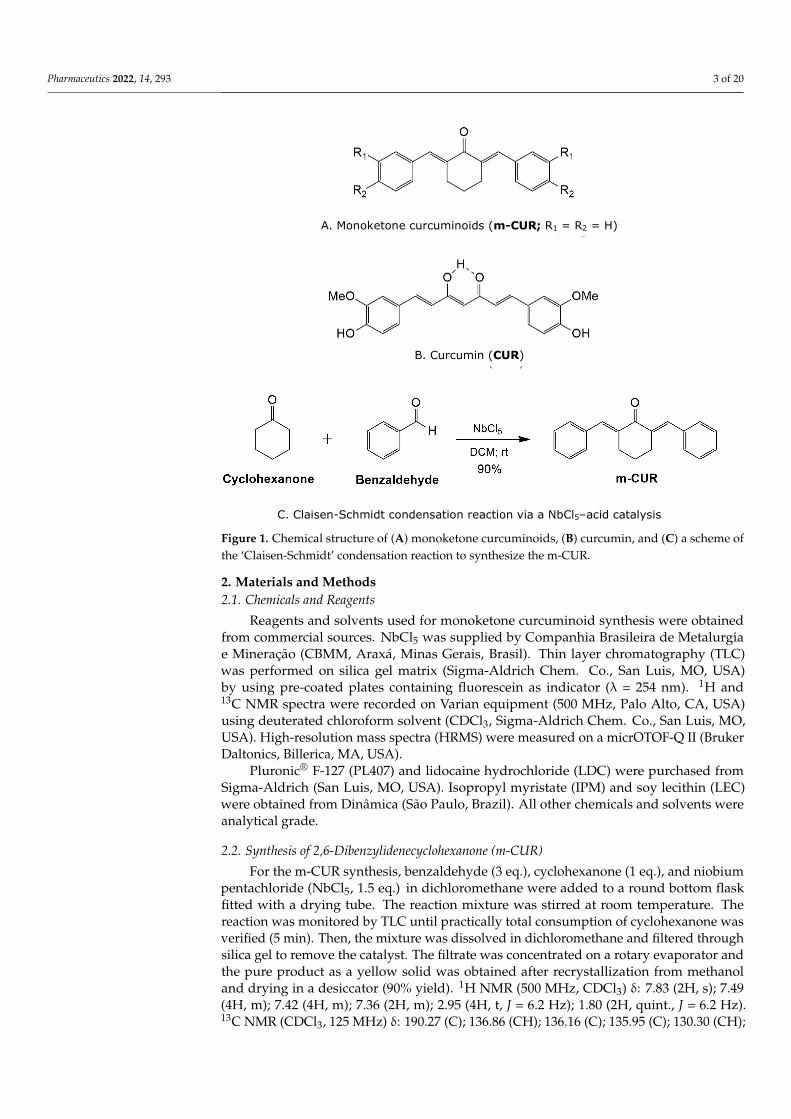
Figure 1. Chemical structure of (A) monoketone curcuminoids, (B) curcumin, and (C) a scheme of the ‘Claisen-Schmidt’ condensation reaction to synthesize the m-CUR.
2. Materials and Methods 2.1. Chemicals and Reagents
Reagents and solvents used for monoketone curcuminoid synthesis were obtained from commercial sources. NbCl5 was supplied by Companhia Brasileira de Metalurgia e Mineração (CBMM, Araxá, Minas Gerais, Brasil). Thin layer chromatography (TLC) was performed on silica gel matrix (Sigma-Aldrich Chem. Co., San Luis, MO, USA) by using pre-coated plates containing fluorescein as indicator (λ = 254 nm). 1H and 13C NMR spectra were recorded on Varian equipment (500 MHz, Palo Alto, CA, USA) using deuterated chloroform solvent (CDCl3, Sigma-Aldrich Chem. Co., San Luis, MO, USA). High-resolution mass spectra (HRMS) were measured on a micrOTOF-Q II (Bruker Daltonics, Billerica, MA, USA).
Pluronic® F-127 (PL407) and lidocaine hydrochloride (LDC) were purchased from Sigma-Aldrich (San Luis, MO, USA). Isopropyl myristate (IPM) and soy lecithin (LEC) were obtained from Dinâmica (São Paulo, Brazil). All other chemicals and solvents were analytical grade.
2.2. Synthesis of 2,6-Dibenzylidenecyclohexanone (m-CUR) For the m-CUR synthesis, benzaldehyde (3 eq.), cyclohexanone (1 eq.), and niobium
pentachloride (NbCl5, 1.5 eq.) in dichloromethane were added to a round bottom flask fitted with a drying tube. The reaction mixture was stirred at room temperature. The reaction was monitored by TLC until practically total consumption of cyclohexanone was
A. Monoketone curcuminoids (m-CUR; R1 = R2 = H)
B. Curcumin (CUR)
C. Claisen-Schmidt condensation reaction via a NbCl5–acid catalysis
Figure 1. Chemical structure of (A) monoketone curcuminoids, (B) curcumin, and (C) a scheme ofthe ‘Claisen-Schmidt’ condensation reaction to synthesize the m-CUR.
2. Materials and Methods2.1. Chemicals and Reagents
Reagents and solvents used for monoketone curcuminoid synthesis were obtainedfrom commercial sources. NbCl5 was supplied by Companhia Brasileira de Metalurgiae Mineração (CBMM, Araxá, Minas Gerais, Brasil). Thin layer chromatography (TLC)was performed on silica gel matrix (Sigma-Aldrich Chem. Co., San Luis, MO, USA)by using pre-coated plates containing fluorescein as indicator (λ = 254 nm). 1H and13C NMR spectra were recorded on Varian equipment (500 MHz, Palo Alto, CA, USA)using deuterated chloroform solvent (CDCl3, Sigma-Aldrich Chem. Co., San Luis, MO,USA). High-resolution mass spectra (HRMS) were measured on a micrOTOF-Q II (BrukerDaltonics, Billerica, MA, USA).
Pluronic® F-127 (PL407) and lidocaine hydrochloride (LDC) were purchased fromSigma-Aldrich (San Luis, MO, USA). Isopropyl myristate (IPM) and soy lecithin (LEC)were obtained from Dinâmica (São Paulo, Brazil). All other chemicals and solvents wereanalytical grade.
2.2. Synthesis of 2,6-Dibenzylidenecyclohexanone (m-CUR)
For the m-CUR synthesis, benzaldehyde (3 eq.), cyclohexanone (1 eq.), and niobiumpentachloride (NbCl5, 1.5 eq.) in dichloromethane were added to a round bottom flaskfitted with a drying tube. The reaction mixture was stirred at room temperature. Thereaction was monitored by TLC until practically total consumption of cyclohexanone wasverified (5 min). Then, the mixture was dissolved in dichloromethane and filtered throughsilica gel to remove the catalyst. The filtrate was concentrated on a rotary evaporator andthe pure product as a yellow solid was obtained after recrystallization from methanoland drying in a desiccator (90% yield). 1H NMR (500 MHz, CDCl3) δ: 7.83 (2H, s); 7.49(4H, m); 7.42 (4H, m); 7.36 (2H, m); 2.95 (4H, t, J = 6.2 Hz); 1.80 (2H, quint., J = 6.2 Hz).13C NMR (CDCl3, 125 MHz) δ: 190.27 (C); 136.86 (CH); 136.16 (C); 135.95 (C); 130.30 (CH);
Pharmaceutics 2022, 14, 293 4 of 20
128.52 (CH); 128.32 (CH); 28.39 (CH2); 22.96 (CH2). HRMS m/z [M + H]+ calcd: 275.1430;found: 275.1450.
2.3. PL-Based Organogels Preparation
Firstly, the oil phase (OP) containing 2 mL isopropyl myristate (IPM) and the addi-tives, LEC (2% w/v) and m-CUR (1 mg/mL), was prepared under magnetic stirring in awater bath (~60 ◦C) until homogenization. The aqueous phase (AP), consisting of PL407(30% w/v) and sodium benzoate/methylparaben/propylparaben (0.25, 0.1, and 0.05% w/v)as preservatives, was prepared in an ice bath under magnetic stirring (300 rpm). Then, LDC(25 mg/mL) was added to AP. After solubilization, the AP was kept at 8 ◦C until use. Tofinally prepare the ORG formulations, OP was added to AP (1:4 v/v) and mixed with a glassrod until obtaining a homogeneous system. The final concentrations of m-CUR and LDCwere 0.02% and 2% (w/v), respectively, for all formulations. The final ORGs formulationswere (Table 1): ORG, ORG-LDC, ORG-LEC, ORG-LDC/LEC, ORG-LEC/m-CUR, andORG-LDC/LEC/m-CUR, where the ORG formulation (control) consists of PL407 30% v/v(aqueous phase) and IPM (oil phase) [11,25].
Table 1. Prepared organogels (ORG) formulations. All ORG are composed of an aqueous phase ofPL407 30% w/v and an oil phase of isopropyl myristate (IPM). LDC: Lidocaine hydrochloride, LEC:soy lecithin, m-CUR: synthesized monoketone curcuminoid.
Formulations Additives Composition (%, w/v)
ORG - PL407 (30%) and IPM (1%)ORG-LDC LDC ORG composition + LDC (2%)ORG-LEC LEC PL407 (30%) and IPM (1%) + LEC (2%)
ORG-LDC/LEC LDC + LEC ORG-LEC composition + LDC (2%)ORG-LEC/m-CUR LEC + m-CUR ORG-LEC composition + m-CUR (0.02%)
ORG-LDC/LEC/m-CUR LDC + LEC + m-CUR ORG-LEC composition + LDC (2%) and m-CUR (0.02%)
2.4. Organogels Physico-Chemical Characterization2.4.1. Organoleptic, pH and Morphological Characterization
The ORGs were assessed by visual inspection of color, odor, phase separation, andaggregate formation. The pH of every ORG was recorded with a pH meter after electrodeequilibrium. Morphological analysis of the ORGs was performed by using an FEI Quanta 250scanning electron microscope (SEM). The ORGs were spread on a microscope slide forminga thin film and dried in a desiccator for 24 h. The resultant dry powder was depositedon carbon tape, which was previously attached to an aluminum stub. SEM images wereacquired under electron acceleration voltage of 5 kV and image magnification of 196×.
2.4.2. Differential Scanning Calorimetry (DSC)
Calorimetric analyses were performed using a Netzsch DSC Polyma calorimeter(Netzsch, Selb, Germany). The ORG formulations were weighed in hermetic aluminumpans (20 mg) and measured under three thermal cycles (heating–cooling–heating) from 0to 50 ◦C at 5 ◦C/min ratio. All measurements were executed in triplicate. Thermogramdata were represented by the heat flux (J/g) versus temperature (◦C). An empty aluminumcrucible was used as the reference.
2.4.3. Rheology
Rheological analyses were performed using a cone-plate type geometry in an oscil-latory rheometer (Kinexus Lab., Malvern Instruments, Malvern, UK). The ORGs wereanalyzed in the 0–50 ◦C temperature range at 1 Hz frequency to determine the sol-geltransition temperature (Tsol-gel). Oscillatory analyses were performed under variable fre-quency interval (0.1–10 Hz) at 32.5 ◦C, for correlation tests related to the elastic module(G′), viscous module (G”) and apparent viscosity (η*). The relation between G′ module andoscillation frequency (v) was used to calculate the ORG′s strength using Equation (1):
G′ = S · vn (1)
Pharmaceutics 2022, 14, 293 5 of 20
where S is the formulation strength (Pa·s) and n is the viscoelastic exponent.
2.4.4. Small Angle Neutron Scattering (SANS)
SANS measurements were performed using the V16 instrument at Helmholtz-ZentrumBerlin (HZB). The scattering data were recorded at two different distances from the detector:2 m (wavelength 1.8–3.8 Å) and 11 m (wavelength 1.6–9.2 Å), covering the 0.007–0.5 Å−1 qinterval. Organizational samples were prepared in D2O, arranged in quartz cubes (1 mmoptical path), and measured at 25 and 40 ◦C. The MANTID data reduction package, adaptedfor V16, was used to process the generated data.
2.5. In Vitro Permeation Experiments
In vitro permeation tests were carried out by a vertical diffusion cells system with apermeation area of 1.72 cm2 (Microette Plus; Hanson Research, Chatsworth, CA, USA). Thedonor and recipient compartments were separated by an artificial membrane that mimicsthe organization of the human skin (Strat-M®) [26]. The receptor compartment was filledwith 7 mL of water/ethanol 70:30 v/v solution and kept under constant magnetic stirring(350 rpm) at 32.5 ± 0.5 ◦C. At regular intervals (0.5 to 48 h), aliquots from the receptorcompartment were collected and then analyzed by HPLC. LDC cumulative amounts wereexpressed as µg·cm−2. Drug flux values were obtained from the slope of the curves overthe 24-h period. Data were analyzed by the following Equation (2):
J = P·Cd (2)
where J (µg·cm−2·h−1) is the drug flux through of the membrane, P (cm·h−1) is the perme-ability coefficient, and Cd (µg·cm−2) is the drug concentration in the donor compartment.The lag time was calculated by extrapolating the time axis in the graph [27].
2.6. LDC and m-CUR Chromatographic Conditions
LDC concentrations were quantified by a HPLC system (Ultimate 3000, Chromeleon7.2 software, Thermo Fisher Scientific, Waltham, MA, USA) coupled to DAD detector,and to a C18 column (150 × 4.6 mm, 5 µm; Phenomenex, Torrance, CA, USA) at 30 ◦C.LDC samples were analyzed with a 0.6 mL/min flow, using a 20:80 mixture of acetonitrileand acetic acid solution 0.05% v/v as the mobile phase. Drug retention time was 3.5 min.The limits of detection (LD) and quantification (LQ) were determined from a previousstandard curve of LDC at 20, 25, 50, 60, 80, 100, 200 and 250 µg/mL. LD and LQ valuesfound were 3.31 and 9.73 µg/mL, respectively, and LDC concentration was derived fromthe equation y = 0.0244x + 0.0011 (R2 = 0.998). For the m-CUR molecule, the employedflow was 0.8 mL/min, using a mobile phase of 90:10 mixture of acetonitrile and acetic acidsolution 0.05% v/v. Molecule retention time was 4.7 min. LD and LQ values found were0.34 and 1.03, respectively, and the m-CUR concentration was derived from the equationy = 0.7483x + 0.0054 (R2 = 0.995) in concentrations between 5 and 100 µg/mL. All methodsfollowed the recommendations from the International Conference on Harmonization, andthe results represent three experiments performed in triplicate.
2.7. In Vitro Cell Viability Assays
HaCaT epidermal keratinocytes (Thermo Fisher Sci., Waltham, MA, USA) were seededin 96-wells plates (2.104 cells/well) for 48 h under humidified atmosphere (37 ◦C and5% CO2), using DMEM (Gibco Laboratories, Grand Island, NY, USA) supplemented with10% (v/v) fetal bovine serum (pH 7.2–7.4) and 100 µg/mL of penicillin/streptomycin.Predetermined amounts of ORGs were homogenized in DMEM by using a vortex for5 min., in a concentration interval from 0.005 to 0.7 mg/mL (corresponding to 0.001 to 0.14and from 1 to 140 µg/mL of m-CUR and LDC, respectively), and were used for treating cellsduring 24 h. For cell viability determination, Methylthiazolyldiphenyl-tetrazolium bromide(MTT) solution (100 µL, at 5 mg/mL in solution in phosphate buffered saline) was added towells and incubated for 4 h. After this period, MTT solution was removed, and DMSO was
Pharmaceutics 2022, 14, 293 6 of 20
added (50 µL) for 10 min. Absorbance measurements were acquired at 570 nm. Non-toxiccontrol wells were treated with DMEM at the same conditions used for ORGs formulations.
2.8. Epidermis Structural Analysis2.8.1. Porcine Ear Skin Preparation
Porcine ear samples were obtained from a local slaughterhouse and the experimentalprotocol was approved by the UFABC Institutional Committee for the Care and Use ofAnimals (#8719010318). Blood vessels and subcutaneous tissue were extracted, obtaining a2 mm-thick structure which was then dermatomed (0.45 mm, Nouvag, Rorschach, Switzer-land) to completely remove the subcutaneous tissue. The dermatomed skin was immersedin a water bath at 60 ◦C for 3 min, and then the isolated epidermis was detached using aspatula. The epidermis samples were stored at −20 ◦C for a maximum of three months.For structural analysis, epidermis was treated with ORG formulations on a delineatedarea of 1.72 cm2 (~0.6 g of ORG) and kept in contact for 24 h at room temperature [27].Finally, formulations were removed with the aid of a soft spatula, and the treated epidermiswere analyzed by FTIR and OCT. Formulations containing the drug of interest (LDC) wereexcluded from this study in order to investigate the influence of the ORG matrix on thestructure of the stratum corneum, without LDC interference.
2.8.2. Fourier Transform Infrared Spectroscopy (FTIR)
Fourier transform infrared spectra were obtained for ORG individual components,ORG formulations, and epidermis samples (1 cm2) before and after formulations treatment.FTIR spectra were recorded in the 4000–650 cm−1 range with 1 cm−1 resolution using aPerkinElmer Spectrum Two 160,000A on ATR mode [21,28,29].
2.8.3. Optical Coherence Tomography (OCT)
In vitro cross-sectional tomographic images of ORG-treated epidermis were performedusing an Optical Coherence Tomography device (Fourier domain) Callisto110C1 (ThorLabsInc., Newton, NJ, USA). Epidermises surfaces (1 cm2) were treated with different ORG for-mulations (10 mg) for 4 and 24 h, keeping the samples in closed flasks at room temperature.Non-treated epidermises samples were used as a control for all image scans.
The OCT device operates at a central wavelength of 930 nm, with axial resolutionof 7 µm, transverse resolution of 8 µm, and maximum penetration depth of 1.71 mmin air. The system was programmed to automatically save the images in transmissionmode, which presented numerical matrices of 1497 columns (width) × 449 lines (depth). Intomographic images, each column corresponds to an A-scan, and the pixel resolution was3.23 µm × 3.23 µm.
Epidermis samples were individually positioned perpendicular to the light beam onan X-Y-Z micrometric control platform. Each sample was scanned in the central region(B-scan), resulting in a sectional image (tomogram) composed of several A-scans (columnsof pixel intensity) aligned side by side. B-scans were registered in triplicate to reducespeckle noise, being composed of numerical matrices of intensities in dB with the filesbeing saved in *.oct and *.png format.
Acquired data were analyzed using an algorithm developed in MATLAB (MathWorksInc., Natick, MA, USA) adapted for skin samples. The background was subtracted fromeach B-scan, and then a low pass filter was applied to minimize speckle noise. Ten A-scans (regions of interest, ROIs) manually selected and equidistant from each other wereanalyzed from each B-scan, which started at the pixel with the highest intensity located onthe sample surface. ROIs were carefully positioned in areas not obstructed by large signs ofhyporeflection, such as the presence of hair.
Image linearization and consequent signal normalization was performed using thepixel with the highest surface intensity as a reference. For this, all previous pixels wereerased reading from top to bottom of the image, i.e., from the surface to the depths of thesample, which contributed to reduce the Fresnel reflection. Therefore, each A-scan was
Pharmaceutics 2022, 14, 293 7 of 20
normalized by the maximum intensity value and the depth was adjusted considering afurther 77 subsequent points (248.38 µm) for the dermatomed skin samples.
Two parameters were calculated from each A-scan: the optical attenuation coefficient(µ) and the integrated reflectivity (∆R). To calculate the µ, a modified equation based onthe Beer–Lambert Law was used Equation (3), where the I(z) is the intensity as a functionof depth (z) [30].
I(z) = I0e(−2µz) (3)
The integrated reflectivity (∆R) was calculated from the area under the A-scan profile,and the depth was adjusted by the number of points as described in the µ calculation.Finally, the area under each A-scan curve was integrated and the ∆R was given by theaverage of the obtained areas [31].
2.9. Statistical Analysis
Data were expressed as the mean ± standard deviation and analyzed by one-wayanalysis of variance (one-way ANOVA) using a Tukey–Kramer test. All data were analyzedby Graph Pad Prism (Graph Pad Software Inc., San Diego, CA, USA), and statisticaldifferences were defined as ** p < 0.05 or * p < 0.1.
3. Results3.1. Synthesis and Structural Characterization of the Monoketone Curcuminoid
Curcumin exhibits a wide variety of biological activities and is considered a naturalanti-inflammatory [16,24]. However, it has the disadvantage of low stability and bioavail-ability due to the β-diketone moiety. Some synthetic monoketone curcumin analoguesare more stable than their parent molecules, being potential anti-inflammatory pharma-cophores [32]. Monoketone curcuminoids can be obtained from the Claisen–Schmidtcondensation reaction between aromatic aldehydes and ketones. In general, such reactionsare carried out in a strongly basic medium; however, the presence of hydroxyl groups inthe aromatic ring (for example) can promote parallel reactions [33]. Here, the monoketonecurcuminoid m-CUR was synthesized by acid-catalyzed Claisen–Schmidt condensation us-ing cyclohexanone and benzaldehyde (Figure 1C). The synthesis of m-CUR was performedusing niobium pentachloride (NbCl5) as a Lewis acid catalyst that spontaneously releaseshydrochloric acid into the reaction medium. According to the mechanism, NbCl5 favorsthe reaction by increasing the reactivity of the aldehyde due to its action as a Lewis acid, inaddition to favoring the enol form of the keto-enolic balance of cyclohexanone.
Niobium compounds are most commonly used as efficient Lewis acids, where theniobium pentachloride (NbCl5) is rapidly hydrolyzed on contact with moisture to becomeHCl and Nb2O5·nH2O [34]. Thus, the decomposition of NbCl5 assisted the formation of thedesired m-CUR product. A variety of applications of NbCl5 in organic synthesis have beenreported, for example, in Diels–Alder reactions, multicomponent reactions (MCR), andone-pot reactions, among others [35,36]. In this context, the use of NbCl5 consists of a newalternative to Claisen–Schmidt condensation. All procedures in the synthesis of m-CURwere shown to be efficient, easy to perform and safe.
The m-CUR product was obtained in 90% yield and the structure was confirmed bynuclear magnetic resonance spectroscopy (NMR). 1H-NMR spectrum (Figure S1) confirmedthe cyclohexanone ring at δ 1.80 ppm (2H, quintet) and δ 2.95 ppm (4H, triplet). The β
hydrogen of the α-β unsaturated ketone was seen as a singlet at δ 7.83 ppm and aromatichydrogen signals were in the δ 7.30–7.50 ppm region. The 13C-NMR spectrum (Figure S2)also confirmed the structure; the carbon signal referring to the carbonyl group was identifiedat δ 190 ppm with a characteristic chemical shift to α-β unsaturated ketone.
3.2. Organoleptic, pH, and Morphological Characterization
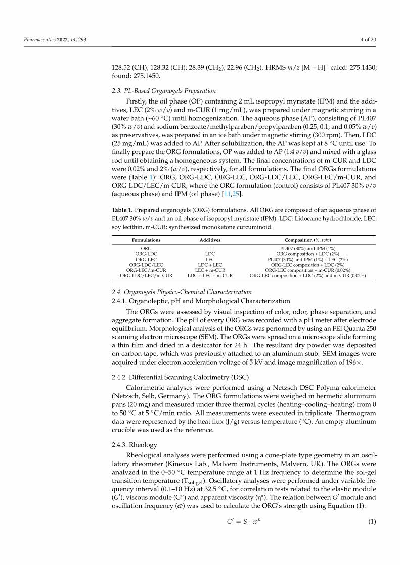
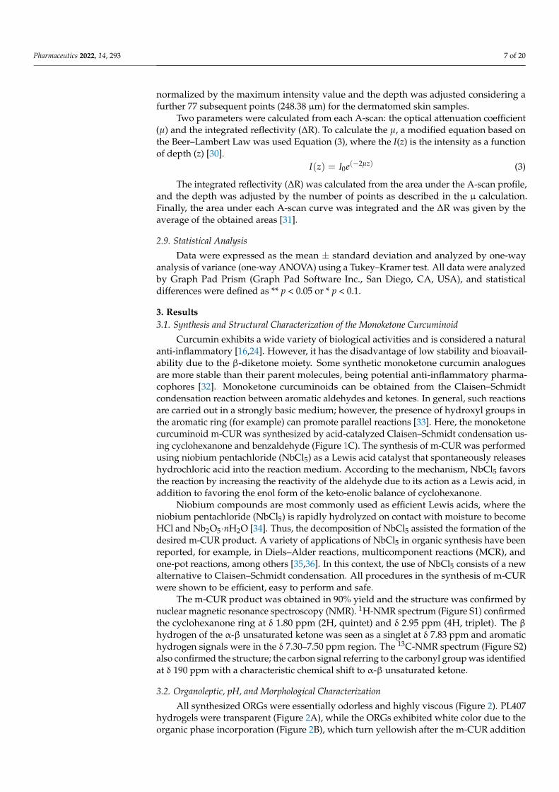
All synthesized ORGs were essentially odorless and highly viscous (Figure 2). PL407hydrogels were transparent (Figure 2A), while the ORGs exhibited white color due to theorganic phase incorporation (Figure 2B), which turn yellowish after the m-CUR addition
Pharmaceutics 2022, 14, 293 8 of 20
(Figure 2C). No phase separation was observed during the six-month period in whichsamples were stored at room temperature, protected from light. Measured pH valuesranged from 5.2 to 6.3 for ORGs containing LDC and LEC, respectively. Such pH valueswere similar to other PL-based ORG systems containing lanolin, which were described tobe adequate for skin application [11,37].
Pharmaceutics 2022, 14, x FOR PEER REVIEW 8 of 22
desired m-CUR product. A variety of applications of NbCl5 in organic synthesis have been reported, for example, in Diels–Alder reactions, multicomponent reactions (MCR), and one-pot reactions, among others [35,36]. In this context, the use of NbCl5 consists of a new alternative to Claisen–Schmidt condensation. All procedures in the synthesis of m-CUR were shown to be efficient, easy to perform and safe.
The m-CUR product was obtained in 90% yield and the structure was confirmed by nuclear magnetic resonance spectroscopy (NMR). 1H-NMR spectrum (Figure S1) confirmed the cyclohexanone ring at δ 1.80 ppm (2H, quintet) and δ 2.95 ppm (4H, triplet). The β hydrogen of the α-β unsaturated ketone was seen as a singlet at δ 7.83 ppm and aromatic hydrogen signals were in the δ 7.30–7.50 ppm region. The 13C-NMR spectrum (Figure S2) also confirmed the structure; the carbon signal referring to the carbonyl group was identified at δ 190 ppm with a characteristic chemical shift to α-β unsaturated ketone.
3.2. Organoleptic, pH, and Morphological Characterization All synthesized ORGs were essentially odorless and highly viscous (Figure 2). PL407
hydrogels were transparent (Figure 2A), while the ORGs exhibited white color due to the organic phase incorporation (Figure 2B), which turn yellowish after the m-CUR addition (Figure 2C). No phase separation was observed during the six-month period in which samples were stored at room temperature, protected from light. Measured pH values ranged from 5.2 to 6.3 for ORGs containing LDC and LEC, respectively. Such pH values were similar to other PL-based ORG systems containing lanolin, which were described to be adequate for skin application [11,37].
Figure 2. PL-based organogels preparation scheme. Inserted pictures below show (A) PL407 hydrogel, (B) PL407 + IPM organogel (ORG), and (C) m-CUR-loaded PL407 + IPM ORG.
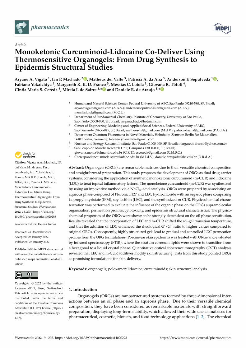
The morphology of all synthesized ORGs was evaluated by scanning electron microscopy (Figure 3). SEM images confirmed the typical layered arrangement for the dried PL407 hydrogel (Figure 3A) as described by Akkari et al. [38]. The control ORG and the ORG-LDC (Figure 3D,E) displayed a shapeless character, exhibiting wrinkled surfaces, as reported in the literature [11,39]. On the other hand, the other ORG samples (Figure 3F–I) showed intermediate morphology between the control ORG and the PL407 hydrogel, i.e., the layered structure was more preserved. All ORG formulations were
m-CUR LDCA B C
BIOACTIVES
Figure 2. PL-based organogels preparation scheme. Inserted pictures below show (A) PL407 hydrogel,(B) PL407 + IPM organogel (ORG), and (C) m-CUR-loaded PL407 + IPM ORG.
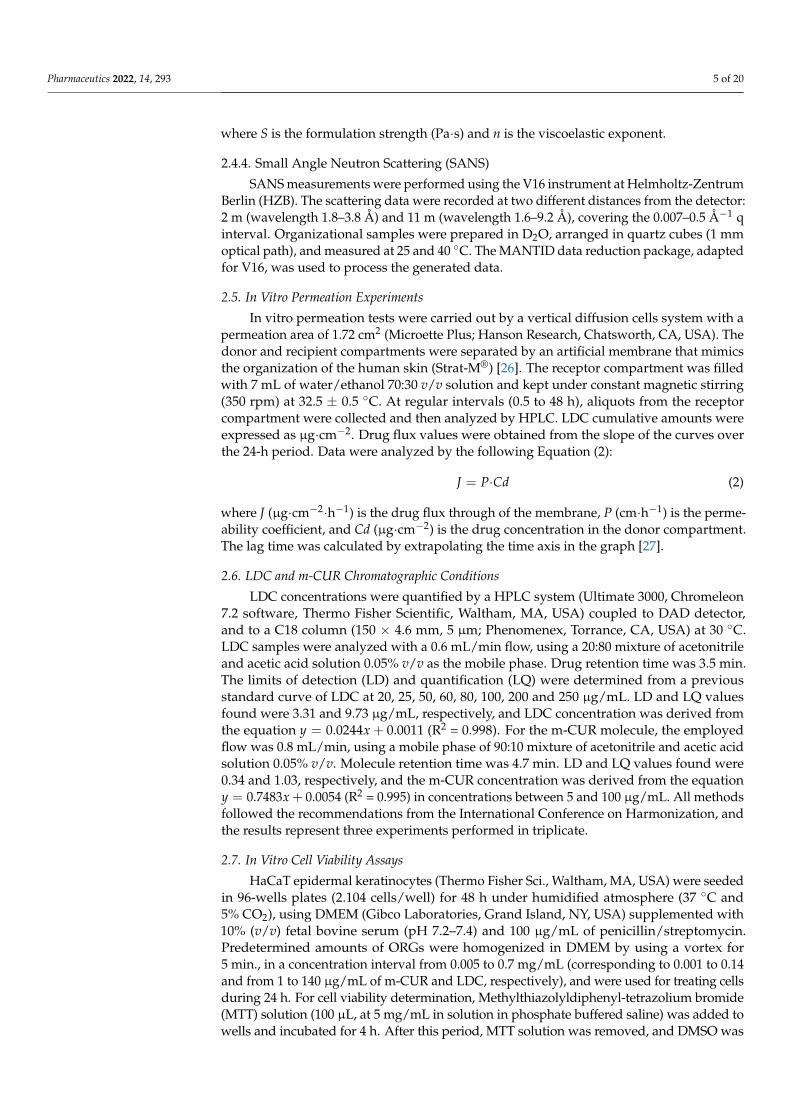
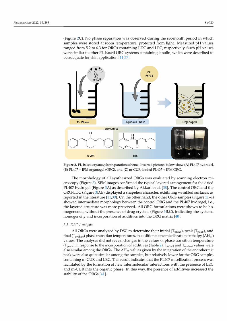
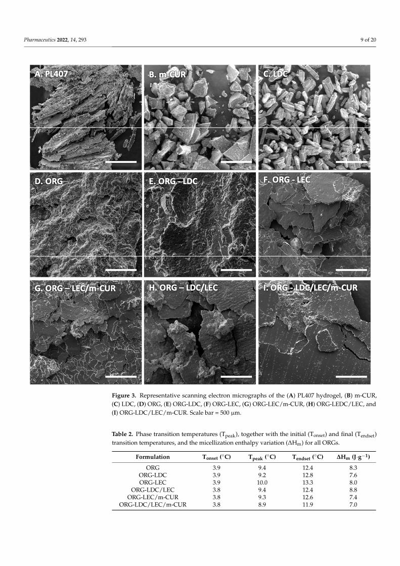
The morphology of all synthesized ORGs was evaluated by scanning electron mi-croscopy (Figure 3). SEM images confirmed the typical layered arrangement for the driedPL407 hydrogel (Figure 3A) as described by Akkari et al. [38]. The control ORG and theORG-LDC (Figure 3D,E) displayed a shapeless character, exhibiting wrinkled surfaces, asreported in the literature [11,39]. On the other hand, the other ORG samples (Figure 3F–I)showed intermediate morphology between the control ORG and the PL407 hydrogel, i.e.,the layered structure was more preserved. All ORG formulations were shown to be ho-mogeneous, without the presence of drug crystals (Figure 3B,C), indicating the systemshomogeneity and incorporation of additives into the ORG matrix [40].
3.3. DSC Analysis
All ORGs were analyzed by DSC to determine their initial (Tonset), peak (Tpeak), andfinal (Tendset) phase transition temperatures, in addition to the micellization enthalpy (∆Hm)values. The analyses did not reveal changes in the values of phase transition temperature(Tpeak) in response to the incorporation of additives (Table 2). Tonset and Tendset values werealso similar among the ORGs. The ∆Hm values given by the integration of the endothermicpeak were also quite similar among the samples, but relatively lower for the ORG samplescontaining m-CUR and LEC. This result indicates that the PL407 micellization process wasfacilitated by the formation of new intermolecular interactions with the presence of LECand m-CUR into the organic phase. In this way, the presence of additives increased thestability of the ORGs [41].
Pharmaceutics 2022, 14, 293 9 of 20
Pharmaceutics 2022, 14, x FOR PEER REVIEW 9 of 22
shown to be homogeneous, without the presence of drug crystals (Figure 3B,C), indicating the systems homogeneity and incorporation of additives into the ORG matrix [40].
Figure 3. Representative scanning electron micrographs of the (A) PL407 hydrogel, (B) m-CUR, (C) LDC, (D) ORG, (E) ORG-LDC, (F) ORG-LEC, (G) ORG-LEC/m-CUR, (H) ORG-LEDC/LEC, and (I) ORG-LDC/LEC/m-CUR. Scale bar = 500 μm.
3.3. DSC Analysis All ORGs were analyzed by DSC to determine their initial (Tonset), peak (Tpeak), and
final (Tendset) phase transition temperatures, in addition to the micellization enthalpy (ΔHm) values. The analyses did not reveal changes in the values of phase transition temperature (Tpeak) in response to the incorporation of additives (Table 2). Tonset and Tendset values were also similar among the ORGs. The ΔHm values given by the integration of the endothermic peak were also quite similar among the samples, but relatively lower for the ORG samples containing m-CUR and LEC. This result indicates that the PL407 micellization process was facilitated by the formation of new intermolecular interactions
Figure 3. Representative scanning electron micrographs of the (A) PL407 hydrogel, (B) m-CUR,(C) LDC, (D) ORG, (E) ORG-LDC, (F) ORG-LEC, (G) ORG-LEC/m-CUR, (H) ORG-LEDC/LEC, and(I) ORG-LDC/LEC/m-CUR. Scale bar = 500 µm.
Table 2. Phase transition temperatures (Tpeak), together with the initial (Tonset) and final (Tendset)transition temperatures, and the micellization enthalpy variation (∆Hm) for all ORGs.
Formulation Tonset (◦C) Tpeak (◦C) Tendset (◦C) ∆Hm (J·g−1)
ORG 3.9 9.4 12.4 8.3ORG-LDC 3.9 9.2 12.8 7.6ORG-LEC 3.9 10.0 13.3 8.0
ORG-LDC/LEC 3.8 9.4 12.4 8.8ORG-LEC/m-CUR 3.8 9.3 12.6 7.4
ORG-LDC/LEC/m-CUR 3.8 8.9 11.9 7.0
Pharmaceutics 2022, 14, 293 10 of 20
Aqueous solutions containing 20–30% of PL407 turn into gel close to body temperaturedue to the dehydration of the hydrophobic units in such temperature. This dehydration pro-cess enhances the interactions between the hydrophobic units, promoting the formation ofPL micelles [42]. The micellization temperature (Tpeak) in PL-based ORGs is affected by thechemical characteristics of the oil phase, tending to be lower than the Tpeak values observedfor the corresponding hydrogels [39,43]. Furthermore, previous studies showed that the ad-dition of the hydrophobic molecules in ORGs increases the micellization enthalpy [11]. Theopposite behavior was observed in this work, i.e., adding LEC and m-CUR into the PL407 +IPM ORGs leads to a decrease of the micellization enthalpy values: ORG 8.3, ORG-LEC 8.0,and ORG-LEC/m-CUR 7.4 J·g−1 (Table 2). Nevertheless, this effect was less pronouncedfor PL407 + IPM ORGs. This fact can be due to the fact that LEC and m-CUR molecules,probably, act as stabilizers in the intermicellar space, decreasing the necessary energy toform PL-based micelles via intermolecular interactions in the hydrophobic–hydrophilicinterface present in the ORGs.
3.4. Rheology Analysis
The investigation of rheological properties of drug-carrier ORG systems is essentialonce the shear rate and the viscosity deeply affect the drug-release rate from the formulationcomplex structure [44]. A thorough rheological characterization is also able to investigatethe interactions between different components in a formulation, analyzing parameterssuch as phase-transition temperature and shear stress. Exploring these parameters leadsto a deeper understanding of the formulations structural and physicochemical properties,which is crucial to further guarantee the quality of the ORGs regarding spreadability andlong-term stability [45]. In addition, modulating the drug release towards slower ratesis interesting once it reduces the reapplications procedures needed and decrease directcontact with the wound, improving patient compliance [46,47].
In this sense, the following rheological parameters were recorded for all prepared ORGsamples (Table 3 and Figure 4): sol–gel transition temperature (Tsol-gel), elastic modulus (G′),viscous modulus (G”), and the apparent viscosity (η*). The sol–gel transition temperature(Figure 4A–C) gives an estimative of the structural influence of the oil phase and thedifferent additives in the PL407 gel arrangement. Tsol-gel was defined as the temperaturepoint where the G′ and G” module values were equal [48]. It was observed that the Tsol-gelis lower than the skin temperature (32.5 ◦C) for all prepared ORGs, which indicates the highstability of these biomaterials regarding phase separation for topical application. Moreover,the ORGs containing additives exhibited higher Tsol-gel compared to the control ORG.This increase in temperature was found more pronounced for the formulations containingm-CUR. Probably, the oil phase of IPM and its additives m-CUR and LEC increase theintermolecular distances between poloxamer micelles, avoiding their interaction. Thus,a higher energy is required to start the gelation process. In this way, m-CUR and LECadditives decrease the enthalpy for the PL407 micelle formation, as observed from the DSCresults (Table 2), but they increase the energy involved in building the three-dimensional gelnetwork originated from the interaction between these micelles (Table 3). Literature reportsTsol-gel values around 9.5 ◦C for PL407 30% hydrogels containing 2% LDC hydrochloride,where the addition of the drug did not change the Tsol-gel temperature [44]. From ourresults, it can be concluded that the oil phase is therefore responsible for the increase onTsol-gel of PL407-based ORGs.
Pharmaceutics 2022, 14, 293 11 of 20
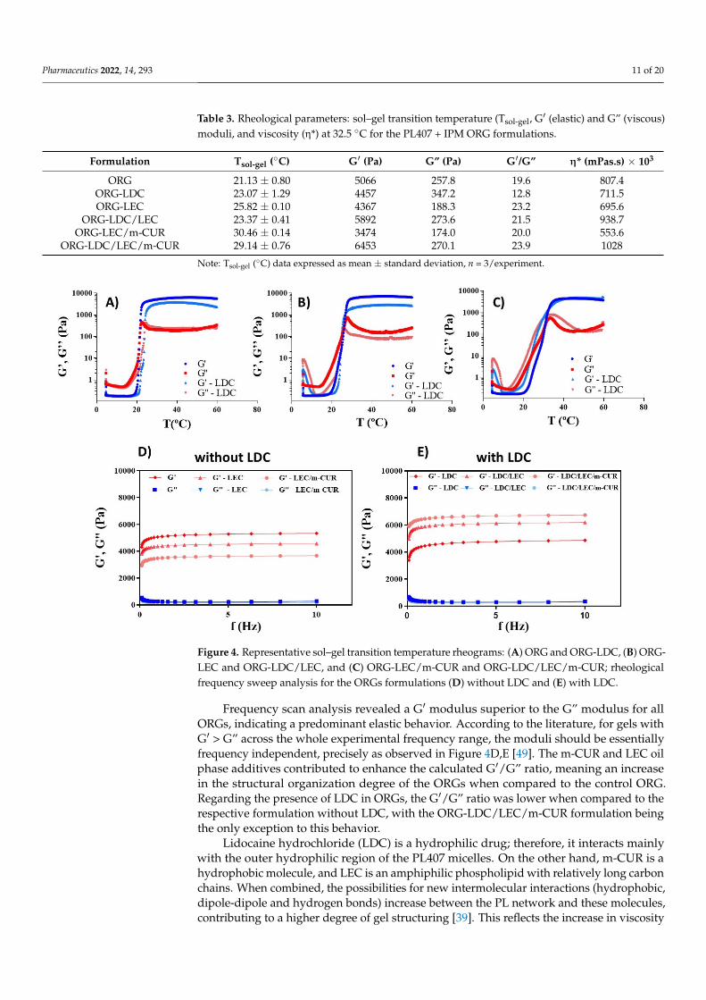
Table 3. Rheological parameters: sol–gel transition temperature (Tsol-gel, G′ (elastic) and G” (viscous)moduli, and viscosity (η*) at 32.5 ◦C for the PL407 + IPM ORG formulations.
Formulation Tsol-gel (◦C) G′ (Pa) G” (Pa) G′/G” η* (mPas.s) × 103
ORG 21.13 ± 0.80 5066 257.8 19.6 807.4ORG-LDC 23.07 ± 1.29 4457 347.2 12.8 711.5ORG-LEC 25.82 ± 0.10 4367 188.3 23.2 695.6
ORG-LDC/LEC 23.37 ± 0.41 5892 273.6 21.5 938.7ORG-LEC/m-CUR 30.46 ± 0.14 3474 174.0 20.0 553.6
ORG-LDC/LEC/m-CUR 29.14 ± 0.76 6453 270.1 23.9 1028
Note: Tsol-gel (◦C) data expressed as mean ± standard deviation, n = 3/experiment.
Pharmaceutics 2022, 14, x FOR PEER REVIEW 12 of 22
Figure 4. Representative sol–gel transition temperature rheograms: (A) ORG and ORG-LDC, (B) ORG-LEC and ORG-LDC/LEC, and (C) ORG-LEC/m-CUR and ORG-LDC/LEC/m-CUR; rheological frequency sweep analysis for the ORGs formulations (D) without LDC and (E) with LDC.
3.5. SANS Analysis Small-angle elastic neutron scattering (SANS) is a powerful tool to investigate the
structure of biomaterials at intermediate dimensions between the bulk and the nano scales. The ability of poloxamer and lipids to form organized structures of different symmetries (e.g., cubic, hexagonal) in aqueous dispersions, has been known for decades [50]. In order to study their organizational structure, the PL407 + IPM-based ORGs were characterized by using the SANS technique, measuring the samples at 40 °C, i.e., after the sol–gel transition for all ORGs (Table 4). All SANS patterns are displayed of Figures S3 and S4 (Supplementary material), for 25 °C and 40 °C, respectively.
From the SANS results, it was possible to assign two different cubic phases coexisting in all ORG samples, the 𝑃𝑚3𝑛 (space group number 223) and the 𝐹𝑑3𝑚 (space group #227), both corresponding to face-centered cubic (FCC) arrangements. No difference in the 1/q values was observed for all samples when comparing the two different temperatures, meaning that the gels were already structurally organized at 25 °C. In addition, it is worth mentioning that the lattice parameters remained practically unchanged for the different formulations, corroborating the discussion on the rheology results, i.e., the oil phase is the main responsible for the structural changes in the PL407 + IPM gel organization, while the additives have little to no influence [51]. This is further evidenced by the SANS results for the ORG-LEC formulation, the single case of different lattice parameters for the 𝑃𝑚3𝑛 and 𝐹𝑑3𝑚 structures.
Figure 4. Representative sol–gel transition temperature rheograms: (A) ORG and ORG-LDC, (B) ORG-LEC and ORG-LDC/LEC, and (C) ORG-LEC/m-CUR and ORG-LDC/LEC/m-CUR; rheologicalfrequency sweep analysis for the ORGs formulations (D) without LDC and (E) with LDC.
Frequency scan analysis revealed a G′ modulus superior to the G” modulus for allORGs, indicating a predominant elastic behavior. According to the literature, for gels withG′ > G” across the whole experimental frequency range, the moduli should be essentiallyfrequency independent, precisely as observed in Figure 4D,E [49]. The m-CUR and LEC oilphase additives contributed to enhance the calculated G′/G” ratio, meaning an increasein the structural organization degree of the ORGs when compared to the control ORG.Regarding the presence of LDC in ORGs, the G′/G” ratio was lower when compared to therespective formulation without LDC, with the ORG-LDC/LEC/m-CUR formulation beingthe only exception to this behavior.
Lidocaine hydrochloride (LDC) is a hydrophilic drug; therefore, it interacts mainlywith the outer hydrophilic region of the PL407 micelles. On the other hand, m-CUR is ahydrophobic molecule, and LEC is an amphiphilic phospholipid with relatively long carbonchains. When combined, the possibilities for new intermolecular interactions (hydrophobic,dipole-dipole and hydrogen bonds) increase between the PL network and these molecules,contributing to a higher degree of gel structuring [39]. This reflects the increase in viscosity
Pharmaceutics 2022, 14, 293 12 of 20
values and G′ modulus, particularly for the ORG-LDC/LEC/m-CUR formulation, whereall additives are present.
3.5. SANS Analysis
Small-angle elastic neutron scattering (SANS) is a powerful tool to investigate thestructure of biomaterials at intermediate dimensions between the bulk and the nano scales.The ability of poloxamer and lipids to form organized structures of different symmetries(e.g., cubic, hexagonal) in aqueous dispersions, has been known for decades [50]. In orderto study their organizational structure, the PL407 + IPM-based ORGs were characterized byusing the SANS technique, measuring the samples at 40 ◦C, i.e., after the sol–gel transitionfor all ORGs (Table 4). All SANS patterns are displayed of Figures S3 and S4 (Supplementarymaterial), for 25 ◦C and 40 ◦C, respectively.
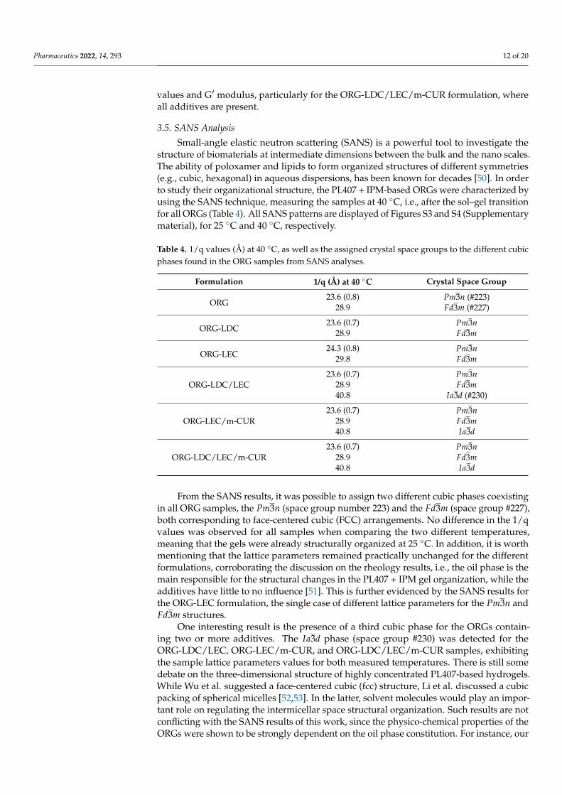
Table 4. 1/q values (Å) at 40 ◦C, as well as the assigned crystal space groups to the different cubicphases found in the ORG samples from SANS analyses.
Formulation 1/q (Å) at 40 ◦C Crystal Space Group
ORG23.6 (0.8) Pm3n (#223)
28.9 Fd3m (#227)
ORG-LDC23.6 (0.7) Pm3n
28.9 Fd3m
ORG-LEC24.3 (0.8) Pm3n
29.8 Fd3m
ORG-LDC/LEC23.6 (0.7) Pm3n
28.9 Fd3m40.8 Ia3d (#230)
ORG-LEC/m-CUR23.6 (0.7) Pm3n
28.9 Fd3m40.8 Ia3d
ORG-LDC/LEC/m-CUR23.6 (0.7) Pm3n
28.9 Fd3m40.8 Ia3d
From the SANS results, it was possible to assign two different cubic phases coexistingin all ORG samples, the Pm3n (space group number 223) and the Fd3m (space group #227),both corresponding to face-centered cubic (FCC) arrangements. No difference in the 1/qvalues was observed for all samples when comparing the two different temperatures,meaning that the gels were already structurally organized at 25 ◦C. In addition, it is worthmentioning that the lattice parameters remained practically unchanged for the differentformulations, corroborating the discussion on the rheology results, i.e., the oil phase is themain responsible for the structural changes in the PL407 + IPM gel organization, while theadditives have little to no influence [51]. This is further evidenced by the SANS results forthe ORG-LEC formulation, the single case of different lattice parameters for the Pm3n andFd3m structures.
One interesting result is the presence of a third cubic phase for the ORGs contain-ing two or more additives. The Ia3d phase (space group #230) was detected for theORG-LDC/LEC, ORG-LEC/m-CUR, and ORG-LDC/LEC/m-CUR samples, exhibitingthe sample lattice parameters values for both measured temperatures. There is still somedebate on the three-dimensional structure of highly concentrated PL407-based hydrogels.While Wu et al. suggested a face-centered cubic (fcc) structure, Li et al. discussed a cubicpacking of spherical micelles [52,53]. In the latter, solvent molecules would play an impor-tant role on regulating the intermicellar space structural organization. Such results are notconflicting with the SANS results of this work, since the physico-chemical properties of theORGs were shown to be strongly dependent on the oil phase constitution. For instance, our

Pharmaceutics 2022, 14, 293 13 of 20
results demonstrated that LEC is an important compound to the structural organization ofPL407 + IPM micellar gels. Further small angle X-ray scattering experiments will be carriedout to investigate the contribution of each cubic phase to the overall structure of the ORGs.
3.6. In Vitro Permeation and Cell Viability Assays
In vitro permeation studies were performed across the Strat-M (Merck Millipore,Darmstadt, Germany), a synthetic membrane with a lipid-impregnated porous struc-ture, which provides similar properties compared to those of animal models and humanskin [26,54]. For all ORGs, permeation profiles through the Strat-M revealed LDC concen-trations increasing cumulatively during the 48 h-experiment (Figure 5). The permeationflux, latency time, and permeation coefficient were then calculated (Table 5). In general,the LDC permeation profile was shown to be similar for all formulations, with no sig-nificant statistical difference between different formulation compositions. The obtainedLDC flux values were comparable to those previously reported by our group using themolecular LDC instead of the LDC hydrochloride [11]. This is an interesting result sincethe hydrochloride LDC is more hydrophilic and therefore has low chemical affinity withthe polymeric Strat-M membrane matrix. In this way, the PL407 + IPM ORGs improved theLDC permeation flux. It is also worth mentioning that the flux values for the ORGs evalu-ated in this work are about 10-fold higher when compared to formulations containing onlyIPM and LDC, when tested in human skin [55]. In fact, those differences observed on LDCpermeation kinetics can be attributed to membrane type and specially the oil formulationscomposition. In another study, LDC permeation kinetics parameters were investigatedacross lanolin-based artificial membranes, reporting similar permeability coefficient valuesto those obtained here [56]. However, low permeability coefficient values were obtained forLDC from ORGs composed of PL40730% associated with oleic acid and lanolin, indicatingthat the incorporation of waxes and free fatty acids into the oil phase can reduce the drugpermeation [11]. In a similar report, only 50% of LDC concentration applied was permeatedacross lanolin-added Strat-M artificial membrane [57].
Pharmaceutics 2022, 14, x FOR PEER REVIEW 14 of 22
were investigated across lanolin-based artificial membranes, reporting similar permeability coefficient values to those obtained here [56]. However, low permeability coefficient values were obtained for LDC from ORGs composed of PL40730% associated with oleic acid and lanolin, indicating that the incorporation of waxes and free fatty acids into the oil phase can reduce the drug permeation [11]. In a similar report, only 50% of LDC concentration applied was permeated across lanolin-added Strat-M artificial membrane [57].
Figure 5. Permeation profiles of lidocaine hydrochloride (LDC) from ORG-LDC, ORG-LDC/LEC and ORG-LDC/LEC/m-CUR across the Strat-M® membrane. Data expressed as mean ± standard deviation, n = 6/experiment.
Hence, the ORGs were characterized as highly structured gels, as concluded from the rheology results. Slow and controlled LDC permeation profiles from the PL407 + IPM was then expected from the LDC permeation results. Furthermore, it was not possible to identify m-CUR in the receptor medium after the 48 h of permeation analysis. This result demonstrates the high affinity of the m-CUR with the Strat-M membrane, corroborating the hydrophobic interactions observed between the membrane and the PL407 + IPM matrix [58].
Table 5. Lidocaine hydrochloride (LDC) permeation parameters across the Strat-M® membrane.
ORGs Drug Flux (μg·cm−2·h−1)
Tlag (h) Permeability Coefficient
(cm·h−1, ×10−2) ORG-LDC LDC 15.54 ± 1.27 8.82 ± 0.27 1.29 ± 0.11
ORG-LDC/LEC LDC 19.92 ± 2.88 9.34 ± 0.03 1.66 ± 0.66 ORG-LDC/LEC/m-CUR LDC 20.76 ± 1.66 9.28 ± 0.75 1.73 ± 0.20
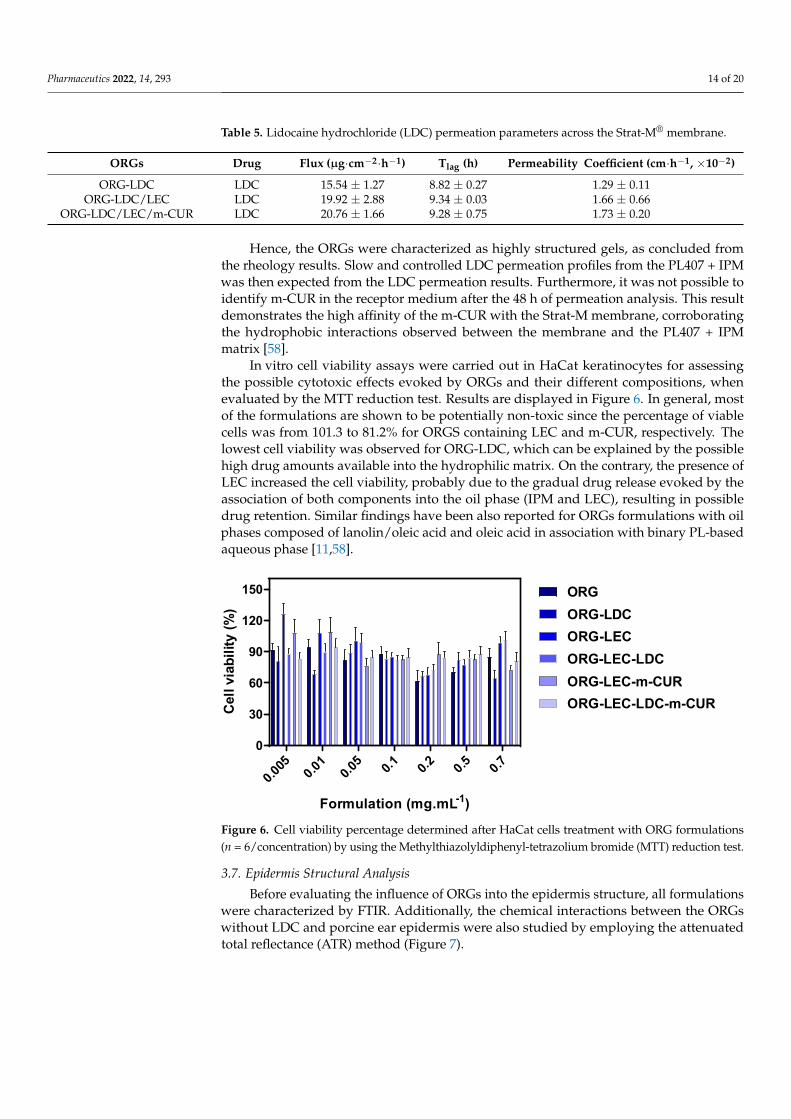
In vitro cell viability assays were carried out in HaCat keratinocytes for assessing the possible cytotoxic effects evoked by ORGs and their different compositions, when evaluated by the MTT reduction test. Results are displayed in Figure 6. In general, most of the formulations are shown to be potentially non-toxic since the percentage of viable cells was from 101.3 to 81.2% for ORGS containing LEC and m-CUR, respectively. The lowest cell viability was observed for ORG-LDC, which can be explained by the possible high drug amounts available into the hydrophilic matrix. On the contrary, the presence of LEC increased the cell viability, probably due to the gradual drug release evoked by the association of both components into the oil phase (IPM and LEC), resulting in possible
Figure 5. Permeation profiles of lidocaine hydrochloride (LDC) from ORG-LDC, ORG-LDC/LECand ORG-LDC/LEC/m-CUR across the Strat-M® membrane. Data expressed as mean ± standarddeviation, n = 6/experiment.
Pharmaceutics 2022, 14, 293 14 of 20
Table 5. Lidocaine hydrochloride (LDC) permeation parameters across the Strat-M® membrane.
ORGs Drug Flux (µg·cm−2·h−1) Tlag (h) Permeability Coefficient (cm·h−1, ×10−2)
ORG-LDC LDC 15.54 ± 1.27 8.82 ± 0.27 1.29 ± 0.11ORG-LDC/LEC LDC 19.92 ± 2.88 9.34 ± 0.03 1.66 ± 0.66
ORG-LDC/LEC/m-CUR LDC 20.76 ± 1.66 9.28 ± 0.75 1.73 ± 0.20
Hence, the ORGs were characterized as highly structured gels, as concluded fromthe rheology results. Slow and controlled LDC permeation profiles from the PL407 + IPMwas then expected from the LDC permeation results. Furthermore, it was not possible toidentify m-CUR in the receptor medium after the 48 h of permeation analysis. This resultdemonstrates the high affinity of the m-CUR with the Strat-M membrane, corroboratingthe hydrophobic interactions observed between the membrane and the PL407 + IPMmatrix [58].
In vitro cell viability assays were carried out in HaCat keratinocytes for assessingthe possible cytotoxic effects evoked by ORGs and their different compositions, whenevaluated by the MTT reduction test. Results are displayed in Figure 6. In general, mostof the formulations are shown to be potentially non-toxic since the percentage of viablecells was from 101.3 to 81.2% for ORGS containing LEC and m-CUR, respectively. Thelowest cell viability was observed for ORG-LDC, which can be explained by the possiblehigh drug amounts available into the hydrophilic matrix. On the contrary, the presence ofLEC increased the cell viability, probably due to the gradual drug release evoked by theassociation of both components into the oil phase (IPM and LEC), resulting in possibledrug retention. Similar findings have been also reported for ORGs formulations with oilphases composed of lanolin/oleic acid and oleic acid in association with binary PL-basedaqueous phase [11,58].
Pharmaceutics 2022, 14, x FOR PEER REVIEW 15 of 22
drug retention. Similar findings have been also reported for ORGs formulations with oil phases composed of lanolin/oleic acid and oleic acid in association with binary PL-based aqueous phase [11,58].
0.005 0.01 0.0
5 0.1 0.2 0.5 0.70
30
60
90
120
150
Formulation (mg.mL-1)
Cell
viab
ility
(%)
ORGORG-LDCORG-LECORG-LEC-LDCORG-LEC-m-CURORG-LEC-LDC-m-CUR
Figure 6. Cell viability percentage determined after HaCat cells treatment with ORG formulations (n = 6/concentration) by using the Methylthiazolyldiphenyl-tetrazolium bromide (MTT) reduction test.
3.7. Epidermis Structural Analysis Before evaluating the influence of ORGs into the epidermis structure, all
formulations were characterized by FTIR. Additionally, the chemical interactions between the ORGs without LDC and porcine ear epidermis were also studied by employing the attenuated total reflectance (ATR) method (Figure 7).
In the FTIR spectra of the isolated ORGs (Figure 7A), one can appoint three main absorption bands assigned to the PL407 structure: (i) symmetrical (2858 cm−1), (ii) asymmetrical (2922 cm−1) stretching of CH2 groups, and (iii) C–O–C stretching at 1080 cm−1. In addition, a C=O carbonyl stretching absorption band can also be spotted at 1641 cm−1 [59], which arises from the IPM oil phase. The broad absorption band centered at 3350 cm−1 corresponds to the O–H vibrations from water molecules.
The effects induced in the epidermis by the treatment with the different ORG formulations were investigated, after 4 and 24 h of treatment (Figure 7B,C). According to Boncheva et al., the CH2 scissor bands at around ~1460 cm−1, together with the symmetric CH2 stretching at ~2855 cm−1, consist in powerful probes to investigate structural changes in the stratum corneum [28]. More specifically, the frequency and bandwidth of such bands are indicators for the conformational order of the SC lipid chain; the increase in the rotational movement of the alkyl chains during the transition from orthorhombic (OR) to hexagonal (HEX) structure, as well as the increase in the isomeric gauche effect on the alkyl chains during the transition from HEX to liquid crystal (LIQ), causes the CH2 absorption bands to broaden and to shift towards higher wavenumbers. In summary, disorganizing/fluidifying the SC structure leads to band broadening and shifting to higher energies.
Figure 6. Cell viability percentage determined after HaCat cells treatment with ORG formulations(n = 6/concentration) by using the Methylthiazolyldiphenyl-tetrazolium bromide (MTT) reduction test.
3.7. Epidermis Structural Analysis
Before evaluating the influence of ORGs into the epidermis structure, all formulationswere characterized by FTIR. Additionally, the chemical interactions between the ORGswithout LDC and porcine ear epidermis were also studied by employing the attenuatedtotal reflectance (ATR) method (Figure 7).
Pharmaceutics 2022, 14, 293 15 of 20Pharmaceutics 2022, 14, x FOR PEER REVIEW 16 of 22
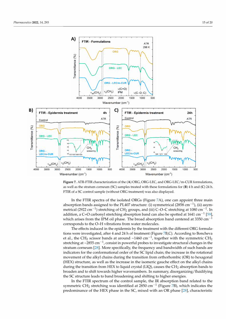
Figure 7. ATR-FTIR characterization of the (A) ORG, ORG-LEC, and ORG-LEC/m-CUR formulations, as well as the stratum corneum (SC) samples treated with these formulations for (B) 4 h and (C) 24 h. FTIR of a SC control sample (without ORG treatment) was also displayed.
In the FTIR spectrum of the control sample, the IR absorption band related to the symmetric CH2 stretching was identified at 2850 cm−1 (Figure 7B), which indicates the predominance of the HEX phase in the SC, mixed with an OR phase [28], characteristic of pig ear skin. For the epidermis treated with the ORGs, the symmetric CH2 stretching bands were identified around 2855 cm−1, indicating a fluidification process, i.e., the LIQ structure being the major phase in the SC. For all samples, after 24 h of application, the symmetric CH2 stretching bands were found in 2854–2855 cm−1 (Figure 7C), retaining the predominance of the LIQ structure. The wavenumber position and bandwidth for the CH2 scissor bands corroborated these results, once the two peaks at 1467 and 1475 cm−1 were shown to merge as a result of the ORG treatment (insets in Figure 7B,C), demonstrating a transition from HEX to LIQ structural organization.
The impacts of ORGs treatment on the epidermis were also investigated by optical coherence tomography (OCT), an innovative technique for the morphological characterization of skin samples since it is non-invasive and provides images with micrometric scale depth and spatial resolution [60,61]. Optical attenuation coefficient (OAC), also called extinction coefficient or total attenuation, is an important quantitative parameter that can be extracted from OCT images [62,63]. As the OCT light attenuates along its path due to absorption and scattering processes, quantifying such attenuation considerably enhances image contrast in areas that otherwise would be difficult to distinguish. This parameter can be estimated from OCT data, and it is particularly
Figure 7. ATR-FTIR characterization of the (A) ORG, ORG-LEC, and ORG-LEC/m-CUR formulations,as well as the stratum corneum (SC) samples treated with these formulations for (B) 4 h and (C) 24 h.FTIR of a SC control sample (without ORG treatment) was also displayed.
In the FTIR spectra of the isolated ORGs (Figure 7A), one can appoint three mainabsorption bands assigned to the PL407 structure: (i) symmetrical (2858 cm−1), (ii) asym-metrical (2922 cm−1) stretching of CH2 groups, and (iii) C–O–C stretching at 1080 cm−1. Inaddition, a C=O carbonyl stretching absorption band can also be spotted at 1641 cm−1 [59],which arises from the IPM oil phase. The broad absorption band centered at 3350 cm−1
corresponds to the O–H vibrations from water molecules.The effects induced in the epidermis by the treatment with the different ORG formula-
tions were investigated, after 4 and 24 h of treatment (Figure 7B,C). According to Bonchevaet al., the CH2 scissor bands at around ~1460 cm−1, together with the symmetric CH2stretching at ~2855 cm−1, consist in powerful probes to investigate structural changes in thestratum corneum [28]. More specifically, the frequency and bandwidth of such bands areindicators for the conformational order of the SC lipid chain; the increase in the rotationalmovement of the alkyl chains during the transition from orthorhombic (OR) to hexagonal(HEX) structure, as well as the increase in the isomeric gauche effect on the alkyl chainsduring the transition from HEX to liquid crystal (LIQ), causes the CH2 absorption bands tobroaden and to shift towards higher wavenumbers. In summary, disorganizing/fluidifyingthe SC structure leads to band broadening and shifting to higher energies.
In the FTIR spectrum of the control sample, the IR absorption band related to thesymmetric CH2 stretching was identified at 2850 cm−1 (Figure 7B), which indicates thepredominance of the HEX phase in the SC, mixed with an OR phase [28], characteristic

Pharmaceutics 2022, 14, 293 16 of 20
of pig ear skin. For the epidermis treated with the ORGs, the symmetric CH2 stretchingbands were identified around 2855 cm−1, indicating a fluidification process, i.e., the LIQstructure being the major phase in the SC. For all samples, after 24 h of application, thesymmetric CH2 stretching bands were found in 2854–2855 cm−1 (Figure 7C), retaining thepredominance of the LIQ structure. The wavenumber position and bandwidth for the CH2scissor bands corroborated these results, once the two peaks at 1467 and 1475 cm−1 wereshown to merge as a result of the ORG treatment (insets in Figure 7B,C), demonstrating atransition from HEX to LIQ structural organization.
The impacts of ORGs treatment on the epidermis were also investigated by opticalcoherence tomography (OCT), an innovative technique for the morphological character-ization of skin samples since it is non-invasive and provides images with micrometricscale depth and spatial resolution [60,61]. Optical attenuation coefficient (OAC), also calledextinction coefficient or total attenuation, is an important quantitative parameter that canbe extracted from OCT images [62,63]. As the OCT light attenuates along its path due toabsorption and scattering processes, quantifying such attenuation considerably enhancesimage contrast in areas that otherwise would be difficult to distinguish. This parametercan be estimated from OCT data, and it is particularly important in the study of hardtissues, such as bones, as well as in skin imaging, facilitating structural segmentation andvisualization [63]. Aiming at skin characterization, it is worth mentioning that filamentousproteins are the main source of light scattering in the skin, therefore the dermis (rich incollagen) and the epidermis (rich in keratin) show different values of OACs [60]. In thiscontext, the OAC for each A-scan (µ) was calculated from the OCT images (Figure 8A,B)and the results were expressed in the graphs of Figure 8C.
Pharmaceutics 2022, 14, x FOR PEER REVIEW 17 of 22
important in the study of hard tissues, such as bones, as well as in skin imaging, facilitating structural segmentation and visualization [63]. Aiming at skin characterization, it is worth mentioning that filamentous proteins are the main source of light scattering in the skin, therefore the dermis (rich in collagen) and the epidermis (rich in keratin) show different values of OACs [60]. In this context, the OAC for each A-scan (μ) was calculated from the OCT images (Figure 8A,B) and the results were expressed in the graphs of Figure 8C.
Results from OCT data treatment show that the optical attenuation coefficient (OAC) decreases in 4 h for the skin treated with ORGs containing the additives LEC and m-CUR, with p < 0.05 statistical difference relative to the control (Figure 8A). A lower OAC can be understood as a sparser distribution of collagen structures, consequently indicating a higher water content. In addition, collagen fibers could be dispersed among other water-absorber high-molecular-weight molecules present in the extracellular matrix, such as hyaluronic acid [64]. When light is scattered through structures such as keratin and dense collagen, e.g., scar tissue, they appear bright on OCT images. It is therefore noticeable from the decreasing OAC values (Figure 8C) that ORGs promote hydration of the analyzed skin samples. This phenomenon may be associated with modifications in the keratin and collagen fiber structures due to two main causes: (i) the water-occlusive effect caused by the three-dimensional structure of poloxamers—occlusion helps preserve natural moisturizing factors to confine water molecules locally; and (ii) the presence of LEC and m-CUR additives intensified the observed phenomenon—in the case of LEC, due to the affinity of the phospholipid for water molecules, and in the case of m-CUR, by a probable intermolecular interaction with hydrophobic components which enhanced the occlusion.
After 24 h, however, no statistical difference in the OAC parameter was observed among the samples (Figure 8B). All calculated OCAs were considerably low, including that of the control sample, which may indicate a degradation of skin structures. Ex vivo samples tend to be more sensitive to external factors such as temperature and relative humidity due to its thickness and lack of functioning cutaneous circulation when compared to in vivo samples [65]. In this way, we concluded that 24 h is too long considering the integrity of skin samples for OCT analyses.
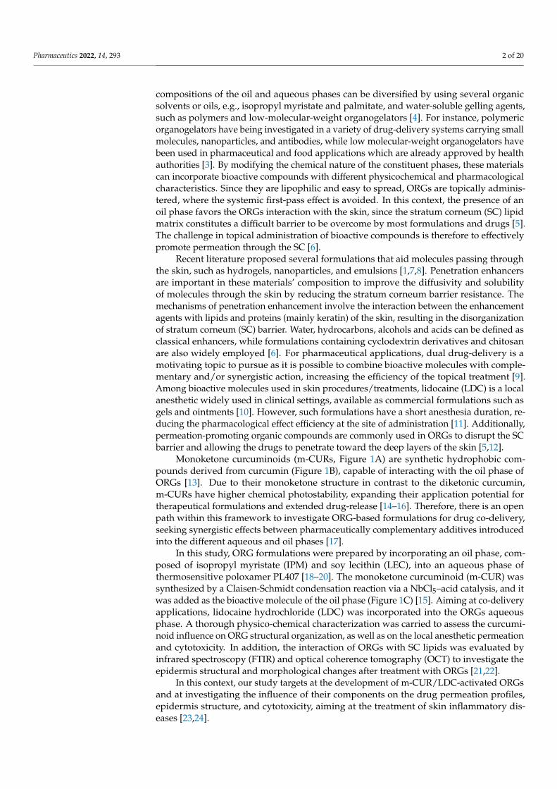
Figure 8. Optical Coherence Tomography (OCT) scans of dermatomed pig ear skin treated with ORGs for (A) 4 h and (B) 24 h, and (C) optical attenuation coefficient. Data are presented as mean±SD (n = 3). Statistical differences by one-away ANOVA test relative to control vs. ORG-LEC and control vs. ORG-LEC/m-CUR, where p < 0.01 (**).
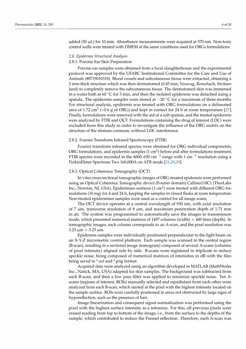
Figure 8. Optical Coherence Tomography (OCT) scans of dermatomed pig ear skin treated with ORGsfor (A) 4 h and (B) 24 h, and (C) optical attenuation coefficient. Data are presented as mean ± SD(n = 3). Statistical differences by one-away ANOVA test relative to control vs. ORG-LEC and controlvs. ORG-LEC/m-CUR, where p < 0.01 (**).
Results from OCT data treatment show that the optical attenuation coefficient (OAC)decreases in 4 h for the skin treated with ORGs containing the additives LEC and m-CUR,with p < 0.05 statistical difference relative to the control (Figure 8A). A lower OAC canbe understood as a sparser distribution of collagen structures, consequently indicating ahigher water content. In addition, collagen fibers could be dispersed among other water-absorber high-molecular-weight molecules present in the extracellular matrix, such ashyaluronic acid [64]. When light is scattered through structures such as keratin and densecollagen, e.g., scar tissue, they appear bright on OCT images. It is therefore noticeablefrom the decreasing OAC values (Figure 8C) that ORGs promote hydration of the analyzedskin samples. This phenomenon may be associated with modifications in the keratin andcollagen fiber structures due to two main causes: (i) the water-occlusive effect caused by the
Pharmaceutics 2022, 14, 293 17 of 20
three-dimensional structure of poloxamers—occlusion helps preserve natural moisturizingfactors to confine water molecules locally; and (ii) the presence of LEC and m-CUR addi-tives intensified the observed phenomenon—in the case of LEC, due to the affinity of thephospholipid for water molecules, and in the case of m-CUR, by a probable intermolecularinteraction with hydrophobic components which enhanced the occlusion.
After 24 h, however, no statistical difference in the OAC parameter was observedamong the samples (Figure 8B). All calculated OCAs were considerably low, including thatof the control sample, which may indicate a degradation of skin structures. Ex vivo samplestend to be more sensitive to external factors such as temperature and relative humidity dueto its thickness and lack of functioning cutaneous circulation when compared to in vivosamples [65]. In this way, we concluded that 24 h is too long considering the integrity ofskin samples for OCT analyses.
4. Conclusions
The physical chemical properties of PL407-based organogels (ORGs), as well as theirinteraction with the skin, were thoroughly explored, aiming at dual drug delivery systemsfor topical applications. This is a comprehensive work which comprises the synthesisof new bioactive molecules, together with the preparation and characterization of ORGformulations, and the ex vivo response of skin models in terms of structural organizationand drug permeation profiles. To the best of our knowledge, this is the first report ofa monoketone curcuminoid (m-CUR) synthesized by a Claisen–Schmidt condensationreaction via NbCl5 acid catalysis.
Our results point to a high versatility for the ORG systems since its structural proper-ties can be modulated by changing its composition, i.e., by the incorporation of differentadditives, obtaining formulations for both transdermal (high permeation flux) and topicalapplications (in which drugs remain trapped in the outer layers of the skin). For instance,soy lecithin (LEC) additive was demonstrated to increase the organizational degree of theORGs three-dimensional lattice, leading to a slower drug permeation flux. Such ORGs aretherefore interesting for topical applications such as scar healing and treatment of painfulinjuries, considering the drug association incorporated into them. It is worth noting that thepresence of additives did not cause substantial changes the ORG supramolecular structureas showed by SANS results, thus preserving the long-term stability for all formulations.
Infrared spectroscopy (FTIR) study of porcine ear epidermis treated with the ORGsrevealed a displacement of the symmetrical and asymmetrical stretching bands of C–Hbonds to higher wavenumbers when compared to the control samples. This result indicatesthat stratum corneum lipids undergo a hexagonal-to-liquid crystal phase transition aftertreatment with ORGs, which agrees with previous works on skin structure. Moreover, fromoptical coherence tomography (OCT), we observed that ORGs containing additives (LECand m-CUR) tend to decrease the skin’s optical attenuation coefficient within 4 h of ORGtreatment. This points to structural modifications in the skin, mainly in the organization offilamentous proteins, such as keratin and collagen. FTIR and OCT were then proved to becomplementary tools for ex vivo skin structural evaluation, more specifically regarding thestudy of lipid organization in the stratum corneum and fibrous proteins in the superficialskin layers, which are in contact with the formulations. Finally, the presence of m-CUR inORGs promoted structural changes in both organogels and skin components. Additionalstudies on vibrational spectroscopic could unravel the mechanism behind such structuralchanges to further explain synergistic pharmacological effects between m-CUR and LDC,in association with future in vivo studies.
Pharmaceutics 2022, 14, 293 18 of 20
Supplementary Materials: The following supporting information can be downloaded at: https://www.mdpi.com/article/10.3390/pharmaceutics14020293/s1. Figure S1: 1H-NMR spectrum of m-CUR; Figure S2: 13C-NMR spectrum of m-CUR; Figures S3 and S4: SANS spectra of ORG formulationsrecorded at 25 and 40 ◦C, respectively.
Author Contributions: Conceptualization, A.A.V., D.R.d.A. and M.I.d.S.; methodology. A.A.V.,A.F.S., M.C.L., F.Y., G.R.T. and C.M.S.C.; investigation, A.A.V., I.P.M., D.R.d.A., M.K.K.D.F., F.Y.,P.A.d.A. and M.d.V.; resources, A.A.V. and D.R.d.A.; writing—original draft preparation, A.A.V.,I.P.M., D.R.d.A. and M.I.d.S.; writing—review and editing, A.A.V., I.P.M. and D.R.d.A. All authorshave read and agreed to the published version of the manuscript.
Funding: This research was funded by The São Paulo Research Foundation, grant numbers 2019/20303-4 (D.R.A.), 2019/14773-8 (A.A.V.) and 2016/18045-9 (M.I.S.), and the Brazilian National Councilfor Scientific and Technological Development, grant number 307718/2019-0 (D.R.A.) and Coordenaçãode Aperfeiçoamento de Pessoal de Nível Superior—Brasil (CAPES, finance code #001).
Institutional Review Board Statement: The animal study protocol was approved by the UFABCInstitutional Committee for the Care and Use of Animals (protocol #8719010318).
Informed Consent Statement: Not applicable.
Data Availability Statement: Data available on motivated request.
Acknowledgments: We gratefully acknowledge UFABC Multiuser Central Facilities (CEM-UFABC)and CBMM for providing niobium pentachloride samples.
Conflicts of Interest: The authors declare no conflict of interest.
References1. Osmałek, T.; Milanowski, B.; Froelich, A.; Górska, S.; Białas, W.; Szybowicz, M.; Kapela, M. Novel organogels for topical delivery
of naproxen: Design, physicochemical characteristics and in vitro drug permeation. Pharm. Dev. Technol. 2017, 22, 521–536.[CrossRef] [PubMed]
2. Vintiloiu, A.; Leroux, J.-C. Organogels and their use in drug delivery—A review. J. Control. Release 2008, 125, 179–192. [CrossRef][PubMed]
3. Esposito, C.L.; Kirilov, P.; Roullin, V.G. Organogels, promising drug delivery systems: An update of state-of-the-art and recentapplications. J. Control. Release 2018, 271, 1–20. [CrossRef] [PubMed]
4. Mady, F.M.; Essa, H.; El-ammaw, T.; Abdelkader, H.; Hussein, A.K. Formulation and clinical evaluation of silymarin pluronic-lecithin organogels for treatment of atopic dermatitis. Hosp. Pharm. 2005, 12, 267–270. [CrossRef]
5. Kumar, R.; Katare, O.P. Lecithin Organogels as a Potential Phospholipid-Structured System for Topical Drug Delivery: A Review.AAPS PharmSciTech 2005, 6, 298–310. [CrossRef]
6. Alexander, A.; Dwivedi, S.; Giri, T.K.; Saraf, S.; Saraf, S.; Tripathi, D.K. Approaches for breaking the barriers of drug permeationthrough transdermal drug delivery. J. Control. Release 2012, 164, 26–40. [CrossRef]
7. Grillo, R.; Dias, F.V.; Querobino, S.M.; Alberto-Silva, C.; Fraceto, L.F.; De Paula, E.; De Araujo, D.R. Influence of hybridpolymeric nanoparticle/thermosensitive hydrogels systems on formulation tracking and in vitro artificial membrane permeation:A promising system for skin drug-delivery. Colloids Surf. B 2019, 174, 56–62. [CrossRef]
8. Bolla, P.K.; Clark, B.A.; Juluri, A.; Cheruvu, H.S.; Renukuntla, J. Evaluation of formulation parameters on permeation of ibuprofenfrom topical formulations using Strat-M® membrane. Pharmaceutics 2020, 12, 151. [CrossRef]
9. Zhang, Y.; Yang, C.; Wang, W.; Liu, J.; Liu, Q.; Huang, F. Co-delivery of doxorubicin and curcumin by pH-sensitive prodrugnanoparticle for combination therapy of cancer. Sci. Rep. 2016, 6, 21225. [CrossRef]
10. Kumar, M.; Chawla, R.; Goyal, M. Topical Anesthesia. J. Anaesthesiol. Clin. Pharmacol. 2015, 31, 450–456. [CrossRef]11. Vigato, A.A.; Querobino, S.M.; De Faria, N.C.; De Freitas, A.C.P.; Leonardi, G.R.; De Paula, E.; Cereda, C.M.S.; Tófoli, G.R.; De
Araujo, D.R. Synthesis and characterization of nanostructured lipid-poloxamer organogels for enhanced skin local anesthesia.Eur. J. Pharm. Sci. 2019, 128, 270–278. [CrossRef] [PubMed]
12. Parhi, R.; Suresh, P.; Pattnaik, S. Pluronic lecithin organogel (PLO) of diltiazem hydrochloride: Effect of solvents/penetrationenhancers on ex vivo permeation. Drug Deliv. Transl. Res. 2016, 6, 243–253. [CrossRef] [PubMed]
13. Noureddin, S.A.; El-Shishtawy, R.M.; Al-Footy, K.O. Curcumin analogues and their hybrid molecules as multifunctional drugs.Eur. J. Med. Chem. 2019, 182, 111631. [CrossRef]
14. Olivera, A.; Moore, T.W.; Hu, F.; Brown, A.P.; Sun, A.; Liotta, D.C.; Snyder, J.P.; Yoon, Y.; Shim, H.; Marcus, A.I.; et al. Inhibition ofthe NF-κB signaling pathway by the curcumin analog, 3,5-Bis(2-pyridinylmethylidene)-4-piperidone (EF31): Anti-inflammatoryand anti-cancer properties. Int. Immunopharmacol. 2012, 12, 368–377. [CrossRef] [PubMed]

Pharmaceutics 2022, 14, 293 19 of 20
15. Huber, I.; Zupkó, I.; Gyovai, A.; Horváth, P.; Kiss, E.; Gulyás-Fekete, G.; Schmidt, J.; Perjési, P. A novel cluster of C5-curcuminoids:Design, synthesis, in vitro antiproliferative activity and DNA binding of bis(arylidene)-4-cyclanone derivatives based on 4-hydroxycyclohexanone scaffold. Res. Chem. Intermed. 2019, 45, 4711–4735. [CrossRef]
16. Zhu, H.; Xu, T.; Qiu, C.; Wu, B.; Zhang, Y.; Chen, L.; Xia, Q.; Li, C.; Zhou, B.; Liu, Z.; et al. Synthesis and optimization of novelallylated mono-carbonyl analogs of curcumin (MACs) act as potent anti-inflammatory agents against LPS-induced acute lunginjury (ALI) in rats. Eur. J. Med. Chem. 2016, 121, 181–193. [CrossRef]
17. Pham, L.; Dang, L.H.; Truong, M.D.; Nguyen, T.H.; Le, L.; Le, V.T.; Nam, N.D.; Bach, L.G.; Nguyen, V.T.; Tran, N.Q. A dualsynergistic of curcumin and gelatin on thermal-responsive hydrogel based on Chitosan-P123 in wound healing application.Biomed. Pharmacother. 2019, 117, 109183. [CrossRef]
18. Bentley, M.V.L.B.; Marchetti, J.M.; Ricardo, N.; Ali-Abi, Z.; Collett, J.H. Influence of lecithin on some physical chemical propertiesof poloxamer gels: Rheological, microscopic and in vitro permeation studies. Int. J. Pharm. 1999, 193, 49–55. [CrossRef]
19. Shchipunov, Y.A. Lecithin organogel: A micellar system with unique properties. Colloids Surf. A 2001, 185, 541–554. [CrossRef]20. Dowling, T.C.; Arjomand, M.; Lin, E.T.; Allen, L.V.; McPherson, M.L. Relative bioavailability of ketoprofen 20% in a poloxamer-
lecithin organogel. Am. J. Health Syst. Pharm. 2004, 61, 2541–2544. [CrossRef]21. Caussin, J.; Gooris, G.S.; Janssens, M.; Bouwstra, J.A. Lipid organization in human and porcine stratum corneum differs widely,
while lipid mixtures with porcine ceramides model human stratum corneum lipid organization very closely. Biochim. Biophys.Acta Biomembr. 2008, 1778, 1472–1482. [CrossRef] [PubMed]
22. Fercher, A.F.; Drexler, W.; Hitzenberger, C.K.; Lasser, T. Optical coherence tomography-principles and applications. Rep. Prog.Phys. 2003, 66, 239–303. [CrossRef]
23. Panahi, Y.; Fazlolahzadeh, O.; Atkin, S.L.; Majeed, M.; Butler, A.E.; Johnston, T.P.; Sahebkar, A. Evidence of curcumin andcurcumin analogue effects in skin diseases: A narrative review. J. Cell. Physiol. 2019, 234, 1165–1178. [CrossRef] [PubMed]
24. Aggarwal, B.B.; Harikumar, K.B. Potential therapeutic effects of curcumin, the anti-inflammatory agent, against neurodegenerative,cardiovascular, pulmonary, metabolic, autoimmune and neoplastic diseases. Int. J. Biochem. Cell Biol. 2009, 41, 40–59. [CrossRef][PubMed]
25. Querobino, S.M.; De Faria, N.C.; Vigato, A.A.; Da Silva, B.G.M.; Machado, I.P.; Costa, M.S.; Costa, F.N.; De Araujo, D.R.; Alberto-Silva,C. Physicochemical data of oleic acid-poloxamer organogel for intravaginal voriconazole delivery. Data Br. 2019, 25, 104180. [CrossRef]
26. Uchida, T.; Kadhum, W.R.; Kanai, S.; Todo, H.; Oshizaka, T.; Sugibayashi, K. Prediction of skin permeation by chemical compoundsusing the artificial membrane, Strat-M. Eur. J. Pharm. Sci. 2015, 67, 113–118. [CrossRef] [PubMed]
27. De Araujo, D.R.; Padula, C.; Cereda, C.M.S.; Tófoli, G.R.; Brito, R.B.; De Paula, E.; Nicoli, S.; Santi, P. Bioadhesive films containingbenzocaine: Correlation between in vitro permeation and in vivo local anesthetic effect. Pharm. Res. 2010, 27, 1677–1686. [CrossRef]
28. Boncheva, M.; Damien, F.; Normand, V. Molecular organization of the lipid matrix in intact Stratum corneum using ATR-FTIRspectroscopy. BBA-Biomembr. 2008, 1778, 1344–1355. [CrossRef]
29. Hasanovic, A.; Winkler, R.; Resch, G.P.; Valenta, C. Modification of the conformational skin structure by treatment with liposomalformulations and its correlation to the penetration depth of aciclovir. Eur. J. Pharm. Biopharm. 2011, 79, 76–81. [CrossRef]
30. Yücel, D.; Themstrup, L.; Manfredi, M.; Jemec, G.B.E. Optical coherence tomography of basal cell carcinoma: Density and signalattenuation. Ski. Res. Technol. 2016, 22, 497–504. [CrossRef]
31. Maia, A.M.A.; De Freitas, A.Z.; De Campello, S.L.; Gomes, A.S.L.; Karlsson, L. Evaluation of dental enamel caries assessmentusing Quantitative Light Induced Fluorescence and Optical Coherence Tomography. J. Biophotonics 2016, 9, 596–602. [CrossRef][PubMed]
32. Kar, S.; Ramamoorthy, G.; Sinha, S.; Ramanan, M.; Pola, J.K.; Golakoti, N.R.; Nanubolu, J.B.; Sahoo, S.K.; Dandamudi, R.B.; Doble,M. Synthesis of diarylidenecyclohexanone derivatives as potential anti-inflammatory leads against COX-2/mPGES1 and 5-LOX.New J. Chem. 2019, 43, 9012–9020. [CrossRef]
33. Kim, B.T.; Chun, J.C.; Hwang, K.J. Synthesis of dihydroxylated chalcone derivatives with diverse substitution patterns and theirradical scavenging ability toward DPPH free radicals. Bull. Korean Chem. Soc. 2008, 29, 1125–1130. [CrossRef]
34. Lacerda, V., Jr.; Santos, D.A.; Silva-Filho, L.C.; Greco, S.J.; Santos, R.B. The Growing Impact of Niobium in Organic Synthesis andCatalysis. Aldrichim. Acta 2012, 45, 19–27.
35. Arpini, B.H.; Bartolomeu, A.A.; Andrade, C.K.Z.; Silva-Filho, L.C. Recent Advances in Using Niobium Compounds as Catalystsin Organic Chemistry. Curr. Org. Synth. 2015, 12, 5. [CrossRef]
36. Rodrigues, S.M.M.; Previdi, D.; Baviera, G.S.; Matias, A.A.; Donate, P.M. Niobium Pentachloride Mediated (Hetero)aromaticAldehyde Friedel-Crafts Hydroxyalkylation with Arenes: An Efficient Strategy to Synthesize Triarylmethanes. Synthesis 2019, 51,4498–4506. [CrossRef]
37. Sagiri, S.S.; Pal, K.; Basak, P. Encapsulation of animal wax-based organogels in alginate microparticles. J. Appl. Polym. Sci. 2014,131, 40910. [CrossRef]
38. Akkari, A.C.S.; Papini, J.Z.B.; Garcia, G.K.; Franco, M.K.K.D.; Cavalcanti, L.P.; Gasperini, A.; Alkschbirs, M.I.; Yokaichyia, F.;Paula, E.; Tófoli, G.R.; et al. Poloxamer 407/188 binary thermosensitive hydrogels as delivery systems for infiltrative localanesthesia: Physico-chemical characterization and pharmacological evaluation. Mater. Sci. Eng. C 2016, 99, 299–307. [CrossRef]
39. Querobino, S.M.; De Faria, N.C.; Vigato, A.A.; da Silva, B.G.; Machado, I.P.; Costa, M.S.; Costa, F.N.; de Araujo, D.R.; Alberto-Silva,C. Sodium alginate in oil-poloxamer organogels for intravaginal drug delivery: Influence on structural parameters, drug releasemechanisms, cytotoxicity and in vitro antifungal activity. Mater. Sci. Eng. C 2019, 99, 1350–1361. [CrossRef]
Pharmaceutics 2022, 14, 293 20 of 20
40. Jhawat, V.; Gupta, S.; Saini, V. Formulation and evaluation of novel controlled release of topical pluronic lecithin organogel ofmefenamic acid. Drug Deliv. 2016, 23, 3573–3581. [CrossRef]
41. Satapathy, D.; Biswas, D.; Behera, B.; Sagiri, S.S.; Pal, K.; Pramanik, K. Sunflower-oil-based lecithin organogels as matrices forcontrolled drug delivery. J. Appl. Polym. Sci. 2013, 129, 585–594. [CrossRef]
42. Almeida, H.; Amaral, M.H.; Lobão, P.; Lobo, J.M.S. Pluronic® F-127 and Pluronic Lecithin Organogel (PLO): Main features andtheir applications in topical and transdermal administration of drugs. J. Pharm. Pharm. Sci. 2012, 15, 592–605. [CrossRef] [PubMed]
43. Shriky, B.; Kelly, A.; Isreb, M.; Babenko, M.; Mahmoudi, N.; Rogers, S.; Shebanova, O.; Snow, T.; Gough, T. Pluronic F127thermosensitive injectable smart hydrogels for controlled drug delivery system development. J. Colloid Interface Sci. 2020, 565,119–130. [CrossRef] [PubMed]
44. Ricci, E.J.; Bentley, M.V.L.B.; Farah, M.; Bretas, R.E.S.; Marchetti, J.M. Rheological characterization of Poloxamer 407 lidocainehydrochloride gels. Eur. J. Pharm. Sci. 2002, 17, 161–167. [CrossRef]
45. De Francisco, L.M.B.; Rosseto, H.C.; de Alcântara, L.S.T.; dos Santos, R.S.; De Souza, F.S.B.; Bruschi, M.L. Organogel composedof poloxamer 188 and passion fruit oil: Sol-gel transition, rheology, and mechanical properties. J. Mol. Liq. 2019, 289, 111170.[CrossRef]
46. Russo, J.; Fiegel, J.; Brogden, N.K. Rheological and drug delivery characteristics of poloxamer-based diclofenac sodium formula-tions for chronic wound site analgesia. Pharmaceutics 2020, 12, 1214. [CrossRef]
47. Moreira, T.S.A.; De Sousa, V.P.; Pierre, M.B.R. Influence of oleic acid on the rheology and in vitro release of lumiracoxib frompoloxamer gels. J. Pharm. Pharm. Sci. 2010, 13, 286–302. [CrossRef]
48. Carlfors, J.; Edsman, K.; Petersson, R.; Jörnving, K. Rheological evaluation of Gelrite® in situ gels for ophthalmic use. Eur. J.Pharm. Sci. 1998, 6, 113–119. [CrossRef]
49. Baloglu, E.; Karavana, S.Y.; Senyigit, Z.A.; Hilmioglu-Polat, S.; Metin, D.Y.; Zekioglu, O.; Guneri, T.; Jones, D.S. In-situ gelformulations of econazole nitrate: Preparation and in-vitro and in-vivo evaluation. J. Pharm. Pharmacol. 2011, 63, 1274–1282.[CrossRef]
50. Tenchov, B.; Koynova, R. Cubic phases in phosphatidylethanolamine dispersions: Formation, stability and phase transitions.Chem. Phys. Lipids 2017, 208, 65–74. [CrossRef]
51. Nascimento, M.H.M.; Ambrosio, F.N.; Ferraraz, D.C.; Windisch-Neto, H.; Querobino, S.M.; Nascimento-Sales, M.; Alberto-Silva,C.; Christoffolete, M.A.; Franco, M.K.K.D.; Kent, B.; et al. Sulforaphane-loaded hyaluronic acid-poloxamer hybrid hydrogelenhances cartilage protection in osteoarthritis models. Mater. Sci. Eng. C 2021, 128, 112345. [CrossRef] [PubMed]
52. Li, Y.; Shi, T.; Sun, Z.; An, L.; Huang, Q. Investigation of sol-gel transition in pluronic F127/D2O solutions using a combination ofsmall-angle neutron scattering and Monte Carlo simulation. J. Phys. Chem. B 2006, 110, 26424–26429. [CrossRef] [PubMed]
53. Wu, C.; Liu, T.; Chu, B.; Schneider, D.K.; Graziano, V. Characterization of the PEO-PPO-PEO triblock copolymer and its applicationas a separation medium in capillary electrophoresis. Macromolecules 1997, 30, 4574–4583. [CrossRef]
54. Haq, A.; Goodyear, B.; Ameen, D.; Joshi, V.; Michniak-Kohn, B. Strat-M® synthetic membrane: Permeability comparison tohuman cadaver skin. Int. J. Pharm. 2018, 547, 432–437. [CrossRef]
55. Lee, P.J.; Ahmad, N.; Langer, R.; Mitragotri, S.; Prasad Shastri, V. Evaluation of chemical enhancers in the transdermal delivery oflidocaine. Int. J. Pharm. 2006, 308, 33–39. [CrossRef]
56. Alonso, C.; Collini, I.; Carrer, V.; Barba, C.; Martí, M.; Coderch, L. Permeation kinetics of active drugs through lanolin-basedartificial membranes. Colloids Surf. B 2020, 192, 111024. [CrossRef]
57. Carrer, V.; Guzmán, B.; Martí, M.; Alonso, C.; Coderch, L. Lanolin-Based Synthetic Membranes as Percutaneous AbsorptionModels for Transdermal Drug Delivery. Pharmaceutics 2018, 10, 73. [CrossRef]
58. Vigato, A.A.; Querobino, S.M.; De Faria, N.C.; Candido, A.C.B.B.; Magalhães, L.G.; Cereda, C.M.S.; Tófoli, G.R.; Campos, E.V.R.;Machado, I.P.; Fraceto, L.F.; et al. Physico-chemical characterization and biopharmaceutical evaluation of lipid-poloxamer-basedorganogels for curcumin skin delivery. Front. Pharmacol. 2019, 10, 1006. [CrossRef]
59. Newa, M.; Bhandari, K.H.; Oh, D.H.; Kim, Y.R.; Sung, J.H.; Kim, J.O.; Woo, J.S.; Choi, H.G.; Yong, C.S. Enhanced dissolution ofibuprofen using solid dispersion with poloxamer 407. Arch. Pharm. Res. 2008, 31, 1497–1507. [CrossRef]
60. Lu, J.; Deegan, A.J.; Cheng, Y.; Liu, T.; Zheng, Y.; Mandell, S.P.; Wang, R.K. Application of OCT-Derived Attenuation Coefficient inAcute Burn-Damaged Skin. Lasers Surg. Med. 2021, 53, 1192–1200. [CrossRef]
61. Rangaraju, L.P.; Kunapuli, G.; Every, D.; Ayala, O.D.; Ganapathy, P.; Mahadevan-Jansen, A. Classification of burn injury usingRaman spectroscopy and optical coherence tomography: An ex-vivo study on porcine skin. Burns 2019, 45, 659–670. [CrossRef][PubMed]
62. Cara, A.C.B.; Zezell, D.M.; Ana, P.A.; Maldonado, E.P.; Freitas, A.Z. Evaluation of two quantitative analysis methods of opticalcoherence tomography for detection of enamel demineralization and comparison with microhardness. Lasers Surg. Med. 2014, 46,666–671. [CrossRef] [PubMed]
63. Del-Valle, M.; Lins, E.; Ana, P. Assessment of simulated osteoporosis in alveolar bone using optical coherence tomography.J. Biophotonics 2019, 12, 201900171. [CrossRef]
64. Kislevitz, M.; Akgul, Y.; Wamsley, C.; Hoopman, J.; Kenkel, J. Use of Optical Coherence Tomography (OCT) in Aesthetic SkinAssessment-A Short Review. Lasers Surg. Med. 2020, 52, 699–704. [CrossRef] [PubMed]
65. Herkenne, C.; Naik, A.; Kalia, Y.N.; Hadgraft, J.; Guy, R.H. Pig ear skin ex vivo as a model for in vivo dermatopharmacokineticstudies in man. Pharm. Res. 2006, 23, 1850–1856. [CrossRef] [PubMed]