Monitoring the effects of different conservation treatments on paper-infecting fungi
9
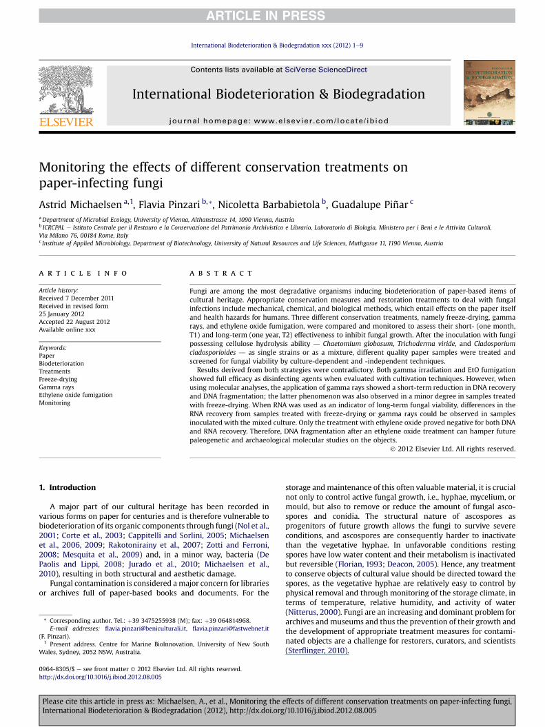
Monitoring the effects of different conservation treatments on paper-infecting fungi Astrid Michaelsen a,1 , Flavia Pinzari b, * , Nicoletta Barbabietola b , Guadalupe Piñar c a Department of Microbial Ecology, University of Vienna, Althanstrasse 14, 1090 Vienna, Austria b ICRCPAL e Istituto Centrale per il Restauro e la Conservazione del Patrimonio Archivistico e Librario, Laboratorio di Biologia, Ministero per i Beni e le Attivita Culturali, Via Milano 76, 00184 Rome, Italy c Institute of Applied Microbiology, Department of Biotechnology, University of Natural Resources and Life Sciences, Muthgasse 11,1190 Vienna, Austria article info Article history: Received 7 December 2011 Received in revised form 25 January 2012 Accepted 22 August 2012 Available online xxx Keywords: Paper Biodeterioration Treatments Freeze-drying Gamma rays Ethylene oxide fumigation Monitoring abstract Fungi are among the most degradative organisms inducing biodeterioration of paper-based items of cultural heritage. Appropriate conservation measures and restoration treatments to deal with fungal infections include mechanical, chemical, and biological methods, which entail effects on the paper itself and health hazards for humans. Three different conservation treatments, namely freeze-drying, gamma rays, and ethylene oxide fumigation, were compared and monitored to assess their short- (one month, T1) and long-term (one year, T2) effectiveness to inhibit fungal growth. After the inoculation with fungi possessing cellulose hydrolysis ability d Chaetomium globosum, Trichoderma viride, and Cladosporium cladosporioides d as single strains or as a mixture, different quality paper samples were treated and screened for fungal viability by culture-dependent and -independent techniques. Results derived from both strategies were contradictory. Both gamma irradiation and EtO fumigation showed full efficacy as disinfecting agents when evaluated with cultivation techniques. However, when using molecular analyses, the application of gamma rays showed a short-term reduction in DNA recovery and DNA fragmentation; the latter phenomenon was also observed in a minor degree in samples treated with freeze-drying. When RNA was used as an indicator of long-term fungal viability, differences in the RNA recovery from samples treated with freeze-drying or gamma rays could be observed in samples inoculated with the mixed culture. Only the treatment with ethylene oxide proved negative for both DNA and RNA recovery. Therefore, DNA fragmentation after an ethylene oxide treatment can hamper future paleogenetic and archaeological molecular studies on the objects. Ó 2012 Elsevier Ltd. All rights reserved. 1. Introduction A major part of our cultural heritage has been recorded in various forms on paper for centuries and is therefore vulnerable to biodeterioration of its organic components through fungi (Nol et al., 2001; Corte et al., 2003; Cappitelli and Sorlini, 2005; Michaelsen et al., 2006, 2009; Rakotonirainy et al., 2007; Zotti and Ferroni, 2008; Mesquita et al., 2009) and, in a minor way, bacteria (De Paolis and Lippi, 2008; Jurado et al., 2010; Michaelsen et al., 2010), resulting in both structural and aesthetic damage. Fungal contamination is considered a major concern for libraries or archives full of paper-based books and documents. For the storage and maintenance of this often valuable material, it is crucial not only to control active fungal growth, i.e., hyphae, mycelium, or mould, but also to remove or reduce the amount of fungal asco- spores and conidia. The structural nature of ascospores as progenitors of future growth allows the fungi to survive severe conditions, and ascospores are consequently harder to inactivate than the vegetative hyphae. In unfavorable conditions resting spores have low water content and their metabolism is inactivated but reversible (Florian, 1993; Deacon, 2005). Hence, any treatment to conserve objects of cultural value should be directed toward the spores, as the vegetative hyphae are relatively easy to control by physical removal and through monitoring of the storage climate, in terms of temperature, relative humidity, and activity of water (Nitterus, 2000). Fungi are an increasing and dominant problem for archives and museums and thus the prevention of their growth and the development of appropriate treatment measures for contami- nated objects are a challenge for restorers, curators, and scientists (Sterflinger, 2010). * Corresponding author. Tel.: þ39 3475255938 (M); fax: þ39 064814968. E-mail addresses: fl[email protected], fl[email protected] (F. Pinzari). 1 Present address. Centre for Marine BioInnovation, University of New South Wales, Sydney, 2052 NSW, Australia. Contents lists available at SciVerse ScienceDirect International Biodeterioration & Biodegradation journal homepage: www.elsevier.com/locate/ibiod 0964-8305/$ e see front matter Ó 2012 Elsevier Ltd. All rights reserved. http://dx.doi.org/10.1016/j.ibiod.2012.08.005 International Biodeterioration & Biodegradation xxx (2012) 1e9 Please cite this article in press as: Michaelsen, A., et al., Monitoring the effects of different conservation treatments on paper-infecting fungi, International Biodeterioration & Biodegradation (2012), http://dx.doi.org/10.1016/j.ibiod.2012.08.005
Transcript of Monitoring the effects of different conservation treatments on paper-infecting fungi
at SciVerse ScienceDirect
International Biodeterioration & Biodegradation xxx (2012) 1e9
Contents lists available
International Biodeterioration & Biodegradation
journal homepage: www.elsevier .com/locate/ ibiod
Monitoring the effects of different conservation treatments onpaper-infecting fungi
Astrid Michaelsen a,1, Flavia Pinzari b,*, Nicoletta Barbabietola b, Guadalupe Piñar c
aDepartment of Microbial Ecology, University of Vienna, Althanstrasse 14, 1090 Vienna, Austriab ICRCPAL e Istituto Centrale per il Restauro e la Conservazione del Patrimonio Archivistico e Librario, Laboratorio di Biologia, Ministero per i Beni e le Attivita Culturali,Via Milano 76, 00184 Rome, Italyc Institute of Applied Microbiology, Department of Biotechnology, University of Natural Resources and Life Sciences, Muthgasse 11, 1190 Vienna, Austria
a r t i c l e i n f o
Article history:Received 7 December 2011Received in revised form25 January 2012Accepted 22 August 2012Available online xxx
Keywords:PaperBiodeteriorationTreatmentsFreeze-dryingGamma raysEthylene oxide fumigationMonitoring
* Corresponding author. Tel.: þ39 3475255938 (M)E-mail addresses: [email protected],
(F. Pinzari).1 Present address. Centre for Marine BioInnovatio
Wales, Sydney, 2052 NSW, Australia.
0964-8305/$ e see front matter � 2012 Elsevier Ltd.http://dx.doi.org/10.1016/j.ibiod.2012.08.005
Please cite this article in press as: MichaelseInternational Biodeterioration & Biodegrada
a b s t r a c t
Fungi are among the most degradative organisms inducing biodeterioration of paper-based items ofcultural heritage. Appropriate conservation measures and restoration treatments to deal with fungalinfections include mechanical, chemical, and biological methods, which entail effects on the paper itselfand health hazards for humans. Three different conservation treatments, namely freeze-drying, gammarays, and ethylene oxide fumigation, were compared and monitored to assess their short- (one month,T1) and long-term (one year, T2) effectiveness to inhibit fungal growth. After the inoculation with fungipossessing cellulose hydrolysis ability d Chaetomium globosum, Trichoderma viride, and Cladosporiumcladosporioides d as single strains or as a mixture, different quality paper samples were treated andscreened for fungal viability by culture-dependent and -independent techniques.
Results derived from both strategies were contradictory. Both gamma irradiation and EtO fumigationshowed full efficacy as disinfecting agents when evaluated with cultivation techniques. However, whenusing molecular analyses, the application of gamma rays showed a short-term reduction in DNA recoveryand DNA fragmentation; the latter phenomenon was also observed in a minor degree in samples treatedwith freeze-drying. When RNA was used as an indicator of long-term fungal viability, differences in theRNA recovery from samples treated with freeze-drying or gamma rays could be observed in samplesinoculated with the mixed culture. Only the treatment with ethylene oxide proved negative for both DNAand RNA recovery. Therefore, DNA fragmentation after an ethylene oxide treatment can hamper futurepaleogenetic and archaeological molecular studies on the objects.
� 2012 Elsevier Ltd. All rights reserved.
1. Introduction
A major part of our cultural heritage has been recorded invarious forms on paper for centuries and is therefore vulnerable tobiodeterioration of its organic components through fungi (Nol et al.,2001; Corte et al., 2003; Cappitelli and Sorlini, 2005; Michaelsenet al., 2006, 2009; Rakotonirainy et al., 2007; Zotti and Ferroni,2008; Mesquita et al., 2009) and, in a minor way, bacteria (DePaolis and Lippi, 2008; Jurado et al., 2010; Michaelsen et al.,2010), resulting in both structural and aesthetic damage.
Fungal contamination is considered amajor concern for librariesor archives full of paper-based books and documents. For the
; fax: þ39 [email protected]
n, University of New South
All rights reserved.
n, A., et al., Monitoring the etion (2012), http://dx.doi.org
storage andmaintenance of this often valuable material, it is crucialnot only to control active fungal growth, i.e., hyphae, mycelium, ormould, but also to remove or reduce the amount of fungal asco-spores and conidia. The structural nature of ascospores asprogenitors of future growth allows the fungi to survive severeconditions, and ascospores are consequently harder to inactivatethan the vegetative hyphae. In unfavorable conditions restingspores have low water content and their metabolism is inactivatedbut reversible (Florian, 1993; Deacon, 2005). Hence, any treatmentto conserve objects of cultural value should be directed toward thespores, as the vegetative hyphae are relatively easy to control byphysical removal and through monitoring of the storage climate, interms of temperature, relative humidity, and activity of water(Nitterus, 2000). Fungi are an increasing and dominant problem forarchives andmuseums and thus the prevention of their growth andthe development of appropriate treatment measures for contami-nated objects are a challenge for restorers, curators, and scientists(Sterflinger, 2010).
ffects of different conservation treatments on paper-infecting fungi,/10.1016/j.ibiod.2012.08.005

A. Michaelsen et al. / International Biodeterioration & Biodegradation xxx (2012) 1e92
A broad spectrum of chemical and non-chemical componentshas been used to sanitize microfungi attacking paper-made objectsin an attempt to inhibit degradation (Magaudda, 2004). In thisstudywe chose three different conservation treatments for paper tocompare their effectiveness over the short- and long-term, namelyfreeze-drying, gamma rays, and ethylene oxide fumigation.
A commonly used strategy in paper conservation is freeze-drying, a method in which the water is frozen and then removedby sublimation, i.e., it goes from the solid phase to water vapor,bypassing the liquid phase. Sublimation allows the water to beremoved without the effects of water evaporative forces, which cancause dimensional changes. In addition, freeze-drying with dehy-dration can kill hydrated conidia and germinating conidia and itstops the growth of fungal mycelia and bacterial cells (Sussman,1966; Mazur, 1968; Florian, 2002). Yet if the moisture content inthe thawed materials remains high, resting dry conidia may beactivated. Freezing can also increase the porosity and thickness oforganic materials and make them more hygroscopic. Although itcannot be considered a disinfecting treatment, freeze-drying is stillthe most effective method known for the physical, chemical, andbiological stabilization of water-damaged archival and librarymaterials, especially when large quantities are involved and time isof the essence (Schmidt, 1985; McCleary, 1987;Walsh,1988; Parker,1989; Florian, 1990).
The application of gamma rays in conservation science datesback to the 1960s, when the radio-resistance of significant mouldfungi from goods of cultural value was tested (Belyakova, 1960).High-energy electromagnetic radiation is deeply penetrating andbiocidal through the denaturation and cleavage of nucleic acids,which leads to a simultaneous and indiscriminate devitalisation ofall living organisms (Magaudda, 2004). Gamma irradiation issuccessfully used to sterilize laboratory and hospital utensils andfood, but it can have unwanted side effects when applied in paperconservation where high irradiation doses, which are oftenrequired in repeated doses, can result in cumulative depolymer-ization of the underlying cellulose in the paper. Severe agingcharacteristics, such as lowered folding endurance and tear resis-tance, increased yellowing, and general embrittlement, have beenreported in paper treated with gamma rays (Butterfield, 1987;Adamo et al., 1998), whereas more recent studies have suggestedthat the damage in terms of mechanicalephysical properties is notsignificant (Adamo et al., 2001; Gonzalez et al., 2002). The observedeffects of gamma rays on fungi from paper have confirmed thatradiation treatment of books and documents is effective, as therewas no fungal growth detectable in cultivation studies (Jörg et al.,1992; Da Silva et al., 2006).
Finally, ethylene oxide (EtO) has been widely used for sterili-zation of objects of cultural heritage since 1933 (Ballard and Baer,1986). Ethylene oxide does not require activation energy, is usedat room temperature, and expresses the high reactivity and diffu-sivity required for the inactivation of microorganisms (Mendeset al., 2007). By adding alkyl groups to DNA, RNA, or proteins, EtOprevents normal cellular metabolism and the ability to reproduce(Rutala and Weber, 1999). This ability to act as an alkylating agentwas widely taken to indicate a carcinogenic potential, an assump-tion that has subsequently been proven for EtO and some of itsresiduals (Bolt, 1996; Angerer et al., 1998), leading to a ban on EtOfor the practice of paper conservation in many countries. Addi-tionally, a study found paper material fumigated with EtO to bemore susceptible to microbial attack after fumigation (Valentin,1986). This phenomenon is not fully understood but it is claimedthat ethylene glycol, formed as a by-product during fumigation,activates spores that contaminate the object in further storage(Florian, 1993). In this study, the effects of these three conservationtreatments on fungal spores and mycelium viability and activity
Please cite this article in press as: Michaelsen, A., et al., Monitoring the eInternational Biodeterioration & Biodegradation (2012), http://dx.doi.org
were investigated by culture-dependent and -independent strate-gies. The main goals were: (a) to compare the efficacy of molecularversus cultivation techniques as models for the monitoring ofconservation treatments, (b) to evaluate massive and aggressivedisinfecting treatments on DNA integrity, and (c) to use rRNAanalyses as a method of determining long-term viability of fungi.
With this end, two paper grades were used for the in-vitroinoculation of spores from different fungal species to obtaininfected paper samples. The physiological state of the fungal strainswas monitored over the short- and long-term after the treatmentsto determine time-dependent effects of the treatments on fungalviability. These effects were examined by culturing and ona molecular level by generating DNA-denaturing gradient gelelectrophoresis (DNA-DGGE) profiles. As only metabolically activecells produce RNA, we used the ability to retrieve fungal-specificrRNA directly from the paper samples as an indicator of the viabilityof the respective fungi after the samples had been left for a year foreventual regrowth. This strategy is commonly used in bacterialecology and is based on the fact that metabolically active speciestranscribe more rRNA for ribosome synthesis than do inactivespecies (Prosser, 2002). As RNA is also highly unstable in theenvironment, the detection of RNA in an environmental sample hasbeen used as a strategy for detecting the most active microorgan-isms within a natural community (Gonzalez et al., 2006). To ourknowledge this is the first study combining molecular methodsincluding RNA detection of fungi with monitoring of conservationtreatments for paper-made objects.
2. Materials and methods
2.1. Sample preparation and cultivation
Fungal growth was induced in vitro by inoculating two types ofpaper with three fungal strains previously isolated: Chaetomiumglobosum Kunze, Cladosporium cladosporioides (Fres.) de Vries, andTrichoderma viride Pers. (Michaelsen et al., 2006). Paper A wasaWhatman paper type (Whatman 1 CHR category No. 3001917, notglued and not sized, 100% cotton linter, pH 6.5e7.0) consisting ofpure cellulosewith lowash content, and paper Dwas a “Mezzofino”type paper with a high lignin content, namely a naturally aged ragpaper produced by the Istituto Poligrafico dello Stato in 1976 (Galloet al., 1999), No. 200953, consisting of bleached cellulose (45%),wood pulp treated with sulphites (25%), wood pulp from softwood(20%), glue (3%), aluminum sulfate (5%), and kaolin (2%), pH 4.5.Each paper sample existed as a set of small discs (about 1 cmdiameter) cut with a puncher (12.5 mm disc punch n. T5443, AgarScientific Ltd., Stansted, Essex, England).
Fungal strains were grown on malt extract agar (2%) to obtaincolonies with mature fruiting bodies or reproductive structures.Conidia and ascospores were harvested with a sterile cotton swaband diluted in water with Tween 80 (0.01%, Sigma, Italia) to obtainsolutions with a standard concentration of about 5000 spores/ml. Afurther dilution was performed with a Czapek broth, to obtain 50spores ml�1. The water vapor-sterilized paper samples were inoc-ulated with 50 ml of broth each to provide a physiological starter forgermination. Each inoculum was applied to the sample disc ina single spot. About 100% relative humidity (RH) was maintainedwith distilled water during fungal growth in double-bottom glasscontainers. Samples were kept in a thermostatic cell at 27 �C for 7days (C. cladosporioides and T. viride), and 14 days (Ch. globosum andmixed inocula).
Chaetomium is a genus offilamentous fungi (phylumAscomycota,class Sordariomycetes) encompassing species that typically possessdensely setose, ovoid to pyriform ostiolate ascomata, clavate asci,andpigmented, one-celled ascospores (Samson et al., 2000). Species
ffects of different conservation treatments on paper-infecting fungi,/10.1016/j.ibiod.2012.08.005

Fig. 1. Example of a 25-point inoculum of Chaetomium globosum on paper A takenfrom control paper samples. The development of mycelium in the points allows fora statistical comparison between different treatments and untreated control samples.In the control sample all the 25 fragments of paper produced a colony after theinoculation on nutritive agar.
A. Michaelsen et al. / International Biodeterioration & Biodegradation xxx (2012) 1e9 3
of Chaetomium are important in the decomposition of cellulose-richmaterials. T. viride is the conidial stage of Hypocrea rufa. Severalstrains of Trichoderma have been developed as biocontrol agentsagainst fungal diseases of plants. The various mechanisms includeantibiosis, parasitism, inducing host-plant resistance, and compe-tition. The T. viride strain used in the production of paper sampleswas characterizedbyproducingonly conidia, andnot the ascosporestypical of its prefect state (Hypocrea). Cladosporium is a dematia-ceous (pigmented) mould widely distributed in air and rottenorganicmaterial. ThegenusCladosporium is oneof the largest generaof dematiaceous hyphomycetes, and is characterized by conidia inacropetal chains and Davidiella teleomorphs. C. cladosporioides hasconidiophora distinct from vegetative hypahe and conidia that areone-celled, thick-walled, and finely roughed, produced in branchedchains (Samson et al., 2000).
One replica of each set was used as a control sample and notsubjected to any treatment. The other replicas were treated withfreeze-drying, gamma rays, or EtO. Viability of fungal spores wastested one month after treatment for all samples (T1) and one yearlater (T2) for the untreated control samples with the 25 pointsinoculum method.
2.2. Treatments of paper samples
To avoid contamination from airborne fungi after the treatment,all samples were placed into sterile paper air-permeable envelopesbefore being treated and left therein until further testing. Sampleenvelopes were stored in petri dishes in dark conditions at roomtemperature. Gamma irradiation and EtO fumigation are hazardousand consequently conducted by specialists and approvedcompanies.
2.2.1. Freeze-dryingThe envelopes containing samples (a single envelope for each
paper grade and each fungal inoculum) were positioned insideblocks of copying paper in order to simulate the treatment ofa thicker volume and to avoid a direct heating of the samples on thewarming grid of the freeze-dryer. A set of five paper leaves wasinterposed between each envelope and a 2-cm layer of paper leaveswas used to isolate the envelopes from the heating plate. Thetreatment was conducted at the Centro di Legatoria e Restauro Fratie Livi, s.r.l. (Castelmaggiore, Bologna, Italy) in a 5-h cycle withwarming temperatures of 50 �C maximum.
2.2.2. Gamma raysIrradiation treatment was performed at the Research Centre of
ENEA Casaccia, Rome, Italy, by Dr. Marianna Adamo. Inoculatedpaper samples underwent a gamma ray dose of 4756 Gy/h with anexposure time of 1 h and 3 min (Adamo et al., 2001).
2.2.3. Ethylene oxideFumigation treatment was performed by Spix Italia s.r.l. in
a vacuum cell device with a mixture of EtO (10%) and CO2 (90%) for48 h at a temperature between 20 and 22 �C. After gas abatement,20 washing cycles with air were performed. The treatment wasverified with chemical (EO stripes n. 6615 EtO chemical indicator,VWR International PBI Srl) and biological (EO vials n. 3166 EtObiological indicator, VWR International PBI Srl) standard indicators(Ballard and Baer, 1986).
2.2.4. Control samplesSets of inoculated paper samples that developed fungal mycelia
were air-dried in sterile polystyrene ventilated petri dishes at about21 �C and 50% RH for one week to stop active growth of fungalmycelia, but to keep conidia and ascospores alive. Following natural
Please cite this article in press as: Michaelsen, A., et al., Monitoring the eInternational Biodeterioration & Biodegradation (2012), http://dx.doi.org
dehydration the samples were sterilely introduced into paperenvelopes and kept in petri dishes in the same manner as thetreated samples.
2.2.5. Culturing of treated and untreated samplesThe treated and the untreated control samples were tested for
viability of the fungal spores by means of the 25 points inoculummethod: Sets of inoculated and treated paper, three each, werewashed three times in sterile water and divided into 25 sub-samples that were inoculated directly on solid nutritive agar(malt extract agar, 20 g l�1) distributed according to a geometricscheme (Fig. 1). The frame consists of a grid with 25 nodes spacedout equally. The development of the fungal mycelium in all ora fraction of the 25 inoculum points allows for a statisticalcomparison between different treatments and untreated samples(Dix and Webster, 1995, after adaptation on cellulosic materials).One-way ANOVA was computed and followed by the Fisher testaccording to Massart et al. (1998) using XLSTAT statistical software(Addinsoft, Paris) to determine the significant differences betweenthe treatments. The inocula on agar were performed directly afterthe treatments and 1 year later (long-term monitoring, T2).
2.3. DNA extraction from paper material
DNAwas extracted directly from the prepared small paper discs(about 1 cm in diameter) by using the FastDNA SPIN Kit for Soil (MPBiomedicals, Solon, OH, USA). The protocol of themanufacturer wasslightly modified, as described by Michaelsen et al. (2006). Toensure the lysis of all cells and spores on the paper surface, thesamples were pre-treated with lysozyme and proteinase K asdescribed by Schabereiter-Gurtner et al. (2001) before the appli-cation of the kit. DNA crude extracts were used directly for PCRamplification analysis. DNA extraction was performed one monthafter treatment (short-term monitoring, T1) and again one yearlater (long-term monitoring, T2) to test the efficiency and long-term effects of the different treatments on the DNA of the inocu-lated fungi.
2.4. DNA amplification and DGGE analyses
All PCR reactions were executed using PCR Master Mix (Prom-ega, Mannheim, Germany). Fragments of about 500e700 bp size
ffects of different conservation treatments on paper-infecting fungi,/10.1016/j.ibiod.2012.08.005
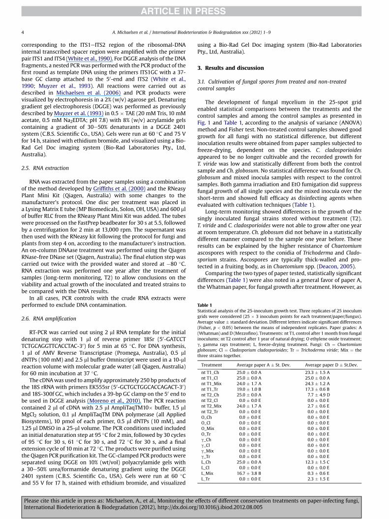
Table 1Statistical analysis of the 25-inoculum growth test. Three replicates of 25 inoculumgrids were considered (25 � 3 inoculum points for each treatment/paper/fungus).Average value � standard deviation. Different letters indicate significant differences(Fisher, p < 0.05) between the means of independent replicates. Paper grades: A(Whatman) and D (Mezzofino). Treatments: nt T1, control after 1 month from fungalinoculums; nt T2 control after 1 year of natural drying; O ethylene oxide treatment;g, gamma rays treatment; L, freeze-drying treatment. Fungi: Ch ¼ Chaetomiumglobosum; Cl ¼ Cladosporium cladosporioides; Tr ¼ Trichoderma viride; Mix ¼ thethree strains together.
Treatment Average paper A � St. Dev. Average paper D � St.Dev.
nt T1_Ch 25.0 � 0.0 A 23.3 � 1.5 Ant T1_Cl 25.0 � 0.0 A 25.0 � 0.0 Ant T1_Mix 24.0 � 1.7 A 24.3 � 1.2 Ant T1_Tr 19.0 � 1.0 B 17.3 � 0.6 Bnt T2_Ch 25.0 � 0.0 A 7.7 � 4.9 Dnt T2_Cl 0.0 � 0.0 E 0.0 � 0.0 Ent T2_Mix 24.0 � 1.7 A 2.7 � 0.6 Ent T2_Tr 0.0 � 0.0 E 0.0 � 0.0 EO_Ch 0.0 � 0.0 E 0.0 � 0.0 EO_Cl 0.0 � 0.0 E 0.0 � 0.0 EO_Mix 0.0 � 0.0 E 0.0 � 0.0 EO_Tr 0.0 � 0.0 E 0.0 � 0.0 Eg_Ch 0.0 � 0.0 E 0.0 � 0.0 Eg_Cl 0.0 � 0.0 E 0.0 � 0.0 Eg_Mix 0.0 � 0.0 E 0.0 � 0.0 Eg_Tr 0.0 � 0.0 E 0.0 � 0.0 EL_Ch 25.0 � 0.0 A 12.3 � 1.5 CL_Cl 0.0 � 0.0 E 0.0 � 0.0 EL_Mix 16.7 � 3.8 B 0.3 � 0.6 EL_Tr 0.0 � 0.0 E 2.3 � 1.5 E
A. Michaelsen et al. / International Biodeterioration & Biodegradation xxx (2012) 1e94
corresponding to the ITS1eITS2 region of the ribosomal-DNAinternal transcribed spacer region were amplified with the primerpair ITS1 and ITS4 (White et al., 1990). For DGGE analysis of the DNAfragments, a nested PCRwas performedwith the PCR product of thefirst round as template DNA using the primers ITS1GC with a 37-base GC clamp attached to the 50-end and ITS2 (White et al.,1990; Muyzer et al., 1993). All reactions were carried out asdescribed in Michaelsen et al. (2006) and PCR products werevisualized by electrophoresis in a 2% (w/v) agarose gel. Denaturinggradient gel electrophoresis (DGGE) was performed as previouslydescribed by Muyzer et al. (1993) in 0.5 � TAE (20 mM Tris, 10 mMacetate, 0.5 mM Na2EDTA; pH 7.8) with 8% (w/v) acrylamide gelscontaining a gradient of 30e50% denaturants in a DGGE 2401system (C.B.S. Scientific Co., USA). Gels were run at 60 �C and 75 Vfor 14 h, stainedwith ethidium bromide, and visualized using a Bio-Rad Gel Doc imaging system (Bio-Rad Laboratories Pty., Ltd,Australia).
2.5. RNA extraction
RNAwas extracted from the paper samples using a combinationof the method developed by Griffiths et al. (2000) and the RNeasyPlant Mini Kit (Qiagen, Australia) with some changes to themanufacturer’s protocol. One disc per treatment was placed ina Lysing Matrix E tube (MP Biomedicals, Solon, OH, USA) and 600 mlof buffer RLC from the RNeasy Plant Mini Kit was added. The tubeswere processed on the FastPrep beadbeater for 30 s at 5.5, followedby a centrifugation for 2 min at 13,000 rpm. The supernatant wasthen used with the RNeasy kit following the protocol for fungi andplants from step 4 on, according to the manufacturer’s instruction.An on-column DNAase treatment was performed using the QiagenRNase-free DNase set (Qiagen, Australia). The final elution step wascarried out twice with the provided water and stored at �80 �C.RNA extraction was performed one year after the treatment ofsamples (long-term monitoring, T2) to allow conclusions on theviability and actual growth of the inoculated and treated strains tobe compared with the DNA results.
In all cases, PCR controls with the crude RNA extracts wereperformed to exclude DNA contamination.
2.6. RNA amplification
RT-PCR was carried out using 2 ml RNA template for the initialdenaturing step with 1 ml of reverse primer 18Sr (50-GATCCTTCTGCAGGTTCACCTAC-30) for 5 min at 65 �C. For DNA synthesis,1 ml of AMV Reverse Transcriptase (Promega, Australia), 0.5 mldNTPs (100 mM) and 2.5 ml buffer Omniscript were used in a 10-mlreaction volume with molecular grade water (all Qiagen, Australia)for 60 min incubation at 37 �C.
The cDNAwas used to amplify approximately 250 bp products ofthe 18S rRNA with primers EK555sr (50-GCTGCTGGCACCAGACT-30)and 18S-300f GC, which includes a 39-bp GC clamp on the 50 end tobe used in DGGE analysis (Moreno et al., 2010). The PCR reactioncontained 2 ml of cDNA with 2.5 ml AmpliTaqTM10� buffer, 1.5 mlMgCl2 solution, 0.1 ml AmpliTaqTM DNA polymerase (all AppliedBiosystems), 10 pmol of each primer, 0.5 ml dNTPs (10 mM), and1.25 ml DMSO in a 25-ml volume. The PCR conditions used includedan initial denaturation step at 95 �C for 2 min, followed by 30 cyclesof 95 �C for 30 s, 61 �C for 30 s, and 72 �C for 30 s, and a finalextension cycle of 10min at 72 �C. The products were purified usingthe Qiagen PCR purification kit. The GC-clamped PCR products wereseparated using DGGE on 10% (wt/vol) polyacrylamide gels witha 30e50% urea/formamide denaturing gradient using the DGGE2401 system (C.B.S. Scientific Co., USA). Gels were run at 60 �Cand 55 V for 17 h, stained with ethidium bromide, and visualized
Please cite this article in press as: Michaelsen, A., et al., Monitoring the eInternational Biodeterioration & Biodegradation (2012), http://dx.doi.org
using a Bio-Rad Gel Doc imaging system (Bio-Rad LaboratoriesPty., Ltd, Australia).
3. Results and discussion
3.1. Cultivation of fungal spores from treated and non-treatedcontrol samples
The development of fungal mycelium in the 25-spot gridenabled statistical comparisons between the treatments and thecontrol samples and among the control samples as presented inFig. 1 and Table 1, according to the analysis of variance (ANOVA)method and Fisher test. Non-treated control samples showed goodgrowth for all fungi with no statistical difference, but differentinoculation results were obtained from paper samples subjected tofreeze-drying, dependent on the species. C. cladosporioidesappeared to be no longer cultivable and the recorded growth forT. viride was low and statistically different from both the controlsample and Ch. globosum. No statistical difference was found for Ch.globosum and mixed inocula samples with respect to the controlsamples. Both gamma irradiation and EtO fumigation did suppressfungal growth of all single species and the mixed inocula over theshort-term and showed full efficacy as disinfecting agents whenevaluated with cultivation techniques (Table 1).
Long-term monitoring showed differences in the growth of thesingly inoculated fungal strains stored without treatment (T2).T. viride and C. cladosporioides were not able to grow after one yearat room temperature. Ch. globosum did not behave in a statisticallydifferent manner compared to the sample one year before. Theseresults can be explained by the higher resistance of Chaetomiumascospores with respect to the conidia of Trichoderma and Clado-sporium strains. Ascospores are typically thick-walled and pro-tected in a fruiting body, as in Chaetomium spp. (Deacon, 2005).
Comparing the two types of paper tested, statistically significantdifferences (Table 1) were also noted in a general favor of paper A,theWhatman paper, for fungal growth after treatment. However, as
ffects of different conservation treatments on paper-infecting fungi,/10.1016/j.ibiod.2012.08.005
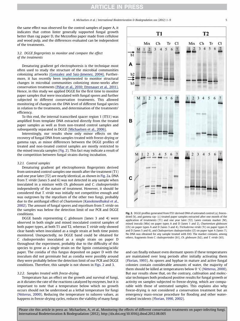
Fig. 2. DGGE profiles generated from ITS1 derived DNA of untreated control (a), freeze-dried (b), and gamma ray- (c) treated paper samples extracted after one month of theapplication of treatments (T1) and one year later (T2). Lanes contain marker (M),mixed inocula (Mix) on paper types A and D (lanes 1 and 2), Chaetomium globosum(Ch) on paper types A and D (lanes 3 and 4), Trichoderma viride (Tr) on paper types Aand D (lanes 5 and 6), and Cladosporium cladosporioides (Cl) on paper type A (lanes 7).No DNA was obtained for any sample treated with EtO. The marker contains, amongothers, fragments from C. cladosporioides (b1), Ch. globosum (b2), and T. viride (b3).
A. Michaelsen et al. / International Biodeterioration & Biodegradation xxx (2012) 1e9 5
the same effect was observed for the control samples of paper A, itindicates that cotton linter generally supported fungal growthbetter than rag paper D, the Mezzofino paper made from celluloseand wood pulp, and the differences evaluated can be independentof the treatments.
3.2. DGGE fingerprints to monitor and compare the effectof the treatments
Denaturing gradient gel electrophoresis is the technique mostoften used to study the structure of the microbial communitiescolonizing artworks (Gonzalez and Saiz-Jimenez, 2004). Further-more, it has recently been implemented to monitor structuralchanges in microbial communities colonizing stone-works afterconservation treatments (Piñar et al., 2010; Ettenauer et al., 2011).Hence, in this study we applied DGGE for the first time to monitorpaper samples that were inoculated with fungal spores and furthersubjected to different conservation treatments. This allowedmonitoring of changes on the DNA level of different fungal speciesin relation to the treatments, and determination of the treatments’efficiency.
To this end, the internal transcribed spacer region 1 (ITS1) wasamplified from template DNA extracted directly from the treatedpaper samples as well as from non-treated control samples andsubsequently separated in DGGE (Michaelsen et al., 2006).
Interestingly, our results show only minor effects on therecovery of fungal DNA from samples treated with freeze-drying orgamma rays, as minor differences between the DGGE profiles oftreated and non-treated control samples are mostly restricted tothe mixed inocula samples (Fig. 2). This fact may indicate a result ofthe competition between fungal strains during incubation.
3.2.1. Control samplesDenaturing gradient gel electrophoresis fingerprints derived
from untreated control samples onemonth after the treatment (T1)and one year later (T2) are nearly identical, as shown in Fig. 2a. DNAfrom T. viride (lanes 5 and 6) was not detected in any sample wheninoculated in a mixture with Ch. globosum and C. cladosporioidesindependently of the nature of treatment. However, it should beconsidered that T. viride was initially not competitive enough andwas outgrown by the mycelium of the other two fungi, probablydue to the antifungal effect of Chaetomium (Kanokmedhakul et al.,2002). The amount of fungal spores and mycelium from T. viride onthe samples was below the detection limit of our PCR and DGGEconditions.
DGGE bands representing C. globosum (lanes 3 and 4) wereobserved in both single and mixed inoculated control samples ofboth paper types, at both T1 and T2, whereas T. viride only showedclear bands when inoculated as a single strain at both time pointsmonitored. Unexpectedly, no DGGE band could be obtained forC. cladosporioides inoculated as a single strain on paper Dthroughout the experiment, probably due to the difficulty of thisspecies to grow as a single strain on the lignin containing/acidicpaper. The conidia of this fungus deposited on paper D with theinoculum did not germinate but as conidia were possibly aroundthey were probably below the detection limit of our PCR and DGGEconditions. Therefore, this sample is not shown in Figs. 2 and 3.
3.2.2. Samples treated with freeze-dryingTemperature has an effect on the growth and survival of fungi,
as it dictates the rate of the reactions catalyzed by enzymes, but it isimportant to note that a temperature below which no growthoccurs should not be understood as a lethal temperature for fungi(Nitterus, 2000). Reducing the temperature to subzero values, ashappens in freeze-drying cycles, reduces the viability of many fungi
Please cite this article in press as: Michaelsen, A., et al., Monitoring the eInternational Biodeterioration & Biodegradation (2012), http://dx.doi.org
and can finally exhaust even dormant spores if these temperaturesare maintained over long periods after initially activating them(Florian, 1993). As spores and hyphae in mature and active fungalcolonies contain considerable amounts of water, the majority ofthem should be killed at temperatures below 0 �C (Nitterus, 2000).But our results show that, on the contrary, cultivation and molec-ular techniques both produced positive results for fungal growth oractivity on samples subjected to freeze-drying, which are compa-rable with those of untreated samples. This explains also whyfreeze-drying is not considered a conservation treatment but anemergency mass-rescue procedure for flooding and other water-related incidents (Florian, 1990, 2002).
ffects of different conservation treatments on paper-infecting fungi,/10.1016/j.ibiod.2012.08.005

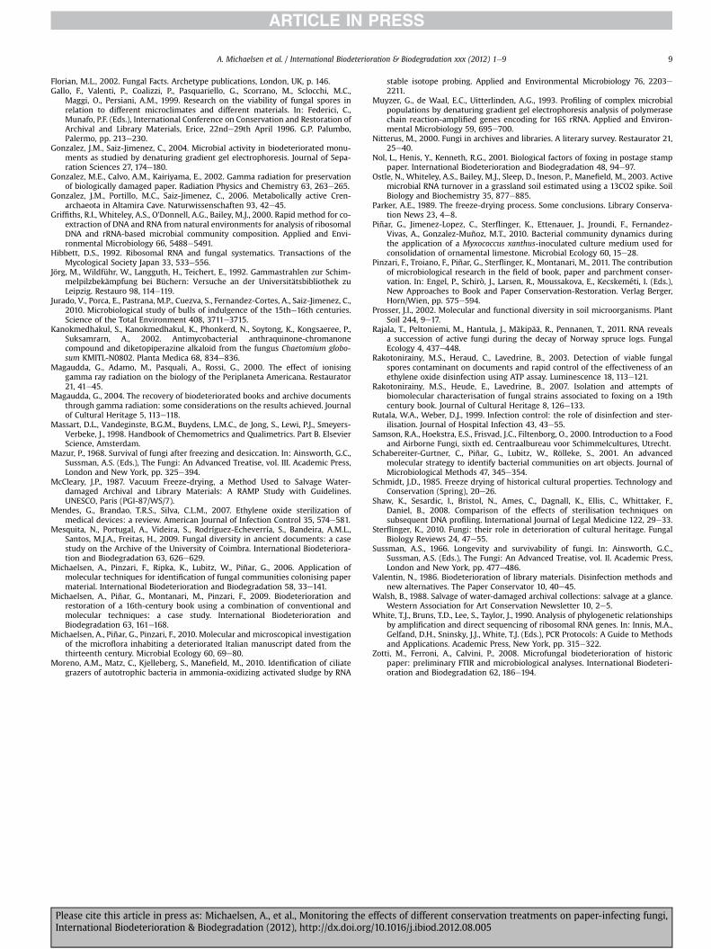
Fig. 3. DGGE profiles generated from 18S cDNA of untreated control (a), freeze-dried (b), and gamma ray- (c) treated paper samples one year after the application of treatments (T2).Lanes contain marker (M), mixed inocula (Mix) on paper types A and D (lanes 1 and 2), Chaetomium globosum (Ch) on paper types A and D (lanes 3 and 4), Trichoderma viride (Tr) onpaper types A and D (lanes 5 and 6), and Cladosporium cladosporioides (Cl) on paper types A (lane 7). Marked bands indicate the position of each fungus on DGGE: C. Cladosporioides(B1, in A and B4 in 3C), Ch. globosum (B2), and T. viride (B3).
A. Michaelsen et al. / International Biodeterioration & Biodegradation xxx (2012) 1e96
DGGE fingerprints derived from samples treated with freeze-drying showed some differences compared to the control samples(Fig. 2b). One month after the treatment (T1) the fingerprintsderived from samples inoculated with the mixed culture differedfrom the control and between paper A, Whatman, and paper D,Mezzofino. The C. cladosporioideswas represented by a strong bandon paper A (T1, lane 1) but it was missing on paper D (T1, lane 2). Bycontrast, Ch. globosum appeared as a strong band on paper D butwas very faint on paper A, whereas T. viridewas not detected in anymixed inoculated sample, as observed in the control samples.Mixed inocula showed a dominance of Ch. globosum that exhibiteda strong antifungal tendency against T. viride. The bands repre-senting each single fungus inoculated as pure culture did not showdifferences due to treatment.
Long-term effects due to the freeze-drying treatment were onlyobserved for Ch. globosum when inoculated as a single strain,showing an unspecific band-pattern (Fig. 2b, T2, lanes 3 and 4).Furthermore, no DGGE band corresponding to this fungus wasdetected when it was inoculated in a mixture with the other twofungi (Fig. 2b, T2, lanes 1 and 2). The other fungal bands showedpatterns in accordance with the control samples.
3.2.3. Samples treated with gamma raysDifferent functions of the paper or its components, such as
cellulose, are affected by gamma irradiation. Furthermore, itsgermicidal power on fungal microflora has been reported to bedose-dependent (Magaudda et al., 2000; Adamo et al., 2001).A dose of about 5 kGy, as applied in our experiment, was found tocause statistically significant variations on almost all paper prop-erties, such as tensile strength, tearing resistance, and foldingendurance, but also reduced the fungal population down to the“blank” samples level (Adamo et al., 2001; Magaudda, 2004).Relatively low irradiation doses of 2e3 kGy delivered comparabledecontaminating power without the increasingly depolymerizingeffects of gamma rays on cellulose.
Samples treated with gamma rays showed DGGE patterns withnumerous unspecific bands one month after treatment (Fig. 2c, T1).Bands corresponding to the individual fungi Ch. globosum, T. viride,and C. cladosporioides were still observed, but their identities were
Please cite this article in press as: Michaelsen, A., et al., Monitoring the eInternational Biodeterioration & Biodegradation (2012), http://dx.doi.org
indecisive in some samples. In addition, new faint bands wereobserved. These patterns indicate a mixed population of intact DNAfrom surviving fungi and DNA artifacts, as a consequence of DNAfragmentation due to the irradiation, from affected fungi.
One year later (Fig. 2c, T2), no more visible effect of gammaradiation on the DNA of pure strains, compared to the controlsamples, was observed. This indicates that the surviving fungalpopulation recovered with time and only the intact DNA of thispopulationwas detected. For paper samples inoculated with mixedinocula, only the band for C. cladosporioides was shown to be weakon paper D (Fig. 2c, T2, lane 2).
3.2.4. Samples treated with EtO fumigationThe observation that EtO can significantly reduce or erase the
amount of amplifiable DNA on small items has been reportedbefore. Ethylene oxide is successfully applied in forensics to removeDNA contamination on items used both at crime scenes and in theforensics laboratory, without significantly affecting any neededdownstream DNA analysis when used to sterilize equipment (Shawet al., 2008). The gaseous EtO is considered a radiomimetic agent,whichmeans it is similar to ionizing radiation by virtue of its abilityto induce the same biological end-points, such as gene mutations,mainly by alkylating and reacting with nucleophile centers, such asnitrogen and oxygen atoms in the DNA bases (Bolt, 1996; Rutala andWeber, 1999; Chovanec et al., 2001). Double-strand-breakages areinduced, leading to fragmentation of the DNA helix. Our resultscould be explained by such a fragmentation of fungal DNA throughEtO fumigation, resulting in PCR not delivering any evidence offungal DNA or RNA on all samples submitted to fumigation. Inaccordance with this, a study on viability of fungal spores on paperafter EtO treatment indicated that fungal spores became non-cultivable and non-viable with EtO inhibiting spore activity(Rakotonirainy et al., 2003).
As Shaw et al. (2008) did, we observed that EtO fumigation wasmore effective than the application of gamma rays in reducing DNAfrom the surface of samples. The superiority of EtO as a sterilizingagent compared to gamma radiation was explained by Chovanecet al. (2001) as a combination of differences in the nature ofdamage caused and the distribution of the target. Ionizing radiation
ffects of different conservation treatments on paper-infecting fungi,/10.1016/j.ibiod.2012.08.005

A. Michaelsen et al. / International Biodeterioration & Biodegradation xxx (2012) 1e9 7
can react more locally and focused with free radicals followingtracks along covalent bonds, whereas small molecules such as EtOcan diffuse quite freely in the cell nucleus, causing damage ona broader scale. Although EtO application is banned for use inconservation, it should still be recommended in certain caseswhere the careful restoration and conservation of valuedmaterial isassessed.
The fumigation of paper with EtO caused the loss of amplifiablefungal DNA from all spore-inoculated samples tested in both singleand mixed cultures. General EtO inhibition properties on PCRperformance were ruled out with PCR controls with DNA extractsspiked with pure fungal DNA as template (data not shown).
3.3. RNA as an indicator for metabolically active fungi
Aswe confirmed the presence of DNA onmost samples, with theexception of those treated with EtO, the next consequent step wasto investigate if the fungi inoculated on the samples were stillmetabolically active. Several methods are available to test fungaland microbial viability, such as the quantitation of cell’s ATPcontent (Rakotonirainy et al., 2003) or fluorescein diacetate (FDA)and acridine-orange stainings that provide a qualitative observa-tion of a microorganism’s active structures by means of anepifluorescent microscope, equipped with specific filters (Pinzariet al., 2011).
In this study we decided to use nucleic acids as markers offungal viability. The existence of ribosomal-DNA alone does notlead to assumptions about viability because rRNA genes can persistin environmental DNA pools for species that are metabolicallyinactive and functionally less important (Ostle et al., 2003). Thus, ifresponses of microbial communities to environmental perturba-tions are investigated, the rRNA gene approach seems problematic,as rRNA genes may be detected in DNA pools for species whosegrowth or cellular activity has declined (Anderson and Parkin,2007). To test the viability of the inoculated fungi, we targetedthe fungal rRNA directly, as it allows the detection of metabolicallyactive and functionally important species, as they transcribe morerRNA, a principle that is commonly exploited in bacterial ecology(Prosser, 2002). Determination of the viability of the causativeagent is important in determining the active infection and in theevaluation of the efficacy of a particular treatment.
In this study, RNA was extracted from paper samples one yearafter the application of the respective treatments (T2), and subse-quently transcribed into cDNA for further DGGE analyses. TheDGGE profiles generated with 18S primers from the fungal cDNAare shown in Fig. 3. Untreated control samples and those subjectedto freeze-drying and gamma irradiation were all positive foramplification of fungal 18S cDNA, whereas no positive RNAextraction or PCR could be established for EtO-treated samples.Strong signals could be observed for all pure strains, with theexception of T. viride on paper D, in the control samples (Fig. 3a).The pattern of single bands obtained for the pure inoculated strainsindicated the presence of only one fungus on each paper sample.On samples inoculated with the mixed cultures, the DGGE bandcorresponding to T. viride was not observable, a pattern that iscongruent with the DNA-generated gels.
RNA analyses revealed a minimal long-term impact of eithergamma rays or freeze-drying treatments on the viability of thefungal spores, when inoculated as single strains (Fig. 3b and c, lanes3e7). All samples deliver clear bands with a pattern that matchesthe expectations and consistent for the different fungi. However,when fungi were inoculated as mixed cultures, no RNA band at allor very faint signals were observed for paper D after freeze-dryingand gamma ray treatments, respectively (Fig. 3b and c, lane 2).
Please cite this article in press as: Michaelsen, A., et al., Monitoring the eInternational Biodeterioration & Biodegradation (2012), http://dx.doi.org
Unfortunately, an amplification of the cDNAwith ITS primers, asused for the DGGE analysis of DNA, failed. Therefore we usedprimers targeting the 18S rRNA to obtain DGGE profiles of cDNA. ITSsequences are the most popular choice for species identification offungi in environmental DNA pools (White et al., 1990), but due topost-transcriptional processing of the main precursor rRNA mole-cules containing 18S, ITS1, 5.8S, ITS2, and 28S rRNA, the ITS spacerregions are spliced out to leave the rRNA genes for ribosomesynthesis (Anderson and Parkin, 2007). Even if there is evidencethat the ITS regions can be used for the detection of active fungi, aswas done by Anderson and Parkin (2007), it is generally consideredimpossible to detect ITS sequences in RNA pools (Hibbett, 1992). Asuccessful amplification of ITS rRNAwas performed by other groupsfrom fungal isolate extracts and soil samples. We assumed, duringthe experimental work, that there was more rRNA available in thesoil samples themselves, as soils are active environments comparedto the paper environment, which resembles starving conditions.While the present paper was under review, a paper by Rajala et al.(2011) was published, which demonstrated that internal tran-scribed spacers (ITS) precursor rRNA are a better marker of activefungi than small subunit rRNA. This is because the ITS sequences,after being excised from the subunits turnover rapidly, and smalland large subunit rRNA particles are relatively stable in the envi-ronment. The work by Rajala et al. (2011) may be important forfollow-up elaboration of the present work.
3.4. Comparison of methods used for the monitoring oftreatments d cultivation versus molecular methods
Our molecular results generally contradicted the cultivationresults obtained in this study. By using culture-dependent tech-niques, samples subjected to freeze-drying showed growth in thecase of T. viride, but is was recorded as low and statistically differentfrom the control sample, whereas no statistical difference wasfound for C. globosum and themixed inocula samplewith respect tothe control samples (see Table 1). However, by using culture-independent techniques we could observe some differences onsamples inoculated with the mixed culture samples one monthafter the freeze-drying treatment (T1) compared to the controlsamples (see Fig. 2b). DGGE bands representing each single fungusinoculated as pure culture did not show differences due to treat-ment, but a long-term effect was only observed for Ch. globosumwhen it was inoculated as a single strain, showing an unspecificband-pattern.
Samples subjected to gamma irradiation and EtO fumigationshowed no fungal growth, either of all single species or the mixedinocula. Therefore, the last two treatments indicated a full efficacyas disinfecting agents when evaluated with cultivation techniques(see Table 1). However, when using culture-independent tech-niques, gamma rays were not able to eliminate fungal colonizationof paper even if relatively high doses (5 kGy) were applied, but theycould potentially reduce or slow down the growing process. DGGEprofiles obtained from DNA extracted shortly after the irradiationprocess display a certain amount of fragmentation and unspecificbands that could be related to the effect of gamma radiation on thetertiary or secondary structure of the DNA itself, but no long-termeffect of gamma rays on DNA or RNA was observed, indicating therecovering of the surviving fungal fraction. Gamma rays have beenreported to inactivate fungi from and related to books (Jörg et al.,1992; Da Silva et al., 2006). However, Da Silva et al. (2006) deter-mined the efficiency of the radiation treatment on fungal spores bycultivation attempts only, and therefore described a higher degreeof reduction than our DNA- and RNA-based studies. Our cultivationresults are in accordance with these observations, but are provenwrong when molecular data are included.
ffects of different conservation treatments on paper-infecting fungi,/10.1016/j.ibiod.2012.08.005

A. Michaelsen et al. / International Biodeterioration & Biodegradation xxx (2012) 1e98
Only in the case of EtO fumigation did both culture-dependentand -independent techniques indicate the total inactivation of theinoculated fungi.
Comparing DNA and RNA recovery, we could observe nodifferences in the case of samples inoculated with pure strains,being able to recover both DNA and RNA, which indicates that thefungi were indeed viable. However, differences in the RNA recoveryfrom samples treated with freeze-drying or gamma rays wereobserved when samples were inoculated with the mixed culture,especially on paper type D, indicating the inactivation of the fungion this kind of paper and the higher sensitivity of RNA- versus DNA-based analyses for the detection of viability.
4. Conclusions
The differences between culture and molecular results obtainedin this study highlight the importance of molecular tools tocomplement conventional techniques, as they display highersensitivity and bypass culturing methods. Retrieval of fungal-specific rRNA directly from paper can, in fact, be successfully usedas an indicator for the viability of fungi after disinfectingtreatments.
Information on the effects of mass treatments on the DNA ofpaper-spoiling fungi is also provided. Our results show, in fact, thatthe freeze-drying treatment displays only minimal effect on DNArecovery over both the short- or long-term when compared tocontrol samples, whereas the application of gamma rays showsa short-term slight reduction of DNA recovery and DNA fragmen-tation but no significant difference in long-term effect.
When RNA was used as an indicator for long-term fungalviability, differences in the RNA recovery from samples treated withfreeze-drying or gamma ray-treated samples were observed whensamples were inoculated with themixed culture, specially on papertype D. Samples inoculated with the single strains showed nodifference in the RNA recovery compared to control samples. Onlytreatment with ethylene oxide proved negative for both DNA andRNA recovery. These results show the higher sensitivity of RNA-over DNA-based molecular analyses for the detection of viability offungi on disinfected paper materials.
Considering our results, we can assume that gamma rays can beused to treat large amounts of paper simultaneously and withoutsubsequent chemical hazard, but this can only be considereda decontamination treatment, a method to remove bio-deteriogenous microorganisms to a controllable or blank level. Thistarget of reducing and holding the biodeteriogens at under thedanger threshold is crucial in paper conservation, and it can beachieved with gamma radiation, whereas freeze-drying can beapplied only to stop heavy mould before further treatment.
Furthermore, our results point to ethylene oxide fumigation asthe only effective and long-lasting treatment when the sterilizationof a fungal infected paper object is needed. Subsequent contami-nation is, however, possible, as ethylene oxide is not a chemical thatremains as a preventive biocide on materials.
It is worth emphasizing that no DNA or RNA could be recoveredfrom paper samples fumigated with EtO, a fact that is of particularimportance for studies that address a molecular analysis of oldcontaminations or biological materials in cultural heritage charac-terization and diagnostics, including paleogenetic and archaeo-logical molecular studies. Ethylene oxide fumigation was used bymuseums and libraries in the recent past to treat insect or mouldinfestations, but it is often not traceable or not clearly mentioned inthe reports that accompany the treated objects. Consequently,misleading diagnostics could be produced in subsequent molecularstudies. What is needed is improved documentation of theconservation history of objects of cultural value, in order to account
Please cite this article in press as: Michaelsen, A., et al., Monitoring the eInternational Biodeterioration & Biodegradation (2012), http://dx.doi.org
for treatments that can alter future studies and analysis and toavoid any impediments by past treatment based on EtO.
Generally, any treatment that is applied to prevent the biode-terioration of paper documents or books has to be carefullyassessed beforehand. The type of biological attack and the urgencyof intervention have to be considered when choosing amongtreatments. Economic factors such as cost of treatment, quantity ofobjects to be treated, and their historical or commercial value alsohave also an impact on the decision of conservators.
Acknowledgements
The molecular analysis included in this study and AstridMichaelsen were financed by the Austrian Science Fund (FWF)within the framework of project P17328-B12. Guadalupe Piñar iscurrently financed by the Austrian Science Fund (FWF) project“Elise-Richter V194-B20.” Part of this work was performed at theCentre for Marine Bioinnovation (CMB) at the University of NewSouth Wales (UNSW) in Sydney, Australia. We thank Prof. StaffanKjelleberg, Dr. Thorsten Thomas, and Dr. Mike Manefield for theirkind support. The authors are grateful to Pietro Livi (Centro diLegatoria e Restauro Frati e Livi s.r.l., Italy), Dr. Marianna Adamo(Research Centre of ENEA Casaccia, Rome, Italy), and the peoplefrom Spix Italia s.r.l. for the treatment of paper samples.
References
Adamo, M., Giovanotti, M., Magaudda, G., Plossi Zappala, M., Rocchetti, F., Rossi, G.,1998. Effect of gamma rays on pure cellulose paper as a model for the studyof treatment of biological recovery of biodeteriorated books. Restaurator 19,41e59.
Adamo, M., Brizzi, M., Magaudda, G., Martinelli, G., Plossi Zappala, M., Rocchetti, F.,Savagnone, F., 2001. Gamma radiation treatment of paper in differentenvironmental conditions: chemical, physical and microbiological analysis.Restaurator 22, 107e131.
Anderson, I.C., Parkin, P.I., 2007. Detection of active soil fungi by RT-PCR amplifi-cation of precursor rRNA molecules. Journal of Microbiological Methods 68,248e253.
Angerer, J., Bader, M., Kramer, A., 1998. Ambient and biochemical effect monitoringof workers exposed to ethylene oxide. International Archives of Occupationaland Environmental Health 71, 14e18.
Ballard, M.W., Baer, N.S., 1986. Ethylene oxide fumigation: results and risk assess-ment. Restaurator 7, 143e168.
Belyakova, L.A., 1960. Gamma radiation as a means of disinfection of books againstspores of mould fungi. Mikrobiologiya 29, 762e765.
Bolt, H.M., 1996. Quantification of endogenous carcinogens. The ethylene oxideparadox. Biochemical Pharmacology 52, 1e5.
Butterfield, F.J., 1987. The potential long-term effects of gamma radiation on paper.Studies in Conservation 32, 181e191.
Cappitelli, F., Sorlini, C., 2005. From papyrus to compact disc: the microbial dete-rioration of documentary heritage. Critical Reviews in Microbiology 31, 1e10.
Chovanec, M., Cedervall, B., Kolma, A., 2001. DNA damage induced by gamma-radiation in combination with ethylene oxide or propylene oxide in humanfibroblasts. Chemico-Biological Interactions 137, 259e268.
Corte, M.A., Ferrroni, A., Salvo, V.S., 2003. Isolation of fungal species from testsamples and maps damaged by foxing, and correlation between these speciesand the environment. International Biodeterioration and Biodegradation 51,167e173.
Da Silva, M., Moraes, A.M.L., Nishikawa, M.M., Gatti, M.J.A., Vallim de Alencar, M.A.,Brandao, L.E., Nobrega, A., 2006. Inactivation of fungi from deteriorated papermaterials by radiation. International Biodeterioration and Biodegradation 57,163e167.
De Paolis, M.R., Lippi, D., 2008. Use of metabolic and molecular methods for theidentification of a Bacillus strain isolated from paper affected by foxing.Microbiological Research 163, 121e131.
Deacon, J., 2005. Fungal Biology. Blackwell Publishers, Cambridge, Massachusetts.Dix, N.J., Webster, J., 1995. Fungal Ecology. Chapman & Hall, Londres, p. 549.Ettenauer, J., Piñar, G., Sterflinger, K., Gonzalez-Muñoz, M.T., Jroundi, F., 2011.
Molecular monitoring of the microbial dynamics occurring on historical lime-stone buildings during and after the in situ application of different bio-consolidation treatments. Science of the Total Environment 409, 5337e5352.
Florian, M.L., 1990. The effects of freezing and freeze-drying on natural historyspecimens. Collection Forum 6, 45e52.
Florian, M.L., 1993. Conidial fungi (mould) activity on artifact materials e a newlook at prevention, control and eradication. ICOM. Committee for Conservation19th Triennal Meeting. Washington, pp. 868e874.
ffects of different conservation treatments on paper-infecting fungi,/10.1016/j.ibiod.2012.08.005
A. Michaelsen et al. / International Biodeterioration & Biodegradation xxx (2012) 1e9 9
Florian, M.L., 2002. Fungal Facts. Archetype publications, London, UK, p. 146.Gallo, F., Valenti, P., Coalizzi, P., Pasquariello, G., Scorrano, M., Sclocchi, M.C.,
Maggi, O., Persiani, A.M., 1999. Research on the viability of fungal spores inrelation to different microclimates and different materials. In: Federici, C.,Munafo, P.F. (Eds.), International Conference on Conservation and Restoration ofArchival and Library Materials, Erice, 22nde29th April 1996. G.P. Palumbo,Palermo, pp. 213e230.
Gonzalez, J.M., Saiz-Jimenez, C., 2004. Microbial activity in biodeteriorated monu-ments as studied by denaturing gradient gel electrophoresis. Journal of Sepa-ration Sciences 27, 174e180.
Gonzalez, M.E., Calvo, A.M., Kairiyama, E., 2002. Gamma radiation for preservationof biologically damaged paper. Radiation Physics and Chemistry 63, 263e265.
Gonzalez, J.M., Portillo, M.C., Saiz-Jimenez, C., 2006. Metabolically active Cren-archaeota in Altamira Cave. Naturwissenschaften 93, 42e45.
Griffiths, R.I., Whiteley, A.S., O’Donnell, A.G., Bailey, M.J., 2000. Rapid method for co-extraction of DNA and RNA from natural environments for analysis of ribosomalDNA and rRNA-based microbial community composition. Applied and Envi-ronmental Microbiology 66, 5488e5491.
Hibbett, D.S., 1992. Ribosomal RNA and fungal systematics. Transactions of theMycological Society Japan 33, 533e556.
Jörg, M., Wildführ, W., Langguth, H., Teichert, E., 1992. Gammastrahlen zur Schim-melpilzbekämpfung bei Büchern: Versuche an der Universitätsbibliothek zuLeipzig. Restauro 98, 114e119.
Jurado, V., Porca, E., Pastrana, M.P., Cuezva, S., Fernandez-Cortes, A., Saiz-Jimenez, C.,2010. Microbiological study of bulls of indulgence of the 15the16th centuries.Science of the Total Environment 408, 3711e3715.
Kanokmedhakul, S., Kanokmedhakul, K., Phonkerd, N., Soytong, K., Kongsaeree, P.,Suksamrarn, A., 2002. Antimycobacterial anthraquinone-chromanonecompound and diketopiperazine alkaloid from the fungus Chaetomium globo-sum KMITL-N0802. Planta Medica 68, 834e836.
Magaudda, G., Adamo, M., Pasquali, A., Rossi, G., 2000. The effect of ionisinggamma ray radiation on the biology of the Periplaneta Americana. Restaurator21, 41e45.
Magaudda, G., 2004. The recovery of biodeteriorated books and archive documentsthrough gamma radiation: some considerations on the results achieved. Journalof Cultural Heritage 5, 113e118.
Massart, D.L., Vandeginste, B.G.M., Buydens, L.M.C., de Jong, S., Lewi, P.J., Smeyers-Verbeke, J., 1998. Handbook of Chemometrics and Qualimetrics. Part B. ElsevierScience, Amsterdam.
Mazur, P., 1968. Survival of fungi after freezing and desiccation. In: Ainsworth, G.C.,Sussman, A.S. (Eds.), The Fungi: An Advanced Treatise, vol. III. Academic Press,London and New York, pp. 325e394.
McCleary, J.P., 1987. Vacuum Freeze-drying, a Method Used to Salvage Water-damaged Archival and Library Materials: A RAMP Study with Guidelines.UNESCO, Paris (PGI-87/WS/7).
Mendes, G., Brandao, T.R.S., Silva, C.L.M., 2007. Ethylene oxide sterilization ofmedical devices: a review. American Journal of Infection Control 35, 574e581.
Mesquita, N., Portugal, A., Videira, S., Rodríguez-Echeverría, S., Bandeira, A.M.L.,Santos, M.J.A., Freitas, H., 2009. Fungal diversity in ancient documents: a casestudy on the Archive of the University of Coimbra. International Biodeteriora-tion and Biodegradation 63, 626e629.
Michaelsen, A., Pinzari, F., Ripka, K., Lubitz, W., Piñar, G., 2006. Application ofmolecular techniques for identification of fungal communities colonising papermaterial. International Biodeterioration and Biodegradation 58, 33e141.
Michaelsen, A., Piñar, G., Montanari, M., Pinzari, F., 2009. Biodeterioration andrestoration of a 16th-century book using a combination of conventional andmolecular techniques: a case study. International Biodeterioration andBiodegradation 63, 161e168.
Michaelsen, A., Piñar, G., Pinzari, F., 2010. Molecular and microscopical investigationof the microflora inhabiting a deteriorated Italian manuscript dated from thethirteenth century. Microbial Ecology 60, 69e80.
Moreno, A.M., Matz, C., Kjelleberg, S., Manefield, M., 2010. Identification of ciliategrazers of autotrophic bacteria in ammonia-oxidizing activated sludge by RNA
Please cite this article in press as: Michaelsen, A., et al., Monitoring the eInternational Biodeterioration & Biodegradation (2012), http://dx.doi.org
stable isotope probing. Applied and Environmental Microbiology 76, 2203e2211.
Muyzer, G., de Waal, E.C., Uitterlinden, A.G., 1993. Profiling of complex microbialpopulations by denaturing gradient gel electrophoresis analysis of polymerasechain reaction-amplified genes encoding for 16S rRNA. Applied and Environ-mental Microbiology 59, 695e700.
Nitterus, M., 2000. Fungi in archives and libraries. A literary survey. Restaurator 21,25e40.
Nol, L., Henis, Y., Kenneth, R.G., 2001. Biological factors of foxing in postage stamppaper. International Biodeterioration and Biodegradation 48, 94e97.
Ostle, N., Whiteley, A.S., Bailey, M.J., Sleep, D., Ineson, P., Manefield, M., 2003. Activemicrobial RNA turnover in a grassland soil estimated using a 13CO2 spike. SoilBiology and Biochemistry 35, 877e885.
Parker, A.E., 1989. The freeze-drying process. Some conclusions. Library Conserva-tion News 23, 4e8.
Piñar, G., Jimenez-Lopez, C., Sterflinger, K., Ettenauer, J., Jroundi, F., Fernandez-Vivas, A., Gonzalez-Muñoz, M.T., 2010. Bacterial community dynamics duringthe application of a Myxococcus xanthus-inoculated culture medium used forconsolidation of ornamental limestone. Microbial Ecology 60, 15e28.
Pinzari, F., Troiano, F., Piñar, G., Sterflinger, K., Montanari, M., 2011. The contributionof microbiological research in the field of book, paper and parchment conser-vation. In: Engel, P., Schirò, J., Larsen, R., Moussakova, E., Kecskeméti, I. (Eds.),New Approaches to Book and Paper Conservation-Restoration. Verlag Berger,Horn/Wien, pp. 575e594.
Prosser, J.I., 2002. Molecular and functional diversity in soil microorganisms. PlantSoil 244, 9e17.
Rajala, T., Peltoniemi, M., Hantula, J., Mäkipää, R., Pennanen, T., 2011. RNA revealsa succession of active fungi during the decay of Norway spruce logs. FungalEcology 4, 437e448.
Rakotonirainy, M.S., Heraud, C., Lavedrine, B., 2003. Detection of viable fungalspores contaminant on documents and rapid control of the effectiveness of anethylene oxide disinfection using ATP assay. Luminescence 18, 113e121.
Rakotonirainy, M.S., Heude, E., Lavedrine, B., 2007. Isolation and attempts ofbiomolecular characterisation of fungal strains associated to foxing on a 19thcentury book. Journal of Cultural Heritage 8, 126e133.
Rutala, W.A., Weber, D.J., 1999. Infection control: the role of disinfection and ster-ilisation. Journal of Hospital Infection 43, 43e55.
Samson, R.A., Hoekstra, E.S., Frisvad, J.C., Filtenborg, O., 2000. Introduction to a Foodand Airborne Fungi, sixth ed. Centraalbureau voor Schimmelcultures, Utrecht.
Schabereiter-Gurtner, C., Piñar, G., Lubitz, W., Rölleke, S., 2001. An advancedmolecular strategy to identify bacterial communities on art objects. Journal ofMicrobiological Methods 47, 345e354.
Schmidt, J.D., 1985. Freeze drying of historical cultural properties. Technology andConservation (Spring), 20e26.
Shaw, K., Sesardic, I., Bristol, N., Ames, C., Dagnall, K., Ellis, C., Whittaker, F.,Daniel, B., 2008. Comparison of the effects of sterilisation techniques onsubsequent DNA profiling. International Journal of Legal Medicine 122, 29e33.
Sterflinger, K., 2010. Fungi: their role in deterioration of cultural heritage. FungalBiology Reviews 24, 47e55.
Sussman, A.S., 1966. Longevity and survivability of fungi. In: Ainsworth, G.C.,Sussman, A.S. (Eds.), The Fungi: An Advanced Treatise, vol. II. Academic Press,London and New York, pp. 477e486.
Valentin, N., 1986. Biodeterioration of library materials. Disinfection methods andnew alternatives. The Paper Conservator 10, 40e45.
Walsh, B., 1988. Salvage of water-damaged archival collections: salvage at a glance.Western Association for Art Conservation Newsletter 10, 2e5.
White, T.J., Bruns, T.D., Lee, S., Taylor, J., 1990. Analysis of phylogenetic relationshipsby amplification and direct sequencing of ribosomal RNA genes. In: Innis, M.A.,Gelfand, D.H., Sninsky, J.J., White, T.J. (Eds.), PCR Protocols: A Guide to Methodsand Applications. Academic Press, New York, pp. 315e322.
Zotti, M., Ferroni, A., Calvini, P., 2008. Microfungal biodeterioration of historicpaper: preliminary FTIR and microbiological analyses. International Biodeteri-oration and Biodegradation 62, 186e194.
ffects of different conservation treatments on paper-infecting fungi,/10.1016/j.ibiod.2012.08.005