Monitoring Coordination during Bimanual Movements: Where Is the Mastermind?
17
Monitoring Coordination during Bimanual Movements: Where Is the Mastermind? Julie Duque 1 , Marco Davare 1 , Ludovic Delaunay 1 , Benvenuto Jacob 1 , Ralf Saur 2 , Friedhelm Hummel 3 , Laurent Hermoye 1 , Bruno Rossion 1,4 , and Etienne Olivier 1 Abstract ■ One remarkable aspect of the human motor repertoire is the multitude of bimanual actions it contains. Still, the neural correlates of coordinated movements, in which the two hands share a common goal, remain debated. To address this issue, we designed two bimanual circling tasks that differed only in terms of goal conceptualization: a “coordination” task that re- quired movements of both hands to adapt to each other to reach a common goal and an “independent” task that imposed a separate goal to each hand. fMRI allowed us to pinpoint three areas located in the right hemisphere that were more strongly activated in the coordination condition: the superior temporal gyrus (STG), the SMA, and the primary motor cortex (M1). We then used transcranial magnetic stimulation (TMS) to disrupt transiently the function of those three regions to determine their causal role in bimanual coordination. Right STG virtual lesions impaired bimanual coordination, whereas TMS to right M1 en- hanced hand independence. TMS over SMA, left STG, or left M1 had no effect. The present study provides direct insight into the neural correlates of coordinated bimanual movements and highlights the role of right STG in such bimanual movements. ■ INTRODUCTION One impressive aspect of the human motor repertoire is the myriad movements it contains that require an intri- cate and subtle coordination between the two hands. How- ever, so far, most bimanual studies have investigated tasks in which the movements of one hand interfere with those of the other one, leading to a “bimanual cross talk” usually observed when both hands perform actions guided by two distinct goals (Wenderoth, Puttemans, Vangheluwe, & Swinnen, 2003; Swinnen, 2002; Serrien, Bogaerts, Suy, & Swinnen, 1999). For example, everybody has experi- enced how tricky it is to draw a line with one hand and, at the same time, a circle with the other one (Franz, 1997). These studies on bimanual movements leading to cross talk have proved very useful in determining the organi- zation, and the limits, of the neural circuit involved in such experimental conditions. However, the tasks investigated in these studies are far from being illustrative of the bi- manual actions we perform daily. In fact, we are able to execute a large number of actions that require nonsym- metrical or nonsynchronous movements of both hands without experiencing any cross talk: For instance, it is very easy to cut a piece of paper while holding it with the other hand. The critical difference between bimanual tasks lead- ing to a cross talk and those that do not is probably the way their goals are conceptualized (Oliveira & Ivry, 2008; Rosenbaum, Dawson, & Challis, 2006; Mechsner, Kerzel, Knoblich, & Prinz, 2001). In fact, bimanual actions that do not generate cross talk rely on the integration of a unique goal (e.g., to cut a piece of paper), which guides the “coordinated” movements of both hands. In contrast, in “independent” actions, each hand has its own goal, and these two separate goals probably compete for com- mon representational processes, leading to a substantial cross talk (Oliveira & Ivry, 2008; Diedrichsen, Hazeltine, Kennerley, & Ivry, 2001; Franz, Zelaznik, Swinnen, & Walter, 2001). So far, the specific neural correlates of coordinated bi- manual movements remain unclear. Many functional im- aging studies have focused on other features of bimanual movements, such as learning and complexity, or have used tasks requiring independent hand movements and were, therefore, unable to address this issue (e.g., Jantzen, Oullier, & Scott Kelso, 2008; Puttemans, Wenderoth, & Swinnen, 2005; Wenderoth, Debaere, Sunaert, & Swinnen, 2005a, 2005b; Debaere, Wenderoth, Sunaert, Van Hecke, & Swinnen, 2004b; Wenderoth, Debaere, Sunaert, van Hecke, & Swinnen, 2004; Aboitiz, Ide, & Olivares, 2003; De Weerd et al., 2003; Meyer-Lindenberg, Ziemann, Hajak, Cohen, & Berman, 2002; Toyokura, Muro, Komiya, & Obara, 1999; Sadato, Yonekura, Waki, Yamada, & Ishii, 1997). In fact, investigating bimanual coordination per se 1 Université Catholique de Louvain, Brussels, Belgium, 2 Eberhard- Karls University Tübingen, Germany, 3 University Medical Center Hamburg-Eppendorf, Germany, 4 Université Catholique de Louvain, Louvain-la-Neuve, Belgium © 2009 Massachusetts Institute of Technology Journal of Cognitive Neuroscience 22:3, pp. 526–542
-
Upload
independent -
Category
Documents
-
view
0 -
download
0
Transcript of Monitoring Coordination during Bimanual Movements: Where Is the Mastermind?

Monitoring Coordination during Bimanual Movements:Where Is the Mastermind?
Julie Duque1, Marco Davare1, Ludovic Delaunay1, Benvenuto Jacob1,Ralf Saur2, Friedhelm Hummel3, Laurent Hermoye1,
Bruno Rossion1,4, and Etienne Olivier1
Abstract
■ One remarkable aspect of the human motor repertoire isthe multitude of bimanual actions it contains. Still, the neuralcorrelates of coordinated movements, in which the two handsshare a common goal, remain debated. To address this issue,we designed two bimanual circling tasks that differed only interms of goal conceptualization: a “coordination” task that re-quired movements of both hands to adapt to each other toreach a common goal and an “independent” task that imposeda separate goal to each hand. fMRI allowed us to pinpoint threeareas located in the right hemisphere that were more strongly
activated in the coordination condition: the superior temporalgyrus (STG), the SMA, and the primary motor cortex (M1). Wethen used transcranial magnetic stimulation (TMS) to disrupttransiently the function of those three regions to determine theircausal role in bimanual coordination. Right STG virtual lesionsimpaired bimanual coordination, whereas TMS to right M1 en-hanced hand independence. TMS over SMA, left STG, or left M1had no effect. The present study provides direct insight intothe neural correlates of coordinated bimanual movements andhighlights the role of right STG in such bimanual movements. ■
INTRODUCTION
One impressive aspect of the human motor repertoire isthe myriad movements it contains that require an intri-cate and subtle coordination between the two hands. How-ever, so far, most bimanual studies have investigated tasksin which the movements of one hand interfere with thoseof the other one, leading to a “bimanual cross talk” usuallyobserved when both hands perform actions guided bytwo distinct goals (Wenderoth, Puttemans, Vangheluwe,& Swinnen, 2003; Swinnen, 2002; Serrien, Bogaerts, Suy,& Swinnen, 1999). For example, everybody has experi-enced how tricky it is to draw a line with one hand and,at the same time, a circle with the other one (Franz, 1997).These studies on bimanual movements leading to crosstalk have proved very useful in determining the organi-zation, and the limits, of the neural circuit involved in suchexperimental conditions. However, the tasks investigatedin these studies are far from being illustrative of the bi-manual actions we perform daily. In fact, we are able toexecute a large number of actions that require nonsym-metrical or nonsynchronous movements of both handswithout experiencing any cross talk: For instance, it is veryeasy to cut a piece of paper while holding it with the other
hand. The critical difference between bimanual tasks lead-ing to a cross talk and those that do not is probably theway their goals are conceptualized (Oliveira & Ivry, 2008;Rosenbaum, Dawson, & Challis, 2006; Mechsner, Kerzel,Knoblich, & Prinz, 2001). In fact, bimanual actions thatdo not generate cross talk rely on the integration of aunique goal (e.g., to cut a piece of paper), which guidesthe “coordinated” movements of both hands. In contrast,in “independent” actions, each hand has its own goal,and these two separate goals probably compete for com-mon representational processes, leading to a substantialcross talk (Oliveira & Ivry, 2008; Diedrichsen, Hazeltine,Kennerley, & Ivry, 2001; Franz, Zelaznik, Swinnen,&Walter,2001).So far, the specific neural correlates of coordinated bi-
manual movements remain unclear. Many functional im-aging studies have focused on other features of bimanualmovements, such as learning and complexity, or haveused tasks requiring independent hand movements andwere, therefore, unable to address this issue (e.g., Jantzen,Oullier, & Scott Kelso, 2008; Puttemans, Wenderoth, &Swinnen, 2005; Wenderoth, Debaere, Sunaert, & Swinnen,2005a, 2005b; Debaere, Wenderoth, Sunaert, Van Hecke,& Swinnen, 2004b; Wenderoth, Debaere, Sunaert, vanHecke, & Swinnen, 2004; Aboitiz, Ide, & Olivares, 2003;De Weerd et al., 2003; Meyer-Lindenberg, Ziemann, Hajak,Cohen, & Berman, 2002; Toyokura, Muro, Komiya, &Obara, 1999; Sadato, Yonekura, Waki, Yamada, & Ishii,1997). In fact, investigating bimanual coordination per se
1Université Catholique de Louvain, Brussels, Belgium, 2Eberhard-Karls University Tübingen, Germany, 3University Medical CenterHamburg-Eppendorf, Germany, 4UniversitéCatholique de Louvain,Louvain-la-Neuve, Belgium
© 2009 Massachusetts Institute of Technology Journal of Cognitive Neuroscience 22:3, pp. 526–542

is challenging because the coordination aspect has to beisolated from all other variables known to affect individualmovements during bimanual tasks, such as complexity,learning stage, and feedback procedure. For this reason,the typical experimental designs that compare brain activa-tion during bimanual movements versus their (simpler)unimanual components or late versus early learning stagesare not adequate to address the issue of the neural corre-lates of coordination.In the present study, we developed a new paradigm
aimed at comparing two bimanual circling tasks, identicalin most aspects, but that required the two hands to reacheither a common (coordinated) or two separate (inde-pendent) goals. We first conducted an fMRI study to iden-tify brain regions more active in the coordinated than inthe independent condition, then we used transcranialmagnetic stimulation (TMS) to induce virtual lesions ofthese regions to determine their causal implications in thistype of bimanual coordination.
METHODS
Subjects
Fifteen healthy volunteers aged 19 to 32 years (25 ±3.4 years) participated in the fMRI and/or TMS study. Allsubjects were right-handed according to the EdinburghHandedness Inventory (score/10 = 9.6 ± 0.7; Oldfield,1971). Their vision was normal or corrected to normal,and none of them had a neurological history. The absenceof contraindications for MRI and TMS was systematicallyscreened. All experimental procedures were approvedby the Ethics Committee of the Université Catholique deLouvain, and all subjects gave written informed consent.
Bimanual Motor Tasks
Experimental Setup
The experiment was implemented by means of theMatlab 7.0 (The Mathworks, Natick, MA) and the Cogent2000 toolbox (FIL, LON, and ICN at the Wellcome Depart-ment of Imaging Neuroscience, London, UK). Subjectswere instructed to perform continuous circular move-ments while holding, with each hand, a two-degree-of-freedom joystick; the external joystick trajectory waslimited by a circular frame. The required speed of the left-and the right-hand movements was indicated by two visualstimuli located in each half of a computer screen posi-tioned in front of the subjects. Each visual stimulus con-sisted of two white balls (180° apart) rotating around thecenter of each half screen (Figure 1A); we used two balls,rather than one, to incite subjects to match the speed rath-er than the position of the visual stimuli. During the wholetrial duration, subjects had to fixate a cross displayed at thecenter of the screen.Each trial lasted 24 sec (fMRI experiment) or 12 sec
(TMS experiment) during which subjects received a con-
tinuous visual feedback about the performance of eachhand. This feedback consisted of a change in the back-ground color of each half screen that was indicative of thespeed of the corresponding hand with respect to the
Figure 1. (A) Representation of the computer screen at thebeginning of a trial. Each visual stimulus consisted of two white balls(180° apart) moving along a circular trajectory. At the beginning of atrial, the screen background color was green because subjects werenot moving yet and, therefore, too slow with respect to thecorresponding rotating balls. (B) Representation of the visualfeedback procedure. The background color of each half screen(left half screen for the left hand and right half screen for the righthand) varied according to the speed error of the correspondinghand circling movement. The x-axis represents the hand speed withrespect to the target ball speed. The y-axis on the left represents theproportion of blue (B) and red (R) in the RGB color, whereas they-axis on the right represents the proportion of green (G) in the overallRGB color. The curve represents how the proportion of these threecolors changed according to the hand speed error. The resultingbackground color is represented below the x-axis. Overall, a whitehalf screen, which corresponds to the same proportion of the threecolors [1, 1, 1], indicated an adequate speed, that is, same speed asthat of the corresponding balls in movement. The more the subjectswere too slow, the more the half screen became green; the morethe subjects were too fast, the more it became magenta. The subjectswere always asked to correct their speed according to the feedback.A schematic representation of the half screen color is provided in thelower part of the figure. Note that the balls were depicted in whiteso that they were invisible to the subjects when the speed was correct(white background color) but reappeared as soon as it became tooslow (green background color) or too fast (magenta background color).
Duque et al. 527

revolving balls (speed averaged over the last 100 msec; Fig-ure 1B). A white background indicated that the hand speedmatched perfectly that of the balls; when the speed of onehand was too low or too high, the correspondinghalf screen became gradually more green or magenta, re-spectively (see Figure 1B). The subjects were asked to takethis color feedback into account and to correct the speedof their hand movements accordingly. It is noteworthy thatbecause the balls were white, they became invisible as soonas the speed of hand movements matched that of the rota-ting balls. This procedure was used to force the subjects togenerate circling movements internally rather than to relycontinuously on the visual stimuli. Finally, during each trial,a change in the ball color was used to signal a forthcomingspeed change: the balls became green or magenta to indi-cate, respectively, a 2-sec deceleration or a 2-sec accelera-tion before their speed remained constant at least foranother 2 sec (see Figure 3). These speed changes occurredtwice in each trial in the fMRI experiment and only once inthe TMS experiment.
Coordination and Independent Tasks
Our goal was to design two bimanual tasks that differedonly in terms of goal conceptualization: a coordinationtask in which the movements of the two hands had toreach a common goal, that is, a given speed ratio be-tween the two hands, and an independent task in whicheach hand had its own goal, that is, to move at its ownspeed. Because all other movement and feedback param-eters were identical in both tasks, it is sensible to assumethat the comparison between these two conditions shouldallow us to isolate the coordination factor.
Before every other trial, an imperative cue [independent(I) or coordination (C)] was displayed on the screen centerfor 1 sec to indicate which task to perform for the next twoconsecutive trials. For the coordination task, the targetspeed ratio between the two hands was also displayed onthe screen together with the imperative cue (C[1:2, 1:3, 2:1, or
3:1]): [1:2] and [1:3] indicated that the right hand had to
turn two or three times faster than the left hand, respec-tively, and [2:1] and [3:1] specified the opposite ratios. Sub-jects were asked to comply with these speed ratios whilerotating both joysticks anticlockwise. For the independenttask, subjects did not receive any information about thehand speed ratio, and they had to turn the two joysticksanticlockwise independently at a speed determined bythe two revolving balls displayed on each half screen.The imperative cue, displayed for 1 sec, was followed by
the rotating balls in each half screen. At the beginning ofeach trial, because hands were not moving yet—and weretherefore always too slow—the background color wasalways green (see Figure 1A and B). Then, as mentionedabove, the color of each half screen was adjusted on-lineto provide a feedback about the hand speed with respectto the target speed.As already stated, in each trial, one or two speed change
(s) occurred in the TMS and the fMRI experiments, res-pectively. These speed changes were used to differen-tiate further the coordination demand in the two tasks.In the independent task, the speed of only one handwas changed at a time while keeping constant the speedof the other hand. These speed changes occurred ran-domly for the left or right hand, and this procedure wasused to ensure that the movements of both hands re-mained as independent as possible. In contrast, in thecoordination task, the speed changes involved the twohands simultaneously, so that the intermanual speed ratioremained constant. In this condition, subjects had imper-atively to rely on the knowledge of this ratio to cope withthis bimanual speed change.Importantly, besides the difference in goal conceptu-
alization, this study was designed to minimize the dispa-rities between these two tasks in terms of motor andvisual aspects. Indeed, despite the fact that the ball speedvaried between and within trials, it was adjusted so that,on average, it was identical in both tasks (see below andTable 1). In addition, subjects were trained so that theyreached a comparable level of performance in both tasks(see Results) to make sure they received the same amountof visual feedback.
Table 1. Behavioral Parameters and Subjectsʼ Performance in the Independent and Coordination Tasks
Parameters
Pretraining Posttraining
Independent Coordination p Independent Coordination p
L ball speed (Hz) 1.3 ± 0.1 1.3 ± 0.1 ns 1.3 ± 0.1 1.3 ± 0.1 ns
R ball speed (Hz) 1.3 ± 0.1 1.3 ± 0.1 ns 1.3 ± 0.1 1.2 ± 0.1 ns
L hand speed error (%) 38 ± 6.9 39 ± 7.1 ns 28 ± 4.6 29 ± 6.9 ns
R hand speed error (%) 33 ± 5.9 36 ± 10.4 ns 26 ± 5.0 26 ± 6.6 ns
Coordination index (%) 55 ± 10 56 ± 15 ns 40 ± 7.5 35 ± 5.9 .003
L = left; R = right; hand speed error = error on the speed of hand circling movements; coordination index = reflects how the subjectsʼ hand speedratio deviates from the required (ball) speed ratio (for details, see Methods).
528 Journal of Cognitive Neuroscience Volume 22, Number 3

Signal Acquisition and Data Analysis
The two joysticks (MACH IV; Megatron, Allinges, France)were modified to be nonferromagnetic, and the originalpotentiometers were replaced by two fMRI-compatibleconductive plastic potentiometers. The signal from thesetwo potentiometers allowed us to record the joystick po-sition along the horizontal and the vertical axes. This sig-nal was low-pass filtered to remove the electromagneticresonance noise (Pi-section capacitive EMI/RFI low-passfilter; TUSONIX, Tucson, AZ) then amplified and filteredto prevent aliasing (Bessel fourth-order low-pass filter, cut-off frequency at 200 Hz; Arsalis, Glabais, Belgium). Thesesignals were then sampled at 400 Hz (National Instrumentsacquisition card, PCI-6014) on a PC for off-line analysis.
fMRI Experiment
Subjects (n = 12, 25 ± 3.5 years) lay down with their up-per limbs next to the body and the forearms nearly ver-tical. In this position, they operated, with both hands, thetwo joysticks that were attached to an adjustable plasticdesk spanning them. Visual stimuli were displayed on acomputer screen projected to a mirror located in the mag-net over the subjectʼs head.
Experimental Design
Each subject was tested across three different days, whichinvolved a familiarization session (Session 1), a trainingsession (Session 2), and a scanning session (Session 3).In all sessions, subjects performed the tasks in a supineposition to homogenize the experimental setup insideand outside the scanner. In Sessions 1 and 3, subjectsperformed five blocks of eight randomized trials (4 ×2 tasks). Session 2 consisted of an intensive training untilsubjectsʼ performance reached a plateau, correspondingto a “hand speed error” of about 30% (see below andTable 1). In the statistical analysis (see below), Sessions 1and 3 were regarded as the pretraining and posttrainingsessions, respectively.
Scanning Procedure
The subjects were scanned on a 1.5-T whole-body MRscanner (Philips Gyroscan Intera Scanner, Best, TheNetherlands) equipped with a SENSE head coil. The func-tional images were acquired in five runs, each consistingof 160 gradient-echo EPI volumes that covered the wholebrain (field of vision = 230 mm, acquisition and recon-struction matrix = 64 × 64, slice thickness = 3.6 mm,gap= 0mm, 35 axial slices, TR= 3000msec, TE= 50msec,flip angle = 90°, NSA = 1, SENSE reduction factor =2). A high-resolution T1-weighted three-dimensional se-quence was also acquired for anatomical guidance (fieldof vision = 230 mm, acquisition and reconstruction ma-trix = 256 × 256, slice thickness = 1.5 mm, gap = 0 mm,
110 slices, TR = 30 msec, TE = 3 msec flip angle = 30°,NSA = 1, SENSE reduction factor = 2).
During one functional run of 160 volumes, four biman-ual tasks (I, C, and two other parallel and mirror tasks notdescribed in this article) were performed four times, eachtask lasting for eight volumes (24 sec). Same tasks weregrouped in pairs and separated from each other by restperiods lasting four volumes (12 sec). The occurrence ofeach task was randomized within each run.
Behavioral Data Analysis
Movement parameters. The speed of the left and theright hands was measured on-line by computing the firstderivative of the joystick position signals. This informa-tion was used to compute the hand speed error, whichprovided an estimate of the ability of each hand to matchthe target speed, and it was calculated every 16.6 msec(60 Hz) as follows:
×Hand speed error ¼ ½absolute ðhand speed
− ball speedÞ=ball speed� 100:
Another variable was defined to assess the differencebetween the target and the actual hand speed ratios inthe two tasks. To do so, we first computed the “ball speedratio” by expressing the speed of the fastest rotating ballswith respect to that of the slowest ones, then the “handspeed ratio” was calculated by expressing the speed ofthe hand on the side of the fastest balls with respect tothe other one. Arbitrarily, a positive ratio indicated thatthe right balls or the right hand was the fastest. For exam-ple, a ratio of 2 indicated that the right balls were twicefaster than the left ones or that the right hand was twicefaster than the left one. Then a coordination index wascomputed as follows:
×
Coordination index ¼ ½absolute ðhand speed ratio− ball speed ratioÞ=
ball speed ratio� 100:
Because subjects were explicitly asked to control the handspeed ratio in the coordination task, we expected thecoordination index to be low in that condition and higherin the independent task. This parameter should, therefore,allow us to confirm that subjects used different strategiesto perform the two tasks.
For each trial, the median value of the hand speed errorand the coordination index was computed both for thewhole trial duration and for each phase performed at aconstant speed (three per trial).
Statistical analysis. First, we performed statistics onthe whole duration of trials: The hand speed error wasanalyzed using a three-way repeated measure ANOVA(ANOVARM), with Training (pretraining and posttraining),Task (independent and coordination), and Hand (left handand right hand) as factors. Analysis of the coordination
Duque et al. 529

index was performed by using a two-way ANOVARM, withTraining (pretraining and posttraining) and Task (inde-pendent and coordination) as factors. Paired t tests wereused for post hoc analyses.
Second, to get further insight into the task performance,we performed additional analyses on the posttrainingsession, that is, when subjectʼs performance was supposedto be stable. To do so, we analyzed the movement param-eters during phases of constant speed (n = 3) in all trials(n= 20) and for all subjects (n= 12); all these data pointswere pooled together (n = 720). The hand speed errorwas analyzed using a three-way ANOVA, with Task (inde-pendent and coordination), Fastest Hand (left hand andright hand), and Hand (left hand and right hand) as fac-tors. The coordination index was analyzed by using atwo-way ANOVA with factors Task (independent and coor-dination) and Fastest Hand (left hand and right hand).t Tests were used for post hoc analyses. Values are ex-pressed as mean ± SD throughout the manuscript.
Imaging Data Analysis
Data processing. BrainVoyager QX (Brain Innovation,Maastricht, The Netherlands) was used for fMRI dataanalyses. Before the statistical analysis, preprocessingconsisted of linear trend removal, temporal high-pass fil-tering (removing frequencies lower than three cycles perrun), and correction of small head movements (Friston,Frith, Turner, & Frackowiak, 1995). The individual datawere spatially and temporally smoothed using a Gaussianfilter of 8 mm and 2.8 sec FWHM, respectively, and trans-formed into Talairach space (Talairach & Tournoux, 1988).For anatomical reference, the statistical maps computedwere overlaid to the three-dimensional T1-weighted scans.
Statistical analysis. In each subject, predictors for thetwo experimental conditions [independent (I) and coor-dination (C)] were obtained by convolution of an idealboxcar response with a linear model of the hemodynamicresponse (Boynton, Engel, Glover, & Heeger, 1996). Forgroup analyses, a random effect general linear model wasused, and statistical maps were derived from the result-ing t values associated with each voxels. First, the globalnetwork involved in the execution of the bimanual taskswas determined by identifying all areas that showed sig-nificant activation during the movements in C and I withrespect to rest (R), that is, (I − R) and (C − R). The ob-tained p values were corrected for multiple comparisonsfollowing the false discovery rate procedure (Genovese,Lazar, & Nichols, 2002) with a probability of false detec-tion set at q = 0.02, which corresponded to a p < .001.The minimum cluster size was set at 50 voxels.
In addition, conjunction analyses (Price & Friston, 1997)were used to identify those voxels showing (1) a higheractivity in C than I and being positively activated in C withrespect to R [(C − R) ∩ (C − I)] and (2) a higher activityin I than C and being positively activated in I with respectto R [(I − R) ∩ (I − C)]. Statistical parametric maps were
derived from the resulting t values associated with eachvoxel and thresholded at p < .001 (uncorrected for mul-tiple comparisons) with a cluster size >50. We choose thisuncorrected threshold because both the random effectanalysis and the method for identifying functional regionsby specifying conjunctions (Friston, Penny, & Glaser, 2005;Nichols, Brett, Andersson, Wager, & Poline, 2005) arehighly conservative techniques. The maximum t valuesfor each cluster are reported in the relevant tables.
TMS Experiment
The TMS experiment was performed about 5 months afterthe fMRI study. The subjects (n = 9, 24 ± 3.1 years), sixof whom had already participated in the fMRI experiment,sat 50 cm in front of a computer screen with their upperlimbs next to the body and the hands holding two joy-sticks attached to a tilted plastic desk as in the fMRI ex-periment; the elbows were flexed and the forearms weresupported by a pillow. As in the fMRI experiment, thesubjects underwent a training session before the TMS ex-periment to make sure they all reached the same level ofperformance. This procedure was of course necessary forthe three new subjects, although they were members ofthe laboratory and had already practiced this task before,and also for the six subjects having participated in the fMRIexperiment because of the long delay between the twoexperiments. Each trial lasted for 12 sec during which sub-jects were asked to perform the same bimanual motor tasksas in the fMRI experiment. However, as already mentioned,in the TMS experiment, only one speed change occurredper trial. TMS was always time locked to the onset of thespeed change to make sure that the interference inducedby TMS occurred when the coordination or the indepen-dence demand was the highest.
Experimental Design
We investigated the effect of virtual lesions induced by re-petitive TMS (rTMS; 10Hz, 500msec) applied over the brainregions in which the fMRI showed a stronger activation inthe coordinated than in the independent task, namely, rightSMA, right primary motor cortex (M1), and right superiortemporal gyrus (STG). The left M1 and the left STG werechosen as control sites for unspecific TMS effects.The experiment was divided into 15 blocks of 30 trials
(3 blocks for each stimulation site); each block lastedabout 6 min. During each block, subjects performed 12coordinated and 18 independent trials; in half of the trials,rTMS was delivered at the onset of the speed change. Eachexperiment consisted of two sessions performed on sepa-rate days: In the first one, the left and the right M1 werestimulated (6 blocks), and in the second one, rTMS wasapplied over the right SMA, the right STG, and the leftSTG (9 blocks). All rTMS sites were counterbalanced withinsubjects. The main experiment was always preceded by atraining session lasting at least half an hour.
530 Journal of Cognitive Neuroscience Volume 22, Number 3

Transcranial Magnetic Stimulation
TMS was delivered with a rapid model 200 stimulator(Magstim, Whitland, UK) through a 70-mm figure-of-eight coil. To target the SMA, the coil was held tangentialto the skull with the handle pointing backward (Meyer-Lindenberg et al., 2002), and for M1 and STG, the coilwas positioned with the handle pointing backward andlaterally at a 45° angle away from the midline. Before eachexperiment, TMS was applied over both M1, and the coilposition was adjusted to optimize, in the contralateralfirst dorsal interosseus (1DI), the motor-evoked potential(MEP) amplitude in response to a single TMS pulse. Oncethe optimal coil position was found, we determined theresting motor threshold (rMT), defined as the minimumintensity that induced 50 μV peak-to-peak MEPs in 5 of10 trials (Rossini et al., 1994). The rMT was measuredseparately for the right and the left 1DI and was averaged.Importantly, there was no significant difference betweenthe rMT in the two M1 (48 ± 6.8% and 48 ± 7.1% of stim-ulator output for the left and the right M1, respectively).The intensity of stimulation was set at 120% of this meanvalue for both STG and SMA but only at 80% for M1 toavoid disturbing movement execution. This later pointwas confirmed by questioning the subject after the firstblock of M1 stimulation. rTMS trains were separated byat least 8 sec (Wassermann, 1998).
Location of Stimulation Sites
As mentioned above, for M1 stimulation, the coil was lo-cated at the optimal position to elicit MEPs in the contra-lateral 1DI. The location of the coil over right SMA andright STG was determined by using the mean Talairachcoordinates of the activation peaks obtained from thecoordination-independent contrast. These coordinateswere then transformed (“denormalization”) to fit the na-tive anatomical MRI of each individual. The coil was pre-cisely positioned by means of an original method thatallowed us to perform an on-line coregistration of the stim-ulation sites onto individual anatomical MRI (Davare,Andres, Clerget, Thonnard, & Olivier, 2007; Davare, Andres,Cosnard, Thonnard, & Olivier, 2006; Noirhomme et al.,2004). The actual Talairach coordinates (x, y, z) of stimula-tion points were 1.4 ± 2.4, −10.2 ± 8.6, and 67.5 ± 4.9(mean ± SD; n = 9) for the SMA and 67.9 ± 2.1, −35.2 ±7.4, and 16.0 ± 7.2 and −64.4 ± 3.6, −41.7 ± 6.6, and18.7 ± 8.7 for the right and the left STG, respectively(see Figure 2).
Behavioral Data Analysis
Movement parameters. The ability of the subjects to copewith speed changes of rotating balls was estimated by fit-ting a regression line on the velocity trace of each handduring these transitory phases lasting 2 sec. We then com-puted a “hand slope error” by comparing the slope of the
regression lines calculated for the hands with that calcu-lated for the corresponding rotating balls:
Hand slope error ¼ ½absolute ðhand slope−ball slopeÞ�:
In addition, we measured the difference between theslopes computed for the two hands (“hand slope differ-ence”) as well as the difference between the slopes com-puted for the two corresponding balls (“ball slopedifference”). To do so, we subtracted the slope comput-ed for the fastest ball from that calculated for the slowestone, and we performed the same measurement for the
Figure 2. Location of the TMS coil positions used to induce virtuallesions of left (red) and right (green) STG and of SMA (yellow).Each ellipse is centered on the mean Talairach coordinates of thestimulation points, and their surface shows the 95% confidence intervalof the normalized coordinates calculated for each subject. The lociof left and right M1 stimulations were determined according tothe amplitude of MEPs in a hand muscle and were, therefore, notcoregistered on the subjectsʼ brain.
Duque et al. 531
corresponding hand slopes, for example, if the left ballwas the fastest, ball slope difference = [absolute (left ballslope − right ball slope)] and hand slope difference =[absolute (left hand slope-right hand slope)]. This al-lowed us to compute a “slope coordination index” de-fined as follows:
Slope coordination index ¼ ½absolute ðhand slope diff−ball slope diffÞ�:
This parameter informed us about the subject ability tomaintain a correct hand speed relationship during thespeed changes. As for the coordination index in the fMRIexperiment, we expected this index to be smaller in thecoordination than independent task.
Statistical analysis. First, in control trials (no TMS), thehand slope error was analyzed using a two-way ANOVARM,with Task (independent and coordination) and Hand (leftand right) as factors, and the slope coordination indexwas analyzed using a one-way ANOVARM, with the Task (in-dependent and coordination) as a factor. Then, for eachTMS site, the hand slope error was analyzed using athree-way ANOVARM, with Task (independent and coordi-nation), TMS (TMS and no TMS), and Hand (left and right)as factors, and the slope coordination index was analyzedusing two-way ANOVAsRM, with Task (independent and co-ordination) and TMS (TMS and no TMS) as factors. Pairedt tests were used for post hoc analyses. Values are ex-pressed as mean ± SD.
RESULTS
fMRI Experiment
Behavioral Results
During the pretraining session, all subjects (n = 12) hadmajor difficulties in performing the two tasks: Theyshowed a strong tendency to rotate the two joysticks atthe same speed and could barely dissociate the move-ments of the two hands (Figure 3, left). Furthermore, inthe coordination task, subjects were unable to maintainthe target speed ratios between the two hands. Then, pro-gressively in the course of the training session, subjectssucceeded in integrating the target speed ratios in the co-ordination task (Figure 3, right) and in overcoming the bi-manual cross talk in the independent task.
Consistently, the ANOVARM showed a significant maineffect of Training on both the hand speed error (F = 34.0,p < .001) and the coordination index (F = 47.8, p < .001;Table 1). Importantly, we found that the hand speed errorwas identical in both tasks (main effect of Task: F = 0.7,p = .4; see Table 1), showing that both tasks were execut-ed with the same accuracy when considering the perfor-mance of each hand individually. It is noteworthy that,in the two tasks, the hand speed error was always largerfor the left than for the right hand (main effect of Hand:
F = 16.1, p = .002; see Table 1). In contrast, as far as thecoordination index is concerned, we found a significantTraining × Task interaction (F = 5.4, p = .04; Table 1).In the pretraining session, the coordination index waslarge and identical in both tasks (paired t = 0.2, p = .8).However, the coordination index was influenced differen-tially by training and became smaller in the coordinationthan in the independent task (paired t = 3.8, p = .003).This indicates that, although the individual performance ofeach hand was identical in both tasks, the coupling be-tween the two hands was greater in the coordination task.This finding allows us to assume that subjects used differ-ent strategies to perform the two tasks investigated in thepresent study.To compare further the two tasks, we analyzed the hand
speed ratio and the coordination index during the threephases of constant speed of trials performed after training,that is, during the scanning session (see Methods). Individ-ual data points for the hand speed ratio and the coordi-nation index, sorted as a function of the ball speed ratio,are shown in Figure 4A and B, respectively. The handspeed ratio matched better the ball speed ratio in the co-ordination than in the independent task, as indicated bya smaller coordination index in the coordination (16%)than in the independent task (33%; see Figure 4B; ANOVA,
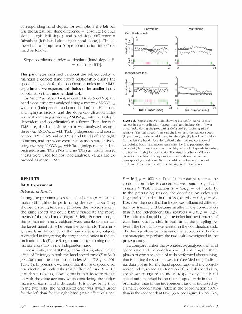
Figure 3. Representative trials showing the performance of onesubject in the coordination (upper trace) and independent (lowertrace) tasks during the pretraining (left) and posttraining (right)sessions. The ball speed (thin straight lines) and the subject speed(larger lines) are depicted in gray for the right (R) hand and in blackfor the left (L) hand. Note the difficulty that the subject showed indissociating both hand movements when he first performed thetasks (left) but then the correct matching of the ball speeds followingthe training (right) for both tasks. The visual feedback (VFback)given to the subject throughout the trials is shown below thecorresponding conditions. Note the whiter background color ofthe L and R half screens after the training in the two tasks.
532 Journal of Cognitive Neuroscience Volume 22, Number 3

F = 130.68, p < .001). In the coordination task, the coor-dination index was not different for the two target ratios(see inset; Figure 4B). In the independent task, the coor-dination index increased when ratios got closer to 1, prob-ably because the smaller difference between the speed ofboth hands made it more difficult to overcome the ten-dency to perform synchronous movements. This resultmight highlight the importance of explicit cues to guideseparate movements of the two hands during bimanualindependent tasks. Importantly, in the independent task,the coordination index was not higher for noninteger ballspeed ratios (e.g., 2.5, 3.5, 4.5) than for integer ratios (e.g.,2, 3, 4; see inset of Figure 4B). This finding rules out the
possibility that the higher coordination index found in theindependent task was due to the presence of more com-plex, noninteger ratios when compared with the coordina-tion task.
Further evidence that subjects used distinct strategiesto perform the two tasks comes from the differential influ-ence of the relative hand speed on the hand speed error(Figure 4C). Indeed, the ANOVA showed a significant Task×Fastest Hand × Hand interaction (F= 89.47, p< .001) onthe hand speed error: Whereas the hand speed error wassystematically smaller for the fastest hand in the coordina-tion task (t tests, all p< .001), no such effect was found inthe independent task (see Figure 4C).
Figure 4. Individual data forthe hand speed ratio (A) andcoordination index (B) as wellas the mean and SD of handspeed error (C) for all thephases of constant speed (n =60) of all subjects (n = 12),as a factor of the ball speed ratio(A), for the coordination (left)and the independent (right)tasks. A ratio <0 representsconditions where the lefthand was faster than the righthand whereas a ratio >0represents the reverseconditions. Abs = absolute;*p < .05; L = left; R = right.
Duque et al. 533

Imaging Results
Brain activation during bimanual tasks versus rest. Wefirst identified the entire network activated during biman-ual movements. To do so, we determined the regionsshowing a stronger activation during the bimanual tasksthan in the rest condition (see Table 2). We found a bi-lateral network, including the primary sensorimotor handarea (S1/M1) extending to the dorsal premotor cortex,SMA, inferior parietal gyrus, basal ganglia, and cerebel-lum. In addition, in the right hemisphere, we found anactivation of the ventral premotor cortex, the dorsolateralprefrontal cortex, and the middle and superior temporalgyri (MTG and STG; see Table 2). This bimanual networkis consistent with that reported in previous studies (for areview, see Swinnen & Wenderoth, 2004).
Areas specifically involved in the coordination task.We found three areas in the right hemisphere that showeda greater activation in the coordination than in the inde-pendent task, namely, M1, t(11) = 5.23, p = .0003, SMA,t(11) = 5.14, p = .0003, and STG, t(11) = 5.85, p =.0001 (see Table 3 and Figure 5).
Areas specifically involved in the independent task.Areas showing a stronger activation in the independentthan in the coordination task were the right inferior pari-etal gyrus, t(11) = 5.80, p= .0001, MTG, t(11) = 6.69, p=.00003, and right fusiform gyrus, t(11) = 5.84, p = .0001(see Table 3 and Figure 6).
TMS Experiment
In control trials (no TMS), we found, consistent with fMRIdata, that the hand speed error computed during thephases of constant speed was comparable in both tasks:The hand speed error was 16.4 ± 5.8% and 18.5 ± 13.9%for the left and the right hands, respectively, in the coor-dination task and 16.4 ± 3.7% and 16.2 ± 3.4% for theindependent task (n = 9). In contrast, we found thatthe hand slope error was larger in the independent thanin the coordinated task, suggesting that speed changeswere easy to perform when occurring in both hands si-multaneously (ANOVARM, main Task effect, F = 29.6, p =.001). Consistently, the slope coordination index (seeMethods) was also higher in the independent than in thecoordination task (F = 44.5, p < .001).
Virtual lesions of the right STG specifically modified thehand slope error in the coordination task, without affect-ing any movement parameters in the independent task.ANOVARM showed a significant effect of Task (F = 11.3,p = .01), TMS (F = 9.8, p = .02), Hand (F = 16.0, p =.004), and a significant Task × TMS interaction (F = 5.6,p= .04) on the hand slope error (see Figure 7A). Post hocanalyses indicated that the virtual lesions of the right STGincreased the hand slope error in the coordination taskwith respect to the no TMS condition, for both the lefthand (paired t test, p= .057) and the right hand ( p= .001).
TMS applied over the right M1 yielded a significant ef-fect of Task (ANOVARM, F= 23.5, p= .001), TMS (F= 7.6,p = .025), Hand (F = 11.7, p = .009), and a Task × TMSinteraction (F = 10.6, p = .01) on the hand slope error. Inparticular, we found a significant decrease in the handslope error of the left hand following right M1 virtuallesions but only in the independent task (paired t test,p = .01; Figure 7B). In other words, a transient disruptionof the right M1 function improved the ability of the lefthand to adapt to the speed changes in the independenttask but had no effect on the coordination task perfor-mance. ANOVARM also showed a significant Task × TMSinteraction (F = 8.1, p = .022) on the slope coordinationindex, and post hoc analyses indicated that rTMS appliedover right M1 decreased the slope coordination index dur-ing the speed change only in the independent task ( p =.006; Figure 7B).No effects were found after virtual lesions of SMA or
when rTMS was applied over the control sites (left M1and left STG).
DISCUSSION
The aim of the present study was to determine the neuralcorrelates of one of the most critical—and probably theleast understood—processes underlying skilled actions,that is, the coordination between both hands during theperformance of bimanual movements. Our fMRI resultsshow that three cortical areas, all located in the right hemi-sphere, are more active in a coordination task than in anindependent task, namely, the STG, the SMA, and the M1.The causal relevance of the activity in these three regionsfor the bimanual coordination was then investigated by us-ing TMS to interfere transiently and reversibly with theirfunction.
Neural Correlates of Bimanual Coordination
Many functional imaging studies have already examinedthe neural correlates of bimanual movements (Puttemanset al., 2005; Debaere et al., 2001, 2004b; De Weerd et al.,2003; Debaere, Wenderoth, Sunaert, Van Hecke, &Swinnen, 2003; Meyer-Lindenberg et al., 2002; Toyokuraet al., 1999; Sadato et al., 1997) with a particular interestfor independent tasks (Wenderoth et al., 2004, 2005a,2005b). However, to our knowledge, very few studies havetried to identify the cortical areas dealing specifically withthe coordination aspect of bimanual movements ( Jantzenet al., 2008; Debaere et al., 2003).Our finding that right STG was more activated in the co-
ordination than in the independent task was unexpectedbecause its contribution to bimanual movements has neverbeen really emphasized before, although many studies re-ported an activation of this area in complex spatial or rhyth-mic coordination movements (Oullier, Jantzen, Steinberg,& Kelso, 2005; Debaere et al., 2004b; Ullen, Forssberg, &
534 Journal of Cognitive Neuroscience Volume 22, Number 3

Table 2. General Motor Network Activated during the Bimanual Tasks
Brain Regions
Coordination Task Independent Task
Peak ActivationCoordinates
Peak ActivationCoordinates
x y z t(11) x y z t(11)
Frontal lobe
R sensorimotor region (BA 3/4), S1/M1 27 −31 46 8.95 33 −25 49 9.00
48 −19 37 11.13
L −33 −22 46 8.60 −33 −22 46 8.68
−48 −19 46 7.37
R inferior frontal gyrus (BA 44), PMv 42 17 22 10.19 45 11 22 9.71
(BA 45), DLPFc 36 35 13 6.67 39 32 10 6.89
R middle frontal gyrus (BA 46) 34 28 25 10.00 30 29 25 11.46
R superior frontal gyrus (BA 6), SMA 3 −25 46 5.70 1 −25 46 4.96
L/MID 0 −25 49 5.34 0 −22 49 5.01
−3 −13 58 4.85
L superior frontal gyrus (BA 4/6), M1 and PMd −30 −16 64 6.22 −27 −16 61 7.41
−18 −25 61 5.67 −18 −25 61 6.07
Parietal lobe
R inferior parietal gyrus (BA 39/40) 30 −58 37 7.77 33 −55 34 8.29
42 −46 46 10.93 45 −40 46 9.10
54 −34 25 6.77
L −45 −43 40 7.50 −45 −43 40 9.46
−24 −64 31 4.92 −27 −58 46 5.94
Temporal lobe
R MTG (BA 37) 45 −64 4 9.14 45 −64 4 10.84
R STG (BA 22) 39 −46 4 6.88 36 −40 7 7.28
Occipital lobe
R middle occipital gyrus (BA 18/19) 39 −64 −5 9.46
27 −79 22 4.94
L −27 −67 −14 7.35 −27 −79 4 6.14
−15 −82 −2 5.62 −24 −76 −8 8.14
Basal ganglia
R 9 8 1 5.90
18 −4 16 4.63
L −21 8 25 6.68 −18 5 25 6.55
−15 2 4 6.09
Duque et al. 535

Ehrsson, 2003; Mayville, Jantzen, Fuchs, Steinberg, & Kelso,2002). In contrast, the possible contribution of right STG,especially its posterior part, to spatial attention has beenmuch debated (Gharabaghi, Fruhmann Berger, Tatagiba,& Karnath, 2006; Meister et al., 2006; Karnath et al., 2005;ThiebautdeSchottenet al., 2005; Ellison, Schindler, Pattison,& Milner, 2004; Mort et al., 2003; Karnath, 2001; Karnath,Ferber, & Himmelbach, 2001). In the present study, it maybe argued that the coordination task required a continuous
monitoring of the spatial location of the two hands to keepthem synchronized tomaintain the required hand speed ra-tio. In fact, the contribution of right STG to this mechanismis compatible with its potential role in spatial attentionand is consistent with the well-known right hemisphericdominance for spatial functions (Serrien, Ivry, & Swinnen,2006; Ellison et al., 2004; Sainburg, 2002). Our TMS studyalso provides evidence for a specific role of right STG inmonitoring this type of spatio-temporal coordination be-cause virtual lesions of this area hampered the subjectsʼability to cope with speed changes in the coordination taskbut not the independent one.Our fMRI results also suggest that right SMA plays a
specific role in bimanual coordination. This area haslong been regarded as a critical region for controlling bi-manual movements (Debaere, Wenderoth, Sunaert, VanHecke, & Swinnen, 2004a; Steyvers et al., 2003; Donchinet al., 2002; Meyer-Lindenberg et al., 2002; Obhi, Haggard,Taylor, & Pascual-Leone, 2002; Serrien, Strens, Oliviero, &Brown, 2002; Immisch, Waldvogel, van Gelderen, &Hallett,2001; Stephan, Binkofski, Halsband, et al., 1999; Stephan,Binkofski, Posse, Seitz, & Freund, 1999; Sadato et al., 1997;Brinkman, 1981, 1984; Laplane, Talairach, Meininger,Bancaud, & Orgogozo, 1977). The extensive connectionsbetween SMA and M1 on both sides and between bothSMA (Liu, Morel, Wannier, & Rouiller, 2002; Rouiller et al.,1994; Porter & Lemon, 1993) are consistent with this view.However, despite the fact that SMA is a plausible candidatefor the implementation of the spatio-temporal goal intothe appropriate bimanual motor commands, TMS-inducedvirtual lesions of this area failed to affect our bimanual tasks,a result that contrasts with previous rTMS studies (Steyverset al., 2003; Meyer-Lindenberg et al., 2002; Obhi et al., 2002;Serrien et al., 2002). However, in these studies, rTMS spe-cifically altered the most “complex” coordination pattern,making it difficult to distinguish between a role of SMA
Table 3. Localization of Peak Activations (TalairachCoordinates) and t Values for the Areas Showing aDifferent Activation Level for the Coordination and theIndependent Tasks
Brain Regions
Peak ActivationCoordinates
t(11)x y z
C > I
R primary motor cortex (BA 4) 31 −27 50 5.23
R SMA (BA 6) 4 −25 48 5.14*
R STG (BA 22) 36 −45 10 5.85
I > C
R MTG (BA 37) 48 −65 2 6.69
R inferior parietal gyrus (BA 40) 30 −51 41 5.80
R fusiform gyrus (BA 19) 29 −48 −13 5.84
C = coordination task; I = independent task; t values indicate the com-parison for the contrast with the largest signal difference ( p < .001,uncorrected).
*Threshold defined at p < .002 (uncorrected) for a cluster size>50 voxels.
Table 2. (continued )
Brain Regions
Coordination Task Independent Task
Peak ActivationCoordinates
Peak ActivationCoordinates
x y z t(11) x y z t(11)
Cerebellum
R cerebellar hemisphere 21 −58 −17 11.05 21 −58 −20 14.10
30 −61 −35 5.81
L −15 −46 −17 14.82
R vermis 6 −52 −17 16.78 6 −52 −17 15.86
1 −61 −2 6.03 1 −61 −2 7.12
L 0 −52 −17 15.54 0 −55 −17 14.19
Location of peak activations and t values for the coordination and independent tasks (false discovery rate < 0.02); L = left; R = right; M1 = primarymotor cortex; PMv = ventral premotor cortex; PMd = dorsal premotor cortex; DLPFc = dorsolateral prefrontal cortex; SI = somatosensory cortex;MTG = middle temporal gyrus; STG = superior temporal gyrus; MD = middle.
536 Journal of Cognitive Neuroscience Volume 22, Number 3

in controlling complex movements and a specific role inmonitoring coordination from these studies. Alternatively,it is worth mentioning that, in the present study, at the be-ginning of each trial, subjects were given an explicit imper-ative cue regarding the required ratio to maintain in thecoordination task; this was done to ensure similar perfor-mance levels despite different cognitive strategies in thetwo tasks. However, this approach, which decreased theneed to retrieve the appropriate coupling ratio in the coor-dination task, might also have diminished the contributionof some areas, such as the SMA, to the coordination task. Inaddition, it is possible that because the SMA area is deeplyburied in the interhemispheric sulcus, it is harder to inter-fere with its activity by using a reasonably low TMS intensity(Meyer-Lindenberg et al., 2002) or with the coil orienta-tion we used. Finally, because other investigators have al-ready managed to stimulate this region at lower intensities(Matsunaga et al., 2005; Civardi, Cantello, Asselman, &Rothwell, 2001), it cannot be excluded that the movementparameters we measured were not sensitive enough to de-tect the effects of small SMA virtual lesions. Further inves-tigations would, therefore, be necessary to isolate thespecific contribution of SMA to bimanual coordination.Finally, our fMRI results indicate that a third area, right
M1, is more active in the coordination than in the indepen-
dent task. However, surprisingly, we found that a virtuallesion of right M1 did not affect the coordination task butrather improved the independent task performance by en-hancing the capacity of the left contralateral hand to copewith the speed changes. One possible explanation for thisapparent contradiction is as follows: In the independenttask, it is plausible that strong inhibitory interactions tookplace between the two motor cortices to minimize biman-ual cross talk (Daffertschofer, Peper, & Beek, 2005; Duque,Mazzocchio, et al., 2005; Ferbert et al., 1992). Consistently,interhemispheric interactions have been shown to varywith the features of the task at hand (Perez & Cohen,2008; Daffertschofer et al., 2005; Andres et al., 1999), andinhibition is known to be stronger from the dominant (left)to nondominant (right) hemisphere than in the oppositedirection (Baumer et al., 2007; Duque et al., 2007; Netz,1999). In the coordination task, this inhibition is likely tobe released to allow a tight coordination between bothhands; therefore, it is sensible to assume that the strongeractivation we found in right M1 when contrasting the acti-vation maps gathered in both conditions was due to adisinhibition in the coordinated task rather than an in-creased activation. This could explain the apparent contra-diction between fMRI and TMS results. Indeed, the betterperformance found in the independent task following right
Figure 5. Mean beta weightand time course of signalchange (% baseline) obtainedfrom regions participating in thecoordination task as evidencedby the conjunction analysis:[(C − R) ∩ (C − I)]. Thresholdset at p < .001 (uncorrected),except for the SMA in whichthe threshold was p < .002(uncorrected) to get asufficiently large cluster size(>50 voxels). C = coordination;I = independence; R = rest.
Duque et al. 537

M1 virtual lesions could have been caused by a change inthe balance between inhibitory and facilitatory interhemi-spheric interactions occurring between both M1 (Duqueet al., 2007, 2008; Duque, Hummel, et al., 2005; Ferbertet al., 1992), favoring the independence between the twohands. However, because this explanation remains largelyspeculative, assessing interhemispheric inhibitory inter-actions using a TMSpaired-pulse design in the coordinationand the independent tasks would be a very interestingissue for future investigation (Duque et al., 2007; Duque,Hummel, et al., 2005; Murase, Duque, Mazzocchio, &Cohen, 2004). Hence, in contrast to the more straight-forward interpretation of our fMRI results, the TMS resultsrather suggest that right M1 is not causally involved in thecoordination aspect of bimanual hand movements, a con-clusion that strengthens even further the benefit of com-bining fMRI and TMS.
It is noteworthy that, in the present study, all areasfound more active in the coordination task were locatedin the nondominant hemisphere. This finding questionsthe long-held belief that the left dominant hemisphere isimportant for bimanual tasks and raises the possibilitythat themotor network underlying bimanual tasks dependson the nature of coordination in play. Here, we used a co-
ordination task where subjects had to reach for a spatio-temporal bimanual goal. Consistently, the nondominanthemisphere is known to be “dominant” in dealingwith suchspatial information. In contrast, it is likely that such a hemi-spheric asymmetry would reverse in bimanual coordinationtasks that do not rely as much on spatio-temporal cues.As a final note, we would like to mention that even if
our two tasks were designed in such a way that the feed-back and the visual stimuli were identical in the two con-ditions, we cannot ascertain that subjects actually usedthis available information similarly in the two tasks. In par-ticular, it is plausible that subjects generated their move-ments more internally in the coordination than in theindependent task and that this slight difference mighthave also influenced our fMRI results. However, note thatthe brain regions evidenced by Debaere et al. (2003), asspecifically involved in internally generated bimanualmovements, differ from those we found in the coordina-tion task, confirming that this factor cannot explain our fMRIand TMS results. In particular, Debaere et al. did not findany difference between STG or M1 activation in internallyand externally generated bimanual movements. As faras SMA is concerned, we cannot rule out the possibilitythat its stronger activation in the coordination condition
Figure 6. Mean beta weightand time course of signalchange (% baseline) obtainedfrom regions participating inthe independent task asevidenced by the conjunctionanalysis: [(I − R) ∩ (I − C)].Threshold set at p < .001(uncorrected). C= coordination;I = independence; R = rest;MTG = middle temporalgyrus; IPG = inferior parietalgyrus; FG = fusiform gyrus.
538 Journal of Cognitive Neuroscience Volume 22, Number 3

was partly due to a higher reliance on internally generatemovements in this task (Debaere et al., 2003; Cunnington,Windischberger, Deecke, & Moser, 2002; Deiber, Honda,Ibanez, Sadato, & Hallett, 1999).
Methodological Consideration:The Bimanual Tasks
A key point of the present study was to isolate the controlmechanisms specifically involved in the “coordination as-pect” of bimanual movements; the coordination aspectrefers to the processes underlying interactions that oc-curred between both hands to achieve the bimanual goal,that is, a given speed ratio between hands. Although thisaspect is one of the most important prerequisites to per-form efficient bimanual movements, it is also the trickiestone to investigate experimentally because of the difficultyto isolate it from all other variables known to affect indi-vidual movements during bimanual actions (complexity,learning stage, feedback procedure, etc.). Here, we at-tempted to solve this problem by using an original exper-imental design in which we used two bimanual tasksdiffering only from the nature of the cognitive controlunderlying their performance: In the coordination task,subjects were explicitly asked to reach a unique bimanualgoal, namely, to maintain a given speed ratio between thetwo hand motions, whereas in the independent task, thetwo hands had to remain as uncoupled as possible toreach distinct goals, that is, different speeds. Importantly,we minimized the differences between the two tasks for
all other aspects: hand speed, performance level, visualdisplay, feedback procedure, etc.
Ourmain concern in designing these tasks was to reducethe possibility that subjects could also use, even if not ex-plicitly provided, the hand speed ratios to perform the in-dependent task, hampering the comparison between thetwo conditions. To avoid this, we selected, for the indepen-dent task, hand speeds that led to noninteger ratios, vary-ing from 1.1 to 4.8; in contrast to the ratios used in thecoordination task, these noninteger ratios are extremelydifficult to produce explicitly, even for expert musicians(Ullen et al., 2003; Peper, Beek, & van Wieringen, 1995;Summers, Todd, & Kim, 1993). In addition, the hand speedratio was modified twice during each trial (once in the TMSexperiment) to make it even more difficult for subjects touse a coordination strategy based on the hand speed ratioin the independent condition. Therefore, it is sensible toassume that subjects had no other option than using dis-tinct strategies to perform the independent and the coor-dinated tasks. This assumption was supported by thesubtle differences we found between the performancesof the two tasks. Indeed, despite an identical amount ofhand speed error in both tasks, the coordination indexwas larger in the independent than in the coordinationtask, an observation consistent with the hypothesis thatsubjects did not control this variable explicitly in the inde-pendent task as they did in the coordination task. Addition-ally, in the coordination task, the hand speed error wasalways smaller for the fastest hand—likely the leadingone ( Johansson et al., 2006)—when compared with theother hand. Such an interaction between the speed errors
Figure 7. Mean and SD of the left and the right hand hand slope error (left and middle traces, respectively) and of the slope coordinationindex (right trace) in the two bimanual tasks (coordination and independence), with (gray bars) and without TMS (white bars) over the right STG(A) and the right M1 (B).
Duque et al. 539

in the two hands was not found in the independent task,suggesting further that, in this condition, the two handmovements were controlled separately. Altogether, theseobservations corroborate the view that subjects used differ-ent cognitive strategies to perform the two tasks, yet iden-tical in most other respects.
Moreover, it is noteworthy that the fact that subjectswere able to perform the independent task with the samelevel of accuracy as the coordination task confirms thatinterference during independent tasks can be attenuatedby training, possibly by strengthening strategies that permitto cancel out the bimanual cross talk (Oliveira & Ivry,2008; Rosenbaum et al., 2006; Diedrichsen et al., 2001).
Finally, in the present study, we only focused on oneparticular type of bimanual coordination, based on theexplicit control of an integrated spatio-temporal goal and,therefore, our results are unlikely to generalize to other co-ordination strategies such as those, for example, in whichbimanual movements are synchronized by external visualgoals. In addition, we cannot completely rule out the possi-bility that subjects somewhat relied on such visual coordi-nation strategy to perform the independent task, althoughwe minimized this option by using a distinct feedback foreach hand, and the behavioral results also support the useof an uncoupled strategy in the independent task. In anycase, we think our experimental design was optimal tohighlight the neural correlates of a specific coordinationstrategy based on the integration of a spatio-temporal goal.
Conclusions
Bimanual coordination is critical for most daily motor ac-tions, and still, its neural correlates remain unclear. A bet-ter knowledge of how hand representations interact toachieve complex tasks could yield substantial improve-ment in the fields of rehabilitation, sport science, corticalprosthetics, and possibly neurosurgery.
The combined use of fMRI and TMS enabled us todemonstrate the specific contribution of some corticalareas in the control of the coordination aspect of biman-ual movements. We propose that right STG plays a role inmonitoring bimanual spatio-temporal goals. Regardingright SMA, its contribution to the implementation of thesespatio-temporal goals into adequate motor commands isplausible but would require further investigation. Finally,in contrast to the fMRI results, the TMS study suggests thatright M1 is not causally involved in the coordination aspectof bimanual movements but that its activity is modulatedby inhibitory influences, likely to be stronger in the inde-pendent than in the coordination task.
Acknowledgments
We are grateful to M. Penta, C. Schiltz, and N. Wenderoth forhelping us with the experimental design and A. Heinecke withthe analysis of the imaging data. We would also like to thank
R. Ivry and J. Diedrichsen for their critical comments on an ear-lier version of this manuscript. This work was supported bygrants from the ARC grant 07/12-007 (Communauté Françaisede Belgique-Actions de Recherche Concertées), the “FondsSpéciaux de Recherche” (FSR) of the Université catholique deLouvain, the “Fonds de la Recherche Scientifique Médicale”(FRSM), and the “Fondation Médicale Reine Elisabeth” (FMRE).J. D. and M. D. are research fellows at the Belgian National Fundsfor Scientific Research (FNRS).
Reprint requests should be sent to Julie Duque, Laboratory ofNeurophysiology, Institute of Neuroscience, Université catholiquede Louvain, Avenue Hippocrate, 54, 1200 Brussels, Belgium, or viae-mail: [email protected].
REFERENCES
Aboitiz, F., Ide, A., & Olivares, R. (2003). Corpus callosummorphology in relation to cerebral asymmetries in thepostmortem human. In The parallel brain: The cognitiveneuroscience of the corpus callosum (pp. 33–46).Cambridge, MA: MIT Press.
Andres, F. G., Mima, T., Schulman, A. E., Dichgans, J., Hallett,M., & Gerloff, C. (1999). Functional coupling of humancortical sensorimotor areas during bimanual skill acquisition.Brain, 122, 855–870.
Baumer, T., Dammann, E., Bock, F., Kloppel, S., Siebner,H. R., & Munchau, A. (2007). Laterality of interhemisphericinhibition depends on handedness. ExperimentalBrain Research, 180, 195–203.
Boynton, G. M., Engel, S. A., Glover, G. H., & Heeger, D. J.(1996). Linear systems analysis of functional magneticresonance imaging in human V1. Journal of Neuroscience,16, 4207–4221.
Brinkman, C. (1981). Lesions in supplementary motor areainterfere with a monkeyʼs performance of a bimanualcoordination task. Neuroscience Letters, 27, 267–270.
Brinkman, C. (1984). Supplementary motor area of themonkeyʼs cerebral cortex: Short- and long-term deficitsafter unilateral ablation and the effects of subsequentcallosal section. Journal of Neuroscience, 4, 918–929.
Civardi, C., Cantello, R., Asselman, P., & Rothwell, J. C. (2001).Transcranial magnetic stimulation can be used to testconnections to primary motor areas from frontal and medialcortex in humans. Neuroimage, 14, 1444–1453.
Cunnington, R., Windischberger, C., Deecke, L., & Moser, E.(2002). The preparation and execution of self-initiatedand externally-triggered movement: A study of event-relatedfMRI. Neuroimage, 15, 373–385.
Daffertschofer, A., Peper, C. L., & Beek, P. J. (2005).Stabilization of bimanual coordination due to activeinterhemispheric inhibition: A dynamical account.Biological Cybernetics, 92, 101–109.
Davare, M., Andres, M., Clerget, E., Thonnard, J. L., & Olivier, E.(2007). Temporal dissociation between hand shaping andgrip force scaling in the anterior intraparietal area. Journalof Neuroscience, 27, 3974–3980.
Davare, M., Andres, M., Cosnard, G., Thonnard, J. L., &Olivier, E. (2006). Dissociating the role of ventral anddorsal premotor cortex in precision grasping. Journal ofNeuroscience, 26, 2260–2268.
De Weerd, P., Reinke, K., Ryan, L., McIsaac, T., Perschler,P., Schnyer, D., et al. (2003). Cortical mechanisms foracquisition and performance of bimanual motor sequences.Neuroimage, 19, 1405–1416.
Debaere, F., Swinnen, S. P., Beatse, E., Sunaert, S., Van Hecke,P., & Duysens, J. (2001). Brain areas involved in interlimbcoordination: A distributed network.Neuroimage, 14, 947–958.
540 Journal of Cognitive Neuroscience Volume 22, Number 3

Debaere, F., Wenderoth, N., Sunaert, S., Van Hecke, P., &Swinnen, S. P. (2003). Internal vs external generationof movements: Differential neural pathways involved inbimanual coordination performed in the presence orabsence of augmented visual feedback. Neuroimage,19, 764–776.
Debaere, F., Wenderoth, N., Sunaert, S., Van Hecke, P., &Swinnen, S. P. (2004a). Cerebellar and premotor function inbimanual coordination: Parametric neural responsesto spatiotemporal complexity and cycling frequency.Neuroimage, 21, 1416–1427.
Debaere, F., Wenderoth, N., Sunaert, S., Van Hecke, P., &Swinnen, S. P. (2004b). Changes in brain activationduring the acquisition of a new bimanual coodination task.Neuropsychologia, 42, 855–867.
Deiber, M. P., Honda, M., Ibanez, V., Sadato, N., & Hallett, M.(1999). Mesial motor areas in self-initiated versus externallytriggered movements examined with fMRI: Effect ofmovement type and rate. Journal of Neurophysiology,81, 3065–3077.
Diedrichsen, J., Hazeltine, E., Kennerley, S., & Ivry, R. B.(2001). Moving to directly cued locations abolishes spatialinterference during bimanual actions. Psychological Science,12, 493–498.
Donchin, O., Gribova, A., Steinberg, O., Mitz, A. R.,Bergman, H., & Vaadia, E. (2002). Single-unit activityrelated to bimanual arm movements in the primary andsupplementary motor cortices. Journal of Neurophysiology,88, 3498–3517.
Duque, J., Hummel, F., Celnik, P., Murase, N., Mazzocchio, R.,& Cohen, L. G. (2005). Transcallosal inhibition in chronicsubcortical stroke. Neuroimage, 28, 940–946.
Duque, J., Mazzocchio, R., Dambrosia, J., Murase, N.,Olivier, E., & Cohen, L. G. (2005). Kinematically specificinterhemispheric inhibition operating in the processof generation of a voluntary movement. Cerebral Cortex,15, 588–593.
Duque, J., Mazzocchio, R., Stefan, K., Hummel, F., Olivier, E.,& Cohen, L. G. (2008). Memory formation in the motorcortex ipsilateral to a training hand. Cerebral Cortex,18, 1395–1406.
Duque, J., Murase, N., Celnik, P., Hummel, F., Harris-Love,M., Mazzocchio, R., et al. (2007). Intermanual differencesin movement-related interhemispheric inhibition. Journalof Cognitive Neuroscience, 19, 204–213.
Ellison, A., Schindler, I., Pattison, L. L., & Milner, A. D. (2004).An exploration of the role of the superior temporal gyrusin visual search and spatial perception using TMS. Brain,127, 2307–2315.
Ferbert, A., Priori, A., Rothwell, J. C., Day, B. L., Colebatch,J. G., & Marsden, C. D. (1992). Interhemispheric inhibitionof the human motor cortex. Journal of Physiology, 453,525–546.
Franz, E. A. (1997). Spatial coupling in the coordinationof complex actions. Quarterly Journal of ExperimentalPsychology A, 50, 684–704.
Franz, E. A., Zelaznik, H. N., Swinnen, S. S., & Walter, C.(2001). Spatial conceptual influences on the coordinationof bimanual actions: When a dual task becomes a singletask. Journal of Motor Behavior, 33, 103–112.
Friston, K. J., Frith, C. D., Turner, R., & Frackowiak, R. S.(1995). Characterizing evoked hemodynamics with fMRI.Neuroimage, 2, 157–165.
Friston, K. J., Penny, W. D., & Glaser, D. E. (2005). Conjunctionrevisited. Neuroimage, 25, 661–667.
Genovese, C. R., Lazar, N. A., & Nichols, T. (2002). Thresholdingof statistical maps in functional neuroimaging using thefalse discovery rate. Neuroimage, 15, 870–878.
Gharabaghi, A., Fruhmann Berger, M., Tatagiba, M., &Karnath, H. O. (2006). The role of the right superiortemporal gyrus in visual search-insights from intraoperativeelectrical stimulation. Neuropsychologia, 44, 2578–2581.
Immisch, I., Waldvogel, D., van Gelderen, P., & Hallett, M.(2001). The role of the medial wall and its anatomicalvariations for bimanual antiphase and in-phasemovements. Neuroimage, 14, 674–684.
Jantzen, K. J., Oullier, O., & Scott Kelso, J. A. (2008).Neuroimaging coordination dynamics in the sportsciences. Methods, 45, 325–335.
Johansson, R. S., Theorin, A., Westling, G., Andersson, M.,Ohki, Y., & Nyberg, L. (2006). How a lateralized brainsupports symmetrical bimanual tasks. PLoS Biology, 4, e158.
Karnath, H. O. (2001). New insights into the functionsof the superior temporal cortex. Nature ReviewsNeuroscience, 2, 568–576.
Karnath, H. O., Ferber, S., & Himmelbach, M. (2001).Spatial awareness is a function of the temporal notthe posterior parietal lobe. Nature, 411, 950–953.
Karnath, H. O., Zopf, R., Johannsen, L., Berger, M. F., Nagele, T.,& Klose, U. (2005). Normalized perfusion MRI to identifycommon areas of dysfunction: Patients with basal ganglianeglect. Brain, 128, 2462–2469.
Laplane, D., Talairach, J., Meininger, V., Bancaud, J., &Orgogozo, J. M. (1977). Clinical consequences ofcorticectomies involving the supplementary motor areain man. Journal of the Neurological Sciences, 34, 301–314.
Liu, J., Morel, A., Wannier, T., & Rouiller, E. M. (2002).Origins of callosal projections to the supplementarymotor area (SMA): A direct comparison between pre-SMAand SMA-proper in macaque monkeys. Journal ofComparative Neurology, 443, 71–85.
Matsunaga, K., Maruyama, A., Fujiwara, T., Nakanishi, R.,Tsuji, S., & Rothwell, J. C. (2005). Increased corticospinalexcitability after 5 Hz rTMS over the human supplementarymotor area. Journal of Physiology, 562, 295–306.
Mayville, J. M., Jantzen, K. J., Fuchs, A., Steinberg, F. L., &Kelso, J. A. (2002). Cortical and subcortical networksunderlying syncopated and synchronized coordinationrevealed using fMRI. Functional magnetic resonanceimaging. Human Brain Mapping, 17, 214–229.
Mechsner, F., Kerzel, D., Knoblich, G., & Prinz, W. (2001).Perceptual basis of bimanual coordination. Nature,414, 69–73.
Meister, I. G., Wienemann, M., Buelte, D., Grunewald, C.,Sparing, R., Dambeck, N., et al. (2006). Hemiextinctioninduced by transcranial magnetic stimulation over theright temporo-parietal junction. Neuroscience, 142,119–123.
Meyer-Lindenberg, A., Ziemann, U., Hajak, G., Cohen, L.,& Berman, K. F. (2002). Transitions between dynamicalstates of differing stability in the human brain. Proceedingsof the National Academy of Sciences, U.S.A., 99,10948–10953.
Mort, D. J., Malhotra, P., Mannan, S. K., Rorden, C., Pambakian,A., Kennard, C., et al. (2003). The anatomy of visualneglect. Brain, 126, 1986–1997.
Murase, N., Duque, J., Mazzocchio, R., & Cohen, L. G. (2004).Influence of interhemispheric interactions on motorfunction in chronic stroke. Annals of Neurology, 55,400–409.
Netz, J. (1999). Asymmetry in transcallosal inhibition.Electroencephalography and Clinical Neurophysiology.Supplement, 51, 137–144.
Nichols, T., Brett, M., Andersson, J., Wager, T., & Poline,J. B. (2005). Valid conjunction inference with the minimumstatistic. Neuroimage, 25, 653–660.
Duque et al. 541

Noirhomme, Q., Ferrant, M., Vandermeeren, Y., Olivier, E.,Macq, B., & Cuisenaire, O. (2004). Registration andreal-time visualization of transcranial magnetic stimulationwith 3-D MR images. IEEE Transactions on BiomedicalEngineering, 51, 1994–2005.
Obhi, S. S., Haggard, P., Taylor, J., & Pascual-Leone, A.(2002). rTMS to the supplementary motor area disruptsbimanual coordination. Motor Control, 6, 319–332.
Oliveira, F. T. P., & Ivry, R. B. (2008). The representationof action: Insights from bimanual coordination. CurrentDirections in Psychological Science, 17, 130–135.
Oullier, O., Jantzen, K. J., Steinberg, F. L., & Kelso, J. A.(2005). Neural substrates of real and imagined sensorimotorcoordination. Cerebral Cortex, 15, 975–985.
Peper, C. E., Beek, P. J., & van Wieringen, P. C. (1995).Frequency-induced phase transitions in bimanual tapping.Biological Cybernetics, 73, 301–309.
Perez, M. A., & Cohen, L. G. (2008). Mechanisms underlyingfunctional changes in the primary motor cortex ipsilateralto an active hand. Journal of Neuroscience, 28, 5631–5640.
Porter, R., & Lemon, R. N. (1993). Corticospinal functionand voluntary movement. Monographs of the PhysiologicalSociety, 45.
Price, C. J., & Friston, K. J. (1997). Cognitive conjunction:A new approach to brain activation experiments.Neuroimage, 5, 261–270.
Puttemans, V., Wenderoth, N., & Swinnen, S. P. (2005).Changes in brain activation during the acquisition of amultifrequency bimanual coordination task: From thecognitive stage to advanced levels of automaticity.Journal of Neuroscience, 25, 4270–4278.
Rosenbaum, D. A., Dawson, A. M., & Challis, J. H. (2006).Haptic tracking permits bimanual independence. Journalof Experimental Psychology: Human Perception andPerformance, 32, 1266–1275.
Rossini, P. M., Barker, A. T., Berardelli, A., Caramia, M. D.,Caruso, G., Cracco, R. Q., et al. (1994). Non-invasive electricaland magnetic stimulation of the brain, spinal cord androots: Basic principles and procedures for routineclinical application. Report of an IFCN committee.Electroencephalography and Clinical Neurophysiology,91, 79–92.
Rouiller, E. M., Babalian, A., Kazennikov, O., Moret, V., Yu,X. H., & Wiesendanger, M. (1994). Transcallosal connectionsof the distal forelimb representations of the primary andsupplementary motor cortical areas in macaque monkeys.Experimental Brain Research, 102, 227–243.
Sadato, N., Yonekura, Y., Waki, A., Yamada, H., & Ishii, Y.(1997). Role of the supplementary motor area and theright premotor cortex in the coordination of bimanual fingermovements. Journal of Neuroscience, 17, 9667–9674.
Sainburg, R. L. (2002). Evidence for a dynamic-dominancehypothesis of handedness. Experimental Brain Research,142, 241–258.
Serrien, D. J., Bogaerts, H., Suy, E., & Swinnen, S. P. (1999).The identification of coordination constraints acrossplanes of motion. Experimental Brain Research, 128,250–255.
Serrien, D. J., Ivry, R. B., & Swinnen, S. P. (2006). Dynamicsof hemispheric specialization and integration in thecontext of motor control. Nature Reviews Neuroscience,7, 160–166.
Serrien, D. J., Strens, L. H., Oliviero, A., & Brown, P. (2002).Repetitive transcranial magnetic stimulation of thesupplementary motor area (SMA) degrades bimanual
movement control in humans. Neuroscience Letters,328, 89–92.
Stephan, K. M., Binkofski, F., Halsband, U., Dohle, C.,Wunderlich, G., Schnitzler, A., et al. (1999). The role ofventral medial wall motor areas in bimanual co-ordination.A combined lesion and activation study. Brain, 122,351–368.
Stephan, K. M., Binkofski, F., Posse, S., Seitz, R. J., & Freund,H. J. (1999). Cerebral midline structures in bimanualcoordination. Experimental Brain Research, 128,243–249.
Steyvers, M., Etoh, S., Sauner, D., Levin, O., Siebner, H. R.,Swinnen, S. P., et al. (2003). High-frequency transcranialmagnetic stimulation of the supplementary motor areareduces bimanual coupling during anti-phase but notin-phase movements. Experimental Brain Research,151, 309–317.
Summers, J. J., Todd, J. A., & Kim, Y. H. (1993). The influenceof perceptual and motor factors on bimanual coordinationin a polyrhythmic tapping task. Psychological Research,55, 107–115.
Swinnen, S. P. (2002). Intermanual coordination: Frombehavioural principles to neural-network interactions.Nature Reviews Neuroscience, 3, 348–359.
Swinnen, S. P., & Wenderoth, N. (2004). Two hands, one brain:Cognitive neuroscience of bimanual skill. Trends inCognitive Sciences, 8, 18–25.
Talairach, J., & Tournoux, P. (1988). Co-planar stereotaxicatlas of the human brain, 3-dimensional proportionalsystem: An approach to cerebral imaging. New York:Thieme.
Thiebaut de Schotten, M., Urbanski, M., Duffau, H., Volle, E.,Levy, R., Dubois, B., et al. (2005). Direct evidence fora parietal-frontal pathway subserving spatial awarenessin humans. Science, 309, 2226–2228.
Toyokura, M., Muro, I., Komiya, T., & Obara, M. (1999). Relationof bimanual coordination to activation in the sensorimotorcortex and supplementary motor area: Analysis usingfunctional magnetic resonance imaging. Brain ResearchBulletin, 48, 211–217.
Ullen, F., Forssberg, H., & Ehrsson, H. H. (2003). Neuralnetworks for the coordination of the hands in time.Journal of Neurophysiology, 89, 1126–1135.
Wassermann, E. M. (1998). Risk and safety of repetitivetranscranial magnetic stimulation: Report and suggestedguidelines from the International Workshop on theSafety of Repetitive Transcranial Magnetic Stimulation,June 5–7, 1996. Electroencephalography and ClinicalNeurophysiology, 108, 1–16.
Wenderoth, N., Debaere, F., Sunaert, S., & Swinnen, S. P.(2005a). The role of anterior cingulate cortex and precuneusin the coordination of motor behaviour. EuropeanJournal of Neuroscience, 22, 235–246.
Wenderoth, N., Debaere, F., Sunaert, S., & Swinnen, S. P.(2005b). Spatial interference during bimanual coordination:Differential brain networks associated with control ofmovement amplitude and direction. Human BrainMapping, 26, 286–300.
Wenderoth, N., Debaere, F., Sunaert, S., van Hecke, P.,& Swinnen, S. P. (2004). Parieto-premotor areas mediatedirectional interference during bimanual movements.Cerebral Cortex, 14, 1153–1163.
Wenderoth, N., Puttemans, V., Vangheluwe, S., & Swinnen,S. P. (2003). Bimanual training reduces spatial interference.Journal of Motor Behavior, 35, 296–308.
542 Journal of Cognitive Neuroscience Volume 22, Number 3