Methodology of Islamic psychotherapy in Islamic boarding school Suryalaya Tasik Malaya
Upload
khangminh22Category
view
1download
0
MOLECULAR EPIDEMIOLOGY OF
ENTAMOEBA HISTOLYTICA, ENTAMOEBA
DISPAR AND ENTAMOEBA MOSHKOVSKII
INFECTIONS IN RURAL YEMEN
MONA ABDULLAH MOHAMMED Al-AREEQI
FACULTY OF
MEDICINE
UNIVERSITY OF
MALAYA KUALA
LUMPUR
2018
Univers
ity of
Mala
ya

MOLECULAR EPIDEMIOLOGY OF ENTAMOEBA
HISTOLYTICA, ENTAMOEBA DISPAR AND ENTAMOEBA
MOSHKOVSKII INFECTIONS IN RURAL YEMEN
MONA ABDULLAH MOHAMMED Al-AREEQI
DISSERTATION SUBMITTED IN FULFILMENT OF THE
REQUIREMENTS FOR THE DEGREE OF MASTER OF
MEDICAL SCIENCE
FACULTY OF MEDICINE
UNIVERSITY OF MALAYA
KUALA LUMPUR
2018
Univers
ity of
Mala
ya

Univers
ity of
Mala
ya

iii
ABSTRACT
Intestinal amoebiasis is highly prevalent in Yemen particularly in rural areas; however
there is a great scarcity of information on the prevalence of species-specific Entamoeba
infections in Yemen and many other countries due to the re-description of pathogenic
Entamoeba histolytica and non-pathogenic E. dispar and E. moshkovskii. Therefore, this
community-based study is the first to provide information on the true prevalence of E.
histolytica, E. dispar and E. moshkovskii infections in rural communities in Yemen. The
study also aimed to examine the association of these Entamoeba infections with some
potential risk factors. A total of 605 stool samples from four provinces namely Sana’a,
Dhamar, Taiz and Hodiedah were randomly collected and examined by wet mount,
formalin-ether sedimentation, trichrome staining and nested multiplex PCR techniques.
Demographic, socioeconomic and environmental information was collected by using a
pre-tested questionnaire. Overall, 324 (53.6%) of the samples were positive for
Entamoeba cysts and/or trophozoites by microscopic examination. The prevalence was
significantly higher among male participants compared to female (P = 0.008). An age-
dependency distribution was also observed (P< 0.001). Using molecular analysis, it was
found that 20.2%, 15.7% and 18.2% of the samples were positive for E. histolytica, E.
dispar and E. moshkovskii, respectively. Multivariate analysis showed different sets of
species-specific risk factors among these communities. Educational level was identified
as the significant risk factor for E. histolytica; age and gender were the significant risk
factors for E. moshkovskii; and sources of drinking water and consumption of unwashed
vegetables were the significant risk factors for E. dispar. Moreover, living in
coastal/foothill areas and presence of other infected family members were risk factors
for both E. histolytica and E. moshkovskii infections. The present study provides new
insight into the distribution and risk factors of intestinal amoebiasis in Yemen and
reveals that Entamoeba spp. infection is highly prevalent among these communities
Univers
ity of
Mala
ya

iv
with E. histolytica, E. dispar and E. moshkovskii differentiated for the first time.Hence,
the study emphasizes the need for molecular methods in the diagnosis of infections and
for conducting a large-scaled study throughout Yemen to determine the actual species-
specific prevalence of Entamoeba spp. Moreover, identifying and treating infected
family members, providing health education pertinent to good personal and food
hygiene practices, and providing clean drinking water should be considered in
developing a strategy to control intestinal parasitic infections in these communities,
particularly in the coastal/foothill areas of the country.
Univers
ity of
Mala
ya

v
ABSTRAK
Amoebiasis usus sangat umum di Yaman khususnya di kawasan luar bandar,
bagaimanapun terdapat satu kekurangan maklumat yang besar mengenai kelaziman
spesies khusus jangkitan Entamoeba di Yaman dan negara-negara lain disebabkan oleh
penerangan semula daripada Entamoeba histolytica patogenik dan bukan patogen E.
dispar dan E. moshkovskii. Oleh itu, kajian berasaskan komuniti ini adalah yang
pertama untuk memberi maklumat tentang kelaziman sebenar E. histolytica, E. dispar
dan jangkitan E. moshkovskii dalam kalangan masyarakat luar bandar di Yaman. Kajian
ini juga bertujuan untuk mengkaji kaitan antara jangkitan Entamoeba dengan beberapa
faktor risiko yang berpotensi. Sebanyak 605 sampel najis daripada empat wilayah iaitu
Sana'a, Dhamar, Taiz dan Hodiedah dikumpulkan secara rawak dan diperiksa melalui
wet mount, pemendapan formalin-eter, pewarnaan trichrome dan teknik PCR multipleks
bersarang. Maklumat demografi, sosioekonomi dan alam sekitar telah dikumpulkan
dengan menggunakan soal selidik pra-diuji. Secara keseluruhan, 324 (53.6%) daripada
sampel adalah positif untuk sista Entamoeba dan/ atau trophozoites melalui
pemeriksaan mikroskopik. Kelaziman adalah jauh lebih tinggi di kalangan peserta lelaki
berbanding perempuan (P = 0.008). Pengagihan usia pergantungan juga diperhatikan
(P<0.001). Dengan menggunakan analisis molekul, ia telah mendapati bahawa 20.2%,
15.7% dan 18.2% daripada sampel adalah positif untuk E. histolytica, E. dispar dan E.
moshkovskii, masing-masing. Analisis multivariat menunjukkan set faktor risiko spesies
khusus di kalangan masyarakat ini. Tahap pendidikan telah dikenal pasti sebagai faktor
risiko yang penting bagi E. histolytica; umur dan jantina merupakan faktor risiko yang
signifikan bagi E. moshkovskii; dan sumber-sumber air minum dan penggunaan sayur-
sayuran yang tidak dibasuh merupakan faktor risiko yang signifikan bagi E. dispar.
Lebih-lebih lagi, yang tinggal di kawasan pantai/ kaki bukit dan kehadiran ahli keluarga
lain yang dijangkiti merupakan faktor risiko untuk kedua-dua E. histolytica dan
Univers
ity of
Mala
ya

vi
jangkitan E moshkovskii. Kajian ini memberikan pandangan baru ke dalam pengedaran
dan risiko faktor amoebiasis usus di Yaman dan mendedahkan bahawa jangkitan
Entamoeba spp. sangat umum di kalangan komuniti ini dengan E. histolytica, E. dispar
dan E. moshkovskii jangkitan untuk kali pertama. Oleh itu, kajian ini menekankan
keperluan untuk kaedah molekul dalam diagnosis jangkitan dan juga dalam
menjalankan kajian berskala besar di seluruh negara untuk menentukan prevalens
spesies khusus sebenar Entamoeba spp. Selain itu, mengenal pasti dan merawat ahli
keluarga yang dijangkiti, memberikan pendidikan kesihatan berkaitan dengan baik
amalan kebersihan diri dan makanan, dan menyediakan air minuman yang bersih perlu
dipertimbangkan dalam membangunkan strategi untuk mengawal jangkitan parasit usus
dalam komuniti ini, terutamanya di kawasan pantai / kaki bukit negara.
Univers
ity of
Mala
ya

vii
Dedication
Every challenging work needs self-efforts as well as guidance of elder especially those
who are very close to our heart.
My humble effort I dedicate to my sweethearts and loving,
Father “Abdullah”
& Mother “Maryiam”,
Whose affection, love, encouragement and prays of day and night make me able to get
such success and honor,
Along with those give me their love and all the support, my lovely sisters & brother,
and my best friend “Abkar”…
May Almighty ALLAH continue to protect, guide and bless them.
Univers
ity of
Mala
ya

viii
ACKNOWLEDGEMENTS
In the name of Allah, the Most Gracious and the Most Merciful’
All praise be to Allah, Lord of the Worlds. I offer to Him all praise and gratitude, and
seek His assistance and forgiveness. I thank Allah (SWT), the Exalted, for the
completion of this master dissertation. Alhamdulillah, Allah gave me the enough
strengths and patience to tackle every problem with calm and ease.
I would like to express my deepest gratitude and my cordial thanks to my
supervisor Assoc. Prof. Dr. Hesham M. Al-Mekhlafi for accepting me as a master
student under his supervision, for his thoughtful guidance and warmth encouragement.
His constructive comments and suggestions throughout the laboratory work and
dissertation writing have contributed to the success of this research. His timely and
efficient contribution helped me shape this dissertation into its final form.
I owe my deepest gratitude to my Supervisor Assoc. Prof. Dr. Lau Yee Ling for
the continuous support of my master study and research, for her patience, motivation,
enthusiasm, and immense knowledge. Her guidance helped me in all the time of
research and writing of this dissertation.
I would like to express my very great appreciation to my co-supervisors,
Professor Dr. Johari Surin for his valuable and constructive suggestions, for his
excellent counselling, and for continuous support and encouragement.
I also would like to express my sincere thanks and appreciation to the
Department of Parasitology, Faculty of Medicine, University of Malaya, the head of the
department, Professor Dr. Suresh Kumar Govind, department staff and all my
colleagues for their support and cooperation to make my research easy to go on.
This research could not have been completed without assistance of several
people. I am very grateful to all of them. I acknowledge my gratitude to my colleagues,
Dr. Hany Sady, Dr. Wahib Atroosh, Mr. Nabil Nasr and Dr. Awatif for their
Univers
ity of
Mala
ya

ix
cooperation and valuable suggestions during my research work.
Furthermore, I acknowledge my main sponsor (Islamic Development Bank) for
awarding me the scholarship to pursue my postgraduate study. In addition I would like
to acknowledge the financial support of this study which was granted by the University
of Malaya Research Grants; RG331-15AFR, and also by the University of Malaya High
Impact Research Grant UM-MOHE (UM.C/625/1/HIR/MOHE/MED/16) from the
Ministry of Higher Education Malaysia.
To my beloved country, I am grateful to Sana’a University, Yemen, and I send
my sincere gratitude and thanks to Dr. Abdulsalam Al-Mekhlafi (Head of Parasitology
Department, Sana’a University), Dr. Latifa Al-Shibani, Dr. Samirah Al-Eryani, and all
the staff and colleagues in the department of parasitology for their continuous guidance
and support. Special thanks also to Mr. Zakria Al-Mekhlafi from Sana’a University for
his cooperationand support.
I owe a deep sense of gratitude to my friend, Abkar, for her constant moral
support and encouragement. She pushed me out through the difficult moments of the
study and always motivated me.
Last but not least important, my heartfelt appreciation and gratefulness thanks to
my parents Mr. Abdullah Mohammed and Mrs. MaryiamAbdullah for their
unconditional love and endless dua (prayers). No words can actually describe their
everlasting love to me. I owe a lot to them, they encouraged and helped me at every
walk of my life. Their unwavering faith and confidence in my abilities always motivated
me. Also my warmth gratitude and thanks to my beloved brother and sisters for their
constant prayers, love, encouragement and moral support rendered to me during all the
period toward this substantial achievement.
Univers
ity of
Mala
ya

x
TABLE OF CONTENTS
PAGE
DECLARATION ii
ABSTRACT iii
ABSTRAK v
DEDICATION vii
ACKNOWLEDGEMENTS viii
TABLE OF CONTENTS x
LIST OF FIGURES xiv
LIST OF TABLES xv
LIST OF SYMBOLS AND ABBERVIATIONS Xvi
CHAPTER 1: INTRODUCTION
1.1 Background 1
1.2 Statement of research problem 4
1.3 Objectives of the study 5
1.3.1 General Objective 6
1.3.2 Specific Objectives 6
1.4 Hypotheses 6
1.5 Significance of the study 6
CHAPTER 2: LITERATURE REVIEW
2.1 Entamoeba 8
2.1.1 Entamoeba histolytica 8
2.1.2 Entamoeba dispar 8
2.1.3 Entamoebamoshkovskii 9
2.2 Classification 9
Univers
ity of
Mala
ya

xi
2.3 Morphology 10
2.3.1 Trophozoite 10
2.3.2 Precyst 13
2.3.3 Cyst 13
2.4 Life cycle 13
2.5 Mode of transmission 16
2.6 Epidemiology of amoebiasis 16
2.6.1 Global prevalence 16
2.6.2 Prevalence of amoebiasis in Yemen 20
2.7 Clinical presentation 25
2.8 Laboratory diagnosis 27
2.8.1 Microscopy 27
2.8.2 Culture 28
2.8.3 Serology 28
2.8.3.1 ELISA 28
2.8.3.2 IHA 29
2.8.3.3 CIE 30
2.8.4 Molecular diagnosis 30
2.9 Treatment 31
2.10 Prevention and control of amoebiasis 32
CHAPTER 3: METHODOLOGY
3.1 Country profile 35
3.2 Study design 35
3.3 Study area 37
3.4 Sample size calculation and study population 39
Univers
ity of
Mala
ya

xii
3.5 Questionnaire survey 39
3.6 Sample collection 41
3.6.1 Stool collection, transport and processing 41
3.6.2 Stool examination by microscopy 42
3.6.2.1 Direct smear 42
3.6.2.2 Formalin-ether sedimentation technique 44
3.6.2.3 Trichrome staining technique 44
3.7 Molecular Analysis 45
3.7.1 Genomic DNA samples and extraction 45
3.7.2 Nested multiplex PCR amplification 45
3.7.3 Sequencing of PCR product 47
3.8 Data management and statistical analysis 47
3.9 Ethical consideration 48
CHAPTER 4: RESULTS
4.1 General characteristics of study population 49
4.2 Prevalence and distribution of Entamoeba complex infection
(microscopy-based results) 49
4.3 Associated factors with Entamoeba complex infection 53
4.3.1 Univariate analysis 53
4.3.2 Multivariate analysis 56
4.4 Prevalence and distribution of E. histolytica, E. dispar and
E.moshkovskii infections (PCR-based results) 56
4.5 Associated factors with E. histolytica, E. dispar and E.
moshkovskii infections-univariate analysis 64
4.6 Risk factors of E. histolytica, E. dispar and E. moshkovskii
Univers
ity of
Mala
ya

xiii
infections-multivariate analysis 66
CHAPTER 5: DISCUSSION 72
CHAPTER 6: CONCLUSION AND RECOMMENDATIONS 81
6.1 Conclusion 81
6.2 Recommendations 82
6.3 Limitations of the study 83
REFERENCES 85
LIST OF PUBLICATIONS AND PRESENTATIONS 103
APPENDIX A: PHOTOS 106
APPENDIX B: SURVEY QUESTIONNAIRE 116
Univers
ity of
Mala
ya

xiv
LIST OF FIGURES
No. Title Page
2.1 Morphology of trophozoites and cysts of Entamoeba species. 12
2.2 Life cycle of Entamoeba species. 15
2.3 Global prevalence of amoebiasis. 18
3.1 Republic of Yemen map. 36
3.2 A geographic map showing the study area in Yemen. 38
3.3 Follow chart of the study. 43
4.1 Prevalence and distribution of Entamoebacomplexinfection among
the participants according to age and gender. 51
4.2 Distribution of signs and symptoms among Entamoebacomplex-
infected participants. 58
4.3 Agarose gel electrophoresis photos. 61
4.4 Prevalence of single and mixed Entamoeba infections among participants. 62
4.5 Prevalence and distribution of Entamoeba infectionsamong the
participants according to age and gender. A:Entamoebahistolytica.
B: E dispar C: E.moshkovskii. 64
Univers
ity of
Mala
ya

xv
LIST OF TABLES
No. Title Page
2.1 The seven species of Entamoeba infecting human intestine. 11
2.2 Characteristics of trophozoites and cysts of common intestinal
Entamoeba species. 14
2.3 A summary of previous studies on amoebiasis from different countries. 20
2.4 A summary of some previous studies on the prevalence of Entamoeba
spp. in Yemen. 26
3.1 Definition of variables 40
4.1 General characteristics of the participants. 50
4.2 Univariate analysis of potential risk factors associated with
Entamoeba spp.infection among participants in rural Yemen. 54
4.3 Multivariate analysis of risk factors associated with Entamoeba
complex infection among participants in rural Yemen. 57
4.4 Results of microscopic examination (Entamoeba complex) and nested
multiplex PCR (E. histolytica, E. dispar and E. moshkovskii)
performed on 605 stool samples. 62
4.5 Prevalence and distribution of Entamoebahistolytica, E.dispar and E.
and E. moshkovskiiinfections among the participants according to age,
Gender, location. 63
4.6 Univariate analysis of factors associated with Entamoebahistolytica, E.
dispar and E.moshkovskiiinfections among the participants from rural
Yemen. 66
4.7 Multivariate analysis of risk factors associated with Entamoeba
histolytica/E.dispar/E.moshkovskiiinfections among the participants
from rural Yemen. 71
Univers
ity of
Mala
ya

xvi
LIST OF SYMBOLS AND ABBERVIATIONS
SPSS Statistical Package for Social Sciences
AOR Adjusted Odds Ratio
OR Odds Ratio
χ2 Chi-square
CI Confidence Interval
IQR Interquartile Range
P Level of significance
SD Standard Deviation
% Percentage
PARF Population Attributable Risk Fraction
mg Milligram
g Gram
m Micrometer
mm Millimeter
mM Millimole
mL Millilitre
L Liter
KDa Kilo Dalton
km Kilometer
km2 Square Kilometer
> Larger than
< Less than
≥ Equals or larger than
≤ Equals or less than
ºC Degree Celsius
E Entamoeba
spp. Species
Gal/GalNAC Galactose/N-Acetyl-D-Galactosamine
GIT Gastrointestinal Tract
ALA Amoebic Liver Abscess
HIV Human Immunodeficiency Virus
Univers
ity of
Mala
ya

xvii
PVA Polyvinyl Alcohol
ELISA Enzyme-Linked Immunosorbent Assay
CIE Counter Immunoelectrophoresis
IHA Indirect Haemagglutination
IgG Immunoglobulin G
bp Base pair
DNA Deoxyribonucleic acid
rDNA Ribosomal DNA
dNTP Deoxynucleoside Triphosphate
RNA Ribonucleic Acid
SSU rRNA Small subunit Ribosomal RNA
PCR Polymerase Chain Reaction
WHO World Health Organization
CDC Center of Disease Control and Prevention
UNICEF United Nations International Children’s Education
Fund
NIC National Informatics Centre
DALYs Disability-Adjusted Life Years
GDP Gross Domestic depending Product
RM Malaysian Ringgit
US$ US Dollar
YER Yemeni Rials
Univers
ity of
Mala
ya

1
CHAPTER 1: INTRODUCTION
1.1 BACKGROUND
Entamoeba histolytica is a protozoan parasite cause a disease called amoebiasis that is
estimated to affect 50 million people annually with about 10% of them develop invasive
amoebiasis that leads to about 40,000-110,000 deaths per annum (WHO, 1997; Baxt
and Singh, 2008; Fletcher et al., 2012). Human gets the infection by ingestion of food or
drinks polluted with Entamoeba cysts and it might be transmitted among homosexual
men frequently due to oral-anal and oral-genital sexual contact (Stanley, 2003; Stark et
al., 2008).E. histolytica infection in its clinical manifestation varies from asymptomatic
colonization, which accounts for 90% of all infections, to fatal complications such as
amoebic dysentery and invasive extra-intestinal amoebiasis which manifests as amoebic
liver abscess (ALA) or spreads to other organs such as the lungs, brain and
subcutaneous tissues (Ximénez et al., 2009). In developed countries, amoebiasis is
commonly reported with a prevalence ranging between 0.2% and 12.5% (Fletcher et al.,
2012), and the risk is increased among immunocompromised, sexually active
homosexuals and institutionalized individuals (Hung et al., 2008; Salit et al., 2009). In
developing countries, a higher prevalence of amoebiasis is associated with poor
socioeconomic, environmental, and sanitary and hygiene conditions, with a high
severity rate among children and malnourished individuals, and ALA is measured as the
third parasitic infection leading to death after malaria and schistosomiasis (Stanley,
2003; Ilikkan et al., 2005; Cairncross et al., 2010; Anuar et al., 2012b; Hegazi et al.,
2013).
The new classification of human intestinal Entamoeba includes eight species
namely E. histolytica, E. dispar, E. moshkovskii, E. coli, E. hartmanni, E. bangladeshi
and E. poleki (Nath et al., 2015). Of these, three species have an identical morphology -
Univers
ity of
Mala
ya

2
E. histolytica, E. dispar, and E. moshkovskii (together mentioned as Entamoeba
complex) - and E. histolytica is the pathogenic and the causative agent of the
symptomatic disease (Ximénez et al., 2009). According to Diamond and Clark,
Entamoeba dispar, a non-invasive species,was first proposed by Brumpt in 1925 when
he differentiated both species (E. histolytica and E. dispar) depends on their
pathogenicity in humans and kittens, and further evidence was provided by Simic in
1931 (Diamond and Clark, 1993). However, the proposed distinction between the two
species was only buttressed in 1978 when two groups of Entamoeba, isolated from
symptomatic and asymptomatic individuals, were distinguished based on their
isoenzymatic profile (zymodemes) (Sargeaunt et al., 1987). Subsequently, several
biochemical, immunological and molecular assays were developed to successfully
distinguish the two species (Tanyuksel and Petri, 2003; Diamond and Clark, 1993).
Another non-pathogenic species is E. moshkovskii, which was initially
discovered in Moscow sewage by Tshalaiain 1941 (Tshalaia, 1941). It was considered
as a free-living environmental amoeba until 1956 when it isolated from a dweller of
Laredo, Texas that existing with reduction of weight, epigastric ache, and diarrhea
(Clark and Diamond, 1991). Since then, many countries such as Italy (Scaglia et al.,
1982), Bangladesh (Ali et al., 2003), Thailand (Hamzah et al., 2006), Turkey
(Tanyuksel et al., 2007), Tunisia (Ayed et al., 2008), Australia (Fotedar et al., 2008),
Colombia (López et al., 2015), Tanzania (Beck et al., 2008), Malaysia (Anuar et al.,
2012b), Iran (Zebardast et al., 2014),and India (Nath et al., 2015) have successfully
discriminated E. moshkovskii strain in humans.
Despite many previous studies that differentiated Entamoeba species in
developing diagnostic molecular protocols or reporting purposes, there is still a scarcity
of information concerningthe aetiological determinants of E. histolytica, E. dispar, and
E. moshkovskii infections between targeted populations.While several studies showed
Univers
ity of
Mala
ya

3
no pathogenic role for both E. dispar, some studies revealed that E. dispar has
proteolytic activity and can produce significant focal intestinal and hepatic lesions in
experimental animals resemble those caused by E. histolytica with controversial strain-
specific role for the presence of microbiota (Vohra et al., 1989; Diamond and Clark,
1993; Costa et al., 2006; Shibayama et al., 2007; Dolabella et al., 2012). Similarly,
previous studies from different countries including Malaysia, Bangladesh, Turkey and
Australia showed that individuals infected with E. moshkovskii can develop
gastrointestinal (GIT) symptoms including abdominal pain, nausea, weight loss,
dysentery, diarrhea, and loss of appetite (Anuar et al., 2012a; Ali et al., 2003;
Tanyuksel et al. 2007; Fotedar et al. 2008).
Amoebiasis is diagnosed by a variety of laboratory techniques. Conventionally,
finding of E. histolytica in a human sample in the laboratory has depended on the
microscopic investigation of fixed or fresh faecal specimens (WHO, 1997; Nath et al.,
2015). Although microscopic examination is cheap and easy to perform, but it has a
number of limitations; the most important being the incapability to discriminate E.
histolytica, E. dispar and E. moshkovskii. (Baxt and Singh, 2008). The epidemiology of
Entamoeba could be further studied through determining isoenzyme patterns using gel
electrophoresis and culturing trophozoites (Sargeaunt et al., 1987). However, these
methods are time consuming, difficult, luxurious, and are not workable for routine
diagnostic laboratories (Fotedar et al., 2007b). Antibodies detection for amoeba in
patients’ sera has been described to elucidate the infection by E. histolytica. On the
other hand, with serological analysis, it might be challenging to discriminate previous
from current infections in persons who travel from, or recently live in endemic regions
(Fotedar et al., 2007b). Currently, many polymerase chain reaction (PCR) protocols
were developed; these methods provide a high sensitivity reaching 100% in the
Univers
ity of
Mala
ya

4
diagnosis of Entamoeba spp. and successfully differentiate the three species of
Entamoeba (Tanyuksel et al., 2003).
Yemen is a low-income country in which over half (50%) of the total population
of about 25 million individuals survive under the country poverty line (World Bank,
2010). There is a severe lack of proper sanitation and safe drinking water, with rain and
ground water being the only source of water and only quarter of the people have access
to health care services and safe drinking water (World Bank, 2004; Oxfam, 2015).
Many previous studies have been published on the prevalence of intestinal parasitic
infections in various parts of Yemen and these have revealed that Entamoeba complex
infection is highly prevalent in the country (Kopecký et al., 1991; Azazy and Raja’a,
2003; Al-Shibani et al., 2009a; Al-Shibani et al., 2009b; Al-Haddad and Baswaid, 2010;
Alyousefi et al., 2011; Bin Mohanna et al., 2014). Overall, there is a dearth of
information on the risk factors related to the intestinal amoebiasis as well as data on the
true prevalence and molecular epidemiology of E. histolytica, E. dispar and E.
moshkovskii infections in Yemen are not available.
1.2 STATEMENT OF RESEARCH PROBLEM
Yemen is located the Middle East at the southern part of the Arabian Peninsula and has
a total population of about 26 million. It is one of the poorest countries with more than
50% of the population lives below the poverty line. The source of water in the country
completely depends on rain and ground water. Yemen suffering a severe water
depletion disaster characterized by very quick taking out of groundwater, risky water
supply diminish in the main cities, and restricted access to the people to drink safe water
(World Bank, 2010).
Amoebiasis remains a significant public health problem in Yemen particularly in
villages with a prevalence that ranged from 2.4% to 52.0% (Kopecký et al., 1991;
Raja’a and Mubarak, 2006; Azazy and Raja’a, 2003; Al-Shibani et al., 2009a; Al-
Univers
ity of
Mala
ya

5
Shibani et al., 2009b; Al-Haddad and Baswaid, 2010; Alyousefi et al., 2011; Al-Qobati
et al., 2012; Bin Mohanna et al., 2014). A previous study showed that the
E.histolytica/dispar comprised 17.7% among 503 patients looking for medical care in
Sana’a City, the capital of Yemen (Alyousefi et al., 2011). In southern Yemen
(Hadhramout governorate), Al-Haddad and Baswaid (2010) reported a prevalence of
16.8% among 600 children of urban and rural areas. Another study was carried out
among 206 patients in an anticancer chemotherapy center in Sana'a City and detected E.
histolytica/ dispar in 2.4% of the patients (Al-Qobati et al., 2012).
However, there is a scarcity of information on the risk factors concomitant with
amoebiasis in the country. Moreover, molecular information on Entamoeba spp. is
lacking. Correct discrimination of E. histolyticafrom the non-pathogenic species, E.
dispar and E. moshkovskii, is crucial to the clinical management of patients. It is a
popular practice in Yemen and other developing countries to prescribe metronidazole
(the drug of choice) for invasive amoebiasis but it is not effective for the treatment of
non-invasive forms of amoebiasis in the lumen of the bowel (Wolfe, 1973) to
Entamoeba-microscopy-positive individuals. This practice may add additional
economic burden on patients due to unnecessary treatment, as well as the underlying
cause of illness, may persist untreated if the detected Entamoeba is the non-pathogenic
species. Thus, the present study aimed to investigate the molecular epidemiology of E.
histolytica/ E. dispar/ E. moshkovskii infections and to identify the potential risk factors
associated with these infections among rural communities in Yemen.
1.3 OBJECTIVES OF THE STUDY
1.3.1 General objective
The aim of this study was to investigate the molecular epidemiology of Entamoeba
histolytica, E. dispar and E. moshkovskii infections among rural communities in
Yemen.
Univers
ity of
Mala
ya

6
1.3.2 Specific objectives
1. To determine the prevalence and distribution of Entamoeba complex infection.
2. To identify the risk factors of Entamoeba complex infection.
3. To differentiate Entamoeba histolytica, E. dispar and E. moshkovskii in stool
samples collected from the study population in the study area using Nested
Multiplex PCR.
4. To determine the species-specific prevalence and distribution of Entamoeba
histolytica, E. dispar and E. moshkovskii infections.
5. To identify the species-specific risk factors of Entamoeba histolytica, E.dispar and
E. moshkovskii infections.
1.4 HYPOTHESES
1. The prevalence of Entamoeba complex infection is high in rural Yemen.
2. There are significant associations between the high prevalence of Entamoeba
complex infection and some demographic, socioeconomic, behavioural and
environmental factors.
3. The three Entamoeba species (Entamoeba histolytica, E. dispar and E. moshkovskii)
exist among the study population from Yemen.
4. The prevalence of Entamoeba histolytica, E. dispar and E. moshkovskii infections is
high in rural Yemen.
5. There are significant associations between the high prevalence of Entamoeba
histolytica, E. dispar and E. moshkovskii and some demographic, socioeconomic,
behavioural and environmental factors of the study population.
1.4 SIGNIFICANCE OF THE STUDY
Entamoeba histolytica plays an important role as a pathogen with an important effect on
human health especially in developing countries including Yemen, one of the poorest
countries in the world. In Yemen, several epidemiological studies provided information
Univers
ity of
Mala
ya

7
on the prevalence of intestinal parasites, however, none of the previous studies
differentiated Entamoeba species (Farag, 1985; Kopecký et al., 1991; Azazy and Al-
Tiar, 1999; Raja’a and Mubarak, 2006; Azazy and Raja’a, 2003; Al-Shibani et al.,
2009a; Al-Shibani et al., 2009b; Al-Haddad and Baswaid, 2010; Alyousefi et al., 2011;
Al-Qobati et al., 2012; Bin Mohanna et al., 2014). Clinically, all Entamoeba-
microscopy-positive individuals in Yemen and some other developing countries are
treated by metronidazole, a practice that may add additional economic burden on
patients due to unnecessary treatment, as well as the underlying cause of illness, may
persist untreated if the detected Entamoeba is the non-pathogenic species.
Within this context, the current study is the first to give information on the
species-specific epidemiology of E. histolytica, E. dispar and E. moshkovskii in Yemen.
It is hoped that the findings of the present study will assist public health authorities to
identify integrated effective measures to control intestinal parasitic infections including
amoebiasis in the targeted communities. Moreover, the findings will serve as a baseline
data for further studies on the prevalence of the Entamoeba species in Yemen and will
help in correcting the overestimation of amoebiasis global burden.
Univers
ity of
Mala
ya

8
CHAPTER 2: LITERATURE REVIEW
2.1 Entamoeba
2.1.1 Entamoeba histolytica
In 1875, Entamoeba histolytica was discovered by FedorLo¨sch, who recognized and
described the parasite in faeces of a patient suffering from dysentery in St. Petersburg,
Russia. In 1903, Fritz Schaudinn was the first who gave the E. histolytica its name.
Later, in 1890, Osler reported a case of a young man with dysentery that he then died
from the liver abscess. Moreover, the role of amoeba in tissue invasion was proven by
Councilman and Lafleur in 1891 and they presented the amoebic liver abscess and
amoebic dysentery. Up till now, the only pathogenic species of Entamoeba is E.
histolytica.
2.1.2 Entamoeba dispar
Emile Brumpt was the first who described Entamoeba dispar in 1925, and he suggested
that E. histolytica and E. dispar were diverse and recommended that they have to name
as pathogenic (E. histolytica) and nonpathogenic (E. dispar) species but his theory was
promptly dismissed at that time. Later, many scientists confirmed his theory and
indicating that E. histolytica and E. dispar are different biochemically,
immunologically, and genetically.
Though E. dispar is considered as a non-pathogenic species and commensal, but
some studies revealed the presence of symptoms of GIT with patients have this species
only. A study carried out in India by Parija and Khairnar showed that 11 samples out of
68 were positive for E. moshkovskii and E. dispar and had gentle abdominal distress
(Parija and Khairnar, 2005). However, the review did not exclude the existence of other
parasites or viral or bacterial pathogens among the 11 positive cases. In addition, it has
been noticed that Entamoeba dispar can cause variable crucial intestinal injuries in
animals and can destroy epithelial cell monolayers in vitro (Costa et al., 2006). There
Univers
ity of
Mala
ya

9
are similarly a few confirmations that E. dispar may cause obsessive intestinal changes
in some humans (Oliveira et al., 2015).
2.1.3 Entamoeba moshkovskii
Entamoeba moshkovskii is mainly a free-living amoeba and is difficult to discriminate it
in its cyst and trophozoite forms from E. histolytica (the pathogenic species) and E.
dispar (a non-pathogenic species), excluding cases of E. histolytica trophozoites that
might possibly hold ingested red blood cells. First isolation of E. moshkovskii was by
Tshalaia in 1941 from sewage in Moscow. In 1956, a dweller of Laredo, TX, who found
with diarrhoea, epigastric pain, and loss of weight noticed with an E. histolytica-like
strain. At that time, the E. histolytica-like strain was called E. histolytica Laredo strain,
and has many characteristics with E. moshkovskii. Later, they reported that this strain is
a type of E. moshkovskii.
In addition, the free-living amoeba (E. moshkovskii) can exist in various
environments fluctuating from clean riverine deposits to brackish coastline ponds. E.
moshkovskii has characteristics that can differentiate it from E. histolytica and E. dispar
including, being osmotolerant, is unaffected by emetine, and able to grow at room
temperature. Some studies in different countries reported that E. moshkovskii is an
enteropathogen causing gastrointestinal symptoms, emphasizing the requirement for
additional studies to look at the pathogenesis of the organism.
2.2 Classification
The genus Entamoeba classifies according to the recent classification system for protists
as; Phylum: Rhizopoda, Class: Entamoebidea, Order: Endamoebida, Family:
Endamoebidae.
Many species fall within the genus Entamoeba, seven of them can live inside
human intestine; these are Entamoeba histolytica, Entamoeba dispar, Entamoeba
moshkovskii, Entamoebacoli, Entamoeba hartmanni, Entamoeba polecki, and
Univers
ity of
Mala
ya

10
Entamoeba Bangladeshi (Table 2.1). The first three species have identical morphology
however they are different in genetic and biochemical features, which lead to the re-
classification of the three species as Entamoeba complex (Ali et al., 2008; Pritt and
Clark, 2011; Hemmati et al., 2015; Lopez et al., 2015). E. histolytica is the only
pathogenic species, while the non-pathogenic E. dispar can be found as a commensal of
the human intestine and E. moshkovskii is a free-living amoeba exist in anoxic residues
(Tanyuksel and Petri, 2003; Fotedar et al., 2008; Anuar et al., 2012c; Lau et al., 2013;
Hemmati et al., 2015; Nath et al., 2015;).
2.3 Morphology
Entamoeba species has three forms namely trophozoite, precyst and cyst (Figure 2.1).
2.3.1 Trophozoite
The trophozoite is also known as an active vegetative stage, or feeding stage. The
trophozoite doesn’t have fixed shape. It is highly variable in size from 5 µm to 60 µm.
This stage is motile and moves by cytoplasmic protrusions called pseudopodia. The
cytoplasm of trophozoite form is separable in two parts, a granular endoplasm, and clear
transparent ectoplasm, and they aren't clearly separated. E. histolytica cytoplasm is
opulent in glycogen and extremely vacuolated, and may occasionally have red blood
cells, white blood cells, cellular remains, and bacteria. Trophozoite has only one nucleus
with 4-6 µm in size, and it is a spherical shape. Nucleus has clearly defined nuclear
membrane. It contains karyosome that is a small, dense structure and an external
"beaded" membrane. Nucleus considered as a marking feature of the genus Entamoeba.
2.3.2 Precyst
This form is colourless, round or oval in shape. It is smaller than trophozoite but larger
than cyst. It ranges from 10-2 µm in size. The endoplasm is free of blood cells and other
food particles. Pseudopodial activity is sluggish and there is no progressive movement.
The nucleus in precyst is same as in trophozoite.
Univers
ity of
Mala
ya

11
TABLE 2.1: The seven species of Entamoeba infecting human intestine (Paniker and
Ghosh, 2013).
Year Species Discovered by
1875 Entamoeba histolytica Losch
1925 Entamoeba dispar Brumpt
1941 Entamoeba moshkovskii Tshalaia
1870 Entamoeba coli Lewis
1912 Entamoeba hartmanni Von Prowazek
1912 Entamoeba polecki Von Prowazek
2012 Entamoeba Bangladeshi Tricia L. Royer
Univers
ity of
Mala
ya

12
FIGURE2.1: Morphology of trophozoites and cysts of Entamoeba species
(Source: Tanyuksel and Petri, 2003; Fotedar et al., 2007b)
Univers
ity of
Mala
ya

13
2.3.3 Cyst
Cysts are round in shape. The size of cyst varies in diameter from 3.8 µm (as in
E. hartmanni) to 33 µm (as in E. coli). Cysts of E. histolytica usually range from 10 µm
to 16 µm. Early cysts contain 1-4 chromatoid bodies that are cigar-shaped refractile bars
which stain black with iron haematoxylin stain and a glycogen mass that stains brown
with iodine. Chromatoid bodies are regularly trademark for specific species, for
example Entamoeba complex (E. histolytica/ E. dispar/ E. moshkovskii) has
chromatodial bars with squared or rounded ends, E. coli contains chromatodial bars with
pointed or angular ends and E. polecki has silver shaped or irregular chromatoidals.
(Tanyuksel and Petri, 2003).
The cyst contains up to eight nuclei and their appearance is analogous to nuclei of
the trophozoite. Table 2.2 shows characteristics of trophozoites and cysts of common
intestinal Entamoeba species.
2.4 Life cycle
Entamoeba species pass frequently in two phases in their life cycle, trophozoite, and
cyst (Figure 2.2). The first phase is the trophozoite, which is the feeding, motile and
reproductive phase. The second phase is the cyst that is considered the infective and
diagnostic stage of human and is resistant to environmental changes. Both stages passed
out through human faeces. Human gets the infection via swallowing or intake of mature
cysts in faecally polluted drinks, food, or hands. Due to the presence of the thick cell
wall structure, cysts can resist the stomach acids. They easily go through the lumen of
the gut toward the small intestine, where each excysts producing eight daughter
trophozoites. The daughter trophozoites are stick to the epithelial cells of the small
intestine which outlines the GIT, invading them and feeding on bacteria. Moreover,
these small trophozoites are motile. Motility and feeding process is easy due to the
presence of projections from trophozoite's cytoplasm called pseudopodia.
Univers
ity of
Mala
ya

14
TABLE 2.2: Characteristics of trophozoites and cysts of common intestinal Entamoeba
species. (Fotedar et al., 2007b; Paniker and Ghosh, 2013).
Characteristics
E. histolytica/E.
dispar/E. moshkovskii
E. hartmani
E. coli
E. polecki
Trophozoite:
Size 12-60 µm 4-12 µm 20-50 µm 15-20 µm
Nucleus Not clearly seen in
unstained preparation
Not visible in
unstained
preparation
Visible in
unstained
preparation
Rarely seen
in wet
preparation
Nuclear
membrane
Delicate with fine
chromatin dots.
Coarse
chromatin
granules
Thick, with
coarse
chromatin granules
Chromatin
with fine
granular
Karyosome Small, central. Small, eccentric Large, eccentric Small,
usually
central
Motility Active Usually
unprogressive but may be
progressive
occasionally
Sluggish Sluggish
Pseudopodia Finger-shaped, rapidly
extruded
Finger-shaped,
rapidly extruded
Short, blunt,
slowly extruded
Finger-
shaped,
rapidly extruded
Inclusions RBCs present, no bacteria. Non invasive
organisms may have bacteria
Bacteria and other particles,
no RBCs
Bacteria and other particles,
no RBCs
No bacteria
Cyst:
Size 10-15 µm 5-10 µm 10-30 µm 10-15 µm
Nuclei in mature
cyst
4 4 8 1 nuclei
Rarely with 2
or 4
Glycogen mass Seen in uninucleate,
but not in
quadrinucleate phase
Seen in
uninucleate, but
not in
quadrinucleate
Seen up to
quadrinucleate
Seen in
immature
cysts
Chromatoid
bodies
1-4 with rounded ends Many with
irregular shape
Splinter like
with angular ends
Many with
various shapes and
sizes
E: Entamoeba
Univers
ity of
Mala
ya
15
FIGURE 2.2: Life cycle of Entamoeba species.
(Source: https://www.cdc.gov/dpdx/amebiasis/)
Univers
ity of
Mala
ya

16
The cytoplasm of invasive amoeba often ingests the red blood cells (RBCs). The
trophozoites of E. histolytica contain only one nucleus. The trophozoite can transform to
a precyst shape that also contains a single nucleus (two nuclei can found in E. coli
precyst), then the precyst develops into a tetranucleated mature cyst which travels down
and out of the colon.
The precyst has chromatoid bodies which are aggregates of ribosomes, in
addition to nourishment vacuoles that are thrown out as the cell contracts to grow into a
mature cyst. Trophozoites are usually occurred in diarrheal faeces, while cysts typically
exist in formed faeces. Different environmental conditions will activate trophozoites to
encyst and go out into the environment via stool to continue the infectious life cycle,
and cysts can stay alive for weeks or months outside the host, particularly under moist
circumstances, but are quickly damaged at temperatures over 40°C and under 5°C.
2.5 Mode of transmission
A human can get Entamoeba infection via oral ingestion of contaminated food and/or
drinks with infective mature cysts. The principal well-known reservoir for E.histolytica
is humans, therefore the carrier considered as the main source of infection. The faecal-
oral transmission might be by way of direct from one person to another transmission or
from polluted hands of food handlers. Oral-anal sexual contact among homosexual men
is one of the transmission modes especially in developed countries (Hung et al., 2008).
Additionally, another way for transmission that has not been proven is a zoonotic
transmission.
2.6 EPIDEMIOLOGY OF AMOEBIASIS
2.6.1 Global prevalence
Amoebiasis is an infection with Entamoeba histolytica with or without clinical
manifestations (WHO, 1997). The World Health Organization (WHO) revealed that
approximately half a billion persons worldwide are diseased with Entamoeba species,
Univers
ity of
Mala
ya
17
and around 50 million develop symptoms of invasive amoebiasis (WHO, 1997). After
the fruitful discrimination of E. histolytica from the other two indistinguishable species,
the high number of infected people was rectifying to 50 million (Fletcher et al., 2012).
Entamoeba histolytica infection is a worldwide medical issue as it is responsible for
40,000 to 100,000 deaths annually and it is considered as the second cause of worldwide
decease caused by protozoa after malaria, and third responsible for mortality after
malaria and schistsomiasis in parasitic disease around the world (Simonishvili et al.,
2005). Extra-amoebic consequences are the main cause of mortality from amoebiasis, of
which ALA is the most widely recognized (Haque et al., 2010). Figure 2.3 shows the
global prevalence of amoebiasis. Generally, around 90% of the cases (45 million) are
asymptomatic as a result of infection with E. histolytica (pathogenic amoeba) or E.
dispar and E. moshkovskii (non-pathogenic species), while just 10% of them (5 million)
produce invasive infection of E. histolytica, the only pathogenic species.
Amoebiasis is highly prevalent in developing countries and this may be
attributed to the poverty, overloaded housing, high population density, low personal
hygienic standards, contaminated food and water (human faeces have not been suitably
separated from food and water supplies), and the hot and humid environments (Stanley,
2003). Infection with E. histolytica is widespread in many countries including, India,
South Africa, Mexico, Asian Pacific countries, and some Central and South American
countries (Ximenez et al., 2009).
The distribution of species appears to vary according to the areas considered. A
high prevalence of E. dispar comparing to E. histolytica have been described in
Nicaragua, Latin American countries, Ecuador and Brazil (Tellez et al., 1997; Calegar
et al., 2016). Nevertheless, countries as Mexico and Venezuela have stated that E.
dispar is less common than E. histolytica (Lopez et al., 2015).
Univers
ity of
Mala
ya

FIGURE 2.3: Global prevalence of amoebiasis in 2011.
(Source: http://www.emedmd.com/content/amoebic-infections
18
Univers
ity of
Mala
ya

19
Additionally, it has been reported in Dhaka, Bangladesh that in urban slum areas
39% of children got the E. histolytica infection throughout one year of study
(Blessmann et al., 2002). Moreover, several epidemiological studies from Iran have
demonstrated Entamoeba spp. infection percentage nearby 2.2 to 30 percent
(Solaymani-Mohammadi et al., 2006; Zebardast et al., 2014; Sharif et al., 2015). In
Malaysia, Entamoeba complex prevalence varies from 1% to 61% (Anuar et al.,
2012b).
Entamoeba histolytica and E. moshkovskii are considered as a potent cause of
diarrheal infection among kids (Royer et al., 2012). Many countries have been reported
the human infection with E. moshkovskii include, Bangladesh, India, Iran, Turkey,
Malaysia, Australia, America, Italy, South Africa, and Tanzania, and in overall, they
were not correlated with serious illness (Haque et al., 1998; Parija and Khairnar, 2005;
Solaymani Mohammadi et al., 2006; Ali et al., 2008; Anuar et al. 2012).In developing
countries, it has been observed that the population below 15 years of age are the most
frequently affected, especially those aged 5-9 years old (Ximenez et al., 2009). A study
carried out in Mozambique among children and youth aged from 7 to 22 years revealed
aprevalence of 31.2% (Oliveira et al., 2015).
In developed countries, amoebiasis occurs in immigrants, tourists who travel to
endemic areas, institutionalized persons, human immunodeficiency virus HIV-positive
individuals, and sexually active homosexual men (Blessmann et al., 2002; Hung et al.,
2008). On the other hand, in industrialized countries, the general prevalence of E.
histolytica infection has been expected to reach 4% yearly, regardless of the existence
of some high-risk groups. In Australia, it has been accounted for that the incidence of
Entamoeba species changes from 4 to 1% in rural and urban populations, respectively
(Fotedar et al., 2007a; Fotedar et al., 2008). Another study revealed a 37% of men who
have sex with men have an infection with Entamoeba (Stark et al., 2008).
Univers
ity of
Mala
ya

20
Table 2.3 summarize the findings of many previous studies from different
countries on amoebiasis.
2.6.2 Prevalence of amoebiasis in Yemen
Yemen is one of the developing countries and situated in the Middle East on the
southern part of the Arabian Peninsula. About 26 million people dwell the country. It is
a poor country listed among the low-income countries in which the income per capita is
$490 (World Bank, 2010). Due to poverty, a wide range of food-borne and waterborne
infections, including E. histolytica, are widely spread in Yemen.
The prevalence of E. histolytica/ E. dispar has been reported in various
governorates in Yemen by different researchers. An earlier study carried out in Sana'a
governorate by Farag (1985) during the period 1980-1982 included more than 37,000
faecal samples revealed that 53% of the subjects were infected with intestinal parasites
including E. histolytica in high prevalence. In 1991, two pilot studies were carried out
in Aden governorate (south of Yemen) covering the villages in the highland and
lowland areas and were including 104 children aged from 6 to 15 years old, and
presented that the prevalence of E. histolytica in highlands and lowland were 36.8% and
42.3%, respectively (Kopecký et al., 1991). Azazy and Al-Tiar (1999) showed that the
prevalence of E. histolytica was 3.5% and 3% in rural and urban areas, respectively
among 958 stool specimens of school children aged 6-13 years, by using normal saline
and formal ethyl-acetate sedimentation techniques. Another study among school
children in seven rural communities of Assahul valley of Ibb governorate carried out by
Raja’a et al. (2000) and found that the prevalence of E. histolytica was 14%.
Subsequently, in 2001, Raja’a et al. carried out a study on 897 pupils, randomly
selected from Al-Mahweet city and from neighboring countrysides, and showed a high
prevalence of E. histolytica (36%).
Univers
ity of
Mala
ya

21
Table 2.3: Continued
Prevalence (%)
Country E.h E.d E.m Entamoeba
spp
Population
studied
References
Angola 0.3 13.1 - - School-Aged
Children
Oliveira et
al., 2015.
Australia
(Sydney)
5.6 70.8 61.8 - Hospital-based;
all ages
Fotedar et
al., 2007a
Brazil - 74.2 - - Community-
based study; all
ages
Pinheiro et
al., 2004
Colombia 0.55 23.2 25.4 - Asymptomatic
children under16
years old from the hamlet
LaVírgen,
Cundinamarca
Lopez et al.,
2015.
Ghana - 82.8 - - Community-
based study; all
ages
Verweij et
al., 2003
India 11.1 11.8 7.8 - Northeast
population
Nath et al.,
2015
India 3.5 9.3 1.9 - Hospital-based; all ages
Khairnar et al., 2007
India 1.7 8.8 2.2 - Hospital-based study; all ages
Parija&Khair nar, 2005
Iran 2.04 - - 8.16 Water samples
including 18 rivers and 6
wetlands
Hemmati et
al., 2015.
Iran Children in community
: 4.2
In rural: 4.3
In urban:
1.0
- - - Hospital-based study; all ages
Haque et al., 2006
Iran 3.45 91.4 3.45 E.d+E.m:
1.7
- Stool samples
from Iranian patients infected
with
gastrointestinal
disorders
Mojarad et al,
2010
TABLE 2.3: A summary of previous studies on amoebiasis from different countries.
Univers
ity of
Mala
ya

22
Table 2.3: Continued
Prevalence (%)
Country E.h E.d E.m Entamoeba
spp
Population
studied
References
Malaysia - - - 19.5 Human stool
samples were collected from
different orang Asli
settlements
Lau et al., 2013
Malaysia - - - 18.6 Faecal specimen
obtained from
Orang Asli
communities
Anuar et al.,
2013.
Malaysia 75 30.8 5.8 17.6 Participants in
five rural villages
Ngui et al.,
2012
Malaysia - - 12.3 18.6 Orang Asli communities in 3
different states
Anuar et al., 2012a.
Malaysia 3.2 13.4 1.0 18.6 Orang Asli tribes Anuar et al., 2012c.
Malaysia - - - 33.4 Community
study; all ages
Zurainee et al.,
2003
Malaysia Child:79,
adult:87
- - - Community study
– Orang Asli
Gilman et al.,
1976
Malaysia 13.2 25.6 - - Community-
based, Orang Asli
Azian et al.,
2006
Mexico 8.4 - - - Community
study; all ages
Caballero-
Salcedo et al., 1994
Nigeria - 0.67 - - Hospital-based; all ages
Fadeyiet al., 2009
Palestine 69.6 E.h+E.d:
7.6
22.8 - - Hospital-based;
children Gaza
Strip
Al-Hindi et al.,
2005
Philippine 1.0 7.1 - - Community-based
study; all ages
Rivera et al.,
1998
Saudi
Arabia
2.7 - - - Hospital-based;
all ages
Barnawi et al.,
2007
Saudi
Arabia
4.3 95.7 - - Hospital study; all
ages
Al-Harthi et al.,
2007
Univers
ity of
Mala
ya

23
Table 2.3: Continued
Country
Prevalence (%)
E.h E.d E.m
Entamoeba
spp.
Population studied References
South Africa 12.5 87.5 E.d+
E.m: - 3.12
HIV patients Hamzah et al,
2010
South Africa 1.0 9.0 - - Community study; all
ages Gathiram & Jackson, 1985
South Africa 4.0 5.0 13.0 - HIV patients Beck et al., 2008
South Africa All ages: 25.3 - - Hospital-based; all Samie et al.,
18.8, Children:
2.1
8.5
ages 2006
South Africa 12.5 87.5 E.d+ HIV patients Hamzah et al,
E.m: - 3.12
2010
Sweden 4.8 79.7 - - Hospital-based study;
all ages Lebbad & Svard,
2005
Thailand 13.3 20.0 0.0 33.3 Hospital-based; all Hamzah et al., ages 2006
Turkey 2.6 7.4 - - Stool specimens of
individuals having
diarrhea/ dysentery
and individuals who were asymptomatic
Araz et al., 2012
Unite
kingdom (London )
0.0 20.0 - - UK; male kingdom
homosexual Jones et al.,
1986.
Vietnam 11.2 - - - Community-based study; adults Blessmann et al.,
2002 Univers
ity of
Mala
ya

24
A retrospective study was carried out by Azazy et al.(2003) in which they
revised the results of 9014 faecal specimens from Yemeni kids in the Paediatric Health
Centre in Sana’a governorate in the period from January 1998 to December 2000. The
study detected that the E. histolytica prevalence was 1.7% - 36%. A previous study on
school children in Sahar district of Sa’dah governorate found a prevalence of 6.4%
(Raja’a1 et al., 2006). Moreover, a previous study was conducted to identify intestinal
parasites among restaurant workers in Mukalla, Yemen showed that the E. histolytica
prevalence was 14.8% (Baswaid and AL-Haddad, 2008). Furthermore, two different
studies were carried out in 2009 by Al-Shibani and her colleagues on the prevalence of
E. histolytica. The first study was conducted among three orphanages in Sana'a
governorate (North of Yemen), and involved subjects aged 4-20 years old (Al-Shibani
et al. 2009a). The study reported that the prevalence of E. histolytica/ E. dispar and E.
coli were 13% and 18.5% respectively. The second study was executed among
apparently healthy workers aged between 12 to 70 years from 58 restaurants in Sana'a
town, in which the prevalence of E. histolytica/ E. dispar was 48.9% (Al-Shibani et al.
2009b). In Hadhramout governorate, Al-Haddad and Baswaid (2010) carried out a study
among children and showed the percentage of E. histolytica/ E. dispar was 16.8%.
Similarly, another study was carried out in Sana'a city and found a prevalence of 17.1%
(Alyousefi et al. 2011).
Al-Qobati et al. (2012) carried out a study in Sana’a governorate on Yemeni
patients with cancer and found the prevalence of intestinal parasitosis was 2.4%. A
recent 3-year study (2011-2013) was done at the Specialized Sam Pediatric Center in
Sana’a city among subjects aged 3-15 years stated that the prevalence of E. histolytica/
E. dispar was 25% (Bin Mohanna et al. 2014). In addition, a prevalence rate of 52%
was reported among school children from urban areas in Sana’a and Al-Mahweet
governorates (Azazy et al., 2002).
Univers
ity of
Mala
ya

25
However, all these previous studies that have been done in different governorates
in Yemen did not discriminate the pathogenic E. histolytica from non-pathogenic E.
dispar or E. moshkovskii. In general, all the above studies just provided prevalence rates
of Entamoeba complex infection (i.e. E. histolytica/ E. dispar/ E. moshkovskii). Hence,
a study to discriminate the three species of Entamoeba is highly required in order to
help in the reassessment of Yemen's epidemiology of amoebiasis and the actual load of
disease as well as to assess the effectiveness cost of the application of specific control
measures among the populations in danger. Table 2.4 shows the prevalence of
Entamoeba spp. according to several previous studies in Yemen.
2.7 CLINICAL PRESENTATION
Entamoeba histolytica infection, with or without clinical manifestations, is called
amoebiasis. About 10%of infected persons are suffering from symptoms. These
symptoms might be mild colitis or severe or could reach the extra-intestinal organs
causing severe illnesses. Essentially, symptoms take days to years to appear from the
first onset of the disease.
Colitis or amoebic inflammation is uncommon, however, it is considered as a
fatal disease. The most noticeable symptoms of colitis are severe abdominal discomfort,
profuse bloody diarrhoea, and fever. The mortality rate in this infection can reach 40%
as result of that patients may develop colonic peritonitis and perforation (Aristizábal
etal., 1991; Lucas and Upcroft, 2001).
The most common form of extra-intestinal amoebiasis is amoebic liver
abscesses (ALA). Patients with ALA presents with pain in the right upper quadrant
abdomen, fever, and weight loss, without simultaneous jaundice. Also E. histolytica can
cause amoebams that are tumor-like inflammatory masses that cause intestinal
obstruction and might be misdiagnosed with cancer. High-risk patients include children,
elderly, pregnant women and immunocompromised patients (Petri and Singh, 1999).
Univers
ity of
Mala
ya

26
TABLE 2.4: A summary of some previous studies on the prevalence of Entamoeba
spp. in Yemen.
Population studied
Prevalence of
Entamoeba spp. (%)
References
Specialized Sam Pediatric Center in Sana’a,
including Ages (3-15 years) for 3 years
(2011-2013)
25.0 Bin Mohanna et al.,
2014
Patients on anticancer chemotherapy in Sana'a city
2.4 Al-Qobati et al., 2012
Hospital-based study in Sana’a city 17.1 Alyousefi et al., 2011
Study in Hadhramout governorate among Children from urban and rural regions
16.8
Al-Haddad &Baswaid, 2010
In Sana'a City among healthy workers in 58
restaurants aging from 12 to 70 years
48.9 Al-Shibani et al.,
2009b.
Among food handler workers in cafeterias,
restaurants, and other food shops
14.0 Wakid&Hamdi, 2009
Include restaurant workers in Hadhramout
University
14.8 Baswaid& Al-Haddad
, 2008
Paediatric Health Centre kids in Sana’a
(January 1998 to December 2000)
1.7-36.0 Azazy&Raja’a, 2003
Schoolchildren from urban areas in Sana’a
and Al-Mahweet provinces
52.0 Azazy et al., 2002
Schoolchildren from Al-Mahweet city and
neighboring countrysides
36.0 Raja’a et al., 2001
All children of 14th October Primary
School
14.0 Raja’a et al., 2000
Urban and rural schoolchildren aged
between 1-13years
3.0- 3.5 Azazy& Al-Tiar,
1999.
6-15 years old children from the rural
community in the lowland and highland areas in Aden
36.8-42.3 Kopecký et al., 1991 Univers
ity of
Mala
ya

27
2.8 LABORATORY DIAGNOSIS
Diagnosis of amoebiasis in the lab mainly depends on microscopical examination and
serological techniques such as enzyme-linked immunosorbent assay (ELISA), indirect
haemagglutination assay (IHA). Nowadays, molecular methods are widely applied and
are the best method for distinguishing the pathogenic E. histolytica from others with
high sensitivity and specificity reaching 100%.
2.8.1 Microscopy
Traditionally, amoebiasis is diagnosed by examination of specimens under the
microscope, using direct, concentrated, temporarily or permanently stained procedures.
Microscopic methods have around 60% sensitivity and might because false-positives
due to the difficulty in discriminating E. dispar and E. moshkovskii (non-pathogenic)
from E. histolytica (pathogenic). Only the presence of ingested RBCs in trophozoites
(erythrophagocytosis) can emphasize the existence of E. histolytica, the pathogen form
(WHO, 1997; Pritt and Clark, 2011).
Different types of stains can be used to help inidentifying Entamoeba spp. under
the microscope such as Lugol's or D'Antoni's iodine, methylene blue, Giemsa,
Wheatley's trichrome, and Chorazole black E, Wright's. If stool specimens not
processed directly using fixatives is very important to avoid degeneration of
trophozoites, as well as saving the morphology of cysts and ova in faecal samples.
These preservatives include 5% or 10% formalin, sodium acetate-acetic acid-formalin
(SAF) and modified polyvinyl alcohol (PVA). Due to the sporadic and unequally
excretion of parasites in stool, therefore it is recommended to collect the samples at
three different times from the patient within 10 days to get an accurate and precise
examination (Fotedar, 2007b). Furthermore, misdiagnosis can occur due to an
inadequate training and diagnostic testing, so correct investigation needs great levels of
skill and experience (Walsh, 1986; Tanyuksel and Petri, 2003).
Univers
ity of
Mala
ya

28
2.8.2 Culture
Culture techniques for the Entamoeba species isolation have been existing more than
eighty years ago. In 1925, E. histolytica cultured for the first time inside a diphasic egg
slant medium by Boeck and Drbohlav, and a modification of this medium (Locke-egg)
is still utilized until today. A variety of monophasic media were used for this purpose
such as, TYSGM-9, egg yolk infusion medium of Balamuth, and Jones’s medium
Specimens taken for amoebic culture include stool samples, rectal biopsy, or liver
abscess aspirates.In general, media that is used in amoeba cultivation could be axenic
and xenic (diphasic and monophasic) methods. The growth of the Entamoeba spp. in
the existence of an undefined flora is named xenic culture. E. histolytica axenic
cultivation was initially developed by Clark and Diamond in 1991.The culture of E.
histolytica has success degree, ranging from 50 to 70% in medical reference
laboratories. Generally, culture technique is not appropriate as a routine diagnostic tool,
which is considered as costly, difficult and labor-intensive, as well as the hazard of
overgrowth of other protozoans, fungi, or bacteria throughout culture that results from
mixed infections.
2.8.3 Serology
Serological tests for E. histolytica identification are useful in industrialized countries,
where infection with E. histolytica is unusual. Nevertheless, in areas where the infection
is endemic areas, serological tests are unable to discriminate the previous infection from
present one makes the diagnosis of them difficult. Usually, using both of serological
tests with finding of the parasite, for example, PCR or antigen detection will provide the
best way for diagnosis; and these may include;
2.8.3.1 Enzyme-linked immunosorbent assay (ELISA)
ELISA used in medical diagnostic laboratories all over the world and considered as the
most popular methods. Antibodies detection is very useful in the case of ALA where the
Univers
ity of
Mala
ya

29
parasites are difficult to find in patient’s sample. It has been reported that the detection
of specific antibodies to E. histolytica in serum has a sensitivity of almost 100%, which
makes it capable of diagnosing ALA. Serum anti-lectin immunoglobulin G (IgG)
antibodies might exist within one week after the beginning of symptoms of amebic
colitis and ALA patients, with a rate of 95%. Occasionally, false positive results could
be found in serological test, so the test must be repeated if the end result is uncertain. E.
histolytica IgG antibodies in serum remain after infection till years, while the
occurrence of IgM antibodies is short-term and can be discovered within one week after
the beginning of symptoms. The sensitivity of ELISA can reach 95% in patients with
high antibody titer in serum and parasites present in their stool specimens. ELISA
assays which based on antigen detection have many substantial advantages, including;
(1) they have high sensitivity and specificity; (2) some of the assays discriminate E.
dispar from E. histolytica;(3) Antigen detection based-assays are easy to perform even
by non-skilled laboratory staffs; (4) very helpful in epidemiological studies as they
possess 96-well in one plate.
The antigens that are specific for E. histolytica and widely used in ELISA kits
against monoclonal antibodies are the serine-rich antigen of E. histolytica (Optimum S
kit; Merlin Diagnostika, Bornheim-Hersel, Germany), the Gal/GalNAc-specific lectin
of E. histolytica (E. histolytica test II; TechLab, Blacksburg, Va.), a salivary 170-kDa
adherence lectin antigen, and lysine-rich surface antigen. Stool antigen detection tests
are specific, sensitive, and useful, however, there is a limitation that fixation of the
faecal samples can damage the antigens detected. Therefore, in antigen-based ELISA
test, fresh or frozen specimens are recommended to use.
2.8.3.2 Indirect haemagglutination (IHA) test
IHA is an easy test to carry out and very useful in diagnosis of amoebiasis in HIV-
infected patients with gastrointestinal symptoms. In ALA patients, the sensitivity of
Univers
ity of
Mala
ya
30
IHA technique was reported as 72.4% in the first and second week of the infection, and
then it reaches 86.9% at the end of the third week. After six months from the onset of
the disease, the antibody titer started to decrease. Lower sensitivity of IHA test could be
brought about false-negative results in comparison with ELISA.
2.8.3.3 Counter immunoelectrophoresis (CIE) test
Previously, CIE was the most commonly used method in diagnosis of amoebiasis. In
this technique, E. histolytica HK-9 antigen reacts at 25ºC against inactivated serum in
1% agarose plates at 20 mA for 1 h. The visualization of a white precipitin band(s)
between the antigen and antibody will be interpreted as a positive reaction (Sheehan et
al., 1979). Although, CIE technique has a high sensitivity (100%) in cases with invasive
amoebiasis, but it is time-consuming.
2.8.4 Molecular diagnosis
PCR-based techniques are the best method of choice for medical and epidemiological
studies, as they have higher sensitivity and specificity than antigen detection. However,
it might not be suitable for routine use in developing countries where amoebiasis is
widespread due to the expensive equipment and expert skills needed to perform the test.
In addition, these techniques are complex and time-consuming. Moreover, cross-
reactions (cross-contamination) or false negative may occur using PCR, especially
while using stool specimens. That, the stool specimen is one of the most complicated
samples for molecular analysis due to the presence of PCR inhibitors, such as bile salts,
heme, and complex carbohydrates (Fotedar et al., 2007b).
PCR is a powerful tool in laboratory diagnosis, and many researchers described
its higher sensitivity value in comparing with other available techniques (Tanyuksel et
al., 2003; Khairnaret al., 2007). PCR is also the appropriate test for ALA diagnosis
when aspirated pus is obtainable. One of the most important steps in PCR is DNA
extraction from faecal samples, and the use of specific and well-designed primers are
Univers
ity of
Mala
ya
31
keys to effective PCR diagnosis. The commercial isolation kits are available for DNA
extraction. Multiplex PCR amplification has been described by Núñez et al. (2001) for
the detection of E. histolytica and E. dispar in faecal specimens (with 94% sensitivity
and 100% specificity) through performing a single reaction mixture with two pairs of
specific primers simultaneously.
Riboprinting is a process that uses restriction fragment length polymorphism to
investigate the small- and large-subunit rDNA after its amplification. It is a very
valuable tool to evaluate and understand the Entamoeba species epidemiology and
detecting the outbreaks of Entamoeba species. However, the procedure of ribotyping is
time consuming and complex. XbaI, RsaI, TaqI, DdeI, and Sau96I are the common
restriction enzymes that used to distinguish E. histolytica from E. dispar. Riboprinting
also can help in studying the polymorphism of the E. moshkovskii positive samples.
2.9 TREATMENT
In 1997, WHO and Pan American Health Organization proposed that only those
infected with E. histolytica should be treated regardless of symptomatic or non-
symptomatic (WHO/PAHO/ UNESCO, 1997). In addition, these guidelines
recommended that no treatment will be needed for E. dispar or E. moshkovskii
infections, excluding particular conditions. Different therapies are used according to the
site affected. In asymptomatic patients and intestinal colitis, the luminal amoebicides
such as oral paromomycin, diloxanidefuroate and iodoquinol are commonly used, which
they do not penetrate intestinal tissues (Ravdin and Stauffer, 2005; Abramowicz, 2010).
Derivatives of nitroimidazoles like metronidazole, ornidazole, and tinidazole are the
treatment of the choice against extra-intestinal or invasive amoebiasis, but not useful for
luminal amoeba. Treatment for an asymptomatic carrier is valuable because they are
responsible for a high percentage of reinfection.
Univers
ity of
Mala
ya

32
Patients with invasive or extraintestinal amoebiasis are controlled by medical
treatment. Procedures as open surgical drainage and percutaneous drainage are not
usually suggested. However, those procedures could be helpful in patients with bacterial
sepsis, and a ruptured abscess (Salles, 2003; Farthing, 2006; Pritt and Clark, 2011).
2.10 PREVENTION AND CONTROLOF AMOEBIASIS
To prevent and control E. histolytica infection along with other enteric infections, many
preventive measures should be considered. These include promoting better public health
practices and personal hygiene, giving adequate potable safe water and adequate
sanitation, and provide proper and adequate health facilities to ensure timely diagnosis
and treatment. One of the United Nations Millennium Development aims is to decrease
by half the number of individuals without access to potable water and proper sanitation
by 2015 (United Nations, 2010). Even though several developing countries have made
high progress in these measures, apart (quarter) of the targeted people in these countries
are still using unsafe water and about 1.1 billion individuals do not own latrine facilities
(United Nations, 2010; Campbell et al., 2014). Unluckily, reaching these targets
requires huge expenses of money that are not readily available in the countries where
the amoebic infection is predominant. Overall, prevention and control programmes
against amoebiasis and other intestinal protozoan and helminth infections should be
implemented in two-dimensional aspects; short-term and long-term elements. For short-
term measures, the target is to curtail the morbidity by these infections and
chemotherapy is the main pillar for this aspect. On the other hand, comprehensive long-
term measures are required to reduce transmission below the level needed to sustain the
infection (WHO, 1997; Campbell et al., 2014). According to WHO, wherever the
overall prevalence rate is more than 10%, a long-term objective should be planned to
reduce the number of cases by non-specific hygienic measures (WHO, 1987).
In amoebiasis, prevention and control can be achieved by mass chemotherapy
Univers
ity of
Mala
ya

33
known as mass drug administration (MDA) in order to reduce the mortality and
morbidity attributed to amoebic dysentery and extra-intestinal amoebiasis, and this
should be focused on the endemic communities of the population as well as on groups
at higher risk of infection such as children, primarily in rural and disadvantaged areas
(Strunz et al., 2014). Besides, efforts to improve the sanitation and water system should
be considered in order to stop the transmission of pathogens in the targeted
communities (Montresor et al., 2008). Meanwhile, improving personal and food
hygiene as well as providing proper diagnosis and treatment of individual cases should
be implemented (WHO, 1997). Other specific measures should be identified to prevent
and control epidemics in population sharing water sources, or in institutions such as
day-care centers, orphanages, and mental hospitals.
Humans are the definitive host for the E. histolytica and many other intestinal
parasites, thus sanitation has a vital role and long-lasting impact on the control of these
parasites. Safe disposal of excreta and the use of proper latrines have been associated
with a significant decline in the prevalence rates of intestinal parasitic infections.
Accessibility to safe water, sanitation, and hygiene (WASH) is essential for a long-term
and sustained control and elimination of intestinal parasitic infections (Campbell et al.,
2014; Strunz et al., 2014). WASH involves a safe water supply, appropriate and
adequate sanitation infrastructure that confirms disposal of human excreta safely, and
the establishing the good personal and household hygiene practices (for instance
washing of hand before eating and/or after defecation, use of soap, wearing shoes while
outdoor, washing fruits/ vegetables before eating, etc).
WASH as an intervention has been revealed to be very effective in decreasing
the contamination of the environment and curtail the spread of intestinal parasitic
infections (Esrey et al., 1991). However, many challenges limit the implementation of
WASH especially in rural areas of developing countries. Examples of these challenges
Univers
ity of
Mala
ya

34
are the high cost, lack of local government contribution, lack of advocacy and lack of
perception among targeted populations about the vital role of improved sanitation
(Cairncross, 2010).One of the main components of any control programme against
intestinal parasites is the health education. The main objective of health education is to
assist people to get control over the factors of health behaviors that affect their health
status and that of others (WHO, 1997). Therefore, intervention on health education is
recommended as the first option in disadvantaged or low socioeconomic communities
in order to make the enabling environment for other strategies to grow well (Ekeh and
Adeniyi, 1988). Thus, providing health education pertaining good practices of personal
hygiene primarily hand hygiene including hand washing before eating or handling food,
after using the toilet and after animals handling are imperative in controlling
amoebiasis.
Overall, the WHO recommends 3 main interventions to control and prevent
intestinal parasitic infections including amoebiasis; chemotherapy, providing a suitable
sanitation and an effective health education (WHO, 2005). With regards to Yemen,
there has been no control programme against amoebiasis or intestinal parasitic
infections in general, however, a single dose of albendazole is distributed together with
praziquantel tablet in schistosomiasis endemic communities. In the country, there are
only two programmes against parasitic infections; Malaria National Control Programme
and Schistosomiasis National Control Programme. Univers
ity of
Mala
ya

35
CHAPTER 3: METHODOLOGY
3.1 COUNTRY PROFILE
Yemen (also known as the Republic of Yemen) is located in the west of the Asian
continent. It is one of the Arab countries that found in the southwestern to the southern
end of the Arabian Peninsula. Yemen is the second biggest country in the peninsula,
covering 527,970 km2with a total population of 26.18 million (World Bank, 2015). It is
surrounded by Oman to the East, the Red Sea to the West, Saudi Arabia to the North,
and the Gulf of Aden and the Arabian Sea to the South. The coastline extends for about
1,200 mi (2,000 km). Yemen has more than 200 islands; as Socotra in the Indian Ocean,
and Kamaran in the Red Sea. Yemen is divided into twenty governorates, plus one
municipality called "Amanat Al-Asemah" (the later containing the constitutional capital,
Sana'a) (Figure 3.1). The governorates are subdivided into 333 districts, which are
subdivided into 2,210 sub-districts, and then into 38,284 villages.
Yemen is considered as one of the least developed countries in the world,
positioning 148 out of 174 countries secured by the United Nation’s Development
Program’s 2003 Global Human Development Report. The World Bank has been stated
that about 73% of Yemen’s residents live in rural areas, and agriculture offers 58% of
all occupation but accounts for only 15% of the Gross Domestic depending Product
(GDP). The growth rate of population in Yemen is the highest in the world reaching
higher than 3% annually. The population is estimated to reach above 40 million in the
next two decades (Boucek, 2009).
3.2 STUDY DESIGN
Cross-sectional community-based surveys were executed in rural communities in four
provinces in Yemen – Taiz, Hodeidah, Sana'a, and Dhamar– between January and July
2012, and February and April 2014.
Univers
ity of
Mala
ya

FIGURE 3.1: Republic of Yemen map (The map was created using the EsriArcMap 10.4.1 software).
3
6
Univers
ity of
Mala
ya
In each province, two rural districts were nominated randomly from the existing
district list and then two villages within the nominated districts were selected for this
study. The number of dwellers per household was recorded and all of them were asked
to contribute in this study. Distinct reference codes were allocated to each household
and study participants. Single faecal samples were collected from the participants and
screened for the presence of Entamoeba species cysts and/or trophozoites using many
diagnostic methods. Moreover, using a pre-tested questionnaire, information about the
demographic, socioeconomic, environmental and health status of the contributors and
their communities were collected.
3.3 STUDY AREA
The present study was conducted in four provinces in Yemen namely Taiz, Hodeidah,
Sana'a, and Dhamar. In each province, two rural districts were randomly selected from
the available district lists provided by the respective health office of each province. The
nominated districts were Maqbanah and Mawzaa (Taiz), GabalRas and Zabid
(Hodiedah) Alhemah Alkharijiah and Manakhah (Sana’a), and Utmah and Gabal
Assharq (Dhamar) (Figure 3.2). The villages in these districts were also randomly
selected from the provided lists according to the following criteria: the village in a rural
area and to have more than 50 houses or ≥ 300 residents.
The provinces of Taiz and Hodeidah, at altitudes of less than 2,000 m, represent
coastal plains and foothills. The two provinces have a total population of five million
altogether (NIC, 2016). The coastal plains and foothills experience a tropical monsoon
in the summer and two rainy seasons (February-April, and July-September); however,
the mean annual rainfall is much lower than in the highlands (200 mm/year). The winter
in Taiz and Hodeidah is dry and cool, and the mean temperature varies between 24.0oC
in the winter and 37.5oC in the summer. These areas have many streams which are
considered the major source of water for both drinking and domestic objectives.
37
Univers
ity of
Mala
ya

FIGURE 3.2: A geographic map showing the study area in Yemen (eight districts within four provinces).
The map was created using the Esri ArcMap 10.4.1 software.
38
Univers
ity of
Mala
ya

39
On the other hand, the provinces of Sana’a and Dhamar, at altitudes of more than
2,000 m above sea level, represent the country’s highlands. The combined population of
both provinces is 2.2 million (NIC, 2016). The climate in the highlands is moderate in
summer and cold and dry in winter, at an annual mean temperature of about 20oC. The
mean rainfall per year is 800 mm/year and many dams are constructed to collect water
for agricultural irrigation as well as groundwater recharge. Moreover, many traditional
small-to-medium uncovered cisterns are scattered throughout every village for domestic
water and other daily needs.
3.4 SAMPLE SIZE CALCULATION AND STUDY POPULATION
The minimum sample size wanted for the present study was assessed depends on the
WHO practical manual for the determination of sample size in health studies (Lwanga
and Lemeshow, 1991). At a 5% level of significance and a 95% confidence level,
design effect of 2, the number of participants was valued as being 576, supposing that
the average prevalence of Entamoeba complex infection in rural Yemen was 30%.
A total of 800 individuals were invited to contribute in the study and those that
agreed to participate did so voluntarily. They received proper containers to bring in
faecal samples and were interviewed by using a pre-tested questionnaire to obtain
information about a range of demographic, socioeconomic and environmental factors.
Overall, 605 individuals were involved in the study and they delivered faecal specimens
for analysis, while 195 subjects who did not deliver the samples (mostly adult females)
were excluded from the study. Table 3.1 shows the definitions of variables were used in
this study.
3.5 QUESTIONNAIRE SURVEY
A pre-tested questionnaire was used in the present study. The questionnaire has been
pretested and validated by a previous study on schistosomiasis and intestinal parasitic
infections among rural communities in Yemen (Sady et al., 2013; Sady et al., 2015).
Univers
ity of
Mala
ya

40
Table 3.1: Definition of variables.
VARIABLES DEFINITION
Age Age of participants in years (according to birth
certificate)
Gender Male or female
Low fathers’ education Fathers’ formal education for less than 6 years
Low mothers’ education Mothers formal education for less than 6 years
Low household monthly income Total monthly income of the family is below or equal
20,000 Yemeni Riyal (< 100 USD) (Alyousefi et al.
2013)
Large family size The number of the family members is > 7 members,
including the parents (Al-Mekhlafi et al. 2007)
Fathers’ employment status The employment status of fathers of participants;
categorized into working and not working (Norhayati
et al. 1998)
Presence of toilet in household Availability of toilet facilities in the house (Ngui et
al. 2011)
Presence of animals at household Presence of pets and other domestic animals at the
households (Abdulsalam et al. 2012)
Water supply status Sources of drinking water (Abdulsalam et al. 2012)
Indiscriminate defecation Places of defecation (Ngui et al. 2011)
Washing hands before eating Practicing hand washing before eating (Mahdy et al.
2008)
Washing vegetables and fruits Practicing of vegetables and fruits washing before
eating (Mahdy et al. 2008)
Univers
ity of
Mala
ya
41
The participants who agreed willingly to contribute were questioned face-to-face in
their house settings by two research aides who were trained on how to manage the
questionnaire for the goal of this study.
The questionnaire involved information about the demographic (age, gender, and
number of family members), socioeconomic (education level, occupation, overall family
monthly income), behavioural (personal hygiene practices including washing hands
prior eating and beyond using toilets, consuming of raw vegetables and fruits and
washing them before consumption, eating by hands, indiscriminate defecation, filtering
or boiling drinking water, cutting nails periodically), medical history (whether the
participant has taken antiparasitic medicines, history of chronic infections), and
environmental factors (housing situation, sanitation and features such as type of water
resource, garbage disposal system, latrine system and existence of domestic animals)
that were applied to evaluate the possible risk factors of amoebiasis and other intestinal
parasitic infections.
3.6 SAMPLE COLLECTION
3.6.1 Stool collection, transport, and processing
Following the administration of the questionnaire, each participant was given a 60-ml
labelled, wide-mouth, screw-capped container and was taught to bring a proper size of
the faecal sample that was not contaminated with urine or water or soil. Each participant
was taught to measure a thumb sized stool sample, using a provided scoop, into the
container. After that, he/ she put the container in a zip-locked plastic bag. During the
sample collection, children were monitored by their parents and guardians as they were
instructed to make sure that the children placed their faecal specimens into the right
containers. All participants were requested to deliver enough quantity of faecal
specimen to be sufficient for both microscopic methods and the molecular technique
could be applied. The samples were placed in zip-locked plastic bags, kept in a suitable
Univers
ity of
Mala
ya

42
cool box (4-6oC) and transported (within 5 hours of collection) for examination at the
nearest health centre equipped with laboratory standard facilities, according to prior
arrangements. The samples were processed immediately for microscopic examination
by different standard methods including wet mount, formalin-ether sedimentation, and
trichrome staining techniques. Almost 1 g of each faecal specimen was conserved in
70% ethanol (DNA-friendly) (Verweij et al., 2007) before being refrigerated and
shipped to the Department of Parasitology, Faculty of Medicine, University of Malaya,
Kuala Lumpur for genomic DNA extraction and molecular analysis.
For quality control, 20% of the trichrome-stained slides as well as duplicate
slides for the wet-mount and formalin-ether sedimentation techniques were read by
another microscopist. Overall, a sample was considered positive if cysts and/or
trophozoites of Entamoeba spp. were investigated by using any one of the three
methods and/or the nested multiplex PCR assay. Figure 3.3 shows the flow chart of the
study.
3.6.2 Stool examination by microscopy
The specimens were treated immediately via wet mount and formalin-ether
sedimentation techniques following Cheesbrough (2005) and examined for the existence
of cysts and/or trophozoites of Entamoeba spp. as well as diagnostic stages of other
intestinal parasites. Moreover, about 5 g of each sample was placed into a 15-ml screw-
cap centrifuge tube containing 3 ml of polyvinyl alcohol (PVA) as a preservative; this
portion was processed by trichrome staining technique (WHO, 1998).
3.6.2.1. Direct smear
A drop of normal saline was placed on a glass slide. A small amount of sample was
picked up using a wooden applicator stick and mixed with normal saline. A coverslip
was placed gently on the preparation with avoiding the formation of air bubbles. The
entire coverslip area was then examined under the light microscope using low power
Univers
ity of
Mala
ya

43
FIGURE 3.3: Flow chart of the study
605 participants were selected randomly from 16 villages in
8 districts, participated voluntarily in this study
Questionnaire
Survey
Stool sample
collection
Microscopy Molecular
identification
Wet
mount
Formal –Ether
Sedimentation Trichrome
stain
Genomic
DNA
extraction
(Qiagen)
Data analysis by SPSS
version 20.
SSU rRNA gene
amplification using
Nested Multiplex
PCR
Univers
ity of
Mala
ya

44
(10X) then high power objectives (40X). A drop of iodine was added to the edge of the
coverslip in order to be absorbed under the coverslip when identifying cysts was
required (Cheesbrough, 2005).
3.6.2.2. Formalin-ether sedimentation technique
Formalin-ether sedimentation technique is the method of choice to detect intestinal
parasites (Cheesbrough, 2005). This method used to concentrate parasitic elements
through sedimentation to enhance recovery.
Briefly, about 1-2 g of each faecal sample was emulsified in 7 mL of 10 %
formalin. The resulting emulsion was filtered through two layers of gauze into a conical
centrifuge tube. Then, 3 mL of ethyl acetate was added to the filtrate; the tube was
locked with a stopper and shacked vigorously, and centrifuged at 3000 rpm for 1 min.
After centrifugation, four layers were separated; ethyl acetate as the top layer, a plug of
faecal debris, formalin, and parasites-containing sediment as the bottom precipitate. The
plug of faecal debris was detached from the side of the tube by using a wooden
applicator stick and then top three layers were poured off leaving a small quantity of
formalin for suspension of the sediment. The remaining sediment was examined as a
direct smear (iodine may be used) for the detection of protozoa, eggs, and larvae of
intestinal parasites.
3.6.2.3. Trichrome staining technique
The faecal sample in PVA was centrifuged at 1500 rpm (3 min) and the PVA was
poured out. With a wooden applicator stick or a plastic Pasteur pipette, some of the
sediment of stool material was transferred and spread to the slide evenly to form a fine
smear. The slide was allowed to dry for an hour at 35-37oC or overnight at room
temperature. Then, the slide was processed for trichrome staining as the following:
placed the slide in Wheatley trichrome stain for 10 min, the slide was placed in 90%
acid ethanol for 1-3 seconds. Then, the slide should be processed immediately and
Univers
ity of
Mala
ya

45
dipped several times in 95% ethanol. The slide was then placed in two changes of 95%
ethanol for 3 min each. After that, the slide was placed in two changes of xylene for 5-
10 min each. Then, the mounting medium was applied to the smear and covered with a
No. 1 thickness coverslip. The smear was allowed to dry overnight at room temperature
or for 1 hour at 35-37 oC and then examined microscopically with the 100X objective
(WHO, 1998; Anuar et al., 2013).
3.7 MOLECULAR ANALYSIS
3.7.1 Genomic DNA samples and extraction
Before DNA extraction, all ethanol-fixed stool samples were washed three times in
MilliQ H2O buffer. Afterward, the samples were centrifuged for 5 min at 2000 rpm to
get rid of the ethanol. Then, extraction was treated using the QIAamp Fast DNA Stool
Mini Extraction Kit (QIAGEN, Hilden, Germany). In the first step, lysis buffer from the
kit was added to each stool sample. Then, DNA was extracted from all faecal specimens
(n = 605) as stated by the manufacturer’s instructions (QIAamp Fast DNA Stool Mini
Extraction Kit).
The extracted DNA was then kept at -20°C until further use. DNA isolated from
axenically grown E.histolytica HM-1: IMSS was a gift from Dr. Lim Boon Huat
(Universiti Sains Malaysia), while DNA isolated from axenically grown E. dispar
SAW760 and E. moshkovskii Laredo were gifts from Dr. Graham Clark (London School
of Hygiene and Tropical Medicine) were used as positive controls in this study.
3.7.2 Nested-multiplex PCR amplification
Nested-multiplex PCR investigation was depended on the amplification of the small
subunit ribosomal RNA (SSU rRNA) gene of Entamoeba sppaccording to an earlier
protocol described by Khairnar and Parija (2007). In the first round of PCR, primers
specific for the genus of Entamoeba species was used, including E-1 (5'-
TAAGATGCACGAGAGCGAAA-3') as a forward primer, and E-2 (5'-
Univers
ity of
Mala
ya

46
GTACAAAGGGCAGGGACGTA-3') as a reverse primer. The secondary PCR was
designed for distinguishing the three different species of Entamoeba using species-
specific primer pairs for each species; a forward EH-1 (5'-AAG CAT TGT TTC TAG
ATC TGA G-3') and reverse EH-2 (5'-AAG CAT TGT TTC TAG ATC TGA G-3')
primers for E. histolytica;ED-1 (5'-TCT AAT TTC GAT TAG AAC TCT-3') forward
primer and ED-2 (5'-TCC CTA CCTATT AGA CAT AGC-3') reverse primer for E.
dispar andMos-1 (5'-GAAACCAAGAGTTTCACAAC-3') forward primer and Mos-2
(5'-CAATATAAGGCTTGGATGAT-3') reverse one for E. moshkovskii.
Briefly, the amplification in nested multiplex PCR was executed in a final
volume of 25 µl, including 2.5 µl of 10X PCR buffer (Biogene), 1.4 µl of
deoxynucleoside triphosphate mix (5 mM each dNTP, ABgene), 1.5 µl of 25 mM
MgCl2 (Bangalore genei), 0.3 µl (5 IU/µl) of Taq polymerase (Biogene), 0.3 µM of
each primer (IDT) and 2.5 µl of template DNA was added in genus specific and species
specific PCR. Finally, the PCR tubes were placed in an Eppendorf Thermal cycler
(Master cycler gradient). The PCR mix was exposed to an initial denaturation at 96°C
for 2 min, followed by 30 cycles of 92°C for 60 seconds, 56°C for 60 seconds and 72°C
for 90 seconds. Lastly, one cycle of extension at 72°C for 7 min was performed. In the
species-specific nested-multiplex PCR (which had multiple primer sets in the same
tube), only the annealing temperature was changed to 48°C, leaving the other
parameters of the amplification cycles unchanged. Ten microliters of the amplification
products were separated by electrophoresis through 2% agarose gel (Agarose Low EEO,
Bangalore genie products, Bangalore, India) in 0.5 × Tris-Acetate EDTA at 100 V for
45 min, then were stained with SYBR® safe DNA gel stain (Invitrogen, USA) and
visualized under UV documenting system for the suitable size of DNA bands compared
to 100 or 50 bp DNA ladder. Positive and negative control reactions were included with
each batch of nested multiplex PCR. The species-specific product size for E. histolytica,
Univers
ity of
Mala
ya

47
E. moshkovskii and E. dispar was 439, 553 and 174 bp, respectively.
3.7.3 Sequencing of PCR product
To confirm the species description, 12 specimens were randomly submitted to DNA
sequencing of the partial region of the SSU rRNA gene for the confirmation of the
diagnosis illustrated by PCR. Sequence chromatograms were viewed using Sequence
Scanner version 1.0 program (Applied Biosystem, USA). Forward and reverse
sequences were edited, manually aligned and the consensus sequence was created for
each sample using the BioEdit software, and compared with the National Centre for
Biotechnology Information (NCBI) reference sequences using the Sequence Basic
Local Alignment Search Tool (BLAST) (Hall, 1999).
The following reference sequences were used in the analyses as reference sequences:
GenBank accession number X56991 for E. histolytica, GenBank accession number
Z49256 for E. dispar and GenBank Accession number AF149906 for E. moshkovskii.
3.8 DATA MANAGEMENT AND STATISTICAL ANALYSIS
Data was entered, reviewed and cleaned via two different research assistants. Afterward,
a third researcher crosschecked the two data sets for accuracy and created a single data
set for data analysis. Data analysis was done by using IBM SPSS Statistics, version 20
(IBM Corporation, NY, USA). Pearson’s Chi Square test was applied to estimate the
association of Entamoeba spp. infection as the main outcome with some socio-
demographic, behavioural and environmental factors as the explanatory variables.
Except for age, all the variables included in the present study were coded as binary
dummy variables (i.e., 0 and 1). For instance, Entamoeba infection (positive = 1,
negative = 0); gender (male = 1, female = 0); presence of toilet in the house (no = 1, yes
= 0); washing hands before eating (no = 1, yes = 0); and family size was (> 7 members
= 1, ≤ 7 members = 0). Similarly, family size (number of people living in the same
house) was classified into two groups (< 8 and ≥ 8 members) according to a previous
Univers
ity of
Mala
ya

48
study conducted among rural populations in the studied governorates (Youssef et al.,
2000). Moreover, multiple logistic regression analysis was used to identify the risk
factors significantly associated with the infection. Odd ratios (OR) and their
corresponding 95% confidence intervals (CI) were calculated using univariate and
multivariable logistic regression analyses. Also, the population attributable risk fraction
(PARF) was calculated for risk factors significantly associated with Entamoeba
complex infection (WHO, 2016). The level of statistical significance was set at P <
0.05.
3.9 ETHICAL CONSIDERATION
Ethical approval was gained from the Medical Ethics Committee of the University of
Malaya Medical Centre (Ref. no: 968.4). It was also permitted by the Hodeidah
University (Ref. no: 2174), Yemen, and the governorate health offices in the provinces
(Ref. no: 228 and 887). All the participants were informed about the aims and
techniques of the study as well as about the nature of their participation. Afterward,
written and signed or thumb-printed informed approvals were gathered from the
contributors before the commencement of the survey. For children, the related
permissions were obtained from their parents or carers, and this procedure was also
approved by the ethics committee.
Univers
ity of
Mala
ya

49
CHAPTER4: RESULTS
4.1 GENERAL CHARACTERISTICS OF STUDY POPULATION
A total of 605 individuals (64% male, 36% female) aged between 1 and 80 years with
median age of 11 years (interquartile range (IQR) = 8, 18) participated in the current
study.
Overall, poverty dominates in these rural communities: more than half of the
participants (68.4%) were on a low monthly household income (< 20,000 Yemeni Rials
(YER) equivalent to US$93), and most of them were unemployed. Moreover, only
22.3% of the participants had at least a primary education, and about half of the families
(52.4%) had more than seven members. Most of the houses were built of stone and mud
(traditional houses) and some were built of bricks and concrete; only 26.0% of the
houses had a piped water supply, 49.8% had toilets and 24.1% had electricity (during
night time). The general characteristics of the participants are shown in Table 4.1.
4.2 PREVALENCE AND DISTRIBUTION OF Entamoeba COMPLEX
INFECTION (MICROSCOPY-BASED RESULTS)
The overall prevalence of Entamoeba complex infection was 53.6% (324/605). An age-
dependency distribution was observed with the prevalence of infection increasing until
the 21-30 years of age group and then reducing among older participants, thus
displaying a convex pattern of distribution (χ2 = 39.373, P< 0.001). Also, male
participants had a significantly higher prevalence rate in comparison with female
(57.6% vs 46.3%, χ2 = 7.149, P = 0.008). The distribution of infection in relation to age
and gender is presented in Figure 4.1.
Figure 4.2 displays the distribution of signs and symptoms among the infected
participants. Overall, loss of appetite, abdominal discomfort and fatigue were the most
common symptoms followed by diarrhoea and nausea.
Univers
ity of
Mala
ya

50
TABLE 4.1: General characteristics of the participants (n= 605)
Characteristics Number
(%)
Age groups (years):
≤ 10 274 (45.3)
11 – 17 158 (26.1)
≥ 18 173 (28.6)
Gender:
Males 387 (64.0)
Females 218 (36.0)
Residency:
Sana’a 149 (24.6)
Dhamar 150 (24.8)
Taiz 159 (26.3)
Hodeidah 147 (24.3)
Socioeconomic status:
Education level (at least 6 years) 135 (22.3)
Occupational status (working) 136 (22.5)
Low household monthly income (< YER 20,000) 414 (68.4)
Large family size (> 7 members) 317 (52.4)
Piped water supply 157 (26.0)
Presence of toilet in house 301 (49.8)
Electricity 146 (24.1)
YER, Yemen Rial; (US$1 = YER216)
Univers
ity of
Mala
ya

51
FIGURE 4.1: Prevalence and distribution of Entamoebacomplexinfection among the
participants according to age and gender.
Univers
ity of
Mala
ya

52
FIGURE 4.2: Distribution of signs and symptoms among Entamoeba complex-infected
participants.
Univers
ity of
Mala
ya

53
The prevalence of Entamoeba complex among the participants who had
abdominal pain (61.1% vs. 50.9%; χ2 = 4.914; P = 0.027) and fatigue (62.0% vs. 50.8%;
χ2 = 5.720; P = 0.027) was significantly high when compared with the non-infected.
4.3 ASSOCIATED FACTORS WITH Entamoeba COMPLEX INFECTION
4.3.1 Univariate analysis
Table 4.2 shows the association of Entamoeba infection with some demographic,
socioeconomic and environmental factors. Besides the effect of the age and gender of
the participants on the prevalence of the infection, it was also significantly higher
among those who had a low educational level compared to those who had at least a
primary education (55.7% vs 45.9%; P = 0.044) as well as among working participants
compared to their unemployed peers (61.0% vs 51.4%; P = 0.047), and those who lived
in coastal/foothill areas compared to those from the highlands (66.7% vs 40.1%; P<
0.001). Moreover, it was found that the presence of other family members infected with
Entamoeba spp. showed a significant association with a higher prevalence of infection
(P< 0.001). Likewise, those who used unsafe sources for drinking water had a higher
prevalence when compared to those who used piped water (59.2% vs 37.6%; P< 0.001).
Similarly, those who lived in houses without toilet facilities were found to have a higher
prevalence of infection as compared with their counterparts (59.5%vs 47.5%; P =
0.003).
In the same vein, significant associations between infection and personal
hygiene practices were also reported as participants who did not wash vegetables before
consumption had a significantly higher prevalence of infection when compared with
those who washed vegetables (65.6% vs 47.1%; P< 0.001). Similarly, the prevalence
was found to be higher among those who did not wash their hands after playing with
soil or gardening compared to their peers who always washed their hands (58.3%
vs50.1%; P =0.048).
Univers
ity of
Mala
ya

54
TABLE 4.2: Univariate analysis of potential risk factors associated with Entamoeba
spp. infection among participants in rural Yemen (n = 605).
Variables No examined % infected OR (95% CI) P
Age group (years):
≥ 18 173 67.6 2.27 (1.57, 3.29) <0.001* < 18 432 47.9 1 Gender:
Male 387 57.6 1.58 (1.13, 2.20) 0.008*
Female 218 47.3 1 Location:
Coastal/foothill areas 306 66.7 2.98 (2.14, 4.16) < 0.001* Highlands 299 40.1 1
Education level:
Non-educated 470 55.7 1.48 (1.01, 2.18) 0.044* Educated 135 45.9 1 Occupational status:
Not working 469 51.4 0.68 (0.46, 0.99) 0.047*
Working 136 61.0 1 Family size
> 7 317 56.5 1.28 (0.93, 1.76) 0.132
≤ 7 288 50.3 1
Household monthly: income
< YER20,000 414 53.9 1.04 (0.74, 1.47) 0.821
≥ YER20,000 191 52.9 1
Presence of other family
members infected with Entamoeba spp.:
Yes 251 70.9 3.47 (2.46, 4.91) < 0.001* No 354 41.2 1
Source of drinking
water:
Unsafe (well, streams, 448 59.2 2.41 (1.67, 3.50) < 0.001*
rain, dams);
Safe (piped water) 157 37.6 1
Presence of toilet in house:
No 304 59.5 1.63 (1.18, 2.24) 0.003* Yes 301 47.5 1
Washing hands before
eating:
No 244 56.1 1.19 (0.86, 1.65) 0.293
Yes 361 51.8 1
Washing hands after
playing with soil or
gardening:
No 254 58.3 1.39 (1.01, 1.92) 0.048*
Yes 351 50.1 1
Univers
ity of
Mala
ya

55
TABLE 4.2: Continued
Variables No examined % infected OR (95% CI) P
Washing hands after defecation:
No Yes
229 376
56.8 51.6
1.23 (0.89, 1.71) 1
0.216
Cutting nails periodically:
No Yes
352 253
51.4 56.5
0.81 (0.59, 1.13) 1
0.215
Close contact
with domestic
animals:
Yes No
402 203
50.7 59.1
0.71 (0.51, 1.01) 1
0.051
Indiscriminate
defecation:
Yes No
332 273
55.1 51.6
1.15 (0.83, 1.59) 1
0.394
Washing
vegetables before consumption:
No Yes
212 393
65.6 47.1
2.14 (1.52, 3.03) 1
< 0.001*
Washing fruits
before
consumption:
No Yes
182 423
57.1 52.0
1.23 (0.87, 1.75) 1
0.246
Boiling drinking water:
No Yes
554 51
53.6 52.9
1.03 (0.58, 1.83) 1
0.927
YER = Yemen Rial; (US$1 = YER216); OR = Odds ratio; CI = Confidence interval; Reference group
marked as OR = 1; *Significant association (P < 0.05).
Univers
ity of
Mala
ya

56
4.3.2 Multivariate analysis
Table 4.3 shows that six factors were retained by the multiple logistic regression model
analysis. Those aged ≥ 18 years were at about twice as likely to be infected with
Entamoeba complex (OR = 2.26) when compared with those aged below 18 years (95%
CI= 1.82, 4.34). Similarly, the presence of another family member infected with
Entamoeba spp. increased the individual’s odds of having Entamoeba complex
infection and living in coastal/foothill areas by four (OR = 3.95; 95% CI = 2.71, 5.73)
and three times (OR = 2.90; 95% CI = 1.98, 4.26), respectively. Likewise, participants
who used unsafe drinking water had 2.52 (95% CI = 1.64, 3.80) times the odds of
having Entamoeba complex infection compared to those who had a piped water supply
in their house.
Moreover, non-educated participants had a 2.53 odds of infection compared to
their educated counterparts (95% CI = 1.56, 4.11). Similarly, not washing vegetables
before consumption increased the participant’s odds of having the disease by 1.63 times
(95% CI = 1.09, 2.43) when compared with those who always washed vegetables. The
PARF analysis showed that the prevalence of Entamoeba complex infection among the
study population could be reduced by 29.8% and 23.0% if they had safe drinking water
and if all infected family members were diagnosed and treated, respectively. Moreover,
14.2% and 12.1% of the cases could be prevented if the participants had at least a
primary education and washed vegetables before consumption, respectively. In addition,
10.5% of the cases could be avoided by focusing control and preventive efforts on
people aged 18 years and above.
4.4 PREVALENCE AND DISTRIBUTION OF E.histolytica, E.dispar AND E.
moshkovskii INFECTIONS (PCR BASED RESULTS)
Using nested multiplex PCR, bands were successfully produced with the expected size
of the three species, 439 bp, 174 bp, and 553 bp for E. histolytica, E. dispar, and E.
Univers
ity of
Mala
ya

57
moshkovskii respectively (Figure 4.3. A&B).
The nested multiplex PCR products were detected in 45.6% (276/605) stool
samples. E. histolytica was detected in 20.2% (122/605) of the samples while E. dispar,
and E. moshkovskii isolates were recovered in 15.7% (95/605) and 18.2% (110/605) of
the samples (Figure 4.4). That said, among the 276 Entamoeba-PCR-positive isolates,
the prevalence of E. histolytica, E. dispar, and E. moshkovskii was 44.2%, 34.4% and
39.9%, respectively. Moreover, single isolations of E. histolytica, E. dispar, and E.
moshkovskii were described in 84, 68 and 83 samples, respectively while 14 samples
were positive for E. histolytica and E. dispar, 13 samples were positive for E.
histolytica and E. moshkovskii, and ten samples were positive for the triad Entamoeba
species (Figure 4.4), (Table 4.4). Overall, 55 microscopy-positive samples were found
to be negative by the PCR. These samples were re-tested by PCR and were still found to
be negative.
The overall prevalence of Entamoeba complex as well as E. histolytica, E. dispar, and
E. moshkovskii according to age groups, gender and provinces is showed in Table 4.5.
The results showed significant differences in prevalence of Entamoeba complex and E.
Moshkovskii with all these three factors while the significant differences of E.
histolytica and E. dispar were found with only one variable each; provinces and age
groups, respectively. The age-dependency distribution was retained for the three
Entamoeba species with an obvious difference in the prevalence of E. moshkovskii
between male and female participants (22.7% vs 10.1%, χ2 = 14.994, P< 0.001). The
distribution of E. histolytica, E. dispar and E. moshkovskii infections according to age
and gender is presented in Figure 4.5.
Univers
ity of
Mala
ya

TABLE 4.3: Multivariate analysis of risk factors associated with Entamoeba complex infection among participants in rural Yemen (n = 605).
Entamoeba spp. Infection
Variables Adjusted OR 95% CI P
Age groups (≥ 18 years) 2.82 1.82, 4.34 < 0.001*
Gender (male) 1.35 0.91, 2.01 0.142
Location (coastal/foothill areas) 2.90 1.98, 4.26 < 0.001*
Education level (non-educated) 2.53 1.56, 4.11 0.001*
Occupational status (working) 0.69 0.36, 1.28 0.195
Presence of other family members infected with Entamoeba spp. 3.95 2.71, 5.73 < 0.001*
Source of drinking water (unsafe) 2.52 1.64, 3.80 < 0.001*
Presence of toilet in house (no) 1.21 0.82, 1.81 0.341
Washing hands after playing with soil or gardening (no) 0.92 0.62, 1.37 0.694
Washing vegetables before consumption (no) 1.63 1.09, 2.43 0.025*
OR, Odds ratio; CI, Confidence interval
*Significant key risk factors (P < 0.05).
58
Univers
ity of
Mala
ya

59
FIGURE 4.3.A: Agarose gel electrophoresis photos
(A) Agarose gel electrophoresis of Entamoeba spp. using a Nested multiplex PCR
assay. Lane 1, 50 bp ladder DNA marker; lane 2, Entamoeba moshkovskii positive
control; lane 3, Entamoeba histolytica positive control; lane 4, Entamoeba dispar
positive control; lane 5, E. moshkovskii isolate; lane 6, E. histolytica isolate; lane
7, E. dispar isolates; lane 8, negative Dnase-free water control.
(B) Positive control (Lane +ve C) and positive DNA samples (well No. 1, 2 &3) for E.
dispar (174bp).
A
553
439
174
B
Univers
ity of
Mala
ya

60
FIGURE 4.3.B: Agarose gel electrophoresis photos
(A) Positive DNA samples of Entamoeba histolytica (Well 3-18) and one mixed
infection with E. dispar (well No. 15).
(B) Shows mixed infections of the Entamoeba species: in Lane 2 and 5. (Entamoeba
moshkovskii+ Entamoeba histolytica), lane 3 (Entamoeba histolytica+ Entamoeba
dispar).
(C) Shows mixed infections of the three species together in lane 2.
A
B
553
439
174
C
553
439
174
Univers
ity of
Mala
ya

61
FIGURE 4.4: Prevalence of single and mixed Entamoeba infections among
participants.
(E. h: Entamoeba histolytica, E. d: Entamoeba dispar, E.m: Entamoeba moshkovskii).
Type of infection
E.d + E.m E.h + E.d + E.m E.h + E.m E.h + E.d E.m E.d E.h
0
5
10
15
20
25
Prc
enta
ge
(%)
Univers
ity of
Mala
ya

TABLE 4.4: Results of microscopic examination (Entamoeba complex) and nested multiplex PCR (E. histolytica, E. dispar and E. moshkovskii)
performed on 605 stool samples
Nested-multiplex PCRb
Total
E.h E.d E.m E.h+E.d E.h+E.m E.d+E.m E.h+E.d+E.m Negative
Positive 82 68 78 14 13 4 10 55 324
Mic
rosc
op
ic
exa
min
ati
on
a
Negative
2
0
4
1
0
0
0
274
281
Total
84
68
82
15
13
4
10
329
605
a Species cannot be distinguished. b Species detected by PCR.
E.h,Entamoebahistolytica; E.d, Entamoebadispar; E.m, Entamoebamoshkovskii.
62
Univers
ity of
Mala
ya

63
TABLE 4.5: Prevalence and distribution of Entamoeba histolytica, E. dispar and E.
moshkovskii infections among the participants according to age, gender and location.
E. h E. d E.m E. complex
Variable N n (%) n (%) n (%) n (%)
Overall 605 122 (20.2) 95 (15.7) 110 (18.2) 324 (53.6)
Age groups (years):
< 18 (children) 432 84 (19.4) 57 (13.2) 70 (16.2) 207 (47.9)
≥ 18 (adults) 173 38 (22.0) 38 (22.0) 40 (23.1) 117 (67.6)
χ2 - 0.488 7.179 3.974 19.300
P - 0.485 0.007* 0.046* < 0.001*
Gender:
Male 387 78 (20.2) 64 (16.5) 88 (22.7) 223 (53.6)
Female 218 44 (20.2) 31 (14.2) 22 (10.1) 101 (46.3)
χ2 - 0.000 0.566 14.994 7.149
P - 0.993 0.452 < 0.001* 0.008*
Location:
Sana’a 149 24 (16.1) 31 (20.8) 10 (6.7) 68 (45.6)
Dhamar 150 23 (15.3) 18 (12.0) 15 (10.0) 52 (34.7)
Taiz 159 36 (22.6) 21 (13.2) 50 (31.4) 108 (67.9)
Hodeidah 147 39 (26.5) 25 (17.0) 35 (23.8) 96 (65.3)
χ2 - 8.005 5.421 41.864 46.630
P - 0.046* 0.143 < 0.001* < 0.001*
N, number of individuals examined. n, number of infected individuals. χ2, Chi-square test statistic. E.h.= Entamoeba histolytica, E.d= Entamoeba dispar, E.m= Entamoeba moshkovskii
Univers
ity of
Mala
ya

64
4.5 ASSOCIATED FACTORS WITH E. histolytica, E.dispar AND E.moshkovskii
INFECTIONS - UNIVARIATE ANALYSIS (PCR BASED RESULTS)
Overall, the prevalence of Entamoeba complex infection was significantly higher
among participants aged from 18 to 40 years (77.2%) compared to other age groups (P<
0.001).
Similarly, the results further revealed that the prevalence rate was significantly
higher among those who live in coastal/foothill areas (i.e. Taiz and Hodeidah provinces)
compared to those from the highlands (i.e. Sana’a and Dhamar) (66.7% vs. 40.1%; P<
0.001). Table 4.6 shows the association of E. histolytica, E. dispar and E. moshkovskii
infections with some demographic, socioeconomic and environmental factors.
Moreover, significantly higher prevalence was reported among those who had a
low educational level compared to those who had at least a primary education (55.7% vs
45.9%; P = 0.044) as well as among working participants compared to their
unemployed peers (61.0% vs 51.4%; P = 0.047).
Moreover, it was found that the presence of other family members infected with
Entamoeba spp. showed a significant association with a higher prevalence of infection
(P< 0.001). Likewise, those who used unsafe sources for drinking water had a higher
prevalence when compared to those who used piped water (59.2% vs 37.6%; P< 0.001).
Similarly, those who lived in houses without toilet facilities were found to have a higher
prevalence of infection as compared with their counterparts (59.5%vs 47.5%; P =
0.003).
In the same vein, significant associations between Entamoeba complex infection
and personal hygiene practices were also reported as participants who did not wash
vegetables before consumption had a significantly higher prevalence of infection when
compared with those who washed vegetables (65.6% vs 47.1%; P< 0.001).
Univers
ity of
Mala
ya

65
FIGURE 4.5: Prevalence and distribution of Entamoeba infections among the
participants according to age and gender. A: EntamoebahistolyticaB: E.dispar C:
E.moshkovskii.
Univers
ity of
Mala
ya

66
Further species-based analyses reported that the prevalence of E. histolytica
infection was significantly higher among participants who lived in coastal/foothill areas
(24.5% vs 15.7%; P = 0.007), those who were not educated (21.9% vs 14.1%; P =
0.045), and presence of other family member infected with E. histolytica (25.9% vs
16.1%; P< 0.003) when compared with their counterparts.
Similarly, the prevalence of E. dispar infection was significantly associated with
five factors, including age group 18 - 40 years ( P = 0.003), unsafe source of drinking
water as well , dams , rain water or streams (P< 0.001), absence of toilet in houses (P =
0.006), indiscriminate defecation near to water sources (P = 0.004) and not washing
vegetables before consumption (P < 0.001). Moreover, the prevalence of E. moshkovskii
infection was significantly higher among males (22.7% vs 10.1%; P< 0.001), those aged
between 18 and 40 years (P< 0.001), those who live in coastal/foothill areas (27.8%
vs8.4%; P< 0.001), and the presence of other family member infected with Entamoeba
(25.5% vs 13.0%; P< 0.001).
4.6 RISK FACTORS OF E. histolytica, E. dispar AND E. moshkovskii
INFECTIONS - MULTIVARIATE ANALYSIS
Table 4.7 shows that seven variables were retained as the significant risk factors of
Entamoeba complex infection by the multiple logistic regression model analysis. Male
participants (OR = 1.63; 95% CI= 1.07, 2.48) and those aged between 18 and 40 years
(OR = 7.82; 95% CI= 3.22, 14.34) had higher odds of having Entamoeba complex when
compared with females and other age groups. Similarly, living in coastal/foothill areas
increased individual’s odds for Entamoeba complex infection by about three times
(2.90; 95% CI = 1.98, 4.26). Moreover, the presence of another family member infected
with Entamoeba complex increased the individual’s odds of having an Entamoeba
complex infection by four (OR = 3.80; 95% CI = 2.54, 5.69).
Univers
ity of
Mala
ya
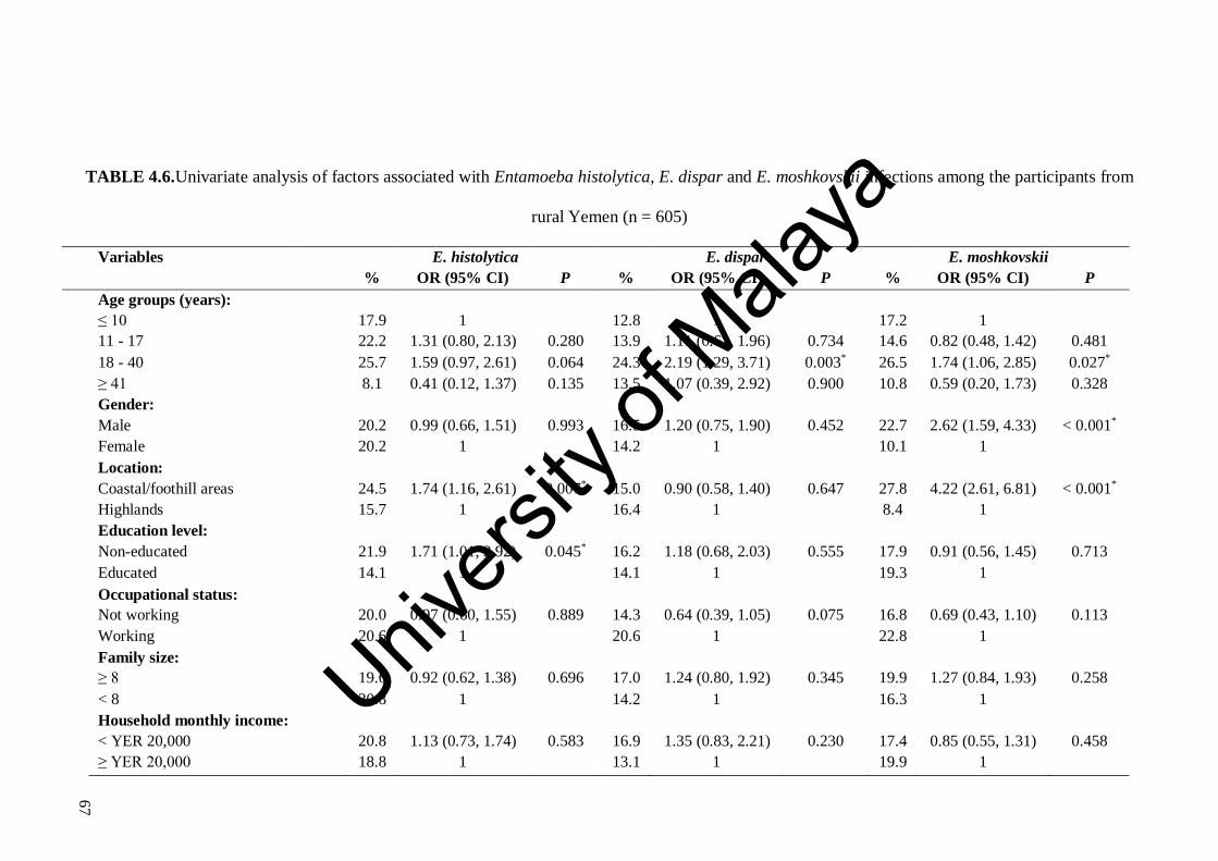
67
TABLE 4.6.Univariate analysis of factors associated with Entamoeba histolytica, E. dispar and E. moshkovskii infections among the participants from
rural Yemen (n = 605)
Variables E. histolytica E. dispar E. moshkovskii
% OR (95% CI) P % OR (95% CI) P % OR (95% CI) P
Age groups (years):
≤ 10 17.9 1 12.8 1 17.2 1
11 - 17 22.2 1.31 (0.80, 2.13) 0.280 13.9 1.11 (0.62, 1.96) 0.734 14.6 0.82 (0.48, 1.42) 0.481
18 - 40 25.7 1.59 (0.97, 2.61) 0.064 24.3 2.19 (1.29, 3.71) 0.003* 26.5 1.74 (1.06, 2.85) 0.027*
≥ 41 8.1 0.41 (0.12, 1.37) 0.135 13.5 1.07 (0.39, 2.92) 0.900 10.8 0.59 (0.20, 1.73) 0.328
Gender:
Male 20.2 0.99 (0.66, 1.51) 0.993 16.5 1.20 (0.75, 1.90) 0.452 22.7 2.62 (1.59, 4.33) < 0.001*
Female 20.2 1 14.2 1 10.1 1
Location:
Coastal/foothill areas 24.5 1.74 (1.16, 2.61) 0.007* 15.0 0.90 (0.58, 1.40) 0.647 27.8 4.22 (2.61, 6.81) < 0.001*
Highlands 15.7 1 16.4 1 8.4 1
Education level:
Non-educated 21.9 1.71 (1.01, 2.92) 0.045* 16.2 1.18 (0.68, 2.03) 0.555 17.9 0.91 (0.56, 1.45) 0.713
Educated 14.1 1 14.1 1 19.3 1
Occupational status:
Not working 20.0 0.97 (0.60, 1.55) 0.889 14.3 0.64 (0.39, 1.05) 0.075 16.8 0.69 (0.43, 1.10) 0.113
Working 20.6 1 20.6 1 22.8 1
Family size:
≥ 8 19.6 0.92 (0.62, 1.38) 0.696 17.0 1.24 (0.80, 1.92) 0.345 19.9 1.27 (0.84, 1.93) 0.258
< 8 20.8 1 14.2 1 16.3 1
Household monthly income:
< YER 20,000 20.8 1.13 (0.73, 1.74) 0.583 16.9 1.35 (0.83, 2.21) 0.230 17.4 0.85 (0.55, 1.31) 0.458
≥ YER 20,000 18.8 1 13.1 1 19.9 1
Univers
ity of
Mala
ya

TABLE 4.6: Continued
Variables E.histolytica E. dispar E.moshkovskii
% OR (95% CI) P % OR (95% CI) P % OR (95% CI) P
Presence of other family members
infected with Entamoeba spp.
Yes
No
25.9
16.1
1.82 (1.22, 2.72)
1
0.003* 15.1
16.1
0.93 (0.60, 1.45)
1
0.749 25.5
13.0
2.29 (1.51, 3.49)
1
< 0.001*
Source of drinking water
Unsafe (well, streams, rain, dams)
Safe (piped water)
20.3
19.7
1.04 (0.66, 1.63)
1
0.879 19.0
6.4
3.44 (1.74, 6.81)
1
< 0.001* 19.0
15.6
1.24 (0.76, 2.02)
1
0.394
Presence of toilet in house
No
Yes
22.4
17.9
1.32 (0.88, 1.97)
1
0.175 19.7
11.6
1.87 (1.19, 2.94)
1
0.006* 17.1
19.3
0.87 (0.57, 1.31)
1
0.490
Washing hands before eating
No
Yes
22.1
18.8
1.23 (0.82, 1.83)
1
0.322 18.4
13.9
1.41 (0.91, 2.19)
1
0.128 20.1
16.9
1.24 (0.81, 1.88)
1
0.319
Washing hands after playing with
soil or gardening
No
Yes
18.1
21.7
0.80 (0.53, 1.20)
1
0.284 16.9
14.8
1.17 (0.75, 1.82)
1
0.481 19.7
17.1
1.19 (0.78, 1.80)
1
0.415
Washing hands after defecation
No
Yes
20.1
20.2
0.99 (0.66, 1.50)
1
0.970 17.9
14.4
1.30 (0.83, 2.03)
1
0.245 18.8
17.8
1.07 (0.70, 1.63)
1
0.767
Cutting nails periodically
No
Yes
18.8
22.1
0.81 (0.54, 1.21)
1
0.306 13.9
18.2
0.73 (0.47, 1.13)
1
0.155 19.6
16.2
1.26 (0.82, 1.93)
1
0.285
68
Univers
ity of
Mala
ya

TABLE 4.6: Continued
Variables E.histolytica E. dispar E.moshkovskii
% OR (95% CI) P % OR (95% CI) P % OR (95% CI) P
Close contact with domestic animals
Yes
No
19.9
21.4
0.91 (0.55, 1.50)
1
0.712 16.4
14.3
1.18 (0.73, 1.89)
1
0.496 19.1
14.3
1.41 (0.80, 2.51)
1
0.236
Indiscriminate defecation
Yes
No
19.6
20.9
0.92 (0.62, 1.37)
1
0.692 19.6
11.0
1.97 (1.24, 3.14)
1
0.004* 19.8
17.3
1.24 (0.81, 1.88)
1
0.319
Washing vegetables before
consumption
No
Yes
21.7
19.3
1.16 (0.77, 1.74)
1
0.490 22.2
12.2
2.05 (1.32, 3.19)
1
< 0.001* 22.2
16.0
1.49 (0.98, 2.27)
1
0.062
Washing fruits before consumption
No
Yes
22.5
19.1
1.23 (0.80, 1.88)
1
0.342 19.8
13.9
1.52 (0.96, 2.40)
1
0.071 17.6
18.4
0.94 (0.60, 1.49)
1
0.802
Boiling drinking water
No
Yes
19.9
23.5
0.81 (0.41, 1.59)
1
0.531 15.5
17.6
0.86 (0.40, 1.83)
1
0.690 18.1
19.6
0.90 (0.44, 1.86)
1
0.783
YER = Yemen Rial; (US$1 = YER216)
OR = Odds ratio; CI = Confidence interval; Reference group marked as OR = 1
* Significant association (P < 0.05).
69
Univers
ity of
Mala
ya

Likewise, participants who used unsafe drinking water had 2.20 (95% CI = 1.37, 3.40)
times the odds of having an Entamoeba spp. infection compared to those who had a
piped water supply in their house. Moreover, non-educated participants had a 3.51 odds
of infection compared to their educated counterparts (95% CI = 2.03, 6.05). Similarly,
not washing vegetables before consumption increased the participant’s odds of having
the disease by 1.59 times (95% CI = 1.05, 2.39) when compared with those who always
washed vegetables.
In the species-based regression models (Table 4.7), three factors were found to
increase the participant’s odds of having E. histolytica infection by 1.7 times, including
living in coastal/foothill areas (95%CI = 1.16, 2.66), illiteracy (95%CI = 1.02, 3.02),
and the presence of other family members infected with Entamoeba (95%CI = 1.13,
2.56). Similarly, using unsafe drinking water (OR = 2.89; 95%CI = 1.44, 5.80) and not
washing vegetables before consumption (OR = 1.68; 95%CI = 1.06, 2.67) were
identified as the significant risk factors of E. dispar infection.
Moreover, four variables were retained as the significant risk factors of E.
moshkovskii infection. Participants aged between 18 - 40 years (95%CI = 1.18, 3.54)
and those who live with other family members infected with Entamoeba (95%CI = 1.37,
3.42) were at about twice as likely to be infected with E. moshkovskii. Similarly, male
gender (95%CI = 1.63, 4.84) and living in coastal/foothill areas (95%CI = 2.21, 6.01)
increased the participant’s odds of having the infection by 2.81 and 3.65 times when
compared with females and those who live in highlands, respectively.
70
Univers
ity of
Mala
ya

TABLE 4.7: Multivariate analysis of risk factors associated with Entamoeba histolytica/E. dispar/E. moshkovskiiinfections among the participants
from rural Yemen (n = 605).
Entamoeba histolytica Entamoeba dispar Entamoeba moshkovskii
Variables AOR (95% CI) P AOR (95% CI) P AOR (95% CI) P
Age group (18 - 40 years) - - 1.64 (0.95, 2.85) 0.078 2.04 (1.18, 3.54) 0.011*
Gender (male) - - - - 2.81 (1.63, 4.84) < 0.001*
Location (coastal/foothill areas) 1.75 (1.16, 2.66) 0.008* - - 3.65 (2.21, 6.01) < 0.001*
Education level (non-educated) 1.75 (1.02, 3.02) 0.044* - - - -
Occupational status (working) - - - - - -
Presence of other family members
infected with Entamoeba spp.
1.70 (1.13, 2.56) 0.011* - - 2.17 (1.37, 3.42) 0.001*
Source of drinking water (unsafe) - - 2.89 (1.44, 5.80) 0.003* - -
Presence of toilet in house (no) - - 1.29 (0.75,2.23) 0.355 - -
Washing hands after playing with soil or
gardening (no)
- - - - - -
Washing vegetables before consumption
(no)
- - 1.68 (1.06, 2.67) 0.027* - -
Indiscriminate defecation (yes) - - 1.35 (0.77, 2.38) 0.296 - -
AOR, adjusted odds ratio; CI, Confidence interval
Significant key risk factors (p < 0.05)
71
Univers
ity of
Mala
ya
72
CHAPTER 5: DISCUSSION
Results of the current study revealed that the prevalence of Entamoeba complex was
high with more than half (53.6%) of the participants being infected. This is consistent
with previous studies that investigated the prevalence of infection among schoolchildren
from urban areas in Sana’a and Al-Mahweet provinces and restaurants workers in
Sana'a City (the capital of Yemen) in which the prevalence rates of Entamoeba complex
were 52% and 48.9% (Azazy et al., 2002; Al-Shibani et al., 2009b).
However, generally, the present study findings are far higher than those reported
in other studies on Yemen and ranges from 6.4% to 33.7% (Azazy and Raja’a, 2003;
Raja’a and Mubarak 2006; Al-Shibani et al., 2009a; Al-Haddad and Baswaid, 2010;
Alyousefi et al., 2011; Bin Mohanna et al., 2014; Alsubaie et al., 2016). Given that the
current study covered a wider area consisting of four diverse provinces as well as a
wider range of participants in terms of age, the variation in the prevalence rates reported
could be attributed to demographic, socioeconomic and environmental differences.
The disparity in results could also reflect differences in hygiene and sanitation
conditions and other unidentified factors among the targeted communities. Moreover,
the variation in the prevalence could also be due to the high sensitivity and accuracy of
the laboratory techniques used in the diagnosis in the current study.
A comparison of the findings of the present study with those of studies on
neighbouring countries revealed that Yemen had the highest prevalence rate in the
region. Previous studies in other Middle Eastern countries have reported a varying
prevalence of Entamoeba complex:4.9% in Jordan (Youssef et al., 2000), 5.5% in Iran
(Sharif et al., 2015), 7.1% in Oman (Prakash, 2008), 8.2%-16.2% in Saudi Arabia (Al-
Mohammed et al., 2010; Amer et al., 2016), 16.8% in Egypt (Hegazy et al., 2014),
19.2% in the United Arab Emirates (ElBakri et al., 2013), 23.4% in Iraq (Al-Kubaisy et
Univers
ity of
Mala
ya

73
al., 2014), and 33.1% in Sudan (Abdel-aziz et al., 2010).
This study differentiates, for the first time, E. histolytica from E. dispar and E.
moshkovskii in Yemen using the nested multiplex PCR assay. It was found that 122
(44.2%) and 95 (34.4%) of the 276 PCR-positive products were identified as E.
histolytica and E. dispar which accounted for 20.2% and 15.7% of the total number of
samples, respectively. Moreover, the present findings demonstrated that E. moshkovskii
was identified in 110 (39.9%) isolates which accounted for 18.2% from the study
participants (110/605). Entamoeba histolytica, E. dispar and E. moshkovskii were
identified in 5.6%, 70.8% and 61.8%, respectively of 89 PCR-positive samples from
Australia (Fotedar et al., 2007a). Likewise, E. moshkovskii and E. dispar were reported
in 25.4% and 23.2% of 181 asymptomatic children under 16 years old from Colombia,
with E. histolytica was reported in one sample only (0.55%) (López et al., 2015).
On the other hand, a much lower prevalence was reported among 120 faecal
samples from the United Arab Emirates, with E. histolytica, E. dispar and E.
moshkovskii were detected in 13.3%, 6.7%, and 3.3%, respectively (ElBakri et al.,
2013). Similarly, a previous study among 500 participants from Peninsular Malaysia
revealed that E. dispar was the highest prevalent species (13.4%), followed by E.
histolytica (3.2%) and E. moshkovskii (1.0%) (Anuar et al., 2012c). However, when
seeking to draw conclusions from a cross-country comparison, the differences in the
socioeconomic status, environmental conditions, and other risk factors should be borne
in mind as well as whether travellers and expatriates and institutionalized members of
the population were included in the samples of the studies undertaken in these countries.
The present study showed that Entamoeba complex was significantly associated
with age, as demonstrated by the convex pattern of distribution. The prevalence of
infection was 45.3% among children aged ≤ 10 years and increased to reach its highest
(83.1%) among participants aged 21–30 years then decreased to the lowest (32.4%)
Univers
ity of
Mala
ya

74
among those aged more than 40 years. It was observed a similar pattern of distribution
with E. histolytica, E. dispar and E. moshkovskii infections. In Brazil, Benetton et al.
(2005) noticed that the prevalence of E. histolytica infection among outpatient clinics
increases with age. However, the present finding contrasts that in previous studies on
Saudi Arabia, Turkey, Cameroon and Malaysia which reported the highest prevalence
among those aged below 12-15 years (Al-Shammari et al., 2001; Ilikkan et al., 2005;
Anuar et al., 2012a; Mbuh et al., 2012). This could be attributed to the fact that most of
adult in Yemen are chewing qat (A type of narcotic leaves or plant & people addict it as
cocaine) every day (mostly after lunch) & usually without washing it; which can cause
infection with a lot of microorganisms including parasites, while the children don’t
chew qat. Other explanation is that most of adult men are farmers and more exposure to
the source of infection. Moreover, this finding needs more confirmation with future
study including more areas of study.
In the present study, 55 microscopy-positive specimens were found to be
negative by PCR. These results can possibly be explained by the fact that most of those
samples were of low intensity which fell below the PCR detection limit (only 1-2 cysts
were detected by either trichrome stain or formalin-ether sedimentation method), in
addition to the presence of faecal inhibitor substances which were not completely
removed former to PCR reaction. Another reason for that may be related to the presence
of other Entamoeba species. However, this must be confirmed by further studies using
more sensitive PCR techniques (khairnar and Parija, 2007) .This in agreement with a
study conducted in Malaysia among orang asli reported that 30 faecal samples that were
positive in microscopy, gave negative results by using PCR (Anuar et al., 2013b).
The present study showed that E. dispar and E. moshkovskii infections were
significantly associated with age, with the prevalence increased to reach its highest rate
among participants aged 21-30 years then decreased to the lowest among those aged
Univers
ity of
Mala
ya

75
more than 40 years. Moreover, E. moshkovskii prevalence was significantly higher
among male participants while neither E. histolytica nor E. dispar showed similar
findings. These differences could be explained by a higher exposure of adult males to
the sources of infections as a result of their daily activities such as the higher access to
contaminated outdoor food, contaminated water sources, and contact with infected
Univers
ity of
Mala
ya

76
individuals.
Moreover, due to religious and cultural practices in rural Yemen, females spend
most of their time at home for family domestic activities and they are not always
allowed to work or to go for outdoor activities. In consistency with the present findings,
a researcher reported that the prevalence of E. dispar infection among residents
subjected to water shortage in the northeast region of Brazil increases with age
(Calegaret al., 2016). By contrast, a previous study among Malaysian rural communities
which reported a significantly higher prevalence of infection with E. dispar among
participants who their age below 15 years than those aged 15 years or more (Anuar et
al., 2012c). On the other hand, previous studies in Colombia and Malaysia found no
significant association between gender and the three species (Anuar et al., 2012c; López
et al., 2015). However, the possible different epidemiological characteristics of the
studied Yemeni communities should be taken into consideration when comparing the
results with other studies conducted abroad.
In addition, Acuna-Soto and his colleagues have reviewed 66 hospital-based
reports of invasive amoebiasis (including dysentery, colonic perforation, , peritonitis,
appendicitis, liver abscess and amoeboma) and 20 community–based parasitological
surveys conducted between 1929 and 1997 and concluded that the percentage of men
affected by all forms of invasive amoebiasis was three times higher than women (with a
higher male frequency was found in all age groups) while almost identical rate of
asymptomatic infection with E. histolytica was observed in both sexes in all age groups
(Acuna-Soto et al., 2000).
The present study also investigated the risk factors of E. histolytica, E. dispar
and E. moshkovskii infections. The age and gender were kept as significant risk factors
of E. moshkovskii infection only. Interestingly, the current study showed a significantly
higher prevalence of E. histolytica and E. moshkovskii infections in the coastal/foothill
Univers
ity of
Mala
ya

77
areas than the highlands of Yemen. Similarly, the present study identified the use of
unsafe drinking water as a strong significant risk factor of E. dispar infection. A
previous study on Sana’a City (Alyousefi et al., 2011) and other reports elsewhere
found that using unsafe drinking water was a risk factor for Entamoeba complex and
other intestinal parasitic infections (Benetton et al., 2005; Anuar et al., 2012a; Al-
Delaimy et al., 2014).
In fact, there is an obvious difference in the sources of drinking water with
villages in the highlands of Yemen still rely on rainwater (dams, tanks and troughs) and
ground water (drilled artesian pump-based wells).While streams and traditional dug
wells are the main drinking water sources in the coastal/foothill areas. Duringthe rainy
season, the floods flow through the valleys spreading human and animal excreta into
fields, dams, pools, wells and other water sources. People collect water from wells,
pools and dams, and store that contaminated water in tanks and troughs, and the water is
also handled by bare hands. Hence, contamination of drinking water with Entamoeba
cysts during the collection, transport and storage of water is also possible (Wright et al.,
2004). A recent study among 200 children in Al-Mahweet province found a
significantly higher prevalence of intestinal parasitic infections particularly Entamoeba
spp. among children who use stream water (60%) as a source of drinking water
compared to those who use water from dams (8.5%), ponds (5%) and wells (16.5%)
(Alwabr and Al-Moayed, 2016). However, in contrast, a recent study on rural and urban
children in Ibb province found a higher prevalence of Entamoeba spp. infection among
children using a piped water supply for drinking water compared to those using water
from other sources (Alsubaie et al., 2016). It could be postulated that contamination of
drinking water sources is more likely to be higher in the coastal/foothill areas; however,
it is not possible to infer causality using this cross-sectional study. Hence, further
studies to explore the presence of parasites in drinking water in the study areas are
Univers
ity of
Mala
ya
78
required to better understand these contradictory findings.
Indeed, within the family, the present findings showed that the presence of other
infected family members significantly increased the odds of an individual who are
infected with E. histolytica and E. moshkovskii. A previous study from Malaysia found a
similar finding with E. dispar (Anuar et al., 2012c). The infection could easily be
passed on through contaminated food and drinks prepared by infected family members
who have inadequate personal hygiene. The trophozoite of Entamoeba spp. survives for
a short time but the cyst lives longer and withstands unfavourable conditions; it can be
found on the hands and clothes of children as well as in containers used for washing
clothes and in shower trays, which implies that contact with an infected person’s
belongings and water they have used also transmits the infection (Roberts, 2009). In
general, several previous studies have revealed that the presence of other family
members infected with soil-transmitted helminthiasis, giardiasis, and schistosomiasis is
significantly associated with a higher prevalence of these infections (Spencer, 1981;
Gathiram and Jackson, 1987; Ruiz-Palacios et al., 1992; Braga et al., 2001; Sady et al.,
2013; Al-Delaimy et al., 2014).
Moreover, the present findings revealed that the prevalence rate of E. histolytica
infection was significantly associated with a low educational level, which is in
agreement with previous reports on Entamoeba complex elsewhere (Al-Shammari et al.,
2001; Al-Mohammed et al., 2010; Alemu et al., 2011; Dawet et al., 2012; Gelaw et al.,
2013). Probably, illiterate participants would have less health-related knowledge and
awareness about intestinal parasitic infections compared to educated individuals.
Therefore, these individuals may also contribute to disseminating infection among other
family members especially if they are responsible for food preparation.
In the same vein, the present study found a strong association between E. dispar
infection and not washing vegetables before consumption, which is in agreement with a
Univers
ity of
Mala
ya

79
preceding study on Malaysia (Anuar et al., 2012c). The vegetables could be
contaminated by either an infected person or contaminated soil; by human or animal
excreta. A similar finding has been reported in previous studies on enteric parasites
from different countries including Brazil, Malaysia, and West African countries (Braga
et al., 2001; Amoah et al., 2007; Al-Delaimy et al., 2014; Elyana et al., 2016).
Moreover, the present study showed that employed participants and those who
do not have a toilet in their houses as well as those who do not wash their hands after
playing with soil or gardening were more prone to infection by Entamoeba complex.
However, these factors were not retained as significant risk factors by the multivariate
logistic regression analysis. Previous studies on Malaysia conducted among the Orang
Asli population conclude that working status and absence of toilet facilities in the house
and are significant risk factors of intestinal parasitic infections (Nasr et al., 2013; Al-
Delaimy et al., 2014; Elyana et al., 2016).
In general, the association between low socioeconomic status and the prevalence
of Entamoeba spp. infection is well documented. It has been reported that the
prevalence of Entamoeba spp. Among those who have a low-to-average socioeconomic
status is higher than those who have an average or above socioeconomic status (Ravdin,
1995; Tellez et al., 1997; Pham Duc et al., 2011). Such an association was not found by
the present study and this could be explained by the high prevalence of poverty in the
studied communities. Lastly, previous studies on Yemen (in Sana’a City), Vietnam and
Malaysia have reported a greater risk of Entamoeba infection among participants who
have close contact with domestic animals (Pham Duc et al., 2011; Alyousefi et al.,
2011; Anuar et al., 2012b).
In the present study, there was no significant variance in the prevalence of
Entamoeba between those who had animals at their households and their counterparts.
Univers
ity of
Mala
ya

80
This could be explained by the fact E. histolytica–like group almost exclusively infects
humans, with exception of E. dispar that may also infect chimpanzees, baboon and
macaques (Hooshyar et al., 2015); animals that do not exist in the study area. However,
recent studies suggested a role for the domestic animalsas dogs and cats in transmitting
intestinal parasites as the infective cysts or eggs can be easily get picked up on the fur of
these animals and then transferred into the houses or to the hands of their owners
(Anuar et al., 2012b; Choy et al., 2014; Elyana et al., 2016).Until now, the only
pathogenic species is E. histolytica; and many studies support the idea that E.
moshkovskii is a non-pathogenic amoeba (Scaglia et al., 1983; Beck et al., 2008). In
accordance, the findings of the current showed a significantly higher frequency of E.
moshkovskii single infections among the asymptomatic individuals compared to those
with GIT symptoms. Nonetheless, a previous study from Malaysia showed the presence
of GIT symptoms such as weight loss, lack of appetite and nausea among E.
moshkovskii-positive individuals (Anuar et al. 2012a). Similarly, a study in India
described an association between E. moshkovskii infection and dysentery (Parija and
Khairnar, 2005).
Moreover, in Australia, a case-control study noticed E. moshkovskii in 11
patients who presented with diarrhoea and other GIT clinical symptoms, and no other
pathogens, such as bacteria and viruses, were isolated from their faecal specimens
(Fotedar et al., 2008). These controversial findings call for further studies to identify
potential pathogenicity and virulence of E. moshkovskii in causing GIT disorders.
In Malaysia, a study carried out by Anuar et al. (2012a) found that subjects with
single infection of E. moshkovskii presented with weight loss, lack of appetite and
nausea. Likewise, a study done by Shimokawa et al. (2012) in Mirpur, Dhaka showed
that cases with E. moshkovskii infection can develop gastrointestinal symptom as
diarrhea among Bangladeshi children. Their study also reported that E. moshkovskii can
Univers
ity of
Mala
ya

81
cause weight loss and colitis in mice. Interestingly, Yacoob et al. (2012) also
demonstrated that E. moshkovskii significantly associated with diarrhoea in Pakistan.
Several studies showed the association of E. moshkovskii infection with gastrointestinal
symptoms and/or dysentery as in Bangladesh, India, Turkey and Australia (Ali et al.
2003; Parija and Khairnar, 2005; Tanyuksel et al. 2007; Fotedar et al. 2008). However,
in the present study, it was not possible to conclude a causal association between
Entamoeba infection and symptoms because there were two limitations in the
methodology of the present study. First, the prevalence of multiple infections was high
among the population studied with Giardia duodenalis, Hymenolepis nana, Ascaris
lumbricoides, and hookworm being the most common intestinal parasites concurrently
detected with the Entamoeba parasite. Second, bacterial and viral causative agents were
not ruled out in the current study.
Univers
ity of
Mala
ya

81
CHAPTER 6: CONCLUSION AND RECOMMENDATIONS
6.1 CONCLUSION
Overall, the present study revealed that intestinal amoebiasis is highly prevalent among
rural communities in Yemen with the occurrence of the three microscopically-identical
species (E. histolytica, E. dispar, and E. moshkovskii). The study found an age-dependency
pattern of distribution for both Entamoeba complex infection as well as the species-specific
infections. Some variables were identified as the key risk factors of Entamoeba complex
infection in these communities with a different species-specific grouping of risk factors.
It is expected that the findings of this study will be helpful to the policy makers and
health authorities for implementing effective control and preventive measures to
significantly decrease the prevalence and burden of amoebiasis as well as other intestinal
parasitic infections in these rural communities in the studied provinces and in other
provinces of Yemen that may share similar epidemiological characteristics. Throughout
Yemen, rural communities share the same environmental, socioeconomic status and health
care conditions. Therefore, the findings of the present study could be generalized to rural
populations in other governorates in Yemen. However, further studies among other rural as
well as urban populations are mandatory to confirm these assumptions.
The following are the main findings of the present study:
i. The overall prevalence of Entamoeba complex infection was 53.6% (324/605). Of these,
single infections of Entamoeba complex were detected in 14.2% (46/324) of the samples
and 85.8% (278/324) were mixed infections of Entamoeba spp. with other intestinal
parasite species.
Univers
ity of
Mala
ya

82
ii. The overall prevalence of Entamoeba complex infection was significantly higher among
male participants in comparison with females (P = 0.008), and among those aged 21 – 30
years with an age-dependency distribution represented by a convex pattern of distribution
(χ2 = 39.373, P< 0.001).
iii. Six factors were identified by the multiple logistic regression model analysis as the
significant risk factors of Entamoeba complex infection among the studied population.
These factors are age of ≥ 18 years, the presence of another family member infected with
Entamoeba spp. using unsafe drinking water, illiteracy, living in coastal/foothill areas, and
not washing vegetables before consumption.
iv. Based on the nested multiplex PCR assay, E. histolytica was detected in 20.2% (122/605)
of the samples while E. dispar and E. moshkovskii isolates were recovered in 15.7%
(95/605) and 18.2% (110/605) of the samples. In other words, the occurrence of Entamoeba
complex among the 276 Entamoeba-PCR-positive samples was 44.2%, 34.4%, and 39.9%,
respectively.
v. The study revealed different sets of species-specific risk factors by the multiple logistic
regression model analysis. Living in coastal/foothill areas, illiteracy, and the presence of
other family members infected with Entamoeba was the significant risk factors of
E.histolyticainfection. Likewise, not washing vegetables before consumption and using
unsafe drinking water were the risk factors of E. dispar. Moreover, age between 18 - 40
years, living with other family members infected with Entamoeba, male gender, and living
in coastal/foothill areas were the significant risk factors of E.moshkovskii infection.
6.2 RECOMMENDATIONS
Further epidemiological and molecular studies on Entamoeba spp. using environmental
samples such as soil and water samples are required for better understanding of the source
Univers
ity of
Mala
ya

83
of Entamoeba spp.in these communities as well as to provide new insights on the
transmission dynamics of these different Entamoeba spp.
Overall, the risk factors recognized by the present study should be considered by
any prevention and control programme against intestinal parasitic infections. Proper
attention should be given to the implement of using molecular methods or other species-
specific diagnostic assays, diagnose and differentiate the pathogenic E. histolytica from E.
dispar, and E. moshkovskii.
6.3 LIMITATIONS OF THE STUDY
Certain limitations should be considered when interpreting the findings of the current study
infections such as using unsafe sources for drinking water, but it is not possible to
confirm the causality between the variables using the present cross-sectional study. Hence,
further studies to investigate the presence of Entamoeba spp. parasites in drinking water in
the study areas are required. Similarly, some risk factors were significantly associated with
one or two Entamoeba species but not with the other species although the three species are
all transmitted in the same way, this could be explained by the findings of the present study
that the prevalence of these Entamoeba species and their major risk factors in Yemen was
high and their distribution varied considerably with geographic areas. Another explanation
could be the intensity of infection with each species. It may be assumed that in a positive
sample, most of the detected cysts are belong to one or two species while very few are
belonging to other species. However, unknown parasite-related factors including potential
parasite-environment interactions could not be ruled out.
Moreover, this study had to depend on a single stool specimen, hence the prevalence
of Entamoeba spp. is likely to be underestimated due to the temporal variation in cyst
shedding over hours and days. However, examining faecal samples by different techniques
Univers
ity of
Mala
ya

84
including the gold standard (trichrome staining) and concentration methods (formalin-ether
sedimentation) as well as the molecular PCR assay could assist to conquer this limitation.
In the present study, 55 microscopy-positive specimens were found to be negative by PCR.
This could be attributed to the fact that the majority of those samples were of low intensity
which fell below the PCR detection limit (only 1-2 cysts were detected by either trichrome
stain or formalin-ether sedimentation method) as well as the presence of faecal inhibitor
substances which were not completely eliminated prior to PCR reaction. Using more
sensitive methods such as real-time PCR should be considered in future studies. A previous
study showed that real-time PCR assay exhibited superior sensitivity in detecting
Entamoeba parasites in faecal samples when compared with microscopy, stool antigen
detection and conventional PCR (Roy et al., 2005). Another limitation was that bacterial
and viral causative agents were not ruled out in the current study, hence reporting a causal
association between Entamoeba infection and symptoms was not possible.
Univers
ity of
Mala
ya

85
REFERENCES
Abdel-aziz, M. A., Afifi, A. A., Malik, E. M., & Adam, I. (2010). Intestinal protozoa and
intestinal helminthic infections among schoolchildren in Central Sudan. Asian Pacific
Journal of Tropical Disease, 3, 292-293.
Abdulsalam, A. M., Ithoi, I., Al-Mekhlafi, H. M., Ahmed, A., Surin, J., &Mak, J.W.
(2012). Drinking water is a significant predictor of Blastocystis infection among rural
Malaysian primary schoolchildren. Parasitology, 139, 1014-1020.
Abramowicz, M., & Duffy, J. F. (2010). The inducement standard of patentability. Yale
Law Journal, 120, 1590-1680.
Acuna-Soto, R., Maguire, J., & Wirth, D. (2000). Gender distribution in asymptomatic and
invasive amebiasis. The American Journal of Gastroenterology, 95, 1277-1283.
Al-Delaimy, A. K., Al-Mekhlafi, H. M., Nasr, N. A., Sady, H., Atroosh, W. M., Nashiry,
M., Anuar, T. S., Moktar, N., Lim, Y. A., & Mahmud, R. (2014). Epidemiology of
intestinal polyparasitism among Orang Asli school children in rural Malaysia. PLOS
Neglected Tropical Diseases, 8, e3074.
Alemu, A., Atnafu, A., Addis, Z., Shiferaw, Y., Teklu, T., Mathewos, B., Birhan, W.,
Gebretsadik, S., & Gelaw, B. (2011). Soil transmitted helminths and schistosoma
mansoni infections among school children in Zarima town, northwest Ethiopia. BMC
Infectious Diseases, 11, 189-195.
Al-Haddad, A., & Baswaid, S. (2010). Frequency of intestinal parasitic infection among
children in Hadhramout governorate (Yemen). Journal of the Egyptian Society of
Parasitology, 40, 479-488.
Al-Harthi, S. A., &Jamjoom, M. (2007). Diagnosis and differentiation of Entamoeba
infection in Makkah Al Mukarramah using microscopy and stool antigen detection kits.
World Journal of Medical Sciences, 2, 15-20.
Al-Hindi, A., Shubair, M., Marshall, I., Ashford, R., Sharif, F., Abed, A., &Kamel, E.
(2005). Entamoeba histolytica or Entamoeba dispar among children in Gaza, Gaza
Strip.Journal of the Egyptian Society of Parasitology, 35, 59-68.
Univers
ity of
Mala
ya

86
Ali, I. K. M., Clark, C. G., & Petri, W. A. (2008). Molecular epidemiology of amebiasis.
Infection, Genetics and Evolution, 8, 698-707.
Ali, I. K., Hossain, M. B., Roy, S., Ayeh-Kumi, P. F., Petri Jr, W. A., Haque, R., & Clark,
C. G. (2003). Entamoeba moshkovskii infections in children, Bangladesh. Emerging
Infectious Diseases, 9, 580-584.
AL-Kubaisy, W., AL-Talib, H., Al-khateeb, A. &Shanshal MM (2014). Intestinal parasitic
diarrhoea among children in Baghdad–Iraq. Tropical Biomedicine, 31, 499-506.
Allason-Jones, E., Mindel, A., Sargeaunt, P., & Williams, P. (1986). Entamoeba histolytica
as a commensal intestinal parasite in homosexual men. New England Journal of
Medicine, 315, 353-356.
Al-Mekhlafi, M. H., Azlin, M., Aini, U. N., Shaik, A., Sa’iah, A., & Norhayati, M. (2007).
Prevalence and predictors of low serum retinol and hypoalbuminaemia among children
in rural Peninsular Malaysia. Transactions of the Royal Society of Tropical Medicine
and Hygiene, 101, 1233-1240.
Al-Mohammed, H. I., Amin, T. T., Aboulmagd, E., Hablus, H. R., & Zaza, B. O. (2010).
Prevalence of intestinal parasitic infections and its relationship with socio–demographics
and hygienic habits among male primary schoolchildren in Al–Ahsa, Saudi Arabia.
Asian Pacific Journal of Tropical Disease, 3, 906-912.
Al-Qobati, S. A., Al-Maktari, M. T., Al-Zoa, A., & Derhim, M. (2012). Intestinal
parasitosis among Yemeni patients with cancer, Sana'a, Yemen. Journal of the Egyptian
Society of Parasitology, 42, 727-734.
Al‐Shammari, S., Khoja, T., El‐Khwasky, F., & Gad, A. (2001). Intestinal parasitic
diseases in Riyadh, Saudi Arabia: prevalence, sociodemographic and environmental
associates. Tropical Medicine and International Health, 6, 184-189.
Al-Shibani, L., Azazy, A., & Alhamd, J. (2009b). Intestinal parasitosis among apparently
healthy workers at restaurants of Sana'a City, Yemen. Journal of the Egyptian Society of
Parasitology, 39, 263-268.
Al-Shibani, L., Azazy, A., & El-Taweel, H. (2009a). Cryptosporidiosis and other intestinal
parasites in 3 Yemeni orphanages: prevalence, risk, and morbidity. Journal of the
Egyptian Society of Parasitology, 39, 327-337.
Univers
ity of
Mala
ya

87
Alsubaie, A. S. R., Azazy, A. A., Omer, E. O., Al-shibani, L. A., Al-Mekhlafi, A. Q., & Al-
Khawlani, F. A. (2016). Pattern of parasitic infections as public health problem among
school children: A comparative study between rural and urban areas. Journal of Taibah
University Medical Sciences, 11, 13-18.
Alwabr, G. M., & Al-Moayed, E. E. (2016). Prevalence of intestinal parasitic infections
among school children of Al-Mahweet Governorate, Yemen. European Journal of
Biological Research, 6, 64-73.
Alyousefi, N. A., Mahdy, M. A., Lim, Y. A., Xiao, L., & Mahmud, R. (2013). First
molecular characterization of Cryptosporidium in Yemen. Parasitology, 140, 729-734.
Alyousefi, N. A., Mahdy, M. A., Mahmud, R., & Lim, Y. A. (2011). Factors associated
with high prevalence of intestinal protozoan infections among patients in Sana'a City,
Yemen. PLOS One, 6, e22044.
Amer, O. H., Ashankyty, I. M., & Haouas, N. A. S. (2016). Prevalence of intestinal parasite
infections among patients in local public hospitals of Hail, Northwestern Saudi Arabia.
Asian Pacific Journal of Tropical Disease, 9, 44-48.
Amoah, P., Drechsel, P., Abaidoo, R., & Klutse, A. (2007). Effectiveness of common and
improved sanitary washing methods in selected cities of West Africa for the reduction of
coliform bacteria and helminth eggs on vegetables. Tropical Medicine and International
Health, 12, 40-50.
Anuar, T. S., Al-Mekhlafi, H. M., Abdul Ghani, M. K., Abu Bakar, E., Azreen, S. N.,
Salleh, F. M., & Moktar, N. (2013a). Evaluation of formalin-ether sedimentation and
trichrome staining techniques: its effectiveness in detecting Entamoeba
histolytica/dispar/moshkovskii in stool samples. Journal of Microbiological Methods,
92, 344-348.
Anuar, T. S., Al-Mekhlafi, H. M., Ghani, M. K. A., Azreen, S. N., Salleh, F. M., Ghazali,
N., Bernadus, M., & Moktar, N. (2012a). First molecular identification of
Entamoebamoshkovskii in Malaysia. Parasitology, 139, 1521-1525.
Anuar, T. S., Al-Mekhlafi, H. M., Ghani, M. K. A., Azreen, S. N., Salleh, F. M., Ghazali,
N., Bernadus, M. & Moktar, N., (2013b). Different Clinical Outcomes of Entamoeba
Univers
ity of
Mala
ya

88
histolytica in Malaysia: Does Genetic Diversity Exist?.Korean Journal of Parasitology,
51, 231-236.
Anuar, T. S., Al-Mekhlafi, H. M., Ghani, M. K. A., Bakar, E. A., Azreen, S. N., Salleh, F.
M., Ghazali, N., Bernadus, M., & Moktar, N. (2012c). Molecular epidemiology of
amoebiasis in Malaysia: highlighting the different risk factors of Entamoeba histolytica
and Entamoeba dispar infections among Orang Asli communities. International Journal
for Parasitology, 42, 1165-1175.
Anuar, T. S., Al-Mekhlafi, H. M., Ghani, M. K. A., Osman, E., Yasin, A. M., Nordin, A.,
Azreen, S. N., Salleh, F. M., Ghazali, N., & Bernadus, M. (2012b). Prevalence and risk
factors associated with Entamoeba histolytica/dispar/moshkovskii infection among three
Orang Asli ethnic groups in Malaysia. PLOS One, 7, e48165.
Araz, R. E., KORU, Ö., TANYÜKSEL, M., ÖZEKİNCİ, T., Ceylan, A., KILBAŞ, H. Z.
G., &Cicek, M. (2012). An investigation of the relationship between clinical features of
amoebiasis and Entamoeba histolytica genotypes. Turkish Journal of Medical Sciences,
42, 1147-1156.
Aristizábal, H., Acevedo, J., & Botero, M. (1991). Fulminant amebic colitis.World Journal
of Surgery, 15, 216-221.
Ayed, S. B., Aoun, K., Maamouri, N., Abdallah, R. B., &Bouratbine, A. (2008). First
molecular identification of Entamoeba moshkovskii in human stool samples in Tunisia.
The American Journal of Tropical Medicine and Hygiene, 79, 706-707.
Azazy, A. A., & Al-Tiar, A. S. (1999). A study survey on intestinal and blood parasites
among school children in Sana'a province, Yemen. Saudi Medical Journal, 20, 422-424.
Azazy, A., &Raja'a, Y. (2003). Malaria and intestinal parasitosis among children presenting
to the paediatriccentre in Sana'a, Yemen. Eastern Mediterranean Health Journal, 9,
1048-1053.
Azazy, A., Al-Mahbashi, T. Y. & Al-Mekhlafi, H. M. (2002). Prevalence of intestinal and
blood parasites among school children in Sana’a and Al-Mahweet provinces, Yemen.
Yemeni Journal for Medical Sciences, 4, 50-55.
Univers
ity of
Mala
ya
89
Azian, M. N., Hakim, S. L., Sumiati, A., &Norhafizah, M. (2006). Seroprevalence of
cysticercosis in a rural village of Ranau, Sabah, Malaysia. The Southeast Asian Journal
of Tropical Medicine and Public Health, 37, 58-61.
Baring, E., &Hotez, P. J. (2014). Yemen: Fighting neglected tropical diseases against all
odds. PLOS Neglected Tropical Diseases, 8, e3292.
Barnawi, A., Tonkal, A. M., Fouad, M., & Al-Braiken, F. A. (2007). Detection of
Entamoeba histolytica/dispar in stool specimens by using enzyme-linked
immunosorbent assay in the population of Jeddah City, Saudi Arabia. Journal of the
Egyptian Society of Parasitology, 37, 143-150.
Baswaid, S. H., & Al-Haddad, A. (2008). Parasitic infections among restaurant workers in
Mukalla (Hadhramout/Yemen). Iranian Journal of Parasitology, 3, 37-41.
Baxt, L. A., & Singh, U. (2008). New insights into Entamoeba histolytica pathogenesis.
Current Opinion in Infectious Diseases, 21, 489-494.
Beck, D. L., Doğan, N., Maro, V., Sam, N. E., Shao, J., &Houpt, E. R. (2008). High
prevalence of Entamoeba moshkovskii in a Tanzanian HIV population. ActaTropica,
107, 48-49.
Benetton, M., Gonçalves, A., Meneghini, M., Silva, E., & Carneiro, M. (2005). Risk factors
for infection by the Entamoeba histolytica/E. dispar complex: an epidemiological study
conducted in outpatient clinics in the city of Manaus, Amazon Region, Brazil.
Transactions of the Royal Society of Tropical Medicine and Hygiene, 99, 532-540.
Blessmann, J., Van Linh, P., Nu, P. A. T., Thi, H. D., Muller-Myhsok, B., Buss, H.,
&Tannich, E. (2002). Epidemiology of amebiasis in a region of high incidence of
amebic liver abscess in central Vietnam. The American Journal of Tropical Medicine
and Hygiene, 66, 578-583.
Boucek, F. (2009). Rethinking factionalism: typologies, intra-party dynamics and three
faces of factionalism. Party Politics, 15, 455-485.
Braga, L., Gomes, M. L., Da Silva, M. W., Façanha, F. E., Fiuza, L., & Mann, B. J. (2001).
Household epidemiology of Entamoeba histolytica infection in an urban community in
northeastern Brazil. The American Journal of Tropical Medicine and Hygiene, 65, 268-
271.
Univers
ity of
Mala
ya

90
Caballero-Salcedo, A., Viveros-Rogel, M., Salvatierra, B., Tapia-Conyer, R., Sepulveda-
Amor, J., Gutierrez, G., & Ortiz-Ortiz, L. (1994). Seroepidemiology of amebiasis in
Mexico. The American Journal of Tropical Medicine and Hygiene, 50, 412-419.
Cairncross, S., Hunt, C., Boisson, S., Bostoen, K., Curtis, V., Fung, I. C., & Schmidt, W.-P.
(2010). Water, sanitation and hygiene for the prevention of diarrhoea. International
Journal of Epidemiology, 39, i193-i205.
Calegar, D. A., Nunes, B. C., Monteiro, K. J. L., Santos, J. P. d., Toma, H. K., Gomes, T.
F., Lima, M. M., Bóia, M. N., &Carvalho-Costa, F. A. (2016). Frequency and molecular
characterisation of Entamoeba histolytica, Entamoeba dispar, Entamoeba moshkovskii,
and Entamoeba hartmanni in the context of water scarcity in northeastern Brazil.
Memorias do Instituto Oswaldo Cruz, 111, 114-119.
Campbell, S. J., Savage, G. B., Gray, D. J., Atkinson. J. A. M., Soares, Magalhães, R. J.,
Nery, S. V., McCarthy J. S., Velleman, Y., Wicken, J. H., Traub, R. J., Williams, J. M.,
Andrews, R. M. & Clements, A. C. A. (2014). Water, Sanitation, and Hygiene (WASH):
A critical component for sustainable soil-transmitted helminth and schistosomiasis
control. PLOS Neglected Tropical Diseases,8, e2651.
Cheesbrough, M. (2005) District laboratory practice in tropical countries: Part 1, 2nd
ed.
Cambridge University Press, London.
Choy, S. H., Al-Mekhlafi, H. M., Mahdy, M. A., Nasr, N. N., Sulaiman, M., Lim, Y. A., &
Surin, J. (2014). Prevalence and associated risk factors of Giardia infection among
indigenous communities in rural Malaysia. Scientific Reports, 4, 6909-6925.
Clark, C. G., & Diamond, L. S. (1991). Ribosomal RNA genes of ‘pathogenic’ and
‘nonpathogenic’ Entamoeba histolytica are distinct. Molecular and Biochemical
Parasitology, 49, 297-302.
Costa, A. O., Gomes, M. A., Rocha, O. A., & Silva, E. F. (2006). Pathogenicity of
Entamoeba dispar under xenic and monoxenic cultivation compared to a virulent E.
histolytica. Revista do Instituto de Medicina Tropical de São Paulo, 48, 245-250.
Dawet, A., Yakubu, D., Remkyes, M., & Daburum, Y. (2012). Prevalence of Entamoeba
histolytica and Entamoeba dispar among School Children in Jos North LGA, Plateau
State, Nigeria. Nigerian Journal of Parasitology, 33, 77-83.
Univers
ity of
Mala
ya

91
Diamond, L. S., & Clark, C. G. (1993). A redescription of Entamoeba histolytica
Schaudinn, 1903 (Emended Walker, 1911) separating it from Entamoeba dispar
Brumpt, 1925. Journal of Eukaryotic Microbiology, 40, 340-344.
Dolabella, S. S., Serrano-Luna, J., Navarro-Garcia, F., Cerritos, R., Ximenez, C., Galvan-
Moroyoqui, J. M., Silva, E. F., Tsutsumi, V., & Shibayama, M. (2012). Amoebic liver
abscess production by Entamoeba dispar. Annals of Hepatology, 11, 107-117.
Ekeh, H. E. &Adeniyi, J.D. (1988). Health education strategies for tropical disease control
in school children. Journal of Tropical Medicine and Hygiene,91, 55-59.
ElBakri, A., Samie, A., Ezzedine, S., & Odeh, R. A. (2013). Differential detection of
Entamoeba histolytica, Entamoeba dispar and Entamoeba moshkovskii in fecal samples
by nested PCR in the United Arab Emirates (UAE). Acta Parasitology, 58, 185-190.
Elyana, F. N., Al-Mekhlafi, H. M., Ithoi, I., Abdulsalam, A. M., Dawaki, S., Nasr, N. A.,
Atroosh, W. M., Abd-Basher, M. H., Al-Areeqi, M. A., & Sady, H. (2016). A tale of two
communities: intestinal polyparasitism among Orang Asli and Malay communities in
rural Terengganu, Malaysia. Parasites & Vectors, 9, 398-414.
Esrey, S.A., Potash, J.B., Roberts, L. & Shiff, C. (1991). Effects of improved water supply
and sanitation on ascariasis, diarrhoea, dracunculiasis, hookworm infections,
schistosomiasis and trachoma. Bulletin of the World Health Organization,69, 609-621.
Fadeyi, A., Nwabuisi, C., Adegboro, B., Akanbi, A., Fowotade, A., &Odimayo, M. (2009).
Apparent rarity of Entamoeba histolytica and other intestinal parasites in acute and
persistent diarrhoeic patients attending Ilorin hospitals: Time for ELISA antigen based
amoebiasis diagnosis. European Journal of Medical Research, 31, 388-397.
Farag, H. (1985). Intestinal parasitosis in the population of the Yemen Arab Republic.
Tropical and Geographical Medicine, 37, 29-31.
Fletcher, S. M., Stark, D., Harkness, J., & Ellis, J. (2012). Enteric protozoa in the
developed world: a public health perspective. Clinical Microbiology Reviews, 25, 420-
449.
Univers
ity of
Mala
ya

92
Fotedar, R., Stark, D., Beebe, N., Marriott, D., Ellis, J., & Harkness, J. (2007b). Laboratory
diagnostic techniques for Entamoeba species. Clinical Microbiology Reviews, 20, 511-
532.
Fotedar, R., Stark, D., Beebe, N., Marriott, D., Ellis, J., & Harkness, J. (2007a). PCR
detection of Entamoeba histolytica, Entamoeba dispar, and Entamoeba moshkovskii in
stool samples from Sydney, Australia. Journal of Clinical Microbiology, 45, 1035-1037.
Fotedar, R., Stark, D., Marriott, D., Ellis, J., & Harkness, J. (2008). Entamoeba
moshkovskii infections in Sydney, Australia. European Journal of Clinical Microbiology
and Infectious Diseases, 27, 133-137.
Gathiram, V., & Jackson, T. F. (1985). Frequency distribution of Entamoeba
histolyticazymodemes in a rural South African population. Lancet, 1, 719-721.
Gathiram, V., & Jackson, T. F. (1987). A longitudinal study of asymptomatic carriers of
pathogenic zymodemes of Entamoeba histolytica. South African Medical Journal, 72,
669-672.
Gelaw, A., Anagaw, B., Nigussie, B., Silesh, B., Yirga, A., Alem, M., Endris, M., &
Gelaw, B. (2013). Prevalence of intestinal parasitic infections and risk factors among
schoolchildren at the University of Gondar Community School, Northwest Ethiopia: a
cross-sectional study. BMC public health, 13, 304-310.
Gilman, R. H., Davis, C., Gan, E., & Bolton, M. (1976). Seroepidemiology of amebiasis in
the Orang Asli (Western Malaysian aborigine) and other Malaysians. The American
Journal of Tropical Medicine and Hygiene, 25, 663-666.
Hall, T. A. (1999). BioEdit: A user friendly biological sequence alignment editor and
analysis program for Windows 95/98/NT. Nucleic Acids Symposium Series,41, 95-98.
Hamzah, Z., Petmitr, S., Mungthin, M., Leelayoova, S., &Chavalitshewinkoon-Petmitr, P.
(2006). Differential detection of Entamoeba histolytica, Entamoeba dispar, and
Entamoeba moshkovskii by a single-round PCR assay. Journal of Clinical Microbiology,
44, 3196-3200.
Hamzah, Z., Petmitr, S., Mungthin, M., Leelayoova, S., &Chavalitshewinkoon-Petmitr, P.
(2010). Development of multiplex real-time polymerase chain reaction for detection of
Univers
ity of
Mala
ya

93
Entamoeba histolytica, Entamoeba dispar, and Entamoeba moshkovskii in clinical
specimens. The American Journal of Tropical Medicine and Hygiene, 83, 909-913.
Haque, Ali, I., Akther, S., & Petri, W. A. (1998). Comparison of PCR, isoenzyme analysis,
and antigen detection for diagnosis of Entamoeba histolytica infection. Journal of
Clinical Microbiology, 36, 449-452.
Haque, R., Kabir, M., Noor, Z., Rahman, S. M., Mondal, D., Alam, F., Rahman, I., Al
Mahmood, A., Ahmed, N., & Petri, W. A., Jr. (2010). Diagnosis of amebic liver abscess
and amebic colitis by detection of Entamoeba histolytica DNA in blood, urine, and
saliva by a real-time PCR assay. Journal of Clinical Microbiology, 48, 2798-2801.
Haque, R., Mondal, D., Duggal, P., Kabir, M., Roy, S., Farr, B. M., Sack, R. B., & Petri,
W. A., Jr. (2006). Entamoeba histolytica infection in children and protection from
subsequent amebiasis. Infction and Immunity, 74, 904-909.
Hegazi, H. A. (2013). Removal of heavy metals from wastewater using agricultural and
industrial wastes as adsorbents. Housing and Building National Research Center, 9,
276-282.
Hegazy, A. M., Younis, N. T., Aminou, H. A., & Badr, A. M. (2014). Prevalence of
intestinal parasites and its impact on nutritional status among preschool children living
in Damanhur City, El Behera Governorate, Egypt. Journal of the Egyptian Society of
Parasitology, 44, 517-524.
Hemmati, A., Hooshmand, E., & Hosseini, M. J. (2015). Identification of Entamoeba
histolytica by Molecular Method in Surface Water of Rasht City, Iran. Iranian Journal
of Public Health, 44, 238-243.
Hooshyar, H., Rostamkhani, P., & Rezaeian, M. (2015). An Annotated checklist of the
human and animal Entamoeba (Amoebida: Endamoebidae) species-A review article.
Iranian Journal of Parasitology, 10, 146-156.
Hotez, P. J., Savioli, L., Fenwick, A. (2012). Neglected tropical diseases of the Middle East
and North Africa: review of their prevalence, distribution, and opportunities for control.
PLOS Neglected Tropical Diseases,6, e1475.
Hung, C. C., Ji, D. D., Sun, H. Y., Lee, Y. T., Hsu, S. Y., Chang, S. Y., Wu, C. H., Chan,
Y. H., Hsiao, C. F., & Liu, W. C. (2008). Increased risk for Entamoeba histolytica
Univers
ity of
Mala
ya
94
infection and invasive amebiasis in HIV seropositive men who have sex with men in
Taiwan. PLOS Neglected Tropical Diseases, 2, e175.
Ilikkan, D. Y., Ilikkan, B., & Vural, M. (2005). Amebiasis in infancy in the middle-high
socioeconomic class in Istanbul, Turkey. Pediatric Infectious Disease Journal, 24, 929-
930.
Khairnar, K., Parija, S. C., & Palaniappan, R. (2007). Diagnosis of intestinal amoebiasis by
using nested polymerase chain reaction-restriction fragment length polymorphism assay.
Journal of Gastroenterology, 42, 631-640.
Kopecký, K., Giboda, M., Aldová, E., Dobahi, S., &Radkovský, J. (1991). Pilot studies on
the occurrence of some infectious diseases in two different areas in south Yemen
(Aden). Part I. Parasitology. Journal of Hygiene, Epidemiology, Microbiology and
Immunology, 36, 253-261.
Lau, Y. L., Anthony, C., Fakhrurrazi, S. A., Ibrahim, J., Ithoi, I., & Mahmud, R. (2013).
Real-time PCR assay in differentiating Entamoeba histolytica, Entamoeba dispar, and
Entamoeba moshkovskii infections in Orang Asli settlements in Malaysia. Parasites &
Vectors, 6, 250-257.
Lebbad, M., &Svärd, S. G. (2005). PCR differentiation of Entamoeba histolytica and
Entamoeba dispar from patients with amoeba infection initially diagnosed by
microscopy. Scandinavian Journal of Infectious Diseases, 37, 680-685.
Lopez, M. C., Leon, C. M., Fonseca, J., Reyes, P., Moncada, L., Olivera, M. J., & Ramirez,
J. D. (2015). Molecular Epidemiology of Entamoeba: First Description of Entamoeba
moshkovskii in a Rural Area from Central Colombia. PLOS One, 10, e0140302.
Lucas, R., & Upcroft, J. A. (2001). Clinical significance of the redefinition of the agent of
amoebiasis. Revista Latinoamericana de Microbiologia, 43, 183-187.
Lwanga, S. K., Lemeshow, S., & Organization, W. H. (1991). Sample size determination in
health studies: a practical manual. Geneva: World Health Organization.
Mahdy, A. M., Lim, Y., Surin, J., Wan, K. L., & Al-Mekhlafi, M. H. (2008). Risk factors
for endemic giardiasis: highlighting the possible association of contaminated water and
food. Transactions of the Royal Society of Tropical Medicine and Hygiene, 102, 465-
470.
Univers
ity of
Mala
ya

95
Mbuh, J., Ntonifor, N., & Ojong, J. (2012). The epidemiology of soil-transmitted helminth
and protozoan infections in south-west Cameroon. Journal of Helminthology, 86, 30-37.
Mohanna, M. A. B., Al-Zubairi, L. M., & Sallam, A. K. (2014). Prevalence of Helicobacter
pylori and parasites in symptomatic children examined for Helicobacter pylori
antibodies, antigens, and parasites in Yemen. Saudi Medical Journal, 35, 1408-1411.
Mojarad, E. N., Nochi, Z., Sahebekhtiari, N., Nejad, M. R., Dabiri, H., Zali, M. R., Kazemi,
B., &Haghighi, A. (2010). Discrimination of Entamoeba moshkovskii in patients with
gastrointestinal disorders by single-round PCR. Japanese Journal of Infectious Diseases,
63, 136-138.
Montresor, A., Sinuon, M., Tsuyuoka, R., Chanthavisouk, C., Strandgaard, H., Velayudhan,
R., Capuano, C. M., Le Anh, T. &Dato, A. S. (2008). Large-scale preventive
chemotherapy for the control of helminth infection in Western Pacific countries: six
years later. PLOS Neglected Tropical Diseases, 2, e278.
Nasr, N. A., Al-Mekhlafi, H. M., Ahmed, A., Roslan, M. A., &Bulgiba, A. (2013).
Towards an effective control programme of soil-transmitted helminth infections among
Orang Asli in rural Malaysia. Part 2: Knowledge, attitude, and practices. Parasites
&Vectors, 6, 28-39.
Nath, J., Ghosh, S. K., Singha, B., & Paul, J. (2015). Molecular epidemiology of
amoebiasis: a cross-sectional study among North East Indian population. PLOS
Neglected Tropical Diseases, 9, e0004225.
Ngui, R., Ishak, S., Chuen, C. S., Mahmud, R., & Lim, Y. A. (2011). Prevalence and risk
factors of intestinal parasitism in rural and remote West Malaysia. PLOS Neglected
Tropical Diseases,5, e974.
Ngui, R., Angal, L., Fakhrurrazi, S. A., Lian, Y. L. A., Ling, L. Y., Ibrahim, J., & Mahmud,
R. (2012). Differentiating Entamoeba histolytica, Entamoeba dispar and Entamoeba
moshkovskii using nested polymerase chain reaction (PCR) in rural communities in
Malaysia. Parasites & Vectors, 5, 1.
NIC. (2016). National Information Centre Yemen (2016).
http://www.yemen-nic.info/sectors/popul (2016).
Univers
ity of
Mala
ya

96
Norhayati, M., Penggabean, M., Oothuman, P., & Fatmah, M. (1998). Prevalence and some
risk factors of Giardia duodenalis infection in a rural community in Malaysia. The
Southeast Asian Journal of Tropical Medicine and Public Health, 29, 735-738.
Núñez, Y. O., Fernández, M. A., Torres-Núñez, D., Silva, J. A., Montano, I., Maestre, J. L.,
& Fonte, L. (2001). Multiplex polymerase chain reaction amplification and
differentiation of Entamoeba histolytica and Entamoeba dispar DNA from stool
samples. The American Journal of Tropical Medicine and Hygiene, 64, 293-297.
Oliveira, D., Ferreira, F. S., Atouguia, J., Fortes, F., Guerra, A., & Centeno-Lima, S.
(2015). Infection by intestinal parasites, stunting and anemia in school-aged children
from Southern Angola. PLOS One, 10, e0137327.
Oxfam. (2015). Two-thirds of people in conflict-hit Yemen without clean water. Oxfam
International Oxford.
https://www.oxfam.org/en/pressroom/pressreleases/2015-05-26/two-thirds-people-conflict-
hit-yemen-without-clean-water (2016).
Paniker, C. J., & Ghosh, S. (2013). Paniker's Textbook of Medical Parasitology: JP
Medical Ltd.
Parija, S. C., & Khairnar, K. (2005). Entamoeba moshkovskii and Entamoeba dispar-
associated infections in Pondicherry, India. The Journal of Health, Population and
Nutrition,23, 292-295.
Petri, W. A., & Singh, U. (1999). Diagnosis and management of amebiasis. Clinical
Infectious Diseases, 29, 1117-1125.
Pham Duc, P., Nguyen-Viet, H., Hattendorf, J., Zinsstag, J., Dac Cam, P., & Odermatt, P.
(2011). Risk factors for Entamoeba histolytica infection in an agricultural community in
Hanam province, Vietnam. Parasites & Vectors, 4, 102-110.
Pinheiro, S. M., Carneiro, R. M., Aca, I. S., Irmao, J. I., MORAIS, M. A., Coimbra, M. R.,
& CARVALHO, L. B. (2004). Determination of the prevalence of Entamoeba
histolytica and E. dispar in the Pernambuco state of northeastern Brazil by a polymerase
chain reaction. The American Journal of Tropical Medicine and Hygiene, 70, 221-224.
Prakash, K. (2008). Epidemiology and antimicrobial resistance of enteric pathogens in
Dhahira region, Oman. Iranian Journal of Public Health, 37(3), 60-69.
Univers
ity of
Mala
ya
97
Pritt, B. S., & Clark, C. G. (2008). Amebiasis. Mayo Clin Proc, 83, 115-1159.
Pritt, B. S., & Clark, C. G. (2011). Entamoeba and Entamoeba histolytica. eLS. John Wiley
& Sons, Ltd: Chichester, 1-14.
Raja'a, Y. A., Assiragi, H. M., Abu-Luhom, A., Mohammed, A., Albahr, M. H., Ashaddadi,
M. A., & Al Muflihi, A. (2000). Schistosomes infection rate in relation to environmental
factors in school children. Saudi Medical Journal, 21, 635-638.
Raja'a, Y. A., Sulaiman, S. M., Mubarak, J. S., El-Bakri, M. M., Al-Adimi, W. H., El-
Nabihi, M. T., El-Dhobri, M. A., &Raja'a, J. A. (2001). Some aspects in the control of
schistosomosis and soil-transmitted helminthosis in Yemeni children. Saudi Medical
Journal, 22, 428-432.
Raja'a, Y., & Mubarak, J. (2006). Intestinal parasitosis and nutritional status in
schoolchildren of Sahar district, Yemen. Eastern Mediterranean Health Journal,12,
189-194.
Ravdin, J. I., & Stauffer, W. M. (2005). Entamoeba histolytica (amoebiasis). Vol 2. 6th
ed.
Philadelphia, PA: Churchill Livingstone, 3097-3111.
Ravdin, J. I. (1995). Amebiasis. Clinical Infectious Diseases, 1453-1464.
Rivera, W. L., Tachibana, H., &Kanbara, H. (1998). Field study on the distribution of
Entamoeba histolytica and Entamoeba dispar in the northern Philippines as detected by
the polymerase chain reaction. The American Journal of Tropical Medicine and
Hygiene, 59, 916-921.
Roberts, L., && Janovy Jr, J. (2009). Foundation of parasitology. 8th ed. McGrawHill.
Roy, S., Kabir, M., Mondal, D., Ali, I. K. M., Petri, W. A., &Haque, R. (2005). Real-time-
PCR assay for diagnosis of Entamoeba histolytica infection. Journal of Clinical
Microbiology, 43, 2168-2172.
Univers
ity of
Mala
ya

98
Royer, T. L., Gilchrist, C., Kabir, M., Arju, T., Ralston, K. S., Haque, R., Clark, C. G., &
Petri, W. A., Jr. (2012). Entamoeba bangladeshi nov. sp., Bangladesh. Emerging
Infectious Dieseases, 18, 1543-1545.
Ruiz-Palacios G.M. , Castaflon G., Bojalil R., Tercero E., Rausser S., Herbert L., Agabian
N., & A., a. M.-P. (1992). Low risk of invasive amebiasis in cyst carriers. A longitudinal
molecular seroepidemiological study. Archives of Medical Research, 23, 289-291.
Sady, H., Al-Mekhlafi, H. M., Atroosh, W. M., Al-Delaimy, A. K., Nasr, N. A., Dawaki,
S., Al-Areeqi, M. A., Ithoi, I., Abdulsalam, A. M., & Chua, K. H. (2015). Knowledge,
attitude, and practices towards schistosomiasis among rural population in Yemen.
Parasites & Vectors, 8, 436-448.
Sady, H., Al-Mekhlafi, H. M., Mahdy, M. A., Lim, Y. A., Mahmud, R., & Surin, J. (2013).
Prevalence and associated factors of schistosomiasis among children in Yemen:
implications for an effective control programme. PLOS Neglected Tropical Diseases, 7,
e2377.
Salit, I. E., Tinmouth, J., Chong, S., Raboud, J., Diong, C., Su, D., Sano, M., Lytwyn, A.,
Chapman, W., & Mahony, J. (2009). Screening for HIV-associated anal cancer:
correlation of HPV genotypes, p16, and E6 transcripts with anal pathology. Cancer
Epidemiology, Biomarkers & Prevention, 18, 1986-1992.
Salles, J. M., Moraes, L. A., &Salles, M. C. (2003). Hepatic amebiasis. Brazilian Journal
of Infectious Diseases, 7, 96-110.
Samie, A., Obi, L. C., Bessong, P. O., Stroup, S., Houpt, E., &Guerrant, R. L. (2006).
Prevalence and species distribution of E. histolytica and E. dispar in the Venda region,
Limpopo, South Africa. The American Journal of Tropical Medicine and Hygiene, 75,
565-571.
Sargeaunt, P. G. (1987). The reliability of Entamoeba histolyticazymodemes in clinical
diagnosis. Parasitology Today, 3, 40-43.
Scaglia, M., Gatti, S., Strosselli, M., Grazioli, V., Villa, M. R. (1983). Entamoeba
moshkovskii (Tshalaia, 1941): morpho-biological characterization of new strains
isolated from the environment, and a review of the literature. Annales de
ParasitologieHumaineetComparee,58, 413-422.
Univers
ity of
Mala
ya

99
Scaglia, M., Villa, M., Gatti, S., Strosselli, M. &Grazioli, V. (1982).
Entamoebamoshkovskii: a new isolate from sewage sludges in Italy.Transactions of the
Royal Society of Tropical Medicine and Hygiene, 76, 703-704.
Sharif, M., Daryani, A., Kia, E., Rezaei, F., Nasiri, M., &Nasrolahei, M. (2015). Prevalence
of intestinal parasites among food handlers of Sari, Northern Iran.Revista do Instituto de
Medicina Tropical de São Paulo,57, 139-144.
Sheehan, D. J., Bottone, E. J., Pavletich, K., & Heath, M. C. (1979). Entamoeba histolytica:
efficacy of microscopic, cultural, and serological techniques for laboratory
diagnosis.Journal of Clinical Microbiology,10, 128-133.
Shibayama, M., Dolabella, S. S., Silva, E. F., & Tsutsumi, V. (2007). A Brazilian species
of Entamoeba dispar (ADO) produces amoebic liver abscess in hamsters. Annals of
Hepatology, 6, 117-118.
Shimokawa, C., Kabir, M., Taniuchi, M., Mondal, D., Kobayashi, S., Ali, I. K. M., Sobuz,
S. U., Senba, M., Houpt, E., & Haque, R. (2012). Entamoeba moshkovskii is associated
with diarrhea in infants and causes diarrhea and colitis in mice. Journal of Infectious
Diseases, 206, 744-751.
Simonishvili, S., Tsanava, S., Sanadze, K., Chlikadze, R., Miskalishvili, A., Lomkatsi, N.,
Imnadze, P., Petri, W. A., Jr., & Trapaidze, N. (2005). Entamoeba histolytica: the
serine-rich gene polymorphism-based genetic variability of clinical isolates from
Georgia. Experimental Parasitology, 110, 313-317.
Solaymani-Mohammadi, S., Rezaian, M., Babaei, Z., Rajabpour, A., Meamar, A. R.,
Pourbabai, A. A., & Petri, W. A., Jr. (2006). Comparison of a stool antigen detection kit
and PCR for diagnosis of Entamoeba histolytica and Entamoeba dispar infections in
asymptomatic cyst passers in Iran. Journal of Clinical Microbiology, 44, 2258-2261.
Spencer, H. C., Sullivan, J. J., Mathews, H. M., Sauerbrey, M., Bloch, M., Chin, W., &
Healy, G. R. (1981). Serologic and parasitologic studies of Entamoeba histolytica in El
Salvador, 1974-1978. The American Journal of Tropical Medicine and Hygiene, 30, 63-
68.
Stanley, S. L. (2003). Amoebiasis. The Lancet, 361, 1025-1034.
Univers
ity of
Mala
ya

100
Stark, D., Van Hal, S., Fotedar, R., Butcher, A., Marriott, D., Ellis, J., & Harkness, J.
(2008). Comparison of stool antigen detection kits to PCR for diagnosis of amebiasis.
Journal of Clinical Microbiology, 46, 1678-1681.
Strunz, E. C., Addiss, D. G., Stocks, M. E., Ogden, S., Utzinger, J., & Freeman, M.C.
(2014). Water, sanitation, hygiene, and soil-transmitted helminth infection: a systematic
review and meta-analysis. PLOS Medicine,11, e1001620.
Tanyuksel, M., & Petri, W. A. (2003). Laboratory Diagnosis of Amebiasis. Clinical
Microbiology Reviews, 16, 713-729.
Tanyuksel, M., Ulukanligil, M., Guclu, Z., Araz, E., Koru, O., & PETRI, W. A. (2007).
Two cases of rarely recognized infection with Entamoeba moshkovskii. The American
Journal of Tropical Medicine and Hygiene, 76, 723-724.
Tellez, A., Morales, W., Rivera, T., Meyer, E., Leiva, B., & Linder, E. (1997). Prevalence
of intestinal parasites in the human population of Leon, Nicaragua. Acta Tropica, 66,
119-125.
Tshalaia, L. (1941). A Species of Entamoeba detected in Sewage.
MeditsinskaiaParazitologiia I ParazitarnyeBolezni,10.
United Nations. (2010). United Nations Millennium Development report 2010.
http://mdgs.un.org/unsd/mdg/Resources/Static/Products/Progress2010/MDG_Report_20
10_En.pdf
Verweij, J. J., Brienen, E. A., Ziem, J., Yelifari, L., Polderman, A. M., & Van Lieshout, L.
(2007). Simultaneous detection and quantification of Ancylostomaduodenale,
Necatoramericanus, and Oesophagostomumbifurcum in fecal samples using multiplex
real-time PCR. The American Journal of Tropical Medicine and Hygiene, 77, 685-690.
Verweij, J. J., Oostvogel, F., Brienen, E. A., Nang‐Beifubah, A., Ziem, J., &Polderman, A.
M. (2003). Short communication: Prevalence of Entamoeba histolytica and Entamoeba
dispar in northern Ghana. Tropical Medicine and International Health, 8, 1153-1156.
Vohra, H., Bhatti, H., Ganguly, N., & Mahajan, R. (1989). Virulence of pathogenic and
non-pathogenic zymodemes of Entamoeba histolytica (Indian strains) in guinea-pigs.
Transactions of the Royal Society of Tropical Medicine and Hygiene, 83, 648-650.
Univers
ity of
Mala
ya

101
Wakid, D., &Hamdi, M. (2009). Intestinal parasitic infection among food handlers in Holy
City Makkah during Hajj season 1428 Hegira (2007). Medical Sciences, 16. 39-52.
Walsh, J. A. (1986). Problems in recognition and diagnosis of amebiasis: estimation of the
global magnitude of morbidity and mortality. Reviews of Infectious Diseases, 8, 228-
238.
WHO. (1987). Prevention and control of intestinal parasitic infections. Report of the WHO
scientific group. WHO Technical Report Series: 749. Geneva: World Health
Organization.
WHO. (1997). Amoebiasis. Pan American Health Organization/UNESCO Expert
Consultation Mexico City Geneva-WHO. Wkly Epidemiol Rec, 72, 97-100.
WHO. (1998). Training manual on diagnosis of intestinal parasites. Geneva: World Health
Organization.
WHO. (2005). Deworming for health and development. Report of the third global meeting
of the partners for parasite control. Geneva: World Health Organization.
WHO. (2016). Health statistics and information systems. Geneva: World Health
Organization. http://www.who.int/healthinfo/global_burden_disease/metrics_paf/en/
(2016).
Wolfe, H. J., Melvin, K. E., Cervi-Skinner, S. J., AL Saadi, A. A., Juliar, J. F., Jackson, C.
E., & Tashjian Jr, A. H. (1973). C-cell hyperplasia preceding medullary thyroid
carcinoma. New England Journal of Medicine, 289, 437-441.
World Bank. (2004). Yemen Republic at a glance. Washington, DC: World Bank.
World Bank. (2010). World development indicators 2010. World Bank Washington, DC.
World Bank. (2015). World Bank Indicators. Washington, DC: World Bank.
Wright, J., Gundry, S., & Conroy, R. (2004). Household drinking water in developing
countries: a systematic review of microbiological contamination between source and
point‐of‐use. Tropical Medicine and International Health, 9, 106-117.
Univers
ity of
Mala
ya
102
Ximénez, C., Morán, P., Rojas, L., Valadez, A., & Gómez, A. (2009). Reassessment of the
epidemiology of amebiasis: state of the art. Infection, Genetics and Evolution, 9, 1023-
1032.
Yakoob, J., Abbas, Z., Beg, M. A., Naz, S., Khan, R., & Jafri, W. (2012). Entamoeba
species associated with chronic diarrhoea in Pakistan. Epidemiology and Infection, 140,
323-328.
Youssef, M., Shurman, A., Bougnoux, M.-E., Rawashdeh, M., Bretagne, S., & Strockbine,
N. (2000). Bacterial, viral and parasitic enteric pathogens associated with acute diarrhea
in hospitalized children from northern Jordan. FEMS Immunology & Medical
Microbiology, 28, 257-263.
Zebardast, N., Haghighi, A., Yeganeh, F., Tabaei, S. J. S., Gharavi, M. J., Fallahi, S.,
Lasjerdi, Z., Salehi, N., Taghipour, N., & Kohansal, C. (2014). Application of Multiplex
PCR for Detection and Differentiation of Entamoeba histolytica, Entamoeba dispar and
Entamoeba moshkovskii. Iranian Journal Parasitology, 9, 466-473.
Zurainee, M. N. (2003). Extra-intestinal amoebiasis in patients in University of Malaya
Medical Center. Proceeding Seminar on Short Seminar Research, University Malaya,
11-12 March, 2003.
Univers
ity of
Mala
ya

103
LIST OF PUBLICATIONS AND PRESENTATIONS
A. Publications during candidature, directly arising from this dissertation
Al‐Areeqi, M. A., Sady, H., Al‐Mekhlafi, H. M., Anuar, T. S., Al‐Adhroey, A. H.,
Atroosh, W. M., Dawaki, S., Elyana, F. N., Nasr, N. A., & Ithoi, I. (2017). First
molecular epidemiology of Entamoebahistolytica, E. dispar and E. moshkovskii
infections in Yemen: Different species‐specific associated risk factors. Tropical
Medicine and, International Health22, 493-504.
B. Publication related, but not directly arising from this thesis
Atroosh, W. M., Al-Mekhlafi, H. M., Al-Jasari, A., Sady, H., Dawaki, S. S., Elyana, F.
N., Al-Areeqi, M. A., Nasr, N. A., Abdulsalam, A. M., & Subramaniam, L. R.
(2016). Different patterns of pfcrt and pfmdr1 polymorphism in Plasmodium
falciparum isolates from Tehama region, Yemen. PeerJ, 4, e2191.
Dawaki, S., Al-Mekhlafi, H. M., Ithoi, I., Ibrahim, J., Abdulsalam, A. M., Ahmed, A.,
Sady, H., Atroosh, W. M., Al-Areeqi, M. A., & Elyana, F. N. (2016). Prevalence and
risk factors of Schistosomiasis among Hausa communities in Kano state, Nigeria.
Revista do Instituto de Medicina Tropical de São Paulo, 58, 54.
Dawaki, S., Al-Mekhlafi, H. M., Ithoi, I., Ibrahim, J., Atroosh, W. M., Abdulsalam, A.
M., Sady, H., Elyana, F. N., Adamu, A. U., Yelwa, S. I., &Al-Areeqi, M. A. (2016).
Is Nigeria winning the battle against malaria? Prevalence, risk factors and KAP
assessment among Hausa communities in Kano State. Malaria journal, 15, 351.
Elyana, F. N., Al-Mekhlafi, H. M., Ithoi, I., Abdulsalam, A. M., Dawaki, S., Nasr, N.
A., Atroosh, W. M., Abd-Basher, M. H., Al-Areeqi, M. A., & Sady, H. (2016). A
tale of two communities: intestinal polyparasitism among Orang Asli and Malay
communities in rural Terengganu, Malaysia. Parasites & Vectors, 9, 398.
Sady, H., Al-Mekhlafi, H. M., Atroosh, W. M., Al-Delaimy, A. K., Nasr, N. A.,
Dawaki, S., Al-Areeqi, M. A., Ithoi, I., Abdulsalam, A. M., & Chua, K. H. (2015).
Knowledge, attitude, and practices towards schistosomiasis among rural population
in Yemen. Parasites & Vectors, 8, 436.
C. Conference presentation from this dissertation:
Mona A. Al-Areeqi, HanySady, Hesham M. Al-Mekhlafi, JohariSurin. Epidemiology
of Entamoeba histolytica/Entamoeba dispar/Entamoeba moshkovskii complex
infection in rural Yemen. 52nd ANNUAL SCIENTIFIC CONFERENCE OF THE
MALAYSIAN SOCIETY OF PARASITOLOGY AND TROPICAL MEDICINE
(MSPTM) 2-3 March 2016 (Oral presentation).
Univers
ity of
Mala
ya
Copyright © 2022 FDOKUMEN