Molecular quantification of sulfate-reducing microorganisms (carrying dsrAB genes) by competitive...
9
Diversity of the dsrAB (dissimilatory sul¢te reductase) gene sequences retrieved from two contrasting mud£ats of the Seine estuary, France Julie Leloup, Laurent Quillet, Thierry Berthe & Fabienne Petit Laboratoire de Microbiologie Du Froid, Groupe Biodiversit ´ e et Environnement, UPRES-2123, Universit ´ e de Rouen, Mont Saint Aignan, France Correspondence: Julie Leloup, Max-Planck Institute for Marine Microbiology, Celsiustraße 1, 28359 Bremen, Germany. Tel.: 1494 212 028 653; fax: 1494 212 028 690; e-mail: [email protected] Received 21 March 2005; revised 12 July 2005; accepted 2 August 2005 doi:10.1111/j.1574.6941.2005.00021.x Editor: Riks Laanbroek Keywords sulfate-reducing microorganisms; dsrAB genes; competitive PCR; phylogenetic analysis; sulfate availability and salinity; estuarine sediments. Abstract The diversity of sulfate-reducing microorganisms was investigated in two con- trasting mudflats of the Seine estuary, by PCR amplification, cloning and sequencing of the genes coding for parts of the alpha and beta subunits of dissimilatory sulfite reductase (dsrAB). One site is located in the mixing-zone and shows marine characteristics, with high salinity and sulfate concentration, whereas the other site shows freshwater characteristics, with low salinity and sulfate concentration. Diversity and abundance of dsrAB genes differed between the two sites. In the mixing-zone sediments, most of the dsrAB sequences were affiliated to those of marine Gram-negative bacteria belonging to the order of Desulfobacter- ales, whereas in the freshwater sediments, a majority of dsrAB sequences was related to those of the Gram-positive bacteria belonging to the genus Desulfoto- maculum. It is speculated that this is related to the salinity and the sulfate concentration in the two mudflats. Introduction The macrotidal Seine estuary (France) is one of the most contaminated estuaries in Europe (Chiffoleau et al., 2001). It transports high quantities of suspended matter, numerous chemical contaminants and organic compounds that are trapped on intertidal mudflats (Bally et al., 2004). These sediments are thus important reactive compartments, acting as a sink or a source for organic matter and contaminants (Bally et al., 2004). In this estuarine environment, anoxic conditions rapidly appear and enhance the production of large amounts of sulfides by the sulfate-reducing micro- organisms (SRMs) which have the ability to use sulfate as a terminal electron acceptor (Jrgensen, 1982; Ouddane et al., 2004). Most of the sulfide produced is scavenged by reactive metal ions, and forms insoluble metallic precipitates, prin- cipally FeS and FeS 2 (pyrite) (Jrgensen, 1982; Barton & Tomei, 1995; Ouddane et al., 2004). Sulfate reduction is considered as one of the main anaerobic processes in the biomineralization of the organic matter in sediments, and may account for up to 50% of the total degradation of the organic matter in marine sediments (Jrgensen, 1982; Barton & Tomei, 1995). This biogeochemical process is widespread and takes place in many diverse environments, such as marine and freshwater sediments, microbial mats, hydrothermal or heavy-metal contaminated sites, perma- nently cold sediments, and also in the aerobic zone of sediments (Li et al., 1999; Ravenschlag et al., 2000; Chang et al., 2001; Nakagawa et al., 2002; Dhillon et al., 2003; Fishbain et al., 2003; Purdy et al., 2003; Detmers et al., 2004; Mußmann et al., 2005). It is therefore of interest to understand which abiotic factors may control the abundance and distribution of SRMs in the environment (Fishbain et al., 2003). Numerous studies have emphasized the important role of temperature and organic matter supply on the seasonal variation of SRM abundance. In a previous study on marine and freshwater sediments of the Seine estuary, we observed that these abiotic parameters play a major role but also that sedimentary dynamics greatly influence their annual distribution (Leloup et al., 2005). Purdy et al. (2002) have observed differences of SRM diversity from low and high sulfate content slurries, but little is known about how SRM diversity may differ under different environmental conditions. The SRMs constitute a highly diverse group, both phy- siologically and phylogenetically. Dissimilatory sulfate FEMS Microbiol Ecol xx (2005) 000–000 c 2005 Federation of European Microbiological Societies Published by Blackwell Publishing Ltd. All rights reserved
-
Upload
independent -
Category
Documents
-
view
2 -
download
0
Transcript of Molecular quantification of sulfate-reducing microorganisms (carrying dsrAB genes) by competitive...
DiversityofthedsrAB(dissimilatorysul¢te reductase)genesequences retrieved fromtwocontrastingmud£atsoftheSeineestuary, FranceJulie Leloup, Laurent Quillet, Thierry Berthe & Fabienne Petit
Laboratoire de Microbiologie Du Froid, Groupe Biodiversite et Environnement, UPRES-2123, Universite de Rouen, Mont Saint Aignan, France
Correspondence: Julie Leloup, Max-Planck
Institute for Marine Microbiology,
Celsiustraße 1, 28359 Bremen, Germany. Tel.:
1494 212 028 653; fax: 1494 212 028 690;
e-mail: [email protected]
Received 21 March 2005; revised 12 July 2005;
accepted 2 August 2005
doi:10.1111/j.1574.6941.2005.00021.x
Editor: Riks Laanbroek
Keywords
sulfate-reducing microorganisms; dsrAB genes;
competitive PCR; phylogenetic analysis; sulfate
availability and salinity; estuarine sediments.
Abstract
The diversity of sulfate-reducing microorganisms was investigated in two con-
trasting mudflats of the Seine estuary, by PCR amplification, cloning and
sequencing of the genes coding for parts of the alpha and beta subunits of
dissimilatory sulfite reductase (dsrAB). One site is located in the mixing-zone and
shows marine characteristics, with high salinity and sulfate concentration, whereas
the other site shows freshwater characteristics, with low salinity and sulfate
concentration. Diversity and abundance of dsrAB genes differed between the two
sites. In the mixing-zone sediments, most of the dsrAB sequences were affiliated to
those of marine Gram-negative bacteria belonging to the order of Desulfobacter-
ales, whereas in the freshwater sediments, a majority of dsrAB sequences was
related to those of the Gram-positive bacteria belonging to the genus Desulfoto-
maculum. It is speculated that this is related to the salinity and the sulfate
concentration in the two mudflats.
Introduction
The macrotidal Seine estuary (France) is one of the most
contaminated estuaries in Europe (Chiffoleau et al., 2001). It
transports high quantities of suspended matter, numerous
chemical contaminants and organic compounds that are
trapped on intertidal mudflats (Bally et al., 2004). These
sediments are thus important reactive compartments, acting
as a sink or a source for organic matter and contaminants
(Bally et al., 2004). In this estuarine environment, anoxic
conditions rapidly appear and enhance the production of
large amounts of sulfides by the sulfate-reducing micro-
organisms (SRMs) which have the ability to use sulfate as a
terminal electron acceptor (J�rgensen, 1982; Ouddane et al.,
2004). Most of the sulfide produced is scavenged by reactive
metal ions, and forms insoluble metallic precipitates, prin-
cipally FeS and FeS2 (pyrite) (J�rgensen, 1982; Barton &
Tomei, 1995; Ouddane et al., 2004). Sulfate reduction is
considered as one of the main anaerobic processes in the
biomineralization of the organic matter in sediments, and
may account for up to 50% of the total degradation of the
organic matter in marine sediments (J�rgensen, 1982;
Barton & Tomei, 1995). This biogeochemical process is
widespread and takes place in many diverse environments,
such as marine and freshwater sediments, microbial mats,
hydrothermal or heavy-metal contaminated sites, perma-
nently cold sediments, and also in the aerobic zone of
sediments (Li et al., 1999; Ravenschlag et al., 2000; Chang
et al., 2001; Nakagawa et al., 2002; Dhillon et al., 2003;
Fishbain et al., 2003; Purdy et al., 2003; Detmers et al., 2004;
Mußmann et al., 2005).
It is therefore of interest to understand which abiotic
factors may control the abundance and distribution of SRMs
in the environment (Fishbain et al., 2003). Numerous studies
have emphasized the important role of temperature and
organic matter supply on the seasonal variation of SRM
abundance. In a previous study on marine and freshwater
sediments of the Seine estuary, we observed that these abiotic
parameters play a major role but also that sedimentary
dynamics greatly influence their annual distribution (Leloup
et al., 2005). Purdy et al. (2002) have observed differences of
SRM diversity from low and high sulfate content slurries, but
little is known about how SRM diversity may differ under
different environmental conditions.
The SRMs constitute a highly diverse group, both phy-
siologically and phylogenetically. Dissimilatory sulfate
FEMS Microbiol Ecol xx (2005) 000–000 c� 2005 Federation of European Microbiological SocietiesPublished by Blackwell Publishing Ltd. All rights reserved
reduction is found in five bacterial and two archaeal phyla
(Castro et al., 2000; Rabus et al., 2000). SRMs oxidize a wide
variety of organic compounds either incompletely to acetate
or completely to CO2 (Barton & Tomei, 1995). Because of
their polyphyletic nature, there is not a single 16S rRNA
gene-targeting probe or primer for the detection of all SRMs
at one time. One alternative approach is the use of func-
tional genes such as the dsrAB genes (dissimilatory sulfite
reductase) (Wagner et al., 1998; Klein et al., 2001; Dhillon
et al., 2003; Fishbain et al., 2003; Elshahed et al., 2003; Perez-
Jimenez & Kerkhof, 2005). These genes encode for the key
enzyme in the anaerobic sulfate respiration pathway, and are
present in all known SRMs. A comparative amino-acid
sequence analysis of this gene has shown a high degree of
conservation, allowing the design of primer sets for ampli-
fication by PCR (Wagner et al., 1998; Klein et al., 2001; Stahl
et al., 2002). Despite some discrepancies between the 16S
rRNA and dsrAB phylogenies explained by possible lateral
gene transfer events, topologies remain congruent for most
of the SRM lineages (Wagner et al., 1998; Zverlov et al.,
2005). The dsrAB approach allows a good illustration of
their diversity, and also the discovery of novel dsrAB genes
not yet identified by cultivation (Dhillon et al., 2003;
Elshahed et al., 2003).
In this study, sulfate reduction rates were measured and
the abundance of dsrAB genes was determined by using a
competitive PCR (Leloup et al., 2004). Comparative analysis
of dsrAB genes was used to investigate the diversity of SRMs.
Our hypothesis was that the strong differences in sulfate
concentration and salinity between two contrasting estuar-
ine mudflats should be reflected in the diversity of SRMs.
Materials andmethods
Locationofthe studysitesand sampling
The study sites were the intertidal Northern mudflat
(491260N, 011140E) and Oissel mudflat (491200N, 011050E)
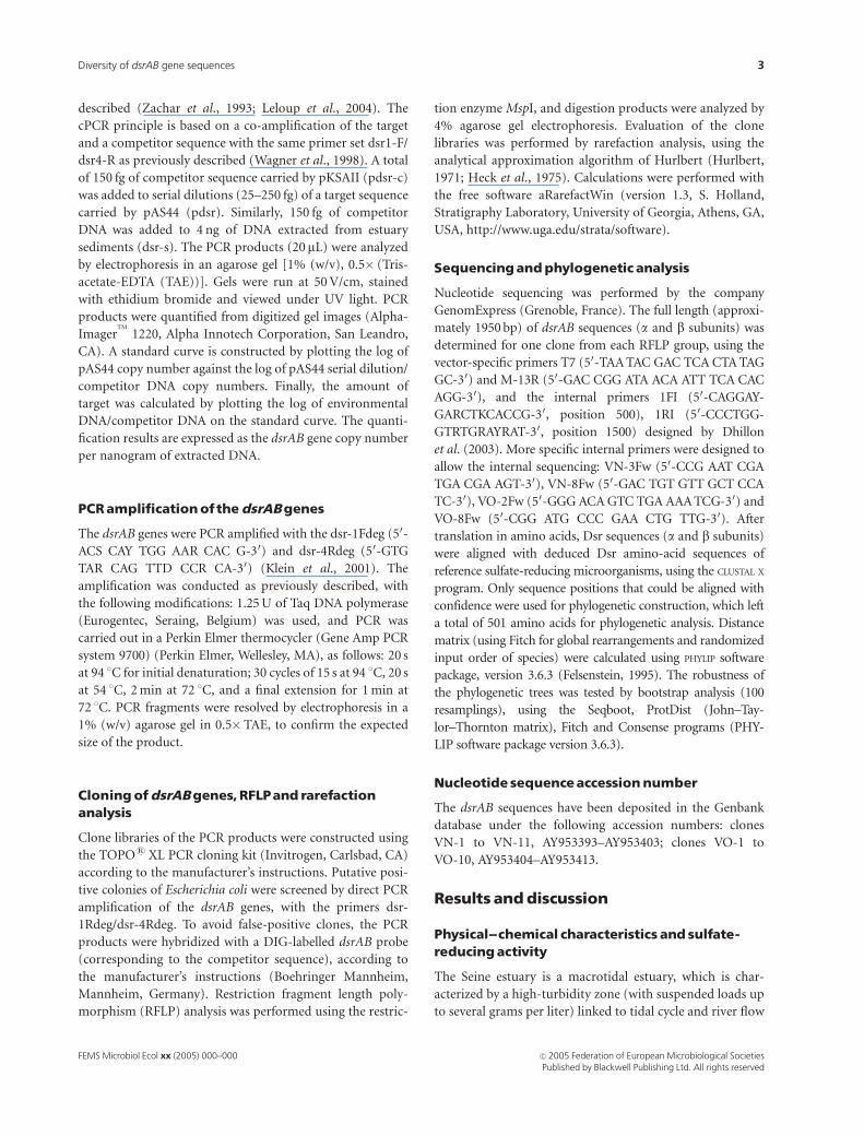
of the Seine estuary (Fig. 1) (Guezennec et al., 1999).
Sediment cores (30 cm long) were collected in June and
July 2001. After cutting 2 cm thick sections, approximately
25 g of each section (wet weight) was transferred to the
laboratory in a sterile polypropylene tube at 4 1C. Subsam-
ples (1 g wet weight) were frozen at � 20 1C for subsequent
molecular analysis (competitive PCR and cloning). For the
determination of sulfate reduction rates, an undisturbed
core was conditioned in a N2 atmosphere before transport to
the laboratory.
Sulfate reduction rate determination
Sulfate reduction rates were determined as previously de-
scribed by Thode-Andersen & J�rgensen (1989). The total
activity of 35S radio-tracer was determined by b-counting of
the S0, AVS and CRS fractions. The sulfate reduction rates
calculated for each of the three fractions were summed, and
are referred to as Sulfate Reduction Rates (SRRs), expressed
in nMol SO42� cm�3 day�1.
Nucleic acid extraction
Total DNA was extracted from 0.5 g sediment samples (wet
weight) with a Bio-101 FastDNA Spin kit in combination
with the FastPrep FP120 bead beating system (MP Biomedi-
cals, Illkirch, France) according to the manufacturer’s in-
structions. Crude total DNA was purified by elution through
Elutip-D columns (Schleicher & Schuell, Dassel, Germany).
The concentration of the resulting DNA was estimated by
UV transillumination of samples spotted on agarose plates
containing ethidium bromide (Sambrook et al., 1989).
Quantificationofthe dsrABgenesbycompetitivePCR
The dsrAB genes present in DNA extracted from sediment
samples were quantified by competitive PCR as previously
Fig. 1. Map of the Seine estuary, Guezennec
et al. (1999). Arrows indicate the two study
sites (Northern and Oissel intertidal mudflats).
kp, kilometric point. This is a unit defined by
the financial Agency of the Seine Normandy
basin and set to kp 0 at Pont Marie in Paris.
FEMS Microbiol Ecol xx (2005) 000–000c� 2005 Federation of European Microbiological SocietiesPublished by Blackwell Publishing Ltd. All rights reserved
2 J. Leloup et al.
described (Zachar et al., 1993; Leloup et al., 2004). The
cPCR principle is based on a co-amplification of the target
and a competitor sequence with the same primer set dsr1-F/
dsr4-R as previously described (Wagner et al., 1998). A total
of 150 fg of competitor sequence carried by pKSAII (pdsr-c)
was added to serial dilutions (25–250 fg) of a target sequence
carried by pAS44 (pdsr). Similarly, 150 fg of competitor
DNA was added to 4 ng of DNA extracted from estuary
sediments (dsr-s). The PCR products (20mL) were analyzed
by electrophoresis in an agarose gel [1% (w/v), 0.5� (Tris-
acetate-EDTA (TAE))]. Gels were run at 50 V/cm, stained
with ethidium bromide and viewed under UV light. PCR
products were quantified from digitized gel images (Alpha-
ImagerTM
1220, Alpha Innotech Corporation, San Leandro,
CA). A standard curve is constructed by plotting the log of
pAS44 copy number against the log of pAS44 serial dilution/
competitor DNA copy numbers. Finally, the amount of
target was calculated by plotting the log of environmental
DNA/competitor DNA on the standard curve. The quanti-
fication results are expressed as the dsrAB gene copy number
per nanogram of extracted DNA.
PCRamplificationofthedsrABgenes
The dsrAB genes were PCR amplified with the dsr-1Fdeg (50-
ACS CAY TGG AAR CAC G-30) and dsr-4Rdeg (50-GTG
TAR CAG TTD CCR CA-30) (Klein et al., 2001). The
amplification was conducted as previously described, with
the following modifications: 1.25 U of Taq DNA polymerase
(Eurogentec, Seraing, Belgium) was used, and PCR was
carried out in a Perkin Elmer thermocycler (Gene Amp PCR
system 9700) (Perkin Elmer, Wellesley, MA), as follows: 20 s
at 94 1C for initial denaturation; 30 cycles of 15 s at 94 1C, 20 s
at 54 1C, 2 min at 72 1C, and a final extension for 1 min at
72 1C. PCR fragments were resolved by electrophoresis in a
1% (w/v) agarose gel in 0.5�TAE, to confirm the expected
size of the product.
CloningofdsrABgenes, RFLPand rarefactionanalysis
Clone libraries of the PCR products were constructed using
the TOPOs XL PCR cloning kit (Invitrogen, Carlsbad, CA)
according to the manufacturer’s instructions. Putative posi-
tive colonies of Escherichia coli were screened by direct PCR
amplification of the dsrAB genes, with the primers dsr-
1Rdeg/dsr-4Rdeg. To avoid false-positive clones, the PCR
products were hybridized with a DIG-labelled dsrAB probe
(corresponding to the competitor sequence), according to
the manufacturer’s instructions (Boehringer Mannheim,
Mannheim, Germany). Restriction fragment length poly-
morphism (RFLP) analysis was performed using the restric-
tion enzyme MspI, and digestion products were analyzed by
4% agarose gel electrophoresis. Evaluation of the clone
libraries was performed by rarefaction analysis, using the
analytical approximation algorithm of Hurlbert (Hurlbert,
1971; Heck et al., 1975). Calculations were performed with
the free software aRarefactWin (version 1.3, S. Holland,
Stratigraphy Laboratory, University of Georgia, Athens, GA,
USA, http://www.uga.edu/strata/software).
Sequencingandphylogenetic analysis
Nucleotide sequencing was performed by the company
GenomExpress (Grenoble, France). The full length (approxi-
mately 1950 bp) of dsrAB sequences (a and b subunits) was
determined for one clone from each RFLP group, using the
vector-specific primers T7 (50-TAA TAC GAC TCA CTA TAG
GC-30) and M-13R (50-GAC CGG ATA ACA ATT TCA CAC
AGG-30), and the internal primers 1FI (50-CAGGAY-
GARCTKCACCG-30, position 500), 1RI (50-CCCTGG-
GTRTGRAYRAT-30, position 1500) designed by Dhillon
et al. (2003). More specific internal primers were designed to
allow the internal sequencing: VN-3Fw (50-CCG AAT CGA
TGA CGA AGT-30), VN-8Fw (50-GAC TGT GTT GCT CCA
TC-30), VO-2Fw (50-GGG ACA GTC TGA AAA TCG-30) and
VO-8Fw (50-CGG ATG CCC GAA CTG TTG-30). After
translation in amino acids, Dsr sequences (a and b subunits)
were aligned with deduced Dsr amino-acid sequences of
reference sulfate-reducing microorganisms, using the CLUSTAL X
program. Only sequence positions that could be aligned with
confidence were used for phylogenetic construction, which left
a total of 501 amino acids for phylogenetic analysis. Distance
matrix (using Fitch for global rearrangements and randomized
input order of species) were calculated using PHYLIP software
package, version 3.6.3 (Felsenstein, 1995). The robustness of
the phylogenetic trees was tested by bootstrap analysis (100
resamplings), using the Seqboot, ProtDist (John–Tay-
lor–Thornton matrix), Fitch and Consense programs (PHY-
LIP software package version 3.6.3).
Nucleotide sequenceaccessionnumber
The dsrAB sequences have been deposited in the Genbank
database under the following accession numbers: clones
VN-1 to VN-11, AY953393–AY953403; clones VO-1 to
VO-10, AY953404–AY953413.
Results anddiscussion
Physical--chemical characteristics and sulfate-reducingactivity
The Seine estuary is a macrotidal estuary, which is char-
acterized by a high-turbidity zone (with suspended loads up
to several grams per liter) linked to tidal cycle and river flow
FEMS Microbiol Ecol xx (2005) 000–000 c� 2005 Federation of European Microbiological SocietiesPublished by Blackwell Publishing Ltd. All rights reserved
3Diversity of dsrAB gene sequences
(Fig. 1). The estuary can be divided in three areas: the
marine estuary, which is submitted to a strong marine
influence; the middle estuary, corresponding to the mixing-
zone between the marine and the fresh waters [kilometric
point (kp) 364–324, limit of the saline intrusion]; and the
upper estuary (kp 324–202, Poses dam), which corresponds
to freshwaters poorly influenced by the tide (Fig. 1). Two
sampling sites were studied: the Northern intertidal mud-
flat, at the mouth of the Seine estuary, and the Oissel
intertidal mudflat, in the upper estuary (kp 229) (Fig. 1).
The Northern intertidal mudflat is located in the mixing-
zone, the hydrodynamic is mainly controlled by the tide, and
the sediments have marine characteristics: the porewater has
salinity in the range 15–20 Practical Salinity Units (PSU),
and a high sulfate concentration (810 mg L� 1) (Table 1).
The Oissel intertidal mudflat is located in the freshwater
zone, and is poorly influenced by the tide. The sediments
have freshwater characteristics: the porewater has a salinity
of about 0.1–0.3 PSU, and a low sulfate concentration
(34 mg L�1) (Table 1).
Sulfate reduction rates and dsrAB gene copy number as a
quantitative measure of SRM abundance were determined
from both sites. For the mixing-zone mudflat, we obtained
6630� 1060 copies of dsrAB genes per nanogram DNA
and a SRR value of 159� 19 nmol SO42� cm�3 day�1. For
the freshwater mudflat, we obtained 1380� 221 copies of
dsrAB genes per nanogram DNA and a SRR value of 176�25 nmol SO4
2� cm�3 day�1. The two sites present high
sulfate reduction activity, but SRM abundance was five-fold
higher in the mixing-zone than the freshwater sediments. As
the specific sulfate reduction rate per cell can also vary, one
explanation could be that the SRMs present in the fresh-
water mudflat show a higher specific cellular SRR than those
in the mixing-zone mudflat (Boudreau & Westrich, 1984;
Table 1. Description and physical–chemical characteristics of the study
sites
Location
Temperature
( 1C)
Eh
(mV) pH
[SO42� ]
(mg L�1)
Salinity
(PSU)
Mixing-zone
mudflat (VN )
17 �150 7.22 810 15–20
Freshwater
mudflat (VO)
22 �160 6.75 34 0.1–0.3
PSU, practical salinity units.
1 2 3 4 5 6 7 8 9 10 11 12 13
100 bp
500 bp
1000 bp
1 2 3 4 5 6 7 8 9 10 11 12
100 bp
500 bp
1000 bp
(a)
(b)
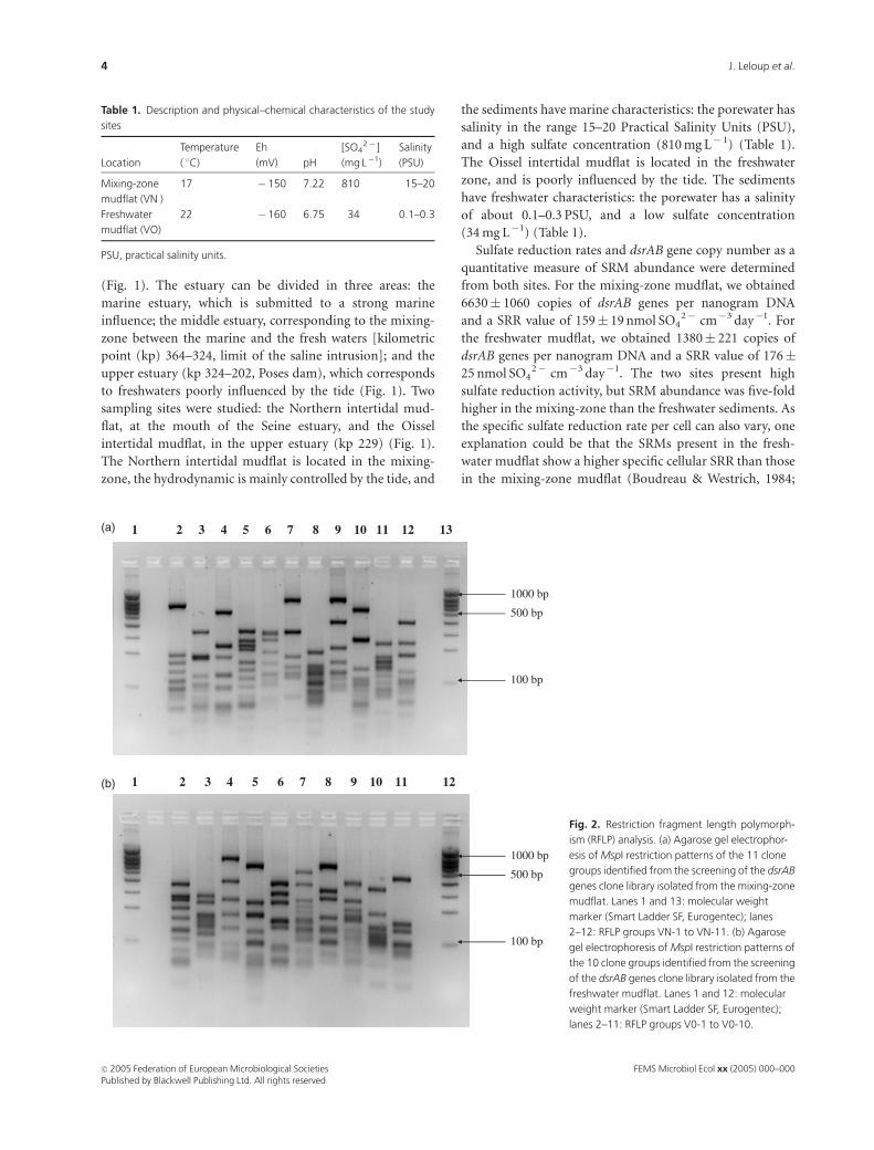
Fig. 2. Restriction fragment length polymorph-
ism (RFLP) analysis. (a) Agarose gel electrophor-
esis of MspI restriction patterns of the 11 clone
groups identified from the screening of the dsrAB
genes clone library isolated from the mixing-zone
mudflat. Lanes 1 and 13: molecular weight
marker (Smart Ladder SF, Eurogentec); lanes
2–12: RFLP groups VN-1 to VN-11. (b) Agarose
gel electrophoresis of MspI restriction patterns of
the 10 clone groups identified from the screening
of the dsrAB genes clone library isolated from the
freshwater mudflat. Lanes 1 and 12: molecular
weight marker (Smart Ladder SF, Eurogentec);
lanes 2–11: RFLP groups V0-1 to V0-10.
FEMS Microbiol Ecol xx (2005) 000–000c� 2005 Federation of European Microbiological SocietiesPublished by Blackwell Publishing Ltd. All rights reserved
4 J. Leloup et al.
Capone & Kiene, 1988; Ravenschlag et al., 2000). Moreover,
it has already been reported that sulfate reduction rates
could be still important in the freshwater environment,
sometimes showing similar rates to marine sediments (Bak
& Pfenning, 1991; Trimmer et al., 1997; Purdy et al., 2002).
Freshwater-adapted SRMs have been described to have a
greater affinity for sulfate (Km = 5–68mM) than marine-
adapted SRMs (Km = 200 mM), allowing the freshwater
SRMs to reduce sulfate at much lower sulfate concentrations
(Sass et al., 1997; Trimmer et al., 1997). As physiological
differences between marine- and freshwater-adapted SRMs
can also be reflected in phylogenetic differences between
these sulfate-reducing communities, the diversity of dsrAB
sequences was investigated (Trimmer et al., 1997; Purdy
et al., 2002).
Phylogenetic analysisofthedsrABsequences
Clone libraries were obtained for the mixing-zone and
freshwater mudflats (201 and 127 positive clones, respec-
tively). The RFLP analysis, using the MspI restriction
enzyme, shows 11 different patterns for the mixing-zone
mudflat, and 10 different patterns for the freshwater mudflat
(Fig. 2a and b). The abundance of the clones in each RFLP
group in the libraries is shown in Table 2. After grouping by
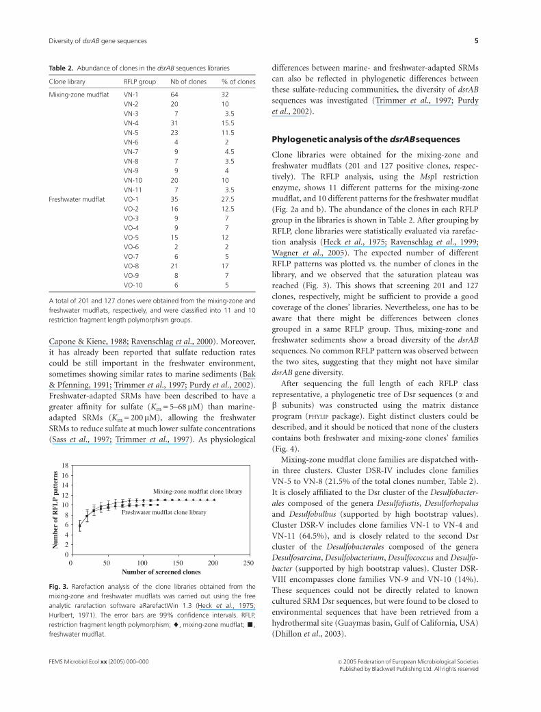
RFLP, clone libraries were statistically evaluated via rarefac-
tion analysis (Heck et al., 1975; Ravenschlag et al., 1999;
Wagner et al., 2005). The expected number of different
RFLP patterns was plotted vs. the number of clones in the
library, and we observed that the saturation plateau was
reached (Fig. 3). This shows that screening 201 and 127
clones, respectively, might be sufficient to provide a good
coverage of the clones’ libraries. Nevertheless, one has to be
aware that there might be differences between clones
grouped in a same RFLP group. Thus, mixing-zone and
freshwater sediments show a broad diversity of the dsrAB
sequences. No common RFLP pattern was observed between
the two sites, suggesting that they might not have similar
dsrAB gene diversity.
After sequencing the full length of each RFLP class
representative, a phylogenetic tree of Dsr sequences (a and
b subunits) was constructed using the matrix distance
program (PHYLIP package). Eight distinct clusters could be
described, and it should be noticed that none of the clusters
contains both freshwater and mixing-zone clones’ families
(Fig. 4).
Mixing-zone mudflat clone families are dispatched with-
in three clusters. Cluster DSR-IV includes clone families
VN-5 to VN-8 (21.5% of the total clones number, Table 2).
It is closely affiliated to the Dsr cluster of the Desulfobacter-
ales composed of the genera Desulfofustis, Desulforhopalus
and Desulfobulbus (supported by high bootstrap values).
Cluster DSR-V includes clone families VN-1 to VN-4 and
VN-11 (64.5%), and is closely related to the second Dsr
cluster of the Desulfobacterales composed of the genera
Desulfosarcina, Desulfobacterium, Desulfococcus and Desulfo-
bacter (supported by high bootstrap values). Cluster DSR-
VIII encompasses clone families VN-9 and VN-10 (14%).
These sequences could not be directly related to known
cultured SRM Dsr sequences, but were found to be closed to
environmental sequences that have been retrieved from a
hydrothermal site (Guaymas basin, Gulf of California, USA)
(Dhillon et al., 2003).
0
2
4
6
8
10
12
14
16
18
0 50 100 150 200 250Number of screened clones
Num
ber
of R
FL
P p
atte
rns
Mixing-zone mudflat clone library
Freshwater mudflat clone library
Fig. 3. Rarefaction analysis of the clone libraries obtained from the
mixing-zone and freshwater mudflats was carried out using the free
analytic rarefaction software aRarefactWin 1.3 (Heck et al., 1975;
Hurlbert, 1971). The error bars are 99% confidence intervals. RFLP,
restriction fragment length polymorphism; ~, mixing-zone mudflat; ’,
freshwater mudflat.
Table 2. Abundance of clones in the dsrAB sequences libraries
Clone library RFLP group Nb of clones % of clones
Mixing-zone mudflat VN-1 64 32
VN-2 20 10
VN-3 7 3.5
VN-4 31 15.5
VN-5 23 11.5
VN-6 4 2
VN-7 9 4.5
VN-8 7 3.5
VN-9 9 4
VN-10 20 10
VN-11 7 3.5
Freshwater mudflat VO-1 35 27.5
VO-2 16 12.5
VO-3 9 7
VO-4 9 7
VO-5 15 12
VO-6 2 2
VO-7 6 5
VO-8 21 17
VO-9 8 7
VO-10 6 5
A total of 201 and 127 clones were obtained from the mixing-zone and
freshwater mudflats, respectively, and were classified into 11 and 10
restriction fragment length polymorphism groups.
FEMS Microbiol Ecol xx (2005) 000–000 c� 2005 Federation of European Microbiological SocietiesPublished by Blackwell Publishing Ltd. All rights reserved
5Diversity of dsrAB gene sequences
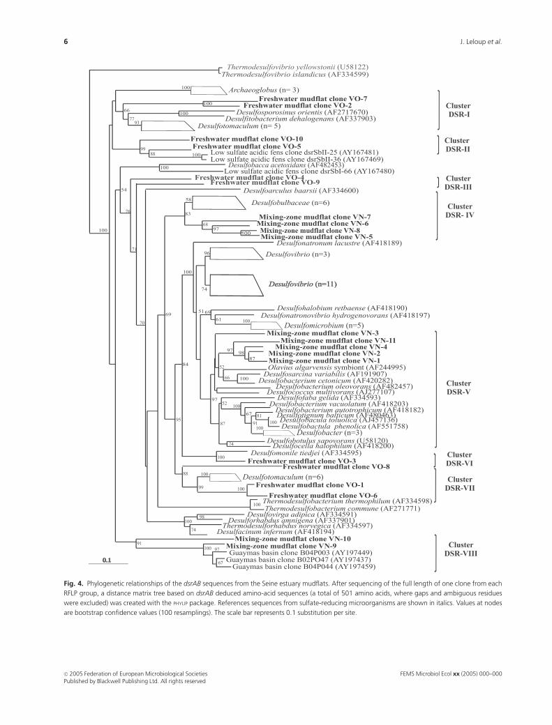
Fig. 4. Phylogenetic relationships of the dsrAB sequences from the Seine estuary mudflats. After sequencing of the full length of one clone from each
RFLP group, a distance matrix tree based on dsrAB deduced amino-acid sequences (a total of 501 amino acids, where gaps and ambiguous residues
were excluded) was created with the PHYLIP package. References sequences from sulfate-reducing microorganisms are shown in italics. Values at nodes
are bootstrap confidence values (100 resamplings). The scale bar represents 0.1 substitution per site.
FEMS Microbiol Ecol xx (2005) 000–000c� 2005 Federation of European Microbiological SocietiesPublished by Blackwell Publishing Ltd. All rights reserved
6 J. Leloup et al.
For the mixing-zone mudflat, presenting high salinity
and sulfate concentration, the major part of the Dsr
sequences (86%) were closely related to those of the SRMs
from the Desulfobacterales. These microorganisms are
Gram-negative SRMs that have been isolated in marine
environment; the genera Desulfofustis and Desulfobulbus
were described as marine and estuarine SRMs (Friedrich
et al., 1996; Ravenschlag et al., 1999; Brandt et al., 2001).
In previous studies of marine and estuarine sediments,
similar results were obtained: Desulfobacter was the predomi-
nant genus, representing more than 50% in marine sediments
from the Tama River (Japan) (Purdy et al., 2001). Using 16S
rRNA gene analysis, the sulfate-reducing community in
Colne Estuary was shown to be dominated by the genera
Desulfobacterium, Desulfobacter and Desulfobulbus (Purdy
et al., 1997). More recently, the genus Desulfosarcina and
microorganisms belonging to the Desulfobulbaceae were
numerically abundant in the Wadden Sea, and in particular
Desulfosarcina appears to be a numerically abundant group of
the SRMs (Ravenschlag et al., 2000; Mußmann et al., 2005).
Freshwater mudflat clone families are dispatched within
five clusters. Cluster DSR-I includes families VO-2 and VO-7
(17.5% of the total clones number, Table 2), and is closely
related to the Dsr sequences of Gram-positive Desulfotoma-
culum species and sulfite-reducing microorganisms Desulfos-
porosinus and Desulfitobacterium (supported by high
bootstrap values). Cluster DSR-VII includes clone families
VO-1, VO-6 and VO-8 (46.5%). These sequences are also
closely related to the Dsr sequences from Gram-positive
Desulfotomaculum species (supported by high bootstrap
values). As also observed by Klein et al. (2001), with 16S
rRNA gene and Dsr phylogenies, the Dsr sequences of Gram-
positive SRMs do not form a monophyletic lineage, the
authors postulated that this feature probably reflects a lateral
gene transfer event. Cluster DSR-II encompasses clone
families VO-5 and VO-10 (17%). These two sequences are
affiliated directly to the clones dsrSbII-25 and dsrSbII-36,
which were retrieved from a low-sulfate forested catchment in
Germany (Loy et al., 2004). Cluster DSR-VI is composed of
the clone family VO-3 (7%). It is affiliated to the Dsr
sequence of Desulfomonile tiedjei, a mesophilic Gram-nega-
tive SRM that grows preferentially in a freshwater medium
(Widdel & Bak, 1992). Cluster DSR-III corresponds to clone
families VO-4 and VO-9 (14%); these sequences could not be
related to known SRM Dsr sequences, and thus might
represent unidentified microorganisms.
The major part of the Dsr sequences (81%), therefore,
was closely related to those of freshwater SRMs, especially
Gram-positive SRMs. These microorganisms have been
described as growing best at low salt concentrations. They
were retrieved preferentially from aquifers and freshwater
sediments presenting low sulfate concentrations (Widdel,
1992; Hansen, 1994; Utkin et al., 1994; Sass et al., 1997;
Stackebrandt et al., 1997; Detmers et al., 2004). Recently
Detmers et al. (2004) showed by fluorescence in situ
hybridization (FISH) that Desulfotomaculum spp. was dom-
inating the sulfate-reducing community present in a pristine
low-sulfate aquifer (Germany). By investigating the phylo-
genetic analysis of dsrAB genes in pristine areas of the
Florida Everglades (Homestead, FL, USA), Castro et al.
(2002) also observed predominance of Desulfotomaculum-
like Dsr sequences. This Gram-positive SRM also has the
ability to survive in low nutrient environments through its
capacity for sporulation (Widdel, 1992; Sass et al., 1997). In
the sediments of Lake Constance, Bak & Pfenning (1991)
found that 50% of the Desulfotomaculum cells were present
as spores. Thus, the difference of dsrAB gene abundance
could also be related to a weak DNA extraction from spores
or weak amplification efficiency from Gram-positive micro-
organisms, leading to an underestimate of their prevalence
(Leloup et al., 2004).
In this study, no Dsr sequences related to the one of
Desulfovibrio species were obtained. Our data indicate that
this microorganism might not play a significant role in the
Seine estuary sediments, and may only be present in low
abundance. Similar results have also been observed in the
Tama River and in a pristine aquifer (Germany) (Detmers
et al., 2004; Purdy et al., 1997).
Conclusions
In this study, we tested the hypothesis that the large
differences in sulfate concentration and salinity in two
contrasting mudflats should be reflected in the abundance
and diversity of SRMs (dsrAB genes). Although SRRs were
not significantly different, the abundance of SRMs was
threefold higher in the mixing-zone than in the freshwater
sediments. Moreover, the phylogenetic analysis has shown
that the dsrAB sequences from Desulfotomaculum were
dominant in the freshwater SRM community, and dsrAB
sequences related to those of the Gram-negative bacteria
were dominant in the mixing-zone sediments. This result is
supported by earlier observations: Gram-positive SRM are
well known to have a well-adapted metabolism for low-
sulfate environment, whereas the Deltaproteobacteria SRMs
are well-adapted to marine environments. The data ob-
tained in our study support the hypothesis that sulfate
availability and salinity play a significant role in structuring
the SRM community in the environment.
Acknowledgements
This work was supported by the Seine-Aval 2, Pnetox 2 and
Rimew scientific research programs. The first author held
a research grant from Seine-Aval 2 scientific research pro-
gram and the Haute Normandie regional council (France).
FEMS Microbiol Ecol xx (2005) 000–000 c� 2005 Federation of European Microbiological SocietiesPublished by Blackwell Publishing Ltd. All rights reserved
7Diversity of dsrAB gene sequences
Prof. R. Amann is acknowledged for critical reading of the
manuscript. We thank Ms D. Moscato for help in English,
and Dr D. Boust for sulfate reduction rates.
Reference
Bak F & Pfenning N (1991) Sulfate-reducing bacteria in littoral
sediment of Lake Constance. FEMS Microbiol Ecol 85: 43–52.
Bally G, Mesnage V, Deloffre J, Clarisse O, Lafite R & Dupont J-P
(2004) Chemical characterization of pore waters of Seine
estuary intertidal mudflats: relations with erosion-deposition
cycles. Mar Poll Bull 49: 163–173.
Barton LL & Tomei F A (1995) Characteristics and activities of
sulfate-reducing bacteria. Sulfate-Reducing Bacteria, Vol. 8
(Barton LL, eds), pp. 1–32. Peplum Press, New York.
Boudreau BP & Westrich JT (1984) The dependence of bacterial
sulfate reduction on sulfate concentration in marine
sediments. Geochim Cosmochim Acta 48: 2503–2516.
Brandt KK, Vester F, Jensen AN & Ingvorsen K (2001) Sulfate
reduction dynamics and enumeration of sulfate-reducing
bacteria in hypersaline sediments of the Great Salt Lake (Utah,
USA). Microb Ecol 41: 1–11.
Capone DG & Kiene RP (1988) Comparison of microbial
dynamics in marine and freshwater sediments: contrasts in
anaerobic carbon catabolism. Limnol Oceanogr 33: 725–749.
Castro H, Reddy KR & Ogram A (2002) Composition and
function of sulfate-reducing prokaryotes in eutrophic and
pristine areas of the Florida Everglades. Appl Environ Microbiol
68: 6129–6137.
Castro HF, Williams NH & Ogram A (2000) Phylogeny of sulfate-
reducing bacteria. FEMS Microbiol Ecol 31: 1–9.
Chang YJ, Peacock AD, Long PE, Stephen JR, McKinley JP,
MacNaughton SJ, Hussain AK, Saxton AM & White DC (2001)
Diversity and characterization of sulfate-reducing bacteria in
groundwater at a uranium mill tailings site. Appl Environ
Microbiol 67: 3149–3160.
Chiffoleau J-F, Auger D, Chartier E, Michel P, Truquet I, Ficht A,
Gonzalez J-L & Romana L-A (2001) Spatiotemporal changes
in cadmium contamination in the Seine estuary. Estuaries 24:
1029–1040.
Detmers J, Strauss H, Bergmann U, Knittel K & Kuever J (2004)
FISH shows that Desulfotomaculum spp. are the dominating
sulfate-reducing bacteria in a pristine aquifer. Microb Ecol 47:
236–242.
Dhillon A, Teske A, Dillon J, Stahl DA & Sogin ML (2003)
Molecular characterization of sulfate-reducing bacteria in the
Guaymas basin. Appl Environ Microbiol 69: 2765–2772.
Elshahed MS, Senko JM, Najar FZ, Kenton SM, Roe BA, Dewers
TA, Spear JR & Krumholz LR (2003) Bacterial diversity and
sulfur cycling in a mesophilic sulfide-rich spring. Appl Environ
Microbiol 69: 5609–5621.
Felsenstein J (1995) PHYLIP: Phylogeny Inference Package.
Department of Genetics, University of Washington, Seattle,
USA. Available online from http://evolution.genetics.
washington.edu/phylip.html.
Fishbain S, Dillon JG, Gough HL & Stahl DA (2003) Linkage of
high rates of sulfate reduction in Yellowstone hot springs to
unique sequences types in the dissimilatory sulfate respiration
pathway. Appl Environ Microbiol 69: 3663–3667.
Friedrich M, Springer N, Ludwig W & Schink B (1996)
Phylogenetic positions of Desulfofustis glycolicus gen. nov., sp.
Nov., and Syntrophobotulus glycolicus gen. nov., sp. Nov., two
new strict anaerobes growing with glycolic acid. Int J Syst Bact
46: 1065–1069.
Guezennec L, Lafite R, Dupont J-P, Meyer R & Boust D (1999)
Hydrodynamics of suspended particulate matter in the tidal
freshwater zone of a macrotidal estuary (Seine, France).
Estuaries 22: 717–727.
Hansen TA (1994) Metabolism of sulfate-reducing prokaryotes.
Antonie Van Leeuwenhoek 66: 165–185.
Heck KL Jr, Van Belle G & Simberloff D (1975) Explicit
calculation of the rarefaction diversity measurement and the
determination of sufficient samples size. Ecology 56:
1459–1461.
Hurlbert SH (1971) The nonconcept of species diversity: a
critique and alternative parameters. Ecology 52: 577–586.
J�rgensen BB (1982) Mineralization of organic matter in the sea
bed – the role of sulphate reduction. Nature 296: 443–645.
Klein M, Friedrich M, Roger AJ, Hugenholtz P, Fishbain S, Abicht
H, Blackall LL, Stahl DA & Wagner M (2001) Multiple lateral
transfers of dissimilatory sulfite reductase genes between
major lineages of sulfate-reducing prokaryotes. Appl Environ
Microbiol 67: 6028–6035.
Leloup J, Quillet L, Oger C, Boust D & Petit F (2004) Molecular
quantification of sulfate-reducing microorganisms (carrying
dsrAB genes) by competitive PCR in estuarine sediments.
FEMS Microbiol Ecol 47: 207–214.
Leloup J, Petit F, Boust D, Deloffre J, Bally G, Clarisse O & Quillet
L (2005) Dynamic of sulfate-reducing microorganisms (dsrAB
genes) in two contrasting mudflats of the Seine Estuary
(France). Microb Ecol, in press.
Li J-H, Purdy KJ, Takii S & Hayashi H (1999) Seasonal changes in
ribosomal RNA of sulfate-reducing bacteria and sulfate-
reducing activity in a freshwater lake sediment. FEMS
Microbiol Ecol 28: 31–39.
Loy A, Kusel K, Lehner A, Drake HL & Wagner M (2004)
Microarray and functional gene analysis of sulfate-reducing
prokaryotes in low-sulfate, acidic fens reveal cooccurence of
recognized genera and novel lineages. Appl Environ Microbiol
70: 6998–7009.
Mußmann M, Ishii K, Rabus R & Amann R (2005) Diversity and
vertical distribution of cultured and uncultured
Deltaproteobacteria in an intertidal mudflat of the Wadden
Sea. Environ Microbiol 7: 405–418.
Nakagawa T, Hanada S, Maruyama A, Marumo K, Urabe T &
Fukui M (2002) Distribution and diversity of thermophilic
sulfate-reducing bacteria within a Cu–Pb–Zn mine (Toyoha,
Japan). FEMS Microbiol Ecol 41: 199–209.
Ouddane B, Abbasse G, Halwani J & Fischer J-C (2004)
Determination of metal partitioning in porewater extracted
FEMS Microbiol Ecol xx (2005) 000–000c� 2005 Federation of European Microbiological SocietiesPublished by Blackwell Publishing Ltd. All rights reserved
8 J. Leloup et al.
from the Seine estuary sediment (France). J Environ Monit 6:
1–12.
Perez-Jimenez JR & Kerkhof LJ (2005) Phylogeography of sulfate-
reducing bacteria among disturbed sediments, disclosed by
analysis of the dissimilatory sulfite reductase genes (dsrAB
genes). Appl Environ Microbiol 71: 1004–1011.
Purdy KJ, Nedwell DB, Embley TM & Takii S (1997) Use of 16S
rRNA-targeted oligonucleotide probes to investigate the
occurrence and selection of sulfate-reducing bacteria in
response to nutrient addition to sediment slurry microcosm
from a Japanese estuary. FEMS Microbiol Ecol 24: 221–234.
Purdy KJ, Nedwell DB, Embley TM & Takii S (2001) Use of 16S
rRNA-targeted oligonucleotide probes to investigate the
distribution of sulfate-reducing bacteria in estuarine
sediments. FEMS Microbiol Ecol 36: 165–168.
Purdy KJ, Embley TM & Nedwell DB (2002) The distribution and
activity of sulfate-reducing bacteria in estuarine and coastal
sediments. Antonie Van Leeuwenhoek 81: 181–187.
Purdy KJ, Nedwell DB & Embley TM (2003) Analysis of the
sulfate-reducing bacterial and methanogenic archaeal
populations in contrasting Antarctic sediments. Appl Environ
Microbiol 69: 3181–3191.
Rabus R, Hansen T & Widdel F (2000) Dissimilatory sulfate- and
sulfur-reducing prokaryotes. The Prokaryotes: an evolving
electronic resource for the microbial community (Dworkin M,
Falkow S, Rosenberg E, Schleifer K-H & Stackebrandt E, eds).
Springer, Berlin. Available online at http://www.
springeronline.com.
Ravenschlag K, Sahm K, Pernthaler J & Amann R (1999) High
bacterial diversity in permanently cold marine sediments. Appl
Environ Microbiol 65: 3982–3989.
Ravenschlag K, Sahm K, Knoblauch C, J�rgensen BB & Amann R
(2000) Community structure, cellular rRNA content and
activity of sulfate reducing bacteria in marine arctic sediments.
Appl Environ Microbiol 66: 3592–3602.
Sambrook J, Fristch EF & Maniatis T (1989) Molecular Cloning: A
Laboratory Manual. 2nd edn. Cold Spring Harbor Laboratory
Press, Cold Spring Harbor, New York.
Sass H, Cypionka H & Babenzien H-D (1997) Vertical
distribution of sulfate-reducing bacteria at the oxic–anoxic
interface in sediments of the oligotrophic Lake Stechlin. FEMS
Microbiol Ecol 22: 245–255.
Stackebrandt E, Sproer C, Rainey FA, Burghardt OP & Hippe H
(1997) Phylogenetic analysis of the genus Desulfotomaculum:
evidence for the misclassification of Desulfotomaculum
guttoideum and description of Desulfotomaculum orientis as
Desulfosporosinus orientis gen. nov., comb. nov. Int J Syst Bact
47: 1134–1139.
Stahl DA, Fishbain S, Klein M, Baker BJ & Wagner M (2002)
Origins and diversification of sulfate-reducing
microorganisms. Antonie Van Leeuwenhoek 81: 189–195.
Thode-Andersen S & J�rgensen BB (1989) Sulfate reduction and
the formation of 35S-labeled FeS, FeS2, and S0 in coastal marine
sediments. Limnol Oceanogr 34: 793–806.
Trimmer M, Purdy KJ & Nedwell DB (1997) Process
measurement and phylogenetic analysis of the sulfate-reducing
bacterial communities of two contrasting benthic sites in the
upper estuary of the Great Ouse, Norfolk, UK. FEMS Microbiol
Ecol 24: 333–342.
Utkin I, Woese C & Wiegel J (1994) Isolation and
characterization of Desulfitobacterium dehalogenans gen.
nov., sp. Nov., an anaerobic bacterium which reductively
dechlorinates chlorophenolic compounds. Int J Syst Bact
44: 613–619.
Wagner M, Roger A, Flax J, Brusseau G & Stahl D (1998)
Phylogeny of dissimilatory reductases supports an early origin
of sulfate respiration. J Bacteriol 180: 2975–2982.
Wagner M, Loy A, Klein M, Lee N, Ramsing NB, Stahl DA &
Friedrich MW (2005) Functional marker genes for
identification of sulfate-reducing prokaryotes. Methods
Enzymol, in press.
Widdel F (1992) The genus Desulfotomaculum. The Prokaryotes:
A Handbook on the Biology of Bacteria: Ecophysiology,
Identification, Application. 2nd edn (Balows A, Truper HG,
Dworkin M, Harder W & Schleifer K-H, eds), pp. 1792–1800.
Springer Verlag, NY, USA.
Widdel F & Bak F (1992) Gram-negative mesophilic sulfate-
reducing bacteria. The Prokaryotes: A Handbook on the
Biology of Bacteria: Ecophysiology, Identification,
Application. 2nd edn (Balows A, Truper HG, Dworkin M,
Harder W & Schleifer K-H, eds), pp. 3353–3378. Springer
Verlag, New York.
Zachar V, Thomas RA & Goustin AS (1993) Absolute
quantification of target DNA: a simple competitive PCR for
efficient analysis of multiple samples. Nucleic Acids Res 21:
2017–2018.
Zverlov V, Klein M, Lucker S, Friedrich MW, Kellermann J, Stahl
DA, Loy A & Wagner M (2005) Lateral gene transfer of
dissimilatory(bi)sulfite reductase revisited. J Bacteriol 187:
2203–2208.
FEMS Microbiol Ecol xx (2005) 000–000 c� 2005 Federation of European Microbiological SocietiesPublished by Blackwell Publishing Ltd. All rights reserved
9Diversity of dsrAB gene sequences