Systems biology of antibiotic production by microorganisms Covering
26
REVIEW www.rsc.org/npr | Natural Product Reports Systems biology of antibiotic production by microorganisms J. Stefan Rokem,† Anna Eliasson Lantz and Jens Nielsen* Received (in Cambridge, UK) 26th March 2007 First published as an Advance Article on the web 30th May 2007 DOI: 10.1039/b617765b Covering: 1995 to 2006 The synthesis of secondary metabolites by microorganisms, specifically antibiotics, is of great scientific and economic importance. The onset (control and regulation) of secondary metabolite formation has and still is intriguing scientists both in industry and academia. Despite many studies, there is little known about the molecular mechanisms underlying the regulation of secondary metabolism. With the recent developments in genomics and further development of advanced post-genomic techniques, it will be possible to apply a more holistic analysis to the regulation of antibiotic production in microorganisms. Here we review current knowledge about the control and regulation of secondary metabolites, with a focus on antibiotics. We will also review developments in the genomics of antibiotic-producing microorganisms, and discuss the use of systems biology for gaining a better understanding of the networks involved in regulation of antibiotic production. 1 Introduction 2 The history of antibiotic production and the lessons from penicillin production 3 The main groups of antibiotics 3.1 Polyketides 3.2 Non-ribosomal polypeptides and derivatives thereof 4 Links between primary and secondary metabolism 5 Nutritional control of antibiotic production 5.1 Carbon source 5.2 Nitrogen source 5.3 Phosphate source 6 Global control and specific regulators in Streptomyces 6.1 Regulatory cascades 6.2 Global and specific regulatory proteins 6.3 ppGpp and relA 6.4 A-factor and other c -butyrolactones 6.5 Oligopeptides as signals 7 Global control in fungi 8 Metabolic engineering for improved antibiotic production 8.1 Improving flux through the biosynthetic pathway 8.2 Precursor supply 8.3 Reduced by-product formation 8.4 Improving other properties 8.5 Heterologous hosts 8.6 New products 9 Perspectives: towards systems biology 10 References Center for Microbial Biotechnology, BioCentrum-DTU, Building 223, Tech- nical University of Denmark, DK-2800 Kgs, Lyngby, Denmark. E-mail: jn@ biocentrum.dtu.dk † Permanent address: Dept. of Molecular Genetics and Biotechnology, Institute of Microbiology, The Hebrew University of Jerusalem, P.O.B. 12272, IL-91120 Jerusalem, Israel. 1 Introduction Antibiotics represent the industrially most important group of secondary metabolites. Our ability to produce these compounds, in most cases by fermentation, has impacted the medical field tremendously. Antibiotics encompass a broad group of chemical compounds with a wide range of different molecular targets in fighting infectious diseases. Some antibiotics have a wide range of applications and are therefore being produced in very large volumes, e.g. penicillins, whereas others have only dedicated applications and are therefore produced only in relatively small amounts, e.g. vancomycin. For all antibiotics there is, however, an interest to obtain high productivities and high titres, as this will ensure cost-effective production. This has traditionally been obtained through classical strain improvement, but with the accumulating information at the genomic level as well as at the molecular level, much insight has been gained on both local and global control of antibiotic production, hence, opening up more targeted approaches. In their classic 1980 paper “Control of Antibiotic Biosynthesis”, Martin and Demain 1 described the accumulated knowledge on this captivating subject. At that time the understanding of control of antibiotics was mainly based on physiological, biochemical and classical genetic evidence. Molecular biology techniques were only just emerging, and they were not well developed for antibiotic- producing microorganisms. Since 1980, a vast effort has been put into research for better understanding of secondary metabolite formation, and there are many reviews on this subject. 2–4 Most reviews are dedicated either to a specific group of antibiotics like b-lactams 5,6 and carbapenems, 7 or to a specific group of microor- ganisms like Streptomyces 4 and Bacillus. 8 Others give accounts of efforts to use available knowledge to improve concentrations of secondary metabolites 9 and the possibilities for producing novel compounds either through combinatorial biosynthesis; 10–12 or through the discovery of cryptic pathways for secondary 1262 | Nat. Prod. Rep., 2007, 24, 1262–1287 This journal is © The Royal Society of Chemistry 2007
Transcript of Systems biology of antibiotic production by microorganisms Covering

REVIEW www.rsc.org/npr | Natural Product Reports
Systems biology of antibiotic production by microorganisms
J. Stefan Rokem,† Anna Eliasson Lantz and Jens Nielsen*
Received (in Cambridge, UK) 26th March 2007First published as an Advance Article on the web 30th May 2007DOI: 10.1039/b617765b
Covering: 1995 to 2006
The synthesis of secondary metabolites by microorganisms, specifically antibiotics, is of great scientificand economic importance. The onset (control and regulation) of secondary metabolite formation hasand still is intriguing scientists both in industry and academia. Despite many studies, there is littleknown about the molecular mechanisms underlying the regulation of secondary metabolism. With therecent developments in genomics and further development of advanced post-genomic techniques, it willbe possible to apply a more holistic analysis to the regulation of antibiotic production inmicroorganisms. Here we review current knowledge about the control and regulation of secondarymetabolites, with a focus on antibiotics. We will also review developments in the genomics ofantibiotic-producing microorganisms, and discuss the use of systems biology for gaining a betterunderstanding of the networks involved in regulation of antibiotic production.
1 Introduction2 The history of antibiotic production and the lessons
from penicillin production3 The main groups of antibiotics3.1 Polyketides3.2 Non-ribosomal polypeptides and derivatives thereof4 Links between primary and secondary metabolism5 Nutritional control of antibiotic production5.1 Carbon source5.2 Nitrogen source5.3 Phosphate source6 Global control and specific regulators in Streptomyces6.1 Regulatory cascades6.2 Global and specific regulatory proteins6.3 ppGpp and relA6.4 A-factor and other c-butyrolactones6.5 Oligopeptides as signals7 Global control in fungi8 Metabolic engineering for improved antibiotic
production8.1 Improving flux through the biosynthetic pathway8.2 Precursor supply8.3 Reduced by-product formation8.4 Improving other properties8.5 Heterologous hosts8.6 New products9 Perspectives: towards systems biology10 References
Center for Microbial Biotechnology, BioCentrum-DTU, Building 223, Tech-nical University of Denmark, DK-2800 Kgs, Lyngby, Denmark. E-mail: [email protected]† Permanent address: Dept. of Molecular Genetics and Biotechnology,Institute of Microbiology, The Hebrew University of Jerusalem, P.O.B.12272, IL-91120 Jerusalem, Israel.
1 Introduction
Antibiotics represent the industrially most important group ofsecondary metabolites. Our ability to produce these compounds,in most cases by fermentation, has impacted the medical fieldtremendously. Antibiotics encompass a broad group of chemicalcompounds with a wide range of different molecular targets infighting infectious diseases. Some antibiotics have a wide rangeof applications and are therefore being produced in very largevolumes, e.g. penicillins, whereas others have only dedicatedapplications and are therefore produced only in relatively smallamounts, e.g. vancomycin. For all antibiotics there is, however,an interest to obtain high productivities and high titres, as thiswill ensure cost-effective production. This has traditionally beenobtained through classical strain improvement, but with theaccumulating information at the genomic level as well as at themolecular level, much insight has been gained on both local andglobal control of antibiotic production, hence, opening up moretargeted approaches.
In their classic 1980 paper “Control of Antibiotic Biosynthesis”,Martin and Demain1 described the accumulated knowledge onthis captivating subject. At that time the understanding of controlof antibiotics was mainly based on physiological, biochemical andclassical genetic evidence. Molecular biology techniques were onlyjust emerging, and they were not well developed for antibiotic-producing microorganisms. Since 1980, a vast effort has been putinto research for better understanding of secondary metaboliteformation, and there are many reviews on this subject.2–4 Mostreviews are dedicated either to a specific group of antibiotics likeb-lactams5,6 and carbapenems,7 or to a specific group of microor-ganisms like Streptomyces4 and Bacillus.8 Others give accountsof efforts to use available knowledge to improve concentrationsof secondary metabolites9 and the possibilities for producingnovel compounds either through combinatorial biosynthesis;10–12
or through the discovery of cryptic pathways for secondary
1262 | Nat. Prod. Rep., 2007, 24, 1262–1287 This journal is © The Royal Society of Chemistry 2007

J. Stefan Rokem received his BSc from the University of Stockhom, Sweden, and his MSc from Imperial College, UK. His PhD was onthe subject of methylotrophic bacteria and was conducted at the Hebrew University of Jerusalem, Israel. After postdoctoral studies onbiosynthesis of antibiotics in Streptomyces he became a senior lecturer at the Department of Molecular Genetics and Biotechnology at TheHebrew University of Jerusalem. Dr Rokem has been a Guest Professor at Yale University, MIT, the Royal Institute of Technology in Swedenand recently at the Danish Technical University. His research interests involve studies of biosynthesis and control of primary and secondarymetabolites from bacteria, yeast and filamentous fungi.
Anna Eliasson Lantz received her MSc in Chemical Engineering from Lund Technical University, Sweden, and later a PhD in AppliedMicrobiology from the same University. She joined the group of Jens Nielsen at the Danish Technical University in 2000 as a postdoctoralfellow and she currently holds the position of Associate Professor in Quantitative Physiology at the Danish Technical University. Herresearch focuses on metabolic engineering of microorganisms, particularly Streptomyces and yeast. She is also involved in research on on-linemonitoring of fermentation processes (Process Analytical Technology).
Jens Nielsen received his MSc in Chemical Engineering from the Danish Technical University and his PhD in Biochemical Engineering fromthe same institution. He did a postdoc at the University of Hannover, and thereafter held various positions at the Danish Technical Universityuntil he was appointed Professor in Fermentation Physiology in 1998. He has been a Guest Professor at MIT, and he serves as an AssociateEditor of several journals. His research interests involve metabolic engineering and systems biology of industrially important microorganisms.
J. Stefan Rokem Anna Eliasson Lantz Jens Nielsen
metabolite formation by non-cultivable microorganisms.13 Theintroduction of recombinant DNA techniques has enabledmolecular engineering approaches to develop new antibiotics.In particular, our understanding of mechanisms and substratespecificities for assembly of polyketides, non-ribosomal peptidesand glycopeptides has allowed the formation of novel compoundsthrough combinatorial biosynthesis, including synthesis of novelstructures based on clinically relevant antibiotics. The joint useof Streptomyces species (where some are effective hosts for pro-duction of heterologous antibiotics, and others are the producersof the antibiotics that are modified) in conjunction with geneticmanipulations has enabled the modification of complex biosyn-thetic pathways.14 Approaches to identify new antibiotics throughthe use of information from genomes of sequenced pathogenicbacteria have also been reviewed,15 in particular through the useof bioinformatics for identification of novel targets for fightingpathogens.16–18 An effort by industry to use genomics with theabove approach, despite the initial high hopes of finding newbacterial targets and novel compounds with unique mechanismsof action, has so far provided relatively low returns.19
Control of secondary metabolite formation typically involvesmany different processes and molecular mechanisms, and in orderto understand how their biosynthesis is controlled it is necessaryto integrate information from molecular studies, physiologicalstudies of antibiotic-producing microorganisms and genomicinformation. Secondary metabolites are accumulated by manydifferent microorganisms, mainly bacteria and fungi, and manydifferent control structures are therefore used. However, it is
still possible to draw some general lessons on control of theproduction of antibiotics, and these lessons are very useful interms of improving the production of new antibiotics.
In order to draw conclusions on control of antibiotic produc-tion, we will review current knowledge on regulation of antibioticproduction, and we will also discuss how the production ofsecondary metabolites is linked to the central metabolism ofantibiotic-producing organisms. Furthermore, with the devel-opments in genomics it has become possible to apply a moreglobal approach to analysis of antibiotic production, and we willtherefore also discuss the impacts of metabolic engineering andsystems biology. We will end our review by giving our perspectiveson the future developments and improvements in the field ofantibiotic production.
2 The history of antibiotic production and the lessonsfrom penicillin production
After the pioneering work of Robert Koch, who proved that manydiseases were linked with microbial infections, there was muchinterest in identifying anti-infectives. After Koch’s discovery inthe 19th century there were many indications of anti-infectivecompounds being produced by different microorganisms, but onlyin 1928, when Alexander Fleming at St Mary’s Hospital MedicalSchool in London discovered penicillin, was it demonstrated thata single chemical compound could serve as an anti-infective.Fleming was studying the pathogenic bacterium Staphylococcus
This journal is © The Royal Society of Chemistry 2007 Nat. Prod. Rep., 2007, 24, 1262–1287 | 1263

aureus, and in connection with this he observed bacterial lysis ona plate with a fungal contamination. He isolated the fungal strainand demonstrated that it was a chemical compound produced bythe fungus that caused the bacterial lysis. The fungus was identifiedas Penicillium notatum (later renamed Penicillium chrysogenum),20
and therefore he called the compound penicillin. Fleming didpursue work on penicillin, but did not have the resources tolook further into the chemistry, and despite the significance ofhis discovery it took more than 10 years before his work waspursued by other research groups. In particular, the instability ofpenicillin to changes in pH (from neutral) was not understoodand affected the ability to obtain larger amounts of penicillin.In the late 1930s, Howard Florey and Ernst Chain at OxfordUniversity started to look for anti-infectives, and immediatelyfound Fleming’s work to be interesting. They managed to purifypenicillin by extraction with water-immiscible solvents, therebyresolving the “pH problem”. They rapidly demonstrated thatpenicillin could be used to treat infected mice, and their findingslead them to proceed with clinical trials, which were successful.This called for large-scale production, and due to the war-timesituation in the UK, Florey and Chain tried to promote interestin the pharmaceutical industry in the USA. At this time there wasno tradition for production of pharmaceuticals using microbialfermentation, and there was therefore some reluctance to startlarge-scale production, as it was believed that there eventuallywould be identified a chemical synthesis route (this was firstdemonstrated in 1956 by Prof. Sheenan at MIT). However,in 1943, after governmental support, the US pharmaceuticalindustry finally initiated large-scale penicillin production usingsubmerged fermentations with P. chrysogenum, and this markedthe first biological-based production of a pharmaceutical. In 1945mass-production took place in the USA, and after the end ofWW2, production was rapidly initiated in a number of Europeancountries. In 1945 Fleming, Florey and Chain were awarded theNobel Prize in Medicine.
The lessons drawn from penicillin led the pharmaceuticalindustry and academic research groups to search for other anti-infectives produced by microorganisms. In the 1940s, SelmanWaksman at Rutgers discovered several new antibiotics, e.g. acti-nomycin, streptomycin and neomycin, produced by soil bacteria,and he defined an antibiotic as “a chemical substance derivedfrom microorganisms which has the capacity of inhibiting growth,and even destroying, other microorganisms in dilute solutions”. In1952 Waksman was awarded the Nobel Prize in Medicine forthe discovery of streptomycin, the first antibiotic effective againsttuberculosis (Table 1).
Table 1 Some key early landmarks in the history of antibiotics
Year Event
1928 Discovery of penicillin by Alexander Fleming1940 Howard Florey and Ernst Chain initiate work on
penicillin at Oxford University1940 Selman Waksman discovers actinomycin1943 Selman Waksman discovers streptomycin1945 Industrial penicillin production is initiatedToday The structure of penicillin is elucidated
6-APA is isolated in crystalline form, allowing for theproduction of semi-synthetic penicillinsAbout 250 antibiotics being produced commercially
Since these early breakthroughs, a very large number ofantibiotics have been discovered (more than 10 000), and about250 antibiotics have found clinical use and are currently beingproduced on a commercial scale. Today the total antibiotics marketexceeds US$25 billion, and it is still rapidly growing. Many ofthe large pharmaceutical companies left the antibiotics field 10–15 years ago, as it was believed that the war against infectiousdiseases had been won, and that antibiotics would becomecommodity pharmaceuticals. However, antibiotics have gainedrenewed interest due to the rise of multi-resistance pathogenicbacteria and due to problems with treatment of fungal infections inimmuno-compromised patients. Thus, there are several new start-up companies that have a business model on discovery, productionand marketing of new antibiotics, and also several of the largepharmaceutical companies have initiated new research programson discovery of antibiotics.21
Today, more than 60 years after the introduction of penicillinas a commercial pharmaceutical, the annual production of thisantibiotic exceeds 60 000 tons, and the cost of penicillin is as lowas US$5 per kg. Most of the penicillin produced is convertedby chemical synthesis to the more effective antibiotics likeampicillin, amoxicillin and cephalexin, and this is in fact a generaltrend, that natural products are further converted by chemicalsynthesis in order to improve their efficacy or pharmacokineticproperties.
In terms of industrial production there are also some interestinglessons to be drawn from the penicillin production process. Theoriginal strain isolated by Fleming, initially called P. notatum,now called P. chrysogenum,20 produced only very low amounts ofpenicillin as a surface culture. In a large screening program it was,however, possible to identify rapidly a strain of P. chrysogenumthat produced almost 100-fold more penicillin, growing undersubmerged conditions. This screening program was organizedby NRRL in Peoria (Illinois, USA), and despite screening ofisolates from all over the world the best producer was found ona cantaloupe at the local fruit market! This strain was furthermutagenized, and better-producing strains were isolated, resultingin a further 10-fold improvement in productivity. The resultingstrain, designated Q176, is believed to be the ancestor of mostindustrial penicillin strains used even today. This rapid increasein productivity was followed by further increases, also obtainedthrough classical strain improvement programs, one driven bythe American–Taiwanese company Panlab, which developed high-yielding penicillin strains for industry (today some of the earlyPanlab strains are available for academic use).22 Today it isestimated that the productivity of strains used in industry are some100 000-fold higher than the original Fleming strain (see Fig. 1).Even though obtaining such high level of production is likely tohave involved a very large number of mutations, still it tells usa key lesson: that microorganisms generally have the capacity toproduce very high levels of antibiotics, despite tight regulation ofboth antibiotic production and of the supply of the precursorsfrom primary metabolism to the antibiotics. It is possible tode-regulate the metabolism such that a large fraction of thecarbon flux can be re-directed towards the antibiotics of interest.Clearly a challenge for the future is to decode these regulatorystructures, such that it will be possible to design high-producingstrains using directed genetic modifications, also called metabolicengineering.
1264 | Nat. Prod. Rep., 2007, 24, 1262–1287 This journal is © The Royal Society of Chemistry 2007

Fig. 1 The development in penicillin production titre over time.
3 The main groups of antibiotics
The variety and complexity of antibiotic compounds producedis enormous. More than half of the drugs approved between1981 and 2002 are based on natural compounds,23 and naturalproducts continue to play an important role in drug discovery.Four of the nine antibacterial and antifungal drugs approved inthe period 2003 to June 2006 were natural products or derivedfrom natural products.24 The wide range of different chemicalstructures found in antibiotics can be divided into the followingchemical groups: polyketides, peptides, glycopeptides, terpenes,nucleosides, alkaloids, aminoglycosides, quinines and coumarins(see examples in Table 2). We will not cover all these groupsof antibiotics, but only focus on peptide antibiotics (includingglycopeptides) and polyketides, as for these antibiotics there hasaccumulated sufficient knowledge to obtain understanding of howtheir production is regulated, hence enabling the application ofmetabolic engineering and systems biology approaches. Theseantibiotics also represent the most important in terms of medicalapplications (Table 2). This section has been written with the aim ofgiving a short background of the mechanisms behind biosynthesisand precursor identification (Table 3), as this will facilitate the
perception of later sections, in particular those dealing with linksto primary metabolism and metabolic engineering.
3.1 Polyketides
The polyketides are an important group of bioactive compoundswith a remarkably wide structural and functional diversity(reviewed by Staunton and Weissman).25 More than 0.3% ofthe known polyketides have been commercialized, which is farhigher than the typical hit rate of <0.1%.12 The biosynthesistakes place in a stepwise manner, whereby coenzyme A esters ofshort fatty acids (most common are acetyl-CoA, propionyl-CoA,malonyl-CoA and methylmalonyl-CoA) are added to the growingchain catalysed by large enzyme complexes named polyketidesynthases (PKSs). Each additional step involves a decarboxylativecondensation, similar to the chain elongation of conventional fattyacid biosynthesis. However, whereas each incorporated unit is fullyreduced in fatty acid biosynthesis, diversity is generated in thepolyketide backbone by allowing an optional degree of reduction.After assembly further structural variety can be achieved bymodifications such as amination, hydroxylation, methylation,oxidation or glycosylation. These modifications are believed tobe crucially important for the biological activity.
Complex polyketides, e.g. macrolides such as erythromycin ortylosin, are synthesized by bacterial type I PKSs, also calledmodular PKSs, which are large multifunctional, multimodularproteins, in which each module is responsible for one condensationcycle and functions in a distinct, non-iterative way. The basicdomains present in each module are acyltransferase (AT), ketosynthase (KS) and acyl carrier protein (ACP), responsible forketo acid activation and coupling (Fig. 2). In addition, a modulemay contain b-ketoreductase (KR), dehydratase (DH) and enoylreductase (ER) domains, which determine the reduced state of theincorporated extender unit. The cofactor NADPH is required forreduction (Table 3). The growing polyketide chain is passed frommodule to module until the completed chain is released from theterminal module by a specific chain-releasing enzyme, thioesterase,which also can be responsible for cyclization.
Table 2 Characteristics of antibiotics classified according to chemical structure
Group and examples Application Microorganism
Peptide antibioticsPenicillins Bacterial meningitis, strep throat and serious infections, Gram+ Penicillium chrysogenumCephalosporins Gram+ and Gram− Streptomyces clavuligerus or
recombinant P. chrysogenumBacitracin Human topical usage, especially against cocci infections Bacillus subtilisPolyketide antibioticsErythromycin Used for people allergic towards penicillins, respiratory tract infections Saccharopolyspora erythreaNystatin Against Candida infections Streptomyces nourseiTetracycline Broad-spectrum, upper respiratory infections; often used in patients with penicillin
allergiesStreptomyces sp.
GlycopeptidesVancomycin Reserved as a drug of “last resort” against Gram+, i.e. methicillin-resistant
Staphylococcus aureusStreptomyces orientalis
AminoglycosidesKanamycin Aminoglycosides are used most widely against Gram− enteric bacteria, especially in
bacteremia and sepsisStreptomyces griseus
Amikacin (semisynthetickanamycin)
For treatment of severe, hospital-acquired infections with multidrug-resistantGram− bacteria such as Pseudomonas aeruginosa, Acinetobacter, and Enterobacter
—
Gentamycin For example, used against P. aeruginosa infections in cystic fibrosis patients Micromonospora sp.
This journal is © The Royal Society of Chemistry 2007 Nat. Prod. Rep., 2007, 24, 1262–1287 | 1265
Table 3 Precursor requirements for different groups of antibiotics (adapted from Gunnarsson et al.46)
Type of antibiotic Antibiotics of this type Precursors (central metabolism) Precursors (intermediate metabolism) Cofactor requirements
Complex polyketides(type I PKS)
Erythromycin Acetyl-CoA NADPH
Nystatin Butyryl-CoAPropionyl-CoAMalonyl-CoAMethylmalonyl-CoAEthylmalonyl-CoAMethoxymalonyl-CoA
Aromatic polyketides(type II PKS)
Tetracyclines Malonyl-CoA NADPH
Actinorhodin
b-Lactams Penicillins Acetyl-CoA L-a-Aminoadipic acid NADPHCephalosporins 2-Oxoglutarate Valine
Pyruvate Cysteine3-Phosphoglycerate
Glycopeptides Vancomycin PEP Tyrosine NADPHTeicoplanin E4P b-HydroxytyrosineDalbavancin Acetyl-CoA p-Hydroxyphenylglycine
Othersa 3,5-DihydroxyphenylglycineG6Pb Other amino acidsa
Sugarsb
Lipidsc
Aminoglucosides Tobramycin G6P 2-Deoxysteptamine ?Tobramycin 2-Oxoglutarate Glucoseamine ATP
N donor; usually glutamate
a While tyrosine, b-hydroxytyrosine and 3,5-dihydroxytyrosine are common to many glycopeptide antibiotics, the heptapeptide backbones of glycopeptidesalso include other amino acids. b Glycopeptide antibiotics also include sugar residues, which typically differ from one glycopeptide to another; thesesugars may be natural or specifically synthesized. c Some glycopeptides, i.e. teicoplanin and dalbavancin, include a fatty acid chain attached to one of thesugars.
Fig. 2 A schematic drawing of the modules and domains in the PKS involved in erythromycin biosynthesis. LM, loading module; AT, acyltransferase;ACP, acyl carrier protein; KS, keto synthase; KR, b-ketoreductase; DH, dehydratase; ER, enoyl reductase.
Furthermore, there are fungal type I PKSs (e.g. those respon-sible for synthesis of lovastatin and 6-methylsalicylic acid), whichalso are multifunctional enzymes, but these work in an iterativemanner and result in aromatic products.
The aromatic bacterial polyketides, such as actinorhodin, tetra-cycline and doxorubicin, are synthesized by type II PKSs, whichare iteratively acting systems consisting of complexes of mono-functional proteins. Aromatization and cyclization are importantsteps in the biosynthesis of these compounds, whereas reductionor reduction/dehydration is involved to a lesser extent. Whereasa variety of coenzyme A esters are used as building blocks fortype I polyketides, type II PKSs only accept malonyl-CoA asan extender unit (Table 3). Acetyl-CoA is not required for thesynthesis of aromatic polyketides with acetate as starter unit,but the starter unit is derived from malonyl-CoA. Type II PKSsconsists of domains with similar activities as type I PKSs, i.e. ACP,AT, KS and KR. In addition, aromatase and cyclase domains arepresent, as well as (uniquely) the presence of a chain length factor(CLF), which is also named KSb, and as the name says determines
the length of the chain. Furthermore, a malonyl-CoA-transacylase(MAT) activity is possibly necessary for biosynthesis of aromaticpolyketides. MAT catalyzes the transfer of the malonyl group frommalonyl-CoA to the ACP.
Recently, a third type of PKS, previously regarded as beingtypical of plant secondary metabolism, was found in bacteria.26
Type III PKSs are also known as chalcone-synthase-like PKSs,and are fairly small proteins which iteratively act as condensingenzymes. In contrast to other PKSs, they do not have a phos-phopantetheinyl arm connecting the growing polyketide chain tothe carrier protein. A type III PKS is involved in the synthesis of3,5-dihydroxyphenylglycine, a precursor of vancomycin.27
3.2 Non-ribosomal polypeptides and derivatives thereof
Another group of natural products with diverse structures andvast activity are the non-ribosomal polypeptides. Peptides man-ufactured by non-ribosomal synthases (NRPSs) include some ofthe most powerful pharmaceuticals known, from penicillin to the
1266 | Nat. Prod. Rep., 2007, 24, 1262–1287 This journal is © The Royal Society of Chemistry 2007

anticancer drug epothilone. Non-ribosomal peptides characteris-tically often contain non-proteinogenic amino acids and D-aminoacids. Moreover, macrocyclic structures and smaller heterocyclicrings are common. Further structural diversity can be achieved bymethylation, halogenation and glycosylation, as well as integrationof fatty acids (reviewed by Schwarzer et al.28).
The biosynthesis of non-ribosomal peptides resembles to a largeextent that of type I polyketides. The responsible enzymes, NRPSs,can also be considered as preassembled, modular factories,whereby one module is responsible for the incorporation ofone amino acid, with domains for activation, condensation etc.The main difference is that the building blocks in this case areamino acids and not acyl-CoA units (thioesters) as for polyketides(Table 3). The three domains present in every module responsiblefor substrate recognition, activation, transport to the catalyticcenters and formation of a peptide bond are adenylation (A),peptidyl carrier protein (PCP) and condensation (C) domains.However, the first module of a NRPS, the initiation module, oftenlacks a C domain. As for the ACP in PKSs, PCPs need to bemodified by a phosphopantetheinyl transferase in order to beactive. Termination and macrocyclisation is catalyzed by a Te-domain. Other domains that can be present are cyclization (Cy)domains, which are responsible for forming small heterocyclicrings in the peptide, and methylation domains (NMe or c-Met).The frequently occurring D-amino acids can be incorporated dueto the specificity of the A domain or, more commonly, the L-formis integrated into the peptide chain and then modified to the D-configuration by an epimerization (E) domain. Activation of thefirst amino acid requires ATP.
The first step in b-lactam biosynthesis, whereby a tripeptideis formed from valine, cysteine and L-a-aminoadipic acid, iscatalysed by an NRPS (see Fig. 9). In P. chrysogenum, L-a-aminoadipic acid is an intermediate on the pathway to lysine,whereas Streptomyces clavuligerus, a producer of cephalosporin,
has a different pathway for lysine which does not involve L-a-aminoadipic acid as an intermediate; specifically, it is formed fromlysine and 2-oxoglutarate. NADPH is also required for b-lactambiosynthesis, and there are indications that the supply of NADPHmight be rate-limiting (see Section 4).
Glycopeptides such as vancomycin are a special group of non-ribosomal peptides. The biosynthesis includes assembly of thepeptide backbone by NRPSs, crosslinking of the peptide by P450oxygenases, halogenation by halogenases and coupling of sugarsby glycosyltransferases. Some glycopeptides also contain a fattyacid tail, which is connected by the action of acyltransferases. Theheptapeptide backbone contains mainly aromatic amino acids,including tyrosine and several non-proteinogenic amino acidsrequiring tyrosine for their biosynthesis (Table 3).
4 Links between primary and secondary metabolism
Primary and secondary metabolism are generally considered tobe regulated differently. However, there are close links betweenthe two, both in terms of precursor supply and through nutrientregulation (both at the metabolic and genetic levels). In termsof precursor supply, thorough studies of antibiotic biosynthesishas led to the recognition that secondary metabolites (antibiotics)are formed from ordinary primary metabolites, and in fact allantibiotics are formed from the 12 precursor metabolites that areused for synthesis of all cellular constituents. Thus metabolismhas a bow-tie structure, whereby many different carbon sourcesare converted to the 12 precursor metabolites, which are thensubsequently converted to all the chemical structures found innature (Fig. 3). Acetyl-CoA is one of the most important precursormetabolites in terms of antibiotic production, as it serves asa precursor for both polyketides and isoprenoids, but oftenadditional chemical groups are added, and these may be formedfrom other precursor metabolites. The 12 precursor metabolites
Fig. 3 The bow-tie structure of metabolism, whereby a wide range of different carbon sources are degraded to only 12 precursor metabolites that aresubsequently converted to the amino acids, lipids, carbohydrates, nucleotides, secondary metabolites, etc. that form the cells. The precursor metabolitesare also converted to various secreted products such as alcohols and organic acids, amino acids, secondary metabolites and extracellular enzymes. Thecatabolism and the anabolism/secondary metabolism are further connected through the common usage of cofactors like ATP, NADH and NADPH. Anon-exhaustive list of carbon sources is provided, and this includes sugars and other carbohydrates, alcohols and acids, and amino acids (which mayserve as both a carbon and a nitrogen source).
This journal is © The Royal Society of Chemistry 2007 Nat. Prod. Rep., 2007, 24, 1262–1287 | 1267

Table 4 Frequency of precursor metabolites etc in the S. coelicolorgenome scale modela
Precursor metabolite No. of reactions Cofactor No. of reactions
Glucose-6P 13 ATP 206Fructose-6P 20 NADH 119Ribose-5P 2 NAD+ 205Erythrose-4P 9 NADPH 59Glyceraldehyde-3P 21 NADP+ 703-Phosphoglycerate 9Phosphoenolpyruvate 21Pyruvate 59Acetyl-CoA 302-Oxoglutarate 55Succinyl-CoA 15Oxaloacetate 15
a The data are taken from the metabolic model developed by Borodinaet al. (2005).29
are: glucose-6P, fructose-6P, ribose-5P, erythrose-4P, glyceralde-hyde-3P, 3-phosphoglycerate, phosphoenolpyruvate, pyruvate,acetyl-CoA, 2-oxoglutarate, succinyl-CoA and oxaloacetate.
Besides the 12 precursor metabolites, there are close linksbetween the primary and secondary metabolism through thecommon use of the cofactors ATP, NADH and NADPH. Thesecofactors play an important role in knitting together the manydifferent parts of the metabolism, and thereby serve an importantrole in global control of the metabolism. This is illustrated inTable 4, in which the number of reactions involving these cofactorsin S. coelicolor are listed. The table also gives the number ofreactions involving the 12 precursor metabolites. It is noteworthythat out of the 971 reactions so far identified in S. coelicolor,more than 21% involve ATP, which is often used as the Gibbs freeenergy input to drive the biosynthesis of antibiotics.29 NADPH isanother frequently used cofactor in antibiotic biosynthesis, namelyas electron acceptor, and as seen from Table 4 this metabolite isinvolved in about 3% of the reactions.
The bow-tie structure of the metabolism and the involvementof precursors and cofactors required for antibiotic biosynthesisin a large number of reactions clearly impose constraints on themetabolism. Thus, transfer from primary metabolism to secondarymetabolism will generally involve a shift from a situation wherethere is a balanced drain of all 12 precursor metabolites to asituation where there is drain of one or a few specific precursormetabolites, e.g. acetyl-CoA for polyketide biosynthesis. If theflux towards the antibiotic is low, this will not cause any problems,but in cases where the flux towards antibiotics is high, this willclearly impose constraints on the primary metabolism. Thereare generally no data available on high-producing strains inthe open literature, but from studies on penicillin production,where relatively high-yielding strains have been studied undercontrolled conditions, it is clearly shown that the drain of precursormetabolites for penicillin biosynthesis is close to the drain forbiomass synthesis (Table 5).30 However, it should be mentionedthat typical antibiotics are produced under conditions wherebiomass growth is constrained, and hence the drain of precursormetabolites for biomass is not at its maximum value. This meansthat theoretically there is often excess capacity for supply ofthe precursor metabolites for antibiotic production, but this will
Table 5 Drain of precursor metabolites (as lmol per g of dry weight perh) for biomass growth and penicillin production by a high-yielding strainof Penicillium chrysogenum under conditions where there is maximumbiomass growth and maximum penicillin production30
Precursormetabolite
Drain for biomasssynthesis
Drain for penicillinsynthesis
3-Phosphoglycerate 40 22Pyruvate 69 442-Oxoglutarate 26 22Acetyl-CoA 57 22
require that the primary metabolism operates in a way similar tohow it operates during growth (see Fig. 4).
Fig. 4 Illustration of the shift in flux distribution from conditions wherethere is primarily drain of precursor metabolites to biomass synthesis (A)to conditions where there is primarily drain of precursors to antibioticsynthesis (B). Clearly, when there is drain of a single (or a few) precursormetabolites for antibiotic production, there needs to be a different fluxdistribution in the cells (illustrated by the thickness of the lines). Thusduring growth there is a high flux through all the key pathways in thecentral carbon metabolism (glycolysis, pentose phosphate pathway andTCA cycle), whereas under conditions of antibiotic production thereis mainly flux towards the precursor metabolite(s) used for antibioticproduction.
The shift in flux distribution in the central carbon metabolismupon a shift from growth to antibiotic production has been studiedin several different antibiotic-producing organisms. Table 6 givesan overview of some of these studies. Using simple metabolitebalancing for quantification of the metabolic fluxes, Jørgensenet al.31 and Henriksen et al.30 found that there is a strong correlationbetween the flux through the pentose phosphate pathway andpenicillin production. They argued that this is due to the large re-quirements for NADPH in the biosynthesis of cysteine, one of theprecursors for penicillin production. Van Gulik et al.32 also appliedstoichiometric modelling and identified four nodes in primarymetabolism where the flux partitioning was significantly affectedby penicillin production, namely around glucose-6-phosphate,3-phosphoglycerate, mitochondrial pyruvate and mitochondrialisocitrate. After investigating the flexibility of these nodes, theyconclude that possible limitations in primary metabolism inconnection to penicillin production are in the supply/regenerationof cofactors (NADPH) rather than in the supply of carbonprecursors. Using 13C-labelled substrates, Christensen et al.33,34
obtained more robust estimates of the metabolic fluxes, and theyconfirmed that the pentose phosphate pathway flux is high duringpenicillin production and that it is higher in a strain having ahigher production level of penicillin. However, they also foundthat the pentose pathway flux in the high-producing strain washigh even under non-penicillin-producing conditions, which seemsto indicate that the high pentose phosphate flux is not directly
1268 | Nat. Prod. Rep., 2007, 24, 1262–1287 This journal is © The Royal Society of Chemistry 2007

Table 6 Overview of studies on flux analysis in antibiotic producing organisms
Organism Objective Reference
Penicillium chrysogenum Quantification of the fluxes and their link to penicillin production Christensen et al. (2000)33
Mapping of pathways for adipate degradation and fluxes in anadipoyl-7-ADCA-producing strain
Thykær et al. (2002)40
Applying a gluconate tracer method for quantification of the flux through PPP Kleijn et al. (2007)35
Streptomyces noursei Linking metabolic fluxes and nystatin production Jonsbu et al. (2001)41
Streptomyces tenebrarius Quantification of fluxes during growth and tobramycin production on glucoseand glycerol
Borodina et al. (2005)38
Nonomuraea sp. ATCC39727 Linking metabolic fluxes with production of the glycopeptides A40926 Gunnarsson et al. (2004)36
coupled with penicillin production, but may be a requirement forhaving a high flux towards penicillin. Kleijn et al.35 used a newlydeveloped method based on co-feeding with trace amounts of13C-labelled gluconate for determination of the flux through thepentose phosphate pathway in connection to penicillin production.Contrary to Christensen et al.,33,34 they conclude that penicillinproduction triggers a higher demand for cytosolic NADPH andconsequently increase in pentose phosphate pathway flux. Theyconclude that the main reason for the discrepancy is that thestudies were performed at different specific growth rates. Duringthe higher growth rates used by Christensen et al.,33,34 the strainalready needs a large supply of NADPH for anabolic reactionsand the increase due to additional penicillin production would behardly measurable. Furthermore, the strain used by Kleijn et al.35
had a higher productivity.In another study, Gunnarson et al.36,37 quantified the metabolic
fluxes in a glycopeptide-producing Nonomuraea species. Theyfound that in this slow-growing bacterium the Entner–Doudoroff(ED) pathway was the main pathway for glucose metabolism,whereas the Emden–Meyerhof–Parnas (EMP) pathway onlycarried a very low flux. However, with the onset of antibioticproduction, the EMP pathway flux increased at the expense of theED pathway flux. The shift in flux distribution was accompaniedwith a decrease in the levels of glucose-6P and fructose-6P, whichare metabolites at the branch point between the two pathways.36
Under all conditions the pentose pathway flux was relatively high,probably due to the requirements for NADPH for both growthand antibiotic formation, and possibly also to ensure the supplyof the precursor metabolite erythrose-4P for production of thearomatic amino acids that are used both for biomass formationand for production of the glycopeptide antibiotics. The activity ofthe ED pathway in an antibiotic-producing microorganism wasquite surprising (before this study, this pathway was not thoughtto be active in filamentous actinobacteria), but it was later shownby Borodina et al. (2005)38 that the ED pathway is also active inStreptomyces tenebrarius, which is used for industrial productionof tobramycin. A further evaluation of several actinobacteriaresulted in the identification of the ED pathway in several species,39
and this pathway therefore seems to be applied in many moreantibiotic-producing microorganisms than traditionally expected.
5 Nutritional control of antibiotic production
A key question in connection with industrial production ofantibiotics is how the change in the external environment resultsin antibiotic accumulation. Here we hypothesize that there areseveral means of transferring this change. It has become more and
more clear that a network or web of intermediate stages/moleculesare involved, and work in concert or independently. In some casesmolecular information is available (A factor, bldA, ppGpp), butthere does not seem to be a general model for regulation ofantibiotic production.
The chemical structure of the antibiotics is a result of themultileveled control of their formation. The control and regulationcan be achieved at different levels from transport and metabolismof extracellular nutrients, through precursor formation and accu-mulation, via the onset of transcription and post-transcriptionalprocesses as well as translational and post-translational control,including stabilities of both mRNA and the actual enzymesinvolved in their biosynthesis (see Fig. 5). Compartmentalization isalso of importance in eukaryotic microorganisms.42 Most of theseevents and features are initiated by some signals that in most caseshave not yet been identified. The use of metabolic engineering43–46 isa comprehensive tool that may allow for interpretation of the largeamount of data that is accumulating, give new insights into thecontrol networks that are involved in the formation of antibiotics,and in particular allow the exploitation of this knowledge inthe design of better-producing strains. This approach requires athorough knowledge of the microorganism and its physiology,biochemistry and genetics, and in the following discussion wetherefore review these different aspects.
Fig. 5 A schematic overview of the many different regulatory structuresthat may be involved in control of antibiotic production.
5.1 Carbon source
The most important medium component for growth and sec-ondary metabolite formation is the carbon source, which generallyalso serves as the energy source. It has always been an intriguingtask to find the optimal carbon source that will allow for
This journal is © The Royal Society of Chemistry 2007 Nat. Prod. Rep., 2007, 24, 1262–1287 | 1269

Table 7 An overview of the interference of some carbon sources with secondary metabolite production (adapted from Sanchez and Demain (2002)2)
Secondarymetabolite Interfering C-source Non-interfering C-source Target of interference
Actinomycin Glucose, glycerol Fructose, galactose Several biosynthetic enzymesCephalosporins Glucose, glycerol, maltose Sucrose, galactose Expandase (R) and cyclase (R)Erythromycin Glucose, glycerol, sucrose, mannose Lactose, L-sorboseKanamycin Glucose Mannose, starchPenicillins Glucose, fructose, sucrose Lactose Phenylacetate uptake system (R) and cyclase (R)Streptomycin Glucose Mannosidostreptomycinase (R)
(R): repression of gene expression.
both good biomass growth and optimal secondary metaboliteformation. There are numerous reports on the effect of differentcarbon sources on growth and secondary metabolite formation,and Table 7 summarizes some of these reports.2 Glucose is themost frequently used carbon source in industrial fermentationprocesses, but often, however, represses antibiotic formation.Penicillin production occurs under glucose repression, and in orderto overcome this phenomenon the first production processes werebased on fermentation with lactose, which was found to be aslowly assimilated carbon source and also not to repress penicillinproduction. Only later was the glucose repression effect overcomeby the introduction of more advanced fermentation processes suchas fed-batch processes. Here glucose is continuously added to thefermentor, such that the glucose concentration in the medium iskept low. As illustrated in Table 7, glucose is, however, not the onlycarbon source that may repress the onset of antibiotic production.
Even though the effects of the carbon source are well docu-mented in many cases, there is generally little information availableof how different carbon sources affect antibiotic formation atthe molecular level. The most frequent effect of carbon sourceson antibiotic production is through carbon catabolite repression,which is the term used when the transcription of genes is repressedand antibiotic production thereby prevented.
Carbon catabolite repression in Streptomyces. The mechanismsfor carbon catabolite repression (ccr) in antibiotic-producingStreptomyces is still not as well defined as for similar systems innon-producing model organisms, like Escherichia coli or Bacillussubtilis. Attempts to identify analogues of ccr, from model or-ganisms, in antibiotic-producing organisms have given ambiguousresults. In Gram-negative bacteria and in low-GC Gram-positivebacteria the PTS transport system plays an important role inccr.47 This is quite different for Streptomyces (high-GC; Gram-positive) that do not contain PTS transport systems. A seriesof recent studies in S. peucetius have shown that there are atleast two independent systems for carbon catabolite repressionin this Streptomyces sp.48–50 One of these systems is ascribed to theactivity of glucose kinase, particularly as this enzyme may playan important role in controlling the glycolytic flux and therebycontrolling the levels of several different catabolite(s) that may beinvolved in carbon catabolite repression.48 Thus, an increase inthe concentrations of products of glucose metabolism have beenshown to influence secondary metabolite production; specifically,the level of fructose 1,6-bis(phosphate) has been found to play arole.48
The role of glucose kinase in ccr has also been compared intwo Streptomyces species (S. coelicolor and S. peucetius). The
tetramer stability of glucose kinase was different in the twospecies studied, and might be involved in ccr since there isalso a difference in ccr between the two species.50 The sameresearch group isolated mutants that were able to grow in thepresence of non-glucose analogues that could not serve as carbonsources.51 These mutants were defective in glucose kinase andtheir phenotype was insensitive to glucose repression of manygenes, including genes encoding enzymes involved in biosynthesisof secondary metabolites (in this case anthracyclines), as well asthose for utilization of alternative carbon sources. However, theinvolvement of glucose kinase in repression must be indirect, asthis enzyme is not likely to serve as a transcription factor (thereare no indications of DNA-binding domains in the enzyme). It istherefore suggested that the enzyme simply affects the glycolyticflux and thereby results in a global response, or that it might takepart in the formation of other catabolites that may be involvedin the onset of repression. The first suggestion is supported bya recent study where a phosphofructokinase deletion mutant ofS. coelicolor was shown to lead to attenuated glycolytic flux, anincreased pentose phosphate flux and an increased productionof actinorhodin.52 This was ascribed to be a consequence ofan altered redox metabolism, i.e. the increased flux through thepentose phosphate pathway resulted in an increased productionof NADPH and this led to increased formation of antibiotics.
A glucose-sensitive galactose-dependent promoter, galP1, hasbeen described for S. coelicolor illustrated with bld mutants.53,54
The bld mutants are pleiotropic and their phenotype shows loss ofaerial mycelium formation, and most are also unable to produceantibiotics.55 In addition, the bld mutants were shown to bedefective in the regulation of galP1, as the promoter in the bldmutants is no longer sensitive to the amount of glucose in thegrowth medium. This indicates that the bld mutants containmutations that affect the regulation of carbon utilization.53,54
Furthermore, the bldA mutants of S. coelicolor were shown tobe suppressed differently on rich versus minimal medium.55,56 Thisseemed to correlate with both the concentration of the phosphatesource and the type of sugar, as for poor carbon sources (e.g.mannitol) a different phenotype was obtained—here the bldAmutant did not show carbon catabolite repression for actinorhodinproduction.57
The effect of glucose as a catabolite repressor is still notfully understood, and even though the concentration in themedium might be high, it does not automatically mean that theconcentration in the cell is at a repressing concentration. Boththe rate of glucose uptake and the rate of intracellular glucosemetabolism will determine the internal glucose level, and therebymay have an influence on carbon catabolite repression.
1270 | Nat. Prod. Rep., 2007, 24, 1262–1287 This journal is © The Royal Society of Chemistry 2007

Carbon catabolite repression in fungi. In filamentous fungithe situation is very different compared with Streptomyces. InAspergillus nidulans the CreA protein was early on shown to beinvolved in ccr.58 CreA is a zinc finger protein, which mediatescarbon catabolite repression through binding to the promoterregion of carbon-catabolite-repressed genes. Recently it was shownthat the onset of ccr in this species is growth-rate-dependent, byusing chemostats at various dilution rates.59 Furthermore, CreAhas been found to involve the transcription of a large number ofgenes, both during growth on glucose and ethanol,60 and the roleof CreA is therefore quite different from that of Mig1, which is theanalogue in the yeast Saccharomyces cerevisiae61 (see Fig. 6).
A protein similar to CreA was also found in the b-lactamproducer Acremonium chrysogenum. This protein, named Cre1,has a 56% sequence homology to CreA of A. nidulans, and is also azinc finger protein.62 It has been shown that the biosynthetic genesinvolved in cephalosporin formation are repressed by the carboncatabolite through Cre1 in the industrial strain A. chrysogenumA3/2.63,64 The situation for penicillin formation by A. nidulanshas still not been resolved, and recently it was stated5 that theregulation of penicillin biosynthesis by repressing carbon sourcesremains to be elucidated. Even though CreA (or Cre1) is very likelyto be involved in ccr of secondary metabolism in filamentous fungi,the upstream mechanisms are not resolved, and considering thequite different transcriptional responses between a CreA deletionstrain of A. nidulans and a mig1 deletion strain of S. cerevisiae,60,61
it is unlikely that the mechanisms are conserved between thesetwo species. Thus, it may not be possible to directly transferinformation from S. cerevisiae to filamentous fungi.
Role of the carbon source. The most desirable phenotype fora good industrial strain for antibiotic production should allowfor quick cell mass formation that results in a high biomassconcentration, followed by conditions where there is no inhi-bition or repression of the activities of the enzymes requiredfor antibiotic production. Clearly, a high biomass concentrationwill potentially have a higher concentration of the catalysts (thebiosynthetic enzymes required for antibiotic production), andit is therefore desirable to get a high biomass concentrationrapidly. There is, however, normally a conflict between growth
and antibiotic production. Through evolution, most microbialcells have evolved towards optimizing their growth, and henceantibiotic production is generally low in natural isolates. Theecological niches where antibiotic-producing microorganisms arenormally found are thought to be scarce in nutrients and oftento involve complex carbohydrates. Thus, as has been revealed bygenomic sequencing of S. coelicolor,65 this Streptomyces speciesholds many genes coding for enzymes involved in degradation ofcomplex carbohydrates and proteins, and thereby the cells cangrow on a wide range of carbon and nitrogen sources.
As mentioned above, glucose is the preferred carbon source inthe fermentation industry due to low cost and high availability.Furthermore, this is a carbon source that is easily assimilated bymost microorganisms, and hence the requirement for rapid growthcan be assured. However, the frequently encountered carboncatabolite repression in many microorganisms limits the use ofglucose as a carbon source for antibiotic production.
The detrimental effect of glucose on b-lactam formation wasdescribed over 50 years ago,66 and as mentioned above this wasovercome by either feeding low doses of glucose or by usinglactose as the carbon source. This knowledge has been of utmostimportance for the design of many other industrial processes forantibiotic production, and it led to the development of the fed-batch process, which is by far the most common mode of operationin industrial fermentations.
One of the more well studied cases of carbon source regulation ispenicillin biosynthesis, and the conclusions are that the regulationseems to act at several different levels (see Fig. 7). Thus, thecarbon source regulates the flux towards one of the precursoramino acids, L-a-amino adipic acid, the rate of activation of theside chain precursors (the conversion of phenoxyacetic acid orphenylacetic acid to their corresponding CoA-esters), the controlof transcription of several of the penicillin biosynthesis genes,and possibly even post-transcriptional regulation of penicillinbiosynthesis genes.5,67–69
The type and amount of the carbon source has different effectson antibiotic formation. This has been illustrated for Streptomyces,where closely related species show different responses to variationin the type of carbon source and the level of the carbon source inthe medium.56 As an example, it has been found that S. lividans
Fig. 6 Comparison of the role of Mig1 in S. cerevisiae and CreA in A. nidulans. Whereas much of the pathway for activation/de-activation of Mig1 hasbeen mapped, little is known of the pathway involved in actication/de-activation of CreA.
This journal is © The Royal Society of Chemistry 2007 Nat. Prod. Rep., 2007, 24, 1262–1287 | 1271

Fig. 7 Influence of glucose at several different levels on penicillinbiosynthesis.
produces actinorhodin when glycerol is used as the sole carbonsource,70 whereas no actinorhodin is formed during growth onglucose. This is quite different from S. coelicolor, which canproduce actinorhodin during growth on glucose. No molecularexplanation for these phenomena is currently available, but it hasbeen found that the expression of the regulatory gene afsR2 inStreptomyces lividans is dependent on the carbon source for cellgrowth.70
5.2 Nitrogen source
The choice of nitrogen source and its concentration in thefermentation medium are also of crucial importance for antibioticproduction. The preferred nitrogen source for most microorgan-isms is ammonium, which is easily assimilated via the glutamatedehydrogenase reaction (by which 2-oxoglutarate is converted toglutamate) or via glutamine synthetase (which converts glutamateto glutamine). The latter reaction has high affinity for ammoniaand is often used for ammonia assimilation at low ammoniumconcentrations. In these cases the glutamine synthetase forms apathway together with glutamate synthetase, often referred to as
the GS-GOGAT pathway. As an easily assimilated nitrogen source,ammonia also serves as a repressor for the utilization of alternativenitrogen sources, and in many cases also causes repression ofantibiotic production (see Table 4). For a few antibiotics theremay, however, also be repression by other nitrogen sources suchas amino acids (see Table 8).2
Nitrogen metabolism has been well studied both in bacteria71,72
and fungi,73 and the complexity of the influence of the nitrogensource, especially on growth, is well described. However, not muchis known on the actual mechanisms behind the observed effects onantibiotic formation, e.g. what the molecular mechanism is thatunderlies repression at high ammonia concentrations.
As mentioned above, the incorporation of ammonia takes placeby glutamate dehydrogenase and/or glutamine synthetase, andeven though S. coelicolor contains both pathways, it is believedthat ammonia is mainly assimilated by glutamine synthetase. Thisis confirmed by the fact that S. coelicolor seems to only containan NADPH-dependent glutamate dehydrogenase (encoded bygdhA or SCO4683), whereas many other bacteria contain bothan NADH- and an NADPH-dependent glutamate dehydrogenase.The enzymes involved in nitrogen metabolism in Streptomyces havebeen shown to function differently from their counterparts in themore well studied enteric bacteria.74 In E. coli, two trimeric signaltransmitter proteins (GlnK, GlnB of the PII family proteins) trans-duces the cellular nitrogen status to uridylyltransferase (GlnD),responsible for the modification of glutamine synthetase. Underconditions of nitrogen limitation (low glutamine), GlnD activitycauses the PII proteins to become highly uridylylated, whereasduring nitrogen excess (high glutamine), the proteins remain un-uridylylated or are specifically de-uridylylated by GlnD. Similargenes, glnK and glnD, were found in Streptomyces coelicolor fromthe genome sequencing project,65 with the difference that glnDcodes for an adenylyltransferase. Mutants of these genes shownormal modification of the glutamine synthetase and neitherthe PII-like protein nor the adenylyltransferase are required forthe regulation of GlnE in response to nitrogen stimuli in S.coelicolor. It is not clear how GlnE-dependent GlnA modificationis regulated. Either GlnE senses the nutritional status directlythrough allosteric interaction with effector molecules such as
Table 8 Nitrogen sources and secondary metabolism
Secondary metabolite Interfering nitrogen source Non-interfering nitrogen source
Actinomycin L-Glutamate, L-alanine, L-phenylalanine, D-valine L-IsoleucineActinorhodin NH4
+ —Cephalosporin C NH4
+, L-lysine L-Asparagine, L-arginineChloramphenicol NH4
+ D-Serine, L-proline, D,L-phenylalanine,D,L-leucine
Erythromycin NH4+ L-isoleucine
Gibberellic acid NH4+ L-glutamine
Leucomycin NH4+ Uric acid
Macbecin L-Tryptophan, p-aminobenzoate, anthranilate —Patulin NH4
+ —Penicillin NH4
+, L-lysine L-GlutamateRifamycin NH4
+ NitrateSpiramycin NH4
+ —Streptomycin NH4
+ L-ProlineStreptothreocin NH4
+ D,L-Aspartate, L-glutamate, D,L-alanine, glycineTetracycline NH4
+ —Trihydroxytoluene NH4
+ —Tylosin NH4
+ L-Valine, L-isoleucine, L-leucine, L-threonine
1272 | Nat. Prod. Rep., 2007, 24, 1262–1287 This journal is © The Royal Society of Chemistry 2007

glutamine, or GlnE regulation requires an unknown mechanismof post-translational control. There are inidcations that the post-translational control involves both a removal of three N-terminalamino acids and adenylylation of a conserved tyrosine of the PIIprotein GlnK.74
In the genome of S. coelicolor, five genes encoding glutaminesynthase homologues have been identified. Two of them, glnA(SCO2198) and glnII (SCO2210), were shown to be functionalin nitrogen metabolism,75 but there are three more genes en-coding putative glutamine synthetases (SCO1613, SCO2241 andSCO6962).29,76 As mentioned above, glutamine synthetase is a keyenzyme in nitrogen metabolism in S. coelicolor and it is bothresponsible for L-glutamine biosynthesis and for assimilation ofnitrogen.76,77 One of the ORFs encoding glutamine synthetasethat have been shown to be functional in nitrogen metabolismis involved in the onset of mycelial differentiation (glnII), theother (glnA) being constitutively expressed.76 The actual role ofthe glutamate synthetases in regulation of secondary metaboliteformation requires further study, but it is thought that the enzymeinvolved in differentiation might also have a role in regulation ofthe formation of secondary metabolites. The control of nitrogenmetabolism in Streptomyces is more complex than in other studiedbacteria. Even though many homologues of proteins involved innitrogen regulation in E. coli have been found, the componentsseem to be interlinked differently and combined with specifictranscriptional OmpR-like regulators (e.g. GlnR and GlnRII), aunique feature for Streptomyces.77
In antibiotic-producing filamentous fungi, GATA-type zincfinger transcription factors, which activate nitrogen structuralgenes, have been identified in the biosynthetic genes for b-lactamformation both in Penicillium chrysogenum and Acremoniumchrysogenum, but not in Aspergillus nidulans.78 Most fungi havea preference for ammonia, glutamine, and glutamate as nitrogensource, but when these nitrogen sources are not available orare present in concentrations low enough to limit growth, manydifferent nitrogen sources can be used, e.g., nitrate, nitrite, purines,amides, most amino acids, and proteins.73 Nitrogen metabolismhas been extensively studied in S. cerevisiae, and here it isfound that there are relatively few genes that are transcriptionallychanged when the nitrogen source changes from preferred nitrogensources like ammonia and glutamine to non-preferred nitrogensources like alanine.79 It is not known how much is conservedbetween S. cerevisiae and filamentous fungi, but future genomicstudies on Aspergillus may provide key information on this issue. Arecent review on the impact of nitrogen-containing compounds onthe biosynthesis of b-lactams in fungi concluded that ammoniumexerts repression, whereas lysine, methionine, glutamate andvaline are involved in both positive and negative regulation.80
Valine is a precursor for b-lactam production, whereas lysine andmethionine may serve as indirect precursors for the biosynthesisof a-aminoadipic acid and cysteine, the two other precursors forb-lactam production.
5.3 Phosphate source
The concentration of phosphate has been shown to have a greatinfluence on many different antibiotic producers with respectto their ability to accumulate the product, e.g. actinorhodin,cephalosporin, clavulanic acid, streptomycin, tetracycline and
vancomycin.2 In most cases a low concentration of phosphateis required for initiation of antibiotic production.
The effect of phosphate concentration on transcription of genesinvolved in candicidin biosynthesis was described as early as1990.81,82 Transcription of the gene encoding PABA synthase,an enzyme required for candicidin production, was found to berepressed in the presence of phosphate, whereas phosphate hada stimulating effect on the total RNA synthesis of Streptomycesgriseus.81 A common mechanism for the influence of phosphateconcentration on a great variety of antibiotics has been elusivefor a long time. There are still question marks remaining on howphosphate regulates antibiotic production, and only recently waslight shed on the involvement of PHO boxes in the control ofantibiotic formation.83,84
PHO boxes are found both in E. coli85 and B. subtilis,86 butdespite many years of searching for similar boxes in Streptomycesand other antibiotic-producing organisms, they were only recentlyidentified in Streptomyces.83 Two eleven-base-pair direct repeatswere found in the promoters of several genes in S. coelicolorand S. lividans,83 and the overall control was mediated by thePhoR–PhoP two-component system. In Streptomyces natalensis,the producer of the antifungal polyene macrolide primarcin, asimilar two-component system has been described,87 with around80% identical amino acid sequences compared with PhoR–PhoPin S. coelicolor, S. avermitilis and S. lividans. PHO boxes (elevenbase pair repeats) are also present in S. natalensis and conservedin the four species of Streptomyces studied so far. It is known thatmacrolide biosynthesis is particularly sensitive to the concentra-tion of phosphate in the growth medium,82,88 but other antibiotics,e.g. glycopeptides like teicoplanin89 and A40926,90 have also beenshown to be inhibited by high phosphate concentration.91
6 Global control and specific regulators inStreptomyces
The control of onset of antibiotic production involves a compli-cated network with many different levels of control, both positiveand negative. The means of control are, as far as they havebeen studied, typically unique for the specific organism, and eventhough similar systems have been found in different organisms,they do not work in exactly the same way.
A multitude of factors have been described as regulatorymechanisms for the onset of antibiotic formation, but typicallythe activation of regulatory cascades that leads to the onset ofantibiotic production is initiated at a “global” level and thendescends to the transcription of dedicated biosynthetic genescoding for the enzyme activities that are responsible for produc-tion of the particular antibiotic.4,5 With the complete genomesequences becoming available for two Streptomyces species, ithas become clear, in part due to the fact of the multitude ofgenes that were assigned as sigma-factors (63 in S. coelicolor65
and 60 in S. avermitilis92), that the regulation of antibioticformation is probably much more complex than was originallythought. In Table 9 the number of sigma factors of the sequencedStreptomycetes and some other actinobacteria are compared withthe Gram-negative Escherichia coli and the Gram-positive Bacillussubtilis.
This journal is © The Royal Society of Chemistry 2007 Nat. Prod. Rep., 2007, 24, 1262–1287 | 1273

Table 9 Sigma-factors in some sequenced actinobacteria compared to E. coli and B. subtilis
Organism Genome size/Mb ORF Total sigma-factors ECF sigma-factors
Streptomyces coelicolor A3(2) 8.3 7846 65 51Streptomyces avermitilis MA-4680 9.0 7577 60 47Mycobacterium tuberculosis H37Rv 4.4 3924 13 10Corynebacterium glutamicumATCC 13032
3.3 3057 7 —
Bacillus subtilis 168 4.2 4100 17 7Escherichia coli K-12 MG1655 4.7 4288 7 2
There is great interest to identify the function of this multitudeof sigma-factors, and hopefully to find their role in the onset of an-tibiotic formation, if any. Four homologues of the principal sigma-factors used for growth have been identified, closely related to r70
in Escherichia coli.93 One sigma-factor, rWhiG, is essential for aerialmycelium septation.94 One group of stress response sigma-factorswith homology to sigma-factors in Bacillus subtilis contains ninehomologues. The 51 sigma-factors remaining in S. coelicolor areconsidered to have extracytoplasmatic functions (ECFs). Only fiveECFs have been characterized, two with roles in morphologicaldifferentiation, one for cell wall homeostasis, one for oxidativestress response and one for light-induced carotegenesis.95–98 Forthose that are studied, the ECF sigma-factor is usually transcribedsimultaneously with other regulators. Among the regulator com-ponents, one frequently has a transmembrane region, working asan anti-sigma-factor, binding and inhibiting the associated sigma-factor. These pairs correspond to the two-component regulatorysystems found in other bacteria.99 Earlier studies of individualsigma-factors allowed for the description of 7 different sigma-factors,93 and their respective uses were assigned.
6.1 Regulatory cascades
The onset of antibiotic formation is controlled by what has beentermed “regulatory cascades”. The specific condition that leadsto activation of antibiotic production will be “sensed”, e.g. byhaving certain metabolite levels or activity of certain enzymes,and a signal is then sent to a specific transcription factor thatactivates the antibiotic biosynthetic genes, e.g. actII-ORF4 foractinorhodin production in S. coelicolor, redD for undecylprodi-giosin production by S. coelicolor, cdaR for calcium-dependentantibiotic production by S. coelicolor, ccaR for cephamycinC and clavulanic acid production by S. clavuligerus, dnrI fordaunorubicin production by S. pecetius, and snoA for nogalamycinproduction by S. nogalater. The signal transduction pathwaysupstream of the transcription factors have only in a few casesbeen mapped, e.g. for the A-factor involvement in streptomycinformation in S. griseus (see below).
In S. coelicolor, one of the pathways starts with a looselyattached membrane protein AfsK, which after activation throughauto-phosphorylation with the help of KbpA, phosphorylatesAfsR present in the cytoplasm. Upon phosphorylation of AfsR,the DNA-binding capability of the protein is greatly enhanced.Other protein kinases also phosphorylate AfsR, indicating itscentral role for numerous activities in the cell. In S. coelicolorthere are at least 40 serine/threonine kinases and at least fiveAfsR-like proteins.100 This demonstrated that, in addition to themultitude of sigma-factors present in this organism, a multitude
of control structures are likely to exist. The presence of a largenumber of protein kinases is quite common in eukaryotic cells,whereas protein phosphorylation has been studied to a muchlesser extent in bacteria, but from the above it seems that proteinphosphorylation cascades are likely to be involved in the controlof antibiotic formation in prokaryotes.
Pathway regulators often have a conserved motif, with a helix–turn–helix at the N-terminus, similar to what is found in OmpRproteins.101 These regulators have been called Streptomyces antibi-otic regulatory proteins (SARPs),102 and are only found in theactinobacteria group. Different types of antibiotics produced byactinobacteria have been found to have transcriptional regulationby these activators.4
6.2 Global and specific regulatory proteins
In the search for better antibiotic-producing strains, many differ-ent mutants have been isolated. Many of these mutants have beenfound to contain multifunctional properties (pleiotropic), oftenbecause the mutations affect multiple pathways, and therefore themutants express several different phenotypic behaviors, e.g. alteredantibiotic production and/or changes in the morphology. Oneclass of mutants that express pleotropic effects are the bld mutants,already mentioned in our discussion on carbon catabolite repres-sion. These mutants were originally identified in S. coelicolor.55
The bld mutants have easily detectable phenotypes since thereis no formation of aerial mycelium, and they have also lost thecapability for production of all four known antibiotics produced byS. coelicolor.103 The bldA,103 bldB54 and bldD104 mutants show bothphenotypes, i.e. no antibiotic production and no aerial myceliumformation. The bldA gene product has been found to be a rarecodon leucyl-tRNA (TTA) and has been linked to actinorhodinproduction through the transcriptional regulator actII-ORF4 (aregulator of the actinorhodin gene cluster) that contains the UUAcodon. In total there are 145 genes in the S. coelicolor chromosomethat contain the rare TTA codon, but most of them are presentin functionally unkown, extrachromosomal or weakly expressedgenes.105 The bldB gene encodes a small acidic homodimericprotein, for which the mechanism of action is unknown.106 Most ofthe bld mutations are reversed by growth on poor carbon sources(e.g. mannitol), but the defects conferred by bldB can not bereversed in this way. The bldD gene of S. coelicolor, while requiredfor antibiotic production and morphological differentiation, wasshown to be non-essential for viability. On minimal mediumcontaining glucose as carbon source, the bldD53 mutant (theonly bldD mutation known) produced none of the four antibioticspossible produced by S. coelicolor. The biochemical role for BldDwas shown to be a DNA-binding protein capable of binding to
1274 | Nat. Prod. Rep., 2007, 24, 1262–1287 This journal is © The Royal Society of Chemistry 2007

its own promoter/operator, but also to several developmentalgenes.107 The morphological defects, but not the loss of antibioticproduction, are overcome by growth of the mutant on minimalmedium containing mannitol as carbon source.108
For regulation on a higher level (not pathway-specific) a numberof proteins have been shown to have effect on a number ofbiosynthetic pathways for secondary metabolite production. Onesuch protein is AfsR in S. coelicolor. This is a SARP, andupon phosphorylation this 63-amino-acid protein complexes withRNA polymerase and thereby activates the transcription of thegene encoding AfsS (see Fig. 8). AfsS is a transcription factorthat then further activates the transcription of pathway-specifictranscription factors, e.g. actII-ORF4, a transcription factor foractivation of the genes involved in actinorhodin production, andredD, a transcription factor involved in activation of the genesinvolved in undecylprodigiosin production (RED).109 Upstreamkinases for AfsR have been identified (see Fig. 8), but the signalsfor activation of these have not been identified yet.
Fig. 8 Transcriptional regulation of secondary metabolism throughAfsR. Various signals can activate protein kinases that perform phospho-rylation of AfsR. Upon phosphorylation, AfsR transcriptionally activatesthe production of AfsS, which serves as a specific transcriptional activatorfor secondary metabolism, e.g. actinorhodin production in S. coelicolor.AfsR may also act on secondary metabolism through other transcriptionfactors. Adapted from Lee et al. (2002).110
Other proteins with regulatory effects in S. coelicolor are AbsA1and AbsA2, which form a two-component system controllingmultiple pathways. It is considered to be one of the many two-component systems in S. coelicolor and has been studied in muchdetail.100 The genes encoding these proteins are situated in thegene cluster involved in biosynthesis of the calcium-dependentantibiotic, but it also influences the formation of the three otherantibiotics formed by S. coelicolor.111 AbsA2 has a negative effecton antibiotic formation when it is phosphorylated, and this isdifferent from how two-component systems generally work.112
There are 84 sensory kinase genes present in the genome sequenceof S. coelicolor, and of these 67 are adjacent to cognate responseregulators. Thus, it is believed that there are at least 67 two-component systems in S. coelicolor. The sensory kinases havebeen classified according to the 16 amino acids surrounding theconserved histidine, and five main groups were defined, but targetsand functions for most of the 67 two-component systems are,however, not known.113
Traditionally it has been believed that there was a hierarchicalorder for control of antibiotic production, e.g. as illustrated forAfsR, which regulates the expression of several transcriptionfactors. However, recent studies seem to indicate the presence ofextensive cross-talk between different signal transduction path-ways. Thus, it was shown that regulators thought to be pathway-specific have both pleiotropic effects as well as effects on whatwere traditionally considered to be “higher level controls”.114 DNAmicroarray experiments with S. coelicolor, comparing strains withor without absB-encoded function, showed six-fold differences inRNA abundance for about 200 genes. AbsB is a global regulatorof antibiotic biosynthesis coding for a RNaseIII homologue. Thegenes affected in the mutant where shown to be involved in amongother functions: secondary metabolism, differentiation and stressresponses. The constitutive expression, by use of an externallycontrolled promoter (tipAp), of pathway-specific regulators, wasalso shown to effect the transcription of disparate antibioticbiosynthetic pathways.114 These results indicate that control ismuch more intricate and interwoven than earlier thought, butwhether the observed pleiotropic effects are direct effects throughpathway interactions, or indirect effects through interactions ofthe mutation with metabolism and/or overall cellular function,requires further analysis. From comparative analysis of Strepto-myces species there is evidence that pathway-specific activators ofantibiotic biosynthesis genes in different Streptomyces species mayshare evolutionarily conserved components. Thus, a transcriptionfactor called AtrA, which regulates the transcription of actII-ORF4, has an orthologue in the Streptomyces avermitilis genome(called atrA). It was further shown that S. coelicolor AtrA canbind in vitro to the promoter of strR, a transcriptional activatorunrelated to actII-ORF4 but serving as a transcriptional regulatorof streptomycin production in Streptomyces griseus.115
6.3 ppGpp and relA
Guanosine tetraphosphate (ppGpp) is a molecule that plays animportant role in signalling nutritional stress. In E. coli, ppGpp isinvolved in the regulation of transcription under stringent growthconditions, e.g. the lack of amino acids in the growth medium(often referred to as the stringent control), and the function ofppGpp is in this organism very well characterized. The ppGppnucleotide plays a central role in transcriptional reprogrammingwithin the cell when changes occur in the environment. Undernutrient-depleting conditions (amino acid starvation, glucosestarvation, or during the stationary phase) ppGpp represses therrn P1 promoter (the major promoter of the rrn operon encodingrRNA). The repression of ppGpp occurs by destabilizing theRNAP rrn P1 promoter complex, and thereby RNAP is relievedand free to transcribe other genes. The ppGpp nucleotide thusaffects genome-wide transcription not only through its directbinding to RNAP, but also as an indirect effect.116
The ppGpp molecule has been designated a role in the onsetof secondary metabolism, but there are only a few direct piecesof evidence for this. The results are not consistent; for example,the effect is seen under nitrogen limitation117–119 but not underphosphate limitation.118 The initial assignment of ppGpp as adirect effecter of onset of secondary metabolite formation isbased on studies of relC mutants of various Streptomyces species.These mutants are defective in ribosomal protein STL-11, and a
This journal is © The Royal Society of Chemistry 2007 Nat. Prod. Rep., 2007, 24, 1262–1287 | 1275

relC mutant of S. antibioticus, unable to produce actinomycin,had reduced levels of mRNA for phenoxazinone synthase, thussuggesting a possible link between ppGpp and transcription ofgenes encoding antibiotic synthetases.120 In another study, a tem-poral correlation of the ppGpp concentration and transcriptionalactivation was shown for an antibiotic regulatory gene, brpA, inS. hygroscopicus.121 However, other studies have cast doubt on thedirect role of ppGpp as a signal molecule for onset of secondarymetabolite formation. In a study of the possible role of ppGpp forthe formation of clavulanic acid in S. clavuligerus,122 it was foundthat under carbon-limiting conditions ppGpp is not involved asa signal molecule initiating transcription of clavaminate synthase;however, under phosphate limitation ppGpp might play a rolein initiating transcription of genes required for clavulanic acidbiosynthesis in S. clavuligerus. In a recent study,123 looking closerat the role of the L11 ribosomal protein, an in-frame deletion of4 amino acids in the N-terminal region of the L11 protein wasshown to interact with relA (ppGpp synthethase). The mutantgrows normally but without sporulation and pigment production,and transcription of regulatory genes of cephamycin C andclavulanic acid production were much reduced. The intracellularlevel of ppGpp in the mutant was very low and was constantthroughout the whole growth cycle. It was therefore suggestedthat the modulation of translation by the L11 ribosomal proteinwas the mechanism controlling the expression of key genes foronset of secondary metabolite formation, and not the appearanceof ppGpp.123 This study showed that there is no strict correlationbetween ppGpp levels and secondary metabolite production, andthe earlier correlations could simply be due to a rise in theppGpp level as a response to low levels of certain nutrients,and in parallel with this other mechanisms are responsible forthe onset of secondary metabolite production, i.e. both are aresponse to nutrient limitation but there is no direct correlationbetween the two. Based on this, it is quite clear that more work isrequired to determine what role, if any, ppGpp plays in the controlof secondary metabolism in streptomycetes under defined andreproducible growth-limiting conditions, and here it is expectedthat system-biological approaches may enable dissection of thedifferent mechanisms observed during nutrient limitation.
6.4 A-factor and other c-butyrolactones
There is a whole family of small molecules, c-butyrolactones, thatact as microbial hormones, with various levels of control in theformation of antibiotics in Streptomyces and probably also inother actinobacteria such as Amycolatopsis, Actinoplanes and Mi-cromonospora.124 Structures of fourteen c-butyrolactones that arefound in seven different Streptomyces species have been elucidated.These compounds are effective at nanomolar concentrations bybinding to cytoplasmic receptor proteins and thereby inhibit theirbinding to specific DNA sequences, resulting in expression of thetarget genes. It has been easier to identify c-butyrolactone receptorproteins then the butyrolactones themselves, probably due to thevery low concentrations of these molecules in the cell.125 Themost studied c-butyrolactone, A-factor in S. griseus, controls bothantibiotic formation (most of the known secondary metabolitesin this bacterium) and morphological differentiation.126 Theregulatory role of the A-factor in the formation of streptomycinand morphological differentiation of S. griseus has been studied
thoroughly.127 The A-factor gradually accumulates in a growth-dependent manner, and when the concentration of the A-factorreaches a critical level it binds a receptor protein, ArpA, resultingin a conformational change of the protein. This changes theability of ArpA to bind to DNA. In the absence of the A-factor,ArpA represses transcription of certain genes, and transcriptionof these genes is therefore turned on when the A-factor bindsto ArpA. The target gene of ArpA was identified to be adpA,and the AdpA protein has been shown to activate various genesrequired both for morphological development and secondarymetabolism.127 There are large differences between how thesedifferent microbial hormones function. Thus, the A-factor isrequired for streptomycin production by S. griseus, whereas inS. coelicolor this kind of microbial hormone is not required foronset of antibiotic production. In S. coelicolor the ScbR receptorprotein is not required for the formation of actinorhodin andundecylprodigiosin, but it is presumably involved in regulation of apathway-specific activator gene (kasO) that activates a gene clustercoding for an unknown polyketide.97 The regulatory cascadesdiffer between S. griseus and S. coelicolor,128 and this is thereforelikely to be the case for other antibiotic producers as well. Itis, however, clear that c-butyrolactones play an active role incontrolling antibiotic production, as besides the role of the A-factor in S. griseus these molecules are involved in production ofvirginiamycin by Streptomyces virginiae4 as well as showdomycinsand minimycin by Streptomyces lavendulae.129 c-Butyrolactonesare also known to play a role in quorum sensing in unicellularbacteria, but the pattern of formation of these compoundsdoes not seem to be the same in Streptomyces and unicellularbacteria. It is also questionable if these molecules act as quorumsensor molecules in Streptomyces, or whether they are formedas a result of nutrient limitation and then used primarily foronset of secondary metabolism. Furthermore, besides serving assmall signalling molecules for regulating secondary metaboliteproduction, they may play a role in sensing environmental changes,physiological signals, communication and coordination with otherbacteria.
6.5 Oligopeptides as signals
Peptides represent another class of small signalling molecules.These compounds are known to act as inducers of the compe-tence state in Bacillus subtilis,130 and uptake of an extracellularoligopeptide is a likely candidate for signal transduction related tothe nutritional status.131,132 Furthermore, the accumulation of S-adenosylmethionine was recently shown to induce oligopeptidetransporters involved in the regulation of actinorhodin andundecylprodigiosin production in S. coelicolor,133 but detailedmechanisms have not been identified.
7 Global control in fungi
In studies of control of secondary metabolite production byfilamentous fungi there has been focus on the b-lactam produc-ers Penicillium chrysogenum and Acremonium chrysogenum, asthese are the commercially most important fungal species. Theregulation of b-lactam production was recently reviewed.5,69,134
Despite more than 50 years’ use of P. chrysogenum for penicillinproduction, our knowledge of their biochemistry and physiology
1276 | Nat. Prod. Rep., 2007, 24, 1262–1287 This journal is © The Royal Society of Chemistry 2007

is far from complete, and it was only recently that the genomesequence of this organism was completed (the sequence is keptas proprietary information by the Dutch company DSM). Manystudies on regulation of b-lactam production have been performedusing the model organism Aspergillus nidulans, a fungus thatis able to produce penicillin and which is much easier to workwith genetically than P. chrysogenum, as it has a sexual cycle.5
From the various studies, the emerging picture for control of theb-lactam biosynthetic genes in fungi shows that this involves acomplex regulatory network that is not restricted to the controlof secondary metabolism. A variety of DNA-binding elementsand regulatory factors are involved. They have the characteristicsof typical eukaryotic proteins, such as basic helix–loop–helixregions, zinc fingers, and CCAAT binding complexes. One ofthese regulators, the protein PacC, has a wide domain controlmechanism regulated by pH. At alkaline pH in A. nidulans,PacC activates transcription of among other enzymes, alkalinephosphatase and the protease genes palD and prtA, none ofwhich are related to penicillin biosynthesis. PacC also activatesipnA, which encodes isopenicillin N synthetase (which catalyzesthe cyclisation of the tripeptide resulting in the formation ofthe b-lactam ring structure), and very likely also acvA, whichencodes the non-ribosimal peptide synthetase ACVS (which formsthe first intermediate of the pathway).69 A similar situation hasbeen reported for the CCAAT-binding complex AnCF, whichwas shown to exhibit positive effects on the expression of thepenicillin biosynthesis genes ipnA and aatA, the latter encodingthe third enzyme in the penicillin biosynthetic pathway and beingresponsible for the side chain change.135
In P. chrysogenum, pcbAB (corresponding to acvA in A. nidu-lans) and pcbC (corresponding to ipnA in A. nidulans) were shownto be transcribed from a bidirectional promoter region situatedbetween the two genes, whereas the third penicillin gene penDE(corresponding to aatA in A. nidulans) has its own promoter. Inthe 1013 bp intergenic sequence of the first two genes, i.e. theregion containing the bidirectional promoters, there are seven pH-dependent Pac sequences present.5,69,136,137 A TTAGTAA sequenceis also present, and this has been shown to be the binding sitefor a protein that will allow high level expression of acvA.138
The strength of the different promoters has been studied in thethree fungi P. chrysogenum, A. nidulans and A. chrysogenum. In allthree fungi, expression of acvA was weaker than that of ipnA.139
Furthermore, the low expression of acvA in A. nidulans showedthat the first enzyme is rate-limiting for penicillin production inthis fungus since over-expression of the gene enabled substantiallyincreased penicillin production.140 Over-expression of ipnA andaatA on the other hand did not result in any enhancement ofpenicillin production in A. nidulans.141
With the long lineage of penicillin-producing strains thathave been obtained through classical strain improvement andselected for improved penicillin production, it has been possibleto use genetics to map some of the mutations that underlie theimprovement of penicillin production. Through analysis of someof these strains it was found that there has been amplification ofthe complete penicillin biosynthetic gene cluster in high-yieldingstrains.142 Thus, 8–16 copies of the penicillin gene cluster arepresent in the high-producer strain P. chrysogenum BW1890. Inthis analysis it was found that the penicillin biosynthetic geneclusters are flanked by a conserved TTTACA hexanucleotide
sequence, which may have been involved in the spontaneousamplification of the complete cluster.142 Commercial productionof penicillin is believed to result in titres above 100 g L−1, whereasproduction of cephalosporin C by A. chrysogenum is estimatedto only give titres around 30 g L−1,143 and there may therefore bepotential to increase the production of this b-lactam significantly.Also, despite some recent studies on regulation, there is generallyless information on A. chrysogenum than P. chrysogenum.6
Control of flux through the penicillin biosynthetic pathway atthe metabolite level has been studied extensively.144–146 In thesestudies, the concept of metabolic control analysis was used todetermine the so-called flux control coefficients (FCCs) of theindividual steps in the penicillin biosynthetic pathway. The FCCsquantify the relative increase in a given flux within the networkupon an increase in a given enzyme activity, and thereby specifythe sensitivity on the flux towards changes in the levels of theindividual enzymes in the pathway. The FCCs can be determinedfrom measurements of the metabolite levels and information aboutthe kinetics of the individual enzymes. Fig. 9 summarizes someof these findings. As shown, the penicillin biosynthetic pathwayconsists of three enzymatic steps. In the first step the threeamino acids L-a-aminoadipic acid, L-valine, and L-cysteine arecondensed into the tripeptide d-(L-a-aminoadipyl)-L-cysteinyl-D-valine, normally abbreviated LLD-ACV. This reaction is catalyzedby ACV synthetase (ACVS), which besides formation of thetwo peptide bonds also performs the epimerisation of the valineresidue. The ACVS is feedback-inhibited by LLD-ACV.147 In thesecond step LLD-ACV is converted to isopenicillin N—a reactioncatalyzed by isopenicillin N synthase (IPNS). In the last reactionthe side-chain of isopenicillin N is exchanged with phenoxyaceticacid (POA), resulting in the formation of penicillin V. This reactionis carried out by acyltransferase (AT). Before incorporation intopenicillin V, the side-chain precursor phenoxyacetic acid has tobe activated as a CoA-ester, which is performed by a specific CoAligase. Based on an enzyme kinetic model and measurements of thepathway intermediates, the FCCs were calculated. From analysisof the pathway intermediates during fed-batch fermentations itwas found that LLD-ACV accumulated during the fermentationwhereas the amount of isopenicillin N was approximately constantduring the fermentation (see Fig. 9). Initially, the ACVS is theflux-controlling enzyme (with an FCC close to 1), but due to thefeedback inhibition of LLD-ACV on the first enzyme, flux controlshifts during the fermentation to IPNS, which in the later partof the fermentation is the flux-controlling enzyme (with an FCCclose to 1). Obviously it makes no sense to talk about a “rate-limiting step” or a “bottleneck enzyme” in this process, since theflux control shifts during the process.
8 Metabolic engineering for improved antibioticproduction
Random mutagenesis and screening has traditionally been usedfor improvement of antibiotic-producing strains. Even though atremendous increase in antibiotic production has been achieved,there are certain drawbacks. Random mutagenesis and screeningis a very time-consuming approach, since several rounds ofimprovement are normally needed, there is a risk of accumulationof unwanted mutations resulting in crippled strains, and random
This journal is © The Royal Society of Chemistry 2007 Nat. Prod. Rep., 2007, 24, 1262–1287 | 1277

Fig. 9 Metabolic control analysis of the penicillin biosynthetic pathway. Based on a kinetic model for the enzymes in this pathway, the FCCs werecalculated at different stages of fed-batch cultivations (data shown in box). During the first part of the cultivation the flux control was mainly exerted bythe first step in the pathway, i.e. the formation of the tripeptide LLD-ACV by ACV synthetase (ACVS), whereas later in the cultivation flux control shiftedto the second step in the pathway, i.e. the conversion of LLD-ACV to isopenicillin N by isopenicillin N synthetase (IPNS). This shift in flux control isdue to intracellular accumulation of LLD-ACV, which is an inhibitor of ACVS. The initial high isopenicillin N concentration is due to the fact that thissample was taken from the inoculum culture, in which the side-chain precursor phenoxyacetic acid (POA) was not present. The data are from Nielsenand Jørgensen (1995).144
approaches may not always enable appearance of the optimalsolution in terms of specific mutations that are required forthe desirable phenotype. There is therefore great potential forusing metabolic engineering to successfully improve antibioticproduction, and over the last two decades the application ofmetabolic engineering has increased as a result of an increasingavailability of genetic engineering tools and information combinedwith a deepened biochemical knowledge and understanding of thebiosynthetic pathways. Antibiotic production can be improvedin several different ways, viz. by increasing the flux biosyntheticpathway, increasing precursor supply or reducing formation ofby-products. A more recent and attractive area of metabolicengineering is within the field of generating new compounds. Inthe following discussion, a number of selected examples will beused to illustrate the potential of metabolic engineering, whereasTable 10 gives a more extensive overview of examples. The focuswill be on antibiotic production, but a few examples regardingother secondary metabolites are included.
8.1 Improving flux through the biosynthetic pathway
In industrial over-producing strains that have undergone classicalstrain improvement by random mutagenesis and screening, mul-tiplication of the whole biosynthetic cluster has been observed(penicillin;242,243kanamycin244). The same approach has also beenapplied using directed genetic approaches. The first attempt to usea metabolic engineering approach for modification of penicillin-producing strains was reported in 1991.148 A 40% increase inpenicillin production was seen upon over-expression of pcbC and
penDE, encoding IPN synthase and acyl-CoA:IPN acyltrans-ferase, respectively, in a single copy P. chrysogenum background.Later different combinations of the three structural genes inpenicillin biosynthesis (pcbAB, pcbC and penDE) were over-expressed.149 The largest increase of 176% in specific penicillinproductivity was found when the whole cluster was amplified.Production of tylosin, a macrolide with antibacterial activityused in veterinary medicine, is accompanied by formation oflarge amounts of macrocin due to limitations in the terminal O-methylation carried out by TylF. Insertion of a second copy oftylF into the genome increased the fraction of tylosin from 50 to80–85% of the total amount of macrolides produced.150
The flux can also be increased by over-expression of positivepathway-specific regulators or disruption of pathway-specific neg-ative regulators. Introduction of additional copies of activators ofthe actinorhodin cluster (actII-ORF4),155 the prodigiosin cluster(redD)154 in S. coelicolor or of the clavulanic acid cluster (ccaRand claR)152,153 in S. clavuligerus led to increased production ofrespective antibiotic.
8.2 Precursor supply
Secondary metabolite production is also dependent on the pre-cursors and cofactors available, particularly in high-producingstrains. The precursors can be derived directly from centralcarbon metabolism or from anabolic reactions of the cell.They may also be specifically synthesized precursors, as arethe non-proteinogenic amino acids frequently present in non-ribosomally synthesized peptides. Polyketides are formed from
1278 | Nat. Prod. Rep., 2007, 24, 1262–1287 This journal is © The Royal Society of Chemistry 2007
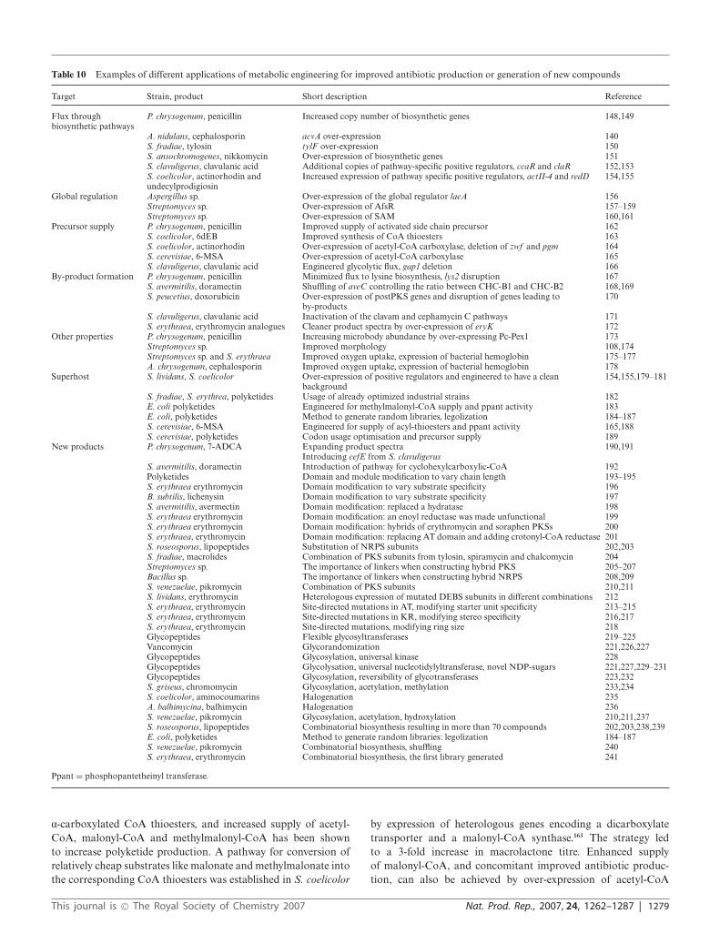
Table 10 Examples of different applications of metabolic engineering for improved antibiotic production or generation of new compounds
Target Strain, product Short description Reference
Flux throughbiosynthetic pathways
P. chrysogenum, penicillin Increased copy number of biosynthetic genes 148,149
A. nidulans, cephalosporin acvA over-expression 140S. fradiae, tylosin tylF over-expression 150S. ansochromogenes, nikkomycin Over-expression of biosynthetic genes 151S. clavuligerus, clavulanic acid Additional copies of pathway-specific positive regulators, ccaR and claR 152,153S. coelicolor, actinorhodin andundecylprodigiosin
Increased expression of pathway specific positive regulators, actII-4 and redD 154,155
Global regulation Aspergillus sp. Over-expression of the global regulator laeA 156Streptomyces sp. Over-expression of AfsR 157–159Streptomyces sp. Over-expression of SAM 160,161
Precursor supply P. chrysogenum, penicillin Improved supply of activated side chain precursor 162S. coelicolor, 6dEB Improved synthesis of CoA thioesters 163S. coelicolor, actinorhodin Over-expression of acetyl-CoA carboxylase, deletion of zwf and pgm 164S. cerevisiae, 6-MSA Over-expression of acetyl-CoA carboxylase 165S. clavuligerus, clavulanic acid Engineered glycolytic flux, gap1 deletion 166
By-product formation P. chrysogenum, penicillin Minimized flux to lysine biosynthesis, lys2 disruption 167S. avermitilis, doramectin Shuffling of aveC controlling the ratio between CHC-B1 and CHC-B2 168,169S. peucetius, doxorubicin Over-expression of postPKS genes and disruption of genes leading to
by-products170
S. clavuligerus, clavulanic acid Inactivation of the clavam and cephamycin C pathways 171S. erythraea, erythromycin analogues Cleaner product spectra by over-expression of eryK 172
Other properties P. chrysogenum, penicillin Increasing microbody abundance by over-expressing Pc-Pex1 173Streptomyces sp. Improved morphology 108,174Streptomyces sp. and S. erythraea Improved oxygen uptake, expression of bacterial hemoglobin 175–177A. chrysogenum, cephalosporin Improved oxygen uptake, expression of bacterial hemoglobin 178
Superhost S. lividans, S. coelicolor Over-expression of positive regulators and engineered to have a cleanbackground
154,155,179–181
S. fradiae, S. erythrea, polyketides Usage of already optimized industrial strains 182E. coli polyketides Engineered for methylmalonyl-CoA supply and ppant activity 183E. coli, polyketides Method to generate random libraries, legolization 184–187S. cerevisiae, 6-MSA Engineered for supply of acyl-thioesters and ppant activity 165,188S. cerevisiae, polyketides Codon usage optimisation and precursor supply 189
New products P. chrysogenum, 7-ADCA Expanding product spectra 190,191Introducing cefE from S. clavuligerus
S. avermitilis, doramectin Introduction of pathway for cyclohexylcarboxylic-CoA 192Polyketides Domain and module modification to vary chain length 193–195S. erythraea erythromycin Domain modification to vary substrate specificity 196B. subtilis, lichenysin Domain modification to vary substrate specificity 197S. avermitilis, avermectin Domain modification: replaced a hydratase 198S. erythraea erythromycin Domain modification: an enoyl reductase was made unfunctional 199S. erythraea erythromycin Domain modification: hybrids of erythromycin and soraphen PKSs 200S. erythraea, erythromycin Domain modification: replacing AT domain and adding crotonyl-CoA reductase 201S. roseosporus, lipopeptides Substitution of NRPS subunits 202,203S. fradiae, macrolides Combination of PKS subunits from tylosin, spiramycin and chalcomycin 204Streptomyces sp. The importance of linkers when constructing hybrid PKS 205–207Bacillus sp. The importance of linkers when constructing hybrid NRPS 208,209S. venezuelae, pikromycin Combination of PKS subunits 210,211S. lividans, erythromycin Heterologous expression of mutated DEBS subunits in different combinations 212S. erythraea, erythromycin Site-directed mutations in AT, modifying starter unit specificity 213–215S. erythraea, erythromycin Site-directed mutations in KR, modifying stereo specificity 216,217S. erythraea, erythromycin Site-directed mutations, modifying ring size 218Glycopeptides Flexible glycosyltransferases 219–225Vancomycin Glycorandomization 221,226,227Glycopeptides Glycosylation, universal kinase 228Glycopeptides Glycolysation, universal nucleotidylyltransferase, novel NDP-sugars 221,227,229–231Glycopeptides Glycosylation, reversibility of glycotransferases 223,232S. griseus, chromomycin Glycosylation, acetylation, methylation 233,234S. coelicolor, aminocoumarins Halogenation 235A. balhimycina, balhimycin Halogenation 236S. venezuelae, pikromycin Glycosylation, acetylation, hydroxylation 210,211,237S. roseosporus, lipopeptides Combinatorial biosynthesis resulting in more than 70 compounds 202,203,238,239E. coli, polyketides Method to generate random libraries: legolization 184–187S. venezuelae, pikromycin Combinatorial biosynthesis, shuffling 240S. erythraea, erythromycin Combinatorial biosynthesis, the first library generated 241
Ppant = phosphopantetheinyl transferase.
a-carboxylated CoA thioesters, and increased supply of acetyl-CoA, malonyl-CoA and methylmalonyl-CoA has been shownto increase polyketide production. A pathway for conversion ofrelatively cheap substrates like malonate and methylmalonate intothe corresponding CoA thioesters was established in S. coelicolor
by expression of heterologous genes encoding a dicarboxylatetransporter and a malonyl-CoA synthase.163 The strategy ledto a 3-fold increase in macrolactone titre. Enhanced supplyof malonyl-CoA, and concomitant improved antibiotic produc-tion, can also be achieved by over-expression of acetyl-CoA
This journal is © The Royal Society of Chemistry 2007 Nat. Prod. Rep., 2007, 24, 1262–1287 | 1279
carboxylase, catalysing the conversion of acetyl-CoA intomalonyl-CoA.164,245
Arginine and glyceraldehyde-3-phosphate are precursors inthe biosynthesis of clavulanic acid. There are indications thatS. clavuligerus uses an unusual urea cycle for prokaryotes tokeep the pool of arginine for clavulinic acid production, andinstead glyceraldehyde-3-phosphate (C3) has been suggested tobe limiting.246–249 There is of course also competition betweenglycolysis and clavulanic acid biosynthesis for glyceraldehyde-3-phosphate. With the aim of reducing the flux through glycolysisand thereby hoping to increase the C3 pool available for biosyn-thesis, gap1, encoding one of the two glyceraldehyde-3-phosphatedehydrogenases (GAPDH) in S. clavuligerus, was disrupted.166 Itturned out to be a successful approach, as GAPDH inactivationresulted in an increased pool of glyceraldehyde-3-phosphate andincreased antibiotic production.
The peptide backbone of glycopeptide antibiotics mainly consistof aromatic amino acids. For synthesis of one mole of teicoplanin,7 moles of tyrosine are needed, corresponding approximately toone third of what is required for biomass formation in a low-producing strain.46 Consequently, high production of glycopep-tides requires high flux through anabolic pathways not normallycarrying high flux, and enhanced flux through the shikimate path-way is an interesting target for improved glycopeptide production.
8.3 Reduced by-product formation
For more efficient use of the raw material and thereby highereconomic viability, it is important to avoid wasting carbon in by-product formation. Another economic aspect is the purificationcost, which might constitute a large part of the total produc-tion cost. In antibiotic production a group of closely relatedcompounds are normally formed with a high similarity to thedesired product, complicating purification. For glycopeptides, itis common to find the aglycone, or analogues with only one outof two or more sugars. For the polyketide erythromycin, severalanalogues are produced which, for example, lack a methyl orhydroxyl group. The analogues normally have a lower biologicalactivity than the desired product and are therefore unwanted,e.g. erythromycin C and D have only half (or less) the activityof erythromycin A.250 The same is true for the anthelminticpolyketide doramectin, an avermectin which is produced by amutant strain of S. avermitilis when cyclohexanecarboxylic acid(CHC) replaces the natural chain initiator. The main by-product,CHC-B2, lacking the C22–C23 double bond found in doramectin(CHC-B1), is less efficient.251 A 23-fold reduction in the ratio ofthe undesired analogue CHC-B2 to the desired analogue CHC-B1 (from 1.6 to 0.07 compared to the wild-type) was achieved bya semi-synthetic DNA shuffling approach.168,169 The gene aveChas been shown to have an important role in controlling theratio of CHC-B1 to CHC-B2; however, the exact function isstill unknown. Over-expression of aveC had no effect on theratio, whereas deletion removed production of both CHC-B1 andCHC-B2.168 The shuffling procedure used is an oligonucleotide-based approach, which enables more efficient recombination andhas a beneficial effect on diversity.252 Improved clones isolatedafter mutagenesis were analyzed to identify advantageous aminoacid substitutions in aveC.169 Oligonucleotides, containing singleor pairs of the identified amino acid substitutions and flanking
regions with perfect match, were synthesized. These were thenshuffled, applying the best full-length variant of aveC as template.The authors observed a considerable increase in probability ofincorporation of a higher number of amino acid substitutions pergene compared to traditional shuffling methods. Moreover, semi-synthetic shuffling facilitates recombination of mutations that areplaced closely together on a gene and unlikely to be recombinedby traditional crossover events.
8.4 Improving other properties
Microbodies are crucial organelles for penicillin production, as theenzymes involved in the final part of penicillin biosynthesis are lo-calized in the microbody matrix.253–255 A significant increase in thenumber of microbodies has been seen for high-yielding industrialstrains of P. chrysogenum.254,256 A metabolic engineering approachto increase microbody abundance was achieved by creating a strainover-producing Pex11p, which is a central microbody membraneprotein.173 The 2–2.5-fold increase in antibiotic production wasexplained by a stimulating effect in flux of penicillin or penicillinprecursors over the microbody membranes due to the enlargedarea, as the levels of biosynthetic enzymes were unaffected.
Most antibiotic producers are either filamentous fungi oractinobacteria. Since both these groups of microorganisms havefilamentous growth, oxygen mass transfer is often limited in highcell density submerged cultivations. First of all, growth mightbe hindered, but furthermore, sufficient oxygen supply is alsoessential for good production of several antibiotics, as a number ofbiosynthetic reactions have been found to be oxygen-dependent.There are for example oxygen-dependent reactions involved bothin b-lactam biosynthesis14 and erythromycin biosynthesis.257,258
Cephalosporin production is severely reduced by oxygen limita-tion, and instead the intermediate penicillin N accumulates. Withthe aim to enhance the oxygen uptake, the bacterial hemoglobingene vhb from Vitreoscilla sp. was over-expressed in A. chryso-genum, resulting in a 5-fold improvement in the cephalosporinlevel.178 Positive effects of vhb expression on antibiotic productionin actinobacteria have also been observed.175–177
8.5 Heterologous hosts
In many cases, the original hosts of natural products are notoptimal to apply as industrial production organisms. They mightbe hard to handle in submerged cultivations, have slow growth andnot be accessible to efficient genetic engineering. Evidently, thedevelopment of general production hosts, so called ’superhosts”,engineered to over-produce any antibiotic of a certain class, isof high interest. This would provide a plug-and-play systemwhereby a biosynthetic cluster could be introduced and promptlyresult in production of that particular secondary metabolite,assisting in studies of biosynthesis and facilitating manipulationof biosynthetic pathways to make new products.
Important characteristics for a superhost are a clean back-ground i.e. no production of native secondary metabolites,availability of precursors, efficient expression and correct proteinfolding. Well-studied strains belonging to the same or closelyrelated genera can serve as hosts for expression of biosyntheticclusters. Several examples of applying S. coelicolor, S. lividansand S. albus as heterologous hosts for natural products from
1280 | Nat. Prod. Rep., 2007, 24, 1262–1287 This journal is © The Royal Society of Chemistry 2007

other streptomycete or actinobacteria strains that are poorlycharacterized or problematic to cultivate can be found in theliterature. There are also examples of studies aiming at improvingtheir qualities as generic production hosts.154,155,179–181
Another strategy tried is to engineer industrial strains alreadyoptimized for high yields into plug-and-play hosts for antibioticproduction.182 The potential of industrial S. erythraea and S.fradiae strains to serve as superhosts for polyketide productionhas been investigated. The native clusters of erythromycin andtylosin were deleted, and an artificial phage �C31 attB locusintroduced into S. erythraea to allow integration of biosyntheticgenes of a new product into the chromosome. Introduction ofthis specific site for integration together with conjugation withE. coli facilitates an efficient transfer of DNA. The biosyntheticgenes for erythromycin and tylosin were then cloned from low-producing wild-type strains and expressed in the engineeredindustrial hosts under their native promoters, resulting in titresas good as those found for the original industrial strains. Thisindicates that the improved production in the industrial strainswas not connected to changes in the biosynthetic cluster itself, butmay instead be due to deregulation of secondary metabolite geneexpression or other factors such as enhanced substrate uptakerates or improved precursor supply, which make them suitableas hosts for heterologous expression of PKSs. To further test thepotential of using optimized industrial strains as superhosts, twomodified DEBS constructs were expressed in the engineered S.erythraea strain.182 Earlier trials to express erythromycin analoguesin S. lividans have not been very successful, suffering fromlow titres and lack of biological activity due to deficiency inglycosylation and oxygenation. A 10- to 50-fold increase in titrecompared to S. lividans was observed in the industrial S. erythraeastrain; moreover, the products were correctly glycosylated as S.erythraea had the capability to add deoxysugars to a polyketidesaglycone.
Lately it has been shown that it is possible to express naturalproducts in bacteria or yeasts not normally producing this type ofproduct. Well-known species such as Escherichia coli and Saccha-romyces cerevisiae would be ideal hosts due to their relatively rapidgrowth, well-known behaviour in fermentors and accessibilityto genetic tools. Pfeifer et al. managed to produce the aglyconeprecursor of erythromycin, 6-deoxyerythronolide B (6dEB), in anengineered E. coli strain at levels which compare well to those ofan industrial optimized S. erythraea.183 6dEB is synthesized frompropionyl-CoA and methylmalonyl-CoA. E. coli is able to providepropionyl-CoA, but in addition to introducing the biosyntheticgenes and co-expression of a phosphopantetheinyl transferase,it was necessary to establish a pathway for methylmalonyl-CoAsupply. This was done by expressing a propionyl-CoA carboxylasefrom S. coelicolor, which in the presence of biotin convertspropionyl-CoA to methylmalonyl-CoA. Furthermore, the nativegene prpE, which is responsible for converting propionate topropionyl-CoA, and birA, encoding a biotin ligase, were over-expressed. For coordinated expression of biosynthetic genes andof genes encoding enzymes involved in precursor supply, the samestrong inducible promoter was used for all genes. Care was alsotaken to avoid drain of propionate into normal metabolism, byintegrating the gene encoding phosphopantetheinyl transferaseinto the prp operon and thereby deleting the prpRBCD genes,responsible for propionate metabolism in E. coli.
S. cerevisiae has also been shown to be a promising host forpolyketide production.165,188 A fairly simple fungal polyketide,6-methylsalicylic acid, was expressed with good yields whenphosphopantetheinyl transferase was co-expressed. Further stepshave been taken in improving S. cerevisiae as a heterologous hostfor production of natural products. A pathway for methylmalonyl-CoA synthesis was established, a necessity for expression ofmodular polyketides.189 Another issue is codon usage. Manynatural products originate from GC-rich actinobacteria, havinga preferred codon usage which largely differs from that in yeastand E. coli. The likelihood of success in heterologous expressiondecreases with the number of rarely used codons that a genecontains, particularly if they are placed in the N-terminal region.There are in principle two strategies to improve expression: eitherto optimize the sequence of the gene so that the codon usage moreclosely reflects that of the host without changing the amino acidarrangement, or to increase the intracellular tRNA pools of therare tRNAs. To facilitate expression of GC-rich genes, five tRNAs,which are rare in yeast but common in actinobacteria, were over-expressed in a protease-deficient strain of S. cerevisiae, which alsowas engineered to supply methylmalonyl-CoA.189 This strain wasable to produce a triketide lactone.
8.6 New products
Extension of product spectra. Expanding the product spectrum of amicroorganism, thereby avoiding additional (often chemical) stepsresults in a more cost-effective and environmentally friendly pro-cess. One nice example in which a process that to a large extent de-pended on chemical modifications was replaced by a fermentation-based route is cephalosporin production. Cephalosporins formedical use are derived from 7-aminocephalosporanic acid(7-ACA) or 7-aminodeacetoxycephalosoranic acid (7-ADCA),which were classically produced in processes involving severalchemical steps using cephalosporin C or penicillin G as startingmaterials, respectively. As the natural cephalosporin producers(S. clavuligerus and Nocardia lactamdurans) have considerablylower productivity than P. chrysogenum, the latter strain was aninteresting target to engineer for production of cephalosporinsdirectly by fermentation. The first two steps in biosynthesis ofpenicillins and cephalosporins are common, but P. chrysogenumlacks the expandase activity necessary for expansion of the 5-membered penicillin ring to the 6-membered ring characteristic ofcephalosporins. Introduction of an expandase gene, cefE from S.clavuligerus or cefEF from A. chrysogenum, and feeding adipate asside chain precursor enabled production of adipyl-7-ADCA andadipyl-7-ACA, respectively, in the recombinant P. chrysogenumstrains.190,191 In 2001, DSM started a new production plant for7-ADCA based on the fermentation route and expanded theircapacity for environmentally friendly production of 7-ADCA evenfurther in 2005.
Simplification of production of doramectin has also beenaccomplished through the use of metabolic engineering.192 Do-ramectin has been produced by feeding cyclohexylcarboxylicacid (or related precursors) to S. avermitilis mutants incapableof producing the normal short branched-chain carboxylic acidstarter units. However, by transferring the genes involved incyclohexylcarboxylic acid synthesis in S. collinus, doramectincould be directly produced in excellent yields in S. avermitilis
This journal is © The Royal Society of Chemistry 2007 Nat. Prod. Rep., 2007, 24, 1262–1287 | 1281

without the need for addition of precursors to the cultivationbroth.192
Combinatorial biosynthesis. PKSs and NRPSs are appealingtargets for combinatorial biosynthesis due to their modularorganization (recent reviews on the topic by Floss, Weissman &Leadley, Baltz, and Blanchard & Thorson).11,12,14,259 It has been cal-culated that a PKS system consisting of six modules is theoreticallyable to produce over 100 000 different structures.260 Erythromycinbiosynthesis has to a large extent functioned as model system forexploring how modular pathways can be engineered to generatenew compounds.224,241,261 More recently, NRPs have also beenmodified, although more knowledge is required to generate NRPsin an efficient way.208,209,262 The term “combinatorial biosynthesis”has in the literature been used both for targeted metabolicengineering approaches, also called pathway engineering, resultingin one or a limited number of new products, and for propercombinatorial biosynthesis where combinations of genes fromdifferent pathways are used to generate analogue libraries. Someexamples of combinatorial biosynthesis are:
• Swapping modules or subunits. Genetic engineering opens forinteresting possibilities to generate new polyketides by simplyswapping, deleting or introducing domains or modules. Geneticengineering of PKS domains has been used to change the chainlength,193–195 to change choice of start or extender unit,196,197
to change degree of reduction of the backbone,198,199 or toalter the stereochemistry.216,217 The mixture of avermectins withdifferent degrees of reduction at the C22–C23 bond produced byS. avermitilis has been ascribed to a defective dehydratase of aveAImodule 2. When the default dehydratase was replaced with afunctional dehydratase from the erythromycin eryAII, via homol-ogous recombination, only unsaturated avermectin compoundswere produced.198 In another example of domain swapping, ahybrid PKS containing domains from a myxobacterium andfrom the erythromycin producer Saccharopolyspora erythraea wereconstructed.200 The DEBS1-loading unit was replaced with theunusual soraphen loading unit having benzoate specificity, anda phenyl-substituted triketide lactone was produced. There arealso reports where whole modules have been substituted.194,205,208,224
Modules in the PKS clusters of tylosin (tylG), spiramycin (srmG),or chalcomycin (chmG) were combined in different ways toyield good amounts of hybrid 16-membered macrolides in anengineered S. fradiae host.204 More than 50 derivatives of 6-dEBwere generated using a multi-plasmid approach for heterologousexpression of DEBS.212 The three DEBS subunits were cloned onseparate plasmids and versions of the plasmids were generatedcontaining mutated genes. Different vector combinations werethen transformed into S. lividans.
• Domain manipulations. New natural compounds have alsobeen generated by specific domain manipulations. AT domainsare normally particular when determining which starter andelongation units that are integrated. It was possible to mod-ify the substrate specificity of AT domains by site-directedmutagenesis.213–215 Until recently, site-directed mutagenesis hadonly been applied on AT domains. An increased understanding ofthe mechanisms behind ketoreduction made it possible to alter thestereospecificity of polyketides by mutagenesis of KR units.216,217
To be able to design specific stereoisomers of important polyke-tides in the future is an attractive thought. Modification of the
ring-size in erythromycins has also been achieved using directedmutagenesis.218
• Post-modifications. The sugar residues are suggested to havean important role in the functionality of natural products, beinginvolved in both drug targeting and biological activity. Hence,varying the glycosylation pattern on natural products has been thefocus of much research aiming at generating novel drug candidates.Glycosyltransferases with relaxed specificity are important toolsfor construction of hybrid glycopeptide antibiotics. The gtfE andgtfD from the vancomycin-producer A. orientalis219–222 accepted anumber of different NDP sugar substrates and could glycosylateseveral different aglycones. Other flexible glycosyl transferases areAveBI from S. avermitilis, DesVII from S. venezuelae and VinCfrom S. halstedii.223–225 Glycorandomization is a chemoenzymaticway to generate diversity in the glycosylation pattern.221,227 Theprinciple is that chemically synthesized natural and unnaturalmonomer sugars are converted into their respective NDP-sugarsin vitro by two enzymes, a kinase and a nucleotidylyltransferase.A glycosyl transferase is then responsible for the coupling toan aglycone. The methodology was applied to generate 33NDP-sugars and concomitantly apply GtfE for coupling to thevancomycin aglycone. Thirty-one glycosylations were successful,and three derivatives having improved properties were identified.221
Thorson and co-workers have taken important steps towardsin vivo glycorandomizaiton by engineering both a kinase anda nucleotidylyltransferase to broaden their substrate specificity.Error-prone PCR and site-directed mutagenesis were applied togenerate mutations in the galactose kinase (GalK from E.coli), andan enzyme capable of accepting nearly 30 diverse monosaccharidesubstrates resulted.228 An E. coli strain expressing the mutatedGalK produced natural and unnatural sugar phosphates withyields even better than those observed for in vitro conversion. Thea-D-glucopyranosyl thymidylyltransferase (Ep) from Salmonellaenterica was found to be capable of converting a broad rangeof sugar phosphates into NDP-sugars.221,227,230 Structure-guidedmutagenesis was applied to make Ep even more universal. Re-cently, another flexible nucleotidylyltransferase from Sulfolobustokodaii has been identified.231 This nucleotidylyltransferase hasthe advantage over Ep of being able to efficiently utilize purinedNTPs in addition to pyrimidine dNTPs.
• True combinatorial synthesis. Daptomycin is a lipopeptideproduced in S. roseosporus and was approved in 2003 (Cubicin R©)for treatment of skin infections by Gram-positive microorganisms,including MRSA strains.238 It is synthesized by an NRPS encodedby three large genes, dptA, dptBC and dptD. Subunit exchangeswere performed with the related lipopeptides A54145 (produced byS. fradiae) and CDA (produced by S. coelicolor), which have partialidentities in subunit organisation. This approach was combinedwith module changes and amino acid modifications.202,203,238,263,264
Further structural diversity was achieved due to the naturalvariations in lipid side chain starter units in S. roseosporus.More than 70 new compounds were produced, and most of themyielded antibacterial activity. Menzella et al.184 have developeda “building-block” methodology for generating large librariesof synthetic polyketides. Central to the strategy is efficientlong-gene synthesis265 and universal linkers. Fourteen codon-optimized modules from eight PKS clusters were synthesized.184
The design was set up to introduce unique restriction sites aroundmodules, but also flanking domains and inter-modular linkers.
1282 | Nat. Prod. Rep., 2007, 24, 1262–1287 This journal is © The Royal Society of Chemistry 2007

As a proof of principle, 154 bimodular combinations enclosedby the loading module and TE from DEBS were assembled.Half of the combinations resulted in detectable amounts oftriketides. The methodology facilitates interchange of polyketidegene cassettes, thereby providing an important tool towards amore high-throughput combinatorial biosynthesis. Another high-throughput strategy was applied by Kim et al.,240 who created4400 variants of the loading module of the typeI polyketidepikromycin by directed evolution. Even if only three clones showedbioactivity, this exemplifies a powerful method to rapidly generatelarge combinatorial libraries.
9 Perspectives: towards systems biology
As mentioned in the introduction, there is a renewed focus onantibiotics in the pharmaceutical industry, and many companiesare focusing on screening for new antibiotics. Even though itis getting harder to identify novel antibiotics, and in particularantibiotics that have new molecular targets,14,266 new screeningmethods will surely lead to the identification of new interestingantibiotics. In order to produce sufficient amounts of these newmolecules it is necessary to improve the productivity. Here classicalstrain improvement will surely continue to play an important role,as it is very efficient and it is easy to implement. As discussedearlier, these techniques are, however, hampered by the fact thatit is a completely random approach, and in some cases it is notpossible to get the desirable phenotype. Metabolic engineeringoffers an alternative to the random approaches, but generallymetabolic engineering requires extensive physiological knowledgeon the organism to be modified, and such knowledge may notbe available. There are two possible solutions to this problem: 1)transfer the biosynthetic pathway for the new antibiotic to anotherhost cell, e.g. a host cell that is optimized for production of a certaingroup of antibiotics, or 2) obtain detailed physiological insightinto the natural producer. There are already several examples ofthe first solution as described above, and there are likely to bemany more in the years to come, as this approach is attractiveboth with respect to optimization of the process and for obtainingproduct approval. The other solution is rather labour-demanding,but with developments in genomics and functional genomics itis now possible to obtain much physiological insight at relativelylow costs, compared with traditional physiological studies. Thus,the cost of genome sequencing is continuously decreasing, andtoday a complete bacterial genome can be sequenced for less thanthe cost of a full-time employee. Furthermore, through the useof transcriptome and proteome data, it is possible to get muchinsight into the operation of different pathways under differentgrowth conditions. It is therefore difficult to predict which of thetwo solutions mentioned above will be the preferred in the future,but it is most likely that both will be used.
Even though genomic sequencing and different functionalgenomics techniques provide much information about the systemunder study, these techniques do not directly enable mapping ofregulatory structures. As discussed above, most of these regulatorystructures have been elucidated through focused molecular biologystudies, and it is therefore a great challenge for the future to morerapidly get insight into the regulatory structures within antibiotic-producing cells, and in particular map those that play a dominant
role during antibiotic production. Here systems biology is likelyto be an important contributor.
There are many definitions of systems biology, but most ofthese contain elements such as mathematical modelling, globalanalysis (or ome analysis), mapping of interactions betweencellular components, and quantification of dynamic responses inliving cells.267–272 In most cases the objective of systems biologyis to obtain a quantitative description of the biological systemunder study, and this quantitative description may be in the formof a mathematical model. In some cases, the model may be thefinal result of the study, i.e. the model captures key featuresof the biological system and can hence be used to predict thebehaviour of the system under conditions different from thoseused to derive the model. In other cases, mathematical modellingrather serves as a tool to extract information from the biologicalsystem, i.e. to enrich the information content in the data. Thereis not necessarily a conflict between the two, and generally,mathematical modelling goes hand-in-hand with experimentalwork. In particular, mathematical models have shown to be usefulin connection with analysis of global data, e.g. transcriptomeand proteome data, as the complexity and integrative natureof biological systems makes it difficult to extract informationon molecular processes from global data without the use ofmodels as either scaffolds for the analysis or for hypothesis-drivenanalysis of the data. One group of mathematical models thathave been found to be very useful, both in systems biology andmetabolic engineering, are the so-called genome-scale metabolicmodels. These models are based on reconstruction of the completemetabolic network operating within a cell, and through the useof flux balance analysis it is possible to use these stoichiometricmodels for simulation of growth and product formation.273 Asmetabolic pathways and architecture are well established, it ispossible to expand this modelling concept to cover practicallyall parts of the metabolism, and it may even be possible to expandthese models to cover regulation.274,275 As genome-scale metabolicmodels are “complete” with respect to metabolic activities, theyprovide a good framework for organizing and integrating x-omedata.276 Thus, it has been shown to be possible to identify keyregulatory structures in the metabolism based on transcriptomedata;277 as well as the models serving as a scaffold for analysisof metabolome data.278 Besides being valuable for upgrading theinformation content in ome data, these models are very useful foridentification of metabolic engineering targets, as it is possible toevaluate different design strategies in silico and even search foroptimal metabolic engineering strategies.279,280
In terms of genomics and systems biology, antibiotic-producingmicroorganisms are somewhat behind key model organisms likeS. cerevisiae and E. coli and different human pathogens. Eventhough several fungal genomes have been sequenced recently,there are not any publically available genomic sequences forkey antibiotic-producing organisms, e.g. for P. chrysogenum, andthis clearly hampers progress in systems biology of antibiotic-producing filamentous fungi. However, with the sequencing oftwo Streptomyces species, the basis for doing systems biologyof Streptomyces has been founded,65,92 and through the use ofbioinformatics tools281 it is expected that this will enable us to geta better understanding of the regulation of antibiotic formation.4
One example is the identification of about 65 sigma-factors inthe genome of S. coelicolor, and with the complete part lists it is
This journal is © The Royal Society of Chemistry 2007 Nat. Prod. Rep., 2007, 24, 1262–1287 | 1283

possible to start evaluating the role of the individual components.Furthermore, based on the genomic sequence of S. coelicolor,a genome-scale metabolic model has been reconstructed, andduring this process several new metabolic features of this organismwere identified.29 Access to genome sequences has also enabledgenome-wide expression analysis under different environmentalconditions282,283 as well as of specific deletion mutants.284,285 Therehave also been a few proteome studies of S. coelicolor, e.g. using2D-electrophoresis followed by MALDI-TOF detection it hasbeen possible to analyze about 10% of the total proteome.74 Inthese studies an integrated analysis in the context of models hasnot been performed, and systems biology of Streptomyces is stillin its infancy. With the rapid developments in systems biology ofyeast and E. coli it will, however, be relatively easy to transfertechnologies and concepts to Streptomyces, and thereby systemsbiology of this important group of antibiotic-producing organismsis likely to develop rapidly in the coming years. As the scientificbasis is available and there is a substantially renewed interestin finding and producing new antibiotics, we envisage that ourunderstanding of regulation of antibiotic production will increaserapidly in the years to come.
10 References
1 J. F. Martin and A. L. Demain, Microbiol. Rev., 1980, 44, 230.2 S. Sanchez and A. L. Demain, Enzyme Microb. Technol., 2002, 31,
895.3 T. Weber, K. Welzel, S. Pelzer, A. Vente and W. Wohlleben, J. Biotech-
nol., 2003, 106, 221.4 M. J. Bibb, Curr. Opin. Microbiol., 2005, 8, 208.5 A. A. Brakhage, P. Sprote, Q. Al-Abdallah, A. Gehrke, H. Plattner
and A. Tuncher, Adv. Biochem. Eng. Biotechnol., 2004, 88, 45.6 E. K. Schmitt, B. Hoff and U. Kuck, Gene, 2004, 342, 269.7 S. J. Coulthurst, A. M. Barnard and G. P. Salmond, Nat. Rev.
Microbiol., 2005, 3, 295.8 T. Stein, Mol. Microbiol., 2005, 56, 845.9 B. N. Mijts and C. Schmidt-Dannert, Curr. Opin. Biotechnol., 2003,
14, 597.10 J. Kantola, T. Kunnari, P. Mantsala and K. Ylihonkoa, Comb. Chem.
High Throughput Screening, 2003, 6, 501.11 H. G. Floss, J. Biotechnol., 2006, 124, 242.12 K. J. Weissman and P. F. Leadlay, Nat. Rev. Microbiol., 2005, 3, 925.13 S. Courtois, C. M. Cappellano, M. Ball, F. X. Francou, P. Normand,
G. Helynck, A. Martinez, S. J. Kolvek, J. Hopke, M. S. Osburne, P. R.August, R. Nalin, M. Guerineau, P. Jeannin, P. Simonet and J. L.Pernodet, Appl. Environ. Microbiol., 2003, 69, 49.
14 R. H. Baltz, Nat. Biotechnol., 2006, 24, 1533.15 S. Donadio, L. Carrano, L. Brandi, S. Serina, A. Soffientini, E.
Raimondi, N. Montanini, M. Sosio and C. O. Gualerzi, J. Biotechnol.,2002, 99, 175.
16 J. M. Buysse, Curr. Med. Chem., 2001, 8, 1713.17 A. F. Chalker and R. D. Lunsford, Pharmacol. Ther., 2002, 95, 1.18 E. D. Brown and G. D. Wright, Chem. Rev., 2005, 105, 759.19 D. J. Payne, M. N. Gwynn, D. J. Holmes and D. L. Pompliano, Nat.
Rev. Drug Discovery, 2007, 6, 29.20 R. A. Samson, R. Hadlok and A. C. Stolk, Antonie van Leeuwenhoek,
1977, 43, 169.21 J. L. Fox, Nat. Biotechnol., 2006, 24, 1521.22 J. Lein, Overproduction of microbial metabolites, Butterworths,
Boston, MA, USA, 1986, p. 105.23 D. J. Newman, G. M. Cragg and K. M. Snader, J. Nat. Prod., 2003,
66, 1022.24 D. J. Newman and G. M. Cragg, J. Nat. Prod., 2007, 70, 461.25 J. Staunton and K. J. Weissman, Nat. Prod. Rep., 2001, 18, 380.26 N. Funa, Y. Ohnishi, I. Fujii, M. Shibuya, Y. Ebizuka and S.
Horinouchi, Nature, 1999, 400, 897.27 V. Pfeifer, G. J. Nicholson, J. Ries, J. Recktenwald, A. B. Schefer, R. M.
Shawky, J. Schroder, W. Wohlleben and S. Pelzer, J. Biol. Chem., 2001,276, 38370.
28 D. Schwarzer, R. Finking and M. A. Marahiel, Nat. Prod. Rep., 2003,20, 275.
29 I. Borodina, P. Krabben and J. Nielsen, Genome Res., 2005, 15, 820.30 C. M. Henriksen, L. H. Christensen, J. Nielsen and J. Villadsen,
J. Biotechnol., 1996, 45, 149.31 H. Jorgensen, J. Nielsen, J. Villadsen and H. Mollgaard, Biotechnol.
Bioeng., 1995, 46, 117.32 W. van Gulik, W. de Laat, J. L. Vinke and J. J. Heijnen, Biotechnol.
Bioeng., 2000, 68, 602.33 B. Christensen and J. Nielsen, Biotechnol. Bioeng., 2000, 68, 652.34 B. Christensen, J. Thykaer and J. Nielsen, Appl. Microbiol. Biotechnol.,
2000, 54, 212.35 R. J. Kleijn, F. Liu, W. A. van Winden, W. M. van Gulik, C. Ras and
J. J. Heijnen, Metab. Eng., 2007, 9, 112.36 N. Gunnarsson, P. Bruheim and J. Nielsen, Biotechnol. Bioeng., 2004,
88, 652.37 N. Gunnarsson, U. H. Mortensen, M. Sosio and J. Nielsen, Mol.
Microbiol., 2004, 52, 895.38 I. Borodina, C. Scholler, A. Eliasson and J. Nielsen, Appl. Environ.
Microbiol., 2005, 71, 2294.39 A. Eliasson Lantz, J. Nielsen and J. S. Rokem, submitted.40 J. Thykaer, B. Christensen and J. Nielsen, Metab. Eng., 2002, 4, 151.41 E. Jonsbu, B. Christensen and J. Nielsen, Appl. Microbiol. Biotechnol.,
2001, 56, 93.42 M. E. Evers, H. Trip, M. A. van den Berg, R. A. L. Bovenberg and
A. J. M. Driessen, Adv. Biochem. Eng. Biotechnol., 2004, 88, 111.43 J. Nielsen, Curr. Opin. Microbiol., 1998, 1, 330.44 J. Nielsen, Appl. Microbiol. Biotechnol., 2001, 55, 263.45 J. Thykaer and J. Nielsen, Metab. Eng., 2003, 5, 56.46 N. Gunnarsson, A. Eliasson and J. Nielsen, Adv. Biochem. Eng.
Biotechnol., 2004, 88, 137.47 J. Stulke and W. Hillen, Curr. Opin. Microbiol., 1999, 2, 195.48 I. Ramos, S. Guzman, L. Escalante, I. Imriskova, R. Rodriguez-
Sanoja, S. Sanchez and E. Langley, Res. Microbiol., 2004, 155, 267.49 S. Guzman, A. Carmona, L. Escalante, I. Imriskova, R. Lopez, R.
Rodriguez-Sanoja, B. Ruiz, L. Servin-Gonzalez, S. Sanchez and E.Langley, Microbiology, 2005, 151, 1717.
50 I. Imriskova, R. Arreguin-Espinosa, S. Guzman, R. Rodriguez-Sanoja, E. Langley and S. Sanchez, Res. Microbiol., 2005, 156, 361.
51 D. Segura, R. Gonzalez, R. Rodriguez, T. Sandoval, L. Escalante andS. Sanchez, Asia–Pac. J. Mol. Biol. Biotechnol., 1996, 4, 30.
52 I. Borodina, J. Siebring, J. Zhang, C. Smith, G. van Keulen, L.Dijkhuizen and J. Nielsen, submitted.
53 M. K. Pope, B. D. Green and J. Westpheling, Mol. Microbiol., 1996,19, 747.
54 M. K. Pope, B. Green and J. Westpheling, J. Bacteriol., 1998, 180,1556.
55 M. J. Merrick, J. Gen. Microbiol., 1976, 96, 299.56 J. White and M. Bibb, J. Bacteriol., 1997, 179, 627.57 W. C. Champness, J. Bacteriol., 1988, 170, 1168.58 C. Bailey and H. N. Arst, Jr, Eur. J. Biochem., 1975, 51, 573.59 H. Ilyes, E. Fekete, L. Karaffa, E. Fekete, E. Sandor, A. Szentirmai
and C. P. Kubicek, FEMS Microbiol. Lett., 2004, 235, 147.60 J. Mogensen, H. B. Nielsen, G. Hofmann and J. Nielsen, Fungal Genet.
Biol., 2006, 43, 593.61 S. L. Westergaard, A. P. Oliveira, C. Bro, L. Olsson and J. Nielsen,
Biotechnol. Bioeng., 2007, 96, 134.62 J. Strauss, R. L. Mach, S. Zeilinger, G. Hartler, G. Stoffler, M.
Wolschek and C. P. Kubicek, FEBS Lett., 1995, 376, 103.63 K. Jekosch and U. Kuck, Curr. Genet., 2000, 37, 388.64 K. Jekosch and U. Kuck, Appl. Microbiol. Biotechnol., 2000, 54,
556.65 S. D. Bentley, K. F. Chater, A. M. Cerdeno-Tarraga, G. L. Challis,
N. R. Thomson, K. D. James, D. E. Harris, M. A. Quail, H. Kieser, D.Harper, A. Bateman, S. Brown, G. Chandra, C. W. Chen, M. Collins,A. Cronin, A. Fraser and A. Goble, Nature, 2002, 417, 141.
66 F. V. Soltero and M. J. Johnson, Appl. Microbiol., 1953, 1, 52.67 J. Nielsen, Physiological engineering aspects of Penicillium chryso-
genum, World Scientific Publishing Co., Singapore, 1997.68 A. A. Brakhage, Microbiol. Mol. Biol. Rev., 1998, 62, 547.69 A. A. Brakhage, Q. Al-Abdallah, A. Tuncher and P. Sprote, Phyto-
chemistry, 2005, 66, 1200.70 E. S. Kim, H. J. Hong, C. Y. Choi and S. N. Cohen, J. Bacteriol., 2001,
183, 2198.71 M. J. Merrick and R. A. Edwards, Microbiol. Rev., 1995, 59, 604.
1284 | Nat. Prod. Rep., 2007, 24, 1262–1287 This journal is © The Royal Society of Chemistry 2007

72 L. Reitzer, Annu. Rev. Microbiol., 2003, 57, 155.73 G. A. Marzluf, Microbiol. Mol. Biol. Rev., 1997, 61, 17.74 A. Hesketh, D. Fink, B. Gust, H. U. Rexer, B. Scheel, K. Chater, W.
Wohlleben and A. Engels, Mol. Microbiol., 2002, 46, 319.75 L. Wray and S. H. Fisher, Gene, 1988, 71, 247.76 H. U. Rexer, T. Schaberle, W. Wohlleben and A. Engels, Arch.
Microbiol., 2006, 186, 447.77 J. Reuther and W. Wohlleben, J. Mol. Microbiol. Biotechnol., 2007, 12,
139.78 H. Haas and G. A. Marzluf, Curr. Genet., 1995, 28, 177.79 R. Usaite, K. R. Patil, T. Grotkjaer, J. Nielsen and B. Regenberg, Appl.
Environ. Microbiol., 2006, 72, 6194.80 A. L. Demain and P. Vaishnav, Crit. Rev. Biotechnol., 2006, 26, 67.81 J. A. Asturias, P. Liras and J. F. Martin, Gene, 1990, 93, 79.82 P. Liras, J. A. Asturias and J. F. Martin, Trends Biotechnol., 1990, 8,
184.83 A. Sola-Landa, A. Rodriguez-Garcia, E. Franco-Dominguez and J. F.
Martin, Mol. Microbiol., 2005, 56, 1373.84 J. F. Martin, J. Bacteriol., 2004, 186, 5197.85 A. Torriani-Gorini, Phosphate in microorganisms, ASM Press, Wash-
ington DC, 1994.86 F. M. Hulett, Mol. Microbiol., 1996, 19, 933.87 M. V. Mendes, S. Tunca, N. Anton, E. Recio, A. J. F. Aparicio and
J. F. Martin, Metab. Eng., 2007, 9, 217.88 R. Masuma, Y. Tanaka, H. Tanaka and S. Omura, J. Antibiot., 1986,
39, 1557.89 A. Heydorn, T. Suhr-Jessen and J. Nielsen, J. Antibiot., 1999, 52, 40.90 N. Gunnarsson, P. Bruheim and J. Nielsen, J. Ind. Microbiol.
Biotechnol., 2003, 30, 150.91 J. S. Rokem and P. Weitzman, Enzyme Microb. Technol., 1987, 9, 153.92 H. Ikeda, J. Ishikawa, A. Hanamoto, M. Shinose, H. Kikuchi, T.
Shiba, Y. Sakaki, M. Hattori and S. Omura, Nat. Biotechnol., 2003,21, 526.
93 M. J. Buttner, Mol. Microbiol., 1989, 3, 1653.94 K. F. Chater, in: Prokaryotic development, ed. Y. V. Brun
and L. J. Shimkets, ASM Press, Washington DC, 1994, p. 33.95 A. M. Gehring, N. J. Yoo and R. Losick, J. Bacteriol., 2001, 183, 5991.96 M. S. Paget, H. J. Hong, B. M. and M. J. Buttner, in SGM Symposium
61: signals, switches, regulons and cascades: control of bacterial geneexpression, ed. D. A. Hodgson and C. M. Thomas, CambridgeUniversity Press, Cambridge, 2002, p. 105.
97 H. Takano, S. Obitsu, T. Beppu and K. Ueda, J. Bacteriol., 2005, 187,1825.
98 V. Mazurakova, B. Sevcikova, B. Rezuchova and J. Kormanec, Arch.Microbiol., 2006, 186, 435.
99 J. D. Helmann, Adv. Microb. Physiol., 2002, 46, 47.100 R. Sawai, A. Suzuki, Y. Takano, P. C. Lee and S. Horinouchi, Gene,
2004, 334, 53.101 L. J. Kenney, Curr. Opin. Microbiol., 2002, 5, 135.102 A. Wietzorrek and M. Bibb, Mol. Microbiol., 1997, 25, 1181.103 E. J. Lawlor, H. A. Baylis and K. F. Chater, Genes Dev., 1987, 1, 1305.104 M. Elliot, F. Damji, R. Passantino, K. Chater and B. Leskiw,
J. Bacteriol., 1998, 180, 1549.105 W. Li, J. Wu, W. Tao, C. Zhao, Y. Wang, X. He, G. Chandra, X. Zhou,
Z. Deng, K. F. Chater and M. Tao, FEMS Microbiol. Lett., 2007, 266,20.
106 M. Eccleston, A. Willems, A. Beveridge and J. R. Nodwell, J. Bacte-riol., 2006, 188, 8189.
107 M. A. Elliot, T. R. Locke, C. M. Galibois and B. K. Leskiw, FEMSMicrobiol. Lett., 2003, 225, 35.
108 G. van Wezel, P. Krabben, B. A. Traag, B. J. Keijser, R. Kerste, E.Vijgenboom, J. J. Heijnen and B. Kraal, Appl. Environ. Microbiol.,2006, 72, 5283.
109 S. Horinouchi, J. Ind. Microbiol. Biotechnol., 2003, 30, 462.110 P. C. Lee, T. Umeyama and S. Horinouchi, Mol. Microbiol., 2002, 43,
1413.111 N. J. Ryding, T. B. Anderson and W. C. Champness, J. Bacteriol.,
2002, 184, 794.112 N. L. Sheeler, S. V. MacMillan and J. R. Nodwell, J. Bacteriol., 2005,
187, 687.113 M. I. Hutchings, P. A. Hoskisson, G. Chandra and M. J. Buttner,
Microbiology, 2004, 150, 2795.114 J. Huang, J. Shi, V. Molle, B. Sohlberg, D. Weaver, M. J. Bibb, N.
Karoonuthaisiri, C. J. Lih, C. M. Kao, M. J. Buttner and S. N. Cohen,Mol. Microbiol., 2005, 58, 1276.
115 G. C. Uguru, K. E. Stephens, J. A. Stead, J. E. Towle, S. Baumbergand K. J. McDowall, Mol. Microbiol., 2005, 58, 131.
116 C. E. Vrentas, T. Gaal, W. Ross, R. H. Ebright and R. L. Gourse,Genes Dev., 2005, 19, 2378.
117 R. Chakraburtty and M. Bibb, J. Bacteriol., 1997, 179, 5854.118 W. Jin, H. K. Kim, J. Y. Kim, S. G. Kang, S. H. Lee and K. J. Lee,
J. Biotechnol., 2004, 114, 81.119 W. Jin, Y. G. Ryu, S. G. Kang, S. K. Kim, N. Saito, K. Ochi, S. H. Lee
and K. J. Lee, Microbiology, 2004, 150, 1485.120 K. S. Kelly, K. Ochi and G. H. Jones, J. Bacteriol., 1991, 173, 2297.121 T. G. Holt, C. Chang, C. Laurent-Winter, T. Murakami, J. I. Garrels,
J. E. Davies and C. J. Thompson, Mol. Microbiol., 1992, 6, 969.122 G. H. Jones, M. S. Paget, L. Chamberlin and M. J. Buttner, Mol.
Microbiol., 1997, 23, 169.123 J. P. Gomez-Escribano, P. Liras, A. Pisabarro and J. F. Martin, Mol.
Microbiol., 2006, 61, 758.124 S. U. Choi, C. K. Lee, Y. I. Hwang, H. Kinosita and T. Nihira, Arch.
Microbiol., 2003, 180, 303.125 E. Takano, Curr. Opin. Microbiol., 2006, 9, 287.126 H. Horinouchi, Front. Biosci., 2002, 7, d2045.127 Y. Ohnishi, H. Yamazaki, J. Y. Kato, A. Tomono and S. Horinouchi,
Biosci., Biotechnol., Biochem., 2005, 69, 431.128 K. F. Chater and S. Horinouchi, Mol. Microbiol., 2003, 48, 9.129 S. Kitani, Y. Yamada and T. Nihira, J. Bacteriol., 2001, 183, 4357.130 B. A. Lazazzera, Peptides, 2001, 22, 1519.131 J. R. Nodwell, K. McGovern and R. Losick, Mol. Microbiol., 1996,
22, 881.132 J. R. Nodwell and R. Losick, J. Bacteriol., 1998, 180, 1334.133 H. S. Park, S. K. Shin, Y. Y. Yang, H. J. Kwon and J. W. Suh, FEMS
Microbiol. Lett., 2005, 249, 199.134 E. K. Schmitt, B. Hoff and U. Kuck, Adv. Biochem. Eng. Biotechnol.,
2004, 88, 1.135 S. Steidl, P. Papagiannopoulos, O. Litzka, A. Andrianopoulos, M. A.
Davis, A. A. Brakhage and M. J. Hynes, Mol. Cell. Biol., 1999, 19, 99.136 J. Tilburn, S. Sarkar, D. A. Widdick, E. A. Espeso, M. Orejas, J.
Mungroo, M. A. Penalva and H. N. Arst, Jr, EMBO J., 1995, 14, 779.137 P. Liras and J. F. Martin, Int. Microbiol., 2006, 9, 9.138 K. Kosalkova, A. T. Marcos, F. Fierro, V. Hernando-Rico, S. Gutierrez
and J. F. Martin, J. Biol. Chem., 2000, 275, 2423.139 O. Litzka, K. Then Berg, J. Van den Brulle, S. Steidl and A. A.
Brakhage, Antonie van Leeuwenhoek, 1999, 75, 95.140 J. Kennedy and G. Turner, Mol. Gen. Genet., 1996, 253, 189.141 J. M. Fernandez-Canon and M. A. Penalva, Mol. Gen. Genet., 1995,
246, 110.142 D. J. Smith, J. H. Bull, J. Edwards and G. Turner, Mol. Gen. Genet.,
1989, 216, 492.143 G. Seidel, C. Tollnick, M. Beyer and K. Schugerl, Adv. Biochem. Eng.
Biotechnol., 2000, 66, 115.144 J. Nielsen and H. S. Jorgensen, Biotechnol. Prog., 1995, 11, 299.145 P. N. Pissarra, J. Nielsen and M. J. Bazin, Biotechnol. Bioeng., 1996,
51, 168.146 H. A. Theilgaard and J. Nielsen, Antonie van Leeuwenhoek, 1999, 75,
145.147 H. B. Theilgaard, K. N. Kristiansen, C. M. Henriksen and J. Nielsen,
Biochem. J., 1997, 327, 185.148 A. E. Veenstra, P. van Solingen, R. A. L. Bovenberg and L. H. M. van
der Voort, J. Biotechnol., 1991, 17, 81.149 H. A. Theilgaard, M. A. van den Berg, C. A. Mulder, R. A. L.
Bovenberg and J. Nielsen, Biotechnol. Bioeng., 2001, 72, 379.150 R. H. Baltz, M. A. McHenney, C. A. Cantwell, S. W. Queener and
P. J. Solenberg, Antonie van Leeuwenhoek, 1997, 71, 179.151 Y. Li, H. Ling, W. Li and H. Tan, Metab. Eng., 2005, 7, 165.152 F. J. Perez-Llarena, P. Liras, A. Rodriguez-Garcia and J. F. Martin,
J. Bacteriol., 1997, 179, 2053.153 R. Perez-Redondo, A. Rodriguez-Garcia, J. F. Martin and P. Liras,
Gene, 1998, 211, 311.154 E. Takano, H. C. Gramajo, E. Strauch, N. Andres, J. White and M. J.
Bibb, Mol. Microbiol., 1992, 6, 2797.155 H. C. Gramajo, E. Takano and M. J. Bibb, Mol. Microbiol., 1993, 7,
837.156 J. W. Bok and N. P. Keller, Eukaryotic Cell, 2004, 3, 527.157 M. Vogtli, P. C. Chang and S. N. Cohen, Mol. Microbiol., 1994, 14,
643.158 O. Sekurova, H. Sletta, T. E. Ellingsen, S. Valla and S. Zotchev, FEMS
Microbiol. Lett., 1999, 177, 297.
This journal is © The Royal Society of Chemistry 2007 Nat. Prod. Rep., 2007, 24, 1262–1287 | 1285

159 J. Lee, Y. Hwang, S. Kim, E. Kim and C. Choi, J. Biosci. Bioeng., 2000,89, 606.
160 S. Okamoto, A. Lezhava, T. Hosaka, Y. Okamoto-Hosoya and K.Ochi, J. Bacteriol., 2003, 185, 601.
161 J. H. Huh, D. J. Kim, X. Q. Zhao, M. Li, Y. Y. Jo, T. M. Yoon,S. K. Shin, J. H. Yong, Y. W. Ryu, Y. Y. Yang and J. W. Suh, FEMSMicrobiol. Lett., 2004, 238, 439.
162 B. Minambres, H. Martinez-Blanco, E. R. Olivera, B. Garcia, B. Diez,J. L. Barredo, M. A. Moreno, C. Schleissner, F. Salto and J. M. Luengo,J. Biol. Chem., 1996, 271, 33531.
163 F. Lombo, B. Pfeifer, T. Leaf, S. Ou, Y. S. Kim, D. E. Cane, P. Licariand C. Khosla, Biotechnol. Prog., 2001, 17, 612.
164 Y. G. Ryu, M. J. Butler, K. F. Chater and K. J. Lee, Appl. Environ.Microbiol., 2006, 72, 7132.
165 S. Wattanachaisaereekul, A. E. Lantz, M. L. Nielsen, O. S. Andressonand J. Nielsen, Biotechnol. Bioeng., 2007, DOI: 10.1002/bit.21286.
166 R. Li and C. A. Townsend, Metab. Eng., 2006, 8, 240.167 J. Casqueiro, S. Gutierrez, O. Banuelos, M. J. Hijarrubia and J. F.
Martin, J. Bacteriol., 1999, 181, 1181.168 K. Stutzman-Engwall, S. Conlon, R. Fedechko, F. Kaczmarek, H.
McArthur, A. Krebber, Y. Chen, J. Minshull, S. A. Raillard and C.Gustafsson, Biotechnol. Bioeng., 2003, 82, 359.
169 K. Stutzman-Engwall, S. Conlon, R. Fedechko, H. McArthur, K.Pekrun, Y. Chen, S. Jenne, C. La, N. Trinh, S. Kim, Y. X. Zhang, R.Fox, C. Gustafsson and A. Krebber, Metab. Eng., 2005, 7, 27.
170 N. Lomovskaya, S. L. Otten, Y. Doi-Katayama, L. Fonstein, X. C. Liu,T. Takatsu, A. Inventi-Solari, S. Filippini, F. Torti, A. L. Colombo andC. R. Hutchinson, J. Bacteriol., 1999, 181, 305.
171 A. S. Paradkar, R. H. Mosher, C. Anders, A. Griffin, J. Griffin,C. Hughes, P. Greaves, B. Barton and S. E. Jensen, Appl. Environ.Microbiol., 2001, 67, 2292.
172 R. P. Desai, E. Rodriguez, J. L. Galazzo and P. Licari, Biotechnol.Prog., 2004, 20, 1660.
173 J. A. Kiel, I. J. van der Klei, M. A. van den Berg, R. A. Bovenbergand M. Veenhuis, Fungal Genet. Biol., 2005, 42, 154.
174 S. Kawamoto, H. Watanabe, A. Hesketh, J. C. Ensign and K. Ochi,Microbiology, 1997, 143, 1077.
175 S. K. Magnolo, D. L. Leenutaphong, J. A. DeModena, J. E. Curtis,J. E. Bailey, J. L. Galazzo and D. E. Hughes, Biotechnology, 1991, 9,473.
176 P. Brunker, W. Minas, P. T. Kallio and J. E. Bailey, Microbiology, 1998,144, 2441.
177 W. Minas, P. Brunker, P. T. Kallio and J. E. Bailey, Biotechnol. Prog.,1998, 14, 561.
178 J. A. DeModena, S. Gutierrez, J. Velasco, F. J. Fernandez, R. A.Fachini, J. L. Galazzo, D. E. Hughes and J. F. Martin, Biotechnology,1993, 11, 926.
179 C. M. Kao, L. Katz and C. Khosla, Science, 1994, 265, 509.180 R. Ziermann and M. C. Betlach, BioTechniques, 1999, 26, 106.181 B. A. Pfeifer and C. Khosla, Microbiol. Mol. Biol. Rev., 2001, 65,
106.182 E. Rodriguez, Z. Hu, S. Ou, Y. Volchegursky, C. R. Hutchinson and
R. McDaniel, J. Ind. Microbiol. Biotechnol., 2003, 30, 480.183 B. A. Pfeifer, S. J. Admiraal, H. Gramajo, D. E. Cane and C. Khosla,
Science, 2001, 291, 1790.184 H. G. Menzella, R. Reid, J. R. Carney, S. S. Chandran, S. J. Reisinger,
K. G. Patel, D. A. Hopwood and D. V. Santi, Nat. Biotechnol., 2005,23, 1171.
185 S. S. Chandran, H. G. Menzella, J. R. Carney and D. V. Santi, Chem.Biol., 2006, 13, 469.
186 H. G. Menzella, S. J. Reisinger, M. Welch, J. T. Kealey, J. Kennedy, R.Reid, C. Q. Tran and D. V. Santi, J. Ind. Microbiol. Biotechnol., 2006,33, 22.
187 H. G. Menzella, J. R. Carney and D. V. Santi, Chem. Biol., 2007, 14,143.
188 J. T. Kealey, L. Liu, D. V. Santi, M. C. Betlach and P. J. Barr, Proc.Natl. Acad. Sci. U. S. A., 1998, 95, 505.
189 S. C. Mutka, S. M. Bondi, J. R. Carney, D. S. NA and J. T. Kealey,FEMS Yeast Res., 2006, 6, 40.
190 L. Crawford, A. M. Stepan, P. C. McAda, J. A. Rambosek, M. J.Conder, V. A. Vinci and C. D. Reeves, Biotechnology, 1995, 13, 58.
191 J. D. Sutherland, R. Bovenberg and J. M. Van der Laan, World Pat.WO9720053-A3, 1997.
192 T. A. Cropp, D. J. Wilson and K. A. Reynolds, Nat. Biotechnol., 2000,18, 980.
193 H. Petkovic, A. Thamchaipenet, L. H. Zhou, D. Hranueli, P. Raspor,P. G. Waterman and I. S. Hunter, J. Biol. Chem., 1999, 274, 32829.
194 C. J. Rowe, I. U. Bohm, I. P. Thomas, B. Wilkinson, B. A. Rudd,G. Foster, A. P. Blackaby, P. J. Sidebottom, Y. Roddis, A. D. Buss, J.Staunton and P. F. Leadlay, Chem. Biol., 2001, 8, 475.
195 C. J. Martin, M. C. Timoney, R. M. Sheridan, S. G. Kendrew, B.Wilkinson, J. C. Staunton and P. F. Leadlay, Org. Biomol. Chem.,2003, 1, 4144.
196 H. Petkovic, R. E. Lill, R. M. Sheridan, B. Wilkinson, E. L.McCormick, H. A. McArthur, J. Staunton, P. F. Leadlay and S. G.Kendrew, J. Antibiot., 2003, 56, 543.
197 M. M. Yakimov, L. Giuliano, K. N. Timmis and P. N. Golyshin,J. Mol. Microbiol. Biotechnol., 2000, 2, 217.
198 J. H. Yong and W. H. Byeon, J. Microbiol., 2005, 43, 277.199 S. Donadio, J. B. McAlpine, P. J. Sheldon, M. Jackson and L. Katz,
Proc. Natl. Acad. Sci. U. S. A., 1993, 90, 7119.200 C. J. Wilkinson, E. J. Frost, J. Staunton and P. F. Leadlay, Chem. Biol.,
2001, 8, 1197.201 D. L. Stassi, S. J. Kakavas, K. A. Reynolds, G. Gunawardana, S.
Swanson, D. Zeidner, M. Jackson, H. Liu, A. Buko and L. Katz,Proc. Natl. Acad. Sci. U. S. A., 1998, 95, 7305.
202 K. T. Nguyen, D. Ritz, J. Q. Gu, D. Alexander, M. Chu, V. Miao,P. Brian and R. H. Baltz, Proc. Natl. Acad. Sci. U. S. A., 2006, 103,17462.
203 V. Miao, M.-F. Coeffet-Le Gal, K. Nguyen, P. Brian, J. Penn, A.Whiting, J. Steele, D. Kau, S. Martin, R. Ford, T. Gibson, M.Bouchard, S. K. Wrigley and R. H. Baltz, Chem. Biol., 2006, 13,269.
204 C. D. Reeves, S. L. Ward, W. P. Revill, H. Suzuki, M. Marcus, O. V.Petrakovsky, S. Marquez, H. Fu, S. D. Dong and L. Katz, Chem. Biol.,2004, 11, 1465.
205 R. S. Gokhale, D. Hunziker, D. E. Cane and C. Khosla, Chem. Biol.,1999, 6, 117.
206 S. Y. Tsuji, D. E. Cane and C. Khosla, Biochemistry, 2001, 40, 2326.207 R. W. Broadhurst, D. Nietlispach, M. P. Wheatcroft, P. F. Leadlay and
K. J. Weissman, Chem. Biol., 2003, 10, 723.208 H. D. Mootz, D. Schwarzer and M. A. Marahiel, Proc. Natl. Acad.
Sci. U. S. A., 2000, 97, 5848.209 H. D. Mootz, D. Schwarzer and M. A. Marahiel, ChemBioChem,
2002, 3, 490.210 Y. Xue and D. H. Sherman, Metab. Eng., 2001, 3, 15.211 Y. J. Yoon, B. J. Beck, B. S. Kim, H. Y. Kang, K. A. Reynolds and
D. H. Sherman, Chem. Biol., 2002, 9, 203.212 Q. Xue, G. Ashley, C. R. Hutchinson and D. V. Santi, Proc. Natl.
Acad. Sci. U. S. A., 1999, 96, 11740.213 C. D. Reeves, S. Murli, G. W. Ashley, M. Piagentini, C. R. Hutchinson
and R. McDaniel, Biochemistry, 2001, 40, 15464.214 P. F. Long, C. J. Wilkinson, C. P. Bisang, J. Cortes, N. Dunster, M.
Oliynyk, E. McCormick, H. McArthur, C. Mendez, J. A. Salas, J.Staunton and P. F. Leadlay, Mol. Microbiol., 2002, 43, 1215.
215 F. Del Vecchio, H. Petkovic, S. G. Kendrew, L. Low, B. Wilkinson,R. Lill, J. Cortes, B. A. Rudd, J. Staunton and P. F. Leadlay, J. Ind.Microbiol. Biotechnol., 2003, 30, 489.
216 A. Baerga-Ortiz, B. Popovic, A. P. Siskos, H. M. O’Hare, D. Spiteller,M. G. Williams, N. Campillo, J. B. Spencer and P. F. Leadlay, Chem.Biol., 2006, 13, 277.
217 H. M. O’Hare, A. Baerga-Ortiz, B. Popovic, J. B. Spencer and P. F.Leadlay, Chem. Biol., 2006, 13, 287.
218 C. M. Kao, G. Luo, L. Katz, D. E. Cane and C. Khosla, J. Am. Chem.Soc., 1995, 117, 9105.
219 P. J. Solenberg, P. Matsushima, D. R. Stack, S. C. Wilkie, R. C.Thompson and R. H. Baltz, Chem. Biol., 1997, 4, 195.
220 H. C. Losey, J. Jiang, J. B. Biggins, M. Oberthur, X. Y. Ye, S. D. Dong,D. Kahne, J. S. Thorson and C. T. Walsh, Chem. Biol., 2002, 9, 1305.
221 X. Fu, C. Albermann, J. Jiang, J. Liao, C. Zhang and J. S. Thorson,Nat. Biotechnol., 2003, 21, 1467.
222 M. Oberthur, C. Leimkuhler, R. G. Kruger, W. Lu, C. T. Walsh andD. Kahne, J. Am. Chem. Soc., 2005, 127, 10747.
223 C. Zhang, C. Albermann, X. Fu and J. S. Thorson, J. Am. Chem. Soc.,2006, 128, 16420.
224 L. Tang and R. McDaniel, Chem. Biol., 2001, 8, 547.225 A. Minami, R. Uchida, T. Eguchi and K. Kakinuma, J. Am. Chem.
Soc., 2005, 127, 6148.226 J. Yang, D. Hoffmeister, L. Liu, X. Fu and J. S. Thorson, Bioorg. Med.
Chem., 2004, 12, 1577.
1286 | Nat. Prod. Rep., 2007, 24, 1262–1287 This journal is © The Royal Society of Chemistry 2007

227 J. M. Langenhan, B. R. Griffith and J. S. Thorson, J. Nat. Prod., 2005,68, 1696.
228 J. Yang, X. Fu, J. Liao, L. Liu and J. S. Thorson, Chem. Biol., 2005,12, 657.
229 W. A. Barton, J. Lesniak, J. B. Biggins, P. D. Jeffrey, J. Jiang, K. R.Rajashankar, J. S. Thorson and D. B. Nikolov, Nat. Struct. Biol., 2001,8, 545.
230 W. A. Barton, J. B. Biggins, J. Jiang, J. S. Thorson and D. B. Nikolov,Proc. Natl. Acad. Sci. U. S. A., 2002, 99, 13397.
231 Z. Zhang, M. Tsujimura, J. Akutsu, M. Sasaki, H. Tajima and Y.Kawarabayasi, J. Biol. Chem., 2005, 280, 9698.
232 C. Zhang, B. R. Griffith, Q. Fu, C. Albermann, X. Fu, I. K. Lee, L.Li and J. S. Thorson, Science, 2006, 313, 1291.
233 N. Menendez, M. Nur-e-Alam, A. F. Brana, J. Rohr, J. A. Salas andC. Mendez, Chem. Biol., 2004, 11, 21.
234 N. Menendez, M. Nur-e-Alam, C. Fischer, A. F. Brana, J. A. Salas, J.Rohr and C. Mendez, Appl. Environ. Microbiol., 2006, 72, 167.
235 A. S. Eustaquio, B. Gust, S. M. Li, S. Pelzer, W. Wohlleben, K. F.Chater and L. Heide, Chem. Biol., 2004, 11, 1561.
236 B. Bister, D. Bischoff, G. J. Nicholson, S. Stockert, J. Wink, C.Brunati, S. Donadio, S. Pelzer, W. Wohlleben and R. D. Sussmuth,ChemBioChem, 2003, 4, 658.
237 S. K. Lee, D. B. Basnet, C. Y. Choi, J. K. Sohng, J. S. Ahn and Y. J.Yoon, Bioorg. Chem., 2004, 32, 549.
238 R. H. Baltz, P. Brian, V. Miao and S. K. Wrigley, J. Ind. Microbiol.Biotechnol., 2006, 33, 66.
239 J. Q. Gu, K. T. Nguyen, C. Gandhi, V. Rajgarhia, R. H. Baltz, P. Brianand M. Chu, J. Nat. Prod., 2007, 70, 233.
240 B. S. Kim, D. H. Sherman and K. A. Reynolds, Protein. Eng., Des.Sel., 2004, 17, 277.
241 R. McDaniel, A. Thamchaipenet, C. Gustafsson, H. Fu, M. Betlachand G. Ashley, Proc. Natl. Acad. Sci. U. S. A., 1999, 96, 1846.
242 F. Fierro, J. L. Barredo, B. Diez, S. Gutierrez, F. J. Fernandez and J. F.Martin, Proc. Natl. Acad. Sci. U. S. A., 1995, 92, 6200.
243 R. W. Newbert, B. Barton, P. Greaves, J. Harper and G. Turner, J. Ind.Microbiol. Biotechnol., 1997, 19, 18.
244 K. Yanai, T. Murakami and M. Bibb, Proc. Natl. Acad. Sci. U. S. A.,2006, 103, 9661.
245 S. Wattanachaisaereekul, A. Eliasson Lantz, M. L. Nielsen and J.Nielsen, submitted.
246 V. Bascaran, C. Hardisson and A. F. Brana, J. Gen. Microbiol., 1989,135, 2475.
247 P. R. Ives and M. E. Bushell, Microbiology, 1997, 143, 3573.248 K. C. Chen, Y. H. Lin, C. M. Tsai, C. H. Hsieh and J. Y. Houng,
Biotechnol. Lett., 2002, 24, 455.249 K. C. Chen, Y. H. Lin, J. Y. Wu and S. Hwang, Enzyme Microb.
Technol., 2003, 32, 152.250 I. O. Kibwage, J. Hoogmartens, E. Roets, H. Vanderhaeghe, L. Verbist,
M. Dubost, C. Pascal, P. Petitjean and G. Levol, Antimicrob. AgentsChemother., 1985, 28, 630.
251 A. C. Goudie, N. A. Evans, K. Gration, B. F. Bishop, S. P. Gibson,K. S. Holdom, B. Kaye, S. R. Wicks, D. Lewis, A. J. Weatherley,C. I. Bruce, A. Herbert and D. J. Seymour, Vet. Parasitol., 1993, 49, 5.
252 A. Crameri, W. P. C. Stemmer, J. Minshull, S. H. Bass, M. Welch, J. E.Ness, C. Gustafson and P. A. Pattern, PCT Int. Pat. Appl., 2000.
253 S. J. Gould, G. A. Keller, N. Hosken, J. Wilkinson and S. Subramani,J. Cell Biol., 1989, 108, 1657.
254 W. H. Muller, T. P. van der Krift, J. Krouwer, H. A. Wosten, L. H. vander Vort, E. B. Smaal and A. J. Verklej, EMBO J., 1991, 10, 489.
255 W. H. Muller, R. A. Bovenberg, M. H. Groothuis, F. Kattevilder, E. B.Smaal, L. H. van der Vort and A. J. Verklej, Biochim. Biophys. Acta,1992, 1116, 210.
256 S. Valenciano, J. R. De Lucas, I. Van der Klei, M. Veenhuis and F.Laborda, Arch. Microbiol., 1998, 170, 370.
257 D. Stassi, S. Donadio, M. J. Staver and L. Katz, J. Bacteriol., 1993,175, 182.
258 L. Katz and S. Donadio, Biotechnology, 1995, 28, 385.259 S. Blanchard and J. S. Thorson, Curr. Opin. Chem. Biol., 2006, 10,
263.260 J. Gonzalez-Lergier, L. J. Broadbelt and V. Hatzimanikatis, J. Am.
Chem. Soc., 2005, 127, 9930.261 R. McDaniel, M. Welch and C. R. Hutchinson, Chem. Rev., 2005,
105, 543.262 S. Doekel and M. A. Marahiel, Metab. Eng., 2001, 3, 64.263 M. Coeffet-Le Gal, L. Thurston, P. Rich, V. Miao and R. H. Baltz,
Microbiology, 2006, 152, 2993.264 K. T. Nguyen, D. Kau, J. Q. Gu, P. Brian, S. K. Wrigley, R. H. Baltz
and V. Miao, Mol. Microbiol., 2006, 61, 1294.265 S. J. Kodumal, K. G. Patel, R. Reid, H. G. Menzella, M. Welch and
D. V. Santi, Proc. Natl. Acad. Sci. U. S. A., 2004, 101, 15573.266 J. Clardy, M. A. Fischbach and C.T. Walsh, Nat. Biotechnol., 2006,
24, 1541.267 T. Ideker, V. Thorsson, J. A. Ranish, R. Christmas, J. Buhler, J. K. Eng,
R. Bumgarner, D. R. Goodlett, R. Aebersold and L. Hood, Science,2001, 292, 929.
268 H. Kitano, Science, 2002, 295, 1662.269 R. Brent, Nat. Biotechnol., 2004, 22, 1211.270 M. W. Kirschner, Cell, 2005, 121, 503.271 C. L. Barrett, T. Y. Kim, H. U. Kim, B. O. Palsson and S. Y. Lee, Curr.
Opin. Biotechnol., 2006, 17, 488.272 F. J. Bruggeman and H. V. Westerhoff, Trends Microbiol., 2007, 15,
45.273 N. D. Price, J. L. Reed and B. O. Palsson, Nat. Rev. Microbiol., 2004,
2, 886.274 C. L. Barrett and B. O. Palsson, PLoS Comput. Biol., 2006, 2.275 M. J. Herrgard, B. S. Lee, V. Portnoy and B. O. Palsson, Genome Res.,
2006, 16, 627.276 I. Borodina and J. Nielsen, Curr. Opin. Biotechnol., 2005, 16, 350.277 K. R. Patil and J. Nielsen, Proc. Natl. Acad. Sci. U. S. A., 2005, 102,
2685.278 T. Cakir, K. R. Patil, Z. Onsan, K. O. Ulgen, B. Kirdar and J. Nielsen,
Mol. Syst. Biol., 2006, 2, 50.279 A. P. Burgard, P. Pharkya and C. D. Maranas, Biotechnol. Bioeng.,
2003, 84, 647.280 K. R. Patil, I. Rocha, J. Forster and J. Nielsen, BMC Bioinf., 2005, 6,
308.281 D. J. Studholme, S. D. Bentley and J. Kormanec, BMC Microbiol.,
2004, 4, 14.282 J. Huang, C. Lih, K. Pan and S. Cohen, Genes Dev., 2001, 15, 3183.283 N. Karoonuthaisiri, D. Weaver, J. Huang, S. N. Cohen and C. M. Kao,
Gene, 2005, 353, 53.284 G. Bucca, A. M. Brassington, G. Hotchkiss, V. Mersinias and C. P.
Smith, Mol. Microbiol., 2003, 50, 153.285 S. Mehra, W. Lian, K. P. Jayapal, S. P. Charaniya, D. H. Sherman and
W. S. Hu, J. Ind. Microbiol. Biotechnol., 2006, 33, 159.
This journal is © The Royal Society of Chemistry 2007 Nat. Prod. Rep., 2007, 24, 1262–1287 | 1287