Análisis narratológico del obra de Henry Bax "El pergamino perdido"
Upload
independentCategory
view
0download
0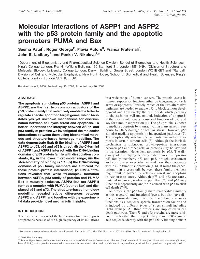
Published online 2 August 2008 Nucleic Acids Research, 2008, Vol. 36, No. 16 5139–5151doi:10.1093/nar/gkn490
Molecular interactions of ASPP1 and ASPP2with the p53 protein family and the apoptoticpromoters PUMA and BaxSeema Patel1, Roger George2, Flavia Autore3, Franca Fraternali3,
John E. Ladbury2 and Penka V. Nikolova1,*
1Department of Biochemistry and Pharmaceutical Science Division, School of Biomedical and Health Sciences,King’s College London, Franklin-Wilkins Building, 150 Stamford St., London SE1 9NH, 2Division of Structural andMolecular Biology, University College London, Darwin Building, Gower Street, London WC1E 6BT and 3RandallDivision of Cell and Molecular Biophysics, New Hunt House, School of Biomedical and Health Sciences, King’sCollege London, London SE1 1UL, UK
Received June 9, 2008; Revised July 15, 2008; Accepted July 16, 2008
ABSTRACT
The apoptosis stimulating p53 proteins, ASPP1 andASPP2, are the first two common activators of thep53 protein family that selectively enable the latter toregulate specific apoptotic target genes, which facil-itates yes yet unknown mechanisms for discrimi-nation between cell cycle arrest and apoptosis. Tobetter understand the interplay between ASPP- andp53-family of proteins we investigated the molecularinteractions between them using biochemical meth-ods and structure-based homology modelling. Thedata demonstrate that: (i) the binding of ASPP1 andASPP2 to p53, p63 and p73 is direct; (ii) the C-terminiof ASPP1 and ASPP2 interact with the DNA-bindingdomains of p53 protein family with dissociation con-stants, Kd, in the lower micro-molar range; (iii) thestoichiometry of binding is 1:1; (iv) the DNA-bindingdomains of p53 family members are sufficient forthese protein–protein interactions; (v) EMSA titra-tions revealed that while tri-complex formationbetween ASPPs, p53 family of proteins and PUMA/Bax is mutually exclusive, ASPP2 (but not ASPP1)formed a complex with PUMA (but not Bax) and dis-placed p53 and p73. The structure-based homologymodelling revealed subtle differences betweenASPP2 and ASPP1 and together with the experimen-tal data provide novel mechanistic insights.
INTRODUCTION
The p53 protein is one of the best known tumour suppres-sor proteins because of the high frequency of its mutations
in a wide range of human cancers. The protein exerts itstumour suppressor function either by triggering cell cyclearrest or apoptosis. Precisely, which of the two alternativepathways are needed to enable p53 to block tumour devel-opment and how exactly the cells decide which pathwayto choose is not well understood. Induction of apoptosisis the most evolutionary conserved function of p53 andkey for tumour suppression (1). The p53 protein is knownto mediate apoptosis by transactivating many genes in res-ponse to DNA damage or cellular stress. However, p53can also mediate apoptosis by independent pathways (2).Transcriptionally inactive p53 mutants can induce apo-ptosis in certain tumour cells (3). Although the intrinsicmechanism is unknown, protein–protein interactionsbetween p53 and other cellular proteins may be involvedin transcription-independent apoptosis by p53. The dis-covery of the phylogenetically older tumour suppressorp53 family members, p73 and p63, brought excitementand controversy over whether and how they cooperatewith p53 in tumour suppression (4–6). It raised the expec-tations that a cross talk between these family membersmight exist to govern the cell cycle arrest and apoptosisin response to stress. Although p73 and p63 are rarelymutated in cancer, studies suggest that p73 and p63 mayfunction independently and/or in concert with p53 to elicitcell death (7–9).As proteins, the p53 family share remarkable similarity
at the structural and functional levels, but also have dis-tinct, non-overlapping functions. Each family memberfunctions as a sequence-specific transcription factor andis induced by different types of stress stimuli includingDNA damage. All three proteins are implicated in celldeath pathways. The p73 and p63 proteins are more simi-lar to each other than to p53. They share >60% aminoacid sequence identity with the p53 DNA-binding domain
*To whom correspondence should be addressed. Tel: +44 207 848 4276; Fax: +44 207 848 4500; Email: [email protected]
� 2008 The Author(s)This is an Open Access article distributed under the terms of the Creative Commons Attribution Non-Commercial License (http://creativecommons.org/licenses/by-nc/2.0/uk/) which permits unrestricted non-commercial use, distribution, and reproduction in any medium, provided the original work is properly cited.
by guest on October 5, 2014
http://nar.oxfordjournals.org/D
ownloaded from
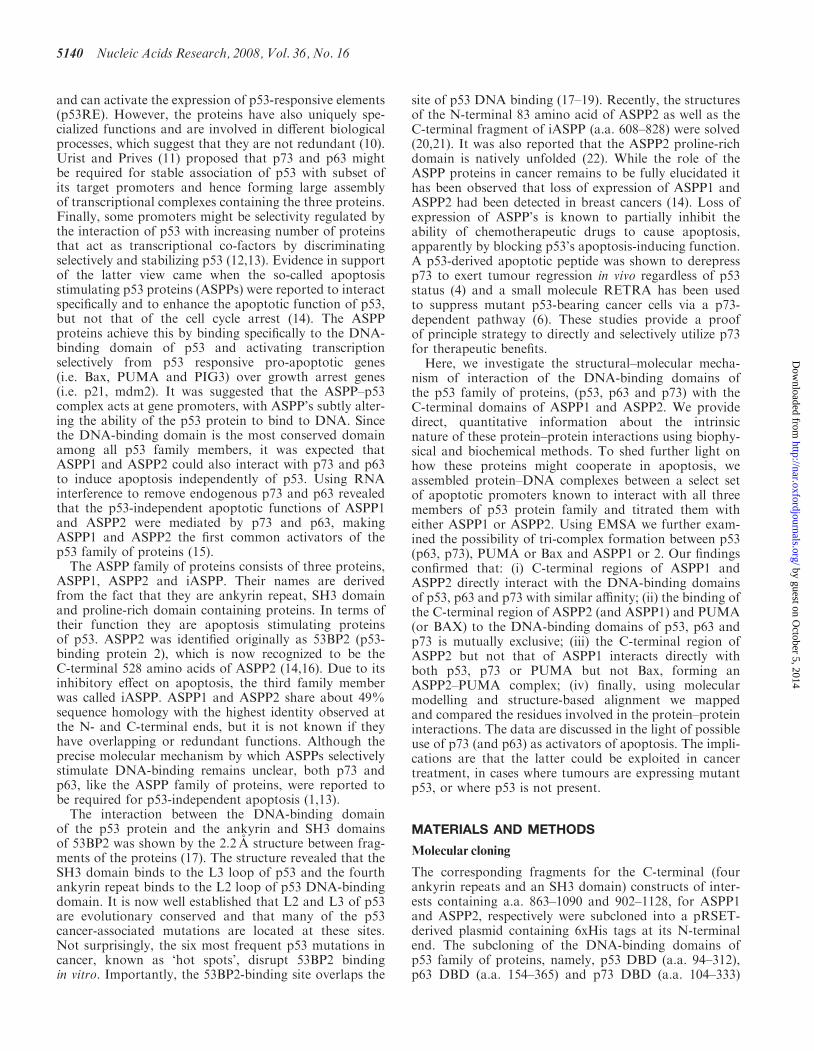
and can activate the expression of p53-responsive elements(p53RE). However, the proteins have also uniquely spe-cialized functions and are involved in different biologicalprocesses, which suggest that they are not redundant (10).Urist and Prives (11) proposed that p73 and p63 mightbe required for stable association of p53 with subset ofits target promoters and hence forming large assemblyof transcriptional complexes containing the three proteins.Finally, some promoters might be selectivity regulated bythe interaction of p53 with increasing number of proteinsthat act as transcriptional co-factors by discriminatingselectively and stabilizing p53 (12,13). Evidence in supportof the latter view came when the so-called apoptosisstimulating p53 proteins (ASPPs) were reported to interactspecifically and to enhance the apoptotic function of p53,but not that of the cell cycle arrest (14). The ASPPproteins achieve this by binding specifically to the DNA-binding domain of p53 and activating transcriptionselectively from p53 responsive pro-apoptotic genes(i.e. Bax, PUMA and PIG3) over growth arrest genes(i.e. p21, mdm2). It was suggested that the ASPP–p53complex acts at gene promoters, with ASPP’s subtly alter-ing the ability of the p53 protein to bind to DNA. Sincethe DNA-binding domain is the most conserved domainamong all p53 family members, it was expected thatASPP1 and ASPP2 could also interact with p73 and p63to induce apoptosis independently of p53. Using RNAinterference to remove endogenous p73 and p63 revealedthat the p53-independent apoptotic functions of ASPP1and ASPP2 were mediated by p73 and p63, makingASPP1 and ASPP2 the first common activators of thep53 family of proteins (15).The ASPP family of proteins consists of three proteins,
ASPP1, ASPP2 and iASPP. Their names are derivedfrom the fact that they are ankyrin repeat, SH3 domainand proline-rich domain containing proteins. In terms oftheir function they are apoptosis stimulating proteinsof p53. ASPP2 was identified originally as 53BP2 (p53-binding protein 2), which is now recognized to be theC-terminal 528 amino acids of ASPP2 (14,16). Due to itsinhibitory effect on apoptosis, the third family memberwas called iASPP. ASPP1 and ASPP2 share about 49%sequence homology with the highest identity observed atthe N- and C-terminal ends, but it is not known if theyhave overlapping or redundant functions. Although theprecise molecular mechanism by which ASPPs selectivelystimulate DNA-binding remains unclear, both p73 andp63, like the ASPP family of proteins, were reported tobe required for p53-independent apoptosis (1,13).The interaction between the DNA-binding domain
of the p53 protein and the ankyrin and SH3 domainsof 53BP2 was shown by the 2.2 A structure between frag-ments of the proteins (17). The structure revealed that theSH3 domain binds to the L3 loop of p53 and the fourthankyrin repeat binds to the L2 loop of p53 DNA-bindingdomain. It is now well established that L2 and L3 of p53are evolutionary conserved and that many of the p53cancer-associated mutations are located at these sites.Not surprisingly, the six most frequent p53 mutations incancer, known as ‘hot spots’, disrupt 53BP2 bindingin vitro. Importantly, the 53BP2-binding site overlaps the
site of p53 DNA binding (17–19). Recently, the structuresof the N-terminal 83 amino acid of ASPP2 as well as theC-terminal fragment of iASPP (a.a. 608–828) were solved(20,21). It was also reported that the ASPP2 proline-richdomain is natively unfolded (22). While the role of theASPP proteins in cancer remains to be fully elucidated ithas been observed that loss of expression of ASPP1 andASPP2 had been detected in breast cancers (14). Loss ofexpression of ASPP’s is known to partially inhibit theability of chemotherapeutic drugs to cause apoptosis,apparently by blocking p53’s apoptosis-inducing function.A p53-derived apoptotic peptide was shown to derepressp73 to exert tumour regression in vivo regardless of p53status (4) and a small molecule RETRA has been usedto suppress mutant p53-bearing cancer cells via a p73-dependent pathway (6). These studies provide a proofof principle strategy to directly and selectively utilize p73for therapeutic benefits.
Here, we investigate the structural–molecular mecha-nism of interaction of the DNA-binding domains ofthe p53 family of proteins, (p53, p63 and p73) with theC-terminal domains of ASPP1 and ASPP2. We providedirect, quantitative information about the intrinsicnature of these protein–protein interactions using biophy-sical and biochemical methods. To shed further light onhow these proteins might cooperate in apoptosis, weassembled protein–DNA complexes between a select setof apoptotic promoters known to interact with all threemembers of p53 protein family and titrated them witheither ASPP1 or ASPP2. Using EMSA we further exam-ined the possibility of tri-complex formation between p53(p63, p73), PUMA or Bax and ASPP1 or 2. Our findingsconfirmed that: (i) C-terminal regions of ASPP1 andASPP2 directly interact with the DNA-binding domainsof p53, p63 and p73 with similar affinity; (ii) the binding ofthe C-terminal region of ASPP2 (and ASPP1) and PUMA(or BAX) to the DNA-binding domains of p53, p63 andp73 is mutually exclusive; (iii) the C-terminal region ofASPP2 but not that of ASPP1 interacts directly withboth p53, p73 or PUMA but not Bax, forming anASPP2–PUMA complex; (iv) finally, using molecularmodelling and structure-based alignment we mappedand compared the residues involved in the protein–proteininteractions. The data are discussed in the light of possibleuse of p73 (and p63) as activators of apoptosis. The impli-cations are that the latter could be exploited in cancertreatment, in cases where tumours are expressing mutantp53, or where p53 is not present.
MATERIALS AND METHODS
Molecular cloning
The corresponding fragments for the C-terminal (fourankyrin repeats and an SH3 domain) constructs of inter-ests containing a.a. 863–1090 and 902–1128, for ASPP1and ASPP2, respectively were subcloned into a pRSET-derived plasmid containing 6xHis tags at its N-terminalend. The subcloning of the DNA-binding domains ofp53 family of proteins, namely, p53 DBD (a.a. 94–312),p63 DBD (a.a. 154–365) and p73 DBD (a.a. 104–333)
5140 Nucleic Acids Research, 2008, Vol. 36, No. 16
by guest on October 5, 2014
http://nar.oxfordjournals.org/D
ownloaded from

into a pRSET(A)-derived plasmid was described pre-viously (23,24). The p53, p63 and p73 constructs do nothave tags.
Protein expression and protein purification
Cell cultures of Escherichia coli C41 (DE3) were grownin 2�TY medium supplemented with ampicillin at 378C(200–250 r.p.m.) to an OD600� 0.6–0.8. The temperaturewas then reduced to 16–228C before overnight induc-tion with 0.5mM IPTG. Cells were harvested by centri-fugation at 48C and then lysed in BugBuster ProteinExtraction Reagent supplemented with BenzonaseNuclease (Novagen, Nottingham, UK), with EDTA-freeprotease inhibitor tablets (Roche, Burgess Hill, UK) and4mM DTT. The protein samples were spun down at13 000 r.p.m. for 20min at 48C. The purification of theDNA-binding domains of p53, p63 and p73 were per-formed as reported previously (23,25,26). For ASPP1and ASPP2, the soluble fraction was purified usingbatch purification with Ni-NTA resin (Qiagen, Crawley,UK) (50mM NaPi, 300mM NaCl, 20mM imidazole,1mM DTT and 10% glycerol). The supernatant obtainedafter the BugBuster extraction procedure was poured intoa glass beaker with the Ni beads and incubated at 48C byshaking for 45min. The suspension was then spun at4000 r.p.m. for 10min at 48C and the supernatant dis-carded. The beads were washed with 10 column volumesof binding buffer as above and the supernatant discarded.The bound protein was eluted from the beads using fivecolumn volumes of elution buffer (5mM NaPi, 300mMNaCl, 300mM imidazole, 1mM DTT and 10% glycerol)by centrifuging at 4000 r.p.m. for 5min at 48C.
Circular dichroism
Proteins were subjected to the circular dichroism (CD)scans at concentrations 0.2mg/ml in 10mM NaPi,100mM NaCl, 4mM DTE buffer. Samples were filteredwith 0.2 mm filters (Whatman). The CD studies were per-formed using an Applied Photophysics Chirascan spectro-polarimeter. Far-UV spectra were recorded in the region260–190 nm, respectively using 0.5mm cell path length,1 nm bandwidth, 0.5 nm scan speed, 3 s time-per-point.The temperature was controlled using Melcor’s thermo-electric temperature controller. Data are represented inellipticity as a function of the wavelength (nm) and calcu-lated according to Schmid (27).
Isothermal titration calorimetry
The buffers for each protein used were 50mM NaPi,200–300mM NaCl, 1–5mM DTT and 0–10% glyceroland filtered via a 0.2 mm filter (Whatman). The proteinconcentrations used for ASPP1:p53 were 11 mM:200 mM,ASPP1:p63 were 4.5:80 mM and ASPP1:p73 were7:100mM; ASPP2:p53 were 15:300 mM, ASPP2:p63 were10:260 mM and ASPP2:p73 were 25:560 mM. All experi-ments were carried out using the VP-(Isothermal titrationcalorimetry) ITC Micro-Calorimeter with the followingparameters: 20 injections of 15 ml with 240 s spacing, refer-ence power 14 pCal/s, stirring speed 300 and at 108C.Controls were performed by titrating each protein
sample used at a higher concentration in the syringe,into the sample cell filled with buffer alone.Experimental data were fitted assuming a single bindingsite (Microcal VPITC user guide).
Analytical gel filtration
Analytical gel filtration was performed in 50mM NaPi,pH 7.2, 200mM NaCl, 5mM DTT using a Superdex200 (10/300) column (GE Healthcare, Little Chalfont,UK). The proteins were injected at a concentration of40 mM with an injection volume of 100 ml and the milli-absorbance units (mAu) at 280 nm were recorded.Complexes were incubated at 228C for 25min.
Electrophoretic mobility shift assays
Electrophoretic mobility shift assays (EMSA) were carriedout with all p53 family of proteins and 50-fluorescein-labelled 30-mer dsDNA promoters of Bax and PUMAusing 0.7% agarose gel and TB buffer at a constant cur-rent of 60V at 48C. The DNA concentrations were keptconstant, while the p53, p63 and p73 protein concentra-tions were increased. The protein concentration at whichmost (or all) of the DNA was shifted was then selected forthe titration with the ASPP proteins. The fluorescein tagenabled sensitive detection under UV light. Protein–DNAcomplexes were incubated on ice for 20min. To investigatethe possibility of tri-complex formation and or displace-ment of the DNA from the protein–DNA complexes, theASPP proteins were then titrated into these complexes andincubated for further 30min at 48C before loading ontothe gel. In addition, we tested if ASPP1 and ASPP2would be able to shift the same DNA promoters underthe experimental conditions. The oligonucleotides used inthe EMSA were annealed by heating at 958C for 5minfollowed by cooling to 48C overnight. The followingsequences were used:Bax (forward): GGGCTCACAAGTTAGAGACAAG
CCTGGGCGBax (reverse): CGCCCAGGCTTGTCTCTAACTTGT
GAGCCCPUMA (forward): CGCGCCTGCAAGTCCTGACTT
GTCCGCGGCPUMA (reverse): GCCGCGGACAAGTCAGGACTT
GCAGGCGCG
Modelling
The model structures of p63, p73 and ASPP1were obtained by homology modelling from the crystalstructure of p53 in complex with ASPP2 (PDB accessionnumber 1ycs) (17). All the modelled sequences share atleast 60% sequence identity with the corresponding tem-plate, therefore ensuring reliable homology models (28).Sequence alignments were generated by using the T-coffeemethod (29). Three-dimensional models were generatedusing the MODELLER package, which can generate alarge number of models and performs an optimizationof the generated models with respect to a defined objectivefunction. This software has been used for wide-scale struc-ture modelling of genomes (30). The selected models werechosen on the basis of the objective function’s score.
Nucleic Acids Research, 2008, Vol. 36, No. 16 5141
by guest on October 5, 2014
http://nar.oxfordjournals.org/D
ownloaded from
To refine the models, energy minimizations were per-formed with the GROMACS package (31) using theGROMOS96 force field (32). Images were producedwith visual molecular dynamics (VMD) 1.8.5 (33). Thecomplexes of the p53 family members and ASPP1 wereconstructed by analogy to the complex of p53 withASPP2 in the 1ycs coordinate file. To characterize theinteraction surfaces, the program POPScomp was used(34,35) to detect the residues that are buried upon complexformation. POPScomp calculates the solvent accessiblesurface area (SASA, in A2) of each residue before andafter the formation of the complex. The structural align-ment was performed with the program aliss (JensKleinjung, personal communication). Electrostatic sur-faces were calculated by the use of the programPYMOL (36) with the implemented subroutine surfaceAPBS (37).
RESULTS
Determination of the oligomerization status of theproteins using gel filtration
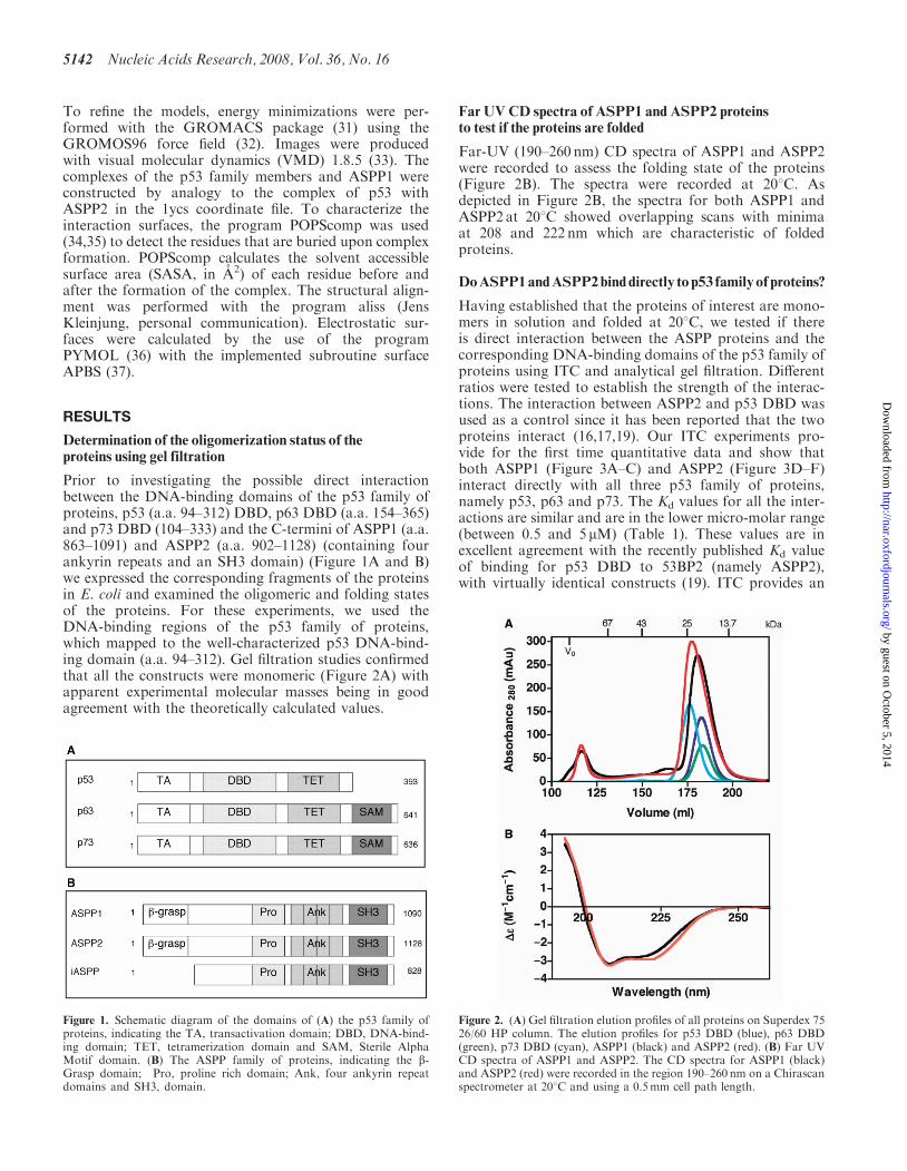
Prior to investigating the possible direct interactionbetween the DNA-binding domains of the p53 family ofproteins, p53 (a.a. 94–312) DBD, p63 DBD (a.a. 154–365)and p73 DBD (104–333) and the C-termini of ASPP1 (a.a.863–1091) and ASPP2 (a.a. 902–1128) (containing fourankyrin repeats and an SH3 domain) (Figure 1A and B)we expressed the corresponding fragments of the proteinsin E. coli and examined the oligomeric and folding statesof the proteins. For these experiments, we used theDNA-binding regions of the p53 family of proteins,which mapped to the well-characterized p53 DNA-bind-ing domain (a.a. 94–312). Gel filtration studies confirmedthat all the constructs were monomeric (Figure 2A) withapparent experimental molecular masses being in goodagreement with the theoretically calculated values.
Far UVCD spectra of ASPP1 and ASPP2 proteinsto test if the proteins are folded
Far-UV (190–260 nm) CD spectra of ASPP1 and ASPP2were recorded to assess the folding state of the proteins(Figure 2B). The spectra were recorded at 208C. Asdepicted in Figure 2B, the spectra for both ASPP1 andASPP2 at 208C showed overlapping scans with minimaat 208 and 222 nm which are characteristic of foldedproteins.
DoASPP1andASPP2binddirectly top53 familyofproteins?
Having established that the proteins of interest are mono-mers in solution and folded at 208C, we tested if thereis direct interaction between the ASPP proteins and thecorresponding DNA-binding domains of the p53 family ofproteins using ITC and analytical gel filtration. Differentratios were tested to establish the strength of the interac-tions. The interaction between ASPP2 and p53 DBD wasused as a control since it has been reported that the twoproteins interact (16,17,19). Our ITC experiments pro-vide for the first time quantitative data and show thatboth ASPP1 (Figure 3A–C) and ASPP2 (Figure 3D–F)interact directly with all three p53 family of proteins,namely p53, p63 and p73. The Kd values for all the inter-actions are similar and are in the lower micro-molar range(between 0.5 and 5 mM) (Table 1). These values are inexcellent agreement with the recently published Kd valueof binding for p53 DBD to 53BP2 (namely ASPP2),with virtually identical constructs (19). ITC provides an
Figure 1. Schematic diagram of the domains of (A) the p53 family ofproteins, indicating the TA, transactivation domain; DBD, DNA-bind-ing domain; TET, tetramerization domain and SAM, Sterile AlphaMotif domain. (B) The ASPP family of proteins, indicating the b-Grasp domain; Pro, proline rich domain; Ank, four ankyrin repeatdomains and SH3, domain.
Figure 2. (A) Gel filtration elution profiles of all proteins on Superdex 7526/60 HP column. The elution profiles for p53 DBD (blue), p63 DBD(green), p73 DBD (cyan), ASPP1 (black) and ASPP2 (red). (B) Far UVCD spectra of ASPP1 and ASPP2. The CD spectra for ASPP1 (black)and ASPP2 (red) were recorded in the region 190–260 nm on a Chirascanspectrometer at 208C and using a 0.5mm cell path length.
5142 Nucleic Acids Research, 2008, Vol. 36, No. 16
by guest on October 5, 2014
http://nar.oxfordjournals.org/D
ownloaded from
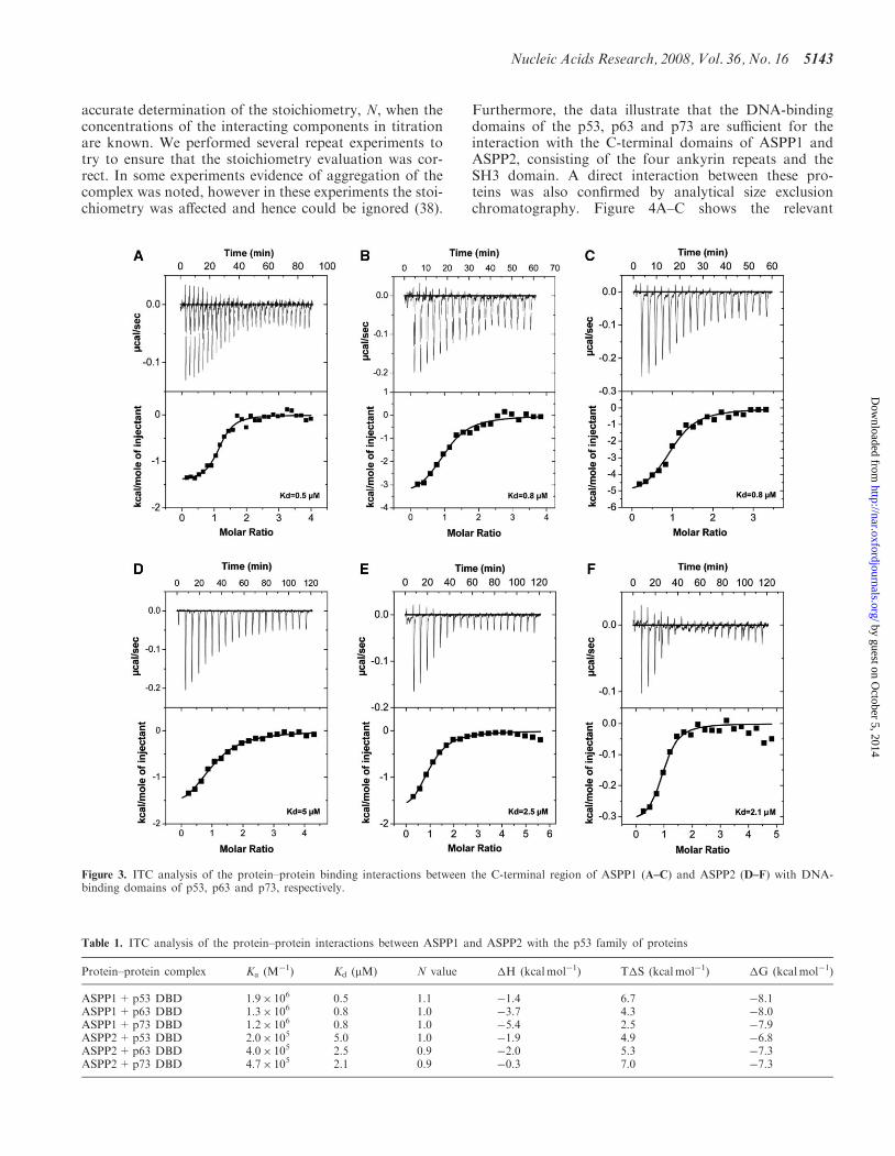
accurate determination of the stoichiometry, N, when theconcentrations of the interacting components in titrationare known. We performed several repeat experiments totry to ensure that the stoichiometry evaluation was cor-rect. In some experiments evidence of aggregation of thecomplex was noted, however in these experiments the stoi-chiometry was affected and hence could be ignored (38).
Furthermore, the data illustrate that the DNA-bindingdomains of the p53, p63 and p73 are sufficient for theinteraction with the C-terminal domains of ASPP1 andASPP2, consisting of the four ankyrin repeats and theSH3 domain. A direct interaction between these pro-teins was also confirmed by analytical size exclusionchromatography. Figure 4A–C shows the relevant
Figure 3. ITC analysis of the protein–protein binding interactions between the C-terminal region of ASPP1 (A–C) and ASPP2 (D–F) with DNA-binding domains of p53, p63 and p73, respectively.
Table 1. ITC analysis of the protein–protein interactions between ASPP1 and ASPP2 with the p53 family of proteins
Protein–protein complex Ka (M�1) Kd (mM) N value �H (kcalmol�1) T�S (kcalmol�1) �G (kcalmol�1)
ASPP1+p53 DBD 1.9� 106 0.5 1.1 �1.4 6.7 �8.1ASPP1+p63 DBD 1.3� 106 0.8 1.0 �3.7 4.3 �8.0ASPP1+p73 DBD 1.2� 106 0.8 1.0 �5.4 2.5 �7.9ASPP2+p53 DBD 2.0� 105 5.0 1.0 �1.9 4.9 �6.8ASPP2+p63 DBD 4.0� 105 2.5 0.9 �2.0 5.3 �7.3ASPP2+p73 DBD 4.7� 105 2.1 0.9 �0.3 7.0 �7.3
Nucleic Acids Research, 2008, Vol. 36, No. 16 5143
by guest on October 5, 2014
http://nar.oxfordjournals.org/D
ownloaded from
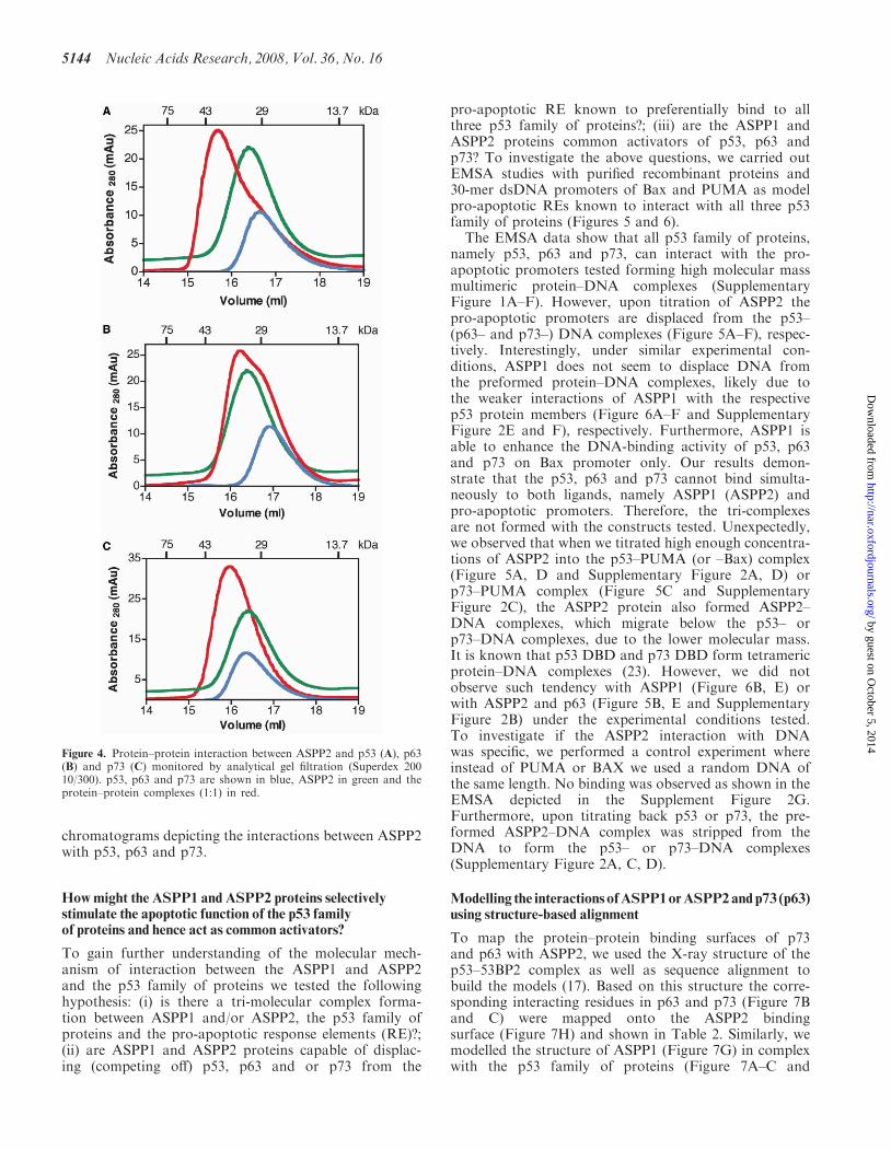
chromatograms depicting the interactions between ASPP2with p53, p63 and p73.
Howmight the ASPP1 and ASPP2 proteins selectivelystimulate the apoptotic function of the p53 familyof proteins and hence act as common activators?
To gain further understanding of the molecular mech-anism of interaction between the ASPP1 and ASPP2and the p53 family of proteins we tested the followinghypothesis: (i) is there a tri-molecular complex forma-tion between ASPP1 and/or ASPP2, the p53 family ofproteins and the pro-apoptotic response elements (RE)?;(ii) are ASPP1 and ASPP2 proteins capable of displac-ing (competing off) p53, p63 and or p73 from the
pro-apoptotic RE known to preferentially bind to allthree p53 family of proteins?; (iii) are the ASPP1 andASPP2 proteins common activators of p53, p63 andp73? To investigate the above questions, we carried outEMSA studies with purified recombinant proteins and30-mer dsDNA promoters of Bax and PUMA as modelpro-apoptotic REs known to interact with all three p53family of proteins (Figures 5 and 6).
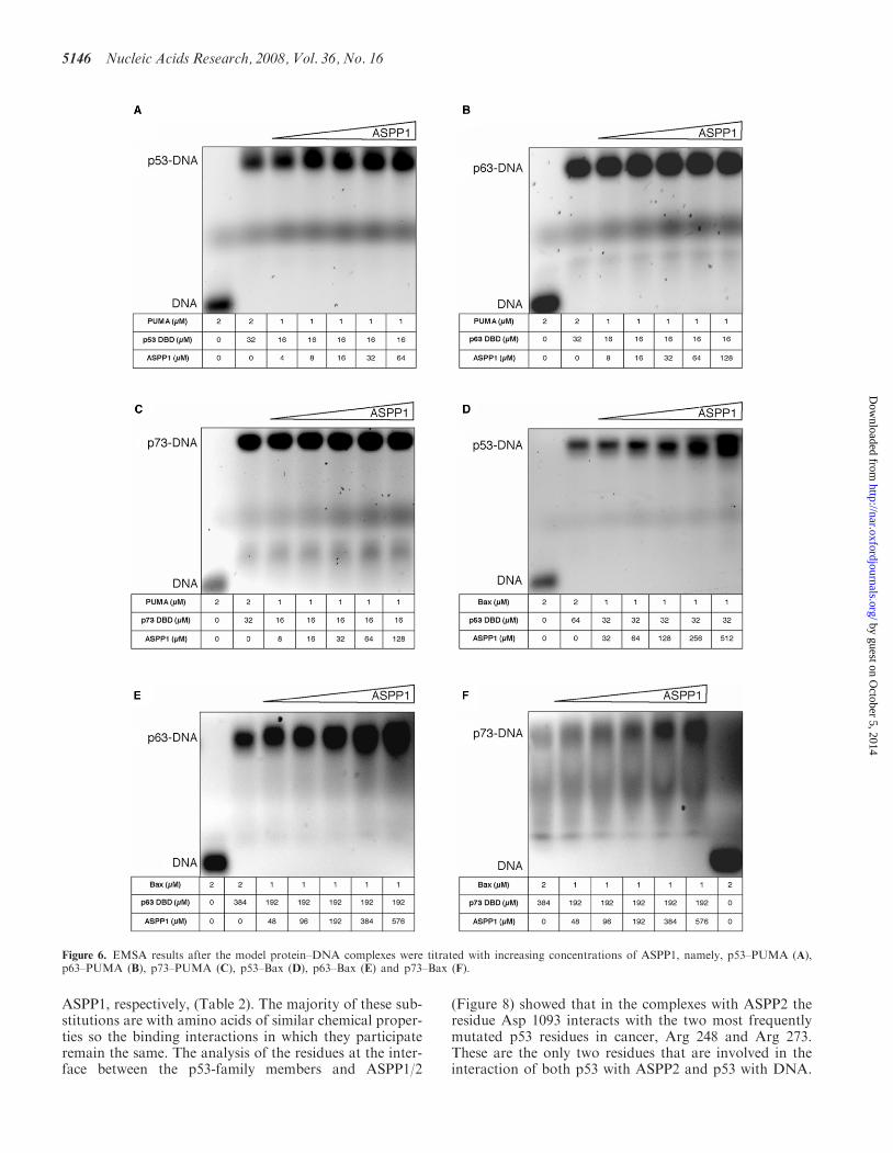
The EMSA data show that all p53 family of proteins,namely p53, p63 and p73, can interact with the pro-apoptotic promoters tested forming high molecular massmultimeric protein–DNA complexes (SupplementaryFigure 1A–F). However, upon titration of ASPP2 thepro-apoptotic promoters are displaced from the p53–(p63– and p73–) DNA complexes (Figure 5A–F), respec-tively. Interestingly, under similar experimental con-ditions, ASPP1 does not seem to displace DNA fromthe preformed protein–DNA complexes, likely due tothe weaker interactions of ASPP1 with the respectivep53 protein members (Figure 6A–F and SupplementaryFigure 2E and F), respectively. Furthermore, ASPP1 isable to enhance the DNA-binding activity of p53, p63and p73 on Bax promoter only. Our results demon-strate that the p53, p63 and p73 cannot bind simulta-neously to both ligands, namely ASPP1 (ASPP2) andpro-apoptotic promoters. Therefore, the tri-complexesare not formed with the constructs tested. Unexpectedly,we observed that when we titrated high enough concentra-tions of ASPP2 into the p53–PUMA (or –Bax) complex(Figure 5A, D and Supplementary Figure 2A, D) orp73–PUMA complex (Figure 5C and SupplementaryFigure 2C), the ASPP2 protein also formed ASPP2–DNA complexes, which migrate below the p53– orp73–DNA complexes, due to the lower molecular mass.It is known that p53 DBD and p73 DBD form tetramericprotein–DNA complexes (23). However, we did notobserve such tendency with ASPP1 (Figure 6B, E) orwith ASPP2 and p63 (Figure 5B, E and SupplementaryFigure 2B) under the experimental conditions tested.To investigate if the ASPP2 interaction with DNAwas specific, we performed a control experiment whereinstead of PUMA or BAX we used a random DNA ofthe same length. No binding was observed as shown in theEMSA depicted in the Supplement Figure 2G.Furthermore, upon titrating back p53 or p73, the pre-formed ASPP2–DNA complex was stripped from theDNA to form the p53– or p73–DNA complexes(Supplementary Figure 2A, C, D).
Modelling the interactions ofASPP1orASPP2andp73 (p63)using structure-based alignment
To map the protein–protein binding surfaces of p73and p63 with ASPP2, we used the X-ray structure of thep53–53BP2 complex as well as sequence alignment tobuild the models (17). Based on this structure the corre-sponding interacting residues in p63 and p73 (Figure 7Band C) were mapped onto the ASPP2 bindingsurface (Figure 7H) and shown in Table 2. Similarly, wemodelled the structure of ASPP1 (Figure 7G) in complexwith the p53 family of proteins (Figure 7A–C and
Figure 4. Protein–protein interaction between ASPP2 and p53 (A), p63(B) and p73 (C) monitored by analytical gel filtration (Superdex 20010/300). p53, p63 and p73 are shown in blue, ASPP2 in green and theprotein–protein complexes (1:1) in red.
5144 Nucleic Acids Research, 2008, Vol. 36, No. 16
by guest on October 5, 2014
http://nar.oxfordjournals.org/D
ownloaded from
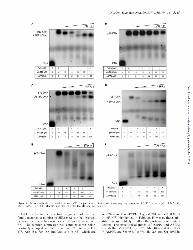
Table 2). From the structural alignment of the p53family members a number of differences can be observedbetween the interacting residues of p53 and those in p63/p73. The tumour suppressor p53 contains more polar,positively charged residues than p63/p73, namely His178, Arg 181, Ser 183 and Met 243 in p53, which are
Asn 246/196, Leu 249/199, Arg 251/201 and Val 313/263in p63/p73 (highlighted in Table 2). However, these sub-stitutions are unlikely to affect the protein–protein inter-actions. The structural alignment of ASPP1 and ASPP2reveals that Met 1021, Tyr 1023, Met 1026 and Asp 1093in ASPP2, are Ser 983, Ile 985, Ile 988 and Ser 1055 in
Figure 5. EMSA results after the model protein–DNA complexes were titrated with increasing concentrations of ASPP2, namely, p53–PUMA (A),p63–PUMA (B), p73–PUMA (C), p53–Bax (D), p63–Bax (E) and p73–Bax (F).
Nucleic Acids Research, 2008, Vol. 36, No. 16 5145
by guest on October 5, 2014
http://nar.oxfordjournals.org/D
ownloaded from
ASPP1, respectively, (Table 2). The majority of these sub-stitutions are with amino acids of similar chemical proper-ties so the binding interactions in which they participateremain the same. The analysis of the residues at the inter-face between the p53-family members and ASPP1/2
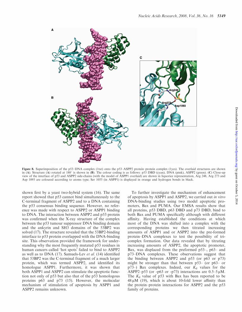
(Figure 8) showed that in the complexes with ASPP2 theresidue Asp 1093 interacts with the two most frequentlymutated p53 residues in cancer, Arg 248 and Arg 273.These are the only two residues that are involved in theinteraction of both p53 with ASPP2 and p53 with DNA.
Figure 6. EMSA results after the model protein–DNA complexes were titrated with increasing concentrations of ASPP1, namely, p53–PUMA (A),p63–PUMA (B), p73–PUMA (C), p53–Bax (D), p63–Bax (E) and p73–Bax (F).
5146 Nucleic Acids Research, 2008, Vol. 36, No. 16
by guest on October 5, 2014
http://nar.oxfordjournals.org/D
ownloaded from
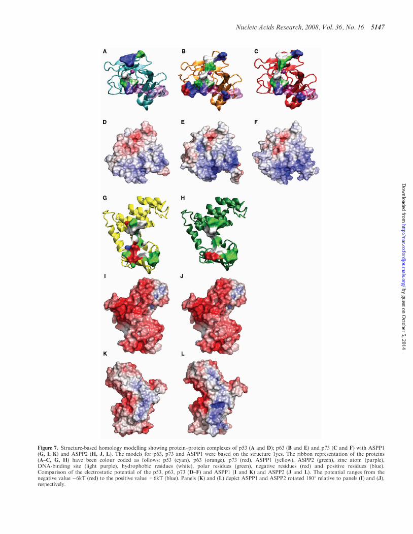
Figure 7. Structure-based homology modelling showing protein–protein complexes of p53 (A and D); p63 (B and E) and p73 (C and F) with ASPP1(G, I, K) and ASPP2 (H, J, L). The models for p63, p73 and ASPP1 were based on the structure 1ycs. The ribbon representation of the proteins(A–C, G, H) have been colour coded as follows: p53 (cyan), p63 (orange), p73 (red), ASPP1 (yellow), ASPP2 (green), zinc atom (purple),DNA-binding site (light purple), hydrophobic residues (white), polar residues (green), negative residues (red) and positive residues (blue).Comparison of the electrostatic potential of the p53, p63, p73 (D–F) and ASPP1 (I and K) and ASPP2 (J and L). The potential ranges from thenegative value �6kT (red) to the positive value +6kT (blue). Panels (K) and (L) depict ASPP1 and ASPP2 rotated 1808 relative to panels (I) and (J),respectively.
Nucleic Acids Research, 2008, Vol. 36, No. 16 5147
by guest on October 5, 2014
http://nar.oxfordjournals.org/D
ownloaded from
The substitution of the negatively charged Asp in ASPP2with the uncharged Ser 1055 in ASPP1 results therefore ina loss of this functional interaction (Figure 8C). In addi-tion, the distance between the OH-group of the Ser 1055side-chain and the NH3+ group of the side chains of thetwo arginines is >4.3 A.With regards to p53 cancer-associated DNA contact
mutants, R248Q (W) and R273H, the following observa-tions could be made based on the structure-based homol-ogy models. In general, the p53 region of 248 is in closeproximity with some bulky residues in ASPP2, creatingsteric hindrance (i.e. the tryptophan at position 1097 inASPP2). Specifically, when R248 in p53 is mutated totryptophan there would be a steric clash with the glutamicacid (a.a.1094) in ASPP2. The arginine substitutionto glutamine (Q) in p53 creates a weaker interactionwith the aspartic acid (D) at position 1093 in ASPP2.There is a stretch of negative residues (a.a. 1090–1096)EDEDEIE in ASPP2 facing the R248 in p53. Thereforea charged residue such as Arg in position 248 seems tofavour the binding with ASPP2. For R273 it has beenreported that this amino acid residue is located at theedge of the ASPP2 binding interface (17). The modelproposed here (Figure 8C) suggests that the substitutionof arginine to histidine at position 273 would disturb theinteraction with aspartic acid 1093 in ASPP2 and there-fore weaken the binding.The structure-based sequence alignment between p53,
p63 and p73 shows that the Arg 248 and Arg 273 areconserved. Hence, the same conclusions could apply top63 and p73. The electrostatic surfaces of the p53 familymembers, namely p53, p63, p73 (Figure 7D–F), respec-tively, are very similar since they are all mainly positivelycharged but p63 and p73 show a relatively more positivesurface than p53 around the DNA-binding site. This could
account for the different selectivity within the p53 familyof proteins for p53 RE. The electrostatic p53-binding sur-faces for ASPP1 and ASPP2 show an overall negativecharge (Figure 7I and J), but a positive charge on theopposite side (Figure 7K and L). It is reasonable toassume that the ASPP–DNA interactions observed inthe EMSA occur on the positive interface. The ‘backview’ of ASPP2 appears to have a more positive surfacearea than ASPP1, which could provide further evidencefor our observation that ASPP2 displaces the DNA morereadily than ASPP1.
DISCUSSION
To shed further light on how ASPP1 and ASPP2 mightstimulate apoptosis we investigated the intrinsic nature ofthe interactions between the ASPP1 and ASPP2 and thep53 family of proteins, namely p53, p63 and p73, usingbiochemical and biophysical methods with purified pro-teins. We have probed the mechanism of these interactionsby using Bax and PUMA as model apoptotic promoters.We first show that ASPP1 and ASPP2 directly interactwith p53, p63 and p73 and that the DNA-binding domainsof the p53 family of proteins are sufficient for the interac-tion with the ASPPs. Our data based on the equilibriumbinding constants obtained by ITC also show that theinteraction between the C-terminal domains of ASPP1and ASPP2 to p53 DBD, p63 DBD and p73 DBD arevery similar and are in the lower micro molar range.This is to be expected given the high amino acid identityin the DNA-binding domains between the p53 familymembers (about 60%) and C-termini of ASPP1 andASPP2 (80%).
Evidence for possible interaction of ASPP2, which wasknown as 53BP2, with p53 (but not with mutant p53) was
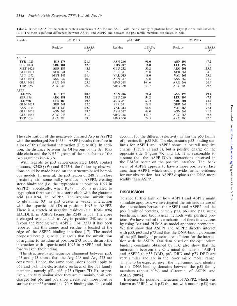
Table 2. Buried SASA for the protein–protein complexes of ASPP2 and ASPP1 with the p53 family of proteins based on 1ycs [Gorina and Pavletich,
(17)]. The most significant differences between ASPP1 and ASPP2 and between the p53 family members are shown in bold
Residue p53 DBD p63 DBD p73 DBD
Residue iSASA Residue iSASA Residue iSASAA2 A2 A2
ASPP2TYR 1023 HIS 178 121.6 ASN 246 91.0 ASN 196 47.2
SER 1024 ARG 181 62.5 HIS 247 16.0 LEU 199 31.0
MET 1026 SER 183 48.3 GLU 252 33.2 ARG 201 153.7
GLN 1071 SER 241 32.9 SER 311 28.8 SER 261 34.0ASN 1072 MET 243 101.4 VAL 313 18.0 VAL 263 73.6
GLU 1094 ASN 247 40.2 ASN 317 22.8 ASN 267 43.7GLU 1096 ARG 248 153.6 ARG 318 164.6 ARG 268 154.4TRP 1097 ARG 280 29.2 ARG 350 53.7 ARG 300 29.5
ASPP1ILE 985 HIS 178 110.6 ASN 246 71.4 ASN 196 49.4
SER 986 ARG 181 58.3 LEU 249 54.2 LEU 199 47.3
ILE 988 SER 183 49.8 ARG 251 63.2 ARG 201 143.2
GLN 1033 SER 241 32.5 SER 311 28.8 SER 261 31.7ASN 1034 MET 243 102.0 VAL 313 54.5 VAL 263 77.1
GLU 1056 ASN 247 39.6 ASN 317 49.0 ASN 267 41.7GLU 1058 ARG 248 151.9 ARG 318 147.7 ARG 268 149.5TRP 1059 ARG 280 29.0 ARG 350 24.5 ARG 300 22.5
5148 Nucleic Acids Research, 2008, Vol. 36, No. 16
by guest on October 5, 2014
http://nar.oxfordjournals.org/D
ownloaded from
shown first by a yeast two-hybrid system (16). The samereport showed that p53 cannot bind simultaneously to theC-terminal fragment of ASPP2 and to a DNA containingthe p53 consensus binding sequence. However, no refer-ence was made with respect to ASPP2 or ASPP1 bindingto DNA. The interaction between ASPP2 and p53 proteinwas confirmed when the X-ray structure of the complexbetween the p53 tumour suppressor DNA binding domainand the ankyrin and SH3 domains of the 53BP2 wassolved (17). The structure revealed that the 53BP2-bindinginterface to p53 protein overlapped with the DNA-bindingsite. This observation provided the framework for under-standing why the most frequently mutated p53 residues inhuman cancers called ‘hot spots’ failed to bind to ASPP2as well as to DNA (17). Samuels-Lev et al. (14) identifiedthat 53BP2 was the C-terminal fragment of a much largerprotein, which was termed ASPP2, and identified itshomologue ASPP1. Furthermore, it was shown thatboth ASPP1 and ASPP2 can stimulate the apoptotic func-tion not only of p53 but also that of the p53 homologousproteins p63 and p73 (15). However, the molecularmechanism of stimulation of apoptosis by ASPP1 andASPP2 remains unknown.
To further investigate the mechanism of enhancementof apoptosis by ASPP1 and ASPP2, we carried out in vitroDNA-binding studies using two model apoptotic pro-moters, Bax and PUMA. Our EMSA results show thatall proteins, p53 DBD, p63 DBD and p73 DBD, bind toboth Bax and PUMA specifically although with differentaffinity. Having established the conditions at whichmost of the DNA was shifted into a complex with thecorresponding proteins we then titrated increasingamounts of ASPP1 and or ASPP2 into the pre-formedprotein–DNA complexes to test the possibility of tri-complex formation. Our data revealed that by titratingincreasing amounts of ASPP2, the apoptotic promoter,Bax, was displaced from the preformed p53–, p63– andp73–DNA complexes. These observations suggest thatthe binding between ASPP2 and p53 (or p63 or p73)might be stronger than that between p53– (or p63– orp73–) Bax complexes. Indeed, our Kd values for theASPP2–p53 (or -p63 or -p73) interactions are 0.5–5mM.The Kd value of p53 with Bax has been reported to be48 mM (19), which is about 10-fold lower affinity thanthe protein–protein interactions for ASPP2 and the p53family of proteins.
Figure 8. Superimposition of the p53–DNA complex (1tsr) onto the p53–ASPP2 protein–protein complex (1ycs). The overlaid structures are shownin (A). Structure (A) rotated at 1808 is shown in (B). The colour coding is as follows: p53 DBD (cyan), DNA (pink), ASPP2 (green). (C) Close-upview of the interface of p53 and ASPP2 side-chains (with the model of ASPP1 overlaid) are shown in liquorice representation, Arg 248, Arg 273 andAsp 1093 are coloured according to atoms type; Ser 1055 (in ASPP1) is displayed in orange and hydrogen bonds in black.
Nucleic Acids Research, 2008, Vol. 36, No. 16 5149
by guest on October 5, 2014
http://nar.oxfordjournals.org/D
ownloaded from
Similar observations were reported recently for p53 and53BP2 with PIG3 and Bax (19). The authors showedthat some key p53 mutants such as R273H can bind to53BP2 with low micro molar range affinity and confirmedthat binding between the p53 and 53BP2, and DNA ismutually exclusive since 53BP2 competes off the DNA(pro-apoptotic RE of p53) (19). Robinson et al. (21)reported the Kd’s for iASPP and ASPP2 with p53 familyof proteins using a solid phase binding assay (rather thanequilibrium studies which were used here) and a shorterconstruct of ASPP2 (a.a. 905–1128). The Kd values aresignificantly different than those observed here and thatof p53 DBD and ASPP2 by Tidow et al. (19). Surprisingly,we observed ASPP2 binding to PUMA when high enoughconcentrations were titrated into p53– or p73–PUMAcomplexes. Furthermore, upon challenging the preformedASPP2–PUMA complex with relatively small excess ofp53 or p73, we observed displacement of ASPP2 fromthe complex, resulting in p53– or p73–PUMA complexes.This suggests that p53 and p73 have higher affinity forPUMA than ASPP2 for PUMA.Our studies with ASPP1 and p53 family of proteins
whilst showing a similar trend as the data with ASPP2,suggest that higher concentrations of ASPP1 are neededto displace the DNA from the preformed protein–DNAcomplexes. It is plausible that ASPP1–p53, –p63 and –p73complexes have different DNA-binding abilities towardsdifferent promoters i.e PUMA versus Bax. We carried outstructure-based alignment to probe the interface betweenthe ASPP2, p63 and p73 based on the X-ray structure ofthe complex between ASPP2 and p53. In addition, webuilt models between ASPP1 and p53, p63 and p73. Themodels provide further evidence that the binding of theDNA and ASPP1 or ASPP2 to p53 or p63 or p73 ismutually exclusive due to the overlapping binding inter-face, which cannot accommodate tri-complex formation(as supported by our structure-based homology modellingdepicted in Figures 7A and B and 8A and B). The modelsprovide further evidence in support of the potential differ-ences in the DNA-binding abilities between ASPP1 and 2as well as the differences in the displacement of the p53–,p63– and p73–DNA complexes.
CONCLUSIONS
Several models were postulated to explain how a cellmight choose between cell cycle arrest and cell death.One of those models (the selective binding model) pro-posed that the ASPP1 and ASPP2 proteins might act asco-factors and hence might alter the p53 binding speci-ficity and selectivity facilitating their binding to theapoptotic promoters (13). Alternatively, co-factors suchas Haematopoietic zinc finger (HZF), which is inducedby p53 and binds to its DNA-binding domain, mightresult in preferential transactivation of cell cycle arrestpromoters.However, our results taken together with reports by
others ruled out the possibility of tri-complex formationas a possible mechanism of stimulation of apoptosis byASPP1 and ASPP2. Could ASPP2 and/or ASPP1 binding
specifically to proapoptotic promoters provide a clue fortheir function as coactivators of p53 protein family?Further structural and molecular studies are needed toprobe deeper into the mechanism and function of theseproteins.
SUPPLEMENTARY DATA
Supplementary Data are available at NAR Online.
ACKNOWLEDGEMENTS
We are grateful to Prof Xin Lu for providing the cDNA ofthe ASPP1 and ASPP2, Dr Peter Wang for providing theclones of p63 and p73, Drs Tam Bui and Alex Drake foraccess and help with CD. We are indebted to Prof. BrianSutton for all of his support and advice during the courseof this study. This study was supported by AICR researchgrant and the Royal Society research grant to Dr P.V.N.Prof. J.E.L. is a Wellcome Trust Senior Research Fellow.Funding to pay the Open Access publication charges forthis article was provided by the School of Biomedical andHeath Sciences, KCL.
Conflict of interest statement. None declared.
REFERENCES
1. Slee,E.A., O’Connor,D.J. and Lu,X. (2004) To die or not to die:how does p53 decide? Oncogene, 23, 2809–2818.
2. Caelles,C., Helmberg,A. and Karin,M. (1994) p53-dependentapoptosis in the absence of transcriptional activation of p53-targetgenes. Nature, 370, 220–223.
3. Haupt,Y., Rowan,S., Shaulian,E., Vousden,K.H. and Oren,M.(1995) Induction of apoptosis in HeLa cells by trans-activation-deficient p53. Genes Dev., 9, 2170–2183.
4. Bell,H.S., Dufes,C., O’Prey,J., Crighton,D., Bergamaschi,D., Lu,X.,Schatzlein,A.G., Vousden,K.H. and Ryan,K.M. (2007) A p53-derived apoptotic peptide derepresses p73 to cause tumor regressionin vivo. J. Clin. Invest., 117, 1008–1018.
5. Ko,L.J. and Prives,C. (1996) p53: puzzle and paradigm. Genes Dev.,10, 1054–1072.
6. Kravchenko,J.E., Ilyinskaya,G.V., Komarov,P.G., Agapova,L.S.,Kochetkov,D.V., Strom,E., Frolova,E.I., Kovriga,I., Gudkov,A.V.,Feinstein,E. et al. (2008) Small-molecule RETRA suppresses mutantp53-bearing cancer cells through a p73-dependent salvage pathway.Proc. Natl Acad. Sci. USA, 105, 6302–6307.
7. Flores,E.R., Tsai,K.Y., Crowley,D., Sengupta,S., Yang,A.,McKeon,F. and Jacks,T. (2002) p63 and p73 are required forp53-dependent apoptosis in response to DNA damage. Nature, 416,560–564.
8. Flores,E.R., Sengupta,S., Miller,J.B., Newman,J.J., Bronson,R.,Crowley,D., Yang,A., McKeon,F. and Jacks,T. (2005) Tumorpredisposition in mice mutant for p63 and p73: evidence for broadertumor suppressor functions for the p53 family. Cancer Cell, 7,363–373.
9. Senoo,M., Manis,J.P., Alt,F.W. and McKeon,F. (2004) p63 andp73 are not required for the development and p53-dependentapoptosis of T cells. Cancer Cell, 6, 85–89.
10. Blandino,G. and Dobbelstein,M. (2004) p73 and p63: why do westill need them? Cell Cycle, 3, 886–894.
11. Urist,M. and Prives,C. (2002) p53 leans on its siblings. Cancer Cell,1, 311–313.
12. Benchimol,S. (2004) p53—an examination of sibling support inapoptosis control. Cancer Cell, 6, 3–4.
13. Espinosa,J.M. (2008) Mechanisms of regulatory diversity within thep53 transcriptional network. Oncogene, 27, 4013–4023.
5150 Nucleic Acids Research, 2008, Vol. 36, No. 16
by guest on October 5, 2014
http://nar.oxfordjournals.org/D
ownloaded from
14. Samuels-Lev,Y., O’Connor,D.J., Bergamaschi,D., Trigiante,G.,Hsieh,J.K., Zhong,S., Campargue,I., Naumovski,L., Crook,T. andLu,X. (2001) ASPP proteins specifically stimulate the apoptoticfunction of p53. Mol. Cell, 8, 781–794.
15. Bergamaschi,D., Samuels,Y., Jin,B., Duraisingham,S., Crook,T.and Lu,X. (2004) ASPP1 and ASPP2: common activators of p53family members. Mol. Cell Biol., 24, 1341–1350.
16. Iwabuchi,K., Bartel,P.L., Li,B., Marraccino,R. and Fields,S. (1994)Two cellular proteins that bind to wild-type but not mutant p53.Proc. Natl Acad. Sci. USA, 91, 6098–6102.
17. Gorina,S. and Pavletich,N.P. (1996) Structure of the p53 tumorsuppressor bound to the ankyrin and SH3 domains of 53BP2.Science, 274, 1001–1005.
18. Cho,Y., Gorina,S., Jeffrey,P.D. and Pavletich,N.P. (1994) Crystalstructure of a p53 tumor suppressor-DNA complex: understandingtumorigenic mutations. Science, 265, 346–355.
19. Tidow,H., Veprintsev,D.B., Freund,S.M. and Fersht,A.R. (2006)Effects of oncogenic mutations and DNA response elements on thebinding of p53 to p53-binding protein 2 (53BP2). J. Biol. Chem.,281, 32526–32533.
20. Tidow,H., Andreeva,A., Rutherford,T.J. and Fersht,A.R. (2007)Solution structure of ASPP2N-terminal domain (N-ASPP2) revealsa ubiquitin-like fold. J. Mol. Biol., 371, 948–958.
21. Robinson,R.A., Lu,X., Jones,E.Y. and Siebold,C. (2008)Biochemical and structural studies of ASPP proteins revealdifferential binding to p53, p63, and p73. Structure, 16, 259–268.
22. Rotem,S., Katz,C., Benyamini,H., Lebendiker,M.,Veprintsev,D.B., Rudiger,S., Danieli,T. and Friedler,A. (2008)The structure and interactions of the proline rich domain ofASPP2. J. Biol. Chem., 283, 18990–18999.
23. Patel,S., Bui,T.T., Drake,A.F., Fraternali,F. and Nikolova,P.V.(2008) The p73 DNA binding domain displays enhanced stabilityrelative to its homologue, the tumor suppressor p53, and exhibitscooperative DNA binding. Biochemistry, 47, 3235–3244.
24. Bullock,A.N., Henckel,J., DeDecker,B.S., Johnson,C.M.,Nikolova,P.V., Proctor,M.R., Lane,D.P. and Fersht,A.R. (1997)Thermodynamic stability of wild-type and mutant p53 core domain.Proc. Natl Acad. Sci. USA, 94, 14338–14342.
25. Nikolova,P.V., Henckel,J., Lane,D.P. and Fersht,A.R. (1998)Semirational design of active tumor suppressor p53 DNA binding
domain with enhanced stability. Proc. Natl Acad. Sci. USA, 95,14675–14680.
26. Nikolova,P.V., Wong,K.B., DeDecker,B., Henckel,J. andFersht,A.R. (2000) Mechanism of rescue of common p53 cancermutations by second-site suppressor mutations. EMBO J., 19,370–378.
27. Schmid,F.X. (1997) Optical spectroscopy to characterize proteinconformation and conformational changes. In Creighton,T.E. (ed.),Protein Structure: A Practical Approach. 2nd edn. OxfordUniversity Press, Oxford, pp. 261–296.
28. Rost,B. (1999) Twilight zone of protein sequence alignments.Protein Eng., 12, 85–94.
29. Notredame,C., Higgins,D.G. and Heringa,J. (2000) T-Coffee: anovel method for fast and accurate multiple sequence alignment.J. Mol. Biol., 302, 205–217.
30. Marti-Renom,M.A., Stuart,A.C., Fiser,A., Sanchez,R., Melo,F. andSali,A. (2000) Comparative protein structure modeling gene andgenomes. Annu. Rev. Biophys. Biomol. Struct., 29, 291–325.
31. Berendsen,H.J.C., van der Spoel,D. and van Drunen,R. (1995)GROMACS: a message-passing molecular dynamics implementa-tion. Comput. Phys. Commun., 91, 43–56.
32. Daura,X., Mark,A.E. and van Gunsteren,W.F. (1998)Parametrization of aliphatic CHn nited atoms of GROMOS96 forcefield. J. Comput. Chem., 19, 535–547.
33. Humphrey,W., Dalke,A. and Schulten,K. (1996) VMD: visualmolecular dynamics. J. Mol. Graph., 14, 33–38.
34. Kleinjung,J. and Fraternali,F. (2005) POPSCOMP: an automatedinteraction analysis of biomolecular complexes. Nucleic Acids Res.,33, W342–W346.
35. Fraternali,F. and Cavallo,L. (2002) Parameter optimized surfaces(POPS): analysis of key interactions and conformational changes inthe ribosome. Nucleic Acids Res., 30, 2950–2960.
36. DeLano,W.L. (2002) The PyMOL Molecular Graphics System.DeLano Scientific, San Carlos, CA, USA.
37. Baker,N.A., Sept,D., Joseph,S., Holst,M.J. and McCammon,J.A.(2001) Electrostatics of nanosystems: application to microtubulesand the ribosome. Proc. Natl Acad. Sci. USA, 98, 10037–10041.
38. Schon,O., Friedler,A., Bycroft,M., Freund,S.M. and Fersht,A.R.(2002) Molecular mechanism of the interaction between MDM2and p53. J. Mol. Biol., 323, 491–501.
Nucleic Acids Research, 2008, Vol. 36, No. 16 5151
by guest on October 5, 2014
http://nar.oxfordjournals.org/D
ownloaded from
Copyright © 2022 FDOKUMEN