Analysis of the genetic basis of porcine meat quality and coat ...
Upload
independentCategory
view
1download
0
Eur. J. Biochem. 214, 17-25 (1993) 0 FEBS 1993
Molecular cloning and nucleotide sequence of cDNA encoding rat kidney long-chain L-2-hydroxy acid oxidase Expression of the catalytically active recombinant protein as a chimaera
Ahmed BELMOUDEN', K. H. Di&p LEI, Florence LEDERER' and Henri-Jean GARCHON2 Centre National de la Recherche Scientifique, Unit6 de Recherche AssociCe 1461, Clinique NCphrologique, HBpital Necker, Paris, France Institut National de la SantC et de la Recherche MCdicale, U25, Clinique NCphrologique, HBpital Necker, Paris, France
(Received November 30, 1992/January 27, 1993) - EJB 92 1714
Long-chain L-a-hydroxy acid oxidase from rat kidney is a member of the family of FMN- dependent a-hydroxy-acid-oxidizing enzymes. With the knowledge of the recently determined amino acid sequence, the cDNA encoding the enzyme has now been cloned using the polymerase chain reaction. The 1648-bp cDNA contains an open reading frame coding for the 352 residues of the previously determined sequence, preceded by a methionine codon. In addition, several clones were found to present a nine-base insertion, predicting the existence of an isoform with a tripeptide VRK inserted between residues 188 and 189 of the mature protein. The presence of about 10% of this isoform in the oxidase purified from rat kidney was indeed identified by amino acid sequencing. A recombinant active enzyme was obtained as a protein fused to glutathione S-transferase using the bacterial expression plasmid pGEX-3X. Physico-chemical characterization indicated, for the fused enzyme, properties similar to those of the rat kidney protein. When the chimaera was submitted to factor Xa, proteolysis at the engineered cleavage point was poor. Separation of hydroxy acid oxidase from glutathione S-transferase could not be achieved with trypsin either. With both proteases, the initial cleavage point appeared to be in a peptide loop internal to the hydroxy acid oxidase sequence, close to or in the tripeptide insertion locus and not at the engineered factor-Xa-cleavage point. Comparative tryptic proteolysis of the rat kidney enzyme yielded a form cleaved in the same loop.
Long-chain a-hydroxy acid oxidase is an FMN-depen- dent dehydrogenase-oxidase, first described by Blanchard et al. (1946). It preferentially oxidizes long-chain aliphatic and aromatic L-a-hydroxy acids in contrast to isozyme A, gly- colate oxidase, which preferentially oxidizes the shortest a- hydroxy acid. The rat kidney enzyme was first isolated as an enzyme with weak L-amino acid oxidase activity (Blanchard et al., 1945). More recently, Brush and Hamilton (1981) sug-
Correspondence to F. Lederer, Unit6 de Recherche AssociCe 1461, Clinique NCphrologique, HBpital Necker, F-75743 Paris Ce- dex 15, France
Fax: t 3 3 143062388. Abbreviations. PCR, polymerase chain reaction; HAO, long-
chain I,-2-hydroxy acid oxidase ; ss, single stranded; IFTG, isopro- pyl P-D-thiogalactopyranoside; ClJnd, 2,6-dichlorophenol indo- phenol; GST, glutathione S-transferase; ANC, anchor adaptor.
Enzymes. L-amino acid oxidase [(S)-amino acid: oxygen 2-oxi- doreductase] (EC 1.4.3.2); factor Xa (EC 3.4.21.6); glutathione S- transferase (EC 2.5.1.18) ; glycolate oxidase [(S)-hydroxy acid: ox- ygen 2-oxidoreductase, isozyme A] (EC 1.1.3.15); L-hydroxy acid oxidase [(S)-hydroxy acid: 2-oxygen oxidoreductase; isozyme B] (EC 1.1.3.15); (5')-1actate:cytochrome c 2-oxidoreductase or flavo- cytochrome b, (EC 1.1.2.3); L-lactate oxidase [(S)-1actate:oxygen 2- oxidoreductase (decarboxylating)] (EC 1.13.12.4); trypsin (EC 3.4.21.4).
Note. The novel nucleotide sequence data published here have been submitted to the EMBL, GenBank and DDBJ sequence data banks and are available under the accession number X67156.
gested thiolglyoxylate adducts were its true physiological substrates, even though these compounds are not processed at a higher rate than hydroxybutyrate. This peroxisomal en- zyme is found together with isozyme A in hog, beef and sheep kidney (Robinson et al., 1962; McGroarty et al., 1974; Zaar and Fahimi, 1991), whereas it represents the only a- hydroxy-acid-oxidizing activity in rat kidney (Blanchard et al., 1946; Nakano and Danowski, 1966; McGroarty et al., 1974; Angermiiller et al., 1986; Zaar and Fahimi, 1991).
The recently determined amino acid sequence of rat kid- ney long-chain hydroxy acid oxidase (HAO) (L& and Led- erer, 1991) established homology between this protein and other FMN-dependent a-hydroxy-acid-oxidizing enzymes, including glycolate oxidase from spinach (Volokita and Som- erville, 1987; Cederlund et al., 1988), lactate oxidase from Mycobacterium smegmatis (Giegel et al., 1990), mandelate dehydrogenase from Pseudomonas putida (Tsou et al., 1990) and flavocytochrome b, or L-lactate dehydrogenase from ye- asts (Lederer et al., 1985; Guiard, 1985; Black et al., 1989; Risler et al., 1989). The reaction catalyzed by all these en- zymes occurs in two stages. In the reductive half-reaction, the substrate is oxidized and FMN reduced; in the oxidative half-reaction, the flavin is reoxidized. Mechanistic studies, combined with sequence comparisons and the knowledge of the three-dimensional structures of glycolate oxidase and fla- vocytochrome b, (Lindqvist, 1989; Xia and Mathews, 1990; Lindqvist et al., 1991), have led to the idea that the flavin
18
reductive half-reaction has a very similar, if not identical, Internal-cDNA amplification mechanism for all these enzymes-(Ghisla, 1982; Urban and Lederer, 1985; Lederer and Mathew 1987; Urban et al., 1988; Lindqvist and Branden, 1989; Giegel et al., 1990; Lederer, 1992). In contrast, the oxidative half-reaction is not identical ; the oxidases utilize oxygen with production of hy- drogen peroxide, whereas the flavocytochrome b, flavin has a built-in monoelectronic acceptor, heme b,. The physiologi- cal acceptor for mandelate dehydrogenase is not known. Clues as to the structural differences which lead to the differ- entiation between the dehydrogenase/oxidase and the de- hydrogenase/electron transferase function were suggested by the comparison between the three-dimensional structures of flavocytochrome b, and glycolate oxidase (Lindqvist et al.,
Single-stranded iSs) cDNA was synthesized from poly- tran-
scriptase and oligo-(dT),, as a primer. Amplification, using the 'mixed oligonucleotide primed amplification of cDNA' procedure (Lee et al., 1988; Girgis et al., 1988), was carried out in 50 p1 with ng ss cDNA and 200 pmol each primer mixture (X1 and X2). After 30 cycles (94°C 1 min; 37"C, 2 min; 72"C, 1.5 min), the reaction mixture was fractionated by electrophoresis (1 % low-melting-point agarose in 40 mM Tridacetate, 1 mM EDTA, pH 7.2) and aliquots from five gel slices, corresponding to the 200 -400-bp region, were further amplified in the same conditions for 30 cycles.
kidney RNA (5 pg) using Mu-MLV
1991). Nowadays, one efficient way of furthering the under-
standing of structure/function relationships is the mechanistic comparison between wild-type enzymes and site-directed mutants. Such a study is underway for flavocytochrome b, from Saccharomyces cerevisiae (Reid et al., 1988; Dubois et al., 1990; Miles et al., 1992). In view of a number of kinetic similarities between this enzyme and HA0 (Cromartie and Walsh, 1975b; Pompon et al., 1980; Urban and Lederer, 1984; Urban et al., 1988), we decided to use this same ap- proach with HA0 in order to pinpoint the requirements for flavin reactivity with oxygen. As a first step in this direction, we report here the cloning of the cDNA encoding HA0 from rat kidney and its expression in Escherichia coli as a cata- lytically active enzyme fused with glutathione S-transferase.
MATERIALS AND METHODS
Materials
The plasmid pKS M13+/-, E. coli strains XL1-blue and HB 101 were from Stratagene. Expression plasmids pGEX- 3X and pKK233-3 were from Pharmacia. All restriction en- donucleases, modifying enzymes and factor Xa were from New England Biolabs, while Mu-MLV reverse transcriptase, terminal deoxynucleotidyl transferase and Taq DNA poly- merase were from Bethesda Research Laboratories. To- sylphenylalanine-chloromethane-treated trypsin was from Merck. DNA sequencing was performed on double-stranded DNA using modified T7 DNA polymerase (sequenase) from U. S. Biochemicals. [a-"PI dCTP and dATP were purchased from Amersham Corporation. All other chemicals were of reagent grade. Oligodeoxynucleotides were synthe- sized on a model 391 Applied Biosystems DNA synthesizer.
Isolation of mRNA and Northern-blot analysis
Total RNA was extracted from fresh kidney tissue from one Wistar rat with the acidphenol method (Chomczynski and Sacchi, 1987). Poly(A)-rich RNA was isolated from total RNA by oligo(dT)-cellulose chromatography (Aviv and Leder, 1972). Total and poly(A)-rich RNA were subjected to electrophoresis on a 0.8 % agarose/formaldehyde gel (Lehrach et al., 1977), transferred to a nitrocellulose filter, hybridized with the 362-bp polymerase chain reaction (PCR) fragment and radiolabelled by random priming (Feinberg and Vogelstein, 1983). Hybridization was at 65 "C in Church's solution (Church and Gilbert, 1984). Washing was at a final stringency of 0.1X solution A at 60°C (20X solution A is 3 M NaCl, 0.2 M NaH,PO,, 0.02 M EDTA, pH 7.4).
5'-end amplification
dG-tailing of ss cDNA was performed according to Dent and Wu (1984). Amplification was carried out in 50 pl with 8 ng dG-tailed ss cDNA, using gene-specific X3 (40 pmol) and oligo-(dC),, (200 pmol). After 30 cycles (94"C, 1.5 min; 45"C, 1.5 min; 72"C, 1.5 min), the reaction product was iso- lated by electrophoresis (1 % low-melting-point agarase) and an aliquot was reamplified with gene-specific X4 and oligo- (dC),, (5 pmol each) for 30 cycles as described above.
3'-end amplification 5 pg poly(A)-rich RNA was reverse transcribed in 50 pl
as described above, except for substitution of 45 pmol (dT),,- ANC to oligo-(dT),, primer. The reaction mixture was di- luted to 1 ml with solution B (10 mM Tris/HCl, 1 mM EDTA, pH 7.5) and stored at 4°C. The cDNA pool (5 p1) was amplified in 50 pl with gene-specific X5 and an anchor adaptor (ANC) as primers (25 pmol each), using 55°C as annealing temperature and 1.5 min for the extension step at 72°C (Frohman and al., 1988). Two additional rounds of am- plification were performed with the nested X6 and X7 pri- mers for the 5'-end and ANC for the 3'-end.
Cloning and sequencing of cDNA PCR products were extracted with phenol/chloroform and
ethanol precipitated. The Klenow fragment and T4 polynu- cleotide kinase were used to fill-in ends and phosphorylate 5'-termini respectively (Sambrook et al., 1989).
After electrophoresis (1 % low-melting-point agarose), the region of the gel containing specific products was iso- lated and the DNA was cloned in the EcoRV or SmaI site of the Bluescript vector. Double-stranded DNA from recombi- nant plasmids was prepared using standard procedures (Sambrook et al., 1989) and sequenced with modified T7 DNA polymerase (sequenase), following the dideoxy-chain- termination procedure (Sanger et al., 1977) according to the recommendations of the supplier.
Attempts at direct expression of HA0 cDNA The entire coding region of HA0 was obtained by PCR
amplification, using suitable primers, of the ss cDNA ob- tained from poly(A)-rich RNA. It was then cloned into the NcoI site of the prokaryotic expression vector pKK233-3. The plasmid was introduced into E. coli strain JM 105. After induction with 1 mM isopropyl P-D-thiogalactopyranoside (IPTG) under standard conditions, no band was observed at
19
flavocytochrome b, ; the latter cannot oxidize mandelate (Lederer, 1991).
the expected position on the Coomassie-blue-stained protein pattern of a SDS/gel electrophoresis. Changing induction conditions, or growth temperature, and introducing the plas- mid into other strains (HB 101, XL1-blue, AR 120, N5151) were unsuccessful.
Construction of the expression plasmid carrying rat hydroxy acid oxidase cDNA fused to glutathione S-transferase cDNA
Procaryotic expression vector pGEX-3X (Smith and Johnson, 1988) was linearized with SmuI and treated with calf intestinal alkaline phosphatase. This vector was then lig- ated to the entire coding region of HA0 cDNA obtained after PCR amplification. The recombinant plasmid (pGHAOX) was introduced into E. coZi strain XLI-blue.
Expression of hydroxy acid oxidase
E. coli transformed with pGHAOX was cultured over- night in Luria-Bertani medium containing 50 pg/ml ampicil- lin and 12.5 pg/ml tetracycline at 37°C. The culture was then diluted 1 : 50 into 4 1 of the same medium and incubated at 37 "C with good aeration. When the absorbance, measured at 600 nm, reached a value of 0.4, IPTG was added to a final concentration of 1 mM and incubation was continued for 3 h. Aliquots of cells (1 ml) were harvested before and after in- duction, pelleted, resuspended in 100 pl SDS-loading buffer, then subjected to 0.1% SDS/10% PAGE (Laemmli, 1970). Protein bands were visualized by Coomassie-blue staining.
Purification of hydroxy acid oxidase Rat kidney enzyme was purified as described by Urban
et al. (1988). For obtaining the recombinant enzyme, frozen cells from a 4-1 culture, grown as described above, were thawed and lysed by sonication in 40ml NaCVP,-Triton (150mM NaC1, 20mM sodium phosphate, 1% Triton X- 100, pH 7.3). After centrifugation, the supernatant was loaded on a glutathione-Sepharose4B column (4 ml total volume) under conditions recommended by the manufac- turer. After elution with 5 mM glutathione in 50 tnh4 Tris/ HC1, pH 8, the fusion protein (300 nmol) was precipitated by addition of ammonium sulphate to 70% saturation; the precipitate was dissolved in a minimal volume of 50 mM Tris/HCl, 10 pM FMN, pH 8.0, dialyzed against the same buffer and stored at -80°C.
Molecular-sieve chromatography
An AcA34 column (1.6 cm X 80 cm) was equilibrated in 100 mM sodiudpotassium phosphate buffer, 1 mM EDTA, pH 7, at a flow rate of 15 mVh. A mixture of 1.2 mg flavocytochrome b, [Mr 230000 (Lederer et al. 1985)], 2.7 mg glutathione-S-transferase-HA0 chimaera (GST, glu- tathione S-transferase) and 0.9 mg rat kidney hydroxy acid oxidase [Mr 156000 (L& and Lederer, 1991)] was chromato- graphed on the column at the same flow rate. The effluent was monitored at 280 nm. Protein-containing fractions were assayed for mandelate/Cl,-Ind oxidoreductase activity; (HA0 activity ; Cl,Ind, 2,6-dichlorophenol indophenol) and lactate-ferricyanide oxidoreductase activity (flavocytoch- rome b,). HA0 can oxidise lactate but its specific activity with this substrate is more than 250-fold lower than that of
Enzymic assays The standard activity assay solution consisted of 25 mM
~,~-2-hydroxybutyrate, 70 pM CI,-Ind (0.002%) in 0.1 M Tris/HCl, pH 8.4, and 1 mM EDTA. Dye reduction was monitored with a Uvikon 930 spectrophotometer at 30°C. An absorption coefficient of 22000 M-' cm-' was used for C1,Ind at 600 nm. The enzyme activity unit is expressed in nmol dye reduced . s-' . mol enzyme-'. Protein concen- trations were determined by the Bio-Rad protein microassay procedure using bovine serum albumin as a standard. The enzyme concentration was expressed relative to its FMN content ( E ~ ~ ~ = 11 700 M-' cm-I).
Proteolysis with trypsin and factor Xa Proteolysis with factor Xa was performed in the buffer
described by Nagai and Thogersen (1984), at a protease/sub- strate ratio of 1 : 100. Proteolysis with trypsin was performed in 50 mM Tris, pH 8, at a protease/substrate ratio of 1 : 500 at 0°C. At each given time, aliquots were removed and quenched with 1 vol. 60 pM tosyllysyl chloromethane in 100 mM imidazole/HCl, pH 7.5.
Peptide sequencing SDS/PAGE was carried out in Tris/borate (45 mM Tris,
44 mM boric acid, 2 mM EDTA, 0.1 % SDS, pH 7). Electro- transfer was carried out in the buffer in the absence of SDS, with the semidry apparatus from Millipore (Miniblot-SDE) at 0.2mA/cm2 for 1 h. Automated Edman degradation was carried out on the excised spots with a gadliquid-phase se- quencer model 477A from Applied Biosystems equipped with on-line phenylthiohydantoin amino acid analysis.
RESULTS Isolation of the cDNA of rat kidney hydroxy acid oxidase
Knowledge of the amino acid sequence of the enzyme (Li2 and Lederer, 1991) was used for synthesizing two sets of degenerate oligonucleotides, X1 and X2 (Table Sl) , as primers for amplifying a 362-bp internal fragment of ss cDNA from rat kidney, using PCR (Figs S1 and S2). After cloning and sequencing, the amplified DNA showed an open reading frame corresponding to the expected amino acid se- quence. Non-degenerate oligonucleotides, corresponding to various parts of this internal sequence, were then chosen to obtain the full-length cDNA sequence following the an- chored PCR protocol (Loh et al., 1989; Table S1 and Figs S1 and S2). For 5'-end amplification, X3 and (dC),, were used after dG-tailing of the ss cDNA. The product was further amplified using (dC),, and the nested primer X4 in order to increase specificity. The resulting 370-bp fragment was cloned and sequenced (three independent clones). For the 3'- end amplification, poly(A)-rich mRNA was first reverse tran- scribed using as a primer (dT),,-ANC (Table Sl), according to Frohman et al. (1988) (the ANC adaptor contains the XhoI, SuZI and CluI restriction sites). The ss cDNA was then sub- mitted to three rounds of amplification using the nested oli- gonucleotides X5, X6 and X7 successively for priming the 5'-side and ANC for the 3'-side. Two fragments of about 1.1
20
Table 1. Comparison of kinetic parameters of the glutathione-S-transferase-hydroxy-acid-oxidase chimaera and of rat kidney hydroxy acid oxidase. The kinetic determinations were carried out in 0.1 M imidazole/HCl, pH 7.5 at 30°C. n.d., not determined.
Enzyme L-Mandelate
& , - 1 s-l mM S-' mM-'s-' mM S-'
GST-HA0 2.25 t 0.1 1.66 2 0.03 0.74 0.42 2 0.01 1.89 2 0.02 4.5 H A 0 (this work) 0.69 t 0.02 1.66 t 0.02 2.40 n.d. n.d. n.d. H A 0 (Urban et al., 1988) 1.0 t 0.1 1.02 t 0.02 1 .0 0.40 -t 0.02 1.52 t 0.08 3.8
kb were obtained, subcloned and sequenced, using the strat- egy shown in Fig. S3.
Fig. S2 shows the nucleotide sequence deduced for the cDNA of the HA0 gene by merging the information obtained from sequencing the three PCR fragments; the 5'-end, the central part and the lower-molecular-mass 3'-end. The se- quence contains a 5'-untranslated region of 202 bp, an open reading frame of 1059 bp and a 3'-untranslated region of 387 bp [excluding the poly(A)-rich tail]. The coding region corresponds to the expected HA0 amino acid sequence, with an additional methionine before Prol of the mature protein. As no other ATG codon is found further upstream in the correct reading frame, this must be the initiator codon, even though, of the consensus sequence for the initiation of pro- tein synthesis in eukaryotes, only G at position -3 and C at position -5 are present (Kozak, 1984, 1986). The methio- nine must be removed by proteolysis during biosynthesis. No N-terminal signal sequence is expected for HAO, which possesses the C-terminal peroxisomal targeting sequence SRL (Gould et al., 1990).
The higher-molecular-mass PCR fragment of about 1.1 kb corresponding to the 3'-end had, after position 568 for the three clones that were examined, a nine-base insertion corre- sponding to TGAGAAAGG. A nine-base 3'-terminal exten- sion (AAAAATGAA) was also present. The nine-base inser- tion corresponds to a 3 amino acid insertion (VRK) between El88 and El89 of the mature protein. The presence of these residues had not been detected in the amino acid sequence work (L& and Lederer, 1991).
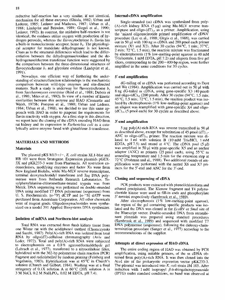
Tissue distribution of hydroxy acid oxidase mRNA Northern-blot analysis of rat kidney poly(A)-rich RNA,
using, as a probe for hybridization under stringent conditions, the 362-bp internal PCR fragment obtained with X1 and X2, revealed the presence of two strong bands of = 1.8 kb and 2.0 kb (Fig. 1A). These bands were also easily visualized in total kidney RNA. No signal was observed in total RNA samples from other rat tissues (lung, heart, brain, muscle, liver and testis ; Fig. 1B). This confirmed the tissue speci- ficity of the kidney enzyme (Tolbert, 1981). The length of the cloned cDNA (1648 bp) suggested it corresponded to the lower-molecular-mass mRNA.
Expression of hydroxy acid oxidase in E. coli as a fusion protein with glutathione S-transferase
The full-length coding region, starting at the Prol codon, was amplified by PCR from total kidney ss cDNA using primers corresponding to the 5'-end and 3'-end of the coding sequence (20 nucleotides). Its sequence was checked and
Fig. 1. Size and tissue distribution of hydroxy acid oxidase mRNA. 5 pg poly(A)-rich RNA from rat kidney (A) and 10 pg total RNA from different rat tissues (B) were analysed by Northern-blot analysis using, as a probe, the 362-bp fragment obtained with X1 and X2 as primers. In (B), RNA was from lung (l), heart (2), brain (3), muscle (4), liver ( S ) , kidney (6) and testis (7). The position of the 18s and 28s ribosomal RNA is indicated. The arrows highlight the two bands observed for hydroxy acid oxidase mRNA.
found to correspond to that shown in Fig. S2, i.e. it did not present the VRK insertion. Initial attempts to express HA0 as an unfused protein using the pKK233-3 bacterial ex- pression vector were unsuccessful, despite a correct sequence and the use of five different E. coli strains. As an alternative, the HA0 coding region was subcloned into the prokaryotic expression vector pGEX-3X. The resulting construct is ex- pected to yield a fusion protein with GST from Schistozoma juponicum. Moreover, the chimaeric protein has been de- signed to contain a specific proteolytic cleavage site for bo- vine factor Xa, between the N-terminal GST sequence and the protein to be expressed (IEGWGIP etc). Insertion of the HA0 coding region at the SmaI site, combined with factor- Xa cleavage of the fusion protein, should thus yield the oxi- dase with an N-terminal extension of three residues (GIP) before Pro 1 of the natural protein. The recombinant plasmid, pGHAOX, was introduced into E. coli strain XL1-blue. A number of clones were examined for protein expression after induction with IPTG. Their total protein content was ana- lyzed by SDS/PAGE and Coomassie-blue staining after cell lysis. Fig. 2A shows a typical example of a clone overex- pressing a 65-kDa protein found only after IPTG treatment of the bacterial culture. The protein was absent from the cells transformed with the original pGEX-3X, which conversely expressed a 26-kDa protein corresponding to the expected
21 A B
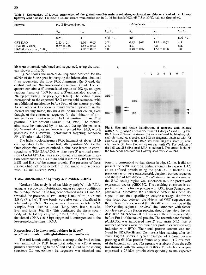
Fig.2. Expression of rat kidney hydroxy acid oxidase in E. coli. (A) Total cell proteins on 12.5% SDSIPAGE. 1-ml cultures were harvested before (-) and after (+) induction with 1 mM IPTG for 3 h; the pellet was dissolved in loading buffer, and one half was electrophoresed after boiling. Lane 1, molecular-mass markers ; lane 2, XL1-blue transfected with pGEX-3X; lane 3, XL1-blue trans- formed with pGHAOX; lane 4, hydroxy acid oxidase purified from rat kidney. (B) Purified fusion protein after chromatography on glu- tathione-Sepharose 4B.
GST. The recombinant fusion protein expressed by cells transfected with pGHAOX had the size expected for the GST-HA0 fusion protein, and it represented about 5 % of the total protein content.
Enzymic and physicochemical characterization of the GST/HAO chimaera
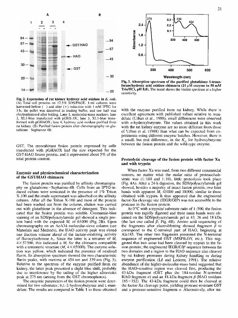
The fusion protein was purified by affinity chromatogra- phy on glutathione- Sepharose4B. Cells from an IPTG-in- duced culture were sonicated in the presence of 1% Triton X-100 and the crude supernatant was adsorbed on the affinity column. After all the Triton X-100 and most of the protein had been washed out from the column, elution was carried out with glutathione in the absence of detergent. This indi- cated that the fusion protein was soluble. Coomassie-blue staining of an SDSlpolyacrylamide gel showed a single pro- tein band with the expected M, of 65000 (Fig. 2B). Upon chromatography on an AcA34 molecular-sieve column (see Materials and Methods), the HA0 activity peak was eluted one fraction volume ahead of the lactate-oxidizing activity of flavocytochrome b,. Since the latter is a tetramer of Mr 4 X 57500, this indicated a M, for the chimaera compatible with a tetrameric structure (Mr 4 X 65 000). The enzyme solu- tion was yellow, which indicated the presence of oxidized flavin. Its absorption spectrum showed the two characteristic flavin peaks, with maxima at 454 nm and 339 nm (Fig. 3). Relative to the spectrum of the enzyme purified from rat kidney, the latter peak presented a slight blue shift, probably due to interference by the tailing of the higher ultraviolet peak at 275 nm (arising from the GST chromophores).
The enzymic parameters of the fusion protein were deter- mined for two substrates ; ~,~-2-hydroxybutyrate and L-man- delate. The results are compared in Table 1 to those obtained
300 400 500 600
Wavelength (nm) Fig. 3. Absorption spectrum of the purified glutathione S-trans- ferasekydroxy acid oxidase chimaera (21 pM enzyme in 50 mM TrisMCl, pH 8.0). The insert shows the visible spectrum at a higher sensitivity.
with the enzyme purified from rat kidney. While there is excellent agreement with published values relative to man- delate (Urban et al., 1988), small differences were observed with a-hydroxybutyrate. The values obtained in this work with the rat kidney enzyme are no more different from those of Urban et al. (1988) than what can be expected from ex- periments using different enzyme batches. However, there is a small, but real difference, in the K,,, for hydroxybutyrate between the fusion protein and the wild-type enzyme.
Proteolytic cleavage of the fusion protein with factor Xa and with trypsin
When factor Xa was used, from two different commercial sources, no matter what the molar ratio of proteasehub- strate was (1 : 100 and 1 : lo), little proteolysis took place (Fig. 4A). After a 24-h digestion, the SDS/polyacrylamide gel showed, besides a majority of intact fusion protein, two faint bands with apparent M, 43000 and 18000, similar to those obtained with trypsin. It thus appeared that the engineered factor-Xa cleavage site (IEGWGIP) was not accessible to the protease in the fusion protein.
At 0°C with a trypsid substrate ratio of 1 :500, the fusion protein was rapidly digested and three main bands were ob- tained on the SDS/polyacrylamide gel at 43, 26 and 18 kDa (the last one called p; Fig. 4B). Amino acid sequencing of the fragments after electroblotting showed fragment p to correspond to the C-terminal part of HAO, beginning at Alal85. The other two fragments presented the N-terminal sequence of engineered GST (MSPILGY, etc.). This sug- gested that two areas had been cleaved by trypsin in the fu- sion protein ; the engineered IEGWGIP sequence between the two domains and a region in the HA0 sequence also cleaved by rat kidney proteases during kidney handling or during enzyme purification (Li? and Lederer, 1991). The relative abundance of the higher-molecular-mass band suggested that the HAO-sensitive region was cleaved first, producing the 43-kDa fragment (GST plus the 184-residue N-terminal HA0 fragment a) and an 18-kDa fragment p (HA0 residues 185-352). The 43-kDa fragment could then be cleaved at the factor-Xa cleavage point, yielding protease-resistant GST and a protease-sensitive fragment a. Alternatively, after ini-
22
Fig. 4. Proteolysis of the glutathione S-transferasehydroxy acid oxidase chimaera (A and B) and of rat kidney hydroxy acid oxidase (C). (A) Proteolysis of the fusion protein with factor Xa (protease/substrate molar ratio 1:15; substrate, 25-30 pM; 50mM Tris/HCl, 100 mM NaCl, 4 mM CaCl,, pH 8.0, 25°C). Lane 1, zero time; lanes 2 and 3, 30 min and 120 min, factor Xa from Biolabs; lanes 4 and 5, factor Xa from Sigma Chemical Co (contains bovine serum albumin which gives rise to the second Coomassie-blue-stained band). (B) Proteolysis of the fusion protein with trypsin (protease/substrate molar ratio, 1 : 500; substrate, 25-30 pM; 50 mM Tris/HCI, pH 8, OOC). Lane 1, zero time; lane 2, 5 min; lane 3, 2 h. (C) Proteolysis of H A 0 purified from rat kidney. Same conditions as for (B).
184 i Major form ... R A L K E - - - E K P T ...
Minor form ... R A L K E V R K E K P T ... 1 : 2 3
t 1
Fig. 5. Tryptic cleavage in the protease-sensitive region of H A 0 purified from rat kidney. Arrows indicate the location of the cleav- age points as deduced from amino acid sequencing results of frag- ment p.
tial HA0 loop cleavage, the GST a fragment could possibly be digested piecemeal yielding GST as a stable end product.
For verification of the conformational integrity of the HA0 domain in the fusion protein, the purified rat ludney enzyme was submitted to trypsin digestion under conditions identical to those used for the chimaera. Both fragments a (a doublet at 22 kDa) and p (18 kDa) were formed (Fig. 4C), confirming that the natural protein contains a protease-sensi- tive loop and suggesting that fragment a is more sensitive to further digestion by trypsin when it is fused to GST than when it is not. The two bands corresponding to fragment a had the same N-terminal sequence (PLV, etc.) after electro- blotting, showing their difference to lie at their C-termini. Amino acid sequencing of fragment p yielded two sequences, and three in a case of prolonged digestion. The major se- quence (ALKEEKP) showed the tryptic cleavage point to lie between R184 and A1 85, as with the fusion protein (Fig. 5) . The minor sequence, which represented about 10- 15 % of the total amount sequenced, was KEKPTQSV. This can only have originated from a protein possessing the VRK insertion, which would be cleaved between R and K (Fig. 5) . In the case of the prolonged digestion, the major sequence was identical. The minor sequence was EKPTQ, etc.; it could have arisen from a cleavage after the lysine of the VRK in-
sertion (Fig. 5) , but the idea of a less probable but not impos- sible tryptic cleavage between El88 and El89 of the main sequence cannot be totally ruled out (Haumont et al., 1989 and references therein). It can be added that enzymic activity monitoring during proteolysis showed HA0 activity to de- crease by about 15% over the first 100min, after which it was as stable as that of the control. This suggested that pro- teolytic cleavage had only a slight effect on the HA0 enzy- mic activity.
DISCUSSION Using the information given by the amino acid sequence
of a-hydroxy acid oxidase, we used PCR for amplifying, cloning and sequencing three overlapping fragments from rat kidney cDNA, namely a central coding segment, a 5’ frag- ment and a longer 3‘ end. Several independent clones were sequenced each time in order to detect possible copying er- rors of Tuq DNA polymerase. Most of the clones gave the sequence shown in Fig. S2, yielding an open reading frame identical to the amino acid sequence previously determined. However, three out of eight clones of the 3’-fragment carried the same nine-base insertion in the open reading frame, which would correspond to the insertion of amino acids VRK between El88 and El89 of the determined amino acid sequence. This cannot be ascribed to a copying error. The three-residue insertion occurs in a region which is sensitive to proteases, as shown in this and previous work (L& and Lederer, 1991), and corresponds to disordered loops in the three-dimensional structures of the homologous enzymes fla- vocytochrome b, (Xia and Mathews, 1990) and glycolate oxidase (Lindqvist, 1989). Thus, this region could probably accommodate a few more residues without alteration of the overall structure. No other sequence difference was detected in the cDNA, apart from an extension of the poly(A) tail. These results suggested the possible existence of a-hydroxy acid oxidase isozymes. While no evidence for amino acid
23
sequence heterogeneity was obtained before (L& and Lederer, 1991), during the present work the tryptic cleavage in the loop region of kidney HA0 followed by automatic Edman degradation provided evidence for the existence of a second sequence compatible with the insertion of the tripeptide VRK. The corresponding isoform was calculated to represent about 10% of the total protein submitted to degradation. In the past, McGroarty et al. (1974) and Cromartie and Walsh (1975a) noted the presence of one major and 1-3 additional faint bands staining for HA0 activity on non-denaturing gels. The molecular nature of these putative isozymes was not elucidated. In contrast, Duley and Holmes (1974) detected only one activity band on starch gels. Thus, all the evidence obtained to date suggests that a second form of HAO, if present, must be minor.
Alternative splicing of the primary transcript from a sin- gle gene could possibly explain the generation of these two HA0 isoforms, given the sequence identity for the untrans- lated regions of their mature mRNA. The existence of the minor isoform at the protein level is intriguing from an evo- lutionary point of view. One of the major differences between the various members of the FMN-dependent a-hydroxy-acid- oxidizing enzymes lies in the length of the polypeptide chain which loops out of the /38a8 barrel between strand /I4 and helix a4 (L& and Lederer, 1991). It is in this segment that the structurally disordered region is found, as well as the pro- tease-sensitive bonds and the tripeptide insertion. It is so far not known whether this region which loops out of the /I bar- rel plays an important role in the structure or function of the enzyme. It was, however, found that proteolytic cleavage of a single peptide bond in flavocytochrome b, led to alterations in a number of kinetic parameters (k,,, K,, K, for inhibitors), suggesting some kind of interaction, direct or indirect, be- tween the active site and the disordered region, either before or after cleavage (Ghrir and Lederer, 1981). Moreover, the folding of the extension between p4 and a4, with respect to the barrel, is very different in glycolate oxidase (Lindqvist, 1989) and in flavocytochrome b, (Xia and Mathews, 1990), probably due to the presence of the heme-binding domain. Thus, this region of the peptide chain may be a hot spot for evolutionary changes in the protein family, and it is tempting to speculate that alternative splicing might be a molecular mechanism favouring such changes (Craik et al., 1983). The data presented in this paper do not exclude, however, the existence of closely related genes as an origin of the iso- forms. An allelic form of the gene is unlikely because the mRNA used for amplification was extracted from a single homozygous Wistar rat. It is to be noted that a single cDNA sequence was identified for the other known members of the family of hydroxy-acid-oxidizing enzymes.
We used the radiolabeled amplified central fragment for characterising HA0 mRNA by Northern blotting. Two mes- sengers of equal abundance and differing size (about 1800 bp and 2000 bp) were detected in rat kidney poly(A)-rich RNA. They were also detected in total kidney RNA; their ex- pression was tissue specific, at least at the sensitivity level of the Northern-blot technique. This tissue specificity is in keeping with previous observations (McGroarty et al., 1974; Zaar and Fahimi, 1991). The origin of the difference between the two messengers remains to be elucidated, since their mass difference cannot be explained by the sequence differ- ences detected in PCR products.
Hydroxy acid oxidase was expressed as a fusion protein with glutathione S-transferase using the vector pGEX-3X. After induction with IPTG, up to 5% of the proteins were
constituted by the chimaera, expressed as soluble protein. The fusion protein was readily purified by affinity chroma- tography on immobilised glutathione. That the folding of the chimaeric HA0 should be similar if not identical to the native HA0 is shown by its flavin spectrum, its enzymic activity and its similar susceptibility to limited proteolysis by trypsin between positions 184 and 185. The enzymic param- eters determined for the substrate L-mandelate are identical to those obtained with the enzyme purified from rat kidney. The small difference found for the K , of D,L-a-hydroxybu- tyrate is somewhat surprising, as one would expect a struc- tural perturbation due to the fusion to have effects on both substrates. Moreover, it can hardly arise from the presence of the second HA0 form in the natural enzyme, since this form is a minor constituent of the purified natural enzyme. Whatever the case, even though we could not obtain intact recombinant HA0 due to the resistance of the chimaera to proteolysis, our results open the way to the study of structure/ function relationships with mutants whose properties should be compared to those of the wild-type fusion protein.
Mammalian GST enzymes are dimeric (Mannervick and Danielson, 1988), but the enzyme from Schistosoma japoni- cum is believed to be a monomer (Tiu et al., 1988; Farmer et al., 1992). Hydroxy acid oxidase itself is a tetramer (Crom- artie and Walsh, 1975b; Duley and Holmes, 1976). All the well characterized homologous enzymes were found to be either tetramers or octamers (Monteilhet and Risler, 1970; Phillips et al., 1976; Sullivan et al., 1977). Furthermore, a monomeric form of Hansenula anomala flavocytochrome b, obtained at low ionic strength was found inactive, even though it still retained both the flavin and the heme (Baudras, 1972) ; activity was recovered together with the tetrameric state upon raising the ionic strength. No evidence exists for an active monomeric state for any of the other members of the family. Furthermore, the crystal structures of glycolate oxidase and flavocytochrome b, show intersubunit contacts to be extensive (Lindqvist, 1989; Xia and Mathews, 1990; Lindqvist et al., 1991). The GST-HA0 chimaera was thus expected, as found, to have a GST domain flanking each subunit of the tetramer. The three-dimensional structures of the homologous enzymes flavocytochrome b, and glycolate oxidase (Lindqvist et al., 1991) show an identical fold start- ing at a position equivalent to mature HA0 Leu2 (a turn followed by a helix). A few residues in this area, at the sur- face of the subunit, are located close to fourfold contacts in the two structures. It would be surprising to find a very dif- ferent topology at the N-terminus of HAO, in view of its degree of similarity with the two other enzymes. This local structure, therefore, as well as the overall architecture of the tetramer, appear compatible with the formation of a more complex and active molecular edifice, which retains affinity for glutathione and has both flavin-binding capacity and hydroxy acid oxidizing activity, even though the fusion site is inaccessible to factor Xa. It is interesting to note that, while there is a rapid increase in the number of described cases of recombinant proteins or domains expressed as fusion proteins with GST, the number of active enzymes expressed in this way is still rather limited. Furthermore, to our knowl- edge, the present case represents the first example of an ac- tive multimeric enzyme fused to GST.
A. Belmouden was a recipient of a fellowship from the Centre National de la Recherche Scientifque (bourse de doctorat pour in- gknieurs). The authors are indebted to D. Mazella for competent secreterial help.
24
REFERENCES Angermiiller, S., Leupold, C., Zaar, K. & Fahimi, H. D. (1986) His-
tochemistry 85,411-418. Aviv, H. & Leder, P. (1972) Proc. Natl Acad. Sci. USA 69, 1408-
1412. Baudras, A. (1972) in Dynamic aspects of conformation changes in
biological macromolecules, (Sadron, C. ed.) pp. 181 -205, D. Reidel Publishing, Dordrecht.
Black, M. T., Gunn, F. J., Chapman, S. K. & Reid, G. A. (1989) Biochem. J. 263,973-976.
Blanchard, M., Green, D. E., Nocito, V. & Ratner, S. (1945) J. Biol. Chem. 161, 583-598.
Blanchard, M., Green, D. E., Nocito, V. & Ratner, S. (1946) J. Biol. Chem. 163, 137-144.
Brush, E. J. & Hamilton, G. A. (1981) Biochem. Biophys. Res. Com- mun. 103, 1194-1200.
Cederlund, E., Lindqvist, Y., Soderlund, G., BrhdCn, C. 1. & Jornvall, H. (1988) Eur. J. Biochem. 173, 523-530.
Chomczynski, P. & Sacchi, N. (1987) Anal. Biochem. 162, 156- 159.
Church, G. M. & Gilbert, W. (1984) Proc. Nut1 Acad. Sci. USA 81,
Craik, C. S., Rutter, W. J. & Fletterick, R. (1983) Science 220,
Cromartie, T. H. & Walsh, C. T. (1975a) Biochemistry 14, 2588-
Cromartie, T. H. & Walsh, C. T. (1975b) Biochemistry 14, 3482-
Deng, G. & Wu, R. (1983) Methods Enzymol. 100, 96-116. Dubois, J., Chapman, S. K., Mathews, F. S., Reid, G. A. & Lederer,
Duley, J. & Holmes, R. S. (1974) Genetics 76, 93-97. Duley, J. & Holmes, R. S. (1976) Eur. J. Biochem. 63, 163-176. Feinberg, A. P. & Vogelstein, B. (1983) Anal. Biochem. 132, 6-13. Farmer, K., Catala, F. & Wright, W. E. (1992) J. Biol. Chem. 267,
Frohman, M. A,, Dush, M. K. & Martin, G. R. (1988) Proc. Nut1
Ghrir, R. & Lederer, F. (1981) Eul: J. Biochem. 120, 279-287. Ghisla, S. (1982) in Flavins andjlavoproteins (Massey, V. & Willi-
ams, C. H., eds) pp. 133-142, Elsevier Science Publishers, Am- sterdam.
Giegel, D. A,, Williams, C. H. & Massey, V. (1990) J. Biol. Chem. 265,6626-6632.
Girgis, S. I., Alevizaki, M., Denny, P., Ferrier, G. J. M. & Legon, S. (1988) Nucleic Acids Res 16, 10371.
Gould, S. J., Krisans, S., Keller, G. A. & Subramani, S. (1990) J. Cell. Biol. 110, 27-34.
Guiard, B. (1985) EMBO J. 4, 3265-3272. Henikoff, S. (1984) Gene (Amst.) 28, 351-359. Haumont, P. Y., Thomas, M. A., Labeyrie, F. & Lederer, F. (1987)
Kozak, M. (1984) Nucleic Acids Res. 12, 857-872. Kozak, M. (1986) Cell 44, 283-292. Laemmli, U. K. (1970) Nature 227, 680-685. LC, K. H. D. & Lederer, F. (1991) J. Biol. Chem. 266, 20877-
Lederer, F. (1991) in Chemistry and biology ofjlavoproteins (Miiller,
Lederer, F. (1992) Protein Science 1, 540-548.
1991 - 1995.
11 25 - 1129.
2596.
3490.
F. (1990) Biochemistry 29, 6393-6400.
5631-5636.
Acad. Sci. USA 85, 8998-9002.
Eul: J. Biochem. 169, 539-546.
20881.
F., ed.) pp. 153-242, CRC Press Inc.
Lederer, F. & Mathews, F. S. (1987) in Flavins and jlavoproteins (Edmondson, D. & Mc Cormick, D. B., eds) pp. 133-142, Walter de Gmyter and Co., Berlin.
Lederer, F., Cortial, S., Becam, A. M., Haumont, P. Y. & Perez, L. (1985) Eur. J. Biochem. 139, 59-65.
Lee, C. C., Wu, X., Gibbs, R. A., Cook, R. G., Muzny, D. M. & Caskey, C. T. (1988) Science 239, 1288-1291.
Lehrach, H., Diamond, D., Wozney, J. & Boedtker, H. (1977) Bio- chemistry 16,4743 -4751.
Lindqvist, Y. (1989) J. Mol. Biol. 209, 151-166. Lindqvist, Y. & BrandCn, C.-I. (1989) J. Biol. Chem. 264, 3624-
Lindqvist, Y., BrhdCn, C. I., Mathews, F. S. & Lederer, F. (1991)
Loh, E. V., Elliott, J. F., Cwirlo, S., Lanier, L. & Davis, M. M.
Mannervik, B. & Danielson, U. H. (1988) CRC Crit. Rev. Biochem.
McGroarty, E., Hsieh, B., Wied, D. M., Gee, R. & Tolbert, N. E. (1974) Arch. Biochem. Biophys. 161, 194-210.
Miles, C. S., Rouvibre-Fourmy, N., Lederer, F., Mathews, F. S., Reid, G. A,, Black, M. T. & Chapman, S. K. (1992) Biochem. J. 285, 187-192.
Monteilhet, C. & Risler, J. L. (1970) Eur. J. Biochem. 12, 165-169. Nagai, K. & Thogersen, H. C. (1984) Nature 309, 810-812. Nakano, M. & Danowski, T. S. (1966) J. Biol. Chem. 241, 2075-
Phillips, D. R., Duley, J. A,, Fennell, D. J. & Holmes, R. S. (1976)
Pompon, D., Iwatsubo, M. & Lederer, F. (1980) Eur. J. Biochem.
Reid, G. A., White, S., Black, M. T., Lederer, F., Mathews, F. S. & Chapman, S. K. (1988) Eul: J. Biochem. 178, 329-333.
Risler, Y., Tegoni, M. & Gervais, M. (1989) Nucleic Acids Res. 17, 8381.
Robinson, J. C., Keay, L., Molinari, R. & Sizen, I. W. (1962) J. Biol. Chem. 237, 2001-2010.
Sambrook, J., Fritsch, E. F. & Maniatis, T. (1989) Molecular clon- ing, a laboratory manual, 2nd edition, Cold Spring Harbor Lab- oratory Press.
Sanger F, Nicklen, S. & Coulson, A. R. (1977) Proc. Natl Acad. Sci.
Smith, D. B. & Johnson, K. S. (1988) Gene (Amst.) 67, 31 -40. Sullivan, P. A., Choong, Y. S., Schreurs, W. J., Cutfield, J. F. &
Tiu, W. U., Davern, K. M., Wright, M. D., Board, P. G. & Mitchell,
Tolbert, N. E. (1981) Annu. Rev. Biochem. 50, 133-157. Tsou, A. Y., Ransom, S. C., Gerlt, J. A., Buechter, D. D., Babbitt,
P. C. & Kenyon, G. L. (1990) Biochemistry 29, 9856-9862. Urban, P. & Lederer, F. (1984) Eur: J. Biochem. 144, 345-351. Urban, P. & Lederer, F. (1985) J. Biol. Chem. 260, 11115-11 122. Urban, P., Chirat, I. & Lederer, F. (1988) Biochemistry 27, 7365-
Volokita, M. & Somerville, C. R. (1987) J. Biol. Chem. 262,
Xia, Z.-X. & Mathews, F. S. (1990) J. Mol. Biol. 212, 837-863. Zaar, K. & Fahimi, H. D. (1991) J. Histochem. Cytochem. 39,801 -
3628.
J. Biol. Chem. 266, 3198-3207.
(1989) Science 243,217-220.
23, 283-337.
2083.
Biochim. Biophys. Acta 427, 679-687.
104,479-488.
USA 74, 5463-5467.
Shepherd, M. G. (1977) Biochem. J. 165, 375-383.
G. F. (1988) Parasite Immunol. 10, 693-706.
7371.
15 825 - 15 828.
808.

Supplement material to :
Molecular cloning and nucleotide sequence of cDNA encoding rat kidney long-chain L-2-hydroxy acid oxidase Expression of the catalytically active recombinant protein as a chimaera
Ahmed BELMOUDEN, K. H. DiCp LE, Florence LEDERER and Henri-Jean GARCHON
K T S W D F I E G E A D D G I T Y S M G ACC TCC TGG GAC TTT ATT G M GGA G M GCT GAC GAC GGC ATC ACC TAC AGT
E N I A A F K R I R L R P R Y L R D GAG M C ATA GCA GCA TTT AM AGA ATC CGC CTC CGC CCC CGA TAC CTG AGA GAT
~ S K V D T R T T I ~ G ~ E ~ S A P ATG TCA M G GTG GAC ACC AGG ACC ACA ATC C M GGG CAG GAG ATC ACT GCT CCC
I C I S P T A F H S I A W P D G E K ATC TGC ATC TCA CCC ACA GCC TTT CAC TCC ATT GCC TGG CCG GAT GGA G M M G
5 T A R A A 3 E A N I C Y V I S S Y AGC ACA GCT AGA GCT GCT CAG GAG GCC ARC ATC TGC TAT GTC ATC AGC AGT TAT
A S Y S L E I) I V A A A P E G F R W GCC AGC TAT TCC CTG G M GAT ATT GTT GCT GCT GCC CCC GAA GGC TTT CGT TGG
>
35 108
53 162
ii 216
89 210
101 324
125 318
ATG Rat HAOX cDNA TAA 5’ 3
Internal cDNA fragment x1- - xz
-.+ t X 3 x5- t
(dC)15 (ANC) t X 4 X6- x7-
Fig.Sl. Strategy for amplifying and cloning the cDNA of rat kidney hydroxy acid oxidase. The exact position in the final se- quence and the sequence of the synthetic oligonucleotides XI -X7 and ANC are given in Table S1. The sequences of X3 and X4 and of X5 to X7 do not overlap (see Table SI) although the arrows symbolizing these primers are overlapping.
Table S1. Sequence and position of the primers used in the PCR amplification steps. Xt, X2 and X7 are degenerate oligonucleotid- es.
Primer Nucleotide Sequence position
xt 64- 80 TGGGATTTTATTGAAGG C C C G
A x2 425 -403 ACCATTTGTTTATTAAAATCCCA
C C G G G x 3 168- I49 TGACATATCTCTCAGGTATC x 4 108-189 ACTGTAGGTGATGCCGTCGT xs 338-358 TGGAAGATATTGTTGCTGCTG X6 374-392 GTTGGTTCCAACTCTACAT x 7 396-416 GTCAGACTGGGATTTCAACM
C T T ANC GACTCGAGTCGACATCG
Eco0109 Nco I Hind 111 n1 I -- -c----+
- --2
H 100 bp
Fig. S3. Restriction map and sequencing strategy for the 3’-end of HA0 cDNA (lower mass 1.1-kb fragment amplified from ss cDNA with X7 and ANC, see text). Restriction enzymes and their sites used to generate small fragments are shown above the thick bar representing the amplified segment. The lines below the bar represent the sequenced length of fragments obtained after subclon- ing (0) or after exonuclease-I11 deletion (X) (Henikoff, 1984). The higher-mass t .t-kb fragment described in the text was sequenced entirely using the exonuclease-I11 deletion method. Arrows represent the direction of the sequencing.
~
A E A L G F K A L v I T I D T e v L 161 GCA G M GCC TTG GGT TTC AAA GCT TTG GTG ATC ACT ATA GAT ACG CCT GTA CTT 486
G N R R R D K R N 0 L N L E A N I L 179 GGC ART AGG CGA CGG GAC M G AGA M C CAG CTG AAT TTG GAG GCA M C ATA TTG 540
L K D L R A L K E K P T O S V P V 191 TTG M G GAT CTC CGA GCC CTC AM GAG G M G CCC ACA CAG TCT GTG CCC GTG 594
S F P K A S F C W t D L 5 L L 0 S I 215 TCT TTT CCG AAA GCA TCT TTC TGC TGG M T GAT CTT TCC TTG CTT CAG AGT ATA 648
T R L P I I L K G I L T K E D A E L 233 ACT CGG TTG CCC ATT ATC CTC AAA GGG ATT TTG ACG AM GAG GAT GCA GAG TTA 102
A n K H N v o G I v v s N H G G R o 251 GCA ATG M G CAC ARC GTC CAR GGC ATC GTT GTT TCC M C CAT GGT GGG AGO CAG 156
L D E V S A S I D A L R E V V A A V 269 CTT GAT GAG GTT TCT GCT TCA ATT GAT GCT CTG AGA G M GTG GTG GCT GCT GTC 810
K G K I E V Y M D G G V R T G T D V 281 AM GGG AM ATT G M GTG TAC ATG GAT GGT GGG GTT CGA ACT GGC ACT GAT GTG 864
L K A L A L G A R C I F L G R P I I 305 TTG M G GCA CTG GCC CTT GGA GCT AGG TGC ATT TTT CTT GGG AGA CCA ATC CTT 918
W G L A C K G E D G V K E V L D I L 323 TGG GGC CTT GCC TGC M G GGT G M GAT GGT GTT M G G M GTT TTA GAT ATT CTA 912
T A E L H R C M T L S G C 0 S V A E 341 ACA GCA GAR CTC CAT AGA TGT ATG ACC CTT TCA GGC TGC CAG TCA GTT GCT GAG 1026
I S P D L I Q F S R L 352 ATT AGT CCA GAC CTG ATT CAG TTC TCC AGA TTA TMGGACCTACTGAGATCCCTACMGA 1086
GGARGACMGACTTCMCATAGTGTGTGAGCCTATTCTTCTT~GGTCCGATCATACCTAGTAGTTTGAGCCC 1158
TCTACCTTGAG~TCCAGATCGATGMGAAMGATAGCTMCAGCTACCAGAGGGGTGCATTTGGATGMG 1230
GMTMCATCTMTGTTCTACAGGATMCTATMCTGACMTTMTTGACTATGTCCMTAGCCATGTTCCA 1302
GAMGAAAGGAAATAGTTACATCTGGTGTGACATGCCMTTACTTTCATTTGATTATTATGTACAC~ATACA 1314
TTATTAAMCGTCATACTGTGCCATMTTATGTGCMTTACTATGTATCAATTAAAACMCAATATATATTT 1446
Fig. S2. Composite nucleotide sequence, and deduced amino acid sequence, of the cDNA of a-hydroxy acid oxidase from rat kid- ney. The boxed sequence is that obtained after the first PCR amplifi- cation with X1 and X2 (Table Sl). The peroxisomal targeting se- quence is underlined. The arrow indicates the position of the nine nucleotide insertion (TGAGAAAGG) found in some clones (see text).
Copyright © 2022 FDOKUMEN