Molecular and Morphological Characterization of Reciprocal F 1 Hybrid Ash ( Fraxinus excelsior ×...
7
http://journals.cambridge.org Downloaded: 05 May 2009 IP address: 134.115.73.187 367 Molecular and morphological characterization of Echinococcus granulosus of human and animal origin in Iran M.FASIHI HARANDI ", R. P. HOBBS #, P. J. ADAMS #, I. MOBEDI $, U.M.MORGAN-RYAN # and R.C.A.THOMPSON #* " Department of Parasitology, School of Medicine, Kerman University of Medical Sciences, Kerman, Iran # WHO Collaborating Centre for the Molecular Epidemiology of Parasitic Infections and State Agricultural Biotechnology Centre, Division of Veterinary and Biomedical Sciences, Murdoch University, Murdoch, Western Australia 6150, Australia $ Department of Parasitology, School of Public Health, Tehran University of Medical Sciences, Tehran, 14155-6446, Iran (Received 18 March 2002; revised 13 May 2002; accepted 13 May 2002) Iran is an important endemic focus of cystic hydatid disease (CHD) where several species of intermediate host are commonly infected with Echinococcus granulosus. Isolates of E. granulosus were collected from humans and other animals from different geographical areas of Iran and characterized using both DNA (PCR-RFLP of ITS1) and morphological criteria (metacestode rostellar hook dimensions). The sheep and camel strains}genotypes were shown to occur in Iran. The sheep strain was shown to be the most common genotype of E. granulosus affecting sheep, cattle, goats and occasionally camels. The majority of camels were infected with the camel genotype as were 3 of 33 human cases. This is the first time that cases of CHD in humans have been identified in an area where a transmission cycle for the camel genotype exists. In addition, the camel genotype was found to cause infection in both sheep and cattle. Results also demonstrated that both sheep and camel strains can be readily differentiated on the basis of hook morphology alone. Key words : Echinococcus granulosus, cystic hydatid disease, Iran, strains, genotypes, morphology, molecular charac- terization. Cystic hydatid disease (CHD), caused by the metacestode of Echinococcus granulosus, is medically and economically one of the most important of the zoonoses (Schantz, 1997 ; Thompson, 2001). Con- siderable genetic and phenotypic variation has been demonstrated in Echinococcus, with the recognition of a series of what appear to be host-adapted genotypes or strains (McManus, 1997 ; Thompson & McManus, 2001), which are likely to represent distinct species (Thompson, Lymbery & Constantine, 1995). Therefore there is a need to characterize the aetiological agents in different endemic areas using molecular epidemiological tech- niques, in order to determine transmission patterns, particularly where there is the possibility of in- teraction between cycles (Thompson, Constantine & Morgan, 1998 ; Thompson & McManus, 2001). For example, in endemic regions where usually a number of species of livestock may be infected with cystic echinococcosis, it is important to determine which species are responsible for maintaining the life-cycle. * Corresponding author : Division of Veterinary and Biomedical Sciences, Murdoch University, South Street, Murdoch, Western Australia. Tel : ›61 08 9360 2466. Fax: ›61 08 9310 4144. E-mail : andrewjt!central. murdoch.edu.au This provides the basis for implementing targeted control efforts and, more fundamentally, for developing a reliable surveillance system which is essential for assessing the success of intervention strategies (Gemmell & Roberts, 1995). Unfortu- nately, the degree of genetic variability in popu- lations of Echinococcus is insufficiently characterized for many geographical regions (Schantz et al. 1995). Determining the geographical distribution and uni- formity of host-adapted genotypes of Echinococcus is also important, so that formal taxonomic designation can be confirmed (Thompson et al. 1995). CHD is a significant economic and public health problem in Iran (Dar & Alkarmi, 1997). High prevalences have been reported in various species of domestic food animals, including sheep, cattle, camels and goats, and at least 2 distinct cycles of transmission are thought to occur (Dar & Alkarmi, 1997). Human cases of CHD are also regularly reported from medical centres in different parts of the country (Noorjah, 1987 ; Mobedi, Arfaa & Farahmandian, 1971 ; Bastani & Dehdashti, 1995). However, the sources of infection to humans, and particularly the role of intermediate host reservoirs, such as the camel, remain to be determined (Dar & Alkarmi, 1997 ; Thompson, 2001). In a preliminary study, molecular charac- terization, using mitochondrial DNA markers, of a Parasitology (2002), 125, 367–373. " 2002 Cambridge University Press DOI : 10.1017}S0031182002002172 Printed in the United Kingdom
Transcript of Molecular and Morphological Characterization of Reciprocal F 1 Hybrid Ash ( Fraxinus excelsior ×...

http://journals.cambridge.org Downloaded: 05 May 2009 IP address: 134.115.73.187
367
Molecular and morphological characterization of
Echinococcus granulosus of human and animal origin in Iran
M. FASIHI HARANDI", R. P. HOBBS#, P. J. ADAMS#, I. MOBEDI$,
U. M. MORGAN-RYAN# and R. C. A. THOMPSON#*
"Department of Parasitology, School of Medicine, Kerman University of Medical Sciences, Kerman, Iran#WHO Collaborating Centre for the Molecular Epidemiology of Parasitic Infections and State Agricultural BiotechnologyCentre, Division of Veterinary and Biomedical Sciences, Murdoch University, Murdoch, Western Australia 6150,
Australia$Department of Parasitology, School of Public Health, Tehran University of Medical Sciences, Tehran, 14155-6446, Iran
(Received 18 March 2002; revised 13 May 2002; accepted 13 May 2002)
Iran is an important endemic focus of cystic hydatid disease (CHD) where several species of intermediate host are
commonly infected with Echinococcus granulosus. Isolates of E. granulosus were collected from humans and other animals
from different geographical areas of Iran and characterized using both DNA (PCR-RFLP of ITS1) and morphological
criteria (metacestode rostellar hook dimensions). The sheep and camel strains}genotypes were shown to occur in Iran.
The sheep strain was shown to be the most common genotype of E. granulosus affecting sheep, cattle, goats and occasionally
camels. The majority of camels were infected with the camel genotype as were 3 of 33 human cases. This is the first time
that cases of CHD in humans have been identified in an area where a transmission cycle for the camel genotype exists.
In addition, the camel genotype was found to cause infection in both sheep and cattle. Results also demonstrated that both
sheep and camel strains can be readily differentiated on the basis of hook morphology alone.
Key words: Echinococcus granulosus, cystic hydatid disease, Iran, strains, genotypes, morphology, molecular charac-
terization.
Cystic hydatid disease (CHD), caused by the
metacestode of Echinococcus granulosus, is medically
and economically one of the most important of the
zoonoses (Schantz, 1997; Thompson, 2001). Con-
siderable genetic and phenotypic variation has been
demonstrated in Echinococcus, with the recognition
of a series of what appear to be host-adapted
genotypes or strains (McManus, 1997; Thompson &
McManus, 2001), which are likely to represent
distinct species (Thompson, Lymbery &
Constantine, 1995). Therefore there is a need to
characterize the aetiological agents in different
endemic areas using molecular epidemiological tech-
niques, in order to determine transmission patterns,
particularly where there is the possibility of in-
teraction between cycles (Thompson, Constantine &
Morgan, 1998; Thompson & McManus, 2001). For
example, in endemic regions where usually a number
of species of livestock may be infected with cystic
echinococcosis, it is important to determine which
species are responsible for maintaining the life-cycle.
* Corresponding author: Division of Veterinary and
Biomedical Sciences, Murdoch University, South Street,
Murdoch, Western Australia. Tel: 61 08 9360 2466.
Fax: 61 08 9310 4144. E-mail : andrewjt!central.
murdoch.edu.au
This provides the basis for implementing targeted
control efforts and, more fundamentally, for
developing a reliable surveillance system which is
essential for assessing the success of intervention
strategies (Gemmell & Roberts, 1995). Unfortu-
nately, the degree of genetic variability in popu-
lations of Echinococcus is insufficiently characterized
for many geographical regions (Schantz et al. 1995).
Determining the geographical distribution and uni-
formity of host-adapted genotypes of Echinococcus is
also important, so that formal taxonomic designation
can be confirmed (Thompson et al. 1995).
CHD is a significant economic and public health
problem in Iran (Dar & Alkarmi, 1997). High
prevalences have been reported in various species of
domestic food animals, including sheep, cattle,
camels and goats, and at least 2 distinct cycles of
transmission are thought to occur (Dar & Alkarmi,
1997). Human cases of CHD are also regularly
reported from medical centres in different parts of
the country (Noorjah, 1987; Mobedi, Arfaa &
Farahmandian, 1971; Bastani & Dehdashti, 1995).
However, the sources of infection to humans, and
particularly the role of intermediate host reservoirs,
such as the camel, remain to be determined (Dar &
Alkarmi, 1997; Thompson, 2001).
In a preliminary study, molecular charac-
terization, using mitochondrial DNA markers, of a
Parasitology (2002), 125, 367–373. " 2002 Cambridge University Press
DOI: 10.1017}S0031182002002172 Printed in the United Kingdom
http://journals.cambridge.org Downloaded: 05 May 2009 IP address: 134.115.73.187
M. F. Harandi and others 368
Fig. 1. Map of the study area showing geographical locations of all isolates collected.
small number (16) of isolates of E. granulosus,
collected from various areas of Iran identified the
occurrence of both the sheep and camel genotypes
(Zhang et al. 1998). The aim of the present study was
to undertake an extensive sampling programme of E.
granulosus from humans and other animals collected
from defined, geographical areas of Iran, and to
characterize the isolates using both DNA and
morphological criteria. Such information should
allow a better understanding of the molecular
epidemiology of CHD in Iran, particularly any
evidence of interaction between cycles of trans-
mission, identifying reservoirs of human infection,
and the provision of a comprehensive data set for
determining the nature and uniformity of the
genotypes of Echinococcus endemic in Iran.
Parasite materials
Hydatid cysts from naturally infected intermediate
hosts were collected from several abattoirs in Iran
(Fig. 1). A total of 213 E. granulosus isolates (all the
above isolates except those from goats, plus an
additional 70) were collected from humans, sheep,
cattle and camels (Camelus dromedarius) in different
geographical and ecological areas of Iran. Human
isolates were obtained at surgery from different
hospitals in Iran and Afghanistan (4 isolates) at the
locations shown in Fig. 1. In the laboratory proto-
scoleces were collected from hydatid materials.
Protoscoleces from each cyst were rinsed in physio-
logical saline solution and then fixed and preserved
in 80–95% (v}v) ethanol.
Morphological studies
Protoscoleces were mounted in polyvinyl lacto-
phenol (RA Lamb) with sufficient cover-slip press-
ure to cause the hooks to lie flat. Details of the hook
components measured are shown in Fig. 2 as in
Hobbs, Lymbery & Thompson (1990). Measure-
ments of the total length and blade length were made
on 2 large and 2 small hooks per rostellum from each
of 10 protoscoleces for each isolate (Kumaratilake,
Thompson & Eckert, 1986). Measurements were
made using an Olympus BX50 microscope with a
100¬ objective and an Optimas image analyser.
Morphological differences between sheep and camel
strains were analysed graphically by 2-dimensional
scatterplots. Analysis of covariance was used to
compare slopes and elevations of regression lines for
sheep strain isolates between different host species
(Zar, 1984).
http://journals.cambridge.org Downloaded: 05 May 2009 IP address: 134.115.73.187
Characterization of Echinococcus in Iran 369
Fig. 2. Diagrammatic representation of hook dimensions
as used in this study. Large hook length (LHL), large
hook blade length (LBL), small hook length (SHL),
small hook blade length (SBL).
Table 1. Echinococcus granulosus isolates according
to host species analysed by PCR-RFLP of the
ITS1 and their genotypic}strain identity
Host Sheep strain Camel strain
Sheep 40 3
Cattle 30 2
Goat 6 0
Camel 8 24
Human 33 3
Total 117 32
Molecular analysis
Total genomic DNA was extracted from ethanol
preserved isolates. Several different extractions
methods were trialed (Sambrook, Fritsch &
Maniatis, 1989) and eventually a modified phenol-
chloroform method (Bowles & McManus, 1993) was
used with subsequent glass milk purification. E.
granulosus DNA samples were characterized by
means of restriction fragment length polymorphism
(RFLP) patterns following polymerase chain re-
action (PCR) amplification of an C1 kb fragment
spanning the non-coding internal transcribed spacer
1 (ITS1) of the ribosomal DNA (rDNA) repeats
(Bowles & McManus, 1993).
A nested-PCR was designed using EGF1 (5«CCAAACTTGATCATTTAGAGGAAG 3«) and
EGR2 (5« TATGGGCCAAATTCACTCATTA-
CC 3«) as outside forward and reverse oligo-
nucleotide primers, specifically designed for this
study based on published Schistosoma mansoni
rDNA sequence data, and BD1 (5« GTCGTAAC-
AAGGTTTCCGTA 3«) and 4S (5« TCTAGATG-
CGTTCGAARTGTCGATG 3«) as inside forward
and reverse primers (Gibco BRL), which were
previously designed by Bowles & McManus (1993)
for this purpose.
PCR reaction volumes of 25 µl contained 1–5 ng of
DNA template, 67 m Tris–HCl (pH 8±8), 16±6 m
(NH%)#SO
%, 0±45% Triton X–100, 150 µ each of
dCTP, d4TP, dTTP, dGTP, 2 m MgCl#,
12±5 pmol of each of the primers and 1 unit Tth Plus
DNA polymerase (Fisher Biotech, Australia) and
Taq ExtenderTM (Stratagene) proof reading DNA
polymerase in reaction buffer.
In the first round of the nested-PCR (using
primers EGF1 and EGR2), the temperature profile
was as follows: 1 cycle of pre-PCR, 95 °C for 2 min
(denaturation), 60 °C for 1 min (annealing), and
72 °C for 1 min (extension), followed by 45 cycles of
95 °C for 30 sec, 60 °C for 20 sec, 72 °C for 30 sec,
and a final extension of 72 °C for 7 min.
The second round of the PCR (using primers BD1
and 4S) for each isolate was carried out by using the
following temperature profile: 1 cycle of pre-PCR,
95 °C for 2 min, 55 °C for 1 min and 72 °C for 1 min,
followed by 45 cycles of 95 °C for 30 sec, 55 °C for
20 sec, 72 °C for 30 sec, and a final extension of
72 °C for 7 min. PCR products were visualized on
1% (w}v) Tris-acetate-EDTA (TAE) agarose
(Promega) gels and stained with ethidium bromide.
The PCR products were digested for 10–12 h with
5 units of each of the restriction endonucleases, AluI,
HhaI, MspI, RsaI and TaqI (New England BioLabs)
according to the manufacturer’s instructions using
20 µl digestion volume. Restriction fragments were
separated by electrophoresis through 3% (w}v) TAE
agarose gels, and then stained with ethidium bromide
and detected on an UV transilluminator.
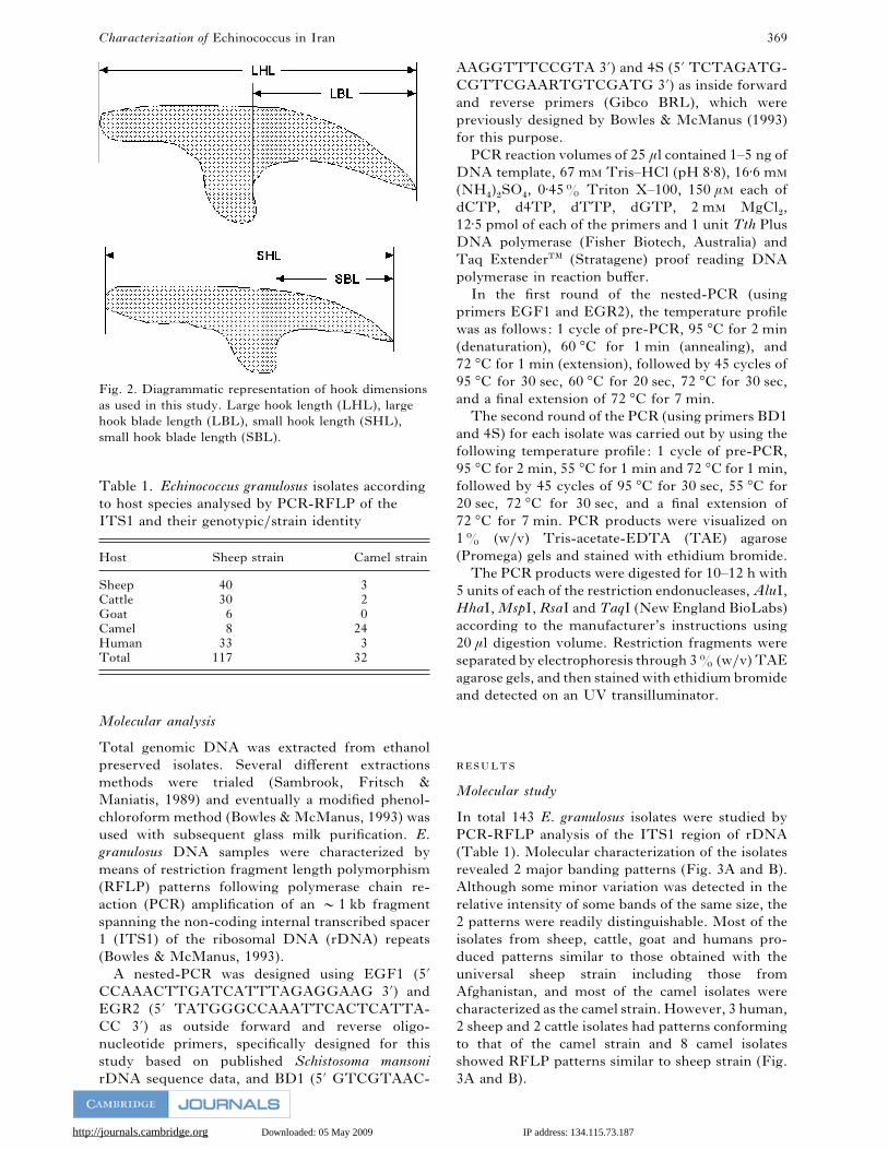
Molecular study
In total 143 E. granulosus isolates were studied by
PCR-RFLP analysis of the ITS1 region of rDNA
(Table 1). Molecular characterization of the isolates
revealed 2 major banding patterns (Fig. 3A and B).
Although some minor variation was detected in the
relative intensity of some bands of the same size, the
2 patterns were readily distinguishable. Most of the
isolates from sheep, cattle, goat and humans pro-
duced patterns similar to those obtained with the
universal sheep strain including those from
Afghanistan, and most of the camel isolates were
characterized as the camel strain. However, 3 human,
2 sheep and 2 cattle isolates had patterns conforming
to that of the camel strain and 8 camel isolates
showed RFLP patterns similar to sheep strain (Fig.
3A and B).
http://journals.cambridge.org Downloaded: 05 May 2009 IP address: 134.115.73.187
M. F. Harandi and others 370
A
B
Fig. 3. ITS1-PCR-RFLP patterns for isolates of Echinococcus granulosus from Iran. ITS1-PCR products were
digested with: (A) RsaI 2 and 3 cattle, 4 goat, 5 and 6 sheep, 7 human, 8–10 camel ; AluI 12 and 13 cattle, 14 goat,
15 and 16 sheep, 17 human, 18–20 camel ; (B) MspI 2–4 cattle, 5–6 sheep, 7–9 human, 10 camel ; RsaII 12–14 cattle,
15–16 sheep, 17–19 human, 20 camel. (.. Lanes 8 and 18 of (B) show camel strain}genotype pattern.)
Morphology
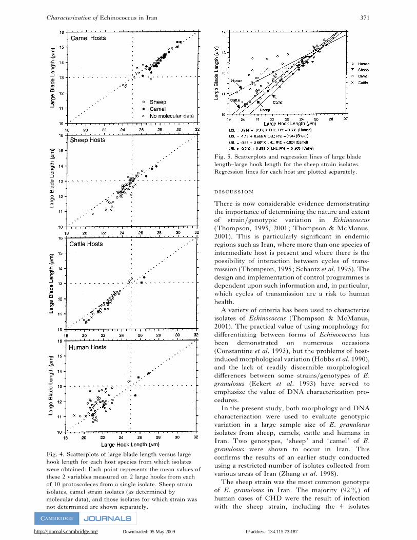
A scatterplot of large blade length against large hook
length (Fig. 4) clearly shows that hook morphology
is influenced by genetic factors. Although other
combinations of characters gave similar results, these
2 characters most clearly demonstrated the mor-
phological differences between the 2 strains. Within
each host species, there was a clear distinction
between these hook measurements of sheep and
camel strains. In sheep, cattle and humans, the single
character of large hook length would be sufficient to
predict the genetic strain of Echinococcus. All 21
isolates from camels which were not characterized by
PCR-RFLP fell within the camel strain cluster.
Similarly, all 18 isolates from sheep, 10 from cattle
and 21 from human hosts which were not charac-
terized by PCR-RFLP, fell within the sheep strain
cluster for each host species.
It may also be seen graphically from Fig. 5 that the
hook size is influenced by the host species. Although
there were too few isolates of camel strain in non-
camel hosts to conduct statistical analysis, hook
measurements in these hosts were consistently at the
lower end of the range found in camel hosts (Fig. 4).
The sheep strain was found in a large number of
non-sheep hosts, and a comparison of the regression
lines for large blade length and large hook length
indicated significantly different slopes (Fig. 5, F¯3±910, P!0±05). Further analysis (Tukey test for
multiple comparisons) indicated that the slope for
human isolates was lower than all the others. The
regression lines derived from the 3 remaining host
species shared a common slope, but elevations were
significantly different (F¯9±526, P!0±001). A
multiple comparison test consequently indicated that
the regression line derived from cattle isolates was
higher than that derived from sheep or camel isolates.
http://journals.cambridge.org Downloaded: 05 May 2009 IP address: 134.115.73.187
Characterization of Echinococcus in Iran 371
Fig. 4. Scatterplots of large blade length versus large
hook length for each host species from which isolates
were obtained. Each point represents the mean values of
these 2 variables measured on 2 large hooks from each
of 10 protoscoleces from a single isolate. Sheep strain
isolates, camel strain isolates (as determined by
molecular data), and those isolates for which strain was
not determined are shown separately.
Fig. 5. Scatterplots and regression lines of large blade
length–large hook length for the sheep strain isolates.
Regression lines for each host are plotted separately.
There is now considerable evidence demonstrating
the importance of determining the nature and extent
of strain}genotypic variation in Echinococcus
(Thompson, 1995, 2001; Thompson & McManus,
2001). This is particularly significant in endemic
regions such as Iran, where more than one species of
intermediate host is present and where there is the
possibility of interaction between cycles of trans-
mission (Thompson, 1995; Schantz et al. 1995). The
design and implementation of control programmes is
dependent upon such information and, in particular,
which cycles of transmission are a risk to human
health.
A variety of criteria has been used to characterize
isolates of Echinococcus (Thompson & McManus,
2001). The practical value of using morphology for
differentiating between forms of Echinococcus has
been demonstrated on numerous occasions
(Constantine et al. 1993), but the problems of host-
induced morphological variation (Hobbs et al. 1990),
and the lack of readily discernible morphological
differences between some strains}genotypes of E.
granulosus (Eckert et al. 1993) have served to
emphasize the value of DNA characterization pro-
cedures.
In the present study, both morphology and DNA
characterization were used to evaluate genotypic
variation in a large sample size of E. granulosus
isolates from sheep, camels, cattle and humans in
Iran. Two genotypes, ‘sheep’ and ‘camel’ of E.
granulosus were shown to occur in Iran. This
confirms the results of an earlier study conducted
using a restricted number of isolates collected from
various areas of Iran (Zhang et al. 1998).
The sheep strain was the most common genotype
of E. granulosus in Iran. The majority (92%) of
human cases of CHD were the result of infection
with the sheep strain, including the 4 isolates
http://journals.cambridge.org Downloaded: 05 May 2009 IP address: 134.115.73.187
M. F. Harandi and others 372
collected in Afghanistan, as were most cases of CHD
in non-human hosts. However, this study also
demonstrated for the first time, in an endemic region
where a transmission cycle for perpetuating the
camel strain exists, cases of CHD in humans caused
by infection with this strain. These results thus serve
to reinforce a recent report from Argentina which
provided evidence of CHD infection with the camel
strain in 4 patients on the basis of molecular data
(Rosenzvit et al. 1999). These results from Argentina
were difficult to interpret in the absence of a local
camel–dog cycle. However, the present study has
demonstrated that host specificity is not necessarily
applicable for all genotypes of E. granulosus. For
example, the sheep strain infects a wide variety of
host species apart from sheep, including camels
(Thompson et al. 1995). In addition to humans, the
camel strain is shown here to cause infection in sheep
and cattle. Previous studies have also reported the
isolation of the camel genotype from goats (Bowles,
Blair & McManus, 1992; Wachira et al. 1993). Thus
the proposal by Rosenzvit et al. (1999) that other
species of intermediate hosts could represent reser-
voirs of the camel strain in Argentina appears likely.
The fact that fewer human cases of CHD were
found to be due to infection with the camel strain
than the sheep strain (3 of 38) raises questions about
the epidemiology of echinococcosis in endemic
regions such as Iran. Is the camel strain of less public
health significance because humans are less sus-
ceptible to this strain, or is there less opportunity for
human contact with definitive hosts infected with the
camel strain in Iran? The latter seems more likely,
since in Iran camels have a more localized geo-
graphical distribution than sheep. According to
Government figures, the total number of camels in
Iran was 143000 in 1997 and they were distributed
in 19 of the 27 provinces of the country (Anon,
1997), including all of those from which human
isolates investigated in this study were obtained. Of
the total camel population, 77±6% (111000) is
distributed in the eastern half of Iran (Khorassan,
Kerman, Sistan-Baluchestan, Yazd and Semnan
(Anon, 1997), which is the driest, hottest and least
populated part of the country, and climatically
represents a less favourable environment for the
survival of Echinococcus eggs. Further, the preva-
lence of hydatid disease is higher in sheep than
camels, and more sheep are slaughtered (Mobedi et
al. 1970). However, it is evident from the present
study that the 2 cycles must overlap and can interact,
since infections caused by the sheep and camel
strains were detected in camels and sheep, re-
spectively, as well as in humans. Thus the camel–dog
cycle should be targeted in any control campaign.
Results presented here demonstrate that the 2
strains present in Iran and Afghanistan may be
differentiated by hook morphology alone. In human
hosts, the single measurement of large hook length
would be sufficient. All of the human isolates
identified as the sheep strain by molecular methods
had mean large hook lengths of !25 µm, whereas
those representing camel strain isolates were
"25 µm. All 70 isolates from humans and other
animals which were not characterized by molecular
methods could be ‘strain-typed’ morphologically.
Hook measurements of both strains were also
found to be influenced by the host species. Although
sample sizes were small, the hook measurements for
the camel strain in all other hosts were consistently at
the low end of the normal range found in camel
hosts. Sample sizes of sheep-strain isolates in hosts
other than sheep were large enough for statistical
analysis. Hook measurements in protoscoleces de-
rived from camel hosts were not different from those
from sheep, but there were quite distinct differences
in human and cattle hosts.
The results of this study lend further support for
the revision of the species-level taxonomy of Echino-
coccus. Over the last 10 years, the discriminatory
power of biochemical and molecular characterization
techniques has provided the data to propose such a
revision (Thompson et al. 1995). However, this
required more detailed comparative studies to be
undertaken in endemic regions in order to determine
the geographical distribution and uniformity of the
putative host-adapted species, as well as evidence of
their sympatric occurrence. This and other recent
studies (e.g. Zhang et al. 1998, and others reviewed
by Thompson (2001) and Thompson & McManus,
2001) have provided a sound foundation on which to
propose such a revised taxonomy.
(1997). Annual Report on Farm and Domestic
Animal Populations. Iranian Veterinary Organization,
Tehran.
, . , . (1995). Hepatic hydatid
disease in Iran, with review of the literature. Mount
Sinai Journal of Medicine 62, 62–69.
, ., , . c, . . (1992). Genetic
variants within the genus Echinococcus identified by
mitochondrial DNA sequencing. Molecular and
Biochemical Parasitology 54, 165–174.
, . c, . . (1993). Rapid
discrimination of Echinococcus species and strains
using a polymerase chain reaction-based RFLP
method. Molecular and Biochemical Parasitology 57,
231–240.
, . ., , . . ., , . .,
, . . , . . (1993). Morphological
characterization of adult Echinococcus granulosus as a
means of determining transmission patterns. Journal
of Parasitology 79, 57–61.
, . . , . (1997). Cystic echinococcosis in
the Gulf Littoral States. In Compendium of Cystic
Echinococcosis in Africa and in Middle Eastern
Countries with Special Reference to Morocco (ed.
http://journals.cambridge.org Downloaded: 05 May 2009 IP address: 134.115.73.187
Characterization of Echinococcus in Iran 373
Anderson, F. L., Ouhelli, H. & Kachani, M.), pp.
281–291. Brigham Young University, Provo, Utah,
USA.
, ., , . . ., , . .,
, . ., , . , . .
(1993). Further evidence for the occurrence of a
distinct strain of Echinococcus granulosus in European
pigs. Parasitology Research 75, 42–48.
, . . , . . (1995). Modelling
Echinococcus life cycles. In Echinococcus and Hydatid
Disease (ed. Thompson, R. C. A. & Lymbery, A. J.),
pp. 333–354. CAB International, Wallingford, Oxon,
UK.
, . ., , . . , . . . (1990).
Rostellar hook morphology of Echinococcus granulosus
(Batsch, 1786) from natural and experimental
Australian hosts and its implications for strain
recognition. Parasitology 101, 273–281.
, . ., , . . . , .
(1986). Echinococcus granulosus of equine origin from
different countries possess uniform morphological
characteristics. International Journal for Parasitology
16, 529–540.
c, . . (1997). Molecular genetic variation in
Echinococcus : an update. Southeast Asian Journal of
Tropical Medicine and Public Health 28, 110–116.
, ., , . , . (1971). Studies
on echinococcosis in Iran. Acta Medica Iranica 14,
221–229.
, ., , . , . (1970). Camel,
Camelus dromedarius, as intermediate host of
Echinococcus granulosus in Iran. Journal of Parasitology
56, 1255.
, . (1987). Hydatidosis-Echinococcosis in Iran,
Evaluating Economic Losses due to the Disease. Ph.D.
thesis (in Persian), Tehran University of Medical
Sciences.
, . ., , .-., , . .,
, . ., , . . c, . .
(1999). Genetic variation and epidemiology of
Echinococcus granulosus in Argentina. Parasitology 118,
523–530.
, ., , . . , . (1989).
Molecular Cloning: A Laboratory Manual. Cold
Spring Harbor Laboratory Press, Cold Spring
Harbor, NY, USA.
, . (1997). Echinococcus granulosus, E.
multilocularis, and E. vogeli (agents of cystic, alveolar,
and polycystic echinococcosis). In Pediatric Infectious
Diseases (ed. Long, S. S., Pickering, L. K. & Prober,
C. G.), pp. 1488–1492. Churchill Livingstone, New
York.
, . ., , ., , . ., , ., ,
. ., , . . . , . (1995).
Epidemiology and control of hydatid disease. In
Echinococcus and Hydatid Disease (ed. Thompson,
R. C. A. & Lymbery, A. J.), pp. 233–331. CAB
International, Wallingford, UK.
, . . . (1995). Biology and systematics of
Echinococcus. In Echinococcus and Hydatid Disease
(ed. Thompson, R. C. A. & Lymbery, A. J.), pp.
1–50. CAB International, Wallingford, UK.
, . . . (2001). Echinococcosis. In Principles
and Practice of Clinical Parasitology (ed. Gillespie,
S. H. & Pearson, R. D.), pp. 595–612. Wiley, Sussex,
UK.
, . . ., , . . , . .
(1998). Overview and significance of molecular
methods: what role for molecular epidemiology?
Parasitology 117, S161–S175.
, . . ., , . . , . .
(1995). Variation in Echinococcus : towards a taxonomic
revision of the genus. Advances in Parasitology 35,
145–176.
, . . . c, . . (2001). Aetiology:
parasites and life-cycles. In WHO}OIE Manual on
Echinococcus in Humans and Animals: a Zoonosis of
Global Concern (ed. Eckert, J., Gemmell, M. A.,
Meslin, F. X. & Pawlowski, Z. S.), pp. 1–19. World
Organization for Animal Health (OIE), Paris.
, . ., , ., , . c, . .
(1993). Molecular examination of the sympatry and
distribution of sheep and camel strains of Echinococcus
granulosus in Kenya. American Journal of Tropical
Medicine and Hygiene 48, 473–479.
, . . (1984). Biostatistical Analysis. Prentice-Hall,
New Jersey, USA.
, ., , ., , . . c, . .
(1998). Indication of the presence of two distinct
strains of Echinococcus granulosus in Iran by
mitochondrial DNA markers. American Journal of
Tropical Medicine and Hygiene 59, 171–174.
![Algorithms for improving consistency or consensus of reciprocal [0,1]-valued preference relations](https://static.fdokumen.com/doc/165x107/63458a6a596bdb97a908f9e2/algorithms-for-improving-consistency-or-consensus-of-reciprocal-01-valued-preference.jpg)