Modulation of gene expression during early stages of reconnection of the turtle spinal cord
11
In mammals, spinal cord injury results in gliosis and limited cellular regeneration, leading to devastating conditions (Thuret et al. 2006). In contrast, larval cyclostomes, some teleosts, and tailed amphibians keep substantial endogenous repair capabilities with restoration of locomotion after injury (Tanaka and Ferretti 2009). It is generally accepted that urodeles are the most evolved vertebrates with a robust regenerative potential of the adult spinal cord (Stensaas, 1983). However, we have shown (Rehermann et al. 2009) that freshwater turtles are able to reconnect their transected spinal cords, leading to some degree of recovery of motor functions. In contrast to mammals, no glial scar is observed in turtles but instead regenerating axons cross the injured site approximately 20 days after the lesion. These regenerating axons travel on a scaffold formed by newly generated glial cells that express the brain lipid binding protein (BLBP) and/or the glial fibrillary acidic protein (GFAP) (Rehermann et al. 2009, 2011). The endogenous repair of the spinal cord in turtles may be linked to the existence of domains of progenitor cells lining the central canal (CC). These cells exhibit proliferative capacity, are coupled via connexin 43 (Cx43), and express both BLBP and the transcription factor Pax6 (Russo et al. 2008). It is believed that connexins play an important role in the regulation of proliferation, migration, and cell differentiation (Bruzzone and Dermietzel 2006). In particular, Cx43 appears essential for maintaining neural progenitors in a proliferative state (Duval et al. 2002; Cheng et al. 2004). BLBP is a member of the large family of hydrophobic ligand-binding proteins that modulate transcription through their interactions with nuclear receptors (Haunerland and Spener 2004) and is required to induce Received January 26, 2012; revised manuscript received March 16, 2012; accepted March 21, 2012. Address correspondence and reprint requests to Dr. Rau ´l E. Russo, Departamento de Neurofisiologı ´a Celular y Molecular, Instituto de In- vestigaciones Biolo ´gicas, Clemente Estable, Avenida Italia 3318, CP 11600, Montevideo, Uruguay. E-mail: [email protected] Abbreviations used: BLBP, brain lipid binding protein; CC, central canal; Cx43, connexin 43; GFAP, glial fibrillary acidic protein; LDH, lactate dehydrogenase; PB, phosphate buffer; PK, pyruvate kinase; RG, radial glial. , *Neurofisiologı ´a Celular y Molecular. Instituto de Investigaciones Biolo ´gicas Clemente Estable, Montevideo, Uruguay Unidad de Biologı ´a Molecular, Institut Pasteur de Montevideo, Montevideo, Uruguay àDepartamento de Bioquı ´mica, Facultad de Medicina, Montevideo, Uruguay Abstract The spinal cord of the freshwater turtle Trachemys dorbignyi regenerates after complete transection (Rehermann et al. J. Comp. Neurol. 515, 2009, 197–214). This remarkable ability may be related to the persistence around the central canal (CC) of progenitors functionally clustered via connexin 43 (Cx43) that express brain lipid binding protein (BLBP) and the transcription factor Pax6 (Russo et al. J. Neurosci. 28, 2008, 8510–8516). Indeed, because BLBP+ cells appear in the bridge joining the rostral and caudal stumps, we speculated that progenitors contacting the central canal may play a key part in spinal cord regeneration. To test this hypothesis, we designed degenerated primers pairing conserved regions for key proteins synthesized in progenitors (BLBP, Cx43, and Pax6) and the neuronal protein HuB. Fragments of these proteins were amplified, cloned, and sequenced. Based on these sequences, we analyzed the changes in the expression levels using quantitative real-time RT-PCR with specific primers, comparing the injured spinal cord at different times after injury (4, 12, 20, and 60 days) with uninjured spinal cords. We found a transient, early increase of BLBP, Cx43 and HuB mRNA, with Pax6 remaining unchanged. These re- sults suggest that the selected genes – active in progenitor cells – play an important part in early mechanisms of spinal cord regeneration. Keywords: BLBP, connexin 43, Pax6, quantitative real-time PCR, regeneration, spinal cord. J. Neurochem. (2012) 121, 996–1006. JOURNAL OF NEUROCHEMISTRY | 2012 | 121 | 996–1006 doi: 10.1111/j.1471-4159.2012.07750.x 996 Journal of Neurochemistry ȑ 2012 International Society for Neurochemistry, J. Neurochem. (2012) 121, 996–1006 ȑ 2012 The Authors
-
Upload
independent -
Category
Documents
-
view
5 -
download
0
Transcript of Modulation of gene expression during early stages of reconnection of the turtle spinal cord
In mammals, spinal cord injury results in gliosis and limitedcellular regeneration, leading to devastating conditions(Thuret et al. 2006). In contrast, larval cyclostomes, someteleosts, and tailed amphibians keep substantial endogenousrepair capabilities with restoration of locomotion after injury(Tanaka and Ferretti 2009). It is generally accepted thaturodeles are the most evolved vertebrates with a robustregenerative potential of the adult spinal cord (Stensaas, 1983).However, we have shown (Rehermann et al. 2009) thatfreshwater turtles are able to reconnect their transected spinalcords, leading to some degree of recovery of motor functions.In contrast to mammals, no glial scar is observed in turtles butinstead regenerating axons cross the injured site approximately20 days after the lesion. These regenerating axons travel on ascaffold formed by newly generated glial cells that express thebrain lipid binding protein (BLBP) and/or the glial fibrillaryacidic protein (GFAP) (Rehermann et al. 2009, 2011).
The endogenous repair of the spinal cord in turtles may belinked to the existence of domains of progenitor cells lining thecentral canal (CC). These cells exhibit proliferative capacity,
are coupled via connexin 43 (Cx43), and express both BLBPand the transcription factor Pax6 (Russo et al. 2008). It isbelieved that connexins play an important role in the regulationof proliferation, migration, and cell differentiation (BruzzoneandDermietzel 2006). In particular, Cx43 appears essential formaintaining neural progenitors in a proliferative state (Duvalet al. 2002; Cheng et al. 2004). BLBP is a member of the largefamily of hydrophobic ligand-binding proteins that modulatetranscription through their interactions with nuclear receptors(Haunerland and Spener 2004) and is required to induce
Received January 26, 2012; revised manuscript received March 16,2012; accepted March 21, 2012.Address correspondence and reprint requests to Dr. Raul E. Russo,
Departamento de Neurofisiologıa Celular y Molecular, Instituto de In-vestigaciones Biologicas, Clemente Estable, Avenida Italia 3318, CP11600, Montevideo, Uruguay. E-mail: [email protected] used: BLBP, brain lipid binding protein; CC, central
canal; Cx43, connexin 43; GFAP, glial fibrillary acidic protein; LDH,lactate dehydrogenase; PB, phosphate buffer; PK, pyruvate kinase; RG,radial glial.
,
*Neurofisiologıa Celular y Molecular. Instituto de Investigaciones Biologicas Clemente Estable,
Montevideo, Uruguay
�Unidad de Biologıa Molecular, Institut Pasteur de Montevideo, Montevideo, Uruguay
�Departamento de Bioquımica, Facultad de Medicina, Montevideo, Uruguay
Abstract
The spinal cord of the freshwater turtle Trachemys dorbignyi
regenerates after complete transection (Rehermann et al.
J. Comp. Neurol. 515, 2009, 197–214). This remarkable ability
may be related to the persistence around the central canal
(CC) of progenitors functionally clustered via connexin 43
(Cx43) that express brain lipid binding protein (BLBP) and the
transcription factor Pax6 (Russo et al. J. Neurosci. 28, 2008,
8510–8516). Indeed, because BLBP+ cells appear in the
bridge joining the rostral and caudal stumps, we speculated
that progenitors contacting the central canal may play a key
part in spinal cord regeneration. To test this hypothesis, we
designed degenerated primers pairing conserved regions for
key proteins synthesized in progenitors (BLBP, Cx43, and
Pax6) and the neuronal protein HuB. Fragments of these
proteins were amplified, cloned, and sequenced. Based on
these sequences, we analyzed the changes in the expression
levels using quantitative real-time RT-PCR with specific
primers, comparing the injured spinal cord at different times
after injury (4, 12, 20, and 60 days) with uninjured spinal
cords. We found a transient, early increase of BLBP, Cx43
and HuB mRNA, with Pax6 remaining unchanged. These re-
sults suggest that the selected genes – active in progenitor
cells – play an important part in early mechanisms of spinal
cord regeneration.
Keywords: BLBP, connexin 43, Pax6, quantitative real-time
PCR, regeneration, spinal cord.
J. Neurochem. (2012) 121, 996–1006.
JOURNAL OF NEUROCHEMISTRY | 2012 | 121 | 996–1006 doi: 10.1111/j.1471-4159.2012.07750.x
996 Journal of Neurochemistry � 2012 International Society for Neurochemistry, J. Neurochem. (2012) 121, 996–1006� 2012 The Authors
morphological changes in radial glial (RG) in response toneuronal cues (Feng et al. 1994; Anton et al. 1997). Finally,the transcription factor Pax6 is expressed in RG lining theventricle cavities and is an important regulator of theneurogenic capabilities of progenitors during development(Heins et al. 2002; Pinto and Gotz 2007) and in neurogenicniches of the adult brain (Kohwi et al. 2005). In the spinalcord, Pax6 is part of the transcriptional code that determinessubsets of progenitors giving rise to specific neuronal types(Lee and Pfaff 2001), controlling the tempo of neuronaldifferentiation (Bel-Vialar et al. 2007).
Because the persistence of CC-contacting progenitors maybe one of the central features supporting the outstandingregeneration capability in turtles (Rehermann et al. 2009,2011), we hypothesized that spinal cord injury may triggerchanges in the expression of key genes that drive proteinsynthesis in these cells. To test this idea, we selected theBLBP, Pax6, and Cx43 genes expressed in CC-contactingprogenitors and HuB, a gene active from the earliest stages ofneuronal differentiation (Marusich et al. 1994). Becausethere is no information about the sequence of these genes inturtles, we first designed degenerate oligonucleotides primingconserved regions of the genes, together with specificprimers for the putative control genes pyruvate kinase (PK)and lactate dehydrogenase (LDH). We next amplifiedfragments of these genes, cloned, and sequenced them.Based on these data, we analyzed the changes in mRNAexpression levels using real-time RT-PCR with specificprimers, comparing injured segments (T10-11) with theircounterparts in control animals at different time points afterspinal cord transection. To analyze the spatial profile of thechange in gene expression, we performed immunohisto-chemistry in injured and normal animals. Our resultsrevealed significant increments in the expression of BLBP,Cx43, and HuB genes 4 days after injury while Pax6remained unchanged at the same time point. However, BLBPand Cx43 returned to normal values around day 12, a timepoint when anatomical reconnection is still incipient (Reher-mann et al. 2009). Conversely, Hu remained underexpressedat 12, 20, and 60 days post injury. The immunohistochemicaldata showed that the spatial profile of expression of thesegenes was conserved in spinal cord stumps. Our findingssuggest that modulation of BLBP and Cx43 gene expressionis transiently involved in the early plasticity triggered byspinal cord injury. It remains to be clarified whether this earlyresponse is instrumental for later stages of post-transcrip-tional mechanisms commanding endogenous neural repair.
Methods
AnimalsFresh-water turtles (Trachemys dorbignyi, 5–12 cm carapace length)were used (n = 22). These juvenile turtles (2 months to 1 year old)behave like fully mature animals in terms of their sensory-motor
behavior. The animals were maintained in temperate aquaria (24–26�C) under natural illumination and fed daily with smallearthworms. The ARRIVE guidelines have been followed, and allexperimental procedures were performed in accordance with theethical guidelines established by our local Committee for AnimalCare and Research at the Instituto de Investigaciones BiologicasClemente Estable. Every precaution was taken to minimize animalstress and the number of animals used.
Surgical proceduresThe detailed surgical procedures to lesion the spinal cord have beendescribed elsewhere (Rehermann et al. 2009). Briefly, animals weresedated by intraperitoneal injection of ketamine chlorhydrate(40 mg/kg b.w.). Then, the legs were secured to a rigid supportby means of rubber bands, and complete anesthesia was achieved byinhalation of isofluorane (1-chloro-2,2,2-trifluoroethyl difluorom-ethyl ether; Forane, Abbot Laboratories, Berkshire, UK). To preventinfection, we rubbed the carapace with a cotton pad previouslyimmersed in an alcohol-iodine solution, and all surgical instrumentswere sterilized by using chemical procedures. After the animal wasunresponsive to nociceptive stimuli, we opened a window in thecarapace following the limits of the third dorsal scute to expose thespinal cord. Complete transection of the spinal cord (T10-11) wasperformed with a thin-blade scalpel. After washing the injured sitewith sterile saline, we replaced the lifted scute and sealed the injuredcarapace with cyanoacrylate adhesive.
Post-operative careWe identified spinalized turtles with indelible marks sculptured ontheir carapace. Turtles do not require special post-operative carebecause, as described for other non-mammalian vertebrates (Mount-castle 1980), spinal shock is very short. Because turtles have aprimitive ‘‘allantoic bladder’’ opening into the cloaca (Parker andHarwell 1949), there is no noticeable urine retention. Despite theirreduced motor capabilities, injured turtles were able to catch livingworms a few days after surgery.
Tissue preparationFor the cloning and sequencing of the genes of interest, the wholespinal cords (n = 3) were rapidly removed and placed in RNAlater(Sigma) for storage at )80�C. For quantitative real-time PCRanalysis, we studied a total of 12 turtles that were examined usingthe following protocol: three turtles were killed 4 days after surgery;three turtles were killed 12 days after surgery; three turtles werekilled 20 days after surgery; three turtles were killed 60 days aftersurgery. In all cases, a 4-mm long spinal cord segment centered onthe lesion epicenter was quickly dissected out, placed in RNAlater(Sigma-Aldrich, St. Louis, MO, USA), and stored at )80�C. Thecorresponding spinal cord segments of control-uninjured and sham-injured animals were processed in the same way.
RNA extraction and reverse transcriptionThe tissue was mechanically homogenized using a Cordless motorand pellet pestle (Sigma-Aldrich). Total RNA from the spinal cordwas extracted and purified using TRIZOL reagent (Invitrogen,Carlsbad, CA, USA) following the manufacturer’s protocol. Theconcentrations of RNA samples were quantified using the Nanodrop(Nano-Drop Inc., Rockland, DE, USA). RNA integrity wasdetermined using Bioanalyzer (Agilent Technologies, Santa Clara,
� 2012 The AuthorsJournal of Neurochemistry � 2012 International Society for Neurochemistry, J. Neurochem. (2012) 121, 996–1006
Gene expression during spinal cord regeneration | 997
CA, USA), and only samples with a RNA Integrity Number (RIN)higher than 7 were used for reverse transcription (Figure S1). Forspinal cord segments of injured and uninjured animals, cDNAsynthesis was performed using the same amount of starting RNA(0.5 lg). The enzyme Superscript III was used (Invitrogen) primedwith oligo(dT) according to the manufacturer’s instructions.
Cloning and sequencingDegenerated and specific primers were designed to hybridize highlyconserved regions of the genes of interest coding sequences(Table 1). The PCR products amplified from spinal cord cDNAwere analyzed by agarose gel electrophoresis. When needed, gelpurification was performed with the kit GenElute gel extraction kit(Sigma-Aldrich). PCR products were cloned using the pGEMT EasyVector System (Promega, Madison, WI, USA). Colonies wereselected and plasmids purified from 3 ml of liquid media LB. Insertswere sequenced with Sp6 and T7 universal primers using anABI3130 (Applied Biosystems, Foster City, CA, USA) DNAautomatic sequencer. Sequences from multiple clones were alignedto generate consensus sequences, which were then used innucleotide basic local alignment search tool (BLAST) searches toretrieve orthologous ones.
Alignment of amplified gene fragments with orthologs from othervertebrates was done online with ClustalW2 (http://www.ebi.ac.uk/Tools/msa/clustalw2/). We used the online ExPASy tool (http://ca.expasy.org/tools/dna.html) for in silico translation of nucleotidesequences to examine conservation at the amino acid level.
Quantitative Real-time PCR AnalysisFour-millimeter long segments of spinal cord centered on the lesionepicenter were compared with corresponding segments of controlanimals. Injured animals (n = 3 for each time point) were killed atdifferent times post injury (4, 12, 20, and 60 days). With the cloned
sequences, specific primers were designed for amplifying smallfragments (� 200 pb, Table 2). Using a real-time cycler Rotor Gene6000 (Corbett Research, Bath, UK) and SYBR Green I, the genes ofinterest were amplified. Briefly, each reaction tube contained 5 lLof 2X SYBR Green Quantimix solution (Biotools), 250 nM of eachforward and reverse primer, 0.5 lL of cDNA and water to bring thetotal reaction volume to 10 lL. The thermal profile was 95�C for5 min followed by 40 cycles of three steps (95�C for 15 s, 55�C for20 s, and 72�C for 20 s). The PCR for each cDNA sample andplasmid DNA standard was performed in duplicate.
Control reactions were performed to verify the absence ofgenomic DNA and cross contamination of any other DNA source. Amelting temperature analysis was added at the end of each real-timePCR to verify that a single product was amplified for each geneanalyzed.
Standard curve constructionBecause the intended housekeeping genes LDH and PK changeddramatically following spinal cord transection (data not shown), weused an absolute transcript quantification method. The plasmidicvectors pGEM-T easy (Promega) containing the sequences of interestused previously were also used for the construction of the standardcurves which were based on real-time PCR of known quantities ofplasmidic DNA. Each standard curve was generated based on five 10-fold serial dilutions of a starting concentration of DNA (100 pg/lL)converted to the number of copies using the molecular mass of theDNA: Attomoles (amoles) of input plasmid DNA = 1.52 · amountof input plasmid DNA/plasmid length, according toWu et al. (2005).A standard curve plotting CT values against the logarithm of inputDNA copy quantity (log amoles) was constructed for each gene. Thenumber of copies in the experimental samples was calculated from theequation of the line for the standard curve.
Data analysisStandard DNAs, negative controls, and cDNA obtained fromuninjured animals and samples obtained at different times afterinjury (4, 12, 20, and 60 days) were run simultaneously for each of
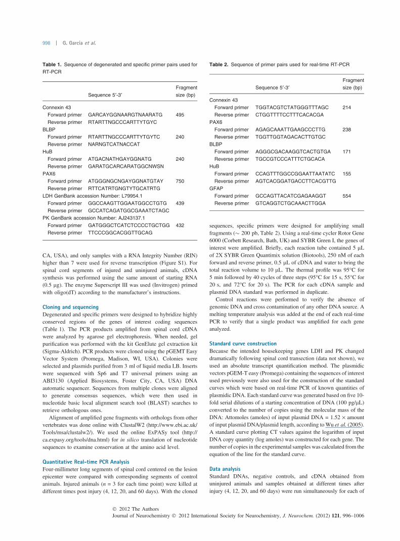
Table 1. Sequence of degenerated and specific primer pairs used for
RT-PCR
Sequence 5¢-3¢Fragment
size (bp)
Connexin 43
Forward primer GARCAYGGNAARGTNAARATG 495
Reverse primer RTARTTNGCCCARTTYTGYC
BLBP
Forward primer RTARTTNGCCCARTTYTGYTC 240
Reverse primer NARNGTCATNACCAT
HuB
Forward primer ATGACNATHGAYGGNATG 240
Reverse primer GARATGCARCARATGGCNWSN
PAX6
Forward primer ATGGGNGCNGAYGGNATGTAY 750
Reverse primer RTTCATRTGNGTYTGCATRTG
LDH GenBank accession Number: L79954.1
Forward primer GGCCAAGTTGGAATGGCCTGTG 439
Reverse primer GCCATCAGATGGCGAAATCTAGC
PK GenBank accession Number: AJ243137.1
Forward primer GATGGGCTCATCTCCCCTGCTGG 432
Reverse primer TTCCCGGCACGGTTGCAG
Table 2. Sequence of primer pairs used for real-time RT-PCR
Sequence 5¢-3¢Fragment
size (bp)
Connexin 43
Forward primer TGGTACGTCTATGGGTTTAGC 214
Reverse primer CTGGTTTTCCTTTCACACGA
PAX6
Forward primer AGAGCAAATTGAAGCCCTTG 238
Reverse primer TGGTTGGTAGACACTTGTGC
BLBP
Forward primer AGGGCGACAAGGTCACTGTGA 171
Reverse primer TGCCGTCCCATTTCTGCACA
HuB
Forward primer CCAGTTTGGCCGGAATTAATATC 155
Reverse primer AGTCACGGATGACCTTCACGTTG
GFAP
Forward primer GCCAGTTACATCGAGAAGGT 554
Reverse primer GTCAGGTCTGCAAACTTGGA
Journal of Neurochemistry � 2012 International Society for Neurochemistry, J. Neurochem. (2012) 121, 996–1006� 2012 The Authors
998 | G. Garcıa et al.
the genes analyzed. The data for the uninjured controls werepresented as amole/lg RNA ± SEM, and the relative gene expres-sion data for the different times after injury were expressed as theratio to the amole/lg RNA of the sample divided by the same valuein the control. Statistical significance was evaluated with theunpaired t-test at p < 0.05.
ImmunohistochemistryThe spinal cords of anesthetized turtles (50 mg/kg pentobarbital)were fixed by intracardiac perfusion. For immunohistochemistry(n = 6 animals), we used 10% paraformaldehyde dissolved in 0.1 Mphosphate buffer (PB; pH 7.4). Fixed material was sectioned in thetransverse plane (60–80 lm) with a vibrating-blade microtome. Weused the following primary antibodies: (i) anti-BLBP (rabbitpolyclonal, 1:1000; Chemicon International Inc., Temecula, CA);(ii) anti-HuC/D (mouse monoclonal, 1:50; Molecular Probes,Eugene, OR), (iii) anti-Cx43 (rabbit polyclonal, 1:3000; kindlyprovided by Dr. J.C. Saez; PUC University, Santiago, Chile), and(iv) anti-GFAP (mouse monoclonal, 1:500, Sigma). Non-specificreactive sites were blocked with 0.5% bovine serum albumin in PB(1 h) followed by overnight incubation with the primary antibody inPB containing 0.3% Triton X-100. After blocking and washing, thetissues were incubated with goat anti-rabbit or goat anti-mousesecondary antibodies conjugated with Alexa 488 (1:1.000, 2 h;Invitrogen, Eugene, OR, USA). Finally, the sections were thor-oughly washed and mounted. Control experiments replacingantibodies with pre-immune serum and negative controls withoutprimary antibodies were routinely performed.
Since Cx43 immunohistochemistry revealed a dotted pattern, itwas possible to use an automatic counting procedure. The number ofpunctae was quantified from four confocal optical sections of controlanimals and turtles with the transected spinal cord at 4 and 12 dayspost injury (DPI). For this purpose, we used the command Analyzeparticles of ImageJ (available at http://rsbweb.nih.gov/ij/), aftersetting appropriately the threshold level.
Results
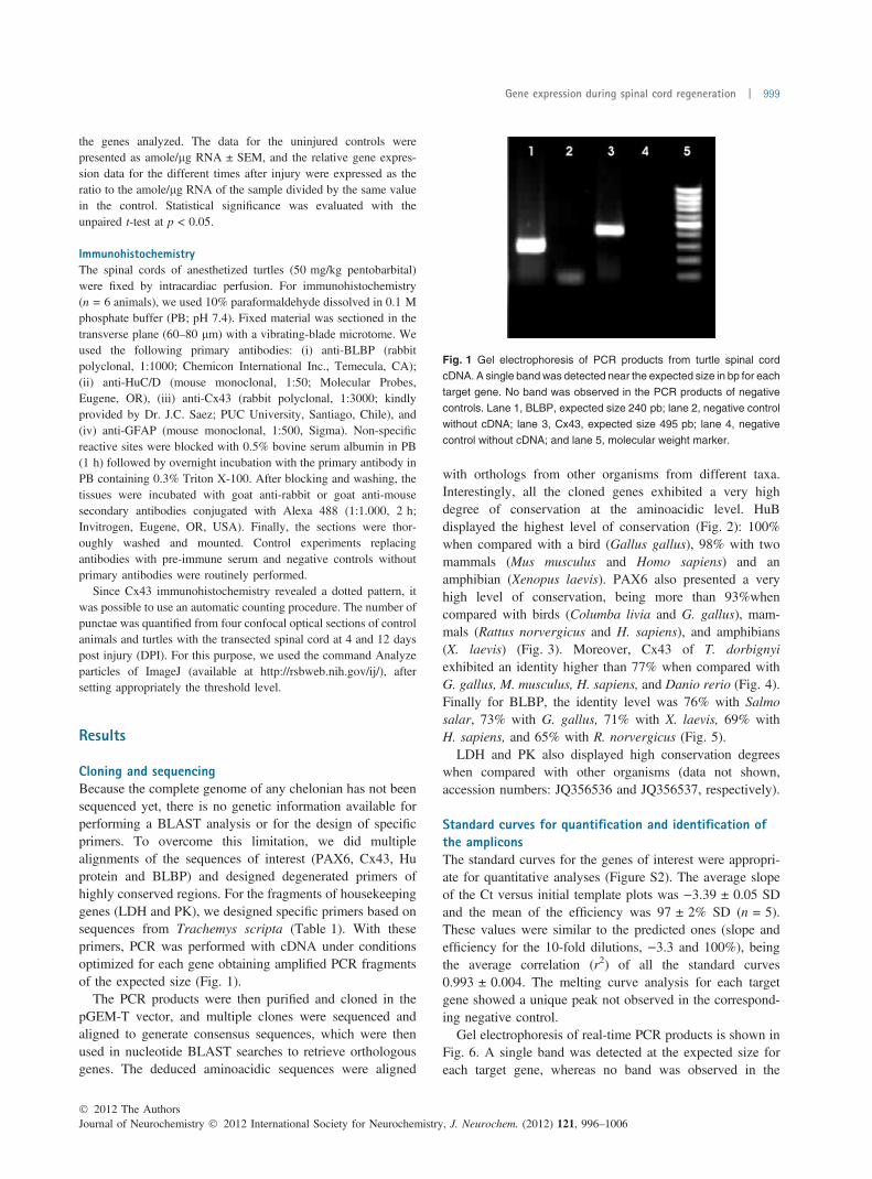
Cloning and sequencingBecause the complete genome of any chelonian has not beensequenced yet, there is no genetic information available forperforming a BLAST analysis or for the design of specificprimers. To overcome this limitation, we did multiplealignments of the sequences of interest (PAX6, Cx43, Huprotein and BLBP) and designed degenerated primers ofhighly conserved regions. For the fragments of housekeepinggenes (LDH and PK), we designed specific primers based onsequences from Trachemys scripta (Table 1). With theseprimers, PCR was performed with cDNA under conditionsoptimized for each gene obtaining amplified PCR fragmentsof the expected size (Fig. 1).
The PCR products were then purified and cloned in thepGEM-T vector, and multiple clones were sequenced andaligned to generate consensus sequences, which were thenused in nucleotide BLAST searches to retrieve orthologousgenes. The deduced aminoacidic sequences were aligned
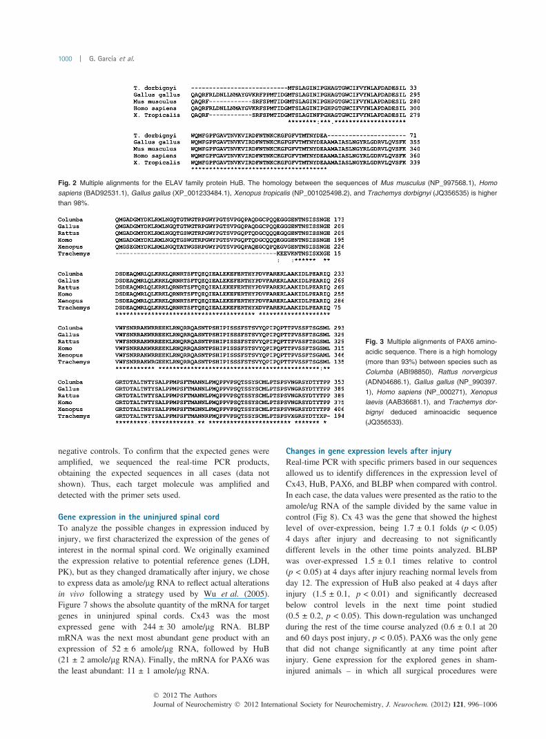
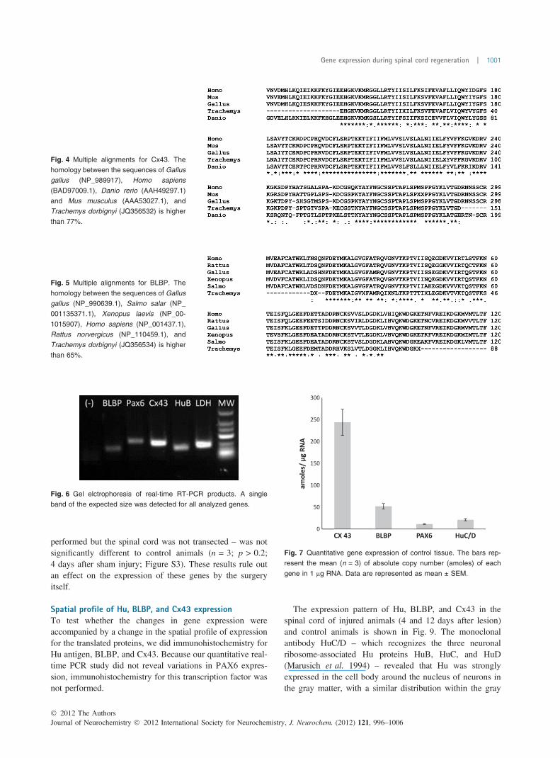
with orthologs from other organisms from different taxa.Interestingly, all the cloned genes exhibited a very highdegree of conservation at the aminoacidic level. HuBdisplayed the highest level of conservation (Fig. 2): 100%when compared with a bird (Gallus gallus), 98% with twomammals (Mus musculus and Homo sapiens) and anamphibian (Xenopus laevis). PAX6 also presented a veryhigh level of conservation, being more than 93%whencompared with birds (Columba livia and G. gallus), mam-mals (Rattus norvergicus and H. sapiens), and amphibians(X. laevis) (Fig. 3). Moreover, Cx43 of T. dorbignyiexhibited an identity higher than 77% when compared withG. gallus, M. musculus, H. sapiens, and Danio rerio (Fig. 4).Finally for BLBP, the identity level was 76% with Salmosalar, 73% with G. gallus, 71% with X. laevis, 69% withH. sapiens, and 65% with R. norvergicus (Fig. 5).
LDH and PK also displayed high conservation degreeswhen compared with other organisms (data not shown,accession numbers: JQ356536 and JQ356537, respectively).
Standard curves for quantification and identification ofthe ampliconsThe standard curves for the genes of interest were appropri-ate for quantitative analyses (Figure S2). The average slopeof the Ct versus initial template plots was )3.39 ± 0.05 SDand the mean of the efficiency was 97 ± 2% SD (n = 5).These values were similar to the predicted ones (slope andefficiency for the 10-fold dilutions, )3.3 and 100%), beingthe average correlation (r2) of all the standard curves0.993 ± 0.004. The melting curve analysis for each targetgene showed a unique peak not observed in the correspond-ing negative control.
Gel electrophoresis of real-time PCR products is shown inFig. 6. A single band was detected at the expected size foreach target gene, whereas no band was observed in the
Fig. 1 Gel electrophoresis of PCR products from turtle spinal cord
cDNA. A single band was detected near the expected size in bp for each
target gene. No band was observed in the PCR products of negative
controls. Lane 1, BLBP, expected size 240 pb; lane 2, negative control
without cDNA; lane 3, Cx43, expected size 495 pb; lane 4, negative
control without cDNA; and lane 5, molecular weight marker.
� 2012 The AuthorsJournal of Neurochemistry � 2012 International Society for Neurochemistry, J. Neurochem. (2012) 121, 996–1006
Gene expression during spinal cord regeneration | 999
negative controls. To confirm that the expected genes wereamplified, we sequenced the real-time PCR products,obtaining the expected sequences in all cases (data notshown). Thus, each target molecule was amplified anddetected with the primer sets used.
Gene expression in the uninjured spinal cordTo analyze the possible changes in expression induced byinjury, we first characterized the expression of the genes ofinterest in the normal spinal cord. We originally examinedthe expression relative to potential reference genes (LDH,PK), but as they changed dramatically after injury, we choseto express data as amole/lg RNA to reflect actual alterationsin vivo following a strategy used by Wu et al. (2005).Figure 7 shows the absolute quantity of the mRNA for targetgenes in uninjured spinal cords. Cx43 was the mostexpressed gene with 244 ± 30 amole/lg RNA. BLBPmRNA was the next most abundant gene product with anexpression of 52 ± 6 amole/lg RNA, followed by HuB(21 ± 2 amole/lg RNA). Finally, the mRNA for PAX6 wasthe least abundant: 11 ± 1 amole/lg RNA.
Changes in gene expression levels after injuryReal-time PCR with specific primers based in our sequencesallowed us to identify differences in the expression level ofCx43, HuB, PAX6, and BLBP when compared with control.In each case, the data values were presented as the ratio to theamole/ug RNA of the sample divided by the same value incontrol (Fig 8). Cx 43 was the gene that showed the highestlevel of over-expression, being 1.7 ± 0.1 folds (p < 0.05)4 days after injury and decreasing to not significantlydifferent levels in the other time points analyzed. BLBPwas over-expressed 1.5 ± 0.1 times relative to control(p < 0.05) at 4 days after injury reaching normal levels fromday 12. The expression of HuB also peaked at 4 days afterinjury (1.5 ± 0.1, p < 0.01) and significantly decreasedbelow control levels in the next time point studied(0.5 ± 0.2, p < 0.05). This down-regulation was unchangedduring the rest of the time course analyzed (0.6 ± 0.1 at 20and 60 days post injury, p < 0.05). PAX6 was the only genethat did not change significantly at any time point afterinjury. Gene expression for the explored genes in sham-injured animals – in which all surgical procedures were
Fig. 2 Multiple alignments for the ELAV family protein HuB. The homology between the sequences of Mus musculus (NP_997568.1), Homo
sapiens (BAD92531.1), Gallus gallus (XP_001233484.1), Xenopus tropicalis (NP_001025498.2), and Trachemys dorbignyi (JQ356535) is higher
than 98%.
Fig. 3 Multiple alignments of PAX6 amino-
acidic sequence. There is a high homology
(more than 93%) between species such as
Columba (ABI98850), Rattus norvergicus
(ADN04686.1), Gallus gallus (NP_990397.
1), Homo sapiens (NP_000271), Xenopus
laevis (AAB36681.1), and Trachemys dor-
bignyi deduced aminoacidic sequence
(JQ356533).
Journal of Neurochemistry � 2012 International Society for Neurochemistry, J. Neurochem. (2012) 121, 996–1006� 2012 The Authors
1000 | G. Garcıa et al.
performed but the spinal cord was not transected – was notsignificantly different to control animals (n = 3; p > 0.2;4 days after sham injury; Figure S3). These results rule outan effect on the expression of these genes by the surgeryitself.
Spatial profile of Hu, BLBP, and Cx43 expressionTo test whether the changes in gene expression wereaccompanied by a change in the spatial profile of expressionfor the translated proteins, we did immunohistochemistry forHu antigen, BLBP, and Cx43. Because our quantitative real-time PCR study did not reveal variations in PAX6 expres-sion, immunohistochemistry for this transcription factor wasnot performed.
The expression pattern of Hu, BLBP, and Cx43 in thespinal cord of injured animals (4 and 12 days after lesion)and control animals is shown in Fig. 9. The monoclonalantibody HuC/D – which recognizes the three neuronalribosome-associated Hu proteins HuB, HuC, and HuD(Marusich et al. 1994) – revealed that Hu was stronglyexpressed in the cell body around the nucleus of neurons inthe gray matter, with a similar distribution within the gray
Fig. 4 Multiple alignments for Cx43. The
homology between the sequences of Gallus
gallus (NP_989917), Homo sapiens
(BAD97009.1), Danio rerio (AAH49297.1)
and Mus musculus (AAA53027.1), and
Trachemys dorbignyi (JQ356532) is higher
than 77%.
Fig. 5 Multiple alignments for BLBP. The
homology between the sequences of Gallus
gallus (NP_990639.1), Salmo salar (NP_
001135371.1), Xenopus laevis (NP_00-
1015907), Homo sapiens (NP_001437.1),
Rattus norvergicus (NP_110459.1), and
Trachemys dorbignyi (JQ356534) is higher
than 65%.
Fig. 6 Gel elctrophoresis of real-time RT-PCR products. A single
band of the expected size was detected for all analyzed genes.
Fig. 7 Quantitative gene expression of control tissue. The bars rep-
resent the mean (n = 3) of absolute copy number (amoles) of each
gene in 1 lg RNA. Data are represented as mean ± SEM.
� 2012 The AuthorsJournal of Neurochemistry � 2012 International Society for Neurochemistry, J. Neurochem. (2012) 121, 996–1006
Gene expression during spinal cord regeneration | 1001
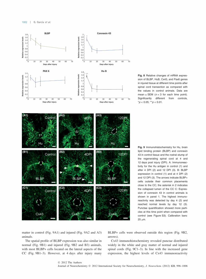
matter in control (Fig. 9A1) and injured (Fig. 9A2 and A3)animals.
The spatial profile of BLBP expression was also similar innormal (Fig. 9B1) and injured (Fig. 9B2 and B3) animals,with most BLBP+ cells located on the lateral aspects of theCC (Fig. 9B1–3). However, at 4 days after injury many
BLBP+ cells were observed outside this region (Fig. 9B2,arrows).
Cx43 immunohistochemistry revealed punctae distributedwidely in the white and gray matter of normal and injuredspinal cords (Fig. 9C1–3). In line with the increased geneexpression, the highest levels of Cx43 immunoreactivity
Fig. 8 Relative changes of mRNA expres-
sion of BLBP, HuB, Cx43, and Pax6 genes
in injured tissue at different time points after
spinal cord transection as compared with
the values in control animals. Data are
mean ± SEM (n = 3 for each time point).
Significantly different from controls,
*p < 0.05; **p < 0.01.
(A1) (A2) (A3)
(B1) (B2) (B3)
(C1) (C2) (C3)
Fig. 9 Immunohistochemistry for Hu, brain
lipid binding protein (BLBP) and connexin
43 in control tissue and the rostral stump of
the regenerating spinal cord at 4 and
12 days post injury (DPI). A: Immunoreac-
tivity for the Hu antigen in control (1) and
after 4 DPI (2) and 12 DPI (3). B: BLBP
expression in control (1) and at 4 DPI (2)
and 12 DPI (3). The arrows indicate BLBP+
cells outside their common placements
close to the CC; the asterisk in 2 indicates
the collapsed lumen of the CC C: Expres-
sion of connexin 43 in control animals is
shown in panel 1. The highest immuno-
reactivity was detected by day 4 (2) and
reached normal levels by day 12 (3).
Punctae quantification showed more parti-
cles at this time point when compared with
control (see Figure S3). Calibration bars:
25 lm.
Journal of Neurochemistry � 2012 International Society for Neurochemistry, J. Neurochem. (2012) 121, 996–1006� 2012 The Authors
1002 | G. Garcıa et al.
were observed at 4 DPI. (Fig. 9C2). Although 12 days afterinjury the Cx43 immunoreactivity decayed (Fig. 9C3), thenumber of punctae was still higher than control (Figure S4).
Because reactive astrogliosis may partly account for theincreased expression of Cx43 around the CC, we didimmunohistochemistry for GFAP in control and injuredanimals (4 DPI). Although the ependyma remained negativefor GFAP, the expression of this protein in the regionsurrounding the CC seemed up-regulated at 4 DPI (Fig-ure S5A). In line with these findings, the expression of theGFAP gene at 4 DPI was significantly increased comparedwith uninjured animals (Figure S5B).
Discussion
Turtles are amniote vertebrates that – unlike mammals – areable of a substantial degree of endogenous repair, probablybecause they retained multi-potent progenitor cells aroundthe CC (Trujillo-Cenoz et al. 2007; Russo et al. 2008;Rehermann et al. 2009, 2011). Understanding the molecularmechanisms involved in injury induced plasticity in newmodel systems with efficient self-repair mechanisms maygive important clues for the design of new therapies fortreatment of spinal cord diseases (Tanaka and Ferretti 2009).A disadvantage of the turtle spinal cord as a model system tostudy regeneration is that no genomic resources fromchelonians are available. In the present study, we used acloning and sequencing strategy through gene amplificationby degenerated primers design and described the partialsequences of genes that our previous studies pointed aspotentially important for spinal cord regeneration in turtles(Rehermann et al. 2009). Surprisingly, our data show a highlevel of conservation of the analyzed genes among evolu-tionary distant organisms, validating the strategy and thebiological model used in this study.
The analysis of mRNA expression changes after spinalcord transection has been a valuable approach to elucidatethe underlying mechanisms of neural repair (Wu et al. 2005;Fujiyoshi et al. 2010; Arocho et al. 2011; Kuo et al. 2011;Nash et al. 2011). A key difference between spinal cord self-repair capabilities between mammals and turtles may berelated with the persistence of progenitors functionallyclustered via Cx43 and with a molecular phenotype charac-terized by the expression of BLBP and Pax6 (Russo et al.2008). By applying quantitative RT-PCR – a highly sensitivetechnique allowing the quantification of small changes ingene expression (Wu et al. 2005) – we found that BLBP andCx43 gene expression increased transiently, whereas Pax6remained unchanged.
The increase in Cx43 mRNA expression induced by injuryin our study resembles the elevation in Cx43 expression aftera complete transection of the spinal cord of the rat asevaluated by in situ hybridization (Lee et al. 2005). Unlikethe transient increase observed in turtles, Cx43 up-regulation
in the rat increases progressively up to 4 weeks (Lee et al.2005). The role played by Cx43 in response to injury of thenervous system is controversial, with some reports suggest-ing that increased levels are detrimental (Frantseva et al.2002a,b), whereas others claim that Cx43 down-regulationresults in an expansion in the lesion size (Siushansian et al.2001; Nakase et al. 2004). The interference with Cx43function after spinal cord injury reduces the reactiveastrogliosis and neuronal death (O’Carroll et al. 2008) andimproves functional recovery (Cronin et al. 2008). Ourfindings suggest that in turtles, the rapid decline of the initialup-regulation of Cx43 may minimize the detrimental effectsof Cx43 mentioned above, possibly by avoiding the spread ofinjury.
It is not clear whether the increased expression of Cx43reported here results in an increased number of hemichannelsor functional gap junctions. Hemichannels have been postu-lated to mediate a component of ATP release (Garre et al.2010) that acts on P2X7 channels during spinal cord injury(Peng et al. 2009). A role for hemichannels in early stages ofspinal cord injury is suggested by the fact that lowconcentrations of a Cx43 mimetic peptide reduce hemichan-nel opening and prevent neuronal loss (O’Carroll et al.2008).
The increased number of Cx43 punctae both in the whiteand gray matter suggests that the enhancement of Cx43mRNA in the turtle spinal cord may be accounted by reactiveastrocytes, as suggested by the increased expression ofGFAP close to the CC at 4 DPI (see Figure S5). Although itis a reasonable assumption that Cx43 mRNA in CC-contacting progenitors is increased, a lack of variation oreven a decreased expression in these cells could be maskedby the over-expression in astrocytes. Single cell RT-PCRwould be necessary to test whether CC-contacting progen-itors react to injury by increasing Cx43 gene expression. Inaddition, given the role of electrical coupling in regulatingthe biology of progenitor cells (Duval et al. 2002; Bruzzoneand Dermietzel 2006), it will be also important to testwhether the changes in Cx43 mRNA match an increasedelectrical and metabolic coupling among BLBP/Pax6 pro-genitors.
The increased expression of BLBP seems to be anevolutionary preserved reaction to spinal injury because itis observed in salamanders (Monaghan et al. 2007), turtles(Rehermann et al. 2009 and this study), and mammals(White et al. 2010). In mammals, BLBP is not expressed inependymal cells under normal conditions, but is transientlyre-expressed after spinal cord injury (White et al. 2010). Thissuggests that when challenged, some cells in the mammalianependyma are transformed into a phenotype of progenitorsobserved in the normal cord of turtles (Russo et al. 2008).BLBP expression in the ependyma of the mammalian cordreturns to control levels 7 days after injury (White et al.2010), paralleling the time course of the increased BLBP
� 2012 The AuthorsJournal of Neurochemistry � 2012 International Society for Neurochemistry, J. Neurochem. (2012) 121, 996–1006
Gene expression during spinal cord regeneration | 1003
mRNA in the turtle spinal cord. In mammals, however, theexpression of BLBP continues in cells that co-express GFAPin the spared white matter and the lesion border, suggestingthese cells are progenitors generating reactive astrocytes(White et al. 2010). Although BLBP+/GFAP+ cells arecomponents of the cellular bridge that reconnects the severedspinal cord in turtles, they do not form a scar but provide ascaffold for regenerating axons (Rehermann et al. 2009).Thus, although BLBP signaling in mammals and turtles seemto share some common features, it is possible that theexpression of BLBP after injury in turtles is linked to moreadaptive mechanisms. The actual function of BLBP duringrepair is still unknown. BLBP belongs to the family of fattyacid binding proteins that regulate various metabolic pro-cesses and ligand trafficking in the CNS (Feng et al. 1994;Haunerland and Spener 2004). Interestingly, lipid turnoverhas been associated with axonal growth and nerve regener-ation (Vance et al. 2010).
Our immunocytochemical study showed a tissue cytoar-chitecture within the spinal cord stumps resembling that of theuninjured spinal cord, with BLBP+ progenitors still restrictedto the lateral aspects of the CC. However, after injury therewere BLBP+ cells at some distance from the CC raising thepossibility they may have detached from the main clusters ofprogenitors. Themigration of ependymal cells to the injury sitehas been shown in the spinal cord of rats (Mothe and Tator2005) and mice (Meletis et al. 2008). However, the factorsdriving the migration of these cells to the injured zone are notclear. Reelin is up-regulated in the injured adult brain and hasbeen proposed to drive subventricular progenitor cells awayfrom the rostral migratory stream toward the lesion site(Courtes et al. 2011). The stromal cell derived factor-1 hasbeen also proposed to attract ependymal cells and macro-phages to the injury site (Tysseling et al. 2011). It remains tobe demonstrated whether BLBP progenitors respond to thesesignals and migrate to the cellular bridge filling the gapbetween stumps (Rehermann et al. 2009).
Unlike the other genes explored in this study, Pax6expression did not change significantly after injury. This isin striking contrast with the situation reported for themammalian spinal cord in which Pax6 is not expressed inependymal cells of the normal adult spinal cord but istransiently expressed in vimentin+/nestin+ ependymal cellsafter spinal cord injury (Yamamoto et al. 2001). The tran-scription factor Pax6 has been shown to regulate the prolif-eration of progenitors (Maekawa et al. 2005; Buffo 2007) andis a key neurogenic determinant both during development andin adult neurogenic niches (Kohwi et al. 2005; Bel-Vialaret al. 2007). The fact that BLBP up-regulation is not paralleledwith an increase in Pax6 may imply that in the turtle, theprogenitors are biased to generate glial cells during the initialstages of repair. This possibility is in line with our previousstudies showing that newly generated cells express glial butnot neuronal markers during spinal cord regeneration in the
turtle (Rehermann et al. 2009). In amphibians and fishes,however, the mechanisms for endogenous repair include thegeneration of new neurons (Benraiss et al. 1999; Reimer et al.2008; Tanaka and Ferretti 2009). Thus, although turtles areable to achieve some degree of functional regeneration afterspinal cord injury, the reaction of the cord in terms of thegenerated cell lineages is more similar to mammals (Meletiset al. 2008) than to other low vertebrates. It has been proposedthat the inability for neurogenesis after injury in the mamma-lian cortex arise from the antagonism of the pro-neural factorPax6 by Olig2 (Buffo 2007). It remains to be explored thecontribution of other transcription factors to set the lineagepotential of progenitors after injury in the turtle spinal cord.
Hu expression exhibited a slight up-regulation 4 days afterinjury to reach half its normal expression levels at 12, 20, and60 days post injury. The Hu family of neuronal proteins (HuB,HuC and HuD) exhibit a high degree of sequence homology(Okano and Darnell 1997) playing important roles in neuronaldifferentiation and plasticity by affecting post-transcriptionalaspects of RNA metabolism (Hinman and Lou 2008). Forexample, over-expression of HuD increases the neuriteoutgrowth (Anderson et al. 2000, 2001) and knockdown ofHuC results in impaired spatial learning in mice (Quattroneet al. 2001). The increase in HuB mRNA after spinal cordinjury in turtles is because of up-regulation of gene expressionin pre-existent neurons because our previous studies did notshow evidence of neurogenesis induced by injury (Rehermannet al. 2011). Because Hu proteins are important to stabilizetargetmRNAs, it is possible that the increasedHu transcriptionis a homeostatic adjustment to spare neuronal elementschallenged by the insult. Although Hu proteins have beenshown to be transiently over-expressed after injury of periph-eral nerves (Anderson et al. 2003), their role in healing is notclear. The late reduction in Hu mRNA levels may reflect theloss of neurons in the stumps. In mammals (Profyris et al.2004) and in salamander (Monaghan et al. 2007), neuronal-related genes also appeared down-regulated after injury. In therat, the decline in the expression of neural-specific transcriptsis accompanied by a significant rise in tissue repairing genesthat are remarkably similar to those acting in dermal woundhealing (Velardo et al. 2004).
The time course of the change in gene expression impliesthat the genes selected in this study participate mostly of theearly response to injury. It is noteworthy that at this time, thebridge linking the rostral and caudal stumps is not fullydeveloped yet, and from a functional point of view, motoractivity is only limited to segmental spinal reflex movementsas stepping patterns appear no less than 60 days after injury(Rehermann et al. 2009). This implies that the endogenousrepair mechanisms involve a more complex set of genesturning on and off at different stages of repair. Future studiesshould explore the genes involved in later stages ofregeneration of the turtle spinal cord leading to some degreeof functional recovery.
Journal of Neurochemistry � 2012 International Society for Neurochemistry, J. Neurochem. (2012) 121, 996–1006� 2012 The Authors
1004 | G. Garcıa et al.
Concluding, our study shows that molecular and bioinfor-matics tools can be used to associate gene expression changeswith cellular and functional aspects of spinal cord regener-ation. The high conservation degree found for the selectedgenes in this study validates our biological model andillustrates the utility of T. dorbignyi for identifying the genesand gene functions that sustain the remarkable self-repairmechanisms within the spinal cord of some vertebrates.
Acknowledgments
We thank M.I. Rehermann for technical assistance and Dr. J Saezfor the generous gift of the antibody against connexin 43. The workdescribed here was supported by Grant # FCE_2920 from ANII andGrant Number R01NS048255 from the National Institute ofNeurological Disorders and Stroke to R.E.R. The content is solelythe responsibility of the authors and does not necessarily representthe official views of the National Institute of Neurological Disordersand Stroke or the National Institutes of Health. The authors declareno conflicts of interest.
GG, GL, CR, OTC, and RER contributed to the conception anddesign of the experimental approach; GG, GL, and CR acquired,analyzed, and interpreted data; GG and RER drafted the paper; allauthors revised the paper for important intellectual content andapproved the final version of the manuscript.
Supporting information
Additional supporting information may be found in theonline version of this article.
Figure S1. Bioanalyzer electrophoresis.Figure S2. Standard curve for Connexin 43.Figure S3. Relative changes of mRNA expression for Cx43,
BLBP, Pax6, and HuB genes 4 days after a sham injury ascompared with values in control animals.
Figure S4. Quantification of Cx43 punctae in the normal andinjured cord.
Figure S5. Expression of GFAP in control and 4 DPI animals.As a service to our authors and readers, this journal provides
supporting information supplied by the authors. Such materials arepeer-reviewed and may be re-organized for online delivery, but arenot copy-edited or typeset. Technical support issues arising fromsupporting information (other than missing files) should beaddressed to the authors.
References
Anderson K. D., Morin M. A., Beckel-Mitchener A., Mobarak C. D.,Neve R. L., Furneaux H. M., Burry R. and Perrone-Bizzozero N. I.(2000) Overexpression of HuD, but not of its truncated form HuDI+II, promotes GAP-43 gene expression and neurite outgrowth inPC12 cells in the absence of nerve growth factor. J. Neurochem.75, 1103–1114.
Anderson K. D., Sengupta J., Morin M., Neve R. L., Valenzuela C. F.and Perrone-Bizzozero N. I. (2001) Overexpression of HuDaccelerates neurite outgrowth and increases GAP-43 mRNA
expression in cortical neurons and retinoic acid-induced embryonicstem cells in vitro. Exp. Neurol. 168, 250–258.
Anderson K. D., Merhege M. A., Morin M., Bolognani F. and Perrone-Bizzozero N. I. (2003) Increased expression and localization of theRNA-binding protein HuD and GAP-43 mRNA to cytoplasmicgranules in DRG neurons during nerve regeneration. Exp. Neurol.183, 100–108.
Anton E.S., Marchionni M. A., Lee K. F. and Rakic P. (1997) Role ofGGF/neuregulin signaling in interactions between migrating neu-rons and radial glia in the developing cerebral cortex. Development124, 3501–3510.
Arocho L. C., Figueroa J. D., Torrado A. I., Santiago J. M., Vera A. E.and Miranda J. D. (2011) Expression profile and role of EphrinA1Ligand after spinal cord injury. Cell. Mol. Neurobiol. 31, 1057–1069.
Bel-Vialar S., Medevielle F. and Pituello F. (2007) The on/off of Pax6controls the tempo of neuronal differentiation in the developingspinal cord. Dev. Biol. 305, 659–673.
Benraiss A., Arsanto J. P., Coulon J. and Thouveny Y. (1999) Neuro-genesis during caudal spinal cord regeneration in adult newts. Dev.Genes. Evol. 209, 363–369.
Bruzzone R. and Dermietzel R. (2006) Structure and function of gapjunctions in the developing brain. Cell Tissue Res. 326, 239–248.
Buffo A. (2007) Fate determinant expression in the lesioned brain: Olig2induction and its implications for neuronal repair. Neurodegener.Dis. 4, 328–332.
Cheng A., Tang H., Cai J., Zhu M., Zhang X., Rao M. and Mattson M.P. (2004) Gap junctional communication is required to maintainmouse cortical neural progenitor cells in a proliferative state. Dev.Biol. 272, 203–216.
Courtes S., Vernerey J., Pujadas L., Magalon K., Cremer H., Soriano E.,Durbec P. and Cayre M. (2011) Reelin controls progenitor cellmigration in the healthy and pathological adult mouse brain. PLoSONE 6, e20430.
Cronin M., Anderson P. N., Cook J. E., Green C. R. and Becker D. L.(2008) Blocking connexin43 expression reduces inflammation andimproves functional recovery after spinal cord injury. Mol. Cell.Neurosci. 39, 152–160.
Duval N., Gomes D., Calaora V., Calabrese A., Meda P. and BruzzoneR. (2002) Cell coupling and Cx43 expression in embryonic mouseneural progenitor cells. J. Cell Sci. 115, 3241–3251.
Feng L., Hatten M. E. and Heintz N. (1994) Brain lipid-binding protein(BLBP): a novel signaling system in the developing mammalianCNS. Neuron 12, 895–908.
Frantseva M. V., Kokarovtseva L., Naus C. G., Carlen P. L., MacFabeD. and Perez Velazquez J. L. (2002a) Specific gap junctions en-hance the neuronal vulnerability to brain traumatic injury.J. Neurosci. 22, 644–653.
Frantseva M. V., Kokarovtseva L. and Perez Velazquez J. L. (2002b)Ischemia-induced brain damage depends on specific gap-junctionalcoupling. J. Cereb. Blood Flow Metab. 22, 453–62.
Fujiyoshi T., Kubo T., Chan C. C., Koda M., Okawa A., Takahashi K.and Yamazaki M. (2010) Interferon-c decreases chondroitin sulfateproteoglycan expression and enhances hindlimb function afterspinal cord injury in mice. J.Neurotrauma 27, 2283–94.
Garre J. M., Retamal M. A., Cassina P., Barbeito L., Bukauskas F. F.,Saez J. C., Bennett M. V. and Abudara V. (2010) FGF-1 inducesATP release from spinal astrocytes in culture and opens pannexinand connexinhemichannels. PNAS 107, 22659–22664.
Haunerland N. H. and Spener F. (2004) Fatty acid-binding proteins-insights from genetic manipulations. Prog. Lipid Res. 43, 328–349.
Heins N., Malatesta P., Cecconi F., Nakafuku M., Tucker K. L., Hack M.A., Chapouton P., Barde Y. A. and Gotz M. (2002) Glial cells
� 2012 The AuthorsJournal of Neurochemistry � 2012 International Society for Neurochemistry, J. Neurochem. (2012) 121, 996–1006
Gene expression during spinal cord regeneration | 1005
generate neurons: the role of the transcription factor Pax6. Nat.Neurosci. 5, 308–315.
Hinman M. N. and Lou H. (2008) Diverse molecular functions of Huproteins. Cell. Mol. Life Sci. 65, 3168–3181.
KohwiM., OsumiN., Rubenstein J. L. andAlvarez-Buylla A. (2005) Pax6is required for making specific subpopulations of granule and peri-glomerularneurons in theolfactorybulb. J.Neurosci.25, 6997–7003.
Kuo H. S., Tsai M. J., Huang M. C., Chiu C. W., Tsai C. Y., Lee M. J.,Huang W. C., Lin Y. L., Kuo W. C. and Cheng H. (2011) Acidfibroblast growth factor and peripheral nerve grafts regulate Th2cytokine expression, macrophage activation, polyamine synthesis,and neurotrophin expression in transected rat spinal cords.J. Neurosci. 31, 4137–4147.
Lee S. K. and Pfaff S. L. (2001) Transcriptional networks regulatingneuronal identity in the developing spinal cord. Nat. Neurosci. 4,1183–1191.
Lee I. H., Lindqvist E., Kiehn O., Widenfalk J. and Olson L. (2005)Glial and neuronal connexin expression patterns in the rat spinalcord during development and following injury. J. Comp. Neurol.489, 1–10.
Maekawa M., Takashima N., Arai Y., Nomura T., Inokuchi K., Yuasa S.and Osumi N. (2005) Pax6 is required for production and main-tenance of progenitor cells in postnatal hippocampal neurogenesis.Genes Cells 10, 1001–1014.
Marusich M. F., Furneaux H. M., Henion P. D. and Weston J. A. (1994)Hu neuronal proteins are expressed in proliferating neurogeniccells. J. Neurobiol. 25, 143–155.
Meletis K., Barnabe-Heider F., Carlen M., Evergren E., Tomilin N.,Shupliakov O. and Frisen J. (2008) Spinal cord injury revealsmultilineage differentiation of ependymal cells. PLoS Biol. 6, e182.
Monaghan J. R., Walker J. A., Page R. B., Putta S., Beachy C. K. andVoss S. R. (2007) Early gene expression during natural spinal cordregeneration in the salamander Ambystomamexicanum. J. Neuro-chem. 101, 27–40.
Mothe A. J. and Tator C. H. (2005) Proliferation, migration, and dif-ferentiation of endogenous ependymal region stem/progenitor cellsfollowing minimal spinal cord injury in the adult rat. Neuroscience131, 177–187.
Mountcastle V.B. (1980). Effects of spinal transection, in: Medicalphysiology V1 (Mountcastle V. B., ed), pp. 781–785. CV MosbyCompany, St. Louis, MO, USA.
NakaseT., SohlG., TheisM.,WilleckeK. andNausC.C. (2004) Increasedapoptosis and inflammation after focal brain ischemia in mice lack-ing connexin43 in astrocytes. Am. J. Pathol. 164, 2067–2075.
Nash B., Thomson C. E., Linington C., Arthur A. T., McClure J. D.,McBride M. W. and Barnett S. C. (2011) Functional duality ofastrocytes in myelination. J. Neurosci. 31, 13028–13038.
O’Carroll S. J., Alkadhi M., Nicholson L. F. and Green C. R. (2008)Connexin 43 mimetic peptides reduce swelling, astrogliosis, andneuronal cell death after spinal cord injury. Cell. Commun. Adhes.15, 27–42.
Okano H. J. and Darnell R. B. (1997) A hierarchy of Hu RNA bindingproteins indevelopingandadultneurons.J.Neurosci.17,3024–3037.
Parker T. J. and Harwell W. A. (1949) A text-book of zoology. Mac-Millan and Co., London.
PengW.,CotrinaM.L.,HanX.,YuH.,Bekar L., BlumL., TakanoT., TianG. F., Goldman S. A. and Nedergaard M. (2009) Systemic admin-istration of an antagonist of the ATP-sensitive receptor P2X7 im-proves recovery after spinal cord injury. PNAS 106, 12489–12493.
Pinto L. and Gotz M. (2007) Radial glial cell heterogeneity—the sourceof diverse progeny in the CNS. Prog. Neurobiol. 83, 2–23.
Profyris C., Cheema S. S., Zang D., Azari M. F., Boyle K. and PetratosS. (2004) Degenerative and regenerative mechanisms governingspinal cord injury. Neurobiol. Dis. 15, 415–436.
Quattrone A., Pascale A., Nogues X., Zhao W., Gusev P., Pacini A. andAlkon D. L. (2001) Posttranscriptional regulation of gene expres-sion in learning by the neuronal ELAV-like mRNA-stabilizingproteins. PNAS 98, 11668–11673.
Rehermann M. I., Marichal N., Russo R. E. and Trujillo-Cenoz O.(2009) Neural reconnection in the transected spinal cord ofthe freshwater turtle Trachemys dorbignyi. J. Comp. Neurol. 515,197–214.
Rehermann M. I., Santinaque F., Lopez-Carro B., Russo R. E. andTrujillo-Cenoz O. (2011) Cell proliferation and cytoarchitecturalremodeling during spinal cord reconnection in the fresh-waterturtle Trachemys dorbignyi. Cell Tissue Res. 344, 415–433.
Reimer M. M., Sorensen I., Kuscha V., Frank R. E., Liu C., Becker C.and Becker T. (2008) Motor neuron regeneration in adult zebrafish.J. Neurosci. 28, 8510–8516.
Russo R. E., Reali C., Radmilovich M., Fernandez A. and Trujillo-CenozO. (2008) Connexin 43 delimits functional domains of neurogenicprecursors in the spinal cord. J. Neurosci. 28, 3298–3309.
Siushansian R., Bechberger J. F., Cechetto D. F., Hachinski V. C. andNaus C. C. (2001) Connexin43 null mutation increases infarct sizeafter stroke. J Comp Neurol. 440, 387–394.
Stensaas L. J. (1983) Regeneration in the spinal cord of the newt Not-opthalmus (Triturus) pyrrhogaster, in Spinal cord reconstruction(Kao C.C., Bunge R.P. and Reier P.J., eds), pp. 121–149. Raven,New York.
Tanaka E. M. and Ferretti P. (2009) Considering the evolution ofregeneration in the central nervous system. Nat. Rev. Neurosci. 10,713–723.
Thuret S., Moon L. D. and Gage F. H. (2006) Therapeutic interventionsafter spinal cord injury. Nat. Rev. Neurosci. 7, 628–643.
Trujillo-Cenoz O., Fernandez A., Radmilovich M., Reali C. and RussoR. E. (2007) Cytological organization of the central gelatinosa inthe turtle spinal cord. J. Comp. Neurol. 502, 291–308.
Tysseling V.M., Mithal D., Sahni V., Birch D., Jung H., Belmadani A.,Miller R.J. and Kessler J.A. (2011) SDF1 in the dorsal corticosp-inal tract promotes CXCR4 + cell migration after spinal cord in-jury. J. Neuroinflammation. 8, 16.
Vance J. A., Campenot R. B. and Vance D. E. (2010) The synthesis andtransport of lipids for axonal growth and nerve regeneration. Bio-chim. Biophys. Acta 1486, 84–96.
Velardo M. J., Burger C., Williams P. R., Baker H. V., Lopez M. C.,Mareci T. H., White T. E., Muzyczka N. and Reier P. J. (2004)Patterns of Gene Expression Reveal a Temporally OrchestratedWound Healing Response in the Injured Spinal Cord. J. Neurosci.24, 8562–8576.
White R. E., McTigue D. M. and Jakeman L. B. (2010) Regional het-erogeneity in astrocyte responses following contusive spinal cordinjury in mice. J. Comp. Neurol. 518, 1370–1390.
Wu X., Yoo S. and Wrathall J. R. (2005) Real-time quantitative PCRanalysis of temporal–spatial alterations in gene expression afterspinal cord contusion. J. Neurochem. 93, 943–952.
Yamamoto S., Nagao M., Sugimori M. et al. (2001) Transcription factorexpression and Notchdependent regulation of neural progenitors inthe adult rat spinal cord. J. Neurosci. 21, 9814–9823.
Journal of Neurochemistry � 2012 International Society for Neurochemistry, J. Neurochem. (2012) 121, 996–1006� 2012 The Authors
1006 | G. Garcıa et al.