Models for oral uptake of nanoparticles in consumer products
8
Toxicology 291 (2012) 10–17 Contents lists available at SciVerse ScienceDirect Toxicology j ourna l h o me page: www.elsevier.com/locate/toxicol Review Models for oral uptake of nanoparticles in consumer products Eleonore Fröhlich a,b,∗ , Eva Roblegg c a Center for Medical Research, Medical University of Graz, Austria b Department of Internal Medicine, Division of Endocrinology and Nuclear Medicine, Medical University of Graz, Austria c Institute of Pharmaceutical Sciences, Department of Pharmaceutical Technology, Karl-Franzens-University of Graz, Austria a r t i c l e i n f o Article history: Received 12 October 2011 Received in revised form 7 November 2011 Accepted 9 November 2011 Available online 18 November 2011 Keywords: Nanomaterials Epithelial barriers Consumer products Titanium dioxide Silicon oxide Silver a b s t r a c t Presently, many consumer products contain nano-sized materials (NMs) to improve material properties, product quality and ease of use. NMs in food additives and in cosmetic articles (e.g., tooth paste) may be taken up by the oral route. As adverse effects of environmental nanoparticles, like ultrafine particles, have been reported, consumers worry about potential risks when using products containing NMs. The review focuses on metal and metal oxide NMs as common additives in tooth paste and in food industry and exposure by the oral route. Testing of NMs for oral exposure is very complex because differences in the diet, in mucus secretion and composition, in pH, in gastrointestinal transit time and in gastrointestinal flora influence NM uptake. Acellular (mucus, saliva) and epithelial layer of the orogastrointestinal barrier are described. Expected exposure doses, interaction of the NMs with mucus and permeation through the epithelium as well as in vivo data are mentioned. The role of in vitro models for the study of parameters relevant for ingested NMs is discussed. © 2011 Elsevier Ireland Ltd. All rights reserved. Contents 1. Introduction . . . . . . . . . . . . . . . . . . . . . . . . . . . . . . . . . . . . . . . . . . . . . . . . . . . . . . . . . . . . . . . . . . . . . . . . . . . . . . . . . . . . . . . . . . . . . . . . . . . . . . . . . . . . . . . . . . . . . . . . . . . . . . . . . . . . . . . . . . 10 2. Estimation of exposure . . . . . . . . . . . . . . . . . . . . . . . . . . . . . . . . . . . . . . . . . . . . . . . . . . . . . . . . . . . . . . . . . . . . . . . . . . . . . . . . . . . . . . . . . . . . . . . . . . . . . . . . . . . . . . . . . . . . . . . . . . . . . . 11 3. Uptake of particles . . . . . . . . . . . . . . . . . . . . . . . . . . . . . . . . . . . . . . . . . . . . . . . . . . . . . . . . . . . . . . . . . . . . . . . . . . . . . . . . . . . . . . . . . . . . . . . . . . . . . . . . . . . . . . . . . . . . . . . . . . . . . . . . . . . 11 3.1. Acellular layers of the orogastrointestinal tract . . . . . . . . . . . . . . . . . . . . . . . . . . . . . . . . . . . . . . . . . . . . . . . . . . . . . . . . . . . . . . . . . . . . . . . . . . . . . . . . . . . . . . . . . . . . . 11 3.2. Interaction of NMs with the mucus layer . . . . . . . . . . . . . . . . . . . . . . . . . . . . . . . . . . . . . . . . . . . . . . . . . . . . . . . . . . . . . . . . . . . . . . . . . . . . . . . . . . . . . . . . . . . . . . . . . . . . 12 3.3. Epithelial layers of the orogastrointestinal tract . . . . . . . . . . . . . . . . . . . . . . . . . . . . . . . . . . . . . . . . . . . . . . . . . . . . . . . . . . . . . . . . . . . . . . . . . . . . . . . . . . . . . . . . . . . . . 12 3.4. Permeation through orogastrointestinal barriers in vitro . . . . . . . . . . . . . . . . . . . . . . . . . . . . . . . . . . . . . . . . . . . . . . . . . . . . . . . . . . . . . . . . . . . . . . . . . . . . . . . . . . . 13 3.5. Permeation through barriers in vivo . . . . . . . . . . . . . . . . . . . . . . . . . . . . . . . . . . . . . . . . . . . . . . . . . . . . . . . . . . . . . . . . . . . . . . . . . . . . . . . . . . . . . . . . . . . . . . . . . . . . . . . . . 14 4. Action on compromised barriers . . . . . . . . . . . . . . . . . . . . . . . . . . . . . . . . . . . . . . . . . . . . . . . . . . . . . . . . . . . . . . . . . . . . . . . . . . . . . . . . . . . . . . . . . . . . . . . . . . . . . . . . . . . . . . . . . . . . 15 5. Conclusion . . . . . . . . . . . . . . . . . . . . . . . . . . . . . . . . . . . . . . . . . . . . . . . . . . . . . . . . . . . . . . . . . . . . . . . . . . . . . . . . . . . . . . . . . . . . . . . . . . . . . . . . . . . . . . . . . . . . . . . . . . . . . . . . . . . . . . . . . . . . 15 Conflict of interest statement . . . . . . . . . . . . . . . . . . . . . . . . . . . . . . . . . . . . . . . . . . . . . . . . . . . . . . . . . . . . . . . . . . . . . . . . . . . . . . . . . . . . . . . . . . . . . . . . . . . . . . . . . . . . . . . . . . . . . . . . 15 Acknowledgements . . . . . . . . . . . . . . . . . . . . . . . . . . . . . . . . . . . . . . . . . . . . . . . . . . . . . . . . . . . . . . . . . . . . . . . . . . . . . . . . . . . . . . . . . . . . . . . . . . . . . . . . . . . . . . . . . . . . . . . . . . . . . . . . . . 15 References . . . . . . . . . . . . . . . . . . . . . . . . . . . . . . . . . . . . . . . . . . . . . . . . . . . . . . . . . . . . . . . . . . . . . . . . . . . . . . . . . . . . . . . . . . . . . . . . . . . . . . . . . . . . . . . . . . . . . . . . . . . . . . . . . . . . . . . . . . . 15 1. Introduction Nanomaterials (NMs) are already included in many consumer products (clothing, food, cosmetics, etc.) to improve handling, stability and efficacy of these products. In nanomedicine nanopar- ticles may find application in drug delivery, bio-imaging and regenerative medicine. Whereas developments in nanomedicine ∗ Corresponding author at: Center for Medical Research, Medical University Graz, Stiftingtalstr. 24, A-8010 Graz, Austria. Tel.: +43 31638573011; fax: +43 31638573009. E-mail address: [email protected] (E. Fröhlich). aim to improve cellular uptake and permeation of NMs to improve efficacy, consumers and workers worry about the risks of non- intended uptake. This article is focused on the evaluation of risk by exposure to consumer products. Sources of NMs relevant for oral exposure comprise mainly cosmetics (sunscreen, lipsticks, skin creams, toothpaste) and food (packaging, storage life sen- sors, food additives, juice clarifiers). Whereas NMs in food are intended to be ingested, nanoparticles for instance in cosmetics and ingredients in food packaging may accidently get into the gastrointestinal tract. Major materials used in these products are: silver, and metal oxides of zinc, silica, and titanium (Hansen et al., 2008)). Nanosilver (Ag) is used in food packaging, amorphous silica (SiO 2 ) in food additives, titanium dioxide (TiO 2 ), gold (Au), 0300-483X/$ – see front matter © 2011 Elsevier Ireland Ltd. All rights reserved. doi:10.1016/j.tox.2011.11.004
-
Upload
meduni-graz -
Category
Documents
-
view
1 -
download
0
Transcript of Models for oral uptake of nanoparticles in consumer products

R
M
Ea
b
c
a
ARRAA
KNECTSS
C
1
pstr
Sf
0d
Toxicology 291 (2012) 10– 17
Contents lists available at SciVerse ScienceDirect
Toxicology
j ourna l h o me page: www.elsev ier .com/ locate / tox ico l
eview
odels for oral uptake of nanoparticles in consumer products
leonore Fröhlicha,b,∗, Eva Robleggc
Center for Medical Research, Medical University of Graz, AustriaDepartment of Internal Medicine, Division of Endocrinology and Nuclear Medicine, Medical University of Graz, AustriaInstitute of Pharmaceutical Sciences, Department of Pharmaceutical Technology, Karl-Franzens-University of Graz, Austria
r t i c l e i n f o
rticle history:eceived 12 October 2011eceived in revised form 7 November 2011ccepted 9 November 2011vailable online 18 November 2011
a b s t r a c t
Presently, many consumer products contain nano-sized materials (NMs) to improve material properties,product quality and ease of use. NMs in food additives and in cosmetic articles (e.g., tooth paste) may betaken up by the oral route. As adverse effects of environmental nanoparticles, like ultrafine particles, havebeen reported, consumers worry about potential risks when using products containing NMs. The reviewfocuses on metal and metal oxide NMs as common additives in tooth paste and in food industry and
eywords:anomaterialspithelial barriersonsumer productsitanium dioxideilicon oxide
exposure by the oral route. Testing of NMs for oral exposure is very complex because differences in thediet, in mucus secretion and composition, in pH, in gastrointestinal transit time and in gastrointestinalflora influence NM uptake. Acellular (mucus, saliva) and epithelial layer of the orogastrointestinal barrierare described. Expected exposure doses, interaction of the NMs with mucus and permeation through theepithelium as well as in vivo data are mentioned. The role of in vitro models for the study of parametersrelevant for ingested NMs is discussed.
ilver © 2011 Elsevier Ireland Ltd. All rights reserved.
ontents
1. Introduction . . . . . . . . . . . . . . . . . . . . . . . . . . . . . . . . . . . . . . . . . . . . . . . . . . . . . . . . . . . . . . . . . . . . . . . . . . . . . . . . . . . . . . . . . . . . . . . . . . . . . . . . . . . . . . . . . . . . . . . . . . . . . . . . . . . . . . . . . . 102. Estimation of exposure . . . . . . . . . . . . . . . . . . . . . . . . . . . . . . . . . . . . . . . . . . . . . . . . . . . . . . . . . . . . . . . . . . . . . . . . . . . . . . . . . . . . . . . . . . . . . . . . . . . . . . . . . . . . . . . . . . . . . . . . . . . . . . 113. Uptake of particles . . . . . . . . . . . . . . . . . . . . . . . . . . . . . . . . . . . . . . . . . . . . . . . . . . . . . . . . . . . . . . . . . . . . . . . . . . . . . . . . . . . . . . . . . . . . . . . . . . . . . . . . . . . . . . . . . . . . . . . . . . . . . . . . . . . 11
3.1. Acellular layers of the orogastrointestinal tract . . . . . . . . . . . . . . . . . . . . . . . . . . . . . . . . . . . . . . . . . . . . . . . . . . . . . . . . . . . . . . . . . . . . . . . . . . . . . . . . . . . . . . . . . . . . . 113.2. Interaction of NMs with the mucus layer . . . . . . . . . . . . . . . . . . . . . . . . . . . . . . . . . . . . . . . . . . . . . . . . . . . . . . . . . . . . . . . . . . . . . . . . . . . . . . . . . . . . . . . . . . . . . . . . . . . . 123.3. Epithelial layers of the orogastrointestinal tract . . . . . . . . . . . . . . . . . . . . . . . . . . . . . . . . . . . . . . . . . . . . . . . . . . . . . . . . . . . . . . . . . . . . . . . . . . . . . . . . . . . . . . . . . . . . . 123.4. Permeation through orogastrointestinal barriers in vitro . . . . . . . . . . . . . . . . . . . . . . . . . . . . . . . . . . . . . . . . . . . . . . . . . . . . . . . . . . . . . . . . . . . . . . . . . . . . . . . . . . . 133.5. Permeation through barriers in vivo . . . . . . . . . . . . . . . . . . . . . . . . . . . . . . . . . . . . . . . . . . . . . . . . . . . . . . . . . . . . . . . . . . . . . . . . . . . . . . . . . . . . . . . . . . . . . . . . . . . . . . . . . 14
4. Action on compromised barriers . . . . . . . . . . . . . . . . . . . . . . . . . . . . . . . . . . . . . . . . . . . . . . . . . . . . . . . . . . . . . . . . . . . . . . . . . . . . . . . . . . . . . . . . . . . . . . . . . . . . . . . . . . . . . . . . . . . . 155. Conclusion. . . . . . . . . . . . . . . . . . . . . . . . . . . . . . . . . . . . . . . . . . . . . . . . . . . . . . . . . . . . . . . . . . . . . . . . . . . . . . . . . . . . . . . . . . . . . . . . . . . . . . . . . . . . . . . . . . . . . . . . . . . . . . . . . . . . . . . . . . . . 15
Conflict of interest statement. . . . . . . . . . . . . . . . . . . . . . . . . . . . . . . . . . . . . . . . . . . . . . . . . . . . . . . . . . . . . . . . . . . . . . . . . . . . . . . . . . . . . . . . . . . . . . . . . . . . . . . . . . . . . . . . . . . . . . . . 15Acknowledgements . . . . . . . . . . . . . . . . . . . . . . . . . . . . . . . . . . . . . . . . . . . . . . . . . . . . . . . . . . . . . . . . . . . . . . . . . . . . . . . . . . . . . . . . . . . . . . . . . . . . . . . . . . . . . . . . . . . . . . . . . . . . . . . . . . 15References . . . . . . . . . . . . . . . . . . . . . . . . . . . . . . . . . . . . . . . . . . . . . . . . . . . . . . . . . . . . . . . . . . . . . . . . . . . . . . . . . . . . . . . . . . . . . . . . . . . . . . . . . . . . . . . . . . . . . . . . . . . . . . . . . . . . . . . . . . . 15
. Introduction aim to improve cellular uptake and permeation of NMs to improveefficacy, consumers and workers worry about the risks of non-intended uptake. This article is focused on the evaluation of risk
Nanomaterials (NMs) are already included in many consumerroducts (clothing, food, cosmetics, etc.) to improve handling,tability and efficacy of these products. In nanomedicine nanopar-icles may find application in drug delivery, bio-imaging andegenerative medicine. Whereas developments in nanomedicine
∗ Corresponding author at: Center for Medical Research, Medical University Graz,tiftingtalstr. 24, A-8010 Graz, Austria. Tel.: +43 31638573011;ax: +43 31638573009.
E-mail address: [email protected] (E. Fröhlich).
300-483X/$ – see front matter © 2011 Elsevier Ireland Ltd. All rights reserved.oi:10.1016/j.tox.2011.11.004
by exposure to consumer products. Sources of NMs relevant fororal exposure comprise mainly cosmetics (sunscreen, lipsticks,skin creams, toothpaste) and food (packaging, storage life sen-sors, food additives, juice clarifiers). Whereas NMs in food areintended to be ingested, nanoparticles for instance in cosmeticsand ingredients in food packaging may accidently get into the
gastrointestinal tract. Major materials used in these products are:silver, and metal oxides of zinc, silica, and titanium (Hansen et al.,2008)). Nanosilver (Ag) is used in food packaging, amorphoussilica (SiO2) in food additives, titanium dioxide (TiO2), gold (Au),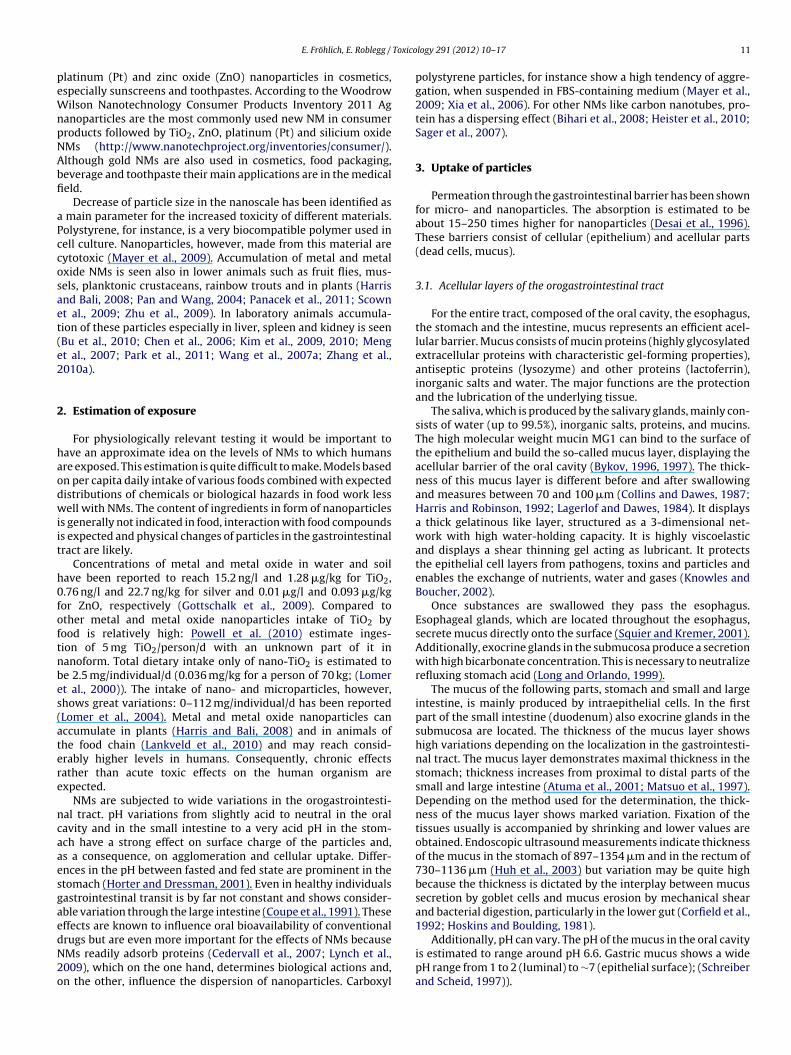
Toxico
peWnpNAbfi
aPccosaet(e2
2
haodwiit
h0foftnbes(atere
ncaaesgaedN2o
1992; Hoskins and Boulding, 1981).
E. Fröhlich, E. Roblegg /
latinum (Pt) and zinc oxide (ZnO) nanoparticles in cosmetics,specially sunscreens and toothpastes. According to the Woodrowilson Nanotechnology Consumer Products Inventory 2011 Ag
anoparticles are the most commonly used new NM in consumerroducts followed by TiO2, ZnO, platinum (Pt) and silicium oxideMs (http://www.nanotechproject.org/inventories/consumer/).lthough gold NMs are also used in cosmetics, food packaging,everage and toothpaste their main applications are in the medicaleld.
Decrease of particle size in the nanoscale has been identified as main parameter for the increased toxicity of different materials.olystyrene, for instance, is a very biocompatible polymer used inell culture. Nanoparticles, however, made from this material areytotoxic (Mayer et al., 2009). Accumulation of metal and metalxide NMs is seen also in lower animals such as fruit flies, mus-els, planktonic crustaceans, rainbow trouts and in plants (Harrisnd Bali, 2008; Pan and Wang, 2004; Panacek et al., 2011; Scownt al., 2009; Zhu et al., 2009). In laboratory animals accumula-ion of these particles especially in liver, spleen and kidney is seenBu et al., 2010; Chen et al., 2006; Kim et al., 2009, 2010; Mengt al., 2007; Park et al., 2011; Wang et al., 2007a; Zhang et al.,010a).
. Estimation of exposure
For physiologically relevant testing it would be important toave an approximate idea on the levels of NMs to which humansre exposed. This estimation is quite difficult to make. Models basedn per capita daily intake of various foods combined with expectedistributions of chemicals or biological hazards in food work lessell with NMs. The content of ingredients in form of nanoparticles
s generally not indicated in food, interaction with food compoundss expected and physical changes of particles in the gastrointestinalract are likely.
Concentrations of metal and metal oxide in water and soilave been reported to reach 15.2 ng/l and 1.28 �g/kg for TiO2,.76 ng/l and 22.7 ng/kg for silver and 0.01 �g/l and 0.093 �g/kgor ZnO, respectively (Gottschalk et al., 2009). Compared tother metal and metal oxide nanoparticles intake of TiO2 byood is relatively high: Powell et al. (2010) estimate inges-ion of 5 mg TiO2/person/d with an unknown part of it inanoform. Total dietary intake only of nano-TiO2 is estimated toe 2.5 mg/individual/d (0.036 mg/kg for a person of 70 kg; (Lomert al., 2000)). The intake of nano- and microparticles, however,hows great variations: 0–112 mg/individual/d has been reportedLomer et al., 2004). Metal and metal oxide nanoparticles canccumulate in plants (Harris and Bali, 2008) and in animals ofhe food chain (Lankveld et al., 2010) and may reach consid-rably higher levels in humans. Consequently, chronic effectsather than acute toxic effects on the human organism arexpected.
NMs are subjected to wide variations in the orogastrointesti-al tract. pH variations from slightly acid to neutral in the oralavity and in the small intestine to a very acid pH in the stom-ch have a strong effect on surface charge of the particles and,s a consequence, on agglomeration and cellular uptake. Differ-nces in the pH between fasted and fed state are prominent in thetomach (Horter and Dressman, 2001). Even in healthy individualsastrointestinal transit is by far not constant and shows consider-ble variation through the large intestine (Coupe et al., 1991). Theseffects are known to influence oral bioavailability of conventional
rugs but are even more important for the effects of NMs becauseMs readily adsorb proteins (Cedervall et al., 2007; Lynch et al.,009), which on the one hand, determines biological actions and,n the other, influence the dispersion of nanoparticles. Carboxyllogy 291 (2012) 10– 17 11
polystyrene particles, for instance show a high tendency of aggre-gation, when suspended in FBS-containing medium (Mayer et al.,2009; Xia et al., 2006). For other NMs like carbon nanotubes, pro-tein has a dispersing effect (Bihari et al., 2008; Heister et al., 2010;Sager et al., 2007).
3. Uptake of particles
Permeation through the gastrointestinal barrier has been shownfor micro- and nanoparticles. The absorption is estimated to beabout 15–250 times higher for nanoparticles (Desai et al., 1996).These barriers consist of cellular (epithelium) and acellular parts(dead cells, mucus).
3.1. Acellular layers of the orogastrointestinal tract
For the entire tract, composed of the oral cavity, the esophagus,the stomach and the intestine, mucus represents an efficient acel-lular barrier. Mucus consists of mucin proteins (highly glycosylatedextracellular proteins with characteristic gel-forming properties),antiseptic proteins (lysozyme) and other proteins (lactoferrin),inorganic salts and water. The major functions are the protectionand the lubrication of the underlying tissue.
The saliva, which is produced by the salivary glands, mainly con-sists of water (up to 99.5%), inorganic salts, proteins, and mucins.The high molecular weight mucin MG1 can bind to the surface ofthe epithelium and build the so-called mucus layer, displaying theacellular barrier of the oral cavity (Bykov, 1996, 1997). The thick-ness of this mucus layer is different before and after swallowingand measures between 70 and 100 �m (Collins and Dawes, 1987;Harris and Robinson, 1992; Lagerlof and Dawes, 1984). It displaysa thick gelatinous like layer, structured as a 3-dimensional net-work with high water-holding capacity. It is highly viscoelasticand displays a shear thinning gel acting as lubricant. It protectsthe epithelial cell layers from pathogens, toxins and particles andenables the exchange of nutrients, water and gases (Knowles andBoucher, 2002).
Once substances are swallowed they pass the esophagus.Esophageal glands, which are located throughout the esophagus,secrete mucus directly onto the surface (Squier and Kremer, 2001).Additionally, exocrine glands in the submucosa produce a secretionwith high bicarbonate concentration. This is necessary to neutralizerefluxing stomach acid (Long and Orlando, 1999).
The mucus of the following parts, stomach and small and largeintestine, is mainly produced by intraepithelial cells. In the firstpart of the small intestine (duodenum) also exocrine glands in thesubmucosa are located. The thickness of the mucus layer showshigh variations depending on the localization in the gastrointesti-nal tract. The mucus layer demonstrates maximal thickness in thestomach; thickness increases from proximal to distal parts of thesmall and large intestine (Atuma et al., 2001; Matsuo et al., 1997).Depending on the method used for the determination, the thick-ness of the mucus layer shows marked variation. Fixation of thetissues usually is accompanied by shrinking and lower values areobtained. Endoscopic ultrasound measurements indicate thicknessof the mucus in the stomach of 897–1354 �m and in the rectum of730–1136 �m (Huh et al., 2003) but variation may be quite highbecause the thickness is dictated by the interplay between mucussecretion by goblet cells and mucus erosion by mechanical shearand bacterial digestion, particularly in the lower gut (Corfield et al.,
Additionally, pH can vary. The pH of the mucus in the oral cavityis estimated to range around pH 6.6. Gastric mucus shows a widepH range from 1 to 2 (luminal) to ∼7 (epithelial surface); (Schreiberand Scheid, 1997)).

1 Toxicology 291 (2012) 10– 17
3
macp
tvpaemtemcswmb(psise1fitsostiwlllclsr
tnr
pcetIt
3
aliuvit
Fig. 1. Different thickness and heterogeneity of orogastrointestinal epithelia. Com-pared to the monostratified squamous epithelium of blood vessels (a) epithelia ofthe orogastrointestinal tract are much thicker: stratified non-keratinized squamousepithelium of the oral cavity and the esophagus (b) and simple columnar epithe-lium present in the stomach, small and large intestine. All epithelia reside on a basallamina (BM). All epithelial layers are composed of different cell types. (d) In theoral cavity, Langerhans cells (LC) and intraepithelial lymphocytes (L) in addition tokeratinocytes (KC) are present in the epithelial layer. The connective tissue belowthe epithelial layer contains dendritic cells (DC), macrophages (M) and lymphocytes(L). (e) The mucosa of the stomach consists of mucus-producing cells (MC), gastricacid producing cells (parietal cells, PC), pepsinogen-producing cells (chief cells, CC)and endocrine hormone producing enteroendocrine cells (EC). Cells of the immunesystems are not shown. (f) In the small intestine, enterocytes (EnC), microfold (M-cells), goblet cells (GC), dendritic cells (DC) and intraepithelial lymphocytes (L) arelinked together by tight junctions (TJ). These junctions show small differences in
2 E. Fröhlich, E. Roblegg /
.2. Interaction of NMs with the mucus layer
The characteristics facilitating the passage through humanucus are relatively well known: electrostatic repulsion from neg-
tively charged sugar moieties favors the penetration of positivelyharged hydrophilic molecules; the passage of lipophilic com-ounds is slow (Avdeef and Testa, 2002).
It was thought that nanoparticles are incapable to penetratehe mucus layer since recent studies demonstrated that specificiruses, like the Norwalk virus with a size of 38 nm and humanapilloma virus with a size of 55 nm diffused in human mucuss rapidly as they do in water (Olmsted et al., 2001; Saltzmant al., 1994). The surface of viruses, which are able to permeate theucus, is densely coated with positive and negative charges, thus,
his net neutral surface charge prevents mucoadhesion (Olmstedt al., 2001). Since the pore size in (cervical) mucus is approxi-ately 100 nm, it is suggested that small particles might also be
apable to diffuse through mucus. Olmsted et al. (2001) demon-trated that small viruses diffused unhindered through mucus,hereas polystyrene microspheres in a size of 59 nm and covalentlyodified with carboxyl-groups, bound more tightly to mucins and
undled them into thick cables. Additional work by Dawson et al.2003) reported that carboxyl and amine-modified polystyrenearticles (100, 200 and 500 nm) were embedded in cystic fibrosisputum. The positively charged particles penetrated more rapidlyn the sputum than the negatively charged particles. Furthermore,maller particles underwent a significantly faster transport. Lait al. (2007) investigated polystyrene particles in a size range of00–500 nm. The surfaces of the particles were covalently modi-ed with a high density of low M.W. polyethylene glycol (PEG) andhe diffusion in human mucus was studied. The results demon-trated that the neutral surface charge increased the diffusion ratef all particles. The large particles (200 nm and 500 nm) demon-trated a 6-fold and 4-fold lower effective diffusion-coefficient thanhat in water, whereas 100 nm particles were found to be immobilen mucus. Other studies showed that 14 nm latex particles, which
ere slightly negatively charged, cross the distal colon mucus gelayer within 2 min and 415 nm larges ones in 30 min, whereas 1 �marges ones did not cross (Szentkuri, 1997). Non-biodegradableatex particles can rapidly permeate human mucus when they areoated with PEG. Surprisingly, 200 nm particles crossed the mucinayer faster than <100 nm NMs (Wang et al., 2007b). These findingsuggest that the surface charge plays a crucial role in the transportates of nanoparticles through a mucus layer.
Mucus lifetime is short and the fastest turnover (i.e., clearanceime) is observed at surfaces with thinnest mucus layers. Thus,anoparticles have to permeate quickly through this barrier toeach the underlying epithelia (Cone, 2009).
Local effects after oral exposure to NMs include abnormal mucusroduction, induced by TiO2 nanoparticles in cultured ChaGo-K1ells (Chen et al., 2011) and by silver nanoparticles in vivo (Jeongt al., 2010). Additionally, pH changes induced by NMs can changehe pH-dependent aggregation of mucins (Bhaskar et al., 1991).n addition, positively charged NMs impede mucin swelling andhereby increase viscosity (Chen et al., 2010).
.3. Epithelial layers of the orogastrointestinal tract
The epithelium generally represents the highest resistancegainst the passage of chemical compounds and NMs. Epithe-ial cells are polarized, they possess an apical surface facing annternal or external surface and a basal site, where they face the
nderlying tissue. Epithelia may consist of several layers and mayary in the height of the cells. Penetration through a monostrat-fied squamous epithelium, like in endothelia (Fig. 1a), is easierhan through the simple columnar epithelium in stomach andcomposition and location in the cell.
Figure adapted from Eom and Choi (2009).
intestine (Fig. 1b) and the squamous epithelium of the oral cavityand the esophagus (Fig. 1c). The thickness of the non-keratinizedsquamous epithelium in the oral cavity ranges between 550 and800 �m (Collins and Dawes, 1987; Harris and Robinson, 1992;Lagerlof and Dawes, 1984). The squamous epithelium of theesophagus shows a thickness of 300–500 �m (Takubo, 2009). Theepithelium of the esophagus has the same structure as that of thebuccal mucosa but is thinner and less variable (Diaz del Consuelo
et al., 2005). The simple columnar epithelium in the gastrointestinaltract measures 20–25 �m (Atuma et al., 2001; Matsuo et al., 1997).In general, only one cell type forms the structural basis of the bar-rier: keratinocytes for the oral cavity and the esophagus, gastric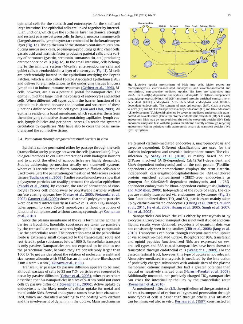
Toxicology 291 (2012) 10– 17 13
ella(ldgeeigaPalcecejetscb
3
(iaSwutp(rs2wtje
bburrit1s3
aodcemia
Fig. 2. Active uptake mechanisms of NMs into cells. Major routes aremacropinocytosis, clathrin-mediated endocytosis and caveolae-mediated andnon-clathrin, non-caveolae mediated uptake. The later are subdivided intoRhoA- (or IL-2R�-) dependent endocytosis, Cdc42/Arf1 or clathrin-independentcargo/glycophosphatidylinositol (GPI)-anchored protein enriched compartment-dependent (GEEC) endocytosis, Arf6- dependent endocytosis and flotillin-dependent endocytosis. The content of macropinosomes (MP), clathrin-coatedvesicles (CC) and GEEC is transported via early endosomes (EE) and late endosomes(LE) to lysosomes (L). Material taken up by caveolae-mediated endocytosis is trans-ported via caveolosomes (Cav) either to the endoplasmic reticulum (ER) or to earlyendosomes. NMs may be removed from the cells by exocytotic vesicles (EV). Earlyendosomes may also fuse with the plasma membrane directly or through recycling
E. Fröhlich, E. Roblegg /
pithelial cells for the stomach and enterocytes for the small andarge intestine. The epithelial cells are linked together by intercel-ular junctions, which give the epithelial layer mechanical strengthnd restrict passage between cells. In the oral mucosa immune cellsLangerhans cells, lymphocytes) are embedded in the keratinocytesayer (Fig. 1d). The epithelium of the stomach contains mucus pro-ucing mucus neck cells, pepsinogen-producing gastric chief cells,astric acid and intrinsic factor producing parietal cells and a vari-ty of hormones (gastrin, serotonin, somatostatin, etc.) producingnteroendocrine cells (Fig. 1e). In the small intestine, cells belong-ng to the immune system (M-cells), enteroendocrine cells andoblet cells are embedded in a layer of enterocytes (Fig. 1f). M-cellsre preferentially located in the epithelium overlying the Peyer’satches, which is also called Follicle Associated Epithelium (FAE),nd deliver foreign substances to the underlying tissues (mucosaymphoid) to induce immune responses (Gerbert et al., 1996). M-ells, however, are also a potential portal for nanoparticles. Thepithelium of the large intestine consists of enterocytes and gobletells. When different cell types adjoin the barrier function of thepithelium is altered because the location and structure of theseunctions differ between the cell types (Eom and Choi, 2009). Allpithelia reside on a basal membrane, which separates them fromhe underlying connective tissue containing capillaries, lymph ves-els, lymph follicles and peripheral nerves. To reach the systemicirculation by capillaries NMs have also to cross the basal mem-rane and the connective tissue.
.4. Permeation through orogastrointestinal barriers in vitro
Epithelia can be permeated either by passage through the cellstranscellular) or by passage between the cells (paracellular). Phys-ological methods to evaluate interactions with biological barriersnd to predict the effect of nanoparticles are highly demanded.tudies addressing permeation usually use transwell systems,here cells are cultured on filters. Moreover, diffusion-cells can besed to evaluate the penetration/permeation of NMs across excisedissues (Sudhakar et al., 2006). Studies on cell monolayers show thatolystyrene particles can readily permeate the alveolar epitheliumYacobi et al., 2008). By contrast, the rate of permeation of ente-ocyte (Caco-2 cell) monolayers by polystyrene particles withouturface coating appears low (Geiser et al., 2005; Pietzonka et al.,002). Gaumet et al. (2009) showed that small polystyrene particlesere observed intracellularly in Caco-2 cells. Also TiO2 nanopar-
icles appear to cross Caco-2 monolayers without disruption ofunctional complexes and without causing cytotoxicity (Koenemant al., 2010).
Since the plasma membrane of the cells forming the epithelialarrier is lipophilic, lipophilic substances are taken up passivelyy the transcellular route whereas hydrophilic drug compoundsse the paracellular route. The penetration area of the paracellularoute is extremely small compared to the transcellular route andestricted to polar substances below 1000 D. Paracellular transports only passive. Nanoparticles are not expected to be able to usehe paracellular route, because they are considerably larger than000 D. To get an idea about the relation of molecular weight andize: serum albumin with 66 kD has an almost sphere-like shape of
nm × 8 nm × 8 nm (Takizawa et al., 1992).Transcellular passage by passive diffusion appears to be rare:
lthough passage of cells by 22 nm TiO2 particles was suggested toccur by passive diffusion (Geiser et al., 2005), other researchersescribed that Au-nanoparticles in sizes of 5–8 nm could not enterells by passive diffusion ((Stoeger et al., 2006)). Active uptake by
ndocytosis is the likely mode of cellular uptake for metal andetal oxide NMs. Several endocytotic routes have been character-zed, which are classified according to the coating with clathrinnd the involvement of dynamin in the uptake. Main mechanisms
endosomes (RE). In polarized cells transcytosis occurs via transport vesicles (TV).Cyto: cytoplasm.
are termed clathrin-mediated endocytosis, macropinocytosis andcaveolae-dependent. Different classifications are used for theclathrin-independent and caveolae-independent routes. The clas-sification by Sahay et al. (2010) is mainly based on theGTPases involved (Arf6-dependent, Cdc42/Arf1-dependent andRhoA-dependent endocytosis) and on the coat protein (Flotillin-dependent). Another nomenclature employs the term clathrin-independent carriers/glycophosphatidylinositol (GPI)-anchoredprotein enriched compartment (GEEC)-type endocytosis assynonym for Cdc42/Arf1-dependent endocytosis and IL-2R�-dependent endocytosis for RhoA-dependent endocytosis (Dohertyand McMahon, 2009). Independent of the route of entry, the car-gos are mainly transported via endosomes to lysosomes (Fig. 2).Non-functionalized silver, TiO2 and SiO2 particles are mainly takenup by clathrin-mediated endocytosis (Chung et al., 2007; Greulichet al., 2011; He et al., 2009; Huang et al., 2005; Singh et al., 2007;Sun et al., 2008).
Nanoparticles can leave the cells either by transcytosis or byexocytosis. Exocytosis of nanoparticles is not well studied and con-flicting results were obtained: exocytosis of quantum dots wasnot consistently seen in the studies (Clift et al., 2008; Jiang et al.,2010). Transcytosis can occur through receptor-mediated uptakeor via adsorptive-mediated uptake. Receptors for BSA, transferrinand opioid peptides functionalized NMs are expressed on sev-eral cell types and BSA-coated nanoparticles have been shown totranscytose through endothelial cells (Wang et al., 2009). For thegastrointestinal tract, however, this type of uptake is not relevant.Absorptive-mediated transcytosis is mediated by the interactionof positively charged substances with anionic sites of the plasmamembrane: cationic nanoparticles had a greater potential thanneutral or negatively charged ones (Harush-Frenkel et al., 2008).Additionally uncoated, not positively charged TiO2 nanoparticlescan cross the intestinal epithelium by the transcellular route(Koeneman et al., 2010).
As mentioned in Section 3.3, the epithelium of the gastrointesti-
nal tract is composed of various cell types and permeation throughsome types of cells is easier than through others. This situationcan be mimicked also in vitro. Kerneis et al. (1997) constructed an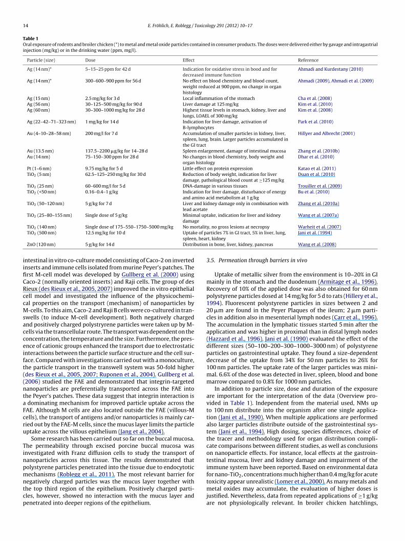
14 E. Fröhlich, E. Roblegg / Toxicology 291 (2012) 10– 17
Table 1Oral exposure of rodents and broiler chicken (*) to metal and metal oxide particles contained in consumer products. The doses were delivered either by gavage and intragastrialinjection (mg/kg) or in the drinking water (ppm, mg/l).
Particle (size) Dose Effect Reference
Ag (14 nm)* 5–15–25 ppm for 42 d Indication for oxidative stress in bood and fordecreased immune function
Ahmadi and Kurdestany (2010)
Ag (14 nm)* 300–600–900 ppm for 56 d No effect on blood chemistry and blood count,weight reduced at 900 ppm, no change in organhistology
Ahmadi (2009), Ahmadi et al. (2009)
Ag (15 nm) 2.5 mg/kg for 3 d Local inflammation of the stomach Cha et al. (2008)Ag (56 nm) 30–125–500 mg/kg for 90 d Liver damage at 125 mg/kg Kim et al. (2010)Ag (60 nm) 30–300–1000 mg/kg for 28 d Highest tissue levels in stomach, kidney, liver and
lungs, LOAEL of 300 mg/kgKim et al. (2008)
Ag (22–42–71–323 nm) 1 mg/kg for 14 d Indication for liver damage, activation ofB-lymphocytes
Park et al. (2010)
Au (4–10–28–58 nm) 200 mg/l for 7 d Accumulation of smaller particles in kidney, liver,spleen, lung, brain. Larger particles accumulated inthe GI tract
Hillyer and Albrecht (2001)
Au (13.5 nm) 137.5–2200 �g/kg for 14–28 d Spleen enlargement, damage of intestinal mucosa Zhang et al. (2010b)Au (14 nm) 75–150–300 ppm for 28 d No changes in blood chemistry, body weight and
organ histologyDhar et al. (2010)
Pt (1–6 nm) 9.75 mg/kg for 5 d Little effect on protein expression Katao et al. (2011)TiO2 (5 nm) 62.5–125–250 mg/kg for 30 d Reduction of body weight, indication for liver
damage, pathological blood count at ≥125 mg/kgDuan et al. (2010)
TiO2 (25 nm) 60–600 mg/l for 5 d DNA-damage in various tissues Trouiller et al. (2009)TiO2 (<50 nm) 0.16–0.4–1 g/kg Indication for liver damage, disturbance of energy
and amino acid metabolism at 1 g/kgBu et al. (2010)
TiO2 (50–120 nm) 5 g/kg for 7 d Liver and kidney damage only in combination withlead acetate
Zhang et al. (2010a)
TiO2 (25–80–155 nm) Single dose of 5 g/kg Minimal uptake, indication for liver and kidneydamage
Wang et al. (2007a)
TiO2 (140 nm) Single dose of 175–550–1750–5000 mg/kg No mortality, no gross lesions at necropsy Warheit et al. (2007)e of p, hear
bution
iifiCRccMsacceift((ntaFcru
Tinpmntcp
TiO2 (500 nm) 12.5 mg/kg for 10 d Uptakspleen
ZnO (120 nm) 5 g/kg for 14 d Distri
ntestinal in vitro co-culture model consisting of Caco-2 on invertednserts and immune cells isolated from murine Peyer’s patches. Therst M-cell model was developed by Gullberg et al. (2000) usingaco-2 (normally oriented inserts) and Raji cells. The group of desieux (des Rieux et al., 2005, 2007) improved the in vitro epithelialell model and investigated the influence of the physicochemi-al properties on the transport (mechanism) of nanoparticles by-cells. To this aim, Caco-2 and Raji B cells were co-cultured in tran-
wells (to induce M-cell development). Both negatively chargednd positively charged polystryrene particles were taken up by M-ells via the transcellular route. The transport was dependent on theoncentration, the temperature and the size. Furthermore, the pres-nce of cationic groups enhanced the transport due to electrostaticnteractions between the particle surface structure and the cell sur-ace. Compared with investigations carried out with a monoculture,he particle transport in the transwell system was 50-fold higherdes Rieux et al., 2005, 2007; Ruponen et al., 2004). Gullberg et al.2006) studied the FAE and demonstrated that integrin-targetedanoparticles are preferentially transported across the FAE intohe Peyer’s patches. These data suggest that integrin interaction is
dominating mechanism for improved particle uptake across theAE. Although M cells are also located outside the FAE (villous-Mells), the transport of antigens and/or nanoparticles is mainly car-ied out by the FAE-M cells, since the mucus layer limits the particleptake across the villous epithelium (Jang et al., 2004).
Some research has been carried out so far on the buccal mucosa.he permeability through excised porcine buccal mucosa wasnvestigated with Franz diffusion cells to study the transport ofanoparticles across this tissue. The results demonstrated thatolystyrene particles penetrated into the tissue due to endocytoticechanisms (Roblegg et al., 2011). The most relevant barrier for
egatively charged particles was the mucus layer together withhe top third region of the epithelium. Positively charged parti-les, however, showed no interaction with the mucus layer andenetrated into deeper regions of the epithelium.
articles 7% in GI tract, 5% in liver, lung,t, kidney
Jani et al. (1994)
in bone, liver, kidney, pancreas Wang et al. (2008)
3.5. Permeation through barriers in vivo
Uptake of metallic silver from the environment is 10–20% in GImainly in the stomach and the duodenum (Armitage et al., 1996).Recovery of 10% of the applied dose was also obtained for 60 nmpolystyrene particles dosed at 14 mg/kg for 5 d to rats (Hillery et al.,1994). Fluorescent polystyrene particles in sizes between 2 and20 �m are found in the Peyer Plaques of the ileum; 2 �m parti-cles in addition also in mesenterial lymph nodes (Carr et al., 1996).The accumulation in the lymphatic tissues started 5 min after theapplication and was higher in proximal than in distal lymph nodes(Hazzard et al., 1996). Jani et al. (1990) evaluated the effect of thedifferent sizes (50–100–200–300–1000–3000 nm) of polystyreneparticles on gastrointestinal uptake. They found a size-dependentdecrease of the uptake from 34% for 50 nm particles to 26% for100 nm particles. The uptake rate of the larger particles was mini-mal. 6.6% of the dose was detected in liver, spleen, blood and bonemarrow compared to 0.8% for 1000 nm particles.
In addition to particle size, dose and duration of the exposureare important for the interpretation of the data (Overview pro-vided in Table 1). Independent from the material used, NMs upto 100 nm distribute into the organism after one single applica-tion (Jani et al., 1990). When multiple applications are performedalso larger particles distribute outside of the gastrointestinal sys-tem (Jani et al., 1994). High dosing, species differences, choice ofthe tracer and methodology used for organ distribution compli-cate comparisons between different studies, as well as conclusionson nanoparticle effects. For instance, local effects at the gastroin-testinal mucosa, liver and kidney damage and impairment of theimmune system have been reported. Based on environmental datafor nano-TiO2, concentrations much higher than 0.4 mg/kg for acute
toxicity appear unrealistic (Lomer et al., 2000). As many metals andmetal oxides may accumulate, the evaluation of higher doses isjustified. Nevertheless, data from repeated applications of ≥1 g/kgare not physiologically relevant. In broiler chicken hatchlings,
Toxico
wc2tbFaetaa
4
tbdsawALtifNtpeeproo
5
NitnLtai
Af(b
atnarmfn
tap
E. Fröhlich, E. Roblegg /
hich were treated with doses below 250 ng/kg silver nanoparti-les (Ahmadi and Kurdestany, 2010; Ahmadi, 2009; Ahmadi et al.,009), adverse effects were already detected at these low concen-rations. The higher toxicity of the silver nanoparticles may eithere due to interspecies differences or to the low age of the chicken.or correct tracing of the organ distribution the choice of the labelnd the mode of detection appear important. In the study of Janit al. (1990) the label was potentially not stable and the localiza-ion of the label may not correspond to that of the particles. If NMsre only detected by chemical analysis it is not clear if they areccumulated in a dissolved form or as intact particles.
. Action on compromised barriers
Few data have been published regarding the permeationhrough diseased barriers. Changes in mucus composition inducedy Ag nanoparticles (Jeong et al., 2010), polystyrene particles andiesel exhaust increased mucus permeability and permeation ofmall molecules by a factor of 5 (McGill and Smyth, 2010). Thedherence of polystyrene nanoparticles to inflamed colonic mucosaas much higher than to normal mucosa (Lamprecht et al., 2001).lso in the elaborated co-culture in vitro model developed byeonhard et al. (2010) smaller particles (50 nm) polystyrene par-icles adhered better to the inflamed monolayer and were taken upnto the cells, whereas larger particles only adhered to the cell sur-ace. Inflammation appears to increase uptake and permeation ofMs in vitro and in vivo. Inflammation caused by Yersinia pseudo-
uberculosis increases the uptake of 100 nm carboxyl polystyrenearticles in cell monolayers and in intestinal biopsies (Ragnarssont al., 2008). In contrast to that, in the in vitro study by Leonhardt al. (2010) no influence on the translocation of the polystyrenearticles was observed. Since in the in vitro studies lipopolysaccha-ide and not intact bacteria were used, effects by the living bacterian cells, mucus production and/or viscosity may account for thebserved differences.
. Conclusion
The assessment of penetration and biological effects of ingestedMs presents many problems because it is very complex. Inter-
ndividual differences in the composition, pH and thickness ofhe mucus layer, in the gastrointestinal flora and in gastrointesti-al passage time complicate in vivo experiments. In the study ofoeschner et al. (2011) on organ distribution of 60 nm Ag nanopar-icles great inter-individual variations were noticed although allnimals were fed the same diet. Also differences in the diet aremportant.
For in vivo testing, rodents also may not be ideal models.lthough men and rodents are omnivorous, function (e.g., region
or absorption of food) and morphology of the gastrointestinal tracte.g., absence of gall bladder in rats) show considerable differencesetween rodents and humans (Kararli, 1995).
Apart from permeating themselves, NMs may have perme-tion enhancing properties for other substances. This phenomenonermed as ‘Trojan horse’ effect, was first identified for metalanoparticles. Whereas plasma membranes restrict the cellularccess for metal ions like silver cations, silver nanoparticles wereeadily internalized and intracellular silver concentrations wereuch higher than for silver ions (Navarro et al., 2008). Studies
or uptake and toxicity should, therefore, include AgNO3 for silveranoparticles (Trojan horse effect) or bulk material.
Other important effects are linked to the tendency of NMso absorb macromolecules. By adsorption of organic compoundslso unintended molecules (undigested and unmetabolized com-ounds) may be absorbed by the gastrointestinal tract. On the other
logy 291 (2012) 10– 17 15
hand adsorption to NMs may also prevent the uptake of necessarymolecules (Alkhamis et al., 2009). Absorption may also be alteredby a changed metabolization by enterocytes. Polystyrene and sil-ver particles have been shown to inhibit the activity of cytochromeP450 enzymes (Fröhlich et al., 2010; Lamb et al., 2010).
To obtain more information about penetration of the orogas-trointestinal barriers and subsequent biological effects physiolog-ically relevant in vitro models should be used, which enable thecontrolled variation of the most important parameters involved.Particle properties should be recorded in mucus of different pH andthe extent of binding to proteins and other macromolecules shouldbe studied. Physiologically relevant in vitro (co-culture) modelsincluding mucus should be established to investigate also the effectof changed mucus structure, inflammation and pH changes. It isobvious that also in vivo experiments are needed but without agood knowledge on the influence of the orogastrointestinal varia-tions on particle parameters and penetration in vivo data may bedifficult to interpret.
Conflict of interest statement
The authors declare that there are no conflicting interests.
Acknowledgements
The authors acknowledge funding by the Austrian Science Fund(FWF) grant P22576-B18 and by the Austrian Research PromotionAgency (FFG) project 826136.
References
Ahmadi, F., Kurdestany, A., 2010. The impact of silver nano particles on growthperformance. Global Veterinaria 5, 366–370.
Ahmadi, J., 2009. Application of different levels of silver nanoparticles in food on theperformance and some blood parameters of broiler chickens. World Appl. Sci. J.7, 24–27.
Ahmadi, J., Irani, M., Choobchian, M., 2009. Pathological study of intestine and liverin broiler chickens after treatment with different levels of silver nanoparticles.World Appl. Sci. J. 7, 28–32.
Alkhamis, K., Salem, M., Khanfar, M., 2009. Determination of the mechanism ofuptake of organic vapors by chitosan. Pharm. Dev. Technol. 14, 90–95.
Armitage, S.A., White, M.A., Wilson, H.K., 1996. The determination of silver in wholeblood and its application to biological monitoring of occupationally exposedgroups. Ann. Occup. Hyg. 40, 331–338.
Atuma, C., Strugala, V., Allen, A., Holm, L., 2001. The adherent gastrointestinal mucusgel layer: thickness and physical state in vivo. Am. J. Physiol. Gastrointest. LiverPhysiol. 280, G922–G929.
Avdeef, A., Testa, B., 2002. Physicochemical profiling in drug research: a brief sur-vey of the state-of-the-art of experimental techniques. Cell. Mol. Life Sci. 59,1681–1689.
Bhaskar, K.R., Gong, D.H., Bansil, R., Pajevic, S., Hamilton, J.A., Turner, B.S., LaMont,J.T., 1991. Profound increase in viscosity and aggregation of pig gastric mucin atlow pH. Am. J. Physiol. 261, G827–G832.
Bihari, P., Vippola, M., Schultes, S., Praetner, M., Khandoga, A.G., Reichel, C.A., Coester,C., Tuomi, T., Rehberg, M., Krombach, F., 2008. Optimized dispersion of nanopar-ticles for biological in vitro and in vivo studies. Part. Fibre Toxicol. 5, 14.
Bu, Q., Yan, G., Deng, P., Peng, F., Lin, H., Xu, Y., Cao, Z., Zhou, T., Xue, A., Wang, Y., Cen,X., Zhao, Y.L., 2010. NMR-based metabonomic study of the sub-acute toxicity oftitanium dioxide nanoparticles in rats after oral administration. Nanotechnology21, 125105.
Bykov, V.L., 1996. The tissue and cell defense mechanisms of the oral mucosa. Mor-fologiia 110, 14–24.
Bykov, V.L., 1997. The functional morphology of the epithelial barrier of the oralmucosa. Stomatologiia (Mosk) 76, 12–17.
Carr, K.E., Hazzard, R.A., Reid, S., Hodges, G.M., 1996. The effect of size on uptake oforally administered latex microparticles in the small intestine and transport tomesenteric lymph nodes. Pharm. Res. 13, 1205–1209.
Cedervall, T., Lynch, I., Foy, M., Berggard, T., Donnelly, S.C., Cagney, G., Linse, S.,Dawson, K.A., 2007. Detailed identification of plasma proteins adsorbed oncopolymer nanoparticles. Angew. Chem. Int. Ed. Engl. 46, 5754–5756.
Cha, K., Hong, H.W., Choi, Y.G., Lee, M.J., Park, J.H., Chae, H.K., Ryu, G., Myung, H.,2008. Comparison of acute responses of mice livers to short-term exposure tonano-sized or micro-sized silver particles. Biotechnol. Lett. 30, 1893–1899.
Chen, E.Y., Garnica, M., Wang, Y.C., Chen, C.S., Chin, W.C., 2011. Mucin secretioninduced by titanium dioxide nanoparticles. PLoS One 6, e16198.

1 Toxico
C
C
C
C
C
CC
C
D
d
d
D
D
D
D
D
E
F
G
G
G
G
G
G
G
H
H
H
H
H
6 E. Fröhlich, E. Roblegg /
hen, E.Y., Wang, Y.C., Chen, C.S., Chin, W.C., 2010. Functionalized positive nanopar-ticles reduce mucin swelling and dispersion. PLoS One 5, e15434.
hen, Z., Meng, H., Xing, G., Chen, C., Zhao, Y., Jia, G., Wang, T., Yuan, H., Ye, C., Zhao,F., Chai, Z., Zhu, C., Fang, X., Ma, B., Wan, L., 2006. Acute toxicological effects ofcopper nanoparticles in vivo. Toxicol. Lett. 163, 109–120.
hung, T.H., Wu, S.H., Yao, M., Lu, C.W., Lin, Y.S., Hung, Y., Mou, C.Y., Chen, Y.C., Huang,D.M., 2007. The effect of surface charge on the uptake and biological function ofmesoporous silica nanoparticles in 3T3-L1 cells and human mesenchymal stemcells. Biomaterials 28, 2959–2966.
lift, M.J., Rothen-Rutishauser, B., Brown, D.M., Duffin, R., Donaldson, K., Proudfoot,L., Guy, K., Stone, V., 2008. The impact of different nanoparticle surface chemistryand size on uptake and toxicity in a murine macrophage cell line. Toxicol. Appl.Pharmacol. 232, 418–427.
ollins, L.M., Dawes, C., 1987. The surface area of the adult human mouth and thick-ness of the salivary film covering the teeth and oral mucosa. J. Dent. Res. 66,1300–1302.
one, R.A., 2009. Barrier properties of mucus. Adv. Drug Deliv. Rev. 61, 75–85.orfield, A.P., Wagner, S.A., Clamp, J.R., Kriaris, M.S., Hoskins, L.C., 1992. Mucin degra-
dation in the human colon: production of sialidase, sialate O-acetylesterase,N-acetylneuraminate lyase, arylesterase, and glycosulfatase activities by strainsof fecal bacteria. Infect. Immun. 60, 3971–3978.
oupe, A.J., Davis, S.S., Wilding, I.R., 1991. Variation in gastrointestinal transit ofpharmaceutical dosage forms in healthy subjects. Pharm. Res. 8, 360–364.
awson, M., Wirtz, D., Hanes, J., 2003. Enhanced viscoelasticity of human cysticfibrotic sputum correlates with increasing microheterogeneity in particle trans-port. J. Biol. Chem. 278, 50393–50401.
es Rieux, A., Fievez, V., Theate, I., Mast, J., Preat, V., Schneider, Y.J., 2007. Animproved in vitro model of human intestinal follicle-associated epithelium tostudy nanoparticle transport by M cells. Eur. J. Pharm. Sci. 30, 380–391.
es Rieux, A., Ragnarsson, E.G., Gullberg, E., Preat, V., Schneider, Y.J., Artursson, P.,2005. Transport of nanoparticles across an in vitro model of the human intestinalfollicle associated epithelium. Eur. J. Pharm. Sci. 25, 455–465.
esai, M.P., Labhasetwar, V., Amidon, G.L., Levy, R.J., 1996. Gastrointestinaluptake of biodegradable microparticles: effect of particle size. Pharm. Res. 13,1838–1845.
har, S., Mali, V., Bodhankar, S., Shiras, A., Prasad, B.L., Pokharkar, V., 2010. Biocom-patible gellan gum-reduced gold nanoparticles: cellular uptake and subacuteoral toxicity studies. J. Appl. Toxicol. 1, 9, doi:10.1002/jat.1595 [Epub ahead ofprint].
iaz del Consuelo, I., Falson, F., Guy, R.H., Jacques, Y., 2005. Transport of fentanylthrough pig buccal and esophageal epithelia in vitro. Influence of concentrationand vehicle pH. Pharm. Res. 22, 1525–1529.
oherty, G.J., McMahon, H.T., 2009. Mechanisms of endocytosis. Ann. Rev. Biochem.78, 857–902.
uan, Y., Li, N., Liu, C., Liu, H., Cui, Y., Wang, H., Hong, F., 2010. Interaction betweennanoparticulate anatase TiO2 and lactate dehydrogenase. Biol. Trace Elem. Res.136, 302–313.
om, H.J., Choi, J., 2009. Oxidative stress of silica nanoparticles in human bronchialepithelial cell, Beas-2B. Toxicol. In Vitro 23, 1326–1332.
röhlich, E., Kueznik, T., Samberger, C., Roblegg, E., Wrighton, C., Pieber, T.R., 2010.Size-dependent effects of nanoparticles on the activity of cytochrome P450isoenzymes. Toxicol. Appl. Pharmacol. 242, 326–332.
aumet, M., Gurny, R., Delie, F., 2009. Localization and quantification of biodegrad-able particles in an intestinal cell model: the influence of particle size. Eur. J.Pharm. Sci. 36, 465–473.
eiser, M., Rothen-Rutishauser, B., Kapp, N., Schurch, S., Kreyling, W., Schulz, H.,Semmler, M., Im Hof, V., Heyder, J., Gehr, P., 2005. Ultrafine particles cross cel-lular membranes by nonphagocytic mechanisms in lungs and in cultured cells.Environ. Health Perspect. 113, 1555–1560.
erbert, A., Rothkotter, H.J., Pabst, R., 1996. M-cells in peyer’s patches of the intestine.Int. Rev. Cytol. 167, 91–159.
ottschalk, F., Sonderer, T., Scholz, R.W., Nowack, B., 2009. Modeled environmentalconcentrations of engineered nanomaterials (TiO(2), ZnO, Ag, CNT, Fullerenes)for different regions. Environ. Sci. Technol. 43, 9216–9222.
reulich, C., Diendorf, J., Simon, T., Eggeler, G., Epple, M., Koller, M., 2011. Uptake andintracellular distribution of silver nanoparticles in human mesenchymal stemcells. Acta Biomater. 7, 347–354.
ullberg, E., Keita, A.V., Salim, S.Y., Andersson, M., Caldwell, K.D., Söderholm, J.D.,Artusson, P., 2006. Identification of cell adhesion molecules in the humanfollicle-associated epithelium that improve nanoparticle uptake into the peyer’spatches. J. Pharmacol. Exp. Ther. 319, 632–639.
ullberg, E., Leonard, M., Karlsson, J., Hopkins, A., Baryden, D., Baird, W., Artursson,P., 2000. Expression of specific markers and particles transport in a new humanintestinal M cell model. Biochem. Biophys. Res. Commun. 279, 803–813.
ansen, S.F., Michelson, E.S., Kamper, A., Borling, P., Stuer-Lauridsen, F., Baun, A.,2008. Categorization framework to aid exposure assessment of nanomaterialsin consumer products. Ecotoxicology 17, 438–447.
arris, A., Bali, R., 2008. On the formation and extent of uptake of silver nanoparticlesby live plants. J. Nanopart. Res. 10, 691–695.
arris, D., Robinson, J.R., 1992. Drug delivery via the mucous membranes of the oralcavity. J. Pharm. Sci. 81, 1–10.
arush-Frenkel, O., Rozentur, E., Benita, S., Altschuler, Y., 2008. Surface charge ofnanoparticles determines their endocytic and transcytotic pathway in polarizedMDCK cells. Biomacromolecules 9, 435–443.
azzard, R.A., Hodges, G.M., Scott, J.D., McGuinness, C.B., Carr, K.E., 1996. Earlyintestinal microparticle uptake in the rat. J. Anat. 189 (Pt 2), 265–271.
logy 291 (2012) 10– 17
He, Q., Zhang, Z., Gao, Y., Shi, J., Li, Y., 2009. Intracellular localization and cyto-toxicity of spherical mesoporous silica nano- and microparticles. Small 5,2722–2729.
Heister, E., Lamprecht, C., Neves, V., Tilmaciu, C., Datas, L., Flahaut, E., Soula, B., Hinter-dorfer, P., Coley, H.M., Silva, S.R., McFadden, J., 2010. Higher dispersion efficacyof functionalized carbon nanotubes in chemical and biological environments.ACS Nano 4, 2615–2626.
Hillery, A.M., Jani, P.U., Florence, A.T., 1994. Comparative, quantitative study of lym-phoid and non-lymphoid uptake of 60 nm polystyrene particles. J. Drug Target.2, 151–156.
Hillyer, J.F., Albrecht, R.M., 2001. Gastrointestinal persorption and tissue distributionof differently sized colloidal gold nanoparticles. J. Pharm. Sci. 90, 1927–1936.
Horter, D., Dressman, J.B., 2001. Influence of physicochemical properties on disso-lution of drugs in the gastrointestinal tract. Adv. Drug Deliv. Rev. 46, 75–87.
Hoskins, L.C., Boulding, E.T., 1981. Mucin degradation in human colon ecosystems.Evidence for the existence and role of bacterial subpopulations producing gly-cosidases as extracellular enzymes. J. Clin. Invest. 67, 163–172.
Huang, D.M., Hung, Y., Ko, B.S., Hsu, S.C., Chen, W.H., Chien, C.L., Tsai, C.P., Kuo, C.T.,Kang, J.C., Yang, C.S., Mou, C.Y., Chen, Y.C., 2005. Highly efficient cellular labelingof mesoporous nanoparticles in human mesenchymal stem cells: implicationfor stem cell tracking. FASEB J. 19, 2014–2016.
Huh, C.H., Bhutani, M.S., Farfan, E.B., Bolch, W.E., 2003. Individual variations inmucosa and total wall thickness in the stomach and rectum assessed via endo-scopic ultrasound. Physiol. Meas. 24, N15–N22.
Jang, M.H., Kweon, M.N., Iwatani, K., Yamamoto, M., Terahara, C., Sasakawa, C.,Suzuki, T., Nochi, T., Yokota, Y., Rennert, P.D., Hiroi, T., Tamagawa, H., Irijima,H., Kunisawa, J., Yuki, Y., Kiyono, H., 2004. Intestinale villous M cells: an antigenentry site in the mucosal epithelium. Proc. Natl. Acad. Sci. U.S.A. 101, 6110–6115.
Jani, P., Halbert, G.W., Langridge, J., Florence, A.T., 1990. Nanoparticle uptake by therat gastrointestinal mucosa: quantitation and particle size dependency. J. Pharm.Pharmacol. 42, 821–826.
Jani, P., McCarthy, D., Florence, A., 1994. Titanium dioxide (rutile) particles uptakefrom the rat GI tract and translocation to the systemic organs after oral admin-istration. Int. J. Pharm. 105, 157–168.
Jeong, G.N., Jo, U.B., Ryu, H.Y., Kim, Y.S., Song, K.S., Yu, I.J., 2010. Histochemical studyof intestinal mucins after administration of silver nanoparticles in Sprague-Dawley rats. Arch. Toxicol. 84, 63–69.
Jiang, X., Rocker, C., Hafner, M., Brandholt, S., Dorlich, R.M., Nienhaus, G.U., 2010.Endo- and exocytosis of zwitterionic quantum dot nanoparticles by live HeLacells. ACS Nano 4, 6787–6797.
Kararli, T.T., 1995. Comparison of the gastrointestinal anatomy, physiology, and bio-chemistry of humans and commonly used laboratory animals. Biopharm. DrugDispos. 16, 351–380.
Katao, K., Honma, R., Kato, S., Watanabe, S., Imai, J., 2011. Influence of platinumnanoparticles orally administered to rats evaluated by systemic gene expressionprofiling. Exp. Anim. 60, 33–45.
Kerneis, S., Bogdonona, A., Kraehenbuhl, J.P., Pringault, E., 1997. Conversion byPeyer’s patches lymphocytes of human enterocytes into M cells that transportbacteria. Science 277, 949–952.
Kim, W.Y., Kim, J., Park, J.D., Ryu, H.Y., Yu, I.J., 2009. Histological study of genderdifferences in accumulation of silver nanoparticles in kidneys of Fischer 344rats. J. Toxicol. Environ. Health 72, 1279–1284.
Kim, Y.S., Kim, J.S., Cho, H.S., Rha, D.S., Kim, J.M., Park, J.D., Choi, B.S., Lim, R., Chang,H.K., Chung, Y.H., Kwon, I.H., Jeong, J., Han, B.S., Yu, I.J., 2008. Twenty-eight-day oral toxicity, genotoxicity, and gender-related tissue distribution of silvernanoparticles in Sprague-Dawley rats. Inhal. Toxicol. 20, 575–583.
Kim, Y.S., Song, M.Y., Park, J.D., Song, K.S., Ryu, H.R., Chung, Y.H., Chang, H.K., Lee,J.H., Oh, K.H., Kelman, B.J., Hwang, I.K., Yu, I.J., 2010. Subchronic oral toxicity ofsilver nanoparticles. Part. Fibre Toxicol. 7, 20.
Knowles, M.R., Boucher, R.C., 2002. Mucus clearance as a primary innate defensemechanism for mammalian airways. J. Clin. Invest. 109, 571–577.
Koeneman, B.A., Zhang, Y., Westerhoff, P., Chen, Y., Crittenden, J.C., Capco, D.G., 2010.Toxicity and cellular responses of intestinal cells exposed to titanium dioxide.Cell Biol. Toxicol. 26, 225–238.
Lagerlof, F., Dawes, C., 1984. The volume of saliva in the mouth before and afterswallowing. J. Dent. Res. 63, 618–621.
Lai, S.K.a., O’Hanlon, D.E.b., Harrold, S.b., Man, S.T.a., Wang, Y.-Y.d., Cone, R.b., Hanes,J.a.c.d.e., 2007. Rapid transport of large polymeric nanoparticles in fresh undi-luted human mucus. Proc. Nat. Acad. Sci. U.S.A. 104, 1482–1487.
Lamb, J.G., Hathaway, L.B., Munger, M.A., Raucy, J.L., Franklin, M.R., 2010. Nanosil-ver particle effects on drug metabolism in vitro. Drug Metab. Dispos. 38,2246–2251.
Lamprecht, A., Schafer, U., Lehr, C.M., 2001. Size-dependent bioadhesion of micro-and nanoparticulate carriers to the inflamed colonic mucosa. Pharm. Res. 18,788–793.
Lankveld, D.P., Oomen, A.G., Krystek, P., Neigh, A., Troost-de Jong, A., Noorlander,C.W., Van Eijkeren, J.C., Geertsma, R.E., De Jong, W.H., 2010. The kinetics ofthe tissue distribution of silver nanoparticles of different sizes. Biomaterials 31,8350–8361.
Leonhard, F., Collnot, E.M., Lehr, C.M., 2010. A three-dimensional coculture of ente-rocytes, monocytes and dendritic cells to model inflamed intestinal mucosa
in-vitro. Mol. Pharm. 7, 2103–2119.Loeschner, K., Hadrup, N., Qvortrup, K., Larsen, A., Gao, X., Vogel, U., Mortensen,A., Lam, H.R., Larsen, E.H., 2011. Distribution of silver in rats following 28 daysof repeated oral exposure to silver nanoparticles or silver acetate. Part. FibreToxicol. 8, 18–20.

Toxico
L
L
L
L
M
M
M
M
N
O
P
P
P
P
P
P
R
R
R
S
S
S
S
E. Fröhlich, E. Roblegg /
omer, M.C., Hutchinson, C., Volkert, S., Greenfield, S.M., Catterall, A., Thompson,R.P., Powell, J.J., 2004. Dietary sources of inorganic microparticles and theirintake in healthy subjects and patients with Crohn’s disease. Br. J. Nutr. 92,947–955.
omer, M.C., Thompson, R.P., Commisso, J., Keen, C.L., Powell, J.J., 2000. Determi-nation of titanium dioxide in foods using inductively coupled plasma opticalemission spectrometry. Analyst 125, 2339–2343.
ong, J.D., Orlando, R.C., 1999. Esophageal submucosal glands: structure and func-tion. Am. J. Gastroenterol. 94, 2818–2824.
ynch, I., Salvati, A., Dawson, K.A., 2009. Protein-nanoparticle interactions: whatdoes the cell see? Nat. Nanotechnol. 4, 546–547.
atsuo, K., Ota, H., Akamatsu, T., Sugiyama, A., Katsuyama, T., 1997. Histochemistryof the surface mucous gel layer of the human colon. Gut 40, 782–789.
ayer, A., Vadon, M., Rinner, B., Novak, A., Wintersteiger, R., Fröhlich, E., 2009. Therole of nanoparticle size in hemocompatibility. Toxicology 258, 139–147.
cGill, S.L., Smyth, H.D., 2010. Disruption of the mucus barrier by topically appliedexogenous particles. Mol. Pharm. 7, 2280–2288.
eng, H., Chen, Z., Xing, G., Yuan, H., Chen, C., Zhao, F., Zhang, C., Zhao, Y., 2007.Ultrahigh reactivity provokes nanotoxicity: explanation of oral toxicity of nano-copper particles. Toxicol. Lett. 175, 102–110.
avarro, E., Piccapietra, F., Wagner, B., Marconi, F., Kaegi, R., Odzak, N., Sigg, L., Behra,R., 2008. Toxicity of silver nanoparticles to Chlamydomonas reinhardtii. Environ.Sci. Technol. 42, 8959–8964.
lmsted, S.S., Padgett, J.L., Yudin, A.I., Whaley, K.J., Moench, T.R., Cone, R.A., 2001.Diffusion of macromolecules and virus-like particles in human cervical mucus.Biophys. J. 81, 1930–1937.
an, J., Wang, W., 2004. Influences of dissolved and colloidal organic carbon on theuptake of Ag, Cd, and Cr by marine mussel Perna viridis. Environ. Pollut. 129,467–477.
anacek, A., Prucek, R., Safarova, D., Dittrich, M., Richtrova, J., Benickova, K., Zboril, R.,Kvitek, L., 2011. Acute and chronic toxicity effects of silver nanoparticles (NPs)on Drosophila melanogaster. Environ. Sci. Technol. 45, 4974–4979.
ark, E., Kim, H., Kim, Y., Choi, K., 2011. Repeated-dose toxicity attributed to alu-minum nanoparticles following 28-day oral administration particularly on geneexpression in mouse brain. Toxicol. Environ. Chem. 93, 110–119.
ark, E.J., Bae, E., Yi, J., Kim, Y.S., Choi, K., Lee, S.H., Yoon, J., Lee, B.C., Park, K., 2010.Repeated-dose toxicity and inflammatory responses in mice by oral administra-tion of silver nanoparticles. Environ. Toxicol. Pharmacol. 30, 162–168.
ietzonka, P., Rothen-Rutishauser, B., Langguth, P., Wunderli-Allenspach, H., Walter,E., Merkle, H.P., 2002. Transfer of lipophilic markers from PLGA and polystyrenenanoparticles to caco-2 monolayers mimics particle uptake. Pharm. Res. 19,595–601.
owell, J.J., Faria, N., Thomas-McKay, E., Pele, L.C., 2010. Origin and fate of dietarynanoparticles and microparticles in the gastrointestinal tract. J. Autoimmun. 34,J226–J233.
agnarsson, E.G., Schoultz, I., Gullberg, E., Carlsson, A.H., Tafazoli, F., Lerm, M., Mag-nusson, K.E., Soderholm, J.D., Artursson, P., 2008. Yersinia pseudotuberculosisinduces transcytosis of nanoparticles across human intestinal villus epitheliumvia invasin-dependent macropinocytosis. Lab. Invest. 88, 1215–1226.
oblegg, E., Fröhlich, E., Meindl, C., Teubl, B., Zaversky, M., Zimmer, A.,2011. Evaluation of a physiological in vitro system to study the trans-port of nanoparticles through the buccal mucosa. Nanotoxicology, 1–15,doi:10.3109/17435390.2011.580863.
uponen, M., Honkakoski, P., Tammi, M., Urtt, A., 2004. Cell-surface glycosamino-glycans inhibit cation-mediated gene transfer. J. Gene Med. 6, 405–414.
ager, T.M., Porter, D., Robinson, V., Lindsley, W.G., Schwegler-Berry, D.E., Castra-nova, V., 2007. An improved method to disperse nanoparticles for in vitro andin vivo investigation of toxicity. Nanotoxicology 1, 118–129.
ahay, G., Alakhova, D.Y., Kabanov, A.V., 2010. Endocytosis of nanomedicines. J.
Control. Release 145, 182–195.altzman, W.M., Radomsky, M.L., Whaley, K.J., Cone, R.A., 1994. Antibody diffusionin human cervical mucus. Biophys. J. 66, 508–515.
chreiber, S., Scheid, P., 1997. Gastric mucus of the guinea pig: proton carrier anddiffusion barrier. Am. J. Physiol. 272, G63–G70.
logy 291 (2012) 10– 17 17
Scown, T.M., van Aerle, R., Johnston, B.D., Cumberland, S., Lead, J.R., Owen, R.,Tyler, C.R., 2009. High doses of intravenously administered titanium dioxidenanoparticles accumulate in the kidneys of rainbow trout but with no observableimpairment of renal function. Toxicol. Sci. 109, 372–380.
Singh, S., Shi, T., Duffin, R., Albrecht, C., van Berlo, D., Hohr, D., Fubini, B., Martra, G.,Fenoglio, I., Borm, P.J., Schins, R.P., 2007. Endocytosis, oxidative stress and IL-8expression in human lung epithelial cells upon treatment with fine and ultrafineTiO2: role of the specific surface area and of surface methylation of the particles.Toxicol. Appl. Pharmacol. 222, 141–151.
Squier, C.A., Kremer, M.J., 2001. Biology of Oral Mucosa and Esophagus. Oxford Jour-nals: JNCI Monographs, pp. 7–15.
Stoeger, T., Reinhard, C., Takenaka, S., Schroeppel, A., Karg, E., Ritter, B., Heyder, J.,Schulz, H., 2006. Instillation of six different ultrafine carbon particles indicates asurface area threshold dose for acute lung inflammation in mice. Environ. HealthPerspect. 114, 328–333.
Sudhakar, Y., Kuotsu, K., Bandyopadhyay, A.K., 2006. Buccal bioadhesive drug deliv-ery – a promising option for orally less efficient drugs. J. Control. Release 114,15–40.
Sun, W., Fang, N., Trewyn, B.G., Tsunoda, M., Slowing, I.I., Lin, V.S., Yeung, E.S., 2008.Endocytosis of a single mesoporous silica nanoparticle into a human lung cancercell observed by differential interference contrast microscopy. Anal. Bioanal.Chem. 391, 2119–2125.
Szentkuri, L., 1997. Light microscopic observation on luminally administered dyes,dextrans, nanospheres and microspheres in the pre-epithelial mucus gel layerof the rat distal colon. J. Control. Release 46, 233–242.
Takizawa, H., Ohtoshi, T., Ohta, K., Hirohata, S., Yamaguchi, M., Suzuki, N., Ueda,T., Ishii, A., Shindoh, G., Oka, T., et al., 1992. Interleukin 6/B cell stimulatoryfactor-II is expressed and released by normal and transformed human bronchialepithelial cells. Biochem. Biophys. Res. Commun. 187, 596–602.
Takubo, K., 2009. Pathology of the Esophageus: An Atlas and Textbook. Springer,Tokyo, Berlin, Heidelberg.
Trouiller, B., Reliene, R., Westbrook, A., Solaimani, P., Schiestl, R.H., 2009. Titaniumdioxide nanoparticles induce DNA damage and genetic instability in vivo in mice.Cancer Res. 69, 8784–8789.
Wang, B., Feng, W.Y., Wang, M., Wang, T., Gu, Y., Zhu, M., Ouyang, H., Shi, J., Zhang,F., Zhao, Y., 2008. Acute toxicological impact of nano- and submicro-scaled zincoxide powder on healthy adult mice. J. Nanopart. Res. 10, 263–276.
Wang, J., Zhou, G., Chen, C., Yu, H., Wang, T., Ma, Y., Jia, G., Gao, Y., Li, B., Sun, J., Li, Y.,Jiao, F., Zhao, Y., Chai, Z., 2007a. Acute toxicity and biodistribution of differentsized titanium dioxide particles in mice after oral administration. Toxicol. Lett.168, 176–185.
Wang, J.J., Sanderson, B.J., Wang, H., 2007b. Cyto- and genotoxicity of ultrafine TiO(2)particles in cultured human lymphoblastoid cells. Mutat. Res. 628, 99–106.
Wang, Z., Tiruppathi, C., Minshall, R.D., Malik, A.B., 2009. Size and dynamics ofcaveolae studied using nanoparticles in living endothelial cells. ACS Nano 3,4110–4116.
Warheit, D.B., Hoke, R.A., Finlay, C., Donner, E.M., Reed, K.L., Sayes, C.M., 2007.Development of a base set of toxicity tests using ultrafine TiO2 particles as acomponent of nanoparticle risk management. Toxicol. Lett. 171, 99–110.
Xia, T., Kovochich, M., Brant, J., Hotze, M., Sempf, J., Oberley, T., Sioutas, C., Yeh,J.I., Wiesner, M.R., Nel, A.E., 2006. Comparison of the abilities of ambient andmanufactured nanoparticles to induce cellular toxicity according to an oxidativestress paradigm. Nano Lett. 6, 1794–1807.
Yacobi, N.R., Demaio, L., Xie, J., Hamm-Alvarez, S.F., Borok, Z., Kim, K.J., Cran-dall, E.D., 2008. Polystyrene nanoparticle trafficking across alveolar epithelium.Nanomed.: Nanotechnol. Biol. Med. 4, 139–145.
Zhang, R., Niu, Y., Li, Y., Zhao, C., Song, B., Zhou, Y., 2010a. Acute toxicity study of theinteraction between titanium dioxide nanoparticles and lead acetate in mice.Environ. Toxicol. Pharmacol. 30, 52–60.
Zhang, X.D., Wu, H.Y., Wu, D., Wang, Y.Y., Chang, J.H., Zhai, Z.B., Meng, A.M., Liu, P.X.,Zhang, L.A., Fan, F.Y., 2010b. Toxicologic effects of gold nanoparticles in vivo bydifferent administration routes. Int. J. Nanomed. 5, 771–781.
Zhu, X., Zhu, L., Chen, Y., Tian, S., 2009. Acute toxicities of six manufactured nano-material suspensions to Daphnia magna. J. Nanopart. Res. 11, 65–75.