WMSDB -Worldwide Mollusc Species Data Base - Family VERTIGINIDAE
*Received 14 June 2006; accepted 17 July 2006© University of Oxford, 2007
Archaeometry
. Printed in Singapore doi: 10.1111/j.1475-4754.2007.00318.x
Blackwell Publishing LtdOxford, UKARCHArchaeometry0003-813X© University of Oxford, 2007XXX
ORIGINAL ARTICLES
Modelling changes in mollusc shell internal microstructure during firingL. Maritan, C. Mazzoli and I. Freestone
*Received 14 June 2006; accepted 17 July 2006
MODELLING CHANGES IN MOLLUSC SHELL INTERNAL MICROSTRUCTURE DURING FIRING:
IMPLICATIONS FOR TEMPERATURE ESTIMATION IN SHELL-BEARING POTTERY*
L. MARITAN,
1
C. MAZZOLI
1,2
and I. FREESTONE
3
1
Dipartimento di Geoscienze, Università di Padova, Via Giotto 1, I-35121 Padova, Italy
2
Istituto di Geoscienze e Georisorse, CNR—Padova, Corso Garibaldi 37, I-35137 Padova, Italy
3
School of History and Archaeology, Cardiff University, Colum Drive, Cardiff CF10 3EU, UK
Firing experiments have been carried out on a clay containing naturally occurringfragments of mollusc shell. The transformation and/or decomposition of mineral phases withtemperature was monitored by thermal analysis on the starting material and compared withX-ray diffraction data on the fired specimens. Scanning electron microscopy revealedsystematic changes in the internal microstructure of the shell fragments. Micrometer-sizedintra- and inter-layer pores formed in the shells before the complete decomposition ofcalcite. The shape, dimension and location of the pores within the shell microstructure werefound to be directly related to the firing temperature. The analysis of these microstructuralfeatures in archaeometric studies offers a good constraint on the estimation of the firingtemperature in shell-bearing pottery.
KEYWORDS:
MOLLUSC SHELL, MICROSTRUCTURE, FIRING EXPERIMENTS, SEM, XRD, POTTERY, FIRING TEMPERATURE
© University of Oxford, 2007
INTRODUCTION
The use of mollusc shell fragments as a temper in pottery production is well known from acrossthe world; for example, from the late prehistoric Mississippian culture of North America(Stimmel
et al
. 1982; Steponaitis 1983), modern Papua New Guinea (Rye 1976), Iron Age andearly medieval Britain (Woods 1986) and Late Bronze Age Syria (Maritan
et al
. 2005).Shell tempering has attracted considerable interest for several reasons. Although this mate-
rial may not represent an obvious solution for ceramic tempering, due to its carbonate nature,which requires particular care in control of the firing conditions, it was clearly the temper ofchoice for many early potters. According to Steponaitis (1983, 1984), coarse shell temper waspreferred during the Mississipian period because of the relatively low thermal expansioncoefficient of calcite and the effectiveness in minimizing crack propagation due to the platyshape of the fragments. For these reasons, Steponaitis (1983, 1984) argued that shells mightrepresent an excellent temper for cooking pots. After scepticism expressed, for example, byWoods (1986), this idea was reconsidered by Tite and co-workers (Tite
et al
. 2001; Tite andKilikoglou 2002), who concluded that shell
probably is an ‘ideal’ temper for cooking pots
.However, on the basis of the available experimental data, these authors were unable to determinewhether the controlling factor over the technical improvement was represented by the smallexpansion coefficient, which reduces thermal stress, or by the platy shape of the fragments,which inhibits crack propagation. Furthermore, it is not even clear whether the choice of shell

2
L. Maritan, C. Mazzoli and I. Freestone
© University of Oxford, 2007,
Archaeometry
as temper was actually related to some improvement in the thermal shock resistance or tough-ness of the pot, or to other reason, such as an effect on the working properties of the clay. Forthese reasons, Tite and Kilikoglou (2002) concluded that further experimental work wasneeded in order to truly understand this technological choice of the potters.
This study addresses two issues. First, under what conditions was shell-tempered potteryfired? If the behaviour of shelly wares is to be modelled and replicated, an understanding ofthe actual firing temperatures is critical. However, firing temperature estimation for such waresis problematic, because they have typically been fired at temperatures below those at whichcarbonates decompose, and below the onset of extensive vitrification. Hence the firing temper-atures of most shelly wares lie outside the range of standard archaeothermometers. The presentexperiments demonstrate that shell undergoes distinct microstructural changes, beginningsome 200
°
C below the complete decomposition of carbonate. These changes may be used tofurther constrain the pottery firing temperatures of shell-tempered ceramics, allowing improvedmodelling and replication experiments on early cooking wares. Second, the structural changesobserved in the shell fragments are likely to affect the physical properties of the shell, and mayin turn affect the properties of the resulting ceramic.
FIRING EXPERIMENTS AND ANALYTICAL METHODS
The experiments were carried out on a Holocene clay from Altino (northeastern Italy), whichnaturally contains abundant fragments of unfossilized mollusc shells. The chemical compositionof the clay was determined by X-ray fluorescence (XRF) spectroscopy, using a Philips PW2400 spectrometer equipped with a Rh tube, at the Dipartimento di Geoscienze (DGS), Uni-versity of Padova. The sample was prepared as a bead of calcined powder and Li
2
B
4
O
7
, usinga dilution ratio of 1:10. Geological standards were used for calibration. The FeO content wasdetermined by titration with potassium permanganate. The analytical data are reported inTable 1.
The clay did not require tempering and was prepared in the form of 17 discs that were 5 cmin diameter and 1.5 cm thick. After drying at room temperature for 7 d and at 50
°
C in an ovenfor 24 h, the samples were fired at temperatures from 300
°
C to 1100
°
C, in successive temperaturesteps of 50
°
C, in air [
f
(O
2
) ~ 0.2 bars], at a heating rate of 200
°
C h
−
1
, with a residence time of6 h and a cooling rate of 50
°
C h
−
1
, using a MT furnace at DGS, equipped with a digital micro-processor (Digitronik DCP200, Yamateke-Honeywell) for firing curve control.
All of the fired samples were then analysed by polarized light microscopy and scanningelectron microscopy (SEM) on a Camscan MX 2040 electron microscope at the School ofHistory and Archaeology (HISAR), University of Cardiff.
Mineral assemblages in the clay and in the fired samples were determined by X-ray diffraction(XRD) analysis on a Philips PW 3710 diffractometer using CuK
α
radiation, at the Istituto di
Table 1 Chemical and mineralogical composition of the clay material used in the firing experiments
SiO2 TiO2 Al2O3 Fe2O3 FeO MnO MgO CaO Na2O K2O P2O5 L.O.I.
49.78 0.59 12.23 2.97 1.73 0.06 8.16 20.28 1.03 2.54 0.22 23.86
Mineral composition: illite, chlorite, quartz, plagioclase, K-feldspar, aragonite, dolomite, calcite
Modelling changes in mollusc shell internal microstructure during firing
3
© University of Oxford, 2007,
Archaeometry
Geoscienze e Georisorse (IGG) of the CNR (Padova, Italy). Fourier transform infrared (FTIR)spectroscopy was also applied, using a Perkin Elmer Spectrum One spectrometer, at HISAR.Samples were previously dried and then dispersed in anhydrous KBr. FTIR spectra werecollected in the range 4000 to 400 cm
−
1
.Thermogravimetric (TGA) and differential thermal analyses (DTA) were performed using
a custom-built instrument at IGG, by heating the sample up to 1000
°
C in an oxidizingatmosphere, with a heating rate of 10
°
C min
−
1
.
RESULTS
Evolution of the mineral assemblage with firing
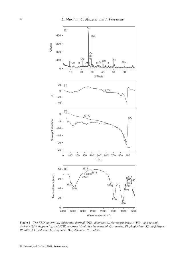
The mineral assemblage of the starting clay material was determined by X-ray diffraction(Fig. 1 (a)), differential thermal (DTA) and thermogravimetric analyses (TGA) (Figs 1 (b) and(c)) and FTIR (Fig. 1 (d)), and found to comprise quartz, calcite, dolomite, aragonite, illite,chlorite, plagioclase, K-feldspar and organic matter. Considering results published by Mackenzie(1957) and Stucki and Bish (1990), the endothermic peak at 130
°
C and the inflexion pointaround 200
°
C in the DTA pattern (Fig. 1 (b)) are attributed to the removal of hygroscopicwater from illite and organic matter. The large exothermic peak between 300
°
C and 550
°
C isdue to the oxidation of organic matter, which is estimated to be ~ 2.5 wt%, as shown from therelative weight loss in the TGA diagram (Fig. 1 (c)), whereas the two endothermic peaks at800
°
C and 870
°
C in the DTA, and the associated loss of weight in the TGA, are related to thedecarbonation of dolomite (~ 21 wt%) and calcite (~ 14 wt%), respectively.
The FTIR spectrum (Fig. 1 (d)) further confirms the XRD, DTA and TGA results. In particular,the O–H stretching region displays three characteristic signals of structural hydroxyl groups insilicate (probably chlorite) at 3690, 3620 and 760 cm
−
1
(Farmer 1974), and those of adsorbedwater at 3429 and 1624 cm
−
1
, respectively. Moreover, the absorption bands at 2515, 1439 and879 cm
−
1
are related to overtones, stretching and bending of the group in calcite anddolomite, the characteristic bending of which is present at 729 and 713 cm
−
1
, respectively,whereas signals at 1030, 798, 778 and 695 cm
−
1
(Fig. 1 (d)) are due to the Si–O stretchingmodes in silicates (Farmer 1974; De Benedetto
et al
. 2002). Finally, the presence of organicmatter is attested by the absorption bands at 2923, 2853 and 2627 cm
−
1
, which are due to CHand Cl
−
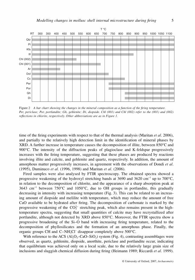
stretching.The mineral assemblages in the fired products were determined by XRD and are represented
in the bar diagram of Figure 2. The first variations in the mineral assemblage with respect to thestarting clay were observed between 400
°
C and 450
°
C when aragonite reflections disappearfrom the XRD pattern, probably due to the polymorphic transformation to calcite (Gmelin-Institut 1961). Increasing the firing temperature, chlorite begins to decompose between 500
°
Cand 650
°
C, this being marked by the disappearance of the (002) peak at low temperature and bythat of the (001) reflection at high temperature. Between 700
°
C and 750
°
C, the decarbonationof dolomite and the crystallization of periclase (MgO) occur. Portlandite (Ca(OH)
2
), whichalso appears in the XRD pattern, probably derived from the hydration of CaO, formed by calcitedecarbonation, which may have reacted with atmospheric humidity after firing. Finally, calcitedecomposes in the temperature range 800–850
°
C, forming gehlenite (Ca
2
Al
2
SiO
7
) and diopside(Ca(Mg,Fe)Si
2
O
6
) by reacting with silicates in the surrounding groundmass. The decarbonationtemperature determined by XRD is about 50
°
C lower than that determined by DTA(Fig. 1 (b)). This might be partially due to kinetic reasons connected with the long residence
CO32−
C NH– 3+
4
L. Maritan, C. Mazzoli and I. Freestone
© University of Oxford, 2007,
Archaeometry
Figure 1 The XRD pattern (a), differential thermal (DTA) diagram (b), thermogravimetric (TGA) and second derivate (SD) diagram (c), and FTIR spectrum (d) of the clay material. Qtz, quartz; Pl, plagioclase; Kfs, K-feldspar;Ill, illite; Chl, chlorite; Ar, aragonite; Dol, dolomite; Cc, calcite.
Modelling changes in mollusc shell internal microstructure during firing
5
© University of Oxford, 2007,
Archaeometry
time of the firing experiments with respect to that of the thermal analysis (Maritan
et al
. 2006),and partially to the relatively high detection limit in the identification of mineral phases byXRD. A further increase in temperature causes the decomposition of illite, between 850
°
C and900
°
C. The intensity of the diffraction peaks of plagioclase and K-feldspar progressivelyincreases with the firing temperature, suggesting that these phases are produced by reactionsinvolving illite and calcite, and gehlenite and quartz, respectively. In addition, the amount ofamorphous matter progressively increases, in agreement with the observations of Dondi
et al
.(1995), Duminuco
et al.
(1996, 1998) and Maritan
et al
. (2006).Fired samples were also analysed by FTIR spectroscopy. The obtained spectra showed a
progressive weakening of the hydroxyl stretching bands at 3690 and 3620 cm
−
1
up to 700
°
C,in relation to the decomposition of chlorite, and the appearance of a sharp absorption peak at3643 cm
−
1
between 750
°
C and 1050
°
C, due to OH groups in portlandite, this graduallydecreasing in intensity with increasing temperature (Fig. 3). This can be related to an increas-ing amount of diopside and melilite with temperature, which may reduce the amount of freeCaO available to be hydrated after firing. The decomposition of carbonate is marked by theprogressive weakening of the stretching peak, which also remains present in the high-temperature spectra, suggesting that small quantities of calcite may have recrystallized afterportlandite, although not detected by XRD above 850
°
C. Moreover, the FTIR spectra show aprogressive broadening of the Si–O band with increasing firing temperature, related to thedecomposition of phyllosilicates and the formation of an amorphous phase. Finally, theorganic groups CH and Cl
−
disappear completely above 500
°
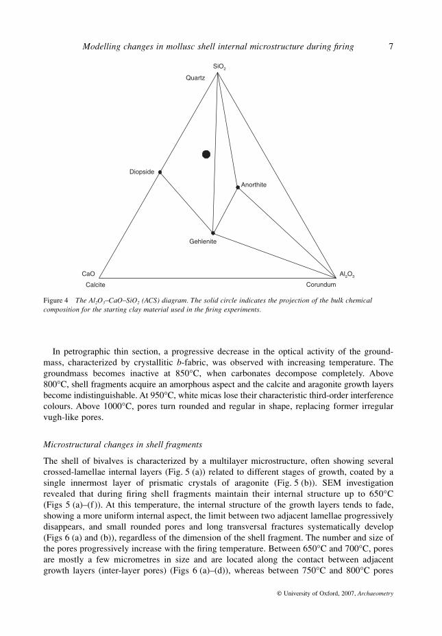
C.With reference to the ACS (Al
2
O
3
–CaO–SiO
2
) system (Fig. 4), contrasting assemblages wereobserved, as quartz, gehlenite, diopside, anorthite, periclase and portlandite occur, indicatingthat equilibrium was achieved only on a local scale, due to the relatively large grain size ofinclusions and sluggish chemical diffusion during firing (Heimann 1989; Riccardi
et al. 1999).
Figure 2 A bar chart showing the changes in the mineral composition as a function of the firing temperature. Per, periclase; Por, portlandite; Gh, gehlenite; Di, diopside. Chl (001) and Chl (002) refer to the (001) and (002)reflections in chlorite, respectively. Other abbreviations are as in Figure 1.
CO32−
C NH– 3+

6 L. Maritan, C. Mazzoli and I. Freestone
© University of Oxford, 2007, Archaeometry
Figure 3 The evolution of the FTIR spectrum as a function of the firing temperature.
Modelling changes in mollusc shell internal microstructure during firing 7
© University of Oxford, 2007, Archaeometry
In petrographic thin section, a progressive decrease in the optical activity of the ground-mass, characterized by crystallitic b-fabric, was observed with increasing temperature. Thegroundmass becomes inactive at 850°C, when carbonates decompose completely. Above800°C, shell fragments acquire an amorphous aspect and the calcite and aragonite growth layersbecome indistinguishable. At 950°C, white micas lose their characteristic third-order interferencecolours. Above 1000°C, pores turn rounded and regular in shape, replacing former irregularvugh-like pores.
Microstructural changes in shell fragments
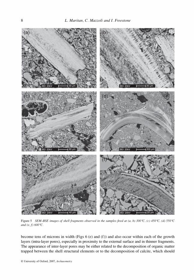
The shell of bivalves is characterized by a multilayer microstructure, often showing severalcrossed-lamellae internal layers (Fig. 5 (a)) related to different stages of growth, coated by asingle innermost layer of prismatic crystals of aragonite (Fig. 5 (b)). SEM investigationrevealed that during firing shell fragments maintain their internal structure up to 650°C(Figs 5 (a)–(f)). At this temperature, the internal structure of the growth layers tends to fade,showing a more uniform internal aspect, the limit between two adjacent lamellae progressivelydisappears, and small rounded pores and long transversal fractures systematically develop(Figs 6 (a) and (b)), regardless of the dimension of the shell fragment. The number and size ofthe pores progressively increase with the firing temperature. Between 650°C and 700°C, poresare mostly a few micrometres in size and are located along the contact between adjacentgrowth layers (inter-layer pores) (Figs 6 (a)–(d)), whereas between 750°C and 800°C pores
Figure 4 The Al2O3–CaO–SiO2 (ACS) diagram. The solid circle indicates the projection of the bulk chemical composition for the starting clay material used in the firing experiments.
8 L. Maritan, C. Mazzoli and I. Freestone
© University of Oxford, 2007, Archaeometry
become tens of microns in width (Figs 6 (e) and (f)) and also occur within each of the growthlayers (intra-layer pores), especially in proximity to the external surface and in thinner fragments.The appearance of inter-layer pores may be either related to the decomposition of organic mattertrapped between the shell structural elements or to the decomposition of calcite, which should
Figure 5 SEM–BSE images of shell fragments observed in the samples fired at (a, b) 300°C, (c) 450°C, (d) 550°Cand (e, f ) 600°C.

Modelling changes in mollusc shell internal microstructure during firing 9
© University of Oxford, 2007, Archaeometry
start in microstructural locations characterized by low superficial tension and a higher Gibbsfree energy, such as the discontinuity between adjacent growth lamellae. The presence ofintra-layer pores requires a higher firing temperature (750–800°C) and is limited, according tothe shell thickness, to the external portion of the fragments. Above 850°C, the shell fragments
Figure 6 SEM–BSE images of shell fragments observed in the samples fired at (a, b) 650°C, (c, d) 700°C, (e) 750°Cand ( f ) 800°C.
10 L. Maritan, C. Mazzoli and I. Freestone
© University of Oxford, 2007, Archaeometry
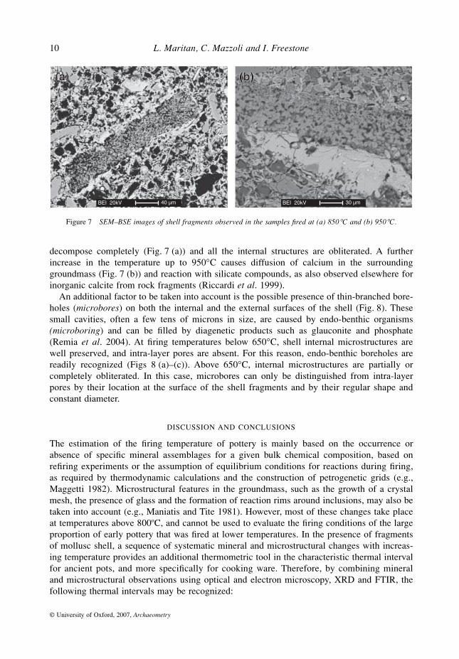
decompose completely (Fig. 7 (a)) and all the internal structures are obliterated. A furtherincrease in the temperature up to 950°C causes diffusion of calcium in the surroundinggroundmass (Fig. 7 (b)) and reaction with silicate compounds, as also observed elsewhere forinorganic calcite from rock fragments (Riccardi et al. 1999).
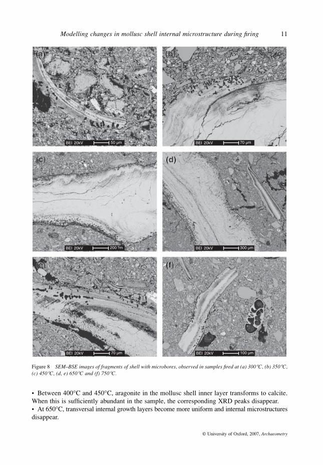
An additional factor to be taken into account is the possible presence of thin-branched bore-holes (microbores) on both the internal and the external surfaces of the shell (Fig. 8). Thesesmall cavities, often a few tens of microns in size, are caused by endo-benthic organisms(microboring) and can be filled by diagenetic products such as glauconite and phosphate(Remia et al. 2004). At firing temperatures below 650°C, shell internal microstructures arewell preserved, and intra-layer pores are absent. For this reason, endo-benthic boreholes arereadily recognized (Figs 8 (a)–(c)). Above 650°C, internal microstructures are partially orcompletely obliterated. In this case, microbores can only be distinguished from intra-layerpores by their location at the surface of the shell fragments and by their regular shape andconstant diameter.
DISCUSSION AND CONCLUSIONS
The estimation of the firing temperature of pottery is mainly based on the occurrence orabsence of specific mineral assemblages for a given bulk chemical composition, based onrefiring experiments or the assumption of equilibrium conditions for reactions during firing,as required by thermodynamic calculations and the construction of petrogenetic grids (e.g.,Maggetti 1982). Microstructural features in the groundmass, such as the growth of a crystalmesh, the presence of glass and the formation of reaction rims around inclusions, may also betaken into account (e.g., Maniatis and Tite 1981). However, most of these changes take placeat temperatures above 800oC, and cannot be used to evaluate the firing conditions of the largeproportion of early pottery that was fired at lower temperatures. In the presence of fragmentsof mollusc shell, a sequence of systematic mineral and microstructural changes with increas-ing temperature provides an additional thermometric tool in the characteristic thermal intervalfor ancient pots, and more specifically for cooking ware. Therefore, by combining mineraland microstructural observations using optical and electron microscopy, XRD and FTIR, thefollowing thermal intervals may be recognized:
Figure 7 SEM–BSE images of shell fragments observed in the samples fired at (a) 850°C and (b) 950°C.
Modelling changes in mollusc shell internal microstructure during firing 11
© University of Oxford, 2007, Archaeometry
• Between 400°C and 450°C, aragonite in the mollusc shell inner layer transforms to calcite.When this is sufficiently abundant in the sample, the corresponding XRD peaks disappear.• At 650°C, transversal internal growth layers become more uniform and internal microstructuresdisappear.
Figure 8 SEM–BSE images of fragments of shell with microbores, observed in samples fired at (a) 300°C, (b) 350°C,(c) 450°C, (d, e) 650°C and (f) 750°C.
12 L. Maritan, C. Mazzoli and I. Freestone
© University of Oxford, 2007, Archaeometry
• Between 650°C and 700°C, small inter-layer pores form along adjacent growth layers andon the external surface, which are a few microns in diameter.• Between 750°C and 800°C, larger (tens of microns) intra-layer pores develop, which aremore frequently close to the external walls of the fragments.• At about 850°C, the crystallization of gehlenite and diopside marks the decomposition ofcalcite. When large fragments of calcite are present, calcium oxide may form, and portlanditemay occur in the potsherd after reaction with water vapour in the air. In ancient pottery, theportlandite may be expected to have fully recrystallized to calcite.
These changes relate specifically to the conditions of our experiments, for a heating rate of200°C h−1 in oxidizing conditions and a residence time of 6 h. They are related to kinetic aswell as thermodynamic factors, and should not be directly applied to ceramics that have beenfired for very short periods without further testing. However, they clearly indicate that in thestudy of pottery containing shell fragments, either naturally present or deliberately added, adetailed analysis of the shell microstructure, associated with the determination of the mineralcomposition by XRD and FTIR, can give precise constraints on the estimate of the firingtemperature. Furthermore, the anticipated effects of the firing duration are of interest as theysuggest that, within the range of firing conditions relevant to early cooking wares, systematicchanges may be observed that will allow constraints to be placed upon both the temperatureand the duration of firing.
The changes observed in the structure of the shell prior to decomposition are major, andinclude the generation of large pores and the opening up of the laminae in the shell particles.These changes may be expected to affect the physical properties of the shell temper, and arelikely to influence the effectiveness of the shell particles in the prevention of crack propaga-tion. Close understanding and control of firing conditions is therefore likely to be critical ininvestigating the widespread choice of shell-tempered wares for cooking pots, whetherthrough modelling or replication experiments. These results emphasize the complexity of theshell–clay ceramic system and the challenge to be met if the science underlying the adoptionof shell tempering is to be fully understood.
ACKNOWLEDGEMENTS
This study was carried out with the financial support of MIUR project PRIN 2004 n.2004041033_005 and benefited from a grant by the ‘Accademia Nazionale dei Lincei’.
REFERENCES
De Benedetto, G. E., Laviano, R., Sabbatini, L., and Zambonin, G. P., 2002, Infrared spectroscopy in the mineralog-ical characterisation of ancient pottery, Journal of Cultural Heritage, 3, 177–86.
Dondi, M., Ercolani, G., Guarini, G., Marsigli, M., and Venturi, I., 1995, Evoluzione della microstruttura durante lacottura rapida di impasti per piastrelle porose, Ceramurgia, 25(6), 301–14.
Duminuco, P., Messiga, B., and Riccardi, M. P., 1998, Firing process of natural clays: some microtextures and relatedphase compositions, Thermochimica Acta, 321, 185–90.
Duminuco, P., Riccardi, M. P., Messiga, B., and Setti, M., 1996, Modificazioni tessiturali e mineralogiche comeindicatori della dinamica del processo di cottura di manufatti ceramici, Ceramurgia, 26(5), 281–8.
Farmer, V. C., 1974, Infrared spectra of minerals, Mineralogical Society, London.Gmelin-Institut für anorganische Chemie und Grenzgebiete in der Max Planck Gesellschaft (eds.), 1961, Gmelins
Handbuch der Anorganischen Chemie, 8 Auflage, Band 28/B, 153, Verlag Chemie, Weinheim.Heimann, R. B., 1989, Assessing the technology of ancient pottery: the use of ceramic phase diagrams, Archeomaterials,
3, 123–48.
Modelling changes in mollusc shell internal microstructure during firing 13
© University of Oxford, 2007, Archaeometry
Mackenzie, R. C., 1957, The differential thermal investigation of clay, Mineralogical Society, London.Maggetti, M., 1982, Phase analysis and its significance for technology and origin, in Archaeological ceramics (eds.
J. S. Olin and A. D. Franklin), 121–33, Smithsonian Institution Press, Washington, DC.Maniatis, Y., and Tite, M. S., 1981, Technological investigation of Neolithic – Bronze Age pottery from central and
southeast Europe and from the Near East, Journal of Archaeological Science, 8, 59–76.Maritan, L., Nodari, L., Mazzoli, C., Milano, A., and Russo, U., 2006, Influence of firing conditions on ceramic products:
experimental study on clay rich in organic matter, Applied Clay Science, 31, 1–15.Maritan, L., Mazzoli, C., Michielin, V., Morandi Bonacossi, D., Luciani, M., and Molin, G., 2005, Provenance and
production technology of Bronze Age and Iron Age pottery from Tell Mishrifeh/Qatna (Syria), Archaeometry, 47,723–44.
Remia, A., Montagna, P., and Taviani, M., 2004, Submarine diagenetic products on the sediment-starved Gorgoniaslope, Tuscan archipelago (Tyrrhenian Sea), Chemistry and Ecology, 20, 131–53.
Riccardi, M. P., Messiga, B., and Duminuco, P., 1999, An approach to the dynamic of clay firing, Applied Clay Science,15, 393–409.
Rye, O. S., 1976, Keeping temperature under control: materials and the manufacture of Papuan pottery, Archaeologyand Physical Anthropology in Oceania, 11, 106–37.
Steponaitis, V. P., 1983, Ceramic, chronology and community patterns—an archaeological study at Moundville,Academic Press, New York.
Steponaitis, V. P., 1984, Technological studies of prehistoric pottery from Alabama: physical properties and vesselfunction, in The many dimensions of pottery (eds. S. E. van der Leeuw and A. C. Pritchard), 79–127, Universityof Amsterdam, Amsterdam.
Stimmel, C., Heimann, R. B., and Hancock, R. G. V., 1982, Indian pottery from the Mississippi Valley: coping with badraw materials, in Archaeological ceramics (eds. J. S. Olin and A. D. Franklin), 89–96, Smithsonian InstitutionPress, Washington, DC.
Stucki, J. W., and Bish, D. L., 1990, Thermal analysis in clay science, Clay Minerals Society, Boulder, CO.Tite, M. S., and Kilikoglou, V., 2002, Do we understand cooking pots and is there an ideal cooking pot?, in Modern
trends in scientific studies on ancient ceramics (eds. V. Kilikoglou, A. Hein and Y. Maniatis), 1–8, BAR Inter-national Series 1011, Archaeopress, Oxford.
Tite, M. S., Kilikoglou, V., and Vekinis, G., 2001, Strength, toughness and thermal resistance of ancient ceramics, andtheir influence on technological choice, Archaeometry, 43, 301–24.
Woods, A. J., 1986, Form, fabric and function: some observations on the cooking pot in antiquity, in Technology andstyle (ed. W. D. Kingery), 157–72, Ceramics and civilization, vol. 2, American Ceramic Society, Columbus, OH.
Copyright © 2022 FDOKUMEN