Mitigating hook effect in one-step quantitative sandwich lateral ...
7
Talanta 240 (2022) 123157 Available online 17 December 2021 0039-9140/© 2021 The Authors. Published by Elsevier B.V. This is an open access article under the CC BY license (http://creativecommons.org/licenses/by/4.0/). Mitigating hook effect in one-step quantitative sandwich lateral flow assay by timed conjugate release Guozhen He a, b , Tao Dong b, * , Zhaochu Yang a , Zhuangde Jiang a a Chongqing Key Laboratory of Micro-Nano Systems and Smart Transduction, Chongqing Key Laboratory of Colleges and Universities on Micro-Nano Systems Technology and Smart Transducing, Collaborative Innovation Center on Micro-Nano Transduction and Intelligent Eco-Internet of Things, Chongqing Academician and Expert Workstation, Chongqing Technology and Business University, Nan’an District, Chongqing, 400067, China b Department of Microsystems (IMS), Faculty of Technology, Natural Sciences and Maritime Sciences, University of South-Eastern Norway, Postboks 235, 3603, Kongsberg, Norway A R T I C L E INFO Keywords: Lateral flow assay Hook effect Time conjugate release Salivary assay Fluorescence ABSTRACT Sandwich lateral flow assay (LFA) is one of the most successfully commercialized paper-based biosensors, which offers a rapid, low-cost, one-step assay. Despite its advantages, conventional sandwich LFA is fundamentally limited by the high-dose “hook” effect—a phenomenon that occurs at very high analyte concentrations and results in false-negative results. In this paper, we present a novel strategy of automatic timed detection antibody release to mitigate the hook effect in sandwich LFA without additional manual steps. We introduced an inter- mediate pad treated with saturated sucrose solution to regulate the flow between the nitrocellulose membrane and the conjugate pad in order to delay the reaction between detection antibodies and analytes. Using C-reactive protein (CRP) as a representative analyte, we demonstrated that our strategy exhibited a range of detection 10 times wider than that of our conventional LFA, without sacrificing the limit of detection. Comparing to other published strategies, our work could offer a one-step, cost-effective approach that is closely unified with the benefits of the LFA. 1. Introduction Sandwich lateral flow assay (LFA) has been widely applied in the rapid point-of-care (POC) testing industry due to its low cost, rapid response, and one-step operation [1]. Despite the advantages and the broad applications, sandwich LFAs produce false-negative results at very high concentrations of analytes, which is described as the high-dose “hook effect” [2–8]. T/C intensity initially increases monotonically but eventually decreases when the analyte concentration surpasses a certain level, exhibiting a hook-like curve in the T/C intensity vs. analyte concentration diagram. First observed in one-step sandwich immuno- assays in the 1980s, the hook effect is an intrinsic phenomenon in sandwich assays [9]. Assuming analytes bind to detection antibodies and capture antibodies simultaneously, the widely accepted explanation is that unlabeled analytes occupy sites at the test line that would have captured labeled analytes. In other words, there is a shortage of free detection antibodies to bind analytes, which are captured at the test lines. Hook effect limits the clinical applications of LFA where bio- markers in real human samples could increase significantly when patients were experiencing severe diseases. Taking the example of C-reactive protein (CRP) concentration in serum, it is below 1 mg/mL for healthy people but can roar up to more than 250 mg/mL in case of severe infection [10]. In conventional sandwich immunoassays, the problem is solved by sample dilution [11], addition washing step [12], and increasing con- centrations of detection antibody [13]. Similar strategies have also been applied in LFAs [6]. Increasing detection antibody leads to higher background noise that limits the lower limit of detection. Attempts have been made to alleviate the hook effect by adding a third line that only binds conjugates [14] and introducing multiple test zones for one ana- lyte [5]. E.G. Rey et al. adapted kinetic measurement with an algorithm to measure the speed at which both sample and control line develop instead of measuring the final intensity [4]. Other researchers also tried to provide theoretical explanations of the “hook” effect in LFAs due to high analyte concentration [3,6]. Hook effect is affected by the capture antibody’s avidity to both free analyte and analyte-detection antibody complex [6], while in most cases, both avidities need to be measured experimentally. Oh et al. reported a timed reagent release strategy by * Corresponding author. E-mail address: [email protected] (T. Dong). Contents lists available at ScienceDirect Talanta journal homepage: www.elsevier.com/locate/talanta https://doi.org/10.1016/j.talanta.2021.123157 Received 21 September 2021; Received in revised form 15 December 2021; Accepted 16 December 2021
-
Upload
khangminh22 -
Category
Documents
-
view
0 -
download
0
Transcript of Mitigating hook effect in one-step quantitative sandwich lateral ...
Talanta 240 (2022) 123157
Available online 17 December 20210039-9140/© 2021 The Authors. Published by Elsevier B.V. This is an open access article under the CC BY license (http://creativecommons.org/licenses/by/4.0/).
Mitigating hook effect in one-step quantitative sandwich lateral flow assay by timed conjugate release
Guozhen He a,b, Tao Dong b,*, Zhaochu Yang a, Zhuangde Jiang a
a Chongqing Key Laboratory of Micro-Nano Systems and Smart Transduction, Chongqing Key Laboratory of Colleges and Universities on Micro-Nano Systems Technology and Smart Transducing, Collaborative Innovation Center on Micro-Nano Transduction and Intelligent Eco-Internet of Things, Chongqing Academician and Expert Workstation, Chongqing Technology and Business University, Nan’an District, Chongqing, 400067, China b Department of Microsystems (IMS), Faculty of Technology, Natural Sciences and Maritime Sciences, University of South-Eastern Norway, Postboks 235, 3603, Kongsberg, Norway
A R T I C L E I N F O
Keywords: Lateral flow assay Hook effect Time conjugate release Salivary assay Fluorescence
A B S T R A C T
Sandwich lateral flow assay (LFA) is one of the most successfully commercialized paper-based biosensors, which offers a rapid, low-cost, one-step assay. Despite its advantages, conventional sandwich LFA is fundamentally limited by the high-dose “hook” effect—a phenomenon that occurs at very high analyte concentrations and results in false-negative results. In this paper, we present a novel strategy of automatic timed detection antibody release to mitigate the hook effect in sandwich LFA without additional manual steps. We introduced an inter-mediate pad treated with saturated sucrose solution to regulate the flow between the nitrocellulose membrane and the conjugate pad in order to delay the reaction between detection antibodies and analytes. Using C-reactive protein (CRP) as a representative analyte, we demonstrated that our strategy exhibited a range of detection 10 times wider than that of our conventional LFA, without sacrificing the limit of detection. Comparing to other published strategies, our work could offer a one-step, cost-effective approach that is closely unified with the benefits of the LFA.
1. Introduction
Sandwich lateral flow assay (LFA) has been widely applied in the rapid point-of-care (POC) testing industry due to its low cost, rapid response, and one-step operation [1]. Despite the advantages and the broad applications, sandwich LFAs produce false-negative results at very high concentrations of analytes, which is described as the high-dose “hook effect” [2–8]. T/C intensity initially increases monotonically but eventually decreases when the analyte concentration surpasses a certain level, exhibiting a hook-like curve in the T/C intensity vs. analyte concentration diagram. First observed in one-step sandwich immuno-assays in the 1980s, the hook effect is an intrinsic phenomenon in sandwich assays [9]. Assuming analytes bind to detection antibodies and capture antibodies simultaneously, the widely accepted explanation is that unlabeled analytes occupy sites at the test line that would have captured labeled analytes. In other words, there is a shortage of free detection antibodies to bind analytes, which are captured at the test lines. Hook effect limits the clinical applications of LFA where bio-markers in real human samples could increase significantly when
patients were experiencing severe diseases. Taking the example of C-reactive protein (CRP) concentration in serum, it is below 1 mg/mL for healthy people but can roar up to more than 250 mg/mL in case of severe infection [10].
In conventional sandwich immunoassays, the problem is solved by sample dilution [11], addition washing step [12], and increasing con-centrations of detection antibody [13]. Similar strategies have also been applied in LFAs [6]. Increasing detection antibody leads to higher background noise that limits the lower limit of detection. Attempts have been made to alleviate the hook effect by adding a third line that only binds conjugates [14] and introducing multiple test zones for one ana-lyte [5]. E.G. Rey et al. adapted kinetic measurement with an algorithm to measure the speed at which both sample and control line develop instead of measuring the final intensity [4]. Other researchers also tried to provide theoretical explanations of the “hook” effect in LFAs due to high analyte concentration [3,6]. Hook effect is affected by the capture antibody’s avidity to both free analyte and analyte-detection antibody complex [6], while in most cases, both avidities need to be measured experimentally. Oh et al. reported a timed reagent release strategy by
* Corresponding author. E-mail address: [email protected] (T. Dong).
Contents lists available at ScienceDirect
Talanta
journal homepage: www.elsevier.com/locate/talanta
https://doi.org/10.1016/j.talanta.2021.123157 Received 21 September 2021; Received in revised form 15 December 2021; Accepted 16 December 2021
Talanta 240 (2022) 123157
2
adding a sample injection zone in the middle of the strip while placing a commercial asymmetric polysulfone membrane (ASPM) with asym-metric pole distribution located horizontally between the buffer pad and sample pad to delay the conjugate release [15].
To resolve the hook effect in sandwich LFA without additional steps and costs, we proposed a strategy based on delaying the release of detection antibodies from the conjugate pad to avoid the competition between free and labeled analytes at the test line. We hypothesized that by avoiding the competitive reactions between the analyte, capture antibody, and detection antibody, the hook effect could be mitigated. We re-designed the lateral flow assay and introduced an intermediate pad treated with sucrose between the nitrocellulose membrane and the conjugated pad to demonstrate our strategy. The intermediate pad regulated the liquid flow into the conjugated pad [16] to achieve the timed-release of detection antibody. The advantage of sucrose treatment was that the sucrose did not interfere with the CRP-antibody binding, while the cost of sucrose treatment was neglectable. We experimentally proved that our designated LFA mitigated false-negative results caused by the high-dose hook effect.
2. Experimental section
2.1. Reagents and instruments
Anti-CRP antibody [C2] (ab136176), anti-CRP antibody [C6] (ab8278) were purchased from Abcam in both Norway and China. High purity native CRP purified from human serum was purchased from Sigma Aldrich (both Norway and China). Nitrocellulose (NC) mem-branes CN140 of 25 mm width were purchased from sartorius. Glass fiber membranes, polyester fiber membranes, absorbent pads, and ad-hesive backing cards were purchased from Jieyi Biotech. CO., LTD. Bovine serum albumin (BSA) and Tween 20 were purchased from Bei-jing Solarbio Science & Technology Co., Ltd.
Saliva oral swab was purchased from Salimetrics, State College, PA. Sucrose (S0389) was purchased from Sigma Aldrich. CdSe-TiO2 quan-tum dots were purchased from Kundao Biotech, Shanghai. CRP human ELISA kits were purchased from Thermo Fisher.
2.2. Quantum dots conjugation
We used a commercial CdSe-TiO2 core-shell quantum dot with polyethylene glycol (PEG) modification as the fluorescence label. Ul-traviolet (UV) light was applied as the light source for excitation. The commercial CdSe QD exhibited an excitation range between 365 and 450 nm and a fluorescence peak at 570 nm. No photobleaching was observed. As-purchased QDs were linked to anti-CRP antibody C6 (detection antibody, CdSe-C6) via EDC-NHS linking. CdSe-C6 was immobilized onto the glass fiber conjugate pad.
2.3. Preparation of the intermediate pad
We compared cellulose membranes treated with different sucrose saturation as the intermediate pad. To treat cellulose membranes with sucrose, we first dissolved excess sucrose in deionized water (DI water) at room temperature for several days to create a saturated sucrose water solution. Sucrose settlements were removed from the saturated solution. Subsequently, the saturated sucrose solution was diluted to prepare 10%–100% saturated solution. Cellulose membranes were also treated with DI water for comparison, noted as 0% saturation. Cellulose mem-branes were firstly wicked from the edges and then immersed into the sucrose solution. After that, cellulose membranes were dried in a vac-uum chamber at 36 ◦C until they were completely dried.
2.4. Preparation of the lateral flow strips
The lateral flow strip is assembled with a sample pad, a conjugate
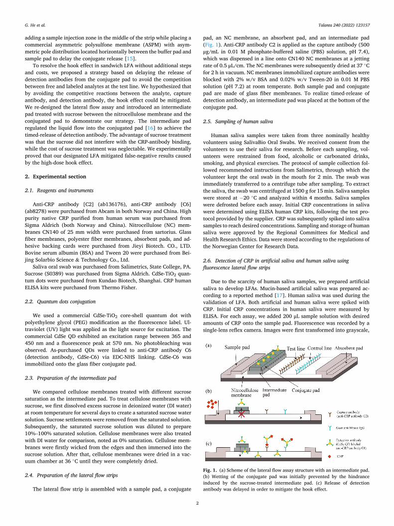
pad, an NC membrane, an absorbent pad, and an intermediate pad (Fig. 1). Anti-CRP antibody C2 is applied as the capture antibody (500 μg/mL in 0.01 M phosphate-buffered saline (PBS) solution, pH 7.4), which was dispensed in a line onto CN140 NC membranes at a jetting rate of 0.5 μL/cm. The NC membranes were subsequently dried at 37 ◦C for 2 h in vacuum. NC membranes immobilized capture antibodies were blocked with 2% w/v BSA and 0.02% w/v Tween-20 in 0.01 M PBS solution (pH 7.2) at room temperate. Both sample pad and conjugate pad are made of glass fiber membranes. To realize timed-release of detection antibody, an intermediate pad was placed at the bottom of the conjugate pad.
2.5. Sampling of human saliva
Human saliva samples were taken from three nominally healthy volunteers using SalivaBio Oral Swabs. We received consent from the volunteers to use their saliva for research. Before each sampling, vol-unteers were restrained from food, alcoholic or carbonated drinks, smoking, and physical exercises. The protocol of sample collection fol-lowed recommended instructions from Salimetrics, through which the volunteer kept the oral swab in the mouth for 2 min. The swab was immediately transferred to a centrifuge tube after sampling. To extract the saliva, the swab was centrifuged at 1500 g for 15 min. Saliva samples were stored at − 20 ◦C and analyzed within 4 months. Saliva samples were defrosted before each assay. Initial CRP concentrations in saliva were determined using ELISA human CRP kits, following the test pro-tocol provided by the supplier. CRP was subsequently spiked into saliva samples to reach desired concentrations. Sampling and storage of human saliva were approved by the Regional Committees for Medical and Health Research Ethics. Data were stored according to the regulations of the Norwegian Center for Research Data.
2.6. Detection of CRP in artificial saliva and human saliva using fluorescence lateral flow strips
Due to the scarcity of human saliva samples, we prepared artificial saliva to develop LFAs. Mucin-based artificial saliva was prepared ac-cording to a reported method [17]. Human saliva was used during the validation of LFA. Both artificial and human saliva were spiked with CRP. Initial CRP concentrations in human saliva were measured by ELISA. For each assay, we added 200 μL sample solution with desired amounts of CRP onto the sample pad. Fluorescence was recorded by a single-lens reflex camera. Images were first transformed into grayscale,
Fig. 1. (a) Scheme of the lateral flow assay structure with an intermediate pad. (b) Wetting of the conjugate pad was initially prevented by the hindrance induced by the sucrose-treated intermediate pad. (c) Release of detection antibody was delayed in order to mitigate the hook effect.
G. He et al.

Talanta 240 (2022) 123157
3
and fluorescence intensities were obtained using the measurement function in ImageJ. Intensities of T lines and C lines were calculated by subtracting background fluorescence intensity from the overall inte-grated intensity of the selected areas.
3. Results and discussion
3.1. Principle of the lateral flow assay with timed conjugate release
In conventional lateral flow sandwich assays, a conjugated pad is placed directly between the sample pad and nitrocellulose membrane so that detection antibodies flow with the analyte together when the con-jugate pad is wetted. In this scenario, analytes will bind both detection antibody and capture antibody simultaneously [18,19]. At high analyte concentrations, when the detection antibody is deficient, unlabeled analytes compete with analytes bound to detection antibody at the test line, leading to false-negative results.
To resolve this hook effect, we hypothesized that by delaying the detection antibody entering the sandwich assay system, the competition between CRP captured at the test line and free CRP could be potentially avoided. In other words, reactions between CRP at the test line and detection antibody would be favored by setting a time interval between the presence of CRP and detection antibody at the sandwich assay sys-tem. We studied two approaches— (1) manually adding CRP and detection antibody in sequence; adding an intermediate pad that is (2) treated by sucrose to delay the release of detection antibody—to step-by- step verify our hypothesis. We deployed an intermediate pad with flow- tuning characteristics to bridge the sample pad and nitrocellulose membrane (Fig. 1). The intermediate pad sits above the conjugate pad in which the detection antibody is immobilized. Such an intermediate pad exhibits a slower vertical flow rate than its lateral counterparts to delay the flow entering the conjugate pad. Detection antibody is released at certain times when analytes are mostly bound to the test line to avoid competitive reactions.
3.2. Intermediate pad treatment with sucrose solution
We dedicatedly designed the intermediate pad by testing different materials and treatments to control the release time smartly. We finally selected a commercial cellulose membrane treated with sucrose to treat the pad. The intermediate pad should not affect the antibody-antigen reaction. It excludes the pad to be treated with most salts since salts significantly affect antibody conformation and their reactions with an-tigens. Sucrose was selected due to its low price and biocompatibility [16]. Sucrose did not interfere with the CRP-antibody binding, nor did it induce false-positive results. It is also believed to be an inexpensive stabilizer against degradation of proteins dried on paper substrates [20]. A barrier with tunable permeability is desired for lateral flow assays to control the delayed release of detection antibody smartly. For this pur-pose, we investigated paper-based barriers consisting of cellulose membrane treated with sucrose solution. We compared cellulose mem-brane treated with different concentrations as the intermediate layer bridging the sample-conducting nitrocellulose membrane and conjugate pad. We treated 330 μm thick cellulose membranes with DI water (which was considered as the blank group with 0% sucrose) and 10%– 100% sucrose saturated solutions. We mixed red dye with sample so-lutions before wicking the LFA strips. The wetting performance was evaluated by recording the time spent before intermediate pad completely turned red (Fig. 2). For a blank intermediate pad without sucrose, it took around 4 min to turn red completely. At the interface between the intermediate pad and the NC membrane, the vertical fluidic resistance was larger than the horizontal fluidic resistance due to gravity and permeability difference in different porous materials. Thus, the fluid initially traveled in the horizontal direction. The intermediate pad also stored solution before it fully turned red. As the sucrose saturation percentage increased, it gradually took more time to completely color
the intermediate pad, which could be considered as an increase in the ability of fluidic hindrance. As the saturation percentage exceeded 70%, the wetting time increased dramatically, from 7.2 min (70% saturation) to 31.3 min (100% saturation). We considered that saturation between 70% and 90% led to moderate time delays between 7.2 min and 19.3 min, which were chosen for further studied for timed-release of detec-tion antibody. Such hindrance to the flow could be potentially attributed to two reasons. Firstly, sucrose from the highly saturated solutions was dried and subsequently formed large amounts of small crystals within the pores of cellulose membranes. When sucrose encountered the flow, sucrose started to dissolve quickly, resulting in a local region with saturated sucrose concentration. Therefore, further dissolution of su-crose was prohibited until local concentration was decreased due to diffusion of sucrose molecules to areas where sucrose concentration was low. Sucrose crystals blocked the solution into the conjugate pad, forming a temporally water-proof barrier. On the other hand, the local region with saturated sucrose concentration exhibited a high viscosity and exerted high flow resistances that delayed the flow through the pad [16]. When sucrose crystals gradually dissolved upon contact with the aqueous solution, a route would be opened to release detection anti-bodies in the conjugate pad into the assay.
3.3. Hook effect in conventional lateral flow assay
The conventional format of a lateral flow assay consists of a sample pad, a conjugate pad, a nitrocellulose membrane immobilized with both a test line and a control line, and a wick (adsorbent) pad. When a sample was added to the test strip, it flowed uniformly without any delay in the release of the detection antibody (detection antibody is also often called the conjugate). To determine analyte concentrations quantitatively, re-searchers often measure either the test line intensity or the ratio of the test line intensity over the control line intensity (T/C). We compared both the test line intensity and the T/C ratios to study their contributions in the hook effect. We noticed that the test line intensity increased monotonically for CRP detection as CRP concentration increased within the region between 1 ng/mL to 1 μg/mL (Fig. 3a and b). The test line intensity reached the peak and saturated around 1 μg/mL. As the CRP concentration continued to increase, the test line intensity started to drop.
Fig. 2. Characterization of time spent to fully wet intermediate pads treated with sucrose solution at different saturation percentages (n = 3). The inset shows representative images of characterization taken from intermediate pads treated by 80% and 90% sucrose saturated solution.
G. He et al.

Talanta 240 (2022) 123157
4
On the other hand, the control line intensity started at the highest intensity and decreased as CRP concentration increased in the region between 1 ng/mL to 1 μg/mL. However, the control line intensity reached the lowest point at around 1 μg/mL while the test line intensity reached the highest peak. With a further increase in CRP concentration, the control line intensity increased while the test line intensity decreased. T/C ratios exhibited a similar trend to the test line intensity and reached a peak around 1000 ng/mL (Fig. 3c). A clear hook was observed in both T line and T/C ratios (Fig. 3b and c). When CRP increased above 1000 ng/mL, the lateral flow assay gave false-negative results. Using T/C as the calibration curve did not exhibit any obvious advantage comparing using the T line intensity. After the hook effect happened around 1000 ng/mL CRP concentration, T intensity and T/C ratios dramatically dropped.
The most widely-accepted explanation is that the hook effect is a concentration effect [9,13]. Excess analytes hinder simultaneous bind-ing of CRP to both capture antibody at the test line and detection anti-body in solution. In conventional lateral flow assays, the detection antibody dried on the conjugate pad diffused in the nitrocellulose membrane with free CRP molecules together. Both detection antibody and capture antibody may recognize more than one epitope of CRP. Besides, one quantum dot was linked to three detection antibodies (C6) on average through our protocol. The multivalent reaction may result in higher-order antibody-CRP complexed and multiple configurations with different binding affinities or associations. The antibody-CRP associa-tion may also go through an intermediate state, forming a transient complex [21]. For simplicity, we assume CRP-C2 binding and CRP-C6 binding are both a one-step reaction and result in a single configura-tion for either CRP-C2 complex or CRP-C6 complex. Then, we will have four association constants for four different reactions in the lateral flow assay. We note these constants as Ka for the reaction between free CRP and capture antibody C2 immobilized at the test line, Kb for the reaction between free CRP and detection antibody C6-QD conjugate in the so-lution, Kc for the reaction between C6-QD conjugate and CRP-C2 at the test line, and Kd for the reaction between CRP-C6-QD complex and antibody C2 at the test line. The magnitudes of these association con-stants often lie in the following sequence: Kc > Ka > Kb > Kd [22]. It
suggested that the association rate constant of analyte binding to either detection antibody in solution or immobilized capture antibody was around the same level. In contrast, these two constants would be 3 to 4 magnitude higher than the association rate constant of detection antibody-analyte conjugate binding to capture antibody [22]. We can also expect more free CRP molecules to bind to the conjugates in solu-tion than they bind to the detection antibody. In case of excess CRP, the binding equilibrium may shift further to the formation of CRP-C6 complexes.
The hook effect observed in the T/C ratio is a synergistic effect of both test line and control line. Before the saturation point at 1000 ng/ mL, the test line intensity increased because of an increasing number of C2-CRP-C6 complexes formed at the test line, while the control line intensity decreased due to less available C6 conjugate could be captured at the control line. It resulted in a monotonic increase in T/C ratios before the hook effect. When the hook effect happened at CRP concen-trations higher than 1000 ng/mL, unlabeled CRP molecules blocked sites that would have captured CRP-C6-QD conjugates. It resulted in increasing numbers of conjugates bound to the control line and an in-crease in control line intensity. The C6-CRP complex usually has a lower avidity than the free C6 antibody does when binding to IgG antibodies at the control line [22]. It may explain that the control line intensity at CRP concentrations between 10 and 100 μg was lower than its intensity at CRP concentrations below 10 ng/mL.
Influence of timed conjugate release on the hook effect in artificial saliva samples and human saliva samples.
Since Kc is often larger than Ka, we came up with a strategy to mitigate the hook effect by avoiding the competition between CRP-C2 binding and CRP-C6 binding. We verified this strategy by comparing premixing to sequentially adding CRP and detection antibodies (Fig. 4a). By increasing the interval between adding CRP and detection antibody, CRP is allowed to associate solely with the capture antibody C2 without interferences from detection antibody C6. We observed the “hook” peak gradually shifted to higher CRP concentrations and eventually dis-appeared by prolonging the time interval (Fig. 4a). T/C signals started to saturate after 10 μg/mL with a 15-min interval. To avoid manually adding detection antibody at certain time intervals, we studied the
Fig. 3. (a) Greyscale photos of T and C line at different CRP concentrations. Calibration curve of conventional CRP LFA testing CRP in artificial saliva: (b) intensity of test line and control at different CRP concentrations; (c) T/C ratios at different CRP concentrations. (n = 3).
G. He et al.

Talanta 240 (2022) 123157
5
timed conjugate release controlled by intermediate pads treated with sucrose solution. By increasing the sucrose concentration, the interme-diate pad exerted more resistance to the flow passing through and increased the delayed time to release detection antibodies from the conjugate pad. The “hook” peak moved to higher concentrations (Fig. 4b) and eventually disappeared, which exhibited a similar trend to
Fig. 4a. The hook effect was resolved by our timed released strategy. What’s more, sucrose treatment didn’t exert any negative influence on CRP detection.
To elucidate the mitigation of the hook effect, we studied the development of both T line and C line in a lateral flow assay with the structure shown in Fig. 1. As the CRP concentration increased in the
Fig. 4. Calibration curve of (a) conventional LFAs testing premixed and sequential addition of CRP and detection antibody with manual time intervals; (b) LFAs integrated with intermediate pads treated with saturated sucrose solution. Results were measured using artificial saliva (n = 3).
Fig. 5. Calibration curve of the CRP LFA with intermediate pad treated with 90% sucrose saturation, (a) test line and control line intensities, and (b) T/C ratios at different CRP concentrations. Histogram of the specificity of the CRP LFA with intermediate pad, (c) test line and control line intensities, and (d) T/C ratios at analyte concentrations of 5 μg/mL. Results were obtained using artificial saliva (n = 3).
G. He et al.
Talanta 240 (2022) 123157
6
region between 1 ng/mL to 10 μg/mL, we observed a monotonic in-crease in the test line intensity and a gradual decrease in the control line intensity (Fig. 5a). The test line intensity stopped to increase in the re-gion between 25 μg/mL to 100 μg/mL. It also leads to a continuous increase in the T/C ratios (Fig. 5b). As the CRP concentration continued to increase from 100 μg/mL to 1000 μg/mL, we observed a plateau in both the test line intensity and the T/C ratios. No clear hook effect was observed.
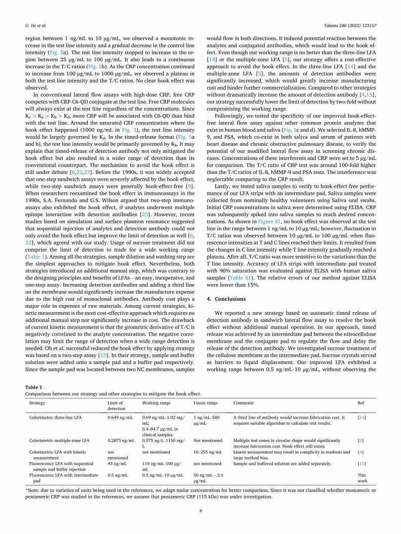
In conventional lateral flow assays with high-dose CRP, free CRP competes with CRP-C6-QD conjugate at the test line. Free CRP molecules will always exist at the test line regardless of the concentrations. Since Kc > Ka > Kb > Kd, more CRP will be associated with C6-QD than bind with the test line. Around the saturated CRP concentration where the hook effect happened (1000 ng/mL in Fig. 3), the test line intensity would be largely governed by Kd. In the timed-release format (Fig. 5a and b), the test line intensity would be primarily governed by Kb. It may explain that timed-release of detection antibody not only mitigated the hook effect but also resulted in a wider range of detection than its conventional counterpart. The mechanism to avoid the hook effect is still under debate [6,22,23]. Before the 1990s, it was widely accepted that one-step sandwich assays were severely affected by the hook effect, while two-step sandwich assays were generally hook-effect-free [9]. When researchers reexamined the hook effect in immunoassays in the 1990s, S.A. Fernando and G.S. Wilson argued that two-step immuno-assays also exhibited the hook effect, if analytes underwent multiple epitope interaction with detection antibodies [23]. However, recent studies based on simulation and surface plasmon resonance suggested that sequential injection of analytes and detection antibody could not only avoid the hook effect but improve the limit of detection as well [6, 22], which agreed with our study. Usage of sucrose treatment did not comprise the limit of detection to trade for a wide working range (Table 1). Among all the strategies, sample dilution and washing step are the simplest approaches to mitigate hook effect. Nevertheless, both strategies introduced an additional manual step, which was contrary to the designing principles and benefits of LFAs—an easy, inexpensive, and one-step assay. Increasing detection antibodies and adding a third line on the membrane would significantly increase the manufacture expense due to the high cost of monoclonal antibodies. Antibody cost plays a major role in expenses of raw materials. Among current strategies, ki-netic measurement is the most cost-effective approach which requires no additional manual step nor significantly increase in cost. The drawback of current kinetic measurement is that the geometric derivative of T/C is negatively correlated to the analyte concentration. The negative corre-lation may limit the range of detection when a wide range detection is needed. Oh et al. successful reduced the hook effect by applying strategy was based on a two-step assay [15]. In their strategy, sample and buffer solution were added onto a sample pad and a buffer pad respectively. Since the sample pad was located between two NC membranes, samples
would flow in both directions. It induced potential reaction between the analytes and conjugated antibodies, which would lead to the hook ef-fect. Even though our working range is no better than the three-line LFA [14] or the multiple-zone LFA [5], our strategy offers a cost-effective approach to avoid the hook effect. In the three-line LFA [14] and the multiple-zone LFA [5], the amounts of detection antibodies were significantly increased, which would greatly increase manufacturing cost and hinder further commercialization. Compared to other strategies without dramatically increase the amount of detection antibody [4,15], our strategy successfully lower the limit of detection by two-fold without compromising the working range.
Followingly, we tested the specificity of our improved hook-effect- free lateral flow assay against other common protein analytes that exist in human blood and saliva (Fig. 5c and d). We selected IL-8, hMMP- 9, and PSA, which co-exist in both saliva and serum of patients with heart disease and chronic obstructive pulmonary disease, to verify the potential of our modified lateral flow assay in screening chronic dis-eases. Concentrations of these interferents and CRP were set to 5 μg/mL for comparison. The T/C ratio of CRP test was around 100-fold higher than the T/C ratios of IL-8, hMMP-9 and PSA tests. The interference was neglectable comparing to the CRP result.
Lastly, we tested saliva samples to verify to hook-effect-free perfor-mance of our LFA strips with an intermediate pad. Saliva samples were collected from nominally healthy volunteers using Saliva oral swabs. Initial CRP concentrations in saliva were determined using ELISA. CRP was subsequently spiked into saliva samples to reach desired concen-trations. As shown in Figure S1, no hook effect was observed at the test line in the range between 1 ng/mL to 10 μg/mL; however, fluctuation in T/C ratios was observed between 10 μg/mL to 100 μg/mL when fluo-rescence intensities at T and C lines reached their limits. It resulted from the changes in C line intensity while T line intensity gradually reached a plateau. After all, T/C ratio was more sensitive to the variations than the T line intensity. Accuracy of LFA strips with intermediate pad treated with 90% saturation was evaluated against ELISA with human saliva samples (Table S1). The relative errors of our method against ELISA were lower than 15%.
4. Conclusions
We reported a new strategy based on automatic timed release of detection antibody in sandwich lateral flow assay to resolve the hook effect without additional manual operation. In our approach, timed release was achieved by an intermediate pad between the nitrocellulose membrane and the conjugate pad to regulate the flow and delay the release of the detection antibody. We investigated sucrose treatment of the cellulose membrane as the intermediate pad. Sucrose crystals served as barriers to liquid displacement. Our improved LFA exhibited a working range between 0.5 ng/mL–10 μg/mL, without observing the
Table 1 Comparison between our strategy and other strategies to mitigate the hook effect.
Strategy Limit of detection
Working range Linear range Comments Ref.
Colorimetric three-line LFA 0.649 ng/mL 0.69 ng/mL–1.02 mg/ mL; 0.4–84.7 μg/mL in clinical samples.
1 ng/mL–500 μg/mL
A third line of antibody would increase fabrication cost. It requires suitable algorithm to calculate test results.
[14]
Colorimetric multiple-zone LFA 0.2875 ng/mL 0.575 ng/L–1150 mg/ L
Not mentioned. Multiple test zones in circular shape would significantly increase fabrication cost. Hook effect still exists.
[5]
Colorimetric LFA with kinetic measurement
not mentioned
not mentioned 10–255 ng/mL kinetic measurement may result in complicity in readouts and large method bias.
[4]
Fluorescence LFA with sequential sample and buffer injection
43 ng/mL 119 ng/mL–100 μg/ mL
not mentioned Sample and buffered solution are added separately. [15]
Fluorescence LFA with intermediate pad
0.5 ng/mL 0.5 ng/mL–10 μg/mL 50 ng/mL – 2.5 μg/mL
This work
*Note: due to varieties of units being used in the references, we adapt molar concentration for better comparison. Since it was not classified whether monomeric or pentameric CRP was studied in the references, we assume that pentameric CRP (115 kDa) was under investigation.
G. He et al.
Talanta 240 (2022) 123157
7
hook effect. Compared to other published strategies, our work offers a cost-effective, one-step approach that is closely unified with the benefits of LFA.
In the future, researchers may combine new label-free strategies based on acoustic, thermal, and other principles to replace the sandwich format in LFAs. Other attempts may be made in researching new bio-recognition elements compatible with LFA or other paper-based assays. Researchers also need to better explain the delivery and distribution of both analytes and detection antibodies and their reactions with each other in the porous medium. After all, LFA is one of the most developed and commercialized paper-based biosensors. Improving LFA is still by large an empirical process. Input-output ratio (in this case, expense- profit ratio) is always an essential factor to be considered when study-ing the LFA and other paper-based biosensors.
Credit author statement
Guozhen He: Conceptualization, Methodology, Software, Validation, Formal analysis, Investigation, Resources, Writing – review & editing, Visualization. Tao Dong: Data curation, Writing – review & editing, Supervision, Project administration, Funding acquisition. Zhaochu Yang: Writing – review & editing, Supervision, Project administration, Resources. Zhuangde Jiang: Supervision, Funding acquisition.
Declaration of competing interest
The authors declare that they have no known competing financial interests or personal relationships that could have appeared to influence the work reported in this paper.
Acknowledgements
This work was supported by projects of RFF Forskningsfond Oslof-jordfondet (Project No. 285575), Regionalt forskningsfond Vestfold og Telemark (Project No. 321814), The support from Chongqing Research Program of Basic Research and Frontier Technology (Project No. cstc2019jcyj-msxmX0776 and cstc2021jcyj-msxmX1038), and Chongq-ing Education Commission – Science and Technology Research Program (Project Nos. KJZD-K201800802, KJZD-K201900802, and KLZD- K202000805) is also acknowledged.
Appendix A. Supplementary data
Supplementary data to this article can be found online at https://doi. org/10.1016/j.talanta.2021.123157.
References
[1] M. Sajid, A.-N. Kawde, M. Daud, Designs, formats and applications of lateral flow assay: a literature review, J. Saudi Chem. Soc. 19 (2015) 689–705, https://doi.org/ 10.1016/j.jscs.2014.09.001.
[2] Y. Gao, Z. Zhu, X. Xi, T. Cao, W. Wen, X. Zhang, S. Wang, An aptamer-based hook- effect-recognizable three-line lateral flow biosensor for rapid detection of thrombin, Biosens. Bioelectron. 133 (2019) 177–182, https://doi.org/10.1016/j. bios.2019.03.036.
[3] N. Sathishkumar, B.J. Toley, Development of an experimental method to overcome the hook effect in sandwich-type lateral flow immunoassays guided by
computational modelling, Sensor. Actuator. B Chem. 324 (2020), 128756, https:// doi.org/10.1016/j.snb.2020.128756.
[4] E.G. Rey, D. O’Dell, S. Mehta, D. Erickson, Mitigating the hook effect in lateral flow sandwich immunoassays using real-time reaction kinetics, Anal. Chem. 89 (2017) 5095–5100, https://doi.org/10.1021/acs.analchem.7b00638.
[5] J. Hu, J.R. Choi, S. Wang, Y. Gong, S. Feng, B. Pingguan-Murphy, T.J. Lu, F. Xu, Multiple test zones for improved detection performance in lateral flow assays, Sensor. Actuator. B Chem. 243 (2017) 484–488, https://doi.org/10.1016/j. snb.2016.12.008.
[6] G.M.S. Ross, D. Filippini, M.W.F. Nielen, G.I.J. Salentijn, Unraveling the hook effect: a comprehensive study of high antigen concentration effects in sandwich lateral flow immunoassays, Anal. Chem. 92 (2020) 15587–15595, https://doi.org/ 10.1021/acs.analchem.0c03740.
[7] A.D. Winder, A.S. Mora, E. Berry, J.R. Lurain, The “hook effect” causing a negative pregnancy test in a patient with an advanced molar pregnancy, Gynecol. Oncol. Rep. 21 (2017) 34–36, https://doi.org/10.1016/j.gore.2017.06.008.
[8] C.-W. Yeung, A.N.Y. Cheung, Negative pregnancy test in patients with trophoblastic diseases, Curr. Obstet. Gynecol. Rep. 3 (2014) 102–106, https://doi. org/10.1007/s13669-013-0067-2.
[9] S. Amarasiri Fernando, G.S. Wilson, Studies of the ‘hook’ effect in the one-step sandwich immunoassay, J. Immunol. Methods 151 (1992) 47–66, https://doi.org/ 10.1016/0022-1759(92)90104-2.
[10] P. Povoa, E. Almeida, P. Moreira, A. Fernandes, R. Mealha, A. Aragao, H. Sabino, C- reactive protein as an indicator of sepsis, Intensive Care Med. 24 (1998) 1052–1056, https://doi.org/10.1007/s001340050715.
[11] L.E.M. Miles, D.A. Lipschitz, C.P. Bieber, J.D. Cook, Measurement of serum ferritin by a 2-site immunoradiometric assay, Anal. Biochem. 61 (1974) 209–224, https:// doi.org/10.1016/0003-2697(74)90347-9.
[12] H.C. Vaidya, B.A. Wolf, N. Garrett, W.J. Catalona, R.V. Clayman, M.H. Nahm, Extremely high values of prostate-specific antigen in patients with adenocarcinoma of the prostate; demonstration of the “hook effect”, Clin. Chem. 34 (1988) 2175–2177, https://doi.org/10.1093/clinchem/34.10.2175.
[13] R.G. Ryall, C.J. Story, D.R. Turner, Reappraisal of the causes of the “hook effect” in two-site immunoradiometric assays, Anal. Biochem. 127 (1982) 308–315, https:// doi.org/10.1016/0003-2697(82)90178-6.
[14] Y.K. Oh, H.-A. Joung, H.S. Han, H.-J. Suk, M.-G. Kim, A three-line lateral flow assay strip for the measurement of C-reactive protein covering a broad physiological concentration range in human sera, Biosens. Bioelectron. 61 (2014) 285–289, https://doi.org/10.1016/j.bios.2014.04.032.
[15] J. Oh, H.-A. Joung, H.S. Han, J.K. Kim, M.-G. Kim, A hook effect-free immunochromatographic assay (HEF-ICA) for measuring the C-reactive protein concentration in one drop of human serum, Theranostics 8 (2018) 3189–3197, https://doi.org/10.7150/thno.24034.
[16] B. Lutz, T. Liang, E. Fu, S. Ramachandran, P. Kauffman, P. Yager, Dissolvable fluidic time delays for programming multi-step assays in instrument-free paper diagnostics, Lab Chip 13 (2013) 2840–2847, https://doi.org/10.1039/ C3LC50178G.
[17] J.J. Pytko-Polonczyk, A. Jakubik, A. Przeklasa-Bierowiec, B. Muszynska, Artificial saliva and its use in biological experiments, J. Physiol. Pharmacol. Off. J. Pol. Physiol. Soc. 68 (2017) 807–813.
[18] D. Gasperino, T. Baughman, H.V. Hsieh, D. Bell, B.H. Weigl, Improving lateral flow assay performance using computational modeling, Annu. Rev. Anal. Chem. 11 (2018) 219–244, https://doi.org/10.1146/annurev-anchem-061417-125737.
[19] M.M. Gong, D. Sinton, Turning the page: advancing paper-based microfluidics for broad diagnostic application, Chem. Rev. 117 (2017) 8447–8480, https://doi.org/ 10.1021/acs.chemrev.7b00024.
[20] G.M. Whitesides, Viewpoint on “Dissolvable fluidic time delays for programming multi-step assays in instrument-free paper diagnostics, Lab Chip 13 (2013) 4004–4005, https://doi.org/10.1039/C3LC90066E.
[21] G. Schreiber, G. Haran, H.-X. Zhou, Fundamental aspects of protein− protein association kinetics, Chem. Rev. 109 (2009) 839–860, https://doi.org/10.1021/ cr800373w.
[22] T. Liang, R. Robinson, J. Houghtaling, G. Fridley, S.A. Ramsey, E. Fu, Investigation of reagent delivery formats in a multivalent malaria sandwich immunoassay and implications for assay performance, Anal. Chem. 88 (2016) 2311–2320, https:// doi.org/10.1021/acs.analchem.5b04222.
[23] S. Amarasiri Fernando, G.S. Wilson, Multiple epitope interactions in the two-step sandwich immunoassay, J. Immunol. Methods 151 (1992) 67–86, https://doi.org/ 10.1016/0022-1759(92)90105-3.
G. He et al.