Missense mutations in the AFG3L2 proteolytic domain account for ∼1.5% of European autosomal...
32
For Peer Review MISSENSE MUTATIONS IN THE AFG3L2 PROTEOLYTIC DOMAIN ACCOUNT FOR ~1.5% OF EUROPEAN AUTOSOMAL DOMINANT CEREBELLAR ATAXIAS Journal: Human Mutation Manuscript ID: humu-2010-0123.R1 Wiley - Manuscript type: Rapid Communication Date Submitted by the Author: 15-Jul-2010 Complete List of Authors: Cagnoli, Claudia; University of Turin, Genetics Biology and Biochemistry Stevanin, Giovanni; INSERM/UPMC UMR_S975 Brussino, Alessandro; University of Turin, Genetics Biology and Biochemistry Mancini, Cecilia; University of Turin, Genetics Biology and Biochemistry Margolis, Russell; Johns Hopkins University School of Medicine Holmes, Susan; Johns Hopkins University School of Medicine Nobili, Marcello; Division of Neurology, Martini Hospital Forlani, Sylvie; INSERM/UPMC UMR_S975 Padovan, Sergio; cnr Pappi, Patrizia; University of Turin, Genetics Biology and Biochemistry Zaros, Cecile; INSERM/UPMC UMR_S975 Le Ber, Isabelle; INSERM/UPMC UMR_S975 Ribai, Pascale; INSERM/UPMC UMR_S975 Pugliese, Luisa; SAFAN bioinformatics assalto, corrado; SAFAN bioinformatics Brice, Alexis; INSERM U679, Hôpital de la Salpêtrière Migone, Nicola; University of Turin, Genetics Biology and Biochemistry Durr, Alexandra; INSERM U679, Hôpital de la Salpêtrière Brusco, Alfredo; University of Turin, Department of Genetics Biology and Biochemistry Key Words: Autosomal Dominant Cerebellar Ataxia, Spinocerebellar ataxia, SCA28, AFG3L2, mutation screening John Wiley & Sons, Inc. Human Mutation
-
Upload
independent -
Category
Documents
-
view
1 -
download
0
Transcript of Missense mutations in the AFG3L2 proteolytic domain account for ∼1.5% of European autosomal...

For Peer Review
MISSENSE MUTATIONS IN THE AFG3L2 PROTEOLYTIC
DOMAIN ACCOUNT FOR ~1.5% OF EUROPEAN AUTOSOMAL
DOMINANT CEREBELLAR ATAXIAS
Journal: Human Mutation
Manuscript ID: humu-2010-0123.R1
Wiley - Manuscript type: Rapid Communication
Date Submitted by the Author:
15-Jul-2010
Complete List of Authors: Cagnoli, Claudia; University of Turin, Genetics Biology and Biochemistry Stevanin, Giovanni; INSERM/UPMC UMR_S975 Brussino, Alessandro; University of Turin, Genetics Biology and Biochemistry Mancini, Cecilia; University of Turin, Genetics Biology and Biochemistry Margolis, Russell; Johns Hopkins University School of Medicine Holmes, Susan; Johns Hopkins University School of Medicine Nobili, Marcello; Division of Neurology, Martini Hospital Forlani, Sylvie; INSERM/UPMC UMR_S975 Padovan, Sergio; cnr Pappi, Patrizia; University of Turin, Genetics Biology and Biochemistry Zaros, Cecile; INSERM/UPMC UMR_S975 Le Ber, Isabelle; INSERM/UPMC UMR_S975 Ribai, Pascale; INSERM/UPMC UMR_S975 Pugliese, Luisa; SAFAN bioinformatics assalto, corrado; SAFAN bioinformatics Brice, Alexis; INSERM U679, Hôpital de la Salpêtrière Migone, Nicola; University of Turin, Genetics Biology and Biochemistry Durr, Alexandra; INSERM U679, Hôpital de la Salpêtrière Brusco, Alfredo; University of Turin, Department of Genetics Biology and Biochemistry
Key Words: Autosomal Dominant Cerebellar Ataxia, Spinocerebellar ataxia, SCA28, AFG3L2, mutation screening
John Wiley & Sons, Inc.
Human Mutation

For Peer Review
Page 1 of 30
John Wiley & Sons, Inc.
Human Mutation
123456789101112131415161718192021222324252627282930313233343536373839404142434445464748495051525354555657585960

For Peer Review
SCA28 Cagnoli C
1
MISSENSE MUTATIONS IN THE AFG3L2 PROTEOLYTIC DOMAIN
ACCOUNT FOR ~1.5% OF EUROPEAN AUTOSOMAL DOMINANT
CEREBELLAR ATAXIAS
Claudia Cagnoli,1,2 Giovanni Stevanin,3,4,5,10 Alessandro Brussino,1,2 Marco Barberis,1,2 Cecilia
Mancini,1,2 Russell L.Margolis,6 Susan E.Holmes,6 Marcello Nobili,7 Sylvie Forlani,3,5 Sergio
Padovan,8 Patrizia Pappi,2 Cécile Zaros,5 Isabelle Leber,3,5 Pascale Ribai,3,4 Luisa Pugliese,9
Corrado Assalto,9 Alexis Brice,3,45 Nicola Migone,1,2 Alexandra Dürr,3,4,5 and Alfredo Brusco1,2
1 Department of Genetics, Biology and Biochemistry, University of Torino 2 S.C. Medical Genetics, A.O.U. San Giovanni Battista, Torino, Italy 3 INSERM, U975 (formerly U679), Paris, France 4 UPMC Univ. Paris 6, UMR_S975, Centre de Recherche de l’Institut du Cerveau et de la Moelle épinière, CNRS 7225,
Pitié-Salpêtrière Hospital, 75013 Paris, France 5 APHP, Pitié-Salpêtrière Hospital, Department of Genetics and Cytogenetics, Paris, France 6 Department of Psychiatry and Behavioral Sciences, Johns Hopkins University School of Medicine, Baltimore,
Maryland, USA 7 Division of Neurology, Martini Hospital, Turin, Italy 8 IBB-CNR c/o Molecular Biotecnology Center University of Torino, Via Nizza 52, I 10125 Torino, Italy 9 S.A.F.AN. BIOINFORMATICS, Turin (Italy) 10 Ecole Pratique des Hautes Etudes (EPHE), Paris, France
Correspondence to: Alfredo Brusco, Dipartimento di Genetica Biologia e Biochimica,
Università degli Studi di Torino, via Santena 19, 10126, Torino, Italy. Fax +390112366662; e-mail:
Running title: SCA28 mutations
Paper word count: 5391
Page 2 of 30
John Wiley & Sons, Inc.
Human Mutation
123456789101112131415161718192021222324252627282930313233343536373839404142434445464748495051525354555657585960

For Peer Review
SCA28 Cagnoli C
2
ABSTRACT
Spinocerebellar ataxia type 28 is an autosomal dominant form of cerebellar ataxia (ADCA) caused
by mutations in AFG3L2, a gene that encodes a subunit of the mitochondrial m-AAA protease. We
screened 366 primarily Caucasian ADCA families, negative for the most common triplet-
expansions, for point mutations in AFG3L2 using DHPLC. Whole-gene deletions were excluded in
300 of the patients, and duplications were excluded in 129 patients. We found six missense
mutations in nine unrelated index cases (9/366, 2.6%): c.1961C>T (p.Thr654Ile) in exon 15,
c.1996A>G (p.Met666Val), c.1997T>G (p.Met666Arg), c.1997T>C (p.Met666Thr), c.2011G>A
(p.Gly671Arg), and c.2012G>A (p.Gly671Glu) in exon 16. All mutated amino acids were located
in the C-terminal proteolytic domain. In available cases, we demonstrated the mutations segregated
with the disease. Mutated amino acids are highly conserved, and bioinformatic analysis indicates
the substitutions are likely deleterious. This investigation demonstrates that SCA28 accounts for
~3% of ADCA Caucasian cases negative for triplet expansions and, in extenso, to ~1.5% of all
ADCA. We further confirm both the involvement of AFG3L2 gene in SCA28 and the presence of a
mutational hotspot in exons 15-16. Screening for SCA28, is warranted in patients who test negative
for more common SCAs and present with a slowly progressive cerebellar ataxia accompanied by
oculomotor signs.
Key Words: Autosomal Dominant Cerebellar Ataxia; Spinocerebellar ataxia; SCA28; AFG3L2;
mutation screening
Page 3 of 30
John Wiley & Sons, Inc.
Human Mutation
123456789101112131415161718192021222324252627282930313233343536373839404142434445464748495051525354555657585960

For Peer Review
SCA28 Cagnoli C
3
INTRODUCTION
Autosomal dominant cerebellar ataxias (ADCAs, or SCAs) are a group of clinically heterogeneous
neurodegenerative disorders primarily characterized by imbalance, progressive gait and limb ataxia,
and dysarthria (Harding, 1993). The clinical phenotype is often complicated by the presence of
additional neurological signs, which are highly variable among and within families (Finsterer, 2009;
Schols, et al., 2004). SCAs are among the most genetically heterogeneous neurodegenerative
diseases, at present nearly thirty genetically distinct subtypes have been defined
(http://www.neuro.wustl.edu/neuromuscular/ataxia/domatax.html). In most countries, an expansion
of a coding CAG-triplet repeat, resulting in production of a protein with an abnormal polyglutamine
(polyQ) stretch, accounts for 40-60% of ADCA. In some populations, as in Southern Brazil,
founder effects have raised this ratio up to ~100% (Jardim, et al., 2001; Storey, et al., 2000). Less
common SCAs are caused by other types of mutations: the expansion of a tri- or penta-nucleotide in
untranslated regions (CAG in SCA12, ATTCT in SCA10, and TGGAA in SCA31), by point
mutations (SCA5, SCA11, SCA13, SCA14, and SCA27), or by gene dosage anomalies (SCA15 and
SCA20) (Fahey, et al., 2005; Holmes, et al., 1999; Houlden, et al., 2007; Ikeda, et al., 2006; Knight,
et al., 2008; Matsuura and Ashizawa, 2002; Sato, et al., 2009; van de Leemput, et al., 2007; Waters,
et al., 2006). Most recently, point mutations of AFG3L2 (OMIM #610246) have been shown to
cause SCA28 (Di Bella, et al., 2010; Edener, et al., 2010). AFG3L2 was originally cloned as a
paralogue of the SPG7 gene (encoding for paraplegin, OMIM *602783) (Banfi, et al., 1999), whose
loss-of-function causes an autosomal recessive form of hereditary spastic paraplegia (HSP) (Casari,
et al., 1998). AFG3L2 encodes for a subunit of the hetero-oligomeric m-AAA protease (ATPases
associated with various cellular activities), a component of the mitochondrial ATP-dependent
metalloprotease located on the inner mitochondrial membrane. The AAA metalloproteases take part
in proteolytic quality control and chaperon-like activities in mitochondria by degrading misfolded
proteins and promoting the assembly of respiratory chain complexes (Leonhard, et al., 1999).
Here we examine the prevalence of SCA28 among Caucasian ADCA families, further defining the
Page 4 of 30
John Wiley & Sons, Inc.
Human Mutation
123456789101112131415161718192021222324252627282930313233343536373839404142434445464748495051525354555657585960

For Peer Review
SCA28 Cagnoli C
4
scope and pathogenicity of AFG3L2 mutations and the clinical features of this disease.
PATIENTS AND METHODS
Patients
We recruited 366 index cases with progressive cerebellar ataxia and a family history of a similar
disorder (defined as the presence of at least two affected individuals in at least two consecutive
generations). The mean age at onset was 41.1 ± 18.3 years (range: 1-79 years; onset defined as the
year of the first symptoms, as reported by the patient or the family). When possible, additional
family members were recruited if a mutation in AFG3L2 was found in a proband. Most patients
originated from France (n= 240), but the collection also included patients from other countries in
Europe (n= 50), the United States (n =66, predominately of European origin), North-Africa/Middle-
East (n= 6), French West-Indies (n= 3) and Madagascar (n= 1). We excluded individuals with
pathogenic expansions in the SCA1-3, 6, 7, 17 and DRPLA loci, or recurrent mutations in the
SCA5, SCA13, SCA14, or FGF14/SCA27 loci (Klebe, et al., 2005) (Stevanin et al., unpublished
data). Clinical and genetic studies were performed after obtaining informed consent from all
participants or the parents of participating minors and with the approval of the local ethics
committees. Ninety-five French and 95 Italian healthy controls were examined to establish the
frequency of variants in the normal population.
Mutation screening
The 17 coding exons of the AFG3L2 gene (RefSeq NM_006796.1) were PCR amplified using
primer and conditions reported in Supp. materials and methods and Supp. Table S1. Mutation
analysis was performed on the amplicons using a DHPLC WAVE System (Transgenomics) with
melting temperatures (Tm and Tm+2°C) determined by DHPLC Melt software (Jones, et al., 1999)
(Supp. Table S1). A normal control profile was always compared with that from a patient. PCR
products showing a DHPLC peak shift were purified using the ExoSAP method (MBI-Fermentas,
Page 5 of 30
John Wiley & Sons, Inc.
Human Mutation
123456789101112131415161718192021222324252627282930313233343536373839404142434445464748495051525354555657585960

For Peer Review
SCA28 Cagnoli C
5
Vilnius, Lithuania), and directly sequenced using the Big-Dye terminator cycle sequencing kit ver.
1.1 and an ABI Prism 3100 Avant automatic sequencer (Applied Biosystems, Foster City, CA). The
17 coding exons of the SPG7 gene (RefSeq NM_003119.2) were also amplified and directly
sequenced following primers and conditions reported in Supp. materials and methods and Supp.
Table S2.
AFG3L2 gene copy number was evaluated by quantitative duplex PCR (qPCR) using a Roche-UPL
assay centered on exon 14 (Roche-Diagnostics, Mannheim, Germany). The gene dosage strategy
was based on the relative amplification of the target sequence (AFG3L2) and the co-amplified
internal standard RNaseP using the comparative delta Ct method described elsewhere (Livak and
Schmittgen, 2001) (see Supp. materials and methods).
Nucleotide numbering throughout the paper follows cDNA numbering: +1 refers to the first
nucleotide of the ATG translation initiation codon of the corresponding RefSeq, according to
journal guidelines (www.hgvs.org/mutnomen). The initiation codon is codon 1.
Haplotype reconstruction
Five 6-FAM fluorescently-labelled microsatellites spanning ~10 Mb on chromosome 18, including
two markers telomeric (D18S1150 and D18S53) and three centromeric (D18453, D18S1104, and
D18S1107) to AFG3L2 were genotyped in all available subjects with a specific mutation whenever
the mutation occurred in at least two unrelated families. We used standard PCR conditions and
primers described at www.genome.ucsc.edu. Haplotypes were manually reconstructed.
Homology modeling of AFG3L2, in-silico
The three-dimensional model of human AFG3L2 protein was generated using “homology
modelling”, a bioinformatics algorithm that builds a model of the "target" protein based on the
homology of its amino acid sequence with that of proteins of known structure (Marti-Renom, et al.,
2000). The AFG3L2 protein model was constructed with the NEST application (Petrey, et al.,
Page 6 of 30
John Wiley & Sons, Inc.
Human Mutation
123456789101112131415161718192021222324252627282930313233343536373839404142434445464748495051525354555657585960

For Peer Review
SCA28 Cagnoli C
6
2003), using the T. maritima FtsH 2CE7 crystal structure as a template (see Supp. materials and
methods). Mutations p.Thr654Ile, p.Met666Val, p.Met666Thr, p.Met666Arg, p.Gly671Glu and
p.Gly671Arg were introduced into the model with the Yasara software (http://www.yasara.org) and
their consequences were evaluated using a series of bioinformatics tools (see also Supp. materials
and methods).
Multiple species alignment of AFG3L2 protein was made with the ClustalW software
(http://www.ebi.ac.uk/Tools/clustalw2/index.html) using orthologue sequences obtained through
the Ensembl genome browser (http://www.ensembl.org). In silico analysis of the possible
pathogenicity of each amino acid substitution was performed with two different applications: (i)
PolyPhen (“Polymorphism Phenotyping”, http://genetics.bwh.harvard.edu/pph/index.html),
selecting the Protein Data-Base (PDB) as a source for sequence alignment, and (ii) SIFT (“Sorting
Intolerant From Tolerant”, http://blocks.fhcrc.org/sift/). Possible effects on splicing were checked
using the applications Splice Site Prediction (http://www.fruitfly.org/seq_tools/splice.html) (Reese,
et al., 1997), ESEfinder 2.0 (http://rulai.cshl.edu/tools/ESE/) (Cartegni, et al., 2003; Smith, et al.,
2006), and PESXs (http://cubweb.biology.columbia.edu/pesx/) (Zhang, et al., 2005). One variant,
predicted to alter splicing, was further investigated using a minigene assay (see Supp. materials and
methods).
RESULTS
SCA28 mutations in ADCA families.
Three-hundred sixty six unrelated patients with ADCA were screened for point mutations in the
AFG3L2 gene using DHPLC, followed by direct sequencing of the amplimers with shifted peaks.
We found six missense changes in nine unrelated index cases: eight were French and one was of
Italian origin (9/366, 2.6%): c.1961C>T (p.Thr654Ile) in exon 15, c.1996A>G (p.Met666Val),
c.1997T>G (p.Met666Arg), c.1997T>C (p.Met666Thr), c.2011G>A (p.Gly671Arg) and
c.2012G>A (p.Gly671Glu) in exon 16 (Figure 1). These mutations were not reported as
Page 7 of 30
John Wiley & Sons, Inc.
Human Mutation
123456789101112131415161718192021222324252627282930313233343536373839404142434445464748495051525354555657585960

For Peer Review
SCA28 Cagnoli C
7
polymorphisms in the dbSNP database build 130 (http://www.ncbi.nlm.nih.gov/projects/SNP/), and
were not found among 380 French or Italian healthy control chromosomes. DNA from 27 additional
family members in six families was available, which allowed us to verify that the mutation
segregated with the disease and was absent in healthy relatives (Figure 2). The mutations were
located in the peptidase-M41 domain of the AFG3L2 protein. Multiple species alignment showed
that the three amino acids altered by mutations (Thr654, Met666 and Gly671) are conserved
through Saccharomyces cerevisiae (Figure 1). The applications PolyPhen and SIFT predicted that
all the amino acid mutations are deleterious. Three mutations (p.Thr654Ile, p.Met666Val and
p.Gly671Arg) were found in two families each. Haplotype reconstruction showed that identical
mutations shared a common haplotype, suggesting that the families have a common ancestor
(Figure 2).
In addition to the six missense mutations, we found 25 variants, 14 of which were neither reported
as polymorphisms in the dbSNP database nor found in healthy controls (Supp. Table S3). Twelve
were intronic nucleotide changes and two were synonymous substitutions. In silico analysis of the
effect of each variant on splicing showed that c.293-13_293-14delTT changes the score of the exon
4 acceptor splice site from 0.22 to 0.03. Neither relatives nor cDNA of this patient were available
for further study. We tested the effect of this mutation using a minigene assay, which showed it has
no effect on splicing.
We also excluded whole-gene deletion/duplication of AFG3L2 in 129/366 subjects (35%) by qPCR
of exon 14, and deletions in 171 patients, who carried one or more heterozygous SNP at DHPLC
and / or direct sequencing.
3D reconstruction of AFG3L2 and predicted effect of the mutations on intermolecular electrostatic
parameters.
The three-dimensional reconstruction of the C-terminal region of AFG3L2, the homohexameric
complex, and the relative positions of mutated amino acids are depicted in Figure 3A-B. Based on
Page 8 of 30
John Wiley & Sons, Inc.
Human Mutation
123456789101112131415161718192021222324252627282930313233343536373839404142434445464748495051525354555657585960

For Peer Review
SCA28 Cagnoli C
8
this model, the amino acids Met666 and Gly671 are always on the surface of the complex,
regardless of the conformation analyzed, suggesting that these two residues may be involved in
interactions with other molecules. p.Gly671Glu significantly increases the electrostatic potential
difference between the inner-mitochondrial-membrane side and the matrix side of the hexamer,
whereas p.Met666Arg and p.Gly671Arg decreased it (Figure 3C and Supp. Table S4). The other
three mutations do not significantly affect the potential difference (Supp. Table S4). Given that the
central pore of the m-AAA complex (Figure 3D) is used to convey substrates to the proteolytic
chamber (Kress and Weber-Ban, 2009), we investigated the effect of the mutations of the amino
acids close to the central pore. As shown in Figure 3E, 3F and Supp. Table S4, p.Gly671Glu
increases the central pore dipole energy, and p.Met666Arg and p.Gly671Arg decreased it. In
addition, we found that all the mutations analyzed decreases the mean interaction energy of the
hexamer, destabilizing the m-AAA complex (Supp. Table S5). Taken together, the bioinformatics
data point to a generalized destabilizing effect of the six mutations detected in our patient
population, with p.Met666Arg, p.Gly671Glu, and p.Gly671Arg also affecting the charge of the
hexamer and, in particular, the charge of the translocation channel.
SPG7 mutation analysis
Since AFG3L2 and paraplegin can interact to form the multimeric m-AAA protease complex, we
speculated that SPG7 variants could modify the SCA28 phenotype. We screened for SPG7 variants
by direct sequencing the coding exons of nine SCA28 patients with the earliest onset (AAD-080_9,
AAR-197_1, SAL-872_6, SAL-872_9, SAL-331_11, AAD-444_3, AAD-444_9, AAD-455_19,
AAD-701_21). Two unreported intronic variants were detected: c.1678+13 C>T in subject 701_21;
c.2196+18 C>G in subject 331_11. Neither variant is predicted to alter splicing.
Clinical features
Table 1 reports the clinical and neuroradiological features of twenty-five SCA28 patients from ten
Page 9 of 30
John Wiley & Sons, Inc.
Human Mutation
123456789101112131415161718192021222324252627282930313233343536373839404142434445464748495051525354555657585960
For Peer Review
SCA28 Cagnoli C
9
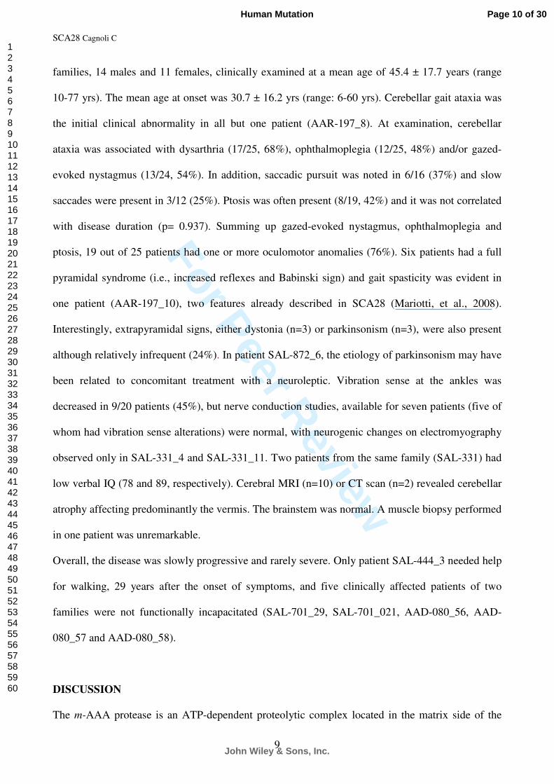
families, 14 males and 11 females, clinically examined at a mean age of 45.4 ± 17.7 years (range
10-77 yrs). The mean age at onset was 30.7 ± 16.2 yrs (range: 6-60 yrs). Cerebellar gait ataxia was
the initial clinical abnormality in all but one patient (AAR-197_8). At examination, cerebellar
ataxia was associated with dysarthria (17/25, 68%), ophthalmoplegia (12/25, 48%) and/or gazed-
evoked nystagmus (13/24, 54%). In addition, saccadic pursuit was noted in 6/16 (37%) and slow
saccades were present in 3/12 (25%). Ptosis was often present (8/19, 42%) and it was not correlated
with disease duration (p= 0.937). Summing up gazed-evoked nystagmus, ophthalmoplegia and
ptosis, 19 out of 25 patients had one or more oculomotor anomalies (76%). Six patients had a full
pyramidal syndrome (i.e., increased reflexes and Babinski sign) and gait spasticity was evident in
one patient (AAR-197_10), two features already described in SCA28 (Mariotti, et al., 2008).
Interestingly, extrapyramidal signs, either dystonia (n=3) or parkinsonism (n=3), were also present
although relatively infrequent (24%). In patient SAL-872_6, the etiology of parkinsonism may have
been related to concomitant treatment with a neuroleptic. Vibration sense at the ankles was
decreased in 9/20 patients (45%), but nerve conduction studies, available for seven patients (five of
whom had vibration sense alterations) were normal, with neurogenic changes on electromyography
observed only in SAL-331_4 and SAL-331_11. Two patients from the same family (SAL-331) had
low verbal IQ (78 and 89, respectively). Cerebral MRI (n=10) or CT scan (n=2) revealed cerebellar
atrophy affecting predominantly the vermis. The brainstem was normal. A muscle biopsy performed
in one patient was unremarkable.
Overall, the disease was slowly progressive and rarely severe. Only patient SAL-444_3 needed help
for walking, 29 years after the onset of symptoms, and five clinically affected patients of two
families were not functionally incapacitated (SAL-701_29, SAL-701_021, AAD-080_56, AAD-
080_57 and AAD-080_58).
DISCUSSION
The m-AAA protease is an ATP-dependent proteolytic complex located in the matrix side of the
Page 10 of 30
John Wiley & Sons, Inc.
Human Mutation
123456789101112131415161718192021222324252627282930313233343536373839404142434445464748495051525354555657585960
For Peer Review
SCA28 Cagnoli C
10
inner mitochondrial membrane and involved, in yeast, in protein quality control and protein
processing (Arlt, et al., 1996; Esser, et al., 2002; Leonhard, et al., 2000). Human m-AAA protease
consists of two isoenzymes: (i) a hetero-oligomeric complex formed by paraplegin (SPG7) and
AFG3L2, and (ii) a homo-oligomeric AFG3L2 complex. Loss-of-function mutations in the SPG7
gene cause an autosomal recessive hereditary spastic paraparesis (HSP), whereas AFG3L2
mutations cause SCA28 (Di Bella, et al., 2010; Edener, et al., 2010). In 366 unrelated ADCA
patients of predominately European ancestry, we found nine affected subjects harboring six
different AFG3L2 mutations, clustered in exons 15 (p.Thr654Ile) and 16 (p.Met666Arg,
p.Met666Thr, p.Met666Val, p.Gly671Arg and p.Gly671Gln). The pathogenicity of these missense
substitutions is supported by multiple lines of evidence: (i) in families where more than one affected
subject was available, we demonstrated that the mutation segregated with the disease and was not
present in healthy subjects of the family. In one of these families (AAD-080), a positive multipoint
LOD score of 2.54 at D18S853 highly suggested its linkage to the SCA28 locus (data not shown).
Moreover, STR-analysis revealed that patients from different families harboring the same mutation
shared a common haplotype, suggesting the presence of common ancestors. (ii) Mutations were not
found in 380 control chromosomes. (iii) The mutated amino acids are evolutionary conserved up to
the S. cerevisiae orthologous protein Yta10, and all are located in the M41-proteolytic domain of
the protein. (iv) Bioinformatic analysis of the protein showed that the mutations alter the interaction
of each monomer with the other five, symptomatic of an altered stability of the complex. Yeast
complementation studies, beyond the scope of this investigation, would further establish the
functional significance of these mutations.
The m-AAA mechanism of action is still under study, although recent investigations of its bacterial
homologue FtsH indicate that the protease ring is stationary and that coordinated ATP-driven
movements of each monomer accomplish substrate unfolding and translocation to the proteolytic
chamber (Augustin, et al., 2009; Bieniossek, et al., 2009). The electrostatic potential difference of
the complex and the dipole of the central pore are altered in three of the six mutations that we
Page 11 of 30
John Wiley & Sons, Inc.
Human Mutation
123456789101112131415161718192021222324252627282930313233343536373839404142434445464748495051525354555657585960

For Peer Review
SCA28 Cagnoli C
11
detected. Furthermore, all six mutations diminish the interaction energy among monomers. These
effects of the mutations suggest that each may have a negative effect on the m-AAA complex
activity. For instance, the capacity of the complex to move substrates towards the proteolytic
chamber through ATPase activity may be impaired by weaker interaction among monomers or by
abnormal electrostatic potentials through the central pore (Striebel, et al., 2009).
Five of the six AFG3L2 missense mutations previously reported, cluster in the proteolytic domain at
amino acids Ser674, Glu691, Ala694, Glu700, and Arg702 (exon 16) (Di Bella, et al., 2010;
Edener, et al., 2010). We found different mutations affecting the same amino acids (p.Gly671Arg
and p.Gly671Glu; p.Met666Arg, p.Met666Thr, and p.Met666Val) indicating that exons 15 and 16
are mutational hotspots for SCA28, and consistent with the hypothesis that disruption of the
peptidase domain is critical to the pathogenesis of SCA28.
Recent evidences from animal models also support the pathogenicity of AFG3L2 mutations: Afg3l2
knock-out mice presents with a severe neuromuscular phenotype, caused by a defect in motor axon
and cerebellar development (Maltecca, et al., 2008), whereas the Afg3l2Emv66 mouse, carrying a
heterozygous loss-of-function mutation, develops a phenotype with similarities to SCA28
(Maltecca, et al., 2009). Whole gene deletions were excluded in 300/366 patients and duplications
in 129/366, suggesting that AFG3L2 deletions/duplications are not common in ADCAs. Moreover,
two related patients with an 18p deletion encompassing AFG3L2 reportedly have not developed
ataxia (Nasir, et al., 2006). Copy number variants encompassing part or whole of AFG3L2 gene
exist in normal individuals (Kim, et al., 2009; Redon, et al., 2006). Therefore, in addition to the
results of our screening designed to detect only large deletions and duplications, the absence of a
phenotype in humans with duplications and deletions affecting AFG3L2, and the different
phenotype observed in Afg3l2 null mice compared to Afg3l2Emv66 mice, suggest that heterozygous
deletions or duplications of the AFG3L2 gene may not cause SCA28. Looking at rearrangements in
SCA28 should therefore not be prescribed in clinical practice.
Our survey shows that SCA28 is rare among European ADCA patients, accounting for ~3% of the
Page 12 of 30
John Wiley & Sons, Inc.
Human Mutation
123456789101112131415161718192021222324252627282930313233343536373839404142434445464748495051525354555657585960
For Peer Review
SCA28 Cagnoli C
12
analyzed cases and ~1.5% of all ADCAs. As a comparison, polyglutamine expansions account for
about 50%. The clinical and neuroradiological phenotype of the newly ascertained families is
similar to that of the SCA28 patients reported so far (Cagnoli, et al., 2006; Edener, et al., 2010;
Mariotti, et al., 2008): disease onset is in adulthood and the disease progresses slowly with
preserved functional capacity, decades after the diagnosis. The mean age at onset and age at onset
variability is similar in our series and in the reported families: in five patients the onset was above
50 yrs, whereas two subjects were symptomatic at 6 and 8 yrs. Even in the young onset cases, the
disease progressed relatively slowly, a much different pattern than has been observed in the
polyglutamine diseases and evidence that SCA28 is not caused by cryptic polyglutamine expansion.
Interestingly, extrapyramidal features were not rare in our series of patients in contrast to previous
reports, of potential value in speculating on the clinical diagnosis prior to genetic testing. As in
many genetic diseases, particularly in those affecting the nervous system, the extent to which
environmental factors affect disease phenotype, including onset age, selective neuronal
involvement, and disease progression, remains to be determined.
Given the signs and symptoms of the disease, we suggest that the diagnosis of SCA28 should be
considered in the presence of slowly progressive ataxia associated with oculomotor abnormalities.
So far, AFG3L2 mutations seem to cluster in exons 10, 15, and 16, which should facilitate genetic
diagnosis in clinical practice and a better understanding of the function and misfunction of
AFG3L2.
Acknowledgments.
We are grateful to the family members for their participation, to the DNA and Cell bank of the
Centre de Recherche de l’Institut du Cerveau et de la Moelle épinière for technical assistance and to
Drs C Marelli, J Yaouang, M Ponsot, H Chneiweiss, D Ménard, S Schaeffer, A Lagueny, L Carluer
and Y Boukhriche as well as Prof A Harding for referring some of the patients and for clinical
examinations.
Page 13 of 30
John Wiley & Sons, Inc.
Human Mutation
123456789101112131415161718192021222324252627282930313233343536373839404142434445464748495051525354555657585960
For Peer Review
SCA28 Cagnoli C
13
This work was funded by Telethon Research grant GGP07110 (to A Brusco), Regione Piemonte
Ricerca Sanitaria Finalizzata, the European Union (to the EUROSCA consortium), the VERUM
foundation (to A Brice) and the Programme Hospitalier de Recherche Clinique (to A Durr).
Page 14 of 30
John Wiley & Sons, Inc.
Human Mutation
123456789101112131415161718192021222324252627282930313233343536373839404142434445464748495051525354555657585960

For Peer Review
SCA28 Cagnoli C
14
FIGURE LEGENDS
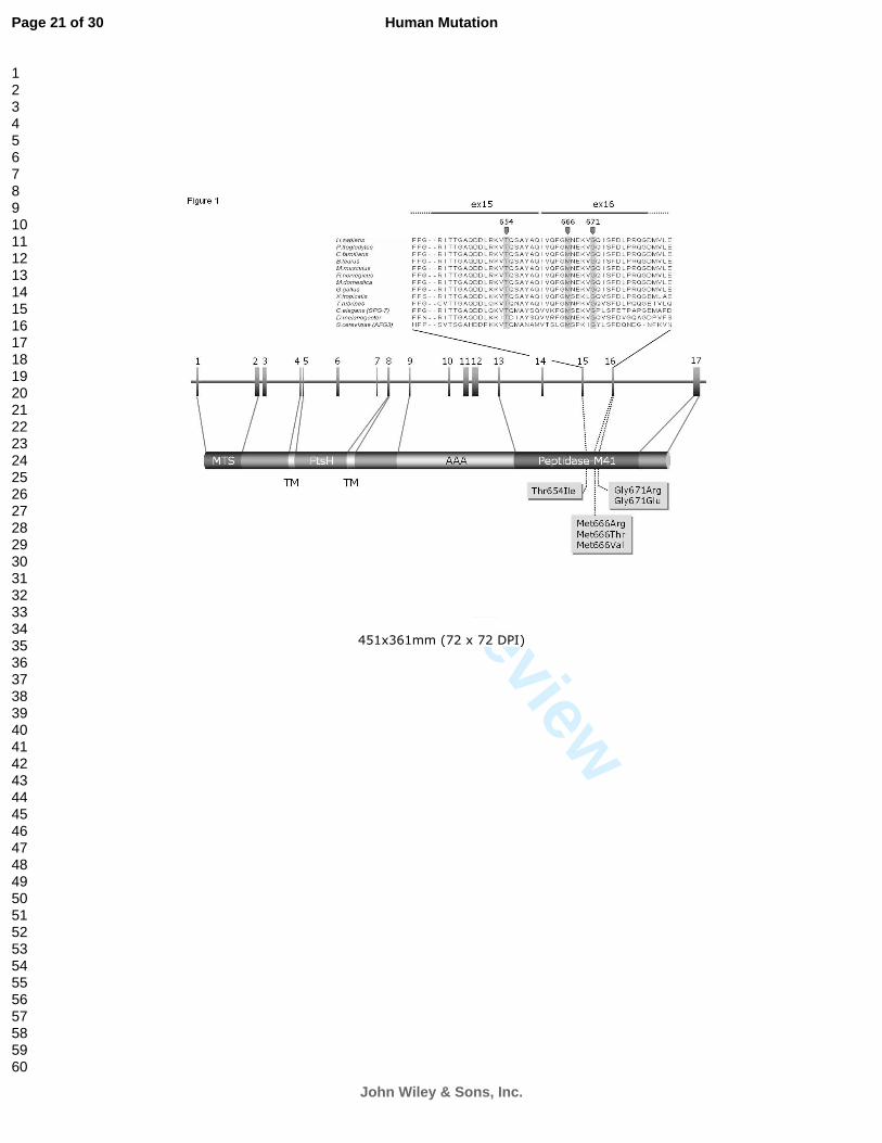
Figure 1.
Localization of the six AFG3L2 missense mutations. In the upper panel the amino acid alignment of
the AFG3L2 protein corresponding to a region including a portion of exons 15 and 16 is shown for
13 orthologous proteins from H. sapiens to S. cerevisiae. The mutated amino acids Thr654, Met666
and Gly671 are highlighted. Below, a scheme of the AFG3L2 genomic region and the protein are
shown. Domain names are: MTS: mitochondrial targeting signal; TM: transmembrane domain;
FtsH: Filamentation temperature sensitive H proteolytic domain; AAA: ATPase Associated with
different cellular activities.
Figure 2.
Pedigrees of the nine families segregating a AFG3L2 mutation. Square and circles shadings: white =
healthy; black = SCA28; gray = presumed SCA28; hash = asymptomatic subject carrying a disease
haplotype. Five STR markers (on the left) were used to reconstruct the SCA28-surrounding
haplotype that suggest a common ancestor for p.Thr541Ile, p.Met666Val and p.Gly671Arg
mutations, in two families each. All subjects whose DNA was available (hyphen above the symbol)
were tested for the AFG3L2 mutation segregating in the family.
Figure 3.
Bioinformatics analysis of AFG3L2 missense changes. Panel A, monomeric structure of the
AFG3L2 protein, with the localization of ATP (yellow), the Zn++ ion (pink) and the three mutated
amino acids, Thr541 (light blue), Met666 (dark blue) and Gly671 (green). Panel B, matrix-side
view of AFG3L2 hexamer (see panel A for color-code of Zn++ and mutated amino acids); a dotted
line shows the cross-section level of panel C. Panel C, electrostatic potential at the surface of the
AFG3L2 hexamer viewed from the matrix side (upper row) and from the IMM side (lower row) is
Page 15 of 30
John Wiley & Sons, Inc.
Human Mutation
123456789101112131415161718192021222324252627282930313233343536373839404142434445464748495051525354555657585960

For Peer Review
SCA28 Cagnoli C
15
reported for the mutations changing the potential difference. The mutation analyzed is reported
between the images of the two surfaces; blue = positive charges, red = negative charges; the black
box identifies the central pore magnified in panels E (membrane side) and F (matrix side). In panels
E and F only the charges of the amino acids closer than 10 Å from the geometric center of the
dipole are shown. Panel D, side-view of the hexamer with the localization of the central
translocation channel colored (blue = positive charges, red = negative charges); wt = wild-type;
IMM = inner mitochondrial membrane.
Page 16 of 30
John Wiley & Sons, Inc.
Human Mutation
123456789101112131415161718192021222324252627282930313233343536373839404142434445464748495051525354555657585960

For Peer Review
SCA28 Cagnoli C
16
REFERENCES
Arlt H, Tauer R, Feldmann H, Neupert W, Langer T. 1996. The YTA10-12 complex, an AAA protease with chaperone-like activity in the inner membrane of mitochondria. Cell 85(6):875-85.
Atorino L, Silvestri L, Koppen M, Cassina L, Ballabio A, Marconi R, Langer T, Casari G. 2003. Loss of m-AAA protease in mitochondria causes complex I deficiency and increased sensitivity to oxidative stress in hereditary spastic paraplegia. J Cell Biol 163(4):777-87.
Augustin S, Gerdes F, Lee S, Tsai FT, Langer T, Tatsuta T. 2009. An intersubunit signaling network coordinates ATP hydrolysis by m-AAA proteases. Mol Cell 35(5):574-85.
Banfi S, Bassi MT, Andolfi G, Marchitiello A, Zanotta S, Ballabio A, Casari G, Franco B. 1999. Identification and characterization of AFG3L2, a novel paraplegin-related gene. Genomics 59(1):51-8.
Bieniossek C, Niederhauser B, Baumann UM. 2009. The crystal structure of apo-FtsH reveals domain movements necessary for substrate unfolding and translocation. Proc Natl Acad Sci U S A 106(51):21579-84.
Cagnoli C, Mariotti C, Taroni F, Seri M, Brussino A, Michielotto C, Grisoli M, Di Bella D, Migone N, Gellera C and others. 2006. SCA28, a novel form of autosomal dominant cerebellar ataxia on chromosome 18p11.22-q11.2. Brain 129(Pt 1):235-42.
Cartegni L, Wang J, Zhu Z, Zhang MQ, Krainer AR. 2003. ESEfinder: A web resource to identify exonic splicing enhancers. Nucleic Acids Res 31(13):3568-71.
Casari G, De Fusco M, Ciarmatori S, Zeviani M, Mora M, Fernandez P, De Michele G, Filla A, Cocozza S, Marconi R and others. 1998. Spastic paraplegia and OXPHOS impairment caused by mutations in paraplegin, a nuclear-encoded mitochondrial metalloprotease. Cell 93(6):973-83.
Di Bella D, Lazzaro F, Brusco A, Plumari M, Battaglia G, Pastore A, Finardi A, Cagnoli C, Tempia F, Frontali M and others. 2010. Mutations in the mitochondrial protease gene AFG3L2 cause dominant hereditary ataxia SCA28. Nat Genet 42(4).
Edener U, Wollner J, Hehr U, Kohl Z, Schilling S, Kreuz F, Bauer P, Bernard V, Gillessen-Kaesbach G, Zuhlke C. 2010. Early onset and slow progression of SCA28, a rare dominant ataxia in a large four-generation family with a novel AFG3L2 mutation. Eur J Hum Genet.
Esser K, Tursun B, Ingenhoven M, Michaelis G, Pratje E. 2002. A novel two-step mechanism for removal of a mitochondrial signal sequence involves the mAAA complex and the putative rhomboid protease Pcp1. J Mol Biol 323(5):835-43.
Fahey MC, Knight MA, Shaw JH, Gardner RJ, du Sart D, Lockhart PJ, Delatycki MB, Gates PC, Storey E. 2005. Spinocerebellar ataxia type 14: study of a family with an exon 5 mutation in the PRKCG gene. J Neurol Neurosurg Psychiatry 76(12):1720-2.
Finsterer J. 2009. Ataxias with autosomal, X-chromosomal or maternal inheritance. Can J Neurol Sci 36(4):409-28.
Gilis D, Rooman M. 2000. PoPMuSiC, an algorithm for predicting protein mutant stability changes: application to prion proteins. Protein Eng 13(12):849-56.
Guerois R, Nielsen JE, Serrano L. 2002. Predicting changes in the stability of proteins and protein complexes: a study of more than 1000 mutations. J Mol Biol 320(2):369-87.
Harding AE. 1993. Clinical features and classification of inherited ataxias. New York: Raven Press. Holmes SE, O'Hearn EE, McInnis MG, Gorelick-Feldman DA, Kleiderlein JJ, Callahan C, Kwak
NG, Ingersoll-Ashworth RG, Sherr M, Sumner AJ and others. 1999. Expansion of a novel CAG trinucleotide repeat in the 5' region of PPP2R2B is associated with SCA12. Nat Genet 23(4):391-2.
Houlden H, Johnson J, Gardner-Thorpe C, Lashley T, Hernandez D, Worth P, Singleton AB, Hilton DA, Holton J, Revesz T and others. 2007. Mutations in TTBK2, encoding a kinase
Page 17 of 30
John Wiley & Sons, Inc.
Human Mutation
123456789101112131415161718192021222324252627282930313233343536373839404142434445464748495051525354555657585960

For Peer Review
SCA28 Cagnoli C
17
implicated in tau phosphorylation, segregate with spinocerebellar ataxia type 11. Nat Genet 39(12):1434-6.
Ikeda Y, Dick KA, Weatherspoon MR, Gincel D, Armbrust KR, Dalton JC, Stevanin G, Durr A, Zuhlke C, Burk K and others. 2006. Spectrin mutations cause spinocerebellar ataxia type 5. Nat Genet 38(2):184-90.
Jardim LB, Silveira I, Pereira ML, Ferro A, Alonso I, do Ceu Moreira M, Mendonca P, Ferreirinha F, Sequeiros J, Giugliani R. 2001. A survey of spinocerebellar ataxia in South Brazil - 66 new cases with Machado-Joseph disease, SCA7, SCA8, or unidentified disease-causing mutations. J Neurol 248(10):870-6.
Jones AC, Austin J, Hansen N, Hoogendoorn B, Oefner PJ, Cheadle JP, O'Donovan MC. 1999. Optimal temperature selection for mutation detection by denaturing HPLC and comparison to single-stranded conformation polymorphism and heteroduplex analysis. Clin Chem 45(8 Pt 1):1133-40.
Jung J, Lee B. 2000. Protein structure alignment using environmental profiles. Protein Eng 13(8):535-43.
Kim KY, Lee GY, Kim J, Jeung HC, Chung HC, Rha SY. 2009. Identification of significant regional genetic variations using continuous CNV values in aCGH data. Genomics 94(5):317-23.
Klebe S, Durr A, Rentschler A, Hahn-Barma V, Abele M, Bouslam N, Schols L, Jedynak P, Forlani S, Denis E and others. 2005. New mutations in protein kinase Cgamma associated with spinocerebellar ataxia type 14. Ann Neurol 58(5):720-9.
Knight MA, Hernandez D, Diede SJ, Dauwerse HG, Rafferty I, van de Leemput J, Forrest SM, Gardner RJ, Storey E, van Ommen GJ and others. 2008. A duplication at chromosome 11q12.2-11q12.3 is associated with spinocerebellar ataxia type 20. Hum Mol Genet 17(24):3847-53.
Kress W, Weber-Ban E. 2009. The alternating power stroke of a 6-cylinder AAA protease chaperone engine. Mol Cell 35(5):545-7.
Krieger E, Darden T, Nabuurs SB, Finkelstein A, Vriend G. 2004. Making optimal use of empirical energy functions: force-field parameterization in crystal space. Proteins 57(4):678-83.
Leonhard K, Guiard B, Pellecchia G, Tzagoloff A, Neupert W, Langer T. 2000. Membrane protein degradation by AAA proteases in mitochondria: extraction of substrates from either membrane surface. Mol Cell 5(4):629-38.
Leonhard K, Stiegler A, Neupert W, Langer T. 1999. Chaperone-like activity of the AAA domain of the yeast Yme1 AAA protease. Nature 398(6725):348-51.
Livak KJ, Schmittgen TD. 2001. Analysis of relative gene expression data using real-time quantitative PCR and the 2(-Delta Delta C(T)) Method. Methods 25(4):402-8.
Maltecca F, Aghaie A, Schroeder DG, Cassina L, Taylor BA, Phillips SJ, Malaguti M, Previtali S, Guenet JL, Quattrini A and others. 2008. The mitochondrial protease AFG3L2 is essential for axonal development. J Neurosci 28(11):2827-36.
Maltecca F, Magnoni R, Cerri F, Cox GA, Quattrini A, Casari G. 2009. Haploinsufficiency of AFG3L2, the gene responsible for spinocerebellar ataxia type 28, causes mitochondria-mediated Purkinje cell dark degeneration. J Neurosci 29(29):9244-54.
Mariotti C, Brusco A, Di Bella D, Cagnoli C, Seri M, Gellera C, Di Donato S, Taroni F. 2008. Spinocerebellar ataxia type 28: a novel autosomal dominant cerebellar ataxia characterized by slow progression and ophthalmoparesis. Cerebellum 7(2):184-8.
Marti-Renom MA, Stuart AC, Fiser A, Sanchez R, Melo F, Sali A. 2000. Comparative protein structure modeling of genes and genomes. Annu Rev Biophys Biomol Struct 29:291-325.
Matsuura T, Ashizawa T. 2002. Spinocerebellar ataxia type 10: a disease caused by a large ATTCT repeat expansion. Adv Exp Med Biol 516:79-97.
Nasir J, Frima N, Pickard B, Malloy MP, Zhan L, Grunewald R. 2006. Unbalanced whole arm translocation resulting in loss of 18p in dystonia. Mov Disord 21(6):859-63.
Page 18 of 30
John Wiley & Sons, Inc.
Human Mutation
123456789101112131415161718192021222324252627282930313233343536373839404142434445464748495051525354555657585960

For Peer Review
SCA28 Cagnoli C
18
Petrey D, Xiang Z, Tang CL, Xie L, Gimpelev M, Mitros T, Soto CS, Goldsmith-Fischman S, Kernytsky A, Schlessinger A and others. 2003. Using multiple structure alignments, fast model building, and energetic analysis in fold recognition and homology modeling. Proteins 53 Suppl 6:430-5.
Redon R, Ishikawa S, Fitch KR, Feuk L, Perry GH, Andrews TD, Fiegler H, Shapero MH, Carson AR, Chen W and others. 2006. Global variation in copy number in the human genome. Nature 444(7118):444-54.
Reese MG, Eeckman FH, Kulp D, Haussler D. 1997. Improved splice site detection in Genie. J Comput Biol 4(3):311-23.
Sato N, Amino T, Kobayashi K, Asakawa S, Ishiguro T, Tsunemi T, Takahashi M, Matsuura T, Flanigan KM, Iwasaki S and others. 2009. Spinocerebellar ataxia type 31 is associated with "inserted" penta-nucleotide repeats containing (TGGAA)n. Am J Hum Genet 85(5):544-57.
Schols L, Bauer P, Schmidt T, Schulte T, Riess O. 2004. Autosomal dominant cerebellar ataxias: clinical features, genetics, and pathogenesis. Lancet Neurol 3(5):291-304.
Smith PJ, Zhang C, Wang J, Chew SL, Zhang MQ, Krainer AR. 2006. An increased specificity score matrix for the prediction of SF2/ASF-specific exonic splicing enhancers. Hum Mol Genet 15(16):2490-508.
Storey E, du Sart D, Shaw JH, Lorentzos P, Kelly L, McKinley Gardner RJ, Forrest SM, Biros I, Nicholson GA. 2000. Frequency of spinocerebellar ataxia types 1, 2, 3, 6, and 7 in Australian patients with spinocerebellar ataxia. Am J Med Genet 95(4):351-7.
Striebel F, Kress W, Weber-Ban E. 2009. Controlled destruction: AAA+ ATPases in protein degradation from bacteria to eukaryotes. Curr Opin Struct Biol 19(2):209-17.
van de Leemput J, Chandran J, Knight MA, Holtzclaw LA, Scholz S, Cookson MR, Houlden H, Gwinn-Hardy K, Fung HC, Lin X and others. 2007. Deletion at ITPR1 underlies ataxia in mice and spinocerebellar ataxia 15 in humans. PLoS Genet 3(6):e108.
Vriend G. 1990. WHAT IF: a molecular modeling and drug design program. J Mol Graph 8(1):52-6, 29.
Waters MF, Minassian NA, Stevanin G, Figueroa KP, Bannister JP, Nolte D, Mock AF, Evidente VG, Fee DB, Muller U and others. 2006. Mutations in voltage-gated potassium channel KCNC3 cause degenerative and developmental central nervous system phenotypes. Nat Genet 38(4):447-51.
Zhang XH, Kangsamaksin T, Chao MS, Banerjee JK, Chasin LA. 2005. Exon inclusion is dependent on predictable exonic splicing enhancers. Mol Cell Biol 25(16):7323-32.
Page 19 of 30
John Wiley & Sons, Inc.
Human Mutation
123456789101112131415161718192021222324252627282930313233343536373839404142434445464748495051525354555657585960

For Peer Review
Table 1. Clinical features of SCA28 patients.
Cerebellar Signs
Subject Mut Sex Yrs at
Onset
Yrs at
Exam
Disease
duration
(yr)
Functional
handicap Gait ataxia Limb ataxia Dysarthria
Horizontal gaze-
evoked
nystagmus
Ophthalmo-
plegia LL Reflexes Ptosis
Bilateral
extensor
plantar
response
Other signs
SAL-331_4 T654I F 20 66 46 Moderate Mild Mild none no no + NA no IQ89
SAL-331_11 T654I M 10 43 33 Moderate Moderate Moderate Severe Yes no + NA no Dystonic head tremor,
IQ78
AAD-274_5 T654I M 60 73 13 Moderate Mild Mild Mild NA no + NA unilateral Parkinsonism
SAL-872_9 M666T M 20 25 5 Moderate Moderate Mild Moderate Yes no N no no Head tremor, cognitive
difficulties
SAL-872_6 M666T F NA 53 NA Moderate Moderate Moderate Severe Yes Vertical N NA no Severe depression, UL
rigidity
AAD-455_19 M666V M 28 39 11 Mild Mild Mild Mild no Vertical
horizontal
N no no -
AAD-455_3 M666V F 50 77 27 Moderate Severe Mild Moderate Yes Vertical,
horizontal
+ Yes Yes -
AAD-701_11 M666V F 50 61 11 Mild Mild Mild Mild no no N no no UL dystonia
AAD-701_15 M666V M 25 58 33 Mild Moderate Moderate Moderate Yes Vertical + Yes no -
AAD-701_29 M666V M 20 30 10 none Mild Moderate Moderate no no + no unilateral
AAD-701_21 M666V M 14 38 24 none Mild Mild none Yes no + no unilateral -
AAD-444_3 M666R M 8 37 29 Severe Moderate Severe Moderate Yes Horizontal + no Yes Memory difficulties
AAD-444_9 M666R M 6 10 4 Mild Mild Mild none no no N no no Behaviour problems
AAD-080_15 G671R F 28 50 22 Moderate Moderate Moderate Moderate no Vertical,
horizontal
+ Yes no -
AAD-080_56 G671R F NA 29 NA none Mild Mild none Yes no + no no -
AAD-080_57 G671R F NA 29 NA none Mild Mild none Yes no + no no -
AAD-080_58 G671R F NA 25 NA none Mild Mild none Yes no + no no -
AAD-080_18 G671R M 44 49 5 Mild Mild Moderate none no Vertical,
horizontal
N Yes no -
AAD-080_31 G671R F 34 39 5 Moderate Moderate Moderate Moderate no no + Yes no -
AAD-080_9 G671R M 23 29 6 Moderate Moderate Moderate Moderate Yes Vertical,
horizontal
+ NA no -
AAR-197_1 G671R F 32 36 4 Moderate Moderate Moderate Mild no no + no no -
AAR-197_10 G671R M 25 51 26 Moderate Moderate Moderate Moderate no Vertical,
horizontal
+ Yes no Cognitive difficulties,
spasticity
AAR-197_21 G671R M 39 43 4 Moderate Moderate Moderate Mild Yes Vertical,
horizontal
NA Yes no -
AAR-197_8 G671R F 53 73 20 Moderate none none none no Vertical,
horizontal
N Yes no Neck dystonia,
Parkinsonism
5336_1 G671E M 55 71 16 Severe Severe Severe Severe Yes no + no Yes -
Notes: LL, lower limbs; UL, upper limbs; N, normal; +, augmented; NA, not available;
Page 20 of 30
John Wiley & Sons, Inc.
Human Mutation
123456789101112131415161718192021222324252627282930313233343536373839404142434445464748495051525354555657585960
For Peer Review
451x361mm (72 x 72 DPI)
Page 21 of 30
John Wiley & Sons, Inc.
Human Mutation
123456789101112131415161718192021222324252627282930313233343536373839404142434445464748495051525354555657585960

For Peer Review
451x361mm (72 x 72 DPI)
Page 22 of 30
John Wiley & Sons, Inc.
Human Mutation
123456789101112131415161718192021222324252627282930313233343536373839404142434445464748495051525354555657585960

For Peer Review
451x361mm (72 x 72 DPI)
Page 23 of 30
John Wiley & Sons, Inc.
Human Mutation
123456789101112131415161718192021222324252627282930313233343536373839404142434445464748495051525354555657585960
For Peer Review
1
SUPPLEMENTARY MATERIALS AND METHODS.
Mutation screening.
The 17 coding exons of the AFG3L2 gene were PCR amplified in a total volume of 50 µl using 30
ng of genomic DNA, 144 µM dNTPs, 2 mM MgCl2, 1 U of Taq Gold (Applied Biosystems, Foster
City, CA, USA) and 200 nM of forward and reverse primers reported in supplementary table S1.
The amplification of exon 1 required a final concentration of 1 M betaine (B0300, Sigma-Aldrich,
St.Louis, MO, USA). Thermal cycling conditions were: 7 min at 95°C, followed by 14 cycles of 30
sec at 95°C, 30 sec at annealing temperature (Ta)+7°C-0.5°C/cycle (see supplementary table 1 and
1 min at 72°C; then 30 cycles consisting of X mn at 95°C, 30 sec at Ta (see supplementary table
S1), and 1 min at 72°C. A final step of 40 cycles consisting of 1 min at 98°C-1°C/cycle was added
in case of DHPLC analysis to increment heteroduplex formation.
The 17 coding exons of the SPG7 gene were amplified and directly sequenced. PCR was performed
in a total volume of 25 µl, using 30 ng of genomic DNA, 144 µM dNTPs, 2.5 mM MgCl2, 0.5 U of
Taq Gold (Applied Biosystems) and 200 nM of forward and reverse primers reported in
supplementary table S2. The amplification of exon 1 required a final concentration of 1 M betaine.
Thermal cycling conditions for exons 1-13 were: 7 min at 95°C, followed by 14 cycles of 30 sec at
95°C, 30 sec at Ta+7°C-0.5°C/cycle (see supplementary table 2 and 1 min at 72°C; then 30 cycles
consisting in X mn at 95°C, 30 sec at Ta, and 1 min at 72°C; with a final extension for 10 min at
72°C. Thermal cycling conditions for exons 14-17 were: 7 min at 95°C, 30 cycles at 95°C, 30 sec
at Ta, and 1 min at 72°C; final extension for 10 min at 72°C. Fragments were purified using the
ExoSAP method (MBI-Fermentas), and directly sequenced using the Big-Dye cycle sequencing kit
and an ABI Prism 3100 Avant automatic sequencer (Applied Biosystems).
Minigene assay.
A minigene assay was set up to test the effect of AFG3L2 c.293-13_293-14delTT sequence variant
on splicing. We amplified a 327 bp fragment from a subject carrying the variant. The forward
Page 24 of 30
John Wiley & Sons, Inc.
Human Mutation
123456789101112131415161718192021222324252627282930313233343536373839404142434445464748495051525354555657585960

For Peer Review
2
primer (5’-ATGCGAATTCTTGTATTCTTTCTCATAGTGCTTCA) introduced a EcoRI
restriction site, and the reverse primer (5’-TACGTCTAGAAATGCCTCCCAACCTTCTCT)
introduced a XbaI restriction site. The fragment was sequenced to exclude the presence of PCR-
introduced mutations. The PCR product was cloned into a pTZ57R/T vector using the InsTA
Cloning kit (Invitrogen), and transformed in DH5α bacterial cells following manufacturer’s
recommendations (RBC Bioscience, Chung Ho City, Taiwan). Plasmids were extracted using
standard methods, checked by sequencing, and digested with EcoRI and XbaI restriction
endonucleases. The insert was gel-purified and sub-cloned into a pAltermax modified vector
(kindly provided by dr. Gareth Eldvige, Wellcome Trust Centre, Oxford, UK, and dr. Ivana
Kurelac, University of Bologna). Plasmids containing the wild-type or the variant sequence were
used to transform HEK293 cells using the TurboFect kit (Fermentas). After 48h, RNA was
extracted with the RNeasy Kit (Qiagen), retrotranscribed with the GoScript kit (Promega). The
cDNA was amplified and sequenced with vector primers.
Page 25 of 30
John Wiley & Sons, Inc.
Human Mutation
123456789101112131415161718192021222324252627282930313233343536373839404142434445464748495051525354555657585960

For Peer Review
3
Supp. Table S1. Primers, PCR and DHPLC conditions for AFG3L2 mutation screening
Exon Primers (Forward/Reverse) PCR Ta (°C) DHPLC Tm (°C) 1
1 5’-GTTGAGAGCTTGGGCTCCTCCGTGA
5’-CCAGTGACCTTGACGTCCGCTCTCC 69 68/69/69.8
2 5’-TTATGACCAGGAAATGAAGC
5’-GGTTGGGTCTTTTGTCTCCTT 59 52.3/ 57.3
3 5’-GATGCATCAGCTGCTTTGAA
5’-GGTAGTTTCCACTGAACAAAG 55 53.8
4 5’-GCTGAGAGAGCTAAAACCTTGC
5’-AGAGAAGGTTGGGAGGCATT 55 58.5/60.0/62.0
5 5’-AGAGAAGGTTGGGAGGCATT
5’-ACCAAAGAAGTGACAGTCAGC 55 56.0/58.0/60.0
6 5’-TGAGCTTAAAAGAGTATCTCAAGTATTTT
5’-TGAGGCAGGTTTTCCTTTCA 56 52.4/53.4/57.0/58.8
7 5’-TTGAACCGTTTAGTGAATTGAACC
5’-GAACCACAGGCAGCACAGTC 56 53.3/55.7/58.1
8 5’-GAAAGCTGGAGGTGAGCAG
5’-GAGGATCGTTTTGGGTCA 55 55.5/57.5/61.8
9 5’-TGTGGATACACAATTTACTTTTCTGGA
5’-GTGCCTCCATCTGTGGTGAA 56 52.2/69.4/62.0
10 5’-CCGATTTATTTCATTTCTTATTCAGAG
5’-GCCTGGACGACAGAGTCA 56 56.4/58.0/60.8
11 5’-GCTATGAATTTGCAGTGCTC
5’-AGGTCCAGTGCTCATGAGTG 57 n.a.2
12 5’-AGGTCCAGTGCTCATGAGTG
5’-TTCTTTACTGTGGGCTTCCT 57 55.6/57.6
13 5’-GATCAGTTTGGGCGTATTTCG
5’-AATCCCTGGCCTCAAATTCA 56 55.5/57.5/58.4
14 5’-TTGTGATAGGCAGCTCAGTC
5’-CAAGCTACACTCCTGCAAAG 57 56.8/61.5/63.0
15 5’-GTCTTCATCTGTAGTAGGATCTTCAA
5’-CGTGCAAATATGAATACATGAGG 56 55.8/56.8
16 5’-GATTTTGTCTGGTTAAAGAACAATCA
5’-CCAACCAAAACAGTCTATCTATCACTTC 56 53.0/56.1/57.8
17 5’-GACTTTGCTGAGTAACTGTATTTAATG
5’-ATGCACCAGCTGAAACCACA 56 53.6/57.2/60.5
Notes: n.a.: not applicable; 1 as determined by DHPLC Melt software;
2 AFG3L2 exon 11 was directly sequenced in
all subjects. Ta=annealing temperature; AFG3L2 RefSeq NM_006796.1
Page 26 of 30
John Wiley & Sons, Inc.
Human Mutation
123456789101112131415161718192021222324252627282930313233343536373839404142434445464748495051525354555657585960

For Peer Review
4
Supp. Table S2. Primers and PCR conditions for SPG7 mutation screening.
Ta=annealing temperature
Exon Primers (Forward/Reverse) Ta (°C)
1 5’-CGCAGGCGCCACGTCAGA
5’-GCCGGGCTGGGCCTTACAGA 63
2 5’-TGTTACCTAAAGCTTTGACCTATTGC
5’-GCTCTGATCACACCATTGTACTGC 58
3 5’-ACACTGTTGTCCTGTATGCCTCC
5’-TCCAGACTGGTTTCACCTTGCTA 58
4 5’-GATGTCGCCCGTGTCTGTTG
5’-CCACAGCCTCACTCTCACAGG 58
5 5’-GGCTCTCTGTTGACTGTAGGGTTG
5’-TCTGTTTCTCAGATTACAAAGCCAA 58
6 5’-CGTAGGGATTCCTCGTCTCATCT
5’-TTCAGGCTACTCTCTGCAACAGG 58
7 5’-GCATCGTGCTGCTGATTTCC
5’-GAGCCCTTCTGGGAGAGGAG 58
8 5’-CGTGACCCAGAGAGACCTTACCT
5’-ACACCAGAGGAAGGATGTGTGAA 58
9 5’-GGGTACAGGAAGAGGCTTTGTTT
5’-CAACCTGTTCTGAAAGACATCGG 58
10 5’-CTCTCTCCCTCCTGTGTCCTG
5’-GGCTTCACACCAAGAAGTGTCTTA 58
11 5’-CGCACCTGTGGCAGTAACTA
5’-AGGCCTCGATGCTGTTTG 56
12 5’-TCCTCCTCTTAAGCCCTGATAGC
5’-TCAATACCTGCCTGGGTATTTCT 58
13 5’-CTGGTCTCGAACTCCTGTCCTCAG
5’-AGGCTTTCCTCTCACATGACCTACA 63
14 5’-GCATCCTGCCTACTGACCTG
5’-GAAAAGCGCTCTGAAACCTC 59
15 5’-TGCTGAGGATGCCTCTGTCT
5’-GCGACCCTTGTGTGGTAGA 59
16 5’-GTGTTCCCAGTCTGCCATTTC
5’-TGTGTGGACACTGTGTGACG 59
17 5’-CCTGGGGACTCACACACTG
5’-CCTCACTTCCCGGACCAC 59
SPG7 RefSeq NM_003119.2
Page 27 of 30
John Wiley & Sons, Inc.
Human Mutation
123456789101112131415161718192021222324252627282930313233343536373839404142434445464748495051525354555657585960

For Peer Review
5
Bioinformatics analysis.
To build a three dimensional model of the C-terminal region of AFG3L2, the sequence
corresponding to residues 306-762 was submitted to PDB-blast
(http://protein.cribi.unipd.it/pdbblast/) that recognized the cell division bacterial protein FtsH with
an R-score of 1E-117. Its crystal structure was present in the protein database (PDB) as 2CE7,
which contains the AAA domain and a protease domain. Crystallization experiments of 2CE7 gave
rise to a tetragonal crystal form containing six monomers per asymmetric unit. These belong to two
virtually identical half-hexamers; the complete hexameric molecules are generated by a
crystallographic two-fold axis.
The AAA ring does not form a regular hexagon and the contacts of one AAA domain with the two
other interacting domains in the ring are much more asymmetric than those observed in the protease
domain.
As a result, three different monomer conformations are present in 2CE7 structure.
In order to build the AFG3L2 hexamer model, we constructed, three different models based on the
conformations of the three monomers present in 2CE7 structure, using the program NEST
(http://trantor.bioc.columbia.edu/programs/jackal/index.html). Subsequently to obtain the hexamer
model we superimposed them to the corresponding 2CE7 monomers in its hexamer using the
program SHEBA (Jung and Lee, 2000). Upon superimposition, the ADP molecule and the Mg2+
and Zn2+ ions were added to the model.
Using the program Yasara (www.yasara.org), we introduced the Thr654Ile, Met666Thr,
Met666Arg, Met666Val, Gly671Glu and Gly671Arg mutations. Next we refined the model using
the program Yasara with the protocol described below.
Each model was refined performing a 150 ps simulation of the homology models using the protocol
described in (Krieger, et al., 2004). It saves one PDB file every 25 picoseconds, and allows the user
to identify the best snapshot giving as output a table with force field energies, PhiPsi, Backbone and
Packing3 WHATIF checks (Vriend, 1990).
Page 28 of 30
John Wiley & Sons, Inc.
Human Mutation
123456789101112131415161718192021222324252627282930313233343536373839404142434445464748495051525354555657585960

For Peer Review
6
In order to choose the best conformer saved during the refinement procedure we derived a quality
parameter defined as follows:
Q = (Energy/10000) - (PhiPsi+Backbone+Packing3) = quality
The conformer having the lowest value of Q was selected for subsequent analyses.
Data on the electric dipole moment calculated using the Yasara software. The difference of
electrostatic potential (mV) was calculated from the measure of the electric dipole moment using
the formula: ( )0
24π εr
d=∆V
⋅⋅. D = electric dipole moment; r = distance between examer surfaces;
ε0 = electric constant in vacuum.
The pH of the AFG3L2 hexamer catalitic site was calculated using pH Calculator software
(http://www.webqc.org/phsolver.php). pKa values were obtained from the PROPKA software ver
2.0 (Guerois, et al., 2002). We calculated the pH of normal hexamer and hexamers containing
identified mutations for all aminoacids distant <0.5 nm from the Zn atom within the catalytic site
(constant arbitrary concentration set at 0.01M).
The energetic stability of AFG3L2 protein and the interaction energy of each monomer were
calculated using the FoldX software ver 3.0 (Guerois, et al., 2002). We calculated both the
interaction energy between one monomer and the two flanking monomers, and between one
monomer and the remaining hexamer.
To verify the possible three-dimensional structure of AFG3L2 homopolymer or heteropolymer, a
3D model of the C-terminal tract of SPG7, a known interactor of AFG3L2 (Atorino, et al., 2003)
was also created following the same protocol as above.
Homo- and Heteropolymers were reconstructed with the software NEST and SHEBA (Jung and
Lee, 2000; Petrey, et al., 2003).
Finally, the software PoPMuSic estimated the stabilizing/destabilizing effect of each aminoacidic
substitution, through the measurement of the free energy difference (∆∆G) of the molecule (Gilis
and Rooman, 2000).
Page 29 of 30
John Wiley & Sons, Inc.
Human Mutation
123456789101112131415161718192021222324252627282930313233343536373839404142434445464748495051525354555657585960

For Peer Review
7
Supp. Table S3. AFG3L2 variants found in 366 ADCA patients.
Variants Exon /
Intron Patients Controls Notes
rs12327346:C>G
c.-92T>C
c.-71C>T
c.-48G>A
c.-32C>T
EX1 20/366
1/366
4/366
2/366
1/366
7/95
0/95
1/95
0/95
0/95
c.293-33G>A
c.293-13_293-14delTT
IVS3 1/366
2/366
0/95
0/95
The variant does not alter splicing
The variant does not alter splicing1
c.400-14C>G IVS4 4/366 2 0/95 The variant does not alter splicing
c.718C>A (p.=) EX7 1/366 n.a. The variant does not alter splicing
rs8097342:G>A IVS7 120/366 n.a.
rs8091858:C>T
c.1027-12T>C
IVS8 27/366
2/366
0/95
0/953
The variant does not alter splicing
rs9966470:A>T
c.1165-24G>A
IVS9 85/366
5/366
21/95
1/95
c.1319-59G>T
c.1319-40C>T
IVS10 3/366
1/366
0/95
0/95
The variants does not alter splicing
The variants does not alter splicing
rs11080572:G>A EX11 349/3664 85/95
5
c.1426+21_1426+22insCAGGTC IVS11 1/366 0/95 The variant does not alter splicing
c.1479G>A (p.=)
EX12 1/366 0/95 The variant does not alter splicing
rs11553521:A>G EX13 125/366 26/95
c.1664-39G>A
c.1664-9T>C
IVS13 8/366
1/366
2/95
0/95
The variant does not alter splicing
c.2175+18G>A IVS16 9/366 4/1906
rs1129115:G>C
c.2325C>T (p.=)
EX17 117/366
1/293
26/95
0/95
Not found in an affected niece
In bold, variants not found among controls and/or in the dbSNP(129). Controls were all of French origin, except for note 3 -
95 Italian controls, and note 6 - 95 Italian and 95 French controls; 1-The effect on splicing was tested using a minigene
assay (see Supp. Materials and Methods); 2-The c.400-14C>G variant was tested in one family and did not segregate from
the affected father to the affected son; 4-Two hundred-24 cases were homozygous (A/A) and 125 heterozygous (G/A); n.a.
= not available; 5-Fifty-seven controls were homozygous (A/A) and 28 heterozygous (G/A).
Page 30 of 30
John Wiley & Sons, Inc.
Human Mutation
123456789101112131415161718192021222324252627282930313233343536373839404142434445464748495051525354555657585960

For Peer Review
8
Supp. Table S4. Mutation effect on the electric dipole of the translocation channel and on the potential
difference between the inner mitochondrial membrane side and the matrix side of the hexamer.
Potential difference - PD
(mV)
PD compared to wild
type (mV)
PD compared to wild
type (%)
Electric dipole
(C m)
wt 22,274 1,79*10-26
T654I 22,60 0,33 1,48% 1,82*10-26
M666V 22,305 0,031 0,14% 1,79*10-26
M666T 22,945 0,671 3,01% 1,85*10-26
M666R 19,548 -2,726 -12,24% 1,57*10-26
G671E 25,736 3,462 15,54% 2,07*10-26
G671R 18,887 -3,387 -15,21% 1,52*10-26
Yellow = wild-type (wt) values; Green = lower dipole and potential difference; Blue = dipole and potential
difference not significantly different from the wild-type; Red = higher dipole and potential difference. mV =
millivolts; C m = Coulomb meter.
Supp. Table S5. Mean interaction energy § (Kcal/Mol) between each monomer of the AAA-
protease (A to F) and the other five monomers
Monomer
A B C D E F Mean1
wt -49.89 -79.31 -57.50 -59.64 -82.24 -54.58 -63.86
T654I -43.23 -76.66 -66.77 -55.32 -76.86 -57.56 -62.73
M666V -56.73 -83.46 -51.03 -37.19 -60.82 -56.99 -57.70
M666T -50.50 -64.60 -46.42 -48.36 -76.81 -58.26 -57.49
M666R -35.73 -71.15 -49.36 -37.83 -58.24 -42.46 -49.13
G671E -49.41 -79.02 -54.77 -61.61 -77.72 -50.24 -62.13
G671R -51.46 -70.63 -59.66 -38.88 -64.79 -50.73 -56.03
1Yellow = wild-type (wt) interaction; Green = lower interaction; Blue = slightly lower interaction
§ Interaction energy was calculated using the application FoldX ver3.0 (Guerois R et al., 2002)
Page 31 of 30
John Wiley & Sons, Inc.
Human Mutation
123456789101112131415161718192021222324252627282930313233343536373839404142434445464748495051525354555657585960