Ministry of Health – Sri Lanka
74
Ministry of Health – Sri Lanka Guidelines for Clinical Management of Dengue Infection in Pregnancy July 2019 In collaboration with SLCOG CCP EPIDEMIOLOGY UNIT SLMA
-
Upload
khangminh22 -
Category
Documents
-
view
0 -
download
0
Transcript of Ministry of Health – Sri Lanka

Ministry of Health – Sri Lanka
Guidelines for Clinical Management of Dengue Infection in Pregnancy
July 2019
In collaboration with
SLCOG CCP EPIDEMIOLOGY
UNIT
SLMA

This document includes the latest concepts on management of dengue haemorrhagic fever in women.
It is an extension to the existing National Guidelines on Clinical Management of DF/DHF in Adults,
published by Epidemiology Unit, Ministry of Health in November 2012.
These guidelines were developed based on the best available evidence at the time of writing. It will
be revised periodically when new evidence becomes available.
Please forward your comments and suggestions to the following address by post or email.
The Epidemiologist
Epidemiology Unit
231, De Saram Place, Colombo 10
e-mail : [email protected]
Electronic version is available on www.epid.gov.lk
ISBN: 978-955-3666-56-7
Printing of this document was supported by Enhanced Dengue Surveillance Project (EDSP),
Epidemiology Unit.

Page i
Guidelines Development and Editorial Committee
Dr. Anula Wijesundere Senior Consultant Physician, President Sri Lanka Medical Association
Dr. Ananda Wijewickrama Consultant Physician, National Institute of Infectious Diseases, IDH
Prof. Athula Kaluarachchi Professor in Obstetrics and Gynecology, Faculty of Medicine, Colombo/ President SLCOG
Prof. Hemantha Senanayake Professor of Obstetrics and Gynecology, Faculty of Medicine, Colombo
Dr. LakKumar Fernando Consultant Paediatrician, DGH Negombo
Dr. Upul Dissanayake Consultant Physician, NHSL, Colombo
Dr. Ramani Pallemulla Consultant Anesthetist, Castle Street Hospital for Women, Colombo
Dr. Priyankara Jayawardena Consultant Physician, De Soysa Maternity Hospital, Colombo
Dr. UDP Ratnasiri Consultant Obstetrician, Castle Street Hospital for Women, Colombo
Dr. Ajitha Wijesundere Consultant Obstetrician
Dr. Damayanthi Idampitiya Consultant Physician, National Institute of Infectious Diseases, IDH
Dr. Lallindra Gooneratne Consultant Haematologist, NHSL, Colombo
Dr. Kapila Jayarathne Consultant Community Physician, Family Health Bureau/ Secretary SLMA
Dr. Dilrukshi Munasinghe Consultant Radiologist, Army Hospital, Colombo
Ms. Geethani Udugamakorala Matron, National Institute of Infectious Disease, IDH
Dr. Jayantha Weeraman Consultant Paediatrician, Epidemiology Unit
Dr. Hasitha Tissera Consultant Epidemiologist/ National Focal Point on Dengue
Editorial Assistance
Dr. Azhar Ghouse Registrar in Community Medicine, Epidemiology Unit
Dr. Ahamed Rikarz Medical officer, National Dengue Control Unit
Cover Design Mr. Ruwan Karunarathne Technical Officer, Epidemiology Unit

Page ii
FOREWORD
Dengue illness continues to be a major health problem in the South and South-east Asian regions and
Sri Lanka is no exception. The out-patient and in-ward departments of most hospitals in Sri Lanka
are seen increasing in the number of adolescent and adult patients with dengue. In this backdrop,
growing number of pregnant women infected with dengue virus can have poor outcome without early
identification and proper medical care.
This new national guidelines on clinical management of dengue in pregnancy, developed by the
Epidemiology Unit, Ministry of Health in collaboration with the Sri Lanka Medical Association, Sri
Lanka College of Obstetricians and Gynecologists and the Ceylon College of Physicians is expected
to further improve existing knowledge and bridge any gaps on this subject. I take this opportunity to
thank all experts who were involved in developing this guideline.
This authoritative document should be used in all levels of health care provision in both public and
private settings in Sri Lanka for the management of dengue and dengue haemorrhagic fever patients
who are pregnant. I am sure this document will help in strengthening the case management and
ultimately reduce the number of severe cases and bring down the deaths due to dengue associated
with pregnancy in Sri Lanka, further.
Dr. Anil Jasinghe Director General of Health Services

Page iii
PREFACE
At the induction ceremony of the President of the SLMA held in January 2019, the title of the address
and the theme for the SLMA for 2019 was introduced as “Facing the challenges and forging ahead
for better health outcomes”. One of the main challenges I have mentioned was reducing the morbidity
and mortality from dengue in Sri Lanka. Soon after my induction, I was requested by the Director,
National Dengue Control Programme, Dr. Hasitha Tissera to invite all stakeholders to form an expert
committee to reduce the maternal morbidity and mortality from dengue fever (DF) and dengue
haemorrhagic fever (DHF).
In recent years Sri Lanka has been successful in reducing the incidence and mortality from DF/DHF.
This has resulted from concerted and persistent efforts by the Central Epidemiology Unit and National
Dengue Control Unit of the Ministry of Health. Public awareness has been increased about causation,
breeding sites and early recognition of DF/DHF. Management of DF/DHF among the medical
profession has improved considerably due to the nationwide dissemination of the national guidelines
regarding management of dengue. This is confirmed by the table below.
Yearly dengue incidence 2016 - 2018
Year No of Cases Case Rate/ 100,000 No of deaths Case fatality rate
2016 55,150 250 97 0.290%
2017 186,101 930 440 0.23%
2018 50,163 250 56 0.11%
Target (2023) 20,000 100 20 <0.1%
Source: National Dengue Control Programme, Annual Report – 2016 - 2018
Despite the overall reduction in morbidity and mortality from DF/DHF, the mortality from maternal
dengue remains unacceptably high. In fact, DHF was the leading cause of maternal deaths in the
analysis of maternal mortality in Sri Lanka, as indicated by the table below.
Maternal Mortality 2016 and 2017
Year Maternal mortality/
100,000 live births
No. of dengue deaths/ total
no. of maternal deaths
Dengue rank in
causes of deaths
2016 33.8 6 / 112 11th cause
2017 39 21 / 127 1st (Leading cause)
Source: Family Health Bureau - Annual Report – 2016 & 2017

Page iv
Needless to say, the mere mention of dengue as the cause of illness results in much anxiety and stress
among the relatives of the affected. The knowledge that a pregnant mother has dengue increases
anxiety tremendously.
The SLMA is extremely happy to give leadership to the development of guidelines to reduce the
maternal morbidity and mobility from DF/DHF. I thank all members of the expert committee who
have worked diligently in the development of the guidelines. My special thanks are due to the
convener, Dr. Hasitha Tissera, the driving force behind the development of this document. I sincerely
hope that the guidelines will be practiced by the clinicians and will assist them in ensuring safe
delivery of mothers affected with dengue and the delivery of healthy babies.
Dr. Anula Wijesundere, President, SLMA

Page v
CONTENTS
Chapter 1 : INTRODUCTION ............................................................................................................. 1 1.1 Clinical manifestations ......................................................................................................... 1 1.2 Gestation and dengue ........................................................................................................... 2
Chapter 2 : NATURAL COURSE OF DENGUE ILLNESS ............................................................... 4 2.1 Clinical diagnosis of dengue illness ..................................................................................... 4 2.2 Dengue fever ........................................................................................................................ 5 2.3 Dengue haemorrhagic fever (DHF) ...................................................................................... 7 2.4 Dengue shock syndrome (DSS) .......................................................................................... 11
Chapter 3 : PHYSIOLOGICAL CHANGES IN PREGNANCY ....................................................... 12 3.1 Cardiovascular changes ...................................................................................................... 12 3.2 Haematological and biochemical changes in pregnancy by trimester ................................ 15 3.3 Respiratory changes in pregnancy ...................................................................................... 17 3.4 Renal changes in pregnancy ............................................................................................... 18
Chapter 4 : HAEMODYNAMIC CHANGES IN DENGUE SHOCK IN PREGNANCY ................ 19 4.1 Natural course of plasma leakage (and acute bleeding) in DHF/DSS ................................ 19 4.2 Changes in vital signs and urine output in compensated and decompensated shock ......... 21 4.3 Features of dengue shock in pregnancy .............................................................................. 22
Chapter 5 : MANAGEMENT OF DENGUE IN PREGNANCY ...................................................... 23 5.1 Early management and admission/referral ......................................................................... 23 5.2 In-ward management of dengue in pregnancy ................................................................... 24 5.3 Management according to trimester by whom and where? ................................................ 24 5.4 Assessment of patient once admitted to relevant unit ........................................................ 25 5.5 General principles of fluid management in DHF ............................................................... 27 5.6 Challenges in recognition of plasma leakage in pregnancy ............................................... 32
Chapter 6 : PARTURITION MANAGEMENT IN DENGUE .......................................................... 33 6.1 Timing and mode of delivery ............................................................................................. 33 6.2 Risk of complications in pregnant women by operative interventions .............................. 35
Chapter 7 : MANAGEMENT OF COMPLICATIONS OF DHF DURING PREGNANCY ............ 37 7.1 Management of shock ......................................................................................................... 37 7.2 Management of bleeding .................................................................................................... 39 7.3 Indications for Other Adjunct Therapy .............................................................................. 43 7.4 Management of multi-organ failure .................................................................................... 43 7.5 Absolute indications for ICU admission ............................................................................ 43 7.6 Managemnt of fluid overload ............................................................................................. 44 7.7 Obstetric conditions which mimic dengue haemorrhagic fever ......................................... 45
Page vi
Chapter 8 : NURSING CARE DURING PREGANCNY AND COUNSELLING ........................... 48
8.1 Nursing care ........................................................................................................................ 48 8.2 Patient communication ....................................................................................................... 50 8.3 Breaking bad news ............................................................................................................. 51 8.4 Convalescent (recovery) phase ........................................................................................... 52 8.5 Discharge criteria ................................................................................................................ 52 8.6 Advice on discharge ........................................................................................................... 53
Chapter 9 : CHALLENGES IN DIAGNOSIS AND MANAGEMENT DURING PREGNANCY .. 54 9.1 Management delays ............................................................................................................ 54 9.2 Steps to minimize management delays ............................................................................... 55
Chapter 10 : CASE STUDIES IN CLINICAL MANAGEMENT ..................................................... 56 10.1 Case - #1 ............................................................................................................................. 57 10.2 Case - #2 ............................................................................................................................. 58 10.3 Case - #3 ............................................................................................................................. 60
Annexure 1 - Observation chart for management of dengue in pregnancy without fluid leakage Annexure 2 - Observation chart for management of dengue in pregnancy with fluid leakage Annexure 3 - Management of massive obstetric haemorrhage (blood loss >40% of blood volume) Annexure 4 - Common haematological parameters during pregancny
Bibliography

Guidelines for Clinical Management of Dengue Infection in Pregnancy Page 1
CHAPTER1:INTRODUCTION
Concern regarding women who are pregnant getting infected with dengue virus has been heightened
in recent years due to an increase in adolescent and adult infections. Pregnant women with dengue
need early identification. Clinical management requires a multi-disciplinary approach and precise
time related interventions for optimal outcome. Early detection and access to proper medical care will
reduce complications and mortality.
This guideline on clinical management of dengue in pregnancy was developed on clinical
experiences, expert committee reports, publications and opinions of practicing clinicians based on
best available evidence at the time of writing. Further, there has been a substantial reduction in
complications and unwarranted deaths in clinical settings practicing the guidance given herein.
1.1 CLINICALMANIFESTATIONS
Dengue virus infection may be asymptomatic or cause a spectrum of symptomatic disease such as
undifferentiated febrile illness, dengue fever (DF), or dengue haemorrhagic fever (DHF) including
dengue shock syndrome (DSS). Out of the symptomatic dengue, DF and DHF are the two main
clinical entities, collectively considered as Dengue Illness. Dengue fever (DF) and DHF are distinct
from each other as DHF is characterized by a period of transient plasma leakage due to increased
capillary permeability, specifically noted in the pleural and peritoneal spaces. When the leaking is
massive and not compensated the resulting hypovolaemia in the vascular space will lead to shock
known as dengue shock syndrome (DSS). The duration of leaking, known as the 'critical phase' is
usually 24-48 hours. Occasionally it can be shorter but unlikely to go beyond 48 hours. DF is
commoner than DHF, but the risk of developing DHF is higher in individuals who have developed
dengue more than once. Haemorrhage, though more likely in DHF, is common to both DF and DHF.
Dengue fever (DF) is more common in older children, adolescents and adults. It is generally an acute
febrile illness with severe headache, myalgia, arthralgia, rashes with leucopenia and
thrombocytopenia. Although DF is usually benign, it could be incapacitating with severe headache,
muscle, joint and bone pain, particularly in adults. Occasionally DF patients will have unusual
haemorrhage such as gastrointestinal bleeding, hypermenorrhoea and massive epistaxis.

Guidelines for Clinical Management of Dengue Infection in Pregnancy Page 2
Traditionally, dengue hamorrhagic fever (DHF) was more common in children less than 15 years of
age in association with repeated dengue infection (secondary infection) with a different virus
serotype. However, the incidence of DHF in adults, including pregnant women, is increasing. Unlike
in DF where usually patients will have a brief febrile phase followed by convalescent phase, in DHF,
patients will have transient plasma leakage into interstitial and serosal spaces (known as Critical
Phase) which has a tendency to develop hypovolemic shock (dengue shock syndrome). Preceding
warning features such as persistent vomiting, abdominal pain, lethargy or restlessness, or irritability
and oliguria are important for early detection of impending shock and intervention to prevent shock.
Altered vascular permeability (Plasma leakage) and abnormal haemostasis (Bleeding) are the main
pathophysiological hallmarks of DHF. Thrombocytopenia and rising haematocrit (HCT) are constant
findings before the onset of shock.
More recently, with the geographical spread of dengue illness and with more involvement of adults,
there has been reports of DF and DHF with unusual or atypical manifestations. These include isolated
organ involvement such as neurological, severe hepatic, renal and other organs. These could be
explained as complications of profound and prolonged shock or associated with underlying co-
infections or co-morbidities. Although a rare clinical entity, such manifestations are now categorized
as expanded dengue syndrome (EDS). Only a rare minority of patients can be classified as EDS.
1.2 GESTATIONANDDENGUE
A higher percentage of severe form of dengue known as Dengue Haemorrhagic Fever (DHF) occurs
among pregnant women compared to non-pregnant women infected with the dengue virus. The
overall severity of DHF is higher in pregnant women than non-pregnant women.
Complications commonly occur when there is dengue infection during the first and third trimesters.
First trimester infection is associated with abortions while third trimester infection may result in
preterm births. Acute dengue illness during third trimester will increase the risk of foetal compromise
due to maternal haemodynamic decompensation inadvertently requiring higher chance of surgical
interventions for delivery.
Early bleeding due to gastric erosions should be anticipated in dengue patients who have taken
NSAIDs or steroids. Significant severe bleeding due to thrombocytopaenia is not commonly seen in
Dengue illness unless there is traumatic procedures during delivery, such as instrumentation or

Guidelines for Clinical Management of Dengue Infection in Pregnancy Page 3
surgery. Dengue illness during labour can be associated with worse maternal outcomes as a result of
massive bleeding due to surgical interventions such as caesarean section and operative vaginal
delivery.
Both mother and the newborn with dengue infection, if progress to DSS undetected, may be at an
increased risk of severe haemorrhage due to coagulopathy. Common causes of death in pregnant
women with dengue can be due prolonged shock with multi-organ failure, massive bleeding, fluid
overload or due to a combination of the above conditions. Delayed or misdiagnosed DHF/DSS in the
early stage will lead to complication and even cause death.
The risk of vertical transmission is well established among women with dengue during the late
pregnancy period. All babies born to such mothers should be closely observed during perinatal period.

Guidelines for Clinical Management of Dengue Infection in Pregnancy Page 4
CHAPTER2:NATURALCOURSEOFDENGUEILLNESS
Dengue viral infection may be asymptomatic or may cause a spectrum of symptomatic clinical disease
with following possible outcomes (Extracted from the National Guidelines) both in pregnant and non-
pregnant women.
1. Undifferentiated Fever
2. Dengue Fever (DF)
3. Dengue Haemorrhagic Fever (DHF)
4. Expanded Dengue Syndrome (EDS)
Among symptomatic dengue patients DF and DHF are the most significant two clinical entities
encountered in practice. During the initial few days termed as febrile phase both these groups are
clinically similar. While DF patients progress to convalescent phase as temperature settles, DHF
patients develop the Critical Phase due to altered vascular permeability leading to plasma leakage.
Hence, the hallmark of DHF is plasma leakage which is not seen in DF.
2.1 CLINICALDIAGNOSISOFDENGUEILLNESS
Early diagnosis of Dengue illness on the first day of fever or at first contact point relies on high index
of suspicion. Clinical features used in non-pregnant women can be used to diagnose probable dengue
in pregnant women as well.
Dengue during pregnancy may lead to unwarranted consequences unless specific attempts are made
to make an early diagnosis. Dengue should be considered high up in the differential diagnosis in any
pregnant woman presenting with acute fever, from any part of Sri Lanka.

Guidelines for Clinical Management of Dengue Infection in Pregnancy Page 5
2.2 DENGUEFEVERDengue Fever (DF) is generally an acute febrile illness (usually due to primary dengue virus infection
from any of the 4 serotypes), with non-specific clinical signs and symptoms. Although DF is a benign
condition, rarely can be associated with unusual bleeding which is a cause of death in DF, if not
recognized and treated early.
Ø Diagnosis of DF is based on clinical, laboratory and epidemiological criteria.
Clinical and laboratory findings are as follows;
Dengue viraemia in a patient is short, typically occurring a day or two before the onset of fever and
lasts for up to four to seven days of illness. During this period the dengue virus, its nucleic acid and
circulating viral antigen can be detected.
Signs and Symptoms of Clinical Dengue Fever (DF)
• Acute onset of high grade fever (Day 1 to 7) with body aches, facial flushing/ diffuse blanching erythema of the skin, back pain, myalgia, arthralgia, retro orbital pain, headache, nausea, vomiting, anorexia, and diarrhoea. Some patients may have sore throat, injected pharynx, and conjunctival injection.
• Rash looks like flushed skin on day 1 to 2, which may resemble measles later or mimic as pregnancy rash.
• Fever associated with respiratory symptoms such as cough and coryza should not exclude the possibility of Dengue illness. Even if such symptoms are suggestive of Influenza, patient should be monitored with repeated FBC and treated symptomatically.
• While rapid NS1/IgM tests will provide an aetiological diagnosis, a negative result should not exclude dengue if clinically suggestive.
• Minor bleeding can manifest as petechial haemorrhages, mucosal bleeding or epistaxis. However, bleeding may be heavy in some patients if they are on medications such as Aspirin, NSAIDS, steroids or long-term anti-platelet drugs.
• Occasionally, unusual haemorrhage such as gastrointestinal bleeding, hypermenorrhea and massive epistaxis may occur, especially GI bleeding is seen in those having an underlying peptic ulcer disease.
• Physical examination may reveal no focus of infection except facial and skin flushing with posterior cervical lymphadenopathy.

Guidelines for Clinical Management of Dengue Infection in Pregnancy Page 6
Viral antigen detection (NS1) has become the most common early diagnostic tool, due to
commercially available rapid test kits.
NS1 Antigen and IgM/IgG Antibodies– the results will depend on the tested day of fever NS1
usually diagnostic on first 3-4 days. Anti-dengue IgM antibody is usually detectable by Day 5 of
the illness, i.e. only NS1 is positive during first few days of illness. In most patients IgM may
persist up to 60 days. In primary infection IgG is usually detectible little after IgM and in
secondary infection IgG will become positive early. Therefore, IgG might be useful to
differentiate primary and secondary dengue infections. If IgG is positive by day 3 it would
indicate a secondary infection and may have some use in predicting DHF. However, results could
also depend on the sensitivity and specificity of the commercially available test kits. If clinically
suggestive of dengue, even if NS1 is negative, consider dengue as a possibility and manage
accordingly.
In specialized laboratories with molecular biology facilities RT-PCR remains the gold standard
of aetiological confirmation. The advantage of this test is its high sensitivity and specificity on
acute sample and identification of serotypes. It is, however, an expensive technology that requires
sophisticated instruments and skilled professionals.
Full Blood Count (FBC) -
• Fever/history of fever and other clinical features with leukopenia (WBC <5,000), is strongly
suggestive of dengue in endemic areas. However, in pregnancy, leukopenia may not be a
feature (often WBC could be normal or high) and serial FBC may only show a drop in the
total WBC count with a significant reversal of lymphocyte to neutrophil ratio with atypical
cells in the blood picture.
• Progressive decrease in WBC count is an early indication of dengue.
• Thrombocytopenia (usually mild <150x109/L)
• Hematocrit (HCT) could be normal or high
UFR – presence of few pus cells and red cells should not exclude dengue (even if UTI is suspected).

Guidelines for Clinical Management of Dengue Infection in Pregnancy Page 7
Epidemiologically dengue is endemic (constantly reported year round) in many parts of Sri Lanka. In
such endemic areas dengue transmission occurs in clusters (several patients reported from one locality
in a particular time period). Therefore, the treating clinician should ask for history of diagnosed cases
of dengue in the family or immediate neighborhood during the past two weeks when a febrile patient
presents to the consultation (spacio-temporal clustering). However, practitioners in non-endemic
areas are likely to receive isolated patients who have acquired dengue infection from endemic areas
(due to travel), particularly during outbreak seasons.
2.3 DENGUEHAEMORRHAGICFEVER(DHF)
In the first few days of DHF, patients will have signs and symptoms similar to DF. However, in DHF,
(usually beyond day 2) patient will develop features of plasma leakage and bleeding which are the
hallmarks of DHF. Abnormal haemostasis in DHF can cause concealed or overt bleeding which may
be significant in some patients.
Features of DHF includes:
1. Fever: acute onset high fever or recent history of acute fever
2. Haemorrhagic manifestations* (at least in the form of a positive tourniquet test)
3. Thrombocytopenia of <130x109/L
4. Objective evidence of selective capillary plasma leaking into chest and abdominal cavities
(visualization of fluid in the peritoneal cavity and pleural space by real-time ultra sound scan
of the abdomen and chest).
*In patients who have evidence of plasma leakage, presence of haemorrhagic manifestations is
not essential for the diagnosis of DHF. However, DHF patients may develop overt or concealed
bleeding during the course of the illness.
Presence of fever with at least 2 signs and symptoms mentioned
above with thrombocytopenia are sufficient to suspect dengue
illness (fever +2+1) in pregnancy.

Guidelines for Clinical Management of Dengue Infection in Pregnancy Page 8
Onset of leaking is heralded by:
- Settling of fever (defervescence)
- FBC changes
- WBC tends to rise following a progressive fall to a nadir (lowest value)
- Thrombocytopenia (<130x109/L)
- Hematocrit (HCT) rising above the baseline
2.3.1 NATURALCAUSEOFDHF
Unlike DF, which has a febrile phase followed by a convalescent phase, DHF patients have 3 phases
which include a critical (leaking) phase between the febrile and convalescent phases. Therefore, DHF
patients are different from DF patients in that they have plasma leakage during the critical phase.
1) Febrile Phase (similar to that of DF - Refer Section 3.2)
2) Critical Phase (Section 3.3.2)
3) Convalescent Phase (Refer Chapter 8)
DHF has two clinical entities: (1) DHF withouth shock and (2) DHF with shock (Dengue Shock
Syndrome - DSS). Therefore, any pregnant woman who prsents with shock (perticularly afebrile at
presentation) consider DSS as a likely dignosis.
2.3.2 CRITICALPHASEINDHF
Critical phase occurs towards the late febrile phase, often after the end of 2nd day of fever, usually
around the 4th or 5th day of illness with defervescence (settling of fever). Some patients may enter the
critical phase while having high fever. However, in pregnancy this may occur outside the usual
duration, earlier or later. Therefore, daily Full Blood Count (FBC) assessments together with
ultrasound scans of abdomen and pelvis of pregnant dengue patients are important to identify plasma
leakage as early as possible.
Dengue fever with haemorrhagic manifestations must be differentiated from
dengue haemorrhagic fever heralded by plasma leakage.
in DF – no plasma leakage
in DHF – plasma leakage is seen

Guidelines for Clinical Management of Dengue Infection in Pregnancy Page 9
Settling of fever is not a sign of recovery, as the patient may leak when fever settles.
2.3.3 WARNINGFEATURESOFCAPILLARYPLASMALEAKAGEINDHF
Patients should be closely observed for following features predicting significant plasma leak. If not
detected and treated appropriately will lead to hypovolemic shock and complications. However, some
DHF patients may develop shock due to significant plasma leakage without any warning features.
• Clinical deterioration with settling of fever
• Severe Abdominal Pain
• Excessive Vomiting
• Bleeding (Epistaxis, Gum Bleeding, Melaena, Vaginal Bleeding)
• Consciousness or Behaviour Changes
• Cold-clammy peripheries
• Prolonged Capillary Refill Time (CRFT >2 seconds)
• No Urine Output for 4-6 hours
2.3.4 FEATURESOFFLUIDLEAKAGE
Features suggestive of fluid leakage:
• Tachycardia with low-volume pulse (>100/bpm), narrowing of pulse pressure with increasing
diastolic pressure, tender hepatomegaly and reduced urine output.
• Haematocrit (HCT) rise (increase of 10% –15% above baseline is an early indirect evidence
of plasma leakage); Rising trend of HCT from baseline value obtained from FBC done on
first few days of febrile phase or normal ANC follow up value. If no baseline hematocrit is
available consider HCT 32 - 34% as the baseline.
• Platelet counts less than 130x109/L or a rapid drop in platelet count (e.g. Platelet count of
200x109/L falls to 80x109/L same day).
• Prolonged CRFT and postural hypotension.
• Biochemical investigations such as serum albumin and non-fasting cholesterol are not useful
during pregnancy.
Plasma leakage in DHF is selective, transient and self-limiting, usually lasting 24-48 hours.

Guidelines for Clinical Management of Dengue Infection in Pregnancy Page 10
Features confirming fluid leakage:
• Selective ultrasonography (USS) of chest and abdomen for evidence of plasma leakage into
the third space. There can be a time gap from initial point of leaking to USS detection of fluid
in pleural and peritoneal cavities. During this period the hematocrit and heart rate may go up
suggesting leaking. In such patients, repeat interval USS to see whether there is objective
evidence of leaking. Eventually in all DHF patients with significant plasma leakage, fluid
will be detectable by USS. If repeated USS does not show fluid and if the patient is
haemodynamically unstable, reduced UOP and low PCV, consider alternative diagnosis
(bleeding).
• Clinical accumulation of fluid in peritoneal and pleural spaces.
2.3.5 ULTRASOUNDFEATURESOFPLASMALEAKAGEINDHF
• Bed side ultrasound scan of the abdomen and the chest on supine position, with no
preparation, identify fluid in body spaces and cavities.
• Presence of oedema of the gall bladder wall with no fluid around it, is not a feature of plasma
leakage. Therefore, it warrants a repeat scan in 4-6 hours to identify possible plasma leakage.
• Gall bladder wall oedema with a thin rim of fluid around it, is the earliest sonographic finding
in plasma leakage.
• Thin rim of fluid in hepato renal recess, indicates approximately six hours has elapsed since
begining of plasma leakage.
• Fluid in the peritoneal cavity, among bowel loops and in the pelvis. This indicates more than
six hours have elapsed since begining of plasma leakage.
Do not depend exclusively on USS for diagnosis of leakage. Other parameters
such as settling of fever, vital signs, platelet count, gradual rise in HCT and
reduced urine output are important to diagnose the onset of leaking in DHF.
However, demostration of fluid in the peritoneal cavity and pleural space on
repeated ultrasound scan confirms that the patient has developed leakage of
plasma and is progressing (patient in critical phase of DHF).
Guidelines for Clinical Management of Dengue Infection in Pregnancy Page 11
• Fluid in the pleural space, is commonly seen on the right side and indicates more than six
hours have elapsed since begining of plasma leakage.
• Fluid in the perirenal space, is commonly seen on the right side. This indicates more than six
hours have elapsed since begining of plasma leakage.
• In a patient where bi-lateral pleaural effusions and ascites is seen; implies that the patient is
in the latter part of the critical phase when major portion of leaking (prabably >50% of
leaking) is over. Fluid therapy for such patients should be carefully guided (vital sings and
HCT) as increased rates of fluid can lead to fluid overload.
2.4 DENGUESHOCKSYNDROME(DSS)Diagnosis of DSS require, DHF criteria mentioned above and evidence of circulatory failure
(hypovolemic shock) in pregnant women observed with;
§ Clinical signs of shock: rapid and weak pulse with delayed CRFT, cold-clammy skin or skin
mottling;
§ Narrowing of pulse pressure to 25mmHg (compensated shock due to plasma leakage);
§ Rising HCT ≥30% from baseline and thrombocytopenia;
§ Hypotension (sBP <80mmHg) with postural giddiness - if present consider possibility of
bleeding in addition to leaking (decompensated shock probably due to leaking and bleeding).
Pathophysiology of shock in DSS is usually due to plasma leakage and subsequent heavy bleeding
when leaking is not corrected. But, presence of hypoglycemia, excessive vomiting leading to
dehydration and sepsis due to co-infection may also contribute towards a state of shock.

Guidelines for Clinical Management of Dengue Infection in Pregnancy Page 12
Chapter3:PHYSIOLOGICALCHANGESINPREGNANCYANDITS
RELATIONSHIPTODENGUE
Significant physiological changes take place in all organ systems during pregnancy, labour and in the
postpartum period. Thorough understanding of these changes and application of that knowledge into
practice is important in the management of a pregnant dengue patient.
3.1 CARDIOVASCULARCHANGES
3.1.1 PULSERATE
Heart rate, which rises throughout gestation, peaks in the late third trimester. The upper limit of resting
heart rate is typically not greater than 95 bpm.
Implications on management of Dengue in pregnancy - Pregnant women with resting heart rates
>100 bpm (without fever) are generally considered to have tachycardia and warrant further
evaluation.
3.1.2 SYSTEMICVASCULARRESISTANCEANDBLOODPRESSURE
Systemic vascular resistance progressively drops by approximately 35 to 40 percent in the mid-second
trimester. Reduced SVR leads to low DBP
DBP and mean arterial pressure decrease more than SBP during pregnancy. This will lead to increase
in pulse pressure. Arterial pressures begin to increase during the third trimester and return close to
preconception levels postpartum.
Implications on management of Dengue in pregnancy – The change in blood pressure will lead to
wider pulse pressure in pregnancy. Narrowed pulse pressure of £ 25mmHg indicates compensatory
shock in dengue with pregnancy. Pulse pressure dropping from 30mmHg to 25mmHg is strongly
suggestive of patient heading towards compensatory shock requiring immediate attention.
Impact of maternal posture on Blood Pressure – The degree of change in BP is acutely influenced
by posture. Assumption of the supine position can lower the venous return to the heart by 30-40%
due to compression of the inferior vena cava by the gravid uterus, leading to a substantial reduction

Guidelines for Clinical Management of Dengue Infection in Pregnancy Page 13
in cardiac output, hence the blood pressure. Maternal hypotension and compression of aorta by gravid
uterus lead to reduction of the utero-placental blood flow.
Implication in the management of Dengue in pregnancy – Always measure the blood pressure in
the complete left lateral or 15-30 degree left laterally tilted position. In order to minimize ineffective
resuscitation of pregnant women with dengue, acquiring this position is crucial.
3.1.3 PULMONARYVASCULARRESISTANCE
There is a reduction in pulmonary vascular resistance, increased pulmonary blood flow with normal
mean pulmonary artery pressure. Serum colloid osmotic pressure is reduced by 10–20% due to
reduced albumin level. The colloid osmotic pressure/pulmonary capillary wedge pressure gradient is
reduced by about 30%, making pregnant women particularly susceptible to pulmonary oedema.
Pulmonary oedema will be precipitated if there is either a sudden increase in cardiac pre-load (such
as rapid fluid bolus) or increased pulmonary capillary permeability (such as in pre-eclampsia) or both.
Implications in the Management of Dengue in pregnancy-Any leaky capillary state may easily
precipitate pulmonary oedema e.g.: excessive use of IVF. As Dengue Haemorrhagic Fever (DHF) is
also a leaky capillary condition, a pre-eclamptic woman with Dengue may have a high-risk of
developing severe pulmonary oedema
SUPINE HYPOTENSIVE SYNDROME IN PREGNANCY
Gravid uterus compresses both IVC and aorta in supine position. Pregnancy results in
development of hypotension, tachycardia (occasionally bradycardia) and syncope called
supine hypotensive syndrome. The normal blood pressure is quickly restored by turning the
patient to left lateral position.
In a DHF patients with plasma leakage, postural hypotension is a feature of volume depletion
(due to leaking/bleeding or both) which may be misinterpreted as supine hypotension
syndrome (and vice versa).
Guidelines for Clinical Management of Dengue Infection in Pregnancy Page 14
3.1.4 PLASMAANDBLOODVOLUME
Plasma volume expands progressively until 30 to 34 weeks, and then plateaus or decreases slightly
through term. The total gain at term is 30 to 50 percent above that in non-pregnant women.
Blood volume gradually increases and reaches about 50% by the 30th week of POA, with a similar
increase in venous return and cardiac output until term. Further increases take place during labour
due to uterine contractions and sympathetic stimulation due to pain. Soon after delivery, release of
aortocaval compression with complete uterine retraction divert about 400-500 ml blood in to
circulation, known as autotransfusion.
Implications on Management of Dengue in pregnancy A pregnant woman may not show features
of hypovolemia even with significant blood loss until late stages (hypotension will appear later than
in a non-pregnant woman).Therefore, a blood transfusion is recommended in DHF after delivery even
with a normal blood loss ( < 500 ml), although its not done in normal pregnant women after delivery.
Utero-placental blood flow has no autoregulation and is dependent
on maternal mean arterial blood pressure for its blood flow. Any
reduction in maternal blood pressure can negatively impact the
uterine blood flow and consequently reduce the utero-placental flow,
thereby compromising foetal perfusion and oxygenation. Uterine
vasculature is exquisitely sensitive to catecholamines and that too
can lead to reduction in utero-placental blood flow. In fact, foetal
distress may be the first indication of maternal hemodynamic
decompensation in pregnancy with leaking in dengue.
Guidelines for Clinical Management of Dengue Infection in Pregnancy Page 15
3.2 HAEMATOLOGICAL AND BIOCHEMICAL CHANGES IN PREGNANCY BY
TRIMESTER
3.2.1 INCREASEDREDCELLMASS
Red blood cell (RBC) mass steadily rises, up to levels 20 to 30 percent higher by the end of pregnancy.
However, the increase in RBC mass is smaller than the increase in plasma volume, which contributes
to the physiologic anemia of pregnancy. Normal Hb is 10.5 - 13.5g/dl and haematocrit is 32-34%.
Implications in the Management of Dengue in pregnancy - HCT of 38-40% may indicate leaking
in DHF (HCT rising towards 20%).
3.2.2 WHITECELLCOUNTANDIMMUNITY
Pregnancy is associated with leukocytosis. WBC counts range from 9000 to 15,000 cells/μl, while
further rise may occur during labour up to 25000/ μl.
Cell mediated immunity is suppressed to prevent rejection of foetus, making pregnant women more
susceptible to viral infections such as Dengue.
Implications in the Management of Dengue in pregnancy – WBC < 5000/ μl. (leucopenia) may
not be seen in febrile phase. A downward trend of WBC is important even in the absence of
leucopenia.
3.2.3 PLATELETCOUNT
A mild decrease in the platelet count, from pre pregnant levels, occurs in all women during an
uncomplicated pregnancy. Gestational thrombocytopenia (GT) is the commonest cause of isolated
thrombocytopenia in pregnancy. However, GT occurs during the second and more commonly in third
trimester. The platelet count seldom drops to <80x109/L in GT. The commonest cause of
thrombocytopenia in the first trimester is immune thrombocytopenia. All women with a platelet count
<100x109/L should be investigated to ascertain its aetiology.

Guidelines for Clinical Management of Dengue Infection in Pregnancy Page 16
Implications in the Management of Dengue in pregnancy – In a patient with fever/history of fever
and a low platelet count it is mandatory to exclude Dengue illness. It is also important to look at
changes in repeat platelet counts and study the trend.
In pre-eclampsia and HELLP syndrome thrombocytopenia is a common feature. Therefore, it is
necessary to exclude dengue before intervention.
If platelet count is <50x109/L spinal anesthesia is contraindicated. Epidural labour analgesia or
anaesthesia is contraindicated when platelet count is <80x109/L. This is to prevent spinal haematoma
formation and resultant spinal cord compression. However, if coagulation is not deranged, an
experienced Anaesthetist may consider a careful single-shot spinal for caesarean section, after platelet
transfusion to increase platelet count to >50x109/L, in special situations such as predicted difficult
airway. General anesthesia is advisable when under experienced hands.
For Caesarean section platelet transfusion is required when the count is <50x109/L and for vaginal
delivery platelet transfusion is required when the count is <30x109/L. Caution should be exercised to
avoid fluid overload in DHF where platelet concentrate might be an option.
3.2.4 COAGULATIONINPREGNANCY
Normal pregnancy is a prothrombotic state. Compared with non-pregnant women, pregnant women
have a marked increase in some coagulation factors, reduced fibrinolysis, and increased platelet
reactivity. As a consequence, there is increased risk for thromboembolic complications.
The activated partial thromboplastin time (APTT) and the prothrombin time are slightly decreased
(shortened). INR is usually < 1.
Fibrinogen levels in normal pregnancy is 3-5g/L, which is much higher than in non-pregnant women;
levels in the third trimester is higher, ranging from 3.7 to 6.2 g/L.
Implications in Management of Dengue in pregnancy - Reduced platelet increases risk of bleeding
with unstable clot. Consumptive coagulopathy can be developed easily, especially in the presence of
a placental abruption.

Guidelines for Clinical Management of Dengue Infection in Pregnancy Page 17
Thromboelastometric (ROTEM) evaluation of coagulation is useful to detect, correct and re-evaluate
any derangement, (using validated dose calculation charts and algorithms), before vaginal delivery,
caesarean section and in patients with heavy bleeding after delivery. The main advantages include
targeted, early correction without overloading the patient by replacing only the required factors
including platelets in smaller volumes and avoidance of unnecessary factor transfusions.
3.2.5 SERUMALBUMINANDNON-FASTINGCHOLESTEROLLEVELS
Serum albumin is low in pregnancy, ranging from 25-28 g/L.
Implications in Management of Dengue in pregnancy - with plasma leakage in DHF serum
albumin also leaks into potential spaces only to return with reabsorption. Indirect feature of
reabsorption is an elevation in serum albumin level in a patient who has completed critical phase.
Non-fasting cholesterol is high in pregnancy (>40 % increase). Therefore, this is not a good indicator
to identify leaking.
3.3 RESPIRATORYCHANGESINPREGNANCY
The normal changes during pregnancy result in a compensated respiratory alkalosis, with a higher
PO2 and a lower PCO2 than in the non-pregnant state.
Decreased FRC and stable FEV1 – Functional residual capacity (FRC) decreases approximately 20
percent during the latter half of pregnancy, making them to have less oxygen reserves than in non-
pregnant.
Increased ventilation and respiratory drive – The most striking change in respiratory physiology
during pregnancy is an increase in resting minute ventilation, which rises by nearly 50 percent at term.
This is primarily due to a larger tidal volume (increased up to 40 percent), whereas the respiratory
rate remains essentially unchanged.
Oxygen consumption is elevated by 25-30%
Therefore, a pregnant woman desaturates faster and becomes hypoxic easily compared to non-
pregnant

Guidelines for Clinical Management of Dengue Infection in Pregnancy Page 18
Implications on Management of Dengue in pregnancy – Respiratory Rate >25 breaths/min with or
without dyspnoea is abnormal and may need attention. Respiratory rate is said to be the first to change
as the woman becomes progressively ill (consider Acidosis/Fluid overload/impending shock).
Respiratory alkalosis - Arterial PCO2 falls to a plateau of 27 to 32 mmHg during pregnancy. Arterial
pH is normal to slightly alkalotic (usually between 7.40 to 7.45).
Implications on Management of Dengue in pregnancy – In a non-pregnant complicated dengue
patient, when pH is ≤7.35 together with HCO3 level <15mmol/l, early correction is recommended to
prevent bleeding and DIC. But in pregnant complicated dengue patient, threshold for correction of
acidosis should be early, if acidosis is persistent even after fluid resuscitation. If the patient has
persistent acidosis, pH <7.35 with HCO3 <15mmol/l it denotes prolonged shock and such patients
are likely to have organ involvement (possible liver, kidney injury.
Maternal arterial oxygen tension - ranges from 106 to 108 mmHg.
Arterial oxygen tension is higher. PaO2 of >70 mmHg and SpO2 of >95% needed for adequate foetal
oxygen delivery.
Implications on Management of Dengue in pregnancy - In DSS (DHF with shock and/or massive
fluid overload) arterial oxygen tension and SpO2 are low due to ventilation perfusion mismatch as a
result of pleural effusion. This may require mask oxygen, positive end expiratory pressure (PEEP) or
mechanical ventilatory support to keep oxygen saturation >95%, and arterial oxygen tension > 70
mmHg.
3.4 RENALCHANGESINPREGNANCY
Glomerular Filtration Rate (GFR) increases by as much as 50-85%. Renal Plasma Flow (RPF)
increases up to 80% as compared with non-pregnant levels. Although frequent urination is often
reported in pregnant women, rarely there could be true polyuria (>3 L/day).
Implications on management of Dengue in pregnancy – In DSS (DHF with shock) with pregnancy,
intense thirst may not appear and despite intravascular volume depletion a normal urine output may
persist. Therefore, urine output may not be a good marker of degree of shock in pregnancy.

Guidelines for Clinical Management of Dengue Infection in Pregnancy Page 19
CHAPTER4:HAEMODYNAMICCHANGESINDENGUESHOCKIN
PREGNANCY
Pregnant woman with DHF in shock is known as Dengue Shock Syndrome (DSS). Any pregnant
woman presenting in shock, DSS should be considered early, in the present hyper-endemic context
in Sri Lanka.
DSS is an extension of DHF state where, excessive plasma leaking has resulted in hypovolemic shock.
However, in a pregnant woman with DSS, severe haemorrhage should be diagnosed early in addition
to plasma leakage. Unless identified early and treated appropriately, this is a life-threatening
condition.
4.1 NATURAL COURSE OF PLASMA LEAKAGE (AND ACUTE BLEEDING) IN
DHF/DSS
X-axis shows time and Y-axis shows the intra-vascular blood volume in a DHF patient. This
illustration of Lines A&B, are shown as linear for explanatory purposes, but in reality are curved
(concave up).
Guidelines for Clinical Management of Dengue Infection in Pregnancy Page 20
Line A
• Shows leaking in a person with normal intravascular volume from onset.
• As the leakage progresses compensatory mechanisms set in resulting in progressive rise
of diastolic blood pressure due to peripheral vasoconstriction, narrowing the pulse
pressure and rise of heart rate. This may require increment of fluid infusion.
• The point at which the intravascular volume where pulse pressure of 25mmHg (α1point)
should be avoided. However, if it occurs fluid has to be replaced soon (i.e. 500ml of NS
within 1 hour as bolus). This α1point is called compensated shock.
• As leakage progresses the compensatory mechanisms breakdown resulting in lower sBP
and the arbitrary point here is set at 80mmHg. This point β1 is called decompensated
shock. In DSS as viscosity of blood is increased due to leaking, organ perfusion may get
compromised earlier than any other condition with low sBP. The fluid replacement here
has to be rapid i.e. pushing the bolus of 500 ml as fast as possible (free flow).
Line B
• Shows leaking in a person who is already in intra-vascular volume depletion from the
onset as a result of dehydration.
• This explains how a person with initial dehydration reaches critical intravascular volume
earlier - α2. If continues to leak without treatment this patient will reach decompensated
shock much earlier – β2 point. This can also be seen in a late presenter who has already
progressive leaking.
Line C (doted and straight)
• C1: Shows a sudden bleed, which brings the intravascular volume to critical state
simultaneously (α2) with steady leakage. Commonly caused by NSAIDs and steroids with
peptic ulcers or underlying bleeding tendency.
• C2: This bleed is more pronounced in later stages of DHF (prolonged and profound
shock) with DIC and associated coagulopathy. Usually results from delayed admission
or late recognition of DSS.

Guidelines for Clinical Management of Dengue Infection in Pregnancy Page 21
The compensatory mechanisms such as progressive rise of heart rate and reduced urine output while
on fluid therapy indicate that the volume replacement is inadequate and need to increase rate of fluid
replacement.
However, along with tachycardia and poor urine output if pulse pressure is narrowed ≤25mmHg
(compensatory shock) or systolic BP is less than 80mmHg (decompensatory shock) resuscitate the
patient with fluid bolus.
Sudden rise of heart rate (without fever or disproportional to fever) may be an indicator of bleeding.
This should be confirmed with a HCT which may be low or equivocal.
4.2 CHANGES IN VITAL SIGNS AND URINE OUTPUT IN COMPENSATED AND
DECOMPENSATEDSHOCK
In this diagram, changes in sBP, dBP and PP are depicted above the dotted line.
Pulse rate, respiratory rate and urine output variations in the compensated and decompensated shock states are depicted below the dotted line.
Temperature during this period is usually normal (afebrile).

Guidelines for Clinical Management of Dengue Infection in Pregnancy Page 22
4.3 FEATURESOFDENGUESHOCKINPREGNANCY
4.3.1 COMPENSATEDSHOCK
Ø Main feature - Narrow pulse pressure ≤25 mmHg in left lateral position.
Ø Associated features (all these may not be present at a given time):
• Clear consciousness
• Cold extremities and prolonged capillary refill time (CRFT) more than 2 seconds
• Tachycardia (>100bpm) with low volume pulse without fever
• Normal sBP with raised dBP in left lateral or seated position
• Postural hypotension
• Intense thirst with reduced urine output or without reduced UOP in pregnancy.
4.3.2 DECOMPENSATEDSHOCK
Ø Main feature
• sBP<80mmHg
• Reduction in sBP >20mmHg
• mean arterial BP <70 mmHg or unrecordable BP at left lateral position
Ø Associated features (all these may not be present at a given time):
• Change of mental state e.g.: restless, combative or lethargy
• Prolonged CRFT with mottled skin, cold-clammy extremities
• Severe tachycardia with weak or absent peripheral pulses
• Oliguria or anuria
• Tachypnea with metabolic acidosis.
In pregnant women with decompensated shock early assessment of degree of shock is important. Such
patients can be further classified as:
a) Prolonged shock – shock lasting more than 4 hours. No passage of urine for 4-6 hours is a
predictor to be confirmed by urinary catheterization (catheter to be inserted provided the
platelet count is reasonable).
b) Prolonged and profound shock – when the patient presents with no peripheral pulses and
BP unrecordable. This condition is complicated with - Acidosis, Bleeding, Hypocalcaemia/
hyponatraemia and hypoglycaemia (A, B, C, S)

Guidelines for Clinical Management of Dengue Infection in Pregnancy Page 23
CHAPTER5:MANAGEMENTOFDENGUEINPREGNANCY
5.1 EARLYMANAGEMENTANDADMISSION/REFERRAL
5.1.1 FIRSTCONTACTCARELEVEL/OPD
• Recognize:
• pregnancy and its trimester
• Suspect dengue early in a patient present with acute febrile illness.
• Look for progressive decrease in WBC count and thrombocytopenia in fresh FBC.
• May screen with RDT-NS1 (within 4 days) or IgM/IgG in late presenters (more than
4 days).
• Even if NS1 is negative clinical features and FBC are suggestive of dengue, the
patient should be managed as dengue.
• Differentiate DHF from DF. Perform ultrasound scan of abdomen and chest to
identify fluid in body cavities and spaces and monitor haematocrit (PCV).
• React:
• 1st and 2nd trimester if patient is haemodynamically stable, admit to ward. If
haemodynamically unstable admit to ETU/HDU.
• 3rd trimester in pregnancy (not in labour) needs early attention through
multidisciplinary team (MDT) and refer to inward care for assessment and close
monitoring by Obstetric and Medical teams.
• Women in labour or in late third trimester should be immediately reviewed by MDT
for further course of action.
• Reduce:
• Reduce late admission to hospital with complication
• Prevent fatalities through early referral to receive appropriate DHF management.

Guidelines for Clinical Management of Dengue Infection in Pregnancy Page 24
5.1.2 ADMISSIONCRITERIA
• All pregnant women with acute onset of fever should be advised to get admitted early on day
1 of illness, where specialists cover is available (despite normal FBC) for further care. First
contact doctor should make a comprehensive assessment of the patient in order to decide on
resuscitation at ETU or management at Obstetric/Medical Unit, irrespective of the duration
of fever.
• Area Medical officer of Health (MOH) should encourage Field Health Staff (Family Health
Officer - PHM, Public Health Inspector - PHI) to identify and refer all pregnant women with
fever seen in the field to local hospital.
• Admission referral letter should include the necessary details and vital parameters.
5.2 IN-WARDMANAGEMENTOFDENGUEINPREGNANCY
Management of Dengue in pregnancy should be of a multi-disciplinary approach involving Obstetric
team, Physician (Internist) and Neonatologist/Paediatrician.
In addition to the obstetric management aspects, obstetrician’s role should include:
a. Identify dengue illness early
b. Differentiate DHF from DF at the early stages (Inward bed side ultrasound scan of the
abdomen and chest to identify fluid in body cavities and spaces).
c. Manage DF patients with the assistance of the medical team
d. Identify early and refer DHF patients to the medical team
e. Inform and update patient’s family regularly and to discuss any interventions in advance
(counselling).
5.3 MANAGEMENTACCORDINGTOTRIMESTERBYWHOMANDWHERE?
All pregnant fever patients irrespective of trimester, should be first admitted to the Obstetric
ward where initial assessment and management plan are to be decided. Further management should
be done in consultation with the medical team.

Guidelines for Clinical Management of Dengue Infection in Pregnancy Page 25
Suggested place of management is as follows, to be decided with the consensus of Medical and
Obstetric team;
Clinical Status 1st Trimester 2nd Trimester 3rd Trimester
(not in labour)
DF in uncomplicated and complicated pregnancy
Whom Obstetric and Medical team
Obstetric and Medical team
Obstetric and Medical Team
Where Obstetric Ward Obstetric Ward Obstetric Ward
DHF in uncomplicated pregnancy
Whom Medical and Obstetric Team
Medical and Obstetric Team
Medical and Obstetric Team
Where Medical Ward HDU
Medical Ward HDU
HDU MICU
DHF/DSS in complicated pregnancy (e.g. GDM, PIH, pre-eclampsia, HELLP, placenta-previa)
Whom Multi-disciplinary Team
Multi-disciplinary Team
Multi-disciplinary Team
Where
MICU or refer to Tertiary Care Center
MICU or refer to Tertiary Care Center
MICU or refer to Tertiary Care Center
Pregnant fever patients presenting in shock should be resuscitated in ETU and admitted to
HDU/MICU for further management with the involvement of a multi-disciplinary team.
5.4 ASSESSMENTOFPATIENTONCEADMITTEDTORELEVANTUNIT
A. Obstetric assessment should be done on daily basis or more often depending on the
trimester
B. Medical assessment includes;
1. Febrile phase monitoring (patient not leaking yet – Ref. 5.4.1)
2. Critical phase monitoring in a leaker (Ref. 5.4.2)
C. Investigation;
1. Serial Full Blood Counts
2. Radiological assessment - bedside ultrasound scan of the abdomen and chest on
admission and thereafter daily or more frequently to detect plasma leakage as
early as possible.

Guidelines for Clinical Management of Dengue Infection in Pregnancy Page 26
5.4.1 FEBRILEPHASEMONITORING
All pregnant women are at high-risk of severe disease and complications. Therefore, should be closely
monitored unit full recovery.
• Once admitted all pregnant women with dengue should be closely monitored in order to
identify leaking and/or bleeding irrespective of platelet count.
• Monitoring is the responsibility of trained nursing officer(s).
• Monitoring of temperature, BP, pulse pressure and CRFT should be done 3 hourly using
the standard Febrile Phase Monitoring Chart (see Annexure 1).
• Chart urine output 4 hourly (minimum 150ml every 4 hours for BW 50kg or more) and
maintain input output chart.
• Essentially monitor inward PCV 6 hourly. Venous PCV is a better assessment of peripheral
perfusion than laboratory HCT.
• Daily FBC and other investigations as necessary (e.g. LFT).
• Notify on-call doctor if any high-risk features are noticed (warning signs, derangement of
vital signs reduced UOP and rising hematocrit).
5.4.2 LEAKINGPHASE(CRITICALPHASE)MONITORING
• The critical (plasma leakage) period of DHF typically starts around the time of the transition
from febrile to afebrile stage.
• When leaking has started, more intense monitoring of parameters on hourly basis is
necessary.
• In a pregnant women with dengue illness when the platelet count drops ≤130x109/L
anticipate leaking shortly. In such patients performing HCT/PCV 3 hourly is likely to show
a rising trend.
• Establish two IV accesses using large bore (preferably 16 G) IV cannulae.

Guidelines for Clinical Management of Dengue Infection in Pregnancy Page 27
Following parameters are to be monitored during the critical phase of DHF in the HDU using the
Critical Phase Monitoring Chart (see Annexure 2)
Monitoring Parameters Frequency
General well-being: • Appetite, vomiting, bleeding, giddiness, intense
thirst, restlessness, clouding of consciousness.
• At least twice-daily
Vital signs: • Temperature, PR, BP, PP and RR (by Multi-
para monitor) • Pulse volume and CRFT
• In Non-shock patients hourly
• In shock patients every 5- 15 minutes (until BP is restored)
Hematocrit (HCT/PCV): • In uncomplicated DHF • In unstable patients or those with suspected/
massive bleeding • Administration of fluid bolus (NS/Dextran-
40/blood)
• Every 3 hours • More frequently if necessary • Before and after each fluid
bolus
Urine output: • Maintained urine output at 0.5-1.0 ml/kg/h
(calculated for pre-pregnancy body weight). Preferred UOP in pregnancy is 0.75ml/kg/h
• Measure 3-4 hourly and calculate
for every hour.
5.5 GENERALPRINCIPLESOFFLUIDMANAGEMENTINDHF
5.5.1 FEBRILEPHASE
• Liquids with electrolyte solutions (minimal use of water) at least double the pre-pregnant
body weight for one hour - approximately 2,500ml/24 hours (for a maximum body weight
of 50kg = 100ml per hour). e.g.: pre pregnant BW 40kg – Fluids 80ml/hr. If unable to take
orally, give partly IV.
• Expected urine output could be calculated according to the pre-pregnant body weight
(measured 4 hourly): e.g. >50kg pre pregnant BW should pass at least 150ml for 4 hrs
(0.75ml/kg/hr).

Guidelines for Clinical Management of Dengue Infection in Pregnancy Page 28
5.5.2 FLUID THERAPY IN NON-SHOCK DHF PATIENTS DURING CRITICAL PERIOD(ORALANDIV)
Fluid management in the leaking phase of dengue is of utmost importance. If too little fluid is given
during leaking, patient may go into shock and may even die. On the other hand, too much of fluid
will lead to fluid overload, which can also lead to death.
• Deciding the onset of the critical phase is very important for the fluid management.
• The amount of fluid and type of fluid given at a particular time will depend on
o the place (how many hours elapsed from the beginning) of the critical phase the
patient is on;
o the amount of fluid used form the total fluid quota up to that time of the critical phase;
o haemodynamic status and urine output;
o haematocrit (PCV).
• Once started, plasma leakage gradually increases, reaching a peak usually around 24 hours
(around the middle of the critical phase). However, high rate of leaking does not persist for
more than a few hours and then it slows down.
• In most patients, the leak is not significant enough to cause haemodynamic instability.
Therefore, they would not require much adjustment of the fluid intake.
• However, in some patients the leak can be significant enough to cause haemodynamic
instability. Identification of such patients and adjusting fluid intake will prevent them from
going into shock.
• Total Fluid Quota (TFQ) should be manipulated according to the patient’s haemodynamic
status, PCV and UOP as too much fluid could lead to fluid overload and too little fluid can
lead to shock.
• Amount of fluid can be gradually increased during the critical phase depending on the need
but should be gradually reduced during the latter part of the critical phase
• If the PCV continued to increase or if the patient’s haemodynamic parameter/s become
abnormal, this indicates significant reduction of intravascular volume.

Guidelines for Clinical Management of Dengue Infection in Pregnancy Page 29
o Amount of fluid should be increased in this instance if this happens during the early
or middle part of the critical phase. (note: see also the indication for blood
transfusion)
o However if this happens in the second half of the critical phase with increasing PCV,
Dextran-40 bolus (500ml over one hour for body weight of 50 kg or more) should be
given and thereafter fluid should be reduced.
• Delayed treatment of intravascular hypovolaemia can result in prolonged shock, leading to
organ failure with a high mortality.
• If the patient is haemodynamically stable (non-shock DHF), the TFQ should spread over 48
hours. In a haemodynamically unstable patient (i.e. presenting in shock), TFQ can be given
over 24-36 hours.
• Once two thirds of the fluid quota is over, assess the patient’s condition to see whether the
patient need continuation of crystalloids, colloid or oral fluids.
• With adequate fluid replacement haemodynamic parameters should be stable and the UOP
should be at least 0.75ml/kg/hr.
• Fluid replacement should not be aimed at normalizing the PCV. If the PCV remains around
more than 10% of the baseline (e.g. PCV of 33 if the baseline PCV is 30) fluid replacement
is adequate provided haemodynamic parameters and UOP are satisfactory.
• If the PCV increases by more than 20% from the baseline, this indicates continuing significant
plasma leakage. Such patients need more fluid (or dextran) even if the haemodynamic
parameters and UOP are normal.
Calculation of the total fluid quota for a patient during the critical phase:
During the critical phase a measured amount of fluids (both oral and IV) should be given.
• Fluid requirement, both oral and intravenous, in critical phase is calculated as M+5%
(Maintenance + 5% deficit) using the pre-pregnancy weight (taking maximum weight as 50kg =
4600ml/48hrs). If pre pregnant weight is <50kg calculate TFQ as given below;
M + 5% is calculated as follows:
- For the first 10 kg - 100ml/kg
- For the second 10 kg - 50ml/kg
- From 20Kg and above up to 50kg - 20ml/kg
(5% deficit is calculated as 50ml/kg up to maximum of 50kg of pre-pregnancy body weight)

Guidelines for Clinical Management of Dengue Infection in Pregnancy Page 30
• While it is not necessary to adhere to this fluid volume strictly, one has to keep in mind that exceeding this may lead to fluid overload.
Adjusting fluid in DHF:
• If a patient, who is on maintenance fluid volume (e.g. 100 ml/hour), drops the output to less than
0.75ml/kg/hour for 3-4 consecutive hours, increase the fluid volume to 125-150 ml/hour for 3-4
hours. Look for other evidence of plasma leakage. If the output remains low, increase the fluid
further to 150-175 ml/hour for next 2-3 hours. (on the other hand, if the output improves, same
fluid volume can be continued for another 2-3 hours and reassess the parameters together with
PCV).
o If the output does not improve with increase of fluid and if the PCV increases, increase the
amount of fluid further (to 200-250 ml/hour). However, such high fluid rate should not be
maintained for many hours as patient can get overloaded. If the out-put does not improve
with this high rate of fluid, fluid should be changed to a dextran bolus.
o If the output does not improve with increase of fluid and if the PCV remain same or decreases,
possibility of bleeding and therefore a blood transfusion should be considered.
• Same fluid adjustment can be applied if other haemodynamic parameters become unstable.
• Decision on fluid therapy (adjustments) should be made on vitals parameters monitored (pulse
pressure/ HR/ BP/ UOP) and capillary haematocrit (PCV)
• Decision to give what type of IV fluid (normal saline/Dextran-40/blood) should be based on
inward PCV and quantity of fluid already used.
5.5.3 INDICATIONANDADMINISTRATIONOF10%DEXTRAN-40:
• Signs of Fluid Overload:
o Dyspnea, tachypnea
o Puffy eyelids, tense/distended abdomen
o Positive lung signs: crepitation, rhonchi, wheezing
For pregnant women in DHF with pre pregnant body weight ≥50kg, recommended
fluid quota during Critical Phase is 4600 ml (both oral and IV) spread over 48 hrs.

Guidelines for Clinical Management of Dengue Infection in Pregnancy Page 31
• Persistently high PCV >30% haemoconcentration for >3 - 6 hours despite adequate crystalloid
resuscitation.
e.g. if baseline PCV is 35 a 10% rise is 3.5 and therefore 30% rise is 3.5x3 = 10.5 (A 30%
haemoconcentration = 35+10.5 = 45.5%) Therefore, PCV of >45.5% for 3-6 hrs is an indication
for Dextran-40.
10% Dextran-40 in NSS is effective in DHF/DSS patients with severe plasma leakage or those with
signs and symptoms of fluid overload is because of its hyper-oncogenicity. It has an osmolarity 2.7
times that of plasma. Dextran-40 is a plasma expander and can hold the volume better than crystalloid
and other colloids that are iso-oncotic to plasma. Other colloidal solutions including Fresh Frozen
Plasma (FFP) and 5% Albumin are in the plasma substitution group (osmolality = plasma); therefore,
is not effective and will worsen fluid overload.
How to give Dextran-40 *
Dextran-40 is given as a bolus only during the Critical Phase of DHF and should be added to the total
fluid quota (M+5%).
• Always give in a bolus dose:
o 10 ml/kg/hr in <50kg pre pregnant weight
o 500 ml/hr in ≥50kg pre pregnant weight at a time
• Do PCV before and immediately after Dextran bolus
o If PCV drops > 10 points, consider significant bleeding
o If PCV drops below baseline, consider bleeding
• Maximum dose
o 30 ml/kg/24 hrs in <50kg pre-pregnant woman
o 1,500 ml/24 hrs in ≥50kg pre-pregnant woman
Clinical and laboratory parameters indicating adequacy of fluid resuscitation/therapy
• Clinical parameters:
o Improvement of general well-being / good orientation & mental state
o Warm peripheries with CRFT ≤ 2 sec
o Adequate urine output (>0.75ml/kg/hr)
*Source: Professor Siripen Kalayanarooj (MD), WHO Collaborating Centre for Case Management of Dengue/DHF/DSS, Bangkok, Thailand.

Guidelines for Clinical Management of Dengue Infection in Pregnancy Page 32
• Vital signs and biochemical parameters:
o Stable BP with maintaining pulse pressure more than 30mmHg
o Reduction in tachycardia or normalization of heart rate
o Reducing tachypnea or normalization of respiratory rate
Improvement in metabolic acidosis and lactate levels (20% decrease in two hours
improves survival)
• HCT/PCV:
o Decrease in HCT ( in the face of hemodynamic stability)
5.6 CHALLENGESINRECOGNITIONOFPLASMALEAKAGEINPREGNANCY
• Hyperemesis during first trimester of pregnancy resembles warning signs of leaking which
may delay the recognition of DHF.
• The lower baseline HCT after the second trimester should be noted. Establishing baseline
hematocrit (by doing FBC) during the first 2 days of fever is essential for early recognition
of plasma leakage (to detect rising towards 20%).
• During pregnancy wide pulse pressure (PP) may delay the detection of compensated shock
(consider PP ≤25 mmHg as cut off point), and low baseline BP may delay the detection of
hypotensive shock
.

Guidelines for Clinical Management of Dengue Infection in Pregnancy Page 33
CHAPTER6:PARTURITIONMANAGEMENTINDENGUE
In a pregnant woman with dengue illness in labour, delivery should take place in a hospital where a
team comprising of Obstetrician, Physician, Paediatrician/Neonatologist, Anaesthetist/Intensivist
(Multi-Disciplinary Team) and blood/blood components are available.
6.1 TIMINGANDMODEOFDELIVERY
Outcome of dengue illness in late third trimester, particularly if woman is in labour, can be
unfavorable unless the delivery is planned properly.
In view of possible high mortality as a consequence of delivery it is advisable to;
1. Avoid Induction of labour or elective Caesarean section during the Critical Phase of the
illness. It is best to delay until the patient reaches recovery phase. However, if the delivery is
inevitable delay it until the platelet count resume to recover and increase to over 50x109
(occasionally therapeutic platelet transfusions may be needed before intervention).
2. If premature labour occurs during Critical Phase, it is advisable to delay the delivery until the
leaking resolves by using tocolytic drugs such as Nifedipine, Atosiban (FDA approved
oxytocin receptor antagonist not currently available in Sri Lanka). In normotensive patient
Nifedipine does not reduce the blood pressure significantly. The most commonly used
regimen for acute tocolytic treatment is 20mg initially, and if contractions persist and no
hypotension, followed by another 20mg 30 minutes later. Followed up with 20mg 8 hourly
(Avoid in cardiac disease and hypotension with BP less than 90/50mmHg, cautioned in
tachycardia). Due to vasodilatation by Nifedipine maternal monitoring should be intensified.
3. If it becomes mandatory or indicated, the delivery, by Induction of labour or a Caesarean
section, may be considered during early febrile phase of the illness before the onset of critical
phase when the platelet count is above 130x109. It is recommended to make this decision by
the multi-disciplinary team (MDT). In such instance, indication and reasons for delivery
should be well documented.
4. During the Critical Phase vaginal delivery or Caesarean section should be undertaken only if
the mother’s life is at risk or the patient develops spontaneous labour during this period. If
there is a fetal indication (Fetal Distress) for delivery it is recommended not to intervene and

Guidelines for Clinical Management of Dengue Infection in Pregnancy Page 34
deliver during Critical Phase. Patient and relatives must be counseled about the risk to
pregnant woman’s life if intervened at this time (either by Induction or Cesarean section).
This decision should be taken by the multi-disciplinary team and indication and reason for
nonintervention should be well documented.
• Pregnant women should be monitored closely for vital signs and bleeding while close
continuous foetal monitoring is required to detect foetal compromise.
• Blood grouping and cross matching must be done for all dengue patients anticipating labour.
• Normal/instrumental delivery in these patients should be conducted under close supervision
of the Obstetrician.
• Majority of patients can be allowed to progress to spontaneous vaginal delivery in the
convalescent (recovery) phase. Elective delivery is best avoided in diagnosed Dengue patient
until patient recovers with a platelet count >50x109/L.
• Intense active management of third stage of labour in preventing postpartum haemorrhage is
required by the use of IV uterotonic agent. Follow the National Obstetric Guideline
regarding active management of third stage of labour. Since this is a high-risk situation, give
05 IU of oxytocin slow iv bolus (No IM injections due to bleeding tendency in Dengue).
This should be followed by oxytocin 10 IU per hour as a concentrated infusion (in 5ml
solution) – standard follow up infusion can cause fluid overload. Alternatively Misoprostol
(800micrograms) could be used rectally. Ergometrine is best avoided in women with
hypotension in DHF (vasoconstriction caused by it can worsen visceral ischaemia).
• In NVD or LSCS it is advisable to replace the estimated blood volume lost concomitantly. In
such patients it’s important to monitor vital parameters and haematocrit (PCV) frequently
until stable.
AIM OF TIMING AND MODE OF DELIVERY
Irrespective of the outcome for the baby, priority should be given to saving the mother’s life.

Guidelines for Clinical Management of Dengue Infection in Pregnancy Page 35
6.2 RISK OF COMPLICATIONS IN PREGNANT WOMEN BY OPERATIVE
INTERVENTIONSPhases of
illness
Before
illness
Febrile phase
(1st 3 days)
Critical/leaky phase
(48h following
febrile phase)
Convalescent phase After
Recovery
Clinical and
laboratory
findings
• Fever, headache, myalgia
• Low WBC • PLT>130x109 • Normal HCT • No evidence of
fluid leak
• Rising WBC >5,000 • PLT dropping
<130x109/L • Rising HCT • USS evidence of
fluid leak
A – Appetite Improved
B – Bradycardia C – Convalescent
Rash /Itching D – diuresis WBC normal PLT rising >50x109/L PCV normal
Risk
category
MODERATE TO
HIGH-RISK
VERY HIGH-RISK
MODERATE RISK
Place of
management
Pre op: Ward Post op: HDU/ICU
(HDU/ICU) Level II-III critical
care
(HDU/Ward) Level 1-11 Critical
care
Specific Ix
before
delivery &
operative
intervention
• NS1Ag • PLT • Hb, HCT • USS- abd &
chest
• NS1Ag and IgM • PLT, Hb, HCT • Coagulation,
ROTEM • USS-abd & chest • SGOT/SGPT
• IgM/IgG • PLT, Hb, HCT • Coagulation,
ROTEM • SGOT/SGPT
Specific
precautions
before
operative
intervention
• MDT discussion • Correct PLT >50x109/L
• Correct deranged coagulation (ROTEM)
• Correct Hb>10 • Optimize organ
function • Be ready to
manage major
post- partum
haemorrhage, DIC
• MDT discussion
• Correct PLT >50x109/L
• Correct deranged coagulation
• Correct Hb>10 • Be ready to manage
PPH • Beware of sepsis in
the presence of IUD
Postpartum
precautions
• 1u Blood Transfusion
• Tranexamic acid
• Monitoring in ICU
• 1u Blood Transfusion
• Tranexamic acid • Managing in ICU
Consider blood and tranexamic acid

Guidelines for Clinical Management of Dengue Infection in Pregnancy Page 36
Management following multi-disciplinary team (MDT) discussion
Risk category Possible indications for Caesarean section during Dengue illness
Moderate
risk
Early febrile
phase
High-risk
Late febrile
phase
First 2 days of fever with PLT>130x109 and no USS evidence of fluid leak:
Foetal-late fetal heart rate decelerations during labour, evidence of fetal hypoxia Maternal - Past CS in labour Bleeding placenta praevia even without hypovolaemia Failed instrumental delivery
3rd day of fever when PLT<130x10
9, but no USS evidence of fluid leak:
Only to save the mother’s life - e.g. significant placental abruption, maternal cardiorespiratory distress due to a cause other than Dengue
Very high-
risk
Critical phase
Only to save the mother’s life - e.g. significant placental abruption, maternal cardiorespiratory distress due to a cause other than Dengue
Moderate
risk
Fetal compromise Maternal compromise, deteriorating but compensated maternal medical condition, postponed CS during febrile or critical phase
Normal risk operation at short notice but no clinical urgency and elective CS
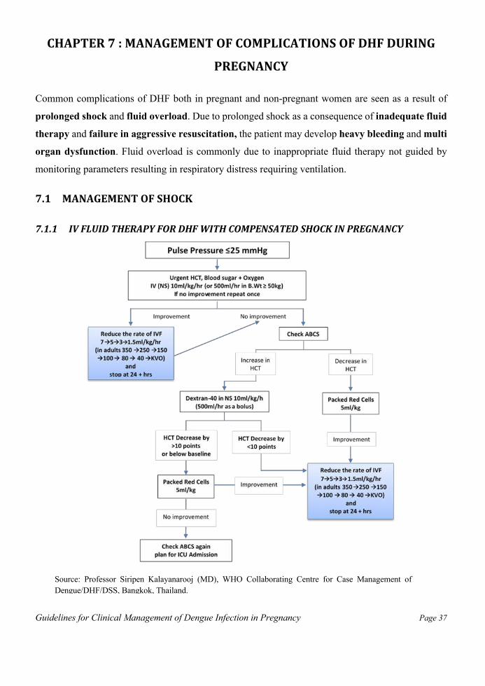
Guidelines for Clinical Management of Dengue Infection in Pregnancy Page 37
CHAPTER7:MANAGEMENTOFCOMPLICATIONSOFDHFDURING
PREGNANCY
Common complications of DHF both in pregnant and non-pregnant women are seen as a result of
prolonged shock and fluid overload. Due to prolonged shock as a consequence of inadequate fluid
therapy and failure in aggressive resuscitation, the patient may develop heavy bleeding and multi
organ dysfunction. Fluid overload is commonly due to inappropriate fluid therapy not guided by
monitoring parameters resulting in respiratory distress requiring ventilation.
7.1 MANAGEMENTOFSHOCK
7.1.1 IVFLUIDTHERAPYFORDHFWITHCOMPENSATEDSHOCKINPREGNANCY
Source: Professor Siripen Kalayanarooj (MD), WHO Collaborating Centre for Case Management of Dengue/DHF/DSS, Bangkok, Thailand.

Guidelines for Clinical Management of Dengue Infection in Pregnancy Page 38
7.1.2 IVFLUIDTHERAPYFORDHFWITHDECOMPENSATEDSHOCKINPREGNANCY
Above figure gives suggested IVF therapy in DHF with shock (DSS) over period of 24 hrs+. IVF is
recommended for 24 hrs+ when a patient presents in shock. Patient should be resuscitated and
followed according to the flow chart above with gradual reduction of fluid rate.
Source: Professor Siripen Kalayanarooj (MD), WHO Collaborating Centre for Case Management of Dengue/DHF/DSS, Bangkok, Thailand.

Guidelines for Clinical Management of Dengue Infection in Pregnancy Page 39
If, stable clinical condition, vital signs and urine output are not achieved with above fluid therapy,
consider complications (A - Acidosis, B - Bleeding, C - Calcium, S - Sugar + F – fluid overload). If
the patient has acidosis (pH ≤7.35 with HCO3 <15mmol/l) it indicates prolonged shock and such
patients are likely to have organ(s) involvement (possible liver and kidney injury). Correct acidosis
quickly, preferably with a bolus of NaHCO3 1ml/kg. Follow up with another 1ml/kg if response is
inadequate. Correcting acidosis quickly will reduce the occurrence of DIC. DIC will result in massive
bleeding and advanced DIC will develop quickly in acidotic environment.
7.2 MANAGEMENTOFBLEEDING
Bleeding is common in dengue illness. This can range from petechial haemorrhages to life threatening
bleeding. In adults, clinically significant bleeding can occur in DF, DHF with shock and in DHF
without shock. As such, bleeding is an important complication in pregnant dengue patients.
Furthermore, if the patient delivers during the illness, bleeding is likely to be more due to oozing from
raw uterine surface. Surgical manoeuvres, including LSCS, can result in severe, life threatening
bleeding and should be avoided in Critical phase of DHF unless to save mother’s life.
7.2.1 DETECTIONOFBLEEDING.
Overt bleeding can manifest as bleeding per vagina or haematemesis. However, significant bleeding
could be often concealed. Such bleeding can occur into GI tract (manifest later as melaena), muscles,
abdominal cavity or thoracic cavity and brain.
During dengue illness early bleeding should be anticipated if the patient has been on NSAIDS, Aspirin
and steroids.
Features of haemodynamic instability, together with reduction of HCT or absence of rise in HCT
indicate possible bleeding. Although tachycardia may not be always present with bleeding,
tachycardia without fever suggests the possibility of significant bleeding.
Suspect bleeding if;
• Any of the haemodynamic parameters become abnormal (e.g. tachycardia disproportionate
to fever, prolonged CRFT, reduction of UOP) with reduction of HCT.

Guidelines for Clinical Management of Dengue Infection in Pregnancy Page 40
• Any of the haemodynamic parameters become abnormal (e.g. tachycardia disproportionate
to fever, prolonged CRFT, reduction of UOP) without a rise in the HCT (equivocal).
• When patients present with shock, especially hypotension, postural hypotension, dizziness,
fainting but no adequate rise in HCT. Further, in these patients AST/ALT will be elevated
>200 IU/L (Can be >1000 IU/L).
• Significant increase in WBC count (neutrophil leucocytosis) with reappearance of fever can
be due to Bleeding, Bacterial infection and Hepatitis (BBH).
• Tachycardia (>110/min) without fever
• Drop of HCT >10 points following a bolus of Dextran-40 (administered 10 ml/kg/ hr)
• Sudden fall of HCT below baseline (following fluid resuscitation in shock)
7.2.2 TREATMENTOFBLEEDING.
Treatment of bleeding is volume replacement with blood. Until blood is available, fluid should be
transfused: crystalloids in early or middle of the critical phase and Dextran-40 in the latter part of the
critical phase. If the patient is unstable, blood should be given rapidly. After the transfusion,
haematocrit (HCT) should be repeated.
Even if bleeding is likely and haematocrit is >45% do not give blood without bringing down the HCT
first by giving a colloid (Dextran-40, 10ml/kg/hr).
• Volume of blood transfused should not be added to total fluid quota during leaky phase.
• Threshold for blood transfusion is low in dengue with pregnancy. Packed Red Cells (PRC)
5ml/kg can be given at a time. HCT is expected to rise by 5 points (e.g. from 30% to 35&)
with this amount of blood.
• Immediately after the baby’s delivery (end of the second stage of labour), the cervix and
vagina should be thoroughly inspected for lacerations and surgical repair should be performed
early.

Guidelines for Clinical Management of Dengue Infection in Pregnancy Page 41
If the patient had NVD or LSCS it is advisable to replace the estimated lost blood volume
immediately. It is essential to monitor haematocrit and haemodynamic parameters frequently in such
patients as they are likely to have continuous bleeding and may need further blood transfusions.
7.2.3 ABSOLUTEINDICATIONSFORBLOODTRANSFUSIONS
• Overt blood loss of 250-300 ml
• Drop in haematocrit without clinical improvement despite adequate fluid replacement.
• A drop in haematocrit of >10 points after Dextran- 40 10 ml/kg (or 500 ml) bolus
• Dengue shock not responding to adequate fluid therapy i.e. 40-60 ml/kg (Refractory shock)
• Hypotensive shock with low or normal haematocrit
• Persistent or worsening metabolic acidosis despite adequate fluid replacement
7.2.4 CONSIDERBLOODTRANSFUSION
1. If the patient is in shock but haematocrit (HCT/PCV) rise is not high enough as expected for
the degree of leak leading to shock
2. Low or normal PCV with unstable haemodynamic parameters such as; cold clammy
extremities, prolonged CRFT, tachycardia, narrow pulse pressure (25-35 mmHg), and
reduced UOP.
3. Increased INR (liver failure) with low PCV
Clinical tip: Guidelines for visual estimation of blood loss
• 60ml: small 10×10cm swab (maximum saturated capacity)
• 140ml: medium 30×30 cm swab (maximum saturated capacity)
• 350ml: large 45×45cm swab (maximum saturated capacity)
• 500ml: 50cm diameter floor spill
v Excessive vaginal bleeding—soaking a pad in an hour

Guidelines for Clinical Management of Dengue Infection in Pregnancy Page 42
4. Anaemia in pregnancy – Pregnant women with low haemoglobin (Hb ≤8g/dl) e.g. Iron
Deficiency, Thalassemia Minor, with dengue illness, blood transfusion should be considered
early.
7.2.5 PLACEFORPLATELETTRANSFUSION
1. Low platelet count is not an indication for prophylactic platelet transfusions as there is no
relationship with bleeding and low platelet count (without trauma).
2. If VD progressing in spite of tocolytics or if LSCS is essential (to save mother’s life) platelet
transfusions would be necessary to keep the platelet count >30x109 – 50x109/L respectively.
3. If the patient continues to bleed in spite of several blood transfusions, there is a place for
therapeutic platelet transfusions.
4. Platelet transfusion may be indicated in the presence of heavy overt bleeding with low platelet
counts.
5. Insertion of central venous catheter/ vas cath for CRRT – aim >30x109 /L
7.2.6 PLACEFORTRANSFUSIONOFOTHERBLOODPRODUCTS.
1. If patient develops liver failure, platelet, FFP and cryoprecipitate would be indicated. However,
clinicians have to be conscious of the possible fluid overload with platelet and FFP
transfusions.
2. Factor VII may have a place only if the bleeding is from a single point (e.g. Gastric ulcer)
3. Prothrombin concentrate complex (containing factors II, VII, IX, X) may have a place in
bleeding with liver failure.
4. For significant bleeding, ROTEM/TEG guided coagulation management is given in Annexure 3.

Guidelines for Clinical Management of Dengue Infection in Pregnancy Page 43
7.3 INDICATIONSFOROTHERADJUNCTTHERAPY
Calcium Gluconate – 10% calcium gluconate 10ml over 10 minutes is justifiable if a patient is in
shock and is not responding to adequate fluid replacement. This may be continued 6 hourly for 24-36
hours.
Hypoglycaemia - if blood glucose level is <70/mg/dl correct it by giving 25-40ml of 25% dextrose
intravenously. Recheck in 15 minutes and repeat.
7.4 MANAGEMENTOFMULTI-ORGANFAILURE
• Hepatic failure / Renal failure / Respiratory failure / Encephalopathy / Encephalitis: manage
according to the standard protocol/guidelines.
• Myocardial involvement: bedside echo cardiogram can assess cardiac function. Continue fluid
replacement (do not restrict fluid).
• Other complications – electrolyte imbalance, co-infections should be managed according to
standard protocols.
7.5 ABSOLUTEINDICATIONSFORICUADMISSION
• Severe plasma leakage leading to shock and/or fluid accumulation with respiratory distress
• Severe bleeding
• Severe organ impairment
• Following inevitable surgical intervention done on a patient in critical phase
• A parturient in critical phase
Indications for intubation and ventilation are:
• Acute respiratory failure (ARDS)
• Severe cardiovascular instability
• Severe metabolic derangement (raised lactate > 10mmol/L)
• Altered mental state

Guidelines for Clinical Management of Dengue Infection in Pregnancy Page 44
Source: Professor Siripen Kalyanarooj (MD), WHO Collaborating Centre for Case Management of Dengue/DHF/DSS, Bangkok, Thailand.
7.6 MANAGEMNTOFFLUIDOVERLOAD
Use of excess fluids or fluids given beyond the time of plasma leakage are the usual causes of fluid
overload.

Guidelines for Clinical Management of Dengue Infection in Pregnancy Page 45
7.7 OBSTETRICCONDITIONSWHICHMIMICDENGUEHAEMORRHAGICFEVER
7.7.1 HELLP SYNDROME (HAEMOLYSIS, ELEVATED LIVER ENZYMES AND LOW
PLATELETS)
• In pre-eclampsia and HELLP syndrome, thrombocytopenia is a common feature. Assess with
serial FBC, NS1 Ag and Dengue IgM in a patient with a history of fever to exclude dengue.
• In some pregnant woman with gestational thrombocytopenia, PLT count is ≤150x109/L. PLT
count is low in HELLP syndrome; very low PLT count <30 x109/L is rarely seen in HELLP.
• If initial FBC is suggestive of Thrombocytopenia follow up with serial FBC to exclude
dengue.
Comparison between HELLP syndrome and DHF
CLINICAL
FEATURES FOR
DIAGNOSIS
HEMATOLOGICAL
FINDINGS
BIOCHEMICAL
FINDINGS MANAGEMENT
HELLP syndrome
• Evidence of pre-eclampsia
• Haemolysis • Elevated liver enzyme • Low platelet counts • H/o flu like illness
Blood picture (MAHA) Thrombocytopenia
AST and ALT both raised >70 IU/L Elevated LDH >600 U/L
Termination of pregnancy
DHF
• Fever/history of recent fever
• Bleeding tendency • Thrombocytopenia
Evidence of plasma leaking identified with ultrasonography
Leukopenia Thrombocytopenia
(often <50x109/L) Hematocrit rise (towards 20%)
In complicated DHF AST markedly elevated than ALT and shows rapid rise
Delay delivery until the end of critical phase
7.7.2 DETECTION AND MANAGEMENT OF COMPLICATIONS DURING POST-PARTUM
PERIOD
• Risk of PPH is higher in instrumental delivery, surgical interventions. Pregnancy related
conditions (multi gravida, placenta previa, etc.)
• Fever during the post-partum period due to acquired dengue infection may be misdiagnosed
as puerperal sepsis.

Guidelines for Clinical Management of Dengue Infection in Pregnancy Page 46
• Newborns with mothers who had dengue just before or at delivery, should be closely
monitored and investigated in hospital after birth up to a week in view of the risk of vertical
transmission.
• Oxytocin infusion should be commenced to contract the uterus after delivery to prevent
postpartum hemorrhage. Misoprostol may be given for PPH prophylaxis/treatment.
• Ensure no retained placenta
• It is estimated that 300-500ml blood enters the circulation with uterine contractions.
However, soon after delivery transfuse the estimated blood loss as recommended in previous
chapters
• After every LSCS, estimated blood loss should be replaced.
7.7.3 MANAGEMENTINTHEPRESENCEOFANINTRAUTERINEFOETALDEATH
The occurrence of intra uterine foetal death (IUD) is rare in Dengue infection of a pregnant woman.
However, it is a known complication in the 3rd trimester.
• Diagnosis of IUD requires appropriate and urgent assessment using real-time ultrasound. Do
not diagnose foetal death only based on absence of fetal heart on auscultation or by hand-
held Doppler.
• Delivery of IUD could be delayed by at least 1 week, if the patient is physically well with
intact membranes and there is no evidence of infection, preeclampsia or bleeding (including
laboratory evidence of DIC).
• If there is evidence of infection, IV antibiotics should be started.
• If labour is delayed for >48hrs, testing for DIC should be done twice weekly or more
frequently as necessary.
7.7.4 MANAGEMENTOFPERINATALANDVERTICALTRANSMISSIONOFDENGUE
Perinatal dengue infection is the result of vertical transmission from mother to offspring. A study in
French Guiana (1992 – 2006) has shown 15% vertical transmission in 53 pregnant dengue patients.

Guidelines for Clinical Management of Dengue Infection in Pregnancy Page 47
In general, dengue infections in the newborn babies are usually mild and DF is more likely. Fever is
the initial presentation in most babies, commonly on 4-5 days after birth or even on day 01 of birth
or later than 6 days. Therefore, suspected neonate should be observed at least for 1 week for evidence
of vertical transmission. Other clinical presentations are petechiae, rash and hepatomegaly.
Convulsions (may mimic CNS infection) and diarrhoea are common unusual presentations. In early
febrile phase leucopenia may not be found as leukocytosis is common in early infancy.
Neonates have less respiratory reserve and they are more susceptible to liver impairment and
electrolyte imbalance (prone to get hyponatraemia). If they develop DHF, duration of plasma leakage
is shorter (usually 12 – 24 hrs.). As leucopenia is not common, increased haematocrit (possibly with
ultrasound evidence) and thrombocytopenia will help clinical diagnosis of DHF.
In the management of DHF in neonates optimum fluid therapy from the onset of entry into the critical
phase is vital for good outcome. Baby should be evaluated frequently for oral fluid intake (i.e. breast
feeding) and urine output (hence early need for catheterization). As IVF, half normal saline (N/2 in
5% Dextrose) should be given. If high rate of crystalloids are required (N/2 + 5% Dextrose) following
fluid resuscitation with crystalloids, colloids (10% Dextran 40) should be considered. They may
respond quickly to fluid resuscitation. Therefore, they should be monitored more frequently for fluid
requirement and urine output.
Fluid overload is common in neonates due to less chest compliance. If IVF are continued beyond
leakage phase it will easily lead to fluid overload. Breast feeding in neonates with DHF can lead to
fluid overload if not cautious. Further, if liver enzymes are elevated to a level of liver failure or
impending liver failure, breast feeding may have to be temporary withheld as proteins in the gut could
worsen liver failure.

Guidelines for Clinical Management of Dengue Infection in Pregnancy Page 48
Chapter8:NURSINGCAREDURINGPREGANCNY,COUNSELLINGOF
PATIENT/FAMILY,ANDTHECONVALESCENT(RECOVERY)PHASE
8.1 NURSINGCARE
All nurses stationed in ETU, OPD, Medical, Obstetric units and ICU need to have basic knowledge
on natural course of DF/DHF/DSS.
Nurses should develop necessary skills to identify the correct phase of DHF/DSS in pregnant women
so that appropriate and proper monitoring can be provided for the individual patient.
8.1.1 GENERALIN-WARDCARE
• Ensure availability of essential equipment in working order – Microhematocrit, Multi-
monitors
• Order and stock essential drugs – including 10% Dextran-40 in Normal Saline
• Maintain mosquito free environment • Provide essential information to both patient and by-stander on dengue management at the
time of admission and give psychological support regularly
• Collect and send blood for laboratory investigations on time and trace reports promptly
• Notify Medical Officer of Health (MOH) where patient has resided two weeks prior to illness
v Febrile Phase
1. High fever, Headache, Arthralgia, Myalgia. • Maintaining QHT chart.
With fever or without fever – detect tachycardia Hypothermia – Inform doctor.
• Give Paracetomol when temperature is ≥39 °C. • Apply warm water compress / tepid sponging. • Adequate fluids – Avoid plain water.
2. Anorexia, Nausea and Vomiting.
• Provide & encourage the patients to have soft balanced nutritious diet – Avoid red, black, brown coloured food.
• Severe vomiting, look for signs of dehydration. • Give antiemetic with the doctor’s instructions. They may need intravenous fluids. • Provide clean, transparent container with a lid to collect & measure vomitus of patients.

Guidelines for Clinical Management of Dengue Infection in Pregnancy Page 49
3. Abdominal Pain
• Help the patient to lie down or to be in a comfortable position. • Provide soft, clean, dry and comfortable wear. • Avoid tight contacts with abdomen. • Peptic ulcers is a common reason of abdominal pain. Give medications according
doctors order; e.g. Ranitidine, Famotidine
4. Bleeding
• Epistaxis, Hematemesis, Bleeding per gum, PV bleeding - Notify on-call doctors - Estimate & record the amount of blood loss.
• Send blood for DT. • No intra muscular injections. • Venipuncture apply pressure to the site with sterile gauze. • In epistaxis put an ice pack or nasal packing. • With gum bleeding – tooth brush is not recommended. Clean oral cavity softly with a
cotton bud. • In case of GI Bleeding observe closely, & check PCV. Content and colour of the stools
for possible GI Bleeding & Melaena • NG tube insertion is contraindicated as it may trigger severe bleeding by trauma.
v Critical phase
Ø Manage the patient in ward HDU (close to nurses’ station).
Ø Regular monitoring according to Guidelines (preferably hourly) Critical Phase Monitoring
Chart – Annexure 2
Always maintain dedicated DENGUE MONITORING CHART
Indications to call for immediate advice
• Pulse rate > 120/min with fever or >100/min without fever.
• Pulse Pressure 30-25 mmHg or less (in left lateral position)
• Postural drop of SBP >20mmHg.
• Significant bleeding (Haematemesis, Melaena, Bleeding PV etc.)
• UOP <0.5ml /kg/hr • CRFT > 2 sec

Guidelines for Clinical Management of Dengue Infection in Pregnancy Page 50
Ø Recognize abnormalities of the vital parameters and inform on-call doctor early.
Ø Perform investigation as instructed and trace the report promptly.
Ø Preparedness and availability of medication in ward HDU (make sure essential medication
and IV solution including Dextran 40 readily available inward)
Ø Trace lab reports and inform doctor.
Ø Strictly adhere to management protocols given by the doctor on fluid therapy ( rate of fluid
infusion)
Ø Perform and monitor inward PCV, especially before and after fluid bolus.
Ø Provide instant care assist during dengue emergency resuscitation (e.g.: Dengue shock)
Ø Detect respiratory distress early – count RR look for tachypnea and dyspnea
v Convalescent phase
Ø Recognize sign and symptoms indicating recovery.
Ø Monitor vital parameters according to the need.
Advice before discharge
Ø Advice to take adequate rest
Ø If platelet count is low to avoid strenuous exercises.
Ø Explain they can now do normal day-to-day activities.
Ø To clean the immediate environment.
8.2 PATIENTCOMMUNICATION
The importance of establishing a good relationship with the patient, husband and other family
members from the time of admission cannot be over emphasized. This can be easily established by
good communication between the medical staff and the patient.
This should involve the following –
1. Discussion about the nature of the illness, natural course, possible outcomes and treatment
options.
2. Information should be given regarding favourable outcomes of dengue, with appropriate
management. However, information must also be given regarding the complications and

Guidelines for Clinical Management of Dengue Infection in Pregnancy Page 51
mortality in the event of dengue illness being complicated by severe dengue shock syndrome
complicated by multi organ failure, in simple language.
3. Study the patient records (BHT) well before discussions are commenced. In particular note
the platelet count, PCV, urine output and blood pressure, as the patient and the relatives too
would be well aware of the latest information.
4. Document every discussion made with the patient and the family in the BHT.
5. Explain that the patient is under the care of a multi-disciplinary team of experts comprising
of obstetricians, physicians, paediatrician, anaesthetists etc. and that the best possible
management is being done according to national guidelines for the safe delivery of the mother
and the baby.
6. Explain that the mode of delivery whether by vaginal or by Caesarian section will be judged,
depending on the duration of pregnancy, the state of the dengue illness and the condition of
the mother, in the best interest of both mother and baby.
7. Reassure the patient, husband and family members that dengue complicating pregnancy is
not fatal and safe delivery of the baby can be ensured.
8.3 BREAKINGBADNEWS
8.3.1 MATERNALDEATH
1. Death of a mother from dengue or any other cause is a devastating experience for the entire
family, especially the husband and the remaining children.
2. If the condition of the pregnant mother with dengue was hopeless from the start, the relatives
should have been informed by the medical professionals about the poor prognosis and the
possibility of death from the very beginning.
3. If the death occurred unexpectedly, explain that everything possible was done to prevent this
catastrophe and that the present situation was unfortunate and unavoidable. The family should
be reassured that every measure treatment options were utilized to save the mother, despite
the unexpected turn of events.
4. Provide spiritual comfort to the patient by inviting known members of the clergy to offer
blessings.
5. Provide psychological and emotional help to the family by trained counsellors.
Guidelines for Clinical Management of Dengue Infection in Pregnancy Page 52
8.3.2 DEATHOFTHEBABY
Explain that everything possible was done in the best interest of the mother and the baby,
with the concurrence of the neonatologists and the obstetricians. However, if the baby had
died due to early termination of pregnancy, explain that this decision was made reluctantly
to save the life of the mother.
8.4 CONVALESCENT(RECOVERY)PHASE
Recovery Phase starts after the end of the critical phase and usually lasts for 2-3 days. Reabsorption
of the extravasated fluid is seen during this period.
8.4.1 SIGNSANDSYMPTOMSOFRECOVERY
• Improved general well-being and improved appetite
• Urine output will be normal or increased (diuresis)
• Haemodynamic stability (normal BP and Pulse)
• Haematocrit returns to baseline (may even be lower than baseline due to fluid reabsorption)
• Rise in white cell count followed by a sustained rise in the platelet count
• Bradycardia, appearance of convalescent rash, generalized itching (more intense in palms and
soles) may be seen in some patients.
8.5 DISCHARGECRITERIA
The following criteria are to be taken into account while contemplating a discharge of a dengue patient
• Afebrile for 48 hours (without antipyretics)
• Stable general condition
• Recovery of appetite
• Stable haematocrit for at least 24 hours
• Rising trend in platelet count with > 50x109/L, (at least two counts done within 12 hours
apart).
• No dyspnea or respiratory distress attributable to pleural effusion or ascites

Guidelines for Clinical Management of Dengue Infection in Pregnancy Page 53
8.6 ADVICEONDISCHARGE
• Advise to take rest at home for 1 week avoiding traumatic activities or procedures.
• There is no risk of spread of dengue virus now and patient can go back to work.
• If anyone in the same household gets high fever they should seek early treatment.

Guidelines for Clinical Management of Dengue Infection in Pregnancy Page 54
CHAPTER9:CHALLENGESINDIAGNOSISANDMANAGEMENT
DURINGPREGNANCY
9.1 MANAGEMENTDELAYS
There are three levels of management delays that may result in an adverse clinical outcome: (1) delay
in presentation to the health care system resulting in delayed admission and hence delayed institution
of appropriate rehydration, (2) delay in instituting appropriate rehydration measures in hospitalized
patients, and (3) delay in discontinuing rehydration towards the end of the critical phase resulting in
fluid overload and iatrogenic death.
(1) Delay in presentation to the health care system: the root cause is often lack of community
awareness leading to late health care seeking behaviour of patients with febrile illnesses including
dengue. Therefore, the strategy is to increase community awareness about dengue. The message
to the public is that all patients with acute febrile episodes need to seek early medical attention. In
addition, the public needs to be aware that the critical phase often starts at the end of the febrile
phase.
(2) Delay in institution of rehydration measures at hospital-level: the root causes include slow
laboratory processes in obtaining laboratory results, non-streamlined work processes leading to
delayed attention of the clinicians in-charge, lack of access to resources to make a prompt
diagnosis of capillary leakage (such as serial haematocrit measurements, ultrasound etc.), and
untrained personnel. The strategy entails improving rapid access to laboratory results such as
platelet count, haematocrit, and clinical assessments including blood pressure, urine output, fluid
accumulation as assessed by ultrasound etc. It also entails a well-planned work flow between
junior and senior doctors, between emergency room, outpatient department and inpatient wards,
and a dedicated high dependency ward with special attention to the care of dengue patients with
capillary leakage. Delay in adequate fluid resuscitation is a major concern with under perfusion of
patients in shock.
(3) Delay in reducing/restricting fluid therapy: the root cause is usually lack of training and
experience in managing dengue patients during the critical phase. The result is over-hydration of
patients, at a time when reabsorption of fluids occurs towards the end of the critical phase, which

Guidelines for Clinical Management of Dengue Infection in Pregnancy Page 55
leads to fluid overload and sometimes, death. The strategy includes better training and monitoring
of clinicians treating complicated dengue cases, careful audits of fatal cases to learn from any
mistakes made, and setting up dedicated high dependency units (HDU).
9.2 STEPSTOMINIMIZEMANAGEMENTDELAYS
Five essential steps are required in proper clinical management of dengue patients to reduce fatal
outcomes: (1) early diagnosis and indication for admission, (2) early detection of plasma leakage and
time-sensitive institution of fluid rehydration, (3) Investigations and correction of common
complications in patients who do not respond to conventional IV fluid therapy, (4) early detection of
bleeding and early blood transfusion, and (5) early diagnosis in cases with unusual manifestations,
especially those with encephalopathy, co-morbidity and co-infections.
By reviewing the processes of dengue case detection and clinical management, gaps in all 5 steps of
clinical management can be identified. However, the main cause of death is fluid overload. The main
reason for death is not failure to diagnose early, but clinical management of shock with a tendency to
over-hydrate over a longer period than required. Delayed blood transfusion and delays in transferring
patients to tertiary care facilities with high dependency wards (HDUs) are additional factors.

Guidelines for Clinical Management of Dengue Infection in Pregnancy Page 56
Chapter10:CASESTUDIESINCLINICALMANAGEMENTAND
SUGGESTEDIMPROVEMENTSFORPREGNANTWOMEN
Table: Steps in clinical management, common findings and suggested improvements
Steps in Clinical
Management Common Findings (Issues) Suggested improvements
1 Early clinical diagnosis of dengue and indication for admission
Early diagnosis by NS1 Ag done, but not admitted properly. More than half admitted early but received too much IV fluid.
Admit on Day 1 of fever. Use proper Risk Assessment at all levels of care using Fever, Warning Signs, Vital Signs and FBC effectively. Diagnose DHF early and manage accordingly
2 2.1 Early detection of plasma leakage
Plasma leak detected too late/overlooked. Patients develop shock/ profound shock while in hospital
Check FBC regularly as PLT drops; Repeat HCT when PLT drop <100x109 Use USS effectively to detect leaking
2.2 Proper IV fluid management
Giving too much IV fluid for too long. Giving too little fluids Resuscitation in shock not aggressive enough
Initially use 3 hrly febrile phase monitoring chart and give measured amount of fluid. When leaking stars change over to Critical phase chart and monitor hourly or more often while giving fluid (both IV and Oral). Stop IV when leaking is over
3 Investigations and correction of common complications if patients do not respond to conventional IV fluid therapy (cannot reduce the rate of IV fluid)
Not investigate and correct common complications in DHF Do not anticipate complications when patient not improving with standard IV therapy
Check and correct; A – Acidosis B - Bleeding C – Calcium S – Sugar F – Fluid Overload
4 Early detection of bleeding and early blood transfusion
Delayed blood transfusion found in half of fatal case
Disseminate list of common indications for blood in Dengue

Guidelines for Clinical Management of Dengue Infection in Pregnancy Page 57
5 Early diagnosis of cases with unusual manifestations, especially those with encephalopathy, co-morbidity and co-infections
Rare due to dengue virus itself Usually seen as a consequence of prolonged shock
Look for complications and treat accordingly
10.1 CASE-#1
A 35 year old previously healthy woman married for 6 months who missed her past regular period
was transferred from a peripheral hospital for specialized management of suspected ectopic
pregnancy.
History: She had fever for 4 days. Complained of faintishness and abdominal pain on the day transfer.
She had no peripheral pulse, Low BP, patient conscious & rational (GCS 15/15) and Urine HCG +ve.
USS abdomen free fluid + with no Intra-uterine pregnancy.
At peripheral hospital an emergency mini laparotomy was performed.
Findings: No tubal pregnancy; No haemo-peritoneum; Free-fluid in the abdomen 200ml.
Preoperative investigations were received after the surgery: Hb 10.5g/dL, PLT 82x109/L
Treatment given post operatively: 02 Units PRC, 01 Unit voluven, 03 Units Normal Saline. Patient
was transferred to ICU for further management.
Referal Hospital ICU Assessment and Care: No fever, conscious & rational (GCS 15/15). Not pale,
mildly icteric, CRFT <2 secs. BP 115/75 mmHg. HR 110/min. Lungs B/L reduced air entry. Abdomen
soft but oozing from surgical site.
Investigations: FBC WBC 6.8 x 10³; PLT 39x109; INR 1.53; Dengue NS1 (–) ve, IgM/IgG (+) ve.
USS B/L pleural effusion and moderate ascites.
Two hours later: Increased oozing from surgical site. BP 88/67mmHg; PLT 39x109, PCV 32%
Guidelines for Clinical Management of Dengue Infection in Pregnancy Page 58
Treatment: IV Hartmann’s boluses given, IV Antibiotics started.
Next day: in spite of fluid therapy, with inotropes BP did not pick up and patient developed
respiratory distress with dyspnea and tachypnea (RR 36/min). Despite ventilator support patient
expired on the same day.
Pit Falls:
1. Did not consider history of fever especially when afebrile on presentation.
2. When presenting in shock ignoring Dengue Shock Syndrome (DSS) as a differential diagnosis.
3. Not considering warning signs of leaking – faintishness, abdominal pain (signs of impending
shock)
4. Little attempt to resuscitate the state of shock, perform an urgent FBC and interpret before
emergency surgical intervention.
5. Patient presenting in shock with free fluid in abdomen (also seen on initial USS) indicates that
the patient has already leaked for significant period of time. With PLT count < 50x109 this patient
was probably in the latter part of the critical phase. Therefore, indiscriminate use of IV fluids
(crystalloids) lead to fluid overload.
6. In patient presenting with hypotensive shock (decompensated) should consider leaking and
bleeding (PCV 32%) both with acidosis, hypocalcaemia, hypoglycemia and early organ
involvement.
7. In a DSS patient with a low PCV of 32% should consider significant bleed requiring early blood
transfusions (rapidly) until patient becomes haemodynamically stable.
8. Important to exclude DHF/DSS when considering surgical intervention for appendicitis, renal
colic, etc. with history of fever and abdominal pain.
10.2 CASE-#2
A 28 year old primi para, at POA 32 weeks, admitted to the casualty ward of a maternity hospital one
morning around 9.00am. Presenting complaint was upper abdominal pain and reduced foetal
movements of one days duration.

Guidelines for Clinical Management of Dengue Infection in Pregnancy Page 59
O/E: BP 150/100mmHg. Albuminuria 1+. Although there was no history of preeclampsia, HELLP
syndrome was suspected. FBC and liver function tests were ordered. USS abdomen revealed normal
foetal heart beat and foetal movements.
She was treated with Nifedipine 10mg SR and her BP dropped to 90/50 mmHg. Her HR remained
around 100 beats per minute. With a NS bolus of 250 ml given over 30 mins her BP picked up to
normal. She was on a NS slow drip thereafter. Medical opinion sought and clinical diagnosis of
HELLP syndrome was established.
That evening around 3.30pm, morning FBC results reveled Platelet count of 9x1009. Attending
Obstetrician thought the counts were too low to be due to HELLP. On further inquiry it was revealed
that the patient has had a history of fever for 4 days and afebrile for 48 hrs prior to admission. At that
point blood was drawn for dengue Ag and IgM, IgG and the patient was moved to ICU for further
management.
She was taken to ICU (6.30pm same day): conscious rational, RR 38, SpO2 100%, bilateral air entry
reduced in lungs R>L, HR 89, BP 123/86, CRFT 5s, abdomen was distended & tender, UOP 70ml/6
hrs (approx. 12 ml/hr), Platelets 9x10⁹, WBC 13,000 (N-82%, L-14%, Hb-15.9%, HCT 48.7%), ALT
147, AST 468, CRP 41.6, Dengue Ag +ve (at 11pm),VBG pH 7.16, PCo2 35, PO2 35, BE -21 mmol/l.
Expert medical opinion sought for further management reveled patient having DHF in latter part of
leaking with fluid overload, liver dysfunction and acidosis.
Following day early morning patient became dyspnoeic, abdominal distension increased and UOP
reduced further. Patient intubated and ventilated while CRRT was also considered.
Pit Falls:
1. At first contact fever history was not elicited which lead to deviation from diagnosing
dengue.
2. Measuring of BP technique was not documented – in pregnancy left later position is ideal
(to get proper values).
3. Warning feature of impending shock of DHF such as abdominal pain was mot evaluated.

Guidelines for Clinical Management of Dengue Infection in Pregnancy Page 60
4. Tachycardia (HR 100) without fever should have been interpreted and acted upon.
Immediate A, B, C, S assessment and consider blood transfusion.
5. FBC done in the morning was available and read that evening – FBC is an urgent
investigation and should be ideally read within one hour of blood collection.
6. Clinical detection of fluid with B/L pleural effusion with a low platelet count of 9x10⁹/L is
seen in latter part of leaking in DHF where major part of leaking is over.
7. More fluid given at late stage of leaking lead to fluid overload and respiratory distress
requiring ventilator support.
8. In this patient acute liver and renal dysfunction with acidosis was probably due to
suboptimal fluid therapy during early critical phase.
10.3 CASE-#3
A 32-year old previously healthy P2C1 woman in her 36th week of POA in early labour indication
being past section was brought for caesarian section to OT. Attending anesthetist noted a low platelet
count of 76x109 With WBC 7200, HCT 36%.
On further inquiry she has had fever for 3 days before admission to hospital. Urgent NS1 Ag test was
found positive. USS abdomen and chest showed no free fluid. On suspicion that the patient may go
into critical phase soon the multidisciplinary team discussed and decided to postpone the delivery
although a concern on possible uterine rupture was noted. Patient was admitted to ICU and closely
monitored for both fluid leakage due to DHF and uterine rupture. It was noted that the uterine
contractions ceased without any tocolytics. However, patient developed plasma leakage about 6 hrs
later. Her platelet dropped to 30x109 and she developed R/S pleural effusion although her HCT
remained within 34-40%. She was managed with fluids according to the guidelines. Continuous CTG
was done to detect any foetal compromise.
Her HR and BP remained within normal ranges throughout the critical phase. Her UOP was adequate.
Liver enzymes were between 100-200/IU/L AST >ALT.
Around 56 hrs she was on recovery phase with platelet count of 75x109 WBC 30,000 with neutrophil
leukocytosis and CRP 118. She also developed itching of palms and soles. She had no fever and was

Guidelines for Clinical Management of Dengue Infection in Pregnancy Page 61
haemodynamically stable. ROTEM testing for coagulation remained within normal ranges.
Suspecting sepsis broad-spectrum antibiotics were started. Foetal condition remained normal. At this
stage decision was made to deliver the foetus by caesarian section under spinal anesthesia. During
surgery there was no blood and pus noted in the peritoneal cavity. Live baby was delivered. She
made an uneventful recover and both mother and baby were discharged 4 days after surgery.
Pitfalls:
1. History of fever was elicited late and FBC was considered just before surgery
2. Not considering close observation of baby after delivery for vertical transmission of dengue
virus.
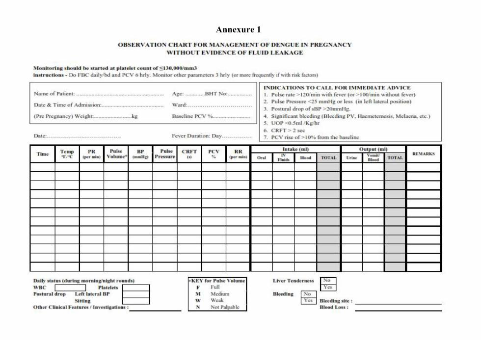
Annexure 1

Annexure 2

Annexure 3
MANAGEMENT OF MASSIVE OBSTETRIC HAEMORRHAGE
Blood loss >40% of blood volume (Blood volume calculated as 100ml/kg)
Guided by results from ROTEM-CSHW
Ref: Dr. Klaus Görlinger A5 Algorithm and LWH UK Protocol
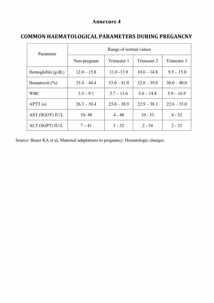
Annexure 4
COMMONHAEMATOLOGICALPARAMETERSDURINGPREGANCNY
Source: Bauer KA et al, Maternal adaptations to pregnancy: Hematologic changes
Parameter Range of normal values
Non-pregnant Trimester 1 Trimester 2 Trimester 3
Hemoglobin (g/dL) 12.0 – 15.8 11.0 -13.9 10.6 – 14.8 9.5 – 15.0
Hematocrit (%) 35.4 – 44.4 33.0 – 41.0 32.0 – 39.0 30.0 – 40.0
WBC 3.5 – 9.1 5.7 – 13.6 5.6 – 14.8 5.9 – 16.9
APTT (s) 26.3 – 39.4 23.0 – 38.9 22.9 – 38.1 22.6 – 35.0
AST (SGOT) IU/L 10- 40 4 - 40 10 - 33 4 - 32
ALT (SGPT) IU/L 7 – 41 1 - 32 2 - 34 2 - 32

BIBLIOGRAPHY
• Bauer KA, Lockwood CJ, Eckler K, Tirnauer JS. Maternal adaptations to pregnancy: Hematologic changes Up to date https://www.uptodate.com/contents/maternal-adaptations-to-pregnancy-cardiovascular-and-hemodynamic-changes (Accessed 1st March 2019)
• Case Management of Dengue: Lessons Learned, Siripen Kalyanarooj, Alan L. Rothman, and Anon Srikiatkhachorn
• Clinical Practice Guidelines of Dengue/Dengue Hemorrhagic Fever Management for Asian Economic Community, WHO Collaborating Centre for Case Management, Thailand, Sept 2014
• Comprehensive guidelines for prevention and control of dengue and dengue haemorrhagic fever, Revised and expanded edition. WHO SEARO Technical Publication Series No. 60. 2011
• CPG Management of Dengue Infection In Adults (Third Edition) Malaysia
• Dengue In Pregnancy(India) : Management Protocols - FOGSI: Dr Suchitra Pandit
• Foley MR, Lockwood CJ, Gersh BJ, Eckler K, Maternal adaptations to pregnancy: Cardiovascular and hemodynamic changes, Up to date https://www.uptodate.com/contents/maternal-adaptations-to-pregnancy-cardiovascular-and-hemodynamic-changes (Accessed 1st March 2019)
• National Guidelines on Dengue Fever & Dengue Haemorrhagic Fever - Sri Lanka, 2012
• Paixao, E. S., Teixeira, M. G., Costa, M. da C. N., & Rodrigues, L. C. (2016). Dengue during pregnancy and adverse fetal outcomes: a systematic review and meta-analysis. The Lancet. Infectious Diseases, 16(7), 857–865.
• Sanghavi M, Rutherford JD. Cardiovascular physiology of pregnancy Circulation. 2014 Sep 16;130(12):1003-8
• Soma-Pillay P, Nelson-Piercy C, Tolppanen H, Mebazaa A, Physiological changes in pregnancy Cardiovasc J Afr 2016; 27: 89–94
• The Singapore Family Physician Vol. 4 1(2) Apr-Jun 2015 - Revisiting the approach to dengue: The primary care perspective
• Weinberger SE, Lockwood CJ , Barnes PJ, Eckler K Maternal adaptations to pregnancy: Physiologic respiratory changes and dyspnea Up to date ,https://www.uptodate.com/contents/maternal-adaptations-to-pregnancy-physiologic-respiratory-changes-and-dyspnea (Accessed 1st March 2019)