Microstructure and anti-adhesion properties of PES/TAP/Ag hybrid ultrafiltration membrane
7
Microstructure and anti-adhesion properties of PES/TAP/Ag hybrid ultrafiltration membrane H. Basri a, b , A.F. Ismail a, c, ⁎, M. Aziz a a Advanced Membrane Technology Research Centre (AMTEC), Universiti Teknologi Malaysia, Skudai 81310 Johor Bahru, Johor, Malaysia b Department of Science and Mathematics, Faculty of Science, Technology and Human Development, Universiti Tun Hussein Onn Malaysia, Parit Raja, 86400 Batu Pahat, Johor, Malaysia c Faculty of Petroleum and Renewable Energy Engineering, Universiti Teknologi Malaysia, Skudai, 81310 Johor Bahru, Johor, Malaysia abstract article info Article history: Received 14 July 2011 Received in revised form 21 September 2011 Accepted 24 September 2011 Available online 21 October 2011 Keywords: Polyethersulfone-silver 2,4,6-triaminopyrimidine Anti- adhesion A novel antibacterial and anti-adhesion membrane was obtained by blending PES with 2,4,6-triaminopyrimi- dine (TAP) as compatibilizer and AgNO 3 as an antibacterial agent. The modified membrane surfaces analyzed by XPS revealed high content of silver (Ag) compared to PES/Ag without compatibilizer. This result was sup- ported by AFM micrographs where PES/TAP/Ag exhibited smoother surface with RMS(nm) 17.49 and R a (nm) 11.74. From the biofouling point of view, the smoother the surface, the lesser contact area provided thus re- ducing bacterial adhesion. In addition, PES/TAP/Ag membrane prepared via simple phase inversion technique showed evidence of an anti-adhesion property when tested on Escherichia coli suspension of 1 × 10 4 cells/mL at 37 °C. The smooth, anti-adhered surface of PES/TAP/Ag however exhibits better tensile strength up to 52% compared to pristine PES membrane, making it an antibacterial membrane with reduced biofouling poten- tials in water and waste water treatment. © 2011 Elsevier B.V. All rights reserved. 1. Introduction In many industrial applications where water is being stored or when a clean membrane surface is exposed to water, biofilm may start to form or grow. According to Al-Ahmad et al., the first step in biofilm formation in water containing organics and microorganisms is rapid adsorption of organic molecules and/or microbes to the mem- brane surface [1]. This organic layer then provides and enhances sub- sequent microbial adhesion. Several authors explain that the prime stage of the bacterial adhesion process is called ‘docking phase’ where bacteria come close to the material surface by Brownian mo- tion, sedimentation and convective mass transport as driving forces. Once the bacteria are close to the surface, the interactions such as van der Waals, electrostatic, hydrophobic and steric interaction occur in another stage called ‘locking phase’ [2,3]. It was quoted that at this stage, bacteria still show Brownian motion. However, the second, crucial locking stage includes mediated binding such as specific ligand–receptor interaction which in turn incites the process of bacterial adhesion to the material surfaces. Dunne elucidates that the adhesion process is dictated by a number of variables including bacterial species, surface composition and environmental factors [3]. The adhesion mechanism is a reversible one, which means with the existence of adhesion and the absence of physical and chemical inter- vention, bacteria can attach firmly to the surface. This will subsequently lead to the biofilm formation and may cause biofouling to membrane surfaces. Depending on the bacterial species involved, the biofilm may be composed of 10–25% bacterial cells and 75–90% extracellular poly- meric matrix (EPM) or also called slime [2]. This EPM helps bacterial cells to adhere and trap nutrients from the contacting medium. It also provides a safety cover that protects bacteria from host immunization, predators as well as antibacterial agents. A number of articles reported on microbial resistance towards antibacterial agents. The attachment and detachment process vary and are influenced by many factors including surface inertness, surface free-energy, surface charge, hydrophobicity, roughness and surface chemistry, while others are specific to the bacteria involved. A study carried out by Rijnaarts et al., highlighted that the attachment and detachment of various bacterial types can be controlled by varying the surface chemical properties of cells and solid supports [4]. The surface roughness appeared to have an important role in the detachment of adhered bacteria or biofilms in which a higher roughness significantly decreases the detachment or vice-versa [5]. A study on Pseudomonas aeruginosa discovered that sur- face free-energy in a range value of 20 to 27 m/Nm are potential in re- ducing its initial retention on inert surfaces [6]. To the best of our knowledge, this work is the first attempt to report the fabrication of PES membrane with silver as antibacterial agent and TAP as compatibilizer which results in better hydrophilicity, high tensile Desalination 287 (2012) 71–77 ⁎ Corresponding author at: Advanced Membrane Technology Research Centre (AMTEC), Universiti Teknologi Malaysia, Skudai, 81310 Johor Bahru, Johor, Malaysia. Tel.: +60 75535592; fax: +60 75535925. E-mail addresses: [email protected], [email protected] (A.F. Ismail). 0011-9164/$ – see front matter © 2011 Elsevier B.V. All rights reserved. doi:10.1016/j.desal.2011.09.031 Contents lists available at SciVerse ScienceDirect Desalination journal homepage: www.elsevier.com/locate/desal
-
Upload
independent -
Category
Documents
-
view
0 -
download
0
Transcript of Microstructure and anti-adhesion properties of PES/TAP/Ag hybrid ultrafiltration membrane
Desalination 287 (2012) 71–77
Contents lists available at SciVerse ScienceDirect
Desalination
j ourna l homepage: www.e lsev ie r .com/ locate /desa l
Microstructure and anti-adhesion properties of PES/TAP/Ag hybridultrafiltration membrane
H. Basri a,b, A.F. Ismail a,c,⁎, M. Aziz a
a Advanced Membrane Technology Research Centre (AMTEC), Universiti Teknologi Malaysia, Skudai 81310 Johor Bahru, Johor, Malaysiab Department of Science and Mathematics, Faculty of Science, Technology and Human Development, Universiti Tun Hussein Onn Malaysia, Parit Raja, 86400 Batu Pahat, Johor, Malaysiac Faculty of Petroleum and Renewable Energy Engineering, Universiti Teknologi Malaysia, Skudai, 81310 Johor Bahru, Johor, Malaysia
⁎ Corresponding author at: Advanced Membrane(AMTEC), Universiti Teknologi Malaysia, Skudai, 81310Tel.: +60 75535592; fax: +60 75535925.
E-mail addresses: [email protected], fauzi.ismail@gma
0011-9164/$ – see front matter © 2011 Elsevier B.V. Alldoi:10.1016/j.desal.2011.09.031
a b s t r a c t
a r t i c l e i n f oArticle history:Received 14 July 2011Received in revised form 21 September 2011Accepted 24 September 2011Available online 21 October 2011
Keywords:Polyethersulfone-silver2,4,6-triaminopyrimidineAnti- adhesion
A novel antibacterial and anti-adhesion membrane was obtained by blending PES with 2,4,6-triaminopyrimi-dine (TAP) as compatibilizer and AgNO3 as an antibacterial agent. The modified membrane surfaces analyzedby XPS revealed high content of silver (Ag) compared to PES/Ag without compatibilizer. This result was sup-ported by AFMmicrographs where PES/TAP/Ag exhibited smoother surface with RMS(nm) 17.49 and Ra(nm)11.74. From the biofouling point of view, the smoother the surface, the lesser contact area provided thus re-ducing bacterial adhesion. In addition, PES/TAP/Ag membrane prepared via simple phase inversion techniqueshowed evidence of an anti-adhesion property when tested on Escherichia coli suspension of 1×10⁴ cells/mLat 37 °C. The smooth, anti-adhered surface of PES/TAP/Ag however exhibits better tensile strength up to 52%compared to pristine PES membrane, making it an antibacterial membrane with reduced biofouling poten-tials in water and waste water treatment.
Technology Research CentreJohor Bahru, Johor, Malaysia.
il.com (A.F. Ismail).
rights reserved.
© 2011 Elsevier B.V. All rights reserved.
1. Introduction
In many industrial applications where water is being stored orwhen a clean membrane surface is exposed to water, biofilm maystart to form or grow. According to Al-Ahmad et al., the first step inbiofilm formation in water containing organics and microorganismsis rapid adsorption of organic molecules and/or microbes to the mem-brane surface [1]. This organic layer then provides and enhances sub-sequent microbial adhesion. Several authors explain that the primestage of the bacterial adhesion process is called ‘docking phase’where bacteria come close to the material surface by Brownian mo-tion, sedimentation and convective mass transport as driving forces.Once the bacteria are close to the surface, the interactions such asvan der Waals, electrostatic, hydrophobic and steric interactionoccur in another stage called ‘locking phase’ [2,3]. It was quotedthat at this stage, bacteria still show Brownian motion. However,the second, crucial locking stage includes mediated binding such asspecific ligand–receptor interaction which in turn incites the processof bacterial adhesion to the material surfaces. Dunne elucidates that
the adhesion process is dictated by a number of variables includingbacterial species, surface composition and environmental factors [3].
The adhesion mechanism is a reversible one, which means with theexistence of adhesion and the absence of physical and chemical inter-vention, bacteria can attach firmly to the surface. This will subsequentlylead to the biofilm formation and may cause biofouling to membranesurfaces. Depending on the bacterial species involved, the biofilm maybe composed of 10–25% bacterial cells and 75–90% extracellular poly-meric matrix (EPM) or also called slime [2]. This EPM helps bacterialcells to adhere and trap nutrients from the contacting medium. It alsoprovides a safety cover that protects bacteria from host immunization,predators as well as antibacterial agents. A number of articles reportedon microbial resistance towards antibacterial agents.
The attachment and detachment process vary and are influenced bymany factors including surface inertness, surface free-energy, surfacecharge, hydrophobicity, roughness and surface chemistry, while othersare specific to the bacteria involved. A study carried out by Rijnaarts etal., highlighted that the attachment and detachment of various bacterialtypes can be controlled by varying the surface chemical properties ofcells and solid supports [4]. The surface roughness appeared to havean important role in the detachment of adhered bacteria or biofilms inwhich a higher roughness significantly decreases the detachment orvice-versa [5]. A study on Pseudomonas aeruginosa discovered that sur-face free-energy in a range value of 20 to 27 m/Nm are potential in re-ducing its initial retention on inert surfaces [6].
To the best of our knowledge, this work is the first attempt to reportthe fabrication of PES membrane with silver as antibacterial agent andTAP as compatibilizerwhich results in better hydrophilicity, high tensile
72 H. Basri et al. / Desalination 287 (2012) 71–77
strength, and smoother surface with anti-adhesion properties. Theseproperties are prominent factors in preventing or minimizing biofoulingproblems in membrane application.
2. Experimental
2.1. Materials
Polyethersulfone (PES, MW 15,000 Da) purchased from AmocoChemicals was used as polymer base and N-methyl-2-pyrrolidone(NMP, 99.5%) purchased fromMerck was used as solvent without fur-ther purification. Silver nitrate (AgNO3, analytical grade), polyvinyl-pyrrolidone (PVP, MW 360,000 Da) and 2,4,6-triaminopyrimidine(TAP) were purchased from Fluka Analytical and were used asreceived.
2.2. Membrane preparation
PES at appropriate weight was first mixed with NMP under me-chanical stirring in one container. After AgNO3 homogeneouslyreacted with compatibilizer (TAP or PVP) in NMP, both PES andAgNO3 solutions were mixed and agitated for 24 h.
Dope solution was poured on a glass plate, fabricated into asym-metric membrane with thickness ~100 μm. Tap water was used asnonsolvent in the coagulation bath. Membrane was left in the immer-sion bath for 24 h before dried for another 24 h at room temperature.
2.3. Membrane characterization
2.3.1. Binding energy of silver, Ag3/2The presence of silver and its chemical state in all membranes
were analyzed by X-ray photoelectron spectrophotometer (XPS)Jeol JPS-9000SX using MgKα (1253.6 eV) as a radiation source. Sam-ples of 1×1 cm2 were taken from random positions then mountedusing double sided adhesive tape. Samples were kept in vacuumchamber at pressure of 7.4×10⁻⁸ Pa for 5 h for analysis that was con-ducted at 10 kV, 10 mA. C-1s peak was observed at binding energy(BE) 286.9 eV during the wide scan and this value was used as a stan-dard of charging effects.
2.3.2. Atomic force microscopyThe topology of membrane top surfaces was structurally character-
ized by AFM using tapping mode nanoscope III equipped with a 1553Dscanner (SPA-300 HV, USA). Each membrane type was cut into smallpieces by using a sharp razor stainless steel blade, fixed onto magneticdisks by using double sided adhesive tape and then attached to a mag-netic sample holder, located on top of the scanner tube [7]. A tappingmode AFM in air was used to take the AFM micrographs. The measure-ment was performed under ambient atmospheric conditions. At leastthree separate scans, each covering of 25 μm2 were acquired for eachmembrane type to determine roughness values and standard deviations.In order to reduce errors, the same tip is used along the measurement.Roughness is reported as average roughness, Ra and root mean-square(RMS).
2.3.3. Tensile strength analysisIn order to measure membrane tensile strength, each type was cut
into strips 5 cm long and 1 cm wide. The measurement was carriedout using mechanical testing system, MTS (LRX 5kN, Lloyd Instru-ments, Fareham hants, UK). Vernier calipers were used to measurethe thickness of each strip where the two ends of each strip werewrapped with adhesive tape before insertion between the retainingclips of the testing machine. The measurement was conducted at5 mm min−1 rate in ambient temperature [8]. At least three samplemeasurements were performed and the average value was obtained.
2.3.4. Mean pore-radiusIn order to determine the membranes mean pore size, the
Guerout-Elford-Ferry equation has been applied. The membrane sur-face porosity was calculated using the following equation [9–11]:
Porosity; ε ¼ Ww�Wd
ρw×Vð1Þ
Ww and Wd were determined by performing these steps: mem-branes were immersed in distilled water for 4 h at 25 °C. Membranein wet state was weighed on an electronic balance after we carefullywiped the surface with a clean tissue, Ww. This wet membrane wasdried in an oven at 50–60 °C for 24 h. Then, the membrane is weighedagain in dry state, Wd. ρw is density of pure water at room tempera-ture (g/cm3) and V is volume of membrane wet state (cm3).
Themembranemean pore radius rm (μm)was determined using thefiltration velocity method. According to the Guerout-Elford-Ferry equa-tion, rm could be experimentally determined by [9]:
Mean pore radius; rm ¼ffiffiffiffiffiffiffiffiffiffiffiffiffiffiffiffiffiffiffiffiffiffiffiffiffiffiffiffiffiffiffiffiffiffiffiffiffiffiffiffiffiffiffi2:9−1:75εÞ×8ηlQ
ε×A×ΔP
�sð2Þ
where η is the water viscosity (8.9×10⁻⁴ Pa s), ℓ is the membranethickness (m), Q is the volume of the permeate water per unit time(m3s−1), A is the membrane effective area (m2) and ΔP is the opera-tional pressure (0.1 MPa).
2.3.5. Membrane hydrophilicity-contact angle measurementThe membrane's hydrophilicity was investigated by measuring
the contact angle (CA). A static CA measurement by the sessile dropmethod has been carried out using CA goniometer, IMC-159D sup-plied by IMOTO Machinery Co. Ltd. Five microliters of water wasinjected onto the membrane surface (1×2 cm2) at random positionsand immediately the position of the moving bed was adjusted sothat the water drop was fitted to the scale when projected on thescreen. The sharpest possible image was captured and CA was mea-sured within 10 s. Each membrane measurement was repeated atthree different spots in order to minimize the experimental errorand the average was reported with the standard deviation values.
2.4. Anti-adhesion test
In the adhesion tests, an E. coli suspension of about×104 cells/mLwasused. The test was adopted from Liu et al., [12]. PES/Ag, PES/PVP/Ag andPES/TAP/Ag membranes (d=24 mm) were immersed together into theE. coli suspension (20 mL) in a sealed conicalflask (250 mL) and themix-ture was shaken in an incubator shaker at 200 rpm and 37 °C for4 h. After that, themembraneswere taken out and rinsed gentlywith sa-line solution. Before the FESEM scanning, the samples were immersed in3% (v/v) glutaraldehyde solution at 4 °C for the fixation of the adheredbacteria. After 5 h, themembranewas taken out from the glutaraldehydesolution, followed by dehydration with 100% ethanol. Membrane wasthen dried at room temperature. The bacteria remaining on each typeof themembranewere observed with FESEM as an indication of bacteriaadhesion.
3. Results and discussion
3.1. Binding energy (BE) of silver, Ag3/2
The interaction of all solid materials with their surroundings oc-curs on their surfaces. The physical and chemical composition of thesurfaces determines the nature of interactions; hence properties likewettability, contact potential, adhesive properties and even failuremechanism will be influenced [13]. In this study, XPS was utilized toevaluate the chemical state of silver in the PES matrix with the
73H. Basri et al. / Desalination 287 (2012) 71–77
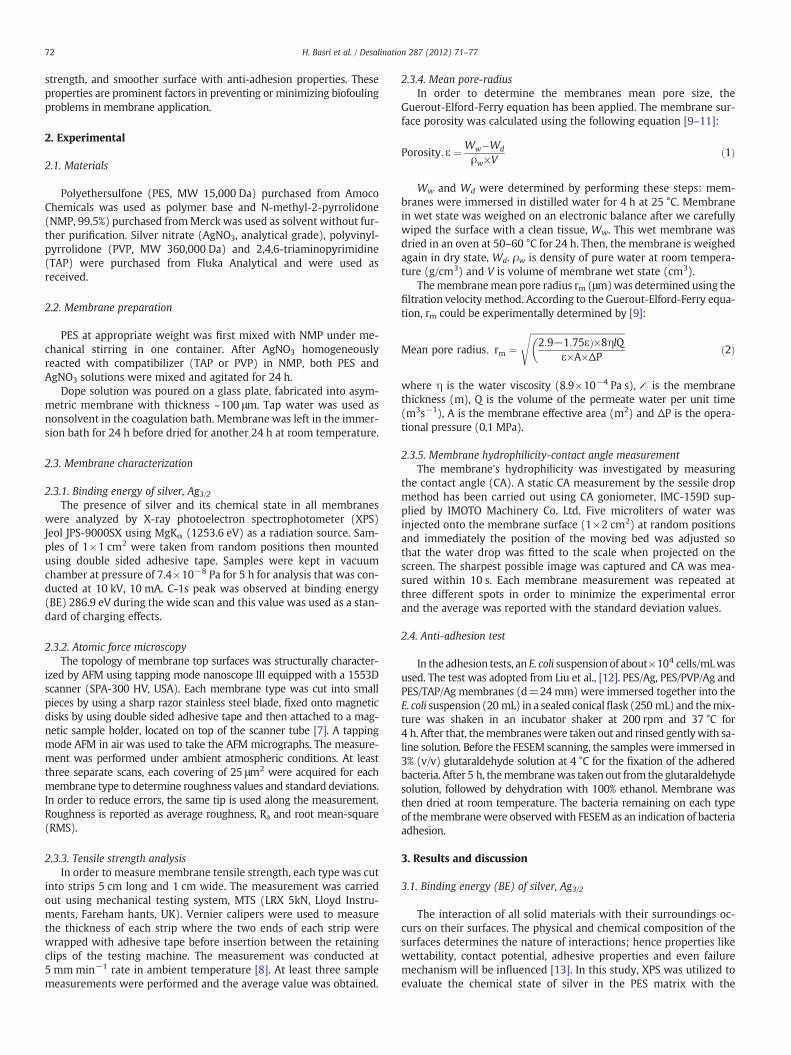
presence of different compatibilizers. Fig. 1 depicts the XPS results forthe BE of silver ion in the fabricated membranes.
The spectra show that all membranes exhibit the same ΔE of Agd3/2 orbitals and Ag d5/2 orbitals which are about 6 eV in agreementwith those reported in previous literature [14,15]. After calibratingwith C-1s data, the BE for Ag d5/2 orbitals was 370.2 eV for PES/Ag,369.8 eV and 370.3 eV for PES/Ag with PVP and TAP respectively. Ingeneral, the chemical shift occurred for all membranes, indicatingthat the oxidation state of Ag remained unchanged. However, thesmall shift for PES/Ag with PVP and TAP may correspond to thechange in the surface structure. We can conclude here that Ag+1 inboth PES-Ag and PES/Ag with TAP have been reduced to Ag0 whileAg+1 in PES/Ag with PVP remained in its +1 form.
It was observed that PES/Ag prepared with TAP exhibited intenseAg peaks for d5/2 and d3/2 orbital which are 3219.2 and 2315.9
Fig. 1. XPS spectra of PES-modified membranes.
respectively. This could be due to the higher content of silver particlesdistributed on the membrane surfaces. In real meaning, the morebonds with electronegative atoms that are in place, the greater isthe chemical shift [13]. As observed, the chemical shift for the sametype of membrane was shifted +0.1 to higher BE owing to greaterloading and better distribution. PVP contains one N atom, while TAPhas two N atoms in the aromatic rings and three N atoms on eachamide groups attached to the benzene ring. Therefore, the interac-tions of silver ions, Ag+ to the lone pair of electrons on each N atomis greater for PES/Ag with TAP.
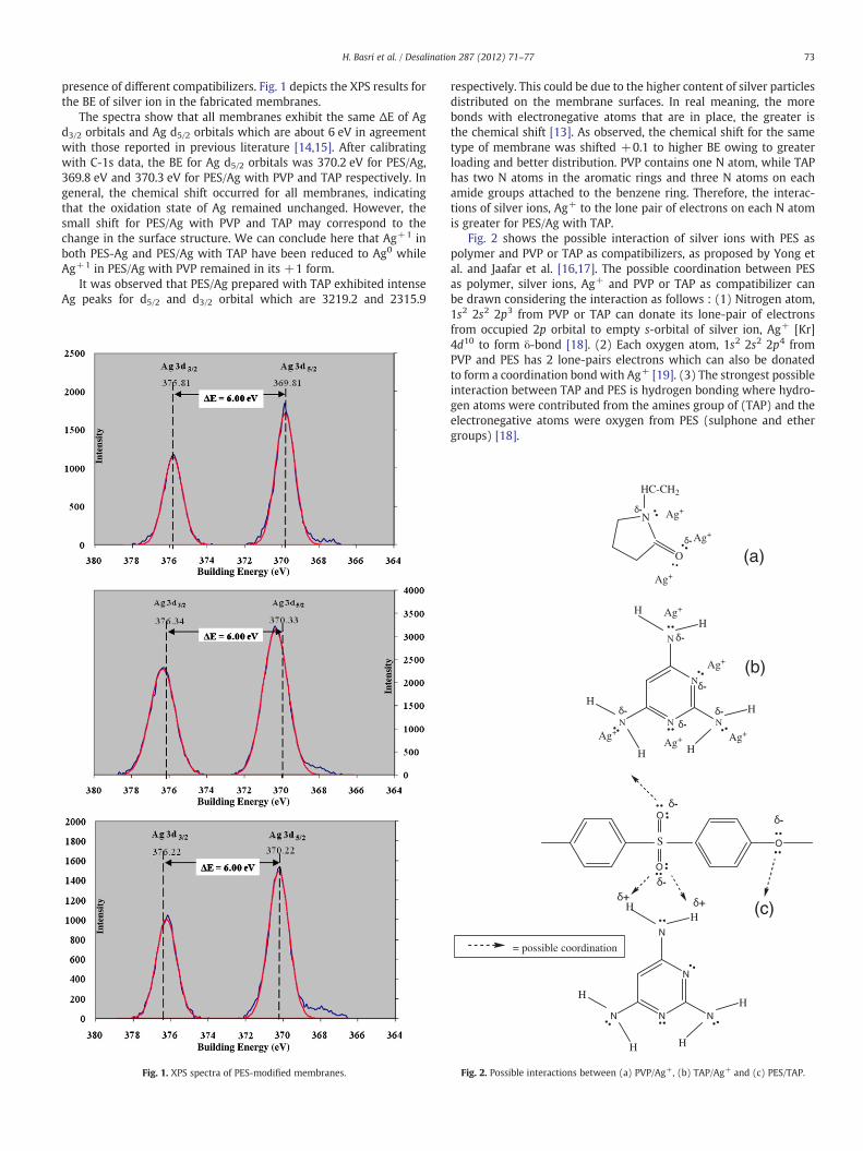
Fig. 2 shows the possible interaction of silver ions with PES aspolymer and PVP or TAP as compatibilizers, as proposed by Yong etal. and Jaafar et al. [16,17]. The possible coordination between PESas polymer, silver ions, Ag+ and PVP or TAP as compatibilizer canbe drawn considering the interaction as follows : (1) Nitrogen atom,1s2 2s2 2p3 from PVP or TAP can donate its lone-pair of electronsfrom occupied 2p orbital to empty s-orbital of silver ion, Ag+ [Kr]4d10 to form δ-bond [18]. (2) Each oxygen atom, 1s2 2s2 2p4 fromPVP and PES has 2 lone-pairs electrons which can also be donatedto form a coordination bond with Ag+ [19]. (3) The strongest possibleinteraction between TAP and PES is hydrogen bonding where hydro-gen atoms were contributed from the amines group of (TAP) and theelectronegative atoms were oxygen from PES (sulphone and ethergroups) [18].
= possible coordination
S O
O
O
N
N
N
NN
H
H
H
H
HH
-
-
-
N
N
N
NN
H
H
H
H
HH Ag+
Ag+
Ag+Ag+Ag+
---
-
-
N
O
HC-CH2
Ag+
Ag+
Ag+
-
-
(a)
(b)
(c)
Fig. 2. Possible interactions between (a) PVP/Ag+, (b) TAP/Ag+ and (c) PES/TAP.
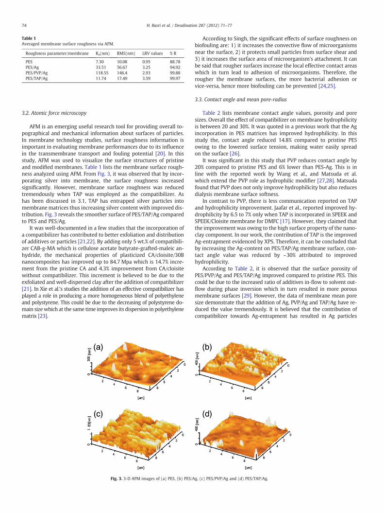
Table 1Averaged membrane surface roughness via AFM.
Roughness parameter/membrane Ra(nm) RMS(nm) LRV values % R
PES 7.30 10.08 0.95 88.78PES/Ag 33.51 56.67 3.25 94.92PES/PVP/Ag 118.55 146.4 2.93 99.88PES/TAP/Ag 11.74 17.49 3.59 99.97
74 H. Basri et al. / Desalination 287 (2012) 71–77
3.2. Atomic force microscopy
AFM is an emerging useful research tool for providing overall to-pographical and mechanical information about surfaces of particles.In membrane technology studies, surface roughness information isimportant in evaluating membrane performances due to its influencein the transmembrane transport and fouling potential [20]. In thisstudy, AFM was used to visualize the surface structures of pristineand modified membranes. Table 1 lists the membrane surface rough-ness analyzed using AFM. From Fig. 3, it was observed that by incor-porating silver into membrane, the surface roughness increasedsignificantly. However, membrane surface roughness was reducedtremendously when TAP was employed as the compatibilizer. Ashas been discussed in 3.1, TAP has entrapped silver particles intomembranematrices thus increasing silver content with improved dis-tribution. Fig. 3 reveals the smoother surface of PES/TAP/Ag comparedto PES and PES/Ag.
It was well-documented in a few studies that the incorporation ofa compatibilizer has contributed to better exfoliation and distributionof additives or particles [21,22]. By adding only 5 wt.% of compatibili-zer CAB-g-MA which is cellulose acetate butyrate-grafted-maleic an-hydride, the mechanical properties of plasticized CA/cloisite/30Bnanocomposites has improved up to 84.7 Mpa which is 14.7% incre-ment from the pristine CA and 4.3% improvement from CA/cloisitewithout compatibilizer. This increment is believed to be due to theexfoliated and well-dispersed clay after the addition of compatibilizer[21]. In Xie et al.'s studies the addition of an effective compatibilizer hasplayed a role in producing a more homogeneous blend of polyethyleneand polystyrene. This could be due to the decreasing of polystyrene do-main sizewhich at the same time improves its dispersion in polyethylenematrix [23].
Fig. 3. 3-D AFM images of (a) PES, (b) PES/A
According to Singh, the significant effects of surface roughness onbiofouling are: 1) it increases the convective flow of microorganismsnear the surface, 2) it protects small particles from surface shear and3) it increases the surface area of microorganism's attachment. It canbe said that rougher surfaces increase the local effective contact areaswhich in turn lead to adhesion of microorganisms. Therefore, therougher the membrane surfaces, the more bacterial adhesion orvice-versa, hence more biofouling can be prevented [24,25].
3.3. Contact angle and mean pore-radius
Table 2 lists membrane contact angle values, porosity and poresizes. Overall the effect of compatibilizer on membrane hydrophilicityis between 20 and 30%. It was quoted in a previous work that the Agincorporation in PES matrices has improved hydrophilicity. In thisstudy the, contact angle reduced 14.8% compared to pristine PESowing to the lowered surface tension, making water easily spreadon the surface [26].
It was significant in this study that PVP reduces contact angle by20% compared to pristine PES and 6% lower than PES-Ag. This is inline with the reported work by Wang et al., and Matsuda et al.which extend the PVP role as hydrophilic modifier [27,28]. Matsudafound that PVP does not only improve hydrophilicity but also reducesdialysis membrane surface softness.
In contrast to PVP, there is less communication reported on TAPand hydrophilicity improvement. Jaafar et al., reported improved hy-drophilicity by 6.5 to 7% only when TAP is incorporated in SPEEK andSPEEK/Cloisite membrane for DMFC [17]. However, they claimed thatthe improvement was owing to the high surface property of the nano-clay component. In our work, the contribution of TAP is the improvedAg-entrapment evidenced by XPS. Therefore, it can be concluded thatby increasing the Ag-content on PES/TAP/Ag membrane surface, con-tact angle value was reduced by ~30% attributed to improvedhydrophilicity.
According to Table 2, it is observed that the surface porosity ofPES/PVP/Ag and PES/TAP/Ag improved compared to pristine PES. Thiscould be due to the increased ratio of additives in-flow to solvent out-flow during phase inversion which in turn resulted in more porousmembrane surfaces [29]. However, the data of membrane mean poresize demonstrate that the addition of Ag, PVP/Ag and TAP/Ag have re-duced the value tremendously. It is believed that the contribution ofcompatibilizer towards Ag-entrapment has resulted in Ag particles
g, (c) PES/PVP/Ag and (d) PES/TAP/Ag.
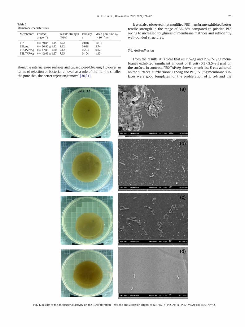
Table 2Membrane characteristics.
Membranes Contactangle (°)
Tensile strength(MPa)
Porosity,ε
Mean pore size, rm,(×10−3 μm)
PES θ=59.85±1.35 5.22 0.038 10.30PES/Ag θ=50.97±1.52 8.22 0.038 3.74PES/PVP/Ag θ=47.85±1.80 7.12 0.203 0.92PES/TAP/Ag θ=42.06±1.67 7.95 0.104 1.45
75H. Basri et al. / Desalination 287 (2012) 71–77
along the internal pore surfaces and caused pore-blocking. However, interms of rejection or bacteria removal, as a rule of thumb; the smallerthe pore size, the better rejection/removal [30,31].
Fig. 4. Results of the antibacterial activity on the E. coli filtration (left) and ant
It was also observed that modified PES membrane exhibited bettertensile strength in the range of 36–58% compared to pristine PESowing to increased toughness of membrane matrices and sufficientlywell-bonded structures.
3.4. Anti-adhesion
From the results, it is clear that all PES/Ag and PES/PVP/Ag mem-branes exhibited significant amount of E. coli (0.5×2.5–3.5 μm) onthe surface. In contrast, PES/TAP/Ag showed much less E. coli adheredon the surfaces. Furthermore, PES/Ag and PES/PVP/Ag membrane sur-faces were good templates for the proliferation of E. coli and the
i adhesion (right) of (a) PES (b) PES/Ag, (c) PES/PVP/Ag (d) PES/TAP/Ag.
76 H. Basri et al. / Desalination 287 (2012) 71–77
formation of biofilm might occur facilely on such surfaces upon con-tact with E. coli.
The filtration of E. coli suspension has also been conducted usingcross-flow permeation system under 1 bar operating pressure. Theplates of feeds and permeates after 24 h incubation are shown inFig. 4 and LRV values calculated for each membrane type are listedin Table 1 using the following formula:
LRV ¼ log10N0ðCFU=mLÞN1ðCFU=mL
Þ×100% ð3Þ
while the rejection was calculated using the following equation:
%R ¼ 1−Concentration of permeate;Cpðbased on CFUÞ
Concentration of feed;Cf ðbased on CFUÞ×100%: ð4Þ
It can be observed from Fig. 4 that PES/Ag with TAP exhibited thehighest inhibition of E. coli with LRV of 3.59 based on 10× dilution.Meanwhile, the other two membranes show acceptable LRV valuesof 2.93 and 3.25 for PES/Ag with PVP and PES/Ag respectively. It isknown that enumeration was based on the colony formed withoutconsidering the size of the colony. From the LRV values, PES-Agrejected better than PES/Ag with PVP, however the exact number ofbacteria that remained in the permeate of PES/Ag could be more(see colony size in Fig. 4a and b). The high LRV values performed bymembrane fabricated with silver and compatibilizers is attributed tothe effective permeation and also effective inhibition contributed bythe high silver content as proven in the XPS analysis.
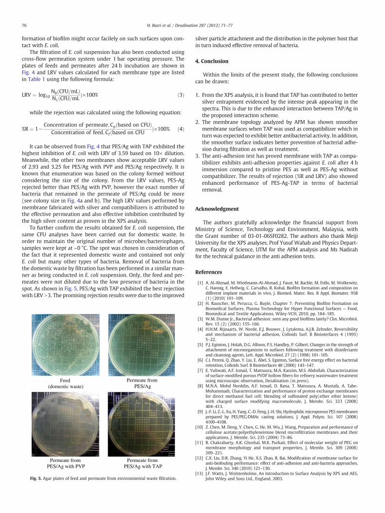
To further confirm the results obtained for E. coli suspension, thesame CFU analyses have been carried out for domestic waste. Inorder to maintain the original number of microbes/bacteriophages,samples were kept at ~0 °C. The spot was chosen in consideration ofthe fact that it represented domestic waste and contained not onlyE. coli but many other types of bacteria. Removal of bacteria fromthe domestic waste by filtration has been performed in a similar man-ner as being conducted in E. coli suspension. Only, the feed and per-meates were not diluted due to the low presence of bacteria in thespot. As shown in Fig. 5, PES/Ag with TAP exhibited the best rejectionwith LRV N3. The promising rejection results were due to the improved
Feed (domestic waste)
Permeate from PES/Ag
Permeate from PES/Ag with PVP
Permeate from PES/Ag with TAP
Fig. 5. Agar plates of feed and permeate from environmental waste filtration.
silver particle attachment and the distribution in the polymer host thatin turn induced effective removal of bacteria.
4. Conclusion
Within the limits of the present study, the following conclusionscan be drawn:
1. From the XPS analysis, it is found that TAP has contributed to bettersilver entrapment evidenced by the intense peak appearing in thespectra. This is due to the enhanced interaction between TAP/Ag inthe proposed interaction scheme.
2. The membrane topology analyzed by AFM has shown smoothermembrane surfaces when TAP was used as compatibilizer which inturnwas expected to exhibit better antibacterial activity. In addition,the smoother surface indicates better prevention of bacterial adhe-sion during filtration as well as treatment.
3. The anti-adhesion test has proved membrane with TAP as compa-tibilizer exhibits anti-adhesion properties against E. coli after 4 himmersion compared to pristine PES as well as PES-Ag withoutcompatibilizer. The results of rejection (%R and LRV) also showedenhanced performance of PES-Ag-TAP in terms of bacterialremoval.
Acknowledgment
The authors gratefully acknowledge the financial support fromMinistry of Science, Technology and Environment, Malaysia, withthe Grant number of 03-01-06SF0282. The authors also thank MeijiUniversity for the XPS analyses, Prof Yusuf Wahab and Physics Depart-ment, Faculty of Science, UTM for the AFM analysis and Ms Nadirahfor the technical guidance in the anti adhesion tests.
References
[1] A. Al-Ahmad, M. Wiedmann-Al-Ahmad, J. Faust, M. Bachle, M. Follo, M. Wolkewitz,C. Hannig, E. Hellwig, C. Carvalho, R. Kohal, Biofilm formation and composition ondifferent implant materials in vivo, J. Biomed. Mater. Res. B Appl. Biomater. 95B(1) (2010) 101–109.
[2] H. Rauscher, M. Perucca, G. Buyle, Chapter 7: Preventing Biofilm Formation onBiomedical Surfaces, Plasma Technology for Hyper Functional Surfaces — Food,Biomedical and Textile Applications, Wiley-VCH, 2010, pp. 184–185.
[3] W.M. Dunne Jr., Bacterial adhesion: seen any good biofilms lately? Clin. Microbiol.Rev. 15 (2) (2002) 155–166.
[4] H.H.M. Rijnaarts, W. Norde, E.J. Bouwer, J. Lytalema, A.J.B. Zehnder, Reversibilityand mechanism of bacterial adhesion, Colloids Surf. B Biointerfaces 4 (1995)5–22.
[5] P.J. Eginton, J. Holah, D.G. Allison, P.S. Handley, P. Gilbert, Changes in the strength ofattachment of microorganisms to surfaces following treatment with disinfectantsand cleansing agents, Lett. Appl. Microbiol. 27 (2) (1998) 101–105.
[6] C.I. Pereni, Q. Zhao, Y. Liu, E. Abel, S. Eginton, Surface free energy effect on bacterialretention, Colloids Surf. B Biointerfaces 48 (2006) 143–147.
[7] E. Yuliwati, A.F. Ismail, T. Matsuura, M.A. Kassim, M.S. Abdullah, Characterizationof surface-modified porous PVDF hollow fibers for refinery wastewater treatmentusing microscopic observation, Desalination (in press).
[8] M.N.A. Mohd Norddin, A.F. Ismail, D. Rana, T. Matsuura, A. Mustafa, A. Tabe-Mohammadi, Characterization and performance of proton exchange membranesfor direct methanol fuel cell: blending of sulfonated poly(ether ether ketone)with charged surface modifying macromolecule, J. Membr. Sci. 323 (2008)404–413.
[9] J.-F. Li, Z.-L. Xu, H. Yang, C.-D. Feng, J.-H. Shi, Hydrophilicmicroporous PESmembranesprepared by PES/PEG/DMAc casting solutions, J. Appl. Polym. Sci. 107 (2008)4100–4108.
[10] Z. Chen, M. Deng, Y. Chen, G. He, M. Wu, J. Wang, Preparation and performance ofcellulose acetate/polyethyleneimine blend microfiltration membranes and theirapplications, J. Membr. Sci. 235 (2004) 73–86.
[11] B. Chakrabarty, A.K. Ghoshal, M.K. Purkait, Effect of molecular weight of PEG onmembrane morphology and transport properties, J. Membr. Sci. 309 (2008)209–221.
[12] C.X. Liu, D.R. Zhang, Yi He, X.S. Zhao, R. Bai, Modification of membrane surface foranti-biofouling performance: effect of anti-adhesion and anti-bacteria approaches,J. Membr. Sci. 346 (2010) 121–130.
[13] J.F. Watts, J. Wolstenholme, An Introduction to Surface Analysis by XPS and AES,John Wiley and Sons Ltd., England, 2003.
77H. Basri et al. / Desalination 287 (2012) 71–77
[14] X.-Y. Gao, S.-Y. Wang, J. Li, Y.-X. Zheng, R.-J. Zhang, P. Zhou, Y.-M. Yang, L.-Y. Chen,Study of structure and optical properties of silver oxide films by ellipsometry,XRD and XPS methods, Thin Solid Films 455-456 (2004) 438–442.
[15] X.L. Yuqing, L. Liming, L. Weigang, Y. Dequan, D. Daoan, Atomic force microscopyand X-ray photoelectron spectroscopy study on nanostructured silver thin filmsirradiated by atomic oxygen, Mater. Sci. Eng. B79 (2001) 68–70.
[16] H. Yong, H.H.C. Park, Y.S. Kang, J.Won,W.N. Kim, Zeolite-filled polyimidemembranecontaining 2,4,6-triaminopyrimidine, J. Membr. Sci. 188 (2001) 151–163.
[17] J. Jaafar, A.F. Ismail, T. Matsuura, Preparation and barrier properties of SPEEK/Cloisite15A/TAP nanocomposite membrane for direct methanol fuel cell application, J.Membr. Sci. 345 (2009) 119–127.
[18] H. Wang, X. Qiao, J. Chen, X. Wang, S. Ding, Mechanisms of PVP in the preparationof silver nanoparticles (review), Mater. Chem. Phys. 94 (2005) 449–453.
[19] L. Han, R. Wang, D. Yuan, B. Wu, B. Lou, M. Hong, Hierarchical assembly of a novelluminescent silver coordination framework with 4-(4-pyridylthiomethyl)benzoicacid, J. Mol. Struct. 737 (2005) 55–59.
[20] K.C. Khulbe, C.Y. Feng, T. Matsuura, Synthetic PolymerMembranes— Characterizationby Atomic Force Microscopy (Chapter 5), Springer, Berlin Heidelberg, 2008.
[21] H.-M. Park, X. Liang, A.K. Mohanty, M. Misra, L.T. Drzal, Effect of compatibilizer onnanostructure of the biodegradable cellulose actetate/organocalay nanocomposites,Macromolecules 37 (24) (2004) 9076–9082.
[22] M. Spirkova, P. Duchak, A. Strachota, R. Poreba, J. Kotek, J. Baldrian,M. Slouf, The role oforganic modification of layered nanosilicates onmechanical and surface properties oforganic-inorganics coatings, J. Coat. Technol. Res. 8 (3) (2011) 311–328.
[23] Z. Xie, J. Shang, Z. Wan, Mechanical properties and morphology of polypropylene/-polystyrene blends, J. Macromol. Sci. B40 (20) (2001) 251–261.
[24] R. Singh, Hybrid Membrane Systems for Water Purification — Technology, System,Design and Operation, Elsevier Ltd, Oxford, UK, 2006, p. 94.
[25] E.M.V. Hoek, S. Bhattacharjee, M. Elimelech, Membrane surface roughness oncolloid-membrane DLVO interactions, Langmuir 19 (2003) 4836–4847.
[26] H. Basri, A.F. Ismail, M. Aziz, Polyethersulfone (PES)-silver composite UFmembrane:the effect of silver content and PVPof differentmolecularweight onmembranemor-phology and antibacterial activity, Desalination 273 (1) (2011) 72–80.
[27] M. Matsuda, M. Sato, H. Sakata, T. Ogawa, K. Yamamoto, T. Yakushiji, M. Fukuda,T. Miyasaka, K. Sakai, Effects of fluid flow on elution of hydrophilic modifier dial-ysis membrane surfaces, J. Artif. Organs 11 (2008) 148–155.
[28] H.Wang, T. Yu, C. Zhao, Q. Du, Improvement of hydrophilicity andblood compatibilityon polyethersulfonemembrane by adding polyvinylpyrrolidone, Fibers Polym. 10 (1)(2009) 1–5.
[29] T.H. Young, L.W. Chen, A diffusion-controlled model for wet-casting membraneformation, J. Membr. Sci. 59 (1991) 169–181.
[30] R. Schnabel, W. Vaulont, High-pressure techniques with porous glass membranes,Desalination 24 (1–3) (1977) 249–272.
[31] S. Bhattacharjee, A. Sharma, P.K. Bhattacharya, Estimation and influence of longrange solute, membrane interactions in ultrafiltration, Ind. Eng. Chem. Res. 35(1996) 3108–3121.