Utilitarian Aspects of Weeds of Wheat Fields in Charbagh ...
Upload
joannecarvCategory
view
0download
0
ORIGINAL ARTICLE
Microfungal “Weeds” in the Leafcutter Ant Symbiosis
A. Rodrigues & M. Bacci Jr & U. G. Mueller &
A. Ortiz & F. C. Pagnocca
Received: 8 August 2007 /Accepted: 17 February 2008 /Published online: 28 March 2008# Springer Science + Business Media, LLC 2008
Abstract Leafcutter ants (Formicidae: tribe Attini) are well-known insects that cultivate basidiomycete fungi (Agaricales:Lepiotaceae) as their principal food. Fungus gardens aremonocultures of a single cultivar strain, but they also harbor adiverse assemblage of additional microbes with largelyunknown roles in the symbiosis. Cultivar-attacking micro-fungi in the genus Escovopsis are specialized parasites foundonly in association with attine gardens. Evolutionary theorypredicts that the low genetic diversity in monoculturesshould render ant gardens susceptible to a wide range ofdiseases, and additional parasites with roles similar to that of
Escovopsis are expected to exist. We profiled the diversityof cultivable microfungi found in 37 nests from tenAcromyrmex species from Southern Brazil and comparedthis diversity to published surveys. Our study revealed atotal of 85 microfungal strains. Fusarium oxysporum andEscovopsis were the predominant species in the surveyedgardens, infecting 40.5% and 27% of the nests, respectively.No specific relationship existed regarding microfungalspecies and ant-host species, ant substrate preference (dicotversus grass) or nesting habit. Molecular data indicated highgenetic diversity among Escovopsis isolates. In contrast tothe garden parasite, F. oxysporum strains are not specificparasites of the cultivated fungus because strains isolatedfrom attine gardens have similar counterparts found in theenvironment. Overall, the survey indicates that saprophyticmicrofungi are prevalent in South American leafcutter ants.We discuss the antagonistic potential of these microorgan-isms as “weeds” in the ant–fungus symbiosis.
Introduction
Insect–fungal mutualisms are interspecies associations ofgreat evolutionary success [5, 6, 32]. One such associationis the mutualism between the farming ants (Hymenoptera:Formicidae: tribe Attini) and their cultivated fungi, anancient symbiosis that likely originated about 50 to 65 mya[30]. Within the tribe Attini, the leaf-cutting ants representone of the most derived groups comprising two ant genera,Atta and Acromyrmex [43]. In many parts of the NewWorld, leafcutter ants are recognized as highly destructivecrop pests [25] because leafcutter nests support millions ofindividuals, and workers forage for large quantities of freshleaf material that they cut and bring to their undergroundnests to use as substrate for fungal cultivation [50].
Microb Ecol (2008) 56:604–614DOI 10.1007/s00248-008-9380-0
A. Rodrigues :M. Bacci Jr (*) : F. C. PagnoccaCenter for the Study of Social Insects,UNESP-São Paulo State University,Av. 24A, n. 1515-Bela Vista,Rio Claro, São Paulo 13506-900, Brazile-mail: [email protected]
A. Rodriguese-mail: [email protected]
A. Rodrigues :M. Bacci Jr : F. C. PagnoccaDepartment of Biochemistry and Microbiology,UNESP-São Paulo State University,Rio Claro, São Paulo 13506-900, Brazile-mail: [email protected]
U. G. MuellerSection of Integrative Biology, University of Texas at Austin,Austin, TX 78712, USAe-mail: [email protected]
A. OrtizConservación, Usos y Biodiversidad, Facultad de Ciencias,Universidad Nacional de Colombia,Medellín AA 3840, Colombiae-mail: [email protected]

The cultivated fungus, Leucoagaricus gongylophorus(Basidiomycota: Agaricales: Lepiotaceae), together with theplant substrate supplied by the ants to sustain the fungalpartner, compose the fungus gardens. The leaf-cutting ants’fungi develop specialized nutritive swellings (gongylidea)that are used by the ants to nourish their brood [50]. Thefungus, in turn, benefits from the association because theants provide a suitable environment for its growth. The antsalso disperse the fungus when young queens carry a smallfungal inoculum from their natal colony for the foundationof a new nest [31].
According to Poulsen and Boomsma [36] and Scott et al.(in preparation), leaf-cutting ants actively inhibit the growthof multiple strains of fungal cultivars within the nest,thereby maintaining their associated partner as single clones(i.e., monocultures). The resulting lack of genetic diversityin the fungus gardens is expected to render gardenssusceptible to diseases and parasites [24]. An analogousproblem exists in human monoculture crops [31]. Indeed,Currie et al. [11], sampling for non-mutualistic fungiassociated with attine nests, discovered that the attinecultivars are host to a specialized fungal parasite in thegenus Escovopsis (Ascomycota: anamorphic Hypocreales)that negatively impacts the ant colony. Escovopsis infectsnests of attine ant species across all genera studied and isthe most frequently encountered non-mutualistic fungusfound so far in attine gardens of Central America [11, 12].Escovopsis acts as a necrotrophic parasite that destroys thecultivar’s hyphae [37] and exhibits a complex pattern of co-evolution with the cultivar. The original claim of ancientEscovopsis-cultivar cocladogenesis by Currie et al. [14]suggested parasite–host specificity at broad phylogeneticlevels (four Escovopsis clades corresponding to four cultivarclades from four ant clades), but more comprehensivesampling [21] revealed occasional switching of Escovopsislineages between cultivar lineages at the finest phylogeneticlevels.
In addition to Escovopsis, attine ants harbor a commu-nity of other microbes in their gardens, including micro-fungi (filamentous fungi and yeasts) and bacteria [2, 9, 19,38]. Leafcutter ants can regulate the microbiota in gardens,for example by actively combing out unwanted fungal spores[13] or by application of germination-inhibiting secretions[18]. However, the function of the associated microbiota inthe garden matrix is largely unknown. These additionalmicroorganisms could be harmful invaders (or “weeds”)when found in high frequency in the ants’ gardens [19, 35],neutral and transient commensals (with negligible effectson garden homeostasis), or potentially beneficial ancillarycomponents serving unknown functions such as productionof enzymes or antibiotics [2, 32].
Poulsen and Currie [35] suggested that the microfungiother than Escovopsis are mere transient guests with no
active role in the fungus garden. This view is consistentwith studies that documented ubiquitous microfungalspecies in attine gardens that are commonly found also inmany other environmental sources. For instance, Carreiroet al. [9] and Craven et al. [10] reported ubiquitous yeastsspecies in the fungus gardens of laboratory nests (e.g.,Candida spp.). However, in a survey of non-mutualisticfilamentous fungi, Rodrigues et al. [38] discovered that somemicrofungi such as Fusarium oxysporum and Trichodermaharzianum occur in higher frequency in leafcutter gardensthan the parasite Escovopsis sp. This was observed in nestsunder stressed conditions (i.e., laboratory nests treated withtoxic baits). The same study also documented a high micro-fungal incidence other than Escovopsis sp. in natural Attasexdens rubropilosa colonies. Fungal species such as Acre-monium kiliense, Cunninghamella elegans, F. oxysporum, T.harzianum, and Syncephalastrum racemosum were fre-quently isolated [38], suggesting that their presence is notcasual. In order to further understand the distribution andprevalence of these and other filamentous fungi in gardensof leaf-cutting ants, we conducted a survey of the micro-fungal species in field nests of leaf-cutting ants from SouthernBrazil.
Previous studies on the microfungal diversity in attinenests focused on specific groups of microorganisms underdiverse conditions. For example, several studies samplednatural nests of Central American attine species for thepresence of Escovopsis [11, 12, 20]. Other studies surveyedthe yeast diversity in laboratory nests of leaf-cutting ants[9, 10]. Fisher et al. [19] reported changes in the com-munity structure of non-mutualistic filamentous fungi ofAtta cephalotes laboratory nests when maintained withdifferent types of leaf diets. Lastly, Möeller [29] reportedmicrofungi species, including Escovopsis sp., from leaf-cutter gardens collected in Southern Brazil and maintainedin the laboratory.
The present study differs from the above surveys [9–11,19, 29, 38] of attine gardens in three main aspects: (1) theleaf-cutting ant species surveyed belonged to the genusAcromyrmex (Atta was largely absent in the surveyed area);(2) the collection sites were located in Southern Brazil(primarily the State of Rio Grande do Sul); and (3) the fieldnests appeared to be in healthy condition at the time ofcollection, with no visible signs of disturbance or stress.The survey addresses two primary questions: (1) Are therespecies-specific relationships among the microfungi andants? (2) Is Escovopsis sp. prevalent in Acromyrmex gardensfrom Southern Brazil, and is its prevalence in SouthernBrazil comparable to that of Central America [11, 12]?
Our study confirms previous reports that the gardensof leaf-cutting ants harbor several soil and plant-bornefungi but also shows a comparatively low infection rateby Escovopsis. The documented diversity of soil and plant-
Microfungi in Attine Gardens 605605

borne fungi may function under certain conditions asopportunistic pathogens in leafcutter gardens, constrainingthe symbiosis by competing with the fungal cultivar fornutrient resources.
Materials and Methods
Fungus Garden Sampling
From 4–17 September 2004, gardens from 37 mature nestsof ten Acromyrmex species were sampled in differentlocalities of the State of Rio Grande do Sul (RS) inSouthern Brazil (see Table 1 for collecting localities). Thetype of substrate carried by foraging workers at the time ofcollection was recorded along with the nesting habitat. Thisinformation was compared with Gonçalves [22] whoprovided detailed descriptions of foraging behavior andnest architecture of Brazilian Acromyrmex species. Whenour observations differed from the species-specific charac-ters reported in the literature [22], our own observationswere used in the analyses, as summarized in Table 1. Thenests were carefully excavated (in the case of soil-dwellingspecies) or carefully opened (in the case of mound-buildingspecies; Table 1) in order to prevent contamination of anyaccessed garden. Large garden fragments (with workers and
brood) were immediately transferred whole (without dis-rupting the garden) with sterilized forceps to sterile plasticcontainers (volume capacity=50 ml).
During the 2-week field expedition, all garden containerswere kept in a cooler in the dark until transported to the“Centro de Estudos de Insetos Sociais” (CEIS) lab at RioClaro, where they were maintained for an additional 3 daysbefore fungal isolation.
Microfungi Isolation
We followed two established isolation techniques [11, 39] forprofiling the microfungal community in the fungus gardens.From each garden collection, (1) ten fragments (3 mm3 indiameter) of the gardens were removed and inoculated inpotato-dextrose agar plates (PDA, DIFCO®) supplementedwith 150 μg ml−1 of chloramphenicol (US Biological); (2)six garden fragments (20 mm3 in diameter) were carefullyfreed of all the workers and brood (by sorting through eachfragment with a sterilized forceps) then placed into a sterile,humidified Petri dish. The dish contained a piece of cottonwith sterile distilled water, which provided humidity forcontinued fungal growth (the so-called “wet chamber”). Allplates were incubated at 25°C for 7–14 days in the dark.
PDA plates and wet chambers were checked daily for signsof any filamentous fungal growth. Once a fungus emerged
Table 1 General characteristics of the ant species used in this study
Acromyrmex species City/State Nest locationb Substrate Nest type
A. ambiguus (2)a Nova Petrópolis/RS S 29°22′38.2″; W 50°57′18.1″ Dicot Mound-builderNear Pelotas/RS S 30°50′10.2″; W 51°55′10.4″ Dicot Mound-builder
A. aspersus (2) São Marcos/RS S 28°58′05.6″; W 51°07′58.0″ Dicot Soil-dwellerA. coronatus (9) near Registro/RS (2) S 25°25′50.5″; W 49°04′56.4″ Dicot Mound-builder
Itajai/RS S 25°25′50.5″; W 49°04′56.4″ Dicot Soil-dwellerNear Pelotas/RS S 30°50′10.2″; W 51°55′10.4″ Dicot Mound-builderVacaria/RS (2) S 28°27′51.7″; W 50°53′07.0″ Dicot Mound-builderBlumenau/SC (2) S 26°53′37.8″; W 49°11′29.0″ Dicot Mound-builderBlumenau/SC S 26°51′48.3″; W 49°16′15.0″ Dicot Mound-builder
A. crassispinus (1) Nova Petrópolis/RS S 29°23′51.4″; W 50°54′27.3″ Dicot Soil-dwellerA. disciger (2) Blumenau/SC S 26°54′04.9″; W 49°10′51.2″ Dicot Mound-builderA. hispidus falax (2) Londrina/PR S 22°47′22.0″; W 51°36′01.6″ Dicot Soil-dwellerA. laticeps (5) Nova Petrópolis/RS (2) S 29°19.05′9″; W 51°10′13.6″ Dicot Soil-dweller
São Marcos/RS S 28°57′16.5″; W 51°08′20.0″ Dicot Soil-dwellerAlto da Serra/RS (2) S 28°12′26.2″; W 50°45′27.4″ Dicot Mound-builder
A. lundi (3) São Marcos/RS (2) S 28°58′02.8″; W 51°08′08.8″ Dicot Soil-dwellerChuvisca/RS S 30°50′10.2″; W 51°55′10.4″ Dicot Soil-dweller
A. heyeri (10) Sentinela do Sul/RS (4) S 30°37.57′9.0″; W 51°33′18.2″ Monocot Soil-dwellerChuvisca/RS S 30°50′10.2″; W 51°55′10.4″ Monocot Mound-buildernear Pelotas/RS (4) S 30°50′10.2″; W 51°55′10.4″ Monocot Mound-builderSantana da Boa Vista/RS S 30°56′40.0″; W 53°05′10.3″ Monocot Mound-builder
A. landolti (1) Taquara/RS S 29°42′55.7″; W 50°50′21.5″ Monocot Soil-dweller
PR Paraná; RS Rio Grande do Sul; SC Santa Catarinaa Figures in parentheses (column 1) indicate the number of colonies sampled for each ant speciesb Figures in parentheses (column 3) indicate the number of colonies found at the same locality
606 A. Rodrigues et al.

from the garden fragment, an inoculumwas transferred tomaltagar 2% plates (MA 2%, DIFCO®) in order to obtain purecultures. When morphologically very similar microfungalcolonies were characterized in a single ant garden, a uniquerepresentative fungal sample was isolated, and the strains werestored in 10% glycerol at −80°C at CEIS. When insufficientgarden material was available to conduct both isolationmethods, only one method was used out of necessity, yielding17 isolations with PDA only, 4 isolations with wet chambersonly, and 16 isolations using both methods.
Fungal Identification
Morphological Methods
Colony macromorphology and micromorpholgy were usedas main characters to identity the isolates. Species wereidentified with the help of general taxonomic keys [4, 15,42] as well as specific taxonomic treatments for somegroups of fungi [26, 28, 33].
Molecular Methods
Microfungi were further identified with the help of DNAsequence information. Genomic DNA was extracted usingthe cetyl trimethylammonium bromide method [20]. Priorto DNA extractions, isolates were grown in aerated liquidcultures (malt extract broth 2%) for 7 days at 25°C, and themycelia were harvested and lyophilized.
A 25 μl polymerase chain reaction was performed usingReady-to-Go™ beads (GE Healthcare) and 1.0 μl of DNAtemplate (>40 ng). ITS4 and ITS5 primers (6 pmol each)were used to amplify the internal transcribed spacer regionsof the ribosomal DNA [51]. For Escovopsis isolates, theprimers eafF (5′CATGATCACTGGTACCTCCCAGG3′)and eafR (5′GCATGTCACGGACGGCGAAACGA3′) mod-ified from [14] were used to amplify a fragment spanningthe exon 6 of the elongation factor 1-alpha (EF1-a) gene.
The amplification protocol consisted of an initial dena-turation step at 94°C for 3 min, followed by 35 cycles of94°C for 1 min, 50°C for 1 min, and 72°C for 1 min, followedby a final extension step of 72°C for 15min. The amplificationproducts were purified with Wizard® Genomic DNA Purifi-cation Kit (Promega Corporation) following the manufac-turer’s protocol.
Cloning was necessary in some cases to obtain goodsequence reads. In these cases, the amplicons were insertedin pGEM® T-vector (Promega Corporation) and transformedin competent Escherichia coli DH10β cells. DNA fromrecombinant cells was purified following the miniprep pro-cedure by Sambrook and Russel [41].
The 10 μl cycle sequencing reaction contained 2.5 μl ofBig Dye terminator (Applied Biosystems); 2.5 μl of 100 mM
Tris and 2.5 mM MgCl2 (pH 9.0), 6 pmol of each primer(the same ones used in the amplification step); and 30–40 ng of the purified polymerase chain reaction products.Reaction conditions included a denaturation step of 96°Cfor 2 min followed by 28 cycles of 96°C for 45 s, 50°C for30 s, and 60°C for 4 min. The amplicons were sequencedon an ABI Prism 377 DNA sequencer (Applied Biosystems).For all samples, both forward and reverse sequenceswere obtained for the internal transcribed spacer (ITS) andEF1-a regions. Sequences from representative isolates aredeposited at Genbank as accessions EU082779–EU082803.
Sequence Analysis
Forward and reverse strands were edited using Bioeditv.7.0.5.3 [23], and the consensus sequence was used inBLASTN similarity searches at the National Center for Bio-technology Information-Genbank [1] or at the TrichoKeydatabases [16] (the latter one just for Trichoderma isolates).Sequences presenting 99% similarity with sequences obtainedfrom databases were considered as identified (Table 2).
Phylogenetic analyses were performed for two types ofmicrofungi that occurred in high proportions in gardens(Escovopsis sp. and F. oxysporum). Escovopsis sequenceswere aligned in ClustalW [49] using default parametersand analyzed in PAUP* v. 4.0b10 [47] under themaximum parsimony (MP) criterion. An heuristic searchwas conducted with 1,000 replicates, random sequenceaddition, tree bisection-reconnection branch swapping,and the collapse and multrees options implemented.Maximum likelihood (ML) analysis was conducted in GARLIv. 0.951 [52] using default parameters as recommended in theUser’s Manual. Branch support for MP and ML analyseswas calculated using 1,000 non-parametric bootstrap pseudo-replicates [17] using the same settings as for initial searches.Bayesian analyses were carried out in MrBayes v. 3.1.2 [40].Four separate runs were conducted, each with four incre-mentally heated chains and uninformative, default priors;converge and optimal burn-in were assessed as describedin [7] using the program MrConverge (Lemmon, inpreparation). After discarding burn-in, the posterior samplesof tree topologies for each run were combined in PAUP* toobtain the posterior probabilities of each node. Sequencesfrom Escovopsis isolates published in other studies [14, 48]were obtained from Genbank (accessions # AY172620,AY172622, EF589910–EF589914, EF589916–EF589919,and EF589921–EF589949).
In order to establish the phylogenetic relationships of F.oxysporum isolates from attine gardens and F. oxysporumfrom other environmental sources, a median-joining net-work [3] was inferred using Network v. 3.1.1.1 (available atwww.fluxus-engineering.com). Sequence information fordifferent F. oxysporum strains were retrieved from Genbank
Microfungi in Attine Gardens 607607

Tab
le2
Microfung
ispeciesin
fung
usgardensof
Acrom
yrmex
speciesfrom
Sou
thernBrazil
Fungi
classified
asClosestidentifiedrelativ
eAcrom
yrmex
spp
%of
nests
(n=37)with
thespecified
microfungus
A.
ambiguus
aA.
aspersus
aA.
coronatusa
A.
crassispinus
aA.
disciger
aA.
hispidus
aA.
laticepsa
A.
lundia
A.
heyerib
A.
landoltib
Fungalspecies
Accession
#%
identity
n=2
n=2
n=9
n=1
n=2
n=2
n=5
n=3
n=10
n=1
Zygom
ycota
Cunninghamella
binariae
C.binariae
AF254935
981c
15
19.0
C.blakesleana
C.blakesleana
AF254932
971
2.8
C.echinulata
C.echinulata
var.
antartica
AF254938
951
11
8.0
Mucor
circinelloides
M.circinelloides
DQ118966
991
2.8
M.racemosus
M.racemosus
AJ271061
991
11
11
10.9
Mucor
sp.1
Mucor
recurvus
AF412294
921
2.8
Mucor
sp.2
n.d.
n.d.
12.8
Syncephalastrum
racemosum
n.d.
n.d.
n.d.
25.4
Ascom
ycota
Aspergillu
sfla
vus
A.fla
vus
AM745114
100
12
8.0
Aspergillu
sversicolor
A.versicolor
AJ937750
991
2.8
Escovopsissp.
See
text
andFig.2
fordetails
12
11
23
27.0
Eupenicillium
javanicum
E.javanicum
U18358
100
12.8
Chaetom
ium
sp.
Chaetom
ium
sp.
AJ279468
891
2.8
Cladosporium
cladosporioides
C.
cladosporioides
AF393691
100
12.8
Fusarium
oxysporum
F.oxysporum
AY462580
100
11
12
32
640.5
F.equiseti
F.equiseti
AY147362
100
12.8
F.solani
F.solani
AM412639
100
12.8
Trichoderm
ahamatum
T.hamatum
DQ286602
100
12.8
T.harzianum
T.harzianum
AF443922
100
11
18.0
T.spirale
T.spirale
AY154939
991
2.8
Trichoderm
asp.
Trichoderm
asp.
AY154942
991
11
15.4
T.virens
T.virens
AF099007
100
12.8
Lecythophorasp.
Lecythophorasp.
AY219880
941
2.8
Moniliella-like
fungus
Hypom
yces
aurantius
AF055297
821
11
8.0
Paecilomyces
lilacinus
n.d.
n.d.
n.d.
12.8
Penicillium
citrinum
P.citrinum
EF127876
991
15.4
Penicillium
sp.1
Penicillium
sp.
DQ092545
991
2.8
Penicillium
sp.2
Penicillium
sp.
DQ279802
100
12.8
608 A. Rodrigues et al.

as accession # U34571, AJ853769, U34566, U28161,X94173, AF165875, AF069310, and U28159.
Results
Microfungal Distribution in Acromyrmex Nests
Aiming to improve our assessment of the microfungal di-versity in the fungus gardens, we have carried out twodifferent isolation techniques. The effect of this strategy canbe evaluated by the results obtained from the 16 nests whichhad enough material to be used in both techniques. These 16nests were found to contain 22 fungal species, but only fourof these species (Cunninghamella binariae, Escovopsis, F.oxysporum, and T. harzianum) were isolated by bothtechnical procedures; eight species (Fusarium solani,Mucor circineloides, Penicillium sp. 2, Penicillium waks-manii, S. racemosum, Trichoderma sp., Xylaria sp. 1, andXylaria sp. 2) were isolated uniquely through the wet-chamber method; and ten species (Chaetomium sp.,Lecithophora sp., Moniliella-like fungi, Mucor sp. 1,Mucor sp. 2, Mucor racemosus, Trichoderma spirale,Volutella sp., and two isolates of non-identified fungi) wererecovered only by the PDA method. These results suggestthat the two isolation methods worked complimentary toeach other in order to depict the microfungal diversity inAcromyrmex gardens.
Application of these two isolation techniques to thegardens of Acromyrmex ants resulted in the recovering of85 microfungal strains. This pool of isolates comprised 33fungal species from 16 genera that were identified either bymorphological or sequencing analyses. In addition, twonon-sporulating, morphologically unidentifiable fungal iso-lates could only be classified based on ITS sequence in-formation (Table 2).
Among the 16 fungal genera found, Cunninghamella,Escovopsis, Fusarium,Mucor, Penicillium, and Trichodermawere the most prevalent, occurring at least in 18% of thegardens (Table 2). Fusarium and Cunninghamella wereisolated in 26% and 19% of grass-cutting ant’s gardens,respectively, whereas Fusarium, Mucor, and Escovopsiswere found in 18.5%, 14.8%, and 13% in dicot-cutting ant’sgardens, respectively (Fig. 1a). Ten out of 16 microfungalgenera were observed in monocot-cutting ants, and 14 out of16 microfungal genera were found in gardens of dicot-cutting ants. Only eight genera (Aspergillus, Cunninghamella,Escovopsis, Fusarium, Moniliella-like, Penicillium, Tricho-derma, and Xylaria) were common in gardens of both mono-cot and dicot-cutting ants (Fig. 1a).
When comparing the microfungi profile between nest-type, Fusarium and Escovopsis were the most prevalent,occurring in 24% and 15% of mound-building ant species,T
able
2(contin
ued)
Fungi
classified
asClosestidentifiedrelativ
eAcrom
yrmex
spp
%of
nests
(n=37)with
thespecified
microfungus
A.
ambiguus
aA.
aspersus
aA.
coronatusa
A.
crassispinus
aA.
disciger
aA.
hispidus
aA.
laticepsa
A.
lundia
A.
heyerib
A.
landoltib
Fungalspecies
Accession
#%
identity
n=2
n=2
n=9
n=1
n=2
n=2
n=5
n=3
n=10
n=1
Penicillium
sp.3
P.minioluteum
AY213674
971
2.8
P.waksm
anii
P.waksm
anii
AY373940
991
2.8
Volutella
sp.
Volutella
sp.
EF029211
992
15.4
Xylaria
sp.1
Xylaria
sp.
EF423534
891
15.4
Xylaria
sp.2
n.d.
n.d.
n.d.
12.8
n.i.ascomycetes
1n.i.ascomycete
AJ279488
100
12.8
n.i.ascomycetes
2uncultu
red
ascomycete
EF027379
891
2.8
nNum
berof
nestssampled
foreach
antspecies;n.d.
nodata;n.i.,
notidentified
aAnt
speciesaredicot-cutting
ants(for
furtherdetails,seeTable
1)bAnt
speciesaregrass-cutting
ants(for
furtherdetails,seeTable
1)cNum
berof
colonies
ofaparticular
antspeciesfrom
which
was
observed
thespecifiedmicrofung
us
Microfungi in Attine Gardens 609609

respectively. Fusarium and Cunninghamella were the twopredominant genera in soil-dwelling ant species (Fig. 1b).Ten out of 16 microfungal species were associated with bothmound-building and soil-dwelling ant species (Fig. 1b).
While ascomycete fungi comprised the majority of theisolates from both monocot and dicot-cutting ants, zygo-mycetes were not found in high frequency in monocot-cutting ants, with the exception of C. binariae which wasfound in Acromyrmex heyeri nests (Table 2). On the otherhand, zygomycete fungi were found in association withseven out of eight dicot-cutting ant species studied (Table 2).
The most frequent fungal species in the present surveywere: F. oxysporum from 40.5% of the colonies acrossseven ant species; E. weberi from 27% of the colonies fromsix ant species; C. binariae from 19% of the colonies ofthree ant species; and M. racemosus from 10.9% of thecolonies of five ant species (Table 2). The remaining micro-fungal species were present in less than 10% of the totalnests sampled.
Phylogenetic Analyses
Because F. oxysporum was the most prevalent speciesin our survey, we evaluated whether any particular F.oxysporum strains were specialized in infecting Acromyr-mex gardens. This was accomplished by assessing thephylogenetic relationship between ITS haplotypes fromour F. oxysporum isolates with published ITS haplotypesfrom F. oxysporum strains commonly found in soil or plantsubstrates. Because ITS2 is known to have paralogous copies
in Fusarium [34], we confirmed first that the major ITS2type present in our isolates were the ITS2-type I described byO’Donnell and Cigelnik [34]. There was a low polymor-phism of the ITS1 and ITS2 regions within the analyzedstrains, with one nucleotide difference on average. Themedian-joining network (not shown) suggested a scenariowhich is not compatible with specialized infection ofAcromyrmex by F. oxysporum, since no genetic groupcontaining only closely related isolates from leafcutter nestswas characterized. In addition, 12 haplotypes were shared byleafcutter isolates and other isolates from several environ-mental sources, including soil and plant substrates.
We also investigated species-specificity regarding Esco-vopsis strains and leafcutter species by inferring thephylogenetic relationships among our isolates as well asother previously studied Escovopsis strains [14, 48]. Thephylogeny inferred from the EF1-a marker (Fig. 2) showedthat all of the Escovopsis isolates in our survey fall withinEscovopsis group A, as defined by Taerum et al. [48]. Nospecies-specificity was detected between Acromyrmex antsand Escovopsis strains from Southern Brazil, since (1)closely related Escovopsis strains were associated withdifferent ant species, and (2) gardens of the same ant specieswere associated with more distantly related Escovopsisstrains (Fig. 2). Although Escovopsis isolates from group Bdid not form a monophyletic clade in our phylogeneticanalyses, as they do in previous studies [48], this discrepancyis a result of the shorter EF1-a fragments used in ouranalyses (697 base pairs versus >1,400 base pairs inTaerum’s et al. study [48]).
Figure 1 Relative abundance of fungal isolates (grouped by genus)found in Acromyrmex leafcutter ant nests in Southern Brazil. Antspecies are grouped according to: a The type of plant material used bythe workers to nourish their cultivars, dicot-cutting ants (n=57 fungal
isolates) and grass-cutting ants (n=26 fungal isolates). b Nestinghabit, mound-builder (n=38 fungal isolates) and soil-dweller (n=45fungal isolates). Unidentified ascomycete fungi are not included
610 A. Rodrigues et al.
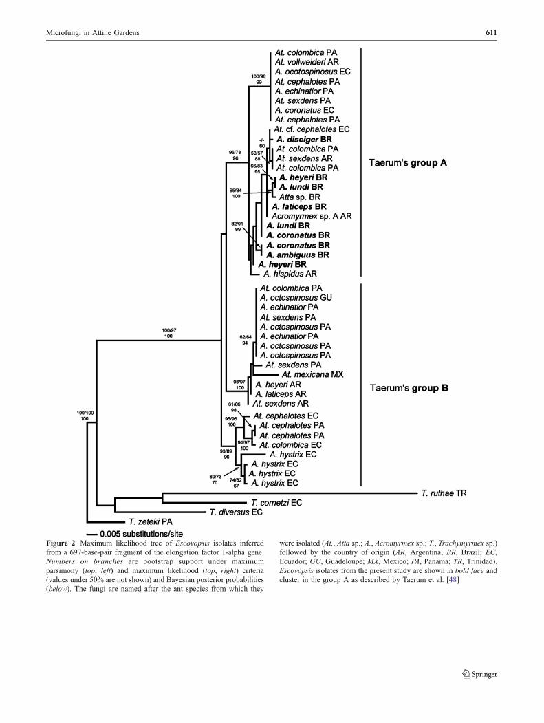
Figure 2 Maximum likelihood tree of Escovopsis isolates inferredfrom a 697-base-pair fragment of the elongation factor 1-alpha gene.Numbers on branches are bootstrap support under maximumparsimony (top, left) and maximum likelihood (top, right) criteria(values under 50% are not shown) and Bayesian posterior probabilities(below). The fungi are named after the ant species from which they
were isolated (At., Atta sp.; A., Acromyrmex sp.; T., Trachymyrmex sp.)followed by the country of origin (AR, Argentina; BR, Brazil; EC,Ecuador; GU, Guadeloupe; MX, Mexico; PA, Panama; TR, Trinidad).Escovopsis isolates from the present study are shown in bold face andcluster in the group A as described by Taerum et al. [48]
Microfungi in Attine Gardens 611611

Discussion
Microfungi in Acromyrmex Nests
Earlier work has shown that attine gardens harbor acomplex microbiota, including soil-inhabiting microfungi[11, 38, 39] as well as epiphytic and endophytic fungi[19]. In the present study, we profiled and characterizedthe cultivable microfungi in the fungus gardens ofAcromyrmex spp. from Southern Brazil by using twodistinct and complimentary isolation techniques. Thisallowed the assessment of the microfungal diversity inthese gardens through the recovering of several micro-fungal isolates.
Gardens of monocot or dicot-cutting and mound-builderor soil-dweller leafcutter ants harbored slightly distinctmicrofungal communities, as would be expected if gardensubstrate and nest-type influenced microfungal contamina-tion (Table 2, Fig. 1). Eight genera of microfungi occurredin gardens of both monocot- and dicot-cutting ants (Fig. 1a)and 10 out of 16 genera occurred in both mound-buildingand soil-dwelling species (Fig. 1b). No microfungal lineagewas clearly specialized on either garden substrate or nesttype. Other factors such as specific plant species harvestedby the ants, the age of the colony, and infestation byarthropod garden commensals that may vector contami-nants into the garden, or interactions between some of thesefactors, may have determined the microfungal gardencommunity. As a classical example of factors influencingthe garden microbiota, Fisher et al. [19] concluded thatchanges in microfungi species composition associated withA. cephalotes nests reared in the laboratory were due tochanges in the plant substrate offered to the ants.
Microfungal profiles also revealed no ant–fungal speciesspecificity with fungi having instead a rather diffuseassociation with Acromyrmex spp. (Table 2). For example,fungi such as Aspergillus sp., Cladosporium sp., Penicilli-um sp., and others were isolated from different ant speciesindependent of nest-type (mound versus soil) or the leafsubstrate (monocot versus dicot). Also, most microfungi inAcromyrmex gardens from this study are species commonlyfound in the environment [15], suggesting no apparentspecialization to the symbiosis. This ubiquity is generallydue to the strategy as saprophytes, i.e., they are importantdue to its general role in the nutrient cycling in ecosystems.For instance, fungi in the genus Xylaria are well-knownsaprophytes, decaying wood of living or dead plants [27].
Despite the lack of specificity between ant and fungalspecies found in our survey, there are some interestingfungi that deserve closer consideration. First, the soil-bornefungal genus Cunninghamella was found in 19% of theleafcutter gardens, a figure rather comparable to the levelsof infection by Escovopsis observed in this study (27%) and
previous studies [11, 38, 39] (see further discussion below).Like Escovopsis, Cunninghamella species can have drasticeffects on leafcutter gardens, overrunning gardens oflaboratory nests within a few days after insecticidetreatment [38], thus suggesting these fungi may have animportant role within the attine ant–microbe symbiosis.However, parsimony analysis (data not shown) indicatedthat Cunninghamella isolates from leafcutter gardensclustered with several isolates of the same genus found inother plant substrates such as nuts [28], indicating that ourCunninghamella strains isolated from gardens are notspecialized on the ant–fungus symbiosis.
Second, F. oxysporum was isolated in 40.5% of thenests, a percentage somewhat higher than the 23% found infield colonies of A. sexdens rubropilosa in southeast Brazil[38]. Apparently, F. oxysporum is a soil-borne fungus thathas a high prevalence in attine gardens [38] (this study) butthere is currently no evidence that F. oxysporum plays adetrimental role in ant gardens. Because F. oxysporumvarieties are disease-causing fungi on plants such as cottonwilt [46] and soil is a natural reservoir for this fungus, wetested whether the strains associated with attine gardensform a specific group. A haplotype analysis revealed thatall attine-associated F. oxysporum strains have plant-associated counterparts (including identical-sequencestrains) which can exist either in soil or in plants, thus outsideof the association with leaf-cutting ants. Although the analysisis based on few strains, it suggests thatF. oxysporum can enternests from the surrounding environment and not via nest-to-nest transmission as hypothesized for Escovopsis [11].
Escovopsis Natural Infection Rates and Diversity
Since 1999, our knowledge on the microfungus genusEscovopsis has been growing [11, 14, 20, 21, 48]. The mainreason for the advances on Escovopsis biology and ecologyis due to several studies that have specifically surveyed forEscovopsis diversity, revealing that this fungus is present ingardens of most attine species. This fungus is currentlythe best well-known pathogen in the attine ant–microbesymbiosis.
Currie et al. [11] studied Escovopsis distribution in avariety of attine genera, mostly from Central America andadjacent areas, and established that Escovopsis could beisolated in 33–51% of the nests, depending on the antgenus. With respect to just the leaf-cutting ants (Atta andAcromyrmex), Currie et al. [11] and Currie [12] reportedEscovopsis infection rates ranging from 51% in some Attaspecies to as high as 68.4% in Acromyrmex octospinosusand 75% in Acromyrmex echinatior. Our study foundcomparatively lower Escovopsis infection rates (27%). Itis unclear whether the levels of Escovopsis infection ofBrazilian subtropical Acromyrmex sp. are naturally lower
612 A. Rodrigues et al.
compared to Central American attines and whether thesedifferences are due to (1) different feeding habits exhibitedby the ants, (2) nest density differences, (3) microhabitatvariations, (4) nest age, or even (5) antagonism by otheralien microorganism. The fact that the present study madeuse of two types of isolation methods (in contrast to otherstudies [11, 12] that used one method) could be anotherfactor contributing to the observed differences.
Our phylogenetic analyses (Fig. 2) corroborate results byTaerum et al. [48] in which Brazilian isolates all fell into asingle group named clade A, which also contained isolatesfrom Argentina, Ecuador, and Panama. However, oursurvey in southern Brazil did not discover any isolates inclade B of Taerum et al., which contained Escovopsis fromArgentina, Ecuador, and Central America. Thus, our resultssuggest that ants in the geographic region in Rio Grande doSul are exclusively infected by Escovopsis strains in cladeA, which is compatible with some geographic structure inthis parasite’s distribution. More extensive sampling isneeded, especially in other regions of Brazil, to investigatewhether other cases of Escovopsis geographic structuringexist. Furthermore, the antibiotic-producing bacteria that theants carry on their cuticle as a defense against Escovopsisare predicted to having corresponding geographic structure[8].
Microfungi as Antagonists in the Ant Farm
According to Poulsen and Boomsma [36] and Scott et al.(in preparation), leafcutter cultivars are maintained by theants as single-genotype fungus garden (monoculture),conditions that are predicted to facilitate the spread andcoevolution of pathogens [24]. Our results indicate thatother non-mutualistic fungi in Acromyrmex nests areindeed prevalent in leafcutter gardens but also that thesepotential pathogens do not appear to be as specialized asEscovopsis [14, 21].
Some of the garden weeds appear to act as antagonists ofthe ant-cultivated fungi, as already documented by Silva etal. [45]. For example, attine gardens can be overgrown byseveral microfungi, all causing garden death [38, 39]similar to garden destruction by Escovopsis sp. [11]. Fungisuch as Cunninghamella species are considered sugar-freefungi and can readily assimilate simple sugars, quicklybuilding up a large biomass of mycelia [15]. It is knownthat fungus gardens of attine ants contain high levels ofsimple sugars (i.e., glucose) [44], and for most fungi, antgardens are therefore a suitable environment for growth.Future studies should address whether the sugars availablein the fungus gardens help non-cultivar fungi to outgrowthe ants’ defense mechanisms of constant weeding. Withinthis nutritional milieu of attine gardens, microfungal weedstherefore can critically impact garden health.
Considering the ecological roles of F. oxysporum andCunninghamella sp., we hypothesize that these micro-organisms act as antagonists in the attine–microbe symbi-osis. However, the negative impact of F. oxysporum andCunninghamella sp. appears to be due to nutritionalcompetition and is not as specific as the impact of thecultivar-infecting parasite Escovopsis, yet some degree ofadaptation and pathogenicity of F. oxysporum and Cunning-hamella species is implied. Future studies should evaluatethe extent of negative impacts of these fungi on theleafcutter ants’ fitness and their usefulness in the biologicalcontrol of these agricultural pests.
Acknowledgements We would like to thank the “Conselho Nacio-nal de Desenvolvimento Científico e Tecnológico (CNPq)” for ascholarship supporting A Rodrigues, “Fundação de Amparo aPesquisa Científica do Estado de São Paulo” (FAPESP) for fundingthe fieldwork; and DC Marini and J Martins Jr for laboratory support.We are also grateful to A Silva for comments on earlier versions ofthis manuscript as well as NM Gerardo and one anonymous referee forkindly reviewing this manuscript. We also thank SE Solomon forhelping with the phylogenetic analyses.
References
1. Altschul SF, Madden TL, Schaffer AA, Zhang J, Zhang Z, Miller W,Lipman D (1997) Gapped BLAST and PSI-BLAST: a newgeneration of protein database search programs. Nucleic Acids Res1:3389–3402
2. Bacci M Jr, Ribeiro SB, Casarotto MEF, Pagnocca FC (1995)Biopolymer-degrading bacteria from nests of the leaf-cutting antAtta sexdens rubropilosa. Braz J Med Biol Res 28:79–82
3. Bandelt H-J, Foster P, Röhl A (1999) Median-joining networks forinferring intraspecific phylogenies. Mol Biol Evol 16:37–48
4. Barron GL (1968) The genera of hyphomycetes from soil. RobertE. Krieger, New York
5. Batra LR (1979) Insect–fungus symbiosis: nutrition, mutualismand commensalisms. Allanheld, Osmun, Montclair
6. Bourtzis K, Miller TA (2006) Insect symbiosis, vol 2. CRC Press,Florida
7. Brown JM, Lemmon AR (2007) The importance of datapartitioning and the utility of Bayes factors in Bayesianphylogenetics. Syst Biol 56:643–655
8. Cafaro MJ, Currie CR (2005) Phylogenetic analysis of mutualisticfilamentous bacteria associated with fungus-growing ants. Can JMicrobiol 51:441–446
9. Carreiro SC, Pagnocca FC, Bueno OC, Bacci M Jr, Hebling MJA,Silva OA (1997) Yeasts associated with nests of the leaf-cutting antAtta sexdens rubropilosa Forel, 1908. Antonie van Leeuwenhoek71:243–248
10. Craven SE, Dix MW, Michaels GE (1970) Attine fungus gardenscontain yeasts. Science 169:184–186
11. Currie CR, Mueller UG, Malloch D (1999) The agriculturalpathology of ant fungus gardens. Proc Natl Acad Sci U S A96:7998–8002
12. Currie CR (2001) Prevalence and impact of a virulent parasite ona tripartite mutualism. Oecologia 128:99–106
13. Currie CR, Stuart AE (2001) Weeding and grooming of pathogensin agriculture by ants. Proc R Soc Lond B 268:1033–1039
14. Currie CR, Wong B, Stuart AE, Schultz TR, Rehner SA, Mueller UG,Sung GH, Spatafora JW, Straus NA (2003) Ancient tripartite co-
Microfungi in Attine Gardens 613613

evolution in the attine ant–microbe symbiosis. Science 299:386–388
15. Domsch KH, Gams W, Anderson T (1980) Compendium of soilfungi, vol. 1 and 2. Academic, London
16. Druzhinina IS, Kopchinskiy AG, Komon M, Bissett J, Szakacs G,Kubicek CP (2005) An oligonucleotide barcode for speciesidentification in Trichoderma and Hypocrea. Fungal Genet Biol42:813–828
17. Felsenstein J (1985) Confidence limits on phylogenies: anapproach using de bootstrap. Evolution 39:783–791
18. Fernández-Marín H, Zimmerman JK, Rehner SA, Wcislo WT(2006) Active use of the metapleural glands by ants in controllingfungal infection. Proc R Soc Lond B 273:1689–1695
19. Fisher PJ, Stradling DJ, Sutton BC, Petrini LE (1996) Microfungiin the fungus gardens of the leaf-cutting ant Atta cephalotes: apreliminary study. Mycol Res 100:541–546
20. Gerardo NM, Currie CR, Price SL, Mueller UG (2004) Exploitinga mutualism: parasite specialization on cultivars within thefungus-growing ants symbiosis. Proc R Soc Lond B 271:1791–1798
21. Gerardo NM, Mueller UG, Currie CR (2006) Complex host–pathogen coevolution in the Apterostigma fungus-growing ant–microbe symbiosis. Evol Biol 6:88–97
22. Gonçalves CR (1961) O Gênero Acromyrmex no Brasil (Hym.Formicidae). Stud Entomol 4:113–180
23. Hall TA (1999) BioEdit: a user-friendly biological sequencealignment editor and analysis program for Windows 95/98/NT.Nucl Acids Symp Ser 41:95–98
24. Hamilton WD, Axelrod R, Tanese R (1990) Sexual reproductionas an adaptation to resist parasites (a review). Proc Natl Acad SciU S A 87:3566–3573
25. Hölldobler B, Wilson EO (1990) The ants. Harvard UniversityPress, Cambridge
26. Klich MA (2002) Identification of common Aspergillus species.Centraalbureau voor Schimmelcultures, Baarn
27. Lee JS, Ko KS, Jung HS (2000) Phylogenetic analysis of Xylariabased on nuclear ribosomal ITS1–5.8S-ITS2 sequences. FEMSMicro Lett 187:89–93
28. Liu XY, Huang H, Zheng RY (2001) Relationships withinCunninghamella based on sequence analysis of ITR rDNA.Mycotaxon 80:77–95
29. Möller A (1893) Die Pilzgärten einiger südamerikanischerAmeisen. Bot Mittl Trop 6:1–127
30. Mueller UG, Schultz TR, Currie CR, Adams RMM, Malloch D(2001) The origin of the attine ant–fungus mutualism. Q Rev Biol76:169–197
31. Mueller UG (2002) Ant versus fungus versus mutualism: Ant-cultivar conflict and the deconstruction of the attine ant–fungussymbiosis. Am Nat 160(Suppl.):S67–98
32. Mueller UG, Gerardo NM, Aanen DK, Six DL, Schultz TR (2005)The evolution of agriculture in insects. Annu Rev Ecol Evol Syst36:563–595
33. Nelson PE, Toussoun TA, Marasas WFO (1983) Fusariumspecies: an illustrated manual for identification. The PennsylvaniaState University Press, Pennsylvania
34. O’Donnell K, Cigelnik E (1998) Two divergent intragenomicrDNA ITS2 types within a monophyletic lineage of the fungusFusarium are nonorthologous. Mol Phylogenet Evol 7:103–116
35. Poulsen M, Currie CR (2006) Complexity of insect–fungalassociations: exploring the influence of microorganisms on attine
ant–fungus symbiosis. In: Bourtzis K, Miller TA (eds) Insectsymbiosis, vol. 2. CRC, Boca Raton, FL, USA, pp 57–77
36. Poulsen M, Boomsma J (2005) Mutualistic fungi control cropdiversity in fungus-growing ants. Science 307:741–744
37. Reynolds HT, Currie CR (2004) Pathogenicity of Escovopsisweberi: The parasite of the attine–microbe symbiosis directlyconsumes the ant-cultivated fungus. Mycologia 96:955–959
38. Rodrigues A, Pagnocca FC, Bacci M Jr, Hebling MJA, Bueno OC,Pfenning LH (2005) Variability of non-mutualistic filamentousfungi associated with Atta sexdens rubropilosa nests. FoliaMicrobiol 50:421–425
39. Rodrigues A, Pagnocca FC, Bueno OC, Pfenning LH, Bacci M Jr(2005) Assessment of microfungi in fungus gardens free of the leaf-cutting ant Atta sexdens rubropilosa (Hymenoptera: Formicidae).Sociobiology 46:329–334
40. Ronquist F, Huelsenbeck JP (2003) MrBayes 3: bayesianphylogenetic inference under mixed models. Bioinformatics 19:1572–1574
41. Sambrook J, Russel DW (2001) Molecular cloning: a laboratorymanual, 3rd ed. Cold Spring Harbor Laboratory Press, NewYork
42. Samson RA, Hoekstra ES, Frisvad JC (2000) Introduction to food-airborne fungi, 6th ed. Centraalbureau voor Schimmelcultures,Baarn
43. Schultz TR, Meier R (1995) A phylogenetic analysis of thefungus-growing ants (Formicidae: Attini) based on morphologicalcharacters of the larvae. Syst Entomol 20:337–370
44. Silva A, Bacci M Jr, Siqueira CG, Bueno OC, Pagnocca FC,Hebling MJA (2003) Survival of Atta sexdens workers ondifferent food sources. J Insect Physiol 49:307–313
45. Silva A, Rodrigues A, Bacci M Jr, Pagnocca FC, Bueno OC(2006) Susceptibility of ant-cultivated fungus Leucoagaricusgongylophorus (Agaricales: Basidiomycota) towards microfungi.Mycopathologia 162:115–119
46. Skovgaard K, Nirenberg HI, O’Donnell K, Rosendahl S (2001)Evolution of Fusarium oxysporum f. sp. vasinfectum racesinferred from multigene genealogies. Phytopathology 91:1231–1237
47. Swofford DL (2002) PAUP*: Phylogenetic analysis using parsi-mony (*: and other methods), ver. 4. Sinauer Associates,Massachusetts
48. Taerum SJ, Cafaro MJ, Little AEF, Schultz TR, Currie CR (2007)Low host–pathogen specificity in the leaf-cutting ant–microbesymbiosis. Proc R Soc Lond B 274:1971–1978
49. Thompson JD, Higgins DG, Gibson TJ (1994) CLUSTAL W:improving the sensitivity of progressive multiple sequencealignment through sequence weighting, position specific gappenalties and weight matrix choice. Nucleic Acids Res 22:4673–4680
50. Weber NA (1972) Gardening ants: the attines, vol. 92 Memoirs ofthe American Philosophical Society, Philadelphia
51. White TJ, Bruns T, Lee S, Taylor J (1990) Amplification anddirect sequencing of fungal ribosomal RNA genes for phyloge-netics. In: Innis MA, Gelfand DH, Sninsky JJ, White TJ (eds)PCR protocols: A guide to methods and applications. Academic,California, pp 315–321
52. Zwickl DJ (2006) Genetic algorithm approaches for the phyloge-netic analysis of large biological sequence datasets under themaximum likelihood criterion. Ph.D. dissertation, The Universityof Texas at Austin
614 A. Rodrigues et al.
Copyright © 2022 FDOKUMEN