Mesozoic marine reptiles from north-east Mexico
98
Mesozoic marine reptiles from north-east Mexico: description, systematics, assemblages and palaeobiogeography Mesozoische marine Reptilien aus Nordostmexiko: Beschreibung, Systematik, Vergesellschaftung und Paläobiogeografie Dissertation von Marie-Céline Buchy aus Kermoroc´h Tag der mündlichen Prüfung: 18. Juli 2007 Referent: Prof. Dr. Wolfgang Stinnesbeck (Geologisches Institut, Universität Heidelberg) Koreferent: Priv. Doz. Dr. Eberhard Frey (Staatliches Museum für Naturkunde Karlsruhe) Die Prüfung wurde bestanden mit der Note 1 (sehr gut) Prof. Dr. Wolfgang Stinnesbeck Priv. Doz. Dr. Eberhard Frey
-
Upload
khangminh22 -
Category
Documents
-
view
0 -
download
0
Transcript of Mesozoic marine reptiles from north-east Mexico

Mesozoic marine reptiles from north-east Mexico: description, systematics,
assemblages and palaeobiogeography
Mesozoische marine Reptilien aus Nordostmexiko: Beschreibung, Systematik,Vergesellschaftung und Paläobiogeografie
Dissertation
von
Marie-Céline Buchyaus Kermoroc´h
Tag der mündlichen Prüfung: 18. Juli 2007Referent: Prof. Dr. Wolfgang Stinnesbeck (Geologisches Institut, Universität Heidelberg)Koreferent: Priv. Doz. Dr. Eberhard Frey (Staatliches Museum für Naturkunde Karlsruhe)
Die Prüfung wurde bestanden mit der Note 1 (sehr gut)
Prof. Dr. Wolfgang Stinnesbeck
Priv. Doz. Dr. Eberhard Frey

Hiermit erkläre ich, dass ich die vorgelegte Dissertation selbst verfasst und mich keineranderen als der von mir ausdrücklich bezeichneten Quellen und Hilfen bedient habe. Alleverwendeten Zitate sind gekennzeichnet und im Literaturverzeichnis angegeben.
Saltillo, den 5. April 2007
Marie-Céline Buchy

Acknowledgements
i
Acknowledgements
These last 6 years, dusk, dawn, parts of week-ends and holidays were devoted to my
daughter Tasmin, and since a few months also to my son Dreo K.; both showed, and still do,
an admirable patience with their very busy mother. Nights were for study, and for that I am
indebted to my colleagues for literature, answers, access to collections, support: Martha
Carolina Aguillón, Javier Banda Leal, José Manuel Padilla Gutiérrez, José "Pato" Lopez
Espinoza and Hector Rivera Sylva (Saltillo), Nathalie Bardet (Paris), Ronald Böttcher and
Rainer Schoch (Stuttgart), Eric Buffetaut (Paris), Lionel Cavin (Geneve), Arthur Cruickshank
and Mark Evans (Leicester), Gilles Cuny (Copenhagen), Mike Everhart (Hays), Zulma
Gasparini and Marta Fernández (La Plata), Samuel Giersch, Christina Ifrim, Martin Rücklin,
Dieter Schreiber and Arne Ziems (Karlsruhe), Stéphane Hua (Paris), Benjamin P. Kear
(Adelaide), Jean Leloeuff (Espéraza), Jeff Liston (Glasgow), Michael Maisch and Andreas
Matzke (Tübingen), Dave Martill (Portsmouth), Franck Métayer (Strasbourg), Leslie Noè and
Marcela Gomez (Cambridge), Steve Salisbury (Brisbane), Patrick Vignaud (Poitiers), Jean-
Noël Martinez (Piura) and most especially Krister T. Smith (Austin).
Though, most of day light time has been devoted, and still is, to the giant pliosaur
from Aramberri, its extraction, preparation, remounting the 3-d 15 m long jigsaw puzzle -
when possible. To this titanic task participated Natalia Amezcua, Alexandra Anders, Gerardo
Balderas Alanis, Martin Bartenbach, Victor Beraza Cardona, Narciso S. Canales Quiroz,
Jorge A. Cervantes Corona, Carlos Rene Delgado (aka 'el maestro de los percutores'), Jürgen
Duberny, Samuel Dubus, Alicia Dulce Decanini, Javier Elenes Escamilla, Jens Erdmann,
Tasmin Frey, José Garcia Quintero, Klaus Geißler, Gustavo González Avendaño, Renee
González Guzmán, Gerd Grochtdreis, Sebastian Guisado, Walter Hähnel, Elke Hermann,
Elizabeth Jardon Nava, René Kastner, André Klicpera, Johanna Kontny, Mario Lang,
Gwendoline Lauzel, Annabelle Lemoine, Rabea Lillich, Leobardo Lopez Ortiz, Mario A.
Mancilla Terán, Bernard Marks, Piedad Esther Mayagoitia Gonzalez, Noé Medellín Molina,
Peter Meiburg, Kristian Nikoloski, Edgar G. Oviedo Padron, Ileana de la Peña Oviedo,
Nelson D. Porras Vázquez, Andrés Ramos Ledezma, Ricardo Rentería del Toro, Miguel A.
Rodriguez Gomez, Pedro Rodriguez Saavedra, Erica J. de la Rosa Camacho, Igor I. Rubio
Cisneros, Pedro Ruiz Gonzalez, Artemio Ruiz Hernandez, Jesus Ruiz Medrano, Salomon
Ruiz, Oscar Salinas Correa, Andrea Schmidt, Ronald Schröder, Tina Schulz, Dominic
Schwarz, Mark Stölpe, Jörg Tensi, Ludowick Thomas, Adalberto Treviño Cázares, Stephan
Unrein, Fernando Vega Siller, Jorge Velasco Segura, Frank Wittler. Essential and friendly

Acknowledgements
ii
support in the field and after came from the whole team of the Hotel Maria Luisa, Aramberri.
Thanks also to the Abarrotes Rio Blanco, Abarrotes Gordo, Abarrotes Guerrero, Fruteria
Argüello, their owners and employees; to the City Council of Aramberri and its president
Eligio del Toro Orozco; to Joe De Ríenzo and Jorge Camacho Rincón, their helicopter and
team; to the governor of Nuevo León, Natividad González Parás. Best of all, Olivier "Jojo"
Grousset was the civil engineer for extraction, scientific and gastronomic counsel - and a great
man to become the best of fathers.
Field work conducted in Gomez Farías was possible thanks to Don Gregorio
‘Dinosaurio’ Sánchez, his wife the lovely Florecita and their family, the Comisariado Ejidal
Guadalupe Lara, as well as the citizens of Gomez Farías. Regulo Cortez found the Zaragoza
pliosaur, and Elvira Reyna Castillo, Margarito Sanchez Arizpe, Catarin Cerda Rodriguez,
Juan Antonio Cerda Rodriguez and Araceli Rivera Estrada were most helpful during our visit
to the site.
Also, the various reviewers of the projects submitted to (and granted by) the DFG
proved extraordinarily open-minded in trusting what appeared at first hopeless to many. The
success of this work is theirs for a great part - they remain anonymous but should recognise
themselves should this volume come to their hands.
The whole teams of the SMNK, Universität Karsruhe and MUDE made work possible
in a pleasant (and sometimes entertaining) atmosphere. Thanks to Ulrike Brecht (Uni KA) for
her marvellous ability to unravel administrative riddles. Above all, thanks to my advisors
Eberhard Frey (SMNK) and Wolfgang Stinnesbeck (Uni KA), to José Guadalupe López Oliva
(Linares) and Arturo González González (Saltillo); the four made it possible.
And, for the spare time I still had, thanks to my friends with whom I usually talk about
other things than dead reptiles: Delphine Denis, Christel Souillat, Claudia Rumayor, Ana
Cortinas, Florence, Patrick, Léo and Rose Yamine, Muriel Deshaies, Dominique Harel,
Bertrand Barbo, Anne Rochas et la petite Svetlana; to my parents and sister. This volume is
dedicated to my late grand-mother Raymonde Rivoallan.

Abstract
iii
Abstract
The result of six years of examination of marine reptiles from north-east Mexico is presented
here. The area yielded specimens from both the Late Jurassic and Late Cretaceous. For a
region where few, undescribed forms were known, the process yielded several new taxa, and a
variety of forms, most of which await preparation, composing rich, hitherto non documented
assemblages.
At present the assemblage of Late Jurassic Mexican Gulf marine reptiles comprises
pliosaurs, thalattosuchians, ichthyosaurs and few plesiosaur remains; no turtle remain was
discovered yet. All proceed from the La Caja / La Casita / La Pimienta Formations
(Kimmeridgian to Tithonian). Except for the ubiquitous ichthyosaur Ophthalmosaurus
icenicus, many specimens, even uncomplete, differ from coeval forms and appear to confirm
the partial isolation of the area that was previously deduced from invertebrates and
microfossils.
During the Cretaceous, the Mexican Gulf connects to both the Western Interior
Seaway and the Atlantic Ocean. Late Cretaceous marine reptiles from north-east Mexico
come from the Late Turonian to early Coniacian Austin Group at Múzquiz, Coahuila, a
promising locality from where few specimens are available for study at present; the
Campanian-Maastrichtian Méndez Formation in Nuevo León yielded few mosasaurid
occurrences; most specimens at present proceed from the Early Turonian Agua Nueva
Formation at Vallecillo, Nuevo León. From Vallecillo is known the only pliosaur remain from
the Mexican Late Cretaceous, a partial tooth attributed to Polyptychodon, and several basal
mosasauroids. These were discovered recently and no taxonomical frame is available at
present, but preservation is usually excellent, including soft parts, and it is expected their
study will greatly increase our knowledge of the group.

Kurzfassung
iv
Kurzfassung
Die vorliegende Dissertation fasst sechs Jahre Untersuchungen an den mesozoischen
Meeresreptilien Nordostmexikos zusammen. Die Gegend lieferte Stücke aus von der
Oberjura- bis in die Oberkreide hinein. Für eine Region, in welcher nur wenige
unbeschriebene Formen bekannt waren, förderte die Arbeit diverse neue Taxa und eine
Vielzahl verschiedener Formen zu Tage, von denen viele noch präpariert werden müssen und
die bislang nicht bekannte Vergesellschaftungen repräsentieren.
Nach bisherigem Stand der Kenntins umfasst die Vergesellschaftung von
Meeresreptilien aus dem spätjurassische mexikaischen Golf Pliosaurier, Thalattosuchier,
Ichthyosaurier und eineige wenige Reste von Plesiosauriern; bislang sind keine marine
Schildkrötenreste bekannt. Alle Stücke stammen aus der La Caja-, La Casita- oder der La
Pimienta-Formation (Kimmeridgium bis Tithonium). Ausser dem kosmopolitischen
Ichthyosaurier Ophthalmosaurus icenicus untescheiden sich viele Stücke, auch die
unvollständigen, von zeitgleichen Formen anderswo und scheinen eine mindestens teilweise
Isolation der Region zu belegen, was aufgrund der Invertebraten und Mikrofossilen bereits
vermutet wurde.
Während der Kreidezeit verband der mexikanische Golf den Western Interior Seaway
mit dem Atlantik. Spätkretazische Meeresreptilien aus Nordostmexiko reichen zeitlich vomm
späten Turonium bis ins frühe Coniacium der Austin-Gruppe bei Múzquiz, Coahuila, einer
vielversprechenden Fundregion, von welcher nur wenige Stücke bisher für Untersuchungen
zur Verfügung stehen. Die Méndez-Formation (Campanium bis Maastrichtium) in Nuevo
León lieferte einige Hinweise auf Mosasaurier; die meisten Stücke jedoch stammen aus der
frühturonischen Aqua Nueva-Formation bei Vallecillo, Nuevo León. In den Steinbrüchen bei
Vallecillo wurde auch der erste Pliosaurierrest aus der Oberkreide Mexikos gefunden, ein
Zahnfragment, welches Polyptychodon zugeordnet wird, sowie einige basale Mosasauroiden.
Diese wurden erst kürzlich geborgen, weshalb noch keine taxonomische Bewertung vorliegt,
aber die Erhaltung ist üblicherweise ausgezeichnet, einschließlich der Weichteile. Es ist zu
erwarten, dass das Studium dieser Stücke, die Kenntnis der Gruppe wesentlich verbessern
wird.

Resumen
v
Resumen
Se presentan los resultados de seis años de trabajo sobre los reptiles marinos del Noreste de
México. El área proporcionó especímenes del Jurásico superior y del Cretácico superior. En
una región donde se conocían pocas formas, aún no descritas, esta investigación reveló varios
taxones nuevos y múltiples formas - la mayoría aún por restaurar - constituyendo conjuntos
faunísticos muy diversificados, que no habían sido registrados hasta la fecha.
Actualmente, la asociación de reptiles marinos del Jurásico superior del Golfo de
México incluye pliosaurios, talatosuquios, ictiosaurios y algunos restos de plesiosaurios; hasta
la fecha, no se ha encontrado ningún resto de tortuga marina. Todo el material procede de las
Formaciones La Caja / La Casita / La Pimienta (del Kimmeridgiano al Tithoniano). Salvo el
caso del ictiosaurio cosmopólito Ophthalmosaurus icenicus, muchos especímenes, aún
incompletos, difieren de otras formas coetáneas, lo cual parece confirmar el aislamiento
parcial de la región previamente deducido de los invertebrados y microfósiles.
Durante el Cretácico, el Golfo de México conecta el Mar Interior Occidental con el
Océano Atlántico. Reptiles marinos del Cretácico superior del noreste de México provienen
del Turoniano tardío - Coniaciano temprano del Grupo Austin observable en Múzquiz
(Coahuila), una localidad prometedora, de la cual pocos especímenes son disponibles por el
momento. La Formación Méndez (Nuevo León), de edad Campaniano-Maastrichtiano ha
proporcionado pocos restos de mosasauridos; actualmente, la mayoría de los especímenes
proceden del Turoniano temprano de la Formación Agua Nueva, en Vallecillo (Nuevo León).
De esta misma localidad de Vallecillo, se conoce la única evidencia de pliosaurio para el
Cretácico superior de México - un diente incompleto atribuido a Polyptychodon - así como
varios mosasauroidos basales, los cuales han sido descubierto recientemente. Ningún marco
taxonómico está actualmente disponible para precisar su posición sistemática; sin embargo, su
preservación es generalmente excelente e incluye partes blandas. Se puede esperar que el
estudio de estos mosasauroidos incrementará considerablemente nuestros conocimientos
acerca de este grupo.

Contents
vi
Contents
Acknowledgements .............................................................................................................. i
Abstract ................................................................................................................................ iii
Kurzfassung ......................................................................................................................... iv
Resumen ............................................................................................................................... v
Contents ............................................................................................................................... vi
Abbreviations ....................................................................................................................... 1
Introduction .......................................................................................................................... 2
Part I. Upper Jurassic La Casita / La Caja Fm ..................................................................... 7
I.1. Palaeogeographical context ........................................................................................... 7
I.2. Catalogue of specimens ................................................................................................. 7
I.2.1. Crocodyliformes ......................................................................................................... 7
I.2.1.1. Thalattosuchia indet. ...................................................................................... 7
I.2.1.2. Metriorhynchinae indet. ................................................................................. 9
I.2.1.3. Geosaurus ...................................................................................................... 12
I.2.1.4. Dakosaurus .................................................................................................... 13
I.2.1.5. Teleosauridae indet. ....................................................................................... 15
I.2.1.6. Discussion ...................................................................................................... 16
I.2.2. Ichthyopterygia ........................................................................................................... 16
I.2.2.1. Euichthyopterygia indet. ................................................................................ 16
I.2.2.2. Ophthalmosaurus icenicus ............................................................................. 18
I.2.2.3. Discussion ...................................................................................................... 23
I.2.3. Sauropterygia .............................................................................................................. 24
I.2.3.1. Plesiosauria indet. .......................................................................................... 24
I.2.3.2. Plesiosauroidea: Elasmosauridae indet. ......................................................... 26
I.2.3.3. Pliosauroidea: Pliosauridae indet. .................................................................. 27
I.2.3.4. Discussion ...................................................................................................... 52
I.3. Discussion ...................................................................................................................... 53
Part II. Upper Cretaceous ..................................................................................................... 55
II.1. Early Turonian Agua Nueva Formation at Vallecillo, Nuevo León ............................ 55
II.1.1. Palaeogeographical context ............................................................................. 55
II.1.2. Pliosauridae cf. Polyptychodon ...................................................................... 56

Contents
vii
II.1.3. 'Aigialosaurs' indet. .......................................................................................... 57
II.1.4. Discussion ........................................................................................................ 67
II.2. Late Turonian to early Coniacian Austin Group at Múzquiz, Coahuila ...................... 69
II.2.1. Palaeogeographical context ............................................................................. 69
II.2.2. Squamata indet. ................................................................................................ 69
II.2.3. Mosasauridae indet. ......................................................................................... 69
II.2.4. Discussion ........................................................................................................ 69
II.3. Campanian-Maastrichtian Méndez Formation ............................................................. 71
II.3.1. Palaeogeographical context ............................................................................. 71
II.3.2. Mosasauridae indet. ......................................................................................... 71
II.3.3. Discussion ........................................................................................................ 72
Conclusions .......................................................................................................................... 74
Literature .............................................................................................................................. 76
Appendix: Reprints

Abbreviations
1
Abbreviations
Institutions
IGPT : Geological Institute in Tübingen, Germany; MUDE: Museo del Desierto, Saltillo
(Coahuila, Mexico); MHM: Museo Historico de Múzquiz, Múzquiz (Coahuila, Mexico); SMNK:
Staatliches Museum für Naturkunde (Karlsruhe, Germany); UANL-FCT: Universidad Autónoma
de Nuevo León, Facultad de Ciencias de la Tierra, Linares (Nuevo León, Mexico); UNAM:
Universidad Nacional Autónoma de México, México, D.F.
Accession numbers
CPC: Colección Paleontológica de Coahuila, MUDE.
Anatomical abbreviations
a: angular; as: astragalus; atax: atlas/axis; ce: centrale; cor: coracoid; cv: cervical vertebra; d:
dentary; dic: distal carpal; dit: distal tarsal; dt: dentary tooth; en: external nare; eo: exoccipital-
opisthotic; epf: epipterygoid facet; f: frontal; h: humerus; ha: haemal arch; i: intermedium; j:jugal;
l: lachrymal; mc: metacarpal; ms: mandibular symphysis; mt: metatarsal; mx: maxillary; mxt:
maxillary tooth; n: nasal; na: neural arch; o: orbit; paf: parietal foramen; pal: palatine; par:
parietal; ph: phalanx; pmx: premaxillary; pmxs: premaxillomaxillary suture; pmxt: premaxillary
tooth; po: postorbital; pof: postfrontal; poz: postzygapophysis; pp: paroccipital process; prf:
prefrontal; pro: prootic; prz: prezygapophysis; pt: pterygoid; Q1: first caudal vertebra; qj:
quadratojugal; qrpt: quadrate ramus of the pterygoid; qrsq: quadrate ramus of the squamosal; R:
radius; r: rib; rc: rostral cavity; re: radiale; s1: first sacral vertebra; s2: second sacral vertebra; sa:
surangular; sc: scapula; sf: supraoccipital facet; spl: splenial; sq: squamosal; st: supratemporal; T:
tibia; tp: transverse process; tr: tooth root; ut: upper tooth; v: vomer.
I-V: digits I to V.

Introduction
2
Introduction
Six years ago, very little was known about Mexican diapsid marine reptiles: Wieland
(1910) named the taxon Plesiosaurus (?Polyptychodon) mexicanus from the Lower Cretaceous of
south Mexico upon a portion of rostrum. This holotype was recently located in the collections of
the UNAM (Perrilliat Montoya, pers. com., 2007); it was briefly mentioned and considered non
diagnostic by Welles (1962). The mosasaur Amphekepubis johnsoni (University of Missouri
509VP) was described by Mehl (1930) from the Upper Cretaceous of the Monterrey region (figs
1, 2); the holotype is now considered a pathological member of the genus Mosasaurus (Camp,
1942; Lingham-Soliar, 1995). An undetermined thalattosuchian was mentioned from the
Callovian of Mexico by Gasparini (1992). None of the preliminarily descriptions of specimens in
the frame of geological studies had made its way into marine reptiles students' literature
(Michalzik, 1988; Schumann, 1988; Götte, 1990; Aranda-Manteca & Stinnesbeck, 1995; compare
with e.g. Bardet, 1995; Vignaud, 1995).
The year 2000 opened a new chapter for north-east Mexican marine reptiles, when
examination of the collections of the UANL-FCT by E. Frey (SMNK) and the author revealed the
existence of a new thalattosuchian (Frey et al., 2002; specimen UANL-FCT-R1, chapter I.2.1),
and the remains of a giant pliosaur previously thought to be a dinosaur were properly identified
(Hähnel, 1988; Buchy et al., 2003; specimen UANL-FCT-R2, nicknamed The Monster of
Aramberri, chapter I.2.3). The recovery of the rest of the skeleton of this specimen was the aim of
several field campaigns financed by the DFG, that yielded remains of many other fossil reptiles,
and the present catalogue of an exceedingly rich region for both Upper Jurassic and Upper
Cretaceous fossil marine reptiles.
The area considered in this work encompasses the states of Nuevo León and Coahuila
(figs 1, 2; except one specimen from Puebla state, Frey et al., 2002), in the frame of a
collaboration between the Universidad Autónoma de Nuevo León, Facultad de Ciencias de la
Tierra, Linares (Nuevo León), the Museo del Desierto, Saltillo (Coahuila), the Universität
Karlsruhe (TH) and the Staatliches Museum für Naturkunde Karlsruhe.
In what is now north-east Mexico the Laramide orogeny folded the Mesozoic sediments
during the latest Cretaceous - early Paleogene. The resulting Sierra Madre Oriental divides north-
east Mexico along a north-west to south-east axis into a continental desert westward and a coastal
plain eastward (fig. 1). Outcropping Upper Jurassic and Upper Cretaceous sediments (and marine
reptiles) are usually found as a result of mining activities in the Sierra Madre.

Introduction
3
Mesozoic sediments of north-east Mexico document a gradual transgression linked with
the opening of the Gulf of Mexico (e.g. Michalzick, 1988; Götte, 1990; Ifrim, 2006; fig. 3),
starting during the Oxfordian with the evaporites of the Minas Viejas Formation. The overlying
limestones of the Zuloaga Formation, deposited in shallow waters of a carbonate ramp, did not yet
yield marine reptiles. Upsection, La Caja and contemporaneous La Casita (and equivalent La
Pimienta) Formations, that yielded all Upper Jurassic marine reptiles described here, indicate
deepening and increased terrigenous influences. Overlying Cretaceous units document pelagic,
open marine environments.

Introduction
4

Introduction
5
Facing the local richness of to-be-prepared, new specimens, priorities were set, according
to available time, money and space, which is of course far from ideal, but yielded a (hopefully)
good preliminary overview of the various taxa. This volume represents the current status of this
process, promising of what should be expected when further explorations and preparations are
conducted. It is necessarily uncomplete, mainly focussing upon the Kimmeridgian/Tithonian La
Casita/La Caja Formations and material from the Upper Cretaceous in official institutions
(material in private collections was ignored, mostly because time was too short to check the
presumed localities). Descriptions and aligned palaeobiogeographical conclusions were published
as work was progressing (Frey et al., 2002; Buchy et al., 2003, 2004, 2005b, c, 2006b-e, 2007, in
press; Buchy & Smith, 2005; Smith & Buchy, submitted). Work being still in progress, also upon
local geology, detailed stratigraphical correlations are not available yet for most of the mentioned
specimens, and therefore neither are detailed stratigraphical comparisons with possibly coeval
occurrences. Though, anatomical comparisons were drawn when the available elements made it
possible. In such a preliminary frame, it is also not the place here to draw definitive phylogenetic
conclusions upon the affinities of the Mexican forms (most being too incomplete). For all groups
dealt with here, cladistic analyses are few and/or yielding results revitalsing former blurred
taxonomies (in the case of Plesiosauria), hotly debated relationships (for squamates) or only
mention the group on the basis of outdated, second-hand data (typically, studies dealing with
Figure 3: Lithostratigraphical column of the Mesozoic innorth-east Mexico (redrawn from Michalzick, 1988 andGötte, 1990).

Introduction
6
Thalattosuchia). Possibly, this has to do with a long-shadowed history of marine reptiles (Taylor,
1997), a poor understanding of the transition from terrestrial to marine life outside general trends,
together with and as a consequence of poor potential for fossilisation of intermediate
environments (Storrs, 1999). This also most likely has to do with the current, quasi exclusive
cladistic approach to phylogeny. Groups dealt with here (Plesiosauria, Mosasauroidea,
Thalattosuchia) all exhibit a very conservative anatomy of the locomotor apparatus (once it has
been modified according to an aquatic life style), but also of the cranium. A high degree of
homoplasy is most likely to be expected, while characters used in current cladistic analyses are
rarely considered in a functional perspective that only could reveal possible convergence in
relation to biomechanics, physiology, feeding behavior, etc. in an obligatory aquatic environment
(see e.g. the recent suggestion by Bell & Polcyn, 2005 and Polcyn & Bell, 2005, that a fin-like
limb developped at least twice within Mosasauroidea). This makes relationships both with
potential terrestrial ancestors and within the group a riddle. It can therefore even not be mentioned
that the current taxonomical status within a cladistic hypothesis of relationships of the groups
dealt with here is ignored, as there is no current, generally accepted taxonomical status.
Traditional, ranked taxonomy is followed, in order to allow future researchers to place the
described specimens within a hopefully soon renewed, better understood frame.
The term 'marine reptile' is often thought of as a shortcut for 'reptile who lives in the sea',
as does 'marine mammal'. Though, in the case of fossils, the 'who lives' dimension is missing, and
the concept slids towards results of an often implicit biomechanical analysis of locomotory
abilities of the considered taxon, in the frame of palaeogeographical finding location. 'Who lives'
therefore becomes 'who swam' - in a more or less open former sea, while in an ecological
perspective, 'marine' implies the ability to deal with excess salt in order to take part occasionally
or obligatorily to marine trophic webs (Hua & Buffetaut, 1997). Salt glands or clear hints at their
presence are rarely preserved in fossils (Fernandez & Gasparini, 2000; Gandola et al., 2006;
Buchy et al., in press); although it is well conceived that this choice is reductionist (excluding at
least some birds and pterosaurs), the definition adopted here of a diapsid marine reptile follows
palaeontology text books, and includes Plesiosauria, Thalattosuchia and Mosasauroidea.

Part I. Upper Jurassic La Casita / La Caja Fm
7
Part I. Upper Jurassic La Casita / La Caja Fm
I.1. Palaeogeographical context
The depositional context of the La Casita and La Caja Formations in north-east Mexico was
summed up by Buchy et al. (2003, 2006d, c). The former represents deltaic and inner shelf
environments, the latter more distal settings; both span from Kimmeridgian to Berriasian. Both
were deposited at a time when regional tectonics caused an irregular sea floor; inner and outer
shelf facies were further affected by sea level variations and consequently, a basinal context (e.g.
Michalzik, 1988; Götte, 1990; Goldhammer, 1999; Goldhammer & Johnston, 2001).
Although classical palaeogeographical reconstructions exclude a direct relation between
European and Pacific provinces before the Upper Jurassic, comparison of marine assemblages
does support the existence of a Carribean corridor since at least the Middle Jurassic (e.g.
Gasparini & Fernández, 1997, 2005; Gasparini et al., 2000; Fernández & Iturralde-Vinent, 2000;
Gasparini & Iturralde-Vinent, 2001; Gasparini et al., 2002). By the Late Jurassic, though,
connections between Tethyan and Pacific realms are established and continuous (e.g. Smith et al.,
1994). However, microfossils and invertebrates indicate at least partial isolation of the Mexican
Gulf until the Middle Berriasian (Salvador et al., 1993; Adatte et al., 1994; 1996). Southward
moving of Yucatan and uplifting of the Florida Strait have been proposed as possible cause for the
isolation, that appears to affect as well marine reptiles (Buchy et al., 2006d).
I.2. Catalogue of specimens
An annotated list of the specimens with origin and description is given here, updated from Buchy
et al. (2006d). For those specimens which were the subject of a separate description, it is referred
to the publications dealing with them (Appendix).
I.2.1. Crocodyliformes
Crocodyliformes Benton & Clark, 1988
Thalattosuchia Fraas, 1901
I.2.1.1. Thalattosuchia indet.
UANL-FCT-R11
Material: One isolated, fragmentary neural arch.

Part I. Upper Jurassic La Casita / La Caja Fm
8
Origin: The neural arch was discovered during the excavation of the pliosaur UANL-FCT-R2, at
Aramberri, Nuevo León (fig. 2; chapter I.2.3). The age of the La Caja deposits at this site, as
determined by the ammonite assemblage, is late Early to early Late Kimmeridgian (see Buchy et
al., 2003).
Comments: This specimen was described by Buchy et al. (2006d).
UANL-FCT-R15 (Buchy et al., 2006d: fig. 4)
Material: One thoracic vertebra with the proximal fragment of the right rib, and the caudal half of
the preceding vertebra.
Origin: Late Early to early Late Kimmeridgian La Casita Formation, determined from the matrix.
The exact origin of the specimen is unclear: probably either Galeana, or Iturbide, Nuevo León
(fig. 2).
Comments: This specimen was described by Buchy et al. (2006d).
Figure 4: Palaeobiogeographical map of theLate Jurassic (modified from Smith et al.,1994). Note the hypothetical barrier formedby the Florida uplift. Insert:palaeobiogeographical map of north-eastMexico during the Tithonian; Cb: Coahuilablock; M: Monterrey; S: Saltillo (redrawnafter Goldhammer, 1999).

Part I. Upper Jurassic La Casita / La Caja Fm
9
UANL-FCT-R25
Material: Two fragmentary thoracic vertebrae.
Origin: Late Early to early Late Kimmeridgian La Casita Formation at Galeana, Nuevo León (fig.
2).
Comments: This specimen was described by Buchy et al. (2006d).
CPC 205; 209; 212; 213; 214; 229; 239; 240
Material: All numbers correspond to isolated vertebrae or partial vertebrae, possibly with rib
fragments; all are awaiting preparation.
Origin: Early Tithonian section of the La Caja Formation at the Sierra El Jabalí, near Gomez
Farías, Coahuila, Mexico (fig. 2). The specimens were collected during field campaigns in March
2004 and September 2006.
Comments: As for most of the material from Gomez Farías, description must await preparation.
Though, in view of the incompleteness of the specimens which will most likely reveal not further
diagnostic, preparation could not be set as a priority. They emphasise the richness of the area
regarding Thalattosuchia (Buchy et al. 2006c, e).
Metriorhynchidae Fitzinger, 1843
Metriorhynchinae Fitzinger, 1843
I.2.1.2. Metriorhynchinae indet.
UANL-FCT-R13
Material: Fragment of a rostrum comprising a fragmentary mandible at the caudal terminus of the
symphysis including the dentition, the crowns of the maxillary teeth and the endocast of the
internal cavity of the nasal duct of the maxillary rostrum.
Origin: Late Early to early Late Kimmeridgian La Casita Formation, as determined from the
matrix. The specimen comes from either Galeana or Iturbide, Nuevo León (fig. 2).
Comments: This specimen was discussed by Buchy et al. (2006d), and is still awaiting
preparation.
UANL-FCT-R16 (Buchy et al., 2006d: fig. 5a)
Material: Nine articulated caudal vertebrae.
Origin: Late Early to early Late Kimmeridgian La Casita Formation, at Galeana, Nuevo León
(fig. 2).
Comments: This specimen was described by Buchy et al. (2006d).

Part I. Upper Jurassic La Casita / La Caja Fm
10
UANL-FCT-R17 (Buchy et al., 2006d: fig. 5b)
Material: Four articulated caudal vertebrae, neural spine of the cranially adjacent vertebra, and
cranial portion of the centrum of the caudally adjacent vertebra.
Origin: Late Early to early Late Kimmeridgian La Casita Formation at Galeana, Nuevo León (fig.
2).
Comments: This specimen was described by Buchy et al. (2006d).
CPC 225
Material: Rostral portion of a rostrum with teeth.
Origin: Early Tithonian section of the La Caja Formation at the Sierra El Jabalí, near Gomez
Farías, Coahuila, Mexico (fig. 2). The specimen was collected during a field campaign in March
2004.
Comments: The specimen comprises about 250 mm of the rostrum in occlusion with teeth,
preserved over several, matching fragments. It is about 25 mm wide, and the teeth have a basal
cross-section of about 5-7 mm. Prior to preparation, the teeth appear slender, sub-circular in cross-
section. The specimen therefore fits the anatomy of either longirostrine metriorhynchs or geosaurs
(Vignaud, 1995; Frey et al., 2002). The dentition of geosaurs in general, and especially of G.
saltillense Buchy et al., 2006e, from the same locality, being poorly known, and the high diversity
of Metriorhynchinae there only suspected at present in view of the assemblage described here,
attribution of the specimen awaits preparation and further studies.
CPC 232; 242 (fig. 5)
Material: Two isolated, partial tooth crowns.
Origin: Early Tithonian section of the La Caja Formation at the Sierra El Jabalí, near Gomez
Farías, Coahuila, Mexico (fig. 2). The former was collected during a field campaign in September
2004; the latter, in February 2006.
Description: Both crowns share the same characteristics: the basal section is slightly
mesiodistally elongate; the enamel is ornamented with thin, closely-spaced ridges that tend to
merge towards the apex; the latter is smooth. The crowns are slightly curved mesially.
Discussion: These crowns closely resemble those attributed to some Metriorhynchus (Vignaud,
1995; 1997). Though, the dentition of geosaurs, especially of those inhabiting the Mexican Gulf
during the Late Jurassic, is poorly known (Vignaud, 1995; Frey et al., 2002; Buchy et al., 2006e).
The specimens are therefore provisionally referred to indeterminate Metriorhynchinae, possibly
pending discovery of more complete specimens.

Part I. Upper Jurassic La Casita / La Caja Fm
11
CPC 221; 230; 231 (fig. 6)
Material: The former two specimens comprise partial skulls; the latter, a partial skull and
extensive portion of post-cranium.
Origin: Early Tithonian section of the La Caja Formation at the Sierra El Jabalí, near Gomez
Farías, Coahuila, Mexico (fig. 2). The former was collected during a field campaign in March
2004; the latter two during several campaigns between September 2004 and February 2006. All
still need preparation.
Comments: Although still mostly unprepared, for both skulls CPC 221 and 231 the extent and
shape of the upper temporal fossae were visible in the field prior to extraction. Preliminary
preparation of CPC 230 was recently conducted at the SMNK (fig. 6). In all three cases, the upper
temporal fossae are subcircular, and although the three specimens could still represent different
taxa, all differ from G. saltillense in this feature (Buchy et al., 2006e; the difference cannot be
ontogenetic as the size of the specimens is equivalent). The subcircular shape of the fossae
resembles the situation in adult members of the genus Metriorhynchus (Vignaud, 1995); however,
Geosaurus araucanensis Gasparini & Dellapé, 1976 also possesses subcircular fossae, and a wide
angle formed by the postorbital rami of the frontal to the intertemporal bar (Gasparini & Dellapé,
1976; Vignaud, 1995). Actually, only few of the diagnostic features of Geosaurus given by
Vignaud (1995) can be observed in partial specimens and used to distinguish gracile members of
the genus Metriorhynchus from members of the genus Geosaurus (Broili, 1932; Vignaud, 1995:
compare e.g. pl. 25a, b and pl. 28a): nares are usually not or poorly preserved, rounded prefrontal
lateral margins can be due to weathering or poor preparation, the original level of the
supratemporal bar compared to the intertemporal bar can rarely be judged due to distortion, and
South American thalattosuchians are poorly ornamented whatever genus is considered (Gasparini
et al., 2005, 2006; Buchy et al., in press). Narial anatomy appears the clearest apomorphy of the
genus Geosaurus (shared with Dakosaurus, which is then defined by its massiveness; Vignaud,
1995; Gasparini et al., 2006). Preservation of most Late Jurassic Mexican specimens at present
prevents evaluation of this feature. Further exploration and the (usually) good condition of the
Figure 5: CPC 242, Metriorhynchinae indet., partial, isolatedtooth crown in labial view. Scale 20 mm.

Part I. Upper Jurassic La Casita / La Caja Fm
12
fossils, may set an opportunity to re-assess this poorly known genus (Broili, 1932; Vignaud,
1995).
I.2.1.3. Geosaurus Cuvier, 1824
Geosaurus vignaudi Frey et al., 2002
UANL-FCT-R1
Material: Holotype skull and mandible, atlas, axis and 3 cervical vertebrae on a limestone slab.
Origin: Tithonian Pimienta Formation near Mazatepec, Puebla state. In central-east Mexico, the
Pimienta Formation is an open marine shelf equivalent of the La Caja Formation (Frey et al.,
2002).
Comments: The holotype and only known specimen was described by Frey et al. (2002) and
further discussed by Buchy et al. (2006c, e).
Geosaurus saltillense Buchy et al., 2006e
CPC 218
Former accession numbers: CEP1823 (Buchy et al., 2005c, 2006e).
Material: Holotype partial cranium and partial mandible, cervical vertebrae including atlas and
axis.
Origin: Early Tithonian section of the La Caja Formation at the Sierra El Jabalí, near Gomez
Farías, Coahuila, Mexico (fig. 2), collected during a field campaign in March 2004.
Comments: The holotype and only known specimen was described and discussed by Buchy et al.
(2006e). Note the new accession number of the holotype.

Part I. Upper Jurassic La Casita / La Caja Fm
13
Dakosaurus Quenstedt, 1856
I.2.1.4. Dakosaurus sp.
CPC 201
Material: Section of maxillary rostrum with poorly preserved teeth.
Origin: Middle Kimmeridgian section of the La Casita Formation at San Juan de los Dolores,
close to Los Lirios, Coahuila, Mexico (fig. 2). The specimen was discovered in spring 2004 by
Daniel Chio Gomez (Los Lirios). A visit to the site in autumn 2005 yielded no additional material,
but confirmed the stratigraphic assignment.
Comments: The description and discussion of the affinities of the specimen are in press (Buchy
et al., in press).
UANL-FCT-R29 (fig. 7)
Material: Fragmentary interorbital portion of a skull.
Origin: Late Early to early Late Kimmeridgian La Casita Formation, about 1 km north-west of
the hamlet of El Salitre, close to the city of Zaragoza, Nuevo León (fig. 2; see UANL-FCT-R8,
chapter I.2.3). The fragmentary nodule was collected in September 2003 at the finding site of the
pliosaur UANL-FCT-R8, together with fragments of the latter, undoubtedly matching the
originally found, pliosaurian material (Buchy et al., 2006d; chapter I.2.3). However, this fragment
fits nowhere within a pliosaur cranium or post-cranium, and instead represents a large
thalattosuchian (fig. 7). The site also yielded ichthyosaur vertebrae (Buchy et al., 2006d). The La
Casita / La Caja nodules are known for their locally rich fossil contents (Buchy et al., 2003 and
references therein): this site may actually reveal very rich. Preliminary gluing was conducted by
F. Wittler in the SMNK and mechanical preparation completed by the author in the MUDE.
Preservation: The original completeness of the specimen cannot be determined. Most of the
edges represent breaks along calcite seams. Possibly more of the animal is to be discovered at the
site. At present, it comprises the rostral-most portion of the intertemporal bar (whose base only is
preserved) and adjacent rostral-most portions of the supratemporal fenestrae; the median portion
of the left postorbital bar is present as well, as is the caudal-most part of the prefrontal. The
specimen is preserved 3-dimensionally, although a poorly preserved, flat element on the ventral
side, possibly a fragment of the palatine, indicates collapse of the cranium.
Description: Most of the specimen is made up of the frontal. The intertemporal bar is about 20
mm wide at its base, slightly enlarging rostrally. The interfrontal suture is visible along the
midline of the broken intertemporal bar, which does not mean it was visible on the surface (see
Wenz, 1968 and Buchy et al., in press, about the occurrence of interfrontal sutures). On either
sides it is bordered by the depressed, sub-horizontal, non-ornamented rostral portions of the

Part I. Upper Jurassic La Casita / La Caja Fm
14
supratemporal fenestrae. No frontoparietal suture appears on the preserved fragment. The frontal
forms most of the postorbital bar as preserved: the 20 mm thick buttress that borders the
supratemporal fenestra rostrolaterally. The caudal margin of the buttress is concave, its rostral
margin convex. Its surface is ornamented, mainly in its rostral-most part, by shallow pitting.
Laterally, the postorbital ramus of the frontal contacts the postorbital in an undulating suture,
whose general direction appears perpendicular to the orientation of the postorbital bar, though is
unclear. A small portion of the postorbital is preserved; it keeps the shape of the postorbital bar in
its frontal portion. The medial-most, preserved portion of the prefrontal appears pushed up by
dorsoventral compression. Its surface is wrinkled and no original ornamentation pattern can be
deduced. The prefrontofrontal suture commences at mid-point of the dorsal margin of the orbit. It
runs medially straight for about 15 mm and then gently curves rostrally. In ventral view, the
prefrontal projects about 5-7 mm ventral to the frontal, which confirms the specimen underwent
deformation. The prefrontofrontal suture in ventral view curves gently from rostrolaterally to
caudomedially. The ventral surfaces, both of the prefrontal and the frontal are smooth. The
median part of the ventral surface is obscured by a flat bone, possibly part of the palatine, whose
broken and weathered margins yield no anatomical details. It is compressed against the lateral
walls of the olfactory tract. The latter is formed by the frontal; it is at least 20 mm high, though its
original extent cannot be determined. On the rostral-most break of the specimen (i.e. between the
orbits), the olfactory tract is visible, as an inverted U-shaped, 20 mm wide, 20 mm deep sulcus
within the frontal. Along the caudal break of the specimen, the thickness of the frontal is about 20
mm laterally, increasing to about 40 mm medially (not counting the height of the broken
intertemporal bar).

Part I. Upper Jurassic La Casita / La Caja Fm
15
Discussion: The specimen is massive (in term of bones in cross-section) and large, with an
estimated 190-200 mm wide interorbital area (therefore larger than CPC 201 described by Buchy
et al., in press). Middle Jurassic members of the genus Metriorhynchus, such as M.
brachyrhynchus can attain large sizes (Vignaud, 1995). The sharp angle formed by the postorbital
bar to the skull midline, the supratemporal fenestra much larger rostrally than the supratemporal
fossa, and the discreet ornamentation of the dorsal surface of the bones strongly recall specimens
attributed to the genus Dakosaurus (Vignaud, 1995; Vignaud & Gasparini, 1996; Gasparini et al.,
2006; Buchy et al., in press). UANL-FCT-R29 is therefore provisionally referred to this genus,
pending possible discovery of more elements, and a revision of the genus which appears less and
less well-defined with attribution of new forms (see the very unusual morphology of the
Argentinian specimens described by Gasparini et al., 2006).
The identification of UANL-FCT-R29 among the material collected when searching for
the pliosaur UANL-FCT-R8 (chapter I.2.3) casts doubt upon the attribution of the isolated, tooth
imprint-bearing fragments. Dakosaurus was originally described with large, trihedral, smooth-
enameled teeth, possibly with serrated carinae (Fraas, 1902; Vignaud, 1995). The holotype of D.
andiniensis Vignaud & Gasparini, 1996, has (poorly preserved) teeth sub-circular in cross-section,
small compared to skull size, while newly referred specimens have large teeth and serrated
carinae (located more caudally in the tooth row than in the holotype; Gasparini et al., 2006). In
any case, none of the tooth imprint-bearing fragments collected in September 2003 appear
trihedral; they all show an ornamentation pattern of regularly-spaced ridges, which was never
described for Dakosaurus until now. They are therefore described together with the pliosaur
UANL-FCT-R8 (chapter I.2.3), although a doubt must remain, especially if Dakosaurus (or
members of a possibly new, closely related genus) does reveal as variable as it is now suspected.
Thalattosuchia Fraas, 1901
Teleosauroidea Geoffroy Saint-Hilaire, 1831
Teleosauridae Geoffroy Saint-Hilaire, 1831
I.2.1.5. Teleosauridae indet.
UANL-FCT-R12 (Buchy et al., 2006d: fig. 6)
Former accession number: LCITØ / 1005
Material: Osteoderms, cranial and post-cranial fragments preserved on both surfaces of a split
concretion.
Origin: Late Early to early Late Kimmeridgian La Casita Formation at Iturbide, Nuevo León (fig.
2).

Part I. Upper Jurassic La Casita / La Caja Fm
16
Comments: A preliminary description of this specimen was given by Buchy et al. (2006d); since
then, no further preparation could be undertaken.
I.2.1.6. Discussion
Buchy et al. (2006d) discuss the thalattosuchian assemblage of the Kimmeridgian of
north-east Mexico; since then only the expected genus Dakosaurus was newly identified (Buchy
et al., in press and specimen UANL-FCT-R29). Teleosaurids appear rare when compared to
European localities (Vignaud, 1995; see a discussion upon possible life-style constrains in Buchy
et al., 2006d), while the fragmentary condition of most other specimens prevents further faunistic
conclusions.
The Early Tithonian at Gomez Farías on the contrary recently revealed extremely rich in
thalattosuchians, even if results are at present preliminary (Buchy et al., 2006c, e). The
assemblage lacks teleosaurids and Dakosaurus is missing, though at present the described
diversity of geosaurs is reminiscent of what is known at the time in Europe (Vignaud, 1995;
Schoch, pers. com.). The absence of the genus Metriorhynchus, as mentioned by Buchy et al.
(2006d), is still intriguing, less so than for the Kimmeridgian, though, as this genus is poorly
represented in the Tithonian of Europe and South America (Vignaud, 1995). Several specimens of
at present uncertain attribution (CPC 221, 230, 231) could change the figure, but further
preparation as well as re-evaluation of European forms are necessary.
I.2.2. Ichthyopterygia
Ichthyopterygia de Blainville, 1835
Euichthyopterygia Motani, 1999
I.2.2.1. Euichthyopterygia indet.
UANL-FCT-R18 (Buchy et al., 2006d: fig. 7a)
Former accession number: LCRPØ / 1003
Material: Portion of caudal vertebral column consisting of 14 centra.
Origin: Late Early to early Late Kimmeridgian La Casita Formation at Rio Pablillo, Nuevo León
(fig. 2).
Comments: This specimen was described by Buchy et al. (2006d).
UANL-FCT-R19 (Buchy et al., 2006d: fig. 7b)
Former accession number: LCRPØ / 1002
Material: Five centra; serial position undetermined.

Part I. Upper Jurassic La Casita / La Caja Fm
17
Origin: Late Early to early Late Kimmeridgian La Casita Formation at Rio Pablillo, Nuevo León
(fig. 2).
Comments: This specimen was described by Buchy et al. (2006d).
UANL-FCT-R20 (Buchy et al., 2006d: fig. 7c)
Former accession number: LCSLØ / 1001
Material: Seven articulated caudal centra.
Origin: Late Early to early Late Kimmeridgian La Casita Formation at San Lucas, Nuevo León
(fig. 2).
Comments: This specimen was described by Buchy et al. (2006d).
UANL-FCT-R21 (Buchy et al., 2006d: fig. 7d)
Material: Three articulated centra; serial position uncertain.
Origin: Late Early to early Late Kimmeridgian La Caja Formation at La Angostura, Nuevo León
(fig. 2).
Comments: This specimen was described by Buchy et al. (2006d).
UANL-FCT-R22 (Buchy et al., 2006d: fig. 7e)
Material: Five articulated centra and adjacent rib fragments.
Origin: Late Early to early Late Kimmeridgian La Casita Formation from 2 km south-east of San
Lucas, Nuevo León (fig. 2).
Comments: This specimen was described by Buchy et al. (2006d).
UANL-FCT-R23 (Buchy et al., 2006d: fig. 7f)
Material: Four articulated centra and rib fragments.
Origin: Late Early to early Late Kimmeridgian La Casita Formation from 2 km south-east of San
Lucas, Nuevo León (fig. 2).
Comments: This specimen was described by Buchy et al. (2006d).
CPC 210; 211; 215; 216; 217; 219; 220; 223; 224; 227; 233; 234; 235; 237
Material: Isolated vertebrae, or series of vertebrae, possibly with fragments of ribs or ?limb
elements.
Origin: Early Tithonian section of the La Caja Formation at the Sierra El Jabalí, near Gomez
Farías, Coahuila, Mexico (fig. 2), collected during field campaigns in March and September 2004,
and February 2006.

Part I. Upper Jurassic La Casita / La Caja Fm
18
Comments: These specimens span the entire vertebral series, without taxonomical value beyond
infraorder (McGowan & Motani, 2003; Buchy et al., 2006d).
Ophthalmosauridae Baur, 1887
Ophthalmosaurus Seeley, 1874b
I.2.2.2. Ophthalmosaurus icenicus Seeley, 1874b
CPC 238 (figs 8-10)
Material: Skull and partial postcranium.
Origin: Early Tithonian section of the La Caja Formation at the Sierra El Jabalí, near Gomez
Farías, Coahuila, Mexico (fig. 2). The first parts of the specimen were encountered together with
the pliosaur CPC 226 (chapter I.2.3) by J. Lopez Espinoza (MUDE) and a team of private
collectors. The skull was then secured in a plaster jacket, while a portion of vertebral column
comprising 17 centra was prepared soon after and became part of the exhibition of the recently
opened MUDE. The rostrum was collected in autumn 2004 by J. Lopez Espinoza, and prepared by
him along with the skull in the SMNK. A field campaign in February 2006 yielded additional
associated vertebrae, according to the original collector.
Preservation and taphonomy: Seen in left lateral view, CPC 238 exhibits skull and mandible in
occlusion (fig. 8), while the other side of the specimen comprises both coracoids close to
anatomical connection, both scapulae, the left humerus, articulated cervical centra, including
fused atlas and axis, ribs and phalanges (fig. 9a, b). As is visible in dorsal view (fig. 9c, d), in the
left and medial portions of the skull, the bones appear mostly undistorted, even if slightly
displaced or twisted, while the right postorbital and orbital regions slid rostrally and the adjacent
part of the right mandible appears 'folded'. The rostrum was broken prior to diagenesis in two
main portions (its rostral extremity was recently broken but was not discovered in the field).
Additional material (an articular, ribs sections, phalanges and centra) does not yet match the main
block. The unusual preservation pattern suggests that the body sunk head first in the soft sediment
(Martill, 1987), followed by at least part of the body, and remained subvertical during early
phases of decay. When the carcass was sufficiently decayed for the weight of the bones to
overcome resistance of the sediment, but being still firmly connected by at least ligaments, gravity
made the post cranium slowly sink down against the right portion of the skull and the right
mandible. Possibly the head was prevented from sinking as well when the rostrum reached a
harder layer or an obstacle; the rostrum may have broken then. Pressure must have applied
gradually over a long period of time, explaining the plastic distortion of the caudal part of the
right mandibular ramus. Finally, winnowing brought the compacted skeleton to rest sub-
horizontally within the coquina.

Part I. Upper Jurassic La Casita / La Caja Fm
19
As is common for fossils from the locality (Buchy et al., 2006c, e), the course of most
sutures cannot be determined due to the poor contrast between matrix and bone, and possibly due
to the taphonomical history of CPC 238. A recent break with subsequent extensive weathering
runs sub-vertically at the level of the nares. As noted by McGowan & Motani (2003), overlapping
of the cranial bones in ichthyosaurs makes it difficult to extrapolate the external course of a suture
when the surface is damaged. Consequently, the respective participation of the premaxilla,
maxilla, nasal, prefrontal and lachrymal to the border of the naris, as well as the contacts of these
bones, cannot be determined.

Part I. Upper Jurassic La Casita / La Caja Fm
20
Description: Cranium - The rostrum being incomplete and the skull distorted, no biometric ratio
can be determined for the skull (e.g. McGowan & Motani, 2003). The orbit is very large, and,
taking into account distortion of the skull, especially shifting of the postorbital, it was probably
sub-circular. It accounts for most of the postnarial part of the skull in lateral view, the postorbital
region being reduced. The sclerotic ring is almost as large as the orbit itself; its caudoventral
portion is displaced by elements of the ?palate. By extrapolation, about 11 sclerotic plates were
present. In some places, the sclerotic plates are covered by a white, translucent substance.
The premaxilla forms most of the rostrum as preserved. A 5 mm high groove runs along
the entire preserved length of the rostrum at about mid-height of the premaxilla. The rostral extent
of the nasopremaxillary suture cannot be determined as the nasal appears incomplete rostrally.
The maxillopremaxillary suture, and indeed the maxilla itself, cannot be identified in the
weathered area ventral to the naris.
The lachrymal is subtriangular, forming the rostral margin of the orbit. A sub-circular
foramen is present in the lachrymal rostral to the orbit, probably on the lachrymojugal suture,

Part I. Upper Jurassic La Casita / La Caja Fm
21
about 30 mm caudal to the naris. What is here interpreted as the medial, preorbital portion of the
lachrymal was apparently pressed laterally and now occupies the rostral-most extremity of the
orbit. Note that this element could also represent a bone of the palatal area. The
lachrymoprefrontal suture commences just dorsal to this element; it undulates rostrodorsally until
it reaches a calcite vein, after which it cannot be further identified.
The caudal extent of the nasal in the interorbital area, and thus its contact with the frontal,
cannot be identified, the area being weathered. In the preorbital region, an internasal suture is
visible; there, the medial surface of both nasals are depressed, the median dorsal surface of the
rostrum therefore forming a gutter. The nasal is excluded from the rostrodorsal margin of the orbit
by the prefrontal and postfrontal.
The suture between prefrontal and postfrontal appears located around the middle of the
dorsal margin of the orbit. The caudal portion of the postfrontal and the other bones forming the
temporal bar are damaged and/or obscured by overlying elements (neural arches, ribs). At the
caudal-most extremity of the postfrontal, a sinusoid line delimits the probable facet for the
supratemporal. The supratemporal itself cannot be identified. The squamosal is probably visible as
a superficial, triangular bone wedged between the caudoventral margin of the postfrontal and the
caudodorsal margin of the postorbital. The rest of the suspensorium is obscured by a thick vein of
calcite. A subrectangular bone with a vertical, straight rostral margin and a convex caudal margin
is identified as the quadratojugal. The caudal margin of the quadrate is straight, vertical.
The medial margin of the postorbital was pressed laterally by the girdle elements: what is
exposed in lateral view of the postorbital is mostly its medial, postorbital flange. It now appears
like a boomerang-shaped bone forming the caudal margin of the orbit and the caudal half of its
ventral margin, though its original extent and shape cannot be determined. Likely as a geometrical
consequence of the shifting of the postorbital, the area of contact with the jugal now protrudes
within the orbit, with the caudal part of the jugal dragged by the rotation of the rostral ramus of
the postorbital. Consequently, the surface of the suborbital portion of the jugal is wrinkled like
corrugated iron. The caudal extent of the jugal is unclear. As preserved, it wraps the caudoventral
corner of the postorbital and extends further dorsally toward the quadratojugal.
The surangular is exposed laterally further rostrally than the naris. The angular is well-
exposed laterally until at least the naris and appears to extend further rostrally; its rostral-most
extent, though, cannot be determined due to breakage. The caudal extent of the mandible is
obscured by the jugal, a phalanx and matrix.
Within the matrix between the premaxilla and the dentary, teeth and teeth fragments are
poorly preserved. In the broken area ventral to the naris, cross-sections through several teeth are
visible. Further caudally, poorly preserved remnants of at least two upper teeth are laterally

Part I. Upper Jurassic La Casita / La Caja Fm
22
exposed occluding over the dentary. These are small, conical, about 5 mm in basal diameter and
less than 10 mm in height. Enamel is not preserved.
Post-cranium - The atlas and axis, although their surface is poorly preserved, are fused
with the suture still apparent. The third cervical centrum is not fused to the atlas-axis complex.
The other preserved centra are craniocaudally compressed, a point of possible taxonomical import
as discussed by Bardet et al. (1997).
The coracoids are preserved close to their original contact, visible in ventral aspect; they
bear a single, cranial notch. Their medial margin is gently rounded. The glenoideal margin is
straight, as is seemingly the scapular facet. One scapula is preserved next to the left coracoid and
appears slender, but few of its margins are clearly original and visible. The second scapula is
probably wedged between the left coracoid and mandible, still in anatomical contact with the left
humerus. The latter is exposed in dorsal view; it bears three distal facets, the middle one the
largest, the cranial one the smallest and oblique compared to the proximodistal axis. All phalanges
are rounded.
Discussion: CPC 238 undoubtedly represents an Ophthalmosauridae as diagnosed by McGowan
& Motani (2003) in having three distal facets on the humerus and in the rostral extent of the
angular in lateral view. Moreover, what is preserved of CPC 238 corresponds to descriptions and
diagnoses of Ophthalmosaurus given by e.g. Seeley (1874b), Appleby (1956), Kirton (1983),
Bardet et al. (1997), Maisch & Matzke (2000) and McGowan & Motani (2003). All differences
may be attributed to preservation.
In any case, CPC 238 differs from other members of the Family Ophthalmosauridae in the
following features: Mollesaurus from the Bajocian of Argentina has a conspicuously small
sclerotic ring compared to orbital diameter (Fernández, 1999). Nannopterygius (Kimmeridgian of
Figure 10: CPC 238, Ophthalmosaurus icenicus, lefthumerus in dorsal view (with overlying phalanx). Scale 50mm.

Part I. Upper Jurassic La Casita / La Caja Fm
23
England) possesses a humerus reduced in size with only two distal facets, contra the familial
diagnosis (McGowan & Motani, 2003). Unodosaurus from the Tithonian of Russia possesses
teeth larger and more robust than CPC 238 (see Efimov, 1999; McGowan & Motani, 2003). This
genus was considered synonymous with Ophthalmosaurus by Maisch & Matzke (2000). The three
distal facets of the humerus of Brachypterygius (Kimmeridgian-Tithonian of England and Russia)
are subequal in size; its teeth are said robust and its orbit relatively small (McGowan & Motani,
2003). The phalanges of Aegirosaurus (Tithonian of Germany) are polygonal, and the middle
facet of its humerus is the smallest of the three (Bardet & Fernández, 2000). Caypullisaurus, from
the Tithonian of Argentina, has polygonal phalanges (Fernández, 1997); its humerus appears very
similar to the humerus of Ophthalmosaurus and McGowan & Motani (2003) suggest the two
genera may be synonymous. The humerus of Platypterygius (Lower Cretaceous) has two distal
facets, its phalanges are rectangular, its teeth robust and its orbit relatively small (McGowan &
Motani, 2003).
Two species pertaining to Ophthalmosaurus are retained by McGowan & Motani (2003):
the English type species O. icenicus and O. natans as a stratigraphic and geographic species for
material from the New World formerly referred to the genus Baptanodon. Synonymy of the latter
genus with Ophthalmosaurus has long been debated (see McGowan & Motani, 2003) and appears
corroborated by anatomical data. O. natans was described from Argentina (Gasparini, 1988;
McGowan & Motani, 2003); during the Upper Jurassic, north-east Mexico was situated at about
middistance between the Western Interior Basin and the European Archipelago (e.g. Smith et al.,
1994, though adding the Florida barrier possibly isolating the Mexican Gulf; fig. 4). CPC 238
differing in no significant feature from the type species O. icenicus, the most conservative option
is to refer it to this type species, even if it is stratigraphically younger.
I.2.2.3. Discussion
As was noted by Buchy et al. (2006d), it is highly unlikely that the ichthyosaurian fragmentary
specimens from the Kimmeridgian Mexican Gulf do not represent Ophthalmosauridae (see
McGowan & Motani, 2003). This conclusion stands as well for the Tithonian specimens from
Gomez Farías, which are numerous but poorly diagnostic at present. The conclusion is further
supported by the identification in the same site of the ubiquitous taxon Ophthalmosaurus icenicus.
At present, nothing can be said about the diversity (high or low) of Late Jurassic Mexican
ichthyosaurs as compared to other assemblages, due to the poor taxonomical value of the known
material.

Part I. Upper Jurassic La Casita / La Caja Fm
24
I.2.3. Sauropterygia
Sauropterygia Owen, 1860
Plesiosauria de Blainville, 1835
I.2.3.1. Plesiosauria indet.
CPC 222
Material: Five associated phalanges.
Origin: Early Tithonian section of the La Caja Formation at the Sierra El Jabalí, near Gomez
Farías, Coahuila, Mexico (fig. 2); collected in March 2004.
Comments: The current whereabouts of the specimen are unknown, therefore no proper
description can be given here. Prior to preparation, the phalanges were exhibiting the typical
plesiosaurian hour-glass shape. They are similar in size to known proximal phalanges of the
pliosaur CPC 226 (see further), about 50 mm in proximodistal length; their location in the site
appears to exclude their belonging to that individual (except in case of human transport). The
phalanges were preserved together but not in natural articulation, hinting at a disarticulated
(portion of) paddle.
CPC 236 (fig. 11)
Material: Portion of limb.
Origin: Early Tithonian section of the La Caja Formation at the Sierra El Jabalí, near Gomez
Farías, Coahuila, Mexico (fig. 2); collected in September 2003.
Preservation: The proximal edge of the specimen is comprised of a thick vein of calcite. The
specimen comprises what is interpreted as the distal half of an epipodial, in articulation with both
distally following preaxial mesopodials (see Discussion). Postaxially, articular facets for central
mesopodials and proximal-most metapodial were free from sediment prior to preparation and
testify that the specimen has been more complete at some recent point, prior to collection.
Classically for limb bones of Plesiosauria, identification of isolated elements is close to
impossible; although some hints exist that the specimen might belong to the forelimb (see
Discussion), it cannot be determined whether right or left. Preparation was conducted by the
author at the MUDE.
Description: Possibly about half the distal portion of the epipodial (radius or tibia) is preserved.
Its preaxial margin is gently rounded. Its distal margin is concave, encompassing the articular
facet for the proximal-most mesopodial (radiale or centrale; nomenclature follows Caldwell, 1997
and Sato, 2003). The postaxial margin of the epipodial slopes proximally, forming the articular
facet for the proximal-most central mesopodial (intermedium or astragalus). The distally

Part I. Upper Jurassic La Casita / La Caja Fm
25
following preaxial element is either the radiale (if a forelimb) or centrale (if a hindlimb). It is
subrectangular, with a slightly convex preaxial margin, and a postaxial margin divided into a
proximal facet for articulation with the centrale or astragalus, and a distal facet for the distal
carpal or tarsal II+III. The distal-most element, being either distal carpal I or distal tarsal I, is
smaller, with an outline similar to that of the radiale or centrale in palmar/plantar view. The
surface of all elements is heavily pitted and grooved, the distal and proximal edges festooned, as
noted by Brown (1981), possibly hinting at strong ligamentous connections and restricted
movements in a dorsoventral plane.
Discussion: Although limb bones of Plesiosauria are generally poorly differenciated, metacarpal
and metatarsal V are still easily recognised, at least when in articulation. No element in CPC 236
can be identified as such, therefore it clearly represents a preaxial series. The postaxial margin of
the median element shows no hint at the presence of the spatium interosseum, which normally is
present between radius and ulna and tibia and fibula (one exception is the elasmosaur
Hydralmosaurus, however, the postaxial margin of the radius is straight in this taxon, which is not

Part I. Upper Jurassic La Casita / La Caja Fm
26
the case of the two distal elements of CPC 236, Welles, 1943; Carpenter, 1999). The proximal-
most element is therefore identified as an epipodium, the spatium interosseum having been
located proximal to the proximal break of the specimen. From a preliminary overview of
published data and personal observations, it appears that hindlimb bones are more rounded than
forelimb ones in Plesiosauria (however few articulated paddles are known with certainty within
their anatomical context, see e.g. Caldwell, 1997). Bones in CPC 236 are rather angular, raising
the possibility that the specimen pertains to a forelimb. The specimen is of poor taxonomical
value.
Plesiosauroidea Welles, 1943
Elasmosauridae Cope, 1869
I.2.3.2. Elasmosauridae indet.
UANL-FCT-R5 (Buchy et al., 2006d: fig. 8)
Former accession number: LCANØ / 1006
Material: Isolated partial dorsal vertebra, with the ventral part of the neural arch and right
transverse process.
Origin: Late Early to early Late Kimmeridgian La Caja Formation at La Angostura, Nuevo León
(fig. 2).
Comments: This specimen was described by Buchy et al. (2006d).
CPC 202; 203; 204; 206; 207
Material: Partial isolated cervical centra.
Former accession numbers: CPC 204: CEP1801; CPC 202: CEP1803; CPC 206: CEP1804;
CPC 203: CEP1809 (Buchy et al., 2006c, e).
Origin: Early Tithonian section of the La Caja Formation at the Sierra El Jabalí, near Gomez
Farías, Coahuila, Mexico (fig. 2); collected in September 2003 and March and September 2004.
Description: Dimensions of the centra are presented in Table 1. The centra are almost
platycoelous, showing only a faint depression in the centre (CPC 204 actually shows two
depressions side by side on its cranial articular face). All centra exhibit a pair of subcentral
foramina (the right foramen on CPC 203 is half the diameter of the left one); only CPC 206
possesses two foramina on the left side. The neural arches were not fused, and lost prior to
embedding as indicated by matrix. The facets for the neural arches are sub-triangular in lateral and
dorsal aspects, the cranial apex of the triangle being truncated cranially: the pedicels of the neural
arches were wider cranially than caudally. The synapophyses are located in the ventrolateral-most
part of the centrum, closer to the caudal articular face than to the cranial one. Viewed end-on, the

Part I. Upper Jurassic La Casita / La Caja Fm
27
synapophyses are broadly triangular, pointed dorsally. On CPC 202, the synapophysis is
incompletely divided by a sub-vertical ridge. CPC 203 and 206 present a groove in place of a
ridge.
Discussion: The subcentral foramina, the general proportions of the platycoelous centra and the
insertion of the rib on the ventrolateral portion of the centrum are characteristic of elasmosaurian
cervical vertebrae. The specimens are not further diagnostic.
Specimen number Length of centrum Height of centrum Width of centrum Distance betweensubcentral foramina
CPC 202 55 ~60 73 23CPC 203 55 53 62 15CPC 204 62 67 ~80 28CPC 206 ~65 67 75 33CPC 207 50 ~50 ~58 /
Table 1: Dimensions of the elasmosaur vertebrae from Gomez Farías, in mm.
Pliosauroidea (Seeley, 1874a) Welles, 1943
Pliosauridae Seeley, 1874a
I.2.3.3. Pliosauridae indet.
UANL-FCT-R2
Material: Seven articulated pectoral vertebrae, rib fragments and portions of the pectoral girdle
mounted on a concrete stand; 9 cervical vertebrae; lost portion of rostrum with teeth (fig. 12); the
left portion of the body and parts of both left limbs were excavated between 2001 and 2005 and
are currently under preparation.

Part I. Upper Jurassic La Casita / La Caja Fm
28
Origin: Late Early to early Late Kimmeridgian La Caja Formation at Aramberri, Nuevo León
(fig. 2; Buchy et al., 2003).
Comments: The history of this specimen, nicknamed “The Monster of Aramberri”, together with
a description of the material mounted in the concrete stand were given by Buchy et al. (2003).
Excavations were undertaken at the finding site from 2001 to 2005. They are now completed and
allowed the recovery of the caudal portion of the animal. Preliminary field data indicate that the
pliosaur was possibly complete prior to weathering of the cranium and most of the left part of the
body (fig. 13). Cranial fragments were found as isolated debris, although most are unidentifiable.
All cranial fragments show open sutures; at least two of them bear bite marks, only one of them,
possibly the ascending process of the pterygoid, is clearly accompanied by a callus. The bite
marks are oval, with their largest dimension about 70 mm; they may have been caused by another
large pliosaur. The articular heads of both femurs are articulated with the pelvic girdle; of the left
femur only the head is preserved. The femoral head has a craniocaudal length of 450 mm, when
compared to the 140 mm length of the femoral head of a subcomplete, 5 meter long Liopleurodon
ferox mounted in the IGPT (uncatalogued specimen; Noé, 2001: fig. 1). This femoral head and all
other fragments confirm the estimation of an animal at least 15 meters in length (Buchy et al.,
2003, 2006d).
Discussion: The current, unprepared status of most of the material renders anatomical
comparisons impossible. Though, the most striking feature of the specimen is its very large size.
An estimated 3 m long mandible, which would be the estimated length of the mandible of UANL-
FCT-R2, is preserved in the Oxford University Museum (Tarlo, 1959; Noè et al., 2004).

Part I. Upper Jurassic La Casita / La Caja Fm
29
Additionally, a 3 m long mandible was recently found in the Kimmeridge Bay (Noè et al., 2004;
Etches, pers. com.). Next to these, prior to the discovery of UANL-FCT-R2, the largest pliosaur
known was Kronosaurus, from the Cretaceous of Australia (Longman, 1924; White, 1935; Romer
& Lewis, 1959; Kear, 2003). The most famous specimen of the genus, a subcomplete skeleton
mounted in the late 1950s at Harvard, reaches about 13 m in length, though some accounts credit
the mount with 10 faked vertebrae and down-size it to 8 to 9 m in length (Creisler, 1998). The
extraction method, that relied upon massive use of explosives, rendered an incomplete specimen
whose missing parts were replaced by plaster; a revolutionary mounting method additionally
altered some of the original parts that were left - so that it will probably be difficult for researchers
in the future to assess the actual anatomy of the specimen as well as its similarities with the
holotype of Kronosaurus and taxonomic status (see an excellent compilation of references at
www.oceansofkansas.com). Kronosaurus was described on a piece of rostrum (Longman, 1924),
and more material was referred to the genus mostly on the basis of large size and geographic and
stratigraphic occurrence (White, 1935; Romer & Lewis, 1959). Adding to the confusion, a second
species was created later upon a 9 m long, sub-complete but poorly preserved skeleton from
Columbia, once again, mostly because of its size and stratigraphic age (Hampe, 1992). Several
specimens from Australia and Colombia are currently under study (McHenry, in prep.; Gomez, in
prep.) and will most likely clarify the issue, at least partly. Until now none of these specimens
reaches the size of UANL-FCT-R2.
Another large, rather enigmatic pliosaur is known in the Upper Jurassic of North America:
Megalneusaurus rex was defined by Knight in 1895 on fragmentary material that is partly lost
(Knight, 1895, 1898; Weems & Blodgett, 1996). Additional postcranial, non diagnostic remains
from Alaska were referred to the taxon by Weems & Blodgett (1996) on the basis of their large
size, in peculiar, a humerus about 900 mm in proximodistal length. The original material of
Knight seems to have been slightly larger (Knight, 1898; Creisler, 1998). Until now only the right
femur head of the Monster was prepared; its rostrocaudal length is about 450 mm, which may
indicate a femur of about 1,20 m in length. Both femur and humerus are preserved on the left side
of UANL-FCT-R2 and were collected in 2005, but are not prepared yet; field data still hint at a
proximodistal length of at least 1,2 m for the left femur (fig. 14). It appears thus that
Megalneusaurus is smaller than UANL-FCT-R2. It also apparently does not represent a juvenile
individual. At present, it is poorly defined, mostly on the basis of geographic location, pliosaurs
being rare from the Upper Jurassic of North America.

Part I. Upper Jurassic La Casita / La Caja Fm
30
Thus UANL-FCT-R2 represents the most complete very large pliosaur known to date, and
additionally exhibits an immature osteological condition, raising the question of the size, a fully
ossified member of the same taxon could have reached. It was shown that forms with unfused
neural arches represent juveniles in small (some meters long) plesiosaurian taxa (Brown, 1981).
Fusion of the neural arch to the vertebral body may have occurred earlier in life in pliosaurs than
in crocodiles (Buchy et al., 2006d); the growth rate of crocodiles most likely cannot be used to
estimate growth rate in pliosaurs. Crocodiles are small when they hatch because they have to
develop within the restricted space of an egg; on the contrary it was argued that pliosaurs were
ovoviviparous (Cheng et al., 2004; Buchy et al., 2005a). Thus newborns could be larger compared
to adults than are hatchling crocodiles, and the growth rate during the first months or years were
not necessarily similar. Another aspect is sexual maturity, which might have occurred before
osteological maturity, a phenomenon that is quite common, and possibly advantageous in
evolutionary terms (e.g. Gould, 1977; McKinney, 1988): the parents of UANL-FCT-R2 were not
necessarily much larger than it was when it died. Other far (but possibly closer) relatives are
lizards. Recent forms, however, differ from pliosaurs in many aspects, such as size, anatomy,
mode of life, etc., so that they cannot represent a reliable model for pliosaurs (see Buchy et al.,
2005a). Moreover, osteological maturity might never have been reached in very large pliosaurs, a
partly cartilaginous skeleton being then no sign of young individual age: adult giant pliosaurs

Part I. Upper Jurassic La Casita / La Caja Fm
31
might have retained juvenile features including non-fusion of the bones (contra Buchy et al.,
2006d) as a consequence of e.g. enormous metabolic requirements or to save weight. Cartilage
might have played an essential role in their biomechanics. It is usually argued that cartilage is less
solid than bone, less resistant against the tensions created by muscles and movement; though, it is
also better absorbing shocks and loads (e.g. Evans, 2005 and references therein). Another, indirect
argument in favour of preservation in adult giant pliosaurs of a partly cartilaginous skeleton is the
rarity of such remains in the fossil record, when marine environments offer better opportunities
for fossilisation than continental ones and remains of sauropods as large as or larger than UANL-
FCT-R2 are relatively common. Giant pliosaurs must have been rare in Late Jurassic ecosystems,
as are normally large carnivores as compared to herbivores; during geological time it must still
have represented series of populations comprising a sufficient number of individuals to breed. A
partly cartilaginous skeleton would help explain why so few among these were fossilised or had
their remains correctly identified.
UANL-FCT-R3
Material: Adjacent portions of the maxillary and mandibular rostra with teeth. The preserved
portion of the mandible represents the caudal part of the mandibular rostrum.
Origin: Late Early to early Late Kimmeridgian La Caja Formation. The specimen was collected
in February 1988 by W. Stinnesbeck and M. Götte in the Sierra El Montelongo Pedregoso, Nuevo
León (fig. 2; Buchy et al., 2006b).
Comments: This specimen was described by Buchy et al. (2006b, d). The condition of the
?parietal and its rostral prolongation is unique among Pliosauridae until now, but the poor
condition of the specimen prevented naming a new taxon.
UANL-FCT-R7 (Buchy et al., 2006d: fig. 9)
Material: Four and a half caudal cervical centra, and adhering portions of the coracoids.
Origin: Late Early to early Late Kimmeridgian La Caja Formation at La Angostura, Nuevo León
(fig. 2), collected by W. Hähnel in 1985.
Comments: This specimen was described by Buchy et al. (2006d).
UANL-FCT-R8 (Buchy et al., 2006d: fig. 10; figs 15-20, table 2)
Material: Fragmentary maxillary and mandibular rostra with teeth; possibly a dorsal vertebra
(Buchy et al., 2006d).
Origin: Late Early to early Late Kimmeridgian La Casita Formation. The material was collected
by a shepherd, Regulo Cortez, in August 2002, about 1 km north-west of the hamlet of El Salitre,

Part I. Upper Jurassic La Casita / La Caja Fm
32
close to the city of Zaragoza, Nuevo León (fig. 2). It was subsequently bought by a private
collector, who partly transferred it to the UANL-FCT. At least an isolated dorsal vertebra
supposedly belonging to the same individual is still in the possession of this collector (Buchy et
al., 2006d). A visit to the site in September 2003 yielded additional fragments, some of which did
find a match with the originally found cranial material. Others where described in chapter I.2.1 as
belonging to Dakosaurus sp. (see a discussion there). It is unclear who conducted preparation of
the original material.
Preservation: The original completeness of the specimen cannot be determined. At present, it
comprises the rostral extremity of the mandible with occluding portion of the palatal area; the
caudally adjacent fragments build up about 450 mm of the maxillary rostrum and an occluding
portion of the right caudal part of the mandibular rostrum (figs 15-18). For convenience in the
following description, each fragment constituting the rostrum was given a Roman number from I
to VIII, as depicted in figure 19.
The specimen being comprised of fragments separated by sub-vertical breaks, further
anatomical features are available in cross-section. These are designated by the number of the
fragment followed by .1 for the rostral section, .2 for the caudal section (fig. 19). Except for
fragment I and dorsal-most part of fragment VII, most of the bone lateral to the teeth is weathered,
and tooth roots are exposed while the crowns are poorly preserved. Several additional, isolated
fragments represent tooth imprints (fig. 20a) and unidentified bone pieces (which could also be
part of UANL-FCT-R29, chapter I.2.1). None of the tooth imprints bearing fragments match the
preserved teeth. The rostrum underwent vertical shearing, that distorted the right side of the
specimen dorsally, obscuring the extent of most individual bones. Moreover, the teeth of the left
row were split down their middle along a sagittal plane, so that the lateral halves of the teeth (both
dentary and occluding premaxillary crowns) slid rostrally against their medial halves.
The individual age of the animal is difficult to assess; bones appear to have slid against
one another without extensive breakage, hinting at loose sutures. On the other hand, a certain
elasticity of the contact between constituting elements of the rostrum may be a biomechanical
requirement in order to absorb torque forces, as was briefly discussed by Buchy et al. (2006b).
Description: Premaxilla - The palatal rostral-most portion of the premaxilla is partly
preserved on fragment I in occlusion with the rostral extremity of the mandibular rostrum;
what is visible is therefore the floor of the rostral cavity. From the location of the preserved
premaxillary teeth, it can be deduced that the maxillary rostrum must have overhung the
mandibular rostrum by several centimetres at least until level with premaxillary tooth 3; the
rostral extremity of the maxillary rostrum most likely had a more rounded profile in dorsal
and ventral view than the mandibular rostrum. The internal surface of the premaxilla appears

Part I. Upper Jurassic La Casita / La Caja Fm
33
smooth and continuous until its rostral-most extremity. Incompetent preparation caused what
superficially may look like an anterior vomerian fenestra bordered rostrally by the premaxilla,
though obvious air scribe scars and the fact that the 'fenestra' is only present on the left proves
that it is artificial. The medial portion of the vomeropremaxillary suture is well exposed in
dorsal view on the left side; on the right side, bone surface is flaked off though the general
course of the suture can still be followed. It undulates caudolaterally from level with the
caudal margin of right dentary tooth 2 to level with the rostral margin of right dentary tooth 4.
Only a fragment of the dentigerous part of the premaxilla is preserved, around the
mesiolingual face of left premaxillary tooth 3. It appears to be set about 3 mm more dorsally
than the medial portion of the premaxilla, possibly due to distortion. In dorsal view, the
alveolar surface of the premaxilla around tooth 3 is waved, forming spikes toward the tooth
root. The palatal portion of the premaxillomaxillary suture is not visible. If a count of 5
premaxillary teeth can be suggested in the specimen as in most other plisosaurs (e.g. Tarlo,
1959, 1960; Taylor & Cruickshank, 1993; Noè et al., 2004), and by extrapolation of the
course of the suture more caudally (see further), it must have been located in the caudal part
of fragment I (fig. 18). What is preserved further caudally of the premaxillomaxillary suture is
better visible on the right side, and can be reconstructed on the left side on the basis of the
sections. It slopes caudodorsally between fragments I and V, then runs sub-horizontally
straight on fragment V; further caudally, on fragment VI (right) and VII (left) it appears to
diverge laterally, possible due to distortion. In section, the premaxilla in its intermaxillary
portion is about 20 mm thick. The premaxillomaxillary suture in section runs from
medioventrally to laterocaudally. Distortion along the midline obscures the anatomy of the
dorsal-most part of the specimen; on section VII.1 an element oval in cross-section possibly
underlies the medial-most portion of the left premaxilla, although it could merely be a broken
portion of the premaxilla itself (fig. 19). Preservation prevents assessing its presence on the
right side and further caudally. This represents the only possible hint at the presence of a
medial bone or bone ramus underlying the premaxillae or excluding them from the midline as
is the case in UANL-FCT-R3 (Buchy et al., 2006b).
Maxilla - Of the external surface of the maxilla, little is left. Where preserved, it is spread
with foramina and wrinkled, whether originally or due to weathering. An oval portion of the bone
is missing at the right dorsorostral portion of fragment V, possibly representing a bite mark. The
ventrolateral margin of the maxilla appears slightly festooned around the teeth. In ventral view,
the lateral margin of the maxilla, as deduced from the alveolar row, is sub-parallel to the midline,
only slightly enlarging caudally. As deduced from the sections, the maxilla is forming rostrally the

Part I. Upper Jurassic La Casita / La Caja Fm
34
palatal surface of the maxillary rostrum together with the vomer (see this section). Somewhere
within fragment IV (or possibly rostral to fragment IV, though section IV.1 is unclear; fig. 19),
between sections IV.1 and V.1, the palatine inserts between vomer and maxilla and from there to
caudally restricts the latter in ventral view to the lateral-most, dentigerous portion of the ventral
surface of the maxillary rostrum. In section, the palatomaxillary suture is sub-vertical.
Vomer - The dorsal surface of the vomer is smooth as well. As mentioned earlier, the
intervomerine suture appears to have been loose, as the left vomer was pushed dorsally compared
to the right one without extensive breakage. On section I.2, the vomer reveals trapezoidal in
section, its largest face the dorsal. Therefore, while the vomer accounts for most of the dorsal
surface of the floor of the rostral cavity, in ventral view it must have had a lateral extent of about
10 mm only, and the premaxilla must have composed most of the lateral portion of the ventral
surface. In section, the vomeropremaxillary suture runs from medioventrally to laterocaudally at
low angle (as are, further caudally, the vomeropalatine and palatopterygoid sutures). Where
visible, on fragments IV, V and VII, the palatal surface appears uniform, with no clearly visible
sutures; these can only be located thanks to the sections (fig 19). Unclearness of sutures in this
area for a specimen whose sutures otherwise appear loose is most likely due to such bevelled
contacts between the bones. On sections IV.1 to V.1, the vomer keeps its trapezoidal section,
forming most of the floor of the rostral cavity. By drawing a line between the ventral exposure of
the vomer on sections IV.1 and IV.2, the lateral margin of the vomer would run caudomedially.
The vomer still appears present caudally as visible on sections V.2 and VII.1, trapezoidal in cross-
section and forming most of the floor of the rostral cavity, though excluded from its ventral face
by underlying palatine and rostral ramus of the pterygoid. The caudal-most extent of the vomer
cannot be determined; it is not visible on section VII.2. For its whole length, the vomer exhibits a
highly porous texture, with large, irregular intertrabecular spaces.
Palatine - On section V.1 (less clearly on section IV.2 due to calcite cover), the palatine is
visible as a triangular bone forming almost half of the ventral face of the palatal area of the
maxillary rostrum, restricting the participation of the maxilla to this face. The palatine keeps its
triangular cross-section until section VII.1, extending dorsally to form the lateroventral corner of
the rostral cavity, separating the maxilla from the vomer. On section VII.2, the palatine is
trapezoidal and forms the floor of the rostral cavity; the palatomaxillary suture is still sub-vertical,
while the palatopterygoid suture undulates from lateroventrally to mediodorsally. The only clear,
genuine structure identifiable on the palatal surface is located toward the caudal margin of
fragment V (only the left portion of the palatal surface at that level is visible). This shallow, 20
mm long, oval depression appears to be located on the palatopterygoid suture (as deduced from
section V.2), about 10 mm lateral to the midline. Keeping in mind distortion the specimen

Part I. Upper Jurassic La Casita / La Caja Fm
35
underwent, this depression is the only structure possibly identifiable as the traditionally called
'choana' (Buchy et al., 2006a, b).

Part I. Upper Jurassic La Casita / La Caja Fm
36

Part I. Upper Jurassic La Casita / La Caja Fm
37

Part I. Upper Jurassic La Casita / La Caja Fm
38

Part I. Upper Jurassic La Casita / La Caja Fm
39

Part I. Upper Jurassic La Casita / La Caja Fm
40

Part I. Upper Jurassic La Casita / La Caja Fm
41
Pterygoid - The rostral ramus of the pterygoid is poorly visible on section V.2 due to
calcite cover, but clearly visible on section VII.1, as a sub-triangular, medial element. Its contact
with the palatine runs from ventromedially to caudolaterally; it underlies the vomer. Caudally, on
section VII.2, it forms the medial half of the ventral surface of the maxillary rostrum. Tracing a

Part I. Upper Jurassic La Casita / La Caja Fm
42
line between sections VII.1 and VII.2, the palatopterygoid suture is oriented caudolareally, though
its actual course in ventral view cannot be determined.
Mandibular rostrum (dentary and splenial) - The mandibular symphysis reaches about the
level of the rostral third of fragment VII and the rostral margin of upper tooth 11 (figs 15-17), that
is, it is about 350 mm in length; the mandibular rostrum includes 9 to 10 teeth (as counted when
reconstructing regularly alternating mandibular and dentary teeth, figs 15-18, table 2; see
Discussion). The external face of the rostral-most portion of the mandibular rostrum (fragment I)
is slightly weathered but close to original. It is spread with numerous millimetric foramina; a row
of these runs 15 to 20 mm ventral to the dorsal margin of the dentary. This margin is sub-
horizontal, slightly festooned around the bases of the teeth. In lateral view, the ventral margin of
the dentary gently slopes dorsally from the level of the fourth dentary tooth. The preserved portion
is horizontal caudally. In ventral and dorsal views (although the latter is obscured by adjacent
portions of the palatal area) the lateral margin of the dentary parallels the midline caudal to the
same fourth dentary tooth; it runs straight rostromedially rostral to it, therefore forming a sharp,
pointed mandibular rostral extremity. On the left side of the dentary an oval, 30 mm in maximal
length, 10 mm in maximal width, depressed area filled with matrix possibly represents a bite
mark; its centre is located about 30 mm lateral to the mandible midline, and about 40 mm rostral
to the caudal break of the fragment. An area located ventral to dentary tooth 3 exhibits a wrinkled,
irregular texture, possibly representing scar of an abscess.
In ventral view, the splenial possibly emerges also around the level of dentary tooth 4, but
the area is obscured by glue and distorted. Due to partial preparation and calcite coverage, the
extent of the splenial is not better visible on section I.2 (fig. 19): it may be reconstructed as a 10
mm high, triangular area with a 5 mm wide, ventral base. Of the caudal-most preserved portion of
the mandible, on fragment VII, only the medial face is original, exposing the splenial whose width
is better visible in section VII.2 (fig. 19): there, its width at mid-height of the mandibular ramus is
5 to 7 mm. Its ventral and dorsal extent cannot be determined.
Dentition - None of the teeth is completely preserved. As far as can be deduced from the
various preserved fragments, the crowns are sub-circular in basal section and may become
labiolingually compressed toward the apex. They exhibit very expressed mesial and distal carinae
(fig. 20). The crowns appear stout, some bulbous at the base (whether originally or due to
distortion); the mesial curvature being little expressed compared e.g. to UANL-FCT-R3 (Buchy et
al., 2006b). Incompleteness of all crowns, though, prevents definitive conclusion. The only
preserved apex, of left premaxillary tooth 2 (fig. 20c), is blunt, possibly due to crushing; in any
case it was not pointed. The carinae almost reach the smooth apex. Unusually, the preserved
imprints of enamel from the lingual face of the crowns are smooth or only faintly ornamented

Part I. Upper Jurassic La Casita / La Caja Fm
43
with discreete ridges; the fragments bearing imprints of the labial face of non-preserved crowns
on the contrary are ornamented with clear, 2 mm-spaced ridges (fig. 20a).
Teeth preserved in occlusion appear to alternate regularly, except that no premaxillary
tooth is preserved on either side between dentary teeth 3 and 4. Otherwise, clearly alternating
teeth are preserved until fragment II on the left and fragment IV on the right (upper tooth 6 and
following dentary tooth; figs 15-18). If a diastema was existing, it must have been located either
between dentary teeth 3 and 4, or at least 2 tooth positions caudal to the premaxillomaxillary
suture, or the premaxilla must have housed either only 3 teeth or more than the usual 5 teeth (all
alternatives appearing unlikely according to the course of the premaxillomaxillary suture in lateral
view, see above). The first right dentary tooth is located so close to the symphysis that its alveolus
appears confluent with it. Its right counterpart cannot be seen due to matrix cover, though the first
right premaxillary tooth occludes over the rostral-most extremity of the right dentary, leaving no
space for a symmetrical dentary tooth (figs 16, 17). As the middle portion of the mandibular
rostrum is missing, it cannot be determined which of the presumed 9-10 symphysial teeth are the
largest. As preserved, they appear to increase abruptly in size between dentary teeth 2 and 3, the
largest being located in positions 6 to 9 (table 2).
Discussion: UANL-FCT-R8 differs from UANL-FCT-R3 in tooth morphology, the absence of a
ventral inflection of the mandibular rostrum rostral to the point where the splenial disappears in
ventral view, of a rostral prolongation of the ?parietal between the premaxillae, of a vomerine
cavity; and in the sub-triangular cross-section of the maxillary rostrum (fig. 19; Buchy et al.,
2006b). These differences would warrant distinction at generic level of the two Mexican
specimens (Noè et al., 2004), though erection of new taxa can only be done in a proper context of
well-defined related and/or coeval forms, which is currently not the case for Late Jurassic
pliosaurs (UANL-FCT-R3 possibly represents a peculiar case, in being the first pliosaur described
as possessing a rostral prolongation of the ?parietal, Buchy et al., 2006b). Late Jurassic English
pliosaurs were last reviewed by Tarlo (1960), and few selective studies since brought spot light
upon some taxa (Bardet et al., 1993, 1994; Taylor & Cruickshank, 1993; Noè et al., 2004).
In their description of a new species of Kimmeridge Clay pliosaur preliminarily assigned
to the genus Pliosaurus, Noè et al. (2004) consider of taxonomical import the 'length of the
mandibular symphysis' (i.e. the number of teeth born by the mandibular rostrum), and the number
of dentary tooth positions (the length of the tooth row); they reject the number of caniniforms
within the mandibular rostrum and the position of the largest caniniform. It is to be noted that the
mandibular rostrum is the most massive part of a pliosaur skull, therefore the most likely portion
to be preserved - and the easiest element to identify. It appears natural to seek diagnostic features
in what is the most abundantly available for study, though, as for most features considered

Part I. Upper Jurassic La Casita / La Caja Fm
44
diagnostic in Plesiosauria, the absence of close, Recent relatives prevents a sufficiently supported
application of the principle of actualism (see e.g. a discussion about tooth count in Buchy et al.,
2005a). Similarly, the length of the tooth row as such has little biological meaning. It should at
least be considered e.g. relative to skull length (from the rostral extremity to either the glenoid or
the occipital condyle - depending on the mechanical model considered), or relative to skull width.
In all instances, a simplistic, strict relationship between length of the rostrum and diet (e.g.
Massare, 1987) does not resist a close examination of anatomy and feeding habits of Recent forms
(Buchy, submitted).
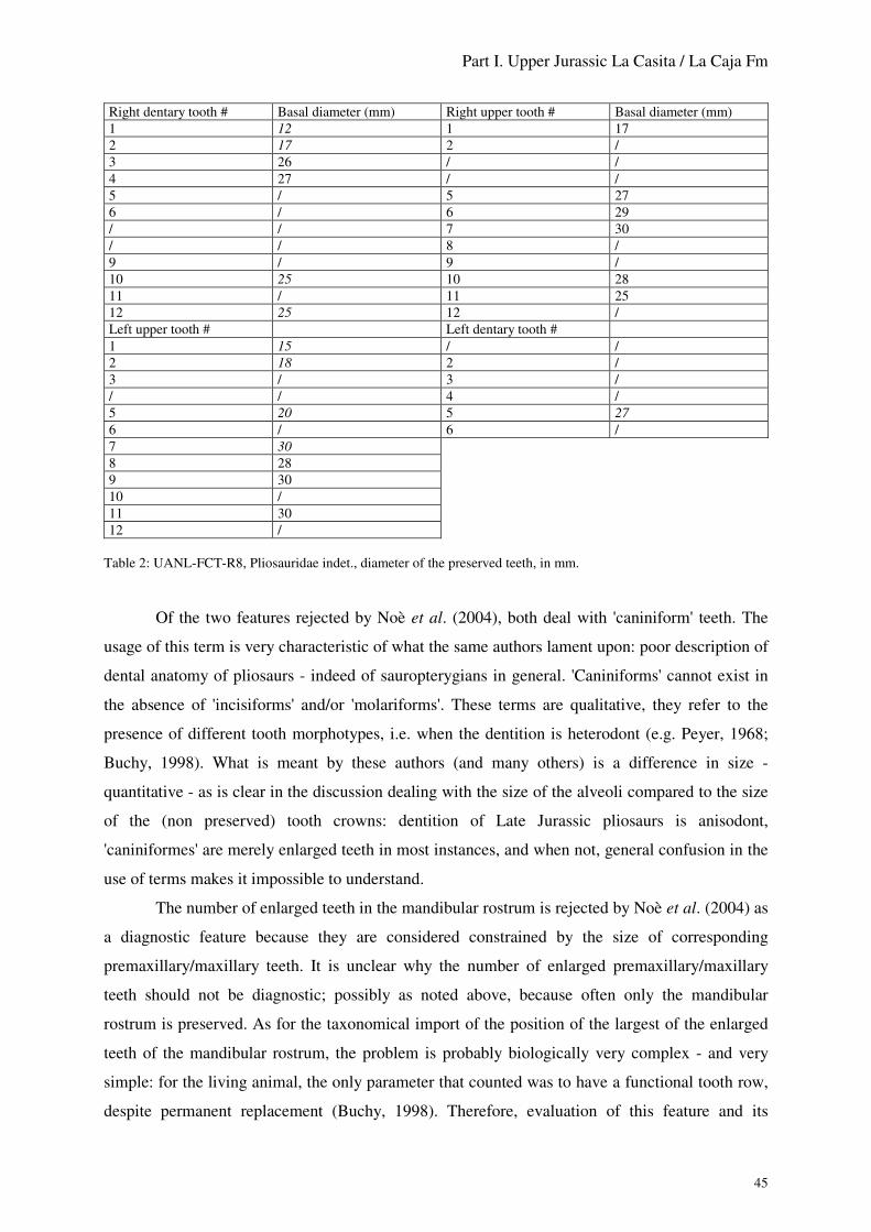
Part I. Upper Jurassic La Casita / La Caja Fm
45
Right dentary tooth # Basal diameter (mm) Right upper tooth # Basal diameter (mm)1 12 1 172 17 2 /3 26 / /4 27 / /5 / 5 276 / 6 29/ / 7 30/ / 8 /9 / 9 /10 25 10 2811 / 11 2512 25 12 /Left upper tooth # Left dentary tooth #1 15 / /2 18 2 /3 / 3 // / 4 /5 20 5 276 / 6 /7 308 289 3010 /11 3012 /
Table 2: UANL-FCT-R8, Pliosauridae indet., diameter of the preserved teeth, in mm.
Of the two features rejected by Noè et al. (2004), both deal with 'caniniform' teeth. The
usage of this term is very characteristic of what the same authors lament upon: poor description of
dental anatomy of pliosaurs - indeed of sauropterygians in general. 'Caniniforms' cannot exist in
the absence of 'incisiforms' and/or 'molariforms'. These terms are qualitative, they refer to the
presence of different tooth morphotypes, i.e. when the dentition is heterodont (e.g. Peyer, 1968;
Buchy, 1998). What is meant by these authors (and many others) is a difference in size -
quantitative - as is clear in the discussion dealing with the size of the alveoli compared to the size
of the (non preserved) tooth crowns: dentition of Late Jurassic pliosaurs is anisodont,
'caniniformes' are merely enlarged teeth in most instances, and when not, general confusion in the
use of terms makes it impossible to understand.
The number of enlarged teeth in the mandibular rostrum is rejected by Noè et al. (2004) as
a diagnostic feature because they are considered constrained by the size of corresponding
premaxillary/maxillary teeth. It is unclear why the number of enlarged premaxillary/maxillary
teeth should not be diagnostic; possibly as noted above, because often only the mandibular
rostrum is preserved. As for the taxonomical import of the position of the largest of the enlarged
teeth of the mandibular rostrum, the problem is probably biologically very complex - and very
simple: for the living animal, the only parameter that counted was to have a functional tooth row,
despite permanent replacement (Buchy, 1998). Therefore, evaluation of this feature and its

Part I. Upper Jurassic La Casita / La Caja Fm
46
taxonomical meaning will require proper understanding of - among others - tooth replacement
pattern, biomechanics of the jaw apparatus, bone/tooth physiology and turn over. In response to
permanent replacement, a very high plasticity in the location of the functional part of the dentition
is to be expected - though the relative size of the teeth and position of largest teeth could still
reveal genetically constrained and therefore taxonomically significant.
In summary, for matter of comparison with the presently discussed specimen, the number
of symphysial teeth has to be given primordial import, both because it is currently the most
considered feature, and what is preserved. The length of the tooth row is available in few taxa
only, and therefore of dubious usefulness (Noè et al., 2004). As for the size of the teeth
(symphysial or not), it is still believed here that the position of the largest enlarged teeth should be
considered, at least in the frame of a comparison of the anatomy of the rostrum in general, as it is
unknown whether the size of the teeth constrains the size of the rostrum or vice-versa; hopefully,
re-assessment of old specimens and discovery of new ones will clarify the debate.
With an estimate of 9-10 symphysial teeth, UANL-FCT-R8 represents a 'longisymphysial'
form, excluding close affinities to forms characterised by a short or spatulate mandibular rostrum,
like e.g. Liopleurodon, Kronosaurus, Simolestes and Maresaurus, (Gasparini, 1997; Noè, 2001;
Noè et al., 2004; pers. obs). Late Jurassic 'longisymphysial' pliosaurs though are very poorly
known, including the historical genus Pliosaurus and forms currently wanting taxonomical re-
assessment (Tarlo, 1959, 1960; Taylor & Cruickshank, 1993; Noè, 2001; Noè et al., 2004). As
noted by Taylor & Cruickshank (1993), the two Kimmeridgian species of Pliosaurus recognised
by Tarlo (1960) differ by poorly diagnostic features; Noè et al. (2004) emphasise the fragmentary
nature of both holotypes and the lack of recent, comprehensive studies cleaning up 150 years of
poorly justified assignment of specimens. Noè (2001) suggests to restrict the genus to forms with
a long mandibular rostrum (bearing 10 to 12 teeth) and trihedral teeth, leaving forms with sub-
symmetrical teeth in a taxonomical fog. At present, Noè et al. (2004) list as Late Jurassic
pliosaurs preliminarily assigned to the genus Pliosaurus (also wanting a thorough comprehensive
review, suspected to belong to at least two different genera, and the tooth anatomy of some being
unknown): P. brachydeirus with 10-11 symphysial teeth, P. brachyspondylus with 9-10 (Bardet et
al., 1993, 1994), P. portentificus with 8, and specimens formerly assigned to Liopleurodon
macromerus reverted to Pliosaurus macromerus, with 5-6. However, Tarlo's (1960) diagnosis of
the genus remains, emphasising trihedral teeth and long symphysis.
UANL-FCT-R8 does not fit the current diagnosis of Pliosaurus: it differs from specimens
currently assigned to the genus in the premaxilla overhanging the rostrally pointed mandibular
rostrum, the tooth crown morphology (at least rostrally), the straight ventral (caudal to dentary
tooth 4) and dorsal margins of the dentary. Pending re-description of European forms, its

Part I. Upper Jurassic La Casita / La Caja Fm
47
taxonomical status is for now unresolved, as are, like for UANL-FCT-R3, its affinities with
European contemporaneous forms.
UANL-FCT-R8, a Kimmeridgian form with teeth sub-circular in cross-section confirms
the doubts expressed by Bardet et al. (1994) and Noè (2001) upon Tarlo's (1960) assumption as to
the stratigraphical import of pliosaurian tooth cross-section. If Tarlo's (1960) conclusions reveal
valid and are to be viewed in an evolutionary perspective (a trend from simple, conical teeth
toward trihedral teeth between Middle and Upper Jurassic), the Mexican form may reveal a relic
in the protected Late Jurassic Mexican Gulf.
CPC 226 (figs 21-23)
Material: Possibly sub-complete skeleton, including weathered skull and mandible.
Former accession number: CEP1843 (Buchy et al., 2006c)
Origin: Early Tithonian section of the La Caja Formation at the Sierra El Jabalí, near Gomez
Farías, Coahuila, Mexico (fig. 2); parts of the skull and cervical vertebrae were first collected by
J. Lopez Espinoza in 1999, more parts of the specimen (a humerus, cervical vertebrae, rib
fragments) were rescued during subsequent field campaigns in March and September 2004, and
February 2006. More postcranial material is awaiting collection in the field. Preparation of the
postorbital portion of the skull and several cervical vertebrae was conducted by J. Lopez Espinoza
and the author. Preparation of what appears to be the rostrum and other postcranial remains
collected recently could not be completed yet.
Preservation: At present, the embedding situation of the skeleton cannot be evaluated as most of
the collected material is awaiting preparation, while possibly the caudal half of the specimen is to
be excavated. Moreover, collection by several persons over many years yielded incomplete data.
The material available for study now comprises the medial portion of the postorbital region of the
skull with underlying elements of brain case, all heavily weathered and/or incompletely prepared;
several cervical vertebrae; phalanxes. Like for all vertebrate remains from the locality, the
contrast is poor between matrix and bone (Buchy et al., 2006c), resulting in many uncertainties
upon the actual anatomy of the specimen. This is especially the case for the skull, where
individual bones were pressed against one another and, also because of partial preparation, whose
outlines are difficult to trace. Neural arches - at least of the known vertebrae - were unfused,
hinting at what is traditionally called a 'juvenile' individual (see UANL-FCT-R2).
Description: Skull - The preserved portion of skull is broken in two parts, approximately level
with the middle of the upper temporal fenestrae; the contact between the two parts is faint and for
convenience they were not glued together (figs 21, 22). The rostal portion of skull is heavily
weathered so that almost no original dorsal surface is preserved; sutures are therefore visible at an

Part I. Upper Jurassic La Casita / La Caja Fm
48
unknown level within the skull thickness, which renders comparisons with described taxa
impossible.
Frontal - A suture runs caudolaterally and individualises a sub-quadrangular portion of
bone at the left rostral-most corner of the specimen. From its location, this bone may be the
frontal, but no anatomical detail is preserved.
Prefrontal - Similarly, a clear suture separates the right laterorostral-most portion of the
specimen. It should separate the prefrontal rostrolaterally from the postfrontal.
Postfrontal - On both sides the postfrontal is poorly preserved; its suture with the parietal
runs caudally, slightly laterally and then slightly medially, from the rostral extremity of the
specimen until approximately level with the rostral fourth of the parietal foramen. The caudal

Part I. Upper Jurassic La Casita / La Caja Fm
49
margin of the postfrontal on both sides may be close to original, according to the breakage pattern
of the lateral margins of the intertemporal bar.
Parietal - About 20 mm lateral to the midline on either side on the rostral-most portion of
the specimen, runs a possible suture for 10 mm on the right side, slightly longer on the left. If this
structure actually represents a suture, it may be marking a ventral process of the frontal or
premaxilla underlying the parietal there, and visible due to weathering. Noè (2001) indicates that
the parietal underlies the frontal in Liopleurodon, while Buchy et al. (2006b) describe a ventral
process of possibly the parietal underlying the premaxillae in the pliosaur UANL-FCT-R3 (see
above); the nature of this element in CPC 226, if not an artefact, is therefore enigmatic.
The parietal foramen is long oval. Its margins are gently rounded. Lateral and rostral to it,
the surface of the parietal slopes ventrally so that the foramen is set higher than the level of the
interobital surface of the skull. Nothing more of the original surface of the intertemporal part of
the parietal is preserved. The lateral margins of the intertemporal bar were pressed dorsally, and
are preserved as broken, overlapping fragments; their original ventral extent cannot be
determined. Level with the caudal extremity of the parietal foramen, an oval, lateroventrally
oriented depression in the lateral margin of the intertemporal bar may represent the epipterygoid
facet. The caudal-most dorsal portion of the parietal is better preserved, except for the temporal
bar itself that most likely was weathered prior to collection. The lateral margin of the parietal
curves gently caudolaterally to form the mediocaudal corner of the upper temporal fossa; due to
distortion over underlying brain case elements, the margin of the upper temporal fossa there
appears asymetrical between right and left side. It is probably closer to original on the left side, as
there, are fewer underlying elements. The rostral-most portion of the contact with the squamosal
is not visible due to overlying unidentified bones. It runs caudally, slightly medially until the
broken caudal extremity of the specimen. Along that break, the parietosquamosal suture is S-
shaped, a flange of the squamosal overlaping the parietal dorsally, while a flange of the parietal
underlaps the squamosal ventrally. The ventral face is difficult to interpret; the parietosquamosal
suture appears to run along the same path as its dorsal part. The visible portion of the ventral
surface of the parietal located where the bone forms the caudomedial corner of the upper temporal
fossa bears large, irregular grooves, possibly the facet for the supraoccipital.
Squamosal - The dorsal surface of the squamosal slopes gently caudally until its caudal
extremity. Its caudal margin, where preserved, is gently rounded. In ventral view, poorly
preserved bone might represent its quadrate ramus and obscures the ventral surface of its parietal
ramus. The rostral margin of the parietal ramus forms a flange (that may have been a buttress
prior to weathering) bordering the upper temporal fossa.

Part I. Upper Jurassic La Casita / La Caja Fm
50
Elements of the brain case - Interpretation of the various bones preserved in the area is
additionally hindered by the generally poor knowledge of pliosaur brain case, possibly due to poor
ossification (e.g. Noè, 2001; O'Keefe, 2001). All conclusions reached here are therefore
provisional, pending further preparation and study of comparable material. Two fan-shaped
elements preserved in the right upper temporal fossa rostral to the rostral-most point of contact
between parietal and squamosal (fig. 21) are similar in outline to the prootic of Muraenosaurus
described by Maisch (1998: fig. 11). However, no surface details are visible. Various fragments of
flat, thin bones, with few identifiable original margins, probably represent parts of the pterygoids
and other bones of the palate. Moreover, the quadrate ramus of the pterygoid is possbily partly
present, ventral to the right caudal-most portion of the parietal (fig. 22; see also White, 1935: pl.
9). It is high oval in cross-section, the visible suface slightly concave along its long axis. The right
exoccipital-opisthotic is visible in caudomedial view, and although distorted, comparable to what
is described in Muraenosaurus by Maisch (1998: fig. 9a; see also Storrs & Taylor, 1996). It
differs from the same element in Liopleurodon described by Noè (2001: fig. 43), possibly due to
incomplete preservation of the latter. In CPC 226, the paroccipital process is sub-triangular in
cross-section where broken about 20 mm away from the main body of the bone. This is high
triangular in outline, its dorsal portion broken and/or obscured by sediment. Its surface is spread
with numerous foraminae, four of which are larger. The first is located on the rostral margin of the
bone, about 10 mm dorsal to its ventral margin; the second is slightly higher, 10 mm further
caudally; the third is the largest, almost 5 mm in diameter and located 20 mm dorsal to the

Part I. Upper Jurassic La Casita / La Caja Fm
51
second; the fourth is at the level of the first, about 15 mm caudal to the second. They could
represent exits for the cranial nerves respectively IX to XII, following the interpretations of
Maisch (1998) and Carpenter (1997: fig. 5). Clearly, taxonomically close specimens with
comparable preserved material would help assess the accuracy of this interpretation.
Cervical vertebrae - The cervical vertebrae available for study are oval in articular aspect,
mediolaterally elongate (fig. 23). They confirm the pliosaurian nature of the specimen in being
craniocaudally short. A notochordal pit is visible, located in the center of a raised area in the
middle of the articular faces.
Discussion: The specimen being mostly unprepared, little can be said upon its affinities. Most of
the skull was weathered, leaving as original only the parietosquamosal suture and possibly the
exoccipital-opisthotic, which are unfortunately poorly known in most pliosaurs (e.g. Noè, 2001).
From the size of the known cervical vertebrae, it most likely represents a medium-sized animal,
possibly 8 to 10 m in length, and uncompletely ossified (see UANL-FCT-R2).
CPC 228
Material: Partial isolated cervical centrum.
Former accession number: CEP1850 (Buchy et al., 2006c)
Origin: Early Tithonian section of the La Caja Formation at the Sierra El Jabalí, near Gomez
Farías, Coahuila, Mexico (fig. 2); collected in March 2004.
Preservation: The specimen is a 'slice' of the centrum, comprising ventrally the medial-most
portion (as testified by both subcentral foramina) and dorsally a portion of the neural arch facet.
The specimen was prepared by S. Stinnesbeck at the SMNK and the author at the MUDE.

Part I. Upper Jurassic La Casita / La Caja Fm
52
Description: The height of the centrum was about 120 mm, its length about 60 mm. Its width
cannot be evaluated, nor whether it was sub-circular or transversely enlarged (as e.g. CPC 226).
The articular faces appear slightly depressed in their middle.
Comments: The size and anatomy of what is preserved of the centrum fits the pliosaur CPC 226.
Though, the finding places within the site of both fossils indicates that they probably belong to
different individuals (except in case of human transport), if possibly to the same taxon.
I.2.3.4. Discussion
Buchy et al. (2006d) identified three, possibly four different pliosaur taxa from the Kimmeridgian
Mexican Gulf. This count is confirmed by the above description of UANL-FCT-R8. The Mexican
assemblage described here comprises a very large form, whose skeleton shows clear signs of
osteological immaturity, whether due to young individual age or of taxonomical import (UANL-
FCT-R2). A medium-sized group (possibly about 10 m in length) is represented by UANL-FCT-
R7 and R8, the former also showing immature bone condition, while the individual age of the
latter cannot be determined - and neither the taxonomic similarity of the specimens. UANL-FCT-
R3 is a smaller form, anatomically distinct from UANL-FCT-R8. The Kimmeridgian Mexican
Gulf was populated by at least three taxa of pliosaurs (see a discussion in Buchy et al., 2006d),
which is similar to the pliosaurian assemblage of the English Kimmeridge Clay (Noè et al., 2004),
although none of the Mexican forms can be clearly referred to a previously known taxon.
As for the Tithonian, with a single, potentially diagnostic pliosaur wanting preparation,
not much can be said, except that it appears to represent a medium-sized taxon, based on the size
of the cervical vertebrae.
At present, Mexican pliosaurs are of unclear affinities, 'longisymphysial', of large size and
massive (in terms of thickness of the bones in cross-section; however, few studies describe cross-
sections as most were re-glued prior to study, see Buchy et al., 2006a).
Elasmosaurs are rare in both the Kimmeridgian and the Tithonian of Mexico, and known
specimens are of no current taxonomic value beyond family level (but see Buchy, 2005: poor
understanding of the group makes taxonomical status of even sub-complete specimens uncertain;
Elasmosauridae appears to be one of the last groups for which 'gattungsmackery' [Owen, 1869: 8]
is still at work, see e.g. Sato, 2003; Druckenmiller & Russell, 2006). The raison d'être of the
exceedingly long neck of elasmosaurs has long been an enigma, possibly due to the lack of proper
biomechanical analysis to the benefit of sole examination of tooth morphology (e.g. Massare,
1987): long, sharp, closely-spaced teeth were long considered a fish-piercing device, by
comparison with Recent piscivorous crocodiles, resulting in the beautiful, biomechanically
unlikely reconstructions of animals with a snake-like neck snapping at fish or even pterosaurs.

Part I. Upper Jurassic La Casita / La Caja Fm
53
Recently, several authors more or less independently came to re-examine the question, and slowly
emerges the picture of sediment sieving elasmosaurs, and a diet mostly relying upon sea floor
invertebrates in the shallow, soft substrate areas available following transgressive phases (Buchy,
1998, 2005, 2006; McHenry et al., 2005; Noè, 2006). Clearly, under this life-style hypothesis, the
Late Jurassic Mexican Gulf, at least where the La Casita/La Caja Formations yielded marine
reptiles for now, did not constitute favourable environments. It is to be noted that elasmosaurs
appear more abundant in the Upper Cretaceous (?Campanian-Maastrichtian) of the region of
Saltillo (fig. 2), represented by several clusters of vertebrae; the material (of at present poor
taxonomical value) is in private hands and of currently unprecise origin, and therefore not dealt
with here, study pending examination of the localities and clarification of the status of the
specimens.
I.3. Discussion
The Kimmeridgian assemblage of marine reptiles in the Mexican Gulf was discussed by Buchy et
al. (2006d), and little can be added for now. Of the two specimens which were not known then,
both referred to Dakosaurus sp. (specimens CPC 201 and UANL-FCT-R29), one comes from the
south of Nuevo León (Zaragoza; fig. 2), the second from a northern site (San Juan de los Dolores;
fig. 2), therefore possibly invalidating the suggested trend of thalattosuchians to become rare in
the south. As was concluded by Buchy et al. (2006), more specimens are to be added to the
sample to deduce ecological partitions.
The same holds for the Tithonian at Gomez Farías. The assemblage is dominated
(numerically) by thalattosuchians and ichthyosaurs. Pliosaurs are rare though one is probably sub-
complete; such large predators must have been rare anyway in the fauna. The preservation status
of elasmosaurs most likely indicates distant living areas. A point of interest is that, after several
field campaigns and exploration of a very rich site (Buchy et al., 2006c) by private collectors as
well as members of institutions, no specimen referrable to Testudines was found yet. It was
suggested by Buchy et al. (2006d) that turtles had not made it from Europe to the Mexican Gulf
by the Kimmeridgian. Marine turtles are present in the Tithonian Neuquén Basin of Argentina
(Fernández & Fuente, 1993; Gasparini & Fernández, 1997, 2005). Their absence in the Tithonian
Mexican Gulf appears the main peculiarity of the assemblage.
As was emphasized by description of new taxa (Frey et al., 2002; Buchy et al., 2006e), but
also by other specimens (Buchy et al., 2003, 2005c, 2006b, d, in press), marine reptiles from the
Late Jurassic Mexican Gulf do not show clear affinities to European and Pacific forms and appear
to confirm the isolation of the area as deduced from invertebrate assemblages (Buchy et al., 2006d

Part I. Upper Jurassic La Casita / La Caja Fm
54
and references therein). The statement has to be somewhat moderated, though, due to the
incompleteness or need of preparation of most of the Mexican specimens, but also to the poor
knowledge of European forms (especially pliosaurs and thalattosuchians) and consequently, of
their palaeobiogeographical history. The rostral prolongation of the ?parietal in UANL-FCT-R3
appears to indicate affinities with Cretaceous Australian forms (Buchy et al., 2006b), though, a
similar anatomy may be present as well in some English Upper Jurassic specimens (Noè, pers.
com., 2006). Pending description of these, and also of Australian specimens, it is unknown
whether the same bone is involved and the feature can be considered as an apomorphy, or if the
structure is a mere biomechanical requirement achieved along different paths. At present, the
identification of Ophthalmosaurus icenicus in the assemblage is interpreted as marking more
ubiquitous habits of ichthyosaurs as compared to other marine reptiles, though discovery of more
complete Mexican specimens might change the figure.
Marine reptiles described here do substantially increase our knowledge of the groups apart
from palaeobiogeographical considerations: the 3-dimensional preservation of several specimens
was the opportunity to reassess the nature of choanae in plesiosaurs (Buchy et al., 2006a;
confirmed by UANL-FCT-R8), and location of salt glands in thalattosuchians (Buchy et al., in
press).

Part II.1. Upper Cretaceous Early Turonian Agua Nueva Formation
55
Part II. Upper Cretaceous
II.1. Early Turonian Agua Nueva Formation at Vallecillo, Nuevo León
II.1.1. Palaeogeographical context
The laminated limestones of the Agua Nueva Formation at Vallecillo (fig. 2) and their fossil
contents were recently the subject of two PhDs (Blanco-Piñon, 2003; Ifrim, 2006). A summary of
both regarding marine reptiles and their palaeogeographical context was given by Buchy et al.
(2004, 2005b). It is of some import for the discussion of the assemblage to re-emphasise that the
Vallecillo limestone represent an open marine Plattenkalk deposit (Ifrim, 2006; fig. 24).
Figure 24: Palaeobiogeographical map ofthe Turonian (modified from Smith et al.,1994). Insert: palaeobiogeographical map ofnorth-east Mexico during the Turonian; M:Monterrey; S: Saltillo (redrawn afterStinnesbeck et al., 2005).

Part II.1. Upper Cretaceous Early Turonian Agua Nueva Formation
56
II.1.2. Pliosauridae cf. Polyptychodon
UANL-FCT-R26 (fig. 25)
Material: Isolated partial tooth.
Origin: Early Turonian Agua Nueva Formation at Vallecillo, Nuevo León (fig. 2), collected in
March 2002 by C. Ifrim ( Ifrim et al., 2004, 2005a, b; Ifrim, 2006, in press; pers. com.).
Preservation: The apex is broken approximately at two-thirds of the crown height; only the
apical-most portion of the root is preserved. Enamel is absent and dentine was probably replaced
by calcite, as is common for fossils from this locality (Blanco-Piñon et al., 2001; Blanco-Piñon,
2003; Ifrim, 2006).
Description: The basal diameter of the crown is approximately 20 mm. It is slightly curved
mesially and labiolingually compressed, though carinae appear absent. Both lingual and labial
surfaces show regularly spaced ridges. A vertically oval wear facet is present distally at the base
of the crown.
Discussion: Absence of shouldering between crown and root excludes mosasaurian affinities. The
pattern of regularly spaced ridges around the crown, the subcircular cross-section at the base of
the crown, the slight mesial curvature of the crown, as well as the palaeogeographical and
stratigraphical occurrences are strongly reminiscent of the pliosaurian genus Polyptychodon
Owen, 1841 from the Cenomanian of England, also known from the Eagle Ford Shales of Texas
(Cenomanian-Turonian; Welles & Slaughter, 1963).
Squamata Oppel, 1811
Anguimorpha Fürbringer, 1900
Mosasauroidea Gervais, 1853
Figure 25: UANL-FCT-R26, cf. Polyptychodon, isolated,partial tooth. Scale 20 mm. Photo C. Ifrim.

Part II.1. Upper Cretaceous Early Turonian Agua Nueva Formation
57
II.1.3. 'Aigialosaurs' indet.
The systematics and taxonomy of what was formerly considered as ancestral forms to mosasaurs
(e.g. Gorjanović-Kramberger, 1892; Dollo, 1892, 1903; Nopcsa, 1903, 1923; see a review in
Dutchak, 2005; Smith & Buchy, submitted) are currently undergoing a maelstrom of cladistic
analyses, that yielded yet no generally accepted, comprehensive, stable results concerning
relationships, and therefore taxonomy (Dutchak, 2005; Smith & Buchy, submitted). Possibly due
to certain similarities to Recent forms, Cretaceous marine squamates appear a favourite subject for
cladistic analyses probing what softwares may add to traditional views of the transition from
terrestrial to marine life, although few independant data sets have been analysed, as pointed out by
Dutchak (2005). Bell's (1993; 1997) matrix is the most commonly used, while its published
version allegedly contains several miscodings (N. Bonde, oral communication, 1st Mosasaur
Meeting, Maastricht, 2004).
Allegedly seeking to clarify the matter and examine specimens within a frame
'unburdened by phylogenetic context' Bell & Polcyn (2005: 178) and Polcyn & Bell (2005)
introduce the terms 'plesiopedal' versus 'hydropedal'. As discussed by Smith & Buchy (submitted),
the use of the new terms by their authors is at best self-contradictory, and in any case of little
more use than an informal 'aigialosaurs'. And, as implied by Dutchak (2005), stable nomenclature
is still wanting following the introduction of the new terminology. While the terms aigialosaur,
coniasaur and dolichosaur are still in 'loose' use (Evans et al., 2006: 1143, 1155), these groups are
also now designated by convoluted expressions, like 'early', 'basal' or 'primitive' mosasauroid (e.g.
Bell & Polcyn, 2005), or 'long-bodied lizard' (Evans et al., 2006). Moreover, an intriguing, fresh
water form was recently discovered in the Santonian of Hungary (Makádi, 2005), which was not
included in these analyses. It is expected that mosasauroid systematics will be explored for a
while, and that possibly cladistic analyses will not have the last word. As this volume is more of a
catalogue of north-east Mexican fossils, the systematic debate is eschewed in favor of
examination of the specimens themselves, in the hope that new data will finally help solve the
issue.
UANL-FCT-R27 (Buchy et al. 2004: fig. 2; 2005b: fig. 3a, b; Buchy & Smith, 2005; Ifrim, 2006:
fig. 7.17c; Smith & Buchy, submitted; figs 26, 27).

Part II.1. Upper Cretaceous Early Turonian Agua Nueva Formation
58

Part II.1. Upper Cretaceous Early Turonian Agua Nueva Formation
59
Material: At present, this specimen (fig. 26) comprises a main slab (UANL-FCT-R27-1)
preserving a long segment of tail, subcomplete left hind limb, right femur, left pelvic girdle and
part of the lumbar region. Two small slabs (UANL-FCT-R27-2, -3) are partial counterparts of the
main slab, mostly comprising fragments of vertebrae. A further slab (UANL-FCT-R27-4) and its
counterpart (UANL-FCT-R27-5) comprise the trunk (lacking vertebrae) and the two subcomplete
manus. The portion comprising the counterpart of the left pelvic region and hind pes and right
femur bears number UANL-FCT-R27-6, while the adjacent portion comprising the counterpart of
the cranial-most portion of the tail bears number UANL-FCT-R27-7. The specimen shows
exquisite preservation of integumentary structures, as well as a possible gastric contents and
pebble-like elements within the abdomen.
As emphasised by the sequence of numbering, the history of discovery (or rather
rediscovery) of the specimen is complex. The fragments were collected by the quarry owner for
the UANL-FCT and transferred first to the UANL-FCT; some then made their way to Germany
for study. Thus, the author and colleagues were first aware of UANL-FCT-R27-1 to -3, and gave
preliminary descriptions of these remains (Buchy et al., 2004, 2005b; Buchy & Smith, 2005).
Together with these fragments was a slab containing a partial manus (Buchy et al., 2004, 2005b;
Buchy & Smith, 2005; fig. 28), thought to belong to the same individual. Smith & Buchy
(submitted) expressed a doubt upon the attribution of this manus, and noted it was given accession
number UANL-FCT-R28. Subsequently, more fragments of UANL-FCT-R27 turned up, re-
discovered (together with UANL-FCT-R30) among the Vallecillo collections at the UANL-FCT
by C. Ifrim and S. Giersch (pers. com., 2006). As the part and counterpart, UANL-FCT-R27-4
and -5, contain both manus, the isolated, partial manus UANL-FCT-R28 obviously does not
belong to UANL-FCT-R27 and will be described together with the newly discovered parts of
UANL-FCT-R27 (Buchy & Smith, in prep.).
Origin: Early Turonian Agua Nueva Formation at Vallecillo, Nuevo León (fig. 2), collected by
the quarry owner for the UANL-FCT.
Comments: A detailed description of parts -1 to -3 of the specimen by Smith & Buchy is in the
review process. A brief account of its anatomy and relationships was given by Buchy et al. (2004;
2005b), reference to which it is here made in order to respect rules of priority. A description of
parts -4 to -7 is in preparation. Here is illustrated the manus (fig. 27), and measurements of its
elements are given in table 3, for comparison with UANL-FCT-R28 (see further). Smith & Buchy
(submitted) indicate that the specimen represents a new taxon, distinguished from other
'aigialosaurs' by, among others, features of its hind limb. Assessment of its affinities, however, is
hindered by its incompleteness, as well as poor understanding of the relationships within the

Part II.1. Upper Cretaceous Early Turonian Agua Nueva Formation
60
group as was noted in introduction. Buchy et al. (2005) and Smith & Buchy (submitted) note that
it shares features with Mosasauroidea but lacks derived features of Mosasauridae.
Element Length Element Lengthmetacarpal I 8.5 phalanx IV.1 5.5phalanx I.1 6.2 phalanx IV.2 5.1metacarpal II 9.6 phalanx IV.3 /phalanx II.1 / metacarpal V 7.7metacarpal III 10.5 phalanx V.1 6.6phalanx III.1 / phalanx V.2 6.7metacarpal IV 10.5 phalanx V.3 /
Table 3: Measurements on the left manus of UANL-FCT-R27, in mm. All measurements made, when possible, at themidpoints of articulation of the bone.
UANL-FCT-R28 (Buchy et al., 2005b: fig. 3c; fig. 28, tables 4, 5)
Material: Partial manus.
Origin: The origin of this specimen is unclear. It was formerly thought to belong to UANL-FCT-
R27, and illustrated as so (Buchy et al., 2005b: fig. 3c; see above). The matrix and repository
conditions indicate an Early Turonian Agua Nueva Formation at Vallecillo, Nuevo León (fig. 2).
Preservation: Metacarpals I–IV are poorly and only partially preserved, while metacarpal V is
missing altogether. Some of the surface bone was most likely lost to the counterpart.

Part II.1. Upper Cretaceous Early Turonian Agua Nueva Formation
61
Description: Measurements of the various elements of the manus can be found in table 4. On
either side of metacarpal I, unidentified bone fragments indicate some disarticulation of the digits
in relation to the carpus. Metacarpal I appears sub-complete, and is short. The length of all other
metacarpals is unknown. Digits III and IV are subequal in length. The phalangeal formula was
probably 2-3-4-5-3.
Discussion: UANL-FCT-R28 is larger than the manus of UANL-FCT-R27; the various elements
also appear more slender, although this might be due to the orientation of the bone and splitting
pattern. The only preserved elements of UANL-FCT-R28 whose length can be determined and are
also known in UANL-FCT-R27 are phalanges I.1, IV.1, IV.2 and V.2. Calculated ratios (table 5)
indicate different proportions for these elements in UANL-FCT-R27 and -R28, although the
significance of the differences is unknown, and could be ontogenetic, related to sexual
dimorphism and/or individual variation.

Part II.1. Upper Cretaceous Early Turonian Agua Nueva Formation
62
Element Length Element Lengthphalanx I.1 11.2 phalanx IV.1 9.7phalanx I.2 5.3 phalanx IV.2 7.5phalanx II.1 10.2 phalanx IV.3 6.4phalanx II.2 10.0 phalanx IV.4 7.1phalanx II.3 4.0 phalanx IV.5 4.1phalanx III.1 10.8 phalanx V.1 /phalanx III.2 8.2 phalanx V.2 7.2phalanx III.3 8.5 phalanx V.3 4.5phalanx III.4 4.6
Table 4: Measurements on the manus UANL-FCT-R28, in mm. All measurements made, when possible, at themidpoints of articulation of the bone.
Ratio UANL-FCT-R27 UANL-FCT-R28I.1/V.2 0.92 1.55I.1/IV.1 1.12 1.5I.1/IV.2 1.2 1,5IV.1/IV.2 1.07 1.29IV.1/V.2 0.82 1.34IV.2/V.2 0.76 1.04
Table 5: Ratios calculated between elements known in both manus UANL-FCT-R27 and -R28.
UANL-FCT-R30 (fig. 29)
Material: Ten articulated vertebrae including first caudal and sacrals, and fragments of 2
additional, cranially adjacent centra, poorly preserved on a slab; articulated ribs and portions of 3
additional ribs pertaining to cranially adjacent, non preserved vertebrae; possibly portions of
pelvic girdle.
Origin: Although the matrix clearly indicates that the specimen comes from the Vallecillo
limestones, the finding data are unknown. The specimen was re-discovered by S. Giersch among
the Vallecillo 'fish collection' of the UANL-FCT in September 2006.
Preservation: The specimen is preserved on a limestone slab broken into 6 fragments. The bone
is weathered so that only the outline of the vertebrae is original. The specimen is currently coated
with glue that further obscures its anatomy; some areas (synapophyses, possibly pre- and
postzygapophyses) are still covered with matrix. The centra are therefore actually identified by the
ribs they bear. It is impossible for now to determine whether the specimen is visible in ventral or
dorsal view; in what follows, 'left' and 'right' refer to the current visible aspect of the specimen
(fig. 29).
Description: All centra measure approximately 20 mm in length, with a minimum, median
diameter of about 12 mm.
The last preserved vertebra on the slab is the first caudal, its transverse process laterally
directed; the following two vertebrae are sacrals, as witnessed by the sacral ribs. These are better
visible on the right side, whereas they are either broken or still partly embedded in matrix on the
left. The caudal-most sacral rib points laterally. The first sacral rib is directed caudolaterally; its

Part II.1. Upper Cretaceous Early Turonian Agua Nueva Formation
63
caudal margin is thickened and forms a buttress. A patch of bone-like substance underlies the
caudal-most sacral rib and could represent part of the girdle.
What appears as a pair of long oval elements, oriented craniolaterally on either side of the
vertebral column, could also represent parts of the girdle, possibly the ilia, then close to their
original contact with the sacral ribs.
Along the caudal break of the specimen, to the right of the vertebral column, a fragment of
bone, 20 mm in proximodistal length, is present lateral to the sacral region; it may represent part
of the girdle or hind limb.
The cranially following vertebra bears no rib, and no bulging of the surface of the slab
indicates that ribs could be still embedded in matrix.

Part II.1. Upper Cretaceous Early Turonian Agua Nueva Formation
64
The four cranially adjacent centra bear short ribs on the right side (about 30 mm in
proximodistal length). These ribs are straight for most of their length, apparently only curved in
their proximal portion; they are laterally, slightly caudally oriented. On the left side, only the
caudal-most of these short ribs is preserved close to articulation: the other 3 are either embedded
or absent. In this region, a fragment of rib parallels the vertebral column; at least two more ribs
are still embedded in the area, being visible along breaks. Possibly, rupture of the abdominal
cavity during decay disarticulated the ribs in this area.
The cranially following vertebra in the series bears slightly longer ribs, about 50 mm in
proximodistal length. Possibly the cranially following vertebra also bears ribs intermediate in
length. The right rib is broken along the edge of the slab, while the left one disappears below the
rib of a cranially adjacent vertebra, but still appears shorter.
The cranially following vertebrae are dorsals with long, smoothly curved ribs that rotated
when the rib cage collapsed.
Lumbar and dorsal synapophyses are located in the cranial third of the centra. The only
well visible synapophysis is on the left of vertebra 'sacrals minus 6'; it is about 10 mm in
proximodistal length and about 5 mm in width, pointing caudolaterally. Where visible, rib and
synapophysis are separated by a seam of sediment, indicating non-fusion of the ribs.
Discussion: Although originally mistaken for a fish, the vertebral anatomy as well as
heteromorphous ribs leave no doubt about the mosasauroid affinities of the specimen (see e.g.
Smith & Buchy, submitted). Its poor condition prevents detailed comparisons, though further
preparation might clarify some issues. It appears to present one vertebra without rib followed by
four vertebrae bearing short ribs and further cranially one, possibly two vertebrae with ribs
intermediate in length. This pattern is similar to that of UANL-FCT-R27 (Smith & Buchy
submitted). For now, it is to be noted that its size exceeds dimension of most known aigialosaurs,
only comparable to the 'Trieste aigialosaur' (Carroll & deBraga, 1992).
CPC 254 (fig. 30)
Material: Nine procoelous, articulated caudal vertebrae, forming a segment of tail that was
disarticulated prior to embedding (as witnessed by the exposed cranial-most cotyle); the condyle
of the terminal-most preserved vertebra is broken along the edge of the slab. Two additional
disarticulated centra, located respectively 50 mm craniolateral and 90 mm cranial to the first
articulated vertebra. Imprints of two long bones, possibly metatarsals.
Origin: Early Turonian Agua Nueva Formation at Vallecillo, Nuevo León (fig. 2), collected in
September 2006 by C. Ifrim and S. Giersch for the MUDE.

Part II.1. Upper Cretaceous Early Turonian Agua Nueva Formation
65
Preservation: As is normal for the locality, the specimen is preserved on a slab. The bones are
visible in subcoronal section, the missing portions presumably having been lost to the counterpart.
Within the tail, the bone substance itself appears better preserved than in e.g. UANL-FCT-R27,
with visible trabeculae. The two isolated centra are badly weathered, partly visible as imprints,
and yield no anatomical detail. Parallel to the right transverse process of the 6th vertebra
preserved in articulation, the articular head of a long bone is preserved, while the rest of it is
visible laterally as a clear imprint. About 20 mm left to the same level of the portion of tail, and
parallel to it, a shallow imprint probably represent another long bone, as far as can be judged, of
similar morphology. Partial haemal arches are preserved slightly displaced, visible to the right of
articulated vertebra 5, to the left of more caudal vertebrae. As deduced from the overlapping
pattern of neural spines, pre- and postzygapophyses and transverse processes, the specimen is
visible in dorsal view, except for the cranial-most vertebra, which is slightly displaced from the
series and visible in left dorsolateral view.
Description: The two isolated vertebrae have a length of about 11 mm, similar to the cranial-most
vertebrae preserved in articulation. The centra very slightly diminish in length terminally, the
penultimate measuring about 10 mm in length. Little can be said on the anatomy of the centra due
to poor preservation. The transverse processes insert near mid-length on the centra. The original
width of the transverse processes cannot be determined, as what is visible is actually their ventral-
most portion, the rest having been lost to the counterpart. The five terminal-most right transverse
processes appear sub-completely preserved, ranging in length from about 11 mm cranially to 8
mm terminally. All exhibit a sinusoidal distal margin, convex cranially and concave terminally.
This could be attributed to a lesser thickness of the process terminally, and consequently
differential matrix covering and breakage pattern. Neural arches appear to have collapsed caudally
and to the right, now overlying the right postzygapophyses. Pre- and postzygapophyses are partly
preserved, depending on the level of the coronal break, and along the preserved fragment of tail
appear well-expressed, although no further detail can be deduced. The haemal arches were not
fused to the centra; their articular head could have been oval instead of rounded.
The two long bones measure about 18 mm in length. The shaft is the narrowest at about
mid length. The right one shows a distinct epiphysis ?proximally. Assuming these elements
pertain to the hind limb(s), and although few anatomical details are preserved, they likely are
central metatarsals, by comparison with UANL-FCT-R27, their length representing about 1.5
times a vertebral length (Smith & Buchy, submitted).

Part II.1. Upper Cretaceous Early Turonian Agua Nueva Formation
66

Part II.1. Upper Cretaceous Early Turonian Agua Nueva Formation
67
Discussion: CPC 254 is of poor taxonomical value, at least within the current taxonomical frame
for 'aigialosaurs'. CPC 254 is slightly smaller than UANL-FCT-R27. No feature contradicts its
belonging to the new taxon represented by UANL-FCT-R27 (Smith & Buchy, submitted), but
neither does any significant feature support it. Smith & Buchy (submitted) indicate that transverse
processes in UANL-FCT-R27 extend until at least caudal vertebra 15, gradually decreasing in
proximodistal length, but incompletely preserved; all caudals except the first one bear haemal
arches. All nine articulated vertebrae of CPC 254 possess a transverse process, whereas haemal
arches are visible only on the terminal six. However, no vertebra (including the two isolated
centra) can be surely identified as a sacral or first caudal (on the basis of either shortened centra,
or sacral rib or wide transverse process as possesses UANL-FCT-R27). CPC 254 is therefore
interpreted as a cranial portion of tail, between caudals 2-15, and on the ground of transverse
processes length, closer to the sacrum than to the point where they disappear.
Preservation hints at a disarticulated carcass, with at least the cranial-most portion of tail
kept in articulation. The cloaca (located level with the first caudal, lacking a haemal arch, Evans
et al., 2006) being usually the area where decay is initiated (see Salisbury et al., 2003), that the
tail was disarticulated just caudal to it appears coherent. Articulated vertebrae of CPC 254 might
therefore, on taphonomical argument, represent caudals 2-10.
II.1.4 Discussion
It is first to be noted that more specimens referable to mosasauroids were recently discovered in
Vallecillo and are presently kept in the MUDE. Some likely represent aigialosaurs, while several
vertebrae of clear mosasaurian anatomy (and much larger than known aigialosaurs) are indicative
of hitherto undocumented taxa. However, these specimen are in crucial need of preparation and it
is not felt a preliminary description of the visible portions of the remains would make sense here.
The extreme paucity of marine reptiles from the Vallecillo deposits (limited to UANL-
FCT-R26 and three turtles awaiting description, e.g. Ifrim, 2006), in view of the richness and
diversity of fishes (Blanco et al., 2001; Blanco-Piñon et al., 2002; Blanco-Piñon, 2003; Ifrim et
al., 2005b; Ifrim, 2006), was intriguing until the discovery of UANL-FCT-R27. Re-discovery of
UANL-FCT-R30, re-evaluation of UANL-FCT-R28 and discovery of the new specimens above-
mentioned further complete the assemblage of marine squamates. Vallecillo limestones can now
be qualified as very rich for these, more so than any other locality that yielded coeval taxa; the
assemblage is unique in its richness, and no faunistic comparison can be made with other
'aigialosaur' yielding sites, most occurrences being unique (see a review in Jacobs et al., 2005).

Part II.1. Upper Cretaceous Early Turonian Agua Nueva Formation
68
Diversity at Vallecillo cannot be evaluated either, as most specimens cannot be assigned yet with
certainty to the species level. As far as can be judged, all may represent members (of different
ontogenetic stages or gender) of the new taxon described by Smith & Buchy (submitted) on the
basis of UANL-FCT-R27.
Pliosaurs (sensu Buchy et al., 2006a) are generally rare and exhibit a low diversity during
the Turonian (e.g. Bardet, 1995). Coeval deposits that yielded marine reptiles exhibit on the
contrary a high diversity of Plesiosauroidea, especially elasmosaurs and polycotylids, with or
without associated marine squamates (e.g. Williston, 1903; Carpenter, 1996; Bardet, 1995; Bardet
et al., 2003a, b; Buchy et al., 2005a). However, most of these deposits represent shallow marine
environments (Jacobs et al., 2005), while the Vallecillo limestone were deposited in an open
marine environment (Ifrim, 2006). The absence of elasmosaurs and polycotylids until now might
actually be a further argument in favour of a shallow coastal life-style of at least the former
(chapter I.2.3). The latter were re-assessed as Plesiosauroidea a decade ago, after having long
been referred to Pliosauroidea (Carpenter, 1996). Their affinities with elasmosaurs appear now
generally accepted (e.g. O'Keefe, 2001; Bardet et al., 2003a; Buchy et al., 2005a). However, in
the shadow of their long-necked cousins, their life style has elicited little attention until now. The
possible significance of their apparent absence in the Vallecillo deposits therefore is yet uncertain.

Part II.2. Upper Cretaceous: Late Turonian to early Coniacian Austin Group
69
Part II.2. Late Turonian to early Coniacian Austin Group at Múzquiz, Coahuila
II.2.1. Palaeogeographical context
Excavations of the late Turonian to early Coniacian fossiliferous limestone of the area north and
northwest of Múzquiz (fig. 2) are currently ongoing, as are sedimentological, palaeontological and
taphonomical studies (Rindfleisch, 2004; Stinnesbeck et al., 2005). What is currently deduced
from preliminary studies in relation to marine reptiles was summed up by Buchy et al. (2004,
2005b). The fossil-yielding lithographic limestone were deposited in an open marine context, at
the junction of the opening Atlantic Ocean and the Western Interior Seaway (Stinnesbeck et al.,
2005; fig. 24).
II.2.2. Squamata indet.
MHM PAS 337 (Buchy et al., 2005b: fig. 4)
Material: Isolated fragmentary vertebra obtained from a local collector.
Comments: This specimen was described by Buchy et al. (2004; 2005b).
MHM PAS 338A and B
Material: Two articulated partial vertebrae.
Comments: This specimen was described by Buchy et al. (2004; 2005b).
II.2.3. Mosasauridae indet.
MHM PAS 336 (Buchy et al., 2005b: fig. 5)
Material: The specimen comprises 16 articulated vertebrae preserved on the edge of a slab; they
are visible in right lateral and partly ventral views.
Comments: This specimen was described by Buchy et al. (2004; 2005b).
II.2.4. Discussion
The locality clearly appears very promising for marine reptiles, when adding specimens currently
in private hands and hearsay from quarrymen. Preservation is usually exquisite, possibly due to
anoxia and stagnation at the sea floor, in an open marine shelf environment (Stinnesbeck et al.,
2005). El Rosario, like Vallecillo, was located at the entrance of the Western Interior Seaway (fig.

Part II.2. Upper Cretaceous: Late Turonian to early Coniacian Austin Group
70
24); stratigraphic correlation with coeval well-documented, mosasauroid-yielding North
American localities will be a further point of interest of the to-be-found marine reptiles of the
Mexican sites.

Part II. Upper Cretaceous: Campanian-Maastrichtian Méndez Fm.
71
Part II.3. Campanian-Maastrichtian Méndez Fm.
II.3.1. Geological setting
The marls, shales and sandstones of the Méndez Formation (Campanian-Maastrichtian) are widely
distributed in the Gulf Coast plain of Mexico, east and southeast of Monterrey; they were
deposited in an open marine shelf environment in water depths from approximately 100 m near
Los Ramones (40 km northeast of Monterrey) to more than 400 m in the La Sierrita region (40 km
north of Linares; Keller et al., 1997; Stinnesbeck et al., 2001; Ifrim et al., 2004, 2005a; figs 2,
31).
II.3.2. Mosasauridae indet.
UANL-FCT-R6
Former accession number: MZ0/0001
Figure 31: Palaeobiogeographical map ofthe Maastrichtian (modified from Smith etal., 1994). Insert: palaeobiogeographicalmap of north-east Mexico during theMaastrichtian; M: Monterrey; S: Saltillo(after Goldhammer & Johnson, 2001).

Part II. Upper Cretaceous: Campanian-Maastrichtian Méndez Fm.
72
Material: A fragmentary jaw with three tooth bases, probably a portion of right mandible.
Origin: The specimen comes from the vicinity of Hualahuises, N.L. (fig. 2), and no co-preserved
fossils permit a stratigraphic assignment more precise than Méndez Formation.
Comments: The specimen was described by Aranda-Manteca & Stinnesbeck (1995) and further
discussed by Buchy et al. (2004, 2005b).
UANL-FCT-R4 (Buchy et al., 2005b: figs 6, 7; Buchy et al., 2007)
Material: UANL-FCT-R4 comprises the skull roof (the frontal, most of the parietal, the medial
portions of both prefrontals and both postorbitofrontals, as well as the caudal portion of the
internarial bar) preserved in articulation, extending rostrally to the caudalmost extremities of the
external nares. The dentigerous portions of both maxillae are almost completely preserved and
articulate with the rostral extremity of the premaxilla. Portions of the jugal and pterygoid are also
preserved, as are the ventral half of the right quadrate and the caudal two-thirds of both anterior
lower jaws.
Origin: The specimen comes from east of Linares (fig. 2). Although the exact origin of the
specimen could not yet be determined, Buchy et al. (2005, 2007) indicate that local geology and
adhering foraminifer-yielding marls assign the specimen to the Early Maastrichtian.
Comments: The specimen was described and its affinities discussed by Buchy et al. (2007). As
noted by these authors, the specimen exhibits the pattern of exit for cranial nerve V on the
premaxillary that has been described in tylosaurs, though lacks the premaxillary rostrum
characteristic of these. The morphology of its quadrate, though, excludes direct affinities with
tylosaurs within the current taxonomical frame for mosasaurs, in the very close proximity of the
infra- and suprastapedial processes.
UANL-FCT-R24 (Buchy et al., 2004: fig. 8; 2005b: fig. 8)
Material: 18 caudal vertebrae from the vicinity of Icamole, N.L. (fig. 2). All were found in the
same area, but excavation at the site did not yield more material (Lopez Oliva, pers. com.). The
sediment at the locality clearly belongs to the Méndez Formation, but a more precise stratigraphic
determination has not yet been possible.
Comments: This specimen was discussed by Buchy et al. (2005b).
II.3.3. Discussion
The paucity of mosasaurs from the Campanian-Maastrichtian of north-east Mexico in regard to
apparently favorable palaeoenvironments and the richness of both the European Archipelago and

Part II. Upper Cretaceous: Campanian-Maastrichtian Méndez Fm.
73
nowadays USA is puzzling. At first sight, this would appear a bias in collection and/or
identification, although the Méndez Formation is reputedly exceedingly poor in macrofossils
(Ifrim et al., 2004, 2005a). In any case, further work, both in collections (especially private ones)
and in the field is necessary.

Conclusions and perspectives
74
Conclusions and perspectives
The region that previously was a 'white spot' for marine reptiles, despite theoretically favorable
palaeoenvironmental conditions, yielded a rich and varied assemblage of Late Jurassic and Late
Cretaceous forms. While 6 years ago little was known, due to that richness, studies now have to
wait until preparation is completed.
Work by the author and co-workers revealed a variety of new taxa, but also, that most
Late Jurassic forms do differ from coeval forms from both the European Archipelago and Pacific
margin. A basinal palaeogeographical context during the Late Jurassic may explain endemism of
thalattosuchians and pliosaurs. That ichthyosaurs were possibly searching for such protected
environement for parturition may explain the presence in the Mexican Gulf of the ubiquitous
Ophthalmosaurus icenicus. The absence of turtles during the Tithonian cannot be explained at
present. While the Kimmeridgian yielded mostly isolated occurrences, the Tithonian at Gomez
Farías was qualified a concentration Lagerstätte for marine vertebrates (Buchy et al., 2006c).
Exploration of this site is one of the main objective of the author in the future, especially
collection, preparation and study of the pliosaur CPC 226.
Another Lagerstätte (Blanco et al., 2001), the Vallecillo deposits yielded the best (in term
of preservation, including integument and gut contents) and richest assemblage of 'aigialosaurs' to
date. Study of these just started, but already indicates at least one new taxon (Smith & Buchy,
submitted); more material awaiting preparation (including cranial material) will yield the most
complete information upon these animals ever. The offshore Vallecillo limestone appear to
contradict the poor swimming abilities and shallow coastal life-style suggested for 'aigialosaurs',
while the absence at present of elasmosaurs tends to confirm that these were shallow dwellers.
More Late Cretaceous specimens hint at an unsuspected richness of the area; though, these
most likely being occurrences, more discoveries will come with chance (and contact with local
collectors).
Clearly, most of the material discussed here was discovered by private collectors, and
came to official institutions very late, when much information had already been lost. Ideally in the
future, exploration by members of institutions, turning occurrences into proper digging sites will
yield proper data for anatomical comparisons as well as stratigraphical settings, taphonomy and
associated elements.
Elucidation of the affinities, in particular of Late Jurassic Mexican pliosaurs and
thalattosuchians, can only be done when the European material is properly restudied; being
preserved in 3-dimensions, Mexican pliosaurs revealed a rostrum anatomy that crushed European
forms had not disclosed previously. The latter now should be reexamined under a new
Conclusions and perspectives
75
perspective, while the biomechanics of the structures should be assessed. Relationships with
Australian Cretaceous forms of (possibly superficially) similar anatomy could then be studied.
Moreover, the holotype of the only plesiosaur described from the Mexican Gulf prior to this work,
Plesiosaurus (Polyptychodon) mexicanus Wieland, 1910 from the Lower Cretaceous of south
Mexico was recently rediscovered. This specimen must be reassessed in oder to understand the
evolution of the Mexican Gulf pliosaurs.

Literature
76
Literature
ADATTE, T., STINNESBECK, W. & REMANE, J. 1994. The Jurassic-Cretaceous boundary in
Northeastern Mexico. Confrontation and correlations by microfacies, clay mineral
mineralogy, calpionellids and ammonites. Geobios 17: 37-56.
ADATTE, T., STINNESBECK, W., REMANE, J. & HUBBERTEN, H. 1996. Paleooceanographic change
at the Jurassic-Cretaceous boundary in the western Tethys, northeastern Mexico.
Cretaceous Research 17: 671-689.
APPLEBY, R. M. 1956. The osteology and taxonomy of the fossil reptile Ophthalmosaurus.
Proceedings of the Zoological Society of London 126: 403- 447.
ARANDA-MANTECA, F. & STINNESBECK, W. 1995. Primer registro de mosasauridos en Mexico.
Actas de la Faculdad de Ciencias de la Tierra, U.A.N.L. 8: 1-8.
BARDET, N. 1995. Évolution et extinction des reptiles marins au cours du Mésozoïque.
Palaeovertebrata 24: 177-283.
BARDET, N. & FERNÁNDEZ, M. S. 2000. A new ichthyosaur from the Upper Jurassic lithographic
limestones of Bavaria. Journal of Paleontology 74: 503-511.
BARDET, N., MAZIN, J.-M. & MARTIN, M. 1993. Une mandibule de Pliosaurus brachyspondylus
(Reptilia, Sauropterygia) dans le Kimméridgien du Boulonnais (France). Neues Jahrbuch
für Geologie und Paläontlogie, Monatshefte 7: 385-397.
BARDET, N., LACHKAR, G. & ESCUILLÉ, F. 1994. Présence à l'Oxfordien de Pliosaurus
brachyspondylus (Reptilia, Pliosauridae) déterminée par les données palynologiques. Revue
de Micropaléontologie 37: 181-188.
BARDET, N., DUFFAUD, S., MARTIN, M., MAZIN, J.-M., PEREDA-SUBERBIOLA, X., & VIDIER, J.-P.
1997. Découverte de l'ichythyosaure Ophthalmosaurus dans le Tithonien (Jurassique
supérieur) du Boulonnais, Nord de la France. Neues Jahrbuch für Geologie und
Paläontologie, Abhandlungen 205: 339-354.
BARDET, N., PEREDA SUBERBIOLA, X. & JALIL, N.-E. 2003a. A new polycotylid plesiosaur from
the Late Cretaceous (Turonian) of Morocco. Comptes Rendus Palevol 2: 307-315.
BARDET, N., PEREDA SUBERBIOLA, X. & JALIL, N.-E. 2003b. A new mosasauroid (Squamata)
from the Late Cretaceous (Turonian) of Morocco. Comptes Rendus Palevol 2: 607-616.
BAUR, G. 1887. Über den Ursprung der Extremitäten der Ichthyopterygia. Jahresberichte und
Mitteilungen des Oberrheinischen geologischen Vereines 20: 17-20.
BELL, G. L. 1993. A phylogenetic analysis of Mosasauroidea (Squamata). Unpublished Ph.D.
thesis, University of Texas at Austin, Texas.

Literature
77
BELL, G. L. 1997. A phylogenetic revision of North American and Adriatic Mosasauroidea. In:
Callaway J. M. & Nicholls E. L. (eds.): Ancient Marine Reptiles. Academic Press, San
Diego, 293-332.
BELL, G. L. & POLCYN, M. J. 2005. Dallasaurus turneri, a new primitive mosasauroid from the
Middle Turonian of Texas and comments on the phylogeny of Mosasauridae (Squamata).
Netherlands Journal of Geosciences 84: 177-194.
BENTON, M. J. & CLARK, J. M. 1988. Archosaur phylogeny and the relationships of the
Crocodylia; pp. 295-338. In BENTON, M. J. (ed.) The phylogeny and classification of the
tetrapods, 1: Amphibians, reptiles, birds. Systematics association, Sp. Vol. 35A.
BLAINVILLE, H. D. DE 1835. Description de quelques espèces de reptiles de la Californie, précédée
de l’analyse d’un système général d’erpétologie et d’amphibiologie. Nouvelles Annales du
Muséum d’Histoire naturelle de Paris, série 3 4: 233-296.
BLANCO, A., STINNESBECK, W., LÓPEZ-OLIVA, J. G., FREY, E., ADATTE, T. & GONZALEZ, A. H.
2001. Vallecillo, Nuevo León: una nueva localidad fosilífera del Cretácico Tardío en el
noreste de México. Revista Mexicana de Ciencias geológicas 18: 186-199.
BLANCO-PIÑÓN, A., FREY, E., STINNESBECK, W. & LÓPEZ-OLIVA, J. G. 2002. Late Cretaceous
(Turonian) fish assemblage from Vallecillo, northeastern Mexico. Neues Jahrbuch für
Geologie und Paläontologie, Abhandlungen 225: 39-54.
BLANCO-PIÑÓN, A. 2003. Peces fósiles de la Formación Agua Nueva (Turoniano) en el Municipio
de Vallecillo, Nuevo León, NE-México. PhD thesis, Universidad Autónoma de Nuevo
León, Linares.
BROILI , F. 1932. Weitere Beobachtungen an Geosaurus. Neues Jahrbuch für Mineralogie,
Geologie und Paläontologie 68B: 127-148.
BROWN, D. S. 1981. The English Upper Jurassic Plesiosauroidea (Reptilia) and a review of the
phylogeny and classification of the Plesiosauria. Bulletin of the British Museum (Natural
History), Geology 35: 253-347.
BUCHY M.-C. 1998. Dédifférenciation et spécialisation dentaires chez les tétrapodes réadaptés au
milieu marin. Symposium international Paléodiversifications, Terres et mers comparées,
Lyon (France), 6-8 juillet 1998, Livre de résumés: 10.
BUCHY, M.-C. 2005. An elasmosaur (Reptilia: Sauropterygia) from the Turonian (Upper
Cretaceous) of Morocco. Carolinea 63: 5-28.
BUCHY, M.-C. 2006. Libonectes atlasense, an elasmosaur (Reptilia: Sauropterygia) from the
Turonian (Upper Cretaceous) of Morocco - why such teeth and such a neck? Hantkeniana
5: 78.

Literature
78
BUCHY, M.-C. submitted. Morphologie dentaire et régime alimentaire des reptiles marins du
Mésozoïque: revue critique et réévaluation. Oryctos.
BUCHY, M.-C. & SMITH, K. T. 2005. A new aigialosaur with soft part preservation from the Early
Turonian of north-east Mexico. Journal of Vertebrate Paleontology, 25 supl. 3: 40A.
BUCHY, M.-C., FREY, E., STINNESBECK, W., & LÓPEZ-OLIVA, J. G. 2003. First occurrence of a
gigantic pliosaurid plesiosaur in the Late Jurassic (Kimmeridgian) of Mexico. Bulletin de la
Société géologique de France 174: 271-278.
BUCHY, M.-C., SMITH, K. T., FREY, E., STINNESBECK, W., GONZÁLEZ GONZÁLEZ, A. H., IFRIM,
C., LÓPEZ OLIVA J. G. & PORRAS-MUZQUIZ, H. 2004. Preliminary catalogue of marine
squamates (Reptilia) from the Upper Cretaceous of northeastern Mexico. First Mosasaur
Meeting, Maastricht (The Netherlands), 8-12 May 2004, Abstract Book: 17-23.
BUCHY, M.-C., MÉTAYER, F. & FREY, E. 2005a. Osteology of Manemergus anguirostris n. gen. et
sp., a new plesiosaur (Reptilia, Sauropterygia) from the Upper Cretaceous of Morocco.
Palaeontographica A 272: 97-120.
BUCHY, M.-C., SMITH, K. T., FREY, E., STINNESBECK, W., GONZÁLEZ GONZÁLEZ, A. H., IFRIM,
C., LÓPEZ OLIVA, J. G. & PORRAS-MUZQUIZ, H. 2005b. Annotated catalogue of marine
squamates (Reptilia) from the Upper Cretaceous of northeastern Mexico. Netherlands
Journal of Geosciences 84: 195-205.
BUCHY, M.-C., VIGNAUD, P., FREY, E., STINNESBECK, W. & GONZÁLEZ GONZÁLEZ, A. H. 2005c.
New occurrence of the genus Geosaurus (Thalattosuchia, Crocodyliformes) in the Tithonian
(Upper Jurassic) of Mexico. Kaupia 14: 72.
BUCHY, M.-C., FREY, E. & SALISBURY, S. W. 2006a. The internal cranial anatomy of the
Plesiosauria (Reptilia, Sauropterygia): evidence for a functional secondary palate. Lethaia
39: 289-303.
BUCHY, M.-C., FREY, E., SALISBURY, S. W., STINNESBECK, W., LÓPEZ OLIVA, J. G. & GÖTTE, M.
2006b. An unusual pliosaur (Reptilia, Sauropterygia) from the Kimmeridgian (Upper
Jurassic) of northeastern Mexico. Neues Jahrbuch für Geologie und Paläontologie 240:
241-270.
BUCHY, M.-C., FREY, E., STINNESBECK, W. & GONZÁLEZ GONZÁLEZ, A. H. 2006c. A new
Tithonian (Upper Jurassic) marine vertebrate concentration Lagerstätte in north-eastern
Mexico. Hantkeniana 5: 17-19.
BUCHY, M.-C., FREY, E., STINNESBECK, W. & LÓPEZ OLIVA, J. G. 2006d. An annotated catalogue
of the Upper Jurassic (Kimmeridgian and Tithonian) marine reptiles in the collections of the
Universidad Autónoma de Nuevo León, Facultad de Ciencias de la Tierra, Linares, Mexico.
Oryctos 6: 1-18.

Literature
79
BUCHY, M.-C., VIGNAUD, P., FREY, E., STINNESBECK, W. & GONZÁLEZ GONZÁLEZ, A. H. 2006e.
A new thalattosuchian crocodyliform from the Tithonian (Upper Jurassic) of north-eastern
Mexico. Comptes Rendus Palevol 5: 785-794.
BUCHY, M.-C., FREY, E., STINNESBECK, W. & LÓPEZ OLIVA, J. G. 2007. Cranial anatomy of a
Maastrichtian (Upper Cretaceous) mosasaur (Squamata, Mosasauridae) from northeastern
Mexico. Revista Mexicana de Ciencias Geológicas 24: 89-103.
BUCHY, M.-C., STINNESBECK, W., FREY, E. & GONZÁLEZ GONZÁLEZ, A. H. in press. Première
mention du genre Dakosaurus (Crocodyliformes, Thalattosuchia) dans le Jurassique
supérieur du Mexique. Bulletin de la Société géologique de France.
CALDWELL, M. W. 1997. Limb osteology and ossification patterns in Cryptoclidus (Reptilia:
Plesiosauroidea) with a review of Sauropterygian limbs. Journal of Vertebrate Paleontology
17: 295-307.
CAMP, C. L. 1942. California mosasaurs. Memoirs of the University of California 13: 1-68.
CARPENTER, K. 1996. A review of short-necked plesiosaurs from the Cretaceous of the Western
Interior, North America. Neues Jahrbuch für Geologie und Paläontologie, Abhandlungen
201: 259-287.
CARPENTER, K. 1997. Comparative cranial anatomy of two North American Cretaceous
pleiosaurs. In: Callaway, J. M. & Nicholls, E. L. (eds): Ancient Marine Reptiles. Academic
Press, San Diego, 191-216.
CARPENTER, K. 1999. Revision of North American elasmosaurs from the Cretaceous of the
western interior. Paludicola 2: 148-173.
CARROLL, R. L. & DEBRAGA, M. 1992. Aigialosaurs: mid-Cretaceous varanoid lizards. Journal of
Vertebrate Paleontology 12: 66-86.
CHENG, Y.-M., WU, X.-C. & QIANG, J.-I. 2004. Triassic marine reptiles gave birth to live young.
Nature 432: 383-386.
COPE, E. D. 1869. Synopsis of the extinct Batrachia and Reptilia of North America. Transactions
of the American Philosophical Society 14: 1-252.
CREISLER, B. 1998. Giant pliosaurs -- real and imaginary. Dino-Dispatches 1, 11/08/1998
http://www.dinosauria.com/dispatches/19981108001.html
CUVIER, G. 1824. Sur les ossements fossiles de crocodiles; pp. 143-160. In Recherche sur les
ossemens fossiles, 2e édition, vol. 5 (2). Dufour & D’Occagne, Paris.
DEBRAGA, M. & CARROLL, R. L. 1993. The origin of mosasaurs as a model of macroevolutionary
patterns and processes. Evolutionary Biology 27: 245-322.
DOLLO, L. 1892. Nouvelle note sur l'ostéologie des Mosasauriens. Bulletin de la Société Belge de
Géologie, Paléontologie et Hydrologie 6: 219-259.

Literature
80
DOLLO, L. 1903. Les ancêtres des Mosasauriens. Bulletin scientifique de la France et de la
Belgique 38: 137-139.
DRUCKENMILLER, P. S. & RUSSELL, A. P. 2006. A new elasmosaurid plesiosaur (Reptilia:
Sauropterygia) from the Lower Cretaceous Clearwater Formation, Northeastern Alberta,
Canada. Paludicola 5: 184-199.
DUTCHAK, A. R. 2005. A review of the taxonomy and systematics of aigialosaurs. Netherlands
Journal of Geosciences 84: 221-229.
EVANS, R. C. 2005. Interstitial solute transport in articular cartilage and the role of dynamic
compression. Thèse EPFL, no 3415.
EVANS, S. E., MANABE, M., NORO, M., ISAJI, S. & YAMAGUCHI, M. 2006. A long-bodied lizard
from the Lower Cretaceous of Japan. Palaeontology 49: 1143-1165.
EFIMOV V. M. 1999. A new family of ichthyosaurs, the Undorosauridae fam. nov. from the
Volgian Stage of the European Part of Russia. Paleontological Journal 33:174-181.
FERNÁNDEZ, M. S. 1997. A new ichthyosaur from the Tithonian (Late Jurassic) of the Neuquén
Basin, northwestern Patagonia, Argentina. Journal of Paleontology 71: 479-484.
FERNÁNDEZ, M. S. 1999. A new ichthyosaur from the Los Molles Formation (Early Bajocian),
Neuquén Basin, Argentina. Journal of Paleontology 73:677-681.
FERNÁNDEZ, M. S. & FUENTE, M. S. DE LA 1993. Las tortugas casiquelidias de las calizas
litográficas del area Los Catutos, Neuquén, Argentina. Ameghiniana 30: 283-295.
FERNÁNDEZ, M. & GASPARINI, Z. 2000. Salt glands in a Tithonian metriorhynchid crocodyliform
and their physiological significance. Lethaia 33: 269-276.
FERNÁNDEZ, M. & ITURRALDE-VINENT, M. 2000. An Oxfordian ichthyosaur (Reptilia) from
Viñales, western Cuba : paleobiogeographic significance. Journal of Vertebrate
Paleontology 20: 191-193.
FITZINGER, L. 1843. Systema Reptilium. Braumüller & Seidel, Wien.
FRAAS, E. 1901. Die Meerkrokodile (Thalattosuchia n.g.) eine Sauriergruppe der Juraformation.
Jahreshefte des Vereins für vaterländische Naturkunde Württemberg 57: 409-418.
FRAAS, E. 1902. Die Meer-Crocodilier (Thalattosuchia) des oberen Jura unter specieller
Berücksichtigung von Dacosaurus und Geosaurus. Palaeontographica 49: 1-79.
FREY, E., BUCHY, M.-C., STINNESBECK, W. & LÓPEZ-OLIVA, J. G. 2002. Geosaurus vignaudi n.
sp. (Crocodylia, Thalattosuchia), first evidence of metriorhynchid crocodilians in the late
Jurassic (Tithonian) of central-east Mexico (State of Puebla). Canadian Journal of Earth
Sciences 39: 1467-1483.
FÜRBRINGER, M. 1900. Zur vergleichenden Anatomie des Brustschulterapparates und der
Schultermuskeln. Jenaische Zeitschrift für Naturwissenschaft 34: 215-718.

Literature
81
GANDOLA, R., BUFFETAUT, E., MONAGHAN, N. & DYKE, G. 2006. Salt glands in the fossil
crocodile Metriorhynchus. Journal of Vertebrate Paleontology 26: 1009-1010.
GASPARINI, Z. 1988. Ophthalmosaurus monocharactus Appleby (Reptilia, Ichthyopterygia), en las
calizas litograficas titonianas del area Los Catutos, Neuquén, Argentina. Amehiniana 25: 3-
16.
GASPARINI, Z. 1992. Marine reptiles of the Circum-Pacific region. In: The Jurassic of the Circum-
Pacific, G. E. G. WESTERMANN ed., World and regional geology 3, Cambridge Univ. Press,
361-364.
GASPARINI, Z. 1997. A new pliosaur from the Bajocian of the Neuquen Basin, Argentina.
Palaeontology, 40: 135-147.
GASPARINI, Z. & DELLAPÉ, D. 1976. Un nuevo cocodrilo marino (Thalattosuchia,
Metriorhynchidae) de la Formacion Vaca Muerta (Jurassico, Tithoniano) de la provincia de
Neuquén (Republica Argentina). Primer Congreso Geologico Chileno, 2-7 Agosto 1976,
Santiago, Chile: 1-22.
GASPARINI, Z. & FERNÁNDEZ, M. 1997. Tithonian marine reptiles of the eastern Pacific. In:
CALLAWAY, J. M. & NICHOLLS, E. L. (eds.): Ancient marine reptiles, Academic Press, San
Diego: 435-450.
GASPARINI, Z. & FERNÁNDEZ, M. 2005. Jurassic marine reptiles of the Neuquén Basin: records,
faunas and their palaeobiological significance. In: VEIGA, G. D., SPALLETTI, L. A., HOWELL,
J. A. & SCHWARZ, E. (eds.): The Neuquén Basin, Argentina: a case study in sequence
stratigraphy and basin dynamics. Geological Society, London, Special Publications 252:
279-294.
GASPARINI, Z. & ITURRALDE-VINENT, M. 2001. Metriorhynchid crocodiles (Crocodyliformes)
from the Oxfordian of western Cuba. Neues Jahrbuch für Geologie und Paläontologie,
Monatshefte 9: 534-542.
GASPARINI, Z., VIGNAUD, P. & CHONG, G. 2000. The Jurassic Thalattosuchia (Crocodyliformes)
of Chile: a palaeobiogeographic approach. Bulletin de la Société géologique de France 171:
657-664.
GASPARINI, Z., BARDET, N. & ITURRALDE-VINENT, M. 2002. A new cryptoclidid plesiosaur from
the Oxfordian (late Jurassic) of Cuba. Geobios 35: 201-211.
GASPARINI, Z., CICHOWOLSKI, M., & LAZO, D. G. 2005. First record of Metriorhynchus (Reptilia:
Crocodyliformes) in the Bathonian (Middle Jurassic) of the Eastern Pacific. Journal of
Paleontology 79: 801-805.
GASPARINI, Z., POL, D., & SPALLETTI, L. A. 2006. An unusual marine crocodyliform from the
Jurassic-Cretaceous boundary of Patagonia. Science 311: 70-73.

Literature
82
GEOFFROY SAINT-HILAIRE, E. 1831. Recherche sur de grands sauriens trouvés à l’état fossile aux
confins maritimes de la Basse-Normandie, attribués d’abord au Crocodile, puis déterminés
sous les noms de Teleosaurus et Steneosaurus; pp. 1-138. Firmin Didot (ed.), Mémoire de
l’Académie des Sciences. Paris.
GERVAIS, P. 1853. Observations relatives aux reptiles fossiles de France. Comptes Rendus de
l'Académie des Sciences, Paris 36: 374-377, 470-474.
GOLDHAMMER, R. K. 1999. Mesozoic sequence stratigraphy and paleogeographic evolution of
North East Mexico. In: BARTOLINI, C., WILSON, J. L. & LAWTON, T. F. (eds.): Sedimentary
and tectonic history of North-Central Mexico. Geological Society of America, Special
Papers 340: 1–31.
GOLDHAMMER, R. K. & JOHNSON, C. A. 2001. Middle Jurassic – Upper Cretaceous
Paleogeographic Evolution and Sequence-stratigraphic Framework of the Northwest Gulf of
Mexico Rim. In: BARTOLINI, C, BUFFLER, R. T. & CANTÚ-CHAPA, A. (eds.): The Western
Gulf of Mexico Basin – Tectonics, Sedimentary Basins, and Petroleum Systems. American
Association of Petroleum Geologists, Memoir 75: 45-82.
GORJANOVIĆ-KRAMBERGER C. 1892. Aigialosaurus, eine neue Eidechse aus den Kreideschiefern
der Insel Lesina, mit Rücksicht auf die bereits beschriebenen Lacertiden von Comen und
Lesina. Glasnik hrvatskoga naravoslovnoga druztva (Societas historico-matulis croatica) u
Zagrebu 7: 74-106.
GÖTTE, M. 1990. Halotektonische Deformationsprozesse in Sulfatgesteinen der Minas-Viejas-
Formation (Ober-Jura) in der Sierra Madre Oriental, Nordost-Mexiko. Unpublished Ph.D.
thesis, University of Darmstadt, Germany.
GOULD, S. J. 1977. Ontogeny and phylogeny. The Belknap Press of Harvard University Press,
Cambridge.
HÄHNEL, W. 1988. Hallazgo de restos de dinosaurio en Aramberri, N.L., México. Actas de la
Facultad de Ciencias de la Tierra, U.A.N.L. 3: 245-250.
HAMPE, O. 1992. Ein grosswüchsiger Pliosauride (Reptilia : Plesiosauria) aus der Unterkreide
(oberes Aptium) von Kolumbien. Courier Forschungsinstitut Senckenberg 145: 1-32.
HAUFF, B. 1953. Das Holzmadenbuch. F. Rau, Öhringen.
HUA, S. & BUFFETAUT, E. 1997. Crocodylia. Introduction. In: Callaway J. M. & Nicholls E. L.
(eds.): Ancient Marine Reptiles. Academic Press, San Diego, 703-717.
IFRIM, C. 2006. The Fossil Lagerstätte at Vallecillo, North-Eastern Mexico: Pelagic Plattenkalks
related to Cenomanian-Turonian Boundary Anoxia. Unpublished Ph.D. Thesis, Universität
Karlsruhe, Fak. f. Bauingenieur-, Geo- und Umweltwissenschaften.

Literature
83
IFRIM, C. in press. Composition and taphonomy of the early Turonian fish assemblage at
Vallecillo, N.L., Mexico. Semana de Paleontología 1996-2005, Special Publication. INAH
Coahuila, Saltillo
IFRIM, C., STINNESBECK, W. & LÓPEZ-OLIVA, J. G. 2004. Maastrichtian cephalopods from
Cerralvo, north-eastern Mexico. Palaeontology 47: 1575-1627.
IFRIM, C., STINNESBECK, W. & SCHAFHAUSER, A. 2005a. Maastichtian shallow-water ammonites
of northeastern Mexico. Revista Mexicana de Ciencias Geológicas 22: 48-64.
IFRIM, C., FREY, E., STINNESBECK, W., BUCHY, M.-C., GONZÁLEZ GONZÁLEZ, A. H. & LÓPEZ
OLIVA, J. G. 2005b. Fish assemblage in Lower Turonian carbonates at Vallecillo, N.L.,
México. Paleos Antiguo 1: 43-51.
JACOBS L. L., POLCYN, M. J., TAYOR, L. H. & FERGUSON, K. 2005. Sea-surface temperatures and
palaeoenvironments of dolichosaurs and early mosasaurs. Netherlands Journal of
Geosciences 84: 269-282.
KELLER, G., LÓPEZ-OLIVA, J. G., STINNESBECK, W. & ADATTE, T., 1997. Age, stratigraphy and
deposition of near K/T siliciclastic deposits in Mexico: relation to bolide impact?
Geological Society of American Bulletin 109: 410-428.
KEAR B. P. 2003. Cretaceous marine reptiles of Australia: a review of taxonomy and distribution.
Cretaceous Research 24: 277-303.
KIRTON, A. M. 1983. A review of British Upper Jurassic ichthyosaurs. Unpublished Ph.D. thesis,
University of Newcastle-upon-Tyne.
KNIGHT, W. C. 1895. A new Jurassic plesiosaur from Wyoming. Science 2: 449.
KNIGHT, W. C. 1898. Some new Jurassic Vertebrates from Wyoming. American Journal of
Sciences 4: 378-381.
LINGHAM-SOLIAR, T. 1995. Anatomy and functional morphology of the largest marine reptile
known, Mosasaurus hoffmanni (Mosasauridae, Reptilia) from the Upper Cretaceous, Upper
Maastrichtian of the Netherlands. Philosophical Transactions of the Royal Society of
London B347: 155-180.
LONGMAN, H. A. 1924. Kronosaurus queenslandicus. A gigantic Cretaceous Pliosaur.
Memoirs of the Queensland Museums 10: 1-7.
MAISCH, M. W. 1998. Notes on the cranial osteology of Muraenosaurus Seeley, 1874
(Sauropterygia, Jurassic), with special reference to the neurocranium and its
implications for sauropterygian phyologeny. Neues Jahrbuch für Geologie und
Paläontologie, Abhandlungen 207: 207-253.
MAISCH, M. W. & MATZKE, A. T. 2000. The Ichthyosauria. Stuttgarter Beiträge zur Naturkunde,
Serie B 298:1-159.

Literature
84
MAKÁDI, L. 2005. A new aquatic varanoid lizard from the Upper Cretaceous of Hungary. Kaupia
14: 127.
MARTILL, D. M. 1987. The taphonomic and diagenetic case study of a partially articulated
ichthyosaur. Palaeontology 30:543-555.
MASSARE, J. A. 1987. Tooth morphology and prey preference of Mesozoic marine reptiles.
Journal of Vertebrate Paleontology 7: 121-137.
MCGOWAN, C. & MOTANI, R. 2003. Handbook of Paleoherpetology, Part 8, Ichthyopterygia.
Pfeil Verlag, München.
MCHENRY, C. R., COOK, A. G. & WROE, S. 2005. Bottom-feeding plesiosaurs. Science 310: 75.
MCKINNEY, M. L. 1988. Heterochrony in evolution: a multidisciplinary approach. Plenum, New
York.
MEHL, M. G. 1930. A new genus of mosasaurs from Mexico, and notes on the pelvic girdle of
Platecarpus. Journal of the Scientific Laboratories of Denison University 24: 383-400.
MICHALZIK, D. 1988. Trias bis tiefste Unter-Kreide der nordöstlichen Sierra Madre Oriental,
Mexiko. Inaug.-Diss. TH-Darmstadt.
MOTANI, R. 1999. Phylogeny of the Ichthyopterygia. Journal of Vertebrate Paleontology 19: 473-
496.
NOÈ, L. F. 2001. A taxonomic and functional study of the Callovian (Middle Jurassic)
Pliosauroidea (Reptilia, Sauropterygia). Unpublished Ph.D. thesis, University of Derby.
NOÈ, L. F. 2006. The role of the plesiosaurian long neck - a new model. Journal of Vertebrate
Paleontology 26: 105A.
NOÈ, L. F., SMITH, D. T. J. & WALTON, D. I. 2004. A new species of Kimmeridgian pliosaur
(Reptilia; Sauropterygia) and its bearing on the nomenclature of Liopleurodon macromerus.
Proceedings of the Geologists’ Association 115: 13-24.
NOPCSA, F. 1903. Über die Varanus-artigen Lacerten Istriens. Beiträge zur Paläontologie und
Geologie Österreich-Ungarn und des Orients 15: 31-42.
NOPCSA, F. 1923. Eidolosaurus und Pachyophis: zwei neue Neocom-Reptilien.
Palaeontographica 65: 96-154.
O'KEEFE, F. R. 2001. A cladistic analysis and taxonomic revision of the Plesiosauria (Reptilia:
Sauropterygia). Acta Zoologica Fennica 213: 1-63.
OPPEL, M. 1811. Die Ordnungen, Familien, und Gattungen der Reptilien. Lindauer, München.
OWEN, R. 1841. Odontography. Bailliere, London.
OWEN, R. 1860. On the orders of fossil and recent Reptilia, and their distributions in time. Report
of the British Association for the Advancement of Science 29: 153-166.

Literature
85
OWEN, R. 1869. Monographs on the British fossil Reptilia from the Kimmeridge Clay.
Monographs Palaeontological Society 3: 1-12.
PEYER, B. 1968. Comparative odontology. The University of Chicago Press, Chicago.
POLCYN, M. J. & BELL, G. L. 2005. Russellosaurus coheni n. gen., n. sp., a 92 million-year-old
mosasaur from Texas (USA), and the definition of the parafamily Russellosaurina.
Netherlands Journal of Geosciences 84: 321-333.
QUENSTEDT, F. A. 1856. Sonst und Jetzt. Populäre Vorträge über Geologie. Laupp, Tübingen.
RINDFLEISCH, A. 2004. Das El Rosario Profil (Muzquiz, Coahuila). Sedimentologie und Fazies-
Entwicklung. Unpublished Diploma thesis, University of Karlsruhe (TH).
ROMER, A. S & LEWIS, A. D. 1959. A mounted skeleton of the giant plesiosaur Kronosaurus.
Breviora 112: 1-14.
SALISBURY, S. W., FREY, E., MARTILL, D. M. & BUCHY, M.-C. 2003. A new crocodilian from the
Lower Cretaceous Crato Formation of north-eastern Brazil. Palaeontographica A 270: 3-
47.
SALVADOR, A., WESTERMANN, G. E. G., OLÓRIZ, F., GORDON, M. B. & GURSKY, H. J. 1993.
Meso-America. In: WESTERMANN, G. E. G. (ed.): The Jurassic of the Circum-Pacific.
World and Regional Geology 3, Cambridge University Press, Cambridge: 93-121.
SATO, T. 2003. Terminonatator ponteixensis, a new elasmosaur (Reptilia; Sauropterygia) from the
Upper Cretaceous of Saskatchewan. Journal of Vertebrate Paleontology 23: 89-103.
SCHUMANN, D. 1988. Observaciones paleoecológicas en secuencias del Kimmeridgiano /
Tithoniano (Formación La Casita) de Nuevo León, Noreste de México. Actas de la
Facultad de Ciencias de la Tierra, U.A.N.L. 3: 43-59.
SEELEY, H. G. 1874a. Note on some of the generic modifications of the plesiosaurian pectoral
arch. Quarterly Journal of the Geological Society of London 30: 436-449.
SEELEY, H. G. 1874b. On the pectoral arch and fore limb of Ophthalmosaurus, a new
ichthyosaurian genus from the Oxford Clay. Quarterly Journal of the Geological Society of
London 36: 635-646.
SMITH, A. G., SMITH, D. G. & FUNNEL, B. M. 1994. Atlas of Mesozoic and Cenozoic coastlines.
Cambridge University Press, Cambridge.
SMITH, K. T. & BUCHY, M.-C. submitted. A new aigialosaur (Squamata: Anguimorpha) with soft
tissue remains from the Upper Cretaceous of Nuevo León, Mexico. Journal of Vertebrate
Paleontology.
STINNESBECK, W., SCHULTE, P., LINDENMAIER, F., ADATTE, T., AFFOLTER, M., SCHILLI, L.,
STÜBEN, D., BERNER, Z., KRAMAR, U. & LÓPEZ-OLIVA, J. G. 2001. Late Maastrichtian age

Literature
86
of spherule deposits in northeastern Mexico: implications for Chicxulub scenario. Canadian
Journal of Earth Sciences 38: 229-238.
STINNESBECK, W., IFRIM, C., SCHMIDT, H., RINDFLEISCH, A., BUCHY, M.-C., FREY, E., GONZÁLEZ
GONZÁLEZ, A. H., VEGA, F. J., CAVIN, L., KELLER, G. & SMITH, K. T. 2005. A new
lithographic limestone deposit in the Upper Cretaceous (Coniacian) Austin Group at El
Rosario, municipio of Múzquiz, Coahuila, northeastern Mexico. Revista Mexicana de
Ciencias Geológicas 22: 401-418.
STORRS, G. W. 1999. An examination of Plesiosauria (Diapsida: Sauropterygia) from the Niobrara
Chalk (Upper Cretaceous) of Central North America. University of Kansas Paleontological
Contributions, new series 11: 1-15.
STORRS, G. W. & TAYLOR, M. A. 1996. Cranial anatomy of a new plesiosaur genus from the
Lowermost Lias (Rhaetian/Hettangian) of Street, Somerset, England. Journal of Vertebrate
Paleontology 16: 403-420.
TARLO, L. B. 1959. Stretosaurus gen. nov., a giant pliosaur from the Kimeridge Clay.
Palaeontology 2: 39-55.
TARLO, L. B. 1960. A review of Upper Jurassic Pliosaurs. Bulletin of the British Museum of
Natural History, Geology 45: 145-189.
TAYLOR, M. A. 1997. Before the dinosaur: the historical significance of the fossil marine reptiles.
In: Callaway, J. M. & Nicholls, E. L. (eds): Ancient Marine Reptiles. Academic Press, San
Diego, XIX-XLVI.
TAYLOR, M. A. & CRUICKSHANK, A. R. I. 1993. Cranial anatomy and functional morphology of
Pliosaurus brachyspondylus (Reptilia: Plesiosauria) from the Upper Jurassic of Westbury,
Wiltshire. Philosophical Transactions of the Royal Society of London, B 341: 399–418.
URRUTIA FUCUGAUCHI, J., RUIZ MARTÍNEZ, M. A., WERNER, J., HUBBERTEN, H.-W., ADATTE, T.,
ESCOBAR HERNÁNDEZ, E., ARCINIEGA CEBALLOS, A., HENÁNDEZ, M. & DE LEÓN A. 2003.
Sedimentation conditions and rock-magnetic properties of Quarternary deposits from
Laguna de Santa Rosa, Iturbide region, northeastern Mexico. Geofísica International 42:
25-39.
VIGNAUD, P. 1995. Les Thalattosuchia, crocodiles marins du Mésozoïque: systématique
phylogénétique, paléoécologie, biochronologie et implications paléogéographiques.
Unpublished Thèse de Doctorat de l'Université de Poitiers.
VIGNAUD, P. 1997. La morphologie dentaire des Thalattosuchia (Crocodylia, Mesosuchia).
Palaeovertebrata 26: 35-59.
VIGNAUD, P. & GASPARINI, Z. 1996. Dakosaurus andiniensis n.sp. (Crocodylia, Thalattosuchia),
first record of Dakosaurus in the Upper Jurassic of Argentina. Comptes Rendus de

Literature
87
l'Académie des Sciences de Paris, série IIa 322: 245-250.
WEEMS, R. E. & BLODGETT, R. B. 1996. The pliosaurid Megalneusaurus: A newly recognized
occurrence in the Upper Jurassic Neknek Formation of the Alaska Peninsula. U.S.
Geological Survey Bulletin 2152: 169-175.
WELLES, S. P. 1943. Elasmosaurid plesiosaurs with a description of new material from California
and Colorado. Memoirs, University of California 13: 125-215.
WELLES, S. P. 1962. A new species of elasmosaur from the Aptian of Colombia and a review of
the Cretaceous plesiosaurs. University of California Publications, Geological Sciences 44:
1-96.
WELLES, S. P. & SLAUGHTER, B. H. 1963. The first record of the plesiosaurian genus
Polyptychodon (Pliosauridae) from the New World. Journal of Paleontology 37: 131-133.
WENZ S. 1968. Contribution à l'étude du genre Metriorhynchus. Crâne et moulage endocrânien de
Metriorhynchus superciliosus. Annales de Paléontologie, Vertébrés 54: 149-183.
WHITE, T. E. 1935. On the skull of Kronosaurus queenslandicus Longman. Occasional Papers of
the Boston Society of Natural History 8: 219-228.
WIELAND , G.R. 1910. Plesiosaurus (Polyptychodon) mexicanus WIELAND. Parergones del
Instituto geologico de Mexico 3(6): 359-365.
WILLISTON, S. W. 1903. North American plesiosaurs. Field Columbian Museum Publications,
Geological Series 2: 3-77.

Appendix: Reprints
BUCHY M.-C. 1998. Dédifférenciation et spécialisation dentaires chez les tétrapodes réadaptés au
milieu marin. Symposium international Paléodiversifications, Terres et mers comparées,
Lyon (France), 6-8 juillet 1998, Livre de résumés: 10.
BUCHY, M.-C. 2005. An elasmosaur (Reptilia: Sauropterygia) from the Turonian (Upper
Cretaceous) of Morocco. Carolinea 63: 5-28.
BUCHY, M.-C. 2006. Libonectes atlasense, an elasmosaur (Reptilia: Sauropterygia) from the
Turonian (Upper Cretaceous) of Morocco - why such teeth and such a neck? Hantkeniana
5: 78.
BUCHY, M.-C. & SMITH, K. T. 2005. A new aigialosaur with soft part preservation from the Early
Turonian of north-east Mexico. Journal of Vertebrate Paleontology, 25 supl. 3: 40A.
BUCHY, M.-C., FREY, E., STINNESBECK, W., & LÓPEZ-OLIVA, J. G. 2003. First occurrence of a
gigantic pliosaurid plesiosaur in the Late Jurassic (Kimmeridgian) of Mexico. Bulletin de la
Société géologique de France 174: 271-278.
BUCHY, M.-C., SMITH, K. T., FREY, E., STINNESBECK, W., GONZÁLEZ GONZÁLEZ, A. H., IFRIM,
C., LÓPEZ OLIVA J. G. & PORRAS-MUZQUIZ, H. 2004. Preliminary catalogue of marine
squamates (Reptilia) from the Upper Cretaceous of northeastern Mexico. First Mosasaur
Meeting, Maastricht (The Netherlands), 8-12 May 2004, Abstract Book: 17-23.
BUCHY, M.-C., MÉTAYER, F. & FREY, E. 2005a. Osteology of Manemergus anguirostris n. gen. et
sp., a new plesiosaur (Reptilia, Sauropterygia) from the Upper Cretaceous of Morocco.
Palaeontographica A 272: 97-120.
BUCHY, M.-C., SMITH, K. T., FREY, E., STINNESBECK, W., GONZÁLEZ GONZÁLEZ, A. H., IFRIM,
C., LÓPEZ OLIVA, J. G. & PORRAS-MUZQUIZ, H. 2005b. Annotated catalogue of marine
squamates (Reptilia) from the Upper Cretaceous of northeastern Mexico. Netherlands
Journal of Geosciences 84: 195-205.
BUCHY, M.-C., VIGNAUD, P., FREY, E., STINNESBECK, W. & GONZÁLEZ GONZÁLEZ, A. H. 2005c.
New occurrence of the genus Geosaurus (Thalattosuchia, Crocodyliformes) in the Tithonian
(Upper Jurassic) of Mexico. Kaupia 14: 72.
BUCHY, M.-C., FREY, E. & SALISBURY, S. W. 2006a. The internal cranial anatomy of the
Plesiosauria (Reptilia, Sauropterygia): evidence for a functional secondary palate. Lethaia
39: 289-303.
BUCHY, M.-C., FREY, E., SALISBURY, S. W., STINNESBECK, W., LÓPEZ OLIVA, J. G. & GÖTTE, M.
2006b. An unusual pliosaur (Reptilia, Sauropterygia) from the Kimmeridgian (Upper

Jurassic) of northeastern Mexico. Neues Jahrbuch für Geologie und Paläontologie 240:
241-270.
BUCHY, M.-C., FREY, E., STINNESBECK, W. & GONZÁLEZ GONZÁLEZ, A. H. 2006c. A new
Tithonian (Upper Jurassic) marine vertebrate concentration Lagerstätte in north-eastern
Mexico. Hantkeniana 5: 17-19.
BUCHY, M.-C., FREY, E., STINNESBECK, W. & LÓPEZ OLIVA, J. G. 2006d. An annotated catalogue
of the Upper Jurassic (Kimmeridgian and Tithonian) marine reptiles in the collections of the
Universidad Autónoma de Nuevo León, Facultad de Ciencias de la Tierra, Linares, Mexico.
Oryctos 6: 1-18.
BUCHY, M.-C., VIGNAUD, P., FREY, E., STINNESBECK, W. & GONZÁLEZ GONZÁLEZ, A. H. 2006e.
A new thalattosuchian crocodyliform from the Tithonian (Upper Jurassic) of north-eastern
Mexico. Comptes Rendus Palevol 5: 785-794.
BUCHY, M.-C., FREY, E., STINNESBECK, W. & LÓPEZ OLIVA, J. G. 2007. Cranial anatomy of a
Maastrichtian (Upper Cretaceous) mosasaur (Squamata, Mosasauridae) from northeastern
Mexico. Revista Mexicana de Ciencias Geológicas 24: 89-103.
BUCHY, M.-C., STINNESBECK, W., FREY, E. & GONZÁLEZ GONZÁLEZ, A. H. in press. Première
mention du genre Dakosaurus (Crocodyliformes, Thalattosuchia) dans le Jurassique
supérieur du Mexique. Bulletin de la Société géologique de France, accepted for
publication 31/01/07.