Mesenchymal stem cells rescue cardiomyoblasts from cell death in an in vitro ischemia model via...
11
Cselenyák et al. BMC Cell Biology 2010, 11:29 http://www.biomedcentral.com/1471-2121/11/29 Open Access RESEARCH ARTICLE BioMed Central © 2010 Cselenyák et al; licensee BioMed Central Ltd. This is an Open Access article distributed under the terms of the Creative Commons Attribution License (http://creativecommons.org/licenses/by/2.0), which permits unrestricted use, distribution, and reproduction in any medium, provided the original work is properly cited. Research article Mesenchymal stem cells rescue cardiomyoblasts from cell death in an in vitro ischemia model via direct cell-to-cell connections Attila Cselenyák*, Eszter Pankotai, Eszter M Horváth, Levente Kiss and Zsombor Lacza Abstract Background: Bone marrow derived mesenchymal stem cells (MSCs) are promising candidates for cell based therapies in myocardial infarction. However, the exact underlying cellular mechanisms are still not fully understood. Our aim was to explore the possible role of direct cell-to-cell interaction between ischemic H9c2 cardiomyoblasts and normal MSCs. Using an in vitro ischemia model of 150 minutes of oxygen glucose deprivation we investigated cell viability and cell interactions with confocal microscopy and flow cytometry. Results: Our model revealed that adding normal MSCs to the ischemic cell population significantly decreased the ratio of dead H9c2 cells (H9c2 only: 0.85 ± 0.086 vs. H9c2+MSCs: 0.16 ± 0.035). This effect was dependent on direct cell-to- cell contact since co-cultivation with MSCs cultured in cell inserts did not exert the same beneficial effect (ratio of dead H9c2 cells: 0.90 ± 0.055). Confocal microscopy revealed that cardiomyoblasts and MSCs frequently formed 200-500 nm wide intercellular connections and cell fusion rarely occurred between these cells. Conclusion: Based on these results we hypothesize that mesenchymal stem cells may reduce the number of dead cardiomyoblasts after ischemic damage via direct cell-to-cell interactions and intercellular tubular connections may play an important role in these processes. Background Cardiovascular diseases represent an enormous medical and social burden [1,2] and the pathophysiology of most of these diseases, such as myocardial infarction or heart failure, involves death of cardiac myocytes leading to a loss of functional tissue. Cell based therapies are com- monly believed to be the next generation of therapies for replacing such lost tissue [3-5]. Several in vivo animal and human studies have found that implantation of various cell types, typically bone marrow derived stem cells, into damaged myocardium improved cardiac performance. Also where the experimental protocol allowed, surviving grafted cells were detected in the myocardium [6,7], sug- gesting that grafting is an effective treatment of acute myocardial infarction [8]. However, the extent of the ben- eficial effect, the optimal cell type and number, the best method of administration, and the mechanism of action need to be further evaluated [9]. One important issue is the exact mechanism of action, in other words, the interaction between graft and host. Paracrine factors, transdifferentiation and cell fusion are the three generally accepted hypotheses explaining the beneficial effects of stem cell grafting. Paracrine factors through various effects, such as increased angiogenesis or modulation of postinfarct remodeling, may represent an important aspect of the benefits [10,11]. On the other hand, several studies have found that co-culture of cardi- omyocytes with pluripotent stem cells resulted in trans- differentiation of these cells into cardiomyocytes, which raised the hope that in vitro cultured tissue blocks can later be used for cardiac repair [12,13]. However, although it is possible to construct a tissue in vitro this does not mean that its building blocks will perform simi- larly when implanted in vivo [14]. Indeed, recent investi- gations found difficult to reproduce transdifferentiation and that bone marrow derived cells generate cardiomyo- cytes not by transdifferentiation but rather through cell * Correspondence: [email protected] 1 Institute of Human Physiology and Clinical Experimental Research, Semmelweis University, H-1094, Tűzoltó utca 37-47, Budapest, Hungary Full list of author information is available at the end of the article
Transcript of Mesenchymal stem cells rescue cardiomyoblasts from cell death in an in vitro ischemia model via...
Cselenyák et al. BMC Cell Biology 2010, 11:29http://www.biomedcentral.com/1471-2121/11/29
Open AccessR E S E A R C H A R T I C L E
Research articleMesenchymal stem cells rescue cardiomyoblasts from cell death in an in vitro ischemia model via direct cell-to-cell connectionsAttila Cselenyák*, Eszter Pankotai, Eszter M Horváth, Levente Kiss and Zsombor Lacza
AbstractBackground: Bone marrow derived mesenchymal stem cells (MSCs) are promising candidates for cell based therapies in myocardial infarction. However, the exact underlying cellular mechanisms are still not fully understood. Our aim was to explore the possible role of direct cell-to-cell interaction between ischemic H9c2 cardiomyoblasts and normal MSCs. Using an in vitro ischemia model of 150 minutes of oxygen glucose deprivation we investigated cell viability and cell interactions with confocal microscopy and flow cytometry.
Results: Our model revealed that adding normal MSCs to the ischemic cell population significantly decreased the ratio of dead H9c2 cells (H9c2 only: 0.85 ± 0.086 vs. H9c2+MSCs: 0.16 ± 0.035). This effect was dependent on direct cell-to-cell contact since co-cultivation with MSCs cultured in cell inserts did not exert the same beneficial effect (ratio of dead H9c2 cells: 0.90 ± 0.055). Confocal microscopy revealed that cardiomyoblasts and MSCs frequently formed 200-500 nm wide intercellular connections and cell fusion rarely occurred between these cells.
Conclusion: Based on these results we hypothesize that mesenchymal stem cells may reduce the number of dead cardiomyoblasts after ischemic damage via direct cell-to-cell interactions and intercellular tubular connections may play an important role in these processes.
BackgroundCardiovascular diseases represent an enormous medicaland social burden [1,2] and the pathophysiology of mostof these diseases, such as myocardial infarction or heartfailure, involves death of cardiac myocytes leading to aloss of functional tissue. Cell based therapies are com-monly believed to be the next generation of therapies forreplacing such lost tissue [3-5]. Several in vivo animal andhuman studies have found that implantation of variouscell types, typically bone marrow derived stem cells, intodamaged myocardium improved cardiac performance.Also where the experimental protocol allowed, survivinggrafted cells were detected in the myocardium [6,7], sug-gesting that grafting is an effective treatment of acutemyocardial infarction [8]. However, the extent of the ben-eficial effect, the optimal cell type and number, the best
method of administration, and the mechanism of actionneed to be further evaluated [9].
One important issue is the exact mechanism of action,in other words, the interaction between graft and host.Paracrine factors, transdifferentiation and cell fusion arethe three generally accepted hypotheses explaining thebeneficial effects of stem cell grafting. Paracrine factorsthrough various effects, such as increased angiogenesis ormodulation of postinfarct remodeling, may represent animportant aspect of the benefits [10,11]. On the otherhand, several studies have found that co-culture of cardi-omyocytes with pluripotent stem cells resulted in trans-differentiation of these cells into cardiomyocytes, whichraised the hope that in vitro cultured tissue blocks canlater be used for cardiac repair [12,13]. However,although it is possible to construct a tissue in vitro thisdoes not mean that its building blocks will perform simi-larly when implanted in vivo [14]. Indeed, recent investi-gations found difficult to reproduce transdifferentiationand that bone marrow derived cells generate cardiomyo-cytes not by transdifferentiation but rather through cell
* Correspondence: [email protected] Institute of Human Physiology and Clinical Experimental Research, Semmelweis University, H-1094, Tűzoltó utca 37-47, Budapest, HungaryFull list of author information is available at the end of the article
BioMed Central© 2010 Cselenyák et al; licensee BioMed Central Ltd. This is an Open Access article distributed under the terms of the Creative CommonsAttribution License (http://creativecommons.org/licenses/by/2.0), which permits unrestricted use, distribution, and reproduction inany medium, provided the original work is properly cited.

Cselenyák et al. BMC Cell Biology 2010, 11:29http://www.biomedcentral.com/1471-2121/11/29
Page 2 of 11
fusion [15,16]. Alvarez-Dolado et al demonstrated thatbone marrow derived cells fused with cardiomyocytes[17], but the importance of cell fusion events was ques-tioned by an other investigation [18]. Even in studieswhich found morphologically adequate new cardiomyo-cytes, the volume of this newly formed tissue seemed tobe inadequate to account for the functional benefits.Other hypotheses have also emerged to resolve theapparent controversy among the clinical findings and thecell culture studies, such as the most recently proposedpartial cell fusion through direct cell-to-cell interactions.This novel intercellular communication route depends onshort cell-to-cell interactions, during which the two con-nected cells exchange membrane and organelle partssuch as mitochondria or other cytoplasmatic compo-nents [19]. Recently, it was reported that cardiomyocytesand human mesenchymal stem cells appear to communi-cate through small diameter nanotubes, and mitochon-dria can migrate from MSCs to cardiomyocytes [20].However, the physiological purpose of this constantlychanging nanotubular network and its possible role dur-ing ischemic conditions is unclear. We hypothesized thatstem cells and post-ischemic cardiomyoblasts interactwith each other via this novel mechanism and that thismechanism may play a role in the beneficial effect of stemcell transplantation.
The aim of our study was to examine the possibility ofrescuing ischemically damaged H9c2 cardiomyoblastsfrom cell death by adding mesenchymal stem cells to thecultures after ischemia. Furthermore we investigated theimportance of direct cell-to-cell interactions during co-cultivation of these cells.
MethodsIsolation and culture of cardiomyoblasts and MSCsH9c2 rat cardiomyoblasts were obtained from ATCC(Wesel, Germany) and expanded in high glucose (4.5 g/L)DMEM containing 10% fetal bovine serum, 4 mM L-glu-tamine, 100 U/ml penicillin and 100 μg/ml streptomycin.Mouse mesenchymal stem cells (MSCs) were harvestedfrom the femur of C57Bl/6 mice. Isolation and primaryculture was performed according to Tropel's method withsmall alterations [21]. Briefly, animals were anaesthetizedwith pentobarbital (ip, 50 mg/kg, Nembutal, Ovation,Deerfield, IL, USA), lower limbs were removed andfemurs were cleaned of tissue. Bone marrow was col-lected by flushing femurs with low glucose DMEM con-taining 10% fetal bovine serum, 2 mM L-glutamine, 100U/ml penicillin and 100 μg/ml streptomycin. Cells werecentrifuged at 1200 rpm and plated in a T75 flask. After4-5 days, non-adherent cells were removed by washingtwice with PBS and adherent cells were then cultured inlow glucose DMEM complete medium. Characterizationof the cultured MSCs showed that these cells were
strongly positive for the specific surface antigen Sca-1and negative for differentiation markers of other cell lin-eages (CD34, CD3ε, CD45R/B220, CD11b, 6G, and TER-119) and were able to differentiate into the osteoblast andadipocyte lineages in vitro, verifying the MSC phenotype[22]. Cell culture media was replaced every 2-3 daysthereafter. All investigations conformed to the Guide forthe Care and Use of Laboratory Animals published by theNational Institutes of Health (NIH Publication No. 85-23,Revised 1985), and were approved by the local ethicscommittee.
In vitro ischemia modelIschemia-reperfusion was simulated in vitro by perform-ing oxygen glucose deprivation (OGD) on H9c2 cell cul-tures. Cells were incubated in glucose-free DMEM in anatmosphere of 0.5% O2 and 99.5% N2 for 150 minutes.This procedure was performed on the stage of the confo-cal microscope (PECON incubation system, Erbach-Bach, Germany) allowing the observation of the cells dur-ing OGD. To evaluate cell viability we used calcein-AM(excitation/emission 494/517 nm) to identify live cells,and ethidium-homodimer (excitation/emission 528/617nm) to stain dead or damaged cells [23].
Co-cultivation of cardiomyoblasts and MSCs after OGDH9c2 cells and MSCs were labeled before co-cultivationwith Vybrant DiO (excitation/emission: 488/501 nm) andDiD (excitation/emission: 633/665 nm) (MolecularProbes, USA) membrane dyes in a dilution of 1:200according to the manufacturer's description for 30 min-utes at 37°C. The DiO-labeled H9c2 cells were plated in12 well-plates at a density of 30,000 cells/well in 2 ml cul-ture medium. Cells were subjected to 150 min OGD, thenthe medium was changed and 20,000 DiD-labeled MSCs/well were added to the damaged H9c2 cells 30 minutesafter the end of OGD either directly or in cell cultureinserts (0.4 μm pore size, Becton Dickinson, NJ, USA).H9c2 cells not receiving MSCs after OGD were used ascontrols. Cells were cultivated for a further 24 hours,then labeled with the dead cell stain ethidium homodi-mer (4 μM, 30 minutes, and 37°C), then investigatedeither with confocal microscopy (Zeiss LSM 510 META,Carl Zeiss, Jena, Germany) or with flow cytometry (BDFACSCalibur™, Becton Dickinson, NJ, USA).
Confocal microscopy and flow cytometryTime lapse video microscopy was performed during andafter in vitro ischemia to investigate morphologicalchanges and possible interactions among the cells overtime (1 picture/3 minutes). The H9c2 cardiomyoblastsand MSCs were co-cultured on 42 mm coverslips andstained with Vybrant DiO and DiD, respectively. Inexperiments to observe mitochondria, all cells were
Cselenyák et al. BMC Cell Biology 2010, 11:29http://www.biomedcentral.com/1471-2121/11/29
Page 3 of 11
stained after OGD with MitoTracker Red (MolecularProbes, USA) in a dilution of 1:2000 for 10 minutes at37°C according to the manufacturer's description. Flowcytometric measurements were performed on single cellsuspensions of trypsinized (0.05% trypsin-EDTA) cellcultures 24 hours after OGD and on normal cell cultures(control) using BD FACSCalibur™. DiO-labeled H9c2cells were identified and gated. Fluorescence data werecollected using logarithmic amplification until 10,000counts were reached.
Evaluation of fluorescence images and statistical analysisThe evaluation of confocal images for live and dead cellsselected by morphology and fluorescence was performedwith ImageJ software (National Institutes of Health,USA). In case of co-cultures, MSCs were distinguishedfrom H9c2 cells due to their Vybrant DiD cell labeling.The ratio of dead cells was evaluated in 4 independentfields of view (objective 10×) for each culture in a blindfashion. The evaluation of flow cytometry files was car-ried out using BD CellQuest™ Pro (Becton Dickinson, NJ,USA). Statistical analysis of data was carried out usingone-way analysis of variance with Tukey's multiple com-parison post hoc test. Data are expressed as mean ± SEM.
ResultsIn vitro ischemia modelThe optimal duration of OGD to induce cell damage was150 minutes (Figure 1A and additional file 1:video1.mov). This result is based on microscopic obser-vations of morphological changes in the cell shape and onethidium homodimer staining which determinedwhether a particular cell was dying. Flow cytometricanalysis was also used to determine that the selected timeinterval was sufficient to injure the majority of the cardio-myoblasts (Figure 1B).
Experiments showed that 4-6 hours was not adequatefor the added MSCs to attach to the surface of the 12-wellplates and exert their effect, 48 hours produced a cultureovergrown by cardiomyoblasts and MSCs (data notshown). Therefore, the time point for microscopic evalu-ation and flow cytometry analysis subsequent to additionto MSCs was determined to be 24 hours.
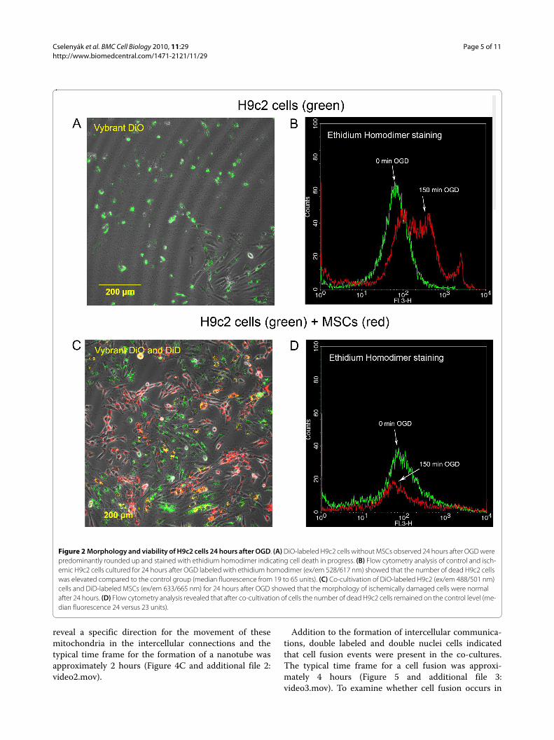
Co-cultivation of cardiomyoblasts and MSCs after OGDConfocal microscopy showed that cardiomyoblasts cul-tured alone displayed the same rounded and blebbedmorphology immediately following as well as 24 hoursafter OGD (Figure 2A). Flow cytometry analysis showedthat OGD significantly increased the cell death rate inthis group as shown by the enhanced ethidium homodi-mer fluorescence intensity (median fluorescence from 19to 65 units, Figure 2B). Figure 2B also shows that a por-tion of cells remained unstained with ethidium homodi-
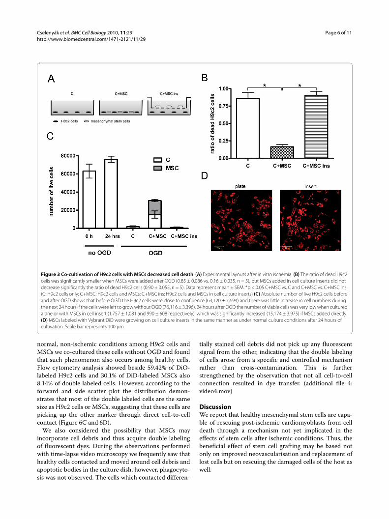
mer even after 150 minutes due to the variability of themodel. When MSCs were added to post-ischemic cardio-myoblasts, the morphology of the damaged cells was sim-ilar to cells cultured in normal conditions without OGD(Figure 2C). In this group, flow cytometry analysisrevealed that the deleterious results of ischemia weredecreased (median fluorescence 24 versus 23 units, Fig-ure 2D). To quantify the effect of added MSCs, confocalimages were used. This approach revealed that the ratioof dead H9c2 cells to all H9c2 cells in the wells 24 hoursafter OGD was significantly higher when the cardiomyo-blasts were cultured alone compared to when healthyMSCs were added to the cultures 30 minutes after OGD(0.85 ± 0.086 vs. 0.16 ± 0.035, respectively, p < 0.05, n =5). MSCs added in cell culture inserts, which physicallyseparate the two cell populations growing in the samemedium, failed to decrease the ratio of dead cells (0.90 ±0.055, n = 5), indicating that the rescue factor is less likelyto be a soluble one and direct cell-to-cell contact may berequired (Figure 3A and 3B). The absolute number of liveH9c2 cells before and after OGD and the number ofadded MSCs after OGD was also investigated. BeforeOGD H9c2 cells were close to confluence (63,120 ±7,694) and there was little increase in cell numbers duringthe next 24 hours if the cells were left to grow withoutOGD (76,116 ± 3,396). The number of viable cells 24hours after OGD was very low when cultured alone orwith MSCs in cell insert (1,757 ± 1,081 and 990 ± 608respectively), but significantly increased (15,174 ± 3,975)if MSCs were added directly. It can be assumed that theinjured H9c2 cells were washed out during mediumchange so only a part of H9c2 cells remained in the wells(Figure 3C). We also examined the inserts with confocalmicroscopy to eliminate the possibility of decreased MSCviability on the cell culture inserts. MSCs were labeledwith Vybrant DiD before seeding on the cell inserts. Wefound that Vybrant DiD labeled MSCs were attached tothe surface of the inserts and showed normal cell mor-phology (Figure 3D). We also investigated whether any ofthe MSCs had contaminated the underlying H9c2 cardio-myoblast culture, but found no trace of MSCs among theH9c2 cells; therefore no direct cell-to-cell contact couldbe formed (data not shown).
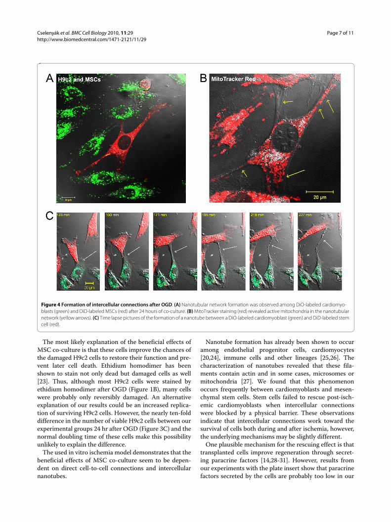
Confocal microscopy and flow cytometryDevelopment of intercellular connections, so-called nan-otubes, between cardiomyoblasts and MSCs during the24 hr period after OGD was frequently observed (Figure4A). These nanotubes were long enough to span dis-tances of several cell diameters, and their diameters werebetween 200 and 500 nm. MitoTracker Red stainingrevealed that these nanotubes connecting stem cells tocardiomyoblasts contained functionally active mitochon-dria (Figure 4B). Time lapse video microscopy did not

Cselenyák et al. BMC Cell Biology 2010, 11:29http://www.biomedcentral.com/1471-2121/11/29
Page 4 of 11
Figure 1 Ischemia model on cardiomyoblasts. (A) Follow up of OGD on cardiomyoblasts. Cells were stained with calcein-AM (ex/em 494/517 nm) for live cells (green) and ethidium homodimer (ex/em 528/617 nm) for dead cells (red). (B) Flow cytometry analysis of control and ischemic cardio-myoblasts labeled with ethidium homodimer after OGD. The green curve represents the control cardiomyoblasts and the red shows the ischemic cardiomyoblasts. The complete rightward shift of the red curve based on these representative data indicates that OGD increased the number of dead cells nearly maximally.
Cselenyák et al. BMC Cell Biology 2010, 11:29http://www.biomedcentral.com/1471-2121/11/29
Page 5 of 11
reveal a specific direction for the movement of thesemitochondria in the intercellular connections and thetypical time frame for the formation of a nanotube wasapproximately 2 hours (Figure 4C and additional file 2:video2.mov).
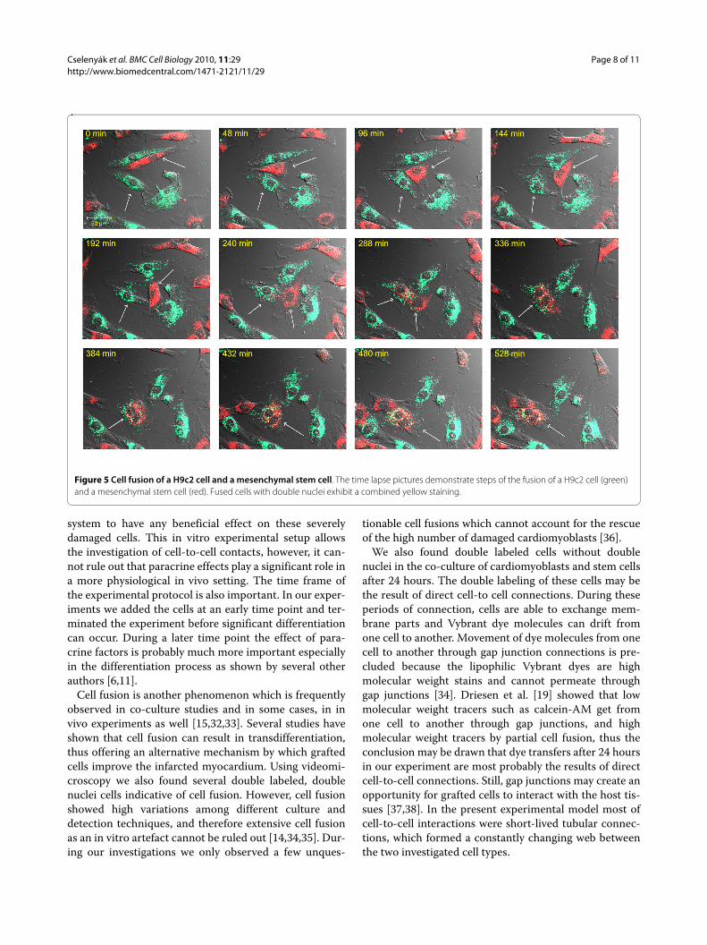
Addition to the formation of intercellular communica-tions, double labeled and double nuclei cells indicatedthat cell fusion events were present in the co-cultures.The typical time frame for a cell fusion was approxi-mately 4 hours (Figure 5 and additional file 3:video3.mov). To examine whether cell fusion occurs in
Figure 2 Morphology and viability of H9c2 cells 24 hours after OGD. (A) DiO-labeled H9c2 cells without MSCs observed 24 hours after OGD were predominantly rounded up and stained with ethidium homodimer indicating cell death in progress. (B) Flow cytometry analysis of control and isch-emic H9c2 cells cultured for 24 hours after OGD labeled with ethidium homodimer (ex/em 528/617 nm) showed that the number of dead H9c2 cells was elevated compared to the control group (median fluorescence from 19 to 65 units). (C) Co-cultivation of DiO-labeled H9c2 (ex/em 488/501 nm) cells and DiD-labeled MSCs (ex/em 633/665 nm) for 24 hours after OGD showed that the morphology of ischemically damaged cells were normal after 24 hours. (D) Flow cytometry analysis revealed that after co-cultivation of cells the number of dead H9c2 cells remained on the control level (me-dian fluorescence 24 versus 23 units).
Cselenyák et al. BMC Cell Biology 2010, 11:29http://www.biomedcentral.com/1471-2121/11/29
Page 6 of 11
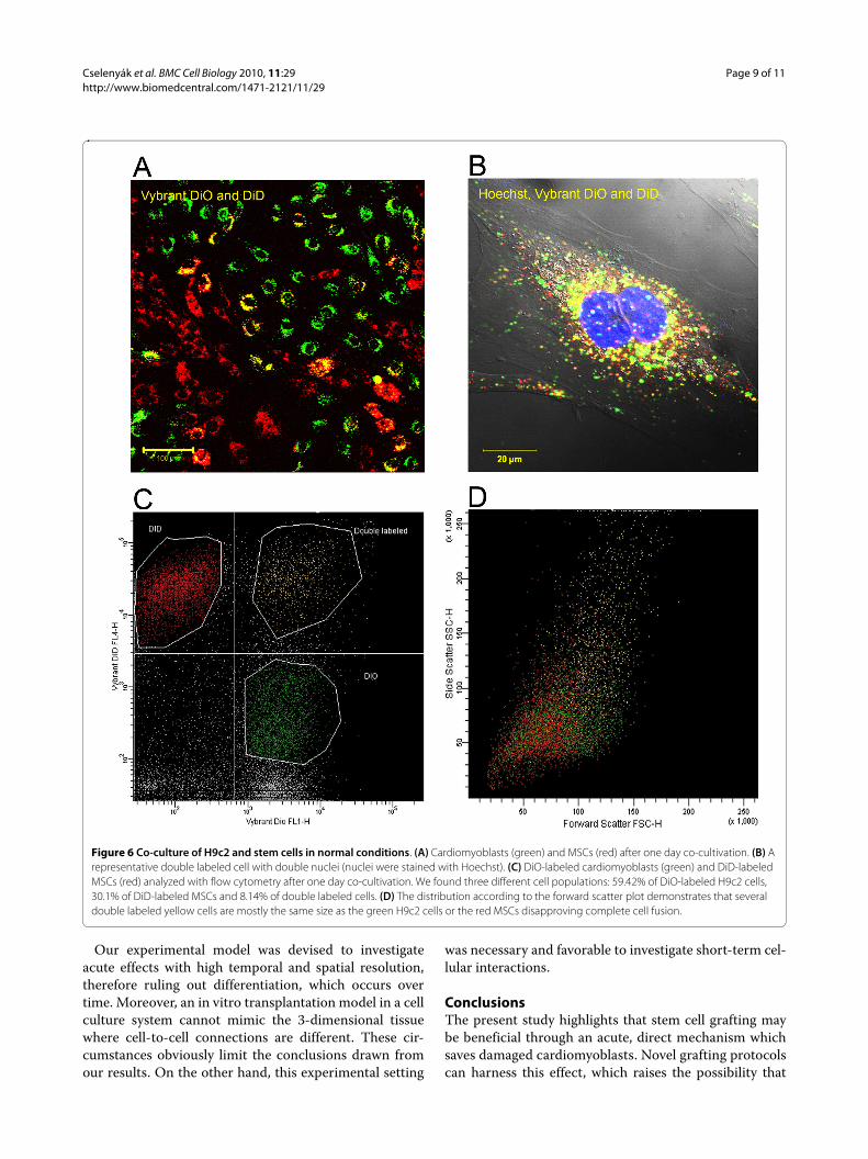
normal, non-ischemic conditions among H9c2 cells andMSCs we co-cultured these cells without OGD and foundthat such phenomenon also occurs among healthy cells.Flow cytometry analysis showed beside 59.42% of DiO-labeled H9c2 cells and 30.1% of DiD-labeled MSCs also8.14% of double labeled cells. However, according to theforward and side scatter plot the distribution demon-strates that most of the double labeled cells are the samesize as H9c2 cells or MSCs, suggesting that these cells arepicking up the other marker through direct cell-to-cellcontact (Figure 6C and 6D).
We also considered the possibility that MSCs mayincorporate cell debris and thus acquire double labelingof fluorescent dyes. During the observations performedwith time-lapse video microscopy we frequently saw thathealthy cells contacted and moved around cell debris andapoptotic bodies in the culture dish, however, phagocyto-sis was not observed. The cells which contacted differen-
tially stained cell debris did not pick up any fluorescentsignal from the other, indicating that the double labelingof cells arose from a specific and controlled mechanismrather than cross-contamination. This is furtherstrengthened by the observation that not all cell-to-cellconnection resulted in dye transfer. (additional file 4:video4.mov)
DiscussionWe report that healthy mesenchymal stem cells are capa-ble of rescuing post-ischemic cardiomyoblasts from celldeath through a mechanism not yet implicated in theeffects of stem cells after ischemic conditions. Thus, thebeneficial effect of stem cell grafting may be based notonly on improved neovascularisation and replacement oflost cells but on rescuing the damaged cells of the host aswell.
Figure 3 Co-cultivation of H9c2 cells with MSCs decreased cell death. (A) Experimental layouts after in vitro ischemia. (B) The ratio of dead H9c2 cells was significantly smaller when MSCs were added after OGD (0.85 ± 0.086 vs. 0.16 ± 0.035, n = 5), but MSCs added in cell culture inserts did not decrease significantly the ratio of dead H9c2 cells (0.90 ± 0.055, n = 5). Data represent mean ± SEM. *p < 0.05 C+MSC vs. C and C+MSC vs. C+MSC ins. (C: H9c2 cells only; C+MSC: H9c2 cells and MSCs; C+MSC ins: H9c2 cells and MSCs in cell culture inserts) (C) Absolute number of live H9c2 cells before and after OGD shows that before OGD the H9c2 cells were close to confluence (63,120 ± 7,694) and there was little increase in cell numbers during the next 24 hours if the cells were left to grow without OGD (76,116 ± 3,396). 24 hours after OGD the number of viable cells was very low when cultured alone or with MSCs in cell insert (1,757 ± 1,081 and 990 ± 608 respectively), which was significantly increased (15,174 ± 3,975) if MSCs added directly. (D) MSCs labeled with Vybrant DiD were growing on cell culture inserts in the same manner as under normal culture conditions after 24 hours of cultivation. Scale bar represents 100 μm.
Cselenyák et al. BMC Cell Biology 2010, 11:29http://www.biomedcentral.com/1471-2121/11/29
Page 7 of 11
The most likely explanation of the beneficial effects ofMSC co-culture is that these cells improve the chances ofthe damaged H9c2 cells to restore their function and pre-vent later cell death. Ethidium homodimer has beenshown to stain not only dead but damaged cells as well[23]. Thus, although most H9c2 cells were stained byethidium homodimer after OGD (Figure 1B), many cellswere probably only reversibly damaged. An alternativeexplanation of our results could be an increased replica-tion of surviving H9c2 cells. However, the nearly ten-folddifference in the number of viable H9c2 cells between ourexperimental groups 24 hr after OGD (Figure 3C) and thenormal doubling time of these cells make this possibilityunlikely to explain the difference.
The used in vitro ischemia model demonstrates that thebeneficial effects of MSC co-culture seem to be depen-dent on direct cell-to-cell connections and intercellularnanotubes.
Nanotube formation has already been shown to occuramong endothelial progenitor cells, cardiomyocytes[20,24], immune cells and other lineages [25,26]. Thecharacterization of nanotubes revealed that these fila-ments contain actin and in some cases, microsomes ormitochondria [27]. We found that this phenomenonoccurs frequently between cardiomyoblasts and mesen-chymal stem cells. Stem cells failed to rescue post-isch-emic cardiomyoblasts when intercellular connectionswere blocked by a physical barrier. These observationsindicate that intercellular connections work toward thesurvival of cells both during and after ischemia, however,the underlying mechanisms may be slightly different.
One plausible mechanism for the rescuing effect is thattransplanted cells improve regeneration through secret-ing paracrine factors [14,28-31]. However, results fromour experiments with the plate insert show that paracrinefactors secreted by the cells are probably too low in our
Figure 4 Formation of intercellular connections after OGD. (A) Nanotubular network formation was observed among DiO-labeled cardiomyo-blasts (green) and DiD-labeled MSCs (red) after 24 hours of co-culture. (B) MitoTracker staining (red) revealed active mitochondria in the nanotubular network (yellow arrows). (C) Time lapse pictures of the formation of a nanotube between a DiO-labeled cardiomyoblast (green) and DiD-labeled stem cell (red).
Cselenyák et al. BMC Cell Biology 2010, 11:29http://www.biomedcentral.com/1471-2121/11/29
Page 8 of 11
system to have any beneficial effect on these severelydamaged cells. This in vitro experimental setup allowsthe investigation of cell-to-cell contacts, however, it can-not rule out that paracrine effects play a significant role ina more physiological in vivo setting. The time frame ofthe experimental protocol is also important. In our exper-iments we added the cells at an early time point and ter-minated the experiment before significant differentiationcan occur. During a later time point the effect of para-crine factors is probably much more important especiallyin the differentiation process as shown by several otherauthors [6,11].
Cell fusion is another phenomenon which is frequentlyobserved in co-culture studies and in some cases, in invivo experiments as well [15,32,33]. Several studies haveshown that cell fusion can result in transdifferentiation,thus offering an alternative mechanism by which graftedcells improve the infarcted myocardium. Using videomi-croscopy we also found several double labeled, doublenuclei cells indicative of cell fusion. However, cell fusionshowed high variations among different culture anddetection techniques, and therefore extensive cell fusionas an in vitro artefact cannot be ruled out [14,34,35]. Dur-ing our investigations we only observed a few unques-
tionable cell fusions which cannot account for the rescueof the high number of damaged cardiomyoblasts [36].
We also found double labeled cells without doublenuclei in the co-culture of cardiomyoblasts and stem cellsafter 24 hours. The double labeling of these cells may bethe result of direct cell-to cell connections. During theseperiods of connection, cells are able to exchange mem-brane parts and Vybrant dye molecules can drift fromone cell to another. Movement of dye molecules from onecell to another through gap junction connections is pre-cluded because the lipophilic Vybrant dyes are highmolecular weight stains and cannot permeate throughgap junctions [34]. Driesen et al. [19] showed that lowmolecular weight tracers such as calcein-AM get fromone cell to another through gap junctions, and highmolecular weight tracers by partial cell fusion, thus theconclusion may be drawn that dye transfers after 24 hoursin our experiment are most probably the results of directcell-to-cell connections. Still, gap junctions may create anopportunity for grafted cells to interact with the host tis-sues [37,38]. In the present experimental model most ofcell-to-cell interactions were short-lived tubular connec-tions, which formed a constantly changing web betweenthe two investigated cell types.
Figure 5 Cell fusion of a H9c2 cell and a mesenchymal stem cell. The time lapse pictures demonstrate steps of the fusion of a H9c2 cell (green) and a mesenchymal stem cell (red). Fused cells with double nuclei exhibit a combined yellow staining.
Cselenyák et al. BMC Cell Biology 2010, 11:29http://www.biomedcentral.com/1471-2121/11/29
Page 9 of 11
Our experimental model was devised to investigateacute effects with high temporal and spatial resolution,therefore ruling out differentiation, which occurs overtime. Moreover, an in vitro transplantation model in a cellculture system cannot mimic the 3-dimensional tissuewhere cell-to-cell connections are different. These cir-cumstances obviously limit the conclusions drawn fromour results. On the other hand, this experimental setting
was necessary and favorable to investigate short-term cel-lular interactions.
ConclusionsThe present study highlights that stem cell grafting maybe beneficial through an acute, direct mechanism whichsaves damaged cardiomyoblasts. Novel grafting protocolscan harness this effect, which raises the possibility that
Figure 6 Co-culture of H9c2 and stem cells in normal conditions. (A) Cardiomyoblasts (green) and MSCs (red) after one day co-cultivation. (B) A representative double labeled cell with double nuclei (nuclei were stained with Hoechst). (C) DiO-labeled cardiomyoblasts (green) and DiD-labeled MSCs (red) analyzed with flow cytometry after one day co-cultivation. We found three different cell populations: 59.42% of DiO-labeled H9c2 cells, 30.1% of DiD-labeled MSCs and 8.14% of double labeled cells. (D) The distribution according to the forward scatter plot demonstrates that several double labeled yellow cells are mostly the same size as the green H9c2 cells or the red MSCs disapproving complete cell fusion.
Cselenyák et al. BMC Cell Biology 2010, 11:29http://www.biomedcentral.com/1471-2121/11/29
Page 10 of 11
stem cells given early and locally can preserve heart tissuerather than simply help to replace what is already lost.
Additional material
Authors' contributionsACS and ZSL conceived the study. ACS completed the majority of the confocalmicroscopy experiments, made the flow cytometry measurements, performedstatistical analysis, and helped draft the manuscript. EP helped draft the manu-script and performed statistical analysis. EMH participated in the flow cytome-ter measurements. KL participated in the time lapse video microscopyexperiments and helped draft the manuscript. In addition to collaborating onthe conception of the study, ZSL participated in the study design, providedcoordination among the researchers and experiments, and helped draft themanuscript. All authors read and approved the final manuscript.
AcknowledgementsThis work was supported by OTKA (Hungarian Scientific Research Fund) D45933, T049621, TÉT (Hungarian Science and Technology Foundation) A4/04 and Arg-17/2006, Bolyai, Öveges Fellowships and TÁMOP 4.2.2-08/1/KMR-2008-0004. We would like to thank Dr. Ferenc Uher for providing the mouse mesenchymal stem cells. We are grateful to Nancy Busija for copyediting the manuscript and we thank David Busija and Márk Kollai for critically revising the manuscript.
Author DetailsInstitute of Human Physiology and Clinical Experimental Research, Semmelweis University, H-1094, Tűzoltó utca 37-47, Budapest, Hungary
References1. Dayer M, Cowie MR: Heart failure: diagnosis and healthcare burden.
Clin Med 2004, 4(1):13-18.2. Kesteloot H, Sans S, Kromhout D: Dynamics of cardiovascular and all-
cause mortality in Western and Eastern Europe between 1970 and 2000. Eur Heart J 2006, 27(1):107-113.
3. Antonio Giordano UGIRM: From the laboratory bench to the patient's bedside: An update on clinical trials with mesenchymal stem cells. Journal of Cellular Physiology 2007, 211(1):27-35.
4. Stefan Bajada IMJBRNA: Updates on stem cells and their applications in regenerative medicine. Journal of Tissue Engineering and Regenerative Medicine 2008, 2(4):169-183.
5. Jawad H, Lyon AR, Harding SE, Ali NN, Boccaccini AR: Myocardial tissue engineering. Br Med Bull 2008, 87(1):31-47.
6. Dai W, Hale SL, Martin BJ, Kuang J-Q, Dow JS, Wold LE, Kloner RA: Allogeneic Mesenchymal Stem Cell Transplantation in Postinfarcted Rat Myocardium: Short- and Long-Term Effects. Circulation 2005, 112(2):214-223.
7. Orlic D, Kajstura J, Chimenti S, Jakoniuk I, Anderson SM, Li B, Pickel J, McKay R, Nadal-Ginard B, Bodine DM, et al.: Bone marrow cells regenerate infarcted myocardium. Nature 2001, 410(6829):701.
8. Collins SD, Baffour R, Waksman R: Cell therapy in myocardial infarction. Cardiovascular Revascularization Medicine 2007, 8(1):43.
9. Dimmeler S, Burchfield J, Zeiher AM: Cell-Based Therapy of Myocardial Infarction. Arterioscler Thromb Vasc Biol 2007.
10. Sadat S, Gehmert S, Song YH, Yen Y, Bai X, Gaiser S, Klein H, Alt E: The cardioprotective effect of mesenchymal stem cells is mediated by IGF-I and VEGF. Biochem Biophys Res Commun 2007, 363(3):674-679.
11. Kinnaird T, Stabile E, Burnett MS, Lee CW, Barr S, Fuchs S, Epstein SE: Marrow-derived stromal cells express genes encoding a broad spectrum of arteriogenic cytokines and promote in vitro and in vivo arteriogenesis through paracrine mechanisms. Circ Res 2004, 94(5):678-685.
12. Masuda S, Shimizu T, Yamato M, Okano T: Cell sheet engineering for heart tissue repair. Adv Drug Deliv Rev 2008, 60(2):277-285.
13. Yoon J, Shim WJ, Ro YM, Lim DS: Transdifferentiation of mesenchymal stem cells into cardiomyocytes by direct cell-to-cell contact with neonatal cardiomyocyte but not adult cardiomyocytes. Ann Hematol 2005, 84(11):715-721.
14. Menasche P: You Can't Judge a Book by Its Cover. Circulation 2006, 113(10):1275-1277.
15. Nygren JM, Jovinge S, Breitbach M, Sawen P, Roll W, Hescheler J, Taneera J, Fleischmann BK, Jacobsen SE: Bone marrow-derived hematopoietic cells generate cardiomyocytes at a low frequency through cell fusion, but not transdifferentiation. Nat Med 2004, 10(5):494-501.
16. Murry CE, Soonpaa MH, Reinecke H, Nakajima H, Nakajima HO, Rubart M, Pasumarthi KB, Virag JI, Bartelmez SH, Poppa V, et al.: Haematopoietic stem cells do not transdifferentiate into cardiac myocytes in myocardial infarcts. Nature 2004, 428(6983):664-668.
17. Alvarez-Dolado M, Pardal R, Garcia-Verdugo JM, Fike JR, Lee HO, Pfeffer K, Lois C, Morrison SJ, Alvarez-Buylla A: Fusion of bone-marrow-derived cells with Purkinje neurons, cardiomyocytes and hepatocytes. Nature 2003, 425(6961):968.
18. Kajstura J, Rota M, Whang B, Cascapera S, Hosoda T, Bearzi C, Nurzynska D, Kasahara H, Zias E, Bonafe M, et al.: Bone marrow cells differentiate in cardiac cell lineages after infarction independently of cell fusion. Circ Res 2005, 96(1):127-137.
19. Driesen RB, Dispersyn GD, Verheyen FK, Eijnde SM van den, Hofstra L, Thone F, Dijkstra P, Debie W, Borgers M, Ramaekers FC: Partial cell fusion: a newly recognized type of communication between dedifferentiating cardiomyocytes and fibroblasts. Cardiovasc Res 2005, 68(1):37-46.
20. Plotnikov EY, Khryapenkova TG, Vasileva AK, Marey MV, Galkina SI, Isaev NK, Sheval EV, Polyakov VY, Sukhikh GT, Zorov DB: Cell-to-cell cross-talk between mesenchymal stem cells and cardiomyocytes in co-culture. J Cell Mol Med 2008, 12(5A):1622-1631.
21. Tropel P, Noel D, Platet N, Legrand P, Benabid AL, Berger F: Isolation and characterisation of mesenchymal stem cells from adult mouse bone marrow. Exp Cell Res 2004, 295(2):395-406.
22. Urbán VS, Kiss J, Kovács J, Gócza E, Vas V, Edot , Monostori v, Uher F: Mesenchymal Stem Cells Cooperate with Bone Marrow Cells in Therapy of Diabetes. Stem Cells 2008, 26(1):244-253.
23. Kaplan LD: The analysis of articular cartilage after thermal exposure: "Is red really dead?". Arthroscopy 2003, 19(3):310-313.
24. Koyanagi M, Brandes RP, Haendeler J, Zeiher AM, Dimmeler S: Cell-to-Cell Connection of Endothelial Progenitor Cells With Cardiac Myocytes by Nanotubes: A Novel Mechanism for Cell Fate Changes? Circ Res 2005, 96(10):1039-1041.
25. Onfelt B, Nedvetzki S, Yanagi K, Davis DM: Cutting edge: Membrane nanotubes connect immune cells. J Immunol 2004, 173(3):1511-1513.
26. Onfelt B, Purbhoo MA, Nedvetzki S, Sowinski S, Davis DM: Long-distance calls between cells connected by tunneling nanotubules. Sci STKE 2005, 2005(313):pe55.
27. Spees JL, Olson SD, Whitney MJ, Prockop DJ: Mitochondrial transfer between cells can rescue aerobic respiration. Proc Natl Acad Sci USA 2006, 103(5):1283-1288.
28. Gnecchi M, Zhang Z, Ni A, Dzau VJ: Paracrine Mechanisms in Adult Stem Cell Signaling and Therapy. Circ Res 2008, 103(11):1204-1219.
29. Paul WMF: Paracrine Effects of Cell Transplantation: Modifying Ventricular Remodeling in the Failing Heart. Seminars in thoracic and cardiovascular surgery 2008, 20(2):87.
30. Takahashi M, Li TS, Suzuki R, Kobayashi T, Ito H, Ikeda Y, Matsuzaki M, Hamano K: Cytokines produced by bone marrow cells can contribute to functional improvement of the infarcted heart by protecting cardiomyocytes from ischemic injury. Am J Physiol Heart Circ Physiol 2006, 291(2):H886-893.
Additional file 1 Follow up of 150 minutes long OGD on cardiomyo-blasts with time lapse video microscopy.Additional file 2 Nanotubular network formation among DiO-labeled cardiomyoblasts (green) and DiD-labeled MSCs (red) after 24 hours of co-culture.Additional file 3 Time lapse video microscopy pictures demonstrates fusion of a H9c2 cell (green) and a mesenchymal stem cell (red).Additional file 4 14 hours of time lapse video microscopy of H9c2 cells (green) and MSCs (red) after OGD.
Received: 2 September 2009 Accepted: 20 April 2010 Published: 20 April 2010This article is available from: http://www.biomedcentral.com/1471-2121/11/29© 2010 Cselenyák et al; licensee BioMed Central Ltd. This is an Open Access article distributed under the terms of the Creative Commons Attribution License (http://creativecommons.org/licenses/by/2.0), which permits unrestricted use, distribution, and reproduction in any medium, provided the original work is properly cited.BMC Cell Biology 2010, 11:29
Cselenyák et al. BMC Cell Biology 2010, 11:29http://www.biomedcentral.com/1471-2121/11/29
Page 11 of 11
31. Uemura R, Xu M, Ahmad N, Ashraf M: Bone marrow stem cells prevent left ventricular remodeling of ischemic heart through paracrine signaling. Circ Res 2006, 98(11):1414-1421.
32. Ishikawa F, Shimazu H, Shultz LD, Fukata M, Nakamura R, Lyons B, Shimoda K, Shimoda S, Kanemaru T, Nakamura K, et al.: Purified human hematopoietic stem cells contribute to the generation of cardiomyocytes through cell fusion. Faseb J 2006, 20(7):950-952.
33. Lacza Z, Horvath E, Busija DW: Neural stem cell transplantation in cold lesion: a novel approach for the investigation of brain trauma and repair. Brain Research Protocols 2003, 11(3):145.
34. Garbade J, Schubert A, Rastan AJ, Lenz D, Walther T, Gummert JF, Dhein S, Mohr F-W: Fusion of bone marrow-derived stem cells with cardiomyocytes in a heterologous in vitro model. European Journal of Cardio-Thoracic Surgery 2005, 28(5):685.
35. Kajstura J, Rota M, Whang B, Cascapera S, Hosoda T, Bearzi C, Nurzynska D, Kasahara H, Zias E, Bonafe M, et al.: Bone Marrow Cells Differentiate in Cardiac Cell Lineages After Infarction Independently of Cell Fusion. Circ Res 2005, 96(1):127-137.
36. Rose RA, Jiang H, Wang X, Helke S, Tsoporis JN, Gong N, Keating SC, Parker TG, Backx PH, Keating A: Bone marrow-derived mesenchymal stromal cells express cardiac-specific markers, retain the stromal phenotype, and do not become functional cardiomyocytes in vitro. Stem Cells 2008, 26(11):2884-2892.
37. Horackova M, Arora R, Chen R, Armour JA, Cattini PA, Livingston R, Byczko Z: Cell transplantation for treatment of acute myocardial infarction: unique capacity for repair by skeletal muscle satellite cells. Am J Physiol Heart Circ Physiol 2004, 287(4):H1599-1608.
38. Xu M, Wani M, Dai YS, Wang J, Yan M, Ayub A, Ashraf M: Differentiation of bone marrow stromal cells into the cardiac phenotype requires intercellular communication with myocytes. Circulation 2004, 110(17):2658-2665.
doi: 10.1186/1471-2121-11-29Cite this article as: Cselenyák et al., Mesenchymal stem cells rescue cardio-myoblasts from cell death in an in vitro ischemia model via direct cell-to-cell connections BMC Cell Biology 2010, 11:29