Devonian climate and reef evolution: Insights from oxygen isotopes in apatite

Acta Orthop Scand 2002; 73 (2): 157–164 157
Mechanical evaluation of a carbonated apatite cement in the � xation of unstable intertrochanteric fractures
Duran N Yetkinler1, Stuart B Goodman2, Eric S Reindel3, Dennis Carter4, Robert D Poser5 and Brent R Constantz4
1ArthroCare Corporation, 680 Vaqueros Ave., Sunnyvale, CA 94085, 2Stanford University School of Medicine, Division of Orthopaedic Surgery, Stanford, CA, 94305, 3KMI, Kinetikos Medical Incorporated, San Diego, CA 92024, 4Stanford University, Biomechanical Engineering Division, Stanford, CA, 94305, 5Corazon Corp., 199 Jefferson Dr., Menlo Park, CA, 94025, USA. E-mail: [email protected] 00-10-16. Accepted 01-09-13
Copyright © Taylor & Francis 2002. ISSN 0001–6470. Printed in Sweden – all rights reserved.
ABSTRACT – We created three-part unstable inter-trochanteric fractures in 6 pairs of aged, osteopenic, human, cadaveric femora. Fractures were reduced and � xed with a Dynamic Hip Screw (DHS) (Synthes, Paoli, PA). Two test groups were evaluated: 1. Fixation with a DHS, and 2. Fixation with a DHS and calcium phosphate bone cement (Norian SRS (Skeletal Repair System)) augmentation of the fracture line and posteromedial calcar region of the proximal femur. Each femur was loaded to 1,650 N (2.5 body weight) for 10,000 cycles to simulate postoperative load transmission across the fracture construct during normal gait. The load was further increased successively by one body weight for another 10,000 cycles until failure. We evaluated � xa-tion by measuring the amount of sliding of the lag screw of the DHS (shortening) and stiffness of the overall frac-ture construct (stability). SRS cement-augmented spec-imens had less shortening (1 mm versus 17 mm) and twice the initial construct stiffness compared to control specimens.
n
Intertrochanteric fractures are usually � xated by a sliding hip screw device. Normal forces around the hip tend to cause impaction of the proximal and distal fragments by sliding of the lag screw within the barrel. Thus, the device provides con-trolled impaction across the fracture site resulting in a more stable construct. However, controlled impaction of unstable fractures can result in excessive shortening of the limb, sub-optimal hip
mechanics, and altered gait. Unstable intertro-chanteric fractures are usually associated with a fractured lesser trochanteric region. Despite the mechanical advantage of re-establishing the load-transferring ability of this region, this posterome-dial fragment often cannot be reduced and stabi-lized surgically without extensive dissection and periosteal stripping.
Norian SRS is an injectable, biodegradable bone cement that cures at physiologic temperature and pH with high compressive strength, and crystal-lographic and chemical characteristics similar to the mineral phase of bone. We hypothesized that accurate reduction and � xation of intertrochanteric fractures using a sliding hip screw, augmented with Norian SRS cement injected into areas of commi-nution and compromised cancellous bone in the proximal femur, especially in the lesser trochan-teric area, would provide a more stable mechani-cal environment then the sliding hip screw alone. We tested this hypothesis in a cadaveric mechani-cal study by measuring the sliding of the hip screw (shortening) and the stiffness (stability) of the frac-ture construct.
Material and methods
Material
Norian SRS cement Skeletal Repair System is an injectable, fast setting, high-strength, cancel-lous bone cement that cures in situ to form a
Act
a O
rtho
p D
ownl
oade
d fr
om in
form
ahea
lthca
re.c
om b
y 92
.43.
140.
122
on 0
5/20
/14
For
pers
onal
use
onl
y.

158 Acta Orthop Scand 2002; 73 (2): 157–164
carbonated apatite of low crystalline order and small grain size similar to the mineral phase of bone (Constantz et al. 1995). The cement paste is formed by mixing a phosphate-buffered solution with a powder consisting of calcium carbonate (CaCO3), a -tricalcium phosphate (Ca3(PO4)2), and monocalcium phosphate mono-hydrate (Ca(H2PO4)2.H20). Once implanted, the material equilibrates to body temperature, accel-erating the formation of carbonated apatite. It hardens in about 10 minutes after implantation at body temperature. At 1 hour it acquires 50% of its ultimate compressive strength. Full compressive strength of 55 megapascals is attained about 12 hours after implantation (Constantz et al. 1995). It has been shown to remodel and to be replaced by host bone while maintaining structural integ-rity in the implanted region (Frankenburg et al. 1998).
Specimen selection
6 pairs of fresh frozen human adult cadaveric femora were prescreened with anteroposterior radiographs to exclude gross anatomical pathology. All femora were wrapped in saline soaked towels and stored in tightly sealed plastic bags at or below –20 °C. All specimens were defrosted for at least 8 hours at room temperature prior to radiographic assessment, bone density measurements, and cre-ation of the intertrochanteric fracture. Screening anteroposterior and lateral radiographs of unfro-zen specimens were taken as a baseline. Bone min-eral density (BMD) of the intertrochanteric region was determined using Hologic 4500 (Hologic Inc., Waltham, MA) dual energy x-ray absorptiometry (DEXA).
Specimen preparation
Femora were cleaned of all soft tissue and tran-sected 23 cm below the distal aspect of the lesser trochanter. The distal end of each specimen was potted with dental acrylic (PERM Reline and Repair Resin, Hygenic Corp., Akron, OH) in a 15 cm section of 6 cm diameter tubular aluminum tube stock. In addition, an acrylic acetabular cup was molded by using individual femoral heads as a template. These acrylic cups were used during test-ing to simulate in vivo loading conditions around the femoral head.
Fracture creation
A three-part intertrochanteric fracture was created by making 1.5 mm drill holes 2–3 mm apart in the cortex of the proximal femur along the inter-trochanteric line anteriorly, through the superior aspect of the greater trochanter, and along the inter-trochanteric crest posteriorly. The drill holes were continued above and below the lesser trochanter to de� ne the posteromedial fragment of the fracture. The specimen was then mounted on an MTS ser-vohydraulic machine and loaded axially to create a three-part intertrochanteric fracture. If the lesser trochanteric fragment did not form, an osteotome was used to displace the lesser trochanter from the femur.
Fracture � xation
1 specimen from each pair was randomly assigned to be the experimental side with internal � xation using a sliding hip screw augmented with Norian SRS cement, and the contralateral side was assigned to be a control specimen with the sliding hip screw alone. The standard surgical technique for implant-ing a sliding hip screw and four hole sideplate (Dynamic Hip Screw, Synthes, Paoli, PA) was car-ried out on the fractured femur by an experienced orthopedic surgeon. After placement of a guide pin, drilling, reaming, and tapping for the lag screw were carried out. After inserting the lag screw and 135º four hole sideplate, 4.5 mm sideplate screws were placed.
In the experimental group, before placement of the sideplate, loose cancellous bone in the inter-trochanteric region was impacted using a tamp through the lag screw hole. SRS cement was then injected along the shaft of the lag screw into the intertrochanteric region with either a straight 2.7 mm or 2.0 mm needle under image intensi� cation. The sideplate was immediately guided over the lag screw and secured to the lateral aspect of the femur with a self-tapping 4.5 mm cortical screw through the second most proximal screw hole in the sideplate. All remaining sideplate screws, except the most proximal, were inserted in the sideplate. A 4.5 mm drill was then used to direct a hole through the proximal screw hole of the plate towards the lesser trochanter area. A curved probe was used through this hole to create a pathway to the lesser trochanter region while visualizing
Act
a O
rtho
p D
ownl
oade
d fr
om in
form
ahea
lthca
re.c
om b
y 92
.43.
140.
122
on 0
5/20
/14
For
pers
onal
use
onl
y.
Acta Orthop Scand 2002; 73 (2): 157–164 159
Figure 1. Anteroposterior and lateral radiographs of paired experimental (� gures on the left) and control specimens (� gures on the right). Note the posteromedial defect resulting from the displaced lesser tro-chanter on the � gure at the right bottom and the same region � lled with Norian SRS cement on the � gure at the left bottom.
mm drill and the proximal sideplate screw was quickly inserted to avoid disruption of the cement while setting. In both experimental and control groups, the posteromedial lesser trochanteric frag-ment was not replaced (Figure 1). Lateral-medial � uoroscopic images were essential to visualize adequately posteromedial void preparation and � lling.
this area � uoroscopically. The pathway and the posteromedial region were cleared of debris using this probe and lavaged. The posteromedial defect in the area of the lesser trochanter was then � lled with Norian SRS cement using a curved 2.7 mm needle inserted through the proximal screw hole in the plate oriented posteromedially. The medial cortex of the femur was drilled with a 3.2
Act
a O
rtho
p D
ownl
oade
d fr
om in
form
ahea
lthca
re.c
om b
y 92
.43.
140.
122
on 0
5/20
/14
For
pers
onal
use
onl
y.
160 Acta Orthop Scand 2002; 73 (2): 157–164
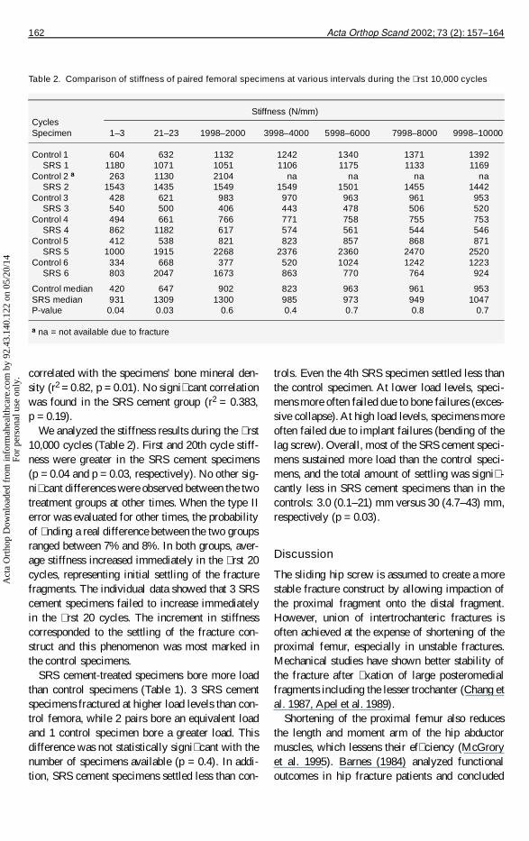
Table 1. Comparison of survival of paired femoral specimens at 10,000 cyclic loading intervals with 2.5, 3.5, 4.5 and 5.5 body weights
Specimen # DEXA 2.5 BW 3.5 BW (g/cm2) Comp. Sliding Total Vertical Comp. Sliding Total cycles (mm) sliding slidinga cycles (mm) sliding (mm) (mm) (mm)
SRS 1 0.97 10,000 2.1 2.1 3.0 10,000 0.7 2.8 Control 1 0.99 10,000 29 29 26 2,804 7.5 36 SRS 2 0.56 10,000 0.1 0.1 1.8 10,000 21 21 Control 2 0.57 3,808 43 43 56 N/A N/A N/A SRS 3 0.86 10,000 1.0 1.0 4.9 10,000 0.5 1.5 Control 3 0.90 10,000 2.9 2.9 3.8 10,000 0.7 3.6 SRS 4 0.80 10,000 0.1 0.1 3.9 1,300 0.0 0.2 Control 4 0.68 10,000 4.8 4.8 7.2 10,000 7.1 12 SRS 5 0.58 10,000 0.1 0.1 1.2 10,000 0.0 0.1 Control 5 0.62 10,000 4.8 4.8 7.2 10,000 0.7 5.5 SRS 6 0.76 10,000 1.0 1.0 3.5 10,000 1.6 2.7 Control 6 0.76 10,000 28 28 26 N/A N/A N/A
a Vertical displacement only calculated for 2.5 BW testing.
All test femora were immersed in a container with 1 L phosphate-buffered solution at 37 degrees Celsius and placed in an incubator which main-tains 100% relative humidity and 37 °C. The SRS cement was allowed to cure in the incubator for at least 12 hours. No specimen was refrozen after � xation.
Testing after fracture
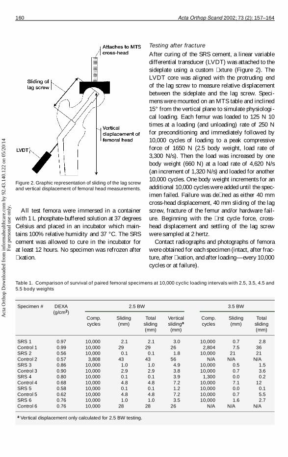
After curing of the SRS cement, a linear variable differential transducer (LVDT) was attached to the sideplate using a custom � xture (Figure 2). The LVDT core was aligned with the protruding end of the lag screw to measure relative displacement between the sideplate and the lag screw. Speci-mens were mounted on an MTS table and inclined 15° from the vertical plane to simulate physiologi-cal loading. Each femur was loaded to 125 N 10 times at a loading (and unloading) rate of 250 N for preconditioning and immediately followed by 10,000 cycles of loading to a peak compressive force of 1650 N (2.5 body weight, load rate of 3,300 N/s). Then the load was increased by one body weight (660 N) at a load rate of 4,620 N/s (an increment of 1,320 N/s) and loaded for another 10,000 cycles. One body weight increments for an additional 10,000 cycles were added until the spec-imen failed. Failure was de� ned as either 40 mm cross-head displacement, 40 mm sliding of the lag screw, fracture of the femur and/or hardware fail-ure. Beginning with the � rst cycle force, cross-head displacement and settling of the lag screw were sampled at 2 hertz.
Contact radiographs and photographs of femora were obtained for each specimen (intact, after frac-ture, after � xation, and after loading—every 10,000 cycles or at failure).
Figure 2. Graphic representation of sliding of the lag screw and vertical displacement of femoral head measurements.
Act
a O
rtho
p D
ownl
oade
d fr
om in
form
ahea
lthca
re.c
om b
y 92
.43.
140.
122
on 0
5/20
/14
For
pers
onal
use
onl
y.

Acta Orthop Scand 2002; 73 (2): 157–164 161
Outcome parameters and statistical analysis
These included: 1. Sliding of the lag screw calcu-lated as total DHS lag screw displacement from the beginning of the � rst cycle until the end of each 10,000 cycles. 2. Total sliding as calculated by adding individual sliding results as the load is increased by one body weight after each 10,000 cycle loading. 3. Total actuator displacement of MTS from the beginning of the � rst cycle until the end of each 10,000 cycles as the vertical dis-placement of the femoral head (Figure 2). 4. Stiff-ness of the construct was taken as the slope in the linear region of the force vs. displacement curve generated from each given period of 10,000 cycles. For this computation, displacement was taken as the MTS cross-head displacement and stiffness was then determined by averaging three consecutive loading cycles at predetermined time periods.
The data were examined for normality and equal variance. If they were found to be parametric, a student t-test was used. If they were found to be non-parametric, the Wilcoxon signed rank test was used. If they were found to be consistent with the normality assumption, but had an unequal vari-ance, the unequal variance F-test was applied. One-way analysis of variance was used if there were more than two variables and Pearson chi-square test statistics were used for categorical
variables. Finally, if no signi� cant difference was found between the two groups in outcome param-eters due to small sample size, type II error was calculated to show the probability that there really was a difference between the two groups although it was not detectable using the above-mentioned statistical analysis.
Results
The median and range for the bone mineral density of the intertrochanteric region of the control and SRS cement-treated specimens (n = 6) were simi-lar, 0.72 (0.56–0.97) g/cm2 and 0.78 (0.57–0.99) g/cm2, respectively (p = 0.7) (Table 1). In evalu-ating the type II error for this analysis, the prob-ability that there really was a difference between the two groups was 7%. The median sliding of the lag screw after the � rst 10,000 cycles was 0.6 (0.1–2.1) mm for the SRS cement-treated speci-mens and 17 (2.9–43.3) mm for the control speci-mens (p = 0.03). The median vertical displacement of the femoral head after the � rst 10,000 cycles was 3.2 (1.2–4.9) mm for the SRS cement-treated specimens and 21 (3.8–56) mm for the controls (p = 0.07). The type II error evaluation indicated 96% probability of a real difference. In the control specimens, the amount of settling was inversely
Table 1. Continued
Specimen # 4.5 BW 5.5 BW Failure mechanism Comp. Sliding Total Comp. Sliding Total cycles (mm) sliding cycles (mm) sliding (mm) (mm)
SRS 1 1,782 1.3 4.1 N/A N/A N/A ImplantControl 1 N/A N/A N/A N/A N/A N/A Bone collapseSRS 2 N/A N/A N/A N/A N/A N/A Bone collapse and screw cut-outControl 2 N/A N/A N/A N/A N/A N/A Bone collapseSRS 3 10,000 0.2 1.6 5,385 0.3 1.9 ImplantControl 3 10,000 0.5 4.1 4,516 0.6 4.7 ImplantSRS 4 N/A N/A N/A N/A N/A N/A Implant and screw cut-outControl 4 2,803 20 32 N/A N/A N/A Bone collapse and screw cut-outSRS 5 8,488 0.0 0.1 N/A N/A N/A ImplantControl 5 25 1.5 7.0 N/A N/A N/A Implant and screw cut-outSRS 6 3,701 1.7 4.4 N/A N/A N/A ImplantControl 6 N/A N/A N/A N/A N/A N/A Bone collapse
Act
a O
rtho
p D
ownl
oade
d fr
om in
form
ahea
lthca
re.c
om b
y 92
.43.
140.
122
on 0
5/20
/14
For
pers
onal
use
onl
y.
162 Acta Orthop Scand 2002; 73 (2): 157–164
correlated with the specimens’ bone mineral den-sity (r2 = 0.82, p = 0.01). No signi� cant correlation was found in the SRS cement group (r2 = 0.383, p = 0.19).
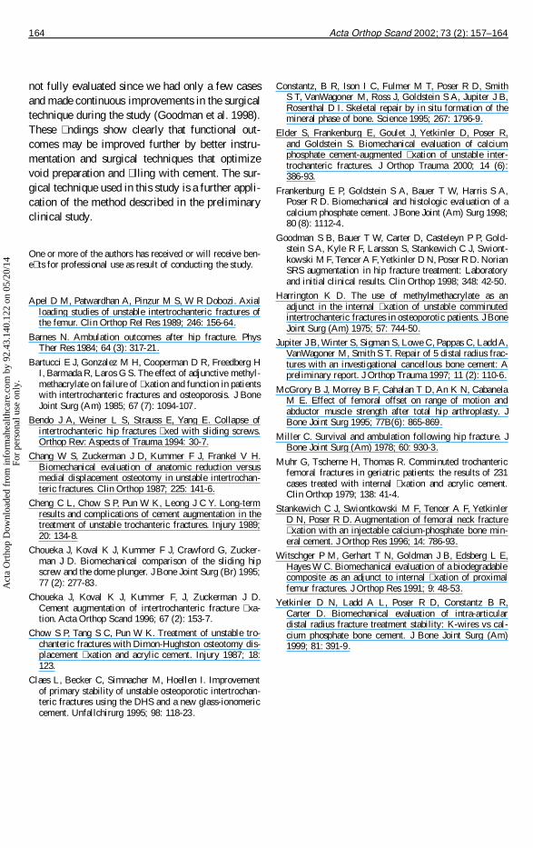
We analyzed the stiffness results during the � rst 10,000 cycles (Table 2). First and 20th cycle stiff-ness were greater in the SRS cement specimens (p = 0.04 and p = 0.03, respectively). No other sig-ni� cant differences were observed between the two treatment groups at other times. When the type II error was evaluated for other times, the probability of � nding a real difference between the two groups ranged between 7% and 8%. In both groups, aver-age stiffness increased immediately in the � rst 20 cycles, representing initial settling of the fracture fragments. The individual data showed that 3 SRS cement specimens failed to increase immediately in the � rst 20 cycles. The increment in stiffness corresponded to the settling of the fracture con-struct and this phenomenon was most marked in the control specimens.
SRS cement-treated specimens bore more load than control specimens (Table 1). 3 SRS cement specimens fractured at higher load levels than con-trol femora, while 2 pairs bore an equivalent load and 1 control specimen bore a greater load. This difference was not statistically signi� cant with the number of specimens available (p = 0.4). In addi-tion, SRS cement specimens settled less than con-
trols. Even the 4th SRS specimen settled less than the control specimen. At lower load levels, speci-mens more often failed due to bone failures (exces-sive collapse). At high load levels, specimens more often failed due to implant failures (bending of the lag screw). Overall, most of the SRS cement speci-mens sustained more load than the control speci-mens, and the total amount of settling was signi� -cantly less in SRS cement specimens than in the controls: 3.0 (0.1–21) mm versus 30 (4.7–43) mm, respectively (p = 0.03).
Discussion
The sliding hip screw is assumed to create a more stable fracture construct by allowing impaction of the proximal fragment onto the distal fragment. However, union of intertrochanteric fractures is often achieved at the expense of shortening of the proximal femur, especially in unstable fractures. Mechanical studies have shown better stability of the fracture after � xation of large posteromedial fragments including the lesser trochanter (Chang et al. 1987, Apel et al. 1989).
Shortening of the proximal femur also reduces the length and moment arm of the hip abductor muscles, which lessens their ef� ciency (McGrory et al. 1995). Barnes (1984) analyzed functional outcomes in hip fracture patients and concluded
Table 2. Comparison of stiffness of paired femoral specimens at various intervals during the � rst 10,000 cycles
Stiffness (N/mm)CyclesSpecimen 1–3 21–23 1998–2000 3998–4000 5998–6000 7998–8000 9998–10000
Control 1 604 632 1132 1242 1340 1371 1392 SRS 1 1180 1071 1051 1106 1175 1133 1169Control 2 a 263 1130 2104 na na na na SRS 2 1543 1435 1549 1549 1501 1455 1442Control 3 428 621 983 970 963 961 953 SRS 3 540 500 406 443 478 506 520Control 4 494 661 766 771 758 755 753 SRS 4 862 1182 617 574 561 544 546Control 5 412 538 821 823 857 868 871 SRS 5 1000 1915 2268 2376 2360 2470 2520Control 6 334 668 377 520 1024 1242 1223 SRS 6 803 2047 1673 863 770 764 924
Control median 420 647 902 823 963 961 953SRS median 931 1309 1300 985 973 949 1047P-value 0.04 0.03 0.6 0.4 0.7 0.8 0.7
a na = not available due to fracture
Act
a O
rtho
p D
ownl
oade
d fr
om in
form
ahea
lthca
re.c
om b
y 92
.43.
140.
122
on 0
5/20
/14
For
pers
onal
use
onl
y.

Acta Orthop Scand 2002; 73 (2): 157–164 163
that abductor muscle strength was positively asso-ciated with independence in ambulation.
The use of the sliding hip screw in intertro-chanteric hip fractures leads to fracture impaction within days to weeks of � xation (Bendo et al. 1994). Movement of the fracture fragments in this unstable situation causes pain on ambulation which may increase morbidity and mortality. Bendo et al. (1994) found that 20/26 patients with moderate and severe shortening had considerable pain and poor function. Accoarding to Miller et al. (1978), early ambulation after surgery is important for sur-vival of intertrochanteric hip fracture patients.
To address these clinical problems, polymethyl-methacrylate bone cement has been used to increase the stability of intertrochanteric fractures. Lag screw augmentation in the femoral head (Witsch-ger et al. 1991, Choueka et al. 1995, 1996, Claes et al. 1995) and augmentation of intertrochanteric fractures at the fracture site have also been studied (Harrington 1975, Muhr et al. 1979, Bartucci et al. 1985, Chow et al. 1987, Cheng et al. 1989). The early results were all reported to be good and few complications from the polymethyl methacrylate cement have occurred. Cheng et al. (1989) aug-mented the sliding hip screw by placing polymeth-ylmethacrylate cement at the fracture site. How-ever, after following the patients for 2–5 years, they reported late complications of up to 16%. They attributed this to the use of polymethylmethacry-late which is non-absorbable, exothermic, inhibits fracture healing, and may cause osteolysis and sub-sequent loosening.
Calcium phosphate cement has been used to increase the stability of several fractures in biome-chanical and clinical studies (Constantz et al. 1995, Stankewich et al. 1996, Jupiter et al. 1997, Good-man et al. 1998, Yetkinler et al. 1999). Unlike poly-methylmethacrylate, however, Norian SRS cement does not inhibit healing and it is replaced by the host bone while maintaining compressive strength in the region (Frankenburg et al. 1998). Elder et al. (2000) studied the augmentation of unstable inter-trochanteric fractures with SRS cement in a similar biomechanical model. They found that load trans-fer on the medial side of the test femur was closer to the load levels seen in the intact femur as mea-sured by strain gauges. However, in their experi-ment, specimens were loaded with only one body
weight and for 1,000 cycles during cyclic loading. This inadequate loading method caused only 1.1 mm and 0.1 mm sliding of the lag screw in the control and experimental groups, respectively. In our study, we found sliding of the lag screw to be about 17 mm in control specimens. This clinically relevant sliding was achieved by 2.5 times body weight for 10,000 cycles. Sliding of the lag screw was less than 1 mm in the SRS cement-treated specimens. It was achieved by proper preparation and localization of the SRS cement, which pro-vided a buttress or “neocalcar” on the medial aspect of the femur allowing for transfer of forces to the medial cortex (Elder et al. 2000).
Construct stiffness results indicated that, com-pared to the control specimens, SRS cement speci-mens were signi� cantly stiffer than controls during the � rst 20 cycles. In the control group, stiffness gradually increased due to impaction of the bony fragments. Greater construct stiffness and stability are an indication that cement augmentation results in better load transmission and less movement of fragments.
In general, SRS cement-treated specimens failed at higher loads than control specimens. In the former specimens, we observed sliding of the hip screw with higher load levels, which suggests that the presence of cement did not inhibit the sliding mechanism, but simply prevented excessive impac-tion by improving the load transfer along the calcar femorale.
In our study, the results of vertical displacement were generally larger than those of sliding along the lag screw. This � nding was surprising since we know that using a 135° plate vertical displace-ment should be roughly equal to sine 45° ´ sliding along the lag screw. We believe that the sliding hip screw results were more accurate, since measure-ments were taken directly from the sliding action of the lag screw. Vertical displacement results were obtained from the actuator of the MTS machine, which included sliding of the lag screw in the verti-cal direction, lag screw movement in the femoral head, and increased laxity of the specimen and the (whole) testing apparatus during repeated loading.
The initial clinical experience of using Norian SRS cement in hip fractures showed the safety of this product in intertrochanteric hip fracture patients; however, functional improvements were
Act
a O
rtho
p D
ownl
oade
d fr
om in
form
ahea
lthca
re.c
om b
y 92
.43.
140.
122
on 0
5/20
/14
For
pers
onal
use
onl
y.
164 Acta Orthop Scand 2002; 73 (2): 157–164
not fully evaluated since we had only a few cases and made continuous improvements in the surgical technique during the study (Goodman et al. 1998). These � ndings show clearly that functional out-comes may be improved further by better instru-mentation and surgical techniques that optimize void preparation and � lling with cement. The sur-gical technique used in this study is a further appli-cation of the method described in the preliminary clinical study.
One or more of the authors has received or will receive ben-e� ts for professional use as result of conducting the study.
Apel D M, Patwardhan A, Pinzur M S, W R Dobozi. Axial loading studies of unstable intertrochanteric fractures of the femur. Clin Orthop Rel Res 1989; 246: 156-64.
Barnes N. Ambulation outcomes after hip fracture. Phys Ther Res 1984; 64 (3): 317-21.
Bartucci E J, Gonzalez M H, Cooperman D R, Freedberg H I, Barmada R, Laros G S. The effect of adjunctive methyl-methacrylate on failure of � xation and function in patients with intertrochanteric fractures and osteoporosis. J Bone Joint Surg (Am) 1985; 67 (7): 1094-107 .
Bendo J A, Weiner L S, Strauss E, Yang E. Collapse of intertrochanteric hip fractures � xed with sliding screws. Orthop Rev: Aspects of Trauma 1994: 30-7.
Chang W S, Zuckerman J D, Kummer F J, Frankel V H. Biomechanical evaluation of anatomic reduction versus medial displacement osteotomy in unstable intertrochan-teric fractures. Clin Orthop 1987; 225: 141-6.
Cheng C L, Chow S P, Pun W K, Leong J C Y. Long-term results and complications of cement augmentation in the treatment of unstable trochanteric fractures. Injury 1989; 20: 134-8.
Choueka J, Koval K J, Kummer F J, Crawford G, Zucker-man J D. Biomechanical comparison of the sliding hip screw and the dome plunger. J Bone Joint Surg (Br) 1995; 77 (2): 277-83.
Choueka J, Koval K J, Kummer F, J, Zuckerman J D. Cement augmentation of intertrochanteric fracture � xa-tion. Acta Orthop Scand 1996; 67 (2): 153-7.
Chow S P, Tang S C, Pun W K. Treatment of unstable tro-chanteric fractures with Dimon-Hughston osteotomy dis-placement � xation and acrylic cement. Injury 1987; 18: 123.
Claes L, Becker C, Simnacher M, Hoellen I. Improvement of primary stability of unstable osteoporotic intertrochan-teric fractures using the DHS and a new glass-ionomeric cement. Unfallchirurg 1995; 98: 118-23.
Constantz, B R, Ison I C, Fulmer M T, Poser R D, Smith S T, VanWagoner M, Ross J, Goldstein S A, Jupiter J B, Rosenthal D I. Skeletal repair by in situ formation of the mineral phase of bone. Science 1995; 267: 1796-9.
Elder S, Frankenburg E, Goulet J, Yetkinler D, Poser R, and Goldstein S. Biomechanical evaluation of calcium phosphate cement-augmented � xation of unstable inter-trochanteric fractures. J Orthop Trauma 2000; 14 (6): 386-93.
Frankenburg E P, Goldstein S A, Bauer T W, Harris S A, Poser R D. Biomechanical and histologic evaluation of a calcium phosphate cement. J Bone Joint (Am) Surg 1998; 80 (8): 1112-4.
Goodman S B, Bauer T W, Carter D, Casteleyn P P, Gold-stein S A, Kyle R F, Larsson S, Stankewich C J, Swiont-kowski M F, Tencer A F, Yetkinler D N, Poser R D. Norian SRS augmentation in hip fracture treatment: Laboratory and initial clinical results. Clin Orthop 1998; 348: 42-50.
Harrington K D. The use of methylmethacrylate as an adjunct in the internal � xation of unstable comminuted intertrochanteric fractures in osteoporotic patients. J Bone Joint Surg (Am) 1975; 57: 744-50.
Jupiter J B, Winter S, Sigman S, Lowe C, Pappas C, Ladd A, VanWagoner M, Smith S T. Repair of 5 distal radius frac-tures with an investigational cancellous bone cement: A preliminary report. J Orthop Trauma 1997; 11 (2): 110-6.
McGrory B J, Morrey B F, Cahalan T D, An K N, Cabanela M E. Effect of femoral offset on range of motion and abductor muscle strength after total hip arthroplasty. J Bone Joint Surg 1995; 77B(6): 865-869.
Miller C. Survival and ambulation following hip fracture. J Bone Joint Surg (Am) 1978; 60: 930-3.
Muhr G, Tscherne H, Thomas R. Comminuted trochanteric femoral fractures in geriatric patients: the results of 231 cases treated with internal � xation and acrylic cement. Clin Orthop 1979; 138: 41-4.
Stankewich C J, Swiontkowski M F, Tencer A F, Yetkinler D N, Poser R D. Augmentation of femoral neck fracture � xation with an injectable calcium-phosphate bone min-eral cement. J Orthop Res 1996; 14: 786-93.
Witschger P M, Gerhart T N, Goldman J B, Edsberg L E, Hayes W C. Biomechanical evaluation of a biodegradable composite as an adjunct to internal � xation of proximal femur fractures. J Orthop Res 1991; 9: 48-53.
Yetkinler D N, Ladd A L, Poser R D, Constantz B R, Carter D. Biomechanical evaluation of intra-articular distal radius fracture treatment stability: K-wires vs cal-cium phosphate bone cement. J Bone Joint Surg (Am) 1999; 81: 391-9.
Act
a O
rtho
p D
ownl
oade
d fr
om in
form
ahea
lthca
re.c
om b
y 92
.43.
140.
122
on 0
5/20
/14
For
pers
onal
use
onl
y.
Copyright © 2022 FDOKUMEN