Salient features of otoacoustic emissions are common across ...
35
International Tinnitus Journal, Vol. 18, No 1 (2013)www.tinnitusjournal.com
Measurement of subtle auditory deficit in tinnitus patients with normal audiometric thresholds using evoked otoacoustic
emissions and threshold equalizing noise tests
Wan Syafira Ishak1
Fei Zhao2
Deepak Rajenderkumar3
Mohammed Arif3
1 Program Audiologi, Pusat Pengajian Sains Rehabilitasi, Fakulti Sains Kesihatan - Universiti Kebangsaan Malaysia - Kuala Lumpur - AC - Malaysia. E-mail: [email protected] Centre for Hearing and Balance Studies - University of Bristol - Bristol - United Kingdom. E-mail: [email protected] Welsh Hearing Institute - University Hospital of Wales - Cardiff - United Kingdom. E-mail: [email protected]. E-mail: Arif - [email protected]: Universiti Kebangsaan MalaysiaSend correspondence to:Wan Syafira Ishak.Program Audiologi, Pusat Pengajian Sains Rehabilitasi, Fakulti Sains Kesihatan, Universiti Kebangsaan Malaysia, Jalan Raja Muda Abdul Aziz, 50300 Kuala Lumpur, Malaysia. E-mail: [email protected] submitted to the ITJ-SGP (Publishing Management System) on February 17, 2014;and accepted on February 20, 2014. cod. 154
ORIGINAL ARTICLEInternational Tinnitus Journal. 2013;18(1):35-44.
Abstract
Introduction: The general consensus on the roles of hearing loss in triggering tinnitus seems not applicable in patients with normal hearing thresholds. The absence of hearing loss on the audiogram in this group of patients poses a serious challenge to the cochlear theories in explaining tinnitus generation in this group of patients. Objective: To scrutinize auditory functioning in a sample of tinnitus subjects with normal hearing thresholds and non-tinnitus normally hearing control participants using transient evoked and distortion product otoacoustic emission (TEOAE and DPOAE) and Threshold Equalising Noise (TEN) test. Methods: Twenty-seven tinnitus adult patients with normal hearing thresholds and 27 normally hearing volunteers were tested with TEOAE, DPOAE and TEN test. Results: Abnormal TEOAE was significantly more in tinnitus group than in controls. No significant difference was observed in DPOAE and TEN test. Only one patient was found with a positive TEN test result, who was confirmed by Magnetic Resonance Imaging to have acoustic neuroma on the affected ear. Conclusion: These results suggest the possible existence of subtle auditory deficit in normally hearing tinnitus patients, which may be an early sign of diseases that are only diagnosed after the onset of hearing loss.
Keywords: coustic neuroma, otoacoustic emissions, tinnitus, threshold equalising noise test.
DOI: 10.5935/0946-5448.20130006
36
International Tinnitus Journal, Vol. 18, No 1 (2013)www.tinnitusjournal.com
INTRODUCTION
Tinnitus is defined as the perception of sound that results completely from activity within the nervous system without any corresponding mechanical, vibratory activity within the cochlea, and not related to external stimulation of any kind1. For many years, progress towards finding the effective treatments for tinnitus was hindered by vague understanding of its underlying pathologic mechanisms. The last half of the twentieth century saw considerable advances in our understanding of tinnitus from both physiological and psychological perspectives.
An increasing amount of epidemiology studies provide both self-reported data on tinnitus and hearing status which allows closer examination on the relationship between tinnitus and hearing status. The association between hearing loss and tinnitus has been well documented in the previous literature2-4. It has been generally accepted that any severe damage along the auditory system may lead to a certain degree of hearing loss, which is generally associated with tinnitus5,6. More commonly, various studies have shown that sensorineural hearing loss plays an important role in tinnitus generation. Evidence showed a higher incidence of tinnitus patients with sensorineural hearing loss compared with conductive hearing loss7.
However, the role of sensorineural hearing loss in tinnitus generation is not conclusive. Some studies found a correlation between severity of tinnitus and degree of hearing loss3,8 while others found no such correlation9. Moreover, the incidence of tinnitus in patients with sensorineural hearing loss varies widely across studies. The incidence ranges from less than 15%6,10 to as high as 79%7. In addition, not all individuals with hearing loss experience tinnitus, and not all tinnitus patients have hearing loss. Jastreboff & Jastreboff11 reported that about 27% of totally deaf patients do not have tinnitus, whereas 20% of normal hearing people have tinnitus. It can be assumed that the role of sensorineural hearing loss as a tinnitus generator is complex and the mechanism of tinnitus may rely on the presence of other contributing factors such as damage to specific hair cells or the auditory nerve fibers.
Although some studies have different theoretical assumptions and approaches, the significance of hearing loss in tinnitus generation is not questioned. On the other hand, clinical observations have shown that 8 to 10 per cent of tinnitus patients had normal audiometric thresholds12. These observations may have several indications, such as hearing loss is not necessary in tinnitus generation, pure tone audiogram does not always indicate auditory damage, tinnitus may be a primary symptom of diseases that are only diagnosed after the occurrence of hearing loss, or several mechanisms
exist in a group of patients. Therefore, the absence of hearing loss on the audiogram in this group poses a serious challenge to the aforementioned theories in explaining tinnitus generation in tinnitus patients with normal hearing. While the theories seem cannot fit in this minority group, evidence obtained from this group may have huge implication for a better tinnitus mechanisms understanding as well as assist in developing and shaping tinnitus management. This is because the results obtained from this special group may directly related to the tinnitus perception, but not to the concomitant hearing loss.
Thus, several attempts have been made to explain pathophysiology mechanisms of tinnitus in patients with normal hearing ability particularly using otoacoustic emissions (OAEs). It is hypothesised that outer hair cells damage results in stereocilia decoupling from the tectorial membrane, thus causing them to depolarise and subsequently resulting in tinnitus perception1. The different functionality of outer and inner hair cells can also cause imbalanced activity in the dorsal cochlear nucleus which in turn causes changes at higher levels of the auditory system that may be perceived as tinnitus1.
Several studies have revealed that TEOAEs and DPOAEs can be used as a tool to detect early hearing loss before it can be diagnosed by PTA13-15. An association of tinnitus with the changes in amplitude of TEOAEs and DPOAEs has been reported. In some studies, lower amplitude of TEOAEs13,16 or DPOAEs13,17-19 have been observed in tinnitus patients compared to in non-tinnitus controls. Contrarily, some other studies found a reverse direction in the amplitude of OAEs. The studies reported that tinnitus subjects were found to have increased TEOAEs20,21 or DPOAEs22,23 compared with non-tinnitus counterparts.
The contradict findings from previous research motivates researchers to find alternative theories to explain tinnitus mechanisms in tinnitus patients with normal hearing thresholds. One study by Weisz et al.24 proposed that subtle auditory abnormality may also arise from the inner hair cells (IHCs) dysfunction. They proposed that the presence of altered IHCs and/or neuron activity function can also be found in tinnitus patients with normal audiometric thresholds. In the study, they investigated the inner hair cells functions in 11 chronic tinnitus patients with normal hearing thresholds using Threshold Equalising Noise (TEN) test and found 8 of them with possible inner hair cells problems. Thabet25 attempted to replicated the earlier study by investigating 20 normally hearing unilateral tinnitus patients with TEN test and TEOAE. In contrast, he found only three tinnitus ears with IHCs problem. Meanwhile, 85% of tinnitus ears have abnormal TEOAE. The three tinnitus ears with IHCs problem were also in the abnormal TEOAE group. The
37
International Tinnitus Journal, Vol. 18, No 1 (2013)www.tinnitusjournal.com
TEN test appears to be a potential non-invasive and quick test of inner hair cells function. Nonetheless, it is too early to make a conclusion based on the aforementioned two studies on tinnitus. More research is needed in order to validate the findings. Therefore, this study aimed to investigate subtle cochlear abnormality in tinnitus patients with normal hearing thresholds using evoked OAEs (TEOAEs and DPOAEs) and TEN test.
METHOD
ParticipantsThis study investigated 27 tinnitus subjects
(42 tinnitus ears and 12 contralateral non-tinnitus ear) with normal hearing thresholds who were recruited from Welsh Hearing Institute, University Hospital of Wales. The subjects who were attending the Institute from January to December 2010, were identified by an audiology physician and audiologists based in this hospital. All participants had no history or current external and middle ear problem, noise exposure and psychiatric problem. Of those, 14 were males (51.9%) and 13 were females (48.1%). The mean age of this group was 36.84 years (SD = 10.2). A group of volunteers without tinnitus were also recruited as control group.
Tests and ProcedureFull history taking and otoscopy examination
were carried out at the beginning of the session. Then, all subjects underwent basic audiological tests which include pure tone audiometry and tympanometry. The tests were then followed by TEOAE and DPOAEs measurements and the TEN (HL) test. These tests are described in the following:
a) TEOAEs TestTEOAEs test was performed using unfiltered clicks
of 80 µs duration presented in non-linear modes. The stimulus level in the outer ear canal was 80 ± 2 dB peSPL. The click rate was 50 per second. Responses to 260 sweeps of stimulus packets were averaged, with a 0.5 - 6.0 kHz pass-band. TEOAEs responses were considered when the reproducibility was greater than 50%, stimulus stability was better than 70% and the different between TEOAEs amplitude and corresponding noise floor was at least 3 dB in at least four out of five 1/2 octave frequency bands centered at 1.0, 1.5, 2.0, 3.0 and 4.0 kHz26. The test was performed in a sound treated room where the ambient noise was maintained between 20 to 40 dB A.
b) DPOAEs recordingThe stimulus comprised of a pair of primary pure
tones (f1 and f2) at a frequency ratio equal to 1.22. The primary tones’ levels were presented at 65 dBSPL (f1) and 55 dBSPL (f2). In this experiment, a 2f1 - f2 distortion
product was used because of its established clinical value. The results were presented as a distortion-product audiogram i.e the amplitude of the 2f1 - f2 DPOAE was plotted against the f2 from 1000 to 6169 Hz. The presence of DP amplitude was accepted as valid when the signal to + 2 SD noise floor ratio exceeded 3 dB and the absolute DPOAE amplitude was at least - 10 dB SPL at each individual f2.
c) Threshold Equalising Noise (TEN) TestThe basic principle of this test is to control for
“off-frequency listening” from occurring by using a broad band noise (the threshold equalising noise). Off-frequency listening occurs when the presented stimulus is being picked up by other parts of the cochlea instead of the targeted frequency area of the cochlea. This off frequency listening occurs when the targeted area of inner hair cells or neurons are not functioning well and is referred to as a dead region27. The broad band noise is designed to produce almost equal masked thresholds over a wide frequency range, for listeners with normal hearing and hearing impairment but without dead regions28.
The procedure of conducting TEN test has been thoroughly described elsewhere29,30. A dead region for a particular frequency is indicated by the masked threshold if the following two conditions are fulfilled31:
1. The masked threshold is 10 dB or more than the absolute threshold
2. The masked threshold is 10 dB or more than the TEN level/ERBN
Statistical analysisAll relevant analyses were carried out on the Excel
and SPSS (version 17.0) for Windows.
Ethical ConsiderationsThis research has received the ethical approvals
from the School of Applied Community and Health Studies (SACHS), University of Bristol Human Participants Ethics Committee and the South East Wales Research Ethics Committee.
RESULTS
From the 27 tinnitus subjects, 15 subjects had bilateral tinnitus (30 ears, 71.4%) and the remaining 12 subjects (12 ears, 28.6%) had unilateral tinnitus. Thus, a total of 42 ears with tinnitus were analysed in this study. Fourteen of the subjects (51.9%) were males whereas 13 of them were females (48.1%). The mean age of this group was 36.89 (SD = 10.28). A total of 27 normal hearing volunteers (54 ears) were recruited as controls. Of those, 12 were males (44.4%) and 15 were females (55.6%). The mean age in this group was 33.74
38
International Tinnitus Journal, Vol. 18, No 1 (2013)www.tinnitusjournal.com
(SD = 9.69). A Mann-Whitney U test revealed that there was no statistically significant difference between age of tinnitus subjects and controls (U = 296.5, p = 0.252). The mean pure tone audiometric thresholds of tinnitus and control groups are shown in Table 1. An independent t-test revealed that there was no significant difference in hearing thresholds between tinnitus patients and controls at all frequencies except at 6 and 8 kHz. At both these frequencies, tinnitus patients had significantly higher thresholds than control individuals.
Frequency (kHz) Mean TEOAE amplitude (dB SPL)
Tinnitus ears (27) Control ears (46)
1 5.42 4.86
1.5 8.26 7.86
2 7.01 5.84
3 4.91 4.71
4 1.24 1.66
Table 3. Mean TEOAE amplitude for tinnitus and control ears at each frequency band.
Outer hair cells damage: EOAEs resultsThe present study found 35.7% of tinnitus ears
with abnormal TEOAEs. The controls were observed to have only 14.8% of the ears with abnormal TEOAEs. A Chi square test shows that the percentages of abnormal TEOAEs in the tinnitus group was significantly higher than the control group (χ2 = 5.67, df = 1, p = 0.017). The abnormal TEOAEs in the tinnitus group were present for 1 and 4 kHz mostly, while in the control group, the abnormalities were mostly at 4 kHz. Table 2 shows the normal and abnormal TEOAEs at each frequency tested in both the tinnitus and control group. The difference between the two groups is highly significant at frequencies of 1, 1.5 and 2 kHz, but not at 3 and 4 kHz.
This analysis only included those with normal TEOAEs results. From the analysis, the parameters of the TEOAEs for tinnitus ears are not significantly different from the control ears. Further analysis was done based on frequency band (Table 3). A t-test revealed that with regard to mean amplitude of TEOAE for tinnitus ears, there were no statistically significant differences for all frequencies tested (p > 0.05).
Initially, the left and right of DPOAE results were compared within the tinnitus and control groups. Since no statistically significant difference was observed between
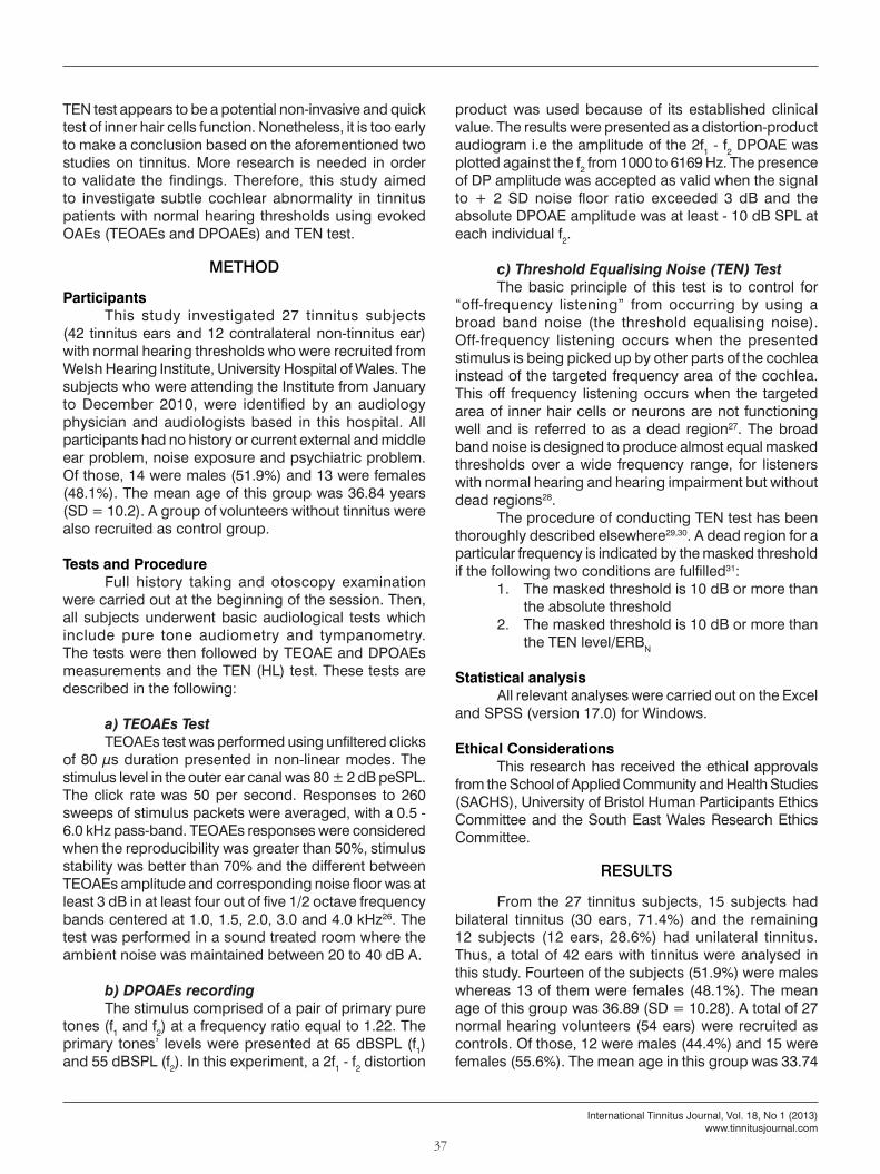
both ears, further statistical analyses were performed using combined data from both sides together. A decrease in DPOAE amplitude (DPOAE amplitude less than the mean -2SD of normal individuals) over a limited frequency range was observed in 31.7% of the tinnitus ears (13 of 41 ears) and in 25.9% of the control ears (14 of 54 ears). The percentage of tinnitus ears with decreased DPOAE at limited frequency range had no statistically significant difference from the control ears (χ2 = 0.383, df = 1, p = 0.536). From the 13 tinnitus ears with abnormal DPOAE, 12 (29.3%) had abnormal DPOAE in 3 or more frequencies, whereas 8 (14.8%) out of 14 control ears had abnormal DPOAE in 3 or more frequencies (Figure 1).
Further analysis was carried out in terms of frequency specific. Based on the chi-square analysis, tinnitus ears have a statistically significant higher percentage of abnormal DPOAEs at some frequencies, mainly at 2000 and 4000 Hz regions (Table 4).
Then, the differences of overall mean DPOAE amplitude between tinnitus and control ear were compared. For this analysis, only ears with normal DPOAE were selected. There was no significant difference of mean amplitude observed between the tinnitus and control ears (t = -0.595; df = 93; p = 0.553). Later, the mean amplitude of DPOAE for tinnitus and control ears against the corresponding f2 was compared. Generally, an independent t-test showed no statistically significant differences observed between tinnitus and control ears at all frequencies except for frequency 1414, 4757, 5187 and 6169 Hz (Table 5).
Frequency (Hz)
Tinnitus Group (42 ears)
Control Group (54 ears) p
Mean SD Mean SD
250 8.78 6.3 8.3 6.5 p > 0.05
500 7.67 6.0 8.04 5.3 p > 0.05
750 6.33 5.5 7.41 5.0 p > 0.05
1000 7.56 6.8 5.98 5.8 p > 0.05
1500 5.89 5.6 6.96 4.8 p > 0.05
2000 6.22 6.0 5.54 5.1 p > 0.05
3000 5.78 6.0 4.91 6.1 p > 0.05
4000 9.89 7.4 7.32 5.3 p > 0.05
6000 13.22 6.6 7.95 6.2 p < 0.05*
8000 9.11 7.9 5.18 5.2 p < 0.05*
Table 1. Averaged hearing thresholds of tinnitus and control group of participants.
* t-test significant at p < 0.05.
Frequency (kHz)
Tinnitus Group (n = 42)
Control Group (n = 54) p*
Normal (%)
Abnormal (%)
Normal (%)
Abnormal (%)
1 61.9 38.1 88.9 11.1 0.002*
1.5 85.7 14.3 98.1 1.9 0.020*
2 83.3 16.7 96.3 3.7 0.031*
3 90.5 9.5 88.9 11.1 0.801
4 69.0 31.0 83.3 16.7 0.099
Table 2. Percentage of ears with normal and abnormal TEOAE according to frequencies.
* χ2 test significant at p < 0.05.
39
International Tinnitus Journal, Vol. 18, No 1 (2013)www.tinnitusjournal.com
OAE status Tinnitus ears (n = 41) Control ears (n = 54)
All Passed 21 (51.2%) 38 (70.4%)
TEOAE Failed Only 7 (17.1%) 2 (3.7%)
DPOAE Failed Only 6 (14.6%) 8 (14.8%)
All Failed 7 (17.1%) 6 (11.1%)
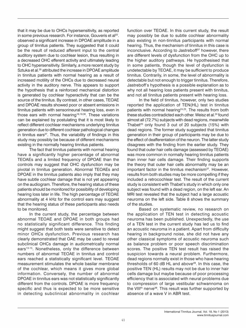
Table 6. OAE status for both tinnitus and control ears.
Figure 1. Distribution of the number of test frequencies with abnormal DPOAE in tinnitus and control ears.
In addition to the analyses described previously, the results were summarized in Table 6. From a total of 41 tinnitus ears, about half (21 ears) passed both TEOAE and DPOAE tests. For the control ears, 38 out of 54 ears passed TEOAE and DPOAE tests. From 20 tinnitus patients with abnormal OAEs, 13 of them exhibit either failed TEOAE or DPOAE results.
Meanwhile, from 16 control ears with abnormal OAEs, 10 ears exhibit either failed TEOAE or DPOAE results. The Chi-square test showed no statistical significant difference between tinnitus and the control ears (χ2 = 6.379, df = 3, p = 0.095).
Inner hair cellsFrom the 27 tinnitus patients with normal hearing,
only one patient (3.7%; 1/27) had a positive TEN test result on her left ear. No one in the control group demonstrated an abnormal TEN test result. In this case (Subject RX), the TEN (HL) test revealed that dead regions might exist at 1.5, 3
f2 (Hz) Tinnitus ears (%) (n = 41)
Control ears (%) (n = 54) p
1000 21.4 5.6 p < 0.05*
1091 21.4 9.3 p > 0.05
1189 19 7.4 p > 0.05
1297 19 9.3 p > 0.05
1414 16.7 7.4 p > 0.05
1542 19 5.6 p < 0.05*
1682 11.9 5.6 p > 0.05
1834 9.5 3.7 p > 0.05
2000 16.7 3.7 p < 0.05*
2181 21.4 3.7 p < 0.05*
2378 14.3 1.9 p < 0.05*
2594 11.9 3.7 p > 0.05
2828 7.1 5.6 p > 0.05
3084 7.1 5.6 p > 0.05
3364 11.9 9.3 p > 0.05
3668 7.1 5.6 p > 0.05
4000 7.1 7.4 p > 0.05
4362 9.5 0 p < 0.05*
4757 16.7 1.9 p < 0.05*
5187 14.3 3.7 p > 0.05
5657 16.7 9.3 p > 0.05
6169 19 11.1 p > 0.05
Table 4. Percentage of ears with abnormal DPOAE for each f2.
* χ2 test significant at p < 0.05.
f2 (Hz) Tinnitus Ears Control Ears
pN Mean SD N Mean SD
1000 26 7.98 5.8 42 6.85 5.1 p > 0.05
1091 28 9.7 6.1 45 7.71 4.7 p > 0.05
1189 29 10.7 5.5 45 8.6 5.4 p > 0.05
1297 31 11.21 5.5 45 9.83 4.7 p > 0.05
1414 31 11.5 4.8 48 9.07 5.2 p < 0.05*
1542 33 10.21 5.6 50 9.87 5.7 p > 0.05
1682 35 10.79 6.7 52 9.08 5.9 p > 0.05
1834 36 9.32 6.1 51 9 5.3 p > 0.05
2000 37 8.3 6.2 53 8.67 6.2 p > 0.05
2181 33 8.2 6.1 50 7.31 5.6 p > 0.05
2378 38 6.56 5.2 53 6.79 4.8 p > 0.05
2594 37 6.91 5.6 51 6.08 5.4 p > 0.05
2828 38 5.48 5.7 52 5.55 5.5 p > 0.05
3084 39 6.5 4.4 50 6.44 5.3 p > 0.05
3364 40 5.81 5 52 6.83 4.9 p > 0.05
3668 39 7.25 4.2 53 6.59 5.1 p > 0.05
4000 40 8.09 5 52 7.72 4.8 p > 0.05
4362 39 8.42 5.61 54 8.9 5.1 p > 0.05
4757 40 8.58 5.5 53 11.16 4.3 p < 0.05*
5187 37 8.15 5.5 52 10.29 5.6 p < 0.05*
5657 37 6.2 7 49 8.44 5.4 p > 0.05
6169 33 3.9 7.2 50 7.19 6.6 p < 0.05*
Table 5. Comparison of the mean DPOAE amplitude between tinnitus and control ears.
* t-test significant at p < 0.05.
40
International Tinnitus Journal, Vol. 18, No 1 (2013)www.tinnitusjournal.com
Frequency (kHz)
0.5 0.75 1.0 1.5 2.0 3.0 4.0
Right Ear
Absolute Threshold 10 5 5 5 5 5 5
Masked Threshold 50 50 50 50 50 50 50
Left Ear
Absolute Threshold 15 10 15 15 20 15 10
Masked Threshold 52 52 58** 62* 52 62* 60** indicates masked thresholds that fulfilled the dead regions criteria ** indicates masked threshold that is higher than normal, but does not fulfil the dead region criteria.
Table 7. TEN Test results for Subject RX with the TEN level set at 50 dBHL/ERBN.
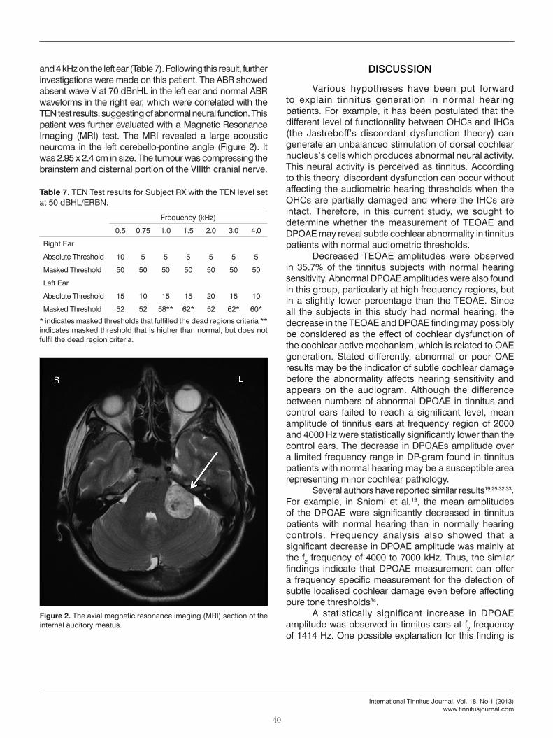
Figure 2. The axial magnetic resonance imaging (MRI) section of the internal auditory meatus.
and 4 kHz on the left ear (Table 7). Following this result, further investigations were made on this patient. The ABR showed absent wave V at 70 dBnHL in the left ear and normal ABR waveforms in the right ear, which were correlated with the TEN test results, suggesting of abnormal neural function. This patient was further evaluated with a Magnetic Resonance Imaging (MRI) test. The MRI revealed a large acoustic neuroma in the left cerebello-pontine angle (Figure 2). It was 2.95 x 2.4 cm in size. The tumour was compressing the brainstem and cisternal portion of the VIIIth cranial nerve.
DISCUSSION
Various hypotheses have been put forward to explain tinnitus generation in normal hearing patients. For example, it has been postulated that the different level of functionality between OHCs and IHCs (the Jastreboff’s discordant dysfunction theory) can generate an unbalanced stimulation of dorsal cochlear nucleus’s cells which produces abnormal neural activity. This neural activity is perceived as tinnitus. According to this theory, discordant dysfunction can occur without affecting the audiometric hearing thresholds when the OHCs are partially damaged and where the IHCs are intact. Therefore, in this current study, we sought to determine whether the measurement of TEOAE and DPOAE may reveal subtle cochlear abnormality in tinnitus patients with normal audiometric thresholds.
Decreased TEOAE amplitudes were observed in 35.7% of the tinnitus subjects with normal hearing sensitivity. Abnormal DPOAE amplitudes were also found in this group, particularly at high frequency regions, but in a slightly lower percentage than the TEOAE. Since all the subjects in this study had normal hearing, the decrease in the TEOAE and DPOAE finding may possibly be considered as the effect of cochlear dysfunction of the cochlear active mechanism, which is related to OAE generation. Stated differently, abnormal or poor OAE results may be the indicator of subtle cochlear damage before the abnormality affects hearing sensitivity and appears on the audiogram. Although the difference between numbers of abnormal DPOAE in tinnitus and control ears failed to reach a significant level, mean amplitude of tinnitus ears at frequency region of 2000 and 4000 Hz were statistically significantly lower than the control ears. The decrease in DPOAEs amplitude over a limited frequency range in DP-gram found in tinnitus patients with normal hearing may be a susceptible area representing minor cochlear pathology.
Several authors have reported similar results19,25,32,33. For example, in Shiomi et al.19, the mean amplitudes of the DPOAE were significantly decreased in tinnitus patients with normal hearing than in normally hearing controls. Frequency analysis also showed that a significant decrease in DPOAE amplitude was mainly at the f2 frequency of 4000 to 7000 kHz. Thus, the similar findings indicate that DPOAE measurement can offer a frequency specific measurement for the detection of subtle localised cochlear damage even before affecting pure tone thresholds34.
A statistically significant increase in DPOAE amplitude was observed in tinnitus ears at f2 frequency of 1414 Hz. One possible explanation for this finding is
41
International Tinnitus Journal, Vol. 18, No 1 (2013)www.tinnitusjournal.com
that it may be due to OHCs hypersensitivity, as reported in some previous research. For instance, Gouveris et al22.observed a significant increase in DPOAE amplitude in a group of tinnitus patients. They suggested that it could be the result of reduced afferent input to the central auditory system due to cochlear lesion, thus resulting in a decreased OHC efferent activity and ultimately leading to OHC hypersensitivity. Similarly, a more recent study by Sztuka et al.35 attributed the increase in DPOAE amplitude in tinnitus patients with normal hearing as a result of increased motility of the OHCs due to decreased neural activity in the auditory nerve. This appears to support the hypothesis that a reinforced mechanical distortion is generated by cochlear hyperactivity that can be the source of the tinnitus. By contrast, in other cases, TEOAE and DPOAE results showed poor or absent emissions in tinnitus patients with sensorineural hearing loss, even in those ears with normal hearing16,19,36. These variations can be explained by postulating that it is most likely to reflect different cochlear mechanisms underlying tinnitus generation due to different cochlear pathological changes in tinnitus ears37. Thus, the variability of findings in this study may possibly be because of different mechanisms existing in the normally hearing tinnitus patients.
The fact that tinnitus patients with normal hearing have a significantly higher percentage of abnormal TEOAEs and a limited frequency of DPOAE than the controls may suggest that OHC dysfunction may be pivotal in tinnitus generation. Abnormal TEOAEs and DPOAE in the tinnitus patients also imply that they may have subtle cochlear damage that is not yet appearing on the audiogram. Therefore, the hearing status of these patients should be monitored for possibility of developing hearing loss later in life. The high percentage of TEOAE abnormality at 4 kHz for the control ears may suggest that the hearing status of these participants also needs to be monitored.
In the current study, the percentage between abnormal TEOAE and DPOAE in both groups had no statistically significant difference. This finding might suggest that both tests were sensitive to detect minor OHCs dysfunction. Previous research has clearly demonstrated that OAE may be used to reveal subclinical OHCs damage in audiometrically normal ears13-15. Nonetheless, only the difference between numbers of abnormal TEOAE in tinnitus and control ears reached a statistically significant level. TEOAE measurement stimulates the whole basilar membrane of the cochlear, which means it gives more global information. Conversely, the number of abnormal DPOAE in tinnitus ears was not statistically significantly different from the controls. DPOAE is more frequency specific and thus is expected to be more sensitive in detecting subclinical abnormality in cochlear
function over TEOAE. In this current study, the result may possibly be due to subtle cochlear abnormality also existing in non-tinnitus participants with normal hearing. Thus, the mechanism of tinnitus in this case is inconclusive. According to Jastreboff37 however, there are different levels of dysfunction from the OHC up to the higher auditory pathways. He hypothesised that in some patients, though the level of dysfunction is undetectable by TEOAE, it may be sufficient to produce tinnitus. Contrarily, in some, the level of abnormality is detectable but not enough to trigger tinnitus. Therefore, Jastreboff’s hypothesis is a possible explanation as to why not all hearing loss patients present with tinnitus, and not all tinnitus patients present with hearing loss.
In the field of tinnitus, however, only two studies reported the application of TEN(HL) test in tinnitus patients with normal hearing24,25. The results from both these studies contradicted each other. Weisz et al.24 found almost all (72.7%) subjects with dead regions, meanwhile Thabet25 only found 3 out of 20 subjects (15%) with dead regions. The former study suggested that tinnitus generation in their group of participants may be due to the effect of deafferentation of the neuron. The latter study disagrees with the finding from the earlier study. They found that outer hair cells damage (assessed by TEOAE) was more common in normally hearing tinnitus patients than inner hair cells damage. Their finding supports the theory that outer hair cells abnormality may be an important factor in the tinnitus mechanism37. However, results from both studies may be more compelling if they included a retrocochlear test. The result of the current study is consistent with Thabet’s study in which only one subject was found with a dead region, on the left ear. An MRI test revealed that the subject had a large acoustic neuroma on the left side. Table 8 shows the summary of the studies.
Based on systematic review, no research on the application of TEN test in detecting acoustic neuroma has been published. Unexpectedly, the use of a TEN test in the current study has led us to find an acoustic neuroma in a patient. Apart from difficulty hearing in background noise, she did not have any other classical symptoms of acoustic neuroma such as balance problem or poor speech discrimination scores. The positive TEN test result has raised the suspicion towards a neural problem. Furthermore, dead regions normally exist in those who have hearing thresholds of 60 dB HL and above28. In this case, the positive TEN (HL) results may not be due to inner hair cells damage but maybe because of poor processing efficiency that is associated with neural problems due to compression of large vestibular schwannoma on the VIIIth nerve38. This result was further supported by absence of a wave V in ABR test.
42
International Tinnitus Journal, Vol. 18, No 1 (2013)www.tinnitusjournal.com
Author(s) Abnormal TEN Test (n %)
Abnormal TEOAE (n %) Conclusion
Weisz et al. (2006) 8/11, 72.7% NT Tinnitus generation may be because of deafferentiation of the neuron
Thabet (2009) 3/20, 27.3% 85% OHCs damage is still more important in tinnitus generation than IHCs damage
Current study 1/27, 3.7% 35.7%OHCs damage is more important in tinnitus generation in normal hearing patients; however, abnormal TEN test may indicate more serious problem such as acoustic neuroma
Table 8. Comparison between previous TEN test studies on tinnitus and current study.
NT: Not tested.
The MRI result confirmed the suspicion of retrocochlear pathology in this patient. The result can be inferred two-fold. Firstly, it implies that a TEN test may offer a quick, inexpensive and non-invasive method for retrocochlear screening. Secondly, tinnitus patients may have a serious problem that triggers the tinnitus perception despite having normal hearing thresholds and normal OAE test results. In other words, even though dead regions normally exist in patients with hearing loss of more than 55 dBHL, normal hearing people can also have a ‘dead region’ which may be due to retrocochlear abnormality.
No published data was found on the application of a TEN test in detecting acoustic neuroma. Vinay & Moore38 studied a group of auditory neuropathy patients using the TEN test. In their study, they found 7 out of 8 auditory neuropathy patients with abnormal TEN test results. Patients with auditory neuropathy are known to have problems with transmission of the auditory signal to the brain, thus abnormal TEN test results in this group of patients were assumed to be due to the neuron dysfunction. Similarly, the abnormal result in Subject RX in the current study may also be because of the problem in neurons rather than in the inner hair cells. However, caution should be taken in interpreting the TEN test results as 10 dB-above-thresholds criteria is just a binary criteria that means passing or failing the TEN test, but does not mean dead or not dead. For this reason, more research needs to be carried out on the clinical benefit of the TEN test before applying this method clinically.
CONCLUSION
This study showed that subtle cochlear problem may exist in tinnitus patients before it is significantly appeared in the audiogram. The use of evoked OAEs in this group of patients seems justified. The finding also supported the possible clinical advantage of the TEN test as a screening tool for retrocochlear problem. However, more research needs to be carried out in this area in order to support the current finding.
AcknowledgementThanks to University Hospital of Wales, UK for
providing the facilities to conduct this research. Sincere thanks to Ministry of Higher Education (MOHE), Malaysia, Universiti Kebangsaan Malaysia (UKM) and University of Bristol for providing sponsorship to pursue this PhD study.
Conflict of interestNone declared.
FundingThis study was supported by the University of
Bristol, UK and Ministry of Higher Education (MOHE) Malaysia and Universiti Kebangsaan Malaysia (UKM) as parts of the first author’s PhD research.
Summary• Progress towards finding the effective treatments
for tinnitus was hindered by vague understanding of its underlying pathological mechanisms.
• The absence of any grossly abnormal audiometric findings in tinnitus patients with normal hearing thresholds leaves clinicians with difficulties to explain the occurrence of tinnitus.
• Therefore, this study aimed to investigate subtle cochlear abnormality in tinnitus patients with normal hearing thresholds using Transient Evoked Otoacoustic Emissions (TEOAEs), Distortion Product Otoacoustic Emissions (DPOAEs) and Threshold Equalising Noise Test (TEN).
• Twenty seven tinnitus patients with normal hearing thresholds who attended Welsh Hearing Institute, University Hospital of Wales from January to December 2010 were recruited. A similar age and gender non-tinnitus volunteers were also recruited as controls.
• The TEOAEs results revealed that 35.7% of tinnitus patients have abnormal results, which was significantly higher than the control group (p < 0.05).
43
International Tinnitus Journal, Vol. 18, No 1 (2013)www.tinnitusjournal.com
• The Chi-square test revealed that the tinnitus ears have significantly higher percentage of abnormal DPOAE mainly when f2 was at 2000 and 4000 Hz frequency regions. Nonetheless, there was no statically significant different in term of percentage of abnormal amplitudes in overall frequency range (p > 0.05).
• Only one patient was found with a positive TEN test result. The TEN test showed that dead regions might exist at 1500, 3000 and 4000 Hz. MRI revealed a large vestibular schwannoma in the left cerebello-pontine angle which was compressiong the brainstem and cisternal portion of the VIIIth cranial nerve.
• This study showed that subtle cochlear problem may exist in tinnitus patients before it is significantly appeared in the audiogram. The finding also supported the possible clinical advantage of the TEN test as a screening tool for retrocochlear problem. However, more research needs to be carried out in this area in order to support the current finding.
REFERENCES
1. Jastreboff PJ, Hazell JWP. Tinnitus Retraining Therapy: Implementing the Neurophysiological Model. New York: Cambridge University Press; 2004.
2. Axelsson A, Ringdahl A. Tinnitus--a study of its prevalence and characteristics. Br J Audiol. 1989;23(1):53-62. PMID: 2784987
3. Chung DY, Gannon RP, Mason K. Factors affecting the prevalence of tinnitus. Audiology. 1984;23(5):441-52. PMID: 6487142 DOI: http://dx.doi.org/10.3109/00206098409070084
4. Sindhusake D, Golding M, Newall P, Rubin G, Jakobsen K, Mitchell P. Risk factors for tinnitus in a population of older adults: the blue mountains hearing study. Ear Hear. 2003;24(6):501-7. DOI: http://dx.doi.org/10.1097/01.AUD.0000100204.08771.3D
5. Axelsson A, Sandh A. Tinnitus in noise-induced hearing loss. Br J Audiol. 1985;19(4):271-6
6. Nicolas-Puel C, Faulconbridge RL, Guitton M, Puel JL, Mondain M, Uziel A. Characteristics of tinnitus and etiology of associated hearing loss: a study of 123 patients. Int Tinnitus J. 2002;8(1):37-44.
7. Man A, Naggan L. Characteristics of tinnitus in acoustic trauma. Audiology. 1981;20(1):72-8. PMID: 7213203 DOI: http://dx.doi.org/10.3109/00206098109072684
8. Mazurek B, Olze H, Haupt H, Szczepek AJ. The more the worse: the grade of noise-induced hearing loss associates with the severity of tinnitus. Int J Environ Res Public Health. 2010;7(8):3071-9. DOI: http://dx.doi.org/10.3390/ijerph7083071
9. McShane DP, Hyde ML, Alberti PW. Tinnitus prevalence in industrial hearing loss compensation claimants. Clin Otolaryngol Allied Sci. 1988;13(5):323-30. DOI: http://dx.doi.org/10.1111/j.1365-2273.1988.tb00760.x
10. Sanchez L. The epidemiology of tinnitus. Audiol Med. 2004;2(1):8-17. DOI: http://dx.doi.org/10.1080/16513860410027781
11. Jastreboff PJ, Jastreboff MM. Tinnitus retraining therapy for patients with tinnitus and decreased sound tolerance. Otolaryngol Clin North Am. 2003;36(2):321-36. DOI: http://dx.doi.org/10.1016/S0030-6665(02)00172-X
12. Barnea G, Attias J, Gold S, Shahar A. Tinnitus with normal hearing sensitivity: extended high-frequency audiometry and auditory-nerve brain-stem-evoked responses. Audiology. 1990;29(1):36-45. PMID: 2310352 DOI: http://dx.doi.org/10.3109/00206099009081644
13. Kowalska S, Sułkowski W. Tinnitus in noise-induced hearing impairment. Med Pr. 2001;52(5):305-13. PMID: 11828843
14. Lucertini M, Moleti A, Sisto R. On the detection of early cochlear damage by otoacoustic emission analysis. J Acoust Soc Am. 2002;111(2):972-8. PMID: 11863199
15. Attias J, Furst M, Furman V, Reshef I, Horowitz G, Bresloff I. Noise-induced otoacoustic emission loss with or without hearing loss. Ear Hear. 1995;16(6):612-8. PMID: 8747810 DOI: http://dx.doi.org/10.1097/00003446-199512000-00007
16. McKee GJ, Stephens SD. An investigation of normally hearing subjects with tinnitus. Audiology. 1992;31(6):313-7. DOI: http://dx.doi.org/10.3109/00206099209072919
17. Ami M, Abdullah A, Awang MA, Liyab B, Saim L. Relation of distortion product otoacoustic emission with tinnitus. Laryngoscope. 2008;118(4):712-7. PMID: 18176342 DOI: http://dx.doi.org/10.1097/MLG.0b013e318161e521
18. Qasem H, Assaf S, Jamous NA, Hroot A, Tubishi K, Husban A, et al. Otoacoustic Emissions and Tinnitus in Normal Hearing. J Med Serv. 2010;17(2):27-31.
19. Shiomi Y, Tsuji J, Naito Y, Fujiki N, Yamamoto N. Characteristics of DPOAE audiogram in tinnitus patients. Hear Res. 1997;108(1-2):83-8. PMID: 9213125 DOI: http://dx.doi.org/10.1016/S0378-5955(97)00043-9
20. Ceranic BJ, Prasher DK, Raglan E, Luxon LM. Tinnitus after head injury: evidence from otoacoustic emissions. J Neurol Neurosurg Psychiatry. 1998;65(4):523-9. PMID: 9771778 DOI: http://dx.doi.org/10.1136/jnnp.65.4.523
21. Norton SJ, Schmidt AR, Stover LJ. Tinnitus and otoacoustic emissions: is there a link? Ear Hear. 1990;11(2):159-66. DOI: http://dx.doi.org/10.1097/00003446-199004000-00011
22. Gouveris H, Maurer J, Mann W. DPOAE-grams in patients with acute tonal tinnitus. Otolaryngol Head Neck Surg. 2005;132(4):550-3. PMID: 15806043 DOI: http://dx.doi.org/10.1016/j.otohns.2004.09.031
23. Janssen T, Kummer P, Arnold W. Growth behavior of the 2 f1-f2 distortion product otoacoustic emission in tinnitus. J Acoust Soc Am. 1998;103(6):3418-30. PMID: 9637029 DOI: http://dx.doi.org/10.1121/1.423053
24. Weisz N, Hartmann T, Dohrmann K, Schlee W, Norena A. High-frequency tinnitus without hearing loss does not mean absence of deafferentation. Hear Res. 2006;222(1-2):108-14. DOI: http://dx.doi.org/10.1016/j.heares.2006.09.003
25. Thabet EM. Evaluation of tinnitus patients with normal hearing sensitivity using TEOAEs and TEN test. Auris Nasus Larynx. 2009;36(6):633-6. PMID: 19285816 DOI: http://dx.doi.org/10.1016/j.anl.2009.01.002
26. Korres SG, Balatsouras DG, Nikolopoulos T, Korres GS, Economou NC, Ferekidis E. The effect of the number of averaged responses on the measurement of transiently evoked otoacoustic emissions in newborns. Int J Pediatr Otorhinolaryngol. 2006;70(3):429-33. PMID: 16140396 DOI: http://dx.doi.org/10.1016/j.ijporl.2005.07.012
27. Moore BC. Dead regions in the cochlea: diagnosis, perceptual consequences, and implications for the fitting of hearing aids. Trends Amplif. 2001;5(1):1-34. DOI: http://dx.doi.org/10.1177/108471380100500102
28. Moore BC. Dead regions in the cochlea: conceptual foundations, diagnosis, and clinical applications. Ear Hear. 2004;25(2):98-116. PMID: 15064655 DOI: http://dx.doi.org/10.1097/01.AUD.0000120359.49711.D7
29. Moore BC, Glasberg BR, Stone MA. New version of the TEN test with calibrations in dB HL. Ear Hear. 2004;25(5):478-87. PMID: 15599194 DOI: http://dx.doi.org/10.1097/01.aud.0000145992.31135.89
44
International Tinnitus Journal, Vol. 18, No 1 (2013)www.tinnitusjournal.com
30. Moore BC, Huss M, Vickers DA, Glasberg BR, Alcántara JI. A test for the diagnosis of dead regions in the cochlea. Br J Audiol. 2000;34(4):205-24. PMID: 10997450 DOI: http://dx.doi.org/10.3109/03005364000000131
31. Cairns S, Frith R, Munro KJ, Moore BC. Repeatability of the TEN(HL) test for detecting cochlear dead regions. Int J Audiol. 2007;46(10):575-84. DOI: http://dx.doi.org/10.1080/14992020701264128
32. Granjeiro RC, Kehrle HM, Bezerra RL, Almeida VF, Sampaio AL, Oliveira CA. Transient and distortion product evoked oto-acoustic emissions in normal hearing patients with and without tinnitus. Otolaryngol Head Neck Surg. 2008;138(4):502-6. PMID: 18359362 DOI: http://dx.doi.org/10.1016/j.otohns.2007.11.012
33. Sirimanna T, Stephens D, Board T. Tinnitus and Audioscan notches. Audiol Med. 1996;5(1):38-48.
34. Lonsbury-Martin BL, Harris FP, Stagner BB, Hawkins MD, Martin GK. Distortion product emissions in humans. I. Basic properties in normally hearing subjects. Ann Otol Rhinol Laryngol Suppl. 1990;147:3-14.
35. Sztuka A, Pospiech L, Gawron W, Dudek K. DPOAE in estimation of the function of the cochlea in tinnitus patients with normal hearing. Auris Nasus Larynx. 2010;37(1):55-60. DOI: http://dx.doi.org/10.1016/j.anl.2009.05.001
36. Janssen T, Boege P, Oestreicher E, Arnold W. Tinnitus and 2f1-f2 distortion product otoacoustic emissions following salicylate overdose. J Acoust Soc Am. 2000;107(3):1790-2. PMID: 10738835 DOI: http://dx.doi.org/10.1121/1.428578
37. Jastreboff PJ. Phantom auditory perception (tinnitus): mechanisms of generation and perception. Neurosci Res. 1990;8(4):221-54. DOI: http://dx.doi.org/10.1016/0168-0102(90)90031-9
38. Vinay, Moore BC. Ten(HL)-test results and psychophysical tuning curves for subjects with auditory neuropathy. Int J Audiol. 2007;46(1):39-46. DOI: http://dx.doi.org/10.1080/14992020601077992
Copyright © 2022 FDOKUMEN