materials - ScienceOpen
52
materials Review Synchrotron Scattering Methods for Nanomaterials and Soft Matter Research Theyencheri Narayanan * and Oleg Konovalov European Synchrotron Radiation Facility (ESRF), F-38043 Grenoble, France; [email protected] * Correspondence: [email protected]; Tel.: +33-4-76-88-21-21 Received: 14 December 2019; Accepted: 31 January 2020; Published: 6 February 2020 Abstract: This article aims to provide an overview of broad range of applications of synchrotron scattering methods in the investigation of nanoscale materials. These scattering techniques allow the elucidation of the structure and dynamics of nanomaterials from sub-nm to micron size scales and down to sub-millisecond time ranges both in bulk and at interfaces. A major advantage of scattering methods is that they provide the ensemble averaged information under in situ and operando conditions. As a result, they are complementary to various imaging techniques which reveal more local information. Scattering methods are particularly suitable for probing buried structures that are difficult to image. Although, many qualitative features can be directly extracted from scattering data, derivation of detailed structural and dynamical information requires quantitative modeling. The fourth-generation synchrotron sources open new possibilities for investigating these complex systems by exploiting the enhanced brightness and coherence properties of X-rays. Keywords: X-ray scattering; nanomaterials; soft matter; SAXS; GISAXS; XPCS; XRR; GID; USAXS 1. Introduction A large fraction of modern engineering materials are based on nanomaterials, composed of nanometer sized building blocks, and their organization at multiple scales. Examples include nanoparticles, nanocomposites, quantum dots, fullerenes, nanofoams, nanoporous materials, and so on [1]. Soft matter systems, such as colloids, polymers, surfactants, liquid crystals, biological macromolecules, etc., are another class of nanomaterials in which organization at the nanoscale often determines their macroscopic properties [2,3]. Often these building blocks self-assemble through competing interactions resulting in an hierarchical structure which is key to their bottom-up fabrication. Many fascinating macroscopic features exhibited by these systems are direct results of such hierarchical structural organization. Many advanced materials are two-dimensional (2D) in extent and confined in one direction to nanometer scale. These two-dimensional materials (2DM) have great promise for applications in emerging technologies such as organic solar cells, light-emitting diodes, field effect transistors, topological insulators, biosensors, biomembranes, etc. 2DM can be formed on hard solids, soft, and liquid substrates, of which latter media are particularly interesting due to their flexibility. Building blocks (macromolecules or nanoparticles) that are trapped in one dimension have a higher mobility (both translational and rotational) in the plane, owing to weak interactions with the support and thermal fluctuations. The high mobility in combination with relatively weak interactions lead to self-assembly of these building blocks into a perfect 2D order, thereby enhancing their desired physical properties. To advance the design of nanomaterials, powerful tools for observation and quantification of their structures are required [4]. X-ray scattering methods are well suited for the structural investigation of these materials both in bulk and at the interfaces. The scattering contrast originates from the spatial Materials 2020, 13, 752; doi:10.3390/ma13030752 www.mdpi.com/journal/materials
-
Upload
khangminh22 -
Category
Documents
-
view
3 -
download
0
Transcript of materials - ScienceOpen

materials
Review
Synchrotron Scattering Methods for Nanomaterialsand Soft Matter Research
Theyencheri Narayanan * and Oleg Konovalov
European Synchrotron Radiation Facility (ESRF), F-38043 Grenoble, France; [email protected]* Correspondence: [email protected]; Tel.: +33-4-76-88-21-21
Received: 14 December 2019; Accepted: 31 January 2020; Published: 6 February 2020
Abstract: This article aims to provide an overview of broad range of applications of synchrotronscattering methods in the investigation of nanoscale materials. These scattering techniques allowthe elucidation of the structure and dynamics of nanomaterials from sub-nm to micron size scalesand down to sub-millisecond time ranges both in bulk and at interfaces. A major advantage ofscattering methods is that they provide the ensemble averaged information under in situ andoperando conditions. As a result, they are complementary to various imaging techniques which revealmore local information. Scattering methods are particularly suitable for probing buried structures thatare difficult to image. Although, many qualitative features can be directly extracted from scatteringdata, derivation of detailed structural and dynamical information requires quantitative modeling.The fourth-generation synchrotron sources open new possibilities for investigating these complexsystems by exploiting the enhanced brightness and coherence properties of X-rays.
Keywords: X-ray scattering; nanomaterials; soft matter; SAXS; GISAXS; XPCS; XRR; GID; USAXS
1. Introduction
A large fraction of modern engineering materials are based on nanomaterials, composed ofnanometer sized building blocks, and their organization at multiple scales. Examples includenanoparticles, nanocomposites, quantum dots, fullerenes, nanofoams, nanoporous materials, andso on [1]. Soft matter systems, such as colloids, polymers, surfactants, liquid crystals, biologicalmacromolecules, etc., are another class of nanomaterials in which organization at the nanoscaleoften determines their macroscopic properties [2,3]. Often these building blocks self-assemble throughcompeting interactions resulting in an hierarchical structure which is key to their bottom-up fabrication.Many fascinating macroscopic features exhibited by these systems are direct results of such hierarchicalstructural organization.
Many advanced materials are two-dimensional (2D) in extent and confined in one directionto nanometer scale. These two-dimensional materials (2DM) have great promise for applicationsin emerging technologies such as organic solar cells, light-emitting diodes, field effect transistors,topological insulators, biosensors, biomembranes, etc. 2DM can be formed on hard solids, soft, andliquid substrates, of which latter media are particularly interesting due to their flexibility. Buildingblocks (macromolecules or nanoparticles) that are trapped in one dimension have a higher mobility(both translational and rotational) in the plane, owing to weak interactions with the support andthermal fluctuations. The high mobility in combination with relatively weak interactions lead toself-assembly of these building blocks into a perfect 2D order, thereby enhancing their desiredphysical properties.
To advance the design of nanomaterials, powerful tools for observation and quantification of theirstructures are required [4]. X-ray scattering methods are well suited for the structural investigation ofthese materials both in bulk and at the interfaces. The scattering contrast originates from the spatial
Materials 2020, 13, 752; doi:10.3390/ma13030752 www.mdpi.com/journal/materials

Materials 2020, 13, 752 2 of 52
fluctuations of the electron density that systematically varies with the atomic number of constituentelements. Small-angle X-ray scattering (SAXS) provides structural resolution of the order of 1–100 nmfor investigations in the bulk [5]. Wide-angle X-ray scattering (WAXS) pushes the range down tosmaller scales, enabling structural elucidation at atomic and molecular ranges in systems lackingperfect crystalline order, albeit at lower resolution compared to crystallography. In the case of 2DM,relatively weak scattering signal from the nanometer-thick specimen needs to be discriminated fromthe bulk scattering of the substrate material. This is achieved by measurements carried out at grazingincidence (GI) geometry as opposed to bulk transmission configuration used in SAXS and WAXS. Theanalogous surface sensitive techniques are grazing incidence SAXS and WAXS (GISAXS and GIWAXS,respectively). The X-ray reflectivity (XRR) is the most widely used surface scattering technique thatprobes the density profile across an interface [6]. GISAXS and GIWAXS elucidate in-plane structuralorganization and provide complementary information to that obtained by XRR. Traditionally, GISAXSand GIWAXS have been used to study noncrystalline organization of macromolecules and nanoscaleobjects. With the emergence of nanoscience, GISAXS is routinely employed for studying crystallineorganization of nanoparticles and other self-assembled systems.
Advances in instrumentation have allowed SAXS to reach length scales up to µm and aboveby ultra small-angle X-ray scattering (USAXS) method, thereby providing a good overlap with abroad range of imaging techniques [3,7]. The higher angular resolution of the USAXS setup is alsoimportant for the investigation of long-range periodic order in self-assembled nanomaterials andsoft matter systems [8]. The high brilliance offered by modern synchrotrons enables investigationsof time-dependent processes and transient dynamics in real-time by X-ray scattering methods [3].In addition, the partial coherence of the synchrotron X-ray beam can be exploited to probe theequilibrium dynamics in appropriate systems by X-ray photon correlation spectroscopy (XPCS) [9],which is X-ray analog of dynamic light scattering (DLS). The 2D speckle pattern generated when anoncrystalline specimen is illuminated by a coherent X-ray beam also permits lensless imaging, whichis known as coherent X-ray diffractive imaging (CDI or CXDI) [10].
This review focuses on some specific synchrotron X-ray scattering experiments, which investigatednanomaterials and soft matter in bulk and at interfaces primarily carried out at the ESRF. The focusis mainly on in situ and in operando studies. As a result, the literature covered is not exhaustivebut presents representative examples from recent years. Moreover, the high X-ray contrast of manynanomaterials would allow their structural investigations using laboratory X-ray instruments andsuch examples are not included in this review.
2. X-ray Scattering Methods
This section outlines the basic principles of X-ray scattering methods used in the examplespresented in this article. The main purpose is to define the notations and then show typical informationthat can be derived.
2.1. Small-Angle X-ray Scattering
The basic formalism of small-angle scattering method is similar for light, neutrons, and X-rays,but the main difference is the interaction between the incident radiation and the scattering medium.The scattering contrast in the case of X-rays originates from the spatial fluctuations of electron density,and it is given by the difference in the scattering length density (SLD) of the structural units andthe surrounding medium [5]. Figure 1 shows the scattering geometry of a typical SAXS experimentset-up. A highly collimated and monochromatic X-ray beam of wavelength, λ, traverses a sampleand the scattered intensity in the forward direction is recorded by a 2D detector, i.e., the number ofphotons scattered as a function of the scattering angle, θ. The transmitted X-ray beam is blocked bya beamstop in front of the detector, and the flight paths before and after the sample are in vacuumto avoid absorption and scattering by air. Scattering at small angles is fully elastic and therefore
Materials 2020, 13, 752 3 of 52
the magnitudes of incident and scattered wave vectors (ki and ks, respectively) are equal to 2π/λ.The scattering vector, q = ks − ki, and its magnitude (q) is given by
q =4π
λsin(θ/2) (1)
The nominal size scale probed by a scattering experiment is given by the range of 2π/q covered.The quantity that can be compared in different measurements is the number of photons scattered intounit solid angle of the detector normalized by the incident flux (photons per second per unit area) andsample thickness, and it is called the differential scattering cross section per unit volume (dΣ/dΩ).Indeed, the measured intensity also needs to be normalized by the sample transmission (for absorptionlosses in the illuminated volume) and detector efficiency [5]. The resulting quantity (dΣ/dΩ) containsinformation on the structure and the interactions among the scattering objects in the illuminatedvolume over the range of q spanned by the scattering experiment. It is expressed in units of reciprocallength per solid angle (m−1 sterad−1) and in practice denoted by I(q). For a given λ, depending uponthe sample-detector distance (i.e., θ), different size scales are probed. Typically, SAXS probes sizes fromapproximately 1 nm to above 100 nm, WAXS elucidates sizes smaller than 1 nm down to the atomicscale, and USAXS explores sizes larger than 100 nm up to several microns. The scale is continuouswith overlapping ranges between SAXS and WAXS, and SAXS and USAXS techniques. The highbrilliance of synchrotron X-ray enables time-dependent measurements down to the millisecond rangeand investigations of kinetic processes, which will be described in the subsequent Sections.
Figure 1. Schematic representation of a SAXS experiment set-up showing the incident, scattered, andtransmitted beams; the 2D detector; and the beamstop.
2.2. X-ray Photon Correlation Spectroscopy
The experiment set-up for multispeckle XPCS measurement is similar to SAXS and WAXS,but the coherent fraction of the incident beam is selected by small slits or apertures (10–20 µm).The measured scattering patterns then display speckles which represent the diffraction-limitedstructure function of the scattering units in the medium. The measurement involves recording of asequence of 2D speckle patterns with exposure and lag time between frames much shorter than thetypical relaxation times probed within the sample. From the temporal fluctuations of the speckles, theintensity–intensity autocorrelation function, g2(q, t), is calculated pixel by pixel. In multispeckle XPCS,g2(q, t)s corresponding to the same q can be averaged to obtain the ensemble averaged g2(q, t).
g2(q, t) =〈I(q, t0)I(q, t0 + t)〉〈I(q, t0)〉2
(2)
Materials 2020, 13, 752 4 of 52
with I(q, t) being the scattered intensity measured at a given q at time t and <·> denotes the timeaverage. The g2(q, t) is related to the corresponding electric field-field autocorrelation function, g1(q, t),via the Siegert-relation,
g2(q, t) = 1 + g0|g1(q, t)|2 (3)
where g0 is the speckle contrast, which depends on not only the coherence properties of the incomingX-ray beam but also the angular resolution of the scattering setup. In the ideal case of a perfectcoherent beam and speckle size larger than the detector pixel size, g0 ' 1. However, due to the limitedcoherence of the synchrotron beam and detector resolution, this factor is usually much smaller thanone. The underlying dynamics of the system is manifested in the g1(q, t).
2.3. Surface and Interface X-ray Scattering
In the grazing incidence geometry, the grazing angles of incidence (αi) and refraction (αr) arerelated by the Snell’s law cos(αi)/cos(αr) = n, with n the refractive index, as shown in Figure 2a.The refractive index, n = 1− δ− iβ, in the X-ray region it is slightly below one, and for typical softmatter systems, the real (δ) and imaginary (β) parts of the decrement are of the order of 10−6 and10−8, respectively [11]. As a result, there is a critical angle (αc) below which there is no refraction andthe beam is specularly reflected. From the Snell’s law, the critical angle, αc = (2δ)1/2, as cos(αr) ' 1.Below αc, the penetration of X-rays in the medium is very small and only an evanescent wave thatdecays exponentially over several nanometers is present below the surface [11]. The penetration depthof this wave, Λ, defined as the distance over which the intensity is attenuated by a factor e can becalculated from the imaginary part of the grazing beam wave number ki =
√n2 − cos2(αi)
Λ = − 12k0 Im(ki)
(4)
where k0 = 2π/λ. Figure 2b (blue curve) shows the variation of Λ with normalized grazing angleof X-rays impinging on water surface. Λ does not change much up to 80% of αi and it is ~5 nm butexponentially grows above αc. The small and adjustable penetration of X-rays at grazing incidence is akey feature for enhancing the surface scattering as compared to the bulk contribution. Although, thegrazing angle defines the maximum penetration depth, the effective penetration depth, Λe f f , dependsalso on the scattering angle βs as follows,
Λe f f = −1
2k0 Im(ki + ks)(5)
where ks =√
n2 − cos2(βs). The red line in Figure 2b shows Λe f f for water at different βs andαi/αc = 0.65. Notice that for βs < αc, the probing depth of an interface is smaller than that at αc andat larger exit angles. In conclusion, to be surface-sensitive, the measurements have to be performedbelow the critical angle of total reflection that is typically about a few milliradians. Therefore, GISAXSrequires a small vertical beam size (VBS) to match the beam footprint (FP) with the sample size,FP = VBS/sin(αi).
There are several surface sensitive X-ray scattering methods based on the measurement geometry,the variation of incoming angle (αi), and scattering angle described by the out-of-plane (βs) andin-plane (γ) angles illustrated in Figure 3. Here, in-plane angle is in the plane of the interface andout-of-plane angle is in the plane perpendicular to the interface. Correspondingly, the scattering vectoris composed of three components shown in Figure 3,
qx
qy
qz
=2π
λ
cosβs · cosγ− cosαicosβs · sinγ
sinαi + sinβs
(6)

Materials 2020, 13, 752 5 of 52
Figure 2. (a) Reflection and refraction of an incident X-ray beam at the plane of incidence. (b) Thepenetration depth in water depending on the incident grazing angle (blue curve). Red curve shows theeffective penetration depth depending on the outgoing grazing angle with the incident angle fixed at65% of the critical angle.
In the case of XRR, αi and βs are changed simultaneously (i.e., αi = βs), and γ = 0, keeping thescattering vector normal to the interface. The XRR derives the average SLD profile of the interfacealong its normal, and the corresponding continuous electron density profile is interpreted in terms ofthickness and density of constituting layers, and associated interfacial roughness. GISAXS is analogousto SAXS on a surface and applied to study particle geometry, size distribution, and spatial correlationsat the interface. When crystalline features of 2DM are investigated, GIWAXS is also referred to asgrazing incidence X-ray diffraction (GID or GIXD). The geometry of these grazing incidence scatteringmethods is αi < αc, γ ≥ 0, βs ≥ 0. Analogous to WAXS, GIWAXS is applied to study the structure of2DM at the intermolecular and interatomic scales. GID elucidates the structural details of 2D crystalssuch as lattice parameter, molecular structure, tilt angle, and tilt azimuth of rod-like molecules, aswell as in-plane correlation lengths. In the absence of crystalline order, GIWAXS provides informationabout the short-range fluctuations on a sample surface, e.g., capillary waves at a liquid surface [6].
Another surface sensitive X-ray technique is grazing incidence X-ray fluorescence (GIXF), whichmeasures the florescence signal as a function of grazing angle by an energy-resolved detector placedperpendicular to the X-ray beam and installed either above the sample surface (corresponding toαi < αc, γ = 90) or laterally close to the sample surface. GIXF method brings additional informationabout depth-resolved elemental distribution profile [12].
2.4. Instrumentation for Bulk and Interface Scattering
Beamline ID02 at the ESRF is a multipurpose instrument optimized for time-resolved small-angleX-ray scattering (TR-SAXS) with high angular resolution [13]. Figure 4 depicts the experiment set-upwith 34 m long evacuated detector flight tube. The sample–detector distance can be varied from about1 m corresponding to the conventional SAXS range to 31 m spanning the USAXS region. In combinationwith WAXS and USAXS, the instrument covers a broad range of q, from 10−3 nm−1 to approximately60 nm−1, corresponding to a nominal real space dimension from about 6 µm down to 0.1 nm. Accessto such a broad range of size scales is useful for elucidating the hierarchical structure in many complexsystems. In time-dependent studies, SAXS, WAXS, and USAXS measurements can be performed withmillisecond time resolution. All detectors are 2D, which, in the case of isotropic scattering, improvesthe intensity statistics by azimuthal averaging. In addition, the transmitted primary beam intensity issimultaneously recorded by a point detector embedded in the beamstop and thereby enabling precisenormalization of measured scattered intensities to dΣ/dΩ. A variety of sample environments enable

Materials 2020, 13, 752 6 of 52
in situ and kinetic investigations [13]. By strongly collimating, a nearly coherent X-ray beam can beobtained that enables XPCS measurements in the SAXS and USAXS range.
Figure 3. Schematic illustration of the scattering geometry at an interface. Here, αi, βs, and γ areincidence, exit, and in-plane angles, respectively. XRR (βs = αi) probes the density profile alongthe z direction while the off-specular scattering (GISAXS or GIWAXS) elucidates lateral structuralorganization at the interface.
The ID10 beamline at the ESRF hosts a multipurpose instrument for the study of liquid and solidinterfaces, combining GID, XRR, and GISAXS techniques in a single set-up. The surface scatteringinstrument comprises a beam deflector stage for experiments on liquid surfaces and a multipurpose2 + 2 circle diffractometer as displayed in Figure 5. The double crystal deflector rotates the X-raybeam around a fixed point on the liquid surface so that the sample does not move in space during ameasurement. High precision studies on liquid surfaces are possible thanks to the Langmuir throughmounted on an active antivibration table. The diffractometer has a detector arm carrying up tothree detectors and sample stages for both horizontal and vertical scattering geometries. Severaltechniques, like GISAXS, GIWAXS, and GIXF, can be performed simultaneously. With these techniques,length scales from sub-nm to 100 nm, in some cases even up to 1000 nm, can be probed. This allowstime-resolved investigations of self-organization processes at surfaces, interfaces, and in thin films.High-resolution studies are possible in both scattering geometries via the use of analyzer crystalstages in different orientations. In addition, this instrument can be used for XPCS in GI configuration(GIXPCS) to study interface dynamics. In situ and often simultaneous measurements of scatteringsignals on different length scales in reciprocal space require a complex sample environment to controldifferent physical parameters, such as temperature, pressure, solvent partial pressure, evaporation rate,etc., e.g., such a set-up has been developed for real-time GISAXS, GIWAXS, and XRR measurementson organic photovoltaics films [14].
Materials 2020, 13, 752 7 of 52
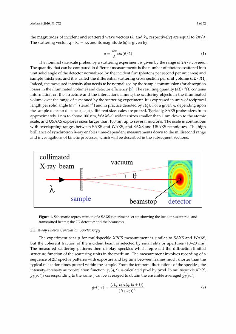
Figure 4. Experiment station at ID02 beamline at the ESRF. (a) The photo displays the sample table andstages, incident side telescopic tube, and the entrance cone of the detector tube. (b) Inside view of the34 m long and 2 m diameter detector tube showing the detector carriage at the end.
The second experiment station at ID10 beamline is optimized for coherent X-ray scattering.The XPCS can be performed in both SAXS and WAXS configurations down to the sub-millisecond timerange. This setup also permits full-field imaging of three-dimensional (3D) specimen by CDI in theSAXS geometry combined with tomographic reconstruction.
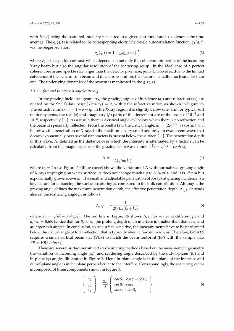
Figure 5. Surface and Interface scattering setup at ID10 beamline at the ESRF. The photo displays the2 + 2 circle diffractometer, the double crystal beam deflector, Langmuir trough, and the detector arm.
3. Investigations of Structure and Dynamics in Bulk Samples
This section presents selected examples of static and time-dependent studies of nanomaterials andsoft matter by X-ray scattering predominantly in their suspensions. The list is certainly not exhaustive

Materials 2020, 13, 752 8 of 52
but illustrates certain unique information that can be derived from scattering experiments underappropriate thermodynamic conditions.
3.1. Equilibrium Nanostructure and Interactions
Here, some representative examples in which SAXS played an important role in elucidating thenanostructure and interactions are described. Traditionally, SAXS and related methods have beenwidely employed for the characterization of particulate systems such as colloids [5], polymers [15],surfactant micelles [16] and vesicles [17], lipid membranes [18] and particles (e.g., cubosomes andhexosomes) [19], proteins [20], etc. SAXS and GISAXS methods have been extensively used innanoparticle research in particular for in situ studies [21,22]. In dilute samples, the main structuralfeatures derived are average size, polydispersity, shape and morphology of particles, and the internaldensity distribution [5,22]. The high brilliance of synchrotron X-rays has enabled studies of extremelydilute systems, such as aerosol suspensions [23], dusty plasmas [24], etc., and allowed obtaining themean size and size distribution of primary particles as well as their aggregates and agglomerates.In sterically stabilized colloids, a systematic variation of surface grafts and their influence on thecolloidal stability has been probed by USAXS [25], and, surprisingly, the shorter grafts were found toprovide a better stability against the salting-out effect [26]. In concentrated systems, the interparticleinteractions are significant, and the measured SAXS intensity becomes dominant of the structure factor,S(q), of interactions [5,22,27]. A quantitative analysis of S(q) provides the strength and range of thepotential of mean force between the particles. Highly concentrated samples of uniform particles forma variety of ordered states such as colloidal crystals [8] or lyotropic phases in the case of anisotropicparticles [28] and high-resolution SAXS revealed their structure and long-range order within.
Colloidal systems also turn into gels and glasses depending on the concentration and interactions,and SAXS has been used to probe the underlying long-range and short-range interactions [27,29,30].A similar approach has been employed to unravel the combined effects of ionic strength, temperature,and pressure on protein–protein interaction potential and the phase behavior in dense lysozymesolutions [31]. A recent SAXS investigation probed the evolution of protein–protein interactions andliquid–liquid phase separation induced by trivalent salts and temperature in concentrated bovineserum albumin (BSA) solutions [32]. Proteins immobilized on polyelectrolyte brushes is another topicstudied by SAXS, which enabled quantitative estimation of the concentration and location of adsorbedproteins within the brush layer [33,34]. SAXS can be used for easy screening of different micellarmorphologies in interpolyelectrolyte complexes of miktoarm star polymers and diblock copolymerwhen the soluble arm number is systematically varied [35]. The mesoscopic scale structural complexityin room temperature ionic liquids (RTIL) was elucidated by SAXS and found that an intermediaterange order appears to drive their peculiar properties [36].
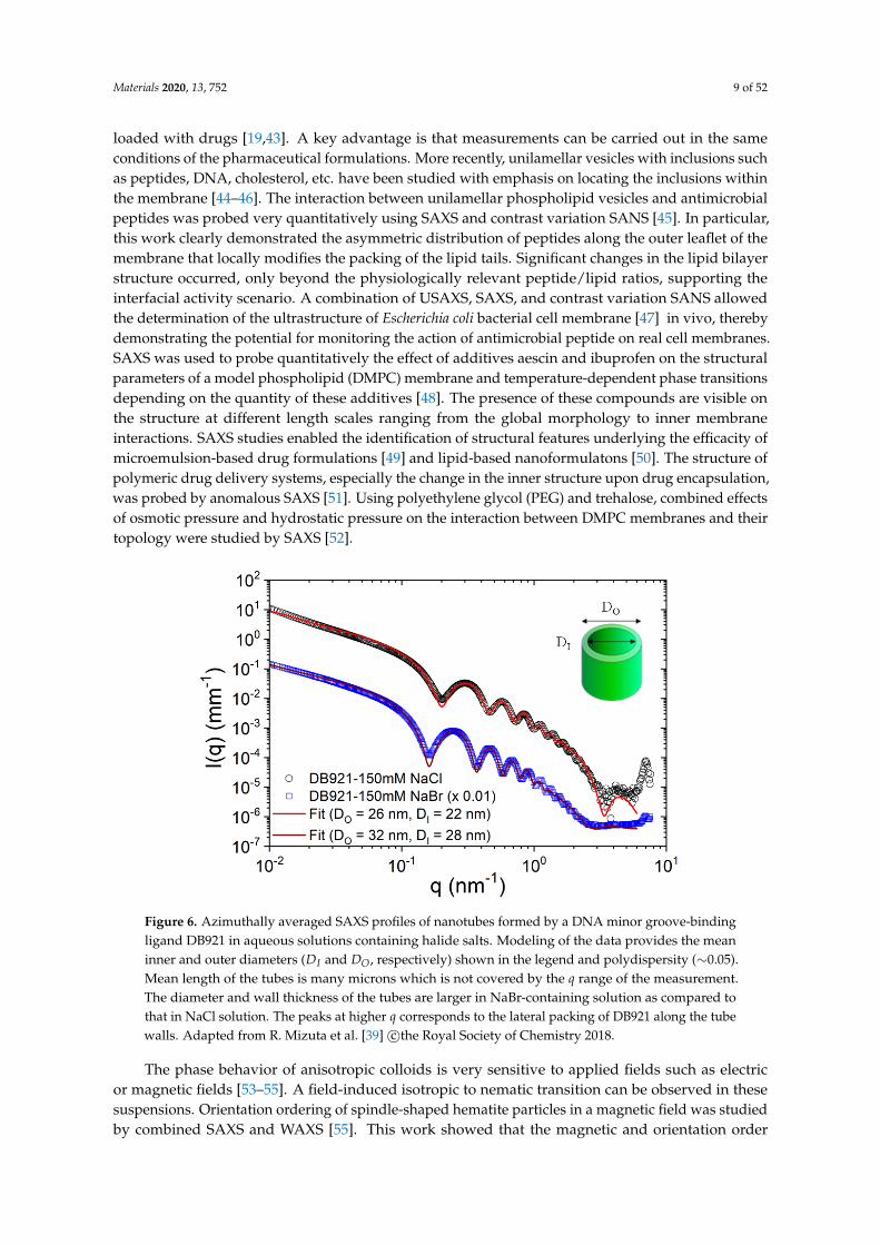
The broad range of size scales accessible by combined SAXS, WAXS, and USAXS is particularlysuitable for elucidating the hierarchical supramolecular organization in a variety of self-assembledsystems [3]. For instance, a combination of SAXS and USAXS elucidated the multiscale morphologyin a prototypical photovoltaics (OPV) thin film consisting poly(3-hexylthiophene) (P3HT) and[6,6]-phenyl-C61-butyric acid methyl ester (PCBM) [37]. Combined SAXS and WAXS revealed thehierarchical morphology of molten and semicrystalline vitrimers [38]. A spectacular case is thehierarchical organization of certain amphiphilic molecules to form well-defined nanotubes [39,40]and microtubes [41], which can be unraveled by SAXS. For example, the nanotubes formed by someamphiphilic peptides such as amyloid β-peptide, which display multiple structural levels from themolecular scale up to the long range ordering of nanotubes [3]. Figure 6 depicts the structural featuresof a suspension of nanotubes formed by an heterocyclic ligand DB921 [39]. In this class of systems, thecompeting hydrophobic and electrostatic interactions lead to a variety of self-assembly pathways toform helical ribbons [39,42] and their closed conformation such as nanotubes [39,40].
Small-angle neutron scattering (SANS) and SAXS methods have been widely used for theelucidation of the morphology and internal organization of liposomes and other lipid nanoparticles
Materials 2020, 13, 752 9 of 52
loaded with drugs [19,43]. A key advantage is that measurements can be carried out in the sameconditions of the pharmaceutical formulations. More recently, unilamellar vesicles with inclusions suchas peptides, DNA, cholesterol, etc. have been studied with emphasis on locating the inclusions withinthe membrane [44–46]. The interaction between unilamellar phospholipid vesicles and antimicrobialpeptides was probed very quantitatively using SAXS and contrast variation SANS [45]. In particular,this work clearly demonstrated the asymmetric distribution of peptides along the outer leaflet of themembrane that locally modifies the packing of the lipid tails. Significant changes in the lipid bilayerstructure occurred, only beyond the physiologically relevant peptide/lipid ratios, supporting theinterfacial activity scenario. A combination of USAXS, SAXS, and contrast variation SANS allowedthe determination of the ultrastructure of Escherichia coli bacterial cell membrane [47] in vivo, therebydemonstrating the potential for monitoring the action of antimicrobial peptide on real cell membranes.SAXS was used to probe quantitatively the effect of additives aescin and ibuprofen on the structuralparameters of a model phospholipid (DMPC) membrane and temperature-dependent phase transitionsdepending on the quantity of these additives [48]. The presence of these compounds are visible onthe structure at different length scales ranging from the global morphology to inner membraneinteractions. SAXS studies enabled the identification of structural features underlying the efficacity ofmicroemulsion-based drug formulations [49] and lipid-based nanoformulatons [50]. The structure ofpolymeric drug delivery systems, especially the change in the inner structure upon drug encapsulation,was probed by anomalous SAXS [51]. Using polyethylene glycol (PEG) and trehalose, combined effectsof osmotic pressure and hydrostatic pressure on the interaction between DMPC membranes and theirtopology were studied by SAXS [52].
Figure 6. Azimuthally averaged SAXS profiles of nanotubes formed by a DNA minor groove-bindingligand DB921 in aqueous solutions containing halide salts. Modeling of the data provides the meaninner and outer diameters (DI and DO, respectively) shown in the legend and polydispersity (∼0.05).Mean length of the tubes is many microns which is not covered by the q range of the measurement.The diameter and wall thickness of the tubes are larger in NaBr-containing solution as compared tothat in NaCl solution. The peaks at higher q corresponds to the lateral packing of DB921 along the tubewalls. Adapted from R. Mizuta et al. [39] c©the Royal Society of Chemistry 2018.
The phase behavior of anisotropic colloids is very sensitive to applied fields such as electricor magnetic fields [53–55]. A field-induced isotropic to nematic transition can be observed in thesesuspensions. Orientation ordering of spindle-shaped hematite particles in a magnetic field was studiedby combined SAXS and WAXS [55]. This work showed that the magnetic and orientation order

Materials 2020, 13, 752 10 of 52
parameters of magnetic single-domain nanospindles can be described by an oriented ellipsoid with theeasy axis of magnetization lying in the equatorial plane of the particle. Texture analysis of the WAXSdata further confirmed that the magnetic easy axis is located in the basal plane of the hematite crystallattice [55]. The field-induced orientation of hematite particles can be used to probe the viscoelasticresponse of a gel such as that formed by poly(N-isopropylacrylamide) (PNIPAM) and correlate withthe microrheological parameters [53]. With increasing elasticity of the gel, the transition to the nematicorder occurred at progressively large value of the magnetic field. TR-SAXS was used to follow therotational dynamics of anisotropic magnetic particles (e.g., hexaferrite platelets) in an alternatingmagnetic field [54] and explore the magneto-optical properties of the system.
The structural colors in both natural and synthetic systems have been the subject of investigationby SAXS [56,57]. These natural colors, such as in bird feathers, butterfly wings, insect scales, etc.,originate purely from the underlying microstructure or biophotonic morphology developed by thephase separation of polymerizing β-keratin. The microstructure can be tuned continuously by theextent of phase separation and the measured structural colors can be analyzed in terms of onedimensional correlation functions [57]. Synthetic systems consisting of chameleon-like elastomersformed by the self-assembly of linear–bottlebrush–linear triblock copolymers display molecularlyencoded strain-adaptive stiffening and coloration [58]. In these systems, the microphase separation ofthe architecturally distinct blocks results in physically cross-linked networks which can be exploredby SAXS. The polymerization-induced self-assembly is a powerful approach for the synthesis of arange of block copolymer morphologies such as spheres, worms, vesicles, etc. [59]. SAXS has beenused to follow the evolution of these morphologies and gain insight into their formation mechanismdepending on the reaction conditions [59]. It is possible to encapsulate large amounts of nanoparticlesin these large block copolymer vesicles during the synthesis and trigger their release over slower timescales by temperature or pH change [60]. SAXS studies showed that at lower loading densities, thecomplete release of particles is associated with a block copolymer vesicle to micelle transition, whereasat higher loading, the release is via perforations on the vesicles and remaining particles stabilize thevesicle structure [60].
The availability of extremely brilliant sources and high-performance detectors will improve thedetection capability of SAXS and allied techniques, thereby enabling investigations of broader sizescales and weaker structural features, which cannot be resolved by direct imaging methods [13].Indeed, an appropriate sample environment is essential for performing an advanced in situ scatteringexperiment [61].
3.2. Probing the Pathways of Self-Assembly
As shown earlier, SAXS has been extensively used for the characterization of multiscale structurein self-assembled systems. Elucidating the energetic pathways of self-assembly processes is not only offundamental interest but also important for the rational design of many functional materials. In thisrespect, TR-SAXS experiments have provided valuable structural insights with model systems [17].The stopped-flow rapid mixing that allows easy change of concentration or pH or ionic strength is apractical method for initiating the self-assembly process in the millisecond range. This approachhas been used for probing the pathways of amphiphilic self-assembly including micellization,micelle–vesicle transition, micellar shape transformation, etc. [3]. In oppositely charged mixedsurfactant systems that form unilamellar vesicles over a broad concentration range, the same finalstructure can be obtained by different routes involving disk-like or cylindrical and torus-like mixedmicelles revealing the energetic stability of unilamellar vesicles [62]. Figure 7 illustrates a commonpathway followed in the formation of unilamellar vesicles from oppositely charged surfactant micellesvia disk-like mixed micelles. Such transient intermediate structures can be stabilized by admixingwith an amphiphilic copolymer having an hydrophobic block length comparable to the surfactantbilayer thickness [63], providing long-term stability desired in their potential applications such asnanoreactors, nanocarriers, etc.

Materials 2020, 13, 752 11 of 52
Similarly, the morphological transformations of surfactant micelles can be followed bystopped-flow TR-SAXS with millisecond range time-resolution. A nice illustration is the formation oflong flexible cylindrical, worm-like, micelles when NaCl is added to an aqueous sodium dodecyl sulfate(SDS) solution [64]. The initial spherical micelles first transformed to elongated globular structures andthen fused together to form long flexible cylinders approximately following a step-like polymerizationtype kinetics. In another investigation, the transition from spherical to cylindrical micelles upon mixingnonionic and anionic micelles revealed a two-step process involving unimer exchange between micellesfollowed by fusion of mixed spherical micelles to form cylindrical micelles [65]. In the case of a so-calledplatonic micellar system, a sharp transition from dodecamer to icosamer morphology was detectedwith the change in ionic strength [66]. Resolving this type of shape transformations require veryprecise measurements with SAXS intensities comparable on an absolute scale. TR-SAXS combined withstopped-flow mixing has also been applied in the investigation of phase transitions in amphiphilicliquid crystalline systems, which are relevant to therapeutic applications [67,68]. These systems arecharacterized by stimuli-responsive nanochannel architectures consisting of hydrophobic membraneouscompartments and aqueous channels. When cationic lipid cubosome nanocarriers uptake neurotrophicplasmid DNA that loads into hydrated channels, lipoplexes with a multilamellar architecture areformed in millisecond timescale [67]. With the uptake of a neurotrophic protein, a sequence of transientstructures has been observed where the lipid membrane curvature changed continuously resulting in atransition from inverted hexagonal–lamellar bilayer–bicontinuous cubic double diamond (Pn3m) to thefinal bicontinuous cubic gyroid (Ia3d) structure within a second [68]. In these studies, nanometer spatialresolution and millisecond time resolution make TR-SAXS unique and allowing to stitch together thelimited real space information obtained from electron microscopy. This aspect of TR-SAXS has been keyto the investigation of rapid structural changes during the osmotic shrinkage in a pharmacologicallyrelevant liposome system [69]. Here, the quantitative analysis of the TR-SAXS intensity provided thetime evolution of the radial electron density profile of the complex particles, which in turn revealed thestructural dynamics of the liposomes at the nanoscale.
The temperature and pressure are important parameters for controlling the nanostructure oflipid vesicular and liquid crystalline phases [70]. In stimuli-responsive drug-loaded formulationsand vesicles loaded with gold nanoparticles, structural transformations and millisecond rangeintermediates have been detected by TR-SAXS combined with rapid temperature jump [70].The lamellar–gyroid cubic phase transition in partially hydrated monolinolein was probed on themillisecond time scale following a pressure jump [71]. Results showed that the phase transitionproceeds via a structural intermediate, the elastic energy in the bilayer drives the initially formed gyroidcubic phase to its equilibrium lattice parameter. Using highly swollen cubic phases of ternary lipidmixtures sensitive to temperature and pressure, it has become possible to achieve lattice dimensionscomparable to those observed in biological systems [72]. Such systems offer the possibility for rationaldesign of lyotropic phases suitable for investigating enzymatic studies, drug encapsulation, therapeuticdelivery, etc. A spectacular example of thermally controlled hierarchical self-assembly has beenrevealed in a relatively simple system composed of a naturally abundant circular polysaccharideβ-cyclodextrin (β-CD) and surfactant SDS (in 2:1 molar ratio) resulting in hierarchically organizedmicrotubes of macroscopic dimension [41,73]. The self-assembly is induced by cooling the solutionfrom 75 C to 25 C and SAXS revealed the in-plane ordering of the SDS-cyclodextrin capsids, thelamellar staking of the membranes and USAXS elucidated the structure of microtubes. TR-SAXSenabled following the exact sequence of steps in the assembly process of the membrane and theirclosing to form micron size microtubes, and uncover their subsequent inward growth as depicted inFigure 8 [74]. An important insight from the TR-SAXS experiment is that the multilamellar structuredeveloped after the closure of the membrane to single-walled tubes by further nucleation and growthinwards which cannot be inferred from a static measurement. This type of self-assembly process mayoccur in a broader class of systems forming ordered multilamellar structures.

Materials 2020, 13, 752 12 of 52
Figure 7. Self-assembly of unilamellar vesicles upon rapid mixing of 25 mM solutions of anionic lithiumperfluorooctanoate (LPFO) surfactant and zwitterionic tetradecyldimethylamine oxide (TDMAO)micelles. (a) Schematic representation of the initial morphologies of LPFO (below the critical micellarconcentration) and TDMAO cylindrical micelles. (b) Time evolution of the SAXS intensity from a fewmilliseconds up to several seconds. (c) Modelling of selected SAXS profiles depicting the morphologicalchange from the initially formed disk-like micelles to unilamellar vesicles when the size of the disksbecome larger. Adapted from J. Gummel et al. [62] c©the Royal Society of Chemistry 2011.
Figure 8. Hierarchical self-assembly of SDS and β-CD to form microtubules upon cooling a 10 wt%solution from 75 C to 25 C. (a) Schematic representation of the structural moieties involved in theassembly process. Two β-CD molecules and one SDS molecule form a capsid at higher temperature dueto hydrophobic interactions as the interior of circular polysaccharides is slightly hydrophobic. Uponcooling, the capsids organize to crystalline bilayers, which then roll to form single-walled microtubesand subsequently the multilamellar structure develops by an inward growth. (b) Evolution of the SAXSand USAXS profiles showing the sequence of growth represented in panel (a). (c) 2D SAXS patternfrom an oriented sample in the final state depicting the sawtooth Bragg peaks from a curved rhombiclattice, and form and structure factors of the multilamellar stacks within the tube walls. Adapted fromJ. Landman et al. [74] distributed under a Creative Commons Attribution Non Commercial License 4.0(CC BY-NC).

Materials 2020, 13, 752 13 of 52
More recently, the assembly pathways in the formation of interpolyelectrolyte complexes(or polyelectrolyte coacervates) have been investigated by TR-SAXS combined with stopped-flowmixing [75–78]. For example, the complex formation between sodium polyacrylate (SPA) andpolyallylamine hydrochloride (PAH) in aqueous NaCl solution was investigated by TR-USAXS fordifferent NaCl concentrations from 0 to 1 M at equimolar concentrations of the monomer units [75].Within the mixing dead time (∼2.5 ms), percolated aggregate-like structures were observed suggestingthat the initially formed small charge neutral aggregates further assembled to form higher orderagglomerates within a short time. The early stage time evolution of the molar mass of the largeglobular structure was found to be comparable with the Brownian-coagulation rate. The kineticsof complexation between the oppositely charged ionic/nonionic block copolymers with a branchedstar-shaped architecture and a thermoresponsive diblock (PNIPAM block) was investigated by mixingaqueous solutions (0.3 M NaCl) of both polymers for a charge ratio of 1 [76]. The complexationwas essentially completed during the mixing and the resulting micelles remained stable over themeasurement time, but their number density increased over the initial few seconds. Polyelectrolytecomplex micelles formed from an anionic-neutral block copolymer, and a cationic-neutral blockcopolymer in aqueous NaCl solution exhibit different morphologies such as spheres or cylindersdepending on their mixing ratios. A transition from sphere to cylinder and vice versa can be inducedby mixing complex micelles with respectively pure cationic or anionic copolymers in aqueous NaClsolutions [77]. Morphological transformations in these systems take place on much longer time scalethan in surfactant solutions. The cylindrical micelles transformed to spherical shape via the randomscission of the cylinders along their contours in minute scale and the reverse process from spherical tocylindrical micelles was even slower with a high activation energy. The formation of polyelectrolytecoacervates with spherical core–shell morphology via a chain exchange mechanism has also beenproposed [78].
3.3. Assembly of Biomacromolecular Complexes
Traditionally, TR-SAXS and related methods have been used for probing the structural dynamicsin biomacromolecular systems which shed light on a broad range of biological functions. A well-knownexample is the study of muscle contraction along the pathway of physiological activation [79]. Duringthis decade, TR-SAXS enabled probing in depth the self-assembly of virus particles [80,81], tubulinsingle rings [82], etc. from their constituting subunits. Detailed knowledge of virus assemblyand disassembly under conditions similar to the host cells is important for the development ofmore effective vaccines. The rapid assembly of Simian vacuolating virus 40 (SV40) icosahedralparticles from 12 pentameric viral protein capsids induced by a short RNA was investigated bystopped-flow mixing combined with SAXS. The observed kinetics was modelled by a two-state processwithout an intermediate [80]. The encapsidation process once nucleated at the RNA continued by astepwise addition mechanism in which the growing nucleoprotein complex acts as an electrostaticantenna attracting other capsid subunits. In another study involving the self-assembly kineticsof norovirus capsid proteins, three species were found to contribute to the total SAXS intensities:dimers, intermediates comprising 11 dimers, and the icosahedral capsids made up of 90 dimers [81].This biphasic kinetics involved a fast step in which dimers are assembled into intermediates, followedby a slow step in which intermediates interlock into capsids shedding new light on the generallyaccepted models for the assembly of norovirus capsids. The pH-driven disassembly of viral capsidsderived from an icosahedral plant virus, the cowpea chlorotic mottle virus (CCMV), to dimers wasfound to be different from the assembly pathway and involved two distinct intermediates [83].More recently, TR-SAXS was used to elucidate the nonequilibrium self-assembly dynamics of CCMVcapsids packaging their RNA genome [84]. As shown in Figure 9, the experiment revealed theformation of amorphous complexes collectively with the genome acting as a template for the assembly,capturing a large number of subunits. These complexes relaxed into virions via a synchronouspathway in a slower process. The low q SAXS intensity was used to estimate the mean number of

Materials 2020, 13, 752 14 of 52
subunits bound on the genome as a function of time and the corresponding binding time constant,whereas the structural information was derived from the analysis of the scattering form factor. Thetemperature dependence of the relaxation time of the viral complexes allowed the determination ofthe activation energy of viral complexes to fully grown virus particles with the correct sequence ofsubunits. The binding energy of subunits on the genome was found to be moderate (≈7 kBT) while theself-organization of nucleoprotein complexes into viruses involved a high energy barrier (≈20 kBT).This barrier is significantly lower for a synthetic polyelectrolyte, such as poly(styrene sulfonic acid), ascompared to RNA genome, but the resulting structure lacked the icosahedral symmetry.
Figure 9. Assembly of icosahedral virus particles from the constituent capsids. (a) Schematic view of avirus particle with dimeric subunits and RNA genome. The expanded view displays the molecularstructure of the subunit with one of its RNA binding domains. (b) Evolution of the SAXS intensityduring the assembly process. The cartoon shows the rapid binding of subunits onto the genome andthen the slow reorganization of the complex to the final structure. (c) Arrhenius plot of the bindingtime constant (τbind) deduced from low q SAXS intensity at different temperatures, which providedan estimate of the activation energy (Ea). Adapted from M. Chevreuil et al. [84] licensed under theCreative Commons Attribution 4.0 International License.
In other TR-SAXS studies, the crystallization of wild type SV40 virus particles to body-centeredcubic (bcc) structure upon dialysis with MgCl2 and their reentrant melting at higher MgCl2concentrations were investigated [85]. Thermodynamic modeling of the transition at different saltconcentrations suggested that the entropy of counterions is the driving mechanism. Swelling processof SV40 virus particles upon chelating calcium ions and reducing disulfide bonds was probed bySAXS and results provided a better insight into internal domain interactions and the binding of thecapsid proteins in compact conformation [86]. TR-SAXS was also used for monitoring the large-scaleconformational transitions of a two-state DNA origami switch from its open to closed conformationupon increasing the ionic strength in millisecond time scale, and found that the kinetics is close tothe limit set by diffusion [87]. Time-resolved USAXS investigations of liquid–liquid phase separationkinetics in concentrated BSA solutions induced by trivalent salts and temperature revelaed spinodaldecomposition and an arrested spinodal process [88]. Arrested and temporarily arrested states wereobserved in protein (bovine γ-globulin)–PEG mixtures upon quenching to the two-phase regiondepending on the magnitude of the temperature change [89]. In a more recent study, the influence oftuning the protein interactions on the spinodal decomposition process and formation of arrested stateswere systematically investigated [90].
3.4. In Situ Studies of Nucleation and Growth
Time-resolved simultaneous SAXS and WAXS methods have been extensively used forinvestigating the early stage of nucleation and growth in a variety of systems such as the pyrolyticsynthesis of nanoparticles, precipitation of inorganic materials from supersaturated solutions [5], etc.This allowed the observation of nucleation process and clarifying the growth mechanism in thesesystems. More recent studies include in situ investigations of nucleation and growth of nanocrystallineparticles such as quantum dots [91], porous metal organic frameworks (MOF) [92], etc. The different

Materials 2020, 13, 752 15 of 52
steps involved in the nucleation and growth of cadmium selinide (CdS) quantum dots from a surfactantmesophase precursor have been identified [91]. In this case, the surfactant lamellar phase transformedto micelles, and within which the activated monomers nucleated and formed nanocrystals stabilizedby an outer surfactant layer. In the MOF case, the nanocrystal formation process involved the initialnucleation of amorphous clusters and their further growth by a coagulation mechanism and subsequenttransformation to crystalline particles with specific zeolite topology, via intraparticle nucleation andstructural reorganization [92]. In the case of strongly scattering samples, a stable free-jet of reactantscan be combined with the SAXS set-up. This has allowed probing the prenucleation and nucleationstages in the sub-millisecond time as demonstrated in the case of CdS quantum dot formation [93].
TR-SAXS allowed a direct structural study of the micellization kinetics of surfactants that hasbeen elusive for a century [16]. Figure 10 presents the experimental scheme and the main resultswhich revealed a single step kinetics akin to nucleation and growth. The micelle formation isdescribed as an insertion/expulsion process of unimers (isolated surfactant molecules) withoutany intermediate pre-micellar structures. A similar self-assembly and the opposite process ofdisintegration have been monitored in photosensitive surfactant (azo-benzene-based surfactantsthat can isomerize upon photon absorption) system by shining light of the appropriate wavelength(blue and ultraviolet, respectively) [94]. The kinetics appeared to follow a similar pathway of singlestep for micellization though the underlying kinetics is primarily determined by the quantum yieldof light absorption. The demicellization involved a two-step process of the fast release of unimersfollowed by slower disintegration.
Figure 10. Nucleation and growth of surfactant (dodecyl maltoside) micelles from a solution ofmonomers in dimethylformamide (DMF) upon mixing with water. (a) Schematic drawing of thestopped-flow mixing device and the scattering cell with surfactants. (b) Time evolution of the SAXSintensity during the nucleation and growth process for 1 wt% solution. (c) Time evolution of themean aggregation number derived from the modeling of SAXS intensities for different surfactantconcentrations. The continuous lines show the good agreement with nucleation and growth model.Adapted from G.V. Jensen et al. [16] c©the American Chemical Society 2013.
A combination of SAXS and USAXS allowed the elucidation of the mechanism underlyingthe formation of ultrathin (∼2 nm) colloidal Cu2−xS nanosheets with well-defined shape andsize [95]. The thermal decomposition of copper–dodecanethiolates usually leads to spheroidalCu2−xS nanocrystals, but chloride stabilization of the stacks of lamellar copper-thiolate supramolecularcomplexes led to 2D-constrained stack-templated nucleation and growth, in which growth in thethickness direction is inhibited (allowing only the lateral growth). Another fascinating example isthe formation of supraparticles (∼700 nm) from nanocrystals (∼12 nm) confined within oil dropletsin an oil-in-water emulsion upon slow evaporation [96]. The nanoparticles consisted of an FeO core,a CoFe2O4 shell, and oleate capping ligands. Upon evaporation, the volume fraction of particles insidethe oil droplets gradually increased up to ~20%, at which crystallization occurred instantaneously
Materials 2020, 13, 752 16 of 52
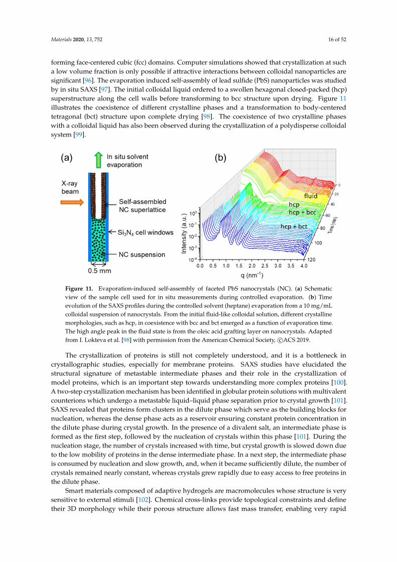
forming face-centered cubic (fcc) domains. Computer simulations showed that crystallization at sucha low volume fraction is only possible if attractive interactions between colloidal nanoparticles aresignificant [96]. The evaporation induced self-assembly of lead sulfide (PbS) nanoparticles was studiedby in situ SAXS [97]. The initial colloidal liquid ordered to a swollen hexagonal closed-packed (hcp)superstructure along the cell walls before transforming to bcc structure upon drying. Figure 11illustrates the coexistence of different crystalline phases and a transformation to body-centeredtetragonal (bct) structure upon complete drying [98]. The coexistence of two crystalline phaseswith a colloidal liquid has also been observed during the crystallization of a polydisperse colloidalsystem [99].
Figure 11. Evaporation-induced self-assembly of faceted PbS nanocrystals (NC). (a) Schematicview of the sample cell used for in situ measurements during controlled evaporation. (b) Timeevolution of the SAXS profiles during the controlled solvent (heptane) evaporation from a 10 mg/mLcolloidal suspension of nanocrystals. From the initial fluid-like colloidal solution, different crystallinemorphologies, such as hcp, in coexistence with bcc and bct emerged as a function of evaporation time.The high angle peak in the fluid state is from the oleic acid grafting layer on nanocrystals. Adaptedfrom I. Lokteva et al. [98] with permission from the American Chemical Society, c©ACS 2019.
The crystallization of proteins is still not completely understood, and it is a bottleneck incrystallographic studies, especially for membrane proteins. SAXS studies have elucidated thestructural signature of metastable intermediate phases and their role in the crystallization ofmodel proteins, which is an important step towards understanding more complex proteins [100].A two-step crystallization mechanism has been identified in globular protein solutions with multivalentcounterions which undergo a metastable liquid–liquid phase separation prior to crystal growth [101].SAXS revealed that proteins form clusters in the dilute phase which serve as the building blocks fornucleation, whereas the dense phase acts as a reservoir ensuring constant protein concentration inthe dilute phase during crystal growth. In the presence of a divalent salt, an intermediate phase isformed as the first step, followed by the nucleation of crystals within this phase [101]. During thenucleation stage, the number of crystals increased with time, but crystal growth is slowed down dueto the low mobility of proteins in the dense intermediate phase. In a next step, the intermediate phaseis consumed by nucleation and slow growth, and, when it became sufficiently dilute, the number ofcrystals remained nearly constant, whereas crystals grew rapidly due to easy access to free proteins inthe dilute phase.
Smart materials composed of adaptive hydrogels are macromolecules whose structure is verysensitive to external stimuli [102]. Chemical cross-links provide topological constraints and definetheir 3D morphology while their porous structure allows fast mass transfer, enabling very rapid

Materials 2020, 13, 752 17 of 52
structural adaption to changing environment. This structural evolution during the transformation ofPNIPAM microgel having a flexible macromolecular network with a fuzzy interface to particle withwell defined surface and homogeneous density was probed by TR-SAXS. Results revealed a two-stagekinetics involving a very fast process in which the collapsed clusters forming at the periphery (hollowcore–shell) and a slower process in which the hollow core–shell structure transforming to a globularparticle. This structural evolution appeared to be independent of the type of stimulus such as atemperature jump or a change of solvent quality suggesting the generality of the mechanism.
3.5. Equilibrium Dynamics
The equilibrium microstructures of soft matter and many nanomaterials are not rigid and fluctuatein time due to ambient fluctuations, which could be of thermal origin or similar. There is an associateddynamics that can be Brownian motion in particulate systems, chain reptation in an entangled polymermelt, or membrane undulations in the case of a lyotropic system. It is often more challenging to probethis equilibrium dynamics as compared to the corresponding microstructure. DLS is a well-establishedmethod for investigating the dynamics in suspensions. However, there are limitations in terms of qrange and concentrations which can be studied, especially when the sample becomes turbid. With theadvent of third generation synchrotron sources, XPCS has emerged as an alternative method forprobing the equilibrium dynamics in such systems [9,103–105]. The XPCS technique exploits thecoherence properties of the X-ray beam and the measured scattering patterns display speckles asshown in Figure 12. The corresponding pattern registered with a partially coherent beam appears lessgrainy with nearly smooth intensity distribution since the speckle size in that case is much smaller thanthe detector resolution. The visibility or contrast of the speckle depends not only on the longitudinaland transverse coherence of the beam, but also on the angular resolution defined by the detectorpixel elements. The g2(q, t) function of the fluctuating speckles reveal the dynamics within the systemfor a given q value as displayed in Figure 12. The time scale accessible by XPCS is determined bythe scattering power of the sample, available coherent photon flux, and detector capabilities. Themain applications of XPCS have been to probe the dynamics of colloids, especially those that areturbid in visible light [106] and slow dynamics in arrested systems such as gels [29] and glasses [27].Faster dynamics at a given q can be accessed with a point detector [106]; however, an additionallimitation is the onset of radiation damage with longer exposure of the sample to the X-ray beam.In dilute suspensions of Brownian particles, the dynamics is purely diffusive, and g2(q, t) decays by anexponential function and the decay rate is related to the diffusion coefficient, D0.
Figure 12. Multispeckle XPCS from a dilute colloidal suspension of silica particles (radius: ~215 nm)in water/3-methylpyridine mixture. (a) Representative 2D speckle patterns recorded in the USAXSrange and the white circle indicating a given q. (b) Typical ensemble averaged intensity-intensityautocorrelation function (g2(q, t)) and fits to an exponential decay with rate, Γ. The inset shows thatΓ = D0q2, with D0 ∼ 0.6 µm2 s−1, as expected for dilute Brownian particles.
Materials 2020, 13, 752 18 of 52
The XPCS can be performed in both SAXS and WAXS configurations. The wide-angle XPCSprovides access to atomic dynamics in relatively slow systems such as supercooled liquids andmolecular glasses [104,105]. Recent studies of atomic dynamics include aging behavior of metallicglasses [107] and beam induced dynamics of oxide glasses [108]. For the investigation of arrestedsystems, XPCS performed with a 2D detector has a clear advantage, as multiple speckles along anazimuthal circle for any given q (multispeckle XPCS) are recorded simultaneously as demonstrated inFigure 12. This readily allows obtaining the ensemble averaged g2(q, t) of these non-ergodic systems.In addition, the two time correlation function, g2(q, t1, t2), can be used to visualize the ageing behaviorof the system as a function of time [3,105]. A common feature of these systems is hyperdiffusivedynamics characterized by a compressed exponential decay of g2(q, t) [9,105]. Although such anon-diffusive behavior could arise due to various reasons, a generally assumed mechanism is thepresence of dynamic heterogeneities and associated internal stress relaxations [105,109].
The multispeckle XPCS is also a valuable tool for analysing the direction dependent dynamics,as illustrated in the case of shear flow [103] and sedimentation [110], where g2(q, t) involvesboth diffusive and advective contributions. Disentangling the information requires simultaneousmeasurements at many q values both along the vertical and horizontal directions. This enabledthe measurement of velocity fluctuations at the early stage of sedimentation in brownian colloidalsuspensions [110]. Similar approach is required for probing the anisotropic dynamics in an appliedmagnetic field [106,109] and in confined geometry of microfluidic channels [111]. In these cases,the measured microstructure and dynamics display strong anisotropy. Figure 13 illustrates theobserved anomalous dynamics and chain formation by peanut-shaped anisotropic magnetic particlesin a magnetic field. Fast multispeckle XPCS has allowed the investigation of phoretic dynamicsof colloids [112] and active motions of Janus colloids [113] in phase separating solvent mixtures.The availability of fast 2D detectors such as the Eiger-500k pixel detector has enabled multispeckleXPCS measurements in the sub-millisecond range [114].
Another important application of XPCS is to probe dynamics at surfaces and interfaces [9] andin films [115] using grazing incidence geometry (GIXPCS). For instance, this method has enabledquantitative studies of capillary wave fluctuations on liquid surfaces and in polymer films [9]and to verify the predictions of hydrodynamic continuum theories. The heterogeneous dynamicsof 2D gels was probed in Langmuir films [116], which allowed for the derivation of the fourthorder time correlation function and to detect the characteristic time of dynamical heterogeneity.For off-specular geometries, nanoparticles were used as tracer particles to probe the interior dynamicsin polymer films and monolayers [9]. An induced dynamical arrest transition was observed inphospholipid/nanoparticle monolayers that is again featured by an hyperdiffusive dynamics [115].
The scope of XPCS will be significantly enlarged with the availability of extremely brilliantsynchrotron sources and even faster pixel array detectors. The feasibility of studying ultrafast dynamicsusing an alternative method—speckle visibility spectroscopy—has already been demonstrated at theX-ray free-electron laser (XFEL) [117]. However, this method has limitations when studying complexrelaxation processes involving non-exponential g2(q, t) and non-monotonic q dependence. As a result,the multispeckle XPCS at synchrotrons remains a more straightforward tool for investigating complexdynamics involving multiple length and time scales.
3.6. Flow-Induced Structures
The characteristic feature of soft matter systems is that they are easily deformable by an appliedshear stress. As a result, a large number of investigations have been performed to elucidate themicrostructure as a function of applied shear rate or shear stress [3,118]. A major goal, which is also ofgreat practical interest, has been relating the microstructure to rheological properties. The high degreeof collimation and small size of synchrotron X-ray beams are important for probing highly orderedsystems. For example, elucidating the order–disorder transition underlying shear thickening in densesuspensions of colloidal particles subjected to different oscillatory shear stresses [119]. When combined

Materials 2020, 13, 752 19 of 52
with large amplitude oscillatory shear, XPCS can detect nonlinear rheological behavior such as yieldingand plastic flow during an oscillation [103]. For example, the local signature of yielding duringlarge amplitude oscillatory shear has been detected in concentrated oil-in-water nanoemulsions [120].The shear flow is also an elegant method to orient the sample and deduce high resolution structuralinformation as shown in the case of hexagonal honeycomb and kagome superlattices in surfactantlyotropic hexagonal phase intercalated with single-walled carbon nanotubes [121] and polymernanorods [122], respectively. Figure 14 illustrates the shear-induced orientation of binary supperlatticeswith hexagonal honeycomb and kagome structures.
Figure 13. Anomalous dynamics of peanut-shaped anisotropic magnetic colloids (silica-coated hematitein an aqueous solution) in a magnetic field. (a) Schematic view of the experiment set-up used formultispeckle XPCS measurements. (b) Intensity autocorrelation functions (β ≡ g0: ~0.3) along anhorizontal sector parallel to the applied magnetic field (20 mT) and fits to compressed exponentialfunctions (exponent about 1.5). (c) Anomalous dynamics showing Γ(q)‖ ∝ qn‖ . The inset displays thevariation of n‖ with magnetic field (B). (d) Corresponding intensity autocorrelation functions along thevertical sector perpendicular to the applied magnetic field. In this case, the dynamics is dominated byvelocity fluctuations due to sedimentation [110]. (e) The Brownian part of the dynamics follows theexpected behavior, Γ(q)⊥ ∝ qn⊥ , with n⊥ ∼ 2. Adapted from A. Pal et al. [109] c©Weiley-VCH VerlagGmbH & Co. KGaA 2018.
Model colloidal fluids in nanofluidic channel arrays displayed strongly anisotropic structurefactor due to the confinement [111]. This confirmed the theoretical prediction of a confinement-inducedanisotropy of the pair correlation function of a fluid. SAXS allowed the characterization ofmicrostructure within the concentration polarization layer during the cross-flow ultrafiltration processof cellulose nanocrystal dispersions, including the orientation and packing as a function of the distancefrom the membrane, and link to the concentration polarization and identifying the fouling layer nearthe membrane [123].
Recent advances in microfabrication techniques have enabled the realization of microfluidicdevices that can deliver small quantities of liquids (nanolitres) and combine with X-ray scatteringexperiments [124]. They also offer new possibilities for the investigation of soft matter in low Reynoldsnumber flows and under confinement [125]. When combined with microbeam X-ray scatteringmeasurements, the local structure in the flow field can be mapped, thereby offering the possibility ofinvestigating unexplored nonequilibrium states of soft matter. A straightforward application is theinvestigation of flow alignment of complex fluids in microfluidic channels especially from the pointof view of materials processing [125]. For example, the orientation dynamics of a nematic director ina thermotropic liquid crystal was probed under different flow rates and boundary conditions [125].

Materials 2020, 13, 752 20 of 52
Microfluidics are convenient environment for probing conformational changes of proteins and otherbiomacromolecules upon change of the buffer condition [124], and nucleation and growth of a specificpolymorph that may be difficult to isolate in the bulk as shown in the case of calcium carbonateprecipitation [126]. Another important advantage is that the radiation damage can be minimized by thecontinuous flow without consuming too much sample but at the expense of a reduced scattering signalto background ratio. A microfluidic environment can be used to investigate biological supramolecularassemblies by mimicking conditions involved in blood vessels and explore the new physics of suchsystems under confinement [124].
Figure 14. Shear-induced alignment of binary supperlattices formed by hexagonally packed cylindricalmicelles of nonionic surfactants penta(ethylene glycol) monododecyl ether (C12E5) in water and rod-likemicelles of polymerizable cationic surfactants, n-alkyltrimethylammonium 4-vinylbenzoate (CnTVB,with alkyl chain length, n = 10, 12, 14 or 16. (a) Oriented SAXS pattern along the flow direction forp− C14TVB/C12E5/water (10/45/55 weight ratio). (b) Azimuthal variation of intensities along the (10)and (11) reflections (which make the peak position ratio of 1:
√3) for AB2 type structure. Inset shows
the azimuthally averaged intensity. (c) Schematic representation of a AB2 type binary superlattice withan hexagonal array of p− CnTVB rods (smaller spots) intercalated in an honeycomb lattice of C12E5
cylinders. (d) Oriented SAXS pattern along the flow direction for p− C10TVB/C12E5/water (5/45/55weight ratio). (e) Azimuthal distribution of intensities along the (10) and (20) reflections (whichmake the peak position ratio of 1:2) for AB3 type structure. Inset displays the azimuthally averagedintensity. (f) Schematic representation of a AB3 type binary superlattice in which an hexagonal latticeof p− C10TVBs is embedded in a kagome lattice of C12E5 cylinders (larger spots). Adapted from S.-H.Lim et al. [122] licensed under the Creative Commons Attribution 4.0 International License.
Using SAXS combined with a microfluidic cell and optical spectroscopy techniques,the hydrophobic collapse of grafted polystyrene chains on gold nanoparticles suspended intetrahydrofuran (THF) was observed upon mixing with a poor solvent (water), as depicted inFigure 15 [127]. The thickness of polymer shell derived from the SAXS analysis and numericalsimulation of the solvent composition allowed the mapping of the interaction energy between particles,and it was found that the rate of hydrophobic collapse depends on water concentration, ranging

Materials 2020, 13, 752 21 of 52
between 100 and 500 nm/s, and that the polymer shell collapses prior to the onset of clustering of goldparticles.
Figure 15. Probing the hydrophobic collapse of grafted polystyrene chains on gold nanoparticlesin THF upon mixing with water. Initially, repulsive particles became attractive and clustered uponcollapse of the grafted chains. (a) Schematic representation of the 3D flow-focusing microfluidic reactorwith a middle channel (MC) and two side channels (SC), and the simulated concentration pattern ofTHF/water mixture. The X-ray beam passed orthogonal to the flow and the lateral focusing directions.(b) Evolution of the SAXS profiles of the particles as a function of the distance from the mixing point(XC) for a flow rate ratio (FRR) of 2. The peak corresponds to the structure factor of the particles uponclustering and modeling of which provided an effective size of particles. (c) The variation of effectiveparticle radius as a function of XC, which is equivalent to a residence time (tres) given by the flow rate,for different FRR. The chain collapse transition occurred in the millisecond time scale. Adapted from S.Merkens et al. [127] with permission from the American Chemical Society, c©ACS 2019.
3.7. High Resolution Diffraction and Imaging
This subsection describes some of the studies which will greatly benefit from the upgradesof synchrotrons such as the ESRF extremely brilliant source (EBS) or the Advanced Photon Sourceupgrade (APS-U). Indeed, time-resolved SAXS and GISAXS and USAXS will gain from the highbrilliance in terms of time and angular resolutions, whereas XPCS takes advantage of the increase intransverse coherence in dynamic studies. The order of magnitude increase in angular resolution willbe even more significant for small-angle X-ray diffraction (SAXD). Similarly, the enlarged transversecoherence could improve the spatial resolution in coherent diffractive imaging (CDI) down to 10 nmrange. The CDI is a lensless imaging method based on phase retrieval by an iterative procedure from anoversampled (as compared to the Nyquist period) speckle pattern from a noncrystalline specimen [10].Together with tomographic reconstruction, this method can yield 3D images of micron sized objects.
Colloidal crystals are another class of systems which require very high angular resolution inorder to elucidate the long-range order and defects within them [8]. Very high-resolution SAXS(a few microradian) has been pivotal in distinguishing different crystalline as well as liquid crystallinephases formed by anisotropic colloidal particles [8]. This has been nicely illustrated in the caseof superball (shape between a sphere and a cube) particles, which showed a plastic crystal phasewith translational order and orientational disorder and two distinct rhombohedral crystalline phaseswith different stacking variants [128]. Transition between these solid phases occurs depending onthe osmotic pressure. The large transverse coherence of the X-ray beam is essential for revealingthe long range order that may be several orders of magnitude larger than the typical 2π/q scaleprobed by the measurement [8]. Elucidating the different liquid crystalline phases coexisting insuspensions of clay particles with large aspect ratios (diameter: approximately 100 nm to 1 µmthickness: ~1 nm) is a challenge due large periodicity and small domain sizes [129], e.g., differentiatingthe lamellar phase from a nematic phase with strong stacking local order, the so-called columnarnematic. The high-resolution SAXS measurements allowed the clear identification of such phases indilute aqueous suspensions of synthetic Sb3P2O14 nanosheets. Analysis of their sharp X-ray reflectionprofiles in the direction perpendicular to the director revealed that two coexisting mesophases are a
Materials 2020, 13, 752 22 of 52
columnar nematic and a lamellar phase with domain sizes of about 20 µm (deduced from the width ofthe Bragg peak), which corresponds to about 600 nanosheets.
An important motivation for studying long-range ordered colloidal systems has been fromthe point of view of potential photonic materials [8]. More recent investigations employedhigh-resolution SAXS for elucidating the structural basis of colors in biophotonic specimens [57,130,131]. High-resolution studies of butterfly single scales revealed the 3D photonic nanostructureas a single network of gyroid morphology formed by chitin and air which is reminiscent of cubicphases observed in amphiphilic soft matter systems such as surfactants, block copolymers and lipidsin water or block selective solvents. Results supported the hypothesis that color-producing protein andair nanostructures in feather barbs are self-assembled by arrested phase separation of polymerizingβ-keratin [56], and the nanostructure can be varied continuously by regulating the time the keratinnetwork is allowed to phase separate before mobility in the system is arrested [131]. In arthropodscales and setae, a richer nanostructural diversity, including triply periodic bicontinuous networks,close-packed spheres, inverse columnar, perforated lamellar, and disordered spongelike morphologieshave been reported [130]. The challenge in investigating these systems is that not only high angularresolution required to resolve long periodicities, but also the beam cross section should be small toprobe the gradient in color (e.g., feather barbs).
SAXD continued to be valuable for structure–function studies in biological tissues and fibers.A notable example is muscle, where SAXD allows probing the different regulatory states during thecontraction and relaxation [79,132]. For example, high-resolution SAXD together with sarcomere-levelmechanics enabled the identification of a downstream mechanism in muscle regulation, namely thethick filament (myosin) based regulation, which involves a mechano-sensing role of the myosinfilament [133]. Most myosin motors are in a constitutively off state and the transition to on stateoccurs based on the load sensed by the myosin filament. This additional mechanism preservesthe high metabolic efficiency and offers a molecular basis for the length-dependent activationin cardiac muscle [134]. The approach developed for simultaneous investigation of micrometerscale supramolecular organization and nanometer scale filamental protein movements is useful foridentifying the elements of muscle dysfunction [79]. Another example is the investigation of theultrastructural mechanics of collageneous tissues. By measuring the time-dependent changes infibrillar structure during in situ tensile testing, the ultrastructural mechanics could be probed atdifferent chemically induced mechanical states [135]. The variable interfibrillar stiffness can be relatedto the mechanism of mutability at the nanoscale. A model of stiffness modulation via enhancedfibrillar recruitment explains the underlying biophysical mechanism which may have applications indevelopment of new types of mechanically tunable biomaterials.
CDI is an emerging method suitable for obtaining real space images of biological specimen andnanomaterial assemblies in 3D. Figure 16 illustrates the optical and detection schemes employedfor CDI in the SAXS configuration (SAXS-CDI) and the iterative phase retrieval procedure [10].The resolution is primarily limited by the radiation damage and the q range of the speckle patternwith sufficient signal. Radiation sensitive biological cells are typically imaged in the frozen hydratedstate [136,137]. Resolution obtained are in the range of 75 to 100 nm with Neospora caninum [136]and malaria-infected human erythrocyte [137] cells, which can barely resolve the organelles within.At present, the best 3D resolution obtained is in the range 30 to 50 nm for strongly scattering samplessuch as porous calcium carbonate [138] or silica [139] microparticles, titania sponge [140], or goldnanoparticles in polymeric capsules [141]. A very clear illustration of the 3D imaging capability byCDI is the recent study of coccolithophores, unicellular algae of size a few microns, that achieved aresolution close to 30 nm [142]. The images displayed in Figure 16 reveal coccoliths morphology inunprecedented detail and quantitative analysis established a relationship between their mass and thenumber of calcite segments [142]. In addition, a correlation between their overall dimension and size ofcertain organelles within the cell was noticed which may correspond to different stages of their growth.

Materials 2020, 13, 752 23 of 52
Figure 16. (a) The optical and detection schemes used in SAXS-CDI. A plane wave illuminatesthe noncrystalline specimen, and an oversampled speckle pattern is recorded by a high-resolutiondetector. (b) The phase retrieval procedure involves iterating back and forth between real and reciprocalspace. In each iteration, various real space physical constraints such as positive electron density,partially overlapping regions, etc. are imposed, while the measured Fourier amplitude is updated inreciprocal space. Panels (a,b) are adapted from J. Miao et al. [10] with permission from the AmericanAssociation for the Advancement of Science, c©AAAS 2015. (c) 3D images of coccospheres of G. oceanicaRCC1314 reconstructed by tomographic SAXS-CDI. (d) Distal view of the coccolith and a side viewalong the major axis after sectioning half of the extracted coccolith. Panels (c,d) are adapted fromT. Beuvier et al. [142] licensed under the Creative Commons Attribution 4.0 International License.
With the increase in brightness and coherence, and advances of detector technology with betterspatial resolution and larger area, the 3D imaging resolution is expected go down to 10 nm or so.An important goal is to enable imaging of functional systems in nanoscience and biology with nmrange resolution. In this respect, the potential of CDI for imaging a transient process has beendemonstrated [143]. Indeed, the radiation damage is a limiting factor as the imaging requires at least anorder of magnitude higher radiation dose than the corresponding diffraction measurement. Therefore,SAXD will remain relevant because the nanoscale structural and dynamical information, albeit inreciprocal space, can be derived prior to the onset of radiation damage. When investigating complexfunctional systems by CDI, simultaneous analysis of SAXD could provide additional constraints to theimage reconstruction procedure.
4. Surface and Interface Studies
This section describes some representative investigations of nanomaterials and soft matter atsurfaces and interfaces using X-ray scattering methods. X-ray and neutron scattering can derive uniqueinformation at the nanoscale which is not easily obtained by other surface sensitive techniques [3,144].

Materials 2020, 13, 752 24 of 52
4.1. Nanoparticles at Interfaces
Two-dimensional self-assembly of colloidal nanocrystals and nanoparticles at liquid–liquidinterfaces is of both fundamental scientific interest and vast technological relevance. For example,these structures are promising candidates for the fabrication of new metamaterials, photovoltaicdevices, miniature light sources, magnetic storage media, etc. XRR enables measuring the locationof nanoparticles with respect to the interface of immiscible liquids and determine quantitiessuch as contact angle, binding energy, and interparticle distance, which control their lateralorganization [145,146]. A GISAXS study of the CdSe-CdS core–shell nanorods [147] self-organizationat the liquid–vapor interface into 2D crystals revealed an unexpected change from parallel toperpendicular orientation of the rods with respect to the liquid surface. Simultaneous measurementsof GISAXS and GIWAXS during evaporation-driven precipitation of wurtzite ZnS bifrustum-shapednanocrystals at toluene–air interface [148] provided, for the first time, information about different stagesof development of 2D superstructure with long-range orientational order from a disordered assemblyof nanoparticles as depicted in Figure 17. Here, the atomic plane orientation was induced by oleic acidligands, thereby promoting the superlattice formation. Further application of simultaneous GISAXSand GIWAXS measurements to probe the self-assembly of PbSe nanocrystals during evaporationat liquid–vapor interface [149] demonstrated that particles with a different shape, e.g., truncatedcube, also form atomically coherent 2DM. This behavior of nanocrystals with active facets appearsto be general as suggested by these multiple observations. The experiment revealed for the first timethe sequence of four phase transitions during the self-assembly process of nanocrystals on a liquidinterface as shown in Figure 18. In stage 1 of the self-assembly process, the apolar solvent of thesuspension gradually evaporates and the increasing concentration forces nanoparticles to slowly attachto the interface. During stage 2, most of the apolar solvent has evaporated, and the nanocrystalsself-assemble into an energetically favored dense hexagonal array. In stage 3, this hexagonal monolayerstarts to deform towards the final square lattice. In stage 4, the nanoparticles are in close proximityand they atomically fuse together to form crystalline bridges between adjacent planes. The insightsgained from these observations may lead to bottom-up routes for fabrication of diverse 2D electronicor photonic materials based on nanocrystals. One of the important ingredients for the self-assembly ofnanoparticles at the liquid interface is surfactants or ligands on their surface. Final structure depends onthe length of the grafted moelcules, type and exchange between nanoparticles, and the bulk liquid thatplays the role of a draining reservoir. Time-resolved GISAXS study of PbS nanocrystal self-assemblyon the acetonitrile–air interface shed light on the role of oleic acid exchange by tetrabutylammoniumtetrathiafulvalenedicarboxylate molecules on the final structure of the assembly [150]. It was found thatthe final interparticle spacing is precisely one molecular length of the bidentate molecule, suggestingthat it rigidly connects adjacent nanocrystals. It is remarkable that nanoparticles assembled at theliquid surface allowed subsequent modifications without deterioration of their structural integrity.This work is intended to help improving the long-range order in nanocrystal superlattices and toprovide guidelines for optimized ligand exchange conditions.
Ordering of the functionalized monodomain magnetic nanoparticles assembled at theliquid–vapor interface depends on the combined van der Waals and magnetic interactions. There isan optimal nanoparticle size, in other words, magnetic moment, to achieve a large area of a highlyordered monolayer [151,152]. Binary mixtures of nearly monodisperse iron oxide particles of 10, 15,and 20 nm in diameter self-assemble differently when spread on a liquid surface. XRR and GIDrevealed that a 3:1 binary mixture of 10 nm and 20 nm particles self-assemble to a structure wherethe pristine monolayer of 10 nm particles is perturbed by the larger particles. Nontrivial mixingcauses an enlargement of interparticle distance but maintains the symmetry of a 2D lattice of smallernanoparticles [153]. Nanoparticle layer formed at liquid–vapor interface can be transferred on to asolid substrate via Langmuir-Blodgett (LB) method to produce a mono or multilayers. XRR study ofstructural aging of a freshly prepared LB multilayer of dodecanethiol-encapsulated Au nanoparticles

Materials 2020, 13, 752 25 of 52
on a year scale revealed the gradual transition from periodic alignment of nanoparticles along surfacenormal to a disordered state with decrease of film thickness and coalescence of Au core [154].
Figure 17. (a) Schematic representation of the in situ simultaneous GISAXS and GIWAXS measurementsof 2D zinc sulphide (ZnS) nanocrystal superlattice structure developing at a liquid–vapor interface.The inset shows an enlarged wurtzite hexagonal bifrustum-shaped ZnS nanocrystal. Bottom panelsshow GISAXS patterns during the formation of the 2D hexagonal ZnS superstructure with scatteringrods in the in-plane scattering direction (b) without and (c) with oleic acid (OA) ligands whichinduce atomic scale alignment of nanocrystals and promote the superlattice formation. Adapted fromVan der Stam et al. [148] with permission from the American Chemical Society under ACS AuthorChoice License.
Figure 18. GISAXS and GIWAXS measurements during the self-assembly of PbSe nanocrystals.The lower panels display the corresponding structural models as side view (middle row) and top view(bottom row). All GISAXS scale bars denote 1 nm−1. Adapted from Geuchies et al. [149] c©SpringerNature 2016.

Materials 2020, 13, 752 26 of 52
Contrary to the Langmuir monolayer of insoluble surfactant coated nanoparticles, a Gibbsmonolayer formed from soluble particles allows additional degrees of freedom to arrange 2Dpatterns at a fluid interface, e.g., hydrophilic SiO2 particles in a solution of cationic surfactant,cetyltrimethylammonium bromide (CTAB), form complexes and move to the oil–water interface,organizing in 2D arrays with interparticle distance varying over a large range depending on CTABconcentration and the ionic strength [155]. Lattice parameters of the 2D structure obtained from GISAXSare in good agreement with atomic force microscope (AFM) measurements at the liquid–liquid interface.
Many organic molecules tend to form nanoclusters, which in turn order into superstructures.Surfactants with perfluorocarbon blocks in their chains spread on water surface spontaneously formingnearly monodisperse hemi-micelles. The size of these micelles can be controlled by the molecularlength and the block ratio. These molecules are used to design new types of colloidal systems, targetingpotential medical applications. The structure and lateral correlation of fluorocarbon-hydrocarbontetrablock di(F10Hm) domains at the air–water interface [156] have been determined by quantitativeanalysis of GISAXS data. The lateral correlation was found to extend more than 14 times the distancebetween the nearest neighbors. Simulation of the 2D GISAXS intensity in terms of the form andstructure factors suggests that di(F10Hm) domains take a hemiellipsoid shape. Both major and minoraxes of the hemiellipsoids monotonically increased in response to the elongation of the hydrocarbonblocks, which can be explained by the concominant increase in van der Waals interaction. Studies on theFn Hm familiy of fluorocarbon-hydrocarbon, namely F10H16, showed that the inter-domain correlationcan reach a distance that is more than 25 times larger than the size of individual domains [157].This opens the possibility towards the hierarchical design of mesoscale domains of self-assembledsmall organic molecules to large 2D structures. GISAXS measurements on hemi-micelles with an aspectratio (diameter/height) necessitated a very long detector in qz direction as the Bragg rods spacingis very short in comparison with the extension of rods. An intriguing question about this system ishow the hydrogenated and fluorinated moieties are arranged with respect to the water surface beforeand after the collapse of the film. An in situ GISAXS study of multilayer films of semifluorinatedalkanes (SFA) F8H18 at the air–water interface provided evidence that the first layer in contact with thewater subphase, buried below the overlayers, exhibits the same supramolecular hexagonal structurethat is observed in the monolayer before the collapse, at non-zero surface pressure [158]. This resultdemonstrates the major role of the interactions between the first layer of SFA and the water subphasein the formation of the structure. Disk-like hemi-micelles of SFA on the water surface stay nearlymonodisperse and do not coalesce upon increasing surface pressure. To understand this behavior,a comprehensive analysis of Langmuir monolayer of F8H18 SFA by in situ GISAXS was carried out.Results showed that the structure of their condensed phase consists of domains of upright moleculessurrounded by molecules which are lying down [159]. Such a model explains the non-coalescenceof the domains and structuration is driven by the interaction of the lying molecular dipoles with thedipole of water at the surface.
In view of rapid progress in the field of spintronics, multilayer nanostructures consisting ofalternating ferromagnetic and semiconductor layers have received significant attention. Amorphousmultilayer nanostructures are of particular interest since they combine high magnetic andmagnetoresistance characteristics with better structural stability due to the absence of grainboundaries and the homogeneity of interfaces. GISXAS and neutron reflectivity structural studiesof [(Co45Fe45Zr10)x(Al2O3)100−x/a-Si:H]m (m = 36) nanocomposite as a function of the a-Si:H layerthickness established that the magnetization and electrical resistance of the film is a nonmonotonicfunction of the a-Si:H layer thickness [160]. Both characteristics were found to be at a minimum for astructure with a semiconductor layer thickness of 0.4 nm, which is identified to a weak ordering ofCo45Fe45Zr10 grains. A GISAXS study on multilayer of nanocomposite [(Co40Fe40B20)34(SiO2)66]/[C]47
did not reveal an ordering of (Co40Fe40B20) nanoclusters in the multilayer along the surface normal;however, a weak lateral organization was found [161]. The homogeneous semiconductor interlayer

Materials 2020, 13, 752 27 of 52
identified there leads to modification of the metal–insulator transition that drives the changes in themagnetic and electrical properties.
Mesoscale structures and processes are among the most intensely studied topics in physics,chemistry, and nanotechnology. Being intermediate between the atoms or molecules and macroscopicor continuum length scales, they often require multiscale theories and experimental approaches forgaining a deeper understanding. Although the GISAXS is a very powerful technique to study mesoscalestructures, the information from scattering patterns is not often fully exploited. Only geometricalinformation such as peak positions and widths are usually extracted. Unravelling the structuralproperties of mesostructured thin films containing highly organized internal 3D structures remain achallenging issue because of the lack of efficient algorithms, which allow prediction of the GISAXSintensity patterns. A quantitative GISAXS analysis using the distorted wave Born approximation(DWBA) was performed in the case of CTAB-templated mesoporous silica thin films [162]. Tinyfeatures in the GISAXS intensities of these films produced by evaporation-induced self-assemblyshowed that nanopores are not spherical but slightly deformed, elliptical, along the surface normal.Grazing incidence scattering under certain conditions provides information similar to XRR but enablestime-dependent structural studies of rapidly evolving interfaces with poorly ordered 2D structures.An example is a study of protein layer under lateral compression that enabled measurement of filmelastic properties [163,164].
4.2. Model Biological Membranes
Proteins and lipids are often used in biomimetic systems and biomedical applications in whichthe role of active components is probed. Langmuir layers at the air–water interface continue toserve as a simplified model for biologically relevant membranes. Although the Langmuir layer is farfrom the real bilayer in a living cell membrane, this approach allows easy control of thermodynamicparameters of the membrane (surface pressure, temperature, and composition), mimic the inner or outermembrane environment and follow structural modifications upon adsorption of proteins, peptides,and macromolecules from the aqueous sub-phase with selected pH or ionic strength. For example, XRRmeasurements on a DPPC Langmuir monolayer interacting with hyaluronan of different molecularweights and ions dissolved in water [165] yielded a better understanding of the joint lubricationrole. The combination of XRR and GIXF permitted studying the adsorption of histidine-taggedcadherin proteins on to lipid monolayers at the air–water interface [166], and the ion distribution atthe hydrophilic surface of peptide monolayers [167] and the impact of lipid oxidation on the layerstructure [168].
High-energy X-rays permit an extension of the conventional Langmuir techniques to studybulk liquid–liquid interfaces and therefore biologically relevant structures formed at these interfaces.For example, an investigation of the alternating electric-field effect on supported lipid membranes byGISAXS revealed a strong decrease in the membrane tension (up to 1 mN/m) and a large increase inmembrane rigidity (up to 300 kBT) for local electric potentials of the order of 1 V [169]. These resultswere quantitatively interpreted in terms of the amplification of membrane surface charge fluctuationsresulting in lower tension and increased interaction between charges in the electric double layer leadingto the increase of bending rigidity [169]. These effects eventually destabilized the lipid bilayers andtransformed to unilamellar vesicles. In many cases, membranes are formed on a polymer cushion inorder to shield the lipids and proteins from detrimental interactions with the solid support. Adsorptionof a model cell membrane on to hydrated biopolymers located at the solid–liquid interface (the cushionlayer mentioned above) allowed adjusting the interfacial interactions and creating more biologicallyrelevant membranes [170]. High energy XRR is mandatory for studies of buried interfaces, yieldingthe equilibrium membrane–substrate distances, which can quantitatively be modeled by computingthe interplay of van der Waals interaction, hydration repulsion, and repulsion caused by the thermalundulations of the membrane.

Materials 2020, 13, 752 28 of 52
Lipid-porphyrin conjugates are considered, nowadays, as promising building blocks for theconception of supramolecular structures with multifunctional properties, required for efficient cancertreatment by photodynamic therapy. Two lipid-porphyrin conjugates synthesized by couplingpheophorbide-a (Pheo-a), a photosensitizer derived from chlorophyll-a, to either chemically modifiedlyso-phosphatidylcholine (PhLPC) or egg lyso-sphingomyelin (PhLSM) were investigated to understandthe impact of the lipid backbone of these conjugates on their self-assembly, as well as their physicochemicalproperties, including interfacial behavior at the air–buffer interface, fluorescence and absorption properties,thermotropic behavior, and incorporation rate in the membrane of liposomes [171]. XRR demonstratedthat both lipid-porphyrin conjugates could be efficiently incorporated into lipid vesicles, with higherloading rates than unconjugated Pheo-a, as illustrated in Figure 19.
Figure 19. (a) XRR curves of monolayers of a Pheo-a derivatives at a surface pressure of 30 mN/m.The solid lines represent the best fit models to the experimental data. (b) The reconstructed electrondensity profiles along the z-axis. (c) Schematic representation of the orientation of Pheo-a derivativesat the air-buffer interface. Adapted from Massiot et al. [171] with permission from the John Wiley andSons c©Wiley 2018.
Nanoparticle toxicity to functioning of living cells is a topic of growing importance. In situ andreal-time GIXF and GID study of the structural rearrangement in a model membrane (an arachidicacid monolayer) on a colloidal solution of cerium dioxide or magnetite showed that the characterof the interaction of nanoparticles with the monolayer is determined by their chemical nature andsize [172]. The interaction of membrane surfaces composed of lipids and lipopolymers was quantifiedat controlled dehydrating pressures using GIXF [173]. This technique yielded specific density profilesof the chemical elements P and S belonging to lipid headgroups and polymer chains, as well ascounterion profiles for charged surfaces.

Materials 2020, 13, 752 29 of 52
Nonionic dendritic amphiphiles that self-assemble into well-defined supramolecular aggregatesare useful for the efficient solubilization of active agents, for example, in drug delivery. Dendriticamphiphiles based on a hydrophilic polyol dendron head connected to two-chain hydrophobic blockbehave analogous to phospholipids and form well-organized layers in bulk (vesicles) or at the watersurface (Langmuir monolayer). Understanding the structure of mixed lipid membranes is importantfor the preparation of artificial membranes and vesicles with adjustable properties suitable for drugdelivery applications. Phase behavior and microscopic structures of mixed Langmuir layers of thesedendritic amphiphiles with well-known phospholipid DPPC were characterized by GID along thesurface-area isotherm [174]. It was found that the dendritic generation and, by this, the headgroupsize of the amphiphilic molecules have a significant influence on their interaction with DPPC at theair–water interface. Interaction between behenic acid (BA) spread on aqueous solution of chitosan wasstudied by XRR at different surface pressures of BA, mimicking the cell membrane, pH, and molecularweight of the polysaccharide [175]. The thickness of the polysaccharide layer derived confirmed thatchitosan adsorbs flat at the interface at a specific range of pH. Numerous approaches exist for thedevelopment of new drug formulations aiming for advanced dissolution properties, including moderntechniques like the preparation of water soluble inclusion complexes, self-(micro)emulsifying drugdelivery systems, solid dispersions and solutions, nanosuspensions, and nano-extrusion. However,these approaches frequently turn out to be highly complex and hard to control on a large scale.A model drug phenytoin, 5,5-diphenyl-2,4-imidazolidinedione, which is applicable in various fields,as having anticonvulsive, antiepileptic, and antiarrhythmic effects in the human organism, wassolution-processed onto SiO2 surfaces, and the films were investigated by a combination of XRR, GID,and AFM [176]. By altering the preparation parameters, a new surface-induced polymorph of themodel drug was found.
4.3. Liquid Interfaces
Many natural processes take place at interfaces, e.g., a liquid–liquid interface, can adsorb variousmolecules and facilitate reactions such as growth of nanoparticles that can further self-assemble atthe interface and form ordered structures [177]. Interfacial properties can be tailored by amphiphilicmolecules that form monolayers [178] and an electric field can be used to drive ions from one fluidto another [179]. The XRR has been pivotal for the molecular scale understanding of liquid–liquidinterfaces [6,177]. Selective extraction of metal ions from a complex aqueous mixture into an organicphase by means of amphiphilic carriers is used nowadays to separate toxic and radioactive metalsfrom polluted environments and nuclear wastes. The molecular mechanism of the ion transport froman aqueous to an organic phase taking place at an aqueous–organic interface has been studied by acombination of XRR with neutron reflectivity [180]. It was shown that hard trivalent cations can berepelled or attracted by the extractant-enriched interface according to the nature of the ligand [181].The process of metal ion extraction starts with hydration of ions in the aqueous phase, which arethen transformed at the aqueous–organic interface into supramolecular complexes in the form of aninverted bilayer that dissolves in the oil phase. Surface layering at the mercury–electrolyte interfaceis another topic that has received considerable attention using high-resolution XRR [179]. The workdemonstrated an ion-specific accumulation of anions and cations at the mercury–electrolyte interface.The high surface tension and electron density of the liquid mercury allow XRR measurements on amercury surface to high q values and reach atomic-scale resolution.
Ionic liquids and deep eutectic solvents composed of two ionic components are of topical interestas green-chemicals, alternative solvents for amphiphilic self-assembly, electrochemistry, catalysis, etc.XRR study of mercury supported Langmuir films of imidazolium-based ionic liquids showed a gradualpenetration of mercury into the ionic liquid film over several hours [182]. Surface-induced smecticorder was found for the ionic liquid 1-methyl-3-docosylimidazolium bis(trifluoromethlysulfonyl)imide by XRR and GID experiments [183]. An ordered structure of alternating layers composedof polar and nonpolar moieties was observed near the free liquid surface that persists up to 88 K

Materials 2020, 13, 752 30 of 52
above the bulk melting temperature. Identical results were obtained with similar type of RTILexhibiting liquid crystal bulk phases (1-alkyl-3-methylimidazolium bis(trifluoromethylsulfonyl)imides[C22mim]+[NTf2]−) [184]. Measured thickness of the liquid crystal at the interface confirmed thetheoretically predicted logarithmic temperature dependence. GID measurements uncovered a 2 nmthick, hexagonally packed, crystalline monolayer formed at the sample surface. Generality of thisbehavior of RTIL [Cnmim]+[NTf2]− was confirmed by XRR and GID study of an homologousseries with n varied from 4 to 22 [185]. Layering of cations and anions of RTIL in the vicinity ofan interface is common for different type of liquids. This layering of electrolyte molecules graduallydecays towards the bulk and the layering periodicity increases with the salt concentration. Indeed,the electrolyte ordering near a solid surface is of vital importance in physical chemistry, energy storage,and heterogeneous catalysis. e.g., a detailed picture of the electrode–electrolyte interface is relevant toLi-ion batteries [186]. The GIXF in combination with XRR can resolve the distribution of individualelements. This enabled to reveal the unequal partitioning of hydrophilic cations and anions at theliquid–vapor interface in a critical binary mixture of water and 2,6-dimethylpyridine containing KClbelow its lower critical temperature [187,188]. The results with a resolution of one excess ion per200 nm2 or better provided an unambiguous experimental evidence for microscopic segregation ofhydrophilic ions in aqueous critical binary mixtures.
Nanosheets–free standing 2DM–with long-range structural order have been attractingconsiderable attention because of their intrinsic properties derived from their low dimensionality andtheir potential as ultrathin components in nanoelectronic devices, molecular separation and detection,catalysis, and energy conversion and storage. Incorporation of highly regulated nanopores into the 2Dstructure leads to enormous additional capacity in diverse applications. However, the availability ofnanosheets has been hitherto restricted to layered parent materials, covalently bonded sheets, which arelayered via relatively weak electrostatic interactions. A rational bottom-up methodology that enablesnanosheet fabrication beyond the layered systems has been demonstrated with a triphenylbenzenederivative at the liquid–vapor interface under ambient conditions. The GID revealed the formation ofa perfectly oriented highly crystalline noncovalent-bonded organic nanosheets [189]. Each molecularbuilding unit connects laterally by hydrogen bonding, endowing the nanosheets with size and positionregulated permanent nanoporosity as depicted in Figure 20. The synthesis of large scale 2DM onaqueous surface with long-range order (periodicity, crystallinity, etc.) out of 2D polymers is ofsignificant interest. The behavior of compound 2 [1,3,5-tri(2,2′-bipyridin-5-yl)benzene] with threebipyridine units arranged in a star geometry was investigated in the presence and absence of Ni(ClO4)2
by XRR [190]. It was found that the compound 2 stays almost vertical at the interface and at highNi2+/2 ratios, two of the three bipyridine units are complexed, resulting in supramolecular sheets thatare likely composed of arrays of linear metal-organic complex of the polymer.
The 2DM have already found numerous applications in materials science, and recently, a newfamily of 2DM, the so-called MXenes were discovered. Most MXene compounds exhibit metallicconductivity, hydrophilicity, and high flexibility. Unique combination of these properties makes MXenecoatings significantly cheaper compared to graphene layers formed by conventional chemical vapordeposition. At a liquid–vapor interface, Ti3C2Tx MXene flakes spontaneously assemble into monolayerfilms. An XRR and GIXF investigation showed that both the structure of the layers and assemblykinetics depend on the pH of the solution [191]. Graphene oxide is another type of 2DM with increasinginterest as a precursor for graphene based materials and as sensors, batteries, etc. These sheets adsorbto the air–water interface forming films and the characterization by XRR and GID elucidated that atnon-zero surface pressures, the film is organized as a bilayer of sheets interfaced between air and waterwith water molecular bridges [192]. These results should allow precise control of the density of sheetsdeposited by the LB or Langmuir–Schaefer (LS) procedures. Graphene oxide is also extremely attractivefor air and natural gas dehumidification and an operando study of ultrathin (50 nm) membranes byGID during air dehumidification has been reported [193]. It was found that the absorption of watervapor in graphene oxide layers follows a modified Kelvin equation similar to condensation in an elastic

Materials 2020, 13, 752 31 of 52
slit, whereas desorption of water is limited by a few outer layers. The interlayer distance dependson the partial pressure of water like in a feed stream and permeate [194]. Memristive elements arenon-volatile electronic components with prospective application as artificial synapses, components ofperceptrons, and more complex neuromorphic networks. Langmuir and LB/LS techniques allowedassembling model memristive elements using polymers polyaniline (PANI) and poly(ethylene oxide)(PEO) on planar thin films. The structure of planar conductive PANI-based materials were investigatedby XRR and GID on liquid and solid surfaces depending on the PANI molar mass, nature of the solventand sub-phase on the crystalline structure, thickness and conductivity of planar LB films, and thePANI/PEO ratio [195].
Figure 20. Formation stages of perfectly oriented nanosheets, out of building blocksof (1,3,5-tris-(4-carboxyphenyl)-benzene), with long-range molecular columns, and size- and
position-regulated nanopores on an aqueous surface (a–c); (d) typical GID profile with qxy =√
q2x + q2
y;and (e) reconstructed structure of the 2D crystal. Adapted from Makiura et al. [189] with permissionfrom the American Chemical Society under ACS Author Choice License.
The increase in brilliance of the synchrotron sources, higher efficiency of detectors and bettercomputing power allowed revisiting XRR studies of several longstanding questions on solid–fluidinterfaces. For example, a study of naturally oxidized silicon surface immersed in water and coveredby self-assembled monolayers (SAM) of octadecyltrichlorosilane revealed a very thin layer of lowdensity in between the Si-substrate and SiO2 interface [196]. The angstrom-resolution of XRR and GIDstudy of the structure of n-alkyltrichlorosilane (with n = 12, 14, 18, and 22) SAM on the amorphousnative oxide of Si(100) showed that the alkyl chain packing order changes along its long axis in such away that a moiety of molecules on the substrate side have higher crystallinity as compared to the partfacing towards the gas media [197]. GID employed to probe the structure of atomically thin carbonlayers (a monolayer and a bilayer graphene) on SiC(0001): the so-called buffer layer, demonstratedthat it possesses a different lattice parameter, corrugation, and strain on the graphene layer [198].

Materials 2020, 13, 752 32 of 52
4.4. Polymer Films
The structure and properties of polymer thin films can be quite different from the propertiesof bulk polymers. For example, the glass transition temperature is often reduced in thin films asa consequence of the formation of a more mobile surface layer. Thin and ultrathin polymer films,with thicknesses comparable to one or a few polymer chains, also serve as models for studying thedynamical and structural properties of nanoscopically confined soft matter. XRR is ideal for probingthe structure of buried polymer interfaces and their relation to the film properties. An XRR study ofsupported polystyrene thin films as a function of thickness and temperature [199] demonstrated anincrease in the density and a decrease in glass transition temperature with thinning of the film. XRRstudy of polystyrene thin films isothermally exposed to CO2 as a function of pressure, starting fromambient pressure up to the supercritical state of CO2, revealed a swelling of the film and CO2 trappingat different stages of the pressure cycle [200]. High penetrability of supercritical CO2 and its efficiencyas solvent were also found by XRR and GISAXS study of nonionic fluorinated surfactant removal frommesoporous films [201], as depicted in Figure 21. The removal of the fluorinated surfactant did noteffect the lateral organization of the pores but led to shrinking of the film thickness, via reduction ofthe spacing between the rod-like pores.
Nanostructured polymer thin and ultrathin films have recently emerged as model systems tostudy the soft matter behavior under nano-confinement as well as promising active materials ina number of applications such as organic electronics, photovoltaics, biosensing, and biomaterials.In most of these cases, it is of paramount importance to control the film nanostructure, as well asthe polymer conformation, to clearly define the confinement condition of the polymer and therebyoptimize the device properties. Most common approaches to attain this goal are by exploiting theself-assembly properties of the polymer itself, tuning the interactions with the substrate to adaptpolymer orientation, or applying the so-called top-down techniques. However, it is generally possibleto control the film nanostructure only towards one direction with respect to the surface, that is, in eitherparallel or perpendicular, and only in special cases, mainly involving block copolymers, a full 3Dcontrol is attainable. A novel approach enabling the full control of the 3D nanostructure of P3HT thinfilms is realized using a nanoparticle monolayer as a template [202]. An ordered nanoporous filmof P3HT is obtained upon etching the nanoparticles. The pores are then filled with a second organiccomponent, PCBM, via spin coating. By modulating the deposition parameters enabled the controlover the degree of filling with nanometric precision which was monitored by GISAXS measurementsat each step of the film formation.
The photo-orientation process is a powerful and effective tool for the local manipulation ofmolecular orientation and controlling the optical properties of photochromic compounds. The origin ofthis process is associated with the selective polarized light excitation of the chromophores aligned alongthe polarization plane. The effects of molecular and supramolecular structure, thermal prehistory of thebent-shaped azobenzene-containing thin films, wavelength of the excitation light on the photo-opticalproperties, photo-orientation processes, and films morphology were revealed using GID [203,204].It was shown that UV-irradiation leads to E-Z isomerization of azobenzene fragments in bothamorphous and crystalline films of the synthesized compounds. This process is partially suppressedin crystalline films; nevertheless, UV-irradiation of the bichromophoric compounds resulted in atransition from crystalline to amorphous state and decrease in the surface roughness. Irradiation ofamorphous films with polarized visible and UV light induced the photo-orientation of chromophoresin a direction perpendicular to the polarization plane of the incident light. Observed photo-inducedisothermal melting and photo-orientation processes could be exploited for the photo-optical datarecording and storage.
Molecular self-assembly typically involving non-covalent interactions can be used as a bottom-upapproach towards nanostructured functional materials. Supramolecular assemblies exhibit a highersensitivity to external stimuli such as temperature, solvent, and magnetic and electric fields due tothe intrinsic lability offered by the weak bonding. As a result, their properties can be fine-tuned to

Materials 2020, 13, 752 33 of 52
suit a wide range of applications without laborious chemical modification of molecular components.The thermotropic and lyotropic liquid crystalline phases formed by wedge-shaped amphiphiles havegreat potential for practical applications such as controlled delivery. Particular attention has beenrecently focused on bicontinuous liquid crystalline cubic phases. These phases are characterized by amacroscopic channel continuity even in the absence of orientation, which makes them interestingfor proton conduction. Using room temperature GIWAXS, for the first time, a mechanism forphysical activation of the alkyl-substituted wedge-shaped sulfonate molecule (C8Pyr) films, involving atransformation of a monoclinic columnar structure to a bicontinuous cubic phase, was revealed [205].
Figure 21. (a) Evolution of XRR profiles during in situ pressurization of CO2 in the cell. Curves aretranslated vertically for clarity (logarithmic scale). In the top inset, a zoom of the evolution of theintensity is shown in a linear scale. (b) Evolution of the total electron density of the layer 1 and layer 2during the pressurization and after the depressurization. Adapted from Chavez Panduro et al. [201]with permission from the American Chemical Society under ACS Author Choice License.

Materials 2020, 13, 752 34 of 52
4.5. Block Copolymer Nanostructures
The ability of block copolymers to spontaneously self-assemble to well-ordered arrays ofmicrodomains with a typical spatial period of a few tens of nanometers make them attractive formany applications. GISAXS is among the principal techniques to characterize these structures,essential for fine-tuning their properties and obtaining the desired morphology. For example, an insitu GISAXS study of solvent vapor annealing revealed the orientation changes of microdomainsin polystyrene-block-poly(4-vinylpyridine) (PS-b-P4VP) thin films [206]. The swelling of P4VPperpendicular cylinders in chloroform, a non-selective solvent vapor, led to the reorientation ofcylinders to in-plane configuration through a disordered state along a particular kinetic pathway inthe phase diagram as shown in Figure 22. On the other hand, the swelling of the P4VP perpendicularcylinders in a block selective solvent vapor (1,4-dioxane) induced a morphological transition fromcylindrical to spherical microdomains via an ellipsoidal transient structure. The solvent evaporationresulted in shrinkage of the matrix in the vertical direction, merging the ellipsoidal domains to formperpendicularly aligned cylinders.
Materials 2020, 13, 0 37 of 53
lifetime, interfacial energetics, or the light absorption are strongly correlated with the anisotropicnature of molecules. GIWAXS study of thin DIP film on MoS2 showed that DIP forms separate islandson the top of the MoS2 monolayer with lying-down orientation of the molecules in a triclinic lattice[222]. GID study of planar-heterojunctions of molecular donor material α-sexithiophene with DIPacceptor molecules described structural difference, crystallinity, and the molecular orientation, infilms grown at room and 100 C [223] and linked them with the characteristic parameters of thecorresponding OPV cells.
Figure 23. Schematic representations of molecular arrangements in (a) PDIR-CN2 RT-phase,(b) PDIR-CN2 HT-phase, (c) amorphous PDIF-CN2, and (d) PDIF-CN2 thin film crystal phases together
with corresponding GID measurements, where qxy =√
q2x + q2
y. Adapted from Belova et al. [219] withpermission from the American Chemical Society under ACS Author Choice License.
Efficiency of OPV cells comprising a mixtures of two organic compounds crucially depends onthe extent of phase separation of the electron donating and electron accepting materials, particularlyin comparison to the length-scales of the electronic processes (e.g., exciton diffusion length) in thesystem. A comprehensive study of all important parameters that can be used to tailor the extent ofphase separation in OSC mixtures was performed in a 1:1 blend of DIP and buckminster fullerene(C60) grown by molecular beam deposition at different substrate temperatures, different growth rates,time-dependent deposition rates, and surface functionalization layers [224]. The same question wasaddressed by combining GID and XRR to study the growth kinetics of ultrathin films of a prototypicalOSC DIP [225]. The identical character of the substrate temperature for growth of OPV and organic LEDand FET from small-molecule OSC was demonstrated by growth, structure, and anisotropic opticalproperties of difluoroanthradithiophene thin films [226]. The prototypical n-type OSC PDIF-CN2was assembled into well-ordered nanoarchitectures by LB technique and a multiscale analysis of thecorrelation between their structural and electrical properties was performed using XRR and GID[227]. LB-deposited monolayers of PDIF-CN2 are arranged in upright standing molecular packingon different substrates. Post-deposition thermal treatment leads to a reorganization into layeredultrathin crystalline nanostructures, exhibiting structural and photophysical properties similar to thoseof microscopic crystals obtained by solvent-induced precipitation. The commonality of the influenceof surface energies and polarities of the substrate surfaces on the organization of organic moleculeshas also been reported [228].
A real-time simultaneous GISAXS and GIWAXS study of pentacene growth on epitaxial grapheneon silicon carbide with millisecond time resolution identified two distinct anisotropic growth stagesafter the nucleation of the first monolayer [229]. Another in situ investigation of the growth behaviorof pentacene within thin films as a function of film thickness ranging from 20 to 300 nm provided abetter insight into the transition from the metastable thin film phase to bulk phase polymorphs [230].Despite the fact that both pentacene and its isomer picene being planar π-conjugated compounds,
Figure 22. Schematic representation of microdomain reorientation during swelling of a block copolymerthin film in a non-selective solvent (chloroform) vapors together with the GISAXS pattern at the initial(1) and the final states (3). Phase diagram in the center of the bottom raw shows the pathway during theswelling-drying procedure. Adapted from Gowd et al. [206] with permission from the Royal Society ofChemistry c©2014.
A promising class of ultrathin films is Langmuir monolayers of amphiphilic block copolymers,as they can self-assemble into unique microphase-separated 2D nanostructures by simply tuningparameters such as surface pressure and block length ratio [207]. 2D self-assembly at the air–waterinterface can also be used to tune polymer–polymer interactions at the interface and the morphologyof the film that can be subsequently transferred onto a solid substrate. PS-b-PAA block copolymermonolayers self-organized at an air–water interface can reversibly expel the remaining solvent, DMF, bycyclic variation of lateral compression [208,209]. GID peaks from block copolymer changed intensitydepending on the surface pressure applied to the monolayer. GIWAXS measurements revealedunprecedented details on the crystallization of PEO during the compression of Langmuir films on a 4molar aqueous solution of K2CO3, which is a kosmotropic salt according to the Hofmeister series [210].The initial pancake structure that is flat-lying PEO molecules adsorbed at the liquid–vapor interface hadlow surface coverage, identical to the behavior of PEO on neat water. With the onset of desorption fromthe interface during compression, the PEO started to entangle and formed a 3–4 nm thick amorphouslayer. The PEO layer crystallized with further compression, and the crystal orientation was observedby GIWAXS.

Materials 2020, 13, 752 35 of 52
4.6. Polymer Photovoltaics
Semiconducting polymer films and polymer blends form the basis of modern plastic electronicssuch as organic solar cells, light-emitting diodes (LED), field effect transistors (FET), as well asbiosensors, which are mechanically flexible and lightweight materials. Organic photovoltaics (OPV) isone of the most promising technologies for sustainable green energy supply. However, to exploit theirgreat potential, further improvements in their device performance are required. In situ X-ray scatteringtechniques are powerful to investigate these materials whose performance depends critically on theirmicro- and nanostructure at buried interfaces [211,212], as the charge carriers are located within the firstmonolayer of molecules in close proximity to the dielectric interface. The charge transport in organicthin film is directly related to the orientation, the domain size and the efficient interaction of π-stackedorganic semiconducting molecules. The XRR is an ideal tool for establishing a detailed understandingof the vertical structure of conjugated-polymer fullerene thin films in bulk-heterojunction and OPV.In spite of the low X-ray scattering contrast between different polymers and organic materials, theobtained SLD profile between the polymer matrix and the fullerene allows the determination of thedistribution of components and correlate it with the electronic properties. For example, the use ofgrowth interruptions and the deposition rate to manipulate the phase separation mechanism [213];the correlation and balance between the miscibility and optoelectronic properties of polymer–fullerenesolar cells [214]; the formation of SAM of functionalized fluorinated alkyl fullerenes on aluminum oxidesurfaces [215]; the development of unidimensional fullerene channels along the π-stacking directionof the polymer crystallites during doctor blading with subsequent controlled polymer drying [216];and the alignment of the C60 head groups within the SAM tuned by the alkyl phosphonic acids chainlength [217], etc. In situ measurements also revealed a previously unknown mechanism of howadditives can influence the polymer: fullerene bulk-heterojunction microstructure formation [218].
Organic semiconductors (OSC) as active layers in OPV devices, and organic FET and LED providenot only benefits such as high efficiency, low costs, processability, and device flexibility, but alsoenormous possibilities for tuning their optoelectronic properties in a controlled way by changingtheir chemical composition and structural organization. The GID in combination with XRR providethe structural information both in-plane and along the surface normal. Among different types oforganic donors (p-type polymers) and organic acceptors (n-type polymers), perylene diimide (PDI)derivatives represent a well-known family of electron-accepting small molecule OSC. HydrogenatedPDIR-CN2 and fluorinated PDIF-CN2 version of PDI molecules deposited with organic molecular beamdeposition method on top of native silicon oxide demonstrated an increase of lateral and transversalcrystallinity upon heating the film from room temperature to 110–160 C, whereas PDIF-CN2 ispoorly ordered at room temperature right after deposition [219] as shown in Figure 23. The chargetransfer between electron donor and electron acceptor molecules at interfaces or in mixed molecularsystems is one of the fundamental processes in devices based on OSC. The charge transfer stronglydepends on the molecular composition, mixing ratio and the structural architecture. A systematic XRRand GID study of the charge transfer mechanism in a two-component system of diindenoperylene(DIP) and PDIR-CN2 with different architectures, namely a mixed bulk-heterojunction film at variousconcentrations, a planar-heterojunction and a superlattice, showed [220] that the strong intermolecularcoupling together with the intimately mixed structure without phase separation contribute to thelarge energy losses in solar cells of these materials. Effects of the side chains and thermal annealing ofthe OSC films on their electrical characteristics in organic FET have been explored using a series ofeight substituted PDI [221]. Clear correlations between the thermally-induced improvement in thecrystallinity of PDI films evidenced by GIWAXS, their phase transition temperatures and the electricalcharacteristics of the organic FET have been revealed. Determination of the molecular orientationon a solid substrate is especially important in organic (opto)electronics, where the properties suchas the charge-carrier lifetime, interfacial energetics, or the light absorption are strongly correlatedwith the anisotropic nature of molecules. GIWAXS study of thin DIP film on MoS2 showed thatDIP forms separate islands on the top of the MoS2 monolayer with lying-down orientation of the

Materials 2020, 13, 752 36 of 52
molecules in a triclinic lattice [222]. GID study of planar-heterojunctions of molecular donor materialα-sexithiophene with DIP acceptor molecules described structural difference, crystallinity, and themolecular orientation, in films grown at room and 100 C [223] and linked them with the characteristicparameters of the corresponding OPV cells.
Figure 23. Schematic representations of molecular arrangements in (a) PDIR-CN2 RT-phase,(b) PDIR-CN2 HT-phase, (c) amorphous PDIF-CN2, and (d) PDIF-CN2 thin film crystal phases together
with corresponding GID measurements, where qxy =√
q2x + q2
y. Adapted from Belova et al. [219] withpermission from the American Chemical Society under ACS Author Choice License.
Efficiency of OPV cells comprising a mixtures of two organic compounds crucially depends onthe extent of phase separation of the electron donating and electron accepting materials, particularlyin comparison to the length-scales of the electronic processes (e.g., exciton diffusion length) in thesystem. A comprehensive study of all important parameters that can be used to tailor the extent ofphase separation in OSC mixtures was performed in a 1:1 blend of DIP and buckminster fullerene(C60) grown by molecular beam deposition at different substrate temperatures, different growthrates, time-dependent deposition rates, and surface functionalization layers [224]. The same questionwas addressed by combining GID and XRR to study the growth kinetics of ultrathin films of aprototypical OSC DIP [225]. The identical character of the substrate temperature for growth of OPVand organic LED and FET from small-molecule OSC was demonstrated by growth, structure, andanisotropic optical properties of difluoroanthradithiophene thin films [226]. The prototypical n-typeOSC PDIF-CN2 was assembled into well-ordered nanoarchitectures by LB technique and a multiscaleanalysis of the correlation between their structural and electrical properties was performed using XRRand GID [227]. LB-deposited monolayers of PDIF-CN2 are arranged in upright standing molecularpacking on different substrates. Post-deposition thermal treatment leads to a reorganization intolayered ultrathin crystalline nanostructures, exhibiting structural and photophysical properties similarto those of microscopic crystals obtained by solvent-induced precipitation. The commonality of theinfluence of surface energies and polarities of the substrate surfaces on the organization of organicmolecules has also been reported [228].
A real-time simultaneous GISAXS and GIWAXS study of pentacene growth on epitaxial grapheneon silicon carbide with millisecond time resolution identified two distinct anisotropic growth stagesafter the nucleation of the first monolayer [229]. Another in situ investigation of the growth behaviorof pentacene within thin films as a function of film thickness ranging from 20 to 300 nm provided abetter insight into the transition from the metastable thin film phase to bulk phase polymorphs [230].Despite the fact that both pentacene and its isomer picene being planar π-conjugated compounds,their electronic properties are very different in thin films as evidenced by a real-time XRR and GIDstudy [231]. Blends of OSC are relevant for numerous electronic and optoelectronic applications,but their properties in thin films strongly depend on the morphology, crystallinity, and degree of
Materials 2020, 13, 752 37 of 52
intermixing. There are several factors contributing to these effects, most notably the intermolecularinteraction energies of combinatorial pairs and the steric compatibility, i.e., the relative size andshape of individual components. These aspects of binary OSC mixtures of picene (C22H14) andperfluoropentacene (C22F14) as well as picene and pentacene, simultaneously coevaporated onsubstrates onto thin films were systematically investigated using GID, AFM, and X-ray absorptionspectroscopy [232]. The OSC doping is an effective method for tuning the ground-state charge transfer,and, in this case, X-ray scattering techniques are useful for obtaining an understanding of the linkbetween the electronic properties and structural organization of co-evaporated polymers on molecularscale. Although the doping-induced relative increase in conductivity appears comparable betweenP3HT, the benchmark polymer, and quaterthiophene films, the charge transfer scenario found forP3HT does not occur for p-doped quaterthiophene, despite their similar microstructure [233].
Hybrid organic–inorganic perovskites are another promising material for OPV possessing highefficiencies, which can be created by mixing different halides or by mixing different organic cations.The use of multiple halides or cations can help to tune the band gaps and improve the stabilityof perovskite films. Furthermore, the addition of chloride or bromide to methylammonium leadiodide (MAPbI3) has been shown to improve the transport and diffusion lengths of charge carriers.The exchange of ions in hybrid organic–inorganic perovskites with the general formula APb3 (A = MA,FA; X = I, Cl, Br) was studied in five different systems using in situ real-time GID [234]. In systemswhere the organic cation is exchanged, a continuous shift of the lattice parameter was found andused that to quantify the ion exchange. GIWAXS study of the inorganic CsPbI3 planar junctionperovskite solar cells allowed to find an optimal structure for photovoltaic performance amongdifferent temperature-dependent phases [235]. Perovskite solar cells with CuSCN hole extractionlayers yield stable efficiencies greater than 20%. This is associated with a preferential out-of-planeorientation of CuSCN domains with the long unit cell axis parallel to the substrate as observed inGIWAXS patterns [236].
Benzothieno[3,2-b][1]benzothiophene (BTBT) is another organic p-type semiconductor whichforms 2D SAM with π-staking. XRR and GID study of BTBT linked to a C11 or C12 alkylphosphonicacid revealed an excellent correlation of structural morphologies with predictions by moleculardynamics simulations and semiempirical molecular-orbital electronic-structure calculations [237].Films with C11 showed better crystallinity and improved charge transfer thanks to the larger domainsize and the lower tilt angle of the BTBT functional end groups in BTBT-C11-PA SAM. Monomolecularlayers of BTBT organosilicon dimer D2-Und-BTBT-Hex, prepared on the water surface and transferredon to solid substrates, also showed better electronic properties of fabricated FET devices with increasein the crystallinity of the film [238].
In the examples presented above, the OSC films were prepared using molecular beam deposition,LB, LS, or spin-coating methods. The blade-coating is another widely used way to prepare OSC frompolymer blend solutions. The final microstructure of a polymer bulk-heterojunction film is a fineinterplay between the solution thermodynamics and kinetics during the drying process. The useof solvent additive has become a successful strategy to control the structural evolution upon filmformation in solar cells [239]. The specific role of 1,8-octanedithiol additive on the crystallizationdirectly after wet film deposition using blade-coating was studied from the earliest time of solventevaporation using in situ X-ray scattering at different blend concentrations [240]. Multiple chargetransfer pathways in OSC films were identified by XRR and GID investigations of different π-motifs,with π-system shapes ranging from stick-like structures, to spherical shapes such as in fullerenes [241].The structure-charge transfer correlation found in OSC polymer blends is also applicable for blockcopolymers, as demonstrated using a combination of X-ray scattering, AFM and vertical chargetransport measurements in diode devices on thin films of donor–acceptor block copolymers [242].

Materials 2020, 13, 752 38 of 52
5. Summary and Outlook
The examples presented in the previous sections clearly illustrate that X-ray scattering methods arevery powerful to address a large number of issues in nanomaterials and soft matter science. As thesematerials are involved in modern technology and a large fraction of consumer products in everydaylife, a deeper understanding of their fundamental physics and chemistry is important for the rationaldesign and engineering of new functional materials with long-term sustainability. In addition to theirimportance in practical applications, soft matter and nanomaterials figure in the broader picture ofcomplex systems as models for fundamental investigations. Indeed, these materials are very relevantto better understanding of many biological processes, and development of new methodologies inbiophysical and medical research.
Although, direct imaging techniques are fast advancing, scattering methods remain relevant forinvestigations under in situ and real-life conditions. Furthermore, the ensemble averaged informationis readily obtained, which is important for predicting the macroscopic behavior on the basis ofmicrostructural models. The key strength of synchrotron radiation is the high brilliance which implieshigh flux, low divergence, and small beam size. In combination with advanced detectors, this translatesto high angular, spatial, and temporal resolutions in the investigation of microstructure and dynamics.Figure 24 summarizes the nominal length and time scales accessible by real-time X-ray scatteringmethods at synchrotron sources. High energy X-rays (>20 keV) have a clear advantage for studyingburied interfaces due to their high penetration power. Micro/nanobeams allow exploration of smallspecimens and scanning experiments with high spatial resolution, which can then decipher largerscale microstructures in real space. However, a serious disadvantage of high brilliance X-rays is theimpingent radiation damage which is inherent with soft materials, especially that most studies needto be performed under the appropriate thermodynamic conditions. However, the rapid advancesin detector technology together with optimized data acquisition strategies help to overcome thislimitation and obtain good data prior to the onset of radiation damage in most cases.
Figure 24. Nominal size and time scales accessible by different synchrotron X-ray scattering methodsusing 2D detectors. The exact range depends on the scattering features and contrast of the sample.For XPCS the smaller scales are limited by the longitudinal coherence, flux and detector resolution.

Materials 2020, 13, 752 39 of 52
The new generation synchrotrons based on multi-bend achromat storage ring lattices (suchas the ESRF EBS, APS-U, etc.) are expected to enhance the brightness by at least two orders ofmagnitude, whereas XFELs have 4–6 orders of magnitude higher peak brightness as compared to thethird generation synchrotrons. The broad range of issues in nanomaterials and soft matter may notjustify the access to XFEL instruments. As a result, synchrotron facilities are likely to remain at thecenter stage for X-ray investigations of these materials. The high degree of coherence of the upcomingsources would close the gap between XPCS and dynamics accessible by quasi-elastic neutron scattering.With extremely brilliant synchrotron sources, the coherent flux at high energies will be significantlyhigher that will enable probing the dynamics at buried interfaces by XPCS. The advances in X-rayinstrumentation must be complemented by development of new sample environments for in situ andcomplex conditions. Advanced modeling and computer simulations are indispensable for derivingquantitative structural and dynamical information from scattering experiments. In this respect, themachine learning techniques are likely to be more exploited in the analysis of scattering data in thefuture [243,244]. The ultimate goal is to derive the real space images of functional systems withnanometer scale resolution from the scattering data by exploiting the coherence and inherent featureswithin the scattering patterns.
Author Contributions: O.K. wrote the surface and interface scattering sections and T.N. did the remainingsections. All authors have read and agreed to the published version of the manuscript.
Funding: This research received no external funding.
Acknowledgments: We thank the ESRF for provision of synchrotron beam time and financial support. The workpresented in this review involved contribution from a large number of colleagues at the ESRF and outside.In particular, we thank many colleagues for allowing us to reproduce their work. We are grateful to A.V. Petukhovfor the invitation to write this review and his tenderness with respect to the submission of the manuscript.
Conflicts of Interest: The authors declare no conflict of interest.
Abbreviations
The following abbreviations are used in this manuscript:
2D Two-dimensional2DM Two-dimensional materials3D Three-dimensionalAFM Atomic force microscopeCDI Coherent diffractive imagingDMF N,N-dimethylformamideFET field effect transistorsGI Grazing incidenceGID Grazing incidence diffractionGISAXS Grazing incidence small-angle X-ray scatteringGIWAXS Grazing incidence wide-angle X-ray scatteringGIXF Grazing incidence X-ray fluorescenceGIXPCS Grazing incidence X-ray photon correlation spectroscopyLB Langmuir-BlodgettLED light-emitting diodesLS Langmuir–SchaeferOPV Organic photovoltaicsOSC Organic semiconductorsRTIL room temperature ionic liquidsSAM self-assembled monolayersSANS Small-angle neutron scatteringSAXD Small-angle X-ray diffractionSAXS Small-angle X-ray scatteringSLD scattering length densityTR-SAXS Time-resolved small-angle X-ray scattering

Materials 2020, 13, 752 40 of 52
USAXS Ultra small-angle X-ray scatteringWAXS Wide-angle X-ray scatteringXFEL X-ray free-electron laserXPCS X-ray photon correlation spectroscopyXRR X-ray reflectivity
References
1. Cao, G.; Wang, Y. Nanostructures and Nanomaterials: Synthesis, Properties, and Applications; World Scientific:Singapore, 2011.
2. Hamley, I.W. Introduction to Soft Matter: Synthetic and Biological Self-Assembling Materials; John Wiley & Sons:Hoboken, NJ, USA, 2013.
3. Narayanan, T.; Wacklin, H.; Konovalov, O.; Lund, R. Recent applications of synchrotron radiation andneutrons in the study of soft matter. Crystallogr. Rev. 2017, 23, 160–226. [CrossRef]
4. Richman, E.K.; Hutchison, J.E. The nanomaterial characterization bottleneck. ACS Nano 2009, 3, 2441–2446.[CrossRef] [PubMed]
5. Narayanan, T. Small-Angle Scattering. In Structure from Diffraction Methods; Wiley Online Library: Hoboken,NJ, USA, 2014; pp. 259–324.
6. Pershan, P.S.; Schlossman, M. Liquid Surfaces and Interfaces: Synchrotron X-ray Methods; Cambridge UniversityPress: Cambridge, UK, 2012.
7. Ilavsky, J.; Zhang, F.; Andrews, R.N.; Kuzmenko, I.; Jemian, P.R.; Levine, L.E.; Allen, A.J. Developmentof combined microstructure and structure characterization facility for in situ and operando studies at theAdvanced Photon Source. J. Appl. Crystallogr. 2018, 51, 867–882. [CrossRef] [PubMed]
8. Petukhov, A.V.; Meijer, J.M.; Vroege, G.J. Particle shape effects in colloidal crystals and colloidal liquidcrystals: Small-angle X-ray scattering studies with microradian resolution. Curr. Opin. Colloid Interface Sci.2015, 20, 272–281. [CrossRef]
9. Sinha, S.K.; Jiang, Z.; Lurio, L.B. X-ray Photon Correlation Spectroscopy Studies of Surfaces and Thin Films.Adv. Mater. 2014, 26, 7764–7785. [CrossRef] [PubMed]
10. Miao, J.; Ishikawa, T.; Robinson, I.K.; Murnane, M.M. Beyond crystallography: Diffractive imaging usingcoherent X-ray light sources. Science 2015, 348, 530–535. [CrossRef] [PubMed]
11. Gibaud, A.; Vignaud, G. Specular reflectivity from smooth and rough surfaces. In X-ray and NeutronReflectivity; Springer: Berlin, Germany, 2009; pp. 85–131.
12. Schneck, E.; Schubert, T.; Konovalov, O.V.; Quinn, B.E.; Gutsmann, T.; Brandenburg, K.; Oliveira, R.G.;Pink, D.A.; Tanaka, M. Quantitative determination of ion distributions in bacterial lipopolysaccharidemembranes by grazing-incidence X-ray fluorescence. Proc. Natl. Acad. Sci. USA 2010, 107, 9147–9151.[CrossRef] [PubMed]
13. Narayanan, T.; Sztucki, M.; Van Vaerenbergh, P.; Léonardon, J.; Gorini, J.; Claustre, L.; Sever, F.; Morse, J.;Boesecke, P. A multipurpose instrument for time-resolved ultra-small-angle and coherent X-ray scattering.J. Appl. Crystallogr. 2018, 51, 1511–1524. [CrossRef]
14. Berlinghof, M.; Bär, C.; Haas, D.; Bertram, F.; Langner, S.; Osvet, A.; Chumakov, A.; Will, J.; Schindler, T.;Zech, T.; et al. Flexible sample cell for real-time GISAXS, GIWAXS and XRR: Design and construction.J. Synchrotron Radiat. 2018, 25, 1664–1672. [CrossRef]
15. Sakurai, S. Recent developments in polymer applications of synchrotron small-angle X-ray scattering.Polym. Int. 2017, 66, 237–249. [CrossRef]
16. Jensen, G.V.; Lund, R.; Gummel, J.; Monkenbusch, M.; Narayanan, T.; Pedersen, J.S. Direct observation ofthe formation of surfactant micelles under nonisothermal conditions by synchrotron SAXS. J. Am. Chem. Soc.2013, 135, 7214–7222. [CrossRef] [PubMed]
17. Narayanan, T.; Gummel, J.; Gradzielski, M. Probing the self-assembly of unilamellar vesicles usingtime-resolved SAXS. In Advances in Planar Lipid Bilayers and Liposomes; Elsevier: Amsterdam, The Netherlands,2014; Volume 20, pp. 171–196.
18. Pabst, G.; Kucerka, N.; Nieh, M.P.; Rheinstädter, M.; Katsaras, J. Applications of neutron and X-ray scatteringto the study of biologically relevant model membranes. Chem. Phys. Lipids 2010, 163, 460–479. [CrossRef][PubMed]

Materials 2020, 13, 752 41 of 52
19. Angelova, A.; Garamus, V.M.; Angelov, B.; Tian, Z.; Li, Y.; Zou, A. Advances in structural design oflipid-based nanoparticle carriers for delivery of macromolecular drugs, phytochemicals and anti-tumoragents. Adv. Colloid Interface Sci. 2017, 249, 331–345. [CrossRef] [PubMed]
20. Svergun, D.I.; Koch, M.H.; Timmins, P.A.; May, R.P. Small Angle X-ray and Neutron Scattering from Solutions ofBiological Macromolecules; Oxford University Press: Oxford, UK, 2013; Volume 19.
21. Ingham, B. X-ray scattering characterisation of nanoparticles. Crystallogr. Rev. 2015, 21, 229–303. [CrossRef]22. Li, T.; Senesi, A.J.; Lee, B. Small angle X-ray scattering for nanoparticle research. Chem. Rev. 2016,
116, 11128–11180. [CrossRef]23. Bauer, P.; Amenitsch, H.; Baumgartner, B.; Köberl, G.; Rentenberger, C.; Winkler, P. In situ aerosol
nanoparticle characterization by small angle X-ray scattering at ultra-low volume fraction. Nat. Commun.2019, 10, 1122. [CrossRef]
24. Meir, Y.; Jerby, E.; Barkay, Z.; Ashkenazi, D.; Mitchell, J.; Narayanan, T.; Eliaz, N.; LeGarrec, J.L.;Sztucki, M.; Meshcheryakov, O. Observations of ball-lightning-like plasmoids ejected from silicon bylocalized microwaves. Materials 2013, 6, 4011–4030. [CrossRef]
25. Ulama, J.; Zackrisson Oskolkova, M.; Bergenholtz, J. Polymer-graft-mediated interactions between colloidalspheres. Langmuir 2016, 32, 2882–2890. [CrossRef]
26. Jonsson, G.K.; Ulama, J.; Persson, R.A.; Oskolkova, M.Z.; Sztucki, M.; Narayanan, T.; Bergenholtz, J.Stabilizing colloidal particles against salting-out by shortening surface grafts. Langmuir 2019, 35, 11836–11842.[CrossRef]
27. Angelini, R.; Zaccarelli, E.; de Melo Marques, F.A.; Sztucki, M.; Fluerasu, A.; Ruocco, G.; Ruzicka, B.Glass–glass transition during aging of a colloidal clay. Nat. Commun. 2014, 5, 4049. [CrossRef]
28. Paineau, E.; Krapf, M.E.M.; Amara, M.S.; Matskova, N.V.; Dozov, I.; Rouziere, S.; Thill, A.; Launois, P.;Davidson, P. A liquid-crystalline hexagonal columnar phase in highly-dilute suspensions of imogolitenanotubes. Nat. Commun. 2016, 7, 10271. [CrossRef]
29. Zhang, Q.; Bahadur, D.; Dufresne, E.M.; Grybos, P.; Kmon, P.; Leheny, R.L.; Maj, P.; Narayanan, S.;Szczygiel, R.; Ramakrishnan, S.; et al. Dynamic scaling of colloidal gel formation at intermediateconcentrations. Phys. Rev. Lett. 2017, 119, 178006. [CrossRef] [PubMed]
30. Zhang, F.; Allen, A.J.; Levine, L.E.; Tsai, D.H.; Ilavsky, J. Structure and dynamics of bimodal colloidaldispersions in a low-molecular-weight polymer solution. Langmuir 2017, 33, 2817–2828. [CrossRef] [PubMed]
31. Möller, J.; Grobelny, S.; Schulze, J.; Bieder, S.; Steffen, A.; Erlkamp, M.; Paulus, M.; Tolan, M.; Winter, R.Reentrant liquid–liquid phase separation in protein solutions at elevated hydrostatic pressures. Phys. Rev.Lett. 2014, 112, 028101. [CrossRef] [PubMed]
32. Matsarskaia, O.; Roosen-Runge, F.; Lotze, G.; Möller, J.; Mariani, A.; Zhang, F.; Schreiber, F. Tuningphase transitions of aqueous protein solutions by multivalent cations. Phys. Chem. Chem. Phys. 2018,20, 27214–27225. [CrossRef]
33. Henzler, K.; Haupt, B.; Rosenfeldt, S.; Harnau, L.; Narayanan, T.; Ballauff, M. Interaction strength betweenproteins and polyelectrolyte brushes: A small angle X-ray scattering study. Phys. Chem. Chem. Phys. 2011,13, 17599–17605. [CrossRef]
34. Wang, W.; Li, L.; Henzler, K.; Lu, Y.; Wang, J.; Han, H.; Tian, Y.; Wang, Y.; Zhou, Z.; Lotze, G.; Narayanan, T.;Ballauff, M.; Guo, X. Protein immobilization onto cationic spherical polyelectrolyte brushes studied by smallangle X-ray scattering. Biomacromolecules 2017, 18, 1574–1581. [CrossRef]
35. Steinschulte, A.A.; Gelissen, A.P.; Jung, A.; Brugnoni, M.; Caumanns, T.; Lotze, G.; Mayer, J.; Pergushov, D.V.;Plamper, F.A. Facile screening of various micellar morphologies by blending miktoarm stars and diblockcopolymers. ACS Macro Lett. 2017, 6, 711–715. [CrossRef]
36. Russina, O.; Lo Celso, F.; Plechkova, N.V.; Triolo, A. Emerging evidences of mesoscopic-scale complexity inneat ionic liquids and their mixtures. J. Phys. Chem. Lett. 2017, 8, 1197–1204. [CrossRef]
37. Patil, N.; Narayanan, T.; Michels, L.; Skjønsfjell, E.T.B.; Guizar-Sicairos, M.; Van den Brande, N.; Claessens, R.;Van Mele, B.; Breiby, D.W. Probing Organic Thin Films by Coherent X-ray Imaging and X-ray Scattering.ACS Appl. Polym. Mater. 2019, 1, 1787–1797. [CrossRef]
38. Ricarte, R.G.; Tournilhac, F.; Leibler, L. Phase Separation and Self-Assembly in Vitrimers: HierarchicalMorphology of Molten and Semicrystalline Polyethylene/Dioxaborolane Maleimide Systems. Macromolecules2018, 52, 432–443. [CrossRef]

Materials 2020, 13, 752 42 of 52
39. Mizuta, R.; Devos, J.; Webster, J.; Ling, W.L.; Narayanan, T.; Round, A.; Munnur, D.; Mossou, E.; Farahat, A.;Boykin, D.; et al. Dynamic self-assembly of DNA minor groove-binding ligand DB921 into nanotubestriggered by an alkali halide. Nanoscale 2018, 10, 5550–5558. [CrossRef]
40. Takahashi, R.; Kawauchi, H.; Kameta, N.; Lee, J.H.; Fujii, S.; Shimizu, T.; Sakurai, K. Observing the KineticPathway of Nanotube Formation from Bolaamphiphiles by Time-Resolved Small-Angle X-ray Scattering.J. Phys. Chem. B 2019, 123, 4340–4345. [CrossRef] [PubMed]
41. Ouhajji, S.; Landman, J.; Prévost, S.; Jiang, L.; Philipse, A.P.; Petukhov, A.V. In situ observation ofself-assembly of sugars and surfactants from nanometres to microns. Soft Matter 2017, 13, 2421–2425.[CrossRef] [PubMed]
42. Dhasaiyan, P.; Prévost, S.; Baccile, N.; Prasad, B.L. pH-and Time-Resolved in Situ SAXS Study ofSelf-Assembled Twisted Ribbons Formed by Elaidic Acid Sophorolipids. Langmuir 2018, 34, 2121–2131.[CrossRef] [PubMed]
43. Di Cola, E.; Grillo, I.; Ristori, S. Small angle X-ray and neutron scattering: Powerful tools for studying thestructure of drug-loaded liposomes. Pharmaceutics 2016, 8, 10. [CrossRef]
44. Narayanan, T.; Weerakkody, D.; Karabadzhak, A.G.; Anderson, M.; Andreev, O.A.; Reshetnyak, Y.K. pHLIPpeptide interaction with a membrane monitored by SAXS. J. Phys. Chem. B 2016, 120, 11484–11491. [CrossRef]
45. Nielsen, J.E.; Bjørnestad, V.A.; Lund, R. Resolving the structural interactions between antimicrobial peptidesand lipid membranes using small-angle scattering methods: The case of indolicidin. Soft Matter 2018,14, 8750–8763. [CrossRef]
46. Sreij, R.; Dargel, C.; Hannappel, Y.; Jestin, J.; Prévost, S.; Dattani, R.; Wrede, O.; Hellweg, T. Temperaturedependent self-organization of DMPC membranes promoted by intermediate amounts of the saponin aescin.Biochim. Biophys. Acta (BBA) Biomembr. 2019, 1861, 897–906. [CrossRef] [PubMed]
47. Semeraro, E.F.; Devos, J.M.; Porcar, L.; Forsyth, V.T.; Narayanan, T. In vivo analysis of the Escherichia coliultrastructure by small-angle scattering. IUCrJ 2017, 4, 751–757. [CrossRef]
48. Sreij, R.; Prévost, S.; Dargel, C.; Dattani, R.; Hertle, Y.; Wrede, O.; Hellweg, T. Interaction of the SaponinAescin with Ibuprofen in DMPC Model Membranes. Mol. Pharm. 2018, 15, 4446–4461. [CrossRef] [PubMed]
49. Pescina, S.; Garrastazu, G.; del Favero, E.; Rondelli, V.; Cantù, L.; Padula, C.; Santi, P.; Nicoli, S.Microemulsions based on TPGS and isostearic acid for imiquimod formulation and skin delivery. Eur. J.Pharm. Sci. 2018, 125, 223–231. [CrossRef] [PubMed]
50. Clemente, I.; Menicucci, F.; Colzi, I.; Sbraci, L.; Benelli, C.; Giordano, C.; Gonnelli, C.; Ristori, S.; Petruccelli, R.Unconventional and sustainable nanovectors for phytohormone delivery: Insights on Olea europaea. ACSSustain. Chem. Eng. 2018, 6, 15022–15031. [CrossRef]
51. Sanada, Y.; Akiba, I.; Sakurai, K.; Shiraishi, K.; Yokoyama, M.; Mylonas, E.; Ohta, N.; Yagi, N.; Shinohara, Y.;Amemiya, Y. Hydrophobic molecules infiltrating into the poly (ethylene glycol) domain of the core/shellinterface of a polymeric micelle: Evidence obtained with anomalous small-angle X-ray scattering. J. Am.Chem. Soc. 2013, 135, 2574–2582. [CrossRef]
52. Al-Ayoubi, S.R.; Schinkel, P.K.; Berghaus, M.; Herzog, M.; Winter, R. Combined effects of osmotic andhydrostatic pressure on multilamellar lipid membranes in the presence of PEG and trehalose. Soft Matter2018, 14, 8792–8802. [CrossRef]
53. Nack, A.; Seifert, J.; Passow, C.; Wagner, J. Hindered nematic alignment of hematite spindles in poly(N-isopropylacrylamide) hydrogels: A small-angle X-ray scattering and rheology study. J. Appl. Crystallogr.2018, 51, 87–96. [CrossRef]
54. Eliseev, A.A.; Eliseev, A.A.; Trusov, L.A.; Chumakov, A.P.; Boesecke, P.; Anokhin, E.O.; Vasiliev, A.V.;Sleptsova, A.E.; Gorbachev, E.A.; Korolev, V.V.; et al. Rotational dynamics of colloidal hexaferrite nanoplates.Appl. Phys. Lett. 2018, 113, 113106. [CrossRef]
55. Zákutná, D.; Falke, Y.; Dresen, D.; Prévost, S.; Bender, P.; Honecker, D.; Disch, S. Morphological andcrystallographic orientation of hematite spindles in an applied magnetic field. Nanoscale 2019, 11, 7149–7156.[CrossRef]
56. Saranathan, V.; Forster, J.D.; Noh, H.; Liew, S.F.; Mochrie, S.G.; Cao, H.; Dufresne, E.R.; Prum, R.O. Structureand optical function of amorphous photonic nanostructures from avian feather barbs: A comparative smallangle X-ray scattering (SAXS) analysis of 230 bird species. J. R. Soc. Interface 2012, 9, 2563–2580. [CrossRef]
57. Burg, S.L.; Parnell, A.J. Self-assembling structural colour in nature. J. Phys. Condens. Matter 2018, 30, 413001.[CrossRef]

Materials 2020, 13, 752 43 of 52
58. Vatankhah-Varnosfaderani, M.; Keith, A.N.; Cong, Y.; Liang, H.; Rosenthal, M.; Sztucki, M.; Clair, C.;Magonov, S.; Ivanov, D.A.; Dobrynin, A.V.; et al. Chameleon-like elastomers with molecularly encodedstrain-adaptive stiffening and coloration. Science 2018, 359, 1509–1513. [CrossRef] [PubMed]
59. Brotherton, E.E.; Hatton, F.L.; Cockram, A.A.; Derry, M.J.; Czajka, A.; Cornel, E.J.; Topham, P.D.; Mykhaylyk, O.O.;Armes, S.P. In situ small-angle X-ray scattering studies during reversible addition–fragmentation chain transferaqueous emulsion polymerization. J. Am. Chem. Soc. 2019, 141, 13664–13675. [CrossRef] [PubMed]
60. Mable, C.J.; Derry, M.J.; Thompson, K.L.; Fielding, L.A.; Mykhaylyk, O.O.; Armes, S.P. Time-resolvedsaxs studies of the kinetics of thermally triggered release of encapsulated silica nanoparticles from blockcopolymer vesicles. Macromolecules 2017, 50, 4465–4473. [CrossRef] [PubMed]
61. Feng, J.; Kriechbaum, M.; Liu, L.E. In situ capabilities of Small Angle X-ray Scattering. Nanotechnol. Rev.2019, 8, 352–369. [CrossRef]
62. Gummel, J.; Sztucki, M.; Narayanan, T.; Gradzielski, M. Concentration dependent pathways in spontaneousself-assembly of unilamellar vesicles. Soft Matter 2011, 7, 5731–5738. [CrossRef]
63. Bressel, K.; Muthig, M.; Prévost, S.; Gummel, J.; Narayanan, T.; Gradzielski, M. Shaping vesicles–controllingsize and stability by admixture of amphiphilic copolymer. ACS Nano 2012, 6, 5858–5865. [CrossRef]
64. Jensen, G.V.; Lund, R.; Gummel, J.; Narayanan, T.; Pedersen, J.S. Monitoring the transition from sphericalto polymer-like surfactant micelles using small-angle X-ray scattering. Angew. Chem. Int. Ed. 2014,53, 11524–11528. [CrossRef]
65. Jensen, G.V.; Lund, R.; Narayanan, T.; Pedersen, J.S. Transformation from Globular to Cylindrical MixedMicelles through Molecular Exchange that Induces Micelle Fusion. J. Phys. Chem. Lett. 2016, 7, 2039–2043.[CrossRef]
66. Takahashi, R.; Matsumoto, S.; Fujii, S.; Narayanan, T.; Sakurai, K. Monitoring the DiscontinuousDodecamer–Icosamer Transition of a Calix [4] arene-Derived Surfactant by Time-Resolved Small-AngleX-ray Scattering. Angew. Chem. Int. Ed. 2017, 56, 6734–6738. [CrossRef]
67. Angelov, B.; Angelova, A.; Filippov, S.K.; Narayanan, T.; Drechsler, M.; Stepanek, P.; Couvreur, P.; Lesieur,S. DNA/fusogenic lipid nanocarrier assembly: Millisecond structural dynamics. J. Phys. Chem. Lett. 2013,4, 1959–1964. [CrossRef]
68. Angelov, B.; Angelova, A.; Filippov, S.K.; Drechsler, M.; Stepanek, P.; Lesieur, S. Multicompartmentlipid cubic nanoparticles with high protein upload: Millisecond dynamics of formation. ACS Nano 2014,8, 5216–5226. [CrossRef]
69. Varga, Z.; Wacha, A.; Bóta, A. Osmotic shrinkage of sterically stabilized liposomes as revealed bytime-resolved small-angle X-ray scattering. J. Appl. Crystallogr. 2014, 47, 35–40. [CrossRef]
70. Yaghmur, A.; Rappolt, M. Structural characterization of lipidic systems under nonequilibrium conditions.Eur. Biophys. J. 2012, 41, 831–840. [CrossRef] [PubMed]
71. Tang, T.Y.D.; Brooks, N.J.; Ces, O.; Seddon, J.M.; Templer, R.H. Structural studies of the lamellar tobicontinuous gyroid cubic (QG
II) phase transitions under limited hydration conditions. Soft Matter 2015,11, 1991–1997. [CrossRef]
72. Barriga, H.; Tyler, A.; McCarthy, N.; Parsons, E.; Ces, O.; Law, R.; Seddon, J.; Brooks, N. Temperature andpressure tuneable swollen bicontinuous cubic phases approaching nature’s length scales. Soft Matter 2015,11, 600–607. [CrossRef]
73. Jiang, L.; Peng, Y.; Yan, Y.; Deng, M.; Wang, Y.; Huang, J. Annular Ring microtubes formed by SDS@2β-CDcomplexes in aqueous solution. Soft Matter 2010, 6, 1731–1736. [CrossRef]
74. Landman, J.; Ouhajji, S.; Prévost, S.; Narayanan, T.; Groenewold, J.; Philipse, A.P.; Kegel, W.K.; Petukhov, A.V.Inward growth by nucleation: Multiscale self-assembly of ordered membranes. Sci. Adv. 2018, 4, eaat1817.[CrossRef]
75. Takahashi, R.; Narayanan, T.; Sato, T. Growth Kinetics of Polyelectrolyte Complexes Formed fromOppositely-Charged Homopolymers Studied by Time-Resolved Ultra-Small-Angle X-ray Scattering. J. Phys.Chem. Lett. 2017, 8, 737–741. [CrossRef]
76. Dähling, C.; Lotze, G.; Mori, H.; Pergushov, D.V.; Plamper, F.A. Thermoresponsive segments retard theformation of equilibrium micellar interpolyelectrolyte complexes by detouring to various intermediatestructures. J. Phys. Chem. B 2017, 121, 6739–6748. [CrossRef]

Materials 2020, 13, 752 44 of 52
77. Takahashi, R.; Narayanan, T.; Yusa, S.i.; Sato, T. Kinetics of Morphological Transition between Cylindricaland Spherical Micelles in a Mixture of Anionic–Neutral and Cationic–Neutral Block Copolymers Studied byTime-Resolved SAXS and USAXS. Macromolecules 2018, 51, 3654–3662. [CrossRef]
78. Amann, M.; Diget, J.S.; Lyngsø, J.; Pedersen, J.S.; Narayanan, T.; Lund, R. Kinetic Pathways for PolyelectrolyteCoacervate Micelle Formation Revealed by Time-Resolved Synchrotron SAXS. Macromolecules 2019,52, 8227–8237. [CrossRef]
79. Piazzesi, G.; Caremani, M.; Linari, M.; Reconditi, M.; Lombardi, V. Thick filament mechano-sensing inskeletal and cardiac muscles: A common mechanism able to adapt the energetic cost of the contraction to thetask. Front. Physiol. 2018, 9, 736. [CrossRef] [PubMed]
80. Kler, S.; Asor, R.; Li, C.; Ginsburg, A.; Harries, D.; Oppenheim, A.; Zlotnick, A.; Raviv, U. RNA encapsidationby SV40-derived nanoparticles follows a rapid two-state mechanism. J. Am. Chem. Soc. 2012, 134, 8823–8830.[CrossRef] [PubMed]
81. Tresset, G.; Le Coeur, C.; Bryche, J.F.; Tatou, M.; Zeghal, M.; Charpilienne, A.; Poncet, D.; Constantin, D.;Bressanelli, S. Norovirus capsid proteins self-assemble through biphasic kinetics via long-lived stave-likeintermediates. J. Am. Chem. Soc. 2013, 135, 15373–15381. [CrossRef]
82. Shemesh, A.; Ginsburg, A.; Levi-Kalisman, Y.; Ringel, I.; Raviv, U. Structure, assembly, and disassembly oftubulin single rings. Biochemistry 2018, 57, 6153–6165. [CrossRef]
83. Law-Hine, D.; Sahoo, A.K.; Bailleux, V.; Zeghal, M.; Prévost, S.; Maiti, P.K.; Bressanelli, S.; Constantin, D.;Tresset, G. Reconstruction of the disassembly pathway of an icosahedral viral capsid and shape determinationof two successive intermediates. J. Phys. Chem. Lett. 2015, 6, 3471–3476. [CrossRef]
84. Chevreuil, M.; Law-Hine, D.; Chen, J.; Bressanelli, S.; Combet, S.; Constantin, D.; Degrouard, J.; Möller, J.;Zeghal, M.; Tresset, G. Nonequilibrium self-assembly dynamics of icosahedral viral capsids packaginggenome or polyelectrolyte. Nat. Commun. 2018, 9, 3071. [CrossRef]
85. Asor, R.; Ben-nun Shaul, O.; Oppenheim, A.; Raviv, U. Crystallization, reentrant melting, and resolubilizationof virus nanoparticles. ACS Nano 2017, 11, 9814–9824. [CrossRef]
86. Asor, R.; Khaykelson, D.; Ben-nun Shaul, O.; Oppenheim, A.; Raviv, U. Effect of Calcium Ions and DisulfideBonds on Swelling of Virus Particles. ACS Omega 2019, 4, 58–64. [CrossRef]
87. Bruetzel, L.K.; Walker, P.U.; Gerling, T.; Dietz, H.; Lipfert, J. Time-resolved small-angle X-ray scatteringreveals millisecond transitions of a DNA origami switch. Nano Lett. 2018, 18, 2672–2676. [CrossRef]
88. Da Vela, S.; Braun, M.K.; Dörr, A.; Greco, A.; Möller, J.; Fu, Z.; Zhang, F.; Schreiber, F. Kinetics of liquid–liquidphase separation in protein solutions exhibiting LCST phase behavior studied by time-resolved USAXS andVSANS. Soft Matter 2016, 12, 9334–9341. [CrossRef] [PubMed]
89. Da Vela, S.; Exner, C.; Schäufele, R.S.; Möller, J.; Fu, Z.; Zhang, F.; Schreiber, F. Arrested and temporarilyarrested states in a protein–polymer mixture studied by USAXS and VSANS. Soft Matter 2017, 13, 8756–8765.[CrossRef] [PubMed]
90. Matsarskaia, O.; Da Vela, S.; Mariani, A.; Fu, Z.; Zhang, F.; Schreiber, F. Phase Separation Kinetics inProtein-Salt Mixtures with Compositionally Tuned Interactions. J. Phys. Chem. B 2019, 123, 1913–1919.[CrossRef] [PubMed]
91. Abécassis, B.; Bouet, C.; Garnero, C.; Constantin, D.; Lequeux, N.; Ithurria, S.; Dubertret, B.; Pauw, B.R.;Pontoni, D. Real-time in situ probing of high-temperature quantum dots solution synthesis. Nano Lett. 2015,15, 2620–2626. [CrossRef]
92. Saha, S.; Springer, S.; Schweinefuß, M.E.; Pontoni, D.; Wiebcke, M.; Huber, K. Insight into fast nucleation andgrowth of zeolitic imidazolate framework-71 by in situ time-resolved light and X-ray scattering experiments.Cryst. Growth Des. 2016, 16, 2002–2010. [CrossRef]
93. Schiener, A.; Schmidt, E.; Bergmann, C.; Seifert, S.; Zahn, D.; Krach, A.; Weihrich, R.; Magerl, A.The formation of CdS quantum dots and Au nanoparticles. Z. Krist.-Cryst. Mater. 2017, 232, 39–46.[CrossRef]
94. Lund, R.; Brun, G.; Chevallier, E.; Narayanan, T.; Tribet, C. Kinetics of photocontrollable micelles:Light-induced self-assembly and disassembly of azobenzene-based surfactants revealed by TR-SAXS.Langmuir 2016, 32, 2539–2548. [CrossRef]
95. Van Der Stam, W.; Rabouw, F.T.; Geuchies, J.J.; Berends, A.C.; Hinterding, S.O.; Geitenbeek, R.G.;Van Der Lit, J.; Prévost, S.; Petukhov, A.V.; de Mello Donega, C. In Situ Probing of Stack-TemplatedGrowth of Ultrathin Cu2−XS Nanosheets. Chem. Mater. 2016, 28, 6381–6389. [CrossRef]

Materials 2020, 13, 752 45 of 52
96. Montanarella, F.; Geuchies, J.J.; Dasgupta, T.; Prins, P.T.; Van Overbeek, C.; Dattani, R.; Baesjou, P.; Dijkstra,M.; Petukhov, A.V.; Van Blaaderen, A.; et al. Crystallization of nanocrystals in spherical confinement probedby in Situ X-ray scattering. Nano Lett. 2018, 18, 3675–3681. [CrossRef]
97. Lokteva, I.; Koof, M.; Walther, M.; Grübel, G.; Lehmkühler, F. Monitoring Nanocrystal Self-Assembly in RealTime Using In Situ Small-Angle X-ray Scattering. Small 2019, 15, 1900438. [CrossRef]
98. Lokteva, I.; Koof, M.; Walther, M.; Grübel, G.; Lehmkühler, F. Coexistence of hcp and bct Phases during InSitu Superlattice Assembly from Faceted Colloidal Nanocrystals. J. Phys. Chem. Lett. 2019, 10, 6331–6338.[CrossRef]
99. Cabane, B.; Li, J.; Artzner, F.; Botet, R.; Labbez, C.; Bareigts, G.; Sztucki, M.; Goehring, L. Hiding in plainview: Colloidal self-assembly from polydisperse populations. Phys. Rev. Lett. 2016, 116, 208001. [CrossRef]
100. Zhang, F. Nonclassical nucleation pathways in protein crystallization. J. Phys. Condens. Matter 2017,29, 443002. [CrossRef] [PubMed]
101. Sauter, A.; Roosen-Runge, F.; Zhang, F.; Lotze, G.; Jacobs, R.M.; Schreiber, F. Real-time observation ofnonclassical protein crystallization kinetics. J. Am. Chem. Soc. 2015, 137, 1485–1491. [CrossRef] [PubMed]
102. Keidel, R.; Ghavami, A.; Lugo, D.M.; Lotze, G.; Virtanen, O.; Beumers, P.; Pedersen, J.S.; Bardow, A.;Winkler, R.G.; Richtering, W. Time-resolved structural evolution during the collapse of responsive hydrogels:The microgel-to-particle transition. Sci. Adv. 2018, 4, eaao7086. [CrossRef] [PubMed]
103. Leheny, R.L.; Rogers, M.C.; Chen, K.; Narayanan, S.; Harden, J.L. Rheo-XPCS. Curr. Opin. Colloid Interface Sci.2015, 20, 261–271. [CrossRef]
104. Bikondoa, O. X-ray Photon Correlation Spectroscopy for the Characterization of Soft and Hard CondensedMatter. In X-ray and Neutron Techniques for Nanomaterials Characterization; Springer: Berlin, Germany, 2016;pp. 95–156.
105. Madsen, A.; Fluerasu, A.; Ruta, B. Structural Dynamics of Materials Probed by X-ray Photon CorrelationSpectroscopy; Springer: Berlin, Germany, 2016; pp. 1617–1641.
106. Wagner, J.; Märkert, C.; Fischer, B.; Müller, L. Direction dependent diffusion of aligned magnetic rods bymeans of X-ray photon correlation spectroscopy. Phys. Rev. Lett. 2013, 110, 048301. [CrossRef]
107. Lüttich, M.; Giordano, V.M.; Le Floch, S.; Pineda, E.; Zontone, F.; Luo, Y.; Samwer, K.; Ruta, B. Anti-Aging inultrastable metallic glasses. Phys. Rev. Lett. 2018, 120, 135504. [CrossRef]
108. Ruta, B.; Zontone, F.; Chushkin, Y.; Baldi, G.; Pintori, G.; Monaco, G.; Ruffle, B.; Kob, W. Hard X-rays aspump and probe of atomic motion in oxide glasses. Sci. Rep. 2017, 7, 3962. [CrossRef]
109. Pal, A.; Zinn, T.; Kamal, M.A.; Narayanan, T.; Schurtenberger, P. Anomalous Dynamics of MagneticAnisotropic Colloids Studied by XPCS. Small 2018, 14, 1802233. [CrossRef]
110. Möller, J.; Narayanan, T. Velocity fluctuations in sedimenting brownian particles. Phys. Rev. Lett. 2017,118, 198001. [CrossRef] [PubMed]
111. Nygård, K.; Buitenhuis, J.; Kagias, M.; Jefimovs, K.; Zontone, F.; Chushkin, Y. Anisotropic de Gennesnarrowing in confined fluids. Phys. Rev. Lett. 2016, 116, 167801. [CrossRef] [PubMed]
112. Dattani, R.; Semeraro, E.F.; Narayanan, T. Phoretic motion of colloids in a phase separating medium.Soft Matter 2017, 13, 2817–2822. [CrossRef]
113. Semeraro, E.F.; Dattani, R.; Narayanan, T. Microstructure and dynamics of janus particles in a phaseseparating medium. J. Chem. Phys. 2018, 148, 014904. [CrossRef]
114. Zinn, T.; Homs, A.; Sharpnack, L.; Tinti, G.; Fröjdh, E.; Douissard, P.A.; Kocsis, M.; Möller, J.;Chushkin, Y.; Narayanan, T. Ultra-small-angle X-ray photon correlation spectroscopy using the Eigerdetector. J. Synchrotron Radiat. 2018, 25, 1753–1759. [CrossRef]
115. Orsi, D.; Guzmán, E.; Liggieri, L.; Ravera, F.; Ruta, B.; Chushkin, Y.; Rimoldi, T.; Cristofolini, L. 2D dynamicalarrest transition in a mixed nanoparticle-phospholipid layer studied in real and momentum spaces. Sci. Rep.2015, 5, 17930. [CrossRef]
116. Orsi, D.; Cristofolini, L.; Baldi, G.; Madsen, A. Heterogeneous and anisotropic dynamics of a 2D gel.Phys. Rev. Lett. 2012, 108, 105701. [CrossRef]
117. Perakis, F.; Camisasca, G.; Lane, T.J.; Späh, A.; Wikfeldt, K.T.; Sellberg, J.A.; Lehmkühler, F.; Pathak, H.;Kim, K.H.; Amann-Winkel, K.; et al. Coherent x-rays reveal the influence of cage effects on ultrafast waterdynamics. Nat. Commun. 2018, 9, 1917. [CrossRef]
118. Kato, T. Shear-Induced Lamellar/Onion Transition in Surfactant Systems. In Advances in Biomembranes andLipid Self-Assembly; Elsevier: Amsterdam, The Netherlands, 2018; Volume 27, pp. 187–222.

Materials 2020, 13, 752 46 of 52
119. Lee, J.; Jiang, Z.; Wang, J.; Sandy, A.R.; Narayanan, S.; Lin, X.M. Unraveling the role of order-to-disordertransition in shear thickening suspensions. Phys. Rev. Lett. 2018, 120, 028002. [CrossRef]
120. Rogers, M.C.; Chen, K.; Pagenkopp, M.J.; Mason, T.G.; Narayanan, S.; Harden, J.L.; Leheny, R.L.Microscopic signatures of yielding in concentrated nanoemulsions under large-amplitude oscillatory shear.Phys. Rev. Mater. 2018, 2, 095601. [CrossRef]
121. Lim, S.H.; Jang, H.S.; Ha, J.M.; Kim, T.H.; Kwasniewski, P.; Narayanan, T.; Jin, K.S.; Choi, S.M. HighlyOrdered and Highly Aligned Two-Dimensional Binary Superlattice of a SWNT/Cylindrical-Micellar System.Angew. Chem. Int. Ed. 2014, 126, 12756–12762. [CrossRef]
122. Lim, S.H.; Lee, T.; Oh, Y.; Narayanan, T.; Sung, B.J.; Choi, S.M. Hierarchically self-assembled hexagonalhoneycomb and kagome superlattices of binary 1D colloids. Nat. Commun. 2017, 8, 360. [CrossRef] [PubMed]
123. Rey, C.; Hengl, N.; Baup, S.; Karrouch, M.; Gicquel, E.; Dufresne, A.; Djeridi, H.; Dattani, R.; JIN, Y.; Pignon, F.Structure, Rheological Behavior and in-situ Local Flow Fields of Cellulose Nanocrystal Dispersions duringCross-flow Ultrafiltration. ACS Sustain. Chem. Eng. 2019, 7, 10679–10689. [CrossRef]
124. Ghazal, A.; Lafleur, J.P.; Mortensen, K.; Kutter, J.P.; Arleth, L.; Jensen, G.V. Recent advances in X-raycompatible microfluidics for applications in soft materials and life sciences. Lab Chip 2016, 16, 4263–4295.[CrossRef] [PubMed]
125. Silva, B.F. SAXS on a chip: From dynamics of phase transitions to alignment phenomena at interfaces studiedwith microfluidic devices. Phys. Chem. Chem. Phys. 2017, 19, 23690–23703. [CrossRef] [PubMed]
126. Beuvier, T.; Panduro, E.A.C.; Kwasniewski, P.; Marre, S.; Lecoutre, C.; Garrabos, Y.; Aymonier, C.;Calvignac, B.; Gibaud, A. Implementation of in situ SAXS/WAXS characterization into silicon/glassmicroreactors. Lab Chip 2015, 15, 2002–2008. [CrossRef]
127. Merkens, S.; Vakili, M.; Sánchez-Iglesias, A.; Litti, L.; Gao, Y.; Gwozdz, P.V.; Sharpnack, L.; Blick, R.H.;Liz-Marzán, L.M.; Grzelczak, M.; et al. Time-Resolved Analysis of the Structural Dynamics of AssemblingGold Nanoparticles. ACS Nano 2019, 13, 6596–6604. [CrossRef]
128. Meijer, J.M.; Pal, A.; Ouhajji, S.; Lekkerkerker, H.N.; Philipse, A.P.; Petukhov, A.V. Observation of solid–solidtransitions in 3D crystals of colloidal superballs. Nat. Commun. 2017, 8, 14352. [CrossRef]
129. Davidson, P.; Penisson, C.; Constantin, D.; Gabriel, J.C.P. Isotropic, nematic, and lamellar phases in colloidalsuspensions of nanosheets. Proc. Natl. Acad. Sci. USA 2018, 115, 6662–6667. [CrossRef]
130. Saranathan, V.; Seago, A.E.; Sandy, A.; Narayanan, S.; Mochrie, S.G.; Dufresne, E.R.; Cao, H.; Osuji, C.O.;Prum, R.O. Structural diversity of arthropod biophotonic nanostructures spans amphiphilic phase-space.Nano Lett. 2015, 15, 3735–3742. [CrossRef]
131. Parnell, A.J.; Washington, A.L.; Mykhaylyk, O.O.; Hill, C.J.; Bianco, A.; Burg, S.L.; Dennison, A.J.; Snape, M.;Cadby, A.J.; Smith, A.; et al. Spatially modulated structural colour in bird feathers. Sci. Rep. 2015, 5, 18317.[CrossRef] [PubMed]
132. Bershitsky, S.Y.; Koubassova, N.A.; Ferenczi, M.A.; Kopylova, G.V.; Narayanan, T.; Tsaturyan, A.K. TheClosed State of the Thin Filament Is Not Occupied in Fully Activated Skeletal Muscle. Biophys. J. 2017,112, 1455–1461. [CrossRef] [PubMed]
133. Linari, M.; Brunello, E.; Reconditi, M.; Fusi, L.; Caremani, M.; Narayanan, T.; Piazzesi, G.; Lombardi, V.;Irving, M. Force generation by skeletal muscle is controlled by mechanosensing in myosin filaments. Nature2015, 528, 276. [CrossRef] [PubMed]
134. Reconditi, M.; Caremani, M.; Pinzauti, F.; Powers, J.D.; Narayanan, T.; Stienen, G.J.; Linari, M.; Lombardi,V.; Piazzesi, G. Myosin filament activation in the heart is tuned to the mechanical task. Proc. Natl. Acad.Sci. USA 2017, 114, 3240–3245. [CrossRef]
135. Mo, J.; Prévost, S.F.; Blowes, L.M.; Egertová, M.; Terrill, N.J.; Wang, W.; Elphick, M.R.; Gupta, H.S.Interfibrillar stiffening of echinoderm mutable collagenous tissue demonstrated at the nanoscale. Proc. Natl.Acad. Sci. USA 2016, 113, E6362–E6371. [CrossRef]
136. Rodriguez, J.A.; Xu, R.; Chen, C.C.; Huang, Z.; Jiang, H.; Chen, A.L.; Raines, K.S.; Pryor, A., Jr.; Nam, D.;Wiegart, L.; et al. Three-dimensional coherent X-ray diffractive imaging of whole frozen-hydrated cells.IUCrJ 2015, 2, 575–583. [CrossRef]
137. Frank, V.; Chushkin, Y.; Fröhlich, B.; Abuillan, W.; Rieger, H.; Becker, A.S.; Yamamoto, A.; Rossetti, F.F.;Kaufmann, S.; Lanzer, M.; et al. Lensless Tomographic Imaging of Near Surface Structures of FrozenHydrated Malaria-Infected Human Erythrocytes by Coherent X-ray Diffraction Microscopy. Sci. Rep. 2017,7, 14081. [CrossRef]

Materials 2020, 13, 752 47 of 52
138. Cherkas, O.; Beuvier, T.; Breiby, D.W.; Chushkin, Y.; Zontone, F.; Gibaud, A. Direct observation ofmicroparticle porosity changes in solid-state vaterite to calcite transformation by coherent X-ray diffractionimaging. Cryst. Growth Des. 2017, 17, 4183–4188. [CrossRef]
139. Chushkin, Y.; Zontone, F.; Cherkas, O.; Gibaud, A. Quantitative nanotomography of amorphous andpolycrystalline samples using coherent X-ray diffraction. J. Appl. Crystallogr. 2019, 52, 571–578. [CrossRef]
140. Sanzaro, S.; Zontone, F.; Grosso, D.; Bottein, T.; Neri, F.; Smecca, E.; Mannino, G.; Bongiorno, C.; Spinella,C.; La Magna, A.; et al. Bimodal Porosity and Stability of a TiO2 Gig-Lox Sponge Infiltrated withMethyl-Ammonium Lead Iodide Perovskite. Nanomaterials 2019, 9, 1300. [CrossRef]
141. Erokhina, S.; Pastorino, L.; Di Lisa, D.; Kiiamov, A.G.; Faizullina, A.; Tayurskii, D.A.; Iannotta, S.; Erokhin, V.Coherent X-ray diffraction imaging of nanoengineered polymeric capsules. JETP Lett. 2017, 106, 540–543.[CrossRef]
142. Beuvier, T.; Probert, I.; Beaufort, L.; Suchéras-Marx, B.; Chushkin, Y.; Zontone, F.; Gibaud, A. X-raynanotomography of coccolithophores reveals that coccolith mass and segment number correlate with gridsize. Nat. Commun. 2019, 10, 751. [CrossRef] [PubMed]
143. Lo, Y.H.; Zhao, L.; Gallagher-Jones, M.; Rana, A.; Lodico, J.J.; Xiao, W.; Regan, B.; Miao, J. In situ coherentdiffractive imaging. Nat. Commun. 2018, 9, 1826. [CrossRef] [PubMed]
144. Skoda, M.W. Recent developments in the application of X-ray and neutron reflectivity to soft matter systems.Curr. Opin. Colloid Interface Sci. 2019. [CrossRef]
145. Calzolari, D.C.; Pontoni, D.; Deutsch, M.; Reichert, H.; Daillant, J. Nanoscale structure of surfactant-inducednanoparticle monolayers at the oil–water interface. Soft Matter 2012, 8, 11478–11483. [CrossRef]
146. Isa, L.; Calzolari, D.C.; Pontoni, D.; Gillich, T.; Nelson, A.; Zirbs, R.; Sánchez-Ferrer, A.; Mezzenga, R.;Reimhult, E. Core–shell nanoparticle monolayers at planar liquid–liquid interfaces: Effects of polymerarchitecture on the interface microstructure. Soft Matter 2013, 9, 3789–3797. [CrossRef]
147. Pietra, F.; Rabouw, F.T.; Evers, W.H.; Byelov, D.V.; Petukhov, A.V.; de Mello Donega, C.; Vanmaekelbergh, D.Semiconductor nanorod self-assembly at the liquid/air interface studied by in situ GISAXS and ex situ TEM.Nano Lett. 2012, 12, 5515–5523. [CrossRef]
148. Van Der Stam, W.; Rabouw, F.T.; Vonk, S.J.; Geuchies, J.J.; Ligthart, H.; Petukhov, A.V.; de Mello Donega,C. Oleic acid-induced atomic alignment of ZnS polyhedral nanocrystals. Nano Lett. 2016, 16, 2608–2614.[CrossRef]
149. Geuchies, J.J.; Van Overbeek, C.; Evers, W.H.; Goris, B.; De Backer, A.; Gantapara, A.P.; Rabouw, F.T.;Hilhorst, J.; Peters, J.L.; Konovalov, O.; et al. In situ study of the formation mechanism of two-dimensionalsuperlattices from PbSe nanocrystals. Nat. Mater. 2016, 15, 1248. [CrossRef]
150. Maiti, S.; André, A.; Banerjee, R.; Hagenlocher, J.; Konovalov, O.; Schreiber, F.; Scheele, M. Monitoringself-assembly and ligand exchange of PbS nanocrystal superlattices at the liquid/air interface in real-time.J. Phys. Chem. Lett. 2018, 9, 739–744. [CrossRef]
151. Vorobiev, A.; Khassanov, A.; Ukleev, V.; Snigireva, I.; Konovalov, O. Substantial difference in ordering of 10,15, and 20 nm iron oxide nanoparticles on a water surface: In situ characterization by the grazing incidenceX-ray scattering. Langmuir 2015, 31, 11639–11648. [CrossRef] [PubMed]
152. Ukleev, V.; Khassanov, A.; Snigireva, I.; Konovalov, O.; Vorobiev, A. X-ray scattering characterization ofiron oxide nanoparticles Langmuir film on water surface and on a solid substrate. Thin Solid Films 2016,616, 43–47. [CrossRef]
153. Ukleev, V.; Khassanov, A.; Snigireva, I.; Konovalov, O.; Dudnik, M.; Dubitskiy, I.; Vorobiev, A. Self-assemblyof a binary mixture of iron oxide nanoparticles in Langmuir film: X-ray scattering study. Mater. Chem. Phys.2017, 202, 31–39. [CrossRef]
154. Das, K.; Kundu, S. Variation in surface plasmonic response due to the reorganization of Au nanoparticles inLangmuir-Blodgett film. J. Appl. Phys. 2014, 116, 024316. [CrossRef]
155. Costa, L.; Li-Destri, G.; Thomson, N.H.; Konovalov, O.; Pontoni, D. Real Space Imaging of NanoparticleAssembly at Liquid–Liquid Interfaces with Nanoscale Resolution. Nano Lett. 2016, 16, 5463–5468. [CrossRef][PubMed]
156. Abuillan, W.; Veschgini, M.; Mielke, S.; Yamamoto, A.; Liu, X.; Konovalov, O.; Krafft, M.P.; Tanaka, M.Long-Range Lateral Correlation between Self-Assembled Domains of Fluorocarbon-Hydrocarbon Tetrablocksby Quantitative GISAXS. ChemPhysChem 2019, 20, 898–904. [CrossRef]
Materials 2020, 13, 752 48 of 52
157. Veschgini, M.; Abuillan, W.; Inoue, S.; Yamamoto, A.; Mielke, S.; Liu, X.; Konovalov, O.; Krafft, M.P.;Tanaka, M. Size, Shape, and Lateral Correlation of Highly Uniform, Mesoscopic, Self-Assembled Domainsof Fluorocarbon–Hydrocarbon Diblocks at the Air/Water Interface: A GISAXS Study. ChemPhysChem 2017,18, 2791–2798. [CrossRef]
158. Fontaine, P.; Fauré, M.C.; Bardin, L.; Filipe, E.J.; Goldmann, M. Evidence for Interaction with the WaterSubphase As the Origin and Stabilization of Nano-Domain in Semi-Fluorinated Alkanes Monolayer at theAir/Water Interface. Langmuir 2014, 30, 15193–15199. [CrossRef]
159. Fontaine, P.; Bardin, L.; Fauré, M.C.; Filipe, E.J.; Goldmann, M. Evidence of lying molecules in the structure ofthe most condensed phase of semi-fluorinated alkane monolayers. Nanoscale 2018, 10, 2310–2316. [CrossRef]
160. Dyadkina, E.; Vorobiev, A.; Ukleev, V.; Lott, D.; Sitnikov, A.; Kalinin, Y.E.; Gerashchenko, O.; Grigoriev, S.Morphology and the magnetic and conducting properties of heterogeneous layered magnetic structures[(Co45Fe45Zr10)35(Al2O3)65/a− Si : H]36. J. Exp. Theor. Phys. 2014, 118, 410–416. [CrossRef]
161. Ukleev, V.; Dyadkina, E.; Vorobiev, A.; Gerashchenko, O.; Caron, L.; Sitnikov, A.; Kalinin, Y.E.; Grigoriev, S.Morphology and magnetic properties of nanocomposite magnetic multilayers [(Co40Fe40B20)34(SiO2)66]/[C]47.J. Non-Cryst. Solids 2016, 432, 499–504. [CrossRef]
162. Panduro, E.A.C.; Granlund, H.; Sztucki, M.; Konovalov, O.; Breiby, D.W.; Gibaud, A. Using three-dimensional3D grazing-incidence small-angle X-ray scattering (GISAXS) analysis to probe pore deformation inmesoporous silica films. ACS Appl. Mater. Interfaces 2014, 6, 2686–2691. [CrossRef] [PubMed]
163. Singh, A.; Konovalov, O. Measuring elastic properties of a protein monolayer at water surface by lateralcompression. Soft Matter 2013, 9, 2845–2851. [CrossRef]
164. Singh, A.; Konovalov, O. Elastic response of a protein monolayer adsorbed at decorated water surface.AIP Conf. Proc. 2015, 1661, 110020.
165. Wieland, D.F.; Degen, P.; Zander, T.; Gayer, S.; Raj, A.; An, J.; Dedinaite, A.; Claesson, P.; Willumeit-Römer, R.Structure of DPPC–hyaluronan interfacial layers–effects of molecular weight and ion composition. Soft Matter2016, 12, 729–740. [CrossRef] [PubMed]
166. Körner, A.; Abuillan, W.; Deichmann, C.; Rossetti, F.F.; Köhler, A.; Konovalov, O.V.; Wedlich, D.; Tanaka, M.Quantitative determination of lateral concentration and depth profile of histidine-tagged recombinantproteins probed by grazing incidence X-ray fluorescence. J. Phys. Chem. B 2013, 117, 5002–5008. [CrossRef]
167. Stefaniu, C.; Brezesinski, G. X-ray investigation of monolayers formed at the soft air/water interface.Curr. Opin. Colloid Interface Sci. 2014, 19, 216–227. [CrossRef]
168. Korytowski, A.; Abuillan, W.; Makky, A.; Konovalov, O.; Tanaka, M. Impact of lipid oxidization onvertical structures and electrostatics of phospholipid monolayers revealed by combination of specular X-rayreflectivity and grazing-incidence X-ray fluorescence. J. Phys. Chem. B 2015, 119, 9787–9794. [CrossRef]
169. Hemmerle, A.; Fragneto, G.; Daillant, J.; Charitat, T. Reduction in tension and stiffening of lipid membranesin an electric field revealed by X-ray scattering. Phys. Rev. Lett. 2016, 116, 228101. [CrossRef]
170. Rossetti, F.F.; Schneck, E.; Fragneto, G.; Konovalov, O.V.; Tanaka, M. Generic role of polymer supports in thefine adjustment of interfacial interactions between solid substrates and model cell membranes. Langmuir2015, 31, 4473–4480. [CrossRef]
171. Massiot, J.; Rosilio, V.; Ibrahim, N.; Yamamoto, A.; Nicolas, V.; Konovalov, O.; Tanaka, M.; Makky, A.Newly Synthesized Lipid–Porphyrin Conjugates: Evaluation of Their Self-Assembling Properties, TheirMiscibility with Phospholipids and Their Photodynamic Activity In Vitro. Chem. Eur. J. 2018, 24, 19179–19194.[CrossRef] [PubMed]
172. Novikova, N.; Kovalchuk, M.; Yakunin, S.; Konovalov, O.; Stepina, N.; Rogachev, A.; Yurieva, E.; Marchenko,I.; Bukreeva, T.; Ivanova, O.; et al. Possibilities of surface-sensitive X-ray methods for studying the molecularmechanisms of interaction of nanoparticles with model membranes. Crystallogr. Rep. 2016, 61, 857–865.[CrossRef]
173. Schneck, E.; Rodriguez-Loureiro, I.; Bertinetti, L.; Marin, E.; Novikov, D.; Konovalov, O.; Gochev, G.Element-specific density profiles in interacting biomembrane models. J. Phys. Appl. Phys. 2017, 50, 104001.[CrossRef]
174. Degen, P.; Wieland, D.F.; Strötges, C. Mixed layers of nonionic dendritic amphiphiles and DPPC at the watersurface. Langmuir 2015, 31, 11851–11857. [CrossRef] [PubMed]

Materials 2020, 13, 752 49 of 52
175. Nervo, R.; Khokhlova, M.A.; Konovalov, O.; Rinaudo, M. Colloid Stabilization by an Oppositely ChargedPolysaccharide: Mechanism of Interaction and Interface Studied with Synchrotron X-rays. Int. J. Polym.Anal. Charact. 2014, 19, 1–9. [CrossRef]
176. Reischl, D.; Röthel, C.; Christian, P.; Roblegg, E.; Ehmann, H.M.; Salzmann, I.; Werzer, O. Surface-inducedpolymorphism as a tool for enhanced dissolution: The example of phenytoin. Cryst. Growth Des. 2015,15, 4687–4693. [CrossRef] [PubMed]
177. Bu, W.; Schlossman, M.L. Synchrotron X-ray Scattering from Liquid Surfaces and Interfaces; Springer: Berlin,Germany, 2016; pp. 1579–1616.
178. Takiue, T.; Tottori, T.; Tatsuta, K.; Matsubara, H.; Tanida, H.; Nitta, K.; Uruga, T.; Aratono, M. MultilayerFormation of the Fluoroalkanol-ω-Hydrogenated Fluorocarbon Mixture at the Hexane/Water InterfaceStudied by Interfacial Tensiometry and X-ray Reflection. J. Phys. Chem. B 2012, 116, 13739–13748. [CrossRef]
179. Duval, J.F.; Bera, S.; Michot, L.J.; Daillant, J.; Belloni, L.; Konovalov, O.; Pontoni, D. X-ray reflectivity atpolarized liquid-Hg–Aqueous-Electrolyte interface: Challenging macroscopic approaches for ion-specificityissues. Phys. Rev. Lett. 2012, 108, 206102. [CrossRef]
180. Scoppola, E.; Watkins, E.; Destri, G.L.; Porcar, L.; Campbell, R.; Konovalov, O.; Fragneto, G.; Diat, O.Structure of a liquid/liquid interface during solvent extraction combining X-ray and neutron reflectivitymeasurements. Phys. Chem. Chem. Phys. 2015, 17, 15093–15097. [CrossRef]
181. Scoppola, E.; Watkins, E.B.; Campbell, R.A.; Konovalov, O.; Girard, L.; Dufrêche, J.F.; Ferru, G.; Fragneto,G.; Diat, O. Solvent extraction: Structure of the liquid–liquid interface containing a diamide ligand.Angew. Chem. Int. 2016, 128, 9472–9476. [CrossRef]
182. Elfassy, E.; Mastai, Y.; Pontoni, D.; Deutsch, M. Liquid-mercury-supported Langmuir films of ionic liquids:Isotherms, structure, and time evolution. Langmuir 2016, 32, 3164–3173. [CrossRef] [PubMed]
183. Mars, J.; Hou, B.; Weiss, H.; Li, H.; Konovalov, O.; Festersen, S.; Murphy, B.M.; Rütt, U.; Bier, M.; Mezger, M.Surface induced smectic order in ionic liquids—An X-ray reflectivity study of [C22C1im]+[NTf2]−.Phys. Chem. Chem. Phys. 2017, 19, 26651–26661. [CrossRef] [PubMed]
184. Pontoni, D.; Haddad, J.; Murphy, B.M.; Festersen, S.; Konovalov, O.; Ocko, B.M.; Deutsch, M. Surface Phasesand Surface Freezing in an Ionic Liquid. J. Phys. Chem. C 2019, 123, 3058–3066. [CrossRef]
185. Haddad, J.; Pontoni, D.; Murphy, B.M.; Festersen, S.; Runge, B.; Magnussen, O.M.; Steinrück, H.G.;Reichert, H.; Ocko, B.M.; Deutsch, M. Surface structure evolution in a homologous series of ionic liquids.Proc. Natl. Acad. Sci. USA 2018, 115, E1100–E1107. [CrossRef] [PubMed]
186. Steinrück, H.G.; Cao, C.; Tsao, Y.; Takacs, C.J.; Konovalov, O.; Vatamanu, J.; Borodin, O.; Toney, M.F. Thenanoscale structure of the electrolyte–metal oxide interface. Energy Environ. Sci. 2018, 11, 594–602. [CrossRef]
187. Witala, M.; Nervo, R.; Konovalov, O.; Nygård, K. Microscopic segregation of hydrophilic ions in criticalbinary aqueous solvents. Soft Matter 2015, 11, 5883–5888. [CrossRef]
188. Witala, M.; Konovalov, O.; Nygård, K. Relative adsorption excess of ions in binary solvents determined bygrazing-incidence X-ray fluorescence. J. Colloid Interface Sci. 2016, 484, 249–253. [CrossRef]
189. Makiura, R.; Tsuchiyama, K.; Pohl, E.; Prassides, K.; Sakata, O.; Tajiri, H.; Konovalov, O. Air/LiquidInterfacial Nanoassembly of Molecular Building Blocks into Preferentially Oriented Porous OrganicNanosheet Crystals via Hydrogen Bonding. ACS Nano 2017, 11, 10875–10882. [CrossRef]
190. Dai, W.; Lee, L.T.; Schütz, A.; Zelenay, B.; Zheng, Z.; Borgschulte, A.; Döbeli, M.; Abuillan, W.; Konovalov,O.V.; Tanaka, M.; et al. Three-Legged 2,2′-Bipyridine Monomer at the Air/Water Interface: MonolayerStructure and Reactions with Ni (II) Ions from the Subphase. Langmuir 2017, 33, 1646–1654. [CrossRef]
191. Petukhov, D.I.; Chumakov, A.; Kan, A.S.; Lebedev, V.A.; Eliseev, A.A.; Konovalov, O.; Eliseev, A.A.Spontaneous MXene monolayer assembly at the liquid–air interface. Nanoscale 2019, 11, 9980. [CrossRef]
192. Bonatout, N.; Muller, F.; Fontaine, P.; Gascon, I.; Konovalov, O.; Goldmann, M. How exfoliated grapheneoxide nanosheets organize at the water interface: Evidence for a spontaneous bilayer self-assembly. Nanoscale2017, 9, 12543–12548. [CrossRef] [PubMed]
193. Eliseev, A.A.; Poyarkov, A.; Chernova, E.; Eliseev, A.A.; Chumakov, A.; Konovalov, O.; Petukhov, D.Operando study of water vapor transport through ultra-thin graphene oxide membranes. 2D Mater. 2019,6, 035039. [CrossRef]
194. Petukhov, D.; Chernova, E.; Kapitanova, O.; Boytsova, O.; Valeev, R.; Chumakov, A.; Konovalov, O.;Eliseev, A. Thin graphene oxide membranes for gas dehumidification. J. Membr. Sci. 2019, 577, 184–194.[CrossRef]

Materials 2020, 13, 752 50 of 52
195. Malakhova, Y.N.; Korovin, A.N.; Lapkin, D.A.; Malakhov, S.N.; Shcherban, V.V.; Pichkur, E.B.; Yakunin, S.N.;Demin, V.A.; Chvalun, S.N.; Erokhin, V. Planar and 3D fibrous polyaniline-based materials for memristiveelements. Soft Matter 2017, 13, 7300–7306. [CrossRef] [PubMed]
196. Steinrück, H.G.; Schiener, A.; Schindler, T.; Will, J.; Magerl, A.; Konovalov, O.; Li Destri, G.; Seeck, O.H.;Mezger, M.; Haddad, J.; et al. Nanoscale structure of Si/SiO2/organics interfaces. ACS Nano 2014,8, 12676–12681. [CrossRef] [PubMed]
197. Steinrück, H.G.; Will, J.; Magerl, A.; Ocko, B. Structure of n-Alkyltrichlorosilane Monolayers on Si (100)/SiO2.Langmuir 2015, 31, 11774–11780. [CrossRef] [PubMed]
198. Schumann, T.; Dubslaff, M.; Oliveira Jr, M.H.; Hanke, M.; Lopes, J.M.J.; Riechert, H. Effect of buffer layercoupling on the lattice parameter of epitaxial graphene on SiC (0001). Phys. Rev. B 2014, 90, 041403.[CrossRef]
199. Vignaud, G.; S. Chebil, M.; Bal, J.; Delorme, N.; Beuvier, T.; Grohens, Y.; Gibaud, A. Densification anddepression in glass transition temperature in polystyrene thin films. Langmuir 2014, 30, 11599–11608.[CrossRef]
200. Chebil, M.S.; Vignaud, G.; Grohens, Y.; Konovalov, O.; Sanyal, M.; Beuvier, T.; Gibaud, A. In situ X-rayreflectivity study of polystyrene ultrathin films swollen in carbon dioxide. Macromolecules 2012, 45, 6611–6617.[CrossRef]
201. Chavez Panduro, E.A.; Assaker, K.; Beuvier, T.; Blin, J.L.; Stébé, M.J.; Konovalov, O.; Gibaud, A. NonionicFluorinated Surfactant Removal from Mesoporous Film Using sc-CO2. ACS Appl. Mater. Interfaces 2017,9, 3093–3101. [CrossRef]
202. Li-Destri, G.; Tummino, A.; Gasperini, A.M.; Monreal, L.P.; Messina, G.; Spampinato, V.; Ceccone, G.;Konovalov, O. Filling nanoporous polymer thin films: An easy route toward the full control of the 3Dnanostructure. RSC Adv. 2016, 6, 9175–9179. [CrossRef]
203. Bobrovsky, A.; Shibaev, V.; Hamplová, V.; Bubnov, A.; Novotná, V.; Kašpar, M.; Piryazev, A.; Anokhin, D.;Ivanov, D. Photo-optical properties of amorphous and crystalline films of azobenzene-containingphotochromes with bent-shaped molecular structure. J. Photochem. Photobiol. A Chem. 2016, 316, 75–87.[CrossRef]
204. Bobrovsky, A.; Shibaev, V.; Piryazev, A.; Anokhin, D.V.; Ivanov, D.A.; Sinitsyna, O.; Hamplova, V.; Kaspar, M.;Bubnov, A. Photo-Orientation Phenomena in Photochromic Liquid Crystalline Azobenzene-ContainingPolymethacrylates with Different Spacer Length. Macromol. Chem. Phys. 2017, 218, 1700127. [CrossRef]
205. Grafskaia, K.; Anokhin, D.; Zimka, B.; Izdelieva, I.; Zhu, X.; Ivanov, D. An on–off switchable cubic phasewith exceptional thermal stability and water sorption capacity. Chem. Commun. 2017, 53, 13217–13220.[CrossRef] [PubMed]
206. Gowd, E.B.; Koga, T.; Endoh, M.K.; Kumar, K.; Stamm, M. Pathways of cylindrical orientations in PS-b-P4VPdiblock copolymer thin films upon solvent vapor annealing. Soft Matter 2014, 10, 7753–7761. [CrossRef][PubMed]
207. Li Destri, G.; Gasperini, A.A.M.; Konovalov, O. The link between self-assembly and molecular conformationof amphiphilic block copolymers monolayers at the air/water interface: The spreading parameter. Langmuir2015, 31, 8856–8864. [CrossRef] [PubMed]
208. Guennouni, Z.; Cousin, F.; Fauré, M.C.; Perrin, P.; Limagne, D.; Konovalov, O.; Goldmann, M.Self-organization of polystyrene-b-polyacrylic acid (PS-b-PAA) monolayer at the air/water interface:A process driven by the release of the solvent spreading. Langmuir 2016, 32, 1971–1980. [CrossRef]
209. Guennouni, Z.; Goldmann, M.; Fauré, M.C.; Fontaine, P.; Perrin, P.; Limagne, D.; Cousin, F. Coupled Effectsof Spreading Solvent Molecules and Electrostatic Repulsions on the Behavior of PS-b-PAA Monolayers atthe Air–Water Interface. Langmuir 2017, 33, 12525–12534. [CrossRef]
210. Busse, K.; Fuchs, C.; Hasan, N.; Pulst, M.; Kressler, J. Crystallization of Poly (ethylene oxide) on the Surfaceof Aqueous Salt Solutions Studied by Grazing Incidence Wide-Angle X-ray Scattering. Langmuir 2018,34, 12759–12763. [CrossRef]
211. Khassanov, A.; Steinrück, H.G.; Schmaltz, T.; Magerl, A.; Halik, M. Structural investigations of self-assembledmonolayers for organic electronics: Results from X-ray reflectivity. Acc. Chem. Res. 2015, 48, 1901–1908.[CrossRef]

Materials 2020, 13, 752 51 of 52
212. Liscio, F.; Albonetti, C.; Broch, K.; Shehu, A.; Quiroga, S.D.; Ferlauto, L.; Frank, C.; Kowarik, S.;Nervo, R.; Gerlach, A.; et al. Molecular reorganization in organic field-effect transistors and its effecton two-dimensional charge transport pathways. ACS Nano 2013, 7, 1257–1264. [CrossRef]
213. Banerjee, R.; Hinderhofer, A.; Weinmann, M.; Reisz, B.; Lorch, C.; Gerlach, A.; Oettel, M.; Schreiber, F.Interrupted Growth to Manipulate Phase Separation in DIP: C60 Organic Semiconductor Blends. J. Phys.Chem. C 2018, 122, 1839–1845. [CrossRef]
214. Zhang, C.; Langner, S.; Mumyatov, A.V.; Anokhin, D.V.; Min, J.; Perea, J.D.; Gerasimov, K.L.; Osvet, A.;Ivanov, D.A.; Troshin, P.; et al. Understanding the correlation and balance between the miscibility andoptoelectronic properties of polymer–fullerene solar cells. J. Mater. Chem. A 2017, 5, 17570–17579. [CrossRef]
215. Clikeman, T.T.; Schmaltz, T.; Halik, M.; Hirsch, A.; Strauss, S.H.; Boltalina, O.V. Formation of PerfluoroalkylFullerene Alkylphosphonic Acid Self-Assembled Monolayers on Aluminum Oxide. ECS J. Solid State Sci.Technol. 2017, 6, M3163–M3167. [CrossRef]
216. Kassar, T.; Güldal, N.S.; Berlinghof, M.; Ameri, T.; Kratzer, A.; Schroeder, B.C.; Destri, G.L.; Hirsch, A.;Heeney, M.; McCulloch, I.; et al. Real-Time Investigation of Intercalation and Structure Evolution in PrintedPolymer: Fullerene Bulk Heterojunction Thin Films. Adv. Energy Mater. 2016, 6, 1502025. [CrossRef]
217. Schmaltz, T.; Khassanov, A.; Steinrück, H.G.; Magerl, A.; Hirsch, A.; Halik, M. Tuning the molecular order ofC 60-based self-assembled monolayers in field-effect transistors. Nanoscale 2014, 6, 13022–13027. [CrossRef][PubMed]
218. Güldal, N.S.; Berlinghof, M.; Kassar, T.; Du, X.; Jiao, X.; Meyer, M.; Ameri, T.; Osvet, A.; Li, N.; Destri, G.L.;et al. Controlling additive behavior to reveal an alternative morphology formation mechanism in polymer:Fullerene bulk-heterojunctions. J. Mater. Chem. A 2016, 4, 16136–16147. [CrossRef]
219. Belova, V.; Wagner, B.; Reisz, B.; Zeiser, C.; Duva, G.; Rozboril, J.; Novák, J.; Gerlach, A.; Hinderhofer,A.; Schreiber, F. Real-Time Structural and Optical Study of Growth and Packing Behavior of PeryleneDiimide Derivative Thin Films: Influence of Side-Chain Modification. J. Phys. Chem. C 2018, 122, 8589–8601.[CrossRef]
220. Belova, V.; Beyer, P.; Meister, E.; Linderl, T.; Halbich, M.U.; Gerhard, M.; Schmidt, S.; Zechel, T.; Meisel, T.;Generalov, A.V.; et al. Evidence for anisotropic electronic coupling of charge transfer states in weaklyinteracting organic semiconductor mixtures. J. Am. Chem. Soc. 2017, 139, 8474–8486. [CrossRef]
221. Kuznetsova, L.I.; Piryazev, A.A.; Anokhin, D.V.; Mumyatov, A.V.; Susarova, D.K.; Ivanov, D.A.; Troshin, P.A.Disubstituted perylene diimides in organic field-effect transistors: Effect of the alkyl side chains and thermalannealing on the device performance. Org. Electron. 2018, 58, 257–262. [CrossRef]
222. Mrkyvkova, N.; Hodas, M.; Hagara, J.; Nadazdy, P.; Halahovets, Y.; Bodik, M.; Tokar, K.; Chai, J.W.; Wang, S.;Chi, D.; et al. Diindenoperylene thin-film structure on MoS2 monolayer. Appl. Phys. Lett. 2019, 114, 251906.[CrossRef]
223. Hörmann, U.; Lorch, C.; Hinderhofer, A.; Gerlach, A.; Gruber, M.; Kraus, J.; Sykora, B.; Grob, S.; Linderl,T.; Wilke, A.; et al. V OC from a morphology point of view: The influence of molecular orientation on theopen circuit voltage of organic planar heterojunction solar cells. J. Phys. Chem. C 2014, 118, 26462–26470.[CrossRef]
224. Lorch, C.; Frank, H.; Banerjee, R.; Hinderhofer, A.; Gerlach, A.; Li Destri, G.; Schreiber, F. Controllinglength-scales of the phase separation to optimize organic semiconductor blends. Appl. Phys. Lett. 2015,107, 201903. [CrossRef]
225. Frank, C.; Novák, J.; Banerjee, R.; Gerlach, A.; Schreiber, F.; Vorobiev, A.; Kowarik, S. Island size evolution andmolecular diffusion during growth of organic thin films followed by time-resolved specular and off-specularscattering. Phys. Rev. B 2014, 90, 045410. [CrossRef]
226. Storzer, T.; Hinderhofer, A.; Zeiser, C.; Novák, J.; Fišer, Z.; Belova, V.; Reisz, B.; Maiti, S.; Duva, G.;Hallani, R.K.; et al. Growth, Structure, and Anisotropic Optical Properties of Difluoro-anthradithiopheneThin Films. J. Phys. Chem. C 2017, 121, 21011–21017. [CrossRef]
227. Musumeci, C.; Salzmann, I.; Bonacchi, S.; Röthel, C.; Duhm, S.; Koch, N.; Samorì, P. The Relationshipbetween Structural and Electrical Characteristics in Perylenecarboxydiimide-Based Nanoarchitectures.Adv. Funct. Mater. 2015, 25, 2501–2510. [CrossRef]
228. Truger, M.; Jones, A.O.; Coclite, A.M.; Pachmajer, S.; Kriegner, D.; Röthel, C.; Simbrunner, J.; Salzmann, I.;Resel, R. Crystallization of Tyrian purple (6,6′-dibromoindigo) thin films: The impact of substrate surfacemodifications. J. Cryst. Growth 2016, 447, 73–79. [CrossRef]

Materials 2020, 13, 752 52 of 52
229. Hodas, M.; Siffalovic, P.; Nadazdy, P.; Mrkyvkova, N.; Bodík, M.; Halahovets, Y.; Duva, G.; Reisz, B.;Konovalov, O.; Ohm, W.; et al. Real-Time Monitoring of Growth and Orientational Alignment of Pentaceneon Epitaxial Graphene for Organic Electronics. ACS Appl. Nano Mater. 2018, 1, 2819–2826. [CrossRef]
230. Pachmajer, S.; Jones, A.O.; Truger, M.; Röthel, C.; Salzmann, I.; Werzer, O.; Resel, R. Self-Limited Growth inPentacene Thin Films. ACS Appl. Mater. Interfaces 2017, 9, 11977–11984. [CrossRef]
231. Hosokai, T.; Hinderhofer, A.; Bussolotti, F.; Yonezawa, K.; Lorch, C.; Vorobiev, A.; Hasegawa, Y.; Yamada, Y.;Kubozoro, Y.; Gerlach, A.; et al. Thickness and substrate dependent thin film growth of picene and impacton the electronic structure. J. Phys. Chem. C 2015, 119, 29027–29037. [CrossRef]
232. Dieterle, J.; Broch, K.; Hinderhofer, A.; Frank, H.; Novák, J.; Gerlach, A.; Breuer, T.; Banerjee, R.; Witte, G.;Schreiber, F. Structural properties of picene–perfluoropentacene and picene–pentacene blends: Superlatticeformation versus limited intermixing. J. Phys. Chem. C 2015, 119, 26339–26347. [CrossRef]
233. Méndez, H.; Heimel, G.; Winkler, S.; Frisch, J.; Opitz, A.; Sauer, K.; Wegner, B.; Oehzelt, M.; Röthel, C.;Duhm, S.; et al. Charge-transfer crystallites as molecular electrical dopants. Nat. Commun. 2015, 6, 8560.[CrossRef] [PubMed]
234. Greco, A.; Hinderhofer, A.; Dar, M.I.; Arora, N.; Hagenlocher, J.; Chumakov, A.; Grätzel, M.; Schreiber, F.Kinetics of Ion-Exchange Reactions in Hybrid Organic–Inorganic Perovskite Thin Films Studied by In SituReal-Time X-ray Scattering. J. Phys. Chem. Lett. 2018, 9, 6750–6754. [CrossRef] [PubMed]
235. Frolova, L.A.; Anokhin, D.V.; Piryazev, A.A.; Luchkin, S.Y.; Dremova, N.N.; Stevenson, K.J.; Troshin, P.A.Highly efficient all-inorganic planar heterojunction perovskite solar cells produced by thermal coevaporationof CsI and PbI2. J. Phys. Chem. Lett. 2016, 8, 67–72. [CrossRef] [PubMed]
236. Arora, N.; Dar, M.I.; Hinderhofer, A.; Pellet, N.; Schreiber, F.; Zakeeruddin, S.M.; Grätzel, M. Perovskitesolar cells with CuSCN hole extraction layers yield stabilized efficiencies greater than 20%. Science 2017,358, 768–771. [CrossRef]
237. Schmaltz, T.; Gothe, B.; Krause, A.; Leitherer, S.; Steinrück, H.G.; Thoss, M.; Clark, T.; Halik, M.Effect of structure and disorder on the charge transport in defined self-assembled monolayers of organicsemiconductors. ACS Nano 2017, 11, 8747–8757. [CrossRef]
238. Trul, A.A.; Sizov, A.S.; Chekusova, V.P.; Borshchev, O.V.; Agina, E.V.; Shcherbina, M.A.; Bakirov, A.V.;Chvalun, S.N.; Ponomarenko, S.A. Organosilicon dimer of BTBT as a perspective semiconductor materialfor toxic gas detection with monolayer organic field-effect transistors. J. Mater. Chem. C 2018, 6, 9649–9659.[CrossRef]
239. Güldal, N.S.; Kassar, T.; Berlinghof, M.; Ameri, T.; Osvet, A.; Pacios, R.; Destri, G.L.; Unruh, T.; Brabec, C.J.Real-time evaluation of thin film drying kinetics using an advanced, multi-probe optical setup. J. Mater.Chem. C 2016, 4, 2178–2186. [CrossRef]
240. Buss, F.; Schmidt-Hansberg, B.; Sanyal, M.; Munuera, C.; Scharfer, P.; Schabel, W.; Barrena, E. Gaining furtherinsight into the solvent additive-driven crystallization of bulk-heterojunction solar cells by in situ X-rayscattering and optical reflectometry. Macromolecules 2016, 49, 4867–4874. [CrossRef]
241. Gothe, B.; De Roo, T.; Will, J.; Unruh, T.; Mecking, S.; Halik, M. Self-assembled monolayer field-effecttransistors based on oligo-9, 9’-dioctylfluorene phosphonic acids. Nanoscale 2017, 9, 18584–18589. [CrossRef]
242. Gupta, G.; Singh, C.R.; Lohwasser, R.H.; Himmerlich, M.; Krischok, S.; Müller-Buschbaum, P.; Thelakkat, M.;Hoppe, H.; Thurn-Albrecht, T. Morphology, crystal structure and charge transport in donor–acceptor blockcopolymer thin films. ACS Appl. Mater. Interfaces 2014, 7, 12309–12318. [CrossRef]
243. Franke, D.; Jeffries, C.M.; Svergun, D.I. Machine learning methods for X-ray scattering data analysis frombiomacromolecular solutions. Biophys. J. 2018, 114, 2485–2492. [CrossRef] [PubMed]
244. Greco, A.; Starostin, V.; Karapanagiotis, C.; Hinderhofer, A.; Gerlach, A.; Pithan, L.; Liehr, S.; Schreiber, F.;Kowarik, S. Fast fitting of reflectivity data of growing thin films using neural networks. J. Appl. Crystallogr.2019, 52, 1342–1347. [CrossRef] [PubMed]
c© 2020 by the authors. Licensee MDPI, Basel, Switzerland. This article is an open accessarticle distributed under the terms and conditions of the Creative Commons Attribution(CC BY) license (http://creativecommons.org/licenses/by/4.0/).


![Aqua(4,4'-bipyridine-[kappa]N)bis(1,4-dioxo-1 ... - ScienceOpen](https://static.fdokumen.com/doc/165x107/63262349e491bcb36c0aa51f/aqua44-bipyridine-kappanbis14-dioxo-1-scienceopen.jpg)

