Researching the researcher-as-Instrument: An exercise in researcher reflexivity
Upload
independentCategory
view
0download
0
Final version published article: Senior, C., Smyth, H., Cooke, R., Shaw, R.L. & Peel, E.A. (2007). Mapping the mind of the modern market researcher. Qualitative Market Research: an international journal, 10(2), 153-167.
MAPPING THE MIND FOR THE
MODERN MARKET RESEARCHER
By
Carl Senior, Hannah Smyth, Richard Cooke,
Rachel L. Shaw and Elizabeth Peel
School of Life & Health Sciences,
Aston University, England.

Final version published article: Senior, C., Smyth, H., Cooke, R., Shaw, R.L. & Peel, E.A. (2007). Mapping the mind of the modern market researcher. Qualitative Market Research: an international journal, 10(2), 153-167.
The authors
CS, RC, RS and EP are University Lecturers in Psychology at the School of Life &
Health Sciences, Aston University, UK. HS is an undergraduate research assistant
working in the School. Please contact CS [email protected] for
correspondence.
Keywords
Neuromarket Research, Cognitive neuroscience, functional magnetic resonance
(fMRI), magnetoencephalography (MEG), transcranial magnetic stimulation (TMS),
participants perspectives.

Final version published article: Senior, C., Smyth, H., Cooke, R., Shaw, R.L. & Peel, E.A. (2007). Mapping the mind of the modern market researcher. Qualitative Market Research: an international journal, 10(2), 153-167.
ABSTRACT Purpose: To describe the utility of three of the main cognitive neuroscientific
techniques currently in use within the neuroscience community, and how they can
be applied to the emerging field of neuromarket research.
Approach: A brief development of Functional Magnetic Resonance Imaging (fMRI),
Magnetoencephalography (MEG) and Transcranial magnetic stimulation (TMS) are
described, as are the core principles behind their respective use. Examples of
actual data from each of the brain imaging techniques are provided to assist the
neuromarketer with subsequent data for interpretation. Finally, to ensure the
neuromarketer has an understanding of the experience of neuroimaging, qualitative
data from a questionnaire exploring attitudes about neuroimaging techniques are
included which summarize participants’ experiences of having a brain scan.
Findings: Cognitive neuroscientific techniques have great utility in market research
and can provide more ´honest´ indicators of consumer preference where traditional
methods such as focus groups can be unreliable. These techniques come with
complementary strengths which allow the market researcher to converge onto a
specific research question. In general participants considered brain imaging
techniques to be relatively safe. However care is urged to ensure that participants
are positioned correctly in the scanner as incorrect positioning is a stressful factor
during an imaging procedure that can impact data quality.
Value of paper: This paper is an important and comprehensive resource to the
market researcher who wishes to use cognitive neuroscientific techniques.

Final version published article: Senior, C., Smyth, H., Cooke, R., Shaw, R.L. & Peel, E.A. (2007). Mapping the mind of the modern market researcher. Qualitative Market Research: an international journal, 10(2), 153-167.
NEUROMARKET RESEARCH
On July 17th 1990 President George Bush issued 'Proclamation #6158' which
boldly declared the following ten years would be called the 'Decade of the Brain'
(Bush, 1990). Accordingly, the research mandates of all the US federal biomedical
institutions worldwide were redirected towards the study of the brain in general and
cognitive neuroscience specifically.
One of the greatest legacies of this ´decade of the brain´ is an impressive array of
techniques that can be used to study cortical activity. We now stand at a junction
where cognitive function can be mapped in time, space and frequency domains, as
and when such activity occurs. These advanced techniques have led to discoveries
in many fields of science including psychology and psychiatry. Unfortunately,
neuroscientific techniques have yet to be enthusiastically adopted by the social
sciences. Market researchers, as specialized social scientists, have an unparalleled
opportunity to adopt cognitive neuroscientific techniques and completely redefine
the field. The redefinition of market research to incorporate such techniques will
see the further evolution of ´neuromarketing´ - the research of market behavior
mediated by a specific cortical response. Bear in mind that, like cognitive
neuroscience, market research is evolving. In light of this symbiotic development
application of brain imaging to discover the 'buy button' in the brain cannot be the
sole remit of neuromarketing (Lee et al, 2006). Cognitive neuroscience will help the
neuromarketers to move away from traditional market research and towards

Final version published article: Senior, C., Smyth, H., Cooke, R., Shaw, R.L. & Peel, E.A. (2007). Mapping the mind of the modern market researcher. Qualitative Market Research: an international journal, 10(2), 153-167.
research in marketing which has implications for understanding organizational
behavior in a social context (see e.g. Lieberman, 2005)
The evolution of neuromarketing has already begun and, as is the case with any
fledgling science, heated debate is regularly seen in the literature. One recent
example, in the prestigious pages of the ultra high impact journal Nature
Neuroscience, questions the ethics behind neuromarketing and as such is
fundamental reading (Nature Neuroscience, 2004; see also Brammer, 2004).
Putting ethics aside, this editorial highlights some of the key regions of the brain that
would be implicated in consumer preferences. For example, one study cited
revealed activity in the brain areas that mediate reward processing when the
participants tasted their preferred cola (either Coca Cola or Pepsi Cola).
Furthermore, when these respondents were told that the drink they had just imbibed
was Coca Cola compared to their brand rival, Pepsi cola, a wider network of brain
reward areas was activated, which was interpreted as indicating that Coca Cola had
a more efficient advertising campaign (McClure et al, 2004). Another study
examined the possible marketability of different types of cars and found that
respondents who rated sports cars as being attractive engendered more activity in
these brain reward areas when they were shown such cars compared to other
vehicles (Erk et al, 2002). Attractive human faces also enjoy such privileged status
and activate the brain reward areas more so than unattractive faces and, as our
own experience tells us, are effective drivers of behaviour and thus ideal
mechanisms to initiate consumer behavior (Senior, 2003). Given that emblems such

Final version published article: Senior, C., Smyth, H., Cooke, R., Shaw, R.L. & Peel, E.A. (2007). Mapping the mind of the modern market researcher. Qualitative Market Research: an international journal, 10(2), 153-167.
as facial beauty or a particular brand of cola can activate these 'pleasure centers'
and certainly drive social behavior, their study is thus one possible valid enterprise
for neuromarket research.
These ´brain reward areas´ are parts of the same cortical network that people
addicted to drugs stimulate with their drug of choice, and animal studies showed
that female rats will ignore their pups to self administer electrical stimulation to
these areas until they die of exhaustion (Valenstein and Beer, 1964; Routtenberg
and Lindy, 1965). Knowing that such appetitive behavior is mediated by a specific
network of brain areas that are also active for perception of a particular product
brand, can provide insight into factors influencing consumer behavior. Moreover,
knowledge of the areas in a consumer’s brain that are activated when they are
shown a particular product can be a much more ´honest´ indicator of their cognition
compared with other traditional measures such as focus groups where responses
can be biased (see e.g, Wolpe et al, 2005).From a commercial standpoint the
successful market researcher would need to employ as many different approaches
as possible to ensure maximum market gain for a specific product. It's clear that an
an integrative approach leading to 'methodological pluralism' should be adopted by
neuromarket researchers.
The purpose of this paper is to introduce two main brain imaging techniques, these
being functional magnetic resonance imaging (fMRI) and magnetoencephalography
(MEG) as well as a third technique called transcranial magnetic stimulation (TMS).
Final version published article: Senior, C., Smyth, H., Cooke, R., Shaw, R.L. & Peel, E.A. (2007). Mapping the mind of the modern market researcher. Qualitative Market Research: an international journal, 10(2), 153-167.
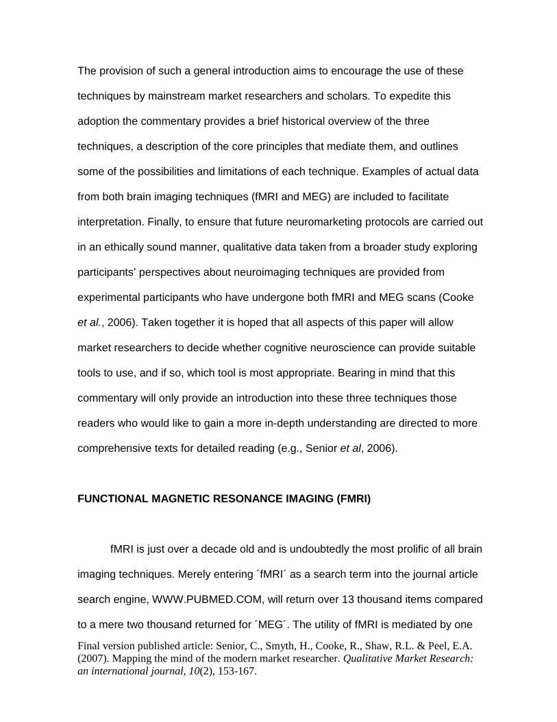
The provision of such a general introduction aims to encourage the use of these
techniques by mainstream market researchers and scholars. To expedite this
adoption the commentary provides a brief historical overview of the three
techniques, a description of the core principles that mediate them, and outlines
some of the possibilities and limitations of each technique. Examples of actual data
from both brain imaging techniques (fMRI and MEG) are included to facilitate
interpretation. Finally, to ensure that future neuromarketing protocols are carried out
in an ethically sound manner, qualitative data taken from a broader study exploring
participants’ perspectives about neuroimaging techniques are provided from
experimental participants who have undergone both fMRI and MEG scans (Cooke
et al., 2006). Taken together it is hoped that all aspects of this paper will allow
market researchers to decide whether cognitive neuroscience can provide suitable
tools to use, and if so, which tool is most appropriate. Bearing in mind that this
commentary will only provide an introduction into these three techniques those
readers who would like to gain a more in-depth understanding are directed to more
comprehensive texts for detailed reading (e.g., Senior et al, 2006).
FUNCTIONAL MAGNETIC RESONANCE IMAGING (FMRI)
fMRI is just over a decade old and is undoubtedly the most prolific of all brain
imaging techniques. Merely entering ´fMRI´ as a search term into the journal article
search engine, WWW.PUBMED.COM, will return over 13 thousand items compared
to a mere two thousand returned for ´MEG´. The utility of fMRI is mediated by one

Final version published article: Senior, C., Smyth, H., Cooke, R., Shaw, R.L. & Peel, E.A. (2007). Mapping the mind of the modern market researcher. Qualitative Market Research: an international journal, 10(2), 153-167.
key factor - it is relatively easy to implement. It is a completely non-invasive
procedure where the volunteer is simply moved into the centre of a high field
circular magnet bore (See Fig. 1). Various experimental stimuli, such as
advertisements for particular products, can then be projected into the centre of the
bore, which the subject views via a small prism mirror placed just above the face.
A variety of neurophysiological information can be obtained using fMRI. For
example, baseline cerebral blood volume measurements, changes in this blood
volume, quantitative changes in the levels of blood oxygenation, as well as the rate
of resting state oxygen extraction. More detailed descriptions of each of these
measures are provided in Russell et al (2003). One measure that will have great
utility for the neuromarket researcher is the ´BOLD´ contrast (See Fig. 1) and this is
described further.
The ´BOLD´ in ´BOLD´ contrast stands for Blood Oxygenation Level
Dependant (Tank et al. 1992). In brief, this signal is driven by a difference in the
blood oxygenation levels in capillaries and veins compared to the arteries during a
particular task. Deoxygenated blood is paramagnetic (attracted to a magnetic field)
as opposed to when it is oxygenated (Pauling and Coryell, 1936). On presentation
of a specific stimulus, oxygenated blood flow will increase locally within an ´active´
region of the brain. This will cause deoxygenated blood levels to decrease and
subsequently decrease the magnitude of the magnetic field distortions between the

Final version published article: Senior, C., Smyth, H., Cooke, R., Shaw, R.L. & Peel, E.A. (2007). Mapping the mind of the modern market researcher. Qualitative Market Research: an international journal, 10(2), 153-167.
two molecules which ultimately leads to a signal increase in the fMRI dataset
(Ogawa et al, 1992).
The main limitation of this technique is that the signal begins to increase
approximately two seconds after stimulus presentation, and reaches a plateau after
about seven to 10 seconds (Logothetis et al, 2001). In specialist terminology, fMRI
has excellent spatial resolution but relatively poor temporal resolution, i.e., it can be
used to detect activity in specific and, in some cases, quite small regions of the
brain but it can tell you very little about the timing of that activity. Therefore, as a
tool, fMRI should generally be used for the identification of certain brain areas only.
_____________________________
Please place Fig 1 about here
_____________________________
The functional activity revealed with the BOLD contrast then needs to be mapped
on to a picture of the subject’s brain. To create a neuroanatomical picture, a rapid
radiofrequency pulse is applied which forces the hydrogen protons in the various
tissues of the brain to become aligned to it. When the radiofrequency pulse is
switched off these protons relax and return to their original alignment with the
magnetic field emitted by the MRI scanner. The different rates of proton relaxation
across the various structures of the brain allow an image to be constructed (Russell
et al, 2003).

Final version published article: Senior, C., Smyth, H., Cooke, R., Shaw, R.L. & Peel, E.A. (2007). Mapping the mind of the modern market researcher. Qualitative Market Research: an international journal, 10(2), 153-167.
However, the rapid switching of the radiofrequency pulse required for the
neuroanatomical MR image is loud and can cause permanent hearing loss in some
cases, so most laboratories require subjects to wear ear protection during a scan.
This, coupled with the fact that subjects are positioned inside the center of a very
large superconducting magnet, the inner surface of which is sometimes inches
away from the subject's face and body, would suggest that fMRI can be quite a
stressful or traumatic procedure to undergo.
The obvious lack of ecological validity needs to borne in mind when carrying
out neuromarket research. Obtaining responses from a subject in a potentially
stressful environment may bias any results. However, our questionnaire data
derived from experimental subjects suggest that, in general, they tend to consider
fMRI to be a ‘surprisingly relaxing’ experience.
Participants’ perspectives about fMRI
As part of a broader study investigating participants’ perspectives about fMRI and
MEG procedures 44 experiment participants were asked to complete an extensive
questionnaire about their experiences, knowledge and attitudes to each of these
brain imaging procedures. In addition to the quantitative questionnaire data which is
reported elsewhere (Cooke et al, 2006) some participants provided qualitative
comments regarding their neuroimaging experience(s) and these data are reported
here. These subjects were undergraduate and postgraduate volunteers which are
largely representative of a market research subject population. Questionnaire

Final version published article: Senior, C., Smyth, H., Cooke, R., Shaw, R.L. & Peel, E.A. (2007). Mapping the mind of the modern market researcher. Qualitative Market Research: an international journal, 10(2), 153-167.
respondents were invited to: describe their previous experience of fMRI; and explain
their response to questions including ‘do you think you could have refused to have
fMRI if you had wanted?’; ‘did you feel you experienced any side effects during the
fMRI procedure?’; and ‘did you feel that you could contact the researcher at any
point during the procedure?’. Space was provided at the end of the questionnaire
for respondents to add any additional comments about fMRI.
These data suggest that in general participants viewed the fMRI experience
positively, reporting that they ‘enjoyed taking part in the fMRI’, found the procedure
‘interesting’, and the technology ‘fascinating’. Participants’ positive comments about
fMRI appeared to be underpinned by three main areas. First, perceptions of the
experimenter; second, awareness of informed consent procedures; and third, the
contrast between negative expectations and the actual experience in the scanner.
The nature of the experimenter’s/researcher’s interaction with participants was
significant in terms of how participants experienced the scanning process. A
number of participants reported that: ‘The researchers made me feel comfortable’
and that they ‘trust[ed] the researchers would not put me in any danger’. The sense
of trust and comfort conveyed by participants was underpinned by an awareness of
the nature of informed consent procedures discussed with them. As one respondent
reported ‘everything was very clearly explained beforehand’. Participants made
numerous comments demonstrating their knowledge of informed consent
procedures, especially the right to withdraw: ‘they told me at any point that if I didn’t
want to continue or felt uncomfortable, there was no problem with me not doing it’.
Final version published article: Senior, C., Smyth, H., Cooke, R., Shaw, R.L. & Peel, E.A. (2007). Mapping the mind of the modern market researcher. Qualitative Market Research: an international journal, 10(2), 153-167.
This thorough consenting process, which was communicated to participants in both
written and spoken forms, encouraged a favourable perception of the experimenter,
as one respondent wrote:
‘I know about the ‘right to withdraw’ and researchers seemed friendly and
understanding’. Thus feeling fully informed about the procedure coupled with a
reassuring researcher (s/he ‘frequently asked if I was ok with the situation’) led
participants to report that they were not ‘pressured into it in any way’ and they did
not ‘ever feel pressured’ either to undergo the experiment or remain in the scanner
if they became uncomfortable.
Clearly, given the technological intensiveness of the fMRI hardware, a thorough
explanation of fMRI and a friendly approach adopted by the experimenter is
essential for participants to view the experience favourably. However, some of the
data did reveal that the fMRI procedure did have an ominous reputation. Such a
reputation is probably due to negative preconceptions as the contrast between
these preconceptions, and the actual experience were clearly embedded in
participants’ comments. For example: ‘I was slightly apprehensive before the
procedure as I had heard it could be very claustrophobic and noisy. However it was
not unpleasant at all’ do show that the actual scan is a relatively acceptable
experience. It seems that some participants reported feeling relieved that the actual
procedure was relatively non-invasive. Comments such as ´It wasn’t as scary as I
thought from what friends/relatives described to me’ and ‘it was much less
Final version published article: Senior, C., Smyth, H., Cooke, R., Shaw, R.L. & Peel, E.A. (2007). Mapping the mind of the modern market researcher. Qualitative Market Research: an international journal, 10(2), 153-167.
claustrophobic than I had feared´ strengthened the notion that prior to the actual
procedure participants considered fMRI to be daunting. Whilst ´I had been scanned
before…it was still reassuring to have the various safety measures explained to me´
shows that the daunting reputation of the technique persists to some degree after
experience with the scanner. This may adversely affect recruitment of subjects to
participate in any neuromarket research program, and again underscores the
importance of thorough and ongoing information provision and reassurance from
the experimenter. However, participants also commented that they felt they would
be happy to talk to other volunteers prior to participation because it would be
´reassuring for them´ thus suggesting a possible strategy to avoid future
misconceptions about participation in fMRI experiments.
Evidence of potential side effects, both short- and long-term, of exposure to MRI is
currently limited because fMRI is a relatively recent research technology.
Respondents described their fMRI experience mostly in positive terms one
participant reported that s/he ‘became very sleepy towards the end’ of their time in
the scanner. Nevertheless, participants were asked about any possible side effects.
Only three of the 20 participants who provided qualitative questionnaire data
described experiencing physical sensations following scanning. One participant
reported that s/he felt ‘a bit ‘numb’ in the head’, another ‘had a pins and needles
sensation in my left hand’. The third reflected that: ‘It was in no way a frightening
experience but an experience I am glad I have had. I would take part in more

Final version published article: Senior, C., Smyth, H., Cooke, R., Shaw, R.L. & Peel, E.A. (2007). Mapping the mind of the modern market researcher. Qualitative Market Research: an international journal, 10(2), 153-167.
studies. The only trouble was the headache I experienced after but it wasn’t or
didn’t affect any day to day procedures’. Physical contraindications tend to be rare
and these participants were more than likely reporting the effects of incorrect
positioning rather than side effects of the scanning procedure per se e.g., the
subject's head not resting comfortably etc. The fMRI technique is extremely
sensitive to subject movement and the slightest shifting can result in motion artifacts
that can seriously impact the quality of the data. For this reason, and also to
maintain subject comfort, it is important to attain correct positioning from the start.
Taking in hand the fact that this is a cursory glance into participants’ experiences
during an fMRI scan it is worth noting the salient aspects that would affect the
neuromarketer. First, even though the technique itself is non-invasive it does have a
slightly negative reputation and this may impact on possible participation for market
research studies. Secondly, to minimize perceived and actual side effects and to
also ensure the integrity of the data, the subject must be correctly positioned in the
scanner prior to beginning the procedure.
MAGNETOENCEPHALOGRAPHY (MEG)
Whilst fMRI is an ideal tool for locating cortical activity it may also be identified - this
type of neurophysiological data is best collected with MEG. Obtaining MEG data is
a very different process from fMRI as it involves the measurement of extremely
weak magnetic fields generated by the electrical activity of neuronal populations

Final version published article: Senior, C., Smyth, H., Cooke, R., Shaw, R.L. & Peel, E.A. (2007). Mapping the mind of the modern market researcher. Qualitative Market Research: an international journal, 10(2), 153-167.
(see Hämäläinen and Hari, 2002 for review). Compared with fMRI, MEG has
excellent temporal resolution but relatively poor spatial resolution, i.e., it can detect
cortical activity at the millisecond level but it is not very good at distinguishing the
space where this activity originated. However, contemporary imaging techniques
developed from radar technology do considerably improve on this poor spatial
resolution (Hillebrand et al, 2005).
Measuring such minute neural activity is challenging due to the very weak nature of
the neuronal clusters and interference with nearby electromagnetic noise. Noise
sources arising from the subject’s own body, such as coughing etc., can have
serious implications for the integrity of the data. As is the case with fMRI scanning,
contemporary image analysis software can ensure that most of these artifacts are
controlled for at source. During a MEG scan the subject's head is raised into a
´dewar´ which houses an array of superconducting sensors called SQUIDS
(Superconducting Quantum Interference Devices). To collect the optimal signal it is
preferential to use dewars with as large a collection of SQUIDS as possible and the
latest generation of MEG scanners can contain up to 300 separate SQUID
detectors (Singh, 2006).
The temporal resolution of MEG is close to real time but its ability to detect the
onset of cortical activity is not its only advantage. The use of MEG also allows study
of changes in neuronal oscillatory rhythms, i.e., the specific frequency at which
neurons in a particular cluster fire together (Hillebrand et al., 2005). A specific

Final version published article: Senior, C., Smyth, H., Cooke, R., Shaw, R.L. & Peel, E.A. (2007). Mapping the mind of the modern market researcher. Qualitative Market Research: an international journal, 10(2), 153-167.
oscillatory frequency range, e.g., 28-40Hz, will either increase or decrease during
an experimentally salient period of time, such as when participants view a visual
stimulus (see e.g, Singh, 2006). Take for instance the modulation in activity
between 28-40Hz that occurs when a subject recognizes a face (Rodriguez et al,
1999). This modulation in task related oscillatory behaviour is sometimes called
Event Related Synchronization (ERS) or Event Related Desynchronization (ERD),
depending on the direction of the change, i.e., either in the same frequency band or
towards a different frequency band (Pfurtscheller, 2001; see Fig. 2). Whilst still
preliminary there is emerging evidence that certain frequency bands can be
identified as signatures for specific cognitive tasks e.g, 28-40Hz ERS for object
recognition (see above) or 14-28 Hz ERS for verbal working memory (Hwang et al,
2005) or even 4-8Hz ERS for episodic recall (Klimesch et al, 2001). This may
provide a further tool for neuromarketer in the understanding of market behavior.
________________________
Please place fig 2 here
________________________
Due to the sensitive nature of the SQUIDS it is essential to implement a number of
conditions prior to carrying out a MEG procedure. The dewar needs to be isolated in
a double magnetically shielded room to ensure that data are not biased by transient
electromagnetic fields in the local environment. Additionally, to ensure SQUID
sensitivity it is imperative that the distance between the sensors and the subject’s
head is minimized as much as possible, thus many MEG laboratories use inflatable

Final version published article: Senior, C., Smyth, H., Cooke, R., Shaw, R.L. & Peel, E.A. (2007). Mapping the mind of the modern market researcher. Qualitative Market Research: an international journal, 10(2), 153-167.
cuffs to ensure that the subject’s head is tightly held in the dewar (Singh, 2006). The
fact that the subject is held tightly in the MEG dewar may lead to the assumption
that it is an uncomfortable procedure. However, examination of the subject
responses in our qualitative questionnaire data show that whilst it is considered very
different to fMRI it is not an uncomfortable procedure.
Participants’ perspectives about MEG
Questionnaire respondents provided 31 written comments about their experience of
MEG scanning. Again, comments such as ´would be happy to reassure anyone
with concerns’ were observed throughout the questionnaires returned, suggesting
that this procedure, like fMRI, does have an anxiety provoking reputation. Even
though MEG procedures are silent and carried out in the dark, participants still
found that ´it was an extremely uncomfortable procedure to undertake’. The need to
position the surface of the subject’s head as close to the Dewar as possible may
result in stressful head restriction. However, the neuromarketer can circumvent this
problem by ensuring that the participant is lowered out of the Dewar at regular
intervals during a specific procedure in order to alleviate any stress brought on by
restricted movement. Participants described feeling fatigued and restricted: 'As the
experiment progressed I became incredibly tired – it felt quite taxing, although it
wasn’t particularly mentally or physically demanding. I also found it difficult to stay
very still for such a long time. I think this is why my muscles ached afterwards’;
‘Although I found the experiment interesting, while I was in the machine I found

Final version published article: Senior, C., Smyth, H., Cooke, R., Shaw, R.L. & Peel, E.A. (2007). Mapping the mind of the modern market researcher. Qualitative Market Research: an international journal, 10(2), 153-167.
myself becoming very drowsy’. This highlights the neuromarketer’s need to be
aware of participant fatigue and the measures used to combat it in order to prevent
any possible corruption of data as well as discomfort to the volunteer.
As can be seen, MEG clearly has great utility for neuromarket research. Not only
can it provide information about the onset of any cortical activity but can also
provide information about the specific frequency by which clusters of neurons fire.
However, it is surprising to note that any cortical activation revealed with a particular
task cannot be used to infer that the cortical area implicated is actually necessary.
Any engendered brain activation from MEG or fMRI may be epiphenomenal in
nature - much like the heat given off by a light bulb (see Kosslyn, 1999).
Functional necessity can only be inferred if a decrement in a particular task is
revealed when a specific area of the brain is removed. Obviously the neuromarketer
cannot remove parts of potential consumers brains for the sake of science.
However, Transcranial magnetic stimulation (TMS) is one way in which a safe and
repeatable 'virtual lesion' can be created in healthy participants (Walsh and Cowey,
2000).
TRANSCRANIAL MAGNETIC STIMULATION (TMS) ?
TMS is not a brain imaging tool per se as one cannot apply it and 'see' activity in the
brain. Rather TMS allows neuromarketers to 'switch off' part of the cortex for very

Final version published article: Senior, C., Smyth, H., Cooke, R., Shaw, R.L. & Peel, E.A. (2007). Mapping the mind of the modern market researcher. Qualitative Market Research: an international journal, 10(2), 153-167.
brief periods of time and the index of functional necessity is revealed by differences
in response times or other behavioral measures (Senior, 2001).
TMS operates by inducing a brief electrical current in areas of the cortex that cause
the excitatory behavior of focused clusters of nerve cells. The effect is brought
about by Michael Faradays principal of electromagnetic induction, which states that
a single pulse of electric current flowing through a coil of wire will generate a
magnetic field (see Mills, 1999, ch. 3). By alternating the magnitude of the magnetic
field over a short period of time an electrical current will be induced in a nearby
secondary conductor. In TMS investigations, a stimulating coil is placed over the
subjects scalp and the magnetic field travels through the scalp and skull to induce
the secondary electrical current in the cortex. The technique is ingenious insofar as
the human scalp and skull have a relatively high resistance to electrical currents
whilst no impedance to a magnetic field (Jahansahahi and Rothwell, 2000).
The early technique enjoyed a flourishing interest within the field of neuromuscular
disorders (Mills, 1999) but it was Barker who produced muscular twitches with
stimulation of the primary motor cortex and ensured that the application of TMS to
cognitive studies was born (Barker et al, 1985). However, it was not until the
discovery that a single magnetic pulse to the visual cortex, (approx 60-140 msecs
after stimulus presentation) could render subjects incapable of detecting stimuli in a
letter discrimination task that the use of this technology in cognitive neuroscience
really began in earnest (Amassian et al, 1989). The same group further explored the
Final version published article: Senior, C., Smyth, H., Cooke, R., Shaw, R.L. & Peel, E.A. (2007). Mapping the mind of the modern market researcher. Qualitative Market Research: an international journal, 10(2), 153-167.
finding that magnetic stimulation can be used to disrupt cortical function some years
later where they found that a visual mask effect could be lost, and performance on a
letter discrimination task improved with a single pulse of magnetic stimulation
(Amassian et al, 1993). These two studies formed the bedrock of the contemporary
development of TMS within the cognate sciences. The disruption of cognitive
function that is caused with magnetic stimulation can be used to impair a behavioral
task or improve performance on a particular task by disrupting cortical function that
may be irrelevant or competitive in nature (Walsh and Cowey, 1998).
The application of a virtual lesion to induce disruption or improvement in a specific
task is of particular interest to the neuromarketer. Application of TMS resulting in an
improvement in, for example memory recall, for a particular brand product can help
to identify the competing cognitive factor that was impeding previous recall.
Whilst the application of a virtual lesion is the primary role of TMS the manipulation
of the timing and foci of this disruption has allowed investigation of the timing of
psychological function and the connectivity of the neural areas that mediate these
functions (see Pascual-Leone et al, 2000 for a review). The ability to repeatedly
disrupt cortical processing gives TMS a ‘functional resolution’ which is unique. Place
this functional resolution alongside the superior temporal resolution of MEG and the
spatial resolution of fMRI and one quickly becomes aware of the immense potential
available to the neuromarketer(see Pascual-Leone et al, 1999).

Final version published article: Senior, C., Smyth, H., Cooke, R., Shaw, R.L. & Peel, E.A. (2007). Mapping the mind of the modern market researcher. Qualitative Market Research: an international journal, 10(2), 153-167.
Participants’ perspectives about TMS
To date there has been no examination of healthy subjects experiences during a
TMS procedure. However, Walter et al. (2001) reported that psychiatric patients
classified as depressed found transcranial magnetic stimulation (TMS) a positive
procedure and would recommend it to friends and family. Clearly, further work is
needed examining the perceptions of TMS by volunteers as this will help to facilitate
best practice with the procedure.
DISCUSSION AND CONCLUSIONS
This paper aimed to introduce cognitive neuroscientific techniques that could be
employed by the market researcher who wished to develop a specialized
neuromarketing profile. Three ´mainstream´ techniques were discussed, these
being fMRI, MEG and TMS. A brief overview of the central principles and
procedures involved in each technique was provided as well as a discussion of their
limitations. fMRI is an excellent tool for the localization of a specific area of the brain
implicated in a particular task (See Fig. 1). However, fMRI is dependant on cortical
hemodynamics and as such suffers from a lag of up to several seconds; it is thus a
poor tool for study of the timing of cortical activity. On the other hand, as MEG
detects the very small electromagnetic changes in neuronal clusters it is freed from
the temporal constraints that the hemodynamic response imposes (See Fig. 2).

Final version published article: Senior, C., Smyth, H., Cooke, R., Shaw, R.L. & Peel, E.A. (2007). Mapping the mind of the modern market researcher. Qualitative Market Research: an international journal, 10(2), 153-167.
MEG also allows study of the oscillatory frequency of neuronal clusters that are
linked to a specific event – be it a response to tasting a particular brand of cola, or
perceiving a particular advertisement. Finally, TMS operates by inducing neuronal
activity for very brief periods of time. This serves to interfere with any cortical
processing that may be going on at that point, thereby creating a safe and
repeatable 'virtual lesion'. The ability to create a neuropsychological ‘virtual patient’
gives TMS a functional resolution, which in turn can be used to explore the
connectivity and the timing of cognitive events.
The experimental subjects positive experiential accounts of participating in
experiments with these three techniques permit an optimistic endorsement for use
in new fields such as neuromarket research. It is also clear that each technique
allows the neuromarketer to study a unique perspective of the consumer's
cognitions towards a specific product. However, it is the integration of each
technique that could be extremely beneficial for the future of neuromarket research
(Wasserman & Grafman, 1997).
An example of this novel approach would require a combination of fMRI, MEG and
TMS. Take, for example, this thought experiment examining differences in memory
for sexual vs non sexual imagery in perfume adverts. First you would show
consumers one of two advertisements, one which contains sexual imagery and one
which is non-sexual, whilst undergoing an fMRI procedure. The subjects will
engender activity in a wide variety of cortical areas. Such an outcome is predictable
Final version published article: Senior, C., Smyth, H., Cooke, R., Shaw, R.L. & Peel, E.A. (2007). Mapping the mind of the modern market researcher. Qualitative Market Research: an international journal, 10(2), 153-167.
and likely given that both types of advertisements are visually complex and have
been designed to elicit appetitive behavior (i.e., purchasing a particular product).
However, this outcome tells the neuromarketer nothing about whether
advertisement one is a more effective visual cue than the second. To complete the
neuromarket research process, the engendered activity from the fMRI procedure
can then be used to guide analysis of subsequently collected MEG data. From this
the neuromarketer can study differences in the onset of neuronal activity between
the two advertisements and also identify differences in the neuronal frequency
bands. Such an approach allows the both the localization of activity to be studied as
well as the onset of that activity and its specific frequency signature. At this stage
the neuromarketer would have a comprehensive data set converging on the neural
activity engendered by two sets of advertisements. TMS could then be applied at
the specific onset of activity for each of the two distinct advertisements in a memory
task and subsequent recall could be tested. As the virtual lesion is applied at a point
in time corresponding to brain activity revealed by MEG, which was in turn guided
by fMRI, a subsequent memory decrement would allow you to infer that this activity
is due to memory processes.
Bearing in mind the rapid evolution of other ´neuro´ techniques such as eye
movement analysis (Henderson, 2006) and even neurogenetics (Mattey et al, 2006)
the future possibilities for neuromarket research are promising. Embracing
contemporary techniques such as those described here will enable neuromarketers
to compete alongside other social scientists in a growing and exciting field.

Final version published article: Senior, C., Smyth, H., Cooke, R., Shaw, R.L. & Peel, E.A. (2007). Mapping the mind of the modern market researcher. Qualitative Market Research: an international journal, 10(2), 153-167.
Final version published article: Senior, C., Smyth, H., Cooke, R., Shaw, R.L. & Peel, E.A. (2007). Mapping the mind of the modern market researcher. Qualitative Market Research: an international journal, 10(2), 153-167.
REFERENCES
Aharon, I., Etcoff, N., Ariely, D., Chabris, CF., O'Connor, E. and Breiter, H.C.
(2001), "Beautiful faces have variable reward value: fMRI and behavioral evidence",
Neuron, Vol. 32, No. 3, pp. 537-551.
Amassian, V.E., Cracco, R.Q., Maccabee, P.J., Cracco, J.B., Rudell, A. and Eberle,
L. (1989), "Suppression of visual perception by magnetic coil stimulation of human
occipital cortex", Electroencephalography and Clinical Neurophysiology, Vol. 74,
No.6, pp. 458-462.
Amassian, V.E., Cracco, R.Q., Maccabee, P.J., Cracco, J.B., Rudell, A.P. and
Eberle, L. (1993), "Unmasking human visual perception with the magnetic coil and
its relationship to hemispheric asymmetry", Brain Research, Vol. 605, No. 2, pp.
312-316.
Barker, A.T., Jalinous, R. and Freeston, I.L. (1985), "Non-invasive magnetic
stimulation of the human motor cortex", Lancet, Vol. 1, No. 8437, pp. 1106-1107.
Brain scam ? (2004).Editorial, Nature Neuroscience,Vol.7, No. 7, pp. 683.
Brammer, M. (2004), "Brain scam ?", Nature Neuroscience, Vol. 7, No. 10, pp.
1015.
Bush, G. (1990), "Presidential Proclamation #6158". Library of Congress, DC.
http://www.loc.gov/loc/brain/proclaim.html. Accessed 25/03/06 at 1300 hrs.
Cooke, R., Peel, E., Shaw, R.L. and Senior, C. (2006, in press), "The neuroimaging
process from the participants perspective", International Journal of
Psychophysiology.
Final version published article: Senior, C., Smyth, H., Cooke, R., Shaw, R.L. & Peel, E.A. (2007). Mapping the mind of the modern market researcher. Qualitative Market Research: an international journal, 10(2), 153-167.
Erk, S., Spitzer, M., Wunderlich, A.P., Galley, L. and Walter, H. (2002), "Cultural
objects modulate reward circuitry", Neuroreport, Vol. 13, No. 18, pp. 2499-503.
Hämäläinen, M.S. and Hari, R. (2002), "Magnetoencephalographic Characterization
of Dynamic Brain Activation: Basic Principles and Methods of Data Collection and
Source Analysis" In Brain Mapping: The Methods, Toga AW & Mazziotta JC, (eds).
Academic Press, USA pp. 227-53.
Henderson, J. (2006), "Eyemovements" In Methods in Mind, Senior C, Russell, T, &
Gazzaniga MS (eds). The MIT Press, USA. pp. 1-35.
Hillebrand, A., Singh, K.D., Furlong P.L., Holliday, I.E. & Barnes, G.R. (2005), "A
new approach to neuroimaging with magnetoencephalography" Human Brain
Mapping, Vol. 25. pp. 199 –211.
Hwang, G., Jacobs, J., Geller, A., Danker, J., Sekuler, R. and Kahana, M.J. (2005),
"EEG Correlates of verbal and non verbal working memory" Behavioral and Brain
Function, Vol. 15, pp. 1-20.
Jahanshahi, M. and Rothwell, J.C. (2000), "Transcranial magnetic stimulation
studies of cognition: An emerging field", Experimental Brain Research,Vol. 131,
No.1. pp. 1-9.
Klimesch, W., Doppelmayr, M., Yonelinas, A., Kroll, N.E., Lazzara, M., Rohm, D.
and Gruber, W. (2001), "Theta syncronisation during episodic retrieval: neural
correlates of conscious awareness", Cognitive Brain Research, Vol. 12, No.1. pp.
33-38.

Final version published article: Senior, C., Smyth, H., Cooke, R., Shaw, R.L. & Peel, E.A. (2007). Mapping the mind of the modern market researcher. Qualitative Market Research: an international journal, 10(2), 153-167.
Kosslyn, S.M. (1999), "If neuroimaging is the answer, what is the question ?"
Philosophical Transactions of the Royal Society of London B, Vol. 354, No.1387.
pp.1283-1294.
Lee, N., Broderick, A.J. and Chamberlain, L (2006, in press), "What is
Neuromarketing' ? A Discussion and Agenda for Future Research", International
Journal of Psychophysiology.
Lieberman, M.D. (2005), "Principles, processes and puzzles of social cognition: An
introduction for the special issue on social cognitive neuroscience" Neuroimage,
Vol.28, pp. 745-756.
Logothetis, N.K., Pauls, J., Augath, M., Trinath, T. and Oeltermann, A. (2001),
"Neurophysiological investigation of the basis of the fMRI signal", Nature, Vol. 412.
pp. 150-157.
Mattay, V., Lindenberg, A. and Weinberger, D. (2006), "Imaging Genomics". In
Methods in Mind, Senior C, Russell, T, Gazzaniga MS (eds). The MIT Press, USA.
pp. 36-60.
McClure, S.M., Li, J., Tomlin, D., Cypert, K.S., Montague, L.M. and Montague, P.R.
(2004), "Neural correlates of behavioral preference for culturally familiar drinks",
Neuron, Vol. 44, No. 2. pp. 379-387.
Mills, K.R. (1999), "Magnetic stimulation of the human nervous system" Oxford
University Press. Oxford, UK.
Final version published article: Senior, C., Smyth, H., Cooke, R., Shaw, R.L. & Peel, E.A. (2007). Mapping the mind of the modern market researcher. Qualitative Market Research: an international journal, 10(2), 153-167.
Ogawa, S., Tank, D.W., Menon, R., Ellermann, J. M., Kim, S. G., Merkle, H. and
Ugurbil, K. (1992), "Intrinsic signal changes accompanying sensory stimulation:
functional brain mapping with magnetic resonance imaging", Proceedings of the
National Academy of Sciences of the United States of America, Vol. 89, pp. 5951-
5955.
Pascual-Leone, A., Bartres-Faz, D. and Keenan, J.P. (1999), "Transcranial
magnetic stimulation: studying the brain-behavior relationship by induction of ‘virtual
lesions’", Philosophical Transactions of the Royal Society of London B. Vol. 354,
No. 1387, pp. 1229-1238.
Pascual-Leone, A., Walsh, V. and Rothwell, J.C. (2000), "Transcranial magnetic
stimulation in cognitive neuroscience – virtual lesion, chronometry, and functional
connectivity", Current Opinion in Neurobiology. Vol. 10, No. 2, pp. 232-237.
Pauling, L. and Coryell, C.D.(1936), "The magnetic properties and structure of
hemoglobin, oxyhemoglobin, and carbonmonoxyhemoglobin", Proceedings of the
National Academy of Sciences of the United States of America. USA, Vol. 22, pp. 210–216.
Pfurtscheller, G. (2001), "Functional Brain Imaging Based on ERD/ERS" Vision
Research, Vol. 41, pp. 1257-1260.
Rodriguez, E., George, N., Lachaux, J.P., Martinerie, J., Renault, B., and Varela,
F.J. (1999), "Perception's shadow: long-distance synchronization of human brain
activity", Nature, Vol. 397, No. 6718, pp. 430-433.
Routtenberg, A. and Lindy, J. (1965), "Effects of the availability of rewarding septal
and hypothalamic stimulation on bar pressing for food under conditions of

Final version published article: Senior, C., Smyth, H., Cooke, R., Shaw, R.L. & Peel, E.A. (2007). Mapping the mind of the modern market researcher. Qualitative Market Research: an international journal, 10(2), 153-167.
deprivation", Journal of Comparative and Physiological Psychology, Vol. 60, No. 2,
pp. 158-161.
Russell, T., Zelaya, F., Bressan, R.A. and Bandettini, P.A. (2003), In Neuroimaging
in Psychiatry. Senior, C., Fu, C.H.Y., Russell. T., Weinberger, D. and Murray, R.
Dunitz Press, UK. pp 1-51.
Senior, C. (2003), "Beauty in the brain of the beholder", Neuron. Vol. 38, No. 4. pp.
525-528.
Senior, C. (2001), "Safety, efficacy, and utility of transcranial magnetic stimulation in
cognitive neuropsychology", Australian Journal of Psychology, Vol. 54, No.1, pp.40-
45.
Senior, C., Evans, G., Adjamian, P., and Singh, K.D. (2005), "Facial attraction in the
brain: A Magnetoencephalography study" Neuroimage, Vol. 26, No. 1, pp. s28.
Senior, C., Russell, T. and Gazzaniga, M.S. (2006). "Methods in Mind" The MIT
Press, USA.
Singh, K.D. (2006), "Magnetoencephalography". In Methods in Mind, Senior, C.,
Russell, T. and Gazzaniga, M.S. (eds). The MIT Press, USA. pp. 190-225.
Tank, D.W., Ogawa, S. and Ugurbil, K. (1992), "Mapping the brain with MRI"
Current Biology. Vol. 2, No.10,pp. 525-528.
Valenstein, E.S. and Beer, B. (1964), "Continious opportunity for reinforcing Brain
stimulation" Journal of the Experimental Analysis of Behavior. Vol. 20, pp. 183-4.

Final version published article: Senior, C., Smyth, H., Cooke, R., Shaw, R.L. & Peel, E.A. (2007). Mapping the mind of the modern market researcher. Qualitative Market Research: an international journal, 10(2), 153-167.
Walsh, V. and Cowey, A. (1998), "Magnetic stimulation studies of visual cognition",
Trends in Cognitive Sciences, Vol. 2, No.3, pp. 103-108.
Walsh, V. and Cowey, A. (2000), "Transcranial magnetic stimulation and cognitive
neuroscience", Nature Reviews: Neuroscience, Vol. 1, No. 1, pp. 73-79.
Walter, G., Martin, J., Kenneth, K. and Pridmore, S. (2001),
"Transcranial magnetic stimulation: experience, knowledge and attitudes of
recipients", Australian and New Zealand Journal of Psychiatry, Vol. 35. No. 1, pp.
58-61.
Wasserman, E.M. and Grafman, J. (1997), "Combining transcranial magnetic
stimulation and neuroimaging to map the brain" Trends in Cognitive Science, Vol. 1,
No. 6, pp.199-200.
Wolpe, P.R., Foster, K.R. and Langleben, D.D.(2005)," Emerging neurotechnologies
for lie-detection: promises and perils", American Journal of Bioethics, Vol. 5, No. 2.
pp. 39-49.
Acknowledgements: We would like to thank the British Academy for financial
support and the anonymous reviewer for helpful comments.
Final version published article: Senior, C., Smyth, H., Cooke, R., Shaw, R.L. & Peel, E.A. (2007). Mapping the mind of the modern market researcher. Qualitative Market Research: an international journal, 10(2), 153-167.
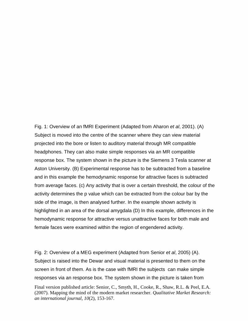
Fig. 1: Overview of an fMRI Experiment (Adapted from Aharon et al, 2001). (A)
Subject is moved into the centre of the scanner where they can view material
projected into the bore or listen to auditory material through MR compatible
headphones. They can also make simple responses via an MR compatible
response box. The system shown in the picture is the Siemens 3 Tesla scanner at
Aston University. (B) Experimental response has to be subtracted from a baseline
and in this example the hemodynamic response for attractive faces is subtracted
from average faces. (c) Any activity that is over a certain threshold, the colour of the
activity determines the p value which can be extracted from the colour bar by the
side of the image, is then analysed further. In the example shown activity is
highlighted in an area of the dorsal amygdala (D) In this example, differences in the
hemodynamic response for attractive versus unattractive faces for both male and
female faces were examined within the region of engendered activity.
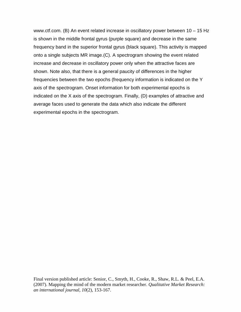
Fig. 2: Overview of a MEG experiment (Adapted from Senior et al, 2005) (A).
Subject is raised into the Dewar and visual material is presented to them on the
screen in front of them. As is the case with fMRI the subjects can make simple
responses via an response box. The system shown in the picture is taken from
Final version published article: Senior, C., Smyth, H., Cooke, R., Shaw, R.L. & Peel, E.A. (2007). Mapping the mind of the modern market researcher. Qualitative Market Research: an international journal, 10(2), 153-167.
www.ctf.com. (B) An event related increase in oscillatory power between 10 – 15 Hz
is shown in the middle frontal gyrus (purple square) and decrease in the same
frequency band in the superior frontal gyrus (black square). This activity is mapped
onto a single subjects MR image.(C). A spectrogram showing the event related
increase and decrease in oscillatory power only when the attractive faces are
shown. Note also, that there is a general paucity of differences in the higher
frequencies between the two epochs (frequency information is indicated on the Y
axis of the spectrogram. Onset information for both experimental epochs is
indicated on the X axis of the spectrogram. Finally, (D) examples of attractive and
average faces used to generate the data which also indicate the different
experimental epochs in the spectrogram.
Copyright © 2022 FDOKUMEN