manuscript-Kovács-Planta-10.1007 s00425-016-2502-x - CORE
46
1 The final publication is available at Springer via http://dx.doi.org/10.1007/s00425-016-2502-x Krisztina Kovács a , Jiří Pechoušek b , Libor Machala b , Radek Zbořil b , Zoltán Klencsár c , Ádám Solti d , Brigitta Tóth e , Brigitta Müller d , Hong Diep Pham d , Zoltán Kristóf f , Ferenc Fodor d Revisiting the iron pools in cucumber roots: identification and localization a Institute of Chemistry, Eötvös Loránd University, P.O. Box 32, Budapest 1512, Hungary b Regional Centre of Advanced Technologies and Materials, Departments of Experimental Physics and Physical Chemistry, Faculty of Science, Palacký University in Olomouc, Olomouc 771 46, Czech Republic c Institute of Materials and Environmental Chemistry, Research Centre for Natural Sciences, Hungarian Academy of Sciences, Magyar tudósok körútja 2, 1117 Budapest, Hungary d Department of Plant Physiology and Molecular Plant Biology, Institute of Biology, Eötvös Loránd University, Pázmány Péter lane 1/c, Budapest 1117, Hungary e Department of Botany, Crop Physiology and Biotechnology, Institute of Plant Sciences, Faculty of Agricultural and Food Sciences and Environmental Management, University of Debrecen, 138 Böszörményi Str. Debrecen 4032, Hungary f Department of Plant Anatomy, Institute of Biology, Eötvös Loránd University, Pázmány Péter lane 1/c, Budapest 1117, Hungary Main Conclusion Fe deficiency responses in Strategy I causes a shift from the formation of partially removable hydrous ferric oxide on the root surface to the accumulation of Fe-citrate in the xylem.
-
Upload
khangminh22 -
Category
Documents
-
view
0 -
download
0
Transcript of manuscript-Kovács-Planta-10.1007 s00425-016-2502-x - CORE

1
The final publication is available at Springer via http://dx.doi.org/10.1007/s00425-016-2502-x
Krisztina Kovácsa, Jiří Pechoušekb, Libor Machalab, Radek Zbořil b, Zoltán Klencsárc, Ádám
Soltid, Brigitta Tóthe, Brigitta Müllerd, Hong Diep Phamd, Zoltán Kristóff, Ferenc Fodord
Revisiting the iron pools in cucumber roots: identification and localization
aInstitute of Chemistry, Eötvös Loránd University, P.O. Box 32, Budapest 1512, Hungary
bRegional Centre of Advanced Technologies and Materials, Departments of Experimental
Physics and Physical Chemistry, Faculty of Science, Palacký University in Olomouc,
Olomouc 771 46, Czech Republic
cInstitute of Materials and Environmental Chemistry, Research Centre for Natural Sciences,
Hungarian Academy of Sciences, Magyar tudósok körútja 2, 1117 Budapest, Hungary
dDepartment of Plant Physiology and Molecular Plant Biology, Institute of Biology, Eötvös
Loránd University, Pázmány Péter lane 1/c, Budapest 1117, Hungary
eDepartment of Botany, Crop Physiology and Biotechnology, Institute of Plant Sciences,
Faculty of Agricultural and Food Sciences and Environmental Management, University of
Debrecen, 138 Böszörményi Str. Debrecen 4032, Hungary
fDepartment of Plant Anatomy, Institute of Biology, Eötvös Loránd University, Pázmány
Péter lane 1/c, Budapest 1117, Hungary
Main Conclusion
Fe deficiency responses in Strategy I causes a shift from the formation of partially removable
hydrous ferric oxide on the root surface to the accumulation of Fe-citrate in the xylem.

2
Abstract
Iron may accumulate in various chemical forms during its uptake and assimilation in roots.
The permanent and transient Fe microenvironments formed during these processes in
cucumber which takes up Fe in a reduction based process (Strategy I), have been investigated.
The identification of Fe microenvironments was carried out with 57Fe Mössbauer
spectroscopy and immunoblotting, whereas reductive washing and high resolution
microscopy was applied for the localization. In plants supplied with 57FeIII -citrate, a transient
presence of Fe-carboxylates in removable forms and the accumulation of partly removable,
amorphous hydrous ferric oxide/hydroxyde have been identified in the apoplast and on the
root surface, respectively. The latter may at least partly be the consequence of bacterial
activity at the root surface. Ferritin accumulation did not occur at optimal Fe supply. Under Fe
deficiency, highly soluble ferrous hexaaqua complex is transiently formed along with the
accumulation of Fe-carboxylates, likely Fe-citrate. As 57Fe-citrate is non-removable from the
root samples of Fe deficient plants the major site of accumulation is suggested to be the root
xylem. Reductive washing results in another ferrous microenvironment remaining in the root
apoplast, the FeII-bipyridyl complex, which accounts for ~30% of the total Fe content of the
root samples treated for 10 min and rinsed with CaSO4 solution. When 57FeIII-EDTA or
57FeIII-EDDHA was applied as Fe-source higher soluble ferrous Fe accumulation was
accompanied by a lower total Fe content, confirming that chelates are more efficient in
maintaining soluble Fe in the medium while less stable natural complexes as Fe-citrate may
perform better in Fe accumulation.
Keywords

3
Cucumis sativus L. – ferritin – hydrous ferric oxides – iron uptake – Mössbauer spectroscopy
Corresponding author:
Krisztina Kovács
tel:+3613722500/1565
fax: +3613722592
Abbreviations
Cit Citrate
EDTA Ethylenediaminetetraacetate
EDDHA Ethylenediamine-N,N'-bis(2-hydroxyphenylacetate)
EELS Electron energy loss spectroscopy
BIP 2,2'-bipyridyl
Author Contribution Statement
KK and FF conceived and designed research. KK, JP, FF, BT, ZK, BM and HDP conducted
experiments. LM and RZ supervised the low-temperature Mössbauer experiments at Palacký
University. FF, KK, SA and ZK analyzed data. KK and FF wrote the manuscript. All authors
read and approved the manuscript.
Acknowledgements

4
The authors wish to thank Frits Bienfait for the discussion on the bypridyl method. This work
was supported by the Hungarian National Science Fund grants National Research,
Development and Innovation Office – NKFIH PD 111979, PD 112047 and K 115913. The
authors gratefully acknowledge the support by the project LO1305 of the Ministry of
Education, Youth and Sports of the Czech Republic. This work (Á.S.) was also supported by
the Bolyai János Research Scholarship of the Hungarian Academy of Sciences
(BO/00207/15/4).

5
Introduction
Roots are the major entering point of nutrients in plants. Iron is an essential micronutrient
and its availability to roots is influenced by several factors including pH. Distinct strategies
have evolved among plants to acquire Fe. Grass family (Poaceae) species release
phytosiderophores to bind Fe and then take up Fe-complexes (strategy II) while the rest of the
plants reduce soluble Fe-chelates at the root surface, aided by the release of protons and
phenolic substances (strategy I) (Cesco et al. 2010, Kobayashi and Nishizawa 2012, Sisó-
Terraza et al. 2016). These Fe uptake strategies have been well characterized by describing
the genes coding for the transport proteins and by isolating some plant-born Fe binding
metabolites e.g. mugineic acids, nicotianamine as well as common organic acids such as
citrate participating in Fe trafficking in plants (Abadía 2011). However, much less is known
about transient chemical forms of Fe existing in plant roots during the uptake, although it is
well known that roots adsorb or take up large amounts of Fe. The characterization of these Fe
species suffering reduction, oxidation and complexation processes is a hot topic in improving
Fe nutrition and protecting plants against heavy metal toxicity.
Mössbauer spectroscopy was applied to elucidate Fe pools formed in different plants:
duckweed, soybean, stocks, pea, rice were investigated (Goodman and Dekock 1982a,b,
Kilcoyne et al. 2000, Wade et al. 1993). The main Fe component was identified as ferritin or
γ-FeOOH precipitated on the surface of the roots. Iron biomineralization at acidic conditions
was also observed, producing jarosite on the root surface of a perennial grass (Rodríguez et al.
2005, Amils et al. 2007). Measuring the roots of cucumber grown in Hoagland nutrient
solution three main Fe species could be assigned to the 80 K spectrum of an Fe sufficient
cucumber root: FeIII-carboxylate complexes (FeA), a ferritin-like hydrous ferric oxide (FeB)
and a jarosite-like FeIII-sulphate-hydroxide (FeC) species (Amils et al. 2007, Kovács et al.

6
2009). With the help of further Mössbauer measurements (performed at low temperatures,
without and in the presence of an external magnetic field of 5 T), the presence of a
magnetically ordered (attributed to FeB and FeC) and a paramagnetic (FeA) phase was shown
(Kovács et al. 2010). The measurements have confirmed the strategy I Fe uptake mechanism
and pointed out that ferrous Fe (FeD ) accumulates only in Fe deficient plants where the rate
of reduction exceeds that of the uptake and reoxidation upon a short Fe supply at sufficiently
high concentration (Kovács et al. 2009). The localization of FeA-D within the plant root
apoplast and symplast remain an open question.
In the present study, an attempt was made to clarify the origin and localization of Fe
bearing species in cucumber roots. Mössbauer measurements were used along with several
techniques that are able to separate or remove Fe pools formed in the roots: i) a reductive
washing procedure with bipyridyl solution was applied to mobilize Fe from the apoplast
(Bienfait et al. 1985); ii) root cross sections were analysed by high resolution microscopy to
localize Fe accumulated in the tissue; iii) immunoblot studies were performed to identify
ferritin apoprotein in the root tissues. The identification of permanent and transient Fe
microenvironments in the root would bring us closer to the understanding of differences
between the efficiency of various Fe sources. In this respect, FeIII-citrate (FeIII-Cit) was
compared to FeIII-EDTA (FeIII-ethylenediaminetetraacetate) and FeIII-EDDHA (FeIII-
ethylenediamine-N,N'-bis(2-hydroxyphenylacetate)) as the latter ones – being much more
stable chelates – are known to efficiently prevent chemical precipitation of Fe in the apoplast.
Materials and methods
Plant material

7
Cucumber (Cucumis stivus L. cv. Joker F1) plants were grown on unbuffered, modified
Hoagland nutrient solution of the following composition: 1.25 mM KNO3; 1.25 mM
Ca(NO3)2; 0.5 mM MgSO4; 0.25 mM KH2PO4; 11.6 µM H3BO3; 4.5 µM MnCl2.4H2O; 0.19
µM ZnSO4.7H2O; 0.12 µM Na2MoO4.2H2O; 0.08 µM CuSO4.8H2O in a climate-controlled
growth chamber (14/10 hours light (120 µmol photons m-2 s-1)/dark periods, 24/22°C and
70/75% relative humidity). Iron supply and treatments are described below. Each plant was
grown individually in 400 ml plastic pots. The solution was replaced with fresh solution every
second day, and 16-20 day-old plants were used for the experiments.
Iron supply and treatments
The plants were grown with (Fe sufficient plants) or without Fe (Fe deficient plants). Iron
was supplied for Fe sufficient plants as 56FeIII-citrate (FeIII-Cit), 57FeIII-Cit, 57FeIII-EDTA or
57FeIII-EDDHA in 10 µM concentration in the nutrient solution. For short term (30 min)
treatment the 57FeIII-Cit, 57FeIII-EDTA or 57FeIII -EDDHA was applied in 500 or 100 μM
concentrations in fresh nutrient solutions (the latter concentration was applied in only one
experiment, Fig. 6). All 57Fe-complexes were prepared from 57Fe enriched (95%) 57FeCl3 in
water solution. In the case of 57FeIII-Cit 1:1.1 Fe:ligand ratio was applied while for 57FeIII-
EDTA and 57FeIII-EDDHA, 5% excess of Fe was applied (P. Rodriguez-Lucena et al. 2010).
In all cases, the pH of the stock Fe-solutions was adjusted to 5.5.
Without Fe, the pH of the fresh nutrient solution was 5.06. The pHs of the nutrient
solutions with 500 μM FeIII-Cit, FeIII-EDTA and FeIII-EDDHA were 5.23, 5.25 and 5.30,
while at harvest (after 30 min Fe treatment) they were 5.28±0.11, 4.99±0.18 and 5.17±0.09
(mean±SD, n=7), respectively. In case of the Fe sufficient plants the pHs with 10 μM FeIII-

8
Cit, FeIII-EDTA and FeIII -EDDHA were 5.25, 5.16, 5.08, while at harvest (1 day after solution
replacement), they were 5.72±0.02, 5.72±0.00 and 5.65±0.05 (mean±SD, n=7), respectively.
Pictures of the Fe sufficient and Fe deficient plants are shown in the Electronic
Supplementary Material (Online resource 1).
Removal of apoplastic Fe
To remove apoplast-bound Fe from root samples, a reductive washing procedure was
performed based on the work of Bienfait (1985). Whole excised roots were thoroughly rinsed
with 0.5 mM CaSO4 solution and gently filtered. Then they were incubated in 0.5 mM CaSO4
solution containing 1.5 mM 2,2'-Bipyridyl (BIP) for 10 min under reductive conditions by
adding sodium dithionite at a final concentration of 5 mM under continuous N2 bubbling
through the solution. Apoplastic Fe was removed as FeII-(bipyridyl)2+3 (FeII-BIP) complex,
and the tissues were rinsed with 0.5 mM CaSO4 solution then blotted with filter paper. For
Mössbauer measurements the root samples were then immediately frozen in liquid N2 while
for determination of symplasmic Fe concentration the samples were dried at 80 °C.
Determination of Fe concentration
Measurements were made with three parallel samples following acidic digestion. 5-10 ml
HNO3 was added to 1 g of sample for overnight incubation. Then the samples were pre-
digested for 30 min at 60 °C. Finally, 2-3 ml H2O2 (30 m/m%) was added for a 90 min boiling
at 120 °C. The solutions were made up to 10-50 ml, homogenised and filtered through MN
640W filter paper. The Fe content of the filtrate was determined by ICP-MS. FeII content of

9
the samples have been calculated using the contribution of FeII to the Mössbauer spectrum
area.
EELS measurements
For electron microscopy and electron energy loss spectroscopy (EELS) studies root
samples were fixed primary in 3% glutaraldehyde for 2 h and then postfixed in 1% osmium-
tetroxide for 2 h. After dehydration, samples were embedded in Durcupan resin (Fluka-Sigma
Aldrich), and 50-70 nm thick sections were cut for both elemental analysis and visualisation.
Elemental analysis was carried out with a Gatan 678 post-column imaging filter (GIF)
attached to a 125 kV Hitachi 7100 electron microscope. Spectra were obtained from different
areas of the root sections.
All plastids, mitochondria and cell wall sections have been thoroughly examined for dark
grains or patches. Elemental analysis was made for all objects suspected to contain Fe.
Gel electrophoresis
Seed, root and leaf samples were immunoblotted against plant ferritin. Root tips were
identified as ~ 1 cm tip zone of the individual roots that were covered by root hairs but
showed no visible sign of any lateral root formation. Roots with well-developed lateral roots
were collected as branching zone samples. Lateral roots were cut off to exclude their root tip
zones from the protein samples. Roots that were collected as ‘branching zone’ samples
showed no secondary growth, thus the root parenchyma was well retained in its
macromorphology.
10
Samples were solubilised in 62.5 mM Tris-HCl, pH 6.8, 2% SDS, 2% DTT, 10% glycerol,
and 0.001% bromophenol blue at room temperature for 30 min. Solubilised proteins were
separated according to Laemmli (1970) but in 10-18% gradient polyacrylamide gels in a
MiniProtean apparatus (BioRad) using a constant current of 20 mA per gel at 6 °C. Protein
concentration of samples was determined by comparing the area density with that of a
standard mixture using Phoretix 4.01 software (Phoretix International, Newcastle upon Tyne,
UK).
To identify ferritin in the tissue samples, immunoblots were carried out. Solubilised
proteins were separated by SDS-PAGE were transferred to Whatman® Protran BA83
Nitrocellulose (Whatman GmbH, Germany) membranes in a 25 mM Tris, pH 8.3, 192 mM
glicine, 20% (v/v) methanol and 0.02% (m/v) SDS at 4 °C using 90 V constant voltage (<0.4
A) for 3 h. Membranes were decorated with rabbit polyclonal antibodies against plant Ferritin
(antibody were obtained from Agrisera AG, Vännäs, Sweden). The antibody was dissolved in
20 mM Tris-HCl (pH 7.5), 0.15 M NaCl, 1% gelatine according to the manufacturer’s
instructions. Horseradish peroxidase- (HRP-) conjugated goat-anti-rabbit IgG (BioRad, Inc.)
was used to detect bands following the manufacturer’s instructions. Following the coloration,
the membranes were scanned by Epson Perfection V750 PRO gel scanner. The pixel density
of the different bands were measured by Phoretix 4.01 software (Phoretix International,
Newcastle upon Tyne, UK). The identifications were done following the manufacturers’ data
(product AS10 674; see: http://www.agrisera.com/en/artiklar/ferritin-plant.html).
Mössbauer spectroscopy
57Fe Mössbauer measurements at 80 K were recorded with a conventional Mössbauer
spectrometer (WISSEL) operating in the constant acceleration mode and equipped with a

11
3×10⁹ Bq 57Co/Rh source. Samples were kept in a helium cryostat (JANIS SVT-400-MOSS)
filled with liquid nitrogen. 57Fe Mössbauer spectra were in addition recorded at 5 K and 2 K
in zero external magnetic field as well as at 2 K in a 5 T external magnetic field, using a
Spectromag (Oxford Instruments) cryomagnetic system in conjunction with a Mössbauer
spectrometer MS96 (Pechousek et al. 2010, 2012). The external magnetic field was applied in
the parallel geometry with respect to the propagation of the gamma-rays.
The Mössbauer spectra were evaluated by standard computer-based statistical analysis
methods that included fitting the experimental data to a sum of Lorentzians using a least-
squares minimisation procedure for χ2 with the help of the MOSSWINN program (Klencsár et
al. 1996). 57Fe isomer shifts are given relative to α-iron at room temperature.
Statistical treatment
Statistical analysis was carried out with one-way ANOVA and Tukey-Kramer multiple
comparisons test (p<0.05) using Statistica 2000 (Statsoft) and InStat 3.0 (GraphPad) software.
Results and Discussion
Iron species in Fe sufficient cucumber roots
In our previous work, according to the Mössbauer spectra of the Fe sufficient cucumber roots
without reductive washing taken at both 80 K (Kovács et al. 2009) and 2-5 K (Kovács et al.
2010), the presence of three main iron bearing components could be suggested: paramagnetic
FeIII-complexes (FeA), poorly crystallized hydrous ferric oxide (e.g. ferritin) (FeB) and/or
FeIII-sulphate-hydroxide (FeC). Paramagnetic FeIII-complexes were supposed to be located

12
mainly in the apoplast while the separation and assignment of hydrous ferric oxide and FeIII-
sulphate-hydroxide could not be done unambiguously. (Kovács et al. 2010).After completing
the previous results with the analysis of the Fe sufficient cucumber roots after reductive
washing and a parallel analysis of all the low temperature Mössbauer spectra, a more detailed
description of the main Fe components present in the Fe sufficient roots can be given. The
low-temperature 57Fe Mössbauer spectra of the Fe sufficient cucumber roots before and after
reductive washing (i.e. Fe mobilization and removal from the apoplast) are shown in Fig.1
a,b,c (based on Kovács et al. 2010) and d,e,f, respectively. The spectra recorded without an
external magnetic field (Fig.1 a,b,d,e) reflect contributions from magnetic (sextet) and
paramagnetic (doublet) components. As a result of the application of 5 T external magnetic
field, at T = 2 K the paramagnetic doublet components are apparently transformed,
presumably to a magnetic sextet, resulting in respective components that are not anymore
clearly separated from the broad peaks of the main magnetic component (Fig.1 c,f).
Proper decomposition of the spectra in Fig.1 a-f requires the assumption of at least three
components in each case. The characteristics of these components could be determined with
adequate reliability in the frame of a constrained parallel fit applied separately to the two sets
of spectra reflecting the state of cucumber roots before (Fig. 1a,b,c) and after (Fig. 1d,e,f)
reductive washing. In the applied fit model we assumed paramagnetic (denoted by FeA and
FeD in Table 1,2) and magnetic (denoted by FeB1 and FeB2 in Table 1, 2) components. The
hyperfine magnetic field distributions underlying the two magnetic components each have a
Gaussian shape, and applied the VBF method (Rancourt & Ping 1991) as implemented in the
MOSSWINN program to account for the corresponding spectral components. Details of the
constraints applied during these fits are given in the Electronic Supplementary Material
(Online Resource 2), the corresponding results are listed in Table 1 and Table 2 with the
associated fitting curves being depicted in Fig. 1a-f.

13
The obtained results indicate that the reductive washing alters the nature of the
paramagnetic species: before washing, the component FeA with parameters δ = 0.52(1) mms-1,
∆ = 0.58(2) mms-1, Sr=9(2)% can be attributed to high-spin ferric iron in FeIII-carboxylate
complexes (as assigned already in Kovács et al. 2009, Kovács et al. 2010), whereas after
washing, the doublet component (FeE) is characterized with parameters δ = 0.40(1) mms-1,
∆ = 0.34(2) mms-1 , Sr=5(1)% being indicative of low-spin ferrous iron in distorted octahedral
coordination in FeII-BIP complex (Greenwood & Gibb 1975). At the same time, no FeA can
be found in the spectra recorded after washing. This confirms our previous assignment of FeA
and suggests that these FeIII-carboxylate complexes are mainly located in the apoplast, and
thus, they can be mobilized (via reduction by dithionite and complex formation by BIP)
during the washing. It also clearly demonstrates that the resulting FeII-BIP remains partly
attached or is bound again to the cell wall components.
According to the spectra recorded at 2 K without external magnetic field (Fig. 1b,e) one of
the magnetic components (FeB1) is characterized by parameters δ ≈ 0.5 mms-1, Be,av ≈ 41 T,
Sr=67(1)% (Table 1, Table 2). The relatively low average hyperfine magnetic field is similar
to values found for the hydrous ferric oxide core of bacterial and plant ferritins which are in
the range of 41-45 T (St. Pierre et al. 1986, Wade et al. 1993, Hartnett et al. 2012). The
spectra measured at T = 5 K (Fig 1 a,d) reflects a considerable collapse and broadening of this
main magnetic sextet component suggesting that the corresponding magnetic phase undergoes
a transition to the paramagnetic state at around ∼ 5 K. The excessive broadening of the peaks
of the sextet as observed at 5 K may refer to the presence of magnetic relaxation phenomena
appearing due to the nanosized nature of the associated magnetic particles, as well as to a
distribution in the hyperfine magnetic field occurring due to the presence of a multitude of
iron microenvironments with slightly different characteristic magnetic transition temperatures.

14
It is known that plant and bacterial ferritins have low magnetic ordering temperature
(< 4 K) and exhibit a significantly lower hyperfine magnetic field (40-44 T) than iron-rich
mammalian ferritins (48–50 T) due to their amorphous structure and high phosphate content
(St. Pierre et al. 1986, Chasteen et al. 1999, Wade et al. 1993, Cornell & Schwertmann 2003,
Hartnett et al. 2012). Therefore, it is plausible to regard the well-developed magnetic sextet
component FeB1 in our spectra (at T= 2 K, Fig. 1b,c) as originating from hydrous ferric oxide
particles with probably high phosphate content. It is also noteworthy to mention that the
incorporation of organic materials (e.g. organic acids) can also lower the temperature of
magnetic ordering and the magnetic hyperfine field as it was shown in synthetic ferrihydrites
(Schwertmann et al. 2005)
The FeB2 magnetic component (Sr=24%), while having the same isomer shift, displays a
considerably smaller average effective magnetic field (Be,av≈ 21 T) along with a larger
standard deviation (σ(Be,av)) of the associated magnetic field distribution (Table 1,Table 2),
which can be the result of magnetic relaxation taking place in the case of particles
contributing to FeB2 with a higher frequency than in the case of those contributing to FeB1.
With respect to corresponding spectra recorded without external magnetic field, the spectra
recorded at 2 K in conjunction with an external magnetic field of 5 T (Fig. 1c,f) reveal an
increase in the Be,av effective magnetic field that is ca. twice of the magnitude of the externally
applied field (Table 1,Table 2). This observation clearly confirms our previous supposition
that the particles associated with component FeB2 undergo magnetic relaxation even at 2 K. In
contrast, Be,av of the FeB1 component is perturbed by the external magnetic field to a much
lesser extent. Such difference can be readily explained by supposing that the particles
contributing to FeB2 are smaller in size than those associated with FeB1 and/or may also reflect
differences in their composition (e.g. incorporation of anions or organic acids in its structure).
15
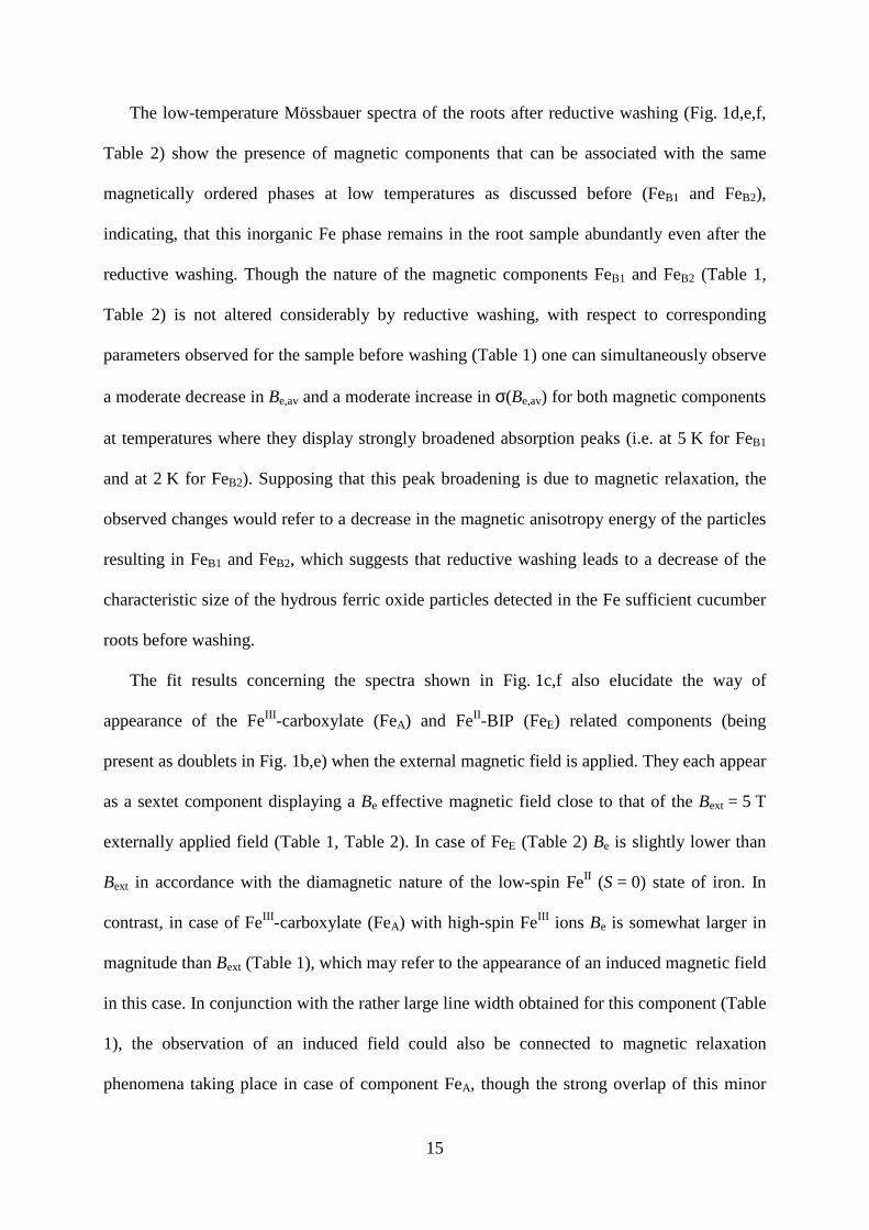
The low-temperature Mössbauer spectra of the roots after reductive washing (Fig. 1d,e,f,
Table 2) show the presence of magnetic components that can be associated with the same
magnetically ordered phases at low temperatures as discussed before (FeB1 and FeB2),
indicating, that this inorganic Fe phase remains in the root sample abundantly even after the
reductive washing. Though the nature of the magnetic components FeB1 and FeB2 (Table 1,
Table 2) is not altered considerably by reductive washing, with respect to corresponding
parameters observed for the sample before washing (Table 1) one can simultaneously observe
a moderate decrease in Be,av and a moderate increase in σ(Be,av) for both magnetic components
at temperatures where they display strongly broadened absorption peaks (i.e. at 5 K for FeB1
and at 2 K for FeB2). Supposing that this peak broadening is due to magnetic relaxation, the
observed changes would refer to a decrease in the magnetic anisotropy energy of the particles
resulting in FeB1 and FeB2, which suggests that reductive washing leads to a decrease of the
characteristic size of the hydrous ferric oxide particles detected in the Fe sufficient cucumber
roots before washing.
The fit results concerning the spectra shown in Fig. 1c,f also elucidate the way of
appearance of the FeIII-carboxylate (FeA) and FeII-BIP (FeE) related components (being
present as doublets in Fig. 1b,e) when the external magnetic field is applied. They each appear
as a sextet component displaying a Be effective magnetic field close to that of the Bext = 5 T
externally applied field (Table 1, Table 2). In case of FeE (Table 2) Be is slightly lower than
Bext in accordance with the diamagnetic nature of the low-spin FeII (S = 0) state of iron. In
contrast, in case of FeIII-carboxylate (FeA) with high-spin FeIII ions Be is somewhat larger in
magnitude than Bext (Table 1), which may refer to the appearance of an induced magnetic field
in this case. In conjunction with the rather large line width obtained for this component (Table
1), the observation of an induced field could also be connected to magnetic relaxation
phenomena taking place in case of component FeA, though the strong overlap of this minor

16
component with the rest of the peaks prevents us to reach an unambiguous conclusion in this
regard on the basis of the present spectra.
Using immunoblotting, ferritin apoprotein was detected in the root branching zone (Fig. 2)
as a ~30 kDa protein band, whereas no ferritin accumulation was found in the root tips. No
ferritin was detected in solubilized seed and leaf homogenate samples, either. Unspecific
binding of the applied antibodies to the non-target proteins was negligible in the samples. In
cucumber roots, Vigani et al. (2013) found mitochondrial ferritin accumulation under control
(50 μM Fe) or Fe-excess (500 μM) growth conditions. In our study the Fe concentration in the
nutrient solution of control plants was 10 μM which seems not to induce ferritin expression.
Confirming this assumption, applying transmission electron microscopy, no Fe aggregates
typical of ferritin in the root cells could be found (Fig. 3B), but it was revealed that in crevices
on the root surface at adjacent cells there is an accumulation of a mucilagous material
containing finely dispersed grains (Fig. 3A, C, D). No evidence of a continuous aggregation
on the root surface e.g. iron plaque was found which is consistent with our previous study,
where Perls/DAB staining and subsequent microscopic analysis of Fe sufficient roots were
performed (Fodor et al. 2012).
To clarify the origin and to identify unequivocally the inorganic Fe compound(s), as the
major Fe species in the Fe sufficient root, a complete scanning of the root tip zone and lateral
root sections have been made by EELS. Our measurements revealed that no ferritin grains or
Fe-containing structures can be identified in any root cell compartments. This implies that the
majority of the inorganic Fe phase is located outside the root cells forming approx. 20–30 nm
grains as it is presented in Fig. 3d and shown by the EELS spectrum taken on the grains (Fig.
4).
Summarizing the results on the major Fe compounds found in the Fe sufficient cucumber
root, it is only possible to identify and differentiate between two main Fe containing phases: i)

17
FeIII-complexes and ii) an amorphous Fe-rich hydrous ferric oxide/hydroxide phase, probably
including several anions (e.g. phosphate, sulphate) and organic acids (e.g. citrate) in its
structure. This is in contrast to previous studies where, in plants, the main Fe bearing
compound was identified as ferritin (Goodman et al 1982a, b) or γ-FeOOH (Kilcoyne et al.
2000). According to our results, FeIII-complexes were shown to be located in the apoplast
while the Fe rich ferric oxide/hydroxide pool is likely to be present mainly on cell surfaces
forming small aggregates and probably exhibiting an amorphous structure. Iron taken up and
associated to cell functions (heme, Fe-S, Fe-carboxilate complexes etc.) obviously does not
give sufficient contribution to the spectra to identify as separate components as its
concentration is too low for Mössbauer analysis.
Iron components in Fe deficient cucumber root
The Mössbauer spectra taken at 80 K of the Fe deficient cucumber roots supplied with 500
µM 57FeIII-Cit for 30 min is presented in Fig. 5A, B. For comparison, the spectra taken after
the same Fe supply of the Fe sufficient cucumber root pregrown in FeIII-Cit containing
nutrient solution are also shown after 30 min 500 µM 57FeIII-Cit supply (Fig. 5C). No
Mössbauer spectrum could be taken after reductive washing (the amount of 57Fe was under
the detection limit; spectrum not shown). In the latter case we utilized the advantage that
Mössbauer spectroscopy can only show 57Fe in the samples so in the spectrum presented in
Fig. 5C, only new Fe compounds formed during the 30 min 500µM 57FeIII-Cit supply can be
seen.
The spectra could be evaluated suggesting one or two paramagnetic doublet components
with parameters listed in Table 3. According to the Mössbauer parameters, FeA component
can be associated to FeIII carboxylate complexes already identified in the case of Fe sufficient

18
roots (Kovács et al. 2009). The FeD component can be assigned to [Fe(H2O)6]2+ (Vértes &
Korecz 1979, Kovács et al. 2009) while the FeE represents FeII-BIP complex (Greenwood &
Gibb 1971).
Taking into account previous Mössbauer results on natural FeIII-complexes applied as Fe
supply for plants (Kovács et al. 2009), the parameters of FeA component are very close to
those of FeIII-Cit complex (δ = 0.48(1) mms-1, ∆ = 0.62(1) mms-1, Solti et al. 2012) supplied
in the nutrient solution thus it is supposed to be present as the major Fe form in the Fe
deficient cucumber root after Fe supply. This result shows that a high amount of the FeIII-Cit
present in the nutrient solution can be attached to the cell wall components and taken up by
the root. The presence of high amount of a non-removable FeIII-Cit component is confirmed
by the Mössbauer spectrum taken after the reductive washing since parameters of FeIII-Cit
remains in the spectrum. As the reductive washing completely removes FeIII-carboxylate
components from the apoplastic spaces as we have seen in the roots of Fe sufficient pants, this
is only possible if FeIII-Cit is located inside the cells e.g. in vacuoles or inside the central
cylinder of the root. The FeIII-Cit may be taken up directly from the nutrient solution.
Nevertheless it can be formed de novo after the reduction, transport and reoxidation of Fe in
root parenchyma cells. Furthermore, FeIII-Cit is considered to be the major transport form of
Fe in the xylem (Rellán-Álvarez et al. 2010) where the complex may be accumulated taken
into account the higher uptake rate of Fe as compared to Fe sufficient roots. We propose that
the FeA component providing the majority of Fe in the washed Fe deficient roots is FeIII-Cit
which is localized mainly in the root xylem vessels.
Approximately 10% of the total Fe is in the form of [Fe(H2O)6]2+ in the Fe deficient plants
supplied with FeIII-Cit that confirms previous results showing the highly increased rate of Fe
reduction (turbo reductase activity) (Kovács et al. 2009). Considering that no reduced Fe (Fig.
2C) and only completely removable ferric carboxylates could be found after the same time

19
and concentration of Fe supply in the Fe sufficient plants pregrown in 56FeIII-Cit it can be
concluded that not only the reduction rate increases under Fe deficiency in Strategy I plants
but also the Fe uptake rate becomes significantly higher.
It is noteworthy to mention that the washing procedure also results to the presence of FeII-
BIP complex (FeE found in Fig. 5B). The relative high amount of this complex (~32%, see
Table 3) compared to the Fe sufficient case (see also Fig. 1E, Table 2) can be explained by the
much lower total Fe content of the Fe deficient roots. The lack of the ferric oxide/hydroxide
species under Fe deficient conditions was also proved by direct low-temperature Mössbauer
measurement of an Fe deficient cucumber root supplied with 100 µM FeIII-Cit for 30 min
(taken at 5 and 2 K). The obtained Mössbauer spectra are presented in Fig. 6, where no
magnetically split subspectrum could be separated. The only component can be identified as
FeIII-Cit (δ=0.53(2) mms-1, ∆=0.60(4) mms-1, Γ=0.6(1) mms-1) confirming the presence of the
FeIII-Cit component.
Iron components with different Fe supply
Literature data on the uptake of different Fe complexes indicated that there are large
differences in the reduction and uptake rate when different FeIII-chelates were applied
(García-Marco et al. 2006, Lucena & Chaney 2007). The stability and size of the Fe-chelate
are major factors influencing the rate of the reduction but similar uptake rate was found for
FeIII-EDTA and FeIII-EDDHA complexes (Lucena & Chaney 2007). On the other hand, the Fe
uptake and transport from 59FeIII-Cit compared to 59FeIII-EDTA containing nutrient solution to
the shoot of Fe deficient cucumber seedlings was much (2-5 fold) higher (Cseh et al., 1994).

20
With the help of Mössbauer spectroscopy, the Fe species formed in Fe sufficient and Fe
deficient conditions were investigated in the case of FeIII-EDTA and FeIII-EDDHA applied as
Fe supply in the nutrient solution in comparison to FeIII-Cit. The Mössbauer results are
presented in Fig. 7 for both the Fe sufficient and deficient case. The corresponding Mössbauer
parameters are listed in Table 4.
The Mössbauer spectra and the related Fe-components found in the Fe sufficient Fe-
chelate grown-plants differ from those of the FeIII-Cit-grown plants. The larger quadrupole
doublet characteristic of FeIIIE than found for FeIII A (Table 1 and Table 4) and the quite large
line width (0.6/0.7 mm s-1) can refer to the presence of similar but slightly different O6
chemical environment around the FeIII ions. This may be explained by the partial attachment
of the EDTA, EDDHA ligands to the central FeIII but also by the formation of FeIII-
carboxylate complexes with cell-wall components.
Unfortunately, it was difficult to get any information on this component since the very
low amount of total Fe in the samples (see also Fig. 8) made impossible even after freeze-
drying to measure the low-temperature spectra as presented for FeIII-Cit supplied roots in Fig.
1 and 6. For the same reason, Mössbauer spectroscopy could not be applied following a
reductive washing to separate apoplastic and symplastic Fe pools neither in Fe sufficient nor
in Fe deficient case. This also suggests that the majority of Fe is taken up and immediately
transported to other plant tissues following the reduction and thus, no significant amount of Fe
accumulates in the root parenchyma or in the root apoplast.
The relative and absolute content of FeII in the roots was similar in the case FeIII-EDTA
and FeIII-EDDHA (calculated from the ICP measurements with the help of the relative
spectrum areas found in the Mössbauer spectra), while in the case of FeIII-Cit, they were
slightly higher. This shows that the reduction and uptake is favored by the application of FeIII-
Cit compared to the other FeIII-chelates. This is also in good agreement with the results

21
discussed above for the FeIII-Cit, namely, a very high accumulation of Fe was demonstrated
both at Fe sufficient and Fe deficient conditions compared to FeIII-chelates.
The parameters of the FeD component were slightly different in the roots supplied with
FeIII-chelates from those found in the case of FeIII-Cit supply. The quadrupole splitting is
smaller which may show that the Fe2+ may be partly attached to the EDTA/EDDHA ligand
and /or to cell wall –COO- groups. However, no evidence could be found for the formation of
FeII-EDTA (and probably this holds also for FeII-EDDHA) complex since in the latter case,
the quadrupole splitting of the complexed FeII would be much smaller (δ=1.85 mms-1, ∆=2.80
mms-1, measured at 80 K by Epstein 1962). This contradicts the suggestions made according
to previous analytical results that in these systems FeII-EDTA complex is formed after the
reduction in the apoplast (Lucena & Chaney, 2006). Moreover, Mössbauer studies of 57FeCl2-
EDTA systems in aqueous solution in acidic and neutral pH-range showed no complex
formation between FeII and the ligand but the presence of [Fe(H2O)6]2+ species (Szilágyi et
al., 2007). In the case of EDTA, EDDHA and several other complexing agents, the low
stability of the FeII-complexes and thus their fast reoxidation after the enzymatic reduction
during Fe uptake in cucumber was also proposed (Lucena & Chaney 2007).
Summary and conclusion
Attempting to establish a general comparison of Fe uptake from natural complexes e.g.
FeIII-Cit and artificial chelates e.g. FeIII-EDTA or FeIII-EDDHA in Fe sufficient and Fe
deficient plants we have analyzed the microenvironment and localization of Fe components
formed during or after the reduction, uptake and accumulation and making up the total Fe
content of the roots of cucumber, a strategy I plant grown in nutrient solution. A long term Fe
supply with FeIII-Cit results in the accumulation of partially removable nanosized amorphous

22
hydrous ferric oxide/hydroxide depositions in crevices on the root surface in mucilagous
materials. The mucilagous cover on the root surface may be a result of the axenic conditions.
Ferric carboxylates can be removed by a reductive washing leaving no sufficient contribution
(concentration) of this Fe component to the Mössbauer spectra just as other well-known Fe
containing cellular components e.g. Fe-nicotianamine or Fe-S clusters. In Fe deficient plants,
short-term FeIII-Cit supply in high concentration results in fast and efficient accumulation of
Fe-carboxylates (most probably FeIII-Cit) in the xylem and a transient accumulation of FeII at
the apoplastic side of the root cell membranes. Our results also show that the reductive
washing procedure by the ferrous chelator BIP leaves a significant new, not readily removable
Fe component in the root apoplast, the FeII-BIP complex.
In case of roots supplied with Fe-chelates of high stability (FeIII-EDTA, FeIII-EDDHA)
only a FeIII-carboxylate component suggested to be localized in the apoplast/symplast was
identified and no hydrous ferric oxides accumulated. In Fe deficient plants, the ratio of
reduced to total Fe when these stable chelates were supplied almost doubled compared to
FeIII-Cit but it may not necessarily correspond to the higher efficiency of the chelates to
supply Fe as the total Fe content of these roots were much lower.
It is concluded that natural Fe chelates, with a similar characteristics to FeIII-Cit, may
perform better in Fe-accumulation in the roots compared to synthetic ones. This may be
utilized in Fe biofortification, since nanosized Fe deposits on the root surface may be
remobilized upon Fe deficiency. Thus, compared to highly stable synthetic chelates that
perform better in maintaining Fe in soluble form, also natural Fe chelates could provide a
good choice to prevent Fe deficiency in irrigated plantations.

23
References
Abadía J, Vázquez S, Rellán-Álvarez R, El-Jendoubi H, Abadía A, Álvarez-Fernández A,
López-Millán AF (2011) Towards a knowledge-based correction of iron chlorosis. Plant
Physiol Bioch 49: 471-482.
Amils R, de la Fuente V, Rodríguez N, Zuluaga J, Menéndez N, Tornero J (2007)
Composition, speciation and distribution of iron minerals in Imperata cilíndrica. Plant Physiol
Bioch 45: 335-340.
Bienfait HF, Briel WVD, Mesland-Mul NT (1985) Free space iron pools in roots: generation
and mobilization. Plant Physiol 78: 596–600.
Briat JF, Duc C, Ravet K, Gaymard F (2010) Ferritins and iron storage in plants. Biochim
Biophys Acta 1800: 806-814.
Cesco S, Neumann G, Tomasi N, Pinton R, Weisskopf L (2010) Release of plant-borne
flavonoids into the rhizosphere and their role in plant nutrition. Plant Soil 329:1-25.
Chasteen ND, Harrison PM (1999) Mineralization in Ferritin: An Efficient Means of Iron
Storage. J Struct Biol 126: 182-194.
Cornell RM, Scwertmann U (2003) The iron oxides. Weinheim: Wiley-VCH.

24
Cseh E, Váradi G, Fodor F (1994) Effect of Fe-complexes and N-forms on the Fe absorption,
uptake and translocation of cucumber plants. Bot Kozl 81: 47-55.
Epstein LM (1962) Mössbauer Spectra of Some Iron Complexes. J Chem Phys 36: 2731-
2737.
Fodor F, Kovács K, Czech V, Solti Á, Tóth B, Lévai L, Bóka K, Vértes A (2012)
Effects of short term iron citrate treatments at different pH values on roots of iron deficient
cucumber: a Mössbauer analysis. Journal of Plant Physiology, 169:1615-1622.
García-Marco S, Martínez N, Yunta F, Hernandez-Apaolaza L, Lucena JJ (2006)
Effectiveness of ethylenediamine-N(o-hydroxyphenylacetic)-N’(p-hydroxyphenylacetic) acid
(o,p-EDDHA) to supply iron to plants. Plant Soil 279:31-40.
Goodman BA, DeKock PC (1982a) Mössbauer studies of plant materials. I. Duckweed,
stocks, soyabean and pea. J Plant Nutr 5: 345-353.
Goodman BA, DeKock PC (1982b) Mössbauer studies of plant materials. II. Spectra of 57Fe-
enriched duckweed at low temperatures. J Plant Nutr 5: 355-362.
Greenwood NN, Gibb TC (1971) Mössbauer Spectroscopy. Chapman & Hall Ldt., London,
UK

25
Hartnett A, Böttger LH, Matzanke BF, Carrano CJ (2012) A multidisciplinary study of iron
transport and storage in the marine green alga Tetraselmis suecica. J Inorg Biochem 116:
188–194.
Kilcoyne SH, Bentley PM, Thongbai P, Gordon DC, Goodman BA (2000) The application of
57Fe Mössbauer spectroscopy in the investigation of iron uptake and translocation in plants.
Nucl Instrum Meth B 160: 157-166.
Klencsár Z, Kuzmann E, Vértes A (1996) User-friendly software for Mössbauer spectrum
analysis. J Radioanal Nucl Ch 210: 105.
Kobayashi T, Nishizawa NK (2012) Iron uptake, translocation, and regulation in higher
plants. Annu Rev Plant Biol 63: 131-152.
Kovács K, Kuzmann E, Fodor F, Homonnay Z, Machala L, Vértes A (2010) Low temperature
57Fe Mössbauer study of cucumber root, J Phys Conf Ser 217: 012019
Kovács K, Kuzmann E, Tatár E, Vértes A, Fodor F (2009) Investigation of iron pools in
cucumber roots by Mössbauer spectroscopy: direct evidence for the Strategy I iron uptake
mechanism. Planta 229: 271–278.
Laemmli UK (1970) Cleavage of structural proteins during assembly of the head of
bacteriophage T4. Nature 227: 680-685.

26
Lucena JJ, Chaney RL (2006) Synthetic Iron Chelates as Substrates of Root Ferric Chelate
Reductase in Green Stressed Cucumber Plants. J Plant Nutr 29: 423–439.
Lucena JJ, Chaney RL. (2007) Response of Cucumber Plants to Low Doses of Different
Synthetic Iron Chelates in Hydroponics. J Plant Nutr 30: 795–809.
Pechousek J, Jancik D, Frydrych J, Navarik J, Novak P (2012) Setup of Mössbauer
spectrometers at RCPTM. Mossbauer Spectroscopy in Materials Science 1489: 186-193.
Pechousek J, Prochazka R, Jancik D, Frydrych J, Mashlan M (2010) Universal LabVIEW
powered Mössbauer spectrometer based on the USB, PCI or PXI devices. J Phys Conf Ser
217: 1-4.
Rancourt DG, Ping JY (1991) Voigt-based methods for arbitrary-shape static hyperfine
parameter distributions in Mössbauer spectroscopy. Nuclear Instruments and Methods B 58:
85-97.
Rellán-Álvarez R, Giner-Martínez-Sierra J, Orduna J, Orera I, Rodríguez-Castrillón JÁ,
García-Alonso JI, Abadía J, Álvarez-Fernández A (2010) Identifi cation of a Tri-Iron(III), Tri-
Citrate Complex in the Xylem Sap of Iron-Defi cient Tomato Resupplied with Iron: New
Insights into Plant Iron Long-Distance Transport. Plant Cell Physiol 51(1): 91–102.
Rodríguez N, Menéndez N, Tornero J, Amils R, de la Fuente V (2005) Internal iron
biomineralization in Imperata cylindrica, a perennial grass: chemical composition, speciation
and plant localization. New Phytol 165: 781-789.

27
Rodriguez-Lucena P, Benedicto A, Lucena JJ, Rodriguez-Castrillon JA, Moldovan M, Alonso
JIG and Hernandez-Apaolazaa L (2010) Use of the stable isotope 57Fe to track the efficacy of
the foliar application of lignosulfonate/Fe3+ complexes to correct Fe deficiencies in cucumber
plants. J Sci Food Agr 91: 395-404.
Sisó-Terraza P, Rios JJ, Abadía J, Abadía A, Álvarez-Fernández A (2016) Flavins secreted by
roots of iron-deficient Beta vulgaris enable mining of ferric oxide via reductive mechanism.
New Phytologist 209:733-745.
Schwertmann U, Wagner F, Knicker H. (2005) Ferrihydrite-Humic Associations: Magnetic
Hyperfine Interactions. Soil Sci Soc Am J 69:1009-1015.
Solti Á, Kovács K, Basa B, Vértes A, Sárvári É, Fodor F (2012) Uptake and incorporation of
iron in sugar beet chloroplasts. Plant Physiol Bioch 52: 91-97.
St. Pierre TG, Bell SH, Dickson DPE, Mann S, Webb J, Moore GR and Williams RJP (1986)
Mössbauer spectroscopic studies of the cores of human, limpet and bacterial ferritins.
Biochim Biophys Acta 870: 127-134.
Szilágyi ÁP (2007) Study of iron-chelates in solid state and aqueous solutions using
Mossbauer spectroscopy. Dissertation, Eötvös Lorand University, Budapest, Hungary
Vértes A, Korecz L, Burger K (1979) Mössbauer spectroscopy. Elsevier, Amsterdam

28
Vigani G, Tarantino D, Murgia I (2013) Mitochondrial ferritin is a functional iron-storage
protein in cucumber (Cucumis sativus) roots. Front Plant Sci 4: 316.
Wade VJ., Treffry A, Laulhere JP, Bauminger ER, Cleton MI, Mann S, Briat JF and Harrison
PM (1993) Structure and composition of ferritin cores from pea seed (Pisum sativum).
Biochim Biophys Acta 1161: 91-96.

29
Table 1
57Fe Mössbauer parameters of Fe components found in the Fe sufficient cucumber roots
grown on FeIII-Cit for 3 weeks, as derived on the basis of measurements performed at
temperatures 5 K and 2 K, as well as at 2 K in an external magnetic field of Bext = 5 T being
oriented parallel to the direction of gamma ray (Fig 1a-c). Parameter values that are indicated
to be the same were constrained to be the same during the fit. For magnetic components
where the double quadrupole shift (2ε) is not given, it was assumed to be zero. The numbers
between parentheses give the statistical uncertainty (1× standard deviation) in the last digit.
The suffix F refers to a fixed parameter.
T = 5 K T = 2 K T = 2 K, Bext = 5 T FeA δ
a (mms-1) 0.52(1) 0.52(1) 0.52(1) ∆
b (mms-1) 0.58(4) 0.58(4) - Be
c (T) - - 7.1(2) Γ
d (mms-1) 1.08(7) 1.08(7) 1.08(7) Sr
e (%) 9(2) 9(2) 9(2) FeB1 δ
a (mms-1) 0.504(3) 0.504(3) 0.504(3) 2εf (mms-1) 0F -0.05(1) -0.08(1) Be,av
c(T) 25.7(2) 41.0(1) 43.2(1) σ (Be)
g (T) 14.6(3) 4.8(1) 4.4(1) Γ
d (mms-1) 0.69(1) 0.69(1) 0.69(1) Sr
e (%) 67(1) 67(1) 67(1) FeB2 δ
a (mms-1) 0.504(3) 0.504(3) 0.504(3) ∆
b (mms-1) 0.658(7) - - Be,av
c (T) - 23.5(7) 33.7(7) σ (Be)
g (T) - 10.9(5) 8.6(7) Γ
d (mms-1) 0.69(1) 0.69(1) 0.69(1) Sr
e (%) 24(1) 24(1) 24(1)
aIsomer shift, relative to α-Fe.
bQuadrupole splitting.
cEffective magnetic field.

30
dLine width at half maximum
eRelative resonant absorption areas of the relevant spectral components, which represents
relative contents of the corresponding Fe forms.
fQuadrupole shift.
gStandard deviation of the Gaussian of the hyperfine magnetic field distribution.

31
Table 2
57Fe Mössbauer parameters of Fe components found in the Fe sufficient cucumber roots
grown on FeIII-Cit for 3 weeks and then being subjected to reductive washing with bipyridyl,
as derived on the basis of measurements performed at temperatures 5 K and 2 K, as well as
at 2 K in an external magnetic field of 5 T being oriented parallel to the direction of gamma
ray (Fig 1d-f). Parameter values that are indicated to be the same were constrained to be the
same during the fit. For magnetic components where the double quadrupole shift (2ε) is not
given, it was assumed to be zero. The numbers between parentheses give the statistical
uncertainty (1× standard deviation) in the last digit(s). The suffix F refers to a fixed
parameter.
T = 5 K T = 2 K T = 2 K, Bext = 5 T FeE δ
a (mms-1) 0.40(1) 0.40(1) 0.40(1) ∆
b (mms-1) 0.34(2) 0.34(2) - Be
c (T) - - 4.7(1) Γ
d (mms-1) 0.38(3) 0.38(3) 0.38(3) Sr
e (%) 5(1) 5(1) 5(1) FeB1 δ
a (mms-1) 0.513(4) 0.513(4) 0.513(4) 2εf (mms-1) 0F -0.06(1) -0.05(1) Be,av
c (T) 22.6(6) 42.0(1) 44.3(7) σ (Be)
g (T) 17.7(8) 5.0(1) 4.6(1) Γ
d (mms-1) 0.62(1) 0.62(1) 0.62(1) Sr
e (%) 72(1) 72(1) 72(1) FeB2 δ
a (mms-1) 0.513(4) 0.513(4) 0.513(4) ∆
b (mms-1) 0.71(1) - - Be,av
c (T) - 20.8(9) 29.1(8) σ (Be)
g (T) - 12.3(9) 9.3(1.1) Γ
d (mms-1) 0.62(1) 0.62(1) 0.62(1) Sr
e (%) 24(1) 24(1) 24(1)
aIsomer shift, relative to α-Fe.
bQuadrupole splitting.

32
cEffective magnetic field.
dLine width at half maximum
eRelative resonant absorption areas of the relevant spectral components, which represents
relative contents of the corresponding Fe forms.
fQuadrupole shift.
gStandard deviation of the Gaussian of the hyperfine magnetic field distribution.

33
Table 3
57Fe Mössbauer parameters of the Fe components found in the Fe deficient and Fe sufficient
(grown on FeIII-Cit for 3 weeks) cucumber roots supplied with 500 μM 57FeIII-Cit for 30 min
before and/or after reductive washing with bipyridyl as derived on the basis of measurements
performed at 80 K (Fig 5). The numbers between parentheses give the statistical uncertainty
(1× standard deviation) in the last digit(s).
Fe deficient root Fe sufficient root before washing after washing before washing FeA δ
a (mms-1) 0.51(1) 0.50(1) 0.50(1) ∆
b (mms-1) 0.59(1) 0.59(1) 0.59(1) Γ
c (mms-1) 0.46(2) 0.46(1) 0.46(1) Srd(%) 90(1) 68(3) 100 FeD δ
a (mms-1) 1.37(1) -- -- ∆
b (mms-1) 3.00(1) -- -- Γ
c (mms-1) 0.44(2) -- -- Srd (%) 10(1) FeE δ (mms-1) -- 0.38(1) -- ∆
b (mms-1) -- 0.32(1) -- Γ
c (mms-1) -- 0.33(2) -- Srd (%) 32(3)
aIsomer shift, relative to α-Fe.
bQuadrupole splitting.
cLine width at half maximum
dRelative resonant absorption areas of the relevant spectral components, which represents
relative contents of the corresponding Fe forms.

34
Table 4
57Fe Mössbauer parameters of the Fe components found in the Fe sufficient and Fe deficient
cucumber roots which were supplied with FeIII-EDTA and FeIII-EDDHA complexes as
derived on the basis of measurements performed 80 K (Fig 7). The numbers between
parentheses give the statistical uncertainty (1× standard deviation) in the last digit(s).
Fe sufficient root supplied with 10
µM FeIII for 3 weeks Fe deficient root supplied with 500 µM FeIII for 30 min
FeIII-EDTA FeIII-EDDHA FeIII-EDTA FeIII-EDDHA FeF δ
a (mms-1) 0.48(1) 0.47(1) 0.49(1) 0.49(1) ∆
b (mms-1) 0.67(2) 0.64(1) 0.53(2) 0.57(1) Γ
c (mms-1) 0.59(3) 0.61(2) 0.66(4) 0.57(1) Srd (%) 100 100 76(3) 80(1) FeD (%) δ
a (mms-1) 1.30(2) 1.30(1) ∆
b (mms-1) 2.93(4) 2.95(2) Γ
c (mms-1) 0.48(7) 0.45(3) Srd (%) 24(3) 20(1)
aIsomer shift, relative to α-Fe.
bQuadrupole splitting.
cLine width at half maximum
dRelative resonant absorption areas of the relevant spectral components, which represents
relative contents of the corresponding Fe forms.

35
Captions to Figures
Fig. 1 Low-temperature Mössbauer spectra of lyophilized cucumber roots grown on 10 µM
57FeIII-citrate containing nutrient solution (based on Kovács et al. 2010) without washing
measured at a) T=5 K, b) T=2 K and c) T=2 K, B=5 T and after reductive (BIP) washing
measured at d) T=5 K, e) T=2 K and f) T=2 K, B=5 T

36
Fig. 2 Solubilized proteins on polyacrylamide gel (a) and immunoblot against plant ferritin
(b). Samples were 1 – molecular weight standard; 2 – seed homogenate; 3 – leaf homogenate;
4 – root tip homogenate; 5 – root branching zone homogenate. Molecular weight standards
were serum albumin (66 kDa); ovalbumin (45 kDa); glycerinaldehyde-3-phosphate
dehydrogenase (36 kDa); carbonic anhydrase (29 kDa); trypsinogene (24 kDa); trypsin
inhibitor (20.1 kDa); α-lactalbumin (14.2 kDa). Both protein gels and immunoblots were
loaded with 20 µg of solubilised protein except for the sample (2), where the lanes were
loaded with 20 µg solubilised protein over the storage proteins (at ~50-55 kDa).

37
Fig. 3 Electron micrographs of cucumber root sections. The plants were grown with sufficient
concentration of FeIII-Cit (10 μM Fe) in nutrient solution. (a) Middle section. (b-d) Root tip
zone sections of lateral roots. (a) Fe grains incorporated into mucilage covering root surface.
(b) No Fe can be seen in large quantities. Fe may be dispersed homogeneously in functional
sites but no storage pools are revealed. (c) Fe grains at the root surface with bacteria
embedded in mucilage. The section is parallel to the root surface. (d) Fe grains embedded in
mucilage at the root surface, enlarged. Letters indicate root cells (RC), cell wall (CW),
vacuoles (V), root surface (RS), bacteria (B) and mucilage (M), arrows indicate Fe grains.
Scale bars are equal to (a) 1 µm (b) 5 µm (c) 1 µm (d) 200 nm

38
Fig. 4 EELS spectrum of the finely dispersed grains found in the root tip sections of lateral
roots of cucumber grown with sufficient concentration of Fe (10 μM) in nutrient solution

39
Fig. 5 Mössbauer spectra taken at T=80K of Fe deficient cucumber roots after 30 min 500 µM
57FeIII-citrate supply without washing (a) and after reductive (BIP) washing (b) and of Fe
sufficient cucumber roots grown in 10 µM 56FeIII -citrate containing nutrient solution after 30
min 500 µM 57FeIII-citrate supply without washing (c)

40
Fig. 6 Low-temperature Mössbauer spectra of Fe deficient cucumber roots after 30 min 100
µM 57FeIII-citrate supply taken at T=5 K (a) and at T=2 K (b) without applying external
magnetic field

41
Fig. 7 Mössbauer spectra taken at T=80 K of Fe sufficient cucumber roots supplied with 10
µM 57FeIII-EDTA (a) and 57FeIII-EDDHA (b) for 20 days in the nutrient solution and of Fe
deficient cucumber roots supplied with 500 µM 57FeIII-EDTA (c) and 57FeIII-EDDHA (d) for
30 min

42
Fig. 8 Fe concentration of the Fe deficient cucumber root after 30 min 500 µM FeIII-citrate,
FeIII-EDTA, and FeIII-EDDHA supply. FeII concentration was calculated from the total Fe
concentration using the corresponding Mössbauer spectra. (Data are shown as mean±SD, n=3,
significant differences between data are indicated with different letters, P<0.05, Fe total and
FeII data sets were compared separately as indicated by capital and normal letters,
respectively.)

43
Electronic Supplementary Material 1 Planta Revisiting the iron pools in cucumber roots: identification and localization Krisztina Kovács, Jiří Pechoušek, Libor Machala, Radek Zbořil, Zoltán Klencsár, Ádám Solti, Brigitta Tóth, Brigitta Müller, Hong Diep Pham, Zoltán Kristóf, Ferenc Fodor Corresponding author: Krisztina Kovács Institute of Chemistry, Eötvös Loránd University, P.O. Box 32, Budapest 1512, Hungary [email protected] Pictures of the Fe sufficient and Fe deficient cucumber plants, grown in 400 ml plastic pots. Young Fe deficient plants: Young Fe sufficient plants
:
Fe deficient plants at harvest:

44
Fe sufficient plants at harvest:

45
Electronic Supplementary Material 2 Planta Revisiting the iron pools in cucumber roots: identification and localization Krisztina Kovács, Jiří Pechoušek, Libor Machala, Radek Zbořil, Zoltán Klencsár, Ádám Solti, Brigitta Tóth, Brigitta Müller, Hong Diep Pham, Zoltán Kristóf, Ferenc Fodor Corresponding author: Krisztina Kovács Institute of Chemistry, Eötvös Loránd University, P.O. Box 32, Budapest 1512, Hungary [email protected] Here we describe in detail in which way the results listed in Tables 1 & 2 and shown in Fig. 1a-f were obtained by applying physically reasonable constraints in the frame of parallel spectrum fits. All fits were carried out with the MossWinn program. For detailed description of the applied models, we refer to the associated program manual (Klencsár 2015). Fitting of the spectra observed for iron sufficient cucumber roots before reductive washing (Fig. 1a-c, Table 1) The three spectra shown in Fig. 1a-c were fitted simultaneously for each of the spectra assuming three spectrum components (FeA, FeB1, FeB2) with a shared fit parameter set being subject to the following constraints: (FeA) In the absence of an external magnetic field, the FeA component was described with identical Lorentzian doublets at T = 5 K and 2 K temperatures. In the presence of the 5 T external magnetic field, at 2 K, FeA was described with a sextet component with zero quadrupole shift and the isomer shift and line width parameters being identical with those of the corresponding doublet components at T = 5 K and 2 K temperatures. The relative area fraction of the FeA component was assumed to be the same in all the three spectra. Justification: The isomer shift and quadrupole splitting parameters are not expected to change considerably in the narrow temperature interval of 2…5 K. Assuming nonzero quadrupole shift for the sextet component results in a quadrupole shift value that is in the order of the statistical uncertainty. No appreciable change is expected to occur in the Mössbauer-Lamb factor between 2 K and 5 K. Due to the overlap of this minor component with the peaks of the rest of the components, the independent determination of its line width in the different spectra is unfeasible on the basis of the present spectra. (FeB1) In all the three spectra the FeB1 component was described by a sextet component displaying peak broadening corresponding to an underlying Gaussian-shape hyperfine magnetic field distribution. The VBF method (Rancourt and Ping 1991) was used as implemented in the MossWinn program (Klencsár 2015) to account for this component in all the three spectra. The center (Be,av) and the standard deviation (σ(Be)) of the Gaussian were varied during the fit independently in the three spectra. The same was true for the quadrupole shift (ε) in the case of the spectra recorded at 2 K, whereas for the spectrum recorded at 5 K the quadrupole shift was fixed to zero. The isomer shift of FeB1 was assumed to be the same in all the three spectra. No coupling was assumed to be present between the effective magnetic field and the isomer shift and quadrupole shift parameters. The internal Lorentzian line width (that would be the apparent line width for σ(Be) = 0 ) and the relative area fraction of the FeB1 component was assumed to be the same in all the three spectra. Justification: Initial fits with an arbitrary-shape distribution indicated that a Gaussian would be an acceptable approximation for the hyperfine magnetic field distribution underlying the FeB1 component at 2 K. Though at 5 K the shape of the FeB1 spectrum component is likely to be influenced by relaxation phenomena, the model can apparently describe the spectral shape with fair accuracy even in this case. Assuming the same internal Lorentzian width for the FeB1 component in all the three spectra enables us to quantify changes in the line broadening straightforwardly via changes in the single fit parameter of σ(Be). The isomer shift is not expected to change considerably in the narrow temperature interval of 2…5 K. Due to the broad peaks the quadrupole shift of FeB1 could not be determined reliably on the basis of the spectrum measured at 5 K. No appreciable change is expected to occur in the Mössbauer-Lamb factor between 2 K and 5 K. The lack of appreciable asymmetry in the spectra suggests that there is no appreciable coupling present between the effective magnetic field and the isomer shift and quadrupole shift parameters. (FeB2) In the spectra recorded at 2 K the FeB2 component was described by a sextet displaying peak broadening corresponding to an underlying Gaussian-shape hyperfine magnetic field distribution. The VBF method (Rancourt and Ping 1991) was used as implemented in the MossWinn program (Klencsár 2015) to account for this component. The center (Be,av) and the standard deviation (σ(Be)) of the Gaussian were varied during the fit independently in the two spectra. The quadrupole shift (ε) of this component was assumed to be zero. In the spectrum recorded at 5 K, FeB2 was modeled with a doublet component having a Lorentzian line width identical

46
with the internal Lorentzian line width of the VBF components at 2 K. The latter line width was assumed to be identical with that of the FeB1 component. The isomer shift of FeB2 was assumed to be identical with that of FeB1 in all the three spectra. No coupling was assumed to be present between the effective magnetic field and the isomer shift and quadrupole shift parameters. The relative area fraction of the FeB2 component was assumed to be the same in all the three spectra. Justification: Though the spectrum shape of the FeB2 component at 2 K is expected to be influenced by relaxation phenomena, modeling it with a magnetic component displaying Gaussian hyperfine magnetic field distribution turned out to be an acceptable approximation. Assuming the same internal Lorentzian width for the magnetic FeB1 and FeB2 components enables us to quantify differences between corresponding line broadenings in a straightforward manner via the parameter σ(Be). The isomer shift is not expected to change considerably in the narrow temperature interval of 2…5 K. There was no indication for a difference between the isomer shifts of FeB1 and FeB2. Due to the broad peaks overlapping with those of FeB1 the quadrupole shift of FeB2 could not be determined reliably on the basis of the spectra measured at 2 K. Due to the overlap of its peaks with those of the main component FeB1, in the case of FeB2 the determination of the coupling between the effective magnetic field and the isomer shift and quadrupole shift parameters was unfeasible. There was also no indication for the presence of nonzero couplings. No appreciable change is expected to occur in the Mössbauer-Lamb factor between 2 K and 5 K. At 5 K the FeB2 component apparently contributes mainly to the central doublet component. In addition to the above, in case of the spectrum recorded in an external magnetic field of 5 T (Fig 1c), the relative area ratio of the 2nd and 5th peaks (with respect to that of the 3rd and 4th peaks, A25/A34) of sextet components were allowed to vary in order to account for a magnetic polarization effect. The same A25/A34 ratio was assumed for all the three sextet components in the spectrum. The value of this ratio turned out to be 1.77(3). Justification: The independent determination of the A25/A34 ratio was unfeasible for the minor sextet components. The overall normalized chi-square for the simultaneous fit of these three spectra was ∼ 1.404. Fitting of the spectra observed for iron sufficient cucumber roots after reductive washing (Fig. 1d-f, Table 2) The spectra recorded after reductive washing (Fig. 1d-f) were fitted simultaneously in the same way, by applying exactly the same constraints as described above, with the component FeE taking the place of FeA above. In this case the A25/A34 ratio characterizing the spectrum measured in 5 T external magnetic field turned out to be 1.55(5). The overall normalized chi-square for the simultaneous fit of these three spectra was ∼ 1.235. References Klencsár Z (2015) MossWinn 4.0Pre program manual ( http://www.mosswinn.hu/downloads/mosswinn.pdf ) Rancourt DG, Ping JY (1991) Voigt-based methods for arbitrary-shape static hyperfine parameter distributions in Mössbauer spectroscopy. Nuclear Instruments and Methods B 58: 85-97.
![[MI 016-131] PR Series Platinum Resistance Temperature ...](https://static.fdokumen.com/doc/165x107/632122ff537c10e838028447/mi-016-131-pr-series-platinum-resistance-temperature-.jpg)