Lysostaphin: A Staphylococcal Bacteriolysin with Potential Clinical Applications
23
Pharmaceuticals 2010, 3, 1139-1161; doi:10.3390/ph3041139 pharmaceuticals ISSN 1424-8247 www.mdpi.com/journal/pharmaceuticals Review Lysostaphin: A Staphylococcal Bacteriolysin with Potential Clinical Applications Maria do Carmo de Freire Bastos *, Bruna Gonçalves Coutinho and Marcus Lívio Varella Coelho Departamento de Microbiologia Geral, Instituto de Microbiologia Prof. Paulo de Góes, CCS, Bloco I, UFRJ, 21941-590, Rio de Janeiro, RJ, Brazil, E-mails: [email protected] (B.G.C.); [email protected] (M.L.V.C.) * Author to whom correspondence should be addressed; E-Mail: [email protected] Tel.: +55-21-2560-8344, ext. 149; Fax: +55-21-2560-8344 Received: 24 March 2010; in revised form: 8 April 2010 / Accepted: 14 April 2010 / Published: 19 April 2010 Abstract: Lysostaphin is an antimicrobial agent belonging to a major class of antimicrobial peptides and proteins known as the bacteriocins. Bacteriocins are bacterial antimicrobial peptides which generally exhibit bactericidal activity against other bacteria. Bacteriocin production is a self-protection mechanism that helps the microorganisms to survive in their natural habitats. Bacteriocins are currently distributed into three main classes. Staphylococcins are bacteriocins produced by staphylococci, which are Gram- positive bacteria of medical and veterinary importance. Lysostaphin is the only class III staphylococcin described so far. It exhibits a high degree of antistaphylococcal bacteriolytic activity, being inactive against bacteria of all other genera. Infections caused by staphylococci continue to be a problem worldwide not only in healthcare environments but also in the community, requiring effective measures for controlling their spread. Since lysostaphin kills human and animal staphylococcal pathogens, it has potential biotechnological applications in the treatment of staphylococcal infections. In vitro and in vivo studies performed with lysostaphin have shown that this staphylococcin has potential to be used, solely or in combination with other antibacterial agents, to prevent or treat bacterial staphylococcal infectious diseases. OPEN ACCESS
Transcript of Lysostaphin: A Staphylococcal Bacteriolysin with Potential Clinical Applications

Pharmaceuticals 2010, 3, 1139-1161; doi:10.3390/ph3041139
pharmaceuticalsISSN 1424-8247
www.mdpi.com/journal/pharmaceuticals Review
Lysostaphin: A Staphylococcal Bacteriolysin with Potential Clinical Applications
Maria do Carmo de Freire Bastos *, Bruna Gonçalves Coutinho and Marcus Lívio Varella Coelho
Departamento de Microbiologia Geral, Instituto de Microbiologia Prof. Paulo de Góes, CCS, Bloco I,
UFRJ, 21941-590, Rio de Janeiro, RJ, Brazil, E-mails: [email protected] (B.G.C.);
[email protected] (M.L.V.C.)
* Author to whom correspondence should be addressed; E-Mail: [email protected]
Tel.: +55-21-2560-8344, ext. 149; Fax: +55-21-2560-8344
Received: 24 March 2010; in revised form: 8 April 2010 / Accepted: 14 April 2010 /
Published: 19 April 2010
Abstract: Lysostaphin is an antimicrobial agent belonging to a major class of
antimicrobial peptides and proteins known as the bacteriocins. Bacteriocins are bacterial
antimicrobial peptides which generally exhibit bactericidal activity against other bacteria.
Bacteriocin production is a self-protection mechanism that helps the microorganisms to
survive in their natural habitats. Bacteriocins are currently distributed into three main
classes. Staphylococcins are bacteriocins produced by staphylococci, which are Gram-
positive bacteria of medical and veterinary importance. Lysostaphin is the only class III
staphylococcin described so far. It exhibits a high degree of antistaphylococcal
bacteriolytic activity, being inactive against bacteria of all other genera. Infections caused
by staphylococci continue to be a problem worldwide not only in healthcare environments
but also in the community, requiring effective measures for controlling their spread. Since
lysostaphin kills human and animal staphylococcal pathogens, it has potential
biotechnological applications in the treatment of staphylococcal infections. In vitro and in
vivo studies performed with lysostaphin have shown that this staphylococcin has potential
to be used, solely or in combination with other antibacterial agents, to prevent or treat
bacterial staphylococcal infectious diseases.
OPEN ACCESS
Pharmaceuticals 2010, 3
1140
Keywords: lysostaphin; staphylococci; antimicrobial peptides; bacteriocins;
staphylococcins
1. Introduction
Bacteriocins are proteinaceous compounds produced by bacteria which generally display
bactericidal activity against other bacterial spp. [1]. The bacteriocin-producing strains have developed
a protective immunity system against their own bacteriocin. Each bacteriocin has its own immunity
system, which is generally expressed concomitantly with the bacteriocin structural genes [1].
Bacteriocins produced by Gram-positive bacteria are the most studied ones. They form a
heterogeneous group of peptides and proteins. They can be active against other bacteria, belonging to
only closely-related species (narrow spectrum) or to different bacterial genera (broad spectrum). Their
genetic determinants are generally arranged in the form of operons, which may be encoded on the
bacterial chromosome, although they are usually found on plasmids [1].
Recently, Bierbaum and Sahl [2] and Nissen-Meyer and co-workers [3] proposed modifications in
the classification of the bacteriocins produced by Gram-positive bacteria to accommodate data related
to the annual description of a repertoire of bacteriocins, some of them with distinctive characteristics.
Based on the data so far available, a classification scheme is presented in Table 1, which is a
combination of both classifications, with minor modifications. According to it, bacteriocins produced
by Gram-positive bacteria can be grouped into three main classes, all of them with subdivisions.
Bacteriocins from classes I and II, which comprise small peptides, occur more frequently and possess
potential industrial applications [4,5]. Knowledge of all aspects of bacteriocin biology, including the
elucidation of their structure-function relationships, production, immunity, regulation, and mode of
action, is required when considering bacteriocin applications. However, for a more comprehensive
review on both class I and class II bacteriocins, the reader is referred to recent publications written by
Bierbaum and Sahl [2] and Nissen-Meyer and co-workers [5].
Among the bacteriocins produced by staphylococci [6], lysostaphin, which is the prototype class III
bacteriocin, is the most studied one with regard to clinical applications. Therefore, in the present
review, we attempt to provide the reader with an overview of lysostaphin: its structure, relevant
features, mode of action, and potential practical applications.
2. Relevant Features of Staphylococci
The genus Staphylococcus is composed of Gram-positive cocci arranged in grape-like clusters,
divided currently into 42 recognized species and 24 subspecies [7]. They are widespread in nature,
being a commensal of the skin, skin glands, and mucous membranes of mammals and birds.
Staphylococci generally have a benign or symbiotic relationship with their host. However, they may
develop the life-style of a pathogen if they gain entry into the host tissue. Certain Staphylococcus
species are found frequently as etiologic agents of a variety of human and animal infections [8].
Pharmaceuticals 2010, 3
1141
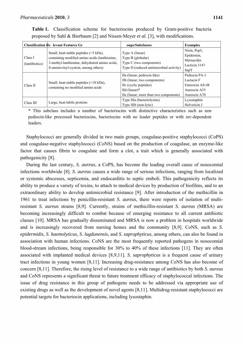
Table 1. Classification scheme for bacteriocins produced by Gram-positive bacteria
proposed by Sahl & Bierbaum [2] and Nissen-Meyer et al. [3], with modifications.
Classification Re levant Features Gr oups/Subclasses Examples
Class I
(lantibiotics)
Small, heat-stable peptides (<5 kDa), containing modified amino acids (lanthionine, 3-methyl-lanthionine, dehydrated amino acids, S-aminovinyl-cystein, among others)
Type A (linear) Type B (globular) Type C (two components) Type D (reduced antimicrobial activity)
Nisin, Pep5, Epidermin, Mersacidin Lacticin 3147 SapT
Class II Small, heat-stable peptides (<10 kDa), containing no modified amino acids
IIa (linear; pediocin-like) IIb (linear; two components) IIc (cyclic peptides) IId (linear)* IIe (linear; more than two components)
Pediocin PA-1 Lactacin F Enterocin AS-48 Aureocin A53 Aureocin A70
Class III Large, heat-labile proteins Type IIIa (bacteriolysins) Type IIIb (non-lytic)
Lysostaphin Helveticin J
* This subclass includes a number of bacteriocins with distinctive characteristics such as non pediocin-like processed bacteriocins, bacteriocins with no leader peptides or with sec-dependent leaders.
Staphylococci are generally divided in two main groups, coagulase-positive staphylococci (CoPS)
and coagulase-negative staphylococci (CoNS) based on the production of coagulase, an enzyme-like
factor that causes fibrin to coagulate and form a clot, a trait which is generally associated with
pathogenicity [8].
During the last century, S. aureus, a CoPS, has become the leading overall cause of nosocomial
infections worldwide [8]. S. aureus causes a wide range of serious infections, ranging from localized
or systemic abscesses, septicemia, and endocarditis to septic emboli. This pathogenicity reflects its
ability to produce a variety of toxins, to attach to medical devices by production of biofilms, and to an
extraordinary ability to develop antimicrobial resistance [9]. After introduction of the methicillin in
1961 to treat infections by penicillin-resistant S. aureus, there were reports of isolation of multi-
resistant S. aureus strains [8,9]. Currently, strains of methicillin-resistant S. aureus (MRSA) are
becoming increasingly difficult to combat because of emerging resistance to all current antibiotic
classes [10]. MRSA has gradually disseminated and MRSA is now a problem in hospitals worldwide
and is increasingly recovered from nursing homes and the community [8,9]. CoNS, such as S.
epidermidis, S. haemolyticus, S. lugdunensis, and S. saprophyticus, among others, can also be found in
association with human infections. CoNS are the most frequently reported pathogens in nosocomial
blood-stream infections, being responsible for 30% to 40% of these infections [11]. They are often
associated with implanted medical devices [8,9,11]. S. saprophyticus is a frequent cause of urinary
tract infections in young women [8,11]. Increasing drug-resistance among CoNS has also become of
concern [8,11]. Therefore, the rising level of resistance to a wide range of antibiotics by both S. aureus
and CoNS represents a significant threat to future treatment efficacy of staphylococcal infections. The
issue of drug resistance in this group of pathogens needs to be addressed via appropriate use of
existing drugs as well as the development of novel agents [8,11]. Multidrug-resistant staphylococci are
potential targets for bacteriocin applications, including lysostaphin.
Pharmaceuticals 2010, 3
1142
3. Lysostaphin General Features
Class III bacteriocins include large peptides (Mr 25 kDa) which are generally heat-labile. This
class of bacteriocins was further subdivided by Heng and co-workers into two distinct groups: (i)
the bacteriolytic enzymes (or bacteriolysins) and (ii) the non-lytic antimicrobial proteins [1].
Staphylococci have been shown to produce bacteriolysins, from which lysostaphin is considered to
be the prototype. Lysostaphin, an extracellular enzyme secreted by strains of S. simulans biovar
staphylolyticus [12], was probably the first staphylococcin (bacteriocin produced by staphylococci)
discovered. Its bactericidal activity against staphylococci relies on its capability of cleaving the
peptidoglycan present in the bacterial cell walls.
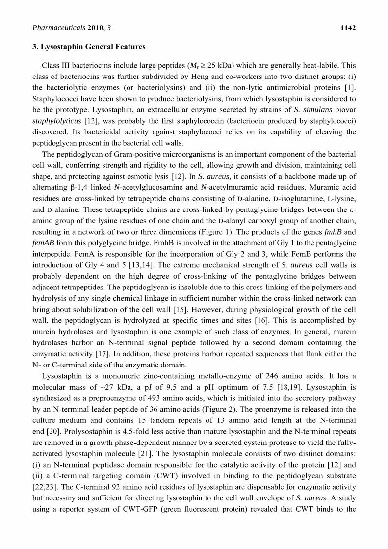
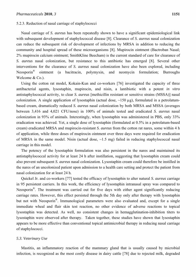
The peptidoglycan of Gram-positive microorganisms is an important component of the bacterial
cell wall, conferring strength and rigidity to the cell, allowing growth and division, maintaining cell
shape, and protecting against osmotic lysis [12]. In S. aureus, it consists of a backbone made up of
alternating β-1,4 linked N-acetylglucosamine and N-acetylmuramic acid residues. Muramic acid
residues are cross-linked by tetrapeptide chains consisting of D-alanine, D-isoglutamine, L-lysine,
and D-alanine. These tetrapeptide chains are cross-linked by pentaglycine bridges between the ε-
amino group of the lysine residues of one chain and the D-alanyl carboxyl group of another chain,
resulting in a network of two or three dimensions (Figure 1). The products of the genes fmhB and
femAB form this polyglycine bridge. FmhB is involved in the attachment of Gly 1 to the pentaglycine
interpeptide. FemA is responsible for the incorporation of Gly 2 and 3, while FemB performs the
introduction of Gly 4 and 5 [13,14]. The extreme mechanical strength of S. aureus cell walls is
probably dependent on the high degree of cross-linking of the pentaglycine bridges between
adjacent tetrapeptides. The peptidoglycan is insoluble due to this cross-linking of the polymers and
hydrolysis of any single chemical linkage in sufficient number within the cross-linked network can
bring about solubilization of the cell wall [15]. However, during physiological growth of the cell
wall, the peptidoglycan is hydrolyzed at specific times and sites [16]. This is accomplished by
murein hydrolases and lysostaphin is one example of such class of enzymes. In general, murein
hydrolases harbor an N-terminal signal peptide followed by a second domain containing the
enzymatic activity [17]. In addition, these proteins harbor repeated sequences that flank either the
N- or C-terminal side of the enzymatic domain.
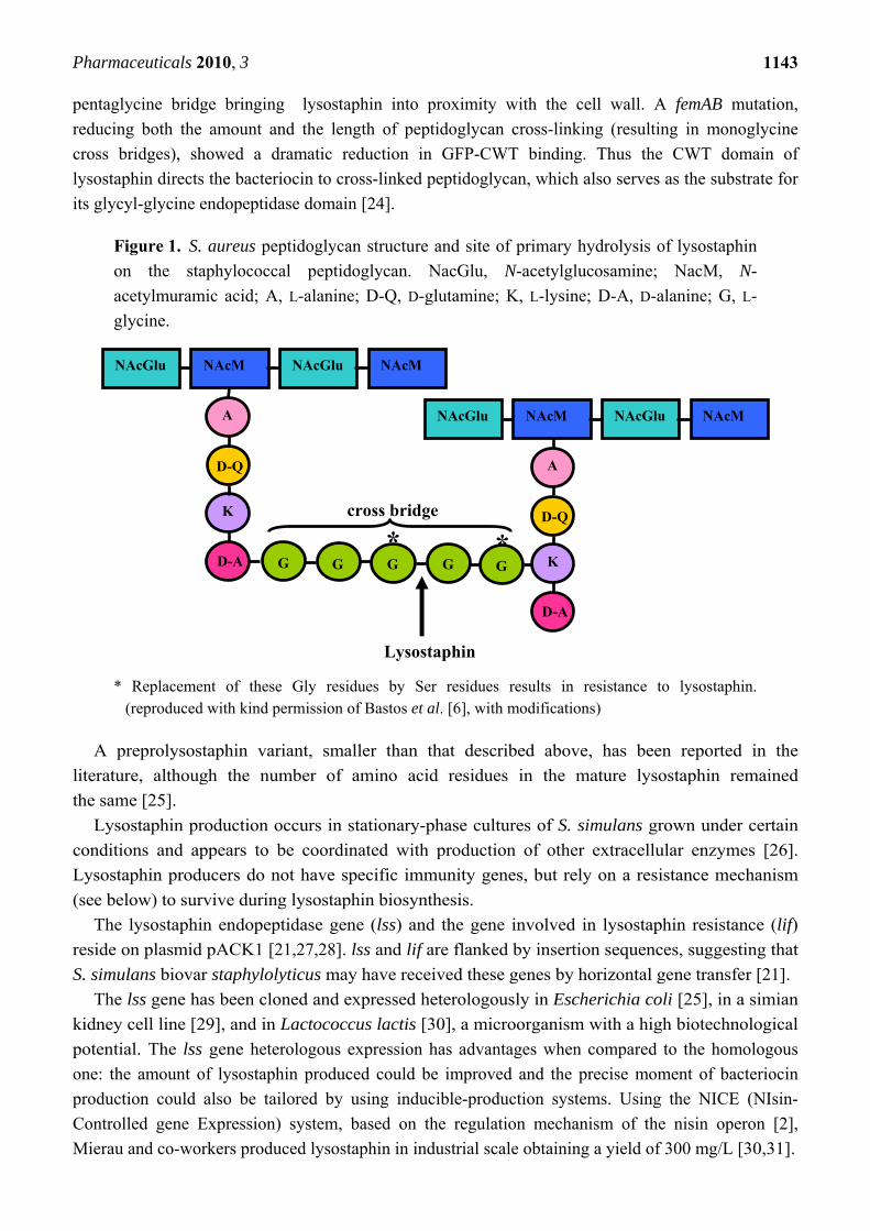
Lysostaphin is a monomeric zinc-containing metallo-enzyme of 246 amino acids. It has a
molecular mass of ~27 kDa, a pI of 9.5 and a pH optimum of 7.5 [18,19]. Lysostaphin is
synthesized as a preproenzyme of 493 amino acids, which is initiated into the secretory pathway
by an N-terminal leader peptide of 36 amino acids (Figure 2). The proenzyme is released into the
culture medium and contains 15 tandem repeats of 13 amino acid length at the N-terminal
end [20]. Prolysostaphin is 4.5-fold less active than mature lysostaphin and the N-terminal repeats
are removed in a growth phase-dependent manner by a secreted cystein protease to yield the fully-
activated lysostaphin molecule [21]. The lysostaphin molecule consists of two distinct domains:
(i) an N-terminal peptidase domain responsible for the catalytic activity of the protein [12] and
(ii) a C-terminal targeting domain (CWT) involved in binding to the peptidoglycan substrate
[22,23]. The C-terminal 92 amino acid residues of lysostaphin are dispensable for enzymatic activity
but necessary and sufficient for directing lysostaphin to the cell wall envelope of S. aureus. A study
using a reporter system of CWT-GFP (green fluorescent protein) revealed that CWT binds to the
Pharmaceuticals 2010, 3
1143
pentaglycine bridge bringing lysostaphin into proximity with the cell wall. A femAB mutation,
reducing both the amount and the length of peptidoglycan cross-linking (resulting in monoglycine
cross bridges), showed a dramatic reduction in GFP-CWT binding. Thus the CWT domain of
lysostaphin directs the bacteriocin to cross-linked peptidoglycan, which also serves as the substrate for
its glycyl-glycine endopeptidase domain [24].
Figure 1. S. aureus peptidoglycan structure and site of primary hydrolysis of lysostaphin
on the staphylococcal peptidoglycan. NacGlu, N-acetylglucosamine; NacM, N-
acetylmuramic acid; A, L-alanine; D-Q, D-glutamine; K, L-lysine; D-A, D-alanine; G, L-
glycine.
* Replacement of these Gly residues by Ser residues results in resistance to lysostaphin.
(reproduced with kind permission of Bastos et al. [6], with modifications)
A preprolysostaphin variant, smaller than that described above, has been reported in the
literature, although the number of amino acid residues in the mature lysostaphin remained
the same [25].
Lysostaphin production occurs in stationary-phase cultures of S. simulans grown under certain
conditions and appears to be coordinated with production of other extracellular enzymes [26].
Lysostaphin producers do not have specific immunity genes, but rely on a resistance mechanism
(see below) to survive during lysostaphin biosynthesis.
The lysostaphin endopeptidase gene (lss) and the gene involved in lysostaphin resistance (lif)
reside on plasmid pACK1 [21,27,28]. lss and lif are flanked by insertion sequences, suggesting that
S. simulans biovar staphylolyticus may have received these genes by horizontal gene transfer [21].
The lss gene has been cloned and expressed heterologously in Escherichia coli [25], in a simian
kidney cell line [29], and in Lactococcus lactis [30], a microorganism with a high biotechnological
potential. The lss gene heterologous expression has advantages when compared to the homologous
one: the amount of lysostaphin produced could be improved and the precise moment of bacteriocin
production could also be tailored by using inducible-production systems. Using the NICE (NIsin-
Controlled gene Expression) system, based on the regulation mechanism of the nisin operon [2],
Mierau and co-workers produced lysostaphin in industrial scale obtaining a yield of 300 mg/L [30,31].
A
D-A
K
NAcGlu NAcGlu NAcM NAcM
G G G G G
A
D-A
D-Q
K
Lysostaphin
* *
NAcGlu NAcGlu NAcM NAcM
D-Q cross bridge
Pharmaceuticals 2010, 3
1144
A homolog of lysostaphin, ALE-1 from Staphylococcus capitis, has also been described and
characterized. Its molecular structure and targeting mechanism, as well as the producer resistance,
appear to be similar to those described for lysostaphin [32,33].
Figure 2. Lysostaphin processing. Lysostaphin is synthesized as a preproenzyme, and after
signal peptide removal, soluble prolysostaphin is released into the extracellular medium.
Proteolytic cleavage of the propeptide of 211 amino acids, from which 195 residues are
organized in 15 tandem repeats of 13-amino acid length, generates the biologically active
lysostaphin, which functions as a glycil-glycine endopeptidase against susceptible
staphylococcal cells. The lysostaphin molecule consists of two distinct domains: an N-
terminal peptidase domain (PD) responsible for its catalytic activity and the C-terminal
wall targeting domain (CWT), which directs lysostaphin to its receptor on the
staphylococcal surface (reproduced with modifications with kind permission of
Bastos et al. [6]).
4. Lysostaphin Mode of Action
Lysostaphin passes freely through the capsule layer of encapsulated staphylococci [34] and is a
cell-wall lytic enzyme. The cell-wall degrading activity of lysostaphin is primarily due to a glycyl-
glycine endopeptidase activity (Figure 1), which lyses many staphylococcal strains [35].
Lysostaphin has been described as primarily active against CoPS [36], retaining some residual activity
against CoNS, although CoNS seem to require increased concentration of the enzyme and larger
incubation times to be killed [37]. Studies performed in our laboratory, however, have shown that
Exportation and removal of the signal peptide (SP)
Preprolysostaphin (493 amino acids)
Prolysostaphin (457 amino
Removal of the repeats by a cystein protease
CWT PD
Mature lysostaphin (246 amino acids)
Repeats
Pharmaceuticals 2010, 3
1145
lysostaphin exhibits an in vitro inhibitory activity against strains of many staphylococcal species,
including CoNS (Table 2).
Table 2. Antimicrobial spectrum of lysostaphin against Staphylococcus spp. strains.
Indicator Strains Origin Inhibition Zones S. aureus MB269, MB274, MB276, MB277, MB288, MB289, MB295, MB302, MB303
Bovine mastitis; Brazil
+++ 2, 3, 7, 9, 10 Bovine mastitis; Argentina +++ 3H1, 13H1 Salad +++ 6H4, 13S2 Salad ++ LI1, A70, A53 Pasteurized milk +++ LF2, LIN4 Sausage +++ Q2, QRFH1 Cheese +++ S. carnosus* CN83 Meat fermentation product ++
S. epidermidis* CN69 Blood ++ (t) CN72 Blood +++
S. haemolyticus* CN61 Blood ++ CN68 Blood -
S. saprophyticus* CN86 Urine ++ CN88 Fistula ++
S. hominis CN70* Blood ++
S. simulans CN87* Blood +++
S. xylosus CN93* Skin +++
S. hyicus ATCC 11249* - +++
S. intermedius ATCC 29663 - ++
The spectrum of activity was tested by the deferred-antagonism test in BHI agar plates using a strain of S. simulans which produces lysostaphin. -, no activity; ++, inhibition zones between 10 and 19 mm; +++, inhibition zones larger than 19 mm; (t), turbid zone of inhibition. The inhibition zones disappear upon treatment with proteolytic enzymes. All strains marked with an asterisk are coagulase-negative staphylococci.
The target of the lysostaphin is the pentaglycine cross-bridge of the peptidoglycan [38], which
in S. aureus and other staphylococcal species is composed of five glycine (Gly) residues [39,40].
Lysostaphin seems to cleave specifically between the third and the fourth Gly residues of the
pentaglycine cross-bridge (Figure 1) [41]. The peptidoglycan of staphylococcal species relatively
resistant to lysostaphin contains higher amount of serine (Ser) than Gly [15].
In a study published in 2008, Francius and co-workers [42] used atomic force microscopy (AFM)
to track the structural and physical dynamics of single S. aureus cells exposed to lysostaphin. They
observed that, after being incubated with 16 mg/mL of lysostaphin for a period of 260 min, the cells
suffered a length increase of 100 nm. Such cell swelling was taken as an evidence for the formation

Pharmaceuticals 2010, 3
1146
of lysostaphin-induced osmotically fragile cells, resulting from peptidoglycan hydrolysis. Progressive
alteration of the cell surface structure following lysostaphin addition was also observed. After 80 min,
nanoscale perforations were seen, which were about 50 to 100 nm in diameter and 25 to 75 nm in
depth. With time (e.g., after 260 min), these holes enlarged until they merged together to form larger
perforations. These structures were related to reminiscent of the small depressions seen at the onset of
normal cell division and could reflect the so-called murosomes, i.e., regions of the cell wall having
high autolytic activity. Besides localized surface modifications, it was also found that lysostaphin
increased the cell surface roughness. Progressive disintegration of the cell wall and separation from the
plasma membrane after exposure to lysostaphin were also observed. These time-dependent cell wall
modifications may result from peptidoglycan digestion, eventually leading to the formation of
osmotically fragile spheroplasts, favoring cell lysis. Therefore, by hydrolyzing the cell wall,
lysostaphin kills the sensitive cells.
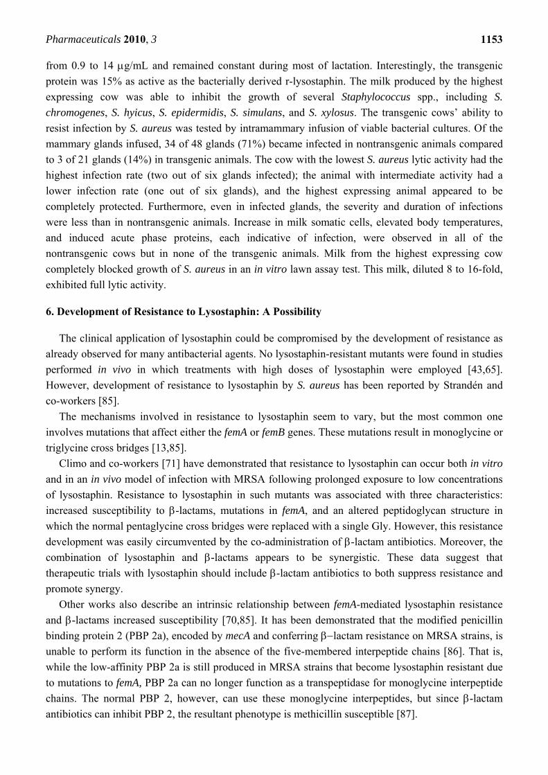
Two basic procedures applied to bacteriocins in general may be used to investigate the lysostaphin
spectrum of activity: the simultaneous-antagonism test and the deferred-antagonism test [1]. Briefly,
simultaneous-antagonism testing involves stab inoculation of the lysostaphin-producing strain into a
freshly seed lawn of a target microorganism (~106 CFU). Following incubation for 18 h, the plate is
examined for zones of growth inhibition surrounding the bacteriocin-producing culture. For deferred-
antagonism testing, the producing strain is grown as a spot on the surface of an agar plate and then,
after sterilization of the agar surface with either chloroform vapor or UV light, the plate is sprayed
with the target strain (~106 CFU). Following incubation for 24 h, the extent of inhibition of the target
strain by the producing strain can be assessed and the inhibition zones measured (Figure 3).
When a partially or totally purified lysostaphin preparation is available, the critical dilution assay
may also be employed. In this method, a 100-L volume of lysostaphin fractions at two-fold dilutions,
and a 100-L volume of the target strain (~5×10⁵ CFU) are added to each well of a microtiter plate.
Dilutions of lysostaphin should be prepared in medium containing 0.1% bovine serum albumin to
prevent adsorption of lysostaphin to polystyrene microtiter wells [43]. After incubation at 37 oC for
24 h, the growth of the target strain is measured spectrophotometrically at 600 nm. One bacteriocin
arbitrary unit is defined as the amount of bacteriocin that inhibits the bacterial growth by 50% (50% of
the turbidity of the control culture without bacteriocin) [44]. Alternatively, a disk-diffusion assay
(50 g lysostaphin/disk) may be used [45] and staphylococcal strains susceptible to lysostaphin give
inhibition zones ≥11 mm.
S. simulans biovar staphylolyticus peptidoglycan is resistant to the hydrolytic activity of
lysostaphin, since the cells produce a resistance factor that causes the incorporation of Ser residues
into the third and fifth positions of the cell wall cross bridges (Figure 1) [21,46,47]. This
incorporation requires the presence of FemA and/or FemB [47]. If one or more Gly residues of the
cross bridge are replaced by Ser residues, the cell wall becomes less susceptible to lysostaphin
[21,48]. Lysostaphin is unable to hydrolyze glycylserine and serylglycine peptide bonds [48]. It
was also shown that lysostaphin binds much more avidly to S. aureus, which possesses a
pentaglycine cross-bridge, than to S. simulans cells. When added to mixed bacterial populations,
purified lysostaphin kills 1,000 S. aureus cells for every S. simulans cell [23].
The S. simulans gene lif is required for incorporation of Ser residues into the
peptidoglycan [21]. Lif shows similarity to FemA and FemB proteins [21], which catalyze the non-
ribosomal synthesis of the pentaglycine cross bridges of the staphylococcal peptidoglycan [13].
Pharmaceuticals 2010, 3
1147
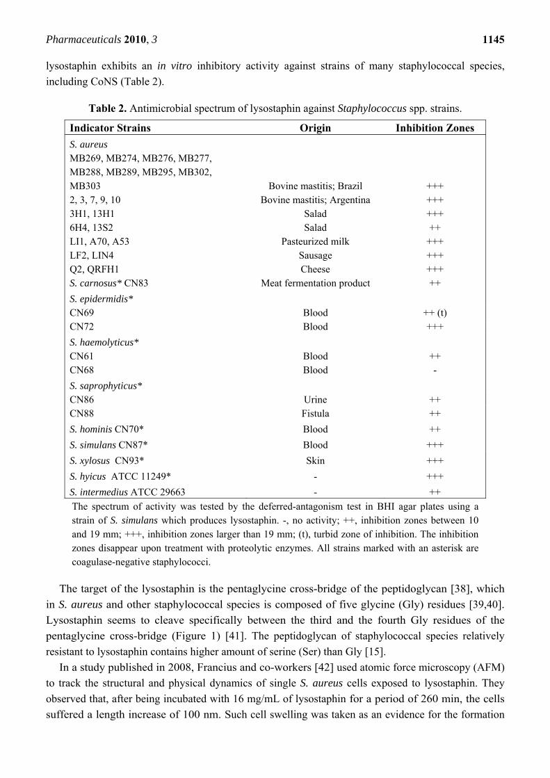
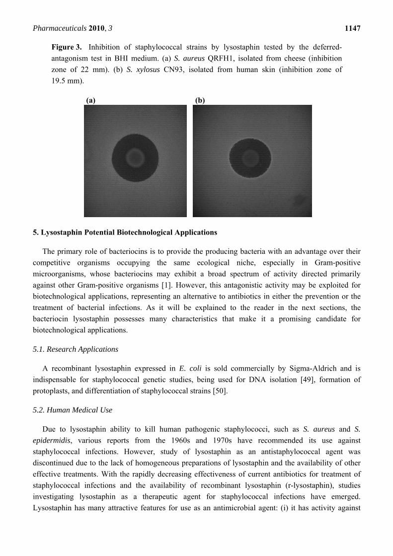
Figure 3. Inhibition of staphylococcal strains by lysostaphin tested by the deferred-
antagonism test in BHI medium. (a) S. aureus QRFH1, isolated from cheese (inhibition
zone of 22 mm). (b) S. xylosus CN93, isolated from human skin (inhibition zone of
19.5 mm).
5. Lysostaphin Potential Biotechnological Applications
The primary role of bacteriocins is to provide the producing bacteria with an advantage over their
competitive organisms occupying the same ecological niche, especially in Gram-positive
microorganisms, whose bacteriocins may exhibit a broad spectrum of activity directed primarily
against other Gram-positive organisms [1]. However, this antagonistic activity may be exploited for
biotechnological applications, representing an alternative to antibiotics in either the prevention or the
treatment of bacterial infections. As it will be explained to the reader in the next sections, the
bacteriocin lysostaphin possesses many characteristics that make it a promising candidate for
biotechnological applications.
5.1. Research Applications
A recombinant lysostaphin expressed in E. coli is sold commercially by Sigma-Aldrich and is
indispensable for staphylococcal genetic studies, being used for DNA isolation [49], formation of
protoplasts, and differentiation of staphylococcal strains [50].
5.2. Human Medical Use
Due to lysostaphin ability to kill human pathogenic staphylococci, such as S. aureus and S.
epidermidis, various reports from the 1960s and 1970s have recommended its use against
staphylococcal infections. However, study of lysostaphin as an antistaphylococcal agent was
discontinued due to the lack of homogeneous preparations of lysostaphin and the availability of other
effective treatments. With the rapidly decreasing effectiveness of current antibiotics for treatment of
staphylococcal infections and the availability of recombinant lysostaphin (r-lysostaphin), studies
investigating lysostaphin as a therapeutic agent for staphylococcal infections have emerged.
Lysostaphin has many attractive features for use as an antimicrobial agent: (i) it has activity against
(a) (b)
Pharmaceuticals 2010, 3
1148
non-dividing as well as dividing cells, (ii) it is digested by intestinal proteinases, having no influence
on the gut microbiota, (iii) it has no toxicity, (iv) it is relatively stable when conjugated with
polyethylene glycol (PEG), and (v) it maintains its activity in human serum [29,51,52]. Moreover,
studies demonstrate that lysostaphin retains its bacteriolytic activity in vivo, without any undesirable
immune reaction, despite the presence of high-neutralizing antibody titer [53,54].
5.2.1. In vitro studies
In 1965, Harrison and Cropp reported the efficacy of lysostaphin over 50 S. aureus clinical isolates,
most of them penicillinase producers [55]. Lysostaphin was compared to the antibiotics penicillin G,
ampicillin, methicillin, ristocetin, vancomycin, and erythromycin. The results have shown that the
minimal inhibitory concentration (MIC) values for lysostaphin ranged from <0.047 to 12.5 µg/mL,
with 96% of the penicillinase-positive strains being inhibited by 1.56 µg/mL of lysostaphin, whereas
3.12 µg/mL of vancomycin and methicillin were required to attain the same degree of inhibition.
Similar findings using other clinical isolates of S. aureus, presenting the most diverse drug-resistance
patterns, were also reported by other authors that together have tested 1,000 strains, detecting no
resistance to lysostaphin [36,56–58].
The use of combinations of antimicrobials is common in the clinical setting since it expands the
spectrum of organisms that can be targeted, prevents the emergence of resistant organisms, decreases
toxicity by allowing lower doses of both agents, and can result in synergistic inhibition. Synergy has
been observed in vitro between lysostaphin and membrane-active agents, such as polymyxins B [59]
and ranalexin [60], a cationic peptide produced the Rana frog species, against MRSA.
The increasing use of indwelling intravascular catheters for diagnostics of diseases and for
therapeutic procedures has led to an increase in the number of medical device-related infections [9].
Sepsis associated with internal luminal colonization of central venous catheters has been described in
many clinical settings and is a frequent occurrence in hospitals today [11,61]. S. epidermidis is by far
the most frequently isolated species of CoNS from these infections, accounting for 74% to 92% of
hospital-acquired CoNS bacteremia [11,62,63]. Therefore, lysostaphin was tested as a preventive agent
in surface colonization by either S. aureus or S. epidermidis. Wu and co-workers [64] reported that
lysostaphin not only killed S. aureus in biofilms but also disrupted the extracellular matrix of S. aureus
biofilms in vitro, on plastic and glass surfaces, at concentrations as low as 1 µg/mL. Scanning electron
microscopy confirmed that lysostaphin eradicated both the sessile cells and the extracellular matrix of
the biofilm. For S. epidermidis, higher concentrations of lysostaphin were needed to achieve the same
effect. In another study, Shah and co-workers [65] tested lysostaphin over different surfaces
(polystyrene plates and fluoroethylene-propylene polymer catheters). These surfaces were challenged
with an inoculum of about 104 colony-forming units (CFU) of S. aureus. On average, 610 CFU were
recovered from the polystyrene control wells, whereas only 3 CFU remained in the lysostaphin-coated
wells, a 99.5% reduction in bacterial counts. The lysostaphin-coated catheters were completely cleared
of bacteria as compared to control catheters, from which an average of 493 CFU were recovered. The
inhibitory effect of lysostaphin-coated catheters was maintained for at least four days after coating.
Both these studies suggest that lysostaphin binds to plastic surfaces, maintaining killing activity
against staphylococci, and may protect catheters from staphylococcal colonization at the time of
insertion and for several days thereafter.
Pharmaceuticals 2010, 3
1149
5.2.2. In vivo studies
Experimental in vivo applications of lysostaphin are also well documented in the literature. The first
report of efficacy of lysostaphin against S. aureus in vivo was published by Schuhardt and Schindler in
1964 [66]. They tested lysostaphin therapy in mice infected with S. aureus. Therapy consisted of a
single injection of a partially purified preparation of lysostaphin either intraperitoneally or
subcutaneously. These experiments indicated that 10 U of lysostaphin (one unit was defined as the
amount required to bring about a 50% reduction in the turbidity of a standard suspension of S. aureus
FDA 209P in 10 min at 37 °C) by either route of injection cured all mice.
Abscesses are the hallmark of staphylococcal disease and many therapeutic failures appear to result
from the inability of available antimicrobials to eliminate staphylococci residing in high titers in
abscess lesions. Although antibiotics do penetrate abscess cavities in high concentration, it has been
shown that these agents are relatively ineffective against sluggishly multiplying bacteria found in such
lesions [67]. Since previous studies had shown that lysostaphin kills CoPS regardless of their
metabolic state [51], Dixon and co-workers [67] tested the effectiveness of lysostaphin (5 mg)
followed by treatment with methicillin (5 mg) against established renal abscess lesions in mice. A
single dose of lysostaphin, followed by four daily doses of methicillin, produced striking reductions
(>99.99%) of the staphylococcal populations. Such reductions were significantly higher than that
achieved by either lysostaphin or methicillin alone, although lysostaphin alone proved to be more
effective than methicillin alone. Similar results were reported by Harrison and Zygmunt [68].
With the development of r-lysostaphin which is more than 90% pure, it was possible to better study
the efficacy of lysostaphin in an experimental S. aureus endocarditis model. Studies, using a rabbit
model of aortic valve endocarditis, caused by clinical isolates of either MRSA [69] or S. aureus with
reduced sensibility to vancomycin (VISA) [43], attempted to evaluate the effectiveness of various
regimens of dosing with intravenous lysostaphin to reduce the infection. Climo and co-workers [69],
using MRSA strains, treated the animals for three days with different doses of r-lysostaphin,
vancomycin or a combination of r-lysostaphin and vancomycin. The most active regimen, r-
lysostaphin given three times daily, produced sterile vegetations in 10 out of 11 treated rabbits, with a
mean reduction in vegetation bacterial counts of 8.5 log10 CFU/g compared to the counts in the
untreated controls. In contrast, vancomycin given twice daily sterilized no vegetations and reduced
vegetation bacterial counts by only 4.8 log10 CFU/g. r-Lysostaphin given once daily was less effective,
reducing mean vegetation bacterial counts by only 3.6 log10 CFU/g, but the combination of r-
lysostaphin once daily and vancomycin twice daily reduced the mean vegetation bacterial density by
7.5 log10 CFU/g, a result that was significantly better than that for either regimen alone. Yet, when
evaluation of the immunological effects of r-lysostaphin administration was carried out, no evidence of
immunological reactions following up to nine weeks of intravenous administration could be detected.
In the study performed by Patron and co-workers [43], using VISA, it was observed that vancomycin
was ineffective, with no evidence of sterilization of aortic valve vegetations. However, rates of
sterilization of aortic valve vegetations were significantly better for animals treated with either a single
dose of r-lysostaphin (43%) or r-lysostaphin given twice daily for three days (83%) than for animals
treated with vancomycin. Rabbits given a single dose of r-lysostaphin followed by a three-day drug-
free period had mean reductions in aortic valve vegetation bacterial counts of 7.27 and
6.63 log10 CFU/g compared with those for untreated control rabbits and the vancomycin-treated group,
Pharmaceuticals 2010, 3
1150
respectively. Using a similar experimental approach but with methicillin-resistant S. epidermidis
isolates (generally less susceptible to lysostaphin) involved in central venous catheter infections, Kiri
and co-workers [70] showed that the rabbits treated with the combination of nafcillin (200 mg/kg
intramuscularly) and r-lysostaphin (1mg/kg intravenously) had a significant reduction in mean log10
vegetation counts (5.32 log10 CFU/g) compared to rabbits treated with r-lysostaphin or nafcillin alone.
Here again, synergy between r-lysostaphin and -lactams was observed as already described in
previous similar experiments performed with MRSA [71].
More recently, Placencia and co-workers [72] performed a study to evaluate the use of lysostaphin
to treat neonatal late-onset sepsis caused by S. aureus. This microorganism is the second most
common pathogen for late-onset sepsis among very low birth weight infants, and nearly 20% die as a
direct result of the infection. In developing countries, neonatal S. aureus bacteremia is even more
prevalent, causing nearly a quarter of all bacteremic episodes. In their study, Placencia’s group
compared lysostaphin versus vancomycin against the MRSA strain US300, in a neonatal mouse model.
Pups were infected subcutaneously and littermates randomized to receive intraperitoneally either saline
(group 1; 81 pups), vancomycin 15 mg/kg (group 2; 77 pups), lysostaphin 10 mg/kg (group 3;
79 pups), and lysostaphin 15 mg/kg (group 4; 75 pups), at time points 0.5, 6, 24 and 30 h after the
infection. Pups were observed for survival and growth during seven days, and quantitative blood
cultures were obtained 24 h after infection. The survival rates were 6.2%, 34%, 41% and 52%,
respectively for groups 1–4. Interestingly, lysostaphin (15 mg/kg; group 4) improved survival
compared with placebo and vancomycin. There was no significant difference in growth among the
groups and all treatment regimens resulted in less bacteremia when compared with placebo. The MIC
and minimum bactericidal concentration (MBC) for vancomycin and lysostaphin were
0.71/1.19 g/mL and <0.008/0.015 g/mL, respectively. The serum concentrations after 48 h were of
2.34g/mL for lysostaphin and 1.72 g/mL, for vancomycin. Notably, both serum concentrations
were greater than the MICs and MCBs and lysostaphin serum concentrations were higher than
vancomycin ones, suggesting a higher half-life for this bacteriocin. These pharmacokinetics data is in
agreement with the literature and similar results can be also seen in Oluola et al. [73] and
Kokai-Kun et al. [74].
Walsh and co-workers [52], using adult mice as model, compared the pharmacokinetics of
r-lysostaphin with a PEG-lysostaphin conjugate (PEGylated). It was found that 24 h after a single dose
of 40 mg/kg of r-lysostaphin, the serum drug concentration dropped 500-fold, whereas for the
PEGylated derivative the drop was only 10-fold. This reflects an increase in r-lysostaphin stability due
to PEG conjugation. This improved retention of r-lysostaphin should reduce the dosing quantities and
frequency needed to maintain plasma drug concentrations above therapeutically effective drug
concentrations. Maintaining these high levels of r-lysostaphin for longer periods of time may also
result in more rapid clearance of bacterial infections and decrease the probability that r-lysostaphin
resistance will emerge.
Eradication of staphylococcal biofilms by lysostaphin has recently been shown to occur also in vivo
[75].
Pharmaceuticals 2010, 3
1151
5.2.3. Reduction of nasal carriage of staphylococci
Nasal carriage of S. aureus has been repeatedly shown to have a significant epidemiological link
with subsequent development of staphylococcal disease [8]. Clearance of S. aureus nasal colonization
can reduce the subsequent risk of development of infections by MRSA in addition to reducing the
community and hospital spread of these microorganisms [8]. Mupirocin ointment (Bactroban Nasal;
2% mupirocin calcium ointment; SmithKline Beecham) is the current standard of care for clearance of
S. aureus nasal colonization, but resistance to this antibiotic has emerged [8]. Several other
interventions for the clearance of S. aureus nasal colonization have also been explored, including
Neosporin® ointment (a bacitracin, polymyxin, and neomycin formulation; Burroughs
Welcome & Co.).
Using the cotton rat model, Kokain-Kun and co-workers [76] investigated the capacity of three
antibacterial agents, lysostaphin, mupirocin, and nisin, a lantibiotic with a potent in vitro
antistaphylococcal activity, to clear S. aureus [methicillin resistant or sensitive strains (MSSA)] nasal
colonization. A single application of lysostaphin (actual dose, ~150 g), formulated in a petrolatum-
based cream, dramatically reduced S. aureus nasal colonization by both MRSA and MSSA (averages
between 3,416 and 4,021 CFU/nose) in 100% of animals tested and eradicated S. aureus nasal
colonization in 93% of animals. Interestingly, when lysostaphin was administered in PBS, only 33%
eradication was achieved. Yet, a single dose of lysostaphin (formulated at 0.5% in a petrolatum-based
cream) eradicated MRSA and mupirocin-resistant S. aureus from the cotton rat nares, some within 4 h
of application, while three doses of mupirocin ointment over three days were required for eradication
of MRSA in the same model. Nisin (actual dose, ~1.5 mg) failed in reducing staphylococcal nasal
carriage in this model.
The potency of the lysostaphin formulation was also persistent in the nares and maintained its
antistaphylococcal activity for at least 24 h after instillation, suggesting that lysostaphin cream could
also prevent subsequent S. aureus nasal colonization. Lysostaphin cream could therefore be instilled in
the nares of an uncolonized patient upon admission to a health care setting and protect the patient from
nasal colonization for at least 24 h.
Quickel Jr. and co-workers [77] tested the efficacy of lysostaphin to alter natural S. aureus carriage
in 95 persistent carriers. In this work, the efficiency of lysostaphin intranasal spray was compared to
Neosporin®. The treatment was carried out for five days with either agent significantly reducing
carriage rates. However, this effect persisted through the 5th day only after therapy with lysostaphin
but not with Neosporin®. Immunological parameters were also evaluated and, except for a single
immediate wheal and flair skin test reaction, no other evidence of adverse reactions to topical
lysostaphin was detected. As well, no consistent changes in hemagglutination-inhibition titers to
lysostaphin were observed after therapy. Taken together, these studies have shown that lysostaphin
appears to be more effective than conventional topical antimicrobial therapy in reducing nasal carriage
of staphylococci.
5.3. Veterinary Use
Mastitis, an inflammatory reaction of the mammary gland that is usually caused by microbial
infection, is recognized as the most costly disease in dairy cattle [78] due to rejected milk, degraded
Pharmaceuticals 2010, 3
1152
milk quality, early culling of cows, drug costs, veterinary expenses, and increased labor costs for
farmers. In the USA, annual losses are estimated at two billion dollars. This is approximately 10% of
the total value of farm milk sales, and about two-thirds of these losses are due to reduced milk
production in subclinically infected cows [79]. Although several bacterial pathogens can cause
mastitis, S. aureus is the major etiological agent. Intrammamary infections caused by S. aureus are
usually chronic and subclinical in nature. Once S. aureus is established in the mammary glands of the
animal, it is very difficult to eradicate [78,80].
Many drugs have been used for mastitis treatment, but several factors, including the ability of S.
aureus to survive inside neutrophils, to induce formation of microabscesses, and its resistance to the
antibiotics used for treatment, result in infections that are difficult to manage therapeutically [80].
Lysostaphin has been suggested to be efficacious in the treatment of bovine mastitis caused by
staphylococci [81].
The kinetics and therapeutic efficacy of the r-lysostaphin (100 mg, administered over three
consecutive p.m. milkings) in S. aureus bovine mastitis have been investigated [82]. In this study,
30 Holstein-Friesian dairy cattle in their first lactation were infected with S. aureus ATCC 29740 in all
quarters. Infections were established and monitored for somatic cell counts and S. aureus CFU three
weeks prior to subsequent treatment. Infected animals were injected through the teat canal with a
single dose of r-lysostaphin (dose response 1 to 500 mg) or after three successive p.m. milkings with
100 mg of r-lysostaphin in 60 mL of sterile phosphate buffered saline. The in vivo titration suggested
that the minimal effective therapeutic dose was 100 mg of r-lysostaphin: 95% of the quarters cleared
the milk of detectable S. aureus for a minimum of one milking after the last intramammary infusion.
Despite the maintenance of a bactericidal activity in the milk under this therapeutic regimen for up
120 h after initiation of therapy, the majority of the r-lysostaphin-treated quarters relapsed within five
to six milkings. The data suggested that r-lysostaphin, although effective at eliminating bacteria
present in the milk for as long as an effective therapeutic level could be maintained, was not sufficient
to elicit cures. Such observations are most likely related to previous observations with S. aureus
mastitis that showed that viable intracellular bacteria contribute to the reinfection and r-lysostaphin
cannot kill intracellular S. aureus [80]. The cure rate of quarters receiving r-lysostaphin (100 mg in
sterile phosphate buffered saline, administered after each of three consecutive p.m. milkings) was 20%
compared with 29% for sodium cephapirin in saline and 57% for a commercial antibiotic formulation
(Cefa-Lak®), respectively. However, the authors proposed that an improved formulation of
r-lysostaphin could be a safe alternative to antibiotic therapy.
Following this study, Daley and Oldham [83] showed that intramammary infusion of r-lysostaphin
failed to elicit significant serum titers in the bovine until 18–21 infusions were administered
(total administered dose of 2–3 g of protein). Antibody titers from dairy cattle which did develop an
immune response were predominantly of the IgG1 subclass. Dairy cattle with significant
antilysostaphin titers showed no deleterious symptoms, such as anaphylaxis, upon subsequent infusion,
and these titers did not affect the in vitro activity of r-lysostaphin. Intramammary infusion of
r-lysostaphin did not elicit any observable effects on the host animal or on the potential efficacy of the
recombinant molecule.
More recently, Wall and co-workers [84] produced transgenic cows expressing a modified
lysostaphin gene whose product lacks two glycosylation motifs, which are responsible for rendering
lysostaphin inactive when glycosylated by mammalian cells. Cow’s lysostaphin concentrations ranged
Pharmaceuticals 2010, 3
1153
from 0.9 to 14 g/mL and remained constant during most of lactation. Interestingly, the transgenic
protein was 15% as active as the bacterially derived r-lysostaphin. The milk produced by the highest
expressing cow was able to inhibit the growth of several Staphylococcus spp., including S.
chromogenes, S. hyicus, S. epidermidis, S. simulans, and S. xylosus. The transgenic cows’ ability to
resist infection by S. aureus was tested by intramammary infusion of viable bacterial cultures. Of the
mammary glands infused, 34 of 48 glands (71%) became infected in nontransgenic animals compared
to 3 of 21 glands (14%) in transgenic animals. The cow with the lowest S. aureus lytic activity had the
highest infection rate (two out of six glands infected); the animal with intermediate activity had a
lower infection rate (one out of six glands), and the highest expressing animal appeared to be
completely protected. Furthermore, even in infected glands, the severity and duration of infections
were less than in nontransgenic animals. Increase in milk somatic cells, elevated body temperatures,
and induced acute phase proteins, each indicative of infection, were observed in all of the
nontransgenic cows but in none of the transgenic animals. Milk from the highest expressing cow
completely blocked growth of S. aureus in an in vitro lawn assay test. This milk, diluted 8 to 16-fold,
exhibited full lytic activity.
6. Development of Resistance to Lysostaphin: A Possibility
The clinical application of lysostaphin could be compromised by the development of resistance as
already observed for many antibacterial agents. No lysostaphin-resistant mutants were found in studies
performed in vivo in which treatments with high doses of lysostaphin were employed [43,65].
However, development of resistance to lysostaphin by S. aureus has been reported by Strandén and
co-workers [85].
The mechanisms involved in resistance to lysostaphin seem to vary, but the most common one
involves mutations that affect either the femA or femB genes. These mutations result in monoglycine or
triglycine cross bridges [13,85].
Climo and co-workers [71] have demonstrated that resistance to lysostaphin can occur both in vitro
and in an in vivo model of infection with MRSA following prolonged exposure to low concentrations
of lysostaphin. Resistance to lysostaphin in such mutants was associated with three characteristics:
increased susceptibility to -lactams, mutations in femA, and an altered peptidoglycan structure in
which the normal pentaglycine cross bridges were replaced with a single Gly. However, this resistance
development was easily circumvented by the co-administration of -lactam antibiotics. Moreover, the
combination of lysostaphin and -lactams appears to be synergistic. These data suggest that
therapeutic trials with lysostaphin should include -lactam antibiotics to both suppress resistance and
promote synergy.
Other works also describe an intrinsic relationship between femA-mediated lysostaphin resistance
and -lactams increased susceptibility [70,85]. It has been demonstrated that the modified penicillin
binding protein 2 (PBP 2a), encoded by mecA and conferring lactam resistance on MRSA strains, is
unable to perform its function in the absence of the five-membered interpeptide chains [86]. That is,
while the low-affinity PBP 2a is still produced in MRSA strains that become lysostaphin resistant due
to mutations to femA, PBP 2a can no longer function as a transpeptidase for monoglycine interpeptide
chains. The normal PBP 2, however, can use these monoglycine interpeptides, but since -lactam
antibiotics can inhibit PBP 2, the resultant phenotype is methicillin susceptible [87].
Pharmaceuticals 2010, 3
1154
Morikawa and co-workers [88], using S. aureus strain N315 and its derivative mutants affected in
sigB gene, showed that cell-wall thickness also plays important roles in lysostaphin sensitivity. Cells
depleted in sigB developed thinner envelopes and demonstrated increased sensitivity to lysostaphin.
These cells were approximately 97% more sensitive than the wild-type N315 cells. On the other hand,
over expressing sigB cells revealed an increased resistance to lysostaphin, being 300% more resistant
than normal N315 cells. The increased resistance of these cells was attributed to an expressive increase
in cell-wall thickness and evidence supportive of this assumption can be find in the literature. For
example, the VISA strain MU50 has also been demonstrated to possess an expanded cell-wall and this
characteristic has been intrinsically associated to its vancomycin resistance [89]. In another work,
Koehl and co-workers [90], studying the low susceptibility of VISA strains to lysostaphin, found out
that besides the enlarged cell-wall phenotype, lysostaphin resistance was also linked to a decreased
autolytic activity. They proposed that in intact cells the cell’s autolysins participate in the lysis seen
upon lysostaphin treatment and that the defective autolysis of VISA strains is mainly responsible for
the poor susceptibility of intact cells to lysostaphin. It appears that changes in the amount or activity of
the autolytic enzymes are mainly responsible for the autolysin deficiency, even though some changes
in peptidoglycan composition were evident in the walls of the VISA strains. Several autolysin genes
are present in S. aureus. atl is the gene that encodes the major S. aureus autolysin that undergoes post-
translation processing to yield a 63-kDa amidase and a 53.5-kDa glucosaminidase. However, in this
work, northern blot analysis suggested that the expression of atl was significantly reduced in the VISA
strains, reinforcing the assumption that these autolysins are responsible for promoting cell lysis upon
lysostaphin treatment.
By transposon mutagenesis, Gründling and co-workers [91] have identified in S. aureus another
gene, lyrA (for lysostaphin resistance protein A), whose inactivation caused a high degree of
lysostaphin resistance. lyrA encodes a 419- amino acid polypeptide with unknown function. In contrast
to the case for femAB mutants, transposon insertion in lyrA did not cause gross alterations of cell wall
cross bridges and did not result in a decrease in -lactam resistance. Therefore, the resistance
phenotype of lyrA mutants has proven difficult to explain. Many factors, including minor alterations in
peptidoglycan, pentaglycine bridges and other envelope components were proposed as possible
explanations for this phenotype.
Despite all of these reports about development of lysostaphin resistance among S. aureus strains, an
important observation was made recently by Kusuma and co-workers [92], which have demonstrated
that the development of lysostaphin resistance, due to mutation in femA, by two strains of MRSA also
led to a loss of fitness in the mutants. The diminished fitness was reflected by: (i) a reduced
logarithmic growth rate, with mutants being outcompeted in co-cultures by their wild-type parental
strains; (ii) increased susceptibility to elevated temperatures, and (iii) at least fivefold less virulence of
the mutants than their wild-type strains in a mouse kidney infection model, with the mutants being
outcompeted in co-infection with their wild-type parental strains. During a 14-day serial passage
without selective pressure, the lysostaphin resistant mutants failed to develop compensatory mutations
which restored their fitness.
Taken together, these results suggest that, should lysostaphin resistance due to an alteration in the
FemA function emerge in S. aureus during therapy with lysostaphin, the resistant variants would be
less fit and less virulent and, in addition, infections with these strains would be easily treatable with -
lactam antibiotics.
Pharmaceuticals 2010, 3
1155
7. Conclusions
A current understanding of lysostaphin, the only class III bacteriocin so far exploited for clinical
applications, has been presented. Examples of its in vitro and in vivo applications as a therapeutic
agent against pathogenic staphylococci, including multiresistant strains, have clearly been shown.
However, its broad clinical application waits for standardization of drug formulations either alone or in
combination with other antibiotics. Moreover, since S. aureus is also a very important pathogen
transmitted by food, being involved in numerous outbreaks in several countries, and since strains of
this microorganism isolated from different foods have been shown by our group to be inhibited by
lysostaphin (Table 2), this bacteriocin might be useful also as a biopreservative in food industry.
Acknowledgements
M.L.V.C. and B.G.C. are recipients of scholarships from CNPq and CAPES, respectively. The
studies developed in our laboratory are supported by grants from CNPq, PRONEX, and FAPERJ to
M.C.F.B.
References
1. Heng, N.C.K.; Wescombe, P.A., Burton, J.P.; Jack, R.W.; Tagg, J.R. The diversity of bacteriocins in
Gram-positive bacteria. In Bacteriocins: Ecology and Evolution; Riley, M.A.; Chavan, M.A., Eds,
Springer: New York, NY, USA, 2007; pp. 45–92.
2. Bierbaum, G.; Sahl, H.-G. Lantibiotics: mode of action, biosynthesis and bioengineering. Curr.
Pharm. Biotechnol. 2009, 10, 2–18.
3. Nissen-Meyer, J.; Rogne, P.; Oppegård, C.; Haugen, H.S.; Kristiansen, P.E. Structure-function
relationships of the non-lanthionine-containing peptide (class II) bacteriocins produced by Gram-
positive bacteria. Curr. Pharm. Biotechnol. 2009, 10, 10–37.
4. Cotter, P.D.; Hill, C.; Ross, R.P. Bacteriocins: developing innate immunity for food. Nat. Rev.
Microbiol. 2005, 3, 777–788.
5. Gálvez, A.; Abriouel, H.; Lopez, R.L; Omar, N.B. Bacteriocin-based strategies for food
preservation. Int. J. Food Microbiol. 2007, 120, 51–70.
6. Bastos, M.C.F.; Ceotto, H.; Coelho, M.L.V.; Nascimento, J.S. Staphylococcal antimicrobial
peptides: relevant properties and potential biotechnological applications. Curr. Pharm.
Biotechnol. 2009, 10, 38–61.
7. Euzéby, J.P. List of prokaryotic names with standing in nomenclature—Genus Staphylococcus.
http://www.bacterio.cict.fr, accessed April 2010.
8. Bannerman, T.L.; Peacock, S.J. Staphylococcus, Micrococcus, and other catalase-positive cocci.
In Manual of Clinical Microbiology; Murray, P.R.; Baron, E.J.; Jorgensen, J.H.; Landry, M.L.;
Pfaller, M.A., Eds.; ASM Press: Washington D.C., USA, 2007; pp. 384–404.
9. Casey, A.L.; Lambert, P.A.; Elliott, T.S.J. Staphylococci. J. Antimicrob. Agents 2007, 29 (Suppl.
3), S23–S32.
10. Evans, H.L.; Saywer, R.G. Cycling chemotherapy: a promising approach to reducing the
morbidity and mortality of nosocomial infections. Drugs Today 2003, 39, 733–738.
Pharmaceuticals 2010, 3
1156
11. Rogers, K.L.; Fey, P.D.; Rupp, M.E. Coagulase-negative staphylococcal infections. Infect. Dis.
Clin. N. Am. 2009, 23, 73–98.
12. Schindler, C.A.; Schuhardt, V.T. Lysostaphin: A new bacteriolytic agent for the Staphylococcus.
Proc. Natl. Acad. Sci. USA 1964, 51, 414–421.
13. Ehlert, K.; Schrodr, W.; Labischinski, H. Specificities of FemA and FemB for different glycine
residues: FemB cannot substitute for FemA in staphylococcal peptidoglycan pentaglycine side
chain formation. J. Bacteriol. 1997, 179, 7573–7576.
14. Rohrer, S.; Ehlert, K.; Tschierske, M.; Labischinski, H.; Berger-Bächi, B. The essential
Staphylococcus aureus gene fmhB is involved in the first step of peptidoglycan pentaglycine
interpeptide formation. Proc. Natl. Acad. Sci. USA. 1999, 96, 9351–9356.
15. Kumar, J.K. Lysostaphin: an antistaphylococcal agent. Appl. Microbiol. Biotechnol. 2008,
80, 555–561.
16. Giesbrecht, P.; Wecke, J.; Recnicke, B. On the morphogenesis of the cell wall of staphylococci.
Int. Rev. Cytol. 1976, 44, 225–318.
17. Joris, B.; Englebert, S.; Chu, C.P.; Kariyama, R.; Daneo-Moore, L.; Shockman, G.D.; Ghuysen,
J.M. Modular design of the Enterococcus hirae muramidase-2 and Streptococcus autolysis. FEMS
Microbiol. Lett. 1992, 91, 257–264.
18. Browder, H.P.; Zygmunt, W.A.; Young, J.R.; Tavormina, P.A. Lysostaphin: enzymatic mode of
action. Biochem. Biophys. Res. Commun. 1965, 19, 383–389.
19. Trayer, H.R.; Buckley III, C.E. Molecular properties of lysostaphin, a bacteriolytic agent specific
for Staphylococcus aureus. J. Biol. Chem. 1970, 245, 4842–4846.
20. Heinrich, P.; Rosenstein, R.; Bohmer, M.; Sonner, P.; Götz, F. The molecular organization of the
lysostaphin gene and its sequences repeated in tandem. Mol. Gen. Genet. 1987, 209, 563–569.
21. Thumm, G.; Götz, F. Studies on prolysostaphin processing and characterization of the lysostaphin
immunity factor (Lif) of Staphylococcus simulans biovar staphylolyticus. Mol. Microbiol. 1997,
23, 1251–1265.
22. Simmonds, R.S.; Simpson, W.J.; Tagg, J.R. Cloning and sequence analysis of zooA, a
Streptococcus zooepidemicus gene encoding a bacteriocin-like inhibitory substance having a
domain structure similar to that of lysostaphin. Gene 1997, 189, 255–261.
23. Baba, T.; Schneewindt, O. Target cell specificity of a bacteriocin molecule: a C-terminal signal
directs lysostaphin to the cell wall of Staphylococcus aureus. EMBO J. 1996, 15, 4789–4797.
24. Gründling, A.; Schneewind, O. Cross-linked peptidoglycan mediates lysostaphin binding to the
cell wall envelope of Staphylococcus aureus. J. Bacteriol. 2006, 188, 2463–2472.
25. Recsei, P.A.; Gruss, A.D.; Novick, R.P. Cloning, sequence, and expression of the lysostaphin
gene from Staphylococcus simulans. Proc. Natl. Acad. Sci. USA 1987, 84, 1127–1131.
26. Larrimore, S.A.; Clark, S.B.; Robinson, J.M.; Heath, H.E.; Sloan, G.L. Coordinate production of
three exoenzymes by Staphylococcus staphylolyticus. J. Gen. Microbiol. 1982, 128, 1529–1535.
27. Heath, S.L.; Heath, H.E.; Sloan, G.L. Plasmid-encoded lysostaphin endopeptidase gene of
Staphylococcus simulans biovar staphylolyticus. FEMS Microbiol. Lett. 1987, 44, 129–133.
28. Heath, H.E.; Heath, S.L.; Nitterauer, J.D.; Rose, K.E.; Sloan, G.L. Plasmid-encoded lysostaphin
endopeptidase resistance of Staphylococcus simulans biovar staphylolyticus. Biochem. Biophys.
Res. Commun. 1989, 160, 1106–1109.
Pharmaceuticals 2010, 3
1157
29. Williamson, C.M.; Bramley, A.J.; Lax, A.J. Expression of the lysostaphin gene of Staphylococcus
simulans in a eukaryotic system. Appl. Environ. Microbiol. 1994, 60, 771–776.
30. Mierau, I.; Leij, P.; van Swam, I.; Blommestein, B.; Floris, E.; Mond, J.; Smid, E.J. Industrial
scale production and purification of a heterologous protein in Lactococcus lactis using the nisin-
controlled gene expression system NICE: The case of lysostaphin. Microb. Cell Fact. 2005, 4, 15.
31. Mierau, I.; Olieman, C.; Mond, J.; Smid, E.J. Optimization of the Lactococcus lactis nisin-
controlled gene expression system NICE for industrial applications. Microb. Cell Fact.
2005, 4, 16.
32. Sugai, M.; Fujiwara, T.; Akiyama, T.; Ohara, M.; Komatsuzawa, H.; Inoue, S.; Suginaka, H.
Purification and molecular characterization of glycylglycine endopeptidase produced by
Staphylococcus capitis. J. Bacteriol. 1997, 179, 1193–1202.
33. Sugai, M.; Fujiwara, T.; Ohta, K.; Komatsuzawa, H.; Ohara, M.; Suginaka, H. epr, which encodes
glycylglycine endopeptidase resistance, is homologous to femAB and affects serine content of
peptidoglycan cross bridges in Staphylococcus capitis and Staphylococcus aureus. J. Bacteriol.
1997, 179, 4311–4318.
34. King, B.F.; Biel, M.L.; Wilkinson, B.J. Facile penetration of the Staphylococcus aureus capsule
by lysostaphin. Infect. Immun. 1980, 29, 892–896.
35. Schindler, C.A.; Schuhardt, V.T. Purification and properties of lysostaphin—A lytic agent for
Staphylococcus aureus. Biochem. Biophys. Acta 1965, 97, 242–250.
36. Zygmunt, W.A.; Harrison, E.F.; Browder, H.P.; Tavormina, P.A. Comparative inhibition of
methicillin-resistant strains of Staphylococcus aureus by lysostaphin and other antibiotics. Appl.
Microbiol. 1968, 16, 1174–1178.
37. Zygmunt, W.A.; Browder, H.P.; Tavormina, P.A. Susceptibility of coagulase-negative
staphylococci to lysostaphin ant other antibiotics. Appl. Microbiol. 1968, 16, 1168–1173.
38. Grüdling, A.; Schneewind, O. Cross-linked peptidoglycan mediates lysostaphin binding to the cell
wall envelope of Staphylococcus aureus. J. Bacteriol. 2006, 188, 2463–2472.
39. Schleifer, K.H.; Kandler, O. Peptidoglycan types of bacterial cell walls and their taxonomic
implications. Bacteriol. Rev. 1972, 36, 407–477.
40. Schleifer, K.H.; Fisher, U. Description of a new species of the genus Staphylococcus:
Staphylococcus carnosus. Int. J. Syst. Bacteriol. 1982, 32, 153–156.
41. Schneewindt, O.; Fowler, A.; Faull, K.F. Structure of the cell wall anchor of surface proteins in
Staphylococcus aureus. Science 1995, 268, 103–106.
42. Francius, G.; Domenech, O.; Mingeot-Leclercq, M.P.; Dufrêne, Y.F. Direct observation of
Staphylococcus aureus cell wall digestion by lysostaphin. J. Bacteriol. 2008, 190, 7904–7909.
43. Patron, R.L.; Climo, M.W.; Goldstein, B.P.; Archer, G.L. Lysostaphin treatment of experimental
aortic valve endocarditis caused by a Staphylococcus aureus isolate with reduced susceptibility to
vancomycin. Antimicrob. Agents Chemother. 1999, 43, 1754–1755.
44. Fimland, G.; Blingsmo, R.; Sletten, K.; Jung, G.; Nes, I.F.; Nissen-Meyer, J. New biologically
active hybrid bacteriocins constructed by combining regions from various pediocin-like
bacteriocins: the C-terminal region is important for determining specificity. Appl. Environ.
Microbiol. 1996, 62, 3313–3318.
Pharmaceuticals 2010, 3
1158
45. Kusuma, C.M.; Kokai-Kun, J.F. Comparison of four methods for determining lysostaphin
susceptibility of various strains of Staphylococcus aureus. Antimicrob. Agents Chemother. 2005,
49, 3256–3263.
46. DeHart, H.; Heath, H.; Heath, L.; LeBlanc, P.; Sloan, G. The lysostaphin endopeptidase resistance
gene (epr) specifies modification of peptidoglycan cross bridges in Staphylococcus simulans and
Staphylococcus aureus. Appl. Environ. Microbiol. 1995, 61, 1475–1479.
47. Ehlert, K.; Tschierske, M.; Mori, C.; Schöder, W., Berger-Bächi, B. Site-specific serine
incorporation by Lif and Epr into positions 3 and 5 of the staphylococcal peptidoglycan
interpeptide bridge. J. Bacteriol. 2000, 182, 2635–2638.
48. Robinson, J.M.; Hardman, J.K.; Sloan, G.L. Relationship between lysostaphin endopeptidase
production and cell wall composition of Staphylococcus aureus. J. Bacteriol. 1979,
137, 1158–1164.
49. Klesius, P.H.; Schuhardt, V.Y. Use of lysostaphin in the isolation of highly polymerized
deoxyribonucleic acid and in the taxonomy of aerobic Micrococcaceae. J. Bacteriol. 1968,
95, 739–743.
50. Geary, C.; Stevens, M. Rapide lysostaphin test to differentiate Staphylococcus and Micrococcus
species. J. Clin. Microbiol. 1986, 23, 1044–1045.
51. Schaffner, W.; Melly, M.A.; Hash, J.H; Koenig, M.G. Lysostaphin: an enzymatic approach to
staphylococcal disease. I. In vitro studies. Yale J. Biol. Med. 1967, 39, 215–229.
52. Walsh, S.; Shah, A; Mood, J. Improved pharmacokinetics and reduced antibody reactivity of
lysostaphin conjugated to polyethylene glycol. Antimicrob. Agents Chemother. 2002,
47, 554–558.
53. Zygmunt, W.A.; Tavormina, P.A. Lysostaphin: model for a specific enzymatic approach to
infectious disease. Prog. Drug Res. 1972, 16, 309–333.
54. Dajcs, J.J.; Thibodeaux, B.A.; Girgis, D.O.; Shaffer, M.D.; Delvisco, S.M.; O’Callaghan, R.J.
Immunity to lysostaphin and its therapeutic value for ocular MRSA infections in the rabbit.
Invest. Opthalmol. Visual Sci. 2002, 43, 3712–3716.
55. Harrison, F.E.; Cropp, C.B. Comparative in vitro activities of lysostaphin and other
antistaphylococcal antibiotics on clinical isolates of Staphylococcus aureus. Appl. Microbiol.
1965, 13, 212–215.
56. Zygmunt, W.A.; Harrison, E.F.; Browder, H.P. Microbiological activities of lysostaphin and
penicillins against bacteriophage 80/81 strains of Staphylococcus aureus. Appl. Microbiol. 1965,
13, 491–493.
57. Huber, M.M.; Huber, T.W. Susceptibility of methicillin-resistant Staphylococcus aureus to
lysostaphin. J. Clin. Microbiol. 1989, 27, 1122–1124.
58. von Eiff, C.; Kokai-Kun, J.F.; Becker, K.; Peters, G. In vitro activity of recombinant lysostaphin
against Staphylococcus aureus isolates from anterior nares and blood. Antimicrob. Agents
Chemother. 2003, 47, 3613–3615.
59. Polak, J.; Della Latta, P.; Blackburn, P. In vitro activity of recombinant lysostaphin-antibiotic
combinations toward methicillin-resistant Staphylococcus aureus. Diagn. Microbiol. Infect. Dis.
1993, 17, 265–270.
Pharmaceuticals 2010, 3
1159
60. Graham, S.; Coote, P.J. Potent, synergistic inhibition of Staphylococcus aureus upon exposure to
a combination of the endopeptidase lysostaphin and the cationic peptide ranalexin. J. Antimicrob.
Chemother. 2007, 59, 759–762.
61. Harbarth, S.; Sax, H.; Gastmeier, P. The preventable proportion of nosocomial infections: an
overview of published reports. J. Hosp. Infect. 2003, 54, 258–266.
62. Vuong, C.; Otto, M. Staphylococcus epidermidis infections. Microbes Infect. 2002, 4, 481–489.
63. Sader, H.S.; Jones, R.N.; Andrade-Baiocchi, S.; Biedenbach, D.J. Four-year evolution of
frequency of occurrence and antimicrobial susceptibility patterns of bacteria from bloodstream
infections in Latin American medical centers. Diagn. Microbiol. Infect. Dis. 2003, 44, 273–280.
64. Wu, J.A.; Kusuma, C.; Mond, J.J.; Kokai-Kun, J.F. Lysostaphin disrupts Staphylococcus aureus
and Staphylococcus epidermidis biofilms on artificial surfaces. Antimicrob. Agents Chemother.
2003, 47, 3407–3414.
65. Shah, A.; Mond, J.; Walsh, S. Lysostaphin-coated catheters eradicate Staphylococcus aureus
challenge and block surface colonization. Antimicrob. Agents Chemother. 2004, 48, 2704–2707.
66. Schudhardt, V.T; Schindler, C.A. Lysostaphin therapy in mice infected with Staphylococcus
aureus. J. Bacteriol. 1964, 88, 815–816.
67. Dixon, R.E.; Goodman, J.S.; Koenig, M.G. Lysostaphin: an enzymatic approach to staphylococcal
disease. III. Combined lysostaphin-methicillin therapy of established staphylococcal abscesses in
mice. Yale J. Biol. Med. 1968, 41, 62–68.
68. Harrison, E.F.; Zygmunt, W.A. Lysostaphin in experimental renal infection. J. Bacteriol. 1967,
93, 520–524.
69. Climo, M.W.; Patron, R.L.; Goldstein, B.P.; Archer, G.L. Lysostaphin treatment of experimental
methicillin-resistant Staphylococcus aureus aortic valve endocarditis. Antimicrob. Agents
Chemother. 1998, 42, 1355–1360.
70. Kiri, N.; Archer, G.; Climo, M.W. Combinations of lysostaphin with -lactams are synergistic
against oxacillin-resistant Staphylococcus epidermidis. Antimicrob. Agents Chemother. 2002, 46,
2017–2020.
71. Climo, M.W.; Ehlert, K.; Archer, G.L. Mechanism and suppression of lysostaphin resistance in
oxacillin-resistant Staphylococcus aureus. Antimicrob. Agents Chemother. 2001, 45, 1431–1437.
72. Placencia, F.X.; Kong, L.; Weisman, L.E. Treatment of methicillin-resistant Staphylococcus
aureus in neonatal mice: lysostaphin vs. vancomycin. Pediatr. Res. 2009, 65, 420–424.
73. Oluola, O.; Kong, L.; Fein, M.; Weisman, L.E. Lysostaphin in treatment of neonatal
Staphylococcus aureus infection. Antimicrob. Agents Chemother. 2007, 51, 2198–2220.
74. Kokai-Kun, J.F.; Chanturiya, T.; Mond, J.J. Lysostaphin as a treatment for systemic
Staphylococcus aureus infection in a mouse model. J. Antimicrob. Chemother. 2007,
60, 1051–1059.
75. Kokai-Kun, J.F.; Chanturiya, T.; Mond, J.J. Lysostaphin eradicates established Staphylococcus
aureus biofilms in jugular vein catheterized mice. J. Antimicrob. Chemother. 2009, 64, 94–100.
76. Kokai-Kun, J.F.; Walsh, S.M.; Chanturiya, T.; Mond, J.J. Lysostaphin cream eradicates
Staphylococcus aureus nasal colonization in a cotton rat model. Antimicrob. Agents Chemother.
2003, 47, 1589–1597.
Pharmaceuticals 2010, 3
1160
77. Quickel Jr., K.E.; Selden, R.; Caldwell, J.R.; Nora, N.F.; Schaffner, W. Efficacy and safety of
topical lysostaphin treatment of persistent nasal carriage of Staphylococcus aureus. Appl.
Microbiol. 1971, 22, 446–450.
78. Zhao, X.; Lacasse., P. Mammary tissue damage during bovine mastitis: causes and control. J.
Anim. Sci. 2008, 86, 57–65.
79. Bradley, A.J. Bovine mastitis: an evolving disease. Vet. J. 2002, 164, 116–128.
80. Barkema, H.W.; Schukken, Y.H.; Zadoks, R.N. The role of cow, pathogen, and treatment regimen
in the therapeutic success of bovine Staphylococcus aureus mastitis. J. Dairy Sci. 2006,
89, 1877–1895.
81. Sears, P.M.; Smith, B.S.; Pollak, J.; Gusik, S.N.; Blackburn, P. Lysostaphin efficacy for treatment
of Staphylococcus aureus intramammary infection. J. Dairy Sci. 1988, 71 (Suppl. 1), 244. 82. Oldham, E.R.; Daley, M.J. Lysostaphin: use of a recombinant bactericidal enzyme as a mastitis
therapeutic. J. Dairy Sci. 1991, 74, 4175–4182.
83. Daley, M.J.; Oldham, E.R. Lysostaphin immunogenicity of locally administered recombinant
protein used in mastitis therapy. Vet. Immunol. Immunopathol. 1992, 31, 301–312.
84. Wall, R.J.; Powell, A.M.; Paape, M.J.; Kerr, D.E.; Bannerman, D.D.; Pursel, V.G.; Wells, K.D.;
Talbot, N; Hawk, H.W. Genetically enhanced cows resist intramammary Staphylococcus aureus
infection. Nat. Biotechnol. 2005, 23, 445–451.
85. Strandén, A.M.; Ehlert, K.; Labischinski, H.; Berger-Bächi, B. Cell wall monoglycine cross-
bridges and methicillin hypersusceptibility in a femAB null mutant of methicillin-resistant
Staphylococcus aureus. J. Bacteriol. 1997, 179, 9–16.
86. Rohrer, S.; Berger-Bächi, B. FemABX peptidyl transferases: a link between branched-chain cell
wall peptide formation and ß-lactam resistance in Gram-positive cocci. Antimicrob. Agents
Chemother. 2003, 47, 837–846.
87. Guignard, B.; Entenza; J.M.; Moreillon, P. Beta-lactams against methicillin-resistant
Staphylococcus aureus. Curr. Opin. Pharmacol. 2005, 5, 479–489.
88. Morikawa, K.; Maruyama, A.; Inose, Y.; Higashide, M.; Hayashi, H.; Ohta, T. Overexpression of
sigma factor, sigma (B), urges Staphylococcus aureus to thicken the cell wall and to resist beta-
lactams. Biochem. Biophys. Res. Commun. 2001, 288, 385–389.
89. Cui, L.; Murakami, H.; Kuwahara-Arai, K.; Hanaki, H.; Hiramatsu, K. Contribution of a
thickened cell wall and its glutamine nonamidated component to the vancomycin resistance
expressed by Staphylococcus aureus Mu50. Antimicrob. Agents Chemother. 2000,
44, 2276–2285.
90. Koehl, J.L.; Muthaiyan, A.; Jayaswal, R.K.; Ehlert, K.; Labischinski, H.; Wilkinson, B.J. Cell
wall composition and decreased autolytic activity and lysostaphin susceptibility of glycopeptide-
intermediate Staphylococcus aureus. Antimicrob. Agents Chemother. 2004, 48, 3749–3757.
91. Gründling, A.; Missiakas, D.M.; Schneewind, O. Staphylococcus aureus mutants with increased
lysostaphin resistance. J. Bacteriol. 2006, 188, 6286–6297.

Pharmaceuticals 2010, 3
1161
92. Kusuma, C.M.; Jadanova, A.; Chanturiya, T.; Kokai-Kun, J.F. Lysostaphin-resistant variants of
Staphylococcus aureus demonstrate reduced fitness in vitro and in vivo. Antimicrob. Agents
Chemother. 2007, 51, 475–482.
© 2010 by the authors; licensee MDPI, Basel, Switzerland. This article is an open-access article
distributed under the terms and conditions of the Creative Commons Attribution license
(http://creativecommons.org/licenses/by/3.0/).