Detection of the location of pneumothorax in chest X-rays ...
Upload
independentCategory
view
1download
0
Lung Cysts, Spontaneous Pneumothorax, andGenetic Associations in 89 Families withBirt-Hogg-Dube SyndromeJorge R. Toro1, Stephen E. Pautler2, Laveta Stewart1, Gladys M. Glenn1, Michael Weinreich1, Ousmane Toure1,Ming-Hui Wei1,3, Laura S. Schmidt2,3, Lewis Davis2, Berton Zbar2, Peter Choyke4, Seth M. Steinberg5,Dao M. Nguyen6, and W. Marston Linehan2
1Genetic Epidemiology Branch, Division of Cancer Epidemiology and Genetics, National Cancer Institute, Bethesda, Maryland; 2UrologicOncology Branch, Center for Cancer Research, National Cancer Institute, Bethesda, Maryland; 3Basic Research Program, SAIC–Frederick,Inc., NCI–Frederick, Frederick, Maryland; and 4Molecular Imaging Program, 5Biostatistics and Data Management Section, and6Surgery Branch, Center for Cancer Research, National Cancer Institute, Bethesda, Maryland
Rationale: Birt-Hogg-Dube syndrome (BHDS) is an autosomal,dominantly inherited genodermatosis that predisposes to fibro-folliculomas, kidney neoplasms, lung cysts, and spontaneouspneumothorax.Objectives: We evaluated 198 patients from 89 families with BHDSto characterize the risk factors for pneumothorax and genotype–pulmonary associations.Methods: Helical computed tomography scans of the chest wereused to screen for pulmonary abnormalities. BHD mutation datawere used for genotype–pulmonary associations. We examined therelationship of pneumothorax with categorical parameters (sex,smoking history, and lung cysts) and continuous parameters (num-ber of cysts, lung cyst volume, and largest cyst diameter and vol-ume). Logistic regression analyses were used to identify the riskfactors associated with pneumothorax.Measurements and Main Results: Twenty-four percent (48/198) ofpatients with BHDS had a history of pneumothorax. The presenceof lung cysts was significantly associated with pneumothorax (p �
0.006). Total lung cyst volume, largest cyst diameter and volume,and every parameter related to the number of lung cysts weresignificantly associated (p � 0.0001) with pneumothorax. A logisticregression analysis showed that only the total number of cysts inthe right parenchymal lower lobe and the total number of cystslocated on the pleural surface in the right middle lobe were neededto classify a patient as to whether or not he or she was likely tohave a pneumothorax. Exon location of the BHD mutation wasassociated with the numbers of cysts (p � 0.0002).Conclusions: This study indicates that patients with BHDS have asignificant association between lung cysts and spontaneouspneumothorax.
Keywords: Birt-Hogg-Dube syndrome; familial spontaneouspneumothorax; lung cysts; fibrofolliculomas; renal neoplasms
(Received in original form October 16, 2006; accepted in final form February 21, 2007)
Supported in part by Intramural Research Program of the National Cancer Institute,National Institutes of Health. This project has been funded in whole or in partwith federal funds from the National Cancer Institute, National Institutes of Health,under contract no. NO1-CO-12400. The content of this publication does notnecessarily reflect the views or policies of the Department of Health and HumanServices, nor does mention of trade names, commercial products, or organizationsimply endorsement by the U.S. government.
Correspondence and requests for reprints should be addressed to Jorge R. Toro,M.D., Genetic Epidemiology Branch, Division of Cancer Epidemiology and Genet-ics, National Cancer Institute, 6120 Executive Boulevard, Executive Plaza South,Room 7012, Rockville, MD 20892-7231. E-mail: [email protected]
Am J Respir Crit Care Med Vol 175. pp 1044–1053, 2007Originally Published in Press as DOI: 10.1164/rccm.200610-1483OC on February 22, 2007Internet address: www.atsjournals.org
AT A GLANCE COMMENTARY
Scientific Knowledge on the Subject
Birt-Hogg-Dube syndrome (BHDS) is an autosomal, domi-nantly inherited genodermatosis that predisposes to skinlesions (fibrofolliculomas), kidney cancer, lung cysts, andspontaneous pneumothorax. Germline mutation in theBHD gene predisposes to BHDS.
What This Study Adds to the Field
Patients with BHDS have a significant association betweenlung cysts and spontaneous pneumothorax.
Birt-Hogg-Dube syndrome (BHDS; OMIM [Online MendelianInheritance in Man] no. 135150) is the autosomal, dominantlyinherited genodermatosis that predisposes to the developmentof fibrofolliculomas, kidney cancer, lung pneumatocysts, andspontaneous pneumothorax (1, 2). In our first article, we identi-fied three kindreds in whom renal neoplasms and fibrofollicu-lomas cosegregated (2). In that report, we also identified twoindividuals with pulmonary cysts and one individual who had ahistory of spontaneous pneumothorax. These preliminary obser-vations and isolated reports in the literature suggested that pul-monary manifestations were a major feature of BHDS (2, 3).In our studies of the first reported large kindred with BHDS(1), we mapped the BHD locus to chromosome 17p11.2 (4).Subsequently, we identified germline mutations in a novel gene,BHD (also known as FLCN), in BHDS kindreds (5). Most muta-tions in BHD are frameshift or nonsense mutations that arepredicted to truncate the BHD protein folliculin (5, 6). Thebiologic significance of the discovery of the BHD gene is sup-ported by the recent identification of germline mutations in BHDhomologs responsible for naturally occurring inherited kidneycancer syndromes in animals (7, 8). Recently, a novel folliculin-interacting protein, FNIP1, was identified that binds to 5�-AMPactivated protein kinase, a negative regulator of mammaliantarget of rapamycin (mTOR), suggesting that folliculin and itsinteracting partner may be involved in energy and/or nutrientsensing through the AMPK and mTOR signaling pathways (9).
BHD mRNA is expressed in stromal cells and type I pneumo-cytes of the lung, suggesting that folliculin plays an importantrole in lung tissues (10). We have also reported that patientswith BHDS are associated with the development of spontaneous
Toro, Pautler, Stewart, et al.: Lung Cysts and Spontaneous Pneumothorax in BHDS 1045
pneumothorax (11). Recently, families with isolated familialspontaneous pneumothorax (SP) and germline BHD mutationshave been described (12, 13).
SP is a rare disorder. A history of smoking, height, male sex,and family history are known risk factors for sporadic SP (14).Most cases of SP are sporadic but familial cases have beenreported (15–18). Familial SP is genetically heterogeneous andvarious patterns of inheritance have been reported, includingautosomal dominant (16, 18), X-linked recessive (15) and autoso-mal recessive patterns (17). However, most cases of familial SPare inherited in an autosomal dominant pattern with incompletepenetrance (19, 20). Familial SP may be a complication of variousinherited disorders, such as �1–antitrypsin deficiency (21), Marfansyndrome (22), Ehlers-Danlos syndrome (23), primary lymphangio-leiomyomatosis (LAM) (24), tuberous sclerosis (TSC) (25), Lang-erhans cell histiocytosis (LCH) (26), cystic fibrosis (CF) (27), andBHDS. Therefore, understanding and defining the pulmonaryfeatures of BHDS are important for the diagnosis as well as forthe treatment of patients. To date, no study has investigatedin detail the pulmonary features of BHDS. In this study, weconducted a family-based investigation of the pulmonary fea-tures, genetic characteristics, and risk factors for pneumothoraxin 198 patients from 89 families with BHDS.
METHODS
Patient Recruitment and Evaluation
We recruited families and individuals to screen for BHDS by mailingfour cycles of letters over a 3-year period seeking referrals from the11,000 members of the American Academy of Dermatology for patientswith cutaneous signs of BHDS. Patients were evaluated in consecutiveorder in a protocol approved by the National Cancer Institute’s Institu-tional Review Board. All members of families screened for BHDS whoparticipated in this study gave written, informed consent.
Families with BHDS were evaluated at the Clinical Center of theNational Institutes of Health. Medical histories (fibrofolliculomas, spon-taneous pneumothorax, and renal tumors) were obtained and physicalexaminations were performed. A detailed dermatologic examinationwas conducted and skin biopsies were obtained of selected lesionssuspicious for fibrofolliculoma. The presence of fibrofolliculomas wasdesignated as the sole criterion for the diagnosis of BHDS becausefibrofolliculomas are rare and specific for BHDS (2). We defined afamily as affected with BHDS when it contained one or more memberswith BHDS cutaneous lesions and a minimum of one lesion histologi-cally confirmed as a fibrofolliculoma. Helical computed tomography(CT) scans of the chest were used to screen for pulmonary abnormalitiesin our patient population. The chest was scanned before and afterintravenous administration of 120 cm3 of Ioxilan 300 (Cook ImagingCorp., Bloomington, IN). High-resolution 1-mm sections were obtainedthrough the chest at 10-mm intervals. Pulmonary cysts were diagnosedon the basis of CT scans, and the numbers, location, and size of cystswere recorded. Pneumothorax was documented by history and a reviewof medical records. We defined recurrence of a pneumothorax as oneoccurring on the ipsilateral side more than 7 days after the most recentprior pneumothorax has resolved. �1-Antitrypsin serum levels wereobtained in 50 family members evaluated in the National Institutes ofHealth (NIH) Clinical Center. In addition, capillary oxygen saturationwas routinely measured, together with vital signs for each personscreened.
The genomic sequence analysis of the BHD gene was performedas previously reported (5). The first reported BHDS family (1), in whoma BHD germline mutation was not identified (6), although haplotypeanalysis showed linkage to chromosome 17p11.2 (4), was included inthe present study.
Statistical Analysis
Data collected from the 198 patients with BHDS consisted of pack-years of cigarette smoking, number of fibrofolliculomas, sex, numberof pneumothoraces, number of cysts in each lung compartment, and
the total volume of cysts in the left lung and right lung. Volumes werederived using an estimated formula under an assumption that the cystswere approximately the shape of prolate spheroids: volume � 4/3 � ab2,where a is one-half the length of the longest semi-axis and b is one-halfthe length of the shorter semi-axis. The sum of the individual cyst volumeswas used to calculate the total cystic space volume in each lung.
Analyses of dichotomized parameters and their relationship to pres-ence or absence of a pneumothorax were done using a chi-square test.Associations between categorical parameters and presence or absenceof a pneumothorax were done using an exact Cochran-Armitage test(28), as were associations between the number of pneumothoracesand dichotomous parameters. Associations between the number ofpneumothoraces and ordered categorical parameters were performedusing an exact Jonckheere-Terpstra test (29). Continuously measuredparameters were compared between subjects with and without a pneu-mothorax using the Wilcoxon rank sum test. The Jonckheere-Terpstratest was used to assess the statistical significance of the trend in continu-ously measured parameters across increasing numbers of pneumothora-ces. A Kruskal-Wallis test was used to determine whether continuouslymeasured parameters differed according to mutation exon location,and to determine the association between exon location of the mutationand the number of pneumothoraces. Mehta and Patel’s version of Fish-er’s exact test was used to evaluate the significance of the associationbetween continuous parameters and the presence of one or more pneu-mothoraces (30).
Logistic regression analysis was used to identify whether a combina-tion of factors could be jointly associated with either the presence orabsence of a pneumothorax or with increasing numbers of pneumotho-races. Many of the variables related to the numbers of cysts werederived from one another (e.g., the number of lung cysts on the rightlung is the sum of the cysts on the right lower and upper lobes). There-fore, the variables included in the model only consisted of parametersthat were not directly calculated from one another. In the final regres-sion modeling for classification of “pneumothorax or not,” all possibleclassifications were examined, and a threshold for classification wasselected that simultaneously provided high sensitivity and specificity.The Kaplan-Meier method was used to describe the association betweenage and the probability of development of a first spontaneous pneumo-thorax (31). All p values are two-sided and were not adjusted formultiple comparisons.
RESULTS
General Clinical and Pulmonary Characteristics
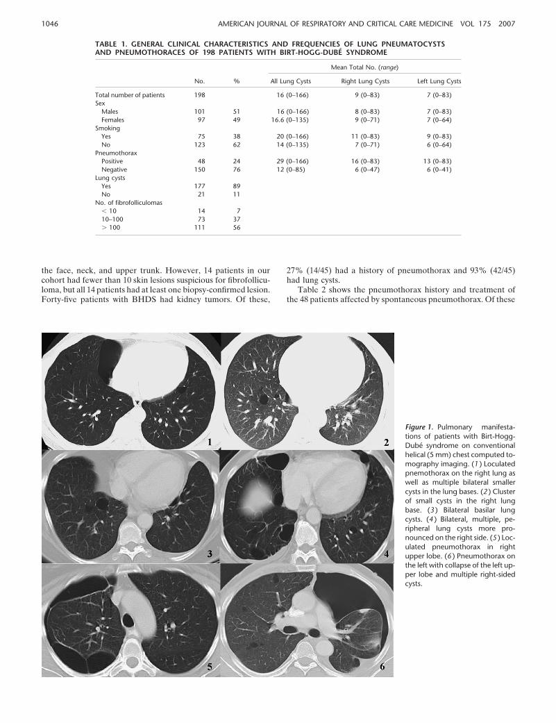
Patients’ clinical characteristics and the frequencies are listed inTable 1. Our cohort included 101 men and 97 women with amedian age of 49 years. The age range was 22 to 77 years, withonly eight patients younger than 30 years. Eighty-nine percent(177/198) of patients with BHDS had lung cysts on CT scans ofthe chest. Approximately 24% (48/198) of patients and 35% (31/89) of BHDS families screened for lung cysts had a history ofspontaneous pneumothorax. In our cohort of patients affectedwith BHDS, we found a relatively equal distribution of pneumo-thorax for men and women. Of the men affected with BHDS,20% (20/101) had a history of a pneumothorax and, of the womenwith BHDS, 29% (28/97) had a history of a pneumothorax. Allpatients with a history of pneumothorax had multiple lung cystsidentified by chest CT imaging (Figure 1). The earliest reportedage of initial pneumothorax was 22 years, the median age ofoccurrence was 38 years (range, 22–71 yr), and the median ageof the last pneumothorax was 42 years (range, 22–75 yr). Seventy-five percent (36/48) of patients had a second pneumothorax, theaverage number of pneumothoraces per patient was two, andfour patients experienced five separate pneumothoraces. Differ-ences in recurrence among patients may reflect efficacy of treat-ment used.
Ninety-three percent of patients with a diagnosis of BHDShad fibrofolliculomas with a distinctive clinical presentation charac-terized by multiple white or skin-colored papules distributed over
1046 AMERICAN JOURNAL OF RESPIRATORY AND CRITICAL CARE MEDICINE VOL 175 2007
TABLE 1. GENERAL CLINICAL CHARACTERISTICS AND FREQUENCIES OF LUNG PNEUMATOCYSTSAND PNEUMOTHORACES OF 198 PATIENTS WITH BIRT-HOGG-DUBE SYNDROME
Mean Total No. (range)
No. % All Lung Cysts Right Lung Cysts Left Lung Cysts
Total number of patients 198 16 (0–166) 9 (0–83) 7 (0–83)Sex
Males 101 51 16 (0–166) 8 (0–83) 7 (0–83)Females 97 49 16.6 (0–135) 9 (0–71) 7 (0–64)
SmokingYes 75 38 20 (0–166) 11 (0–83) 9 (0–83)No 123 62 14 (0–135) 7 (0–71) 6 (0–64)
PneumothoraxPositive 48 24 29 (0–166) 16 (0–83) 13 (0–83)Negative 150 76 12 (0–85) 6 (0–47) 6 (0–41)
Lung cystsYes 177 89No 21 11
No. of fibrofolliculomas� 10 14 710–100 73 37� 100 111 56
the face, neck, and upper trunk. However, 14 patients in ourcohort had fewer than 10 skin lesions suspicious for fibrofollicu-loma, but all 14 patients had at least one biopsy-confirmed lesion.Forty-five patients with BHDS had kidney tumors. Of these,
Figure 1. Pulmonary manifesta-tions of patients with Birt-Hogg-Dube syndrome on conventionalhelical (5 mm) chest computed to-mography imaging. (1 ) Loculatedpnemothorax on the right lung aswell as multiple bilateral smallercysts in the lung bases. (2 ) Clusterof small cysts in the right lungbase. (3 ) Bilateral basilar lungcysts. (4 ) Bilateral, multiple, pe-ripheral lung cysts more pro-nounced on the right side. (5 ) Loc-ulated pneumothorax in rightupper lobe. (6 ) Pneumothorax onthe left with collapse of the left up-per lobe and multiple right-sidedcysts.
27% (14/45) had a history of pneumothorax and 93% (42/45)had lung cysts.
Table 2 shows the pneumothorax history and treatment ofthe 48 patients affected by spontaneous pneumothorax. Of these

Toro, Pautler, Stewart, et al.: Lung Cysts and Spontaneous Pneumothorax in BHDS 1047
TABLE 2. CLINICAL CHARACTERISTICS OF THE 48 PATIENTS WITH BIRT-HOGG-DUBE SYNDROMEWITH PNEUMOTHORAX
Age(s) at Pneumothorax (Treatment)Smoking No. of
No. Sex History Pneumothoraces Right Lung Pneumothorax Left Lung Pneumothorax
1 M N 5 23 (TT), 25 (TT), 25 (TT/TH/MP) 29 (TT), 29 (TH/MP)2 M Y 1 52 (TT/TH/LR)3 F Y 1 30 (TT)4 F N 1 27 (TT)5 F N 4 24(TT), 28(TT/TH/CP), 38(TT/TH/MP) 26 (TT/TH/MP)6 F N 2 38 (N), 43 (TT/TH/LR)7 M Y 4 56 (TT/TH/LR/MP) 43 (TT), 48 (TH/LR/MP),
60 (TT/TH/MP)8 M N 2 42 (N), 46 (TH/MP)9 M Y 3 50 (N), 51 (N) 49 (N)
10 F N 1 43 (N)11 M N 1 45 (TT/TH/LR/MP)12 F Y 2 34 (N), 38 (TH/MP)13 M N 1 31 (TT/TH/MP)14 F N 1 41 (TT)15 M N 3 39 (TT), 50 (TH/LR), 52 (TH/MP)16 F N 2 42 (TT), 42 (TT/CP)17 F Y 1 44 (TT)18 M N 1 49 (TT/TH/LR)19 M Y 5 40 (TT), 46 (TT), 52 (TT/CP) 44 (TT), 48 (TH/MP)20 F Y 1 54 (N)21 M Y 3 36 (TT), 36 (TT/TH/CP),
38 (TH/MP)22 M Y 2 35 (TT), 38 (TT)23 M Y 2 22 (TT), 22 (TT)24 F N 2 47 (TH/MP) 37 (TT/CP)25 M N 2 41 (N), 42 (N)26 F N 2 37 (N), 41 (TT)27 F N 1 44 (TT)28 F Y 5 22 (TT/TH/LR), 22 (TT/TH/LR), 22 (TH/LR) 29 (N), 39 (N)29 M N 1 38 (N)30 M N 1 27 (N)31 F N 3 31 (TT), 41 (TT/CP), 42 (TT/CP)32 M N 1 35(N)33 F N 2 58 (TT), 58 (TH/MP)34 F N 1 26 (N)35 M Y 1 32 (TH/MP) 32 (TH/MP)36 F N 2 43 (TT), 45 (TT/TH/LR)37 M N 4 30 (TT), 30 (TH/CP), 30 (TH/CP) 32 (N)38 F N 1 45 (TT)39 F Y 1 46 (TT)40 F N 4 39 (TT), 39 (TT), 39 (TT/TH/MP) 39 (N)41 F N 1 23 (TT)42 F Y 5 24 (N), 25 (N), 25 (N), 26 (N),
27 (TT/TH/LR/MP)43 F N 1 36 (TH/LR)44 F N 3 50 (TT), 60 (TT/TH/LR/MP) 52 (TT)45 M N 2 55 (TT), 55 (TH/PM)46 F Y 2 36 (TT/TH/LR/CP), 36(TT/TH/MP)47 F N 2 37 (TT), 38 (TT/CP)48 F N 2 71 (TT), 75 (TT/TH/LR/MP)
Definition of abbreviations: CP � chemical pleurodesis; LR � lung resection; MP � mechanical pleurodesis; N � no treatment;TH � thoracostomy; TT � tube thoracostomy.
patients, 58% (28/48) were women and 41% (20/48) were men.Sixty-seven percent (32/48) of patients were nonsmokers. Therewere 101 episodes of pneumothoraces and no patient had simul-taneous bilateral pneumothoraces. The right lung had the highestfrequency of pneumothorax. Approximately 48% (23/48) of pa-tients had a pneumothorax in the right lung only, 29% (14/48)had a pneumothorax in the left lung only, and 23% (11/48) ofpatients had a pneumothorax in both the right and the left lungsat different times.
Twenty-three percent of pneumothoraces were managed withobservation alone. These patients were in stable condition andonly minimally compromised by the pneumothorax. Of the pneu-
mothoraces managed by observation alone, only 39% (9/23)completely resolved, and the remaining 61% (14/23) recurred,requiring medical treatment. Of the 101 pneumothoraces, 77%required medical intervention and were treated by various meth-ods. Thirty-five percent (35/101) were treated with tube thoracos-tomy (chest tube) only. Six pneumothoraces were managed witha combination of tube thoracostomy and chemical pleurodesis(quinacrine, tetracycline, silver nitrate, or talc). Approximately14% (15/101) of pneumothoraces were treated by open thoracot-omy and a second treatment, including mechanical pleurodesis(abrasion to produce adhesion between parietal and visceralpleura) in 10% (10/101), chemical pleurodesis in 2% (2/101),
1048 AMERICAN JOURNAL OF RESPIRATORY AND CRITICAL CARE MEDICINE VOL 175 2007
and lung resection in 3% (3/101). Approximately 13% (13/101)of pneumothoraces were treated with combined tube thoracos-tomy, thoracotomy, and a third treatment, including mechanicalpleurodesis in 7% (7/101), lung resection in 6% (6/101), andchemical pleurodesis in 2% (2/101), respectively. In addition,seven pneumothoraces were treated with a combination of tubeand open thoracostomies, lung resection, and mechanical pleu-rodesis. Another pneumothorax was treated with both a combi-nation of tube and open thoracostomy, lung resection, and chem-ical pleurodesis. Three patients with BHDS treated at the NIHunderwent video-assisted thoracoscopic surgery. Pneumothora-ces were not related to a particular calendar period. After serum�1-antitrypsin levels in 50 patients were within normal limits, weno longer performed the test. Capillary O2 saturation showed nosignificant abnormality in the measurements performed duringroutine screening for BHDS.
Risk Factor Analysis
We examined the association between the history of pneumotho-rax and the following categorical parameters: sex, smoking his-tory, severity of fibrofolliculomas, and lung cysts. The results ofthe univariate analysis of categorical parameters are summarizedin Table 3. The only categorical parameter that was significantlyassociated with pneumothorax was the presence of lung cysts(p � 0.006).
In a second analysis, we investigated the association of contin-uous parameters (the total number of cysts per lung lobe, totalnumber of intraparenchymal cysts, total number of subpleuralcysts, number of lung lobes with cysts, smoking history, age whenscanned, and lung cyst total volume) with pneumothorax. Theresults of tests for significance from the univariate analysis ofcontinuous parameters are summarized in Table 4. We foundthat every parameter related to the number of lung cysts wassignificantly associated with history of pneumothorax. No associ-ation was found between age at scan or smoking history andpresence or frequency of pneumothoraces. In addition, total
TABLE 3. RESULT OF UNIVARIATE ANALYSIS INVESTIGATINGASSOCIATION BETWEEN CATEGORICAL VARIABLES ANDPNEUMOTHORAX PRESENCE AND NUMBERS
Pneumothorax No. of Pneumothoraces
Variable No Yes p Value 0 1 2 3� p Value
SexM 81 20 0.14 81 6 7 7 0.83F 69 28 (C) 69 11 12 5 (C-A)
No. of fibrofolliculomas� 10 9 5 1.00 9 1 2 210–100 9 14 59 4 6 4 0.83� 100 82 29 (C-A) 82 12 11 6 (J-T)
Kidney tumorsYes 119 34 0.22 119 14 12 8 0.13No 31 14 (C) 31 3 7 4 (C-A)
Smoking historyYes 59 16 0.46 59 5 5 6 0.87No 91 32 (C) 91 12 14 6 (C-A)
Smoking statusNonsmoker 91 32 91 12 14 6Quit � 10 yr ago 21 8 21 2 3 3 0.47Quit � 10 yr ago 9 2 0.33 9 0 1 1 (J-T)Current 24 5 (C-A) 24 3 0 2
Lung cystsYes 129 48 0.006 129 17 19 12 0.012No 21 0 (C) 21 0 0 0 (C-A)
Definition of abbreviations: C � chi-square test; C-A � exact Cochran-Armitagetest; J-T � exact Jonckheere-Terpstra test.
lung cyst volume and largest cyst diameter and volume weresignificantly associated (p � 0.0001) with history of pneumotho-rax (Table 5). In addition, we found an association between thenumber of pneumothoraces and the total cyst volume (p �0.0001) (Table 5). There was an increase in median total cystvolume (0.8, 5.1, 8.5, and 10.3 cm3) with increasing number ofpneumothoraces (0, 1, 2, �3, respectively). Similarly, we foundthat the largest cyst diameter and volume were significantlyassociated with the number of pneumothoraces (Table 5).
The relationship between history of pneumothorax and dif-ferent parameters related to lung volume and cysts was investi-gated using a logistic regression model. The logistic regressionmodel determined that only the total number of cysts in theright parenchymal lower lobe (p � 0.050) and the total numberof cysts in the right pleura-based middle lobe (p � 0.002) wereneeded to classify a patient as to whether he or she was likelyto have a pneumothorax or not. These same parameters werealso associated with increasing numbers of pneumothoraces.
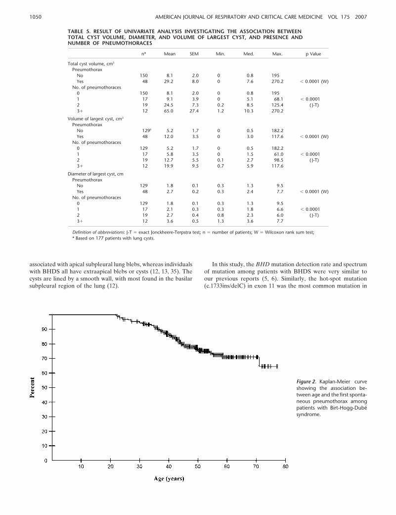
We used the Kaplan-Meier method to describe the associa-tion between age and the first spontaneous pneumothoraxamong all patients in the BHDS cohort (Figure 2). No pneumo-thoraces were diagnosed before age 22 years in this cohort, withthe oldest patient experiencing her first pneumothorax at 71years. By age 30 years, the probability of having the first pneumo-thorax is 6% (95% confidence interval [CI], 3–10%), 14% (95%CI, 10–20%) by 40 years, and 75% (95% CI, 19–32%) by age50. Of the 48 patients who had a pneumothorax, 88% (42/48)of them had his or her first pneumothorax before age 50, 10%(5/48) had one between the ages of 51 and 60 years, whereasonly 1 patient had a pneumothorax that occurred after age 60,at 71 years.
BHD Mutation Detection
BHD mutations were present in 81% (154/190) of patients and85% (68/80) of BHDS families tested. The cytosine mononucleo-tide tract in exon 11 of BHD was most frequently mutated,accounting for 48% (70/154) of patients and 47% (32/68) ofBHDS families who had a mutation detected. Thirteen patientsfrom one large BHDS kindred that previously showed linkageto chromosome 17p11.2 by haplotype (4) had no sequence varia-tion in the BHD gene (6). Nine families were not screened forBHD mutations by direct sequencing.
Patients with a history of pneumothorax share a similarBHDS mutation spectrum with all the patients with BHDS in thisstudy, but with some exceptions. BHD mutations were present in87% (41/47) of individuals and 90% (27/30) of BHDS familieswith a history of pneumothorax tested. Similar to previous stud-ies, the “hot spot” mutation, c.1733ins/delC, in exon 11 was themost frequent site of mutation, accounting for 38% (18/47) ofindividuals with a history of a pneumothorax and 44% (18/41) ofall the mutations detected. We were unable to detect a sequencevariation in BHD by direct sequence in six individuals, but fourof these individuals were part of a family that showed linkageto chromosome 17p11.2 by haplotype analysis (4). In addition,one individual was not screened for BHD mutations.
Analyses of Genotype–Phenotype Correlation
There was no association between BHD mutation status (nomutation vs. mutation and/or linkage to chromosome 17p11.2),mutation types (insertion, deletion, nonsense, and splice site,and frameshift vs. nonsense and splice site; splice site vs. exonmutation, or hot-spot mutation vs. all other BHD mutations, orsplice site vs. all other BHD mutations), or location, and lungcyst parameters or pneumothorax. Our analysis showed a trendfor differences in pneumothoraces according to exon location(p � 0.01), with individuals with BHD mutations in exon 9 and
Toro, Pautler, Stewart, et al.: Lung Cysts and Spontaneous Pneumothorax in BHDS 1049
TABLE 4. RESULTS OF UNIVARIATE ANALYSIS INVESTIGATING ASSOCIATION BETWEENCONTINUOUS PARAMETERS AND PNEUMOTHORAX PRESENCE AND NUMBER
Pneumothorax No. of PneumothoracesVariable (Yes/No) p-Value (0, 1, 2, 3�) p-Value
Pack-years 0.31 0.36Total no. lung cysts/person � 0.0001 � 0.0001Total no. lung cysts, right lobe � 0.0001 � 0.0001Right PUL 0.0045 0.0046Right PML 0.0006 0.0005Right PLL � 0.0001 � 0.0001Right PBUL 0.0005 0.0005Right PBML � 0.0001 � 0.0001Right PBLL 0.0001 � 0.0001Total left lung � 0.0001 � 0.0001Left PUL 0.0011 0.0010Left PLL 0.0071 0.0047Left PBUL � 0.0001 � 0.0001Left PBLL 0.0026 0.0017Left PB (LPPUL � LPBLL) � 0.0001 � 0.0001Left parenchymal (LPUL � LPLL) 0.0003 0.0003Right PB (PBUL � PBML � PBLL) � 0.0001 � 0.0001Right parenchymal (PUL � PML � PLL) � 0.0001 � 0.0001Lower right (PLL � PBLL) � 0.0001 � 0.0001Upper right (PUL � PML � PBML � PBUL) � 0.0001 � 0.0001Lower left (LPLL � LPBLL) 0.0014 0.0008Upper left (LPUL � LPBUL) � 0.0001 � 0.0001Upper right � upper left � 0.0001 � 0.0001Lower left � lower right 0.0001 � 0.0001PB (left and right) � 0.0001 � 0.0001Parenchymal (left and right) � 0.0001 � 0.0001No. of compartments � 0.0001 � 0.0001Scan age 0.90 0.93
Definition of abbreviations: PBLL � pleura-based lower lobe; PBML � pleura-based middle lobe; PBUL � pleura-based upperlobe; PLL � parenchymal lower lobe; PML � parenchymal middle lobe; PUL � parenchymal upper lobe.
exon 12 having more pnemothoraces than individuals with BHDmutations in other exons (Table 6). In addition, our analysisrevealed that mutation exon location was associated with thenumbers of cysts (p � 0.0002), with individuals with BHD muta-tions in exon 9 having more cysts (median � 32) than individualswith mutation in other exons (Table 7). Similarly, we found thatthe BHD mutation exon location was associated with the size(p � 0.005) and volume (p � 0.01) of the largest cysts. Individualswith mutations in exons 9 and 12 had the largest cyst diameters(1.9 and 2.9 cm) and volumes (2.7 cm3) (Table 7).
DISCUSSION
This is the largest and most comprehensive study to date ofindividuals and families affected with BHDS relating the pulmo-nary risk factors, CT screening, and genotype–pulmonary associ-ations. In this study, for the first time, we showed that lung cystsand every parameter related to the number of lung cysts weresignificantly associated with spontaneous pneumothorax. Uni-variate analysis revealed that total lung cyst volume, largest cystdiameter, and largest cyst volume were statistically associatedwith pneumothorax in patients with BHDS. In addition, logisticregression analysis showed that only the total number of cystsin the right parenchymal lower lobe and in the right pleura-based middle lobe were needed to classify a patient as to whetherhe or she was likely to have a pneumothorax. The role of lungcysts in the mechanism leading to a spontaneous pneumothoraxin BHDS has not been established. One explanation is that lungcysts may be a precursor lesion; a second possibility is thatrupture of subpleural blebs on the visceral pleura may lead toa spontaneous pneumothorax. Furthermore, the pathophysiol-ogy of lung cysts in BHDS is unknown. Although inactivation
of the BHD wild-type allele by loss of heterozygosity (LOH)or somatic mutation may explain the BHDS-associated kidneytumors (32), it is possible that haploinsufficiency alone may beresponsible for the development of lung cysts. Age of onset ofthe first reported pneumothorax is also important. Approxi-mately 90% of patients with BHDS had their first pneumothoraxby age 50, suggesting that pneumothorax tends to occur duringadulthood in patients with BHDS.
Screening of 198 patients with BHDS at the NIH ClinicalCenter using high-resolution plus standard CT of the chest re-vealed that most (89%) patients with BHDS have multiple pul-monary cysts. Twenty-four percent of patients with BHDS hada history of one or more pneumothoraces, all of whom hadmultiple lung cysts identified by chest CT imaging. This studyrevealed a relatively equal distribution of pneumothorax amongmen and women. In contrast, previous studies have identifiedmale sex as a risk factor for primary spontaneous pneumothorax(33). In this study, 67% of patients with BHDS who had a historyof pneumothorax were nonsmokers, and 61% of those withouta pneumothorax were also nonsmokers, supporting the view thatsmoking is not a risk factor for pneumothorax in our cohort ofpatients with BHDS. However, in lung studies of other popula-tions, smoking has been shown to be a risk factor for pneumotho-rax (34). It is important to recognize that smoking can lead toemphysematous cystic and bullous changes in the lung in thegeneral population. However, the location and characteristicsof the cystic lesions are usually different from BHDS. The effectsof smoking in exacerbating or worsening the pulmonary lungdisease (lung cyst and spontaneous pneumothorax) in the settingof BHDS is unknown. In this study, we also found that severityof cutaneous involvement or kidney tumors was not a risk factorfor pneumothorax. Sporadic spontaneous pneumothorax is
1050 AMERICAN JOURNAL OF RESPIRATORY AND CRITICAL CARE MEDICINE VOL 175 2007
TABLE 5. RESULT OF UNIVARIATE ANALYSIS INVESTIGATING THE ASSOCIATION BETWEENTOTAL CYST VOLUME, DIAMETER, AND VOLUME OF LARGEST CYST, AND PRESENCE ANDNUMBER OF PNEUMOTHORACES
n* Mean SEM Min. Med. Max. p Value
Total cyst volume, cm3
PneumothoraxNo 150 8.1 2.0 0 0.8 195Yes 48 29.2 8.0 0 7.6 270.2 � 0.0001 (W)
No. of pneumothoraces0 150 8.1 2.0 0 0.8 1951 17 9.1 3.9 0 5.1 68.1 � 0.00012 19 24.5 7.3 0.2 8.5 125.4 ( J-T)3� 12 65.0 27.4 1.2 10.3 270.2
Volume of largest cyst, cm3
PneumothoraxNo 129† 5.2 1.7 0 0.5 182.2Yes 48 12.0 3.5 0 3.0 117.6 � 0.0001 (W)
No. of pneumothoraces0 129 5.2 1.7 0 0.5 182.21 17 5.8 3.5 0 1.5 61.0 � 0.00012 19 12.7 5.5 0.1 2.7 98.5 ( J-T)3� 12 19.9 9.5 0.7 5.9 117.6
Diameter of largest cyst, cmPneumothorax
No 129 1.8 0.1 0.3 1.3 9.5Yes 48 2.7 0.2 0.3 2.4 7.7 � 0.0001 (W)
No. of pneumothoraces0 129 1.8 0.1 0.3 1.3 9.51 17 2.1 0.3 0.3 1.8 6.6 � 0.00012 19 2.7 0.4 0.8 2.3 6.0 ( J-T)3� 12 3.6 0.5 1.3 3.6 7.7
Definition of abbreviations: J-T � exact Jonckheere-Terpstra test; n � number of patients; W � Wilcoxon rank sum test;* Based on 177 patients with lung cysts.
associated with apical subpleural lung blebs, whereas individualswith BHDS all have extraapical blebs or cysts (12, 13, 35). Thecysts are lined by a smooth wall, with most found in the basilarsubpleural region of the lung (12).
Figure 2. Kaplan-Meier curveshowing the association be-tween age and the first sponta-neous pneumothorax amongpatients with Birt-Hogg-Dubesyndrome.
In this study, the BHD mutation detection rate and spectrumof mutation among patients with BHDS were very similar toour previous reports (5, 6). Similarly, the hot-spot mutation(c.1733ins/delC) in exon 11 was the most common mutation in
Toro, Pautler, Stewart, et al.: Lung Cysts and Spontaneous Pneumothorax in BHDS 1051
TABLE 6. ANALYSIS INVESTIGATING THE ASSOCIATIONBETWEEN EXON LOCATION OF THE BHD MUTATION ANDPRESENCE AND NUMBER OF PNEUMOTHORACES
Pneumothorax No. of Pneumothoraces
Exon No Yes p Value 0 1 2 3� p Value
5 7 0 7 0 0 06 4 2 4 2 0 07 10 1 10 1 0 09 9 8 0.01 9 2 3 3 0.012
11 57 18 (M) 57 4 10 4 (K-W OC)12 4 5 4 1 3 113 0 2 0 1 1 014 2 0 2 0 0 0
Definition of abbreviations: K-W OC � Kruskal-Wallis test for ordered columnsM � Mehta’s modification of Fisher’s exact test.
this study. In this report, the hot-spot mutation was present in48% of BHDS families, whereas this mutation in exon 11 wasreported in 53% of BHDS families previously (6). One majordifference from our previous report is that all patients withBHDS included in this study were only evaluated at the NIHClinical Center and patients seen on field trips were excluded.This criterion was needed for a systematic chest screening evalua-tion of lung cysts of all patients. Recently, Painter and colleagues(13) reported on a large Finnish family with primary spontaneouspneumothorax in 8 members and 14 family members who hadlung bullae (cysts) on high-resolution CT examination. Directsequencing of genomic DNA from affected individuals revealeda 4-bp deletion in the first exon of the BHD gene. This mutationwas not present among our patients with BHDS. Similarly, Gra-ham and coworkers reported two different nonsense mutations(E315X and R477X) in two different families with primary spon-taneous pneumothorax (12). These nonsense BHD mutationswere not identified in our cohort of patients. Painter and col-
TABLE 7. ANALYSIS INVESTIGATING THE ASSOCIATION BETWEEN EXON LOCATION OF THE BHDMUTATION AND TOTAL NUMBER OF CYSTS, LARGEST CYST VOLUME, AND DIAMETER
Variable Exon n Mean SEM Med. Min. Max. p Value
Total no. of cysts 5 7 4 1.6 2 0 126 6 18 5.8 19 2 387 11 8.7 2.6 6 0 309 17 34.2 7.2 32 4 135 0.0002 (K-W)
11 75 12.8 1.8 7 0 6612 9 13.7 4.7 9 0 4513 2 34 34 31 3714 2 1.5 1.5 1 2.0
Diameter of largest cysts, cm 5 6 0.8 0.1 0.7 0.6 1.26 6 2.4 1.0 1.6 0.9 7.17 10 1.2 0.2 1.1 0.5 2.19 17 2.6 0.4 1.9 0.8 5.8 0.005 (K-W)
11 68 1.7 0.1 1.3 0.3 6.412 8 3.5 1.0 2.6 0.6 8.013 2 1.7 1.7 1.6 1.814 2 1.0 1.0 1 1.1
Volume of largest cysts, cm3 5 6 0.3 0.1 0.2 0.1 0.66 6 31.5 30.1 0.7 0.4 182.27 10 0.7 0.3 0.4 0.02 2.59 17 10.4 4.6 2.7 0.04 72.9 0.01 (K-W)
11 68 2.7 0.7 0.6 0.01 29.512 8 14.0 7.0 2.7 0.03 52.813 2 1.7 1.7 1.6 1.814 2 0.35 0.35 0.34 0.37
Definition of abbreviation: K-W � Kruskal-Wallis test; n � number of patients.* Volume units.
leagues and Graham and coworkers reported that these familieslack dermatologic findings. However, dermatologic examina-tions were not conducted in both studies. On the other hand,we recognized that BHDS can occur in the absence of skinlesions, although it is uncommon.
In this study, we also investigated potential genotype–pulmonary relationships in our patients with BHDS. In general,we found no associations between BHD mutation status, ormutation types, and lung cysts parameters and pneumothorax.However, an analysis showed that individuals with BHD muta-tions in exon 9 were associated with more lung cysts than individ-uals with mutations in other exons. In addition, we found thatthe size and volume of the largest lung cyst differed significantlyby exon, and that these were greater in individuals with BHDmutation in exons 9 and 12 than in those with mutations in otherexons. These findings suggest that there may be an associationbetween mutation location and lung cyst number and size. It isof interest that recently we also reported that 40% (7/17) ofpatients with BHDS with putative splice-site mutations in intron9 (predicted to cause exon 9 skipping) developed renal tumors(6). This is a significantly higher frequency of renal tumors thanthe overall frequency in all mutation carriers. These two indepen-dent observations suggest that exon 9 may have functional im-portance. These findings need to be confirmed in a future studywith a larger number of patients with BHDS. We also foundvariability of expression of lung cysts and spontaneous pneumo-thorax both between and within families. These findings suggestthat the existence of other genetic and/or environmental factorsmay also influence the pulmonary phenotype. In addition, thenumber of lung cysts and pneumothoraces was not a good pre-dictor for kidney cancer status.
The differential diagnosis for a patient with a history of famil-ial spontaneous pneumothorax and diffuse pulmonary cysticchanges includes TSC (25), �1-antitrypsin deficiency (21), Marfansyndrome (22), Ehlers-Danlos syndrome (23), LAM (24), LCH(26), CF (27), primary spontaneous pneumothorax (15), and
1052 AMERICAN JOURNAL OF RESPIRATORY AND CRITICAL CARE MEDICINE VOL 175 2007
BHDS. The distribution of cystic lung changes in radiologicstudies may be helpful in distinguishing these diseases. Relativesparing of lung bases from cystic changes is seen in LCH butnot in BHDS and LAM. Obstructive findings in a patient withdiffuse lung infiltrates are uncommon but can be seen in LAMand LCH. Pulmonary conditions in the general population, in-cluding idiopathic pulmonary fibrosis, Pneumocystis carinii,lymphocytic interstitial pneumonia, and septic emboli, are alsopart of the differential diagnosis of cystic lung lesions. Integratingthe clinical context is critical in the differential diagnosis offamilial spontaneous pneumothorax. Patients’ family history andphysical examination may provide clues to the nature of thediffuse lung cystic disease. Pulmonary LAM is almost exclusivelyin women of reproductive age (24). Family history of inheritableskin disorders include TSC, BHDS, LCH, Marfan syndrome(22), and Ehlers-Danlos syndrome (23). The dermatologic mani-festations in these syndromes may be helpful in distinguishingthese disorders. Patients with BHDS have multiple fibrofollicu-lomas and/or trichodiscoma, whereas patients with LCH presentwith scaly patches that histologically show an infiltrate of lym-phocytes, eosinophils, and Langerhans cells in the skin. Derma-tologically, BHDS and LCH are very distinct. However, TCSand BHDS may be difficult to distinguish. Patients with TSCusually show angiofibromas, hypopigmented macules, shagreenpatch, and/or periungual fibromas, and patients with Ehlers-Danlos syndrome typically have fragile thin skin, easy bruising,scarring, and/or hyperextensibility.
Treatment of spontaneous pneumothorax in our patients withBHDS varied from simple observation to open thoracotomywith pleurodesis and lung resection. Seven primary treatmentapproaches were reported as being used over the study period,including the following: observation alone, tube thoracostomyalone, tube thoracostomy with chemical pleurodesis, thoracot-omy with mechanical pleurodesis, and thoracotomy with lungresection. Because different physicians at different hospitalstreated patients with a variety of treatment modalities, we cannotexclude that these variables are confounding the risk for recur-rence of pneumothorax.
The treatment of pneumothorax in patients with BHDS issimilar to the approach taken for any patient with spontaneouspneumothorax. It ranges from observation with repeated radio-graphic examinations in asymptomatic patients to urgent inter-vention to evacuate air from the intrapleural space and to pre-vent recurrence. The mode of therapy is dictated by the clinicalpresentation of the patient, the chronicity of the condition, andthe underlying lung conditions that induced the developmentof pneumothorax. Placement of a tube thoracostomy enablesevacuation of pleural air, and reexpansion of the compressedportion of the lung, and provides a means for chemical pleu-rodesis. For patients with discreet lung bullae or blebs, or thosewith recurrent pneumothoraces, treatment may include surgicalintervention (thoracotomy or video-assisted thoracoscopy) incombination with mechanical pleurodesis and resection oflung bullae when present. Prospective treatment trials areneeded to investigate the best treatment of BHDS-associatedpneumothoraces.
The clinical presentation of spontaneous pneumothorax inpatients with BHDS is variable. Furthermore, a spontaneouspneumothorax may not be detected on a plain chest X-ray;therefore, it may be overlooked. We advised our patients toinform medical examiners that they have a condition that pre-disposes them to spontaneous pneumothorax. Although inBHDS it is unknown how to prevent pneumothoraces, certainmeasures can decrease the risk of developing one. Patientsshould be cautioned about the increased risk of pneumothoraxwith scuba diving and air travel due to ambient pressure effects,
especially if they have chest symptoms such as pain, discomfort,and/or shortness of breath. We have not observed fatalitiesor chronic debilitation associated with BHDS lung cysts orpneumothoraces.
In conclusion, this study describes the unique pulmonary fea-tures, genetic characteristics, and risk factors for pneumothoraxin 198 patients with BHDS. It is important to recognize that,based on the temporality limitations of the study, we cannotclearly determine the true relationship between the number oflung cysts and the risk for spontaneous pneumothorax because,in most cases, the pneumothorax was documented and confirmedbefore the initiation of study. However, our study has showna significant association between the lung cysts (number andlocation) and pneumothorax. Our study contributes to the under-standing of the genetic basis of hereditary spontaneous pneumo-thorax. A prospective study following a cohort of patients shouldbe conducted to validate our present findings. Recognition ofthe pulmonary features associated with BHDS will improve thediagnosis and treatment of patients with BHDS. Furthermore,recognition of the diagnosis of BHDS will also provide awarenessto patients and health care providers of the need for screeningand surveillance for renal neoplasms. Future molecular studiesmay be able to demonstrate if the BHD gene is involved inthe etiology of sporadic spontaneous pneumothorax and/oremphysema.
Conflict of Interest Statement : J.R.T. is an inventor on a patent application thathas been filed for the BHD gene. S.E.P. does not have a financial relationship witha commercial entity that has an interest in the subject of this manuscript. L.S.does not have a financial relationship with a commercial entity that has an interestin the subject of this manuscript. G.M.G. does not have a financial relationshipwith a commercial entity that has an interest in the subject of this manuscript.M.W. does not have a financial relationship with a commercial entity that has aninterest in the subject of this manuscript. O.T. does not have a financial relationshipwith a commercial entity that has an interest in the subject of this manuscript.M.-H.W. does not have a financial relationship with a commercial entity that hasan interest in the subject of this manuscript. L.S.S. is an inventor on a patentapplication that has been filed for the BHD gene. L.D. does not have a financialrelationship with a commercial entity that has an interest in the subject of thismanuscript. B.Z. is an inventor on a patent application that has been filed for theBHD gene. P.C. does not have a financial relationship with a commercial entitythat has an interest in the subject of this manuscript. S.M.S. does not have afinancial relationship with a commercial entity that has an interest in the subjectof this manuscript. D.M.N. does not have a financial relationship with a commer-cial entity that has an interest in the subject of this manuscript. W.M.L. is aninventor on a patent application that has been filed for the BHD gene.
Acknowledgment : The authors thank the families of patients with BHDS for theirparticipation in our study and the members of the American Academy of Dermatol-ogy for their help in the recruitment of families. They also thank Cia Manolatos,Robin Eyler, Kathleen Hurley, James Peterson, and Lindsay Middelton for theirmany contributions to this project.
References
1. Birt AR, Hogg GR, Dube WJ. Hereditary multiple fibrofolliculomas withtrichodiscomas and acrochordons. Arch Dermatol 1977;113:1674–1677.
2. Toro JR, Glenn G, Duray P, Darling T, Weirich G, Zbar B, Linehan M,Turner ML. Birt-Hogg-Dube syndrome: a novel marker of kidneyneoplasia. Arch Dermatol 1999;135:1195–1202.
3. Binet O, Robin J, Vicart M, Ventura G, Beltzer-Garelly E. Fibromesperfolliculaires, polypose colique familiale, pneumothorax spontanesfamiliaux. Ann Dermatol Venereol 1986;113:928–930.
4. Schmidt LS, Warren MB, Nickerson ML, Weirich G, Matrosova V, ToroJR, Turner ML, Duray P, Merino M, Hewitt S, et al. Birt-Hogg-Dubesyndrome, a genodermatosis associated with spontaneous pneumotho-rax and kidney neoplasia, maps to chromosome 17p11.2. Am J HumGenet 2001;69:876–882.
5. Nickerson ML, Warren MB, Toro JR, Matrosova V, Glenn G, TurnerML, Duray P, Merino M, Choyke P, Pavlovich CP, et al. Mutationsin a novel gene lead to kidney tumors, lung wall defects, and benigntumors of the hair follicle in patients with the Birt-Hogg-Dube syn-drome. Cancer Cell 2002;2:157–164.
6. Schmidt LS, Nickerson ML, Warren MB, Glenn GM, Toro JR, MerinoMJ, Turner ML, Choyke PL, Sharma N, Peterson J, et al. GermlineBHD-mutation spectrum and phenotype analysis of a large cohort of
Toro, Pautler, Stewart, et al.: Lung Cysts and Spontaneous Pneumothorax in BHDS 1053
families with Birt-Hogg-Dube syndrome. Am J Hum Genet 2005;76:1023–1033.
7. Okimoto K, Sakurai J, Kobayashi T, Mitani H, Hirayama Y, NickersonML, Warren MB, Zbar B, Schmidt LS, Hino O. A germ-line insertionin the Birt-Hogg-Dube (BHD) gene gives rise to the Nihon rat modelof inherited renal cancer. Proc Natl Acad Sci USA 2004;101:2023–2027.
8. Lingaas F, Comstock KE, Kirkness EF, Sorensen A, Aarskaug T, HitteC, Nickerson ML, Moe L, Schmidt LS, Thomas R, et al. A mutationin the canine BHD gene is associated with hereditary multifocal renalcystadenocarcinoma and nodular dermatofibrosis in the German Shep-herd dog. Hum Mol Genet 2003;12:3043–3053.
9. Baba M, Hong SB, Sharma N, Warren MB, Nickerson ML, Iwamatsu A,Esposito D, Gillette WK, Hopkins RF III, Hartley JL, et al. Folliculinencoded by the BHD gene interacts with a binding protein, FNIP1,and AMPK, and is involved in AMPK and mTOR signaling. ProcNatl Acad Sci USA 2006;103:15552–15557.
10. Warren MB, Torres-Cabala CA, Turner ML, Merino MJ, MatrosovaVY, Nickerson ML, Ma W, Linehan WM, Zbar B, Schmidt LS. Expres-sion of Birt-Hogg-Dube mRNA in normal and neoplastic humantissues. Mod Pathol 2004;17:998–1011.
11. Zbar B, Alvord WG, Glenn G, Turner M, Pavlovich CP, Schmidt L,Walther M, Choyke P, Weirich G, Hewitt SM, et al. Risk of renal andcolonic neoplasms and spontaneous pneumothorax in the Birt-Hogg-Dube syndrome. Cancer Epidemiol Biomarkers Prev 2002;11:393–400.
12. Graham R, Nolasco M, Peterlin B, Kim Garcia C. Nonsense mutationsin folliculin presenting as isolated familial spontaneous pneumothoraxin adults. Am J Respir Crit Care Med 2005;172:39–44.
13. Painter JN, Tapanainen H, Somer M, Tukiainen P, Aittomaki K. A 4-bp deletion in the Birt-Hogg-Dube gene (FLCN) causes dominantlyinherited spontaneous pneumothorax. Am J Hum Genet 2005;76:522–527.
14. Primrose WR. Spontaneous pneumothorax: a retrospective review ofaetiology, pathogenesis and management. Scott Med J 1984;29:15–20.
15. Abolnik IZ, Lossos IS, Zlotogora J, Brauer R. On the inheritance ofprimary spontaneous pneumothorax. Am J Med Genet 1991;40:155–158.
16. Bagchi I, Nycyk JA. Familial spontaneous pneumothorax. Arch Dis ChildFetal Neonatal Ed 2002;87:F70.
17. Koivisto PA, Mustonen A. Primary spontaneous pneumothorax in twosiblings suggests autosomal recessive inheritance. Chest 2001;11:1610–1612.
18. Morrison PJ, Lowry RC, Nevin NC. Familial primary spontaneous pneu-
mothorax consistent with true autosomal dominant inheritance.Thorax 1998;53:151–152.
19. Sansonetti M, Sandron D, Pin I, Labrune S, Vervloet D, Dumur JP,Charpin J, Chretien J. Diffuse familial interstitial pulmonary fibrosis:study of a family. [in French] Rev Mal Respir 1985;2:75–81.
20. Vishnevskii AA, Nikoladze GD, Romanov Iu V. Familial bullous diseaseof the lungs as a cause of spontaneous pneumothorax. Grud Serdechno-sosudistaia Khir 1990;2:44–46.
21. Daniel R, Teba L. Spontaneous pneumothorax and alpha 1-antitrypsindeficiency. Respir Care 2000;45:327–329.
22. Hall JR, Pyeritz RE, Dudgeon DL, Haller JA Jr. Pneumothorax in theMarfan syndrome: prevalence and therapy. Ann Thorac Surg 1984;37:500–504.
23. O’Neill S, Sweeney J, Walker F, O’Dwyer WF. Pneumothorax in theEhlers-Danlos syndrome. Ir J Med Sci 1981;150:43–44.
24. Berkman N, Bloom A, Cohen P, Deviri E, Bar-Ziv Y, Shimon D, KramerMR. Bilateral spontaneous pneumothorax as the presenting featurein lymphangioleiomyomatosis. Respir Med 1995;89:381–383.
25. Babcock TL, Snyder BA. Spontaneous pneumothorax associated withtuberous sclerosis. J Thorac Cardiovasc Surg 1982;83:100–104.
26. Mendez JL, Nadrous HF, Vassallo R, Decker PA, Ryu JH. Pneumothoraxin pulmonary Langerhans cell histiocytosis. Chest 2004;125:1028–1032.
27. Flume PA, Strange C, Ye X, Ebeling M, Hulsey T, Clark LL. Pneumotho-rax in cystic fibrosis. Chest 2005;128:720–728.
28. Agresti A. Categorical data analysis, 1st ed. New York: John Wiley andSons; 1990. pp. 79–129.
29. Hollander M, Wolfe DA. Nonparametric statistical methods, 2nd ed.New York: John Wiley and Sons; 1999. pp. 189–269.
30. Mehta CR, Patel NR. A network algorithm for performing Fisher’s exacttest in r c contingency tables. J Am Stat Assoc 1983;78:427–434.
31. Kaplan E, Meier P. Non-parametric estimation from incomplete observa-tions. J Am Stat Assoc 1958;53:457–481.
32. Vocke CD, Yang Y, Pavlovich CP, Schmidt LS, Nickerson ML, Torres-Cabala CA, Merino MJ, Walther MM, Zbar B, Linehan WM. Highfrequency of somatic frameshift BHD gene mutations in Birt-Hogg-Dube–associated renal tumors. J Natl Cancer Inst 2005;9:931–935.
33. Melton LJ III, Hepper NG, Offord KP. Incidence of spontaneous pneu-mothorax in Olmsted County, Minnesota: 1950 to 1974. Am Rev RespirDis 1979;120:1379–1382.
34. Bense L, Eklund G, Wiman LG. Smoking and the increased risk ofcontracting spontaneous pneumothorax. Chest 1987;92:1009–1012.
35. Butnor KJ, Guinee DG Jr. Pleuropulmonary pathology of Birt-Hogg-Dube syndrome. Am J Surg Pathol 2006;30:395–399.
Copyright © 2022 FDOKUMEN