Low-Intensity Pulsed Ultrasound Produced an Increase of Osteogenic Genes Expression During the...
8
d Original Contribution LOW-INTENSITY PULSED ULTRASOUND PRODUCED AN INCREASE OF OSTEOGENIC GENES EXPRESSION DURING THE PROCESS OF BONE HEALING IN RATS ELAINE F A ´ VARO-PI ´ PI,* P AULO BOSSINI,* POLIANI DE OLIVEIRA,* JULIANA UEMA RIBEIRO, y CARLA TIM,* NIVALDO A. P ARIZOTTO,* JOSE MARCOS ALVES, x DANIEL ARAKI RIBEIRO, z HELOI ´ SA SOBREIRO SELISTRE DE ARAU ´ JO, y and ANA CLAUDIA MUNIZ RENNO z * Department of Physiotherapy; y Department of Physiological Sciences, Federal University of Sa ˜o Carlos, Sa ˜o Carlos, Sa ˜o Paulo, Brazil; z Department of Bioscience, Federal University of Sa ˜o Paulo, Santos, Sa ˜o Paulo, Brazil; and x Bioengineering Graduate Program, Department of Electrical Engineering, School of Engineering of Sa ˜o Carlos, University of Sa ˜o Paulo at Sa ˜o Carlos, Sa ˜o Carlos, Sa ˜o Paulo, Brazil (Received 5 May 2010; revised 30 June 2010; in final form 11 July 2010) Abstract—The aim of this study was to measure the temporal expression of osteogenic genes during the process of bone healing in low-intensity pulsed ultrasound (LIPUS) treated bone defects by means of histopathologic and real-time polymerase chain reaction (PCR) analysis. Animals were randomly distributed into two groups (n 5 30): control group (bone defect without treatment) and LIPUS treated (bone defect treated with LIPUS). On days 7, 13 and 25 postinjury, 10 rats per group were sacrificed. Rats were treated with a 30 mW/cm 2 LIPUS. The results pointed out intense new bone formation surrounded by highly vascularized connective tissue present- ing a slight osteogenic activity, with primary bone deposition was observed in the group exposed to LIPUS in the intermediary (13 days) and late stages of repair (25 days) in the treated animals. In addition, quantitative real- time polymerase chain reaction (RT-qPCR) showed an upregulation of bone morphogenetic protein 4 (BMP4), osteocalcin and Runx2 genes 7 days after the surgery. In the intermediary period, there was no increase in the expression. The expression of alkaline phosphatase, BMP4 and Runx2 was significantly increased at the last period. Our results indicate that LIPUS therapy improves bone repair in rats and upregulated osteogenic genes, mainly at the late stages of recovery. (E-mail: [email protected]) Ó 2010 Published by Elsevier Inc. on behalf of World Federation for Ultrasound in Medicine & Biology. Key Words: Bone repair, Rats, Low-intensity pulsed ultrasound, Osteogenesis, Genes, Quantitative PCR. INTRODUCTION Over 6,200,000 fractures of the skeleton occur in the United States each year, with almost 10% complicated by disrupted patterns of bone healing (Hadjiargyrou et al. 2002). Even with a majority of fracture healing appropriately, over 30,000,000 days each year are lost because of disability or confinement of patients, leading to a tremendous loss of productivity and income. Given the great potential of both tissue and genetic engineering, it is anticipated that exogenous acceleration of fracture healing could increase the overall number of fractures that heal successfully, as well as reduce the number of lost patient days due to incapacity (Yang et al. 2005). It is clear the importance of the development of innovative clinical approaches to accelerate bone metab- olism and to repair damage to bone tissue. One promising treatment method is the use of low-intensity pulsed ultra- sound (LIPUS). LIPUS is a form of mechanical energy that is transmitted through and into living tissue as acoustic pressure waves. It has been theorized that the mi- cromechanical strains produced by these pressure waves in biological tissues may result in biochemical events that accelerate tissue healing (Claes and Willie 2007). This therapeutic modality is well established, approved by the FDA (U.S. Food and Drug Administration) and in frequent use (Claes and Willie 2007). Many in vitro works have showed that LIPUS increases prostaglandin E2 (PGE2) production via the Address correspondence to: Ana Claudia Muniz Renno, Depart- ment of Bioscience, Federal University of Sa ˜o Paulo, Av. Ana Costa, 95, Vila Mathias, Santos, Sa ˜o Paulo, Brazil 11050-240. E-mail: [email protected] 2057 Ultrasound in Med. & Biol., Vol. 36, No. 12, pp. 2057–2064, 2010 Ó 2010 Published by Elsevier Inc. on behalf of World Federation for Ultrasound in Medicine & Biology Printed in the USA. All rights reserved 0301-5629/$ - see front matter doi:10.1016/j.ultrasmedbio.2010.07.012
Transcript of Low-Intensity Pulsed Ultrasound Produced an Increase of Osteogenic Genes Expression During the...
Ultrasound in Med. & Biol., Vol. 36, No. 12, pp. 2057–2064, 2010� 2010 Published by Elsevier Inc. on behalf of World Federation for Ultrasound in Medicine & Biology
Printed in the USA. All rights reserved0301-5629/$ - see front matter
asmedbio.2010.07.012
doi:10.1016/j.ultrd Original Contribution
LOW-INTENSITY PULSED ULTRASOUND PRODUCED AN INCREASEOF OSTEOGENIC GENES EXPRESSION DURING THE PROCESS OF BONE
HEALING IN RATS
ELAINE FAVARO-PIPI,* PAULO BOSSINI,* POLIANI DE OLIVEIRA,* JULIANAUEMA RIBEIRO,y CARLATIM,*NIVALDO A. PARIZOTTO,* JOSE MARCOS ALVES,x DANIEL ARAKI RIBEIRO,z
HELOISA SOBREIRO SELISTRE DE ARAUJO,y and ANA CLAUDIA MUNIZ RENNOz
*Department of Physiotherapy; yDepartment of Physiological Sciences, Federal University of Sao Carlos, Sao Carlos, SaoPaulo, Brazil; zDepartment of Bioscience, Federal University of Sao Paulo, Santos, Sao Paulo, Brazil; and xBioengineeringGraduate Program, Department of Electrical Engineering, School of Engineering of Sao Carlos, University of Sao Paulo at Sao
Carlos, Sao Carlos, Sao Paulo, Brazil
(Received 5 May 2010; revised 30 June 2010; in final form 11 July 2010)
Ament o95, Via.renno
Abstract—The aim of this study was to measure the temporal expression of osteogenic genes during the process ofbone healing in low-intensity pulsed ultrasound (LIPUS) treated bone defects by means of histopathologicand real-time polymerase chain reaction (PCR) analysis. Animals were randomly distributed into two groups(n 5 30): control group (bone defect without treatment) and LIPUS treated (bone defect treated with LIPUS).On days 7, 13 and 25 postinjury, 10 rats per group were sacrificed. Rats were treated with a 30 mW/cm2 LIPUS.The results pointed out intense new bone formation surrounded by highly vascularized connective tissue present-ing a slight osteogenic activity, with primary bone deposition was observed in the group exposed to LIPUS in theintermediary (13 days) and late stages of repair (25 days) in the treated animals. In addition, quantitative real-time polymerase chain reaction (RT-qPCR) showed an upregulation of bone morphogenetic protein 4 (BMP4),osteocalcin and Runx2 genes 7 days after the surgery. In the intermediary period, there was no increase in theexpression. The expression of alkaline phosphatase, BMP4 and Runx2 was significantly increased at the lastperiod. Our results indicate that LIPUS therapy improves bone repair in rats and upregulated osteogenic genes,mainly at the late stages of recovery. (E-mail: [email protected]) � 2010 Published by Elsevier Inc. on behalfof World Federation for Ultrasound in Medicine & Biology.
Key Words: Bone repair, Rats, Low-intensity pulsed ultrasound, Osteogenesis, Genes, Quantitative PCR.
INTRODUCTION
Over 6,200,000 fractures of the skeleton occur in theUnited States each year, with almost 10% complicatedby disrupted patterns of bone healing (Hadjiargyrouet al. 2002). Even with a majority of fracture healingappropriately, over 30,000,000 days each year are lostbecause of disability or confinement of patients, leadingto a tremendous loss of productivity and income. Giventhe great potential of both tissue and genetic engineering,it is anticipated that exogenous acceleration of fracturehealing could increase the overall number of fractures
ddress correspondence to: Ana Claudia Muniz Renno, Depart-f Bioscience, Federal University of Sao Paulo, Av. Ana Costa,la Mathias, Santos, Sao Paulo, Brazil 11050-240. E-mail:@unifesp.br
2057
that heal successfully, as well as reduce the number oflost patient days due to incapacity (Yang et al. 2005).
It is clear the importance of the development ofinnovative clinical approaches to accelerate bone metab-olism and to repair damage to bone tissue. One promisingtreatment method is the use of low-intensity pulsed ultra-sound (LIPUS). LIPUS is a form of mechanical energythat is transmitted through and into living tissue asacoustic pressure waves. It has been theorized that the mi-cromechanical strains produced by these pressure wavesin biological tissues may result in biochemical eventsthat accelerate tissue healing (Claes and Willie 2007).This therapeutic modality is well established, approvedby the FDA (U.S. Food and Drug Administration) andin frequent use (Claes and Willie 2007).
Many in vitro works have showed that LIPUSincreases prostaglandin E2 (PGE2) production via the
2058 Ultrasound in Medicine and Biology Volume 36, Number 12, 2010
induction of cyclooxygenase-2 (COX-2) in a mouse oste-oblastic cell line (Kokubu et al. 1999), induces the tran-sient expression of the immediate-early response genec-fos and elevates gene expression for bone sialoprotein(BSP), insulin-like growth factor-1 (IGF-1), osteocalcin(OC) and Runx2 (Naruse et al. 2003). LIPUS also wasshown to upregulate the expression of early responsegenes (c-jun, c-myc, COX-2, Egr-1, TSC-22) as well asthe bone differentiation marker genes, osteonectin andosteopontin (Sena et al. 2005). Moreover, it seems thatLIPUS can accelerate bone formation, callus maturationand increase bone stiffness in tibial osteotemies in ratsand sheep (Chang et al. 2002; Hantes et al. 2004).Some authors suggested that LIPUS might affect theangiogenesis phase of fracture healing, resulting inan increase of the vasculature and decreasing the timeof consolidation (Rawool et al. 2003). In human random-ized trials, it has been shown that LIPUS can reduce thetime to normal fracture repair (Mayr et al. 2001;Tsumaki et al. 2004).
However, the mechanism by which LIPUS acts onosteoblast cultures and bone healing is not fully under-stood and, for many, the use of this therapy as a treatmentmodality is still controversial (Mayr et al. 2001). Thus,there is a clear clinical need to understand the moleculardetails of the pathways that control bone formation afterLIPUS application, which might be possible to accel-erate the healing of fractures and to treat the 5% to10% of fractures that fail to heal satisfactorily (Khoslaet al. 2008).
In this context, it is well known that bone regenera-tion is a complex temporal and spatial interaction of cells,regulated by a series of cell-signaling molecules such ascytokines, and growth factors, which induce or modulateosteoproducing cells to create a competent bone mass(Brick et al. 2009). Osteoblasts are effector cells forbone formation with the widely known ability to formbone tissue by secretion of alkaline phosphatase, type Icollagen, proteoglycan, bone sialoprotein and osteopon-tin. In addition, bone morphogenetic proteins (BMPs)are involved in osteoblast differentiation and bone regen-eration. Evidences suggested that BMP derived frommesenchymal cells and osteoblasts could exhibit chemo-tatic properties to stimulate differentiation of mesen-chymal cells into osteogenic/chondrogenic lineage andincrease expression of alkaline phosphatase and osteocal-cin (Proff and Romer 2009). In addition, BMP affectsbone remodeling through the regulation of osteoclastbone-resorbing activity (Kochanowska et al. 2007).BMP have been reported as having a role in the mechan-ical stimulation of fracture healing and chondrocytedifferentiation (Wang et al. 2003). Four members of theBMP family, BMP-2, BMP-3, BMP-4, and BMP-7,have shown positive effects on facilitating fracture
healing and bone formation (Wang et al. 2003). Anothertranscriptional factor involved in skeletal development isthe Runx2. It regulates the differentiation of chondro-cytes and osteoblasts and the expression of many extra-cellular matrix protein genes during chondrocyte andosteoblast differentiation (Komori 2009).
Although many authors have shown the positiveeffects of LIPUS on tissue repair, the mechanism bywhich this therapy acts on bone is not fully understood(Stein et al. 2008). Since this modality is widely used toaccelerate the process of tissue healing, the aim of thisstudy was to extend prior histologic descriptions ofLIPUS on bone healing by characterizing the temporal–spatial pattern of the expression of bone formation genes.We used quantitative real-time polymerase chain reaction(qPCR), along with histology, to assess gene expressionfollowing LIPUS treatment on created bone defects intibias of rats.
METHODS
AnimalsMale Wistar rats (weighing 300 6 20 g, 12–13
weeks, n 5 60) were assigned randomly to one of twogroups, control or ultrasound group. They were main-tained under controlled temperature (22 6 2�C), light-dark periods of 12 h and with free access to water andcommercial diet. All animal handling and surgical proce-dures were strictly conducted according the GuidingPrinciples for the Use of Laboratory Animals. This studywas approved by the Animal Care Committee guidelinesof the Federal University of Sao Paulo. As describedbelow, a noncritical size bone defects were performedon both tibias. On days 7, 13 and 25 after surgery, 10rats per group were killed.
SurgeryNoncritical size bone defects were surgically created
at the upper third of the tibia (10 mm distal of the kneejoint). Surgery was performed under sterile conditionsandgeneral anesthesia induced by intraperitoneal injectionof xilazin (Syntec�, 20 mg/kg, IP, Syntec, Sao Paulo, SaoPaulo, Brazil) and ketamin (Agener�, at 40 mg/kg, IP;Agener, Fortaleza, Ceara, Brazil). The medial compart-ment of the tibia was exposed through a longitudinal inci-sion on the shaved skin and muscle tissue. A standardized2.5-mm-diameter bone defect was created by usinga motorized drill under copious irrigation with saline solu-tion (12500 rpm; Biomed Drill, Sao Paulo, Sao Paulo,Brazil). The cutaneous flap was replaced and suturedwith resorbable polyglactin and the skin was disinfectedwith povidone iodine. The animals received analgesia(IM, 0.05mg/kg buprenorphine) andwere returned to theircages. The health status of the rats was monitored daily.
Bone healing in LIPUS d E. FAVARO-PIPI et al. 2059
TreatmentsTreatments started 24 h post-surgery and it was per-
formed for 3, 6 and 12 sessions with an interval of 48 h byusing the contact technique on the skin, above the site ofthe bone injury. A low-intensity pulsed ultrasound at 1.5MHz, 1:4 duty cycle, intensity SATA 30 mW/cm2,20 min/session, stationary mode application were used(Exogen; Smith and Nephew, San Francisco, CA,USA). On days 7, 13 and 25 postinjury, rats were sacri-ficed individually by carbon dioxide asphyxia. The tibiaswere removed for analysis.
Histopathologic analysisFor the histopathologic analysis, the right tibiaewere
used. They were fixed in 10% buffer formalin (Merck,Darmstadt, Germany) for 48 hours, decalcified in 4%EDTA (Merck) and embedded in paraffin blocks. Five-micrometer slices were obtained in a serially sectionedpattern and stained with hematoxylin and eosin (H.E.stain; Merck). A descriptive qualitative histopathologicevaluation of the total area of the bone defect was per-formed by a pathologist (blinded to the treatment), undera light microscope (Olympus; Optical Co. Ltd, Tokyo,Japan) at 325 magnification. Any changes in the bonedefect, such as presence of woven bone, bone marrow,inflammatory process, granulation tissue or even tissuesundergoing hyperplastic, metaplastic and/or dysplastictransformation were investigated per animal.
Quantitative RT-PCR (RT-qPCR)Immediately postmortem, right tibias were dissected
(periostium intact) and rapidly frozen in liquid nitrogen.The ends of each tibia and the callus region were removedand stored (280�C) until analysis by quantitative real-time polymerase chain reaction (qPCR). Total RNA wasisolated using standard protocols. Trizol reagent (1 mL;Invitrogen, Sao Paulo, Sao Paulo, Brazil) was added tothe sample and allowed to thaw. The mixture was trans-ferred to a polypropylene tube and incubated (roomtemperature, 5 min). Chloroform (0.2 mL, Sigma, SaoPaulo, Sao Paulo, Brazil) was added, mixed vigorously,and the mixture was transferred to a 2 mL tube (Eppen-dorf) and centrifuged (2�C, 15 min). The nucleic acid
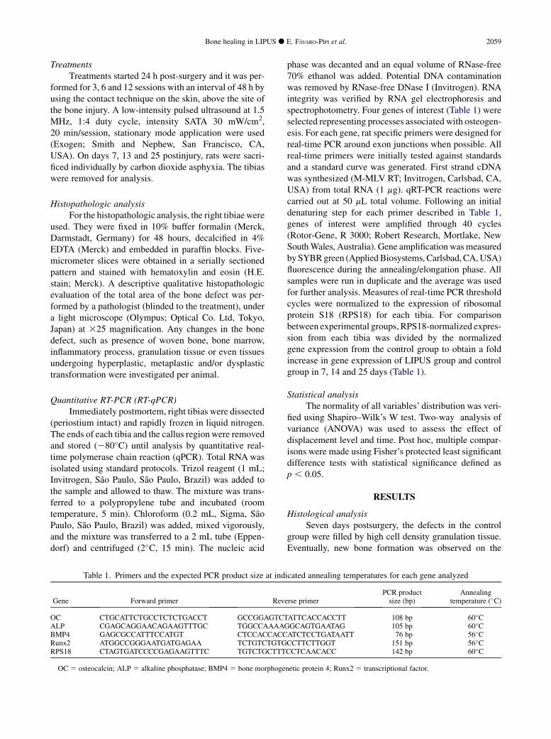
Table 1. Primers and the expected PCR product size at ind
Gene Forward primer Reve
OC CTGCATTCTGCCTCTCTGACCT GCCGGAGTCTALP CGAGCAGGAACAGAAGTTTGC TGGCCAAAAGBMP4 GAGCGCCATTTCCATGT CTCCACCACCRunx2 ATGGCCGGGAATGATGAGAA TCTGTCTGTGRPS18 CTAGTGATCCCCGAGAAGTTTC TGTCTGCTTT
OC 5 osteocalcin; ALP 5 alkaline phosphatase; BMP4 5 bone morphoge
phase was decanted and an equal volume of RNase-free70% ethanol was added. Potential DNA contaminationwas removed by RNase-free DNase I (Invitrogen). RNAintegrity was verified by RNA gel electrophoresis andspectrophotometry. Four genes of interest (Table 1) wereselected representing processes associated with osteogen-esis. For each gene, rat specific primers were designed forreal-time PCR around exon junctions when possible. Allreal-time primers were initially tested against standardsand a standard curve was generated. First strand cDNAwas synthesized (M-MLV RT; Invitrogen, Carlsbad, CA,USA) from total RNA (1 mg). qRT-PCR reactions werecarried out at 50 mL total volume. Following an initialdenaturing step for each primer described in Table 1,genes of interest were amplified through 40 cycles(Rotor-Gene, R 3000; Robert Research, Mortlake, NewSouthWales, Australia). Gene amplificationwasmeasuredbySYBRgreen (AppliedBiosystems, Carlsbad, CA,USA)fluorescence during the annealing/elongation phase. Allsamples were run in duplicate and the average was usedfor further analysis. Measures of real-time PCR thresholdcycles were normalized to the expression of ribosomalprotein S18 (RPS18) for each tibia. For comparisonbetween experimental groups, RPS18-normalized expres-sion from each tibia was divided by the normalizedgene expression from the control group to obtain a foldincrease in gene expression of LIPUS group and controlgroup in 7, 14 and 25 days (Table 1).
Statistical analysisThe normality of all variables’ distribution was veri-
fied using Shapiro–Wilk’s W test. Two-way analysis ofvariance (ANOVA) was used to assess the effect ofdisplacement level and time. Post hoc, multiple compar-isons were made using Fisher’s protected least significantdifference tests with statistical significance defined asp , 0.05.
RESULTS
Histological analysisSeven days postsurgery, the defects in the control
group were filled by high cell density granulation tissue.Eventually, new bone formation was observed on the
icated annealing temperatures for each gene analyzed
rse primerPCR productsize (bp)
Annealingtemperature (�C)
ATTCACCACCTT 108 bp 60�CGCAGTGAATAG 105 bp 60�CATCTCCTGATAATT 76 bp 56�CCCTTCTTGGT 151 bp 56�CCCTCAACACC 142 bp 60�C
netic protein 4; Runx2 5 transcriptional factor.

2060 Ultrasound in Medicine and Biology Volume 36, Number 12, 2010
surface of some bone particles (Fig. 1A). Specifically, thiswas noticed in five animals. Woven bone formation wasobserved in six animals. Moreover, the animals treatedwith LIPUS also showed high cell density granulationtissue composed by collagen fibers andmesenchymal cells(Fig. 1B). Seven animals presentedwoven bone formation.
On day 14 after the surgery, the control groupdemonstrated new bone formation and remodeling bonetrabecula surrounded by granulation tissue (Fig. 2A)marked by basophilic reversal lines. Bone marrow pre-sented high vascularization as well. At the same period,animals treated with LIPUS, presented new bone forma-tion on the walls of the bone defect, surrounded by gran-ulation tissue and with deposition of primary bone(Fig. 2B). In addition, it seems that the amount of neo-formed bone was higher in the LIPUS treated group.
On day 25, mature bone trabeculas were noted fillingthe bone defect in the control group (Fig. 3A). In addition,a good deal of remodeling trabecula was visualized in thegroup treated with LIPUS. Bone marrow showed intensevascularization as well (Fig. 3B).
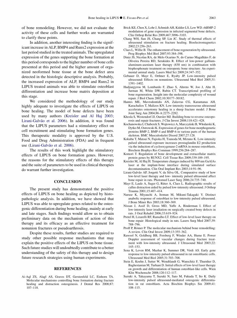
Alkaline phosphataseFigures 4, 5, 6 and 7 represent the osteogenic gene
expression in the control and LIPUS treated group.
Day 7. Of the four genes reflecting osteogeniccoupling potential in this model, BMP4, osteocalcinand Runx2 were consistently and significantly upregu-lated at this time point in the treated animals. At day 7,BMP4 expression was significantly elevated by anaverage of 4.7-fold in bone defect treated with LIPUScompared with nontreated control (Fig. 5). Osteocalcin(Fig. 6) and Runx2 (Fig. 7) expression were significantlyelevated, up by an average of 1.9-fold and 2.2-fold in
Fig. 1. Bone defects after 7 days of surgery from control grogroup treated with ultrasound showing granulation tissue
(H.E. stain
LIPUS-treated bone defect. Interestingly, the expressionof alkaline phosphatase was significantly downregulatedin the LIPUS group compared with the control (Fig. 4).
Day 13. At this intermediary point during theprocess of bone healing, no significant expression ofany of the four genes analyzed was observed after thetreatment with LIPUS compared with the nontreatedcontrol.
Day 25. On day 25, a significant increase in the ALPexpression was observed in the treated animals comparedwith the control (up by an average of 2.5-fold) (Fig. 4).Similarly, the expression of BMP4 (Fig. 5) and Runx2(Fig. 7) were also increased in the LIPUS treated groupcompared with the nontreated group. At day 25, BMP4expression was significantly elevated, up by an averageof 5.3-fold in bone defect treated with LIPUS comparedwith the nontreated control. In addition, the Runx2increased 4.5-fold.
DISCUSSION
The present study aimed to investigate the effects ofLIPUS on histologic modifications (descriptive analysis)and on the expression of osteogenic genes (alkaline phos-phatase, osteocalcin, Runx 2 and BM4) during theprocess of bone healing in tibias of rats. The histologicanalysis showed that the treated animals presented moremature trabeculas, higher amount of bone depositionand highly vascularized connective tissue, especially atthe intermediate and late stages of repair (14 and 25days after surgery) compared with the control. In addi-tion, RT-qPCR analysis showed that, at day 7 post-surgery, LIPUS induced a significant upregulation ofthe osteogenic genes. Interestingly, this treatment
up (a) displaying granulation tissue (asterisks); and (b)(asterisk) and early woven bone formation (arrow)310).
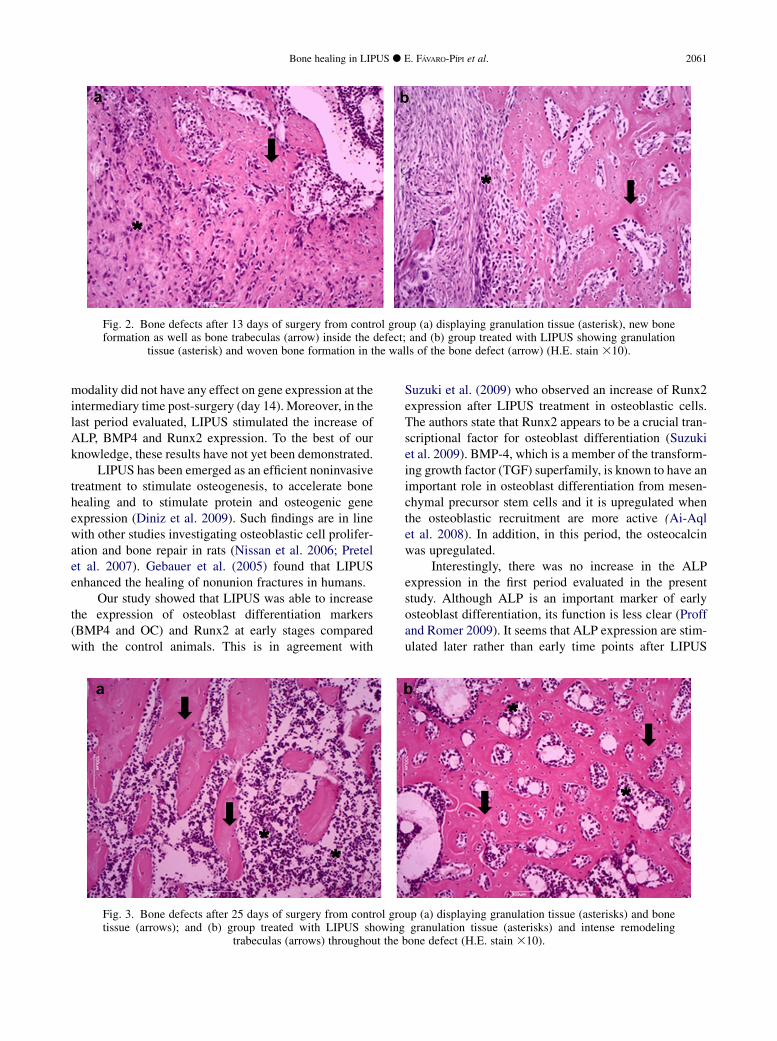
Fig. 2. Bone defects after 13 days of surgery from control group (a) displaying granulation tissue (asterisk), new boneformation as well as bone trabeculas (arrow) inside the defect; and (b) group treated with LIPUS showing granulation
tissue (asterisk) and woven bone formation in the walls of the bone defect (arrow) (H.E. stain 310).
Bone healing in LIPUS d E. FAVARO-PIPI et al. 2061
modality did not have any effect on gene expression at theintermediary time post-surgery (day 14). Moreover, in thelast period evaluated, LIPUS stimulated the increase ofALP, BMP4 and Runx2 expression. To the best of ourknowledge, these results have not yet been demonstrated.
LIPUS has been emerged as an efficient noninvasivetreatment to stimulate osteogenesis, to accelerate bonehealing and to stimulate protein and osteogenic geneexpression (Diniz et al. 2009). Such findings are in linewith other studies investigating osteoblastic cell prolifer-ation and bone repair in rats (Nissan et al. 2006; Pretelet al. 2007). Gebauer et al. (2005) found that LIPUSenhanced the healing of nonunion fractures in humans.
Our study showed that LIPUS was able to increasethe expression of osteoblast differentiation markers(BMP4 and OC) and Runx2 at early stages comparedwith the control animals. This is in agreement with
Fig. 3. Bone defects after 25 days of surgery from control grotissue (arrows); and (b) group treated with LIPUS showing
trabeculas (arrows) throughout the
Suzuki et al. (2009) who observed an increase of Runx2expression after LIPUS treatment in osteoblastic cells.The authors state that Runx2 appears to be a crucial tran-scriptional factor for osteoblast differentiation (Suzukiet al. 2009). BMP-4, which is a member of the transform-ing growth factor (TGF) superfamily, is known to have animportant role in osteoblast differentiation from mesen-chymal precursor stem cells and it is upregulated whenthe osteoblastic recruitment are more active (Ai-Aqlet al. 2008). In addition, in this period, the osteocalcinwas upregulated.
Interestingly, there was no increase in the ALPexpression in the first period evaluated in the presentstudy. Although ALP is an important marker of earlyosteoblast differentiation, its function is less clear (Proffand Romer 2009). It seems that ALP expression are stim-ulated later rather than early time points after LIPUS
up (a) displaying granulation tissue (asterisks) and bonegranulation tissue (asterisks) and intense remodeling
bone defect (H.E. stain 310).
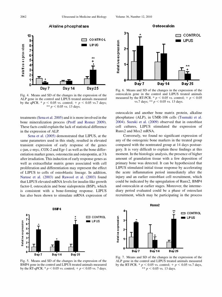
Fig. 4. Means and SD of the changes in the expression of theALP gene in the control and LIPUS treated animals measuredby the qPCR. * p , 0.05 vs. control; 1 p , 0.05 vs.7 days;
** p , 0.05 vs. 13 days.
Fig. 6. Means and SD of the changes in the expression of theosteocalcin gene in the control and LIPUS treated animalsmeasured by the RT-PCR. * p , 0.05 vs. control; 1 p , 0.05
vs.7 days; ** p , 0.05 vs. 13 days.
2062 Ultrasound in Medicine and Biology Volume 36, Number 12, 2010
treatments (Sena et al. 2005) and it is more involved in thebone mineralization process (Proff and Romer 2009).These facts could explain the lack of statistical differencein the expression of ALP.
Sena et al. (2005) demonstrated that LIPUS, at thesame parameters used in this study, resulted in elevatedtransient expression of early response of the genesc-jun, c-myc, COX-2 and Egr-1 as well as the bone differ-entiation marker genes, osteonectin and osteopontin, at 3 hafter irradiation. This induction of early response genes aswell as extracellular matrix genes associated with cellproliferation and differentiation may represent the effectof LIPUS to cells of osteoblastic lineage. In addition,Naruse et al. (2003) and Rawool et al. (2003) foundthat LIPUS elevated mRNA levels for insulin-like growthfactor-I, osteocalcin and bone sialoprotein (BSP), whichis consistent with a bone-forming response. LIPUShas also been shown to stimulate mRNA expression of
Fig. 5. Means and SD of the changes in the expression of theBMP4 gene in the control and LIPUS treated animals measuredby the RT-qPCR. * p, 0.05 vs. control;1 p, 0.05 vs. 7 days.
osteocalcin and another bone matrix protein, alkalinephosphatase (ALP), in UMR-106 cells (Tsumaki et al.2004). Suzuki et al. (2009) observed that in osteoblastcell cultures, LIPUS stimulated the expression ofRunx2 and Msx2 mRNA.
Conversely, we found no significant expression ofany of the osteogenic bone markers in the treated groupcompared with the nontreated group at 14 days postsur-gery. It is very difficult to explain these findings at thismoment. In the histologic analysis, the presence of higheramount of granulation tissue with a few deposition ofprimary bone was detected. It can be hypothesized thatLIPUS stimulated initial tissue response by acceleratingthe acute inflammation period immediately after theinjury and an earlier osteoblast cell recruitment, whichcould be indicated by the upregulation of Runx2, BMP4and osteocalcin at earlier stages. Moreover, the interme-diary period evaluated could be a phase of osteoclastrecruitment, which may be participating in the process
Fig. 7. Means and SD of the changes in the expression of theALP gene in the control and LIPUS treated animals measuredby the RT-PCR. * p , 0.05 vs. control; 1 p , 0.05 vs.7 days,
** p , 0.05 vs. 13 days.
Bone healing in LIPUS d E. FAVARO-PIPI et al. 2063
of bone remodeling. However, we did not evaluate theactivity of these cells and further works are warrantedto clarify these points.
In addition, another interesting finding is the signif-icant increase in ALP, BMP4 and Runx2 expression at thelast period studied in the treated animals. The upregulatedexpression of the genes supporting the bone formation atthis period corresponds to the higher number of bone cellspresented at this period and the higher amount of orga-nized neoformed bone tissue at the bone defect areadetected in the histologic descriptive analysis. Probably,the increased expression of ALP, BMP4 and Runx2 inLIPUS treated animals was able to stimulate osteoblastdifferentiation and increase bone matrix deposition atthis stage.
We considered the methodology of our studyhighly adequate to investigate the effects of LIPUS onbone healing. The model of bone defects have beenused by many authors (Kreisler and Al Haj 2003;Lirani-Galvao et al. 2006). In addition, it was foundthat the LIPUS parameters had a stimulatory effect oncell recruitment and stimulating bone formation genes.This therapeutic modality is approved by the U.S.Food and Drug Administration (FDA) and in frequentuse (Lirani-Galvao et al. 2006).
The results of this work highlight the stimulatoryeffects of LIPUS on bone formation genes. However,the reasons for the stimulatory effects of this therapyand the suite of parameters to be used in clinical therapiesdo warrant further investigation.
CONCLUSION
The present study has demonstrated the positiveeffects of LIPUS on bone healing as depicted by histo-pathologic analysis. In addition, we have showed thatLIPUS was able to upregulate genes related to the osteo-genic differentiation during bone healing, mainly at earlyand late stages. Such findings would allow us to obtainpreliminary data on the mechanism of action of thistherapy and its efficacy as an effective treatment fornonunion fractures or pseudoarthrosis.
Despite these results, further studies are required tostudy other possible response mechanisms that mayexplain the positive effects of the LIPUS on bone tissue.Such future studies will undoubtedly contribute to a betterunderstanding of the safety of this therapy and to designfuture research strategies using human experiments.
REFERENCES
Ai-Aql ZS, Alagl AS, Graves DT, Gerstenfeld LC, Einhorn TA.Molecular mechanisms controlling bone formation during fracturehealing and distraction osteogenesis. J Dental Res 2008;87:107–118.
Brick KE, Chen X, Lohr J, Schmidt AH, Kidder LS, LewWD. rhBMP-2modulation of gene expression in infected segmental bone defects.Clin Orthop Relat Res 2009;467:3096–3103.
Chang WH, Sun JS, Chang SP, Lin JC. Study of thermal effects ofultrasound stimulation on fracture healing. Bioelectromagnetics2002;23:256–263.
Claes L,Willie B. The enhancement of bone regeneration by ultrasound.Prog Biophys Mol Biol 2007;93:384–398.
Diniz JS, Nicolau RA, de Melo Ocarino N, do Carmo Magalhaes F, deOliveira Pereira RD, Serakides R. Effect of low-power gallium-aluminum-asenium laser therapy (830 nm) in combination withbisphosphonate treatment on osteopenic bone structure: An experi-mental animal study. Lasers Med Sci 2009;24:347–352.
Gebauer D, Mayr E, Orthner E, Ryaby JP. Low-intensity pulsedultrasound: Effects on nonunions. Ultrasound Med Biol 2005;31:1391–1402.
Hadjiargyrou M, Lombardo F, Zhao S, Ahrens W, Joo J, Ahn H,Jurman M, White DW, Rubin CT. Transcriptional profiling ofbone regeneration. Insight into the molecular complexity of woundrepair. J Biol Chem 2002;10:30177–30182.
Hantes ME, Mavrodontidis AN, Zalavras CG, Karantanas AH,Karachalios T, Malizos KN. Low-intensity transosseous ultrasoundaccelerates osteotomy healing in a sheep fracture model. J BoneJoint Surg Am 2004;86-A:2275–2282.
Khosla S, Westendorf JJ, Oursler MJ. Building bone to reverse osteopo-rosis and repair fractures. J Clin Invest 2008;118:421–428.
Kochanowska I, Chaberek S,Wojtowicz A,Marczy�nski B,W1odarski K,DytkoM, Ostrowski K. Expression of genes for bonemorphogeneticproteins BMP-2, BMP-4 and BMP-6 in various parts of the humanskeleton. BMC Musculoskelet Disord 2007;27:128.
Kokubu T, Matsui N, Fujioka H, Tsunoda M, Mizuno K. Low-intensitypulsed ultrasound exposure increases prostaglandin E2 productionvia the induction of cyclooxygenase-2 mRNA in mouse osteoblasts.Biochem Biophys Res Commun 1999;256:284–287.
Komori T. Regulation of bone development and extracellular matrixprotein genes by RUNX2. Cell Tissue Res 2009;339:189–195.
Kreisler M, Al Haj H. Temperature changes induced by 809 nmGaAlAslaser at the implant-bone interface during simulated surfacedecontamination. Clin Oral Implant Res 2003;14:91–96.
Lirani-Galvao AP, Jorgetti V, da Silva OL. Comparative study of howlow-level laser therapy and low- intensity pulsed ultrasound affectbone repair in rats. Photomed Laser Surg 2006;24:735–740.
Mayr E, Laule A, Suger G, Ruter A, Claes L. Radiographic results ofcallus distraction aided by pulsed low-intensity ultrasound. J OrthopTrauma 2001;15:407–414.
Naruse K, Miyauchi A, Itoman M, Mikuni-Takagaki Y. Distinctanabolic response of osteoblast to low-intensity pulsed ultrasound.J Bone Miner Res 2003;18:360–369.
Nissan J, Assif D, Gross MD, Yaffe A, Binderman I. Effect oflow-intensity laser irradiation on surgically created bony defects inrats. J Oral Rehabil 2006;33:619–924.
Pretel H, Lizarelli RF, Ramalho LT. Effect of low-level laser therapy onbone repair: Histological study in rats. Lasers Surg Med 2007;39:788–796.
Proff P, Romer P. The molecular mechanism behind bone remodelling:A review. Clin Oral Invest 2009;13:355–362.
Rawool N, Goldberg BB, Forsberg F, Winder AA, Hume E. PowerDoppler assessment of vascular changes during fracture treat-ment with low-intensity ultrasound. J Ultrasound Med 2003;22:145–153.
Sena K, Leven RM, Mazhar K, Summer DR, Virdi AS. Early generesponse to low-intensity pulsed ultrasound in rat osteoblastic cells.Ultrasound Med Biol 2005;31:703–708.
Stein E, Koehn J, Sutter W, Wendtlandt G, Wanschitz F, Thurnher D,BaghestanianM, Turhani D. Initial effects of low-level laser therapyon growth and differentiation of human osteoblast-like cells. WienKlin Wochenschr 2008;120:112–117.
Suzuki A, Takayama T, Suzuki N, Sato M, Fukuda T, Ito K. Dailylow-intensity pulsed ultrasound-mediated osteogenic differentia-tion in rat osteoblasts. Acta Biochim Biophys Sin 2009;41:108–115.

2064 Ultrasound in Medicine and Biology Volume 36, Number 12, 2010
Tsumaki N, Kakiuchi M, Sasaki J, Ochi T, Yoshikawa H. Low-intensity pulsed ultrasound accelerates maturation of callus inpatients treated with opening-wedge high tibial osteotomy byhemicallotasis. J Bone Joint Surg Am 2004;86-A:2399–2405.
Wang FS, Yang KD, Kuo YR, Wang CJ, Sheen-Chen SM, Huang HC,Chen YJ. Temporal and spatial expression of bone morphogenetic
proteins in extracorporeal shock wave-promoted healing ofsegmental defect. Bone 2003;32:387–396.
Yang LW, Zhang JX, Zeng L, Xu JJ, Du FT, Luo W, Luo ZJ, Jiang JH.Vascular endothelial growth factor gene therapy with intramuscularinjections of plasmid DNA enhances the survival of randompattern flaps in a rat model. Br J Plast Surg 2005;58:339–347.