Low environmental calcium blocks long-term memory formation in a freshwater pulmonate snail
12
This article appeared in a journal published by Elsevier. The attached copy is furnished to the author for internal non-commercial research and education use, including for instruction at the authors institution and sharing with colleagues. Other uses, including reproduction and distribution, or selling or licensing copies, or posting to personal, institutional or third party websites are prohibited. In most cases authors are permitted to post their version of the article (e.g. in Word or Tex form) to their personal website or institutional repository. Authors requiring further information regarding Elsevier’s archiving and manuscript policies are encouraged to visit: http://www.elsevier.com/copyright
Transcript of Low environmental calcium blocks long-term memory formation in a freshwater pulmonate snail
This article appeared in a journal published by Elsevier. The attachedcopy is furnished to the author for internal non-commercial researchand education use, including for instruction at the authors institution
and sharing with colleagues.
Other uses, including reproduction and distribution, or selling orlicensing copies, or posting to personal, institutional or third party
websites are prohibited.
In most cases authors are permitted to post their version of thearticle (e.g. in Word or Tex form) to their personal website orinstitutional repository. Authors requiring further information
regarding Elsevier’s archiving and manuscript policies areencouraged to visit:
http://www.elsevier.com/copyright

Author's personal copy
Low environmental calcium blocks long-term memory formation in afreshwater pulmonate snail
Sarah Dalesman ⇑, Marvin H. Braun, Ken LukowiakHotchkiss Brain Institute, Department of Physiology and Pharmacology, University of Calgary, Calgary, Alberta, Canada T2N 4N1
a r t i c l e i n f o
Article history:Received 28 July 2010Revised 17 November 2010Accepted 28 November 2010Available online 2 December 2010
Keywords:Environmental calciumLearningLong-term memoryLymnaea stagnalisPulmonate snail
a b s t r a c t
The freshwater snail Lymnaea stagnalis (L.) is considered a calciphile and exhibits reduced growth andsurvival in environments containing less than 20 mg/l environmental calcium. Although it has no appar-ent effect on survival at 20 mg/l, reducing environmental calcium increases metabolic demand, and assuch we consider that this level of calcium acts as a stressor on the snail. We exposed snails to acute peri-ods of low environmental calcium and tested their ability to form intermediate-term memory (ITM) andlong-term memory (LTM) following one trial operant conditioning (1TT) to reduce aerial respiratoryactivity in hypoxic conditions. We also assessed whether there were changes in the electrophysiologicalproperties of a single neuron, right pedal dorsal 1 (RPeD1), which has been demonstrated to be necessaryfor LTM formation. Following training in high (80 mg/l) environmental calcium, L. stagnalis formed ITMand LTM lasting 24 h and demonstrated a significant reduction in all activity measured from RPeD1; how-ever when snails were exposed to low (20 mg/l) environmental calcium they were able to form ITM butnot LTM. Although no behavioral LTM was formed, a partial reduction in RPeD1 activtiy measured 24 hafter training was observed, indicating a residual effect of training. The strong effect that environmentalcalcium concentration had on physiology and behavior in response to training to reduce aerial respirationin L. stagnalis suggests that it is an element of gastropod husbandry that needs to be carefully consideredwhen studying other traits. This study also indicates that L. stagnalis found naturally in low calcium envi-ronments may be less able to adapt to novel stressors than populations found in harder waters.
� 2010 Elsevier Inc. All rights reserved.
1. Introduction
Memory formation is a dynamic process that can be modulatedby stressful events occurring during, before or after the learningprocess. The effect that stressors have on memory formation variesgreatly depending on the type of stress experienced, the level ofstress applied and the timing of the stressful event relative to thelearning process, and can either enhance or block memory forma-tion (Shors, 2004). The freshwater snail, Lymnaea stagnalis, pro-vides an ideal model species to study learning and memory. Athree-neuron central pattern generator drives aerial respirationin this species (Syed, Bulloch, & Lukowiak, 1990; Syed, Ridgway,
Lukowiak, & Bulloch, 1992), and this behavior can be operantlyconditioned to result in long-term memory (LTM) formation to re-duce aerial respiration in hypoxia (Lukowiak, Ringseis, Spencer,Wildering, & Syed, 1996). This memory formation is flexible andcan be both enhanced and blocked using environmentally relevantstressors, the timing of which relative to the learning procedure iscritical (Lukowiak et al., 2010). For example, cooling immediatelyfollowing a learning procedure blocks both intermediate-termmemory (ITM) and LTM, but has no effect if applied 10–15 min fol-lowing the training procedure (Sangha, Morrow, Smyth, Cooke, &Lukowiak, 2003). Conversely, detection of a sympatric predatorduring the training procedure can enhance the formation of LTM(Orr, Hittel, & Lukowiak, 2009; Orr & Lukowiak, 2008).
L. stagnalis is considered a calciphile, normally requir-ing P 20 mg/l environmental calcium to survive and prosper innatural populations (Boycott, 1936; Madsen, 1987; Young,1975b). Where L. stagnalis populations exist in areas with lowerlevels of environmental calcium, abundance is greatly reducedand no large adults were found, suggesting poor growth and sur-vival (McKillop & Harrison, 1972). Low calcium environments alsoresult in shell thinning in mollusks, potentially making them easierprey and less resistant to damage (Boycott, 1936; Glass & Darby,
1074-7427/$ - see front matter � 2010 Elsevier Inc. All rights reserved.doi:10.1016/j.nlm.2010.11.017
Abbreviations: LTM, long-term memory; ITM, intermediate-term memory; 1TT,one trial operant conditioning; RPeD1, right pedal dorsal 1; CPG, central patterngenerator.⇑ Corresponding author. Address: Hotchkiss Brain Institute, Department of
Physiology and Pharmacology, University of Calgary, Room 2104, Health SciencesCentre, 3330 Hospital Drive NW, Calgary, Alberta, Canada T2N 4N1. Fax: +1 403 2832700.
E-mail addresses: [email protected], [email protected] (S.Dalesman), [email protected] (M.H. Braun), [email protected] (K. Luko-wiak).
Neurobiology of Learning and Memory 95 (2011) 393–403
Contents lists available at ScienceDirect
Neurobiology of Learning and Memory
journal homepage: www.elsevier .com/ locate/ynlme
Author's personal copy
2009; Lewis & Magnuson, 1999; Madsen, 1987; Young, 1975a;Zalizniak, Kefford, & Nugegoda, 2009). For example, L. stagnalisdemonstrates induced shell thickening in the presence of predatorcues when sufficient environmental calcium is available (90 mg/l),but is unable to alter shell growth to the same degree in a moder-ate (45 mg/l) calcium environment (Rundle, Spicer, Coleman,Vosper, & Soane, 2004). Considering this, we would predict thatdespite populations surviving at 20 mg/l environmental calcium,the animals are not in ideal conditions and may be experiencingstress.
Whilst the effects of long-term exposure to low calcium levelson morphology are fairly well understood, behavioral responsesto low environmental calcium are less well studied in freshwatergastropods. Fluctuations in the calcium concentration in freshwa-ter systems are common, and can represent 3 to 10-fold changesin environmental calcium concentration over the period of a year(Macan, 1950; McKillop & Harrison, 1972; Williams, 1970; Dales-man unpublished data). As such, we might expect a rapid behav-ioral response to changes in the external environmental calciumconcentration in addition to long term developmental responses.Aquatic gastropods are able to maintain haemolymph calcium lev-els in a low calcium environment (Dewith, Kok, & Vanderschors,1987; Greenaway, 1971; Grosell & Brix, 2009), even in the com-plete absence of environmental calcium for at least 10 days(Dewith, 1977). However, calcium uptake requires energy in lowexternal calcium concentrations. Passive transport of calcium fromthe external media can occur above 20 mg/l. Below 20 mg/l envi-ronmental calcium concentration in the external environment islower than the snails internal calcium concentration, so all calciumuptake requires energy (Greenaway, 1971). Following acute expo-sure to low environmental calcium (i.e., 1 week), cutaneous respi-ration is increased and motility is decreased at 20 mg/l relative to80 mg/l, indicating an increased basal metabolic rate in 20 mg/lenvironmental calcium, and a reduction in energy available forlocomotion (Dalesman & Lukowiak, 2010). There is also evidencethat L. stagnalis can sense environmental calcium concentrations,orientating toward higher calcium environments when given achoice (Piggott & Dussart, 1995). Consequently, we hypothesizethat L. stagnalis will respond rapidly to low calcium concentrationas an environmental stressor, and that this stress will modulatememory formation.
Here we assessed the potential for acute exposure to low envi-ronmental calcium to modulate memory formation by L. stagnalis,testing both ITM and LTM at two environmental calcium concen-trations, low (20 mg/l) and high (80 mg/l). When we are not testingfor the effects of environmental calcium concentration our labora-tory population is maintained in 80 mg/l environmental calcium.As 1 week exposure to low calcium was sufficient to alter respira-tion and locomotory activity (Dalesman & Lukowiak, 2010), wefirst tested whether this length of exposure can also alter memoryformation. Secondly, we assessed whether the ability to form LTMis rapidly reversed following just 1 h exposure to the alternate cal-cium environment prior to and during training. In addition to test-ing memory formation, we tested whether changes occur in theunderlying neurophysiological properties of the three-neuron cen-tral pattern generator (CPG) that drives aerial respiratory behavior(Lukowiak et al., 1996, 1998, 2008; Syed, Bulloch, & Lukowiak,1992; Syed et al., 1990). Specifically, we tested for changes inone of the CPG neurons, right pedal dorsal 1 (RPeD1). This neuronhas been demonstrated to be necessary for LTM following operantconditioning using our ‘poking’ method where the pneumostome isgently prodded when the snail attempts to open it (Scheibenstock,Krygier, Haque, Syed, & Lukowiak, 2002). However, the effect ofone-trial conditioning on the electrophysiological properties of thisneuron at differing external environmental calcium concentrationshad not yet been determined.
2. Material and methods
2.1. Animal source and maintenance
Adult L. stagnalis (L.), 25 ± 1 mm spire height, were raised fromstock obtained from Vrije Universeit in Amsterdam. This popula-tion originated from wild snails collected in the 1950s from canalsin a polder located near Utrecht. Adult snails were reared in aqua-ria filled with de-chlorinated tap water, containing 60 ± 5 mg/lenvironmental calcium, in the snail rearing facility at the Univer-sity of Calgary, and were transferred 1 week prior to experimentsinto oxygenated artificial pond water (0.26 g/l Instant Ocean�,Spectrum Brands Inc., USA), with additional calcium sulfate dehy-drate added to make to low calcium (20 mg/l) or high calcium(80 mg/l) water depending on treatment group. The calcium con-centrations used were selected as they are within the natural rangeexperienced by L. stagnalis in field conditions (Boycott, 1936), andhave been shown to result in variation in respiration and motilityfollowing acute exposure (Dalesman & Lukowiak, 2010). Snailswere maintained at room temperature (20 ± 1 �C) at a stockingdensity of one snail per liter and fed romaine lettuce ad libitum. Ro-maine lettuce has been used as a food source to successfully rearsnails at this facility for several years, and although it contains asource of calcium that the snails would be able to utilize, the cal-cium content is fairly low (0.36 mg Ca2+ per gram of lettuce), andprevious work has suggested that L. stagnalis obtains the majorityof its calcium requirements from the water (Van Der Borght & VanPuymbroek, 1966).
2.2. Behavioral measurements
2.2.1. Breathing observationL. stagnalis are bi-modal breathers, able to absorb oxygen di-
rectly from the water via cutaneous respiration and also to respireaerially using a rudimentary lung opened to the atmosphere viathe pneumostome (respiratory orifice). In eumoxic conditions,the primary form of respiration is cutaneous, however if oxygensaturation in the water is diminished the snails will switch to usingaerial respiration. Pneumostome opening is an easily observableand recordable behavior, and duration of time spent aerially respir-ing can be increased by making the water hypoxic (Lukowiak et al.,1996).
Breathing observations were carried out in the same way in allexperiments. 500 ml of either high or low environmental calciumpond water was placed in a 1 l beaker and made hypoxic(PO2 < 931 Pa) by bubbling N2 vigorously through the water for20 min before the introduction of snails. N2 bubbling was contin-ued throughout the observation period to maintain hypoxic condi-tions, though at a reduced rate so as not to disturb aerial breathingbehavior. Snails were acclimated to the beaker for 10 min prior toobservation, then we recorded the total time spent aerially respir-ing (i.e., time when the pneumostome is open to the atmosphere)over a period of 30 min. Pre-observations were carried out 24 hprior to training to provide a baseline aerial breathing rate for eachsnail, which were compared with post-training observations to as-sess whether learning and memory formation had taken place.Post-training observations were carried out either 3 h followingtraining to assess whether intermediate-term memory (ITM) hadbeen formed, or 24 h or longer to assess whether long-term mem-ory (LTM) had been formed (Lukowiak, Adatia, Krygier, & Syed,2000; Martens et al., 2007).
2.2.2. Training protocolL. stagnalis can be trained to reduce pneumostome breathing
time in hypoxic conditions, both by operant training where the
394 S. Dalesman et al. / Neurobiology of Learning and Memory 95 (2011) 393–403
Author's personal copy
pneumostome is gently prodded each time it is opened (Lukowiaket al., 1996, 1998, 2000), and via a one-trial conditioning procedure(1TT) detailed below (Martens et al., 2007). In this study, we usedthe 1TT conditioning procedure to assess memory capabilities atdifferent environmental calcium concentrations.
One-trial conditioning uses the pairing of a stressful stimulus, inthis case a 25 mM KCl bath, with the contingent occurrence ofopening the pneumostome, to train the snail to reduce this action(Martens et al., 2007). Training was carried out in all cases 24 hafter initial baseline breathing observations were made. N2 wasvigorously bubbled through 500 ml of artificial pond water for20 min to produce hypoxic conditions. In the 1TT training sessionthe snails were placed into the hypoxic pond water and movedto a 25 mM KCl bath when the first pneumostome opening attemptwas made. They were held for 30 s in the KCl bath, and then re-turned to eumoxic pond water conditions in their home aquaria.This was then followed by testing in hypoxic pond water for mem-ory, either 3 h for ITM or P24 h for LTM, after the trainingprocedure.
Yoked controls, during the first set of experiments, were carriedout to assess whether the experience of the KCl bath had a general(i.e. non-specific sensitizing) effect of altering aerial respiratorybehavior. Breathing observations were carried out 24 h beforeand also 3 h and 24 h following the yoked control procedure. Theyoked controls were carried out under identical conditions to thetraining outlined above, except in this case exposure to the KClbath occurred when the snail to which they were yoked (pairedwith) opened its pneumostome. That is, placement in the KCl bathwas not contingent on the snail opening its pneumostome. There-fore the yoked control snail did not associate opening its pneumos-tome with the KCl stressor.
The snail was considered to have formed memory if the totalbreathing time in the post-training observation period was signif-icantly reduced relative to the pre-training observation period, andalso significantly lower than yoked controls. If the training proce-dure was the cause of the alteration in breathing, the yoked con-trols should not differ significantly between the pre-training andpost-training observations.
2.2.3. Effect of calcium concentration on ITM and LTMTo test for the effect of exposure of L. stagnalis to different envi-
ronmental calcium concentrations on memory formation, wetransferred adult snails into aquaria containing artificial pondwater with either low (20 mg/l) or high (80 mg/l) environmentalcalcium for 1 week prior to testing. Snails were then trained inthe calcium concentration in which they had been held using the1TT conditioning with 25 mM KCl as outlined above (Martenset al., 2007). Snails were returned to their eumoxic aquaria be-tween pre-training observation and training, and again followingtraining until they were tested for memory. We tested snails forITM at 3 h and LTM at 24 h after the 1TT training session usingthe standard observation protocol outlined above. At the sametime as we trained snails, we also carried out the yoked controlprocedure on snails that had been held at each calcium concentra-tion. As memory was formed in the high (i.e. 80 mg/l) calciumgroup but not in the low (i.e. 20 mg/l) calcium group at 24 h, wecarried out a further trial testing for memory at 48 h in highcalcium.
A further experiment (i.e., a 1 h change of the calcium concen-tration) was carried out to assess whether the effect of calciumconcentration on LTM at 24 h could be rapidly reversed. Here snailswere again held at either high (80 mg/l) or low (20 mg/l) calciumconcentration for 1 week prior to the experiment. Pre-trainingobservations were carried out in the initial calcium concentrationin which they had held, and they were then maintained in that cal-cium concentration up to 1 h before training. One hour before
training they were transferred into the alternate new calcium envi-ronment, such that snails that had been held in high environmentalcalcium (80 mg/l) were moved into low environmental calcium(20 mg/l), and snails that had previously been held in low calciumwere moved into high calcium. One-trial conditioning was thencarried out in the new environmental calcium concentration. Fol-lowing the 1TT in the ‘new’ environmental calcium concentration,snails were then transferred back into their initial calcium environ-ment (i.e., that in which they had been held for the previous week).Twenty-four hours following the 1TT training session, snails werethen tested in their initial calcium environment to assess whetherthey had formed LTM.
2.2.4. Data analysis for behavioral trialsData were analyzed using repeated measures ANOVA in SPSS
17.0 (SPSS Inc., Chicago, IL, USA). ITM and LTM, following 1 weekexposure to low or high calcium environments, were tested usingrepeated measures ANOVA, with observation period (pre-trainingvs. post-training) used as the within-subject factor, and calcium le-vel the snails had been maintained at (low vs. high) and trainingprocedure (trained vs. yoked) as between subject factors. LTM at48 h in high calcium was tested separately using a paired t-testin SPSS. A repeated measures ANOVA was used to test for rapidreversibility of the effect of calcium concentration, with observa-tion period (pre-training vs. post-training) used as the within-sub-ject factor, and calcium level the snails experienced during training(low vs. high) as the between subject factor.
All repeated measures data were tested for equality of varianceusing Mauchly’s test for sphericity. Post-hoc paired t-tests with theP-value corrected for multiple comparisons were used to assesswhere significant differences were found within-subject.
2.3. Electrophysiological measurements
2.3.1. Electrophysiological recording from RPeD1Snails were exposed to either high (80 mg/l) or low (20 mg/l)
environmental calcium for 1 week prior to electrophysiologicaldata collection, as outlined above. On the first day a baselinebreathing rate was measured, followed 24 h later by the 1TT proce-dure (i.e., the identical training procedure to that used for behaviormeasures). Similarly to behavioral tests for LTM, trained snails andyoked controls were used to assess electrophysiological activity at24 h post-training, when LTM would be expected to have formed insnails following 1TT (Martens et al., 2007). Naïve snails were alsoassessed to test for effects of calcium concentration alone on elec-trophysiological activity.
Semi-intact preparations were used to measure RPeD1 activity,and were prepared following protocols previously developed in ourlaboratory (McComb, Rosenegger, Varshney, Kwok, & Lukowiak,2005; Orr, El-Bekai, Lui, Watson, & Lukowiak, 2007; Orr &Lukowiak, 2008; Orr et al., 2009). The level of the bathing solutionwas such that the pneumostome was just at the surface, as sub-mersion prevents pneumostome opening from occurring. Semi-intact preparations were rested for 20 min prior to impaling RPeD1with a sharp glass microelectrode filled with saturated K2SO4
solution (tip resistances ranged from 20 to 75 MX), then given afurther 15 min stabilization prior to collection of electrophysiolog-ical data, for a total time of 35 min between dissection and datacollection. Intracellular signals were amplified using a NeurodataInstruments IR283 amplifier (New York, NY, USA) and displayedsimultaneously on a Macintosh PowerLab/4SP (ADInstrumentsInc, Colorado Springs, CO, US) and a Hitachi oscilloscope (Tokyo,Japan). Recordings were stored and analyzed via Chart 5 software(AD Instruments Inc., Colorado Springs, CO, US) using a 600 s tracefor data analysis. Electrode balance was measured at the start and
S. Dalesman et al. / Neurobiology of Learning and Memory 95 (2011) 393–403 395
Author's personal copy
end of each experiment, and if the resistance had changed by morethan 5% the trace was subsequently discarded.
One of the measurements taken from the trace was the numberof action potential bursts fired. Previous work in our lab on isolatedneurons, isolated brains and semi-intact preparations have re-vealed that the RPeD1 bursts of action potentials (APs) which trig-ger pneumostome opening have particular properties. Therefore, aburst was defined as a period of sustained depolarization duringwhich 2 to over 20 action potentials were fired. The burst wasnot considered over until membrane potential had returned tothe resting value.
A measure of RPeD1 excitability was obtained by counting thenumber of action potentials elicited when the impaled cell was dri-ven through a series of 10 depolarizing current steps, from 0.2 to2.0 nA. Each step was 400 ms long, and the cell was allowed to re-cover for 300 ms between steps. The input resistance of RPeD1 wasmeasured by driving the impaled cell through a series of 10 hyper-polarizing current steps from �0.2 to �2.0 nA (step lengths are thesame as above), and calculating the resistance from the resultingcurrent–voltage relationships according to Ohm’s law (R = V/I). Pi-lot studies showed that these measurements of excitability andresistance were very stable and reproducible, with negligiblechanges between trials. For this reason, most experiments only uti-lized two or three replicates (separated by �30 s) to make certainthat the values were not changing.
2.3.2. Data analysis for electrophysiological recordingThe number of action potential bursts recorded from RPeD1 in
600 s, the number of action potentials elicited in response to the10 depolarizing current steps and input resistance were includedin the analysis of electrophysiological data. Due to heterogeneity
of variance between treatment groups, data were log-transformed(X + 1log10) prior to analysis. Log-transformed data were analyzedusing a one-way ANOVA (SPSS 17.0). Comparisons were madebetween the following conditions: naïve in high environmentalcalcium (N = 12); naïve in low environmental calcium (N = 10);trained in high environmental calcium (N = 9); trained in low envi-ronmental calcium (N = 9); yoked high environmental calcium(N = 12); yoked low environmental calcium (N = 9). Student–New-man–Keuls tests were used to carry out pair-wise post-hoc com-parisons where overall differences were found. The restingmembrane potential was also compared among treatment groupsusing a one-way ANOVA in SPSS, with treatment group as thefactor in the analysis.
3. Results
3.1. Behavioral results
We have previously demonstrated that low (20 mg/l) environ-mental calcium significantly affects respiration and locomotion inL. stagnalis (Dalesman & Lukowiak, 2010). We have also shown thatthe 1TT procedure using KCl as a stressor normally results in bothITM at 3 h and LTM at 24 h (Martens et al., 2007). Here we exam-ined the effect of low environmental (20 mg/l) calcium, which is anecologically relevant calcium concentration for our species, on theability of L. stagnalis to form ITM and LTM.
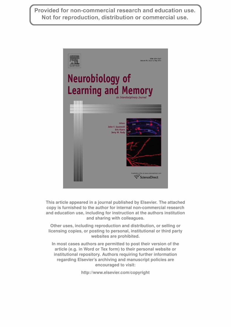
First we examined how exposure to a high (80 mg/l) or low(20 mg/l) calcium environment for a period of 1 week affected bothITM, which is dependent on new protein synthesis alone, and LTM,which is dependent on both new protein synthesis and alteredgene activity (Sangha, Scheibenstock, McComb, & Lukowiak,
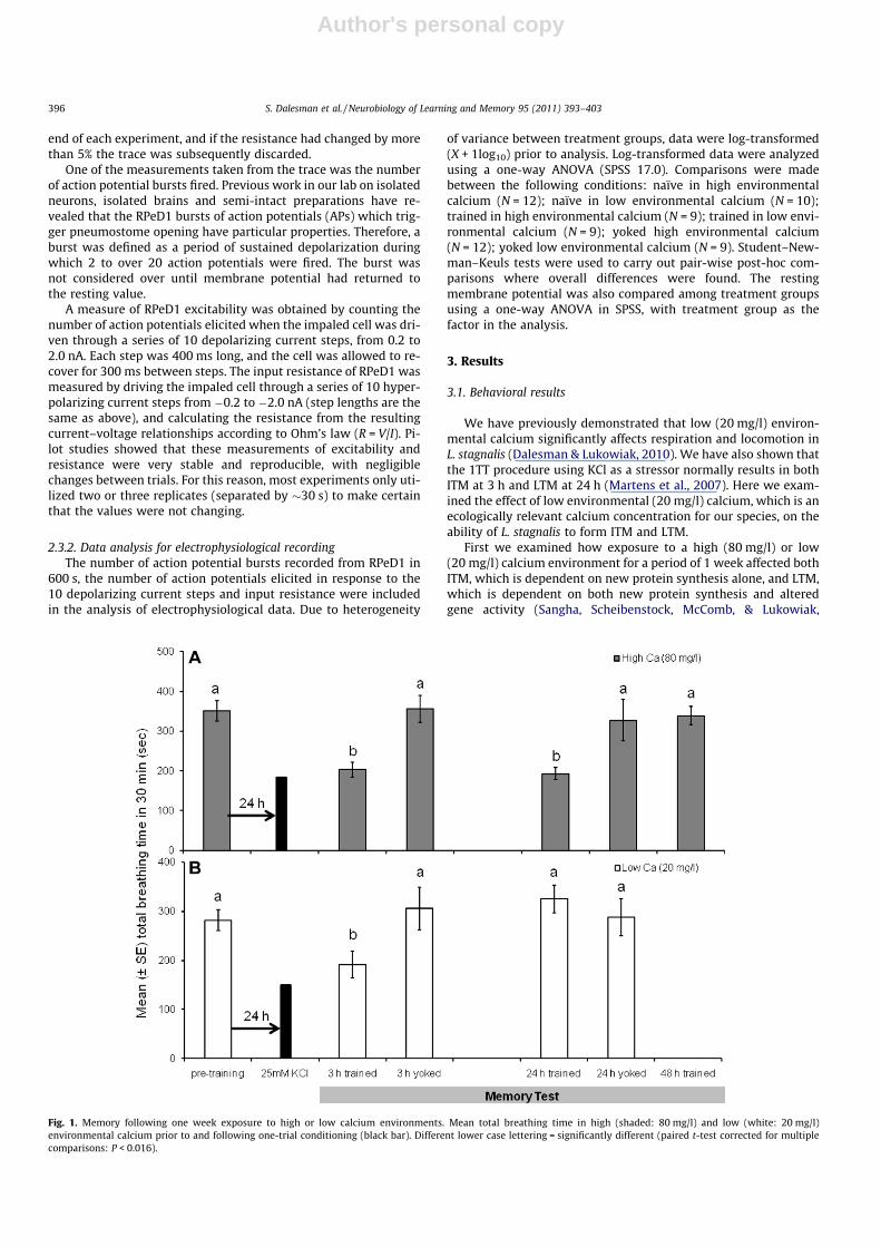
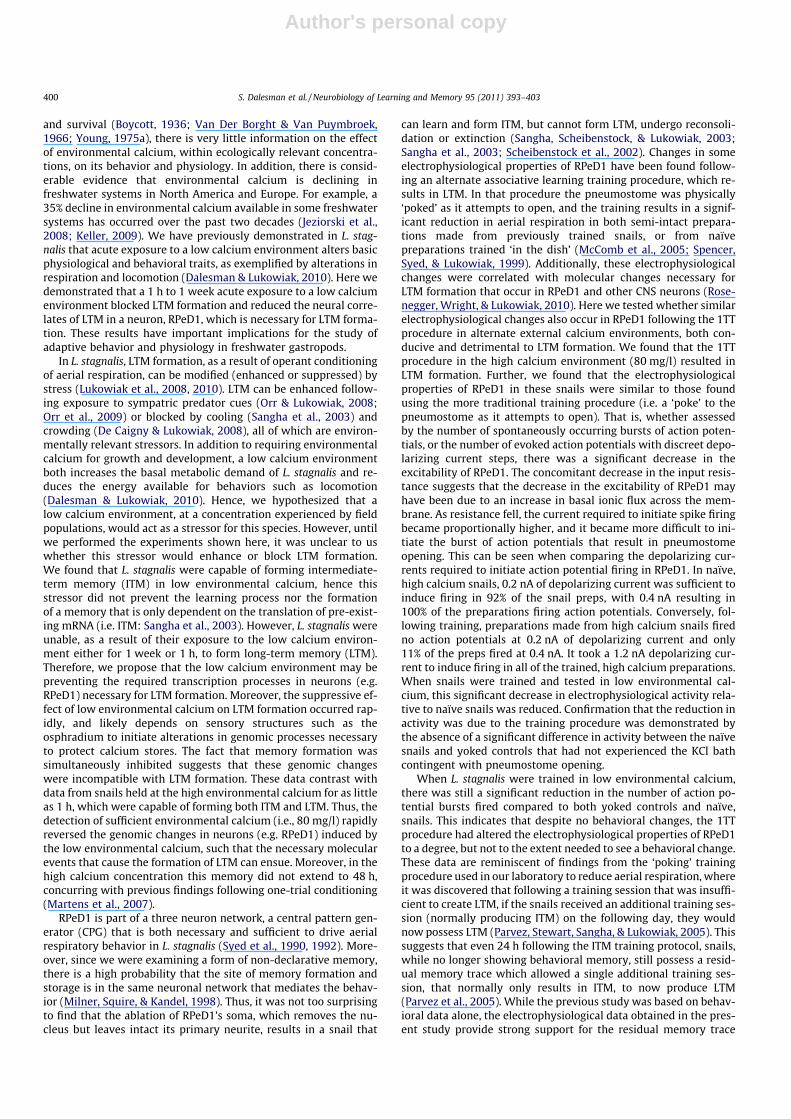
Fig. 1. Memory following one week exposure to high or low calcium environments. Mean total breathing time in high (shaded: 80 mg/l) and low (white: 20 mg/l)environmental calcium prior to and following one-trial conditioning (black bar). Different lower case lettering = significantly different (paired t-test corrected for multiplecomparisons: P < 0.016).
396 S. Dalesman et al. / Neurobiology of Learning and Memory 95 (2011) 393–403
Author's personal copy
2003). ITM, 3 h post-training, was formed in both the low (20 mg/l)and high (80 mg/l) calcium environments (Fig. 1A and B; 2-wayinteraction between pre- vs. post-training observation and trainingprocedure, trained vs. yoked: F1,61 = 11.06, P = 0.002). Total breath-ing time was significantly lower at 3 h following the training pro-cedure compared to the pre-training observation (high Ca: Fig. 1A;t = 4.45, P < 0.001, N = 23; low Ca: Fig. 1B; t = 2.43, P = 0.024,N = 22). There were no significant differences between pre-trainingand post-training observations for the yoked controls.
On the other hand, LTM formation differed depending on envi-ronmental calcium concentration. As Martens et al. (2007) foundpreviously, LTM at 24 h was formed following the 1TT training pro-cedure in the high (80 mg/l) calcium environment. However, fol-lowing exposure to the low calcium (20 mg/l) environment for1 week prior to and during the 1TT, the trained snails did not formLTM (Fig. 1A and B; 3-way interaction between time, pre- vs. post-training, training procedure, trained vs. yoked and calcium concen-tration, high vs. low: F1,61 = 5.43, P = 0.023). Total breathing timefor trained snails decreased significantly between pre- and post-training in the high (80 mg/l) calcium environment (Fig. 1A;t = 4.99, P < 0.001, N = 23), but there was no significant differencebetween total breathing time pre- and post-training in the low(20 mg/l) calcium environment (Fig. 1B; t = 1.29, P = 0.211,N = 22). There were no differences between pre-training andpost-training observations for the yoked controls tested for LTM.At 48 h following 1TT training there was no longer any evidenceof memory formation in the high (80 mg/l) calcium environment(Fig. 1A), concurring with previous results where LTM in artificialpond water alone does not extend to 48 h (Martens et al., 2007).
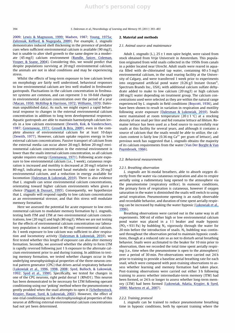
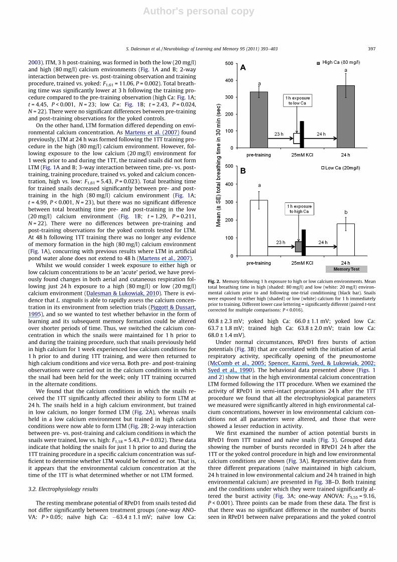
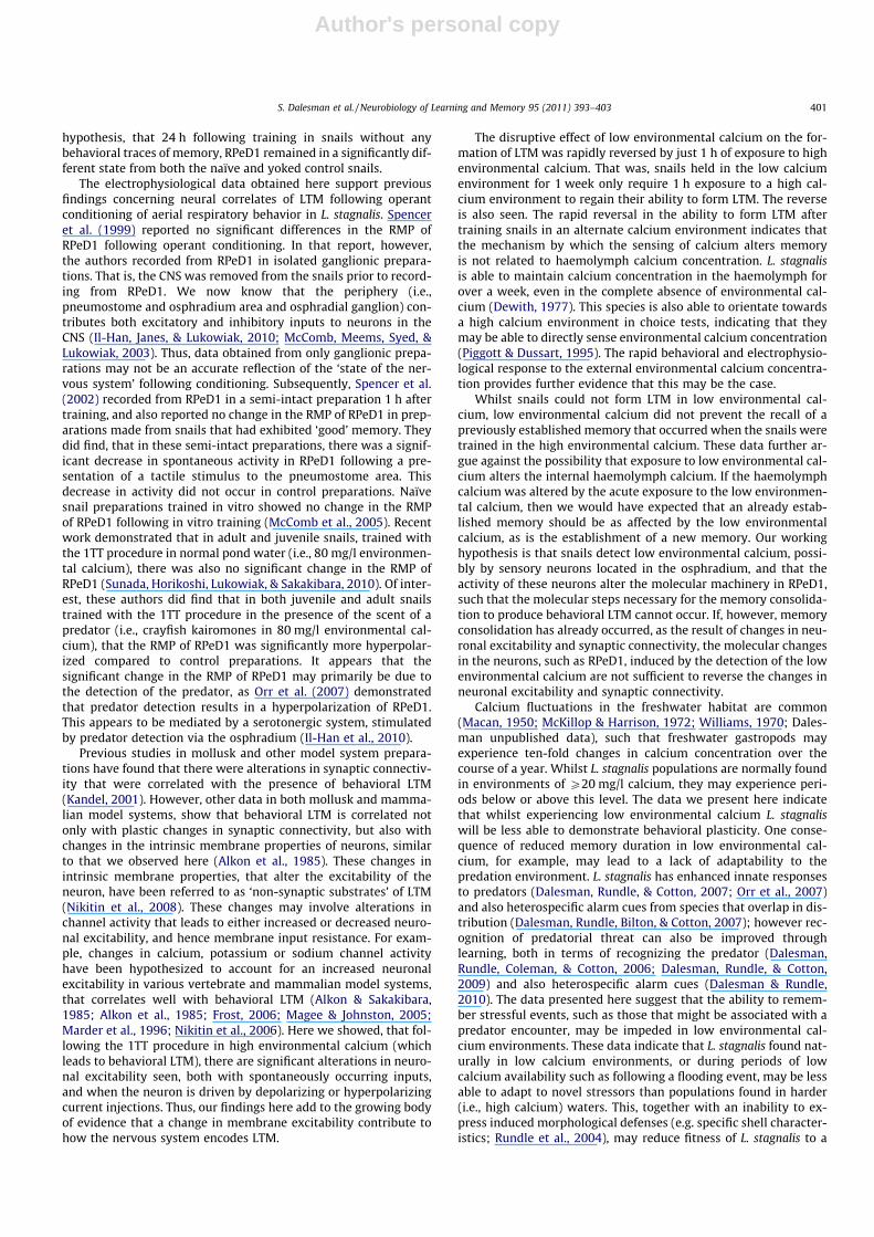
Whilst we would consider 1 week exposure to either high orlow calcium concentrations to be an ‘acute’ period, we have previ-ously found changes in both aerial and cutaneous respiration fol-lowing just 24 h exposure to a high (80 mg/l) or low (20 mg/l)calcium environment (Dalesman & Lukowiak, 2010). There is evi-dence that L. stagnalis is able to rapidly assess the calcium concen-tration in its environment from selection trials (Piggott & Dussart,1995), and so we wanted to test whether behavior in the form oflearning and its subsequent memory formation could be alteredover shorter periods of time. Thus, we switched the calcium con-centration in which the snails were maintained for 1 h prior toand during the training procedure, such that snails previously heldin high calcium for 1 week experienced low calcium conditions for1 h prior to and during 1TT training, and were then returned tohigh calcium conditions and vice versa. Both pre- and post-trainingobservations were carried out in the calcium conditions in whichthe snail had been held for the week; only 1TT training occurredin the alternate conditions.
We found that the calcium conditions in which the snails re-ceived the 1TT significantly affected their ability to form LTM at24 h. The snails held in a high calcium environment, but trainedin low calcium, no longer formed LTM (Fig. 2A), whereas snailsheld in a low calcium environment but trained in high calciumconditions were now able to form LTM (Fig. 2B; 2-way interactionbetween pre- vs. post-training and calcium conditions in which thesnails were trained, low vs. high: F1,18 = 5.43, P = 0.032). These dataindicate that holding the snails for just 1 h prior to and during the1TT training procedure in a specific calcium concentration was suf-ficient to determine whether LTM would be formed or not. That is,it appears that the environmental calcium concentration at thetime of the 1TT is what determined whether or not LTM formed.
3.2. Electrophysiology results
The resting membrane potential of RPeD1 from snails tested didnot differ significantly between treatment groups (one-way ANO-VA: P > 0.05; naïve high Ca: �63.4 ± 1.1 mV; naïve low Ca:
60.8 ± 2.3 mV; yoked high Ca: 66.0 ± 1.1 mV; yoked low Ca:63.7 ± 1.8 mV; trained high Ca: 63.8 ± 2.0 mV; train low Ca:68.0 ± 1.4 mV).
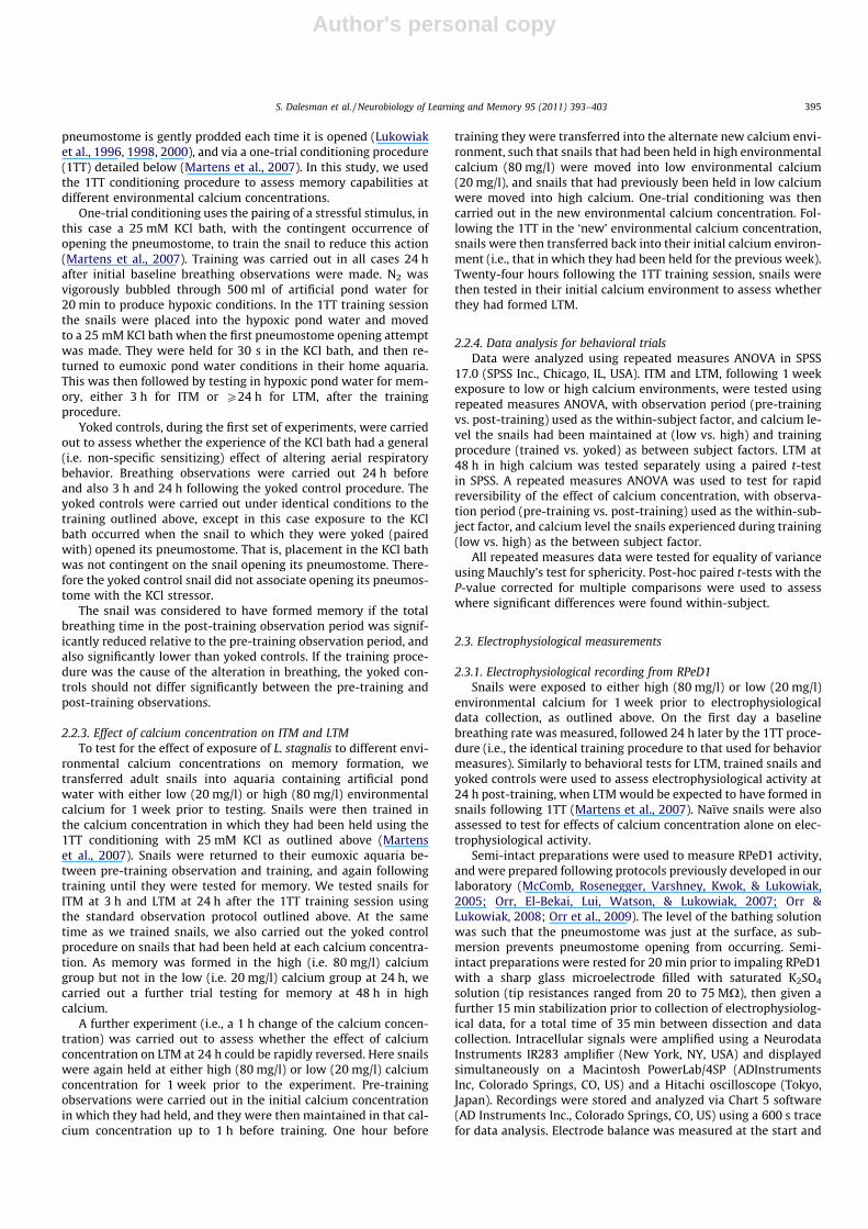
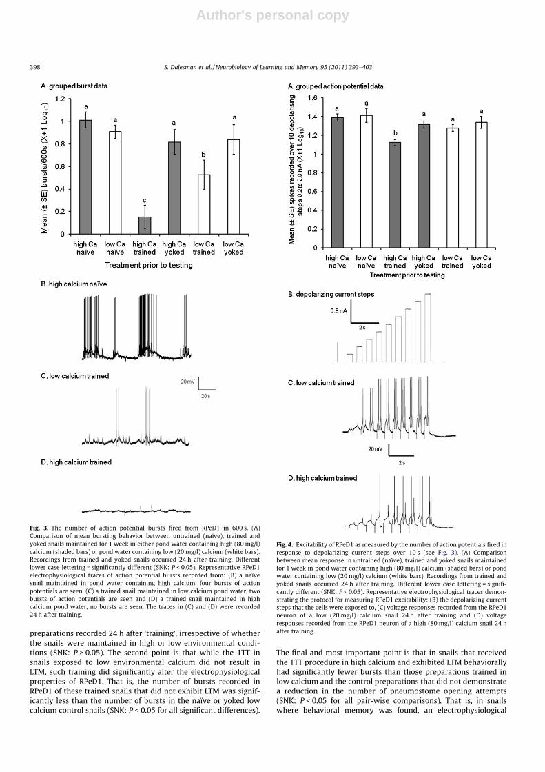
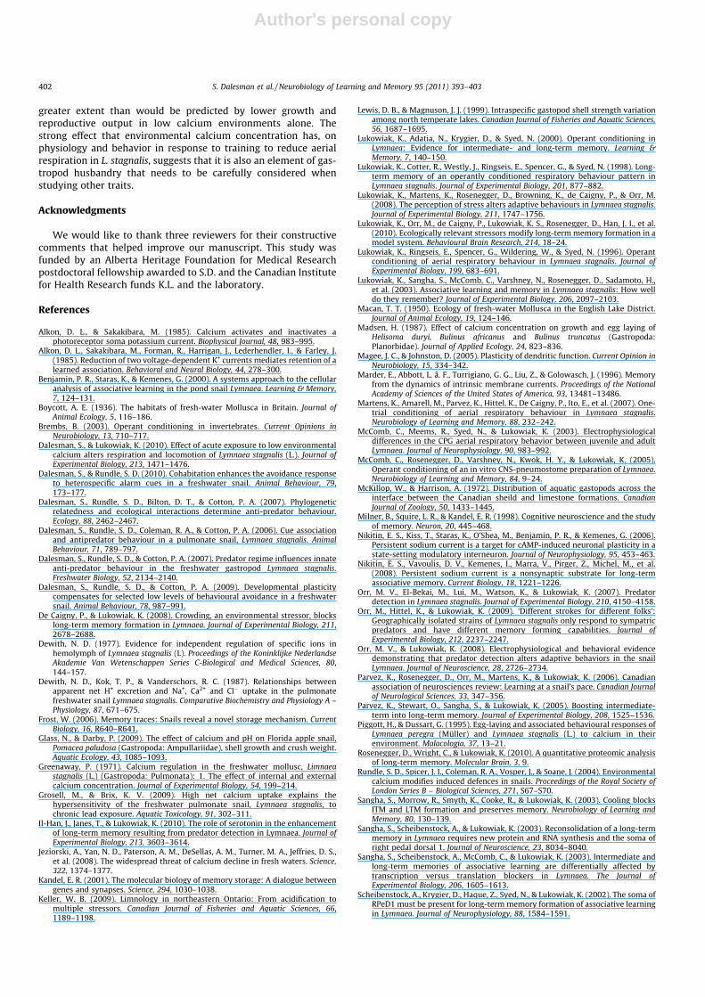
Under normal circumstances, RPeD1 fires bursts of actionpotentials (Fig. 3B) that are correlated with the initiation of aerialrespiratory activity, specifically opening of the pneumostome(McComb et al., 2005; Spencer, Kazmi, Syed, & Lukowiak, 2002;Syed et al., 1990). The behavioral data presented above (Figs. 1and 2) show that in the high environmental calcium concentrationLTM formed following the 1TT procedure. When we examined theactivity of RPeD1 in semi-intact preparations 24 h after the 1TTprocedure we found that all the electrophysiological parameterswe measured were significantly altered in high environmental cal-cium concentrations, however in low environmental calcium con-ditions not all parameters were altered, and those that wereshowed a lesser reduction in activity.
We first examined the number of action potential bursts inRPeD1 from 1TT trained and naïve snails (Fig. 3). Grouped datashowing the number of bursts recorded in RPeD1 24 h after the1TT or the yoked control procedure in high and low environmentalcalcium conditions are shown (Fig. 3A). Representative data fromthree different preparations (naïve maintained in high calcium,24 h trained in low environmental calcium and 24 h trained in highenvironmental calcium) are presented in Fig. 3B–D. Both trainingand the conditions under which they were trained significantly al-tered the burst activity (Fig. 3A; one-way ANOVA: F5,55 = 9.16,P < 0.001). Three points can be made from these data. The first isthat there was no significant difference in the number of burstsseen in RPeD1 between naïve preparations and the yoked control
Fig. 2. Memory following 1 h exposure to high or low calcium environments. Meantotal breathing time in high (shaded: 80 mg/l) and low (white: 20 mg/l) environ-mental calcium prior to and following one-trial conditioning (black bar). Snailswere exposed to either high (shaded) or low (white) calcium for 1 h immediatelyprior to training. Different lower case lettering = significantly different (paired t-testcorrected for multiple comparisons: P < 0.016).
S. Dalesman et al. / Neurobiology of Learning and Memory 95 (2011) 393–403 397
Author's personal copy
preparations recorded 24 h after ‘training’, irrespective of whetherthe snails were maintained in high or low environmental condi-tions (SNK: P > 0.05). The second point is that while the 1TT insnails exposed to low environmental calcium did not result inLTM, such training did significantly alter the electrophysiologicalproperties of RPeD1. That is, the number of bursts recorded inRPeD1 of these trained snails that did not exhibit LTM was signif-icantly less than the number of bursts in the naïve or yoked lowcalcium control snails (SNK: P < 0.05 for all significant differences).
The final and most important point is that in snails that receivedthe 1TT procedure in high calcium and exhibited LTM behaviorallyhad significantly fewer bursts than those preparations trained inlow calcium and the control preparations that did not demonstratea reduction in the number of pneumostome opening attempts(SNK: P < 0.05 for all pair-wise comparisons). That is, in snailswhere behavioral memory was found, an electrophysiological
Fig. 3. The number of action potential bursts fired from RPeD1 in 600 s. (A)Comparison of mean bursting behavior between untrained (naïve), trained andyoked snails maintained for 1 week in either pond water containing high (80 mg/l)calcium (shaded bars) or pond water containing low (20 mg/l) calcium (white bars).Recordings from trained and yoked snails occurred 24 h after training. Differentlower case lettering = significantly different (SNK: P < 0.05). Representative RPeD1electrophysiological traces of action potential bursts recorded from: (B) a naïvesnail maintained in pond water containing high calcium, four bursts of actionpotentials are seen, (C) a trained snail maintained in low calcium pond water, twobursts of action potentials are seen and (D) a trained snail maintained in highcalcium pond water, no bursts are seen. The traces in (C) and (D) were recorded24 h after training.
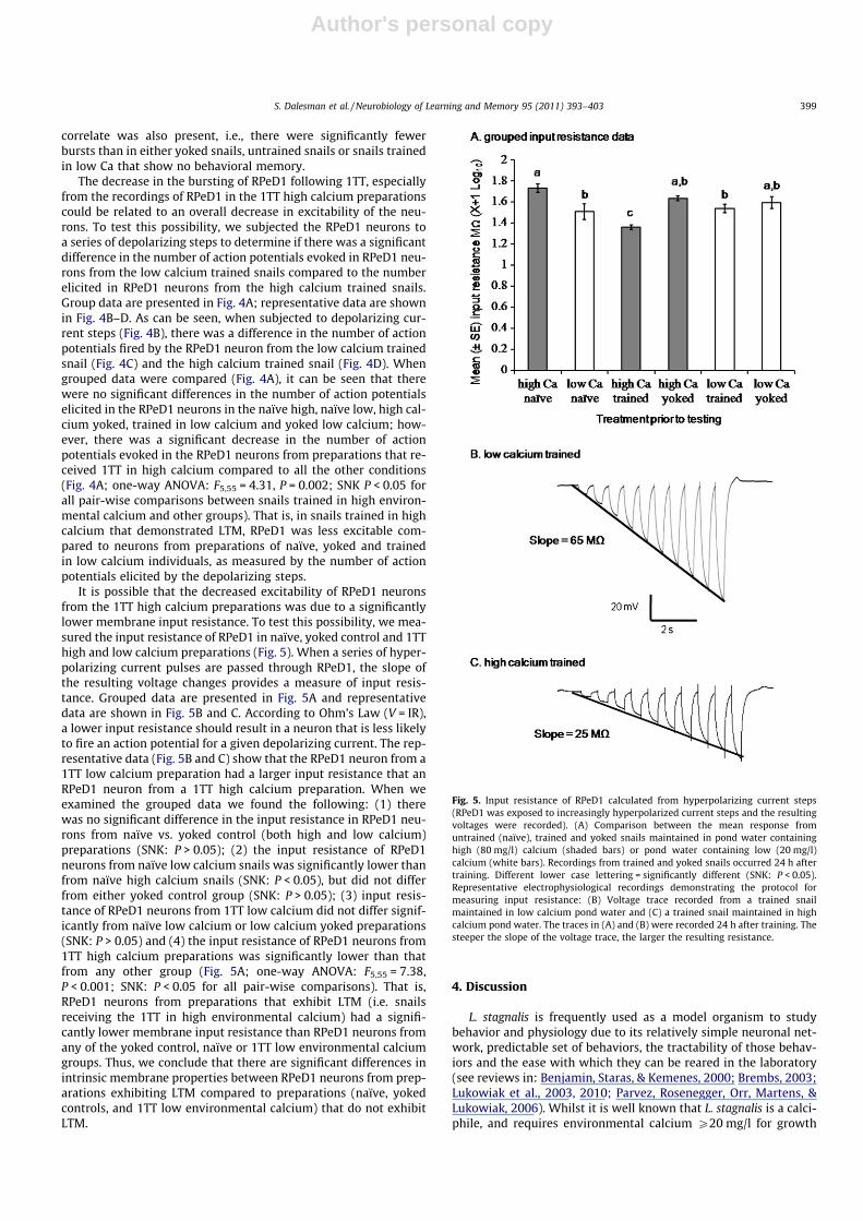
Fig. 4. Excitability of RPeD1 as measured by the number of action potentials fired inresponse to depolarizing current steps over 10 s (see Fig. 3). (A) Comparisonbetween mean response in untrained (naïve), trained and yoked snails maintainedfor 1 week in pond water containing high (80 mg/l) calcium (shaded bars) or pondwater containing low (20 mg/l) calcium (white bars). Recordings from trained andyoked snails occurred 24 h after training. Different lower case lettering = signifi-cantly different (SNK: P < 0.05). Representative electrophysiological traces demon-strating the protocol for measuring RPeD1 excitability: (B) the depolarizing currentsteps that the cells were exposed to, (C) voltage responses recorded from the RPeD1neuron of a low (20 mg/l) calcium snail 24 h after training and (D) voltageresponses recorded from the RPeD1 neuron of a high (80 mg/l) calcium snail 24 hafter training.
398 S. Dalesman et al. / Neurobiology of Learning and Memory 95 (2011) 393–403
Author's personal copy
correlate was also present, i.e., there were significantly fewerbursts than in either yoked snails, untrained snails or snails trainedin low Ca that show no behavioral memory.
The decrease in the bursting of RPeD1 following 1TT, especiallyfrom the recordings of RPeD1 in the 1TT high calcium preparationscould be related to an overall decrease in excitability of the neu-rons. To test this possibility, we subjected the RPeD1 neurons toa series of depolarizing steps to determine if there was a significantdifference in the number of action potentials evoked in RPeD1 neu-rons from the low calcium trained snails compared to the numberelicited in RPeD1 neurons from the high calcium trained snails.Group data are presented in Fig. 4A; representative data are shownin Fig. 4B–D. As can be seen, when subjected to depolarizing cur-rent steps (Fig. 4B), there was a difference in the number of actionpotentials fired by the RPeD1 neuron from the low calcium trainedsnail (Fig. 4C) and the high calcium trained snail (Fig. 4D). Whengrouped data were compared (Fig. 4A), it can be seen that therewere no significant differences in the number of action potentialselicited in the RPeD1 neurons in the naïve high, naïve low, high cal-cium yoked, trained in low calcium and yoked low calcium; how-ever, there was a significant decrease in the number of actionpotentials evoked in the RPeD1 neurons from preparations that re-ceived 1TT in high calcium compared to all the other conditions(Fig. 4A; one-way ANOVA: F5,55 = 4.31, P = 0.002; SNK P < 0.05 forall pair-wise comparisons between snails trained in high environ-mental calcium and other groups). That is, in snails trained in highcalcium that demonstrated LTM, RPeD1 was less excitable com-pared to neurons from preparations of naïve, yoked and trainedin low calcium individuals, as measured by the number of actionpotentials elicited by the depolarizing steps.
It is possible that the decreased excitability of RPeD1 neuronsfrom the 1TT high calcium preparations was due to a significantlylower membrane input resistance. To test this possibility, we mea-sured the input resistance of RPeD1 in naïve, yoked control and 1TThigh and low calcium preparations (Fig. 5). When a series of hyper-polarizing current pulses are passed through RPeD1, the slope ofthe resulting voltage changes provides a measure of input resis-tance. Grouped data are presented in Fig. 5A and representativedata are shown in Fig. 5B and C. According to Ohm’s Law (V = IR),a lower input resistance should result in a neuron that is less likelyto fire an action potential for a given depolarizing current. The rep-resentative data (Fig. 5B and C) show that the RPeD1 neuron from a1TT low calcium preparation had a larger input resistance that anRPeD1 neuron from a 1TT high calcium preparation. When weexamined the grouped data we found the following: (1) therewas no significant difference in the input resistance in RPeD1 neu-rons from naïve vs. yoked control (both high and low calcium)preparations (SNK: P > 0.05); (2) the input resistance of RPeD1neurons from naïve low calcium snails was significantly lower thanfrom naïve high calcium snails (SNK: P < 0.05), but did not differfrom either yoked control group (SNK: P > 0.05); (3) input resis-tance of RPeD1 neurons from 1TT low calcium did not differ signif-icantly from naïve low calcium or low calcium yoked preparations(SNK: P > 0.05) and (4) the input resistance of RPeD1 neurons from1TT high calcium preparations was significantly lower than thatfrom any other group (Fig. 5A; one-way ANOVA: F5,55 = 7.38,P < 0.001; SNK: P < 0.05 for all pair-wise comparisons). That is,RPeD1 neurons from preparations that exhibit LTM (i.e. snailsreceiving the 1TT in high environmental calcium) had a signifi-cantly lower membrane input resistance than RPeD1 neurons fromany of the yoked control, naïve or 1TT low environmental calciumgroups. Thus, we conclude that there are significant differences inintrinsic membrane properties between RPeD1 neurons from prep-arations exhibiting LTM compared to preparations (naïve, yokedcontrols, and 1TT low environmental calcium) that do not exhibitLTM.
4. Discussion
L. stagnalis is frequently used as a model organism to studybehavior and physiology due to its relatively simple neuronal net-work, predictable set of behaviors, the tractability of those behav-iors and the ease with which they can be reared in the laboratory(see reviews in: Benjamin, Staras, & Kemenes, 2000; Brembs, 2003;Lukowiak et al., 2003, 2010; Parvez, Rosenegger, Orr, Martens, &Lukowiak, 2006). Whilst it is well known that L. stagnalis is a calci-phile, and requires environmental calcium P20 mg/l for growth
Fig. 5. Input resistance of RPeD1 calculated from hyperpolarizing current steps(RPeD1 was exposed to increasingly hyperpolarized current steps and the resultingvoltages were recorded). (A) Comparison between the mean response fromuntrained (naïve), trained and yoked snails maintained in pond water containinghigh (80 mg/l) calcium (shaded bars) or pond water containing low (20 mg/l)calcium (white bars). Recordings from trained and yoked snails occurred 24 h aftertraining. Different lower case lettering = significantly different (SNK: P < 0.05).Representative electrophysiological recordings demonstrating the protocol formeasuring input resistance: (B) Voltage trace recorded from a trained snailmaintained in low calcium pond water and (C) a trained snail maintained in highcalcium pond water. The traces in (A) and (B) were recorded 24 h after training. Thesteeper the slope of the voltage trace, the larger the resulting resistance.
S. Dalesman et al. / Neurobiology of Learning and Memory 95 (2011) 393–403 399
Author's personal copy
and survival (Boycott, 1936; Van Der Borght & Van Puymbroek,1966; Young, 1975a), there is very little information on the effectof environmental calcium, within ecologically relevant concentra-tions, on its behavior and physiology. In addition, there is consid-erable evidence that environmental calcium is declining infreshwater systems in North America and Europe. For example, a35% decline in environmental calcium available in some freshwatersystems has occurred over the past two decades (Jeziorski et al.,2008; Keller, 2009). We have previously demonstrated in L. stag-nalis that acute exposure to a low calcium environment alters basicphysiological and behavioral traits, as exemplified by alterations inrespiration and locomotion (Dalesman & Lukowiak, 2010). Here wedemonstrated that a 1 h to 1 week acute exposure to a low calciumenvironment blocked LTM formation and reduced the neural corre-lates of LTM in a neuron, RPeD1, which is necessary for LTM forma-tion. These results have important implications for the study ofadaptive behavior and physiology in freshwater gastropods.
In L. stagnalis, LTM formation, as a result of operant conditioningof aerial respiration, can be modified (enhanced or suppressed) bystress (Lukowiak et al., 2008, 2010). LTM can be enhanced follow-ing exposure to sympatric predator cues (Orr & Lukowiak, 2008;Orr et al., 2009) or blocked by cooling (Sangha et al., 2003) andcrowding (De Caigny & Lukowiak, 2008), all of which are environ-mentally relevant stressors. In addition to requiring environmentalcalcium for growth and development, a low calcium environmentboth increases the basal metabolic demand of L. stagnalis and re-duces the energy available for behaviors such as locomotion(Dalesman & Lukowiak, 2010). Hence, we hypothesized that alow calcium environment, at a concentration experienced by fieldpopulations, would act as a stressor for this species. However, untilwe performed the experiments shown here, it was unclear to uswhether this stressor would enhance or block LTM formation.We found that L. stagnalis were capable of forming intermediate-term memory (ITM) in low environmental calcium, hence thisstressor did not prevent the learning process nor the formationof a memory that is only dependent on the translation of pre-exist-ing mRNA (i.e. ITM: Sangha et al., 2003). However, L. stagnalis wereunable, as a result of their exposure to the low calcium environ-ment either for 1 week or 1 h, to form long-term memory (LTM).Therefore, we propose that the low calcium environment may bepreventing the required transcription processes in neurons (e.g.RPeD1) necessary for LTM formation. Moreover, the suppressive ef-fect of low environmental calcium on LTM formation occurred rap-idly, and likely depends on sensory structures such as theosphradium to initiate alterations in genomic processes necessaryto protect calcium stores. The fact that memory formation wassimultaneously inhibited suggests that these genomic changeswere incompatible with LTM formation. These data contrast withdata from snails held at the high environmental calcium for as littleas 1 h, which were capable of forming both ITM and LTM. Thus, thedetection of sufficient environmental calcium (i.e., 80 mg/l) rapidlyreversed the genomic changes in neurons (e.g. RPeD1) induced bythe low environmental calcium, such that the necessary molecularevents that cause the formation of LTM can ensue. Moreover, in thehigh calcium concentration this memory did not extend to 48 h,concurring with previous findings following one-trial conditioning(Martens et al., 2007).
RPeD1 is part of a three neuron network, a central pattern gen-erator (CPG) that is both necessary and sufficient to drive aerialrespiratory behavior in L. stagnalis (Syed et al., 1990, 1992). More-over, since we were examining a form of non-declarative memory,there is a high probability that the site of memory formation andstorage is in the same neuronal network that mediates the behav-ior (Milner, Squire, & Kandel, 1998). Thus, it was not too surprisingto find that the ablation of RPeD1’s soma, which removes the nu-cleus but leaves intact its primary neurite, results in a snail that
can learn and form ITM, but cannot form LTM, undergo reconsoli-dation or extinction (Sangha, Scheibenstock, & Lukowiak, 2003;Sangha et al., 2003; Scheibenstock et al., 2002). Changes in someelectrophysiological properties of RPeD1 have been found follow-ing an alternate associative learning training procedure, which re-sults in LTM. In that procedure the pneumostome was physically‘poked’ as it attempts to open, and the training results in a signif-icant reduction in aerial respiration in both semi-intact prepara-tions made from previously trained snails, or from naïvepreparations trained ‘in the dish’ (McComb et al., 2005; Spencer,Syed, & Lukowiak, 1999). Additionally, these electrophysiologicalchanges were correlated with molecular changes necessary forLTM formation that occur in RPeD1 and other CNS neurons (Rose-negger, Wright, & Lukowiak, 2010). Here we tested whether similarelectrophysiological changes also occur in RPeD1 following the 1TTprocedure in alternate external calcium environments, both con-ducive and detrimental to LTM formation. We found that the 1TTprocedure in the high calcium environment (80 mg/l) resulted inLTM formation. Further, we found that the electrophysiologicalproperties of RPeD1 in these snails were similar to those foundusing the more traditional training procedure (i.e. a ‘poke’ to thepneumostome as it attempts to open). That is, whether assessedby the number of spontaneously occurring bursts of action poten-tials, or the number of evoked action potentials with discreet depo-larizing current steps, there was a significant decrease in theexcitability of RPeD1. The concomitant decrease in the input resis-tance suggests that the decrease in the excitability of RPeD1 mayhave been due to an increase in basal ionic flux across the mem-brane. As resistance fell, the current required to initiate spike firingbecame proportionally higher, and it became more difficult to ini-tiate the burst of action potentials that result in pneumostomeopening. This can be seen when comparing the depolarizing cur-rents required to initiate action potential firing in RPeD1. In naïve,high calcium snails, 0.2 nA of depolarizing current was sufficient toinduce firing in 92% of the snail preps, with 0.4 nA resulting in100% of the preparations firing action potentials. Conversely, fol-lowing training, preparations made from high calcium snails firedno action potentials at 0.2 nA of depolarizing current and only11% of the preps fired at 0.4 nA. It took a 1.2 nA depolarizing cur-rent to induce firing in all of the trained, high calcium preparations.When snails were trained and tested in low environmental cal-cium, this significant decrease in electrophysiological activity rela-tive to naïve snails was reduced. Confirmation that the reduction inactivity was due to the training procedure was demonstrated bythe absence of a significant difference in activity between the naïvesnails and yoked controls that had not experienced the KCl bathcontingent with pneumostome opening.
When L. stagnalis were trained in low environmental calcium,there was still a significant reduction in the number of action po-tential bursts fired compared to both yoked controls and naïve,snails. This indicates that despite no behavioral changes, the 1TTprocedure had altered the electrophysiological properties of RPeD1to a degree, but not to the extent needed to see a behavioral change.These data are reminiscent of findings from the ‘poking’ trainingprocedure used in our laboratory to reduce aerial respiration, whereit was discovered that following a training session that was insuffi-cient to create LTM, if the snails received an additional training ses-sion (normally producing ITM) on the following day, they wouldnow possess LTM (Parvez, Stewart, Sangha, & Lukowiak, 2005). Thissuggests that even 24 h following the ITM training protocol, snails,while no longer showing behavioral memory, still possess a resid-ual memory trace which allowed a single additional training ses-sion, that normally only results in ITM, to now produce LTM(Parvez et al., 2005). While the previous study was based on behav-ioral data alone, the electrophysiological data obtained in the pres-ent study provide strong support for the residual memory trace
400 S. Dalesman et al. / Neurobiology of Learning and Memory 95 (2011) 393–403
Author's personal copy
hypothesis, that 24 h following training in snails without anybehavioral traces of memory, RPeD1 remained in a significantly dif-ferent state from both the naïve and yoked control snails.
The electrophysiological data obtained here support previousfindings concerning neural correlates of LTM following operantconditioning of aerial respiratory behavior in L. stagnalis. Spenceret al. (1999) reported no significant differences in the RMP ofRPeD1 following operant conditioning. In that report, however,the authors recorded from RPeD1 in isolated ganglionic prepara-tions. That is, the CNS was removed from the snails prior to record-ing from RPeD1. We now know that the periphery (i.e.,pneumostome and osphradium area and osphradial ganglion) con-tributes both excitatory and inhibitory inputs to neurons in theCNS (Il-Han, Janes, & Lukowiak, 2010; McComb, Meems, Syed, &Lukowiak, 2003). Thus, data obtained from only ganglionic prepa-rations may not be an accurate reflection of the ‘state of the ner-vous system’ following conditioning. Subsequently, Spencer et al.(2002) recorded from RPeD1 in a semi-intact preparation 1 h aftertraining, and also reported no change in the RMP of RPeD1 in prep-arations made from snails that had exhibited ‘good’ memory. Theydid find, that in these semi-intact preparations, there was a signif-icant decrease in spontaneous activity in RPeD1 following a pre-sentation of a tactile stimulus to the pneumostome area. Thisdecrease in activity did not occur in control preparations. Naïvesnail preparations trained in vitro showed no change in the RMPof RPeD1 following in vitro training (McComb et al., 2005). Recentwork demonstrated that in adult and juvenile snails, trained withthe 1TT procedure in normal pond water (i.e., 80 mg/l environmen-tal calcium), there was also no significant change in the RMP ofRPeD1 (Sunada, Horikoshi, Lukowiak, & Sakakibara, 2010). Of inter-est, these authors did find that in both juvenile and adult snailstrained with the 1TT procedure in the presence of the scent of apredator (i.e., crayfish kairomones in 80 mg/l environmental cal-cium), that the RMP of RPeD1 was significantly more hyperpolar-ized compared to control preparations. It appears that thesignificant change in the RMP of RPeD1 may primarily be due tothe detection of the predator, as Orr et al. (2007) demonstratedthat predator detection results in a hyperpolarization of RPeD1.This appears to be mediated by a serotonergic system, stimulatedby predator detection via the osphradium (Il-Han et al., 2010).
Previous studies in mollusk and other model system prepara-tions have found that there were alterations in synaptic connectiv-ity that were correlated with the presence of behavioral LTM(Kandel, 2001). However, other data in both mollusk and mamma-lian model systems, show that behavioral LTM is correlated notonly with plastic changes in synaptic connectivity, but also withchanges in the intrinsic membrane properties of neurons, similarto that we observed here (Alkon et al., 1985). These changes inintrinsic membrane properties, that alter the excitability of theneuron, have been referred to as ‘non-synaptic substrates’ of LTM(Nikitin et al., 2008). These changes may involve alterations inchannel activity that leads to either increased or decreased neuro-nal excitability, and hence membrane input resistance. For exam-ple, changes in calcium, potassium or sodium channel activityhave been hypothesized to account for an increased neuronalexcitability in various vertebrate and mammalian model systems,that correlates well with behavioral LTM (Alkon & Sakakibara,1985; Alkon et al., 1985; Frost, 2006; Magee & Johnston, 2005;Marder et al., 1996; Nikitin et al., 2006). Here we showed, that fol-lowing the 1TT procedure in high environmental calcium (whichleads to behavioral LTM), there are significant alterations in neuro-nal excitability seen, both with spontaneously occurring inputs,and when the neuron is driven by depolarizing or hyperpolarizingcurrent injections. Thus, our findings here add to the growing bodyof evidence that a change in membrane excitability contribute tohow the nervous system encodes LTM.
The disruptive effect of low environmental calcium on the for-mation of LTM was rapidly reversed by just 1 h of exposure to highenvironmental calcium. That was, snails held in the low calciumenvironment for 1 week only require 1 h exposure to a high cal-cium environment to regain their ability to form LTM. The reverseis also seen. The rapid reversal in the ability to form LTM aftertraining snails in an alternate calcium environment indicates thatthe mechanism by which the sensing of calcium alters memoryis not related to haemolymph calcium concentration. L. stagnalisis able to maintain calcium concentration in the haemolymph forover a week, even in the complete absence of environmental cal-cium (Dewith, 1977). This species is also able to orientate towardsa high calcium environment in choice tests, indicating that theymay be able to directly sense environmental calcium concentration(Piggott & Dussart, 1995). The rapid behavioral and electrophysio-logical response to the external environmental calcium concentra-tion provides further evidence that this may be the case.
Whilst snails could not form LTM in low environmental cal-cium, low environmental calcium did not prevent the recall of apreviously established memory that occurred when the snails weretrained in the high environmental calcium. These data further ar-gue against the possibility that exposure to low environmental cal-cium alters the internal haemolymph calcium. If the haemolymphcalcium was altered by the acute exposure to the low environmen-tal calcium, then we would have expected that an already estab-lished memory should be as affected by the low environmentalcalcium, as is the establishment of a new memory. Our workinghypothesis is that snails detect low environmental calcium, possi-bly by sensory neurons located in the osphradium, and that theactivity of these neurons alter the molecular machinery in RPeD1,such that the molecular steps necessary for the memory consolida-tion to produce behavioral LTM cannot occur. If, however, memoryconsolidation has already occurred, as the result of changes in neu-ronal excitability and synaptic connectivity, the molecular changesin the neurons, such as RPeD1, induced by the detection of the lowenvironmental calcium are not sufficient to reverse the changes inneuronal excitability and synaptic connectivity.
Calcium fluctuations in the freshwater habitat are common(Macan, 1950; McKillop & Harrison, 1972; Williams, 1970; Dales-man unpublished data), such that freshwater gastropods mayexperience ten-fold changes in calcium concentration over thecourse of a year. Whilst L. stagnalis populations are normally foundin environments of P20 mg/l calcium, they may experience peri-ods below or above this level. The data we present here indicatethat whilst experiencing low environmental calcium L. stagnaliswill be less able to demonstrate behavioral plasticity. One conse-quence of reduced memory duration in low environmental cal-cium, for example, may lead to a lack of adaptability to thepredation environment. L. stagnalis has enhanced innate responsesto predators (Dalesman, Rundle, & Cotton, 2007; Orr et al., 2007)and also heterospecific alarm cues from species that overlap in dis-tribution (Dalesman, Rundle, Bilton, & Cotton, 2007); however rec-ognition of predatorial threat can also be improved throughlearning, both in terms of recognizing the predator (Dalesman,Rundle, Coleman, & Cotton, 2006; Dalesman, Rundle, & Cotton,2009) and also heterospecific alarm cues (Dalesman & Rundle,2010). The data presented here suggest that the ability to remem-ber stressful events, such as those that might be associated with apredator encounter, may be impeded in low environmental cal-cium environments. These data indicate that L. stagnalis found nat-urally in low calcium environments, or during periods of lowcalcium availability such as following a flooding event, may be lessable to adapt to novel stressors than populations found in harder(i.e., high calcium) waters. This, together with an inability to ex-press induced morphological defenses (e.g. specific shell character-istics; Rundle et al., 2004), may reduce fitness of L. stagnalis to a
S. Dalesman et al. / Neurobiology of Learning and Memory 95 (2011) 393–403 401
Author's personal copy
greater extent than would be predicted by lower growth andreproductive output in low calcium environments alone. Thestrong effect that environmental calcium concentration has, onphysiology and behavior in response to training to reduce aerialrespiration in L. stagnalis, suggests that it is also an element of gas-tropod husbandry that needs to be carefully considered whenstudying other traits.
Acknowledgments
We would like to thank three reviewers for their constructivecomments that helped improve our manuscript. This study wasfunded by an Alberta Heritage Foundation for Medical Researchpostdoctoral fellowship awarded to S.D. and the Canadian Institutefor Health Research funds K.L. and the laboratory.
References
Alkon, D. L., & Sakakibara, M. (1985). Calcium activates and inactivates aphotoreceptor soma potassium current. Biophysical Journal, 48, 983–995.
Alkon, D. L., Sakakibara, M., Forman, R., Harrigan, J., Lederhendler, I., & Farley, J.(1985). Reduction of two voltage-dependent K+ currents mediates retention of alearned association. Behavioral and Neural Biology, 44, 278–300.
Benjamin, P. R., Staras, K., & Kemenes, G. (2000). A systems approach to the cellularanalysis of associative learning in the pond snail Lymnaea. Learning & Memory,7, 124–131.
Boycott, A. E. (1936). The habitats of fresh-water Mollusca in Britain. Journal ofAnimal Ecology, 5, 116–186.
Brembs, B. (2003). Operant conditioning in invertebrates. Current Opinions inNeurobiology, 13, 710–717.
Dalesman, S., & Lukowiak, K. (2010). Effect of acute exposure to low environmentalcalcium alters respiration and locomotion of Lymnaea stagnalis (L.). Journal ofExperimental Biology, 213, 1471–1476.
Dalesman, S., & Rundle, S. D. (2010). Cohabitation enhances the avoidance responseto heterospecific alarm cues in a freshwater snail. Animal Behaviour, 79,173–177.
Dalesman, S., Rundle, S. D., Bilton, D. T., & Cotton, P. A. (2007). Phylogeneticrelatedness and ecological interactions determine anti-predator behaviour.Ecology, 88, 2462–2467.
Dalesman, S., Rundle, S. D., Coleman, R. A., & Cotton, P. A. (2006). Cue associationand antipredator behaviour in a pulmonate snail, Lymnaea stagnalis. AnimalBehaviour, 71, 789–797.
Dalesman, S., Rundle, S. D., & Cotton, P. A. (2007). Predator regime influences innateanti-predator behaviour in the freshwater gastropod Lymnaea stagnalis.Freshwater Biology, 52, 2134–2140.
Dalesman, S., Rundle, S. D., & Cotton, P. A. (2009). Developmental plasticitycompensates for selected low levels of behavioural avoidance in a freshwatersnail. Animal Behaviour, 78, 987–991.
De Caigny, P., & Lukowiak, K. (2008). Crowding, an environmental stressor, blockslong-term memory formation in Lymnaea. Journal of Experimental Biology, 211,2678–2688.
Dewith, N. D. (1977). Evidence for independent regulation of specific ions inhemolymph of Lymnaea stagnalis (L). Proceedings of the Koninklijke NederlandseAkademie Van Wetenschappen Series C-Biological and Medical Sciences, 80,144–157.
Dewith, N. D., Kok, T. P., & Vanderschors, R. C. (1987). Relationships betweenapparent net H+ excretion and Na+, Ca2+ and Cl� uptake in the pulmonatefreshwater snail Lymnaea stagnalis. Comparative Biochemistry and Physiology A –Physiology, 87, 671–675.
Frost, W. (2006). Memory traces: Snails reveal a novel storage mechanism. CurrentBiology, 16, R640–R641.
Glass, N., & Darby, P. (2009). The effect of calcium and pH on Florida apple snail,Pomacea paludosa (Gastropoda: Ampullariidae), shell growth and crush weight.Aquatic Ecology, 43, 1085–1093.
Greenaway, P. (1971). Calcium regulation in the freshwater mollusc, Limnaeastagnalis (L.) (Gastropoda: Pulmonata): 1. The effect of internal and externalcalcium concentration. Journal of Experimental Biology, 54, 199–214.
Grosell, M., & Brix, K. V. (2009). High net calcium uptake explains thehypersensitivity of the freshwater pulmonate snail, Lymnaea stagnalis, tochronic lead exposure. Aquatic Toxicology, 91, 302–311.
Il-Han, J., Janes, T., & Lukowiak, K. (2010). The role of serotonin in the enhancementof long-term memory resulting from predator detection in Lymnaea. Journal ofExperimental Biology, 213, 3603–3614.
Jeziorski, A., Yan, N. D., Paterson, A. M., DeSellas, A. M., Turner, M. A., Jeffries, D. S.,et al. (2008). The widespread threat of calcium decline in fresh waters. Science,322, 1374–1377.
Kandel, E. R. (2001). The molecular biology of memory storage: A dialogue betweengenes and synapses. Science, 294, 1030–1038.
Keller, W. B. (2009). Limnology in northeastern Ontario: From acidification tomultiple stressors. Canadian Journal of Fisheries and Aquatic Sciences, 66,1189–1198.
Lewis, D. B., & Magnuson, J. J. (1999). Intraspecific gastopod shell strength variationamong north temperate lakes. Canadian Journal of Fisheries and Aquatic Sciences,56, 1687–1695.
Lukowiak, K., Adatia, N., Krygier, D., & Syed, N. (2000). Operant conditioning inLymnaea: Evidence for intermediate- and long-term memory. Learning &Memory, 7, 140–150.
Lukowiak, K., Cotter, R., Westly, J., Ringseis, E., Spencer, G., & Syed, N. (1998). Long-term memory of an operantly conditioned respiratory behaviour pattern inLymnaea stagnalis. Journal of Experimental Biology, 201, 877–882.
Lukowiak, K., Martens, K., Rosenegger, D., Browning, K., de Caigny, P., & Orr, M.(2008). The perception of stress alters adaptive behaviours in Lymnaea stagnalis.Journal of Experimental Biology, 211, 1747–1756.
Lukowiak, K., Orr, M., de Caigny, P., Lukowiak, K. S., Rosenegger, D., Han, J. I., et al.(2010). Ecologically relevant stressors modify long-term memory formation in amodel system. Behavioural Brain Research, 214, 18–24.
Lukowiak, K., Ringseis, E., Spencer, G., Wildering, W., & Syed, N. (1996). Operantconditioning of aerial respiratory behaviour in Lymnaea stagnalis. Journal ofExperimental Biology, 199, 683–691.
Lukowiak, K., Sangha, S., McComb, C., Varshney, N., Rosenegger, D., Sadamoto, H.,et al. (2003). Associative learning and memory in Lymnaea stagnalis: How welldo they remember? Journal of Experimental Biology, 206, 2097–2103.
Macan, T. T. (1950). Ecology of fresh-water Mollusca in the English Lake District.Journal of Animal Ecology, 19, 124–146.
Madsen, H. (1987). Effect of calcium concentration on growth and egg laying ofHelisoma duryi, Bulinus africanus and Bulinus truncatus (Gastropoda:Planorbidae). Journal of Applied Ecology, 24, 823–836.
Magee, J. C., & Johnston, D. (2005). Plasticity of dendritic function. Current Opinion inNeurobiology, 15, 334–342.
Marder, E., Abbott, L. â. F., Turrigiano, G. G., Liu, Z., & Golowasch, J. (1996). Memoryfrom the dynamics of intrinsic membrane currents. Proceedings of the NationalAcademy of Sciences of the United States of America, 93, 13481–13486.
Martens, K., Amarell, M., Parvez, K., Hittel, K., De Caigny, P., Ito, E., et al. (2007). One-trial conditioning of aerial respiratory behaviour in Lymnaea stagnalis.Neurobiology of Learning and Memory, 88, 232–242.
McComb, C., Meems, R., Syed, N., & Lukowiak, K. (2003). Electrophysiologicaldifferences in the CPG aerial respiratory behavior between juvenile and adultLymnaea. Journal of Neurophysiology, 90, 983–992.
McComb, C., Rosenegger, D., Varshney, N., Kwok, H. Y., & Lukowiak, K. (2005).Operant conditioning of an in vitro CNS-pneumostome preparation of Lymnaea.Neurobiology of Learning and Memory, 84, 9–24.
McKillop, W., & Harrison, A. (1972). Distribution of aquatic gastopods across theinterface between the Canadian sheild and limestone formations. CanadianJournal of Zoology, 50, 1433–1445.
Milner, B., Squire, L. R., & Kandel, E. R. (1998). Cognitive neuroscience and the studyof memory. Neuron, 20, 445–468.
Nikitin, E. S., Kiss, T., Staras, K., O’Shea, M., Benjamin, P. R., & Kemenes, G. (2006).Persistent sodium current is a target for cAMP-induced neuronal plasticity in astate-setting modulatory interneuron. Journal of Neurophysiology, 95, 453–463.
Nikitin, E. S., Vavoulis, D. V., Kemenes, I., Marra, V., Pirger, Z., Michel, M., et al.(2008). Persistent sodium current is a nonsynaptic substrate for long-termassociative memory. Current Biology, 18, 1221–1226.
Orr, M. V., El-Bekai, M., Lui, M., Watson, K., & Lukowiak, K. (2007). Predatordetection in Lymnaea stagnalis. Journal of Experimental Biology, 210, 4150–4158.
Orr, M., Hittel, K., & Lukowiak, K. (2009). ‘Different strokes for different folks’:Geographically isolated strains of Lymnaea stagnalis only respond to sympatricpredators and have different memory forming capabilities. Journal ofExperimental Biology, 212, 2237–2247.
Orr, M. V., & Lukowiak, K. (2008). Electrophysiological and behavioral evidencedemonstrating that predator detection alters adaptive behaviors in the snailLymnaea. Journal of Neuroscience, 28, 2726–2734.
Parvez, K., Rosenegger, D., Orr, M., Martens, K., & Lukowiak, K. (2006). Canadianassociation of neurosciences review: Learning at a snail’s pace. Canadian Journalof Neurological Sciences, 33, 347–356.
Parvez, K., Stewart, O., Sangha, S., & Lukowiak, K. (2005). Boosting intermediate-term into long-term memory. Journal of Experimental Biology, 208, 1525–1536.
Piggott, H., & Dussart, G. (1995). Egg-laying and associated behavioural responses ofLymnaea peregra (Müller) and Lymnaea stagnalis (L.) to calcium in theirenvironment. Malacologia, 37, 13–21.
Rosenegger, D., Wright, C., & Lukowiak, K. (2010). A quantitative proteomic analysisof long-term memory. Molecular Brain, 3, 9.
Rundle, S. D., Spicer, J. I., Coleman, R. A., Vosper, J., & Soane, J. (2004). Environmentalcalcium modifies induced defences in snails. Proceedings of the Royal Society ofLondon Series B – Biological Sciences, 271, S67–S70.
Sangha, S., Morrow, R., Smyth, K., Cooke, R., & Lukowiak, K. (2003). Cooling blocksITM and LTM formation and preserves memory. Neurobiology of Learning andMemory, 80, 130–139.
Sangha, S., Scheibenstock, A., & Lukowiak, K. (2003). Reconsolidation of a long-termmemory in Lymnaea requires new protein and RNA synthesis and the soma ofright pedal dorsal 1. Journal of Neuroscience, 23, 8034–8040.
Sangha, S., Scheibenstock, A., McComb, C., & Lukowiak, K. (2003). Intermediate andlong-term memories of associative learning are differentially affected bytranscription versus translation blockers in Lymnaea. The Journal ofExperimental Biology, 206, 1605–1613.
Scheibenstock, A., Krygier, D., Haque, Z., Syed, N., & Lukowiak, K. (2002). The soma ofRPeD1 must be present for long-term memory formation of associative learningin Lymnaea. Journal of Neurophysiology, 88, 1584–1591.
402 S. Dalesman et al. / Neurobiology of Learning and Memory 95 (2011) 393–403

Author's personal copy
Shors, T. J. (2004). Learning during stressful times. Learning & Memory, 11, 137–144.Spencer, G. E., Kazmi, M. H., Syed, N. I., & Lukowiak, K. (2002). Changes in the
activity of a CPG neuron after the reinforcement of an operantly conditionedbehavior in Lymnaea. Journal of Neurophysiology, 88, 1915–1923.
Spencer, G. E., Syed, N. I., & Lukowiak, K. (1999). Neural changes after operantconditioning of the aerial respiratory behavior in Lymnaea stagnalis. Journal ofNeuroscience, 19, 1836–1843.
Sunada, H., Horikoshi, T., Lukowiak, K., & Sakakibara, M. (2010). Increase inexcitability of RPeD11 results in memory enhancement of juvenile and adultLymnaea stagnalis by predator-induced stress. Neurobiology of Learning andMemory, 94, 269–277.
Syed, N. I., Bulloch, A. G. M., & Lukowiak, K. (1990). Invitro reconstruction of therespiratory central pattern generator of the mollusk Lymnaea. Science, 250,282–285.
Syed, N. I., Bulloch, A. G. M., & Lukowiak, K. (1992). The respiratory central patterngenerator (CPG) of Lymnaea reconstructed invitro. Acta Biologica Hungarica, 409,419.
Syed, N. I., Ridgway, R. L., Lukowiak, K., & Bulloch, A. G. M. (1992). Transplantationand functional-integration of an identified respiratory interneuron in Lymnaeastagnalis. Neuron, 8, 767–774.
Van Der Borght, O., & Van Puymbroek, S. (1966). Calcium metabolism in afreshwater mollusc: Quantitative importance of water and food supply forcalcium during growth. Nature, 210, 791–793.
Williams, N. V. (1970). Studies on aquatic pulmonate snails in central Africa 1. Fielddistribution in relation to water chemistry. Malacologia, 10, 153–164.
Young, J. O. (1975a). A laboratory study, using 45Ca tracer, on the source of calciumduring growth in two freshwater species of Gastropoda. Proceedings of theMalacoglogical Society of London, 41, 439–445.
Young, J. O. (1975b). Preliminary field and laboratory studies on the survival andspawning of several species of gastropoda in calcium-poor and calcium-richwaters. Proceedings of the Malacoglogical Society of London, 41, 429–437.
Zalizniak, L., Kefford, B. J., & Nugegoda, D. (2009). Effects of different ioniccompositions on survival and growth of Physa acuta. Aquatic Ecology, 43,145–156.
S. Dalesman et al. / Neurobiology of Learning and Memory 95 (2011) 393–403 403