Loss of heterozygosity is related to p53 mutations and smoking in lung cancer
6
226 Mutations in p53 are among the most frequent genetic alterations detected in lung cancer and are acquired early in the development of the disease (Bennett et al, 1993; Bennett, 1995). In non-small cell lung cancer (NSCLC) about half of the tumours carry muta- tions in the p53 gene (Takagi et al, 1998). The prevalent type of mutation is G:C → T:A transversion which is related to DNA adducts of benzo(a)pyrene from cigarette smoking (Greenblatt et al, 1994; Denissenko et al, 1996). p53 plays an important role in preserving genomic stability through controlling the cell cycle checkpoints (Hartwell, 1992). Both human and mouse cells homozygous for p53 mutations are genetically unstable and show high rates of homologous recombination (Livingstone et al, 1992; Yin et al, 1992; Mekeel et al, 1997). Other potential tumour suppressor genes are localized on chromosomes 3p, 5q, 9p and 11p (Kohno et al, 1999). It has been shown that deletions of chromo- some 3p are early events in the development of the disease, and such changes are often observed in lung tumours (Wistuba et al, 1997). The FHIT gene at 3p14.2 has been shown to be a target of carcinogens in the tobacco smoke (Sozzi et al, 1997). The hMLH1 gene maps to 3p21.3 and a high frequency of loss of hetero- zygosity (LOH) has been shown at this locus in lung cancer (Benachenhou et al, 1998). A concordant LOH of hMLH1 locus and the hMSH3 locus at 5q11–13 has been reported (Benachenhou et al, 1998). Mutations in the mismatch repair genes (MMR) are associated with a distinct molecular phenotype known as microsatellite instability (MSI) (Eshleman and Markowitz 1996). In lung cancer, the majority of studies have reported a low level (<5%) of MSI (Benachenhou et al, 1998). However, studies have shown that defects in DNA repair may be a predisposing factor in lung cancer (Wei and Spitz, 1997). Lung cancer is characterized by multiple genetic changes (Gazdar, 1994). A relationship between p53 mutations and 3p deletions has been reported in lung cancer (Horio et al, 1993; Burke et al, 1998; Kohno et al, 1999). The purpose of this study was to explore the association between tobacco exposure, p53 mutations and concordant LOH at several loci harbouring potential tumour suppressor genes involved in lung cancer. MATERIALS AND METHODS Tissue samples Surgically resected tumours and corresponding normal lung tissue were obtained from 165 lung cancer patients. The majority of tumours were at stage I according to international TNM staging. Each tissue was snap-frozen after surgery and stored at –80˚C. Prior to DNA isolation the tumour tissues were freeze-sectioned. 5–10 sections of each sample were stained with eosin and haema- toxylin. Based on these slides, the tumours were trimmed to obtain more than 70% tumour cells. Histological classification of tumours was made according to guidelines made by the WHO Histological Typing of Lung Tumours (WHO, 1981). A question- naire was filled out for each patient by the treating physician, including information on smoking habits. The characteristics of the lung cancer patients are summarized in Table 1. Analysis of LOH using microsatellite markers 17 polymorphic dinucleotide (CA) repeats representing regions with known DNA repair genes or tumour suppressor genes were Loss of heterozygosity is related to p53 mutations and smoking in lung cancer S Zienolddiny, D Ryberg, MO Arab, V Skaug and A Haugen Department of Toxicology, National Institute of Occupational Health, P.O. Box 8149 Dep., N-0033 Oslo, Norway Summary Carcinogenesis results from an accumulation of several genetic alterations. Mutations in the p53 gene are frequent and occur at an early stage of lung carcinogenesis. Loss of multiple chromosomal regions is another genetic alteration frequently found in lung tumours. We have examined the association between p53 mutations, loss of heterozygosity (LOH) at frequently deleted loci in lung cancer, and tobacco exposure in 165 tumours from non-small cell lung cancer (NSCLC) patients. A highly significant association between p53 mutations and deletions on 3p, 5q, 9p, 11p and 17p was found. There was also a significant correlation between deletions at these loci. 86% of the tumours with concordant deletion in the 4 most involved loci (3p21, 5q11–13, 9p21 and 17p13) had p53 mutations as compared to only 8% of the tumours without deletions at the corresponding loci (P < 0.0001). Data were also examined in relation to smoking status of the patients and histology of the tumours. The frequency of deletions was significantly higher among smokers as compared to non-smokers. This difference was significant for the 3p21.3 (hMLH1 locus), 3p14.2 (FHIT locus), 5q11–13 (hMSH3 locus) and 9p21 (D9S157 locus). Tumours with deletions at the hMLH1 locus had higher levels of hydrophobic DNA adducts. Deletions were more common in squamous cell carcinomas than in adenocarcinomas. Covariate analysis revealed that histological type and p53 mutations were significant and independent parameters for predicting LOH status at several loci. In the pathogenesis of NSCLC exposure to tobacco carcinogens in addition to clonal selection may be the driving force in these alterations. © 2001 Cancer Research Campaign http://www.bjcancer.com Keywords: non-small cell lung cancer; LOH; p53 mutations; smoking; DNA-adducts Received 17 April 2000 Revised 1 September 2000 Accepted 14 September 2000 Correspondence to: A Haugen British Journal of Cancer (2001) 84(2), 226–231 © 2001 Cancer Research Campaign doi: 10.1054/ bjoc.2000.1528, available online at http://www.idealibrary.com on http://www.bjcancer.com
Transcript of Loss of heterozygosity is related to p53 mutations and smoking in lung cancer

ve in
es
93;dy
g
sueofg.
0˚C.d.ma-
tainfO
-n,of
Loss of heterozygosity is related to p53 mutations andsmoking in lung cancer
S Zienolddiny, D Ryberg, MO Arab, V Skaug and A Haugen
Department of Toxicology, National Institute of Occupational Health, P.O. Box 8149 Dep., N-0033 Oslo, Norway
Summary Carcinogenesis results from an accumulation of several genetic alterations. Mutations in the p53 gene are frequent and occur atan early stage of lung carcinogenesis. Loss of multiple chromosomal regions is another genetic alteration frequently found in lung tumours.We have examined the association between p53 mutations, loss of heterozygosity (LOH) at frequently deleted loci in lung cancer, andtobacco exposure in 165 tumours from non-small cell lung cancer (NSCLC) patients. A highly significant association between p53 mutationsand deletions on 3p, 5q, 9p, 11p and 17p was found. There was also a significant correlation between deletions at these loci. 86% of thetumours with concordant deletion in the 4 most involved loci (3p21, 5q11–13, 9p21 and 17p13) had p53 mutations as compared to only 8% ofthe tumours without deletions at the corresponding loci (P < 0.0001). Data were also examined in relation to smoking status of the patientsand histology of the tumours. The frequency of deletions was significantly higher among smokers as compared to non-smokers. Thisdifference was significant for the 3p21.3 (hMLH1 locus), 3p14.2 (FHIT locus), 5q11–13 (hMSH3 locus) and 9p21 (D9S157 locus). Tumourswith deletions at the hMLH1 locus had higher levels of hydrophobic DNA adducts. Deletions were more common in squamous cellcarcinomas than in adenocarcinomas. Covariate analysis revealed that histological type and p53 mutations were significant and independentparameters for predicting LOH status at several loci. In the pathogenesis of NSCLC exposure to tobacco carcinogens in addition to clonalselection may be the driving force in these alterations. © 2001 Cancer Research Campaign http://www.bjcancer.com
Keywords: non-small cell lung cancer; LOH; p53 mutations; smoking; DNA-adducts
British Journal of Cancer (2001) 84(2), 226–231© 2001 Cancer Research Campaigndoi: 10.1054/ bjoc.2000.1528, available online at http://www.idealibrary.com on http://www.bjcancer.com
Mutations in p53are among the most frequent genetic alteratiodetected in lung cancer and are acquired early in the developof the disease (Bennett et al, 1993; Bennett, 1995). In non-scell lung cancer (NSCLC) about half of the tumours carry mutions in the p53 gene (Takagi et al, 1998). The prevalent typemutation is G:C → T:A transversion which is related to DNAadducts of benzo(a)pyrene from cigarette smoking (Greenbet al, 1994; Denissenko et al, 1996). p53 plays an important rolein preserving genomic stability through controlling the cell cyccheckpoints (Hartwell, 1992). Both human and mouse chomozygous for p53mutations are genetically unstable and shohigh rates of homologous recombination (Livingstone et al, 19Yin et al, 1992; Mekeel et al, 1997). Other potential tumosuppressor genes are localized on chromosomes 3p, 5q, 9p an(Kohno et al, 1999). It has been shown that deletions of chrosome 3p are early events in the development of the diseasesuch changes are often observed in lung tumours (Wistuba e1997). The FHIT gene at 3p14.2 has been shown to be a targecarcinogens in the tobacco smoke (Sozzi et al, 1997). The hMLH1gene maps to 3p21.3 and a high frequency of loss of hetzygosity (LOH) has been shown at this locus in lung can(Benachenhou et al, 1998). A concordant LOH of hMLH1 locusand the hMSH3locus at 5q11–13 has been reported (Benachenet al, 1998). Mutations in the mismatch repair genes (MMR) associated with a distinct molecular phenotype known microsatellite instability (MSI) (Eshleman and Markowitz 1996In lung cancer, the majority of studies have reported a low le
226
nsere
Received 17 April 2000 Revised 1 September 2000 Accepted 14 September 2000
Correspondence to: A Haugen
nsmentmallta-
of
latt
leellsw92;urd 11pmo-, andt al,t of
ero-cer
houareas).vel
(<5%) of MSI (Benachenhou et al, 1998). However, studies hashown that defects in DNA repair may be a predisposing factorlung cancer (Wei and Spitz, 1997).
Lung cancer is characterized by multiple genetic chang(Gazdar, 1994). A relationship between p53 mutations and 3pdeletions has been reported in lung cancer (Horio et al, 19Burke et al, 1998; Kohno et al, 1999). The purpose of this stuwas to explore the association between tobacco exposure, p53mutations and concordant LOH at several loci harbourinpotential tumour suppressor genes involved in lung cancer.
MATERIALS AND METHODS
Tissue samples
Surgically resected tumours and corresponding normal lung tiswere obtained from 165 lung cancer patients. The majority tumours were at stage I according to international TNM staginEach tissue was snap-frozen after surgery and stored at –8Prior to DNA isolation the tumour tissues were freeze-sectione5–10 sections of each sample were stained with eosin and haetoxylin. Based on these slides, the tumours were trimmed to obmore than 70% tumour cells. Histological classification otumours was made according to guidelines made by the WHHistological Typing of Lung Tumours (WHO, 1981). A questionnaire was filled out for each patient by the treating physiciaincluding information on smoking habits. The characteristics the lung cancer patients are summarized in Table 1.
Analysis of LOH using microsatellite markers
17 polymorphic dinucleotide (CA) repeats representing regiowith known DNA repair genes or tumour suppressor genes w

ngere;
edingal-
lossn-
al,
dnd
t al,
ed-
en-d
icallyci.theseductst.
tsic
7p 2p inhend31,
3d atsested22.2uen-d
ntly
r
Relationship between p53 mutations and LOH in NSCLC 227
Table 1 Characteristics of lung cancer patients
Parameter No. (%) Mean (95% CI)
Total 165 Male 122 (73.9) Female 43 (26.1)
Histological subtype Adenocarcinoma 69 (41.8) Squamous cell carcinoma 68 (41.2) Othera 28 (17.0)
Age 62.1 (60.4–63.9) Smoking history
Smokers 147 (89.1) Cigarettes per day 15.6 (14.4–16.7) Pack-years 30.7 (28.1–33.4) Years of smoking 39.6 (37.4–41.7)
Non-smokersb 15 (9.1) Unknown 3 (1.8)
CI, confidence interval; aOther includes unknown, large cell and small cell(2 cases) carcinomas; bNot smoked for the past 20 years.
Table 2 Frequency of LOH at different microsatellite loci in lung cancerpatients
Microsatellite Informative LOH a
marker cases n (%) n (%)
D2S123 127 (77.4) 19 (15.0) D2S391 123 (75.0) 15 (12.2) D3S1611 109 (66.1) 51 (46.8) D3S1612 117 (71.3) 55 (47.0) D3S966 128 (77.6) 61 (47.7) D3S1289 144 (87.3) 66 (45.8) D3S1300 136 (82.4) 64 (47.1) D5S1968 126 (76.8) 56 (44.4) D5S2089 134 (81.7) 56 (41.8) D5S431 131 (79.4) 60 (45.8) D5S495 109 (66.5) 45 (41.3) D7S531 134 (81.2) 18 (13.4) D9S157 136 (82.9) 65 (47.8) D11S4181 124 (79.5) 45 (36.2) D17S799 123 (75.0) 61 (49.6) D17S786 122 (73.9) 62 (50.8) TP53 73 (44.5) 33 (45.2)
aLOH = no. of cases with deletions/no. of informative cases.
analysed. The markers D2S123 and D2S391 are linkedhMSH2-hMSH6 mismatch repair genes on chromosom2p16–21. D3S1300 is mapped within the FHIT gene (3p14.2),D3S1611 is internal to the hMLH1 gene (3p21) and D3S1612 isclosely linked to it. The hMSH3 gene is located near D5S431marker at 5q11–13. hPMS2is mapped near D7S531 locus (7p22The markers TP53 and D17S786 are linked to the p53 gene.The other markers represent chromosomal regions often delin lung cancer. The PCR methods and sequences of the mers except for D2S123 and D11S4181 are previously pushed (Lindstedt et al, 1999). The primer sequences for thmarkers are: D2S123, 5′-GCCTGCCTTTAACAGTGCTA-3′, 5′-AGGGGACTTTCCACCTATG-3′; D11S4181,5′-AGAGGC-AGGAGAATCACTTG3′,5′-CACTAAACATCCAGCTCAA-A-3′.The map positions of the markers are based on the HumGenetic Map available in various databases. DNA samples wanalysed for LOH by multiplex PCR and capillary electrophoresis as described (Canzian et al, 1996). To facilitate muplexing, the size of the amplified product and the annealitemperatures were considered. The oligonucleotides wlabelled fluorescently with one of the 3 dyes (6-FAM, TET, HEXApplied Biosystems) and the fourth dye TAMRA was reservfor the size standard. After collection, data were analysed usthe Gene Scan software (Applied Biosystems) and allelic imbance was determined and LOH was defined as at least 50%of one allele in the tumour tissue after correction for the notumour tissue.
Determination of PAH-DNA adducts
The PAH-DNA adducts were analysed by the 32P-postlabellingmethod as described previously and published (Mollerup et1999).
Analysis of p53 mutations
The p53 mutation analysis was performed using a modifiesingle-strand conformation polymorphism (SSCP) procedure adirect sequencing as described (Kure et al, 1996; Skaug e2000).
© 2001 Cancer Research Campaign
toe
).
etedpri-
bli-e 2
anere-lti-
Statistical methods
The hypothesis of independence in LOH at the 4 most involvloci was tested using likelihood ratio test with correction for continuity. Logistic regression analysis (SPSS v. 10) was used to idtify important variables predicting LOH and to calculate adjusteP values. The following variables were included in the model: p53mutational status, smoking status, gender, age and histologtypes. p53 mutational status and histological types were the onsignificant parameters predicting LOH status at most of the loSince the number of non-smoker patients was low in this study, association between smoking status and LOH status was analyusing Fishers exact test. The relationship between PAH addlevels and LOH status was analysed by Wilcoxon rank sum teThe StatXact version 4 program was used for trend analysis.
RESULTS
Paired tumour/normal DNA samples from 165 NSCLC patien(Table 1) were examined for allelic imbalance at 17 polymorphmicrosatellite markers in 7 different chromosomes. High LOHfrequencies (about 50%) were observed with 3p, 5q, 9p and 1markers, moderate LOH at 11p15, and low frequencies at 7p and(Table 2). There was a significant correlation between deletionsmarkers at 3p, 5q, 9p, 11p and 17p loci (data not shown). Tconcordant LOH at the 4 most deleted loci 3p21, 5q11–13, 9p21 a17p13 based on data from the markers D3S1611-D3S1612, D5S4D9S157 and TP53-D17S786, respectively, was further examined. 8cases were informative for all these 4 loci; 14 cases were deleteall 4 loci, 21 deleted at 3 loci, 14 at 2 loci, 9 at one locus, and 25 cahad retained heterozygosity at all 4 loci. The corresponding expecfrequencies based on independence would be 4.5, 19.3, 31.1, and 5.9 cases. This difference in the observed and expected freqcies of concordant LOH at the 4 loci was statistically significant anthe possibility that these deletions may have occurred independeshould be rejected (P = 0.03 × 10⁻⁵, likelihood ratio test with correc-tion for continuity). Similar results were obtained using othemarkers at 3p, 5q and 17p. Of 115 cases informative for hMLH1(D3S1611-D3S1612) and hMSH3(D5S431), 47 (41%) were deleted
British Journal of Cancer (2001) 84(2), 226–231

ce
rsticant
reearsrd-
had-)
rre-
d
tedso
s
ls.9re
dyeticl
asede
228 S Zienolddiny et al
100
80
60
40
20
00
2(25)
1
3(9)
2
8(14)
3
16(21)
4
12(14)
Number of loci deleted
Cas
es w
ith p
53 m
utat
ions
(%
)
Figure 1 Correlation between incidence of p53 mutations and number ofloci deleted in tumours informative for all the 4 loci, 3p21, 5q11–13, 9p21and 17p13. Figures in ( ) indicate no. of informative cases. Trend test:P = 0.0001
at both loci. However, this was not specific for these 2 loci sinsimilar frequencies were found at other loci investigated.
The p53mutations were found in 86 (52%) of the 165 tumouanalysed (Kure et al, 1996; Skaug et al, 2000). Using logisregression and adjusting for other variables there was a significassociation between the presence of p53mutations and occurrenceof LOH at 3p, 5q, 9p, 11p and 17p (Table 3). Similar results wealso obtained when smoking status was replaced by smoking yor pack-years in the logistic models. Of the 14 cases with concoant LOH at the 4 most deleted regions (see above), 12 (85.7%)mutations in the p53gene. The corresponding frequency of mutations in the 25 tumours without LOH in these loci was only 2 (8%(P < 0.0001, Fishers exact test). There was also a significant colation between the frequency of p53mutations and the number ofloci deleted (Figure 1, P < 0.0001 trend test). Of 47 cases deletein both hMLH1 and hMSH3 loci, 37 (79%) had p53 mutationswhereas only 11 (22%) of the 49 cases without LOH were mutain p53 (P < 0.0001, Fisher exact test). The LOH data were alanalysed with respect to p53 mutational types and positionswithout any particular findings.
The frequency of LOH was significantly lower in non-smokeras compared to smokers for the hMLH1 locus, D3S1300 (FHIT),D5S431 (hMSH3), and D9S157 using table analysis and Fisheexact test (Table 4). Using logistic regression and adjusting for p53mutational status, histological types, age and gender only D9S157 marker gave significant association. Since the numbenon-smokers is low and p53 mutations are nearly absent amonnon-smoking patients, the last method is less reliable. Tumodeleted in the hMLH1 locus showed higher PAH-DNA adductlevels (P = 0.078, Wilcoxon rank test) and there were mortumours with LOH in this locus in the upper adduct tertile (≥12.36adducts/108 nucleotides) than in the lower tertile group (≤6.67adducts/108 nucleotides, P = 0.06, Fisher’s exact test). Other locon 3p showed similar tendency, but the statistics were weaker.
The frequency of LOH was significantly higher in squamoucell carcinomas compared to adenocarcinomas for several after adjusting for p53 mutational status, smoking status, gendeand age (Table 5). Similar results were seen when smoking ye
British Journal of Cancer (2001) 84(2), 226–231
Table 3 Frequencies of LOH at individual locus in relation to mutational status o
p53 gene 2p16–21 3p21.3 3p14.2 5q11–13status (D2S123– (D31611– (D3S1300) (D5S431)
D2S391)a D3S1612)a
Mutated 11/72 52/76 44/71 44/66Normal 5/71 20/69 20/65 16/65Pb NS 0.001 0.03 0.001
aLOH at one or both markers. bP-values based on logistic regression adjusting fo
Table 4 Frequency of LOH in smokers and non-smokers
Chromosome 2p16–21 3p21.3 3p14.2 5q11–13 (D2S123– (D31611– (D3S1300) (D5S431)D2S391)a D3S1612)a
Smokersb 25/134 70/129 63/124 59/115Non-smokersb 0/15 2/14 1/10 1/12P (Fishers exact 0.08 0.005 0.02 0.005
test)
aLOH at one or both markers; NS – not significant.
rs
ther ofgurs
e
i
slocirars
and pack-years replaced smoking status in the logistic modeAmong the 14 tumours with LOH at all the 4 most involved loci, (64.3%) were squamous cell carcinomas and 3 (21.4%) weadenocarcinomas.
DISCUSSION
Lung tumours harbour several genetic abnormalities including p53mutations and loss of several chromosomal regions. In this stuwe have examined possible interactions between several genalterations in the tumours, particularly at loci with potentiatumour suppressor genes.
Analysis of the sequence of abnormalities in lung cancer hshown that deletions in the 3p region are early events and precdeletions in 9p, 17p (TP53 locus) and 5q (Wistuba et al, 1999).
© 2001 Cancer Research Campaign
f the p53 gene
5q21 7p22 9p21 11p15 17p13(D5S495) (D7S531) (D9S157) (D11S4181) (TP53–
D17S786)a
31/56 10/72 48/74 34/65 51/75 14/53 8/62 17/62 11/59 19/67 0.04 NS 0.001 0.003 0.002
r smoking status, gender, age and histological types. NS – not significant.
5q21 7p22 9p21 11p15 7p13 (D5S495) (D7S531) (D9S157) (D11S4181) (TP53–
D17S786)a
43/99 16/121 63/121 38/105 65/129 2/7 2/11 1/14 3/14 4/11 NS NS 0.001 NS NS
ingd
la-ong12 nspre-e tocer
thenble.
om-H
s ofni
ofilen-nly
thiscur-e
r ins-
t al,nal,
an4b).inhisp21i-00)ship
twoat
Relationship between p53 mutations and LOH in NSCLC 229
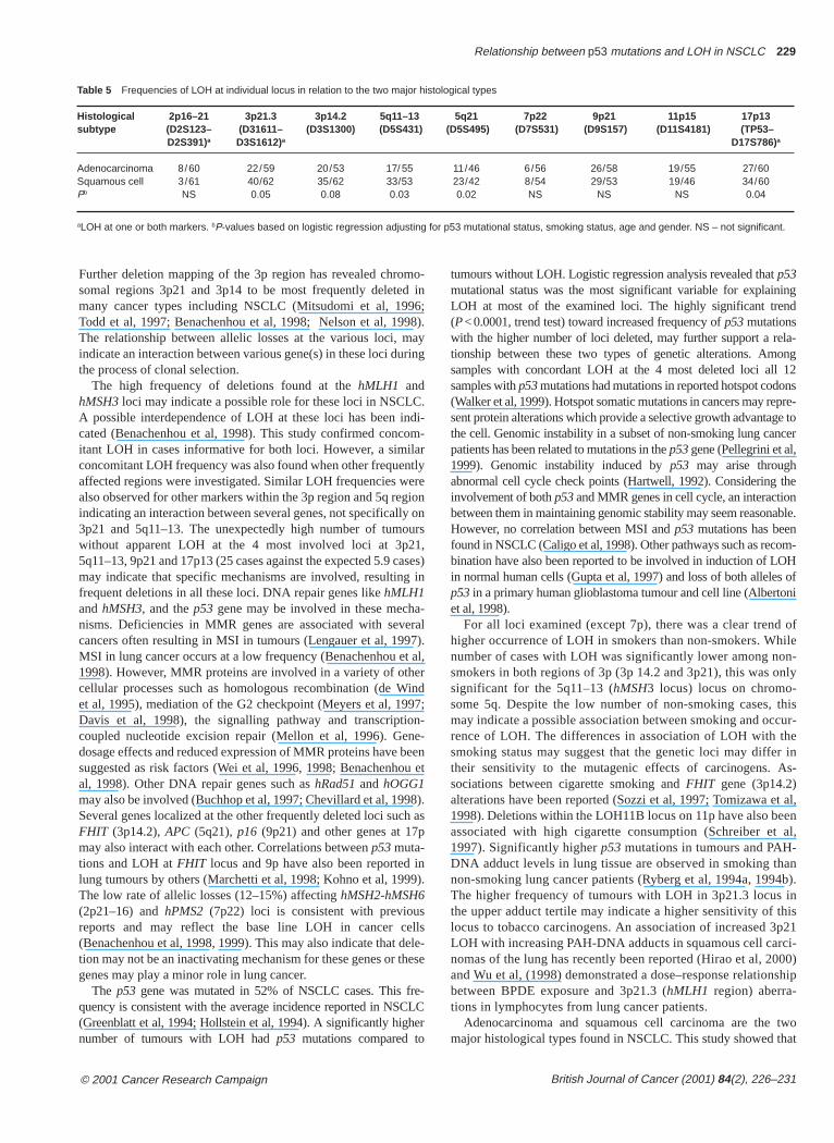
Table 5 Frequencies of LOH at individual locus in relation to the two major histological types
Histological 2p16–21 3p21.3 3p14.2 5q11–13 5q21 7p22 9p21 11p15 17p13subtype (D2S123– (D31611– (D3S1300) (D5S431) (D5S495) (D7S531) (D9S157) (D11S4181) (TP53–
D2S391)a D3S1612)a D17S786)a
Adenocarcinoma 8/60 22/59 20/53 17/ 55 11/46 6/56 26/58 19/55 27/60 Squamous cell 3/61 40/62 35/62 33/53 23/42 8/54 29/53 19/46 34/60 Pb NS 0.05 0.08 0.03 0.02 NS NS NS 0.04
aLOH at one or both markers. bP-values based on logistic regression adjusting for p53 mutational status, smoking status, age and gender. NS – not significant.
Further deletion mapping of the 3p region has revealed chromsomal regions 3p21 and 3p14 to be most frequently deletedmany cancer types including NSCLC (Mitsudomi et al, 1996Todd et al, 1997; Benachenhou et al, 1998; Nelson et al, 199The relationship between allelic losses at the various loci, mindicate an interaction between various gene(s) in these loci durthe process of clonal selection.
The high frequency of deletions found at the hMLH1 andhMSH3loci may indicate a possible role for these loci in NSCLCA possible interdependence of LOH at these loci has been incated (Benachenhou et al, 1998). This study confirmed concoitant LOH in cases informative for both loci. However, a similaconcomitant LOH frequency was also found when other frequenaffected regions were investigated. Similar LOH frequencies wealso observed for other markers within the 3p region and 5q regindicating an interaction between several genes, not specifically3p21 and 5q11–13. The unexpectedly high number of tumouwithout apparent LOH at the 4 most involved loci at 3p215q11–13, 9p21 and 17p13 (25 cases against the expected 5.9 camay indicate that specific mechanisms are involved, resulting frequent deletions in all these loci. DNA repair genes like hMLH1and hMSH3, and the p53 gene may be involved in these mechanisms. Deficiencies in MMR genes are associated with sevecancers often resulting in MSI in tumours (Lengauer et al, 1997MSI in lung cancer occurs at a low frequency (Benachenhou et1998). However, MMR proteins are involved in a variety of othecellular processes such as homologous recombination (de Wet al, 1995), mediation of the G2 checkpoint (Meyers et al, 199Davis et al, 1998), the signalling pathway and transcriptioncoupled nucleotide excision repair (Mellon et al, 1996). Gendosage effects and reduced expression of MMR proteins have bsuggested as risk factors (Wei et al, 1996, 1998; Benachenhoal, 1998). Other DNA repair genes such as hRad51and hOGG1may also be involved (Buchhop et al, 1997; Chevillard et al, 1998Several genes localized at the other frequently deleted loci suchFHIT (3p14.2), APC (5q21), p16 (9p21) and other genes at 17pmay also interact with each other. Correlations between p53muta-tions and LOH at FHIT locus and 9p have also been reported ilung tumours by others (Marchetti et al, 1998; Kohno et al, 1999The low rate of allelic losses (12–15%) affecting hMSH2-hMSH6(2p21–16) and hPMS2 (7p22) loci is consistent with previousreports and may reflect the base line LOH in cancer ce(Benachenhou et al, 1998, 1999). This may also indicate that detion may not be an inactivating mechanism for these genes or thgenes may play a minor role in lung cancer.
The p53 gene was mutated in 52% of NSCLC cases. This frquency is consistent with the average incidence reported in NSC(Greenblatt et al, 1994; Hollstein et al, 1994). A significantly highenumber of tumours with LOH had p53 mutations compared to
© 2001 Cancer Research Campaign
o- in;8).aying
.di-m-rtlyreion onrs,ses)in
-ral).
al,rind7;-
e-eenu et
). as
n).
llsle-
ese
e-LCr
tumours without LOH. Logistic regression analysis revealed that p53mutational status was the most significant variable for explainLOH at most of the examined loci. The highly significant tren(P<0.0001, trend test) toward increased frequency of p53mutationswith the higher number of loci deleted, may further support a retionship between these two types of genetic alterations. Amsamples with concordant LOH at the 4 most deleted loci all samples with p53mutations had mutations in reported hotspot codo(Walker et al, 1999). Hotspot somatic mutations in cancers may resent protein alterations which provide a selective growth advantagthe cell. Genomic instability in a subset of non-smoking lung canpatients has been related to mutations in the p53gene (Pellegrini et al,1999). Genomic instability induced by p53 may arise throughabnormal cell cycle check points (Hartwell, 1992). Considering involvement of both p53and MMR genes in cell cycle, an interactiobetween them in maintaining genomic stability may seem reasonaHowever, no correlation between MSI and p53 mutations has beenfound in NSCLC (Caligo et al, 1998). Other pathways such as recbination have also been reported to be involved in induction of LOin normal human cells (Gupta et al, 1997) and loss of both allelep53in a primary human glioblastoma tumour and cell line (Albertoet al, 1998).
For all loci examined (except 7p), there was a clear trendhigher occurrence of LOH in smokers than non-smokers. Whnumber of cases with LOH was significantly lower among nosmokers in both regions of 3p (3p 14.2 and 3p21), this was osignificant for the 5q11–13 (hMSH3 locus) locus on chromo-some 5q. Despite the low number of non-smoking cases, may indicate a possible association between smoking and ocrence of LOH. The differences in association of LOH with thsmoking status may suggest that the genetic loci may diffetheir sensitivity to the mutagenic effects of carcinogens. Asociations between cigarette smoking and FHIT gene (3p14.2)alterations have been reported (Sozzi et al, 1997; Tomizawa e1998). Deletions within the LOH11B locus on 11p have also beeassociated with high cigarette consumption (Schreiber et 1997). Significantly higher p53mutations in tumours and PAH-DNA adduct levels in lung tissue are observed in smoking thnon-smoking lung cancer patients (Ryberg et al, 1994a, 199The higher frequency of tumours with LOH in 3p21.3 locus the upper adduct tertile may indicate a higher sensitivity of tlocus to tobacco carcinogens. An association of increased 3LOH with increasing PAH-DNA adducts in squamous cell carcnomas of the lung has recently been reported (Hirao et al, 20and Wu et al, (1998) demonstrated a dose–response relationbetween BPDE exposure and 3p21.3 (hMLH1 region) aberra-tions in lymphocytes from lung cancer patients.
Adenocarcinoma and squamous cell carcinoma are the major histological types found in NSCLC. This study showed th
British Journal of Cancer (2001) 84(2), 226–231

nce,
is.
,
-
man
96)
l
f
tic
W,
all
es
230 S Zienolddiny et al
allelic deletions were more frequent in squamous cell carcinomfor most loci. After adjusting for p53mutational status, smoking,gender and age the difference between the two subtypes becsignificant for the 3p, 5q and 17p regions. Different pattern ogenetic changes have previously been reported for the 2 hislogical types (Sato et al, 1994). We have also found a significanhigher frequency of p53 mutations in squamous cell carcinomasthan adenocarcinomas (Kure et al, 1996; Skaug et al, 2000). Tdevelopment of different histological types of lung tumours habeen suggested to reflect a dose effect of tobacco carcinogeMore centrally located cells of the airways giving rise to squamocell carcinomas are exposed to a greater carcinogen concentrathan more distal cells giving rise to the more peripherally locateadenocarcinomas (Burke et al, 1998).
Our data indicate that LOH at 3p, 5q, 9p, 11p and 17p regiooften affected in NSCLC, are interdependent and highly associawith mutated p53 gene. Compounds in the tobacco smoke iaddition to clonal selection may be the driving force in thesalterations.
ACKNOWLEDGEMENTS
We thank Ms Rita Bæra for excellent technical assistance aDr Lodve Stangeland for providing lung tissue and patient daThis project was supported by The Norwegian Research CounThe Norwegian Cancer Society and EU grant ENV4-CT97-0469
REFERENCES
Albertoni M, Daub DM, Arden KC, Viars CS, Powell C and Van Meir EG (1998)Genetic instability leads to loss of both p53alleles in a human glioblastoma.Oncogene16: 321–326
Benachenhou N, Guiral S, Gorska-Flipot I, Labuda D and Sinnett D (1998) Highresolution deletion mapping reveals frequent allelic losses at the DNAmismatch repair loci hMLH1and hMSH3in non-small cell lung cancer. Int JCancer77: 173–180
Benachenhou N, Guiral S, Gorska-Flipot I, Labuda D and Sinnett D (1999) Frequloss of heterozygosity at the DNA mismatch-repair loci hMLH1and hMSH3insporadic breast cancer. Br J Cancer79: 1012–1017
Bennett WP (1995) p53alterations in progenitor lesions of the bronchus, esophaguoral cavity, and colon. Cancer Detect Prev19: 503–511
Bennett WP, Colby TV, Travis WD, Borkowski A, Jones RT, Lane DP, Metcalf RA,Samet JM, Takeshima Y and Gu JR (1993) p53protein accumulates frequentlyin early bronchial neoplasia. Cancer Res53: 4817–4822
Buchhop S, Gibson MK, Wang XW, Wagner P, Sturzbecher HW and Harris CC(1997) Interaction of p53with the human Rad51 protein. Nucleic Acids Res25:3868–3874
Burke L, Khan MA, Freedman AN, Gemma A, Rusin M, Guinee DG, Bennett WP,Caporaso NE, Fleming MV, Travis WD, Colby TV, Trastek V, Pairolero PC,Tazelaar HD, Midthun DE, Liotta LA and Harris CC (1998) Allelic deletionanalysis of the FHIT gene predicts poor survival in non-small cell lung cancer.Cancer Res58: 2533–2536
Caligo MA, Ghimenti C, Marchetti A, Lonobile A, Buttitta F, Pellegrini S andBevilacqua G (1998) Microsatellite alterations and p53, TGFbetaRII, IGFIIRand BAXmutations in sporadic non-small-cell lung cancer. Int J Cancer78:606–609
Canzian F, Salovaara R, Hemminki A, Kristo P, Chadwick RB, Aaltonen LA andde la Chapelle A (1996) Semiautomated assessment of loss of heterozygosiand replication error in tumors. Cancer Res56: 3331–3337
Chevillard S, Radicella JP, Levalois C, Lebeau J, Poupon MF, Oudard S, Dutrillauxand Boiteux S (1998) Mutations in OGG1, a gene involved in the repair ofoxidative DNA damage, are found in human lung and kidney tumours.Oncogene16: 3083–3086
Davis TW, Wilson-Van PC, Meyers M, Kunugi KA, Cuthill S, Reznikoff C, Garces C,Boland CR, Kinsella TJ, Fishel R and Boothman DA (1998) Defective
British Journal of Cancer (2001) 84(2), 226–231
as
amefto-tly
hesns.
ustiond
ns,tedne
ndta.cil,.
ent
s,
ty
B
expression of the DNA mismatch repair protein, MLH1, alters G2-Mcell cycle checkpoint arrest following ionizing radiation. Cancer Res58:767–778
de Wind N, Dekker M, Berns A, Radman M and te RH (1995) Inactivation of themouse Msh2 gene results in mismatch repair deficiency, methylation tolerahyperrecombination, and predisposition to cancer. Cell 82: 321–330
Denissenko MF, Pao A, Tang M and Pfeifer GP (1996) Preferential formation ofbenzo[a]pyrene adducts at lung cancer mutational hotspots in p53. Science274:430–432
Eshleman JR and Markowitz SD (1996) Mismatch repair defects in humancarcinogenesis. Hum Mol Genet5: 1489–1494
Gazdar AF (1994) The molecular and cellular basis of human lung cancer.Anticancer Res14: 261–267
Greenblatt MS, Bennett WP, Hollstein M and Harris CC (1994) Mutations in the p53tumor suppressor gene: clues to cancer etiology and molecular pathogenesCancer Res54: 4855–4878
Gupta PK, Sahota A, Boyadjiev SA, Bye S, Shao C, O’Neill JP, Hunter TC,Albertini RJ, Stambrook PJ and Tischfield JA (1997) High frequency in vivoloss of heterozygosity is primarily a consequence of mitotic recombination.Cancer Res57: 1188–1193
Hartwell L (1992) Defects in a cell cycle checkpoint may be responsible for thegenomic instability of cancer cells. Cell 71: 543–546
Hirao T, Nelson HH, Ashok TDS, Wiencke JK, Zu Z-f, Gunn L, Wain JC, Mark EWChristiani DC and Kelsey KT (2000) Early smoking and increased DNAadduct burden predict LOH at 3p21 in NSCLC. Proc Am Ass Cancer Res41:321 (Abstract)
Hollstein M, Rice K, Greenblatt MS, Soussi T, Fuchs R, Sorlie T, Hovig E, SmithSorensen B, Montesano R and Harris CC (1994) Database of p53gene somaticmutations in human tumors and cell lines. Nucleic Acids Res22: 3551–3555
Horio Y, Takahashi T, Kuroishi T, Hibi K, Suyama M, Niimi T, Shimokata K,Yamakawa K, Nakamura Y and Ueda R (1993) Prognostic significance of p53mutations and 3p deletions in primary resected non-small cell lung cancer.Cancer Res53: 1–4
Kohno H, Hiroshima K, Toyozaki T, Fujisawa T and Ohwada H (1999) p53mutation and allelic loss of chromosome 3p, 9p of preneoplastic lesions inpatients with nonsmall cell lung carcinoma. Cancer85: 341–347
Kohno T, Yokota J (1999) How many tumor suppressor genes are involved in hulung carcinogenesis? Carcinogenesis20: 1403–1410
Kure EH, Ryberg D, Hewer A, Phillips DH, Skaug V, Baera R and Haugen A (19p53mutations in lung tumours: relationship to gender and lung DNA adductlevels. Carcinogenesis17: 2201–2205
Lengauer C, Kinzler KW and Vogelstein B (1997) Genetic instability in colorectacancers. Nature386: 623–627
Lindstedt BA, Ryberg D, Zienolddiny S, Khan H and Haugen A (1999) Hras 1VNTR alleles as susceptibility markers for lung cancer: relationship tomicrosatellite instability in tumors. Anticancer Res19: 5523–5527
Livingstone LR, White A, Sprouse J, Livanos E, Jacks T and Tlsty TD (1992)Altered cell cycle arrest and gene amplification potential accompany loss owild-type p53. Cell70: 923–935
Marchetti A, Pellegrini S, Sozzi G, Bertacca G, Gaeta P, Buttitta F, Carnicelli V,Griseri P, Chella A, Angeletti CA, Pierotti M and Bevilacqua G (1998) Geneanalysis of lung tumours of non-smoking subjects: p53gene mutations areconstantly associated with loss of heterozygosity at the FHIT locus.Br J Cancer78: 73–78
Mekeel KL, Tang W, Kachnic LA, Luo CM, DeFrank JS and Powell SN (1997)Inactivation of p53results in high rates of homologous recombination.Oncogene14: 1847–1857
Mellon I, Rajpal DK, Koi M, Boland CR and Champe GN (1996) Transcription-coupled repair deficiency and mutations in human mismatch repair genes.Science272: 557–560
Meyers M, Theodosiou M, Acharya S, Odegaard E, Wilson T, Lewis JE, Davis TWilson-Van PC, Fishel R and Boothman DA (1997) Cell cycle regulation ofthe human DNA mismatch repair genes hMSH2, hMLH1, and hPMS2. CancerRes57: 206–208
Mitsudomi T, Oyama T, Nishida K, Ogami A, Osaki T, Sugio K, Yasumoto K,Sugimachi K and Gazdar AF (1996) Loss of heterozygosity at 3p in non-smcell lung cancer and its prognostic implication. Clin Cancer Res2: 1185–1189
Mollerup S, Ryberg D, Hewer A, Phillips DH and Haugen A (1999) Sex differencin lung CYP1A1 expression and DNA adduct levels among lung cancerpatients. Cancer Res59: 3317–3320
Nelson HH, Wiencke JK, Gunn L, Wain JC, Christiani DC and Kelsey KT (1998)Chromosome 3p14 alterations in lung cancer: evidence that FHIT exondeletion is a target of tobacco carcinogens and asbestos. Cancer Res58:1804–1807
© 2001 Cancer Research Campaign

Relationship between p53 mutations and LOH in NSCLC 231
ll
ot
g
sis
Pellegrini S, Bertacca G, Buttitta F, Bevilacqua G and Marchetti A (1999) Lungtumours from non-smoking subjects: A p53-related genetic instability in asubset of cases. Int J Mol Med4: 419–424
Ryberg D, Hewer A, Phillips DH and Haugen A (1994a) Different susceptibility tsmoking-induced DNA damage among male and female lung cancer patieCancer Res54: 5801–5803
Ryberg D, Kure E, Lystad S, Skaug V, Stangeland L, Mercy I, Borresen AL andHaugen A (1994b) p53mutations in lung tumors: relationship to putativesusceptibility markers for cancer. Cancer Res54: 1551–1555
Sato S, Nakamura Y and Tsuchiya E (1994) Difference of allelotype betweensquamous cell carcinoma and adenocarcinoma of the lung. Cancer Res54:5652–5655
Schreiber G, Fong KM, Peterson B, Johnson BE, O’Briant KC and Bepler G (19Smoking, gender, and survival association with allele loss for the LOH11Blung cancer region on chromosome 11. Cancer Epidemiol Biomarkers Prev6:315–319
Skaug V, Ryberg D, Kure EH, Arab MO, Stangeland L, Myking AO and Haugen(2000) p53mutations in defined structural and functional domains are relateto poor clinical outcome in non-small cell lung cancer patients. Clin CancerRes6: 1031–1037
Sozzi G, Sard L, De Gregorio L, Marchetti A, Musso K, Buttitta F, Tornielli S,Pellegrini S, Veronese ML, Manenti G, Incarbone M, Chella A, Angeletti CAPastorino U, Huebner K, Bevilaqua G, Pilotti S, Croce CM and Pierotti MA(1997) Association between cigarette smoking and FHIT gene alterations inlung cancer. Cancer Res57: 2121–2123
Takagi Y, Osada H, Kuroishi T, Mitsudomi T, Kondo M, Niimi T, Saji S, Gazdar ATakahashi T and Minna JD (1998) p53mutations in non-small-cell lungcancers occurring in individuals without a past history of active smoking.Br J Cancer77: 1568–1572
Todd S, Franklin WA, Varella-Garcia M, Kennedy T, Hilliker CEJ, Hahner L,Anderson M, Wiest JS, Drabkin HA and Gemmill RM (1997) Homozygous
© 2001 Cancer Research Campaign
onts.
97)
Ad
,
F,
deletions of human chromosome 3p in lung tumors. Cancer Res57: 1344–1352
Tomizawa Y, Nakajima T, Kohno T, Saito R, Yamaguchi N and Yokota J (1998)Clinicopathological significance of Fhit protein expression in stage I non-smacell lung carcinoma. Cancer Res58: 5478–5483
Walker DR, Bond JP, Tarone RE, Harris CC, Makalowski W, Boguski MS andGreenblatt MS (1999) Evolutionary conservation and somatic mutation hotspmaps of p53: correlation with p53protein structural and functional features.Oncogene18: 211–218
Wei Q and Spitz MR (1997) The role of DNA repair capacity in susceptibility tolung cancer: a review. Cancer Metastasis Rev16: 295–307
Wei Q, Cheng L, Hong WK and Spitz MR (1996) Reduced DNA repair capacity inlung cancer patients. Cancer Res56: 4103–4107
Wei Q, Eicher SA, Guan Y, Cheng L, Xu J, Young LN, Saunders KC, Jiang H, HonWK, Spitz MR and Strom SS (1998) Reduced expression of hMLH1andhGTBP/hMSH6: a risk factor for head and neck cancer. Cancer EpidemiolBiomakers Prev7: 309–314
Wistuba II, Montellano FD, Milchgrub S, Virmani AK, Behrens C, Chen H,Ahmadian M, Nowak JA, Muller C, Minna JD and Gazdar AF (1997)Deletions of chromosome 3p are frequent and early events in the pathogeneof uterine cervical carcinoma. Cancer Res57: 3154–3158
Wistuba II, Behrens C, Milchgrub S, Bryant D, Hung J, Minna JD and Gazdar AF(1999) Sequential molecular abnormalities are involved in the multistagedevelopment of squamous cell lung carcinoma. Oncogene18: 643–650
Wu X, Zhao Y, Honn SE, Tomlinson GE, Minna JD, Hong WK and Spitz MR(1998) Benzo[a]pyrene diol epoxide-induced 3p21.3 aberrations and geneticpredisposition to lung cancer. Cancer Res58: 1605–1608
Yin Y, Tainsky MA, Bischoff FZ, Strong LC and Wahl GM (1992) Wild-type p53restores cell cycle control and inhibits gene amplification in cells with mutantp53alleles. Cell 70: 937–948
British Journal of Cancer (2001) 84(2), 226–231