Long-term dietary exposure to low concentration of dichloroacetic acid promoted longevity and...
16
Long-term dietary exposure to low concentration of dichloroacetic acid promoted longevity and attenuated cellular and functional declines in aged Drosophila melanogaster Ashutosh Pandey & Divya Vimal & Swati Chandra & Sanjay Saini & Gopeshwar Narayan & Debapratim Kar Chowdhuri Received: 11 November 2013 /Accepted: 3 February 2014 # American Aging Association 2014 Abstract Dichloroacetic acid (DCA), a water disinfec- tion by-product, has attained emphasis due to its pros- pect for clinical use against different diseases including cancer along with negative impact on organisms. However, these reports are based on the toxicological as well clinical data using comparatively higher concen- trations of DCA without much of environmental rele- vance. Here, we evaluate cellular as well as organismal effects of DCA at environmentally and mild clinically relevant concentrations (0.02–20.0 μg/ml) using an established model organism, Drosophila melanogaster . Flies were fed on food mixed with test concentrations of DCA for 12–48 h to examine the induction of reactive oxygen species (ROS) generation, oxidative stress (OS), heat shock genes (hsps) and cell death along with or- ganismal responses. We also examined locomotor performance, ROS generation, glutathione (GSH) de- pletion, expression of GSH-synthesizing genes (gclc and gclm), and hsps at different days (0, 10, 20, 30, 40, 50) of the age in flies after prolonged DCA expo- sure. We observed mild OS and induction of antioxidant defense system in 20.0 μg/ml DCA-exposed organism after 24 h. After prolonged exposure to DCA, exposed organism exhibited improved survival, elevated expres- sion of hsp27, gclc, and gclm concomitant with lower ROS generation and GSH depletion and improved lo- comotor performance. Conversely, hsp27 knockdown flies exhibited reversal of the above end points. The study provides evidence for the attenuation of cellular and functional decline in aged Drosophila after prolonged DCA exposure and the effect of hsp27 mod- ulation which further incites studies towards the thera- peutic application of DCA. Keywords DCA . Drosophila . Healthspan . ROS . GSH . hsp27 . Prolonged exposure Introduction Dichloroacetic acid (DCA), a water disinfection by- product, is prospected as a potential environmental haz- ard and an investigational drug (Stacpoole 2011). Environmental sources of DCA include chlorinated drinking water, industrial solvents, and pharmaceuticals (Stacpoole et al. 1998). Levels of DCA detected in AGE DOI 10.1007/s11357-014-9628-1 A. Pandey : D. Vimal : S. Chandra : S. Saini : D. Kar Chowdhuri (*) Embryotoxicology Section, CSIR-Indian Institute of Toxicology Research, Lucknow 226001 Uttar Pradesh, India e-mail: [email protected] D. Kar Chowdhuri e-mail: [email protected] A. Pandey : G. Narayan Department of Molecular and Human Genetics, Banaras Hindu University, Varanasi 221005 Uttar Pradesh, India
-
Upload
independent -
Category
Documents
-
view
3 -
download
0
Transcript of Long-term dietary exposure to low concentration of dichloroacetic acid promoted longevity and...

Long-term dietary exposure to low concentrationof dichloroacetic acid promoted longevity and attenuatedcellular and functional declines in aged Drosophilamelanogaster
Ashutosh Pandey & Divya Vimal & Swati Chandra &
Sanjay Saini & Gopeshwar Narayan &
Debapratim Kar Chowdhuri
Received: 11 November 2013 /Accepted: 3 February 2014# American Aging Association 2014
Abstract Dichloroacetic acid (DCA), a water disinfec-tion by-product, has attained emphasis due to its pros-pect for clinical use against different diseases includingcancer along with negative impact on organisms.However, these reports are based on the toxicologicalas well clinical data using comparatively higher concen-trations of DCA without much of environmental rele-vance. Here, we evaluate cellular as well as organismaleffects of DCA at environmentally and mild clinicallyrelevant concentrations (0.02–20.0 μg/ml) using anestablished model organism, Drosophila melanogaster.Flies were fed on food mixed with test concentrations ofDCA for 12–48 h to examine the induction of reactiveoxygen species (ROS) generation, oxidative stress (OS),heat shock genes (hsps) and cell death along with or-ganismal responses. We also examined locomotor
performance, ROS generation, glutathione (GSH) de-pletion, expression of GSH-synthesizing genes (gclcand gclm), and hsps at different days (0, 10, 20, 30,40, 50) of the age in flies after prolonged DCA expo-sure. We observed mild OS and induction of antioxidantdefense system in 20.0 μg/ml DCA-exposed organismafter 24 h. After prolonged exposure to DCA, exposedorganism exhibited improved survival, elevated expres-sion of hsp27, gclc, and gclm concomitant with lowerROS generation and GSH depletion and improved lo-comotor performance. Conversely, hsp27 knockdownflies exhibited reversal of the above end points. Thestudy provides evidence for the attenuation of cellularand functional decline in aged Drosophila afterprolonged DCA exposure and the effect of hsp27 mod-ulation which further incites studies towards the thera-peutic application of DCA.
Keywords DCA .Drosophila . Healthspan . ROS .
GSH . hsp27 . Prolonged exposure
Introduction
Dichloroacetic acid (DCA), a water disinfection by-product, is prospected as a potential environmental haz-ard and an investigational drug (Stacpoole 2011).Environmental sources of DCA include chlorinateddrinking water, industrial solvents, and pharmaceuticals(Stacpoole et al. 1998). Levels of DCA detected in
AGEDOI 10.1007/s11357-014-9628-1
A. Pandey :D. Vimal : S. Chandra : S. Saini :D. Kar Chowdhuri (*)Embryotoxicology Section, CSIR-Indian Institute ofToxicology Research,Lucknow 226001 Uttar Pradesh, Indiae-mail: [email protected]
D. Kar Chowdhurie-mail: [email protected]
A. Pandey :G. NarayanDepartment of Molecular and Human Genetics, BanarasHindu University,Varanasi 221005 Uttar Pradesh, India

drinking water ranged from 4.5–7.5 μg/l (Japan) to200 μg/l (Australia), while its level in surface waterdownstream from a paper mill in Austria was detectedbetween <3 and 522 μg/l (Geist et al. 1991). Humanpopulation has been exposed to drinking water contain-ing up to 160 μg/l DCA for many generations(Stacpoole 2011).
Xenobiotic interaction and response related studieson DCA have been aggravated by both environmentaland therapeutic concerns. DCA has been reported as apossible pharmacological cure for chronic diseases suchas lactic acidosis and other mitochondria associateddiseases (Michelakis et al. 2010; Miquel et al. 2012;Stacpoole et al. 2008). In a number of in vitro studies,DCA is reported to enhance reactive oxygen generation(ROS) generation and oxidative stress leading to celldeath (Ayyanathan et al. 2012; Hassoun and Ray 2003;Wong et al. 2008). In exposed in vivo models, mainlyrodents, DCA is reported to cause developmental-(Smith et al. 1992), spermato- (Linder et al. 1997),immuno-, and hepatotoxicity (Cai et al. 2007; Hassounand Dey 2008). However, concern about DCA adversityunderlies on the data generated in rodents after theirexposure to this chemical at doses (up to ~4,000 mg/kg)thousands of times higher than those to which humansare usually exposed (Stacpoole et al. 1998). In the samecontext, a dose range of 10–100 mg/kg of DCA issuggested as therapeutics for human use via intravenousand oral routes (Stacpoole 2011). On the other, studiesregarding long-term exposure to environmentally rele-vant concentrations (~160–200 μg/l, detected in theenvironment and to which organisms including humanare likely to be exposed) of DCA are elusive.
Organismal susceptibility to chronic and degener-ative diseases is casually linked to aging. Age-relatedbehavioral insufficiencies are one of the main func-tional changes that occur in aged individuals. Inhuman, progressive decline in locomotor ability,memory function, and olfactory sensitivity is imper-ative age-related changes (Grotewiel et al. 2005).Thus, life expectancy may be improved by the ame-lioration of age-related behavioral declines (Jonesand Grotewiel 2011). Strategies for promotingheathspan via reduced ROS generation and increasedantioxidants level have gained much interest (Gruberet al. 2008, 2009). Among the antioxidants, glutathi-one (GSH) is the most abundant nonprotein thiolthat protects the organism against oxidative stressand participates in the detoxification of xenobiotics
and/or their metabolites, antioxidant defense, mainte-nance of redox potential, regulation of cell cycleprogression, and apoptosis (Lu 2013). ReducedGSH levels are reported in aged organisms (Jianget al. 2013; Suh et al. 2004). The rate-limitingenzyme, glutamate cysteine ligase (GCL), is one ofthe key determinants of GSH biosynthesis and iscomposed of catalytic (GCLc) and modifier(GCLm) subunits. In in vitro studies, altered expres-sion of GCL against xenobiotic exposure was report-ed (Ha et al. 2006; Thompson et al. 2009; Zhenget al. 2007). Although DCA has been reported toupregulate GSH biosynthesis via the induction ofGCL in mice (Theodoratos et al. 2012), no informa-tion is available on the modulation of GSH level andthe status of GCL expression (GCLc and GCLm) inDCA-exposed organism in the context of aging.
Premature aging of an organism has been implicat-ed by the alteration in the expression and regulationof a number of genes concurrent with factors likexenobiotics exposure, dietary habits, etc. Heat shockproteins (HSPs) are reported to function as chaper-ones by orchestrating correct folding and unfolding ofproteins. Levels of HSPs increase in response todifferent types of stresses that also include xenobioticexposure. HSPs can counteract proteo-toxicity andfavor stress resistance to the organism which maybe causally linked to an increase in life span vis-à-vis positive impact on the aging-related functionaldeclines (Tower 2009, 2011). Genome-wide studieson age-associated gene expression changes in flieshave shown the upregulation of heat shock genes(hsps) (Curtis et al. 2007; Landis et al. 2012). Inaddition, modulated expression of hsps (hsp70,hsp27, and hsp22) has been reported to alter life spanin flies (Kim et al. 2010; Liao et al. 2008; Tatar et al.1997) and higher level of HSPs is reported in longer-lived mammals and birds (Salway et al. 2011). In thesame context, improved health- and life span wereobserved in DCA-exposed Caenorhabditis elegans(Schaffer et al. 2011). However, studies regardingexpression of hsps in DCA-exposed organisms areinadequate.
Model organisms provide ample opportunities to ex-amine the underlying mechanism of xenobiotic-inducedeffects on exposed organism. Drosophila, an insectmodel, with well-documented genetics and develop-mental biology and high degree of homology of itsgenes with that of higher mammals, is the closest
AGE

invertebrate to the humans and has been used for toxi-cological studies and for studying human diseases(Jeibmann and Paulus 2009). Relevant to the highermammals, Drosophila exhibits an age-related declinein several behaviors such as senescence of motor activ-ity, olfaction, olfactory memory, and noncircadian rest(Grotewiel et al. 2005). Assay for locomotor perfor-mance is one of the reliable assays to examine thesenescence of motor activity in flies (Lliadi et al.2012). Further, this model has been a key to compre-hend the association between hsps and aging processsince the discovery of heat shock response and hsps(Tower 2011). It raises fewer ethical concerns and fallswithin the recommendations of the European Centre forthe Validation of Alternative Methods (ECVAM) andaims to prop up the scientific and regulatory acceptanceof alternative methods that are important in the field ofbiological science and towards reducing, refining, andreplacing the use of laboratory animals (Benford et al.2000).
The present study, therefore, aims to examine thecellular stress inducing potential of DCA in exposednontarget organism, Drosophila melanogaster, and itseffect on life span and age-dependent impairments.Further, the study is intended to provide evidence onthe modulatory effect of small hsp, viz., hsp27, onDCA-exposed organism.
Materials and methods
Fly strain and culture
Wild-type D. melanogaster (Oregon R+), w1118, Gal4-UAS transgenic lines, namely, Act-Gal4/Cyo, UAS-hsp27, UAS-hsp27RNAi, and UAS-hsp22RNAi, were usedin this study. The fly strains were reared on a standardDrosophila food medium (consisting of agar-agar,maize powder, sugar, yeast, nepagin, and propionicacid) at 24±1 °C. Additional yeast supplement wasprovided for healthy growth of the organisms.
Chemical and treatment schedule
DCA (PESTANAL® analytical standard, 99.3 %) ob-tained from Sigma Chemicals, St. Louis, MO, USA,was used in the study. Of the four different concentra-tions of DCA used (0.02, 0.2, 2.0, and 20.0 μg/ml),lower concentrations (0.02 and 0.2 μg/ml) are
environmentally relevant (IARC 2004) while the othertwo higher concentrations (2.0 and 20.0 μg/ml) are lessthan clinically relevant concentrations (up to~100 mg/kg). Flies were allowed to feed on food con-taminated with different concentrations of DCA.Control group received standard Drosophila food.
Chemical estimation
Quantification of DCA in exposed organismwas carriedout by gas chromatography (GC) with an electron cap-ture detector (ECD). In brief, control and exposed flieswere homogenized in Milli-Q water and then treatedwith pyridine and methyl chloroformate to get volatileand nonpolar methyl ester of DCA (Mudiam et al.2013). The ester derivative after its extraction in hexanewas applied on an Agilent GLC7890A GC (Foster City,CA, USA) equipped with an ECD.
Emergence pattern of flies
First instar larvae were transferred to normal food me-dium (control) and to food containing different concen-trations of the DCA (50 larvae/vial, 10 vials/group). Thenumber of flies emerging from different groups wascounted until all the flies emerged (Gayathri andKrishnamurthy 1981).
Survivorship assay
To examine the effect of DCA on the life span, male flieswere fed on the food mixed with different concentra-tions of DCA from day 1 of their emergence. Foreach group, 250 flies (maximum 25 flies were main-tained per vial) were scored. Every alternate day,flies were transferred to fresh vials and the numberof dead flies was scored till the death of the last fly(Nazir et al. 2001).
Reproductive assay
Reproductive assay was performed using a previouslypublished method (Gayathri and Krishnamurthy 1981).Briefly, freshly eclosed first instar larvae were trans-ferred to control and chemical-contaminated food andthey were allowed to grow throughout their develop-ment. Virgin male and female flies emerging from con-trol and treated food were separated and mated in vialscontaining normal food. For each group, 10 pair of flies
AGE

in 10 individual vials were taken and transferred to freshvials everyday for the next 10 days and the number ofeggs laid during this period was scored. The total num-ber of flies emerging from the eggs laid during these10 days was counted; the mean number of flies emergedper pair for 10 days gave a measure of reproductiveperformance.
RNA isolation and quantitative real-time polymerasechain reaction (qPCR)
Total RNA from control and treated flies was extractedusing TRI reagent (Ambion, Austin, TX, USA). Purityand concentration of the isolated RNAwere determinedby measuring the absorbance ratio at 260/280 and 230/260 nm on a NanoDrop spectrophotometer(Wilmington, DE, USA). RNAwas reverse-transcribedinto cDNA using a cDNA synthesis kit (Fermentas,MD, USA) essentially following the manufacturer’sinstructions. qPCR was performed in 96-well PCRplates on a 7900 HT Fast Real-Time PCR(Applied Biosystems, CA, USA) using PowerSYBR Green Master Mix (Applied BiosystemsCA, USA) with gene-specific primers for the targetgenes (hsp22, hsp23, hsp26, hsp27, hsp60, hsp70,hsp83, gclc, and gclm) (primer details in Table 1).Relative quantification of gene expression was car-ried out by concurrent amplification of glyceralde-hyde 3-phosphate dehydrogenase (GAPDH) as anendogenous control.
Assay for oxidative stress markers
To examine the oxidative stress, ROS generation, super-oxide dismutase (SOD), catalase (CAT), and glutathioneS-transferase (GST) activities, GSH content, proteincarbonyl (PC) content, and lipid peroxidation (LPO)product were assayed in control and DCA-exposed flies.The above-mentioned assays were carried out in 10 %fly homogenate.
Preparation of fly homogenate
Flies from control and treated groups were homoge-nized in cold 0.1 M phosphate buffer (pH 7.4) contain-ing 0.15 M KCl to obtain 10 % homogenate. Thesupernatant after centrifugation at 12,000×g for10 min was used for different assays and proteinestimation.
Measurement of ROS generation
ROS generation was estimated by using the dye 2′,7′-dichlorodihydrofluorescein diacetate (DCFH-DA;Sigma, St. Louis, MO, USA). Assay was performed asdescribed by Zhao et al. (2010). In brief, 100 μl of flyhomogenate was incubated with 10 μM DCFH-DA (indimethyl sulfoxide) and fluorescence was measuredusing a microplate reader at an excitation and emissionwavelength of 485 and 535 nm, respectively. The fluo-rescence intensities were normalized to the proteinconcentration.
Superoxide dismutase (SOD) (superoxide: superoxideoxidoreductase EC 1.15.1.1)
The method for estimating SOD described previouslyby Nishikimi et al. (1972) was followed with minormodification (Gupta et al. 2005). The assay mixtureconsisted of 0.052 M sodium pyrophosphate buffer(pH 8.3), 186 μM phenazine methosulphate,300 μM nitroblue tetrazolium, 780 μM reduced nic-otinamide adenine dinucleotide, and the homogenate.
Table 1 Genes and their primer sequences used in RT-PCRamplification
hsp22 Forward (F) 5′ GGATGAACTGGACAAGGCTCTAAA 3′
Reverse (R) 5′ ATATGATTGGCGACTGCTTCTCC 3′
hsp23 Forward (F) 5′ GAGCCTTGCCGACGATTTG 3′
Reverse (R) 5′ GGCGCCCACCTGTTTCTC 3′
hsp26 Forward (F) 5′ GAGCCCCGCAGCCCCATCTA 3′
Reverse (R) 5′ GGAACGGCGCTGCTGAGTGC 3′
hsp27 Forward (F) 5′ GCGTCGCCTGCTACTGCCCAACAC 3′
Reverse (R) 5′ CTCCATTTTCTCGCCGCTGCCATTT 3′
hsp60 Forward (F) 5′ ATGTCGCGCCCCGTTAGCAC 3′
Reverse (R) 5′ GCCATCGCGTCCCACCTTCT 3′
hsp70 Forward (F) 5′ ACGGGCCAAGCGCACACTCTC 3′
Reverse (R) 5′ CCGCGCACAGTCCCTCAAACC 3′
hsp83 Forward (F) 5′ TGCGCACTTTTCGACCGTATCA 3′
Reverse (R) 5′ GAAAAACCCGACCCGAACTGGA 3′
gclc Forward (F) 5′ CGCCGGCGAGCTAATCACC 3′
Reverse (R) 5′ CTCCACCTGCTTGCCCTCCTG 3′
gclm Forward (F) 5′ GGAGCGCGTGGTGGTTGA 3′
Reverse (R) 5′ TGTGGAGTGGCGATTGAGGAA 3′
gapdh Forward (F) 5′ CTGGGCTACACCGATGAGGAG 3′
Reverse (R) 5′ CCACGAGATTAGCTTGACGAA 3′
AGE

One unit of enzyme activity is defined as the en-zyme concentration required for inhibiting chromo-gen production (optical density 560 nm) by 50 % in1 min under assay condition, and the results wereexpressed as specific activity in units per minute permilligram protein.
Catalase (CAT (H2O2: H2O2 oxidoreductase EC1.11.1.6)
CAT activity in the control and treated flies was mea-sured by following the ability of the enzyme to splitH2O2 within 1 min of incubation time. After incubation,the reaction was stopped by adding dichromate/aceticacid reagent (5 % solution of K2Cr2O7/glacial aceticacid, 1:3 by volume) and the remaining H2O2 wasdetermined by measuring chromic acetate at 570 nm asdescribed previously by Sinha (1972). Enzyme activitywas expressed as micromoles H2O2 decomposed perminute per milligram protein.
Assay for lipid peroxidation (LPO)
We followed the method of Ohkawa et al. (1979) forassaying malondialdehyde (MDA) as a measurement ofLPO using tetraethoxypropane as an external standard.The assay mixture consisted of 10 % sodium dodecylsulphate (SDS), 0.8% thiobarbituric acid (TBA), and flyhomogenate. Level of lipid peroxidation was expressedin terms of nanomoles MDA content per hour per mil-ligram protein.
Glutathione S-transferase (GST, EC 2.5.1.18)
GST activity was determined by the method of Habiget al. (1974) with minor modifications. The reactionmixture consisted of 0.2 M sodium phosphatebuffer, reduced glutathione (1.0 mM) and 1-chloro-2,4-dinitrobenzene (CDNB, 5.0 mM). Anincrease in absorbance (340 nm) was measuredfor 3 min at 30-s interval, and the enzyme activitywas calculated as nanomoles CDNB reduced perminute per milligram protein using a molar extinc-tion coefficient of 6.25×10³ M−1 cm−1.
Glutathione (GSH) content
GSH content in the exposed flies was quantified usingEllman’s reagent (Ellman 1959). The assay mixture
consisted of 0.2 M phosphate buffer (pH 8.0), 0.01 %5,5′-dithiobis-2-nitrobenzoic acid (DTNB), and the ho-mogenate. The reaction was monitored at 412 nm, andthe amount of GSH was expressed in terms ofnanomoles per milligram protein.
Determination of protein carbonyl (PC) content
PC content was determined by following the method ofLevine et al. (1990) with minor modifications. Twoequal aliquots of supernatant fraction were taken, onet r e a t e d w i t h e q u a l v o l u m e o f 2 , 4 -dinitrophenylhydrazine (10 mM dissolved in 2 M HCl)(test sample) and the other with 2 M HCl (blank). Eachmixture was incubated for 1 h, followed by precipitationwith 20 % TCA and subsequently extracted withethanol/ethylacetate mixture (1:1). The pellets were thendissolved in 1.0 ml of 6M guanidine hydrochloride. Thespectrum of DNPH-treated sample versus the HCl blankwas determined at 370 nm, and the results wereexpressed in terms of nanomoles DNPH incorporatedper milligram protein based on a molar absorption co-efficient of 22,000 M−1 cm−1.
Protein estimation
Protein concentration was determined by the method ofLowry et al. (1951) using a protein estimation kit(Bangalore Genei, Bangalore, India) essentially follow-ing the manufacturer’s protocol and bovine serum albu-min (BSA) as the standard.
Cell death assay
To examine cell death in control and DCA-exposedflies, DEVDase (caspase-3 like) and IETDase(caspase-9 like) activities were examined. The assay isbased on spectrophotometric detection of the chromo-phore p-nitroanilide (pNA) obtained after specificaction of caspase-3 and caspase-9 on tetrapeptidesubstrates, DEVD-pNA and IETD-pNA, respectively.The assay mixture consisted of fly homogenate,chilled cell lysis buffer, 2× reaction buffer containingdithiothreitol, and 200 μM substrate (BioVision cas-pase assay kit, CA, USA). The reaction mixture wasincubated at 37 °C for 1.5 h, and absorbance of theproduct was measured at 405 nm on a Cintra 20ultraviolet spectrophotometer (GBC ScientificEquipment, Australia).
AGE

Locomotor assay
Locomotor assay was performed as described previous-ly by Feany and Bender (2000). In brief, 20 flies in avertical plastic tube (18 cm×2 cm)were gently tapped tothe bottom and kept for 1 min in order to get acclima-tized with their environment. Flies that crossed the 15-cm line within 30 s from the time they were tapped to thebottom of vials were scored. One hundred male flieswere used for locomotor assay per group (5 replicates,20 flies in each). The locomotor performance was rep-resented in terms of mean percentage of flies thatcrossed the 15-cm line among the total number of fliesper experiment.
Measurement of body weight
Growth of flies was examined in terms of body weight.For each group, 100 male flies (4 replicates of 25 flies ineach) were weighed. Body weights were expressed inmilligrams.
Statistical analysis
Statistical significance of the mean values for differentparameters was monitored in control and exposed fliesusing ANOVA followed by post hoc tests after ascertain-ing the homogeneity of variance and normality of data. Formultiple comparisons, one-way and two-way ANOVAswere followed by Dunnett’s and Bonferroni’s test, respec-tively. Each end point was considered as a dependentvariable while concentration and time of exposure as in-dependent variables. Statistical significance level was as-cribed as P<0.05. Pearson’s correlations were calculated,and then, linear regression analysis was carried out. Prismcomputer program (GraphPad version 5.0, SanDiego, CA,USA) was used for the statistical analysis. Kaplan-Meieranalysis was used for survivorship analysis with stratifiedlog rank tests using SPSS software version 13.0, (SPSSInc., Chicago, IL, USA).
Results
Detection of DCA in exposed flies
DCA residues were detected in the exposed organism at its2.0- and 20.0-μg/ml concentrations, while for the rest twolower concentrations, DCA residues were beyond the
detection limit. We could recover 7.17±0.04 and 4.51±0.29 ng/g DCA from 2.0 μg/ml DCA-treated flies and53.46±0.51 and 31.62±0.96 ng/g DCA from 20.0 μg/mlDCA-exposed flies after 24 and 48 h, respectively.
Effect of DCA on emergence pattern of flies (OregonR+)
Figure 1a, b shows the emergence pattern and percentemergence in control and DCA-treated groups. We ob-served a nonsignificant (P<0.05) change in the emer-gence and larval development of flies in DCA-exposedgroups as compared to control.
Effect of DCA on reproductive performance
Figure 1c shows the reproductive performance in con-trol and DCA-exposed groups. We observed a nonsig-nificant (P>0.05) change in the reproductive perfor-mance of DCA-exposed groups as compared to controlin terms of fertility.
Acute exposure of DCA to Oregon R+ flies causedincreased ROS generation, oxidative stress,and expression of heat shock genes without propellingcell death
Assays for ROS generation, oxidative stress (OS) endpoints, and expression of hsps were performed in theflies exposed to different concentrations of DCA for 12–48 h along with their respective control. We observed asignificant (P<0.05) increase in ROS generation in20.0 μg/ml DCA-treated group after 24 h (~40 %) anda nonsignificant change after 48 h in comparison tocontrol. For the rest of the concentrations of DCA andexposure periods, the level of ROS generation in ex-posed flies was comparable to that in control (Fig. 2a).
Figure 2b–g shows OS parameters assayed in controland DCA-exposed groups. Concomitant with the induc-tion of ROS generation, we observed a significant(P<0.05) increase in MDA and PC contents and GSTactivity in 20.0 μg/ml DCA-treated flies after 24 h (anincrease of ~40, 52, and 47 % in MDA and PC contentsand GST activity, respectively) while the same endpoints were found to be nonsignificantly changed after48 h in comparison to control (Fig. 2d, g, e). Likewise,GSH content was found to be significantly decreased(~23 % decrease in comparison to control) in flies thatwere exposed to DCA for 24 h while the same after 48 h
AGE

was nonsignificantly changed as compared to control(Fig. 2f). Interestingly, we observed a significant in-crease in the activities of antioxidant enzymes (maxi-mum increase of ~59 and ~39 % in SOD and CATactivities, respectively, after 48 h) in 20.0 μg/ml DCA-treated flies after 24 and 48 h as compared to control(Fig. 2b, c). None of the lower concentrations (0.02, 0.2,2.0 μg/ml) of DCA evoked any change in the above endpoints in exposed organism.
To examine the expression of hsps (hsp22, hsp23,hsp26, hsp27, hsp60, hsp70, and hsp83), qPCR assaywas carried out in control and DCA-treated flies. Weobserved a significant (P<0.05) increase in the expres-sion of hsp22, hsp27, and hsp70 (maximum increase of~1.9- and 2.1-fold in hsp22 and hsp70 expression, re-spectively, after 24 h and ~2.3-fold in hsp27 expressionafter 48 h of DCA exposure) in 20.0μg/ml DCA-treatedflies after 24 and 48 h (Fig. 2h). None of the testedconcentrations of DCA evoked any significant increasein the expression of the other tested hsps in exposedorganism during the entire exposure regimen (12–48 h)(data not shown).
To examine whether DCA (20.0 μg/ml) induced OSin exposed organism leads to cell death, we measured
DEVD- and IETD-ase activities (caspase-3- andcaspase-9-like activities) in control and treated flies after24 h and observed a nonsignificant (P>0.05) change inthe enzyme activities in exposed organism as comparedto control (Fig. 2i).
Prolonged exposure of DCA to Oregon R+ fliesimproved their survival
Figure 3a presents the survival of control and DCA-exposed flies. While we did not observe any significant(P>0.05) change in the maximum life span and meansurvival of flies that were exposed to the lower concentra-tions (0.02, 0.2, 2.0 μg/ml, data not shown) of DCA, asignificant (P<0.05) increase in the maximum life span(~14 %) and the mean survival (~15 %), respectively, wasobserved in 20.0 μg/ml DCA-treated flies (Fig. 3b).
Prolonged exposure of DCA to Oregon R+ fliesattenuated their age-dependent decline in locomotorperformance without compromising organismal growth
To examine whether increased survival in DCA-exposedflies compromises with their healthspan, we measured
Fig. 1 Lack of any adverse effecton emergence, development, andreproductive performance ofDCA exposed D. melanogaster(Oregon R+). Graphs showingemergence pattern (a), percentemergence and mean emergencein days (b), and reproductiveperformance in terms of fertility(total progeny per female in10 days) (c) in control and DCA-exposed D. melanogaster. Datarepresent mean ± SD.Significance ascribed as *P<0.05vs. control
AGE

Fig. 2 Induction of ROS generation, oxidative stress end points,and hsps and noninduction of cell death in DCA-exposedD. melanogaster (Oregon R+). Graphs showing ROS generation(a), superoxide dismutase (SOD) and catalase (CAT) activities (b,c), malondialdehyde (MDA) content (d), glutathione S-transferase(GST) activity (e), glutathione (GSH) and protein carbonyl (PC)contents (f, g) in control andDCA-treatedD.melanogaster for 12–
48 h. qPCR data showing mRNA level (in terms of fold changeagainst control) for hsp22, hsp27, and hsp70 in DCA-treatedD. melanogaster for 12–48 h (h). Graph showing IETDase (cas-pase 9-like) and DEVDase (caspase 3-like) activities (i) in controland DCA-treatedD. melanogaster for 24 h. Data represent mean ±SD of three identical experiments made in three replicates. Signif-icance ascribed as *P<0.05 vs. control
Fig. 3 Improved survival ofD. melanogaster (Oregon R+)flies after prolonged exposure toDCA. Graphs showingsurvivorship (a) and maximumlife span and mean survival (b) incontrol and 20.0 μg/ml DCA-exposed D. melanogaster. Datarepresent mean ± SD.Significance ascribed as *P<0.05vs. control
AGE

growth and fitness in terms of their body weight andlocomotor performance, respectively, at different days oflife (0, 10, 20, 30, 40, 50 days) parallel to the age-matchedcontrol. Although we did not observe any apparent changein the body weight of exposed flies in comparison tocontrol (Fig. 4a), a significant (P<0.05) increase in loco-motor performance was observed in 40 and 50-day-oldDCA-exposed flies in comparison to their age-matchedcontrols (~24 and 28 % increased locomotor performancewas observed in 40- and 50-day DCA-exposed flies, re-spectively, in comparison to unexposed control flies ofsimilar age) (Fig. 4b).
Prolonged exposure of DCA to Oregon R+ flies rescuedthem from age-dependent ROS generation and GSHdepletion
To corroborate with the increased life span of20.0 μg/ml DCA-exposed flies, ROS generation andGSH content were also evaluated in exposed flies atthe above stated days of age. We observed a significantreduction in ROS generation in 40 and 50-day-old ex-posed flies (~32 and ~30% lower ROS generation in 40-and 50-day-old exposed flies, respectively, as comparedto unexposed flies of similar age) (Fig. 5a). Parallel tothe lowered ROS levels in these flies, we observed asignificant increase (P<0.05) in the GSH content inexposed flies with an advance in age (~42 and 39 %higher GSH content in 40- and 50-day-old DCA-treatedflies, respectively, as compared to unexposed flies ofsimilar age) (Fig. 5b).
Enhanced expression of gclc and gclm in aged OregonR+ flies after their prolonged exposure to DCA
To substantiate the increased GSH content, expression ofgclc and gclm (genes coding for GCLc and GCLm, re-spectively) was examined in control and 20.0μg/mlDCA-exposed flies at different days of their age (0, 10, 20, 30,40, 50 days) by qPCR assay. We observed a significant(P<0.05) increase in the expression of gclc and gclm in 40-and 50-day-old exposed flies (~1.8- and 1.7-fold increasedexpression of gclc and ~1.9- and 1.6-fold increase in gclmexpression in 40- and 50-day-old exposed flies, respective-ly, as compared to unexposed control flies) (Fig. 6a, b).
Enhanced expression of hsp27 in Oregon R+ fliesafter their prolonged exposure to DCA
To examine whether increased life span of DCA-exposed flies is causally linked to increased expressionof hsps, hsp22, hsp27, and hsp70 expressions in theexposed organism at different days of their age (0, 10,20, 30, 40, 50 days) along with the age-matched controlwere assayed by qPCR. We observed a nonsignificant(P<0.05) change in the expression of hsp22 in exposedflies from 0–50 days in comparison to their respectivecontrol (Fig. 7a). A similar trend was observed for theexpression of hsp70 except that a significant (P<0.05)increase in hsp70 expression was observed in 10- and20-day-old exposed flies in comparison to their corre-sponding control (~1.5- and 1.6-fold increase in hsp70expression in 10- and 20-day DCA-exposed flies,
Fig. 4 Prolonged exposure of DCA to D. melanogaster (OregonR+) flies attenuated an age-dependent locomotor decline withoutcompromising growth. Graphs depicting body weight (a) andlocomotor performance (b) at different days of age (0, 10, 20,
30, 40, 50 days) in control and 20.0 μg/ml DCA-exposedD.melanogaster. Data represent mean ± SD. Significance ascribedas *P<0.05 as compared to control of similar age
AGE

respectively) (Fig. 7c). On the other, hsp27 expressionwas observed to be significantly increased in 30–50-day-old exposed flies (maximum increase of ~1.9-foldin 40-day-old flies) in comparison to the age-matchedcontrol (Fig. 7b).
hsp27 knockdown flies exhibited reduced survivalalong with an age-dependent increase in GSH depletion,ROS generation, and locomotor insufficiency after theirprolonged exposure to DCA
To examine the effect of DCA on hsp27 expression inexposed organism, we generated strains wherein hsp27was either knocked down (Act-Gal4>UAS-hsp27RNAi)or overexpressed (Act-Gal4>UAS-hsp27).We observeda significant (P<0.05) reduction (~19 %) in the mean
survival of Act-Gal4>UAS-hsp27RNAi flies and a nonsig-nificant (P>0.05) increase (~8 %) in the mean survival ofAct-Gal4>UAS-hsp27 flies that were exposed to20.0 μg/ml DCA as compared to unexposed flies. Incomparison to exposed Act-Gal4>w1118 flies (genetic con-trol), a significant (P<0.05) reduction (~43%) in the meansurvival of exposed Act-Gal4>UAS-hsp27RNAi flies and anonsignificant (P>0.05) increase (~5 %) in the meansurvival of exposed Act-Gal4>UAS-hsp27 flies was ob-served (Fig. 8a, b). To investigate whether reduced GSHcontent in hsp27 knockdown flies leads to the reducedsurvival of DCA-exposed organism, GSH content wasmeasured in control and 20.0 μg/ml DCA-treated Act-Gal4>UAS-hsp27RNAi flies at different days of their ageas mentioned above. We observed a significant (P<0.05)decline in the GSH content in 20- and 30-day-old exposed
Fig. 5 Rescued ROS generation and GSH depletion in agedD. melanogaster (Oregon R+) flies after their prolonged exposureto DCA. Graphs depicting ROS generation (a) and GSH content(b) at different days of age (0, 10, 20, 30, 40, 50 days) in control
and 20.0 μg/ml DCA-exposed D. melanogaster. Data representmean ± SD. Significance ascribed as *P<0.05 as compared tocontrol of similar age
Fig. 6 Enhanced expression of gclc and gclm in D. melanogaster(Oregon R+) flies after their prolonged exposure to DCA. Graphsshowing mRNA level by qPCR assay (in terms of fold changeagainst control of 0 days) forgclc (a) and gclm (b) at different days
of age (0, 10, 20, 30, 40, 50 days) in control and 20.0 μg/ml DCA-exposedD. melanogaster.Data represent mean ± SD. Significanceascribed as *P<0.05 as compared to control of similar age
AGE

Act-Gal4>UAS-hsp27RNAi flies in comparison to unex-posed flies of similar age (~28 and 32 % decline in theGSH content in 20- and 30-day-old flies as against control,respectively). However, a decline in the GSH content wasmore apparent when a comparison was made betweenexposed Act-Gal4>UAS-hsp27RNAi and Act-Gal4>w1118
flies (~35 and 48 % decline in the GSH content in 20- and30-day-old exposed Act-Gal4>UAS-hsp27RNAi flies,
respectively) (Fig. 8c). Further, levels of ROS generationwere examined in control and 20.0 μg/ml DCA-treatedhsp27 knockdown flies at different days of age.Concomitant to a decrease in the GSH content, we ob-served a significant (P<0.05) increase in the level of ROSgeneration in 20.0 μg/ml DCA-treated Act-Gal4>UAS-hsp27RNAi flies as compared to the age-matched controland genetic control of similar age (~21 and 15 % increase
Fig. 7 Prolonged exposure of DCA to D. melanogaster (OregonR+) flies enhanced expression of hsp27. Graphs showing mRNAlevel by qPCR assay (in terms of fold change against control of0 days) for hsp22 (a), hsp27 (b), and hsp70 (c) at different days of
age (0, 10, 20, 30, 40, 50 days) in control and 20.0 μg/ml DCA-exposedD. melanogaster.Data represent mean ± SD. Significanceascribed as *P<0.05 as compared to control of similar age
Fig. 8 Prolonged exposure of DCA to hsp27 knockdown fliescaused reduced survival along with increased GSH depletion,ROS generation, and poor locomotor performance with an ad-vancement of age. Graphs showing survivorship (a) and maxi-mum life span and mean survival (b) in control and 20.0 μg/mlDCA-exposed Act-Gal4/w1118, Act-Gal4>UAS-hsp27, and Act-Gal4>UAS-hsp27RNAi flies. Graphs depicting GSH content (c),
ROS generation (d), and locomotor performance (e) at differentdays of age (0, 10, 20, 30, 40, 50 days) in control and 20.0 μg/mlDCA-exposed Act-Gal4/w1118 and Act-Gal4>UAS-hsp27RNAi
flies. Data represent mean ± SD. Significance ascribed as*P<0.05 as compared to unexposed control and #P<0.05 ascompared to exposed Act-Gal4/w1118
AGE

in ROS generation 20- and 30-day-old Act-Gal4>UAS-hsp27RNAi flies as compared to unexposed flies, respec-tively, while a ~26 and 20 % increase in the ROS gener-ation was observed in hsp27 knockdown flies as againstexposed Act-Gal4>w1118 flies) (Fig. 8d). To examine theeffect of hsp27 knockdown on the fitness of DCA-exposedflies, we monitored locomotor performance of Act-Gal4>UAS-hsp27RNAi flies and observed a significant (P<0.05)decline in the locomotor performance of Act-Gal4>UAS-hsp27RNAi flies that were exposed to 20.0μg/mlDCA (~21and 27 % poor performance in 30-day-old flies as com-pared to the age-matched unexposed flies and Act-Gal4>w1118 flies, respectively) (Fig. 8e).
Correlation among different parameters
A correlation was drawn for ROS generation, GSHcontent, expression of hsp27, and locomotor perfor-mance with respect to age in DCA-exposed flies(Table 2). A significant negative correlation (r=−0.966, P=0.0017) was observed between ROS gener-ation and GSH content and between locomotor perfor-mance and ROS generation (r=−0.949, P=0.0038). Weobserved a positive correlation (r=0.996, P<0.0001)between locomotor performance and GSH content.Further, a significant positive correlation (r=0.847, P=0.033) was observed between hsp27 and GSH contentand a negative correlation (r=−0.894, P=0.016) be-tween hsp27 and ROS generation.
Discussion
The in vivo study presented here examined the cellularand organismal effects of DCA in exposedD. melanogaster. The study revealed attenuated anage-related cellular (in terms of GSH and ROS levels)and functional (in terms of locomotor performance)decline and increased longevity after prolonged DCAexposure to D. melanogaster.
Environmentally relevant concentrations of DCA,used in this study, showed no adverse effect in termsof cellular injury (oxidative stress, cell death, etc.),indicating that exposure in such limits is not disparag-ing. However, the highest tested concentration of DCA,i.e., 20.0 μg/ml (lower than that suggested for clinicaluse), evoked cellular as well as organismal responses inexposed Drosophila.
Xenobiotic exposures to organisms leading totheir developmental and reproductive changes arewell reported (Oakes et al. 2004; Singh et al.2009). Earlier reports have shown that DCA causesdevelopmental and reproductive adversities in ex-posed organisms (rat and zebrafish) wherein veryhigh concentrations of DCA (~400–4,000 mg/kg)were used (Hassoun et al. 2005; Linder et al.1997; Smith et al. 1992). However, none of thetested DCA concentrations were found to inducedevelopmental and reproductive adversities in ex-posed Drosophila as evidenced by nonapparentchange in the emergence pattern and reproductiveperformance of exposed flies. Although species-specific sensitivity cannot be ruled out, the impor-tance of dose paradigm as one of the determinantsof any observed response against xenobiotic expo-sure remains a distinct possibility.
Adaptive responses (e.g., increased longevity, resis-tance to stress) as a consequence of repetitive exposureto mild stress, towards biological benefit have been wellreported in mammals and insects (Hercus et al. 2003;Minois 2000; Rattan 1998). In the same context,prolonged exposure of low concentrations of DCA toDrosophila improved the survival of exposed organismas evidenced by a significant increase in maximum lifespan and mean survival of exposed flies. However,acute exposure of DCA to Drosophila evoked theircellular defense system as evident by enhanced antiox-idant enzyme activities along with a moderate increasein the ROS generation without induction of cell death.Thus, increased survival of exposed flies as a
Table 2 Correlation among dif-ferent parameters
LP locomotor performance
*P<0.05 (significant ascribed)
hsp27 LP ROS GSH
hsp27 1
LP 0.835* (P=0.038) 1
ROS −0.894* (P=0.016) −0.949* (P=0.0038) 1
GSH 0.847* (P=0.033) 0.996* (P<0.0001) −0.966* (P=0.0017) 1
AGE
consequence of repetitive stimulation to antioxidant de-fense system can be considered as an adaptive responseagainst prolonged exposure to DCA.
Increased ROS generation is casually linked to agingvis-à-vis decreased life span. When we examined thelevel of ROS generation in DCA-exposed flies at differ-ent days of their life, we observed that prolonged expo-sure of DCA to Drosophila reduced the level of ROSgeneration with an advancement of their age. This is inagreement with a previous study wherein reduced ROSlevel was evident in DCA-exposed C. elegans (Schafferet al. 2011). Concurrent with reduced ROS level, asignificantly higher GSH content was observed inDCA-exposed aged flies as evident by a negative corre-lation drawn between ROS and GSH content (YROS=−0.848GSH+1.881, r=−0.966, P=0.0017) with respectto age. Synthesis of GSH is catalyzed by the rate-limiting enzyme GCL. Parallel to the increased GSHcontent, we observed enhanced expression of gclc andgclm in DCA-exposed flies with respect to age as evi-dent by a significant positive correlation between GSHand gclc (YGSH=0.563gclc+0.396, r=0.889, P<0.018)and gclm (YGSH=0.519gclm+0.471, r=−0.902,P<0.014). Thus, a higher GSH content in DCA-exposed flies might be a consequence of its de novosynthesis stimulated by the elevated expression of gclcand gclm, which is in agreement with earlier studieswherein DCA-induced upregulation of GSH via induc-tion of GCL was observed in mice (Theodoratos et al.2012). Parallel to the above discussion, overexpressionof gclc and gclm was reported to extend life span ofDrosophila (Orr et al. 2005) and de novo synthesis ofGSH was also reported against long-term low-dose γ-irradiation in mice (Lee et al. 2013).
Increased antioxidant levels and decreased ROS gen-eration can play important roles towards betterhealthspan (Gruber et al. 2008, 2009). Our observationof increased healthspan in Drosophila after theirprolonged exposure to DCA as evidenced by their im-proved locomotor performance with an advancement ofage supports the above discussion. In this context, asignificant positive correlation drawn between GSHcontent and locomotor performance (YLA=0.610GSH+0.406, r=−0.996, P<0.0001) and a significant negativecorrelation between locomotor performance and ROSgeneration (YLA=−0.663ROS+1.706, r=−0.949, P=0.0038) also suggest that prolonged exposure of DCAto Drosophila helps them to counter the ROS-mediatedorganismal adversities with respect to age.
Increased survival is casually linked with modulationof hsps. In Drosophila, increased copies of hsps andtheir overexpression are reported to have beneficialeffect on the organism. Increased copy number ofhsp70 has been reported to reduce mortality and im-prove overall survival of organism (Tatar et al. 1997).Overexpression of hsp27 (Liao et al. 2008) and hsp22(Kim et al. 2010) were shown to provide increasedlongevity to flies. We observed a significant increasein hsp22, hsp27, and hsp70 expression in the flies thatwere exposed to DCA up to 48 h. However, in flies thatwere exposed to DCA for a prolonged period, hsp27expression was significantly upregulated in 30–50-day-old organism. hsp27 is reported to maintain cellularredox by upholding intracellular glutathione (Arrigoet al. 2005; Concannon et al. 2003; McCollum et al.2006) and by suppressing ROS generation (Liu et al.2007; Wyttenbach et al. 2002). In agreement with theabove discussion, we observed decreased ROS genera-tion and GSH depletion concurrent with increasedhsp27 expression in DCA-exposed aged flies whichwas further evident by a significant positive correlationdrawn between hsp27 expression and GSH content(YGSH=0.592hsp27+0.341, r=0.847, P=0.033) and a
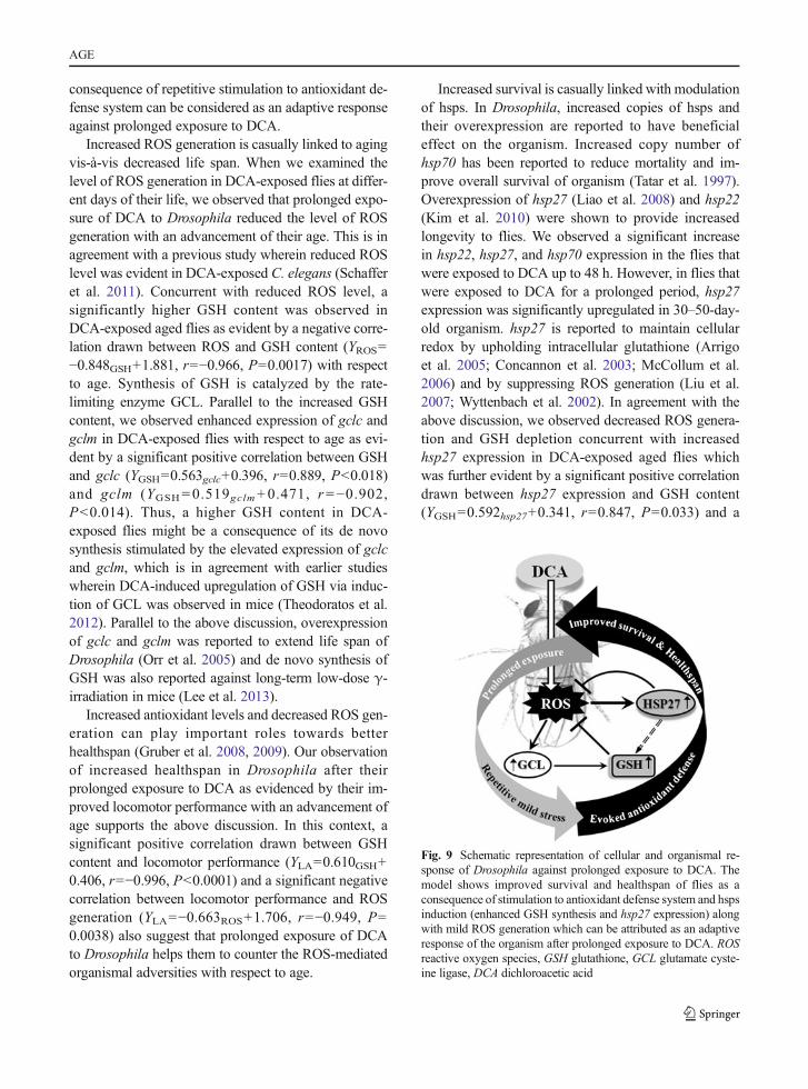
Fig. 9 Schematic representation of cellular and organismal re-sponse of Drosophila against prolonged exposure to DCA. Themodel shows improved survival and healthspan of flies as aconsequence of stimulation to antioxidant defense system and hspsinduction (enhanced GSH synthesis and hsp27 expression) alongwith mild ROS generation which can be attributed as an adaptiveresponse of the organism after prolonged exposure to DCA. ROSreactive oxygen species, GSH glutathione, GCL glutamate cyste-ine ligase, DCA dichloroacetic acid
AGE

negative correlation between hsp27 expression andROS generation (YROS=−0.549hsp27+1.653, r=−0.894,P=0.016). We therefore hypothesized that modulationhsp27 expression might benefit the DCA-exposed or-ganism. When hsp27 was knocked down in Drosophila(Act-Gal4>UAS-hsp27RNAi), survival of these organ-isms significantly went down as compared to control(Act-Gal4>w1118) along with a significant increase inthe ROS generation and GSH depletion and poorerlocomotor activities, suggesting an age-dependent de-cline in the fitness of fly against DCA exposure.To rule out the general effect of knockdown ofhsps, we further analyzed the survival of flieswherein another small hsp, viz., hsp22, wasknocked down (Act-Gal4>UAS-hsp22RNAi). AfterDCA exposure to these flies, we observed a non-significant change in their survival as compared tounexposed flies (data not shown). Thus, longevityand better healthspan of aged flies after theirprolonged exposure to DCA may be a conse-quence of increased hsp27 and increased GSHlevels (Fig. 9).
Taken together, we show here that attenuation of age-dependent cellular (ROS generation and GSH depletion)and functional (survival and organismal fitness) declinesalong with involvement of hsp27 modulation in flies thatwere exposed to DCA for a prolonged period. To the bestof our knowledge, this is the first report on the attenuationof age-dependent insufficiencies byDCA exposure using awell-accepted in vivo modelDrosophila. However, furtherstudies are warranted to understand the possible role ofhsp27 modulation vis-à-vis age-dependent benefit inDCA-exposed organisms towards its therapeutic benefit.
Acknowledgments The authors are grateful to the director ofthe CSIR-Indian Institute of Toxicology Research (CSIR-IITR),Lucknow for the support and to Dr. M. K. R. Mudiam, seniorscientist, and Mr. Ratnashekhar Ch, CSIR-SRF, Analytical Chem-istry Section, CSIR-IITR for the GC-ECD analysis of DCA. Wethank the Drosophila Stock Centre, Bloomington (Oregon R+, Act-Gal4/Cyo); William Ja, The Scripps Research Institute, Florida(w1118); H. D. Wang, California Institute of Technology, California(UAS-hsp27); andVienna Drosophila RNAi Center, Austria (UAS-hsp27RNAi,UAS-hsp22RNAi) for the fly strains. Financial assistanceto AP as SRF [20-6/2008(ii)EU-IV], SC as SRF [20-12/2009(ii)EU-IV] from the University Grants Commission (UGC),New Delhi, DVas JRF [19-12/2010(i)EU-IV] from the Council ofScientific and Industrial Research (CSIR), SS as DBT-ProjectFellow (grant no. BT/PR14716/BRB/10/876/2010), and DKCfrom the CSIR, New Delhi (NWP-BSC0103, UNDO) and DBT(BT/PR14716/BRB/10/876/2010) is thankfully acknowledged.IITR communication number is 3193.
Conflict of interest The authors declare that they have no con-flict of interest.
References
Arrigo AP, Virot S, Chaufour S, Firdaus W, Kretz-Remy C, Diaz-Latoud C (2005) Hsp27 consolidates intracellular redox ho-meostasis by upholding glutathione in its reduced form andby decreasing iron intracellular levels. Antioxid RedoxSignal 7:414–422
Ayyanathan K, Kesaraju S, Dawson-Scully K, Weissbach H(2012) Combination of sulindac and dichloroacetate killscancer cells via oxidative damage. PLoS One 7:e39949
Benford DJ, Hanley AB, Bottrill K, Oehlschlager S, Balls M,Branca F, Castegnaro JJ, Descotes J, Hemminiki K,Lindsay D, Schilter B (2000) Biomarkers as predictive toolsin toxicity testing. ATLA 28:119–131
Cai P, Boor PJ, Khan MF, Kaphalia BS, Ansari GA, Konig R (2007)Immuno- and hepato-toxicity of dichloroacetic acid inMRL(+/+) and B(6)C(3)F(1) mice. J Immunotoxicol 4:107–115
Concannon CG, Gorman AM, Samali A (2003) On the role ofHsp27 in regulating apoptosis. Apoptosis 8:61–70
Curtis C, Landis GN, Folk D, Wehr NB, Hoe N, Waskar M,Abdueva D, Skvortsov D, Ford D, Luu A, Badrinath A,Levine RL, Bradley TJ, Tavare S, Tower J (2007)Transcriptional profiling of MnSOD-mediated lifespan ex-tension in Drosophila reveals a species-general network ofaging and metabolic genes. Genome Biol 8:R262
Ellman GL (1959) Tissue sulfhydryl groups. Arch BichemBiophys 82:70–77
Feany MB, Bender WW (2000) A Drosophila model ofParkinson’s disease. Nature 404:394–398
Gayathri MV, Krishnamurthy NB (1981) Studies on the toxicity ofmercurial fungicide Agallol3 in Drosophila melanogaster.Environ Res 24:89–95
Geist S, Lesemann C, Schutz C, Seif P, Frank E (1991)Determination of chloroacetic acids in surface water. In:Angeletti G, Bjorseth A (eds) Organic micropollutants inthe aquatic environment. Kluwer, Dordrecht, pp 393–397
Grotewiel MS, Martin I, Bhandari P, Cook-Wiens E (2005)Functional senescence in Drosophila melanogaster. AgeingRes Rev 4:372–397
Gruber J, Schaffer S, Halliwell B (2008) The mitochondrial freeradical theory of aging—where dowe stand? Front Biosci 13:6554–6579
Gruber J, Ng LF, Poovathingal SK (2009) Deceptively simple butsimply deceptive—Caenorhabditis elegans lifespan studies:considerations for aging and antioxidant effects. FEBS Lett583:3377–3387
Gupta SC, Siddique HR, Saxena DK, Chowdhuri DK (2005)Hazardous effect of organophosphate compound, dichlorvosin transgenic Drosophila melanogaster (hsp70-lacZ): induc-tion of hsp70, anti-oxidant enzymes and inhibition of acetyl-cholinesterase. Biochim Biophys Acta 1725:81–92
Ha KN, Chen Y, Cai J, Sternberg PJ (2006) Increased glutathionesynthesis through an ARE-Nrf2-dependent pathway by zincin the RPE: implication for protection against oxidativestress. Invest Ophthalmol Vis Sci 47:2709–2715
AGE

Habig WH, Pabst MJ, Jakoby WB (1974) Glutathione S-transferases. The first enzymatic step in mercapturic acidformation. J Biol Chem 249:7130–7139
Hassoun EA, Dey S (2008) Dichloroacetate- and trichloroacetate-induced phagocytic activation and production of oxidativestress in the hepatic tissues of mice after acute exposure. JBiochem Mol Toxicol 22:27–34
Hassoun EA, Ray S (2003) The induction of oxidative stress andcellular death by the drinking water disinfection by-products,dichloroacetate and trichloroacetate in J774.A1 cells. CompBiochem Physiol C Toxicol Pharmacol 135:119–128
Hassoun E, Kariya C, Williams FE (2005) Dichloroacetate-induced developmental toxicity and production of reactiveoxygen species in zebrafish embryos. J BiochemMol Toxicol19:52–58
Hercus MJ, Loeschcke V, Rattan SI (2003) Lifespan extension ofDrosophila melanogaster through hormesis by repeated mildheat stress. Biogerontology 4:149–156
IARC (2004) IARCmonographs on the evaluation of carcinogenicrisk to humans, vol. 84, Some drinking-water disinfectantsand contaminants. IARC, Lyon, p 359
Jeibmann A, Paulus W (2009) Drosophila melanogaster as amodel organism of brain diseases. Int J Mol Sci 10:407–440
Jiang P, Sheng Y, Ji L (2013) The age-related change of glutathi-one antioxidant system in mice liver. Toxicol Mech Methods23:396–401
Jones MA, Grotewiel M (2011) Drosophila as a model for age-related impairment in locomotor and other behaviors. ExpGerontol 46:320–325
Kim HJ, Morrow G, Westwood JT, Michaud S, Tanguay RM(2010) Gene expression profiling implicates OXPHOS com-plexes in lifespan extension of flies over-expressing a smallmitochondrial chaperone, Hsp22. Exp Gerontol 45:611–620
Landis G, Shen J, Tower J (2012) Gene expression changes inresponse to aging compared to heat stress, oxidative stressand ionizing radiation in Drosophila melanogaster. Aging 4:768–789
Lee EK, Kim JA, Kim JS, Park SJ, Heo K, Yang KM, Son TG(2013) Activation of de novo GSH synthesis pathway inmouse spleen after long term low-dose γ-ray irradiation.Free Radic Res 47:89–94
Levine RL, Garland D, Oliver CN, Amici A, Climent I, Lenz AG,Ahn BW, Shaltiel S, Stadtman ER (1990) Determination ofcarbonyl content in oxidatively modified proteins. MethodsEnzymol 186:464–478
Liao PC, Lin HY, Yuh CH, Yu LK,Wang HD (2008) The effect ofneuronal expression of heat shock proteins 26 and 27 onlifespan, neurodegeneration, and apoptosis in Drosophila.Biochem Biophys Res Commun 376:637–641
Linder RE, Klinefelter GR, Strader LF, Suarez JD, Roberts NL(1997) Spermatotoxicity of dichloroacetic acid. ReprodToxicol 11:681–688
Liu L, Zhang X, Qian B, Min X, Gao X, Li C, Cheng Y, Huang J(2007) Over-expression of heat shock protein 27 attenuatesdoxorubicin-induced cardiac dysfunction in mice. Eur JHeart Fail 9:762–769
Lliadi KG, Knight D, Boulianne GL (2012) Healthy aging—insights from Drosophila. Front Physiol 3:106
Lowry OH, Rosebrough NJ, Farr AL, Randall RJ (1951) Proteinmeasurement with the Folin phenol reagent. J Biol Chem193:265–275
Lu SC (2013) Glutathione synthesis. Biochim Biophys Acta 1830:3143–3153
McCollum AK, Teneyck CJ, Sauer BM, Toft DO, Erlichman C(2006) Up-regulation of heat shock protein 27 induces resis-tance to 17-allylamino-demethoxygeldanamycin through aglutathione-mediated mechanism. Cancer Res 66:10967–10975
Michelakis ED, Sutendra G, Dromparis P, Webster L, Haromy A,Niven E, Maguire C, Gammer TL, Mackey JR, Fulton D,Abdulkarim B, McMurtry MS, Petruk KC (2010) Metabolicmodulation of glioblastoma with dichloroacetate. Sci TranslMed 2:31ra34
Minois N (2000) Longevity and aging: beneficial effects of expo-sure to mild stress. Biogerontology 1:15–29
Miquel E, Cassina A, Martinez-Palma L, Bolatto C, Trias E,Gandelman M, Radi R, Barbeito L, Cassina P (2012)Modulation of astrocytic mitochondrial function bydichloroacetate improves survival and motor performancein inherited amyotrophic lateral sclerosis. PLoS One 7:e34776
Mudiam MKR, Jain R, Varshney M, Ch R, Chauhan A, Goyal SK,Khan HA,Murthy RC (2013) In matrix derivatization of trichlo-roethylene metabolites in human plasma with methylchloroformate and their determination by solid-phasemicroextraction-gas chromatography-electron capture detector.J Chromatogr B Analyt Technol Biomed Life Sci 925:63–9
Nazir A, Mukhopadhyay I, Saxena DK, Kar Chowdhuri D (2001)Chlorpyrifos-induced hsp70 expression and effect on repro-ductive performance in transgenic Drosophila melanogaster(hsp70-lacZ) Bg9. Arch Environ Contam Toxicol 41:443–449
Nishikimi M, Appaji N, Yagi K (1972) The occurrence of super-oxide anion in the reaction of reduced phenazinemethosulfate and molecular oxygen. Biochem Biophys ResCommun 46:849–854
Oakes KD, Sibley PK, Solomon KR, Mabury SA, Van der KraakGJ (2004) Impact of perfluorooctanoic acid on fathead min-now (Pimephales promelas) fatty acyl-CoA oxidase activity,circulating steroids, and reproduction in outdoormicrocosms.Environ Toxicol Chem 23:1912–1919
Ohkawa H, Ohishi N, Yagi K (1979) Assay for lipid peroxides inanimal tissues by thiobarbituric acid reaction. Anal Biochem95:351–358
Orr WC, Radyuk SN, Prabhudesai L, Toroser D, Benes JJ, LuchakJM, Mockett RJ, Rebrin I, Hubbard JG, Sohal RS (2005)Overexpression of glutamate-cysteine ligase extends life spanin Drosophila melanogaster. J Biol Chem 280:37331–37338
Rattan SI (1998) Repeated mild heat shock delays ageing incultured human skin fibroblasts. Biochem Mol Biol Int 45:753–759
Salway KD, Gallagher EJ, Page MM, Stuart JA (2011) Higherlevels of heat shock proteins in longer-lived mammals andbirds. Mech Ageing Dev 132:287–297
Schaffer S, Gruber J, Ng LF, Fong S, Wong YT, Tang SY,Halliwell B (2011) The effect of dichloroacetate on health-and lifespan in C. elegans. Biogerontology 12:195–209
Singh MP, Reddy MM, Mathur N, Saxena DK, Chowdhuri DK(2009) Induction of hsp70, hsp60, hsp83 and hsp26 andoxidative stress markers in benzene, toluene and xyleneexposed Drosophila melanogaster: role of ROS generation.Toxicol Appl Pharmacol 235:226–243
AGE

Sinha AK (1972) Colorimetric assay of catalase. Anal Biochem47:389–394
SmithMK, Randall JL, Read EJ, Stober JA (1992) Developmentaltoxicity of dichloroacetate in the rat. Teratology 46:217–223
Stacpoole PW (2011) The dichloroacetate dilemma: environmen-tal hazard versus therapeutic goldmine—both or neither?Environ Health Perspect 119:155–158
Stacpoole PW, Henderson GN, Yan Z, James MO (1998) Clinicalpharmacology and toxicology of dichloroacetate. EnvironHealth Perspect 106:989–994
Stacpoole PW, Kurtz TL, Han Z, Langaee T (2008) Role ofdichloroacetate in the treatment of genetic mitochondrialdiseases. Adv Drug Deliv Rev 60:1478–1487
Suh JH,Wang HD, Liu RM, Liu JK, Hagen TM (2004) (R)-α-Lipoicacid reverses the age-related loss in GSH redox status in post-mitotic tissues: evidence for increased cysteine requirement forGSH synthesis. Arch Biochem Biophys 423:126–135
Tatar M, Khazaeli AA, Curtsinger JW (1997) Chaperoning ex-tended life. Nature 390:30
Theodoratos A, Blackburn AC, Cappello J, Tummala P,Dahlstrom JE, Board PG (2012) Dichloroacetic acid up-regulates hepatic glutathione synthesis via the induction ofglutamate-cysteine ligase. Biochem Pharmacol 83:427–433
Thompson JA,White CC, Cox DP, Chan JY, Kavanagh TJ, FaustoN, Franklin CC (2009) Distinct Nrf1/2-independent mecha-nisms mediate As 3+-induced glutamate-cysteine ligase sub-unit gene expression in murine hepatocytes. Free Radic BiolMed 46:1614–1625
Tower J (2009) Hsps and aging. Trends Endocrinol Metab 20:216–222
Tower J (2011) Heat shock proteins and Drosophila aging. ExpGerontol 46:355–362
Wong JY, Huggins GS, DebiddaM,Munshi NC, De Vivo I (2008)Dichloroacetate induces apoptosis in endometrial cancercells. Gynecol Oncol 109:394–402
Wyttenbach A, Sauvageot O, Carmichael J, Diaz-Latoud C,Arrigo AP, Rubinsztein DC (2002) Heat shock protein 27prevents cellular polyglutamine toxicity and suppresses theincrease of reactive oxygen species caused by huntingtin.Hum Mol Genet 11:1137–1151
Zhao HW, Zhou D, Haddad GG (2010) Antimicrobial peptidesincrease tolerance to oxidant stress in Drosophilamelanogaster. J Biol Chem 286:6211–6218
Zheng S, Yumei F, Chen A (2007) De novo synthesis of glutathi-one is a prerequisite for curcumin to inhibit hepatic stellatecell (HSC) activation. Free Radic Biol Med 43:444–453
AGE