Ocean urea fertilization for carbon credits poses high ecological risks
Upload
independentCategory
view
0download
0
Photochemistry and Photobiology, 1998, 68(3): 407-41 3
Light Irradiation of Mouse Spermatozoa: Stimulation of In Vitro Fertilization and Calcium Signals
Natalie Cohenl, Rachel Lubart2, Sara Rubinstein' and Haim Breitbart*l 'Department of Life Sciences and 2Department of Physics, Bar-llan University, Ramat-Gan, Israel
Received 16 March 1998; accepted 28 May 1998
ABSTRACT Irradiation of mouse spermatozoa by 630 nm He-Ne la- ser was found to enhance the intracellular calcium levels and fertilizing potential of these cells. The effect of light on calcium transport and on fertilization rate was abro- gated in the absence of Ca2+ during the irradiation time, indicating that the effect of light is Ca2+ dependent. The stimulatory effect of light on Ca2+ uptake was abolished in the presence of a voltage-dependent Ca2+-channel in- hibitor nifedipine, indicating the involvement of a plasma membrane voltage-dependent CaZ+ channel. Further- more, the stimulatory effect of light was completely in- hibited by the mitochondrial uncoupler FCCP, indicating that laser irradiation might affect the mitochondrial Ca2+ transport mechanisms. A causal association between la- ser irradiation, reactive oxygen species (ROS) generation and sperm function was indicated by studies with ROS scavengers, superoxide dismutase (SOD) and catalase, and exogenous hydrogen peroxide. The SOD treatment, which enhanced H,Oz production, resulted in increased Ca2+ uptake and enhanced fertilization rate. On the other hand, catalase, which decomposes H20t, impaired the light-induced stimulation in Ca2+ uptake and the fertil- ization rate. Taken together, the data suggest that H,Oz might be involved in the irradiation effects, and indeed laser irradiation enhances the production of H,O, by spermatozoa. These results indicate that the effect of 630 nm He-Ne laser irradiation is mediated through the gen- eration of H,O, by the spermatozoa and that this effect plays a significant role in the augmentation of the sperm cells' capability to fertilize metaphase 11-arrested eggs in vitro.
INTRODUCTION The effects of He-Ne laser irradiation on various aspects of cell metabolism have been recognized in recent years, and extensive literature exists on the application of low-power laser irradiation in various biological systems. Such effects were demonstrated in somatic cells, where laser irradiation was found to affect proliferation, respiration, ATP synthesis
*To whom correspondence should be addressed at: Department of Life Sciences, Bar-Ilan University, Ramat Gan 52900, Israel. Fax: 972-3-5344766: e-mail: [email protected]
0 1998 American Society for Photobiology 003 1 -8655/98 SS.OO+O.OO
and calcium uptake (1-10). Laser irradiation has been shown to induce changes in intracellular Ca2+ concentrations in macrophages (9), fibroblasts and spermatozoa from ram (1 1) and bull (12,13) and to affect biostimulation of gametic and embryonic cells in vitro (14-16). Changes in intracellular Ca2+ have a pivotal role in control of sperm motility, capac- itation and acrosome reaction, all of which are imperative for successful fertilization (17). The Ca2+ influx from the extracellular environment is an essential component of the signaling cascade leading to fertilization.
Light in the visible and far-red regions can be absorbed by mitochondrial enzymes (18-20). Therefore, it has been suggested (21,22) that He-Ne laser irradiation activates the redox reactions in the respiratory chain, possibly leading to changes in the intracellular calcium levels. Several pathways for Ca2+ influx into somatic cells have been described, in- cluding entry through ligand- and voltage-gated ion chan- nels. It is thought that in sperm cells the signaling pathway utilizes a voltage-sensitive Ca2+ channel (23-25).
In this study we show, for the first time, stimulation of the fertilizing ability of mouse sperm by laser irradiation. This effect is correlated to changes in Ca2+ influx into the spermatozoon. It is suggested that changes in Ca2+ influx and fertilization rate by light occurred via light-induced H202 production.
MATERIALS AND METHODS Mediu und reagents. Modified Krebs-Ringer bicarbonate medium containing 119.4 niM NaCl, 4.8 mM KCI, 1.7 mM CaCl,, 1.2 mM MgCl,, 25.1 mM NaHCO,, 1 mM sodium pyruvate, 25 mM sodium lactate, 5.56 mM glucose, 20 mg/mL bovine serum albumin, 0.001% phenol red and 10 IUlmL penicillin was used in all experiments (26,27). The medium was adjusted to pH 7.4 with I M NaOH and sterilized by Millipore (0.22 km) filtration. Fluo-3 AM and Pluronic F-127 were obtained from Molecular Probes: all other reagents were obtained from Sigma Chemical Company.
Collection and culture of mouse spermutozoa and eggs. Mouse sperm cells were recovered from epididymides of mature male BALBK mice. The excised epididymides were minced in 0.4 mL of medium and incubated in 3 7 T , 5% CO, for 15 min to allow motile sperm to escape into the medium before the pieces of tissue were removed. At least two male mice were used in each experi- ment. The spermatozoa were incubated for I h at 37°C 5% CO, in a final concentration of 5 X lo6 cells/mL.
Immature (4-6 weeks old) female BALBK mice were injected with PMSG (7 IU), followed 48-56 h later by human chorionic gonadotropin (7 IU). Then 13-16 h afterward, the oocyte-cumulus complexes were recovered from the oviducts and pooled. At least eight female mice were used in each experiment.
Fertilizution ussnv. Unfertilized mouse eggs were pooled and
407

408 Natalie Cohen et a/.
Light
Figure 1. Sperm cells loaded with Fluo-3 AM were irradiated at 630 nm for 1 min, starting at the indicated point. Pictures were taken successively every 0.5 min
loaded with Hoechst 33342 (5 pM) for 30 min in 37°C. 5% C 0 2 (28). This fluorochrome stains the chromatin of the eggs, thus en- abling assessment of its distribution-metaphase 11-arrested unfer- tilized eggs and a round pronuclei in the fertilized eggs. The oocytes were then washed three times in fresh medium, and groups of 15- 20 oocytes were mixed with spermatozoa (5 X lo6 cellslml) and incubated in 250 pL droplets under mineral oil for 24 h in 3 7 T , 5% CO,. Then, the eggs were removed, washed in fresh medium, fixed in 5% formaldehyde and the percentage of the fertilized eggs in each group was counted using a fluorescence microscope. Each set of experiments included several different treatments and two con- trol groups.
Irradiation. The light sources were a 13 mW He-Ne laser (Spec- traphysics) and a 780 nm, 25 mW, diode laser (Lasotronics). An optical fiber was connected to the lasers and was inserted into a rotating tube at 3 7 T , containing 0.5-1 mL cell suspension of 5 X lo6 cells/mL. The light intensity at the output of the fiber connected to the He-Ne laser was 7-8 mW. The light intensity at the output of the fiber connected to the 780 nm diode laser was 24-25 mW. All light intensities were measured with an Ophir (model PD2-A) powermeter.
Intracellular Ca" measurements. Sperm cells were incubated on 25 mm glass coverslips covered with poly-L-lysine with Fluo-3 AM (2 pM) and pluronic acid (1.5 pM) for 30 min in glucose-enriched phosphate-buffered saline (PBS)? in 37°C 5% CO,. After incuba- tion, each dish was rinsed twice with glucose-enriched PBS and the coverslip was placed in the microscope holder. Cells were covered with 1 mL glucose-enriched PBS. Intracellular calcium measure- ments were performed using a fluorescence microscopic system con- sisting of an inverted epifluorescence microscope (Axiovert 135M; Zeiss, Germany), an intensified couple-charge device C2400 camera (Hamamatsu, Japan) and a frame-grabbing software (Galai, Israel). A 75 W xenon lamp served as the source for excitation. A 510 long- pass emission filter was used to select fluorescence emission. The iirst pictures were taken to estimate basal levels of fluorescence. Afterward, cells were irradiated in siru and the fluorescence was measured every 30 s for 10 min. Analysis of intracellular calcium
tAhhreviation.7: AM, tetraacetoxymethyl ester; FCCP, carbonyl- cyamide-p-trifluoromethoxyphenyl hydrazone; PBS, phosphate- buffered saline; ROS, reactive oxygen species: SOD, superoxide dismutase.
levels was performed employing Scan Array 2 software (Galai, Is- rael).
Caz+ uptake. Uptake of 4sCa2+ was determined by the filtration technique. Sperm cells (5 X lo6 cells/mL) were irradiated under mineral oil for different periods of time prior to the addition of 45Ca2+ (5 p,Ci/mL). In the experiments in which the treatments were performed in medium devoid of CaCl,, CaCI, was added as a mix with the 4sCa2+ to a final concentration of 1.7 mM. After 15 min of incubation, 0.1 mL was removed and immediately vacuum filtered on GF/C filters. The cells trapped on the filter were washed three times with 5 mL of solution composed of 150 mM NaCl, 10 mM Tris (pH 7.4) and 2 mM EGTA. The dry filters were counted in scintillation vials with 4 mL Aquasol (DuPont).
Statistics. The fertilization and the Ca2+ results were analyzed by analysis of variance (ANOVA) for determination of time- and con- centration-dependent significance. Significant differences detected between various treatments and the control were determined by ANOVA, followed by Dunnett's t-test.
RESULTS
The effect of 630 nm He-Ne laser irradiation on intracellular Ca2+ levels
The effect of 630 nm He-Ne laser irradiation on sperm cal- cium was investigated through two approaches, the first em- ploying the fluorescent calcium indicator, Fluo-3 AM, and the Dynamic Calcium Imaging System, and the second em- ploying a radiolabeled Ca2+ uptake. The fluorescent calcium indicator, Fluo-3, was loaded as the membrane permeant, tetraacetoxymethyl (AM) ester, into epididymal mouse sperm in the presence of the dispersant, pluronic acid. One minute of irradiation was found to cause a rapid increase in the intracellular calcium levels (Fig. 1). The first picture de- picts the Fluo-3 fluorescence in the control state. The irra- diation commenced at the indicated point for the duration of 1 min. The following pictures show the increase in the Fluo- 3 fluorescence induced by light. Figure 2 shows a graphic presentation of 10 cells randomly chosen from the cell pop-

Photochemistry and Photobiology, 1998, 68(3) 409
2 5 - - c
0 = 2
? m 1 5 P
+ I
.... - - - m ' 0 5
0 0 100 200 300 400 500 600
Time (sec)
Figure 2. Graphic presentation of 10 randomly chosen sperm cells loaded with Fluo-3 AM and irradiated at 630 nm for 1 min, starting at the indicated point. The Ca2+ concentration scale is in relative units.
ulation shown in Fig. 1. The increase in Ca2+ levels can be observed within 1 min after the irradiation. The light-in- duced rise in calcium levels was calculated to be 3.2-fold.
The radiolabeled Ca2+-uptake approach showed a signifi- cant increase in Ca2+ uptake in 630 nm-irradiated sperm cells relative to the control cells (Fig. 3). One minute of irradia- tion was found to cause a rapid, 1.5-fold increase in Ca2+ uptake, followed by an additional slower increase up to two- fold after 10 min of irradiation. The stimulatory effect of light was impaired after prolonged irradiation (30 min).
The effect of various factors on the laser-induced increase in intracellular Ca2+ levels
When Ca2+ was omitted from the medium, the laser-induced increase in intracellular Ca2+ levels could not be observed in Fluo-3-loaded sperm cells (data not shown). Similarly, when sperm cells were irradiated in medium devoid of Ca2+, no stimulation was evident in the radiolabeled Ca2+ uptake (Fig. 4). These results indicate that the increase in the intra- cellular Ca2+ levels depends on the influx of Ca2+ from the incubation medium into the cell during the irradiation time.
The addition of nifedipine (1-10 pM), a voltage-depen- dent Ca2+-channel inhibitor, prior to the 630 nm He-Ne laser irradiation did not affect the light-induced rise in the intra- cellular Ca2+ levels, as measured with Fluo-3-loaded sperm cells (data not shown). Therefore, it can be concluded that this Ca2+ influx occurs via a voltage-insensitive Ca2+ chan- nel. In contrast, the stimulatory effect of light on the radio- labeled Ca2+ uptake was completely abolished by nifedipine (1 pM), indicating that most of the 45Ca2+ uptake occurs via the plasma membrane voltage-dependent Ca2+ channels (Fig. 4).
The stimulatory effect of light on 45Ca24 uptake was al- most completely abolished (95%) by the mitochondrial un- coupler, carbonylcyamide-p-trifluoromethoxyphenyl hydra- zone (FCCP) (10 pM), while only 50% inhibition was ob- served by the uncoupler in the control cells (Fig. 4). Similar results were obtained by using the mitochondrial respiration inhibitor, antimycin A (data not shown). These data imply that light affects mainly the mitochondrial Ca2+-transport mechanisms.
6 T T
0 1 5 10 30
Time of irra&ation (nun)
Figure 3. Time-course of Ca2' uptake in sperm cells during irra- diation. Cells were irradiated at 630 nm at the indicated times prior to 45CaC12 addition, and Ca2+ uptake was determined after an addi- tional 15 min of incubation, as described in the Materials and Meth- ods. Each bar represents the mean of three individual experiments, with error bars indicating SEM; * P < 0.01 as compared to the con- trol.
The effect of 630 nm He-Ne laser irradiation on in vitro fertilization
Because a rise in intracellular calcium is known to be im- perative for the successful completion of the fertilization process, the effect of 630 nm He-Ne laser irradiation pro- ducing such a rise was tested on in vitro fertilization.
Sperm cells irradiated with a 630 nm He-Ne laser showed an enhanced fertilization rate, as compared to the control, up to 37% or 32% enhancement after 5 or 10 min of irra- diation, respectively (Fig. 5). Such enhancement was not ob- served when the spermatozoa were irradiated with a 780 nm diode laser for 10 min (data not shown). Furthermore, there was no such stimulation when the irradiation was performed in medium devoid of Ca2+ (Fig. 6), similar to the results obtained in the Ca2+ uptake experiments. When sperm cells were irradiated in medium devoid of Ca2+, the Ca2+ neces- sary for the in vitro fertilization was added after the irradi- ation was completed. Nifedipine (1 pM) also prevented the
' T *
basal L FCCP L+FCCP N6 L+Nd LCaZi
Figure 4. Influence of FCCP, nifedipine and CaZ+ on Ca2' uptake in sperm cells, induced (L) or noninduced by irradiation. Cells were irradiated at 630 nm for 10 min in the presence of CaZ+ (1.7 mM) prior to 45CaC1, addition, in the presence of FCCP (10 p,M) and nifedipine (Nif) (1 KM) and in the absence of Ca2' (L-Ca2+). L represents light-irradiated cells. Ca2+ uptake was determined after an additional 15 min of incubation, as described in the Materials and Methods. Each bar represents the mean of three individual ex- periments, with error bars indicating SEM; * P < 0.01 as compared to the control.

410 Natalie Cohen et a/.
140
- c 130 c c
E 2 120
5 110
Ti .-
m S
al
.-
- ._ 100
Y
90 0 1
1 3 5 10
Time of irradiation (min)
Figure 5. Time-course of fertilizing capacity of mouse spermatozoa during irradiation. Cells were irradiated at 630 nm for the indicated periods of time, in incubation medium containing 1.7 mM CaC1,. The treated spermatozoa were mixed with mouse oocytes for the in ipitro fertilization assay, as described in the Materials and Methods. Each sample contained 12-15 oocytes. Each bar represents the mean of at least three individual experiments, with error bars indicating SEM; * P < 0.01 compared to the control.
light from exerting its stimulatory effect on the fertilization rate (Fig. 6). Although treatment with nifedipine was found to inhibit the fertilization rate of the control cells (20% in- hibition), the effect of nifedipine on the irradiated cells shows complete inhibition of the light enhancement. These findings lead to the conclusion that Ca2+ influx via voltage- dependent Ca2+ channels is involved in the light-induced fer- tilization. The mitochondria1 uncoupler FCCP completely abolished the stirnulatory effect of light on the fertilization rate (Fig. 6) in the same manner as it inhibited the stimu- latory effect of light on Ca2+ uptake (Fig. 4).
The effect of reactive oxygen scavengers (ROS) on in vitro fertilization and CaZ+ uptake
In order to determine whether ROS are involved in the light- induced increase in the fertilization rate and the elevation in radiolabeled Ca2+ uptake, sperm cells were treated with ROS
b a d L FCCP L+FCCP Nd LcNif LCaZ+
Figure 6. Influence of FCCP, nifedipine and Ca2+ on the fertilizing capacity of mouse spermatozoa, induced or noninduced by irradia- tion. Cells were irradiated in the presence of CaZ' (1.7 mM) at 630 nm for 10 min, in the presence of FCCP (10 KM), nifedipine (Nif) ( I pA4) or in the absence of added Ca2+ (L-Ca2+). L represents light- irradiated cells. Control cells, without irradiation, were treated in the same way. The treated spermatozoa were mixed with mouse oocytes for the in vifro fertilization assay, as described in the Materials and Methods. Each bar represents the mean of at least three individual experiments, with error bars indicating SEM; * P < 0.01 compared to the control.
BASAL 1 *
T *
L SOD L+SOD CAT L+CAT
Figure 7. Influence of SOD and catalase on Ca2+ uptake in sperm cells, induced or noninduced by 630 nm irradiation. Spermatozoa were incubated with SOD (100 kg/mL) or catalase (10 pg/mL) for 10 min prior to 45Ca2+ addition, and Ca2+ uptake was determined after an additional 15 min of incubation, as described in the Mate- rials and Methods. In the same experiments, the effect of SOD and catalase on Ca2+ uptake, induced by 630 nm irradiation, was also determined. Data represent mean i SEM; n = 3, *P < 0.05 as compared to the control. L = 630 nm light irradiation; CAT = catalase.
scavengers, superoxide dismutase (SOD) or catalase, prior to the fertilization assay or the determination of Ca2+ uptake. The SOD converts superoxide anion to H202 and catalase decomposes H202 to water and oxygen. In nonirradiated cells, the fertilization rate was enhanced 30% by treatment with SOD (100 kg/mL) and inhibited 24% by treatment with catalase (10 (*.g/mL) (Fig. 7). The stimulatory effect of light on fertilization was completely blocked by adding catalase (Fig. 7).
Similar results were obtained employing the radioactive Ca2+-uptake approach (Fig. 8). In nonirradiated cells, SOD (100 pg/mL) was found to induce a 2.7-fold stimulation, while catalase (10 kg/mL) treatment resulted in 20% inhi- bition in Ca2+ uptake. The stimulatory effect of light on Ca2+ uptake was 80% inhibited by catalase. Both the enhanced fertilization rate and the increased Ca2+ uptake achieved by 630 nm He-Ne laser irradiation evinced no further stimu- lation by treatment with SOD. However, catalase treatment was highly competent in blocking the light-induced stimu- lation of fertilization or Ca2+ uptake (Figs. 7 and 8). Taken together, these results suggest that H202 might be the active
BASAL L SOD L+SOD CAT LtCAT
Figure 8. Influence of SOD and catalase on the fertilizing capacity of mouse spermatozoa, with or without irradiation. Mouse sperma- tozoa were treated with SOD (100 pg/mL) for 10 min. The treated spermatozoa were mixed with mouse oocytes for the in vitro fertil- ization assay, as described in the Materials and Methods. Each sam- ple contained 12-15 oocytes. Each bar represents the mean of at least three individual experiments, with error bars indicating SEM; * P < 0.01 as compared to the control. L = 630 nm light irradiation; CAT = catalase.
Photochemistry and Photobiology, 1998, 68(3) 41 1
T
0 1
i
Time of irradiation (rnin)
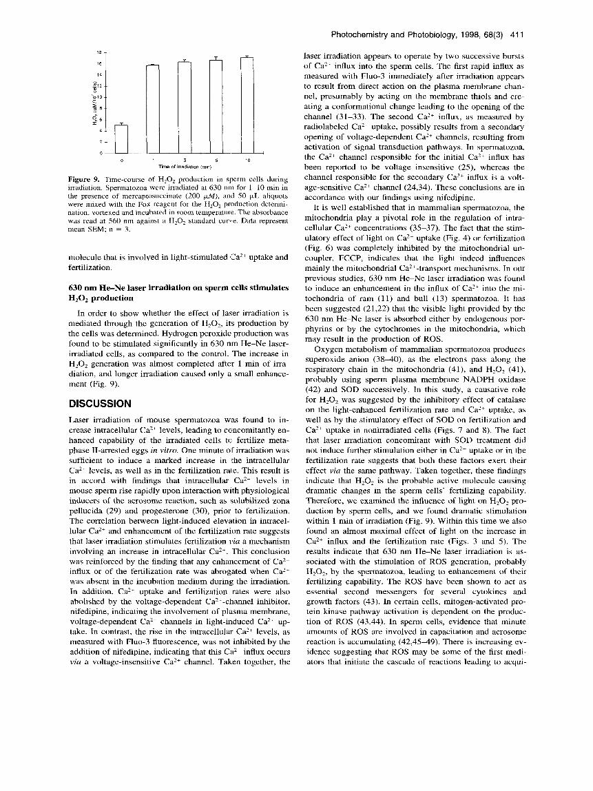
Figure 9. Time-course of H,O, production in sperm cells during irradiation. Spermatozoa were irradiated at 630 nrn for 1-10 min in the presence of mercaptosuccinate (200 (*.M), and 50 pL aliquots were mixed with the Fox reagent for the H,O, production determi- nation, vortexed and incubated in room temperature. The absorbance was read at 560 nm against a H,O, standard curve. Data represent mean SEM; n = 3.
molecule that is involved in light-stimulated @a2+ uptake and fertilization.
630 nm He-Ne laser irradiation on sperm cells stimulates H,02 production
In order to show whether the effect of laser irradiation is mediated through the generation of H202, its production by the cells was determined. Hydrogen peroxide production was found to be stimulated significantly in 630 nm He-Ne laser- irradiated cells, as compared to the control. The increase in Hz02 generation was almost completed after 1 min of irra- diation, and longer irradiation caused only a small enhance- ment (Fig. 9).
DISCUSSION Laser irradiation of mouse spermatozoa was found to in- crease intracellular Ca2+ levels, leading to concomitantly en- hanced capability of the irradiated cells to fertilize meta- phase 11-arrested eggs in vitru. One minute of irradiation was sufficient to induce a marked increase in the intracellular Ca2+ levels, as well as in the fertilization rate. This result is in accord with findings that intracellular Ca2+ levels in mouse sperm rise rapidly upon interaction with physiological inducers of the acrosome reaction, such as solubilized zona pellucida (29) and progesterone (30), prior to fertilization. The correlation between light-induced elevation in intracel- lular Ca2+ and enhancement of the fertilization rate suggests that laser irradiation stimulates fertilization via a mechanism involving an increase in intracellular Ca2+. This conclusion was reinforced by the finding that any enhancement of Ca2+ influx or of the fertilization rate was abrogated when Ca2+ was absent in the incubation medium during the irradiation. In addition, Ca2+ uptake and fertilization rates were also abolished by the voltage-dependent Ca2+-channel inhibitor, nifedipine, indicating the involvement of plasma membrane, voltage-dependent Ca2+ channels in light-induced Ca2+ up- take. In contrast, the rise in the intracellular Ca2+ levels, as measured with Fluo-3 fluorescence, was not inhibited by the addition of nifedipine, indicating that this Ca2+ influx occurs via a voltage-insensitive Ca2+ channel. Taken together, the
laser irradiation appears to operate by two successive bursts of Ca2+ influx into the sperm cells. The first rapid influx as measured with Fluo-3 immediately after irradiation appears to result from direct action on the plasma membrane chan- nel, presumably by acting on the membrane thiols and cre- ating a conformational change leading to the opening of the channel (31-33). The second Ca2+ influx, as measured by radiolabeled Ca2 + uptake, possibly results from a secondary opening of voltage-dependent Ca2+ channels, resulting from activation of signal transduction pathways. In spermatozoa, the Ca2+ channel responsible for the initial Ca2+ influx has been reported to be voltage insensitive ( 2 3 , whereas the channel responsible for the secondary Ca2+ influx is a volt- age-sensitive Ca2+ channel (24,34). These conclusions are in accordance with our findings using nifedipine.
It is well established that in mammalian spermatozoa, the mitochondria play a pivotal role in the regulation of intra- cellular Ca2+ concentrations (35-37). The fact that the stim- ulatory effect of light on Ca2+ uptake (Fig. 4) or fertilization (Fig. 6) was completely inhibited by the mitochondrial un- coupler, FCCP, indicates that the light indeed influences mainly the mitochondria1 Ca2+-transport mechanisms. In our previous studies, 630 nm He-Ne laser irradiation was found to induce an enhancement in the influx of Ca2+ into the mi- tochondria of ram (1 l) and bull (13) spermatozoa. It has been suggested (21,22) that the visible light provided by the 630 nm He-Ne laser is absorbed either by endogenous por- phyrins or by the cytochromes in the mitochondria, which may result in the production of ROS.
Oxygen metabolism of mammalian spermatozoa produces superoxide anion (3840), as the electrons pass along the respiratory chain in the mitochondria (41), and H20, (41), probably using sperm plasma membrane NADPH oxidase (42) and SOD successively. In this study, a causative role for H202 was suggested by the inhibitory effect of catalase on the light-enhanced fertilization rate and Ca" uptake, as well as by the stimulatory effect of SOD on fertilization and Ca2+ uptake in nonirradiated cells (Figs. 7 and 8). The fact that laser irradiation concomitant with SOD treatment did not induce further stimulation either in Ca2+ uptake or in the fertilization rate suggests that both these factors exert their effect via the same pathway. Taken together, these findings indicate that H202 is the probable active molecule causing dramatic changes in the sperm cells' fertilizing capability. Therefore, we examined the influence of light on H202 pro- duction by sperm cells, and we found dramatic stimulation within 1 min of irradiation (Fig. 9). Within this time we also found an almost maximal effect of light on the increase in Ca2+ influx and the fertilization rate (Figs. 3 and 5) . The results indicate that 630 nm He-Ne laser irradiation is as- sociated with the stimulation of ROS generation, probably H,02, by the spermatozoa, leading to enhancement of their fertilizing capability. The ROS have been shown to act as essential second messengers for several cytokines and growth factors (43). In certain cells, mitogen-activated pro- tein kinase pathway activation is dependent on the produc- tion of ROS ( 4 3 9 ) . In sperm cells, evidence that minute amounts of ROS are involved in capacitation and acrosome reaction is accumulating (42,4549). There is increasing ev- idence suggesting that ROS may be some of the first medi- ators that initiate the cascade of reactions leading to acqui-

412 Natalie Cohen et a/.
sition of fertilizing ability (42). The average intrinsic rate of <I2- production in mouse sperm is relatively high (39); therefore the formation and breakdown of endogenous hy- droperoxides should be sufficiently rapid. Mammalian sper- matozoa are known to lack catalase activity (17) but to pos- sess the two alternate enzymatic defense systems against the dioxygen species O2 - and H202, SOD and glutathione per- oxidase/reductase pair (38,39,42). The ROS can have both detrimental and beneficial effects on spermatozoa, and the delicate balance act between the amounts of ROS produced and of ROS scavengers at any moment will determine whether a given sperm function will be jeopardized or pro- moted.
We propose that in spermatozoa, light treatment stimu- lates the generation of superoxide anion, which is dismutated by endogenous SOD into HzOz. Transient local elevation in H202 resulted in the opening of a Ca2+ channel for a first rapid elevation of intracellular Ca2+, as has also been shown by others (30,31,33). The functional changes induced by al- tering the redox status of the spermatozoa may lead to an enhancement of protein tyrosine phosphorylation (50). Hy- drogen peroxide was reported to be extremely effective in inhibiting the tyrosine phosphatases, thereby facilitating the phosphorylation of tyrosine ( 5 1). Regardless of the cellular mechanism involved, such phosphorylation may lead to a secondary protein tyrosine phosphorylation-dependent Caz+ influx (30), probably by opening of a voltage-dependent <:a2+ channel (30,52,53), which is imperative for activation of the spermatozoa.
In summary, our results suggest that H202 may participate in the cascade of biochemical events that transform the light signal into a biological response. An understanding of the stimulatory effect of light on the fertilizing ability of sper- matozoa could lead to novel methods for both contraception and the treatment of infertility in human beings.
Acknowledgements-This study was supported in part by grant no. 2320 from the Chief Scientist's Office of the Ministry of Health, Israel and the Ihel Foundation to H.B.
REFERENCES 1. Passarella, S., E. Casamassima, S. Molinari and D. Pastone
(1984) Increase of proton electrochemical potential and ATP synthesis in rat liver mitochondria irradiated in vitro by HeNe laser. FEBS Lett. 175, 95-99.
2. Boulton, M. and J. Marshall (1986) He-Ne laser stimulation on human fibroblast proliferation. Lasers Life Sci. 1, 125-134.
3. Rochkind, S., L. Barrnea, N. Razon, A. Bartel and M. Schwartz (1987) Stimulatory effect of He-Ne low dose laser on injured sciatic nerves of rats. Neurosurgery 20, 843.
4. Sallet, C., S. Passarella and E. Quagliariello (1987) Effects of selective irradiation on mammalian mitochondria. Photochem. Photobiol. 45, 433438.
5 . Schwartz, M., A. Doron, M. Erich, V. Lavie, S. Benbasat, M. Belkin and S. Rochkind (1987) Effects of low-energy He-Ne laser irradiation of posttraumatic degeneration of adult rabbit optic nerve. Lasers Surg. Med. 7, 51.
6. Greco, M., G. Guida, E. Perlino and E. Quagliariello (1989) Increase in RNA and protein synthesis by mitochondria irradi- ated with helium-neon laser. Biochem. Biophys. Res. Commun.
7. Karu, T. I., T. P. Ryabykh, G. E. Fedoseyeva and N. I. Puchkova (1989) Helium-neon laser induced respiratory burst of phago- cytic cells. Lasers Surg. Med. 9, 585-588.
8. Rochkind, S. and R. Lubart (1990) Central nervous system
29, 1428-1434.
transplantation benefited by low level laser irradiation. SPIE 1200, 301.
9. Young, S. R., M. Dyson and P. Bolton (1990) Effect of light on calcium uptake by macrophages. Laser Ther. 2, 53-57.
10. Van Breugel, H. H. F. I. and P. R. Bar (1993) He-Ne laser irradiation affects proliferation of cultural rat Schwann cells in a dose dependent manner. J. Neurocytol. 22, 185-190.
11. Breitbart, H., T . Levinshal, N. Cohen, H. Friedmann and R. Lubart (1996) Changes in calcium transport in mammalian sperm mitochondria and plasma membrane irradiated at 633 nm (HeNe laser). J. Photochem. Photobiol. B Bid. 34, 117-121.
12. Lubart, R., H. Friedmann, T . Levinshal, R. Lavie and H. Breit- bart (1992) Effect of light on calcium transport in bull sperm cells. J. Photochem. Photobiol. B Biol. 15, 337-341.
13. Lubart, R., Y. Wollman and H. Friedmann (1992) Effects of visible and near IR lasers on cell cultures. J . Photochem. Pho- tobiol. B Biol. 12, 305-31 1.
4. Levi, A. C. S. E., R. Petrino and E. Siccardi (1987) Laser ir- radiation on chicken embryos. Boll. SOC. Ital. Bid. Sper. 3,233- 236.
5. Bielansky, A. and W. C. D. Hare (1992) Development in-vitro of bovine embryos after exposure to continuous helium-neon laser light. Theriogenology 37, 192.
6. Ocana, Q. J. M., V. R. J. Gomez, M. M. Moveno and V. J. M. Santisteban (1995) The effect of He-Ne laser irradiation on in- vitro maturation and fertilization of immature bovine oocytes. Lasers Med. Sci. 10, 113-1 19.
17. Yanagimachi, R. (1994) Mammalian fertilization. In The Phys- iology of Reproduction (Edited by E. Knobil and J. D. Neil), pp. 189-317. Raven Press, New York.
18. Bolognani, L., M. Costato, V. Delmonte, L. Iotti and N. Volpi (1991) Effect of laser on enzymatic systems: inactivation of muscle lactate dehydrogenase by C02 and its reactivation by laser irradiation. Lasers & Technol. 1, 103-107.
19. Bolognani, L. and N. Volpi (1992) Low power laser-enzyme interaction. In Conference Proceeding G. Galleti, L. Bolognani and G. Ussia, pp. 213-222. Monduzzi, Bologna.
20. Tamura, M. (1993) Non-invasive monitoring of the redox state of cytochrome oxidase in living tissue using near infra-red laser lights. Jpn. Circ. J. 57, 817-824.
21. Karu, T . (1988) Molecular mechanism of therapeutic effect of low intensity laser irradiation. Lasers Life Sci. 2, 53-14.
22. Friedmann, H. and R. Lubart (1992) A possible explanation of laser-induced stimulation and damage of cell cultures. Phoro- chern. Photobiol. 2, 263-269.
23. Babcock, D. F. and D. R. Pfeiffer (1987) Independent elevation of cytosolic Ca2+ and pH of mammalian sperm by voltage de- pendent and pH sensitive mechanisms. J. Biol. Chem. 262, 1 SO4 1-1 5047.
24. Florman, H. M., M. E. Carron, T . D. H. Kim and D. F. Babcock (1992) Activation of voltage dependent calcium channels of mammalian sperm is required for zona pellucida induced acro- soma1 exocytosis. Dev. Biol. 152, 304-314.
25. Florman, H. M. (1994) Sequential focal and global elevations of sperm intracellular Ca2+ are initiated by the zona pellucida during acrosomal exocytosis. Dev. Biol. 165, 152-164.
26. Saling, P., B. T. Storey and D. P. Wolf (1978) Calcium depen- dent binding of mouse epididymal spermatozoa to the zona pel- lucida. Dev. Biol. 65, 515-525.
27. Lee, M. A. and B. T . Storey (1986) Bicarbonate is essential for fertilization of mouse eggs; mouse sperm require it to undergo the acrosome reaction. Biol. Reprod. 34, 349-356.
28. Conover, J . C. and R. B. L. Gwatkin (1988) Pre-loading of mouse oocytes with DNA-specific fluorochrome (Hoechst 33342) permits rapid detection of sperm-ocyte fusion. J. Re- prod. Fertil. 82, 681-690.
29. Storey, B. T., C. L. Hourani and J. B. Kim (1992) A transient rise in intracellular Ca2+ is a precursor reaction to the zona pel- lucida induced acrosome reaction in mouse sperm and is blocked by the induced acrosome reaction inhibitor 3-quinucli- dinyl benzilate. Mol. Reprod. Dev. 32, 41-50.
30. Tesarik, J., A. Cerreras and C. Mendoza (1996) Single cell anal- ysis of tyrosine kinase dependent and independent Ca2+ fluxes

Photochemistry and Photobiology, 1998, 68(3) 41 3
in progesterone induced acrosome reaction. Mol. Human Re- prod. 2, 225-232.
31. Mack, S. R., J. L. Han, C. J. DeJonge, R. A. Anderson and L. J. D. Zaneveld (1992) The human sperm acrosome reaction docs not depend on arachidonic acid metabolism via the cyclooxy- genase and lipoxygenase pathways. J. Androl. 13, 551-559.
32. Aoki, T., K. Nihonyanagi and T. Oba (1993) Involvement of thiols in the induction of inward current induced by silver in frog skeletal muscle membrane. Experientia 49, 792-794.
33. Stoyanovsky, D. A. and A. I. Cederbaum (1996) Thiol oxidation and cytochrome p450-dependent metabolism of CC 14 triggers Ca?' release from liver microsomes. Biochemistr?; 35, 15839- 15845.
34. Tsai, J. Y . and L. M. Silver (1996) Technical comments: sperm- egg binding protein or proto-oncogene? Science 271, 143 1- 1435.
35. Zarca, A,, S. Rubinstein and H. Breitbart (1988) Transport mechanism for calcium and phosphate in ram spermatozoa. Biochim. Biophys. Acta 944, 35 1-358.
36. Breitbart, H., R. S. Wehbie and H. A. Lardy (1990) Calcium transport in bovine sperm mitochondria: effect of substrates and phosphate. Biochim. Biophys. Acta 1026, 57-63.
37. Breitbart, H., R. S. Wehbie and H. A. Lardy (1990) Regulation of calcium transport in bovine spermatozoa. Biochim. Biophys. Acta 1027, 72-78.
38. Alvarez, J. G. and B. T. Storey (1984) Lipid peroxidation and the actions of superoxide and hydrogen peroxide in mouse sper- matozoa. Biol. Reprod. 30, 833-841.
39. Alvarez, J . G. and B. T. Storey (1989) Role of glutathione per- oxidase in protecting mammalian spermatozoa from loss of mo- tility caused by spontaneous lipid peroxidation. Gamete Rex 23, 77-90.
40. Aitken, R. J., J. Clarkson and S. Fishel (1989) Generation of reactive oxygen species, lipid peroxidation, and human sperm function. B i d . Reprod. 40, 183-197.
41. Halliwell, B. and J. M. C. Gutteridge (1989) Free Radicals in Biology and Medicine. Clarendon Press, Oxford.
42. de Lamirande, E., H. Jiang, A. Zini, H. Kodama and C. Gagnon (1997) Reactive oxygen species and sperm physiology. Rev. Re- prod. 2, 48-54.
43. Irani, K., Y. Xia, J. L. Zweier, S. J. Sollott, C. J. Der, E. R. Fearon, M. Sundaresan, T. Finkel and P. J. Goldschmidt-Cler- rnont (1997) Mitogenic signalling mediated by oxidants in ras- transformed fibroblasts. Science 275, 1649-1 652.
44. Chen, Q., N. Olashaw and J. Wu (1995) Participation of reactive oxygen species in the lysophosphatidic acid-stimulated mitogen- activated protein kinase activation pathway. J. Biol. Chem. 270, 28499-28502.
45. Aitken, R. J., M. Paterson, H. Fisher, D. W. Buckingham and M. Van Duim (1995) Redox regulation of tyrosine phosphory- lation in human spermatozoa and its role in the control of human sperm function. J. Cell Sci. 108, 2017-2025.
46. Bize, I., G. Santander, P. Cabello, D. Driscoll and C. Sharpe (1991) Hydrogen peroxide is involved in hamster sperm capac- itation in vitro. Biol. Reprod. 44, 398403.
47. Griveau, J. F., P. Renard and I. Le Lannou (1994) An in vitro promoting role of human sperm capacitation for hydrogen per- oxide. Int. J . Androl. 17, 300-307.
48. de Lamirande, E. and C. Gagnon (1995) Capacitation-associated production of superoxide anion by human spermatozoa. Free Radical B id . Med. 18, 487495.
49. de Lamirande, E. and C. Gagnon (1995) Increased production of intra- and extracellular superoxide anion by capacitating hu- man spermatozoa. J. Androl. 1, 54.
50. Aitken, R. J. (1995) Mechanisms and prevention of lipid per- oxidation in human spermatozoa. Human sperm acrosome re- action. Colloque INSERM I995 236, 339-353.
51. Hecht, D. and Y. Zick (1992) Selective inhibition of protein tyrosine phosphatase activities by H,O, and vanadate in vitro. Biochem. Biophys. Rex Commun. 189, 773-779.
52. Wilson, G. F. and L. K. Kaczmarek (1993) Mode-switching of a voltage-gated cation channel is mediated by a protein kinase A-regulated tyrosine phosphatase. Nature 366, 433438.
53. Siegelbaum, S. A. (1994) Ion channel control by tyrosine phos- phorylation. Curr. B id . 4, 242-245.
Copyright © 2022 FDOKUMEN