Light Activated Disinfection in Root Canal Treatment—A ...
18
dentistry journal Review Light Activated Disinfection in Root Canal Treatment—A Focused Review Islam A. Abdelaziz Ali ID and Prasanna Neelakantan * Discipline of Endodontology, Faculty of Dentistry, The University of Hong Kong, Hong Kong, China; [email protected] * Correspondence: [email protected]; Tel.: +852-2859-0581 Received: 13 June 2018; Accepted: 5 July 2018; Published: 10 July 2018 Abstract: Light activated disinfection (LAD) is a strategy for optimizing root canal disinfection by using a highly-selective, targeted killing of bacteria using a combination of photosensitizers and light. Over the past decade, numerous in vitro and clinical studies have been performed to demonstrate the effectiveness of this mode of root canal disinfection. While most studies offer an important understanding of the effectiveness of LAD on monospecies biofilms, few have offered credence to the fact that infections of the root canal system are mediated by polymicrobial biofilms. Hence, it is imperative to understand the effect of LAD on polymicrobial biofilms both in terms of microbial killing and the changes in the biofilm architecture. The aim of this review was to systematically review the literature to evaluate the effect of LAD on dual and multispecies biofilms and demonstrate the antibiofilm effect of LAD. Two databases (PubMed and Scopus) were searched to identify eligible studies using a combination of key words. These studies were reviewed to draw conclusions on the effect of LAD on dual and multi species biofilm and the antibiofilm effect of LAD. It was found that LAD alone may be unable to eradicate dual and multispecies biofilms, but it may enhance the effect of conventional canal debridement strategies. Novel formulations of photosensitizers with nanoparticles showed the potential to inhibit biofilm formation and/or disrupt the biofilm architecture. Keywords: root canal; biofilm; light activated disinfection; photodynamic therapy; photosensitizers 1. Introduction Root canals are cleaned and shaped to remove inflamed and/or necrotic pulp tissue, microbial biofilms, and microbial toxins, which prevents or allows healing of the periradicular tissues [1,2]. Cleaning and shaping (which for the purposes of this review will, hence, be collectively termed root canal debridement) is accomplished by using a combination of instruments and chemical adjuncts. Thus far, achieving sterility of the root canal system appears to be an intangible goal. That said, it remains unknown if such sterility is required for successful clinical outcomes. It is more important to reduce the microbial load to a specific threshold at which the body’s immune system can initiate healing [3]. Interestingly, this threshold has not been defined and, hence, clinical protocols must be designed to achieve as much microbial reduction as possible. There are two main issues that mitigate “optimal” disinfection of the root canal system: (i) Organization of microbes into biofilm communities and (ii) the anatomical complexities of the root canal system, which result in hard-to-reach areas, such as accessory canals, isthmi, and ramifications. Furthermore, the complex dentin structure, with numerous dentinal tubules, serve as niche areas for microbes [3]. With regards to biofilms, these complex, organized microbial entities are at least 1000-fold more resistant to antimicrobial therapies than their planktonic counterparts [4]. The extracellular polymeric matrix of these biofilms serves as a diffusion barrier, thereby, preventing penetration of antimicrobial agents inside the biofilms to kill the microbes and further disrupting the biofilm Dent. J. 2018, 6, 31; doi:10.3390/dj6030031 www.mdpi.com/journal/dentistry
-
Upload
khangminh22 -
Category
Documents
-
view
0 -
download
0
Transcript of Light Activated Disinfection in Root Canal Treatment—A ...

dentistry journal
Review
Light Activated Disinfection in Root CanalTreatment—A Focused Review
Islam A. Abdelaziz Ali ID and Prasanna Neelakantan *
Discipline of Endodontology, Faculty of Dentistry, The University of Hong Kong, Hong Kong, China;[email protected]* Correspondence: [email protected]; Tel.: +852-2859-0581
Received: 13 June 2018; Accepted: 5 July 2018; Published: 10 July 2018�����������������
Abstract: Light activated disinfection (LAD) is a strategy for optimizing root canal disinfection byusing a highly-selective, targeted killing of bacteria using a combination of photosensitizers and light.Over the past decade, numerous in vitro and clinical studies have been performed to demonstratethe effectiveness of this mode of root canal disinfection. While most studies offer an importantunderstanding of the effectiveness of LAD on monospecies biofilms, few have offered credence tothe fact that infections of the root canal system are mediated by polymicrobial biofilms. Hence, it isimperative to understand the effect of LAD on polymicrobial biofilms both in terms of microbialkilling and the changes in the biofilm architecture. The aim of this review was to systematicallyreview the literature to evaluate the effect of LAD on dual and multispecies biofilms and demonstratethe antibiofilm effect of LAD. Two databases (PubMed and Scopus) were searched to identify eligiblestudies using a combination of key words. These studies were reviewed to draw conclusions on theeffect of LAD on dual and multi species biofilm and the antibiofilm effect of LAD. It was found thatLAD alone may be unable to eradicate dual and multispecies biofilms, but it may enhance the effect ofconventional canal debridement strategies. Novel formulations of photosensitizers with nanoparticlesshowed the potential to inhibit biofilm formation and/or disrupt the biofilm architecture.
Keywords: root canal; biofilm; light activated disinfection; photodynamic therapy; photosensitizers
1. Introduction
Root canals are cleaned and shaped to remove inflamed and/or necrotic pulp tissue, microbialbiofilms, and microbial toxins, which prevents or allows healing of the periradicular tissues [1,2].Cleaning and shaping (which for the purposes of this review will, hence, be collectively termed rootcanal debridement) is accomplished by using a combination of instruments and chemical adjuncts.Thus far, achieving sterility of the root canal system appears to be an intangible goal. That said,it remains unknown if such sterility is required for successful clinical outcomes. It is more importantto reduce the microbial load to a specific threshold at which the body’s immune system can initiatehealing [3]. Interestingly, this threshold has not been defined and, hence, clinical protocols must bedesigned to achieve as much microbial reduction as possible.
There are two main issues that mitigate “optimal” disinfection of the root canal system:(i) Organization of microbes into biofilm communities and (ii) the anatomical complexities of the rootcanal system, which result in hard-to-reach areas, such as accessory canals, isthmi, and ramifications.Furthermore, the complex dentin structure, with numerous dentinal tubules, serve as niche areas formicrobes [3]. With regards to biofilms, these complex, organized microbial entities are at least 1000-foldmore resistant to antimicrobial therapies than their planktonic counterparts [4]. The extracellularpolymeric matrix of these biofilms serves as a diffusion barrier, thereby, preventing penetrationof antimicrobial agents inside the biofilms to kill the microbes and further disrupting the biofilm
Dent. J. 2018, 6, 31; doi:10.3390/dj6030031 www.mdpi.com/journal/dentistry

Dent. J. 2018, 6, 31 2 of 18
architecture [5,6]. Of the chemical agents used as irrigating solutions in contemporary endodontics,only sodium hypochlorite (NaOCl) appears to be able to disrupt biofilms. Thus far, this antisepticremains the gold standard as it is the only chemical adjunct that can dissolve pulp tissue, disruptbiofilms, and kill microbes in biofilms [7]. However, delivery of chemical adjuncts to the anatomicaleccentricities is still a clinical challenge.
One of the main disadvantages of NaOCl is its non-specific interaction with organic matter.This includes microbial cells, biofilm matrix, pulp tissue, host cells, and dentinal collagen.Such non-specificity is a clinical problem in terms of cytotoxicity to the host tissues and proteolyticeffects on root dentin collagen, resulting in weakening of the tooth [8]. This is further exemplifiedwhen sequential irrigating regimens of sodium hypochlorite and demineralizing agents are used. Overthe past decade, substantial efforts have been focused on developing alternate irrigation strategiesthat are specific to the microbial cells with either added or no benefit to the host tissues, specificallydentinal collagen. The most promising approach in this context is the use of photosensitizers to achievedisinfection [9,10].
Light activated disinfection (LAD), also termed photodynamic therapy (PDT) or photoactivateddisinfection, is based on three elements: The photosensitizer (a non-toxic dye), the target cell or tissue,and a low-intensity light source with a specific wave length [11]. On sensitization of the tissue with thephotosensitizer (PS) and subsequent light exposure, singlet oxygen and oxygen radicals are released,resulting in the rupture of microbial cells. Internalization of the PS results in further damage to themicrobial cellular biomolecules [12,13]. Since the introduction of LAD in endodontics, numerousstudies have been carried out to demonstrate the effect of different photosensitizers on the root canalmicrobiota. However, the diversity of the results as compared to conventional irrigation strategies hasresulted in a reluctance towards the adoption of this strategy in routine clinical practice. This variabilityin the results of these in vitro studies are mainly due to three main factors: (i) Study design—biofilmsvs. planktonic cells; (ii) parameters of light activation; and (iii) differences in PSs used. It remainsinconclusive if LAD is an alternative disinfection strategy or adjunct to conventional disinfection.Most studies have evaluated the effectiveness of LAD on monospecies biofilms. However, root canalinfections in vivo are characterized by a diversity of microbial species enclosed in a self-producedmatrix of different macromolecules i.e., multispecies biofilms. Furthermore, it is also imperative toassess the biofilm architecture after such a disinfection strategy.
Thus far, no review has summarized the effect of LAD on multispecies biofilms and the biofilmstructure. The aim of this focused review was to systematically search the literature to identify andreview the papers that evaluated the effect of LAD on multispecies biofilms (including dual species)and the antibiofilm effect of LAD.
2. Methods
Two databases (PubMed and Scopus) were searched systematically using a combination of keywords (from January 1995 to April 2018) based on the PICO framework: Population: root canal biofilm,Intervention: Light activated disinfection, Comparison: root canal irrigants, Outcome: Antibiofilmeffects. Keywords related to each of these terms were used and the search strategy was modified basedon the database used. A sample search strategy (used for PubMed) has been shown (Table 1).
The search strategy followed the PRISMA guidelines (Figure 1). Articles were included in thisreview if they evaluated the: (i) Effect of LAD on dual and multispecies biofilms (in vitro, ex vivo) or(ii) the antibiofilm effect of LAD.

Dent. J. 2018, 6, 31 3 of 18
Table 1. Key words used for search strategy in PubMed.
Search Builder Words Used Results
#1 “root canal” OR dentin OR biofilm 99,272
#2
photodynamic OR “photodynamic therapy” ORphotosensitizers OR “light activated disinfection” OR
“photo-activated disinfection” OR “photodynamicdisinfection” OR “photodynamic therapy” AND
endodontics OR “light activated disinfection” ANDendodontics OR “light activated disinfection” AND “root
canal” OR “photo-activated disinfection” AND “root canal”OR “photo-activated disinfection” AND endodontics
54,750
#3
root canal irrigants” OR “endodontic irrigants” OR“intracanal medicaments” OR “root canal antiseptics” OR
“intracanal dressings” OR “sodium hypochlorite” ORhypochlorite OR chlorhexidine OR alexidine OR MTAD ORQmix OR “calcium hydroxide” OR “double antibiotic paste”
OR “triple antibiotic paste
26,078
#4 antibacterial OR antimicrobial OR antibiofilm 1,614,194
#5 #1 AND #2 AND #3 AND #4 99
#1: Population, #2: Intervention, #3: Comparison, #4: Outcome, #5: Combined search builder (results of searchbuilders #1, #2, #3 and #4 are combined).
Dent. J. 2018, 6, x FOR PEER REVIEW 3 of 19
Table 1. Key words used for search strategy in PubMed.
Search Builder Words Used Results
#1 “root canal” OR dentin OR biofilm 99,272
#2
photodynamic OR “photodynamic therapy” OR photosensitizers OR
“light activated disinfection” OR “photo-activated disinfection” OR
“photodynamic disinfection” OR “photodynamic therapy” AND
endodontics OR “light activated disinfection” AND endodontics OR
“light activated disinfection” AND “root canal” OR “photo-activated
disinfection” AND “root canal” OR “photo-activated disinfection”
AND endodontics
54,750
#3
root canal irrigants” OR “endodontic irrigants” OR “intracanal
medicaments” OR “root canal antiseptics” OR “intracanal dressings”
OR “sodium hypochlorite” OR hypochlorite OR chlorhexidine OR
alexidine OR MTAD OR Qmix OR “calcium hydroxide” OR “double
antibiotic paste” OR “triple antibiotic paste
26,078
#4 antibacterial OR antimicrobial OR antibiofilm 1,614,194
#5 #1 AND #2 AND #3 AND #4 99
#1: Population, #2: Intervention, #3: Comparison, #4: Outcome, #5: Combined search builder (results
of search builders #1, #2,#3 and #4 are combined).
Figure 1. Flow diagram of the review search process and results. Figure 1. Flow diagram of the review search process and results.

Dent. J. 2018, 6, 31 4 of 18
3. Results
The initial electronic search revealed 576 articles (477 articles in Scopus and 99 articles in Pubmed).After exclusion of duplicates (69 articles), 507 articles were screened and, as a result, 409 articles wereexcluded based on the title and/or abstract. The reasons of exclusion were irrelevant studies, reviews,book sections, in vivo studies, animal studies, case reports, and studies in non-English languages.The relevant articles were screened to determine the in vitro studies that addressed the effect of LADon dual- and multispecies biofilms, and the antibiofilm effect of LAD.
Nine papers evaluated the effect of LAD on dual and multispecies biofilms. Two of theminvestigated the effect of LAD on dual species biofilms, six on multispecies biofilms, and one on bothdual and multispecies biofilms.
Considering the antibiofilm effect of LAD, sixteen studies were included. All of them wereconducted on E. faecalis biofilms developed for 24 h–4 weeks. Enterococcus faecalis was the targetmicroorganism as a monospecies biofilm in most of the studies. Two studies additionally includedPseudomonas aeruginosa as a target microorganism either as a monospecies biofilm [9,14] or in a mixedbiofilm with E. faecalis [15]. One study included Candida albicans monospecies biofilm and in a mixedspecies biofilm of Candida albicans and E. faecalis [16], and one study included in situ developedmultispecies biofilm [17].
Among the identified studies, three were found to demonstrate the antibiofilm effect of LAD ondual or multispecies biofilms [15,16,18].
3.1. Effect of LAD on Dual and Multispecies Biofilm
While most of the studies generated biofilms inside root canals in controlled laboratory conditions,three studies used a rather unconventional method. These methods are interesting as they are exvivo designs, wherein one study [19] was conducted on extracted human teeth ex vivo with pulpnecrosis and a periradicular lesion. This study was included because root canal species in the collectedsamples were partially characterized at the baseline, which revealed 39 species in endodontic infections(indicating a multispecies biofilm). In two other studies, plaque samples were collected form healthyvolunteers from the premolar/molar region and bovine dentine discs fixed on an intraoral orthodonticappliance [17,20]. Of the three studies, one [19] was included while two [17,20] were excluded becausethe microbiome in the collected samples was not reported. The findings of the included studies havebeen summarized (Table 2).
Dent. J. 2018, 6, 31 5 of 18
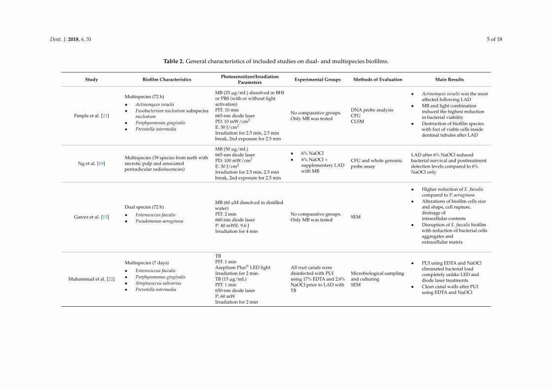
Table 2. General characteristics of included studies on dual- and multispecies biofilms.
Study Biofilm Characteristics Photosensitizer/IrradiationParameters Experimental Groups Methods of Evaluation Main Results
Fimple et al. [21]
Multispecies (72 h)
• Actinomyces israelii• Fusobacterium nucleatum subspecies
nucleatum• Porphyromonas gingivalis• Prevotella intermedia
MB (25 µg/mL) dissolved in BHIor PBS (with or without lightactivation)PIT: 10 min665-nm diode laserPD: 10 mW/cm2
E: 30 J/cm2
Irradiation for 2.5 min, 2.5 minbreak, 2nd exposure for 2.5 min
No comparative groups.Only MB was tested
DNA probe analysisCFUCLSM
• Actinomyces israelii was the mostaffected following LAD
• MB and light combinationinduced the highest reductionin bacterial viability
• Destruction of biofilm specieswith foci of viable cells insidedentinal tubules after LAD
Ng et al. [19]Multispecies (39 species from teeth withnecrotic pulp and associatedperiradicular radiolucencies)
MB (50 µg/mL)665-nm diode laserPD: 100 mW/cm2
E: 30 J/cm2
Irradiation for 2.5 min, 2.5 minbreak, 2nd exposure for 2.5 min
• 6% NaOCl• 6% NaOCl +
supplementary LADwith MB
CFU and whole genomicprobe assay
LAD after 6% NaOCl reducedbacterial survival and posttreatmentdetection levels compared to 6%NaOCl only
Garcez et al. [15]
Dual species (72 h)
• Enterococcus faecalis• Pseudomonas aeruginosa
MB (60 µM dissolved in distilledwater)PIT: 2 min660-nm diode laserP: 40 mWE: 9.6 JIrradiation for 4 min
No comparative groups.Only MB was tested SEM
• Higher reduction of E. faecaliscompared to P. aeruginosa
• Alterations of biofilm cells sizeand shape, cell rupture,drainage ofintracellular contents
• Disruption of E. faecalis biofilmwith reduction of bacterial cellsaggregates andextracellular matrix
Muhammad et al. [22]
Multispecies (7 days)
• Enterococcus faecalis• Porphyromonas gingivalis• Streptococcus salivarius• Prevotella intermedia
TBPIT: 1 minAseptium Plus® LED lightIrradiation for 2 min.TB (15 µg/mL)PIT: 1 min650-nm diode laserP: 60 mWIrradiation for 2 min
All root canals weredisinfected with PUIusing 17% EDTA and 2.6%NaOCl prior to LAD withTB
Microbiological samplingand culturingSEM
• PUI using EDTA and NaOCleliminated bacterial loadcompletely unlike LED anddiode laser treatments
• Clean canal walls after PUIusing EDTA and NaOCl

Dent. J. 2018, 6, 31 6 of 18
Table 2. Cont.
Study Biofilm Characteristics Photosensitizer/IrradiationParameters Experimental Groups Methods of Evaluation Main Results
Schiffner et al. [23]
• Aerobic bacterial mixture (72 h)Enterococcus faecalisShewanella putrefaciens
• Anaerobic bacterial mixture (72 h)
â Actinomyces naeslundiiâ Bifidobacterium adolescentisâ Peptostreptococcus sp.â Eggerthella lenta
TBPIT: 2 min632–644 nm red light.P: 200 mW.Irradiation for 1 min
• 0.9% NaCl• 1.5% NaOCl• 1.5% NaOCl +
supplementary LADwith TB
CFU
• LAD enhanced the bactericidalactivity of NaOCl againstaerobic bacteria mixtureimmediately after treatment
• Anaerobic bacteria mixture wasvery susceptible to NaOCl andNaOCl + PDT and wascompletely eradicated 4 daysafter treatment
Shrestha and Kishen [18]
Multispecies (21 days)
• Streptococcus oralis• Prevotella intermedia• Actinomyces naeslundii
RB (10 mmol/L)RBCnps (0.3 mg/mL)PIT: 15 min540-nm fiber lightE: 60 J/cm2
• RB• RBCnps
SEMCLSM
• Clean dentine surface and opendentinal tubules afterRBCnps treatment
• Dense bacterial aggregate afterRB treatment
• RBCSnps reduced biofilmthickness, killed cells anddisrupted the biofilms
De Oliveira et al. [24]
Multispecies (72 h)
• Enterococcus faecalis• Pseudomonas aeruginosa• Staphylococcus aureus• Candida albicans
MB (15 µg/mL)PIT: 2 min660-nm diode laserP: 100 mWE: 8 J/sampleIrradiation for 90 s
• 1% NaOCl• 1% NaOCl +
supplementary LADwith MB
• 5.25% NaOCl• 5.25% NaOCl +
supplementary LADwith MB
• 0.85% saline• 0.85% saline +
supplementary LADwith MB
CFU
• 5.25% NaOCl + LAD was themost successful protocol ineradicating theinoculated species
• Saline + LAD and 1% NaOClprotocols were not effectiveagainst tested microorganisms
Dent. J. 2018, 6, 31 7 of 18
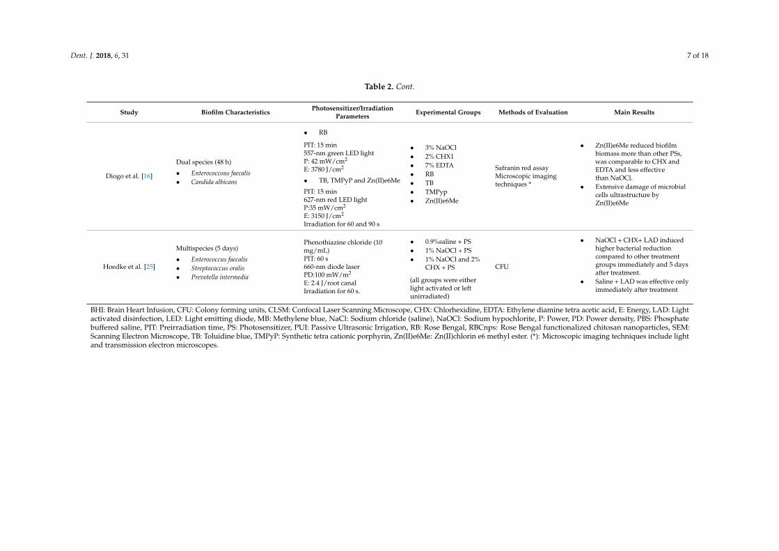
Table 2. Cont.
Study Biofilm Characteristics Photosensitizer/IrradiationParameters Experimental Groups Methods of Evaluation Main Results
Diogo et al. [16]
Dual species (48 h)
• Enterococcous faecalis• Candida albicans
• RB
PIT: 15 min557-nm green LED lightP: 42 mW/cm2
E: 3780 J/cm2
• TB, TMPyP and Zn(II)e6Me
PIT: 15 min627-nm red LED lightP:35 mW/cm2
E: 3150 J/cm2
Irradiation for 60 and 90 s
• 3% NaOCl• 2% CHX1• 7% EDTA• RB• TB• TMPyp• Zn(II)e6Me
Safranin red assayMicroscopic imagingtechniques *
• Zn(II)e6Me reduced biofilmbiomass more than other PSs,was comparable to CHX andEDTA and less effectivethan NaOCl.
• Extensive damage of microbialcells ultrastructure byZn(II)e6Me
Hoedke et al. [25]
Multispecies (5 days)
• Enterococcus faecalis• Streptococcus oralis• Prevotella intermedia
Phenothiazine chloride (10mg/mL)PIT: 60 s660-nm diode laserPD:100 mW/m2
E: 2.4 J/root canalIrradiation for 60 s.
• 0.9%saline + PS• 1% NaOCl + PS• 1% NaOCl and 2%
CHX + PS
(all groups were eitherlight activated or leftunirradiated)
CFU
• NaOCl + CHX+ LAD inducedhigher bacterial reductioncompared to other treatmentgroups immediately and 5 daysafter treatment.
• Saline + LAD was effective onlyimmediately after treatment
BHI: Brain Heart Infusion, CFU: Colony forming units, CLSM: Confocal Laser Scanning Microscope, CHX: Chlorhexidine, EDTA: Ethylene diamine tetra acetic acid, E: Energy, LAD: Lightactivated disinfection, LED: Light emitting diode, MB: Methylene blue, NaCl: Sodium chloride (saline), NaOCl: Sodium hypochlorite, P: Power, PD: Power density, PBS: Phosphatebuffered saline, PIT: Preirradiation time, PS: Photosensitizer, PUI: Passive Ultrasonic Irrigation, RB: Rose Bengal, RBCnps: Rose Bengal functionalized chitosan nanoparticles, SEM:Scanning Electron Microscope, TB: Toluidine blue, TMPyP: Synthetic tetra cationic porphyrin, Zn(II)e6Me: Zn(II)chlorin e6 methyl ester. (*): Microscopic imaging techniques include lightand transmission electron microscopes.
Dent. J. 2018, 6, 31 8 of 18
Methylene blue was the PS used in five studies [15,19,21,24,25], while toluidine blue wasused in three studies [16,22,23], and Rose Bengal was used in three studies either alone [16,18] orfunctionalized on cationic nanoparticles [18]. One study used Zn(II)chlorin e6 methyl ester andsynthetic porphyrin [16].
3.2. Antibioiflm Effect of Light Activated Disinfection
In this section, the included articles were screened to identify the studies which addressed theantibiofilm effect of LAD. The criteria [26,27] to include the studies are:
- Inhibition or reduction of the biofilm formation of the target microorganisms in response to LAD;and/or
- changes in biofilm characteristics, such as thickness, biomass, biovolume, and biofilm architecturein response to LAD.
Based on the previous criteria, 16 studies were identified that evaluated the antibiofilm efficacy ofLAD. Among the included studies, eight studies used methylene blue [9,14,15,17,28–31]. Rose Bengalwas used in six studies [9,16,18,29,32,33]. Three studies used Indocyanine green [31,34,35], and one ofthem used Indocyanine green loaded on nano-graphene oxide [35]. Two studies reported the effect ofchitosan-Rose Bengeal conjugate [9,32] and two other studies used Rose Bengal functionalized chitosannanoparticles [18,33]. Two studies used curcumin [8,36], while synthetic tetracationic porphyrin andZn(II)chlorin e6 methyl ester were used in one study [16].
In the selected studies, experiments were conducted on monospecies biofilms. One study involvedmono- and dual-species biofilm [16], and one study on multispecies biofilms [18]. The selectedstudies in this section addressed the effect of LAD on established biofilms except two studies,which demonstrated the inhibition of biofilm formation by light activated disinfection [31,35]. Table 3summarizes the general characteristics of these studies. The characteristics of the studies [15,16,18] arepresented in Table 2.

Dent. J. 2018, 6, 31 9 of 18
Table 3. General characteristics of studies on the antibiofilm effect of light activated disinfection (LAD).
Study Biofilm Characteristics Photosensitizer/IrradiationParameters Experimental Groups Methods of Evaluation Main Results
George and Kishen [28] Enterococcus faecalis(7 days)
Water-based MB (100 µmol/L)Emulsion-based MB (*)PIT: 10 min664-nm diode laserP: 30 mWE:31.84 J/cm2
• Water-based MB• Emulsion-based MB CLSM
Emulsion-based MBreduced biofilmthickness and caused marked biofilmdisruption compared to water-based MB
Kishen et al. [29] Enterococcus faecalis(4 days, 2 weeks)
MB, MB + EPI, and RB (100 µM)PIT: 15 min.Non-coherent light (660-nm for MB,540-nm for RB)P: 300–600 mWE: 10–40 J/cm2
• MB• MB + EPI• RB
CFU assay of biofilm cellsand biofilm derived cells(4 days biofilm)CLSM (2 weeks biofilm)
• MB + EPI induced maximumreduction of bacterial cellscompared to MB and RB
• LAD with MB produced greaterreduction of biofilm thicknesscompared to RB
Upadya and Kishen [14]Enterococcus faecalisPseudomonas aeruginosaMonospecies biofilms (2 weeks)
Water-based MBMIX-based MB (**)MB in MIX and EmulsioncombinationPIT: 15 min.660-nm non-coherent lightP: 0.106 WE: 2–40 J/cm 2
• Water-based MB• MIX-based MB CLSM
• MB in MIX and emulsioncombination was the most effectivein disrupting the biofilm structureand killing biofilm cells
• More apparent damage of E. faecalisbiofilm structure compared toP. aeruginosa
Upadya et al. [30] Enterococcus faecalis(4 days)
MB, MB + EPIPIT: 15 min660-nm non-coherent lightP: 0.106 WE: 2–40 J/cm 2
• Ca(OH)2• Chitosan NPs• MB• Ca(OH)2 with EPI• Chitosan NPs
with EPI• MB + EPI
CFU assay of biofilm cellsand biofilm derived cells
• light activated MB was moreeffective than Ca(OH)2 andChitosan Nps
• Effect of EPI was more significanton antibiofilm effect of MB thanthat of Ca(OH)2 and Chitosan Nps

Dent. J. 2018, 6, 31 10 of 18
Table 3. Cont.
Study Biofilm Characteristics Photosensitizer/IrradiationParameters Experimental Groups Methods of Evaluation Main Results
Shrestha and Kishen [9]Enterococcus faecalisPseudomonas aeruginosaMonospecies biofilm (7 days)
MB (10 µM)RB (10 µM)CSRB (0.3 mg/mL)White light source (540-nm for RBand CSRB, 660-nm for MB)E: 20, 40 and 60 J/cm2 (PIT: 15 min.)40 J/cm2 (PIT: 30 and 60 min.)
• MB• RB• CSRB
CFU assay of biofilm cellsCLSM
• light activated CSRP inducedhigher antibiofilm effect on biofilmsof both microorganisms comparedto MB and RB
• CSRP was the most effective inreduction of viable cells, biofilmthickness and disruption ofbiofilm architecture
• MB and RB were unable todisrupt biofilms
Shrestha et al. [32] Enterococcus faecalis(7 days)
RB (10 µM)CSRB (0.3 mg/mL)PIT: 15 min.540-nm lightE: 20, 40 and 60 J/cm2
RBCSRB CFU assay of biofilm cellsCSRP induced a significantly higherLAD mediated bacterial killingcompared to RB at 40 and 60 J/cm2
Shrestha et al. [33] Enterococcus faecalis(21 days)
RB (10 µM)RBCnps (0.1 and 0.3 mg/mL)PIT: 15 min.540-nm lightP: 50 mWE: 20, 40, 60 and fractionated dosageof 10 and 20 J/cm2 twice
• RB• RBCnps
CFU assay of biofilm cellsCLSM
• Complete elimination of biofilmcells by RBCnps (0.3 mg/mL) andRB exposed to fractionated dosageof LAD
• Complete killing was not achievedat higher light doses regardless thePS used
• Superior bacterial killing andcomplete disruption of biofilmstructure following lightactivated RBCnps
Neelakantan et al. [8] Enterococcus faecalis(4 weeks)
Curcumin (2.5 mg/mL)380–515 nm blue lightE:1200 mW/cm2
Irradiation for 4 min
• Saline• 3% NaOCl• 3% NaOCl with PUI• 3% NaOCl with LAD• Curcumin• Curcumin with PUI• Curcumin with light
CLSM
• Light activated curcumin was ableto achieve higher killing of biofilmcells compared to sodiumhypochlorite irrigation
• Curcumin and ultrasonicallyactivated curcumin reduced biofilmmass more than lightactivated curcumin

Dent. J. 2018, 6, 31 11 of 18
Table 3. Cont.
Study Biofilm Characteristics Photosensitizer/IrradiationParameters Experimental Groups Methods of Evaluation Main Results
Chiniforush et al. [34] Enterococcus faecalis(24 h)
ICG (3–2000 µg/mL)PIT: 5 min808-nm diode laserP: 250 mWE: 39.06 J/cm2
Irradiation for 60 s
No comparative groupsOnly ICG was tested CV assay
Non-washed ICG induced higherreduction in biofilm formation,development and higher rate of biofilmdegradation compared to washed ICG
Deveraj et al. [36] Enterococcus faecalis(4 weeks)
Curcumin (2.5 mg/mL ofpolyethylene glycol)380–315 nm blue lightP: 1200 mW/cm2
Irradiation for 4 min
• TAP• DAP• 2% CHX gel• Ca(OH)2 gel• Curcumin
CLSM
Light activated curcumin and TAPreduced biofilm thickness, disruptedbiofilm architecture and killed bacterialcells more than other medicaments
Pourhajibagher et al. [31] Enterococcus faecalis(24 h)
Sublethal concentrations of TB, MB(6.2 µg/mL) and ICG (31.2 µg/mL)Diode laser: 660 nm (MB), 635 nm(TB) and 810 nm (ICG)P: 150 mW (MB), 220 mW (TBO) and200 mW (ICG)E: 70.31 J/cm2 (MB)103.12 J/cm2 (TBO)15.62 J/cm2 (ICG)
• TB• MB• ICG
CV assaySEM
• ICG-sPDT inhibited biofilmformation more thanTBO-, MB-sPDT
• Lower cell density and moreirregular shaped cells wereobserved in ICG-sPDTtreated biofilms
Akbari et al. [35] Enterococcus faecalis(24 h)
ICG (1000 µg/mL)NGO-ICG (200 µg/mL)810-nm diode laserP: 250 mWE: 31.2 J/cm2
Irradiation for 60 s
• ICG• NGO-ICG CV assay
Photoactivated NGO-ICG reducedbiofilm formation more thanphotoactivated ICG
Rosa et al. [17] Multispecies biofilm developedintraorally (72 h)
0.01%MB.PIT: 1 min650-nm red diode laserP: 100 mWIrradiation for 1 min
• Saline• Saline + PDT
with MB• 2.5% NaOCl• 2.5% NaOCl + PDT
with MB• 2% CHX• 2% CHX + PDT
with MB
CLSM LAD after NaOCl reduced biofilmbiovolume
CV: Crystal Violet, Ca(OH)2: Calcium hydroxide, CSRB: Chitosan rose Bengal conjugate, DAP: Double antibiotic paste. EPI: Efflux pump inhibitor. ICG: Indocyanine green, NGO-ICG:Nano-graphene oxide loaded with Indocyanine green, sPDT: Sub-lethal doses of photodynamic therapy. TAP: Triple antibiotic paste. (*) Emulsion-based MB: MB in an emulsion mixture ofperfluoro-decahydronaphthalene, H2O2, and triton X-100. (**) MIX-based MB: Methylene blue dissolved in a mixture of glycerol, ethanol, and water.

Dent. J. 2018, 6, 31 12 of 18
4. Discussion
4.1. Effect of LAD on Dual and Multispecies Biofilm
Fimple et al. used methylene blue (MB) as a photosensitizer against four predominant bacterialspecies of infected root canals [21]. In this study, the photosensitizer was left unirradiated for 2.5 minbetween two episodes of light activation to allow oxygen diffusion into the oxygen deprived areasof infected root canals, since the presence of oxygen in the vicinity is necessary for the generation ofcytotoxic singlet oxygen and reactive oxygen species upon photoactivation, which destroy bacterialcells [13,26]. Greater reduction of bacterial viability was observed when photoactivated MB wasdissolved in PBS compared to that observed when it was dissolved in BHI. This was attributed tothe presence of serum in the BHI, which affected the antibacterial performance of photoactivated MB.This highlighted the inhibitory effect of serum on the antibacterial mechanism of LAD as shown laterby Shrestha and Kishen [27].
Root canal microorganisms of ex vivo root canal infections were sensitive to LAD as an adjunct toconventional chemomechanical debridement as shown by Ng et al. In this study, bacterial survival wasobserved when methylene blue was light activated after chemomechanical debridement. Viability ofkey endodontic pathogens was reduced after exposure to LAD. However, more bacteria were recoveredwhen the dentinal shavings were collected compared to those recovered from flushing the root canalsafter root canal disinfection, indicating that currently used disinfection approaches, including LAD,were unable to eradicate bacteria from the anatomical eccentricities of the root canal system [19].
Schniffer et al. demonstrated that supplementary LAD enhanced the killing activity of sodiumhypochlorite immediately after treatment. Interestingly, bacteria were able to repopulate when themicrobial samples were taken two and four days after the initial treatment, indicating that theseapproaches were unable to prevent reinfection. This was explained by the survival of E. faecalis insidethe dentinal tubules. Aerobic bacterial mixtures were eradicated immediately after chemocmechanicaldebridement and PDT treatment. However, this effect was abolished a few days after the treatment.In contrast, the anaerobic bacterial mixture was highly susceptible to NaOCl irrigation and NaOClirrigation followed by LAD, with no significant difference between them. This effect was long lasting,and bacteria were eradicated four days after treatment [23].
In the same context, Hoedke et al. evaluated the effect of LAD as an adjunct to current root canalirrigation protocols. Reduction of bacterial load was evaluated immediately and five days after thetreatment to simulate the situation of unobturated root canals encountered during retreatment. It wasconcluded that LAD is unable to eradicate intracanal bacterial to a satisfactory level without priorchemomechanical debridement. Bacteria were recovered a few days after saline-LAD combinationtreatment. In contrast, when LAD was applied after NaOCl and CHX irrigation, it enhanced bacterialreduction compared to non-photodisinfected root canals [25].
One of the most promising approaches in light activated disinfection has been functionalizing PSon nanoparticles for better penetration into the biofilms and microbial cells, as well as into the intricateanatomy of the radicular space. Shrestha and Kishen developed a mature multispecies biofilm modelto simulate the in vivo root canal microflora of infected teeth. In this study, Rose Bengal functionalizedchitosan nanoparticles (RBCnps) were compared to Rose Bengal photosensitizer alone. The resultswere promising in the favor of RBCnps in terms of dentin cleanliness and the disruption of the biofilmstructure, as shown by SEM and CLSM, respectively. The study emphasized the positive impact ofchitosan nanoparticles on the antibacterial properties of photoactivated Rose Bengal [18].
Despite the differences in the cell wall composition between gram-positive and gram-negativebacteria, 5.25% NaOCl followed by LAD was the most effective protocol against the testedmicroorganisms, according to the study by de Oliveira et al. in which multispecies biofilms of grampositive, gram-negative bacteria, and fungi were inoculated into root canals prepared by single fileinstrumentation technique [24]. It is worth mentioning that the multispecies biofilm was developed for72 h, which could be sufficient for adhesion and biofilm formation, but not enough for the formation of

Dent. J. 2018, 6, 31 13 of 18
a mature complex multilayered biofilm. The results might have been different if the biofilm had beenallowed to grow for a longer period due to deposition of extracellular polymeric substance that protectsindividual cells against antimicrobial therapeutics. Furthermore, the thicker the biofilm, the lesser thepenetration of photosensitizers into the deep layers of biofilms and, thereby, a lesser effect of LADis expected.
More recently, Zn(II)chlorin e6 methyl ester, derived from natural sources, has been used as aphotosensitizer against E. faecalis and C. albicans cells in a mixed species biofilm. While this PS wassimilar in its antimicrobial efficacy to Chlorhexidine and EDTA, it was significantly less effectivethan NaOCl. In the same study, the effect of LAD on the cellular morphology was examined usingmicroscopy imaging. At the cellular level, display of the “ghost cells” feature of E. faecalis revealed theevacuation of intracellular contents with an intact cell wall as a result of photoactivation of Zn(II)e6Me,while C. albicans cells showed disruption of the cytoplasmic membrane, cell membrane invaginations,and presence of extracellular vesicles at the cell surface. This possibly showed that Zn(II)e6Me mightpossess microorganism specific inactivation mechanisms [16].
From these studies, it is apparent that that effect of LAD on dual and multispecies biofilmsremains to be extensively studied. Furthermore, the identified studies show diversity in theconstituent microorganisms that form the biofilms. Also, it is obvious that LAD with conventionalPS, such as MB and RB alone, are unable to eradicate dual and multispecies biofilm. Light activateddisinfection can, thus, enhance the antimicrobial efficacy of root canal irrigants and disinfectionstrategies. Some photosensitizers, like RBCSnps and a natural chlorophyll derived photosensitizer, arepromising, and they can eradicate polymicrobial biofilms more effectively compared to traditionalphotosensitizers. However, more studies are required to evaluate their effects on different combinationsof microorganisms in a polymicrobial biofilm.
4.2. Antibiofilm Effect of Light Activated Disinfection
4.2.1. Methylene Blue
Eight studies used methylene blue and its formulations as photosensitizers. Methylene blue,in association with verapamil hydrochloride as an efflux pump inhibitor, was used in twostudies [29,30]. Methylene blue dissolved in different solvents was used in two studies [14,28]. In onestudy, despite the finding that photoactivated MB and RB were not significantly different in theirbacterial killing effect on 4-days biofilms, photoactivated MB induced greater reduction in biofilmthickness and killed more bacterial cells compared to photoactivated Rose Bengal, as shown by3-dimensional imaging using confocal microscopy [29]. The inferior results of RB compared to MBwere attributed to the repulsion between the negatively charged cell wall of E. faecalis cells (dueto lipoteichoic acid residues) and the anionic Rose Bengal molecules. The biofilm disruption byphotoactivated MB was evident as it reduced the biofilm covered surface, with relatively few cellaggregates and a reduced extracellular matrix [15]. In another study, light activated MB exhibitedhigher antibiofilm efficacy compared to Ca(OH)2 and polycationic chitosan nanoparticles at higherconcentrations. Addition of an efflux pump inhibitor improved the antibiofilm effect of MB to a higherextent compared to the other two disinfection strategies as EPI enhanced the diffusion of MB intothe biofilm matrix and, subsequently, increased the production of singlet oxygen, resulting in thedisruption of the extracellular polymeric matrix [30].
Variations in the biofilm thickness and oxygen concentration across the multi-layered biofilmresults in a less than optimal performance of LAD using PSs. To allow better penetration of the PS toenhance the antibiofilm action, several modifications have been attempted [14,28]. These modificationsincluded either inclusion of an oxygen carrier and oxidizer with MB [28] or dissolving of MB in amixture of glycerol, ethanol, and water or dual stage approach of LAD, which consists of dissolvingMB in a glycerol, ethanol, and water mixture, followed by illumination in an oxygen carrier solution.

Dent. J. 2018, 6, 31 14 of 18
The latter approach was more effective than 1% NaOCl in eradicating E. faecalis biofilms from deepdentine layers [28,37].
Chemical modifications of MB formulations were found to exert more substantial biofilmdestruction, reduction of the biofilm thickness, and more bacterial killing than MB dissolved inwater. The superior bactericidal effect of MIX-based MB photosensitizer was attributed to the longerlife of the generated singlet oxygen, impairment of cell membrane integrity, and extensive damageof chromosomal DNA induced by MIX-based formulations compared to the water-based MB [38].Perfluoro-decahydronaphthalene serves as an oxygen carrier, which facilitated light penetrationduring the irradiation phase and increased the rate of singlet oxygen production [28,39]. Hydrogenperoxide as an oxidizer disrupted the biofilm matrix and facilitated penetration of PS into the biofilm.Upon interaction with MB, hydroxyl radicals are generated, which then inactivate the bacterial growth.
4.2.2. Chitosan Rose Bengal Conjugate
Rose Bengal as a photosensitizer did not completely eradicate the biofilm bacteria due to theelectrostatic repulsion between the Rose Bengal molecules and the negatively charged bacterialcells and exopolysaccharides, as mentioned previously [9,29]. Conjugation of the Rose Bengalphotosensitizer with natural polymers was, therefore, proposed to improve the overall effect ofRose Bengal mediated LAD against resistant root canal pathogens. Two of the selected articles usedRose Bengal-chitosan conjugate and compared it with conventional PSs [9,32] Chitosan is a naturalpolymer with antimicrobial properties. Its unique chemical nature allows functionalization withvarious antimicrobial agents [40,41].
Chitosan-RB conjugate (CSRB) was able to substantially reduce the biofilm thickness anddisrupt the biofilm architecture of E. faecalis and P. aeruginosa, unlike methylene blue and RB alone,which exhibited a lesser disrupting effect on the biofilm structure [9]. In addition, the efficacyof CSRB on biofilm cells was enhanced by increasing the photosensitization time, which did notinfluence the killing effect of light activated MB and RB. Interestingly, P. aeruginosa biofilm cellswere completely eliminated in response to light activated CSRB at a light dose higher than 40 J/cm2.These findings were attributed to the overall positive charge of CSRB, which ensured an intimatecontact of CSRB to the bacterial cell wall and a higher uptake of photosensitizers by bacterial cells [32].In addition, the hydrophilic nature of chitosan allows deep penetration of CSRB through the waterrich extracellular polymeric substance. The CSRB was further modified by functionalization of RoseBengal with chitosan nanoparticles (RBCnps). Nanoparticles per se are known for their favorablephysicochemical properties, including ultra-small size, higher chemical reactivity, and larger surfacearea/mass ratio, which suggests its widespread use in targeted drug delivery [42,43]. RBCnps wereevaluated for their antibiofilm activity against mono- and multispecies biofilm [18,33]. In one study,RBCnps achieved complete elimination of E. faecalis mono-species biofilm cells after light activation byfractionated dosage, compared to continuous exposure to different light doses. Fractionation allowsthe replenishment of molecular oxygen, adjacent to the irradiated cells, and generates singlet oxygenmolecules constantly [33]. RBCnps demonstrated disruption of the biofilm architecture and reducedviable bacterial cells of multispecies biofilm compared to Rose Bengal, in which the biofilm structurewas not affected and aggregates of viable cells were still present among the dead cells [18].
The success of RBCnps over conventional photosensitizers was confirmed when RBCnps waschallenged with tissue inhibitors. It was found that RBCnps preincubated with pulp remnants- for24 h eliminated bacterial load after photoactivation. In contrast, photoactivated MB and RB were notable to eradicate bacterial cells [44].
4.2.3. Indocyanine Green (ICG) and Its Modifications
In two studies, unmodified ICG was used [31,34], while one used ICG loaded on nano-grapheneoxide [35]. In clinical situations, photosensitizers might not reach the target microbial cells at their lethalconcentrations mainly due to the limited accessibility of light to the infection site. Therefore, different

Dent. J. 2018, 6, 31 15 of 18
sublethal concentrations of MB, Toluidine Blue (TB), and ICG were tested against E. feacalis biofilmformation [31]. Biofilm formation was significantly reduced when the three photosensitizers wereused at concentrations equivalent to at least one quarter of their minimum inhibitory concentration(MIC) values. ICG maintained its ability to reduce biofilm formation when used at a concentrationequivalent to one sixteenth of the MIC value. On the other hand, one quarter and one eighth MICof MB and TB were the smallest concentrations that were able to reduce E. faecalis biofilm formation.The lack of inhibition of biofilm formation by the lower concentrations creates a caveat for LAD thatproper concentrations of PS are imperative for its clinical effects.
Clinical protocols of washing the PS or not prior to light activation also appears to have aninfluence on the required concentration and, ultimately, the antimicrobial efficacy. Chinifoursh et al.demonstrated that higher ICG concentrations were required to exhibit effective biofilm inhibitionif it was washed prior to light irradiation [34]. This finding should be considered in clinicalsituations in which tissue exudates escape into the canal space, thereby, diluting the concentrationof photosensitizers, and attenuating the effect of LAD on root canal microorganisms. It is worthmentioning that the antibacterial effect of ICG is not only related to generation of reactive oxygenspecies upon the light activation. Indocyanine green is light activated by near infrared light unlikeother photosensitizers that are activated by visible light. ICG can convert most of the near infraredlaser into heat, which causes thermal injury to the adjacent bacterial cells [45].
Despite the promising results of ICG as a photosensitizer with an antibiofilm effect, it stillhas some limitations, including concentration-dependent aggregation, rapid degradation, limitedphotostability, and reduced interaction of anionic ICG with the negatively charged bacterial cellsurface [18,46]. Therefore, a nanoparticle-based approach has been implemented to improve ICGdelivery and interaction with target cells by loading of ICG on nanographene oxide. High surfacearea, functionalization potential, and cost-effective synthesis are favorable characteristics that favorits application as a platform of anticancer drugs, antimicrobials, and proteins [47]. Nanographeneoxide (NGO) loaded ICG-based PDT demonstrated a higher antibiofilm effect compared to ICG-basedPDT due to stabilizing effect of NGO, which increased the bioavailability of ICG and singlet oxygenproduction in biofilms [35].
4.2.4. Curcumin
Curcumin is a natural polyphenolic compound extracted from plants sources that possessantimicrobial properties [48,49]. Additionally, photoactivated curcumin appears to be an effectivetreatment strategy for persistent root canal infections [50,51]. The impact of light activation onantibacterial and antibiofilm properties of curcumin is still controversial. Neelakantan et al. revealedthat light activated curcumin eradicated more viable biofilm cells from superficial and deep layersof the biofilm compared to curcumin alone and ultrasonically activated curcumin [8]. This wasattributed to the release of hydrogen peroxide, which kills bacterial cells. However, curcumin aloneand ultrasonically activated curcumin decreased biofilm mass compared to light activated curcumin.According to Pileggi et al., curcumin had a slight effect on E. faecalis cell viability and light activationwas necessary to reduce the viability of biofilm cells [51]. Also, the irradiation time of curcuminis another critical factor, since curcumin irradiated for five minutes caused the greatest microbialreduction compared to curcumin irradiated for ten minutes [50].
Devaraj et al. used curcumin in a light activated intracanal medicament formulation and left it inthe root canals for 14 days. The superior results of light activated curcumin over other conventionalmedicaments at different depths was attributed to its ability to exert a lethal effect on bacterial cellswithout being in close contact with the bacterial cells’ surface unlike most of the photosensitizers,which requires close approximation to target cells to kill bacterial cells or disrupt biofilms [36].
Based on the reviewed studies, conventional formulations of photosensitizers, such as methyleneblue (MB) and Rose Bengeal (RB), when used alone, are unable to induce substantial alterations in thebiofilm structure and biofilm related characteristics, like biomass and thickness. Modified formulations,

Dent. J. 2018, 6, 31 16 of 18
either by changing the dissolving media (methylene blue dissolved in mixture of glycerol, ethanol,and water) or conjugation of photosensitizers with nanoparticles (such as Rose Bengal functionalizedchitosan nanoparticles), demonstrated a higher efficacy in eradicating biofilm cells and disrupting thebiofilm architecture. Further studies are needed to fabricate Nanocarrier systems for PS and to testtheir effect on the biofilm structure and biofilm matrix components of mono and multispecies biofilmsof microorganisms relevant to persistent root canal infections.
5. Conclusions
The polymicrobial nature and complex structure of root canal biofilms, as well as the hypoxicroot canal environment, are the main factors that influence the antimicrobial efficacy of light activateddisinfection. Conventional root canal debridement using sodium hypochlorite irrigant can be enhancedby light activation of photosensitizers. Functionalization of photosensitizers with nanoparticles (e.g.,chitosan with Rose Bengal) and naturally derived photosensitizers (e.g., Zn(II)e6 methyl ester andcurcumin) can affect dual-and multispecies biofilms, reduce biofilm formation, and exert substantialalteration in biofilm structure.
Author Contributions: Conception: I.A.A.A. and P.N.; Literature review: I.A.A.A., Revision: I.A.A.A. and P.N.;Final Approval: I.A.A.A. and P.N.
Funding: This research received no external funding.
Conflicts of Interest: The authors declare no conflict of interest.
References
1. Bystrom, A.; Sundqvist, G. Bacteriologic evaluation of the efficacy of mechanical root canal instrumentationin endodontic therapy. Scand. J. Dent. Res. 1981, 89, 321–328. [CrossRef] [PubMed]
2. Nair, P.N.; Henry, S.; Cano, V.; Vera, J. Microbial status of apical root canal system of human mandibular firstmolars with primary apical periodontitis after “one-visit” endodontic treatment. Oral Surg. Oral Med. OralPathol. Oral Radiol. Endod. 2005, 99, 231–252. [CrossRef] [PubMed]
3. Siqueira, J.F., Jr.; Rocas, I.N. Clinical implications and microbiology of bacterial persistence after treatmentprocedures. J. Endod. 2008, 34, 1291.e3–1301.e3. [CrossRef] [PubMed]
4. Neelakantan, P.; Romero, M.; Vera, J.; Daood, U.; Khan, A.U.; Yan, A.; Cheung, G.S.P. Biofilms inendodontics-current status and future directions. Int. J. Mol. Sci. 2017, 18. [CrossRef] [PubMed]
5. Donlan, R.M.; Costerton, J.W. Biofilms: Survival mechanisms of clinically relevant microorganisms.Clin. Microbiol. Rev. 2002, 15, 167–193. [CrossRef] [PubMed]
6. Flemming, H.C.; Neu, T.R.; Wozniak, D.J. The EPS matrix: The “house of biofilm cells”. J. Bacteriol. 2007, 189,7945–7947. [CrossRef] [PubMed]
7. Zehnder, M. Root canal irrigants. J. Endod. 2006, 32, 389–398. [CrossRef] [PubMed]8. Neelakantan, P.; Cheng, C.Q.; Ravichandran, V.; Mao, T.; Sriraman, P.; Sridharan, S.; Subbarao, C.; Sharma, S.;
Kishen, A. Photoactivation of curcumin and sodium hypochlorite to enhance antibiofilm efficacy in rootcanal dentin. Photodiagn. Photodyn. Ther. 2015, 12, 108–114. [CrossRef] [PubMed]
9. Shrestha, A.; Kishen, A. Polycationic chitosan-conjugated photosensitizer for antibacterial photodynamictherapy. Photochem. Photobiol. 2012, 88, 577–583. [CrossRef] [PubMed]
10. Kishen, A.; Shrestha, A.; Cohenca, N. Emerging technologies in root canal disinfection. In Disinfection of RootCanal Systems: The Treatment of Apical Periodontitis; Cohenca, N., Ed.; Wiley Blackwell: Hoboken, NJ, USA,2014; pp. 277–296. ISBN 9781118914014.
11. Cieplik, F.; Tabenski, L.; Buchalla, W.; Maisch, T. Antimicrobial photodynamic therapy for inactivation ofbiofilms formed by oral key pathogens. Front. Microbiol. 2014, 5, 405. [CrossRef] [PubMed]
12. Dai, T.; Huang, Y.Y.; Hamblin, M.R. Photodynamic therapy for localized infections-state of the art. Photodiagn.Photodyn. Ther. 2009, 6, 170–188. [CrossRef] [PubMed]
13. Maisch, T.; Baier, J.; Franz, B.; Maier, M.; Landthaler, M.; Szeimies, R.M.; Baumler, W. The role of singletoxygen and oxygen concentration in photodynamic inactivation of bacteria. Proc. Natl. Acad. Sci. USA 2007,104, 7223–7228. [CrossRef] [PubMed]

Dent. J. 2018, 6, 31 17 of 18
14. Upadya, M.H.; Kishen, A. Influence of bacterial growth modes on the susceptibility to light-activateddisinfection. Int. Endod. J. 2010, 43, 978–987. [CrossRef] [PubMed]
15. Garcez, A.S.; Núñez, S.C.; Azambuja, N.; Fregnani, E.R.; Rodriguez, H.M.H.; Hamblin, M.R.; Suzuki, H.;Ribeiro, M.S. Effects of photodynamic therapy on gram-positive and gram-negative bacterial biofilms bybioluminescence imaging and scanning electron microscopic analysis. Photomed. Laser Surg. 2013, 31,519–525. [CrossRef] [PubMed]
16. Diogo, P.; Fernandes, C.; Caramelo, F.; Mota, M.; Miranda, I.M.; Faustino, M.A.F.; Neves, M.; Uliana, M.P.;de Oliveira, K.T.; Santos, J.M.; et al. Antimicrobial photodynamic therapy against endodontic enterococcusfaecalis and candida albicans mono and mixed biofilms in the presence of photosensitizers: A comparativestudy with classical endodontic irrigants. Front. Microbiol. 2017, 8, 498. [CrossRef] [PubMed]
17. Rosa, R.A.D.; Santini, M.F.; Figueiredo, J.A.P.; Visioli, F.; Pereira, J.R.; Vivan, R.R.; Montagner, F.; So, M.V.R.Effectiveness of photodynamic therapy associated with irrigants over two biofilm models. Photodiagn.Photodyn. Ther. 2017, 20, 169–174. [CrossRef] [PubMed]
18. Shrestha, A.; Kishen, A. Antibiofilm efficacy of photosensitizer-functionalized bioactive nanoparticles onmultispecies biofilm. J. Endod. 2014, 40, 1604–1610. [CrossRef] [PubMed]
19. Ng, R.; Singh, F.; Papamanou, D.A.; Song, X.; Patel, C.; Holewa, C.; Patel, N.; Klepac-Ceraj, V.; Fontana, C.R.;Kent, R.; et al. Endodontic photodynamic therapy ex vivo. J. Endod. 2011, 37, 217–222. [CrossRef] [PubMed]
20. Stojicic, S.; Amorim, H.; Shen, Y.; Haapasalo, M. Ex vivo killing of enterococcus faecalis and mixed plaquebacteria in planktonic and biofilm culture by modified photoactivated disinfection. Int. Endod. J. 2013, 46,649–659. [CrossRef] [PubMed]
21. Fimple, J.L.; Fontana, C.R.; Foschi, F.; Ruggiero, K.; Song, X.; Pagonis, T.C.; Tanner, A.C.R.; Kent, R.;Doukas, A.G.; Stashenko, P.P.; et al. Photodynamic treatment of endodontic polymicrobial infection in vitro.J. Endod. 2008, 34, 728–734. [CrossRef] [PubMed]
22. Muhammad, O.H.; Chevalier, M.; Rocca, J.P.; Brulat-Bouchard, N.; Medioni, E. Photodynamic therapy versusultrasonic irrigation: Interaction with endodontic microbial biofilm, an ex vivo study. Photodiagn. Photodyn.Ther. 2014, 11, 171–181. [CrossRef] [PubMed]
23. Schiffner, U.; Cachovan, G.; Bastian, J.; Sculean, A.; Eick, S. In vitro activity of photoactivated disinfectionusing a diode laser in infected root canals. Acta Odontol. Scand. 2014, 72, 673–680. [CrossRef] [PubMed]
24. De Oliveira, B.P.; Aguiar, C.M.; Camara, A.C.; de Albuquerque, M.M.; Correia, A.C.; Soares, M.F. The efficacyof photodynamic therapy and sodium hypochlorite in root canal disinfection by a single-file instrumentationtechnique. Photodiagn. Photodyn. Ther. 2015, 12, 436–443. [CrossRef] [PubMed]
25. Hoedke, D.; Enseleit, C.; Gruner, D.; Dommisch, H.; Schlafer, S.; Dige, I.; Bitter, K. Effect of photodynamictherapy in combination with various irrigation protocols on an endodontic multispecies biofilm ex vivo.Int. Endod. J. 2018, 51 (Suppl. 1), e23–e34. [CrossRef] [PubMed]
26. Diogo, P.; Goncalves, T.; Palma, P.; Santos, J.M. Photodynamic antimicrobial chemotherapy for root canalsystem asepsis: A narrative literature review. Int. J. Dent. 2015, 2015, 269205. [CrossRef] [PubMed]
27. Shrestha, A.; Kishen, A. The effect of tissue inhibitors on the antibacterial activity of chitosan nanoparticlesand photodynamic therapy. J. Endod. 2012, 38, 1275–1278. [CrossRef] [PubMed]
28. George, S.; Kishen, A. Augmenting the antibiofilm efficacy of advanced noninvasive light activateddisinfection with emulsified oxidizer and oxygen carrier. J. Endod. 2008, 34, 1119–1123. [CrossRef] [PubMed]
29. Kishen, A.; Upadya, M.; Tegos, G.P.; Hamblin, M.R. Efflux pump inhibitor potentiates antimicrobialphotodynamic inactivation of enterococcus faecalis biofilm. Photochem. Photobiol. 2010, 86, 1343–1349.[CrossRef] [PubMed]
30. Upadya, M.; Shrestha, A.; Kishen, A. Role of efflux pump inhibitors on the antibiofilm efficacy of calciumhydroxide, chitosan nanoparticles, and light-activated disinfection. J. Endod. 2011, 37, 1422–1426. [CrossRef][PubMed]
31. Pourhajibagher, M.; Chiniforush, N.; Shahabi, S.; Ghorbanzadeh, R.; Bahador, A. Sub-lethal doses ofphotodynamic therapy affect biofilm formation ability and metabolic activity of enterococcus faecalis.Photodiagn. Photodyn. Ther. 2016, 15, 159–166. [CrossRef] [PubMed]
32. Shrestha, A.; Hamblin, M.R.; Kishen, A. Characterization of a conjugate between rose bengal and chitosanfor targeted antibiofilm and tissue stabilization effects as a potential treatment of infected dentin. Antimicrob.Agents Chemother. 2012, 56, 4876–4884. [CrossRef] [PubMed]

Dent. J. 2018, 6, 31 18 of 18
33. Shrestha, A.; Hamblin, M.R.; Kishen, A. Photoactivated rose bengal functionalized chitosan nanoparticlesproduce antibacterial/biofilm activity and stabilize dentin-collagen. Nanomed. Nanotechnol. Biol. Med. 2014,10, 491–501. [CrossRef] [PubMed]
34. Chiniforush, N.; Pourhajibagher, M.; Parker, S.; Shahabi, S.; Bahador, A. The in vitro effect of antimicrobialphotodynamic therapy with indocyanine green on enterococcus faecalis: Influence of a washing vs.non-washing procedure. Photodiagn. Photodyn. Ther. 2016, 16, 119–123. [CrossRef] [PubMed]
35. Akbari, T.; Pourhajibagher, M.; Hosseini, F.; Chiniforush, N.; Gholibegloo, E.; Khoobi, M.; Shahabi, S.;Bahador, A. The effect of indocyanine green loaded on a novel nano-graphene oxide for high performanceof photodynamic therapy against enterococcus faecalis. Photodiagn. Photodyn. Ther. 2017, 20, 148–153.[CrossRef] [PubMed]
36. Devaraj, S.; Jagannathan, N.; Neelakantan, P. Antibiofilm efficacy of photoactivated curcumin, triple anddouble antibiotic paste, 2% chlorhexidine and calcium hydroxide against enterococcus fecalis in vitro.Sci. Rep. 2016, 6. [CrossRef] [PubMed]
37. Lim, Z.; Cheng, J.L.; Lim, T.W.; Teo, E.G.; Wong, J.; George, S.; Kishen, A. Light activated disinfection:An alternative endodontic disinfection strategy. Aust. Dent. J. 2009, 54, 108–114. [CrossRef] [PubMed]
38. George, S.; Kishen, A. Influence of photosensitizer solvent on the mechanisms of photoactivated killing ofenterococcus faecalis. Photochem. Photobiol. 2008, 84, 734–740. [CrossRef] [PubMed]
39. George, S.; Kishen, A. Photophysical, photochemical, and photobiological characterization of methyleneblue formulations for light-activated root canal disinfection. J. Biomed. Opt. 2007, 12. [CrossRef] [PubMed]
40. Veerapandian, M.; Yun, K. Functionalization of biomolecules on nanoparticles: Specialized for antibacterialapplications. Appl. Microbiol. Biotechnol. 2011, 90, 1655–1667. [CrossRef] [PubMed]
41. Rabea, E.I.; Badawy, M.E.T.; Stevens, C.V.; Smagghe, G.; Steurbaut, W. Chitosan as antimicrobial agent:Applications and mode of action. Biomacromolecules 2003, 4, 1457–1465. [CrossRef] [PubMed]
42. Venugopal, J.; Prabhakaran, M.P.; Low, S.; Choon, A.T.; Zhang, Y.Z.; Deepika, G.; Ramakrishna, S.Nanotechnology for nanomedicine and delivery of drugs. Curr. Pharm. Des. 2008, 14, 2184–2200. [CrossRef][PubMed]
43. Shrestha, A.; Kishen, A. Antibacterial nanoparticles in endodontics: A review. J. Endod. 2016, 42, 1417–1426.[CrossRef] [PubMed]
44. Shrestha, A.; Kishen, A. Antibacterial efficacy of photosensitizer functionalized biopolymeric nanoparticlesin the presence of tissue inhibitors in root canal. J. Endod. 2014, 40, 566–570. [CrossRef] [PubMed]
45. Kranz, S.; Huebsch, M.; Guellmar, A.; Voelpel, A.; Tonndorf-Martini, S.; Sigusch, B.W. Antibacterialphotodynamic treatment of periodontopathogenic bacteria with indocyanine green and near-infrared laserlight enhanced by trolox(tm). Lasers Surg. Med. 2015, 47, 350–360. [CrossRef] [PubMed]
46. Ocsoy, I.; Isiklan, N.; Cansiz, S.; Ozdemir, N.; Tan, W. Icg-conjugated magnetic graphene oxide for dualphotothermal and photodynamic therapy. RSC Adv. 2016, 6, 30285–30292. [CrossRef] [PubMed]
47. Wang, Y.-W.; Fu, Y.-Y.; Peng, Q.; Guo, S.-S.; Liu, G.; Li, J.; Yang, H.-H.; Chen, G.-N. Dye-enhanced grapheneoxide for photothermal therapy and photoacoustic imaging. J. Mater. Chem. B 2013, 1, 5762–5767. [CrossRef]
48. Tyagi, P.; Singh, M.; Kumari, H.; Kumari, A.; Mukhopadhyay, K. Bactericidal activity of curcumin i isassociated with damaging of bacterial membrane. PLoS ONE 2015, 10, e0121313. [CrossRef] [PubMed]
49. Santezi, C.; Reina, B.D.; Dovigo, L.N. Curcumin-mediated photodynamic therapy for the treatment of oralinfections-a review. Photodiagn. Photodyn. Ther. 2018, 21, 409–415. [CrossRef] [PubMed]
50. Da Frota, M.F.; Guerreiro-Tanomaru, J.M.; Tanomaru-Filho, M.; Bagnato, V.S.; Espir, C.G.; Berbert, F.L.C.V.Photodynamic therapy in root canals contaminated with enterococcus faecalis using curcumin asphotosensitizer. Lasers Med. Sci. 2015, 30, 1867–1872. [CrossRef] [PubMed]
51. Pileggi, G.; Wataha, J.C.; Girard, M.; Grad, I.; Schrenzel, J.; Lange, N.; Bouillaguet, S. Blue light-mediatedinactivation of enterococcus faecalis in vitro. Photodiagn. Photodyn. Ther. 2013, 10, 134–140. [CrossRef][PubMed]
© 2018 by the authors. Licensee MDPI, Basel, Switzerland. This article is an open accessarticle distributed under the terms and conditions of the Creative Commons Attribution(CC BY) license (http://creativecommons.org/licenses/by/4.0/).