
LifeScienceJournal - Marsland Press
100
~ """"""'" ISSN 1097-8135 r -h CD C/) (") CD :J !;]I ] ~ CD I.- 0 c:: ..., :J ~ Life ScienceJournal Acta ZhengzhouUniversity OverseasEdition ... s -,. ~ '.. " ~ '1' "t. ~ ~ ~'--- , - ~.i :;'4'.' ". ; , . .' ".. '~:,...t.'.' , ~' ,~. ;-"';:~' '. ---" " """- , "' , ,;)r' , . 1.i,... ... .,.,.. . , '" '" , :c..., ~ ~'~ ~", '.~ ': . .. . " - - , t ""-":.h' ~...~.. ' ,--",,' ,,- "', .", "., - ."1..- .- ~~. '..'" J:"".. "'..'" ~.",,'.~."'" p..:!.. ,"::~-,'. ""'- "," '" -~.., ,', ~',."L _J- mo'" " , "~"' , "';A/' , ';o' 'I..:":;':!~' ~~~~ '/! , -'..~~:", :;i:. ~~ , ~{.~""~:-;.i: ,"""" 'yi "Y~""y, ,', '" ,",. "'.!S,J"';";~~~~,;!8 .,. ~'i:~'.:si ~~-S'w/:of,;, :.-:'-F.?t.~ "i " "," '".', ~;;!h:.tf..":r~' ~ ' ~. J,,~";.~.~t 'f'.I ~-i'~~JIi.~""..:""'- 1,~~:';...,.--~. .- ,, -""' ' '[.' . ': 4 or ", . , -""!".. '", ...;:.~ '. ' .. ~ I ,L-.az.:...: ".~ '~ ,~ ~ ~r.?"" , ~~ , .':.~~ ,i1~I'~"l."~ .1","~'."'~;,: , ;::'~":.~i..i::~-::~_.::~ , --'~ .. -"""'~' '." ~". ' .~"" ~'*".""'. ~.:--~-'":c.~';~ '.. - , '-"'.I' ,., H ',.. "-£ , ,-'.' - ., , ~'2L~~ ,..:;, ""'.-: ~ " ' ", .- - , . ,. , " ~ -, - c~ ~ ~ " '~t , ,', """...t , ;.. ..~~;"-'\:~. .~..r~ ~ , .:'-- "" , , ..,.-: " ,:,-"',~ ~ :;.-. ~ ,, '~ ~ , ;;. , ..." " ' ~. '..IIIIS,-'~" , ..,..., ... '.., ,.IL' '~f' " ~""~ ~'A"'~~ ,~,1!." . -r ."~ .~~' 4:.-.(:.'JY'"', ..~- ' ~ ' '...~ " .. ~ ' .,J:.'...:. i-';. ~ ~ .,':,""" ~ -~ ."... ,!!,:,:", - ~ ~'... "' II " ' " , "'- ~ ,.., .." Jf - -- ' .- - """~' , "', ' ~ ~"",,' -..~. ~,...' "'".' "" ""',.' ~. - " '~;-' ," '.-.'" " ,- .." ---""." '7.',... " ...:.- . " '" .,. .. '.' .~ ~, ," '... -.f'. -," .' "', 'r.'.' '.;,' "'"" , ,--",... ,'0"" -, ,~ ". \ ..'. .. ,1" . .,.J",.,:.~~,~. ~~"4 ..~ " ,..' -, ,.~., ~, ", ."'-'- "~,, '1>:'1"' ".c, , ,~ "t'"~,, ' ~,"', ~,. """"'.&.I:~,,"~; ,, '-~j , ... , "'" "', , ' ~'~-.~-:;,;,,-,.~ ".. ",'f"J;,','7.0~":"'IIi"V ifiii",. -'" ...- " .!I ~, '. . ~ ,'; ""'. ,c.. ".. 4.M ." <" .', ..'!!" ,'" It'&'-./j'" ',' , ""'~' ',:" ~ """:.JI..,." ',', &1.,-" ,~,"c..'.r , G'-'~ "I. "'" ' I f ~ L II ,." ,,. "'" ' ...,. .' .'.. " '" .' ..- '1 ... .' 'I' ,," -1\¥'!I """" ... ;-; .' 'o" ' , "" ,' ;-.. , ,ft .. , ' ~I : ",' .,. ", '" . 1'!" -~,",'...~, .:::"',',' .'~-!o.-" '.: . '-Ii . " '., ",:\,,"'"f". -"""' ;j " '7""," - ':r.", ~ i." .;, -" ' "', I' ,.' ~ '.' ""';"~"""'" .-~- , ~ ,,::,."~', '. ~"<Jr :;I,f':. ~ . j' ,'To':'::'" . ~"",,} '>. ~ '.. -;";.)J,-j.;-~~, ' ':.Jt." .. ' " . " ""'" W' , ' t ~ , ' , ""'" '", ~ " .."+t~I';."- ~ ",,;,,,~:,- ,~ - ";'-""""".:1' , ' "" "~ , " .. '" "'.' 11-.,'~,"',," ,...,,7',; :c.~. .,,~ ,,' N '~~~ 7 "'; "" ",; ." ", ""T-""" , '\,,,-=""k '.. ;.'~'( t. ".",~' " ". ~'l-, '~ t .'J 4' ~l: I ' 'V", ;; ~ .~ :,~~:.: ~~-~~, 't::;';~ " , '~l"'" "' ,, ""\:'.; i:Zif"':"" :-,:.-, ~ , "~ , ,...},: ',. . ." .'" .", '" ,I'. .s~' -= ...,,'...:~ "~ ~,.;i-:;':$1' ~ ;, , ,. ..~ (I;' ~,' ~. . ~, :;t ~-.:~..-r-,~, 'y::",.~- :- < '. ,.,::; '. , - , ("'."~~ ~ l;:"~ , "'_.1.l..";":~ ,, <; :- , "~ , '!~"""""' , '~ -> ...~'~ ~" _: , 'O z " ,, . j1T ~' , ~ " .: ., ' ,' """":""5 " '""' ,,, , - , "'f-' , ' , ..' "., ' ~f~;;;'-~ ' "_ ~ ":'--'( ~ , .t ~ " " , " , " ,,- ..' , ".". '\If:' ~ ", ...-"""'. ," "'{" l ." .. ... ,', ! 0 ' " '",t ~ A.i"'" ~'... " . ~'"' , , :i , ,' .. " ~ " ,.. ~ I: f'. -: ~.. .' 't' f..-r U ' .-' "..., ' ::j~, .. 31{"P:~, 'I.-~, c ~ ",""(' ,. ~ I:,{ .'\" 4l: .... CD~" ~ ,4:,~ ' ' , " , ' , .r , ,*1' , \ ,~ : ~ ~" , ';~ , " ,, ' ~.-:' , '" "' , - ~, " ~ ,, ' ~:'" ... : '~ ~ ~ , (:'~,~:- .~ , '.1. ,~. " W I( .., , of; ~ .JA ~, f ~ , tJIF. " . : '" "~ "1~ . ~ '" :\" . '" ' Iji" , L "I, " ,y' I , .. to ,0 " .. :£ ' t... , "'!I!." ,'"'.. to ' , --.<'1{ '"" ' ..:\.- " Z' '.1M ~ . j' .. , . . ',~ ~ ' ,,"- p ,,-,- '{Jr. ~~' , '~'" ~1, q - '8L c: ."'or ,,' > . ..J .. '" ' " ." ~ 3 ' , ""' . -" " -' , .~ ;jq, -'~"" , " ~ " ~,' " " ". . ~, " ,,. ~" ,." :+':L' ,, -' " tl ~ , ' , " "" "4"", .. , ... '), .' . ' "'!,.~ ), " If .. ~ - . 0" 4. ,... .. ,~.. t' " "~"I'Z" ,'. ~'. ," ID " ,'" .. .. ... " 'ill f' 1-" . .. ',' I ..", It. ~ .".', .. ,,' ,.. ,I ",/ " , f!:, .,.I!>." iii'. .:. '~(' ;,. , "~.11 "~ , ' ,"" -i("~. ' .' ~~, ... :..:, t.>..."..'.. .. ,., ~.. . "\>'. ,.'" ".l. I~ / '. ' , ; \. - . ..."". , ' . .. ~ ' .'~ -" .,. -": ;~ f'" ',' . #- ,. ;I. ... '" " .." , .".., ~ ..'~'" '" - ";"'~' I ;o*~ ;' \ ". r' .. . ~ . -' # ,:- . rn - '-,""~" "fcM: ,. , . " - ":lA.' ~ , '" , .'...iIr . \ ' "' 1 :' . ""-."'" " . ~ .,~ """',,,' .. 't-<, Ii.~ ~ ' ,. I.; ;R '" . ..,...,.. ID '(-' " , " I~.. " " " ... ."" , " " "" , " , . '" " ,,' , ~ ..., ,~. ""' " 9 " ' ' " ' , "" _d ot, ,;" " ~""',,If .' ..".,." "iP ("It ", c-.,' " "-J'.r..... ,:"r,'. " ,.,..,';1 " ,'.i"""" , "~'~~.""""'JI', ,~",.4t.. i1f , C""~ " " ,. ~ , .' " .Ot. " '..~~JJIW- , '~ , ' ~..~'f"" IV..,;'< ft,ft""" ' 'ill'" "f'\! "'."'.. .. I, .,;;0,"" "J'" ' 1 '.,. L ".l" ,,"'! 3 .r..i' ~ ' ~ 11""" ...iJ.4I 'PJl> ~ .. ~"S ~', . ~t ' ."~ """ 0" ,i"'-iI " " ti~. :"-"..i. - \ , ' ~ .2" , . '- ~ ,44... " ... .& ,, ~ . -4'~" ,, ' , - , --" -;. " , " . E ~ , "" ID...jII\ - , 4,' . r ~ "I~ ~.- , , J- 8: ~ '.".. ..- '.. . "', . ~ ., ~ ! "-i~""P'\.t:~~~~ . .:..~~..."" ...; _:! -t-.'I__.' :: ~i~f!~.,,~~..£"04:"'" "";1 f.) ~ Volume 3 Number 3, September 2006
-
Upload
khangminh22 -
Category
Documents
-
view
5 -
download
0
Transcript of LifeScienceJournal - Marsland Press

~ """"""'"
ISSN 1097-8135
r-hCDC/)(")
CD
:J
!;]I
]
~
CD
I.-0c::...,:J~
LifeScienceJournalActa ZhengzhouUniversity OverseasEdition
... s -,. ~ '.. " ~ '1' "t. ~ ~ ~'---, - ~.i :;'4'.' ". ; , . .' ".. '~:,...t.'.' , ~' ,~. ;-"';:~' '.---" " """-
, "' , ,;)r' , . 1.i,. .. .. . .,.,.. ., '" '"
,
:c..., ~ ~'~
~",
'.~ ':... . " - - , t ""-":.h' ~...~.. ' ,--",,' ,,- "', .", ".,
- ."1..- .- ~~. '..'" J:"".. "'..'" ~.",,'.~."'"p..:!.. ,"::~-,'.""'-
","
'" -~.., ,', ~',."L _J- mo'" ",
"~"'
,
"';A/',';o' 'I..:":;':!~' ~~~~ '/!
,
-'..~~:", :;i:. ~~,~{.~""~:-;.i:
,"""" 'yi "Y~""y, ,', '" ,",. "'.!S,J"';";~~~~,;!8 .,. ~'i:~'.:si ~~-S'w/:of,;, :.-:'-F.?t.~ "i" "," '".', ~;;!h:.tf..":r~'~' ~. J,,~";.~.~t 'f'.I ~-i'~~JIi.~""..:""'- 1,~~:';...,.--~.
. - , , -""' ' '[.' . ' : 4 or ", . , -""!".. '", ...;:.~ '. ' .. ~I ,L-.az.:...: ".~ '~ ,~ ~~r.?""
,
~~,
.':.~~ ,i1~I'~"l."~.1","~'."'~;,:,;::'~":.~i..i::~-::~_.::~
, --'~ .. -"""'~' '." ~". ' .~"" ~'*".""'. ~.:--~-'":c.~';~ '..-,
'-"'.I' ,., H ',.. "-£, ,-'.' - .,
,~'2L~~
,..:;, ""'.-: ~ " ' " , .- - , . ,. , " ~ -, - c~ ~~" '~t,
,',
"""...t,;....~~;"-'\:~. .~..r~ ~
,
.:'-- ""
,
, ..,.-:",:,-"',~
~:;.-. ~
,,
'~ ~,
;;.
,
..."" ' ~. '..IIIIS,-'~" , ..,..., ... '.., ,.IL' '~f' "~""~ ~'A"'~~ ,~,1!." . -r."~ .~~' 4:.-.(:.'JY'"', ..~-'
~' '...~ " ..~' .,J:.'...:. i-';.~ ~ ., ':,""" ~ -~ ."... ,!!,:,:",- ~ ~'..."'
II" ' " , "'- ~ ,.., .." Jf --- ' .-- """~',
"', ' ~ ~"",,' -..~. ~,...' "'".' "" ""',.' ~. - " '~;-' ," '.-.'"" ,- .." ---""." '7.',... " ...:.- . " '" .,. .. '.'.~ ~, ," '... -.f'. -," .' "', 'r.'.' '.;,' "'"", ,--",... ,'0"" -, ,~ ". \ ..'. ..,1" . .,.J",.,:.~~,~. ~~"4 ..~ " ,..' -, ,.~., ~, ", ."'-'-
"~,, '1>:'1"' ".c, , ,~ "t'"~,, ' ~,"', ~,.""""'.&.I:~,,"~;,,'-~j , ...,
"'" "', ,' ~'~-.~-:;,;,,-,.~ ".. ",'f"J;,','7.0~":"'IIi"Vifiii",.-'" ...- " .!I ~, '. .~ ,'; ""'. ,c.. ".. 4.M ." <" .',..'!!" ,'" It'&'-./j'" ',' ,""'~' ',:" ~ """:.JI..,." ',', &1.,-" ,~,"c..'.r ,G'-'~ "I. "'" ' I f ~L II ,." , ,. "'" ' ...,. .' .'.. " '" . ' ..-'1 ... .' 'I' ,," -1\¥'!I """" ... ;-; .' 'o" '
,
"",'
;-..,
,ft ..,
' ~I : ",' .,. ", '" . 1'!" -~,",'...~, .:::"',',' .'~-!o.-" '.:. '-Ii . " '., ",:\,,"'"f". -"""';j " '7""," - ':r.",~ i." .;,-" ' "', I' ,.' ~ '.' ""';"~"""'" .-~- , ~ ,,::,."~', '. ~"<Jr :;I,f':.~ . j' ,'To':'::'" . ~"",,} '>. ~ '.. -;";.)J,-j.;-~~, ' ':.Jt.".. '"
. " ""'" W'
,
't~,
'
,""'" '", ~
"
.."+t~I';."- ~ ",,;,,,~:,-,~- ";'-""""".:1'
,' "" "~ , " .. '" "'.' 11-.,'~,"',," ,...,,7',; :c.~. .,,~,,' N '~~~ 7"'; "" ",; ." ", ""T-""",
'\,,,-=""k '.. ;.'~'( t. ".",~' " ".~'l-, '~ t .'J 4' ~l: I ' 'V", ;; ~ .~ :,~~:.: ~~-~~, 't::;';~ ", '~l"'" "'
,,""\:'.; i:Zif"':"" :-,:.-, ~
,"~
,,...},: ',.. ." .'" .", '" ,I'. .s~' -= ...,,'...:~ "~ ~,.;i-:; ':$1' ~ ;,
, ,. ..~ (I;' ~,' ~. . ~, :;t ~-.:~..-r-,~, 'y::",.~- :-
< '.
,.,::;
'.
,
-
,("'."~~
~l;:"~,
"'_.1.l..";":~
,,
<;:-
,
"~
,
'!~"""""'
,
'~ -> ...~'~
~"
_:
,
'O
z"
,,
.
j1T~'
,
~
"
.:
.,
'
,'
"""":""5
"
'""'
,,,
,-, "'f-'
,
'
,
..' "., ' ~f~;;;'-~ ' "_~":'--'( ~
,
.t
~""
,
"
,
" ,,-..' , ".". '\If:' ~ ", ... -"""'. ," "'{"l ." .. ... ,', !
0 ' " '",t ~ A.i"'" ~'... " . ~'"' , , :i , ,' .. "~ " ,..~ I: f'. -: ~.. .' 't ' f..-rU ' .-' "..., ' ::j~, ..
31{"P:~, 'I.-~, c ~ ",""(' ,. ~ I:,{ .'\" 4l: ....CD~"~
,4:,~
' '
,
"
,
'
,
.r
,
,*1',
\
,~: ~ ~"
,
';~,
"
,,
' ~.-:',
'" "'
,
-
~,"
~,,
' ~:'" ... : '~ ~~,(:'~,~:- .~
,'.1. ,~. "WI(.., , of; ~ .JA ~, f~, tJIF. " . : '" "~ "1~ .~'" :\". '" ' Iji" , L "I, " ,y' I , .. to ,0
"
..
:£' t...,
"'!I!." ,'"'.. to ' , --.<'1{ '"" ' ..:\.- "Z' '.1M ~ . j' .. , . . ',~
~' ,,"- p,,-,- '{Jr. ~~' , '~'" ~1, q - '8Lc: ."'or ,,' > . . . J .. '" ' " ." ~3 ',
""'
.
-""
-', .~ ;jq, -'~""
, "
~"
~,'
"
"
".
.~,
"
,,.
~"
,."
:+':L'
,,
-'
" tl~
,
'
,
""" "4"", .., ... '), .' . ' "'!,.~ ), " If ..
~ - .0" 4. ,... .. ,~.. t' " "~"I'Z" ,'. ~'. ,"ID " ,'" .. .. ... " 'ill f' 1-" . . . ',' I..", It. ~ .".', .. ,,' ,.. ,I",/ "
,f!:, .,.I!>." iii'. .:. '~(' ;,.
,
"~.11 "~,' ,"" -i("~. ' .' ~~, ... :..:,
t.>..."..'.. .. ,., ~.. . "\>'. ,.'" ".l. I~ / '. ' , ; \. -. ..."". , ' . .. ~ ' .'~ -" .,. -": ;~ f'" ',' . #- , . ;I.... '" " .." , .".., ~ ..'~'" '" - ";"'~'
I;o*~ ;'\". r' .. . ~ . -'
# ,:- .rn - '-,""~" "fcM: ,.,
. "- ":lA.' ~,
'",.'...iIr . \ ' "' 1 :' . ""-."'" ".
~.,~ """',,,' .. 't-<, Ii.~ ~ ' ,. I.;;R'" . ..,...,..ID '(-' "
,
" I~.."
""
... ."",
""
"",
"
,
. '" " ,,'
,~ ..., ,~. ""'
" 9 " ' ' " '
,
"" _dot, ,;" " ~""',,If .' ..".,." "iP ("It ", c-.,' " "-J'.r.....,:"r,'." ,.,..,';1
",'.i""""
,
"~'~~.""""'JI', ,~",.4t.. i1f,
C""~"
" ,. ~,
.'"
.Ot. " '..~~JJIW- , '~,
' ~..~'f""IV..,;'< ft,ft""" ' 'ill'" "f'\! "'."'.. .. I, .,;;0,"" "J'" ' 1 '.,. L ".l" ,,"'!3 .r..i' ~'
~11""" ...iJ.4I 'PJl> ~ .. ~"S ~', . ~t ' ."~ """0" ,i"'-iI" " ti~. :"-"..i. - \
,
' ~ .2",
. '- ~ ,44... " ... .&,,
~.-4'~"
,,
'
,
-,
--" -;. ",
" .E~
,
""ID... jII\- , 4,' .r ~ "I~ ~.- , , J- 8: ~ '.".. ..- '.. . "', . ~., ~ ! "-i~""P'\.t:~~~~ . .:..~~..."" ...; _:! -t-.'I__.' :: ~i~f!~.,,~~..£"04:"'" "";1f.)
~ Volume 3 Number 3, September 2006

Life Science JournalActa Zhengzhou University Overseas Edition )
Life Science Journal, the Acta Zhengzhou University Overseas Edition, is an international journal with thepurpose to enhance our natural and scientific knowledge dissemination in the world under the free publication princi-ple. The journal is calling for papers from all who are associated with Zhengzhou University - home and abroad.Any valuable papers or reports that are related to life science are welcome. Other academic articles that are less rele-vant but are of high quality will also be considered and published. Papers submitted could be reviews, objective de-scriptions, research reports, opinions/debates, news, letters, and other types of writings. All publications of LifeScienceJournal are under vigorouspeer-review. Let's work together to disseminate our research results and ouropinions.
.,
Editorial Board:
Editor- in-Chief:Shen, Changyu, Ph. D. , Zhengzhou University, China
Associate Editors-in-Chief:
Ma, Hongbao, Ph. D. , Michigan State University, USAXin, Shijun, Prof. , Zhengzhou University, ChinaLi, Qingshan, Ph.D., Zhengzhou University, ChinaCherng, Shen, Ph. D. , M. D. , Chengshiu University, China
J
Editors: (in alphabetical order)An, Xiuli, Ph. D. , New York Blood Center, USAChen, George, Ph. D. , Michigan State University, USADong, Ziming, M. D. , Zhengzhou University, ChinaDuan, Guangcai, Ph. D. , M. D. , Zhengzhou University, ChinaEdmondson, Jingjing Z. , Ph. D., Zhejiang University, ChinaLi, Xinhua, M. D. , Zhengzhou University, ChinaLi, Yuhua, Ph. D. , Emory University, USALindley, Mark, Ph. D. , Columbia University, USALiu, Hongmin, Ph. D. , Zhengzhou University, ChinaLiu, Zhanju, Ph. D., M. D. , Zhengzhou University, ChinaLu, Longdou, Ph. D. , Henan Normal University, ChinaQi, Yuanming, Ph. D. , M. D. , Zhengzhou University, ChinaShang, Fude, Ph. D. , Henan University, ChinaSong, Chunpeng, Ph.D., Henan University, ChinaSun, Yingpu, Ph. D., Zhengzhou University, ChinaWang, Lexin, Ph. D., M. D. , Charles Sturt University, AustraliaWang, Lidong, Ph. D. , M. D. , Zhengzhou University, ChinaWen, Jianguo, Ph. D. , M. D. , Zhengzhou University, ChinaXu, Cunshuan, Ph. D. , Henan Normal University, ChinaXue, Changgui, M.D., Zhengzhou University, ChinaZhang, Jianying, Ph. D. , University of Texas, USAZhang, Kehao, Ph. D., M. D. , Zhengzhou University, ChinaZhang, Shengjun, Ph. D. , M. D. , Johns Hopkins University, USAZhang, Xueguo, Ph. D. , Henry Ford Hospital, USAZhang, Zhan, Ph. D. , Zhengzhou University, ChinaZhang, Zhao, Ph. D. , M. D., Zhengzhou University, ChinaZhu, Huaijie, Ph. D. , Columbia University, USA
~
I
~

!..,.I'-Ir..l Life Science Journal, Volume 3, Number 3 ,September 2006 ISSN: 1097 - 8135....
IlI...I'"
L
ll~
tl,lI
lI.l-I
~I
llI..I
CONTENTS
Pages
1. Review and Progress of the Pathologic Research on Esophageal Carcinoma
in Henan, China
Yunhan Zhang, Fengyu Cao
2. Expressions of C-erbB2 and C-myc in Esophageal and Gastric Cardia Multistage
Carcinogenesis from the Subjects at High-risk Area in Linxian, Northern China
Lidong Wang, Xiaoshan Feng, Bin Liu, Yanrui Zhang, Yongjie Lu,
Yongmin Bai, Zongmin Fan, Xin He, Changwei Feng, Shanshan Gao,
Jilin Li, Xinying Jiao, Fubao Chang
3. Expression of MMP-2 and MMP-9 and Its Correlation with Invasion and Metastasis
in Human Esophageal Squamous Cell Carcinoma
Hongtao Wen, Lei Zhang, Qiumin Zhao, Jichang LiL
"lI
4. Expression of Cathepsin B and Its Relationship with Esophageal Squamous Carcinoma
Kuisheng Chen, Zhihua Zhao, Miaomiao Sun, Xin Lou, Dongling Gao
\"
S. Alteration of Telomere Length in Gastric Carcinoma
Shuman Liu, Jie Fang, Wei Zhang, Qinxian Zhang
L
6. Mage-ax mRNA Level in Lung Cancer of Mice Derived by Coal Tar Pitch
Yue Ba, Huizhen Zhang, Qingtang Fan, Xiaoshan Zhou, Yiming Wu
\7. Effect of Folate and Vitamin B12 on Tau Phosphorylation in Aged Rat Brain
Jiewen Zhang, Fen Lu, Xu Li, Aiqin Suo
- 8. Peroxynitrite Mediated Oxidation Damage and Cytotoxicity in Biological Systems
Xu Zhang, Dejia Li
~9. Arsenic Compounds in Carcinogenesis: Cytotoxic Testing by Liver Stem Cells in Culture
Shen Cherng, Hongbao Ma, Jinlian Tsai
I.Il
ll . I .
I-S
6-12
13 - 18
19 - 24
2S - 28
29 - 34
3S - 40
41 - 44
..
4S - 48

11.0L-PCR for Site-directed Mutagenesis of Full-length cDNA of DEN-2
Wei Zhao, Beiguo Long, Zhuqiong Hu, Hao Zhou, Li Zhu, Hong Cao
53 - 57
t1
JJJi~
JIJ
Life Science Journal, Volume 3 ,Number 3 ,September 2006 ISSN: 1097 - 8135
10. Stable Expression of the hBDNF Gene in CHO Cells
Yaodong Zhao, Haifeng Zhang, Weihua Sheng, Yufeng Xie, Jicheng Yang,
Li Miao,S SarodeBhushan, Jingcheng Miao
49 - 52
14. Gaseous Formaldehyde-inducedDNA-protein Crosslinks in Liver, Kidney and Testicle
of Kunming Mice
Guangyin Peng, Xu Yang, Wei Zhao, Junjun Sun, Yi Cao, Qian Xu,
Junlin Yuan, Shumao Ding
82 - 87
j
J
!.
~
J
J
JI
12. An Overview on Bacterial Kidney Disease
Eissa AE, Elsayed EE
58 - 76
13. Effects of Breed and Weight on the Reproductive Status of Zebu Cows Slaughtered
in Imo State Nigeria
Maxwell Nwachukwu Opara, Emelia Chioma Nwachukwu, Oluwatoyin Ajala,
Ifeanyi Charles Okoli
77 - 81
'I
15. Microarchitecture Fabrication Process of the Artificial Bone
Zhongzhong Chen, Cheng Li, Zhiqiang Jiang, Zhiying Luo
88 - 93
16. Author Index and Subject Index . 94
~
IJ.
On the cover: Iron River Brook trout with Bacterial Kidney Disease.
The top left one showed the swollen kidney with multiple creamy- whitish nodules (N). The top right
one showed HE stained slide of kidney with a severe granulomatous reaction that is replacing kidney tissues
of a 3 years old Assinica brook trout. The bottom left one showed kidney tissue of Iron River brook trout
.,J
fingerling with heavy Renibacterium salmoninarum infection stained by an anti-Renibacterium salmoni-
narum antibody based streptavidin-immunoperoxidase immmunolbeling. The bottom right one showed kid-
I
J
-ney tissue of Iron River brook trout fingerling with heavy Renibacterium salmoninarum infection after en-
hanced antigen retrieval procedures using Alkaline Phosphatase Red and goat anti-Renibacterium salmoni-
narum antibody and counterstained with Mayer's Hematoxylin (Blue background). See An Overview on
Bacterial Kidney Disease by Eissa AE & Elsayed EE, page 58 -76 in this issue.
.4.
~.
. II .
LJ
.JIJ
J
J
JJ
~

1I.
Life Science Journal, 3 (3) ,2006, Zhnng, et al, Reviecv and Progress of Esophngeal CArcino11U1in Henan, China
Review apd Progress of the Pathologic Research onEsophageal Carcinoma in Henan, China
Yunhan Zhang, Fengyu Cao
Henan Key Laboratory of Tumor Pathology; Department of Pathology,
The First Affiliated Hospital of Zhengzhou University, Zhengzhou, Henan 450052, China
Abstract: In the first part of this paper, we briefly reviewed the history of pathologic research on esophageal carcino-ma in Henan Province of China. In the research, the excellent work of Professor Qiong Shen, a famous pathologist,is prominently intrcxluced, that includes cytologic diagnosis of esophageal carcinoma, classification of earlyesophageal carcinoma and the prevention of the esophageal carcinoma, etc. And then, the advance of pathologic re-search on esophageal carcinoma in Henan province is described as follows: 1. Morphometry research of esophagealcarcinoma. 2. The relationship between Langerhans cells and esophageal carcinoma. 3. The relationship betweenapoptosis and esophageal carcinoma. 4.The related immunohistochemical markers of esophageal carcinoma. 5. Themolecular biology and therapy research on esophageal carcinoma. [Life Science Journal. 2006 ;3(3): 1 - 5] (ISSN:1097 - 8135).
lllI
~
Keywords: Henan Province; esophageal tumor; carcinoma; pathology
...
It
Abbreviations: AME: alternariol monomethyl ether; AOH: alternariol; ASODN: antisense oligcxleoxyri-bonucleotide; BM: basement membrane; DC: dendritic cells; EC9706: esophageal carcinoma cell-9706; Eca1O9:esophageal carcinoma cell-l09; ECM: extracellular matrix; GST: glutathione s transferring enzyme; HPV: humanpapilloma virus; ICE: interleukinl-~ converting enzyme; LC: Langerhans cells; NDRG: N-myc downstream regu-lated gene; nm23-Hl: Non-metastatic gene; PCR-SSCP: PCR single strand conformation polymorphism; SSH:suppression subtractive hybridization; TRAP-ELISA: telomeric repeat amplification protocol-ELISA; TUNEL: T-mediated d UTP nick end labeling
100
~ 1 Introduction
.~Esophageal carcinoma is a kind of common
malignant tumors which is seriously harmful to hu-man being. In the world, it appears striking andpuzzling differences in geographic incidence. Statis-tics from WHO (World Health Organization) showthat the morbidity and mortality of esophageal car-cinoma was the highest in China. The morbidityand mortality of esophageal carcinoma of Chinesemale was 6.4/100,000,31.66/100,000 and that ofChinese femalewas 20.0/100,000, 15.93/100,000.In 1997, 46. 6% of the dead who died ofesophageal carcinoma were Chinese. Meanwhile, inChina, the highest morbidity and mortality ofesophageal carcinoma was in Henan Province. In1980, the mean mortality of esophageal carcinomawas 33.22/100.000, which was higher than thatof any other areas in China.
In 1958, a preliminary survey on epidemiologyof esophageal carcinoma was finished. It was foundthat the prevalence of esophageal carcinoma wasmuch more severe in north of Henan Province thanin other regions, especially in Linxian County(Linzhou City now). In 1959, under the guidance
t1
l
~
I~
~.
l
w~
...
jrI..I
t
of China national leading pathologists, a doctorteam from Henan Medical College worked in Linxi-an County and studied on etiology, pathogenesis,and cytologic diagnosis of esophageal carcinoma.Professors, such as Qiong Shen, Guiting Liu, andSongliang Qiu et al, were prominent members inthe research group.
2 Cytologic Diagnosis and Pathologic Researchon Early Esophageal Carcinoma
At the beginning of the research, autopsy wasresisted by local people because of some of the obso-lete traditional concepts. At that time, diagnosis ofesophageal carcinoma mainly depended on X-raybarium meal visualization or esophagoscope, whichwas rough and made patients suffer a lot. Aboveall, most of the patients lost ideal chance for opera-tion when they were found ill by the above two di-agnostic methods. Placed in such a predicament,Professor Qiong Shen and his staff members decid-ed to try cellular examination on esophageal carci-noma diagnosis. They invented the" Abrasive Cy-tological Balloon" and tried many times on them-selves. After clinical test, the Balloon was provedto be effective and convenient for diagnosis of
L I
. 1 .

"~
Life Science Journal, 3 (3) ,2006, Zhang, et al, Review and Progress of Esophageal CArcinoma in Henan, China JIJ
: I
I
! I
esophageal carcinoma. From 1962, using the gen-eral survey with the Balloon in high-incidence ar-eas, the diagnostic accuracy of advanced esophagealcarcinoma had been 98. 1%[1], and a lot of precan-cerous lesions of severe atypical hyperplasia and ear-ly esophageal carcinoma of asymptomatic patientswere found. From the above data it was knownthat the Abrasive Cytological Balloon applied to thediagnosis of esophageal carcinoma[2,3]. At thattime, the applications and disseminations of theBalloon gained high appraisal throughout theworld.
Using the Balloon, a lot of asymptomatic ormild-symptomatic patients who suffered fromesophageal carcinoma were found. The team withthe leader of Professor Shen and Professor Qiu et alcollected all the available materials of 362 cases ofearly esophageal carcinoma specimens and made thefollowing conclusions: (1) Most minimal lesionswere too small to be found. Only lO%-fonnalin-fixed lesions and iodine-smeared lesions could befound by naked eyes. (2) Peak morbidity of earlyesophageal carcinoma arose among people of 41-50years old (51. 1%), which was 6.3 years earlierthan that of advanced esophageal carcinoma. (3)Based on the gross features, they, for the firsttime, distinguished esophageal carcinoma into 4types: insidious type, erosion type, plaque typeand papillary type, which was widely accepted andcited. (4) As for histological features, 35 insidiouslesions were squamous cell carcinoma in situ, mosterosion lesions were carcinoma in situ or were con-fined in mucosa, and more than half of plaque le-sions invaded sub-mucosa. (5) Esophageal carcino-ma always arose in more than one site, and it oftenprogressed in the following way: nonnal mucosa ~
simple hyperplasia ~ atypical hyperplasia ~ carci-noma in situ ~invasive carcinoma[4].
I
II
3 Etiology of Esophageal Carcinoma
~
3.1 FungiMuch on-site inspection showed that people of
Linxian County usually took mildewed and rottenfood (such as pickle). Liu et al, for the first time,succeeded in inducing esophageal carcinoma on albi-
. no rats with natural rotten food, and made suchconclusion that rotten food enhanced the carcino-
genesis of nitrosamine[5]. He isolated the fungifrom local grain and found that five kinds of fungi,including Alternaria alternata, had much highercontaminating possibility than that in low-incidenceareas. It was known by animal studies that al-ternariol monomethyl ether (AME) and alternariol(AOH) were active components of Alternaria al-
~
ternata, and both of them enhanced hyperplasia offetal esophageal epithelia in vitro and even cancer-ization[6].3.2 Virus
With electron microscope, for the first time,Hu found virus-like particles in the cytoplasm ofesophageal carcinoma cells[7]. Then, with in situhybridization, Chang et al detected DNA ofHPV6, 11, 16, 18 within precancerous lesions andcancer tissues, which indicated that infection ofHPV might be concerned with development ofesophageal carcinoma[8].
4 Prevention of the Cancerization among High-risk Group
As we know that epithelia hyperplasia was theonly way to cancerization. Based on this theory,using rough riboflavin and rabdosia rubesens, Pro-fessor Shen et al began the research of preventingcancerization. From 1988 to 1992, the resultsproved that long-tenn taking rough riboflavin couldprevent 57. 1% of severe atypical hyperplasia fromcancerization, which indicated that rough riboflavinhad obvious func;tion of preventing cancerization[9].
~
"
I
~
~
I
I
IJ
~
JI
I,I
J
5 Morphometry Research of Esophageal Carcino-ma
J
JIr
At the first time of esophageal cellular re-search, microscope micrometer was used to measurethe nuclear dimension of hyperplasia cells and madequantification to different hyperplasia grades. In1990, Professor Zhang et al started a new method.Using computer image texture analysis and correla-tion grid methods, different texture features of nor-mal mucosa, atypical hyperplasia epithelia and car-cinoma in situ of human esophagus were observed.The texture measures and the data of correlationgrid test showed significant difference between se-vere atypical hyperplasia epithelia and carcinoma insitu. The computer image texture analysis mightcorrectly distinguish atypical hyperplasia inesophageal precancerous change from carcinoma insitu. With double-blind detection, the accuracy ofthis technique reached above 90 %. This methodmight have affinnative practical value in the earlydiagnosis of esophageal carcinoma[lO].
6 Relationship between Langerhans Cells andEsophageal Carcinoma
Langerhans cells (LC) were members of den-dritic cells (DC). They were successively found inepidennis and other squamous cells covered mu-cosa, such as oral cavity, pharynx, larynx, "rec-tum, cervix and vagina. They inlayed among ker-
..
J
J
JI)
)
'"
J
~
~
~
J
Jj
. 2 .

Life Science Journal, 3 (3) ,2006, Zhang, et al, Revie7.£Jand Progress of Esophageal Carcinoma in Henan, China
\.
l
I
atinocytes and were not able to be found with HEstain. While, with ATPase cellular chemistry orimmunohistochemistry of S-1O0 and OKT6, theirmorphous could be clearly observed. The mainfunction of them was to present antigens to T lym-phocytes. There were Fc-receptors, C3b and im-mune associated antigen (Ia antigen) on the surfaceof LC. LC could be classified into 6 types accordingto their different dendrites.
From 1990, Zhang took the lead in observingthe morphous, distribution, and quantity of LCwith ATPase ( + ), S-100 ( + ), OKT6 ( +) inesophageal lesions. Conclusions were made as fol-lows: the expression of S-100 and OKT6 decreasedas the lesion progressed, least ATPase positive LCwere found in severe atypical hyperplasia~pithelia,and most were found in carcinoma in situ. Most
LC in normal mucosa had less but long and obviousdendrites. While the dendrites became more but
shorter in severe atypical hyperplasia epithelia andin carcinoma. LC mainly distributed in the lowerlayers of normal epithelia, but appeared anywherein carcinoma in situ. At the same time, LC werefound to be close to T-lymphocytes and cancercells. All of the above indicated that different sub-
types of LC took part in the progress of esophagealcarcinoma[lI] .
Combining the observation of HPV infectionand LC' change, it was found that HPV infectiondecreased the quantity of LC, which might cooper-ate with other carcinogens and worked in the
progress of esophageal carcinoma[12].
7 Relationship between Apoptosis and EsophagealCarcinoma
~
l
J
~l
l
With TUNEL technique and immunohistoche-mistry, apoptosis was found to be related to differ-entiation of esophageal carcinoma. Change of ICEprotein could be an indicator of early cancerization.Cisplatin could induce apoptosis of Eca-109 cells,and DNA degradation was the important changeduring apoptosis[13].
8 Immunohistochemical Markers of EsophagealCarcinoma
More than 20 targets had been involved in thisresearch and 4 of them would be briefly mentioned.8. 1 P53 protein
The research indicated that 60.0 % of carcino-ma presented P53 protein stain positive, while42.9% - 66. 7% of atypical hyperplasia epitheliaand carcinoma in situ presented P53 protein stainpositive. The positive rate of P53 protein stainingwas related to the differentiation, infiltration and
metastasis of esophageal carcinoma[14].8.2 PI6 protein
The positive rate of P16 protein staining de-creased in the order of normal mucosa, atypical hy-perplasia epithelia and cancer tissue, and it de-
creased as differentiation became poorer[15] .8.3 nm23-HI
In the adjacent non-cancerous mucosa, thepositive rate of nm23-H1 staining decreased as thelesion became poorer; in the cancerous tissue, thepoorer the lesion was, the lower the positive rate ofnm23-H1 staining was; and it was the lowest in
lymph node metastasis cases[16] .8.4 GST-1t
The positive rate of GST-T( staining was rela-tively high in normal and simple hyperplasial ep-ithelia, while it decreased in atypical hyperplasiaepithelia and cancer tissues. The result indicatedthat GST-T( was early enzymologic change of
esophageal carcinoma[17].
9 Molecular Biology and Therapy on EsophagealCarcinoma
9.1 p53 geneWith PCR-SSCP silver staining, mutation of
exon 5,6, 7,8 of p53 gene could be observed. Itwas found that 32. 5 % cases presented p53 genemutation, and the mutation rate of lymph nodemetastasis group was obviously higher than that ofnon-metastasis group, suggesting that mutation ofp53 gene might contribute to the development ofesophageal carcinoma.9.2 pl6 gene
Through observing the mutation of p16 genein adjacent non-cancerous mucosa and in cancer tis-sue, it was found that the mutation rate was27. 5 % in cancerous mucosa and no mutation oc-
curred in adjacent non-cancerous mucosa. Mean-while, the mutation rate decreased as differentia-
tion became poorer and it increased as adventitia in-
filtration and lymph node metastasis happened[18].9.3 GSTs isoenzyme gene
Through RNA dot blot hybridization, it wasfound that transcriptional level of GST gene washigher in cancer tissue. GST-T( was active form ofGSTs isoenzyme in esophageal carcinoma. Altofre-quency of positive GST-T( might indicate its impor-tant role in the process of esophageal carcinoma.9.4 Telomerase activity and targeted therapy ofesophageal carcinoma by antisense oligodeoxynu-cIeotide
Applying TRAP-ELASA quantitative analysisand TRAP silver staining, telomerase activity wasrespectively detected in cancer tissues, atypical
L
. 3 .

tration depth of esophageal carcinoma[22,23].B. With RT-PCR, the expression of hep-
aranse in peripheral blood lymphocytes of patientswith esophageal cancer was detected. The studysuggested that the level of hepatanase expressionwas higher in group with metastasis than that ingroup without metastasis. The result indicated thatexpression of heparanse in peripheral blood lympho-cytes, to some extent, could reflect the metastaticstate of esophageal carcinoma[24].
C. Using antiesense oligodeoxynucleotidetechnique, ASODN of different concentration weretransfected into EC9706, and then the expressionof protein and mRNA of heparanase was detected.It was found that heparanase ASODN could weakenthe infiltrative and metastatic ability of cancer cellsby inhibiting the expression of heparanase[25].
D. Expression of heparanase in nude micetransplanted tumor indicated that hepatanase A-SOND could inhibit the expression of heparanase invivo, which confirmed the above conclusion.9. 8 Expression of differentiation-related geneNDRG I and differentiation-induced therapy
Discovered in 1997, NDRG I (N-myc down-stream regulated gene I ) was a kind of genewhichwas related to differentiation. It always presentedlow expression in many tumors. Until now, no ar-ticle reported about NDRG I expression inesophageal carcinoma. Recently, using molecularbiological tech and IHC, we studied on expressionof NDRG I mRNA and protein in normalesophageal mucosa, atypical hyperplasia and cancertissues. Differentiation-induced therapy was in-volved, too. The study indicated that phorbol es-ter, ratinoic acid, Vit 03 and sodium butyrate could
induce the expression of NDRG I in esophagealcancer cells, which showed us a new clue for gene
therapy of esophageal carcinoma[26,27J .Based on hard work of more than 40 years, we
have made some progresses in the field ofesophageal carcinoma. At present, we have builtHenan Key Laboratory of Tumor Pathology, and alot of young doctors have been trained to be eligibleresearchers. Although there still are a lot of prob-lems to deal with in this field, we are deeply con-vinced that we will have a bright future.
1
JJ
Life Science Journal, 3 (3) ,2006, Zhang, et al, Review and Progress of Esophageal Carcinoma in Henan, China
~
hyperplasia tissues and normal esophageal mucosa.With in situ hybridization, expression of catalyticsubunit hTR mRNA of telomerase was detected.With Southern blot and chemiluminescence meth-ods, the length of the telomere was measured. 5synthetic ASODN with different blocked gene locuswere respectively transfected into esophageal cancercells, and then the cells were subcutaneously plant-ed to nude mice. By this way, the apoptosis in-duced by ASODN and its inhibitory effect on cellproliferation could be directly observed. All resultsindicated that activation of telomerase was an earlyevent of tumorigenesis in esophagus and ASODN-t3could induce apoptosis of tumor cells through in-hibiting telomerase activity[19].9.5 DNA polymerase ~gene (pol~)
Professor Dong was the first to do systemic
study on mutation of pol~ in esophageal carcino-
ma[20]. With RT-PCR, SSCP and sequence analy-sis, pol~ was studied in cancer tissues and adjacentnon-cancerous mucosa. It could be seen that muta-
tion rate of pol~ was as high as 44 % in cancer tis-sues, while it was only 4% in adjacent non-cancer-ous mucosa. So such conclusion could be made that
mutation of pol~ was related to progression ofesophageal carcinoma.9. 6 Screening and identification of esophagealcancer associated gene
With mRNA differential display and suppres-sion subtractive hybridization (SSH) , threeesophageal cancer associated genes 3y59, c57 andECAGl were found in specimens from high-inci-dence areas. Through homology search in Gen-Bank, no identical genes were found. They all hadbeen registered in GenBank. The work is goingon[2J] .9.7 Relationship between heparanase and infil-tration, metastasis of esophageal carcinoma
It was proved that heparanase was able to de-stroy extracellular matrix (ECM) and basementmembrane (BM) and played some roles in cancer-ous angiogenesis, infiltration and metastasis. Butthe definite relationship and mechanism was un-clear. To find the answer, with the guidance ofProfessor Zhang, the following studies had recentlybeen finished:
A. 54 cases of specimens were obtained fromhigh-incidence areas. With in situ hybridizationand RT-PCR, expression of heparanase was detect-ed respectively in cancer tissues, atypical hyperpla-sia of adjacent non-cancerous mucosa and normalmucosa of the above 54 specimens. It was foundthat the protein and mRNA expression of heparansewas related not only to metastasis but also to infil-
~
I~
JI
j~
1
J
'Ij
jJJJI
I
]
j
J
)j10....
AcknowledgmentDuring 40 years of the research, a lot of staff
members showed their devotions. The authors
would like to express sincere thanks to predecessorsfor their preliminary studies and to the young gen-eration for their hard works. We are grateful toleaders at different levels for their moral encourage-
Lr
j
1J
JIJJ
. 4 .

Life Science Journal, 3 ( 3 ) , 2006 , Zhang, et aI, Reviecv and Progress of Esophageal Carcinoma in Henan, China
ment and financial support. We also would like toexpress heartfelt thanks to the patients and medicalworkers in high-incidence areas for their warm as-sistance.
Correspondence to:Yunhan ZhangHenan Key Laboratory of Tumor PathologyPathological Department of the First AffiliatedHospital of Zhengzhou UniversityZhengzhou, Henan 450052, ChinaTelephone : 86-371-6665-8175Email: [email protected]. Crt
l
t
References
1. Shen Q, Qiu SL, Zhang YH, et al. The study of exfo-liative cytology on esophagus. Henan Medical CollegeJoumc.l1966;23:4 - 9.
2. Shen Q, Qiu SL, Zhang YH, et al. Early cytological di-agnosis of esophageal carcinoma. Henan Medical CollegeJournal 1966;23: 10 - 2.
3. Shen Q, Qiu SL, Zhang YH, et al. The use of exfolia-tive cytology in clinical diagnosis and mass surveys forcancer of the esophagus. China's Medicine 1967; 6: 479- 84.
4. Qiu SL, Shen Q, Zhang YH, et al. Pathology of earlyesophageal squamous cell carcinoma. Chinese MedicalJournal 1977; 3(3): 180 - 92.
5. Liu GT, Miao J, Zhen YZ, et al. The progress of re-search on the carcinogenicity of fungi in the esophagus inHenan Province, China. Journal of Henan Medical Uni-versity 1988;2:4 -11.
6. Liu GT, Qian YZ, Zhang P, et al. Etiological role ofAlternaria alternata in human e'iOphageal cancer. ChineseMedical Journal 1992; 105(5):394-400.
7. Hu ZL, Chang KL, Yen CX, et al. Virus-like particlesin cytoplasm and vermicellar bodies in nuclei of epithelialcell from patients with esophageal carcinoma. Ultrastruc-tural Pathology 1986; 10(5):459-61.
8. Chang F, Shen Q, Zhou J, et al. Detection of Humanpapilloma virus DNA in cytologic specimens derived frome~'iOphagealprecancerous lesions and cancer. Scand JournalGastroenterology 1990; 25(4):383-8.
9.Shen Q, Wang DY, Xiang YY, et al. The research re-port of preventive treatment on precancerous hyperplasiaof esophagus using rough riboflavin. Chinese Journal ofClinical Oncology 1994; 21 (4) :250 - 1.
10. Zhang YH, Huang CZ, Zhang SM, et al. The study ofcomputer texture analysis of atypical hyperplasia epitheliaand carcinoma in situ of human esophagus. Journal ofHenan Medical University 1993; 28(3): 199 - 203.
11. Zhang YH, Zhang SM, Gao DL, et al. The quantitativeobservation of the relation between Langerhans cells andpathogenesis of esophageal carcinoma. Henan MedicalResearch 1993; 2(3): 193 -7.
12. Li J, Zhang YH, Gao DL, et al. Study on the interrela-tionship between human papilloma virus infection andLangerhans cell in carcinogenesis of esophagus. ChineseJournal of Pathology 1996; 25(2) :83 - 5.
ll
l
J.L
~
13. Deng LY, Zhang YH, Xu P, et al. Expressionof inter-leukin I ~ converting enzyme in 5-Fu induced apoptosis in
e'iOphageal carcinoma. World Journal of Gastroen-terology. 1999; 5( 1): 50 - 2.
14. Wang YH, Zhang YH, Gao DL, et al. Expression ofP53 protein in carcinogenesis of human esophageal squa-mous cell carcinoma. Journal of Henan Medical Universi-ty 1995; 30(3) :215 - 8.
15.Wang], Zhang YH, Li YN, et al. Alteration of P16protein expre'i.',ion in the carcinogenesis of e'iOphagealsquamous cell carcinoma. Journal of Henan Medical Uni-versity 1997; 32(3):28-30.
16.Zhao GF, Zhang YH, Gao DL, et al. Expression ofnm23-H1 during the carcinogenesis and development ofhuman e'iOphageal squamous cell carcinoma. Journal ofHenan Medical University 1997; 32(3) :65 -7.
17. Fu BJ, Zhang YH, Wang YH, et al. Expression ofGST-11: gene in human esophageal carcinoma. ChineseJournal of Cancer Re'iearch 1999; 11(4) :264 - 6.
18. Wang J, Zhang YH, Fu BJ, et al. Detection of muta-
tion of p16 tumor-suppressing gene in esophageal squa-mous cell carcinoma. Journal of Henan Medical Universi-ty. 1997; 32(2) :30 - 3.
19. Zhang L, Geng L, Zhang YH, et al. Effect on cell pro-liferation and telomerase activity of esophageal carcinomaEC9706 cells transfected by ASODN blocking differenthuman telomerase RNA site'i. Journal of Zhengzhou Uni-versity(Medical Science) 2006; 41 (3) : 406 - 9.
20. Dong ZM,Zheng NG, Wu JL, et al. Difference in expres-sion level and localization of DNA polymerase beta amonghuman esophageal cancer focus, adjacent and corespondingnornlal tissues. Dis Esophagus 2006; 19(3) : 172 - 6.
21. Zhang YH, Yin ZR, Wen HT, et al. Screening and i-dentification of e'iOphageal cancer associated gene. Journalof Henan Medical University 2001; 36(5): 519 - 21.
22. Chen KS, Zhang L, Zhang YH, et al. Expression ofheparanase gene in esophageal carcinoma tissue with dif-ferent depth of infiltration. Journal of Zhengzhou Uni~er-sity (Medical Science) 2004; 39(2):176-8.
23. Chen KS, Tang L, Zhang YH, et al. Relation betweenheparanase mRNA expression and metastasis ofe'iOphageal carcinoma. Journal of Zhengzhou University(Medical Science) 2004; 39(2):179-81.
26. Chen KS, Zhang YH, Gao DL, et al. Expression ofheparanase gene in peripheral blood lymphocytes of pa-ients with e'iOphageal cancer. Journal of Zhengzhou Uni-
versity (Medical Science) 2003; 38(6) :908 -10.
25 . Chen KS, Zhang L, Tang L, et al. Expression of hep-aranase mRNA in human esophageal cancer EC9706 cellstransfected by antisense oligodeoxynucleotide. WorldJournal Gastroenterology 2005; 11 (31) :4916 -7.
26. He FC, Zhang L, Gao DL, et al. Expm;sion of NDRGI mRNA and protein in esophageal squamous cell carci-
noma tissue. Journal of Zhengzhou University 2006; 41(3):395-8.
27. He FC, Zhang YH, Gao DL, et al. The protein expres-sion of NDRG I in e'iOphageal squamous cell carcinomaand its relationship with clinical pathologic factors. LifeScienceJournal 2006; 3(1) : 18 - 22.
LrI
Received July 2, 2006
. 5 .
Life Science Journal, 3 (3) ,2006, Wang, et al, GerbB2 and C-myc in Esophageal and Gastric Cardia Carcinogenesis
;I
J
j
]JJ
1
1
Expressions of C-erbB2 and C-myc in Esophageal andGastric Cardia Multistage Carcinogenesis from the Subjects
at High-risk Area in Linxian, Northern China
~
Abstract: Linxian and nearby counties in Henan Province, northern China have been well-recognized as the highestincidence area for both esophageal squamous cell carcinoma (SCC) and gastric cardia adenocarcinoma (GCA). Themolecular mechanism for SCC and GCA is largely unknown. Recent studies indicate that aberrations of DNA copy
numbers at 8q and 17p in which the genes of Gmyc and C-erbB2 reside, are very common events in SCC and GCAfrom the patients in Linxian. The present study was undertaken to characterize the changes of C-erbB2 and C-mycin protein level on the subjects with different esophageal and gastric cardia precancerous and cancerous lesions fromLinxian. In the study, 144 samples were collected, including 30 SCC and 30 GCA from Linxian Esophageal CancerHospital and 84 biopsies from symptom-free subjects (16 cases with normal esophageal epithelia (ENOR), 34 withesophageal basal cell hyperplasia (BCH), 8 with esophageal dysplasia (EDYS), 7 with normal gastric cardia epithe-lia (GNOR), 6 with chronic superficial gastritis (CSG), 10 with chronic atrophic gastritis (CAG), and 3 withgastric cardia dysplasia (GDYS». The avidin-biotin-peroxidase complex (ABC) method was performed for the ex-pre-'ision of C-erbB2 and C myc. No immunoreactivity was observed for C-erbB2 in normal esophagus, BCH andEDYS. However, 50% of SCC was positive for C-erbB2 immunostaining. In contrast, positive immunostaining forC-myc was observed in normal esophageal epithelia and different lesions. With lesions progres..sed from ENOR-EDYS-SCC, the positive immunostaining rates for C-myc increased apparently. In gastric cardia, positive im-munoreactivity for both C-erbB2 and C-myc was observed in normal gastric cardia epithelia and different lesions.With the le-sions progre&'ied from GNOR-CSG-CAG-GDYS-GCA, an apparent increasing tendency was observed forboth C-erbB2 and C-myc. In gastric cardia multistage carcinogenesis, the positive immunostaining rate for C-erbB2and C-myc was much higher than in esophagus. The present results demonstrate that the immunostaining patternsfor C-erbB2 and C-myc are different between esophageal and gastric cardia carcinogenesis from the population atsame high-risk area in Linxian. Overexpression of both C-erbB2 and C-myc is a common event in gastric cardia mul-tistage carcinogenesis, which may be a promising early biomarker for gastric cardia carcinogenesis. C-erbB2 may bea late event for esophageal carcinogenesis. [Life Science Journal. 2006; 3(3): 6 - 12J (ISSN: 1097 - 8135).
)
J]JIJ
jJ
J
J
J
1
J
J
~
~
Lidong Wangl, Xiaoshan Fengl,2, Bin Liu3, Yanrui Zhang4, Yongjie Lul, Yongmin Bail,
Zongmin Fanl, Xin Hel, Changwei Feng5, Shanshan Gaol, Jilin Li6, Xinying Jiao6, Fubao Chang7
1 . Henan Key Laboratory for Esophageal Cancer j Laboratory for Cancer Research, Basic
Medical College; The First Affiliated Hospital of Zhengzhou University, Zhengzhou,
Henan 450052, China
2 . Department of Oncology, The First Affiliated HosPital of Henan Science
and Technology University, Luoyang, Henan 471001, China
3. Department of Gastroenterology, Tongren Hospital, Capital Medical University,
Beijing 100013, China
4 . Department of Gastroenterology, Henan Provincial People's HosPital,
Zhengzhou, Henan 450003, C'hina
5. Department of Gastroenterology, The Second Affiliated HosPital, Zhengzhou University,
Zhengzhou, Henan 450014, China
6. Department of Pathology, Linzhou Esophageal Cancer Hospital, Linzhou, Henan 456500, China
7. Department of Surgery, Linzhou Center HosPital, Linzhou, Henan 456500, China
..I~
)J
JJJJJJ
1'-
. 6 .
,A
J
jjJJJ
Keywords: squamous cell carcinoma; gastric cardia adenocarcinoma; precancerous le-'iion; C-erbB2; C-myc
Abbreviations: BCH: basal cell hyperplasia; CAG: chronic atrophic gastritis; CGH: comparative genomic hy-bridization; CSG: chronic superficial gastritis; EC: esophageal carcinoma; EDYS: esophageal dysphasia; ENOR:

rt~
lt~
ll
~
~
lll
~
ll~
lI
L
l1
l
lllll
~
llll-'
Life ScienceJournal, 3 (3) ,2006 , Wang, et al, C-erbB2and C-myc in Esophagealand Gastric Cardia Carcinogenesis
normal esophagealepithelium; GDYS: gastric cardiadysphasia; GCA: gastric cardia adenocarcinoma; GNOR: nor-mal gastric cardia epithelium; SCC: esophagealsquamouscell carcinoma
1 Introduction
Linxian and nearby counties in Henan, north-ern China have been well-recognized as the highestincidence areas for esophageal squamous cell carci-noma (SCC)[l], and gastric cardia adenocarcinoma(GCA) seems to occur together with SCC in theseareas[2] and in other countries[3]. SCC and GCAhave a very poor prognosis and remain the leadingcause of cancer-related death in Linxian. In clinic,more than 80% of the patients with SCC and GCAare diagnosed at late stage in these areas, whosefive-year survival rates are less than 10%. In con-trast, the five-year survival rates for the early SCCand GCA are more than 90 % [4]. Apparently, earlydetection and high-risk subject screening is of greatimportance in decreasing the mortality rate for SCCand GCA. The early indicator for the subjects pre-disposed to SCC and GCA is the epithelial cell hy-perproliferation, morphologically, manifested asbasal cell hyperplasia (BCH), dysplasia (DYS) andcarcinoma in situ (CIS) in esophagus[5] and chron-ic atrophic gastritis (CAG) with intestinal metapla-sia (1M), DYS and CIS in gastric cardia[6]. Allorpart of these lesions could be considered as precan-cerous lesions for SCC and GCA[2]. These lesionsare unstable, i. e., they may progress to more se-vere type, or stay in the same stage for long time,or return to less severe type, even to normal,which is difficult to explain based on morphologicalchanges only[7]. Thus it becomes crucial to charac-terize the molecular changes in the early stage ofSCC and GCA carcinogenesis to identify thebiomarker for high-risk subject screening and earlydiagnosis. However, the underlying key molecularchanges for multistage carcinogenesis of SCC andGCA are largely unknown.
Recent studies with comparative genomic hy-bridization (CGH) demonstrate that aberrations inDNA copy number at chromosome 8q and 17q, inwhich the genes of C-myc and C-erbB2 reside, arefrequently observed in SCC and GCA tissues fromthe patients in Linxian[8J. C-myc, an oncogene,belongs to a family of nuclear phosphoproteins.Myc family of proteins influences the expression ofaround 10 % of all human genes[9]. The expressionof C-myc protein is an important factor in cell pro-liferation via activating the cell division cycle genecdc25A, the product of which catalyses the dephos-phorylation of the cyclin-E / cyclin dependent / ki-
.i
tlll
nase 2c (CDK2) complex[lO]. The expression of C-myc also induces apoptosis via interaction with anumber of apoptotic pathwaysl11]. Amplification ofC-myc both in mRNA and protein level has beenfound frequently in SCCl11-13], especially in ad-vanced stages of SCC. Antisense myc gene intro-duced into esophageal cancer cell line (EC8712) iscapable of inhibiting cell proliferation and malig-nancy[14]. Evidence for the expression of C-myc inGCA is very limited. Luo et al reported a 62 % ofpositive immunostaining for C-myc in sporadic GCAfrom Chinese people[15]. But, the expression pat-tern of C-myc is largely unknown both inesophageal and gastric cardia precancerous lesion.
The C-erbB2 (HER-2/neu) oncoprotein is a185-kDa transmembrane receptor[16]. Over expres-sion of C-erbB2 has been found not only in breastand ovarian, but also in gastric and many other hu-man cancers[17]. The C-erbB2 amplification hasbeen known as independent predictor for neoplasticrecurrence and overall survival rate[18,19]. The ac-cumulated evidences indicate that C-erbB2 aberrantexpression occurs more frequently in primaryesophageal adenocarcinoma, but not in SCC[20-22].The expression pattern for C-erbB2 in gastric cardiacarcinogenesis is not clear. To define whether C-myc and C-erbB2 is the target gene in multistsgecarcinogenesis of SCC and GCA at 8q and 17q aber-rations as indicated by CGH from the SCC andGCA patients at Linxian, the highest incidencearea for both SCC and GCA in northern China, thepresent study was undertaken to characterize C-mycand C-erbB2 expression in both esophageal and gas-tric cardia multistage carcinogensis from the pa-tients at same high incidence areas for both SCCand GCA in Linxian, northern China.
2 Materials and Methods
2. 1 Endoscopic examination and biopsyEsophageal endoscopic examination and biopsy
were performed on 84 symptom-free subjects whovolunteered to participate in a routine endoscopicscreening for esophageal cancer (EC)' in Linxian,the highest incidence area for EC in HenanProvince, northern China. No selection processwas involved. Of these subjects, there were 45males (35 -71 years of age with a mean :t SD of51 :t 10 years) and 39 females (32 -71 years of agewith a mean :t SD of 49:t 11 years). Esophagealendoscopic examination was performed with Olym-pus GIF-V70 (Olympus Com., Japan).
i.r
. 7 .

antiserum against the human C-erbB2 (DAKO,Carpinteria, CA, USA). Anti-C-myc antilxxly is apolyclonal rabbit antiserum against human C-myc(Oncogene Science, Manhasset, NY, USA). Theavidin-biotin-peroxidase complex (ABC) methodwas used for the immunostaining of C-erbB2 and C-myc. In brief, after dewaxing, inactivating en-dogenous peroxidase activity and blockingcross-reac-tivity with normal serum (Vectastain Elite Kit; Vec-tor, Burlingame, CA, USA), the sections were in-cubated overnight at 4 t with a diluted solution ofthe primary antilxxlies (1 :200 for C-erbB2 and 1 :150 for C-myc). Location of the primary antilxxlieswas achieved by subsequent application of a biotiny-lated anti-primary antilxxly, an avidin-biotin com-plex conjugated to horseradish peroxidase, and di-aminobenzidine (V ectastain Elite Kit, Burlingame,CA, USA). Normal serum blocking and omissionof the primary antilxxlies were used as negativecontrols. Clear nuclear staining was the criterionfor a positive reaction of C-myc. C-erbB2 positiveimmunoreactivity was localized at cytoplasm.2. 5 Statistical analysis
The X2test was used for the percentage of le-sions with positive immunostaining. Spearman cor-relation test and linear tendency test were used forthe correlation between positive rates and differentseverities of the lesions (P < O. 05 was consideredsignificant) .
J
J
j)Jj
jJJjiJ
J
J
J
J
J
J
J
J
J
J1
J
Life Scieru:eJournal ,3(3) ,2006, Wang, et al, C-erbB2 and C-myc in Esophageal and Gastric Cardia Carcinogenesis
~
Esophageal biopsies were taken from each subject atthe middle third of the esophagus (30 - 32 cm fromincisor teeth). Gastric cardia biopsies were takenwithin 2 cm lower from the esophageal and gastriccardia junction. Additional biopsies were takenwhen there were macroscopic lesions. The biopsyspecimens were fixed in 85 % alcohol, embedded inparaffin, and sectioned at 5 !Lm.2.2 see and GCA specimen collection and pro-cessing
A total of 30 surgically resected primary seespecimens (52 -72 years of age with a mean :t SDof 56:t 11 years) and 30 surgically resected primaryGCA specimens (50 -71 years of age with a mean:t SD of 53:t 10 years) were collected from Linxi-an Esophageal Cancer Hospital from October to De-cember, 2005. All the patients had received neitherchemotherapy nor radiotherapy before surgery. Allthe tissues were fixed with 85% alcohol, embeddedwith paraffin, and sectioned at 5 !Lm. Five adjacentribbons were collected for histopathologic and im-munohistochemical analysis.2. 3 Histopathological analysis
Histopathological diagnosis for esophageal ep-ithelia was made based on the changes in cell mor-phology and tissue architecture using previously es-tablished criteria[2] . In brief, the normalesophageal epithelium contained one to three prolif-erating basal cell layers; the papillae were confinedto the lower half of the whole epithelium thickness.In BCH, the proliferating basal cells surpassed15% of the total epithelial thickness. Dysplasia wascharacterized by nuclear atypia (enlargement, pleo-morphism, and hyperchromasia), loss of normalcell polarity, and abnormal tissue maturation. seewas characterized by confluent and invasive sheetsof cohesive, polymorphous cells with hyperchro-matic nuclei. The following histopathological classi-fication was used for the gastric cardia epithelia:chronic superficial gastritis (CSG), inflammationmanifested by mild lymphocyte and plasma cell in-filtration; chronic atrophic gastritis (CAG), glan-dular morphology disappeared partially or complete-ly absent in the mucosa and replaced by connectivetissue, interglandular space infiltrated mainly byplasma cells and lymphocytes; gastric cardia dys-plasia (GDYS), neoplastic features including nucle-ar atypia and lor architectural abnormalities con-fined to the gastric cardia epithelium, without inva-sion; gastric cardia adenocarcinoma (GCA), inva-sion of neoplastic gastric cells through the basementmembrane[6] .2.4 hnmunohistochemical staining
Anti-C-erbB2 antilxxly is a monoclonal mouse
3 Results
3 .1 Histopathological findingsOf the 84 biopsies, 58 esophageal mucosa and
26 gastric cardia mucosa were identified, respec-tively. Histopathological examination showed that,of the 58 esophageal biopsies, 16 biopsies were i-dentified with normal epithelia (ENOR) (28 % ) ,34 with esophageal basal cell hyperplasia (BCH)( 59 % ), 8 with esophageal dysphasia ( EDYS)( 13% ). In 26 gastric cardia biopsies, there were 7biopsies identified with normal gastric cardia ep-ithelia (GNOR) (27%), 6 with chronic superficialgastritis (CSG) (23 % ), 10 with chronic atrophicgastritis (CAG) (36 % ), 3 with gastric cardia dys-plasia (GDYS) (11 % ). Histopathologically, allthe surgically resected esophageal specimens wereconfirmed as see, and all the gastric cardia cancerspecimens were confirmed as GCA.3. 2 hnmunohistochemical staining for C-mycand C-erbB2
In esophagus (Table 1): positive immunoreac-tivity for C-myc was observed both in esophagealprecancerous and cancerous lesions (Figure 1).With the lesions progressed from BCH-EDYS-
JI
IIJ'
J
JI
~
J
J
l
LJ
JJ
j
j
JJ
. 8 .
l
~
t~
ltlL
ll
~
~
lL
llLll~
lllllll
lllL
L
l.J
Life ScienceJournal ,3(3) ,2006, Wang ,et ai, GerbB2 and Gmy: in Esophageo.land Gastric Cardia Carcioogenesis
sec, the positive immunostainingrate for C-mycincreased. A good correlation between the C-mycpositivestaining rate and lesionprogressionwas ob-served (P < 0.05). However, the positive im-
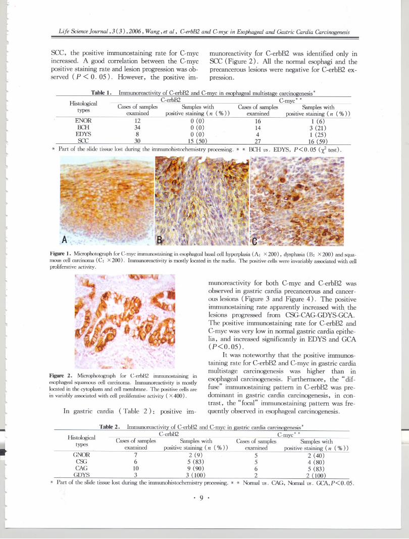
munoreactivity for C-erbB2 was identified only insec (Figure 2). All the normal esophagi and theprecancerous lesions were negative for C-erbB2 ex-pression.
Table1. lnununoreactivityof C-erbB2and C-mycin esophagealmultistagecarcinogenesis*
Hi 1 'cal C-erbB2 C-myc* *sto ogI Cases of samples Samples with Cases of samples Samples withtypes examined positive staining (n (%» examined positive staining (n (%»
ENOR 12 0 (0) 16 1 (6)BCH 34 0 (0) 14 3 (21)
EDYS 8 0 (0) 4 1 (25)see 30 15 (50) 27 16 (59)
* Part of the slide tissue lost during the immunohistochemistry proces.<,ing. * * BCH vs. EDYS, P<0.05 (X2 test).
J'41
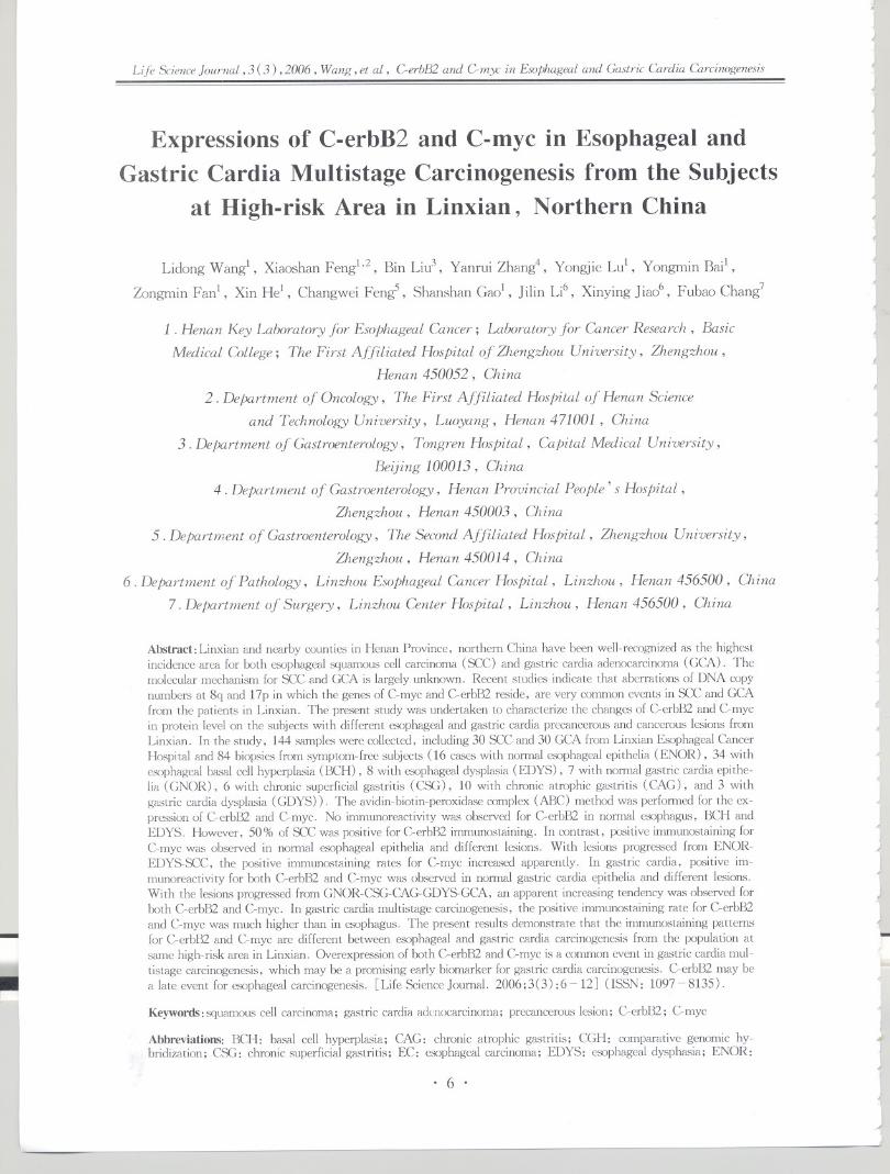
A}'igure 1. Microphotograph for C-myc immunostaining in esophageal basaI cell hyperplasia (A: X 200), dysphasia (B: x 200) and squa-
mous cell carcinoma (C: X 200). Immunoreactivity is mostly located in the nuclia. The positive cells were invariably associated with cellproliferative activity.
Table 2. lnununoreactivity of C-erbB2 and C-myc in gastric cardia carcinogenesis *
Hi 1 'cal C-erbB2 C-mvc* *sto ogl Cases of samples Samples with Cases of samples Samples withtypes . examined positive staining (n (%» examined positive staining (n (%»
GNOR 7 2 (9) 5 2 (40)CSG 6 5 (83) 5 4 (80)CAG 10 9 (90) 6 5 (83)
GDYS 3 3 (100) 2 2 (100)* Part of the slide tissue lost during the inununohistochemistryprocessing.* * Nonnal vs. CAG, Normalvs. GCA,P < O.05.
I
.1iL ~
Figure 2. Microphotograph for C-erbBl immunostaining in
esophageal squamous cell carcinoma. Immunoreactivity is mostly
located in the cytoplasm and cell membrane. The positive cells arein variably associated with cell proliferative activity ( X 400) .
In gastric cardia (Table 2): positive im-
.tlll
munoreactivity for both C-myc and C-erbB2 wasobserved in gastric cardia precancerous and cancer-ous lesions (Figure 3 and Figure 4). The positiveimmunostaining rate apparently increased with. thelesions progressed from CSG-CAG-GDYS-GCA.The positive immunostaining rate for C-erbB2 andC-myc was very low in normal gastric cardia epithe-lia, and increased significantly in EDYS and GCA(P<0.05) .
It was noteworthy that the positive immunos-taining rate for C-erbB2 and C-myc in gastric cardiamultistage carcinogenesis was higher than inesophageal carcinogenesis. Furthermore, the" dif-fuse" immunostaining pattern in C-erbB2 was pre-dominant in gastric cardia carcinogenesis, in con-trast, the" focal" immunostaining pattern was fre-quently observed in esophageal carcinogenesis.
I.
. 9 .

I II
r--<
Life ScienceJournal, 3 (3) ,2006, Wang, et al, verbB2 and Gmy: in Esophageal and Gastric Cardia Carcinogenesis
~.j
J1
J
" ~7.-.,. ~ ~II
r->'" ~~~;t.~ ;.. .~. ~4'i'--. "., .' ~." '
, (r. \ I
~~ \,h <I,
~:,')rt!R'~~~-"7' ~~ ,:J- .. .' .,...
.,.. '";:: I"""f
.. ~It, 4.7t 11.~ .. I
c..' 1:..
f .' J..~~. .,~.
~'
", I,~ 1. " "\:'u
~.!S
4
~
Figure 3. Microphotograph for C-myc immunostaining in gastriccardia dysphasia (A: X 400) and chronic atrophic gastritis (B: x400). Immunoreactivity is mostly located in the nuclia. The posi-tive cells are in variably associated with cell proliferative activity.-- - --
I1..\"
,
I . ,I
...,\. \
t . ,
,.' ., ..t. ,..,\. .~ ."," ~ft..' ¥# '~~
:~,(' ,
I'"
, , ,," ,~-- . ..:: ...::.-.
~" "-
'.~$''.. .. t:.1
.. -
,
~.1. . >J, I ,J.)
Ii
Figure 4. Microphotograph for C-erbB2 immunostaining in gastriccardia adenocarcinoma. Immunoreactivity is mostly located in thecytoplasm and cell membrane. The positive cells are in variablyassociated with cell proliferative activity ( X 200) .
4 Discussion
.J The present study demonstrates that over-ex-pression of C-erB2 and C-myc is a very early fre-quent event in gastric cardia multistage carcinogen-esis. These aberrant protein expressions are wellcorrelated with gastric cardia epithelial lesion pro-gressions, suggesting that C-erbB2 and C-myc mayplay an important role in gastric cardia multistagecarcinogenesis. These results are consistent withour CGH work[4], indicating that CGH is a good
~. I
technique in narrowing down the scope for identify-ing the key related genes with cancers. The presentresults also indicate that C-erbB2 and C-myc aber-rant expression may be a promising early biomarkerto predict the gastric cardia carcinogenesis. The re-cent studies by our group and other laboratorieshave showed that autoantibodies to C-myc could bedetected through cancer patient's blood serum, in-cluding esophageal and gastric cardia cancers, andcould increase the early detection of these can-cers[23-27].
An interesting result in this study is that aber-rant C-erbB2 expression occurs only in see, nonein normal esophagus and esophageal precancerouslesions, suggesting that C-erbB2 may be a lateevent for esophageal carcinogenesis. Accumulatedevidences have demonstrated that aberrant C-erbB2expression occurs more frequently in adenocarcino-ma, e. g. in GCA and Barrett's esophagus-relatedesophageal adenocarcinoma, not in see[20]. Thesedifferent expression patterns may be related withthe different tissue types occurring of tumor cells,which could explain the different immunostainingpatterns observed in GCA and see for C-erbB2 andC-myc in this study. ,
Many studies suggest that tumor occurring andprogression are the result of a multistage and pro-gressive process which may be related with the de-activity of tumor suppressor gene and the activity ofthe tumor oncogene in different stages. The presentstudies demonstrate that, in the multistage pro-gression of the esophageal carcinogenesis, there isfew expression of C-erbB2 in the early stage butsome in SCC; however, the overexpression of C-myc is positive during the esophageal multiple car-cinogenesis, suggesting the possibility of multiplegenetic changes involved in esophageal carcinogene-SIS.
4.j~
j~
~
J~
1 IJ
~
4
~
.J
....
IJ
~
i4
~
J....
~
'j
~
~
~
Historically, EC and GCA have been consid-ered as a single clinical entity for incidence andmortality-rate calculations in Linxian because of thecommon syndrome of dysphagia[28]. The similargeographic distributions of SCC and GCA in Chinasuggest that there may be similar risk factors andgenetic changes involved in these two cancers.GCA is an under-studied subject. The molecularchanges in the early stage of GCA carcinogenesishave not been characterized. There is evidence,however, that GCA differs from cancer of the restof stomach in terms of time trend, risk factors andhistopathogenesis[29]. Because of the common oc-currence both of see and GCA in Henan, it is ofgreat interest to know whether the molecularchanges observed in see also occur in GCA. The
~I~
'"I
~
J
4
JI...
..
~. 10 .
~
4)

!::
~
~ltlllL
lllL
[llll
~
~
ltltttllll~
lll
..I:
Life Science Journal, 3 (3) ,2006 , Wang, et al, GerbB2 and Gmyc in Esophageal and Gastric Cardia Carcinogenesis
present results demonstrate that the aberrant ex-pressions of C-erbB2 and C-myc occur similarly inSCC and GCA, however, the immunostaining pat-tern for C-erbB2 and C-myc in precancerous lesionsof the esophagus and gastric cardia is different, es-pecially in C-erbB2. The significance of these ob-servations needs to be further analyzed.
AcknowledgmentWe are grateful to the helps of Drs. Tao Guo,'
Shaohua Li, Weina Liu, Xianjuan Du and Hui Fanin preparation of the manuscript.
This work was supported in part by: NationalOutstanding Young Scientist Award of China30025016 and Foundations of Henan Education andHealth Committees of China.
Correspondence to:Lidong Wang, M.D., Ph.D.Henan Key Laboratory for EsophagealLaboratory for Cancer Research; BasicCollegeZhengzhou UniversityZhengzhou, Henan 450052, ChinaTelephone and Fax: 86-371-6665-8335Email: [email protected]
Cancer;Medical
References
1. Yang CS. Research on esophageal cancer in China: a re-view. Cancer Res 1980; 40: 2633 - 44.
2. Wang LD, Shi ST, Zhou Q, et al. Change.,>in p53 andcyclin D1 protein levels and cell proliferation in differentstages of human esophageal and gastric-cardia carcinogen-esis. IntJ Cancer 1994; 59: 514-9.
3. Victor T, Du Toit R, Jordan AM, et al. No evidence
for point mutations in ccxlons 12, 13, and 61 of the rasgene in a high-incidence area for esophageal and gastriccancers. Cancer Res 1990; 50: 4911- 4.
4. Wang LD, Zheng S, Zheng ZY, et al. Primary adeno-carcinomas of lower esophagus, esophagogastric junctionand gastric cardia: in special references. World J Gas-troenterol 2003; 9: 1156- 64.
5. Wang LD, Qiu SL, Yang GR, et al. A randomizeddouble-blind intervention study on the effect of calcium
supplementation on esophageal precancerous lesions in ahigh-risk population in China. Cancer EpidemiolBiomarkers Prev 1993; 2: 71 - 8.
6. Wang LD, Zhou Q, Yang CS. Esophageal and gastriccardia epithelial cell proliferation in northern Chinese sub-jects living in a high-incidence area. J CellBiochemSuppl1997: 28-29: 159~65.
7. Wang LD, Zhou Q, Feng CW, et al. Intervention andfollow-up on human esophageal precancerous lesions inHenan, northern China, a high-incidence area foresophageal cancer. Can To Kagaku Ryoho 2002; 29: 159-72.
8. Wang LD, Qin YR, Fan ZM, et al. Comparative ge-nomic hybridization: comparison between esophageal
tl
llll
squamous cell carcinoma and gastric cardia adenocarcino-ma from the patients at high-incidence area for bothesophageal and gastric cardia cancers in Henan, northernChina. Dis Esophagus 2006 (in pre.<;s).
9. Shervington A, Cruickshanks N, Wright H, et al.Glioma: What is the role of C-myc, hsp90 and telom-erase? Mol Cell Biochem2006; 283: 1 - 9.
10. Zornig M, Evan G. Cell cycle: on target with Myc. CurrBioi 1996; 6: 1553-6.
11. Packham G, Cleveland J. C-myc and apoptosis. Biochim. BiophysActa 1995; 1242: 11 ~ 28.
12. Bitzer M, Stahl M, AIjumand J, et al. C-myc gene am-plification in different stages of esophageal squamous cellcarcinoma: prognostic value in relation to treatmentmodality. Anticancer Res 2003; 23: 1489 - 93.
13 . Sarbia M, AIjumand J, Wolter M, et al . Frequent C-myc amplification in high-grade dysplasia and adenocarci-noma in Barrett esophagus. Am J Clin Pathol 2001:115: 835 ~ 40.
14. Ye X, Wu M. Retrovirus mediated transfer of antisensehuman C-myc gene into human esophageal cancer cellssuppressed cell proliferation and malignancy. Sci China B1992; 35: 76-83.
15.Luo B, Wang Y, Wang XF, et al. Correlation of Ep-stein-Barr virus and its enccxled proteins with Helicobacterpylori and expression of c-met and C-myc in gastric carci-noma. World J Gastroenterol2006; 12: 1842 ~ 8.
16. Akiyama T, Sudo C, Ogawara H, et al. The product ofthe human c-erbB-2 gene: a 185-kilodalton glycoproteinwith tyrosine kinase activity. Science 1986; 232: 1644-6.
17. James T. C-erbB2 onmprotein and its soluble ectcxkrnain: anew potential tumor marker for prognosis early detectionand monitoring patients undergoing Herceptin treatment.Clin Chim Acta 2002; 322: 11 ~ 9.
18. Slamon DJ, Clark GM, Wong SG, et al. Human breastcancer correlation of relapse and survival with amplifica-tion of the HER-2/c-erbB-2 oncogene. Science 1987;235: 177- 81.
19. Kyrgidis A, Kountouras J, Zavos C, et al. New molecu-lar concepts of Barrett's esophagus: clinical implicationsand biomarkers. J Surg Re.,>2005; 125: 189 ~ 212.
20. Bahnassy AA, Zekri AR, Abdallah S, et al. Human pa-pillomavirus infection in Egyptian e.'>Ophagealcarcinoma:correlation with p53, p21, mdm2, C-erbB2 and impacton survival. Pathol Int 2005; 55: 53 - 62.
21. Trudgill NJ, Suvama SK, Royds J A, et al. Cell cycleregulation in patients with intestinal metaplasia at thegastro-oe.'>Ophagealjunction. Mol Pathol 2003; 56: 313-7.
22. Suo Z, Holm R, Nesland JM. Squamous cell carcinomas,an immunohistochemical and ultrastructural study. Anti-cancer Res 1992; 12: 2025 ~ 31.
23. Du F , Wang LD, Qi YJ, et al. Detection of multipleserum autoantibody in the subjects with esophageal andgastric cardia precancerous and cancerous lesion using tu-mor-associated antigens mini-array. Chin J Cancer PrevTreat 2006 (in press in Chinese).
24. Zhang JY, Chan EK, Peng XX, et al. A novel cytoplas-mic protein with RNA-binding motifs is an autoantigen inhuman hepatocellular carcinoma. J Exp Med 1999; 189:1101 -10.
L
f.
. 11 .

China. Life ScienceJournal 2006;3(2): 1-11.28. Li JY. Epidemiology of esophageal cancer in China.
Monogr Natl Cancer Inst 1982; 62: 113-20.29. Wang HH, AntonioliDA, Gao HK, et al. Comparative
features of esophagealand gastric adenocarcinomas. HumPatho11986; 17: 482-7.
1
~Jj
JJJJjJ
1
)J}J
J
J
Jt
JJjjJ
1
Life Science Journal, 3 (3) ,2006, Wang, et al, GerbB2 and Gm)(: in Esophageal and Gastric Cardia CarciTWgen£Sis
25. Zhang JY, Wang X, Peng XX, et al. Autoantibody re-sponses in Chinese hepatocellular carcinoma. J Clin Im-munol 2002; 22: 98 - 105.
26. Megliorino R, Shi ill, Peng XX, et al. Autoimmuneresponse to anti-apoptotic protein surviving and its associ-ation with antibodies to p53 and C-myc in cancer detec-tion. Cancer Detect PreY 2005; 29: 241 - 8.
27. Wang ZQ, Wang ill. DNA methylation and esophagealsquamous cell carcinoma: special reference to research in
.
Received June 25, 2006
)
J.I
jI
j
J
~
J
)1..
.JI
J
1
J. 12 . I
J~I.J

tlLl
tltlblLlll~
ll[
lL
llttllLltLl
lllll[~-
Life Science Journal, 3 (3) ,2006, Wen, et al, MMP-2 and MMP-9 and Esophageal Squamous Cell Carcinmna
Expression of MMP.2 and MMP.9 and Its Correlation
with Invasion and Metastasis in Human EsophagealSquamous Cell Carcinoma
Hongtao Wenl, Lei Zhan~, Qiumin Zha03, Jichang Lil
1. Department of Gastroenterology, The First Affiliated Hospital of ZhengzJwu University,
Zhengzhou , Henan 450052, China
2. Department of Oncology, The First Affiliated HosPital of Zhengzhou University,
Zhengzhou , Henan 450052, China
3. Editorial Board of Journal of Zhengzhou University (Medical Sciences),
Zhengzhou , Henan 450052, China
Abstract:Objective. To investigate the significance of MMP-2 mRNA and MMP-9 mRNA expression in humanesophageal squamous cell carcinoma (ESCC). Methods. MMP-2 mRNA, MMP-9 mRNA and proteins were exam-ined by immunohistochemistry, in situ hybridization, RT-PCR, zymographic analysis and Western blot for 41 cas-es of ESCC. Results. The expression rate and value of MMP-9 was significantly higher than that of MMP-2 in tu-mor tissues. Conclusions. MMP-9 has higher sensitivity and specificity in predicting the biologic behavior of inva-sionandmeta.'itasisinESCC. [Life Science Joumal. 2006;3(3):13-18J (ISSN: 1097-8135).
Keywords:MMP-2; MMP-9; esophageal carcinoma; invasion; metastasis
Abbreviations: BM: basement membrane; ECM: extracellular matrix; ESCC: esophageal squamous cell carcino-ma; MMP: matrix metalloproteinase; PAGE: polyacrylamide gel electrophoresis; SDS: sodium dodecyl sulfate;TBS: Tris-HCI buffered saline
1 Introduction
Esophageal carcinoma is one of the most com-
mon cancers and acts as the fourth leading cause ofcancer death in China. It is characterized by poorprognosis and rapid clinical progression with a highfrequency of lymph node metastasis and recurrence.The transition from in situ to invasive tumors is a
very complicated process. Proteolysis of extracellu-lar matrix (ECM) is essential step in tumor inva-sion and metastasis. Numerous proteolytic enzymesincluding the matrix metalloproteinase (MMP)have been implicated in this process. Reportsshowed that both MMP-2 and MMP-9 were highlyexpressed in esophageal tumor tissues[l,2]. In thecurrent study, MMP-2 mRNA and MMP-9 mRNAand their proteins were examined by immunohisto-chemistry, in situ hybridization, RT-PCR, zymo-graphic analysis and Western blot in 41 cases of ES-CC as well as the matched normal mucosa tissues,to compare the potential value of MMP-2 andMMP-9 in estimation of the biologic behavior ofESCC.
..L.
ltt~
2 Materials and Methods
2.1 Tissue samples41 specimens of patients with esophageal squa-
mous cell caricinoma were collected from the First
Affiliated Hospital of Zhengzhou University and theHenan Tumor Hospital. All of them were identifiedby pathology. The resected specimens including theESCC samples and the normal adjacent tissues weresnap-frozen in liquid nitrogen.2.2 Innnunohistochemistry
Specimens were fixed with 10 % neutralbuffered formaldehyde solution and embedded inlow-melting paraffin. Sections of esophageal tumorswere immunostained with monoclonal antibodies to
MMP-2 and MMP-9. Immunohistochemistry forthe individual MMP was performed by an alkalinephosphatase anti-alkaline phosphatase technique.After the immunohistochemistry, the sections wereexamined under microscope. The MMP status ofthe tumors was assessed as positive if any of the tu-mor cells showed significant immunostaining. Neg-ative controls were done by replacing the primaryantibody with TBS and by liquid phase pre-absorp-
. 13 .

III r-
..
.JJ-ILife ScienceJournal, 3 (3) ,2006, Wen, et al, MMP-2 and MMP-9 and EsophagealSquamous Cell Carcinoma
-
tion of primary antibody with the correspondingimmunogen at 10 nmollml antibody. The positivecontrols for both MMP-2 and MMP-9 were lungcontaining intra-alveolar macrophages.2. 3 In situ hybridization
In situ hybridization was performed on sec-tions (4 pm). After deparaffinization and rehydra-tion all samples were treated with proteinase K andwashed in O. 1 M triethanolamine buffer containing0.25% acetic anhydride. Sections were hybridizedovernight at 50 'C to 55 'C with 35S-labelledRNA probe. After hybridization, slides werewashed under stringent conditions and treated withRNase to remove unhybirdized probe. Previouslypositive samples for each anti-sense probe were usedas positive controls. The slides were independentlyassessed by two experienced investigators.2.4 RT-PCR
Total RNA was extracted from shock-frozentissue samples with RNA extract kit. First strandcomplementary DNA was synthesized from 2 fJ.gofDNA-free total RNA in a 20 fJ.I system of: 1mmol/L dNTP; 10 U RNAsin; 20 mmol/L DTT;1 fJ.molRandom Hexamer Primer and 100 U MM-LV. Follow the procedure of 37 'C 10 min,42 'C 1hour and 95 'C for 5 min. PCR was done in a 50 fJ.Isystem including both MMP and ~-actin primers.The annealing temperatures of MMP-9 and MMP-2were 66 'C and 65 'C, respectively. Raw data fromeach samples, were quantified using the eagle eyesystem (Stratagene, American). Data from MMPcDNA were normalized to the respective content of~-actin cDNA. TIN >2.0 was recognized as posi-tive.2.5 Zymographic analysis
Equal amounts of total lysates of esophagealtissue samples were used for determining proteinamounts with the Bio-Rad DC protein assay kit andstored at - 20 'C until assayed. MMP activity inthe lysates was assessed by gelatin zymography:lanes were loaded with 2 fJ.gof total protein each.The concentrated media were run on nonreducingsodium dodecyl sulfate (SDS) -polyacrylamide gel00%) containing 1 mg/ml gelatin. After elec-trophoresis in 25 mM Tris base, 250 mM glycine,and 1% SDS, the gel was washed at room temper-ature in solution (2. 5% Triton X-1O0, 5 mMCaClz, 50 mM Tris-HCI, pH 7.5), and was incu-bated again in the same buffer twice for 1 houreach. After rinsing the gel extensively with sixchanges of distilled water, it was incubatedovernight at 37 'C in 5 mM CaCh and 50 mM Tris-HCl, pH7. 5, followed by Coomassie blue stainingand destaining in solution of methanol, acetic acid
~I
I
~
and water (50 : 10 :40) . Gelatin zymography depictsMMP as negatively staining bands of gelatinolyticactivity.2.6 Western blot
Tissue lysates were separated by SDS-PAGEusing separating gels and stacking gels of 3% poly-acrylamide. Lane was loaded with 2 fJ.gof total pro-tein each. After electrophoresis, the proteins weretransferred to PVDF membrane. Protein bandswere localized by staining with Ponceau S. Blotswere blocked with Tris-buffered saline-NaCl, pH7. 6, containing 10 % bovine serum albumin, 20mM Tris, 137 mM NaCl, and O. 1% NonidetP40; washed; blocked with 10% milk and incubat-ed with antibodies against MMP-2 or MMP-9. Ex-periments were done in triplicate. The bands werescanned by computer analysis. Data from all tissuesamples analyzed were normali,zed by setting theprotein amount in the healthy tissue to 1.0 in eachsample[3] .2.7 Statistical analysis
The SPSS statistical package program wasused for all analysis. Associations between the vari-ables were tested by l test, Student's t test. a =0.05 were set significant.
~..-I~
~.l~J
I
1
1
I
J,J~
oJ
~...~-I~
3 Results"
3. 1 ImmunohistochemistryThe results revealed that there were 26 cases
of positive reaction for MMP-9 in 41 ESCC and 5cases of positive reaction in matched normal mucosatissues. The percentage of positive cases in ESCCwas significantly higher than that in matched nor-mal mucosa tissues (P<O. 01). Faint staining wasalso found in monocyts, fibroblasts, endothelial andsmooth muscle cells. Positive reaction for MMP-9showed a tendency to be stronger in deeply invad-ing nests, especially in peripheral fronts. On thecontrary, MMP-2 levels were lower and positivereaction in carcinoma cells was generally weakerthan that in stromal cells. There were distinct dif-ference between the MMP-9 and MMP-2 expres-sioninESCC(P<O.OI) (Table 1, Figure 1 andFigure 2) .
J"
..
~
I..
"
,J
~
,J
J"
Table 1. Expressions of MMP-2 and MMP-9 by immuno-histochemistry
JJi-
...."
L
,.I
. 14 .~~
~~
MMP Cases Expression
type (n) Positive cases (n) Positive rate (%)
MMP-9 41 26 63.41
MMP-2 41 7 17.07P<O.OI

It
L
flI
lt~L
lllL
~
llLlllllttll~lltll
~
llL[I'
---
Life ScienceJournal, 3 (3) ,2006, Wen, et al, MMP-2 and MMP-9 and EsophagealSquamous Cell Carcinoma
Figure 1. Detection of MMP-9 protein in ESCC by immunohisto-chemical staining SP x 200
Figure 2. Detection of MMP-2 protein in ESCC by immunohisto-chemical staining SP x 200
3 .2 In situ hybridizationThe results revealed that there were 22 cases
of positive reaction for MMP-9 mRNA in 41 ESCCand 5 casesof positive reaction in matched normalmucosa tissues. The percentage of positive cases inESCC was significant higher than that in matchednormal mucosa tissues (P < O. 01). Faint stainingwas also detected in macrophages, fibroblasts, en-dothelial and smooth muscle cells but strongerstaining was observed in plasmacytes. Positive re-action for MMP-9 mRNA showed a tendency to bestronger in deeply invading nests, especially in pe-ripheral fronts. On the contrary, MMP-2 levelswere lower and positive reaction in carcinoma cellswas generally weaker than that in stromal cells.There was distinct difference between the MMP-9
mRNA and MMP-2 mRNA expression in ESCC(P<O.O1) (Table 2, Figure 3 and Figure 4).
~l
ltl[
Table2. Expressionsof MMP-2 mRNA and MMP-9 mR-NA by in situ hybridization
MMP Casestype (n)
MMP-9 41
MMP-2 41P<O.Ol
mRNA expression
Positivecase(n) Positive rate (%)22 53.66
4 9.76
Figure 3. Detection of MMP-9 mRNA in ESCC by in situ hy-bridization X200
Figure4. Detection of MMP-2 mRNA in ESCC by in situ hy-bridization X200
3.3 RT-PCRRT-PCR inspect showed that the positive rate
and semi-quantitative value of MMP-9 mRNA were80.49% (33/41) and 0.57:t0.43 in ESCCs, re-spectively. The positive rate and the semi-quantita-tive value of MMP-2 mRNA were 58. 54%(24/41) and 0.21 :t O. 21 in ESCC, respectively.The positive rate and the semi-quantitative value ofMMP-2 mRNA were distinctly lower than those ofMMP-9 mRNA (P<0.05, P<O.O1) (Table 3,Figure 5 and Figure 6) .
!-
. 15 .

Absorbance value
jj)J
~
J
JJj
1
JJ
J
J
J
J
)J
).I
Life ScienceJournal,3 (3) ,2006, Wen, et al, MMP-2 and MMP-9 and EsophagealSquarrwusCell Carcinoma
Expressions of MMP-2 and MMP-9 mRNA by RT-PCR
mRNA expression
Positive rate( % )
80.49#
58. 54 #
Table 3.
MMP type Cases (n)Positive case (n)
MMP-9 41 33
MMP-2 41 24
# :vs. MMP-9,P<0.05; * :vs. MMP-9,P<0.01
1 2
300bp
200bp
0.57:t O. 44 *
O. 2l:t O. 21 *
3 4
302bp(MMP-2)
233bp(l3-actin)
Figure 5. Detection of MMP-2 rnRNA in ESCC, tissue adjacent to twnor and matched mormal mucosa tissue by RT-PCRLane 1: Marker; Lane 2: normal tissue; Lane 3 : tissue adjacent to twnor; Lane 4 : ESCC
1 2
500bp-.300bp-.200bp-.
3 4~i
jIJJI
-!
~
JJ-<
233bp(l3-actin) !I
j
vated MMP-2 (P < O. 01), proMMP-9 andproMMP-2 (P < o. 01). The activated MMP-9could be detected in 24 biopsy specimens (Figure7) .3.5
.IJJJ
)J
l
Figure 6. Detection of MMP-9 rnRNA in ESCC,tissue adjacent to twnor and matched mormal mucosa tissue by RT-PCRLane 1: Marker: Lane 2: normal tissue: Lane 3: tis.'sue adjacent to twnor: Lane 4: ESCC
~
3.4 Zymographic analysisBy zymographic analysis, there were small
amount secretions of proMMP-9 (92 kD) andproMMP-2(72 kD) in matched nonnal mucosa tis-sues. The activated MMP-2 (62 kD) was detectedin 31 biopsy specimens, but no activated MMP-9(82 kD) could be found. The semi-quantitativevalues of proMMP-9, activated MMP-2 andproMMP-2 in ESCC were 2.52 :t o. 75, 1. 92 :t0.42 and 1. 51 :t O. 42, respectively. There weredistinct differences between proMMP-9 and acti-
Western blotBy Western blot analysis, the semi-quantita-
tive values of MMP-9 and MMP-2 in ESCC were3.02 :t 0.56 and 2.26 :t 0.51, respectively. Therewere distinct differences between MMP-9 andMMP-2(P<0.05) (Figure 8).
~J...
J
~
j
1. 16 .
JJ
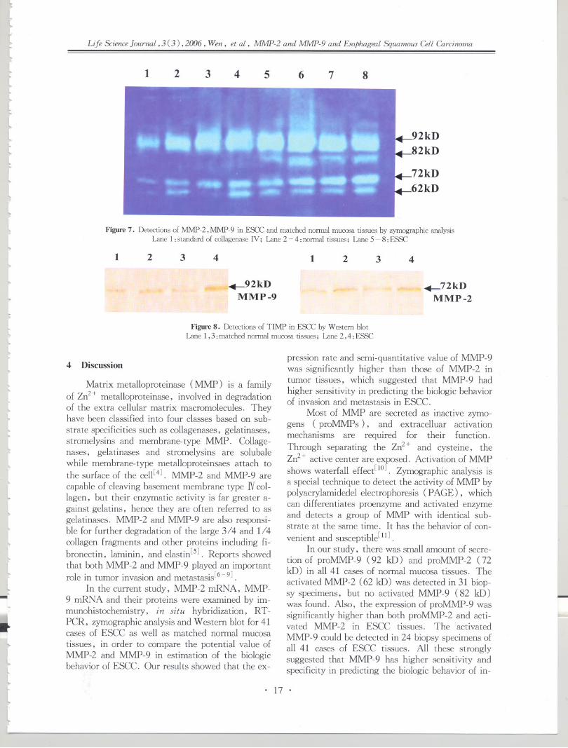
[r[l(...
Ltb
L
lt
[
lllL
lllIl
lLl
tL...IL...Lltll
~
tlt
~.-
Life Science Journal, 3 C3), 2006, Wen, et al, MMP-2 and MMP-9 and Esophageal Squamous Cell Carcinoma
21 3 4 5 6 7 8
Figure 7. Detections of MMP-2, MMP-9 in ESCC and matched normal mucosa tissues by zymographic analysisLane 1 : standard of collagenase IV; Lane 2 - 4 : normal tissues; Lane 5 - 8: ESSe
1 2 3 4
i~2kDMMP -9
1 2 3 4
72kDMMP -2
Figure 8. Detections of TIl\1P in ESCC by Western blot
Lane 1,3: matched normal mucosa tissues; Lane 2 ,4 :ESSe
4 Discussion
Matrix metalloproteinase (MMP) is a family
of Zn2+ metalloproteinase, involved in degradationof the extra cellular matrix macromolecules. Theyhave been classified into four classes based on sub-
strate specificities such as collagenases, gelatinases,stromelysins and membrane-type MMP. Collage-nases, gelatinases and stromelysins are solubalewhile membrane-type metalloproteinsaes attach tothe surface of the cell[4]. MMP-2 and MMP-9 are
capable of cleaving basement membrane type N col-
lagen, but their enzymatic activity is far greater a-gainst gelatins, hence they are often referred to asgelatinases. MMP-2 and MMP-9 are also responsi-ble for further degradation of the large 3/4 and 1/4collagen fragments and other proteins including fi-
bronectin, laminin, and elastin[S]. Reports showedthat both MMP-2 and MMP-9 played an importantrole in tumor invasion and metastasis[6-9].
In the current study, MMP-2 mRNA, MMP-9 mRNA and their proteins were examined by im-munohistochemistry, in situ hybridization, RT-PCR, zymographic analysis and Western blot for 41cases of ESCC as well as matched normal mucosa
tissues, in order to compare the potential value ofMMP-2 and MMP-9 in estimation of the biologicbehavior of ESCC. Our results showed that the ex-
~I..
llt[
pression rate and semi-quantitative value of MMP-9was significantly higher than those of MMP-2 intumor tissues, which suggested that MMP-9 hadhigher sensitivity in predicting the biologic behaviorof invasion and metastasis in ESCC.
Most of MMP are secreted as inactive zymo-gens (proMMPs), and extracelluar activationmechanisms are required for their function.Through separating the Zn2+ and cysteine, theZn2+ active center are exposed. Activation of MMPshows waterfall effect[lO]. Zymographic analysis isa special technique to detect the activity of MMP bypolyacrylamidedel electrophoresis ( PAGE), whichcan differentiates proenzyme and activated enzymeand detects a group of MMP with identical sub-strate at the same time. It has the behavior of con-
venient and susceptible[l1] .In our study, there was small amount of secre-
tion of proMMP-9 (92 kD) and proMMP-2 (72kD) in all 41 cases of normal mucosa tissues. Theactivated MMP-2 (62 kD) was detected in 31 biop-sy specimens, but no activated MMP-9 (82 kD)was found. Also, the expression of proMMP-9 wassignificantly higher than both proMMP-2 and acti-vated MMP-2 in ESCC tissues. The "activatedMMP -9 could be detected in 24 biopsy specimens ofall 41 cases of ESCC tissues. All these stronglysuggested that MMP-9 has higher sensitivity andspecificity in predicting the biologic behavior of in-
L
. 17 .
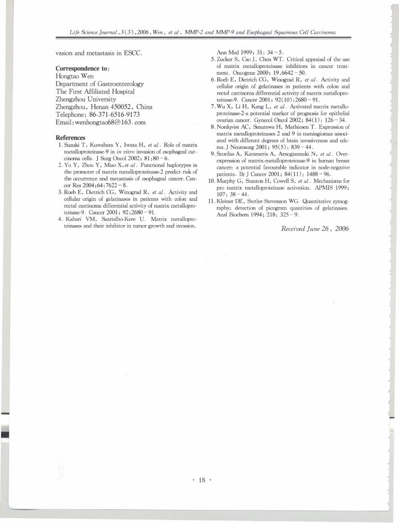
I II r
Ioj
~I
J..Life Science]ourno.t,3 (3) ,2006, Wen, et at, MMP-2 and MMP-9 and EsophageolSquamous Cell Gzrcinmna
vasion and metastasis in ESCC.
Correspondence to:Hongtao WenDepartment of GastroenterologyThe First Affiliated HospitalZhengzhou UniversityZhengzhou, Henan 450052, ChinaTelephone: 86-371-6516-9173Email:[email protected]
References1. Suzuki T, Kuwahara y, Iwata H, et at. Role of matrix
metalloproteinase-9in in vitro invasionof esophagealcar-cinoma cells. ] Surg Oncol 2002; 81: 80 - 6.
2. Yu Y, Zhou Y, Miao X,et at. Functional haplotypes inthe promoter of matrix metalloproteinase-2predict risk ofthe occurrence and metastasis of esophagealcancer. Can-cer Res 2004;64:7622-8.
3. Roeb E, Dietrich CG, Winograd R, et at. Activity andcellular origin of gelatinases in patients with colon andrectal carcinomadifferentialactivity of matrix metallopro-teinase-9. Cancer 2001; 92: 2680 - 91.
4. Kahari VM, Saarialho-Kere U. Matrix metallopro-teinases and their inhibitor in tumor growth and invasion.
-~
I
,Ann Med 1999; 31: 34-5.
5. Zucker S, Cae J. Chen WT. Critical appraisalof the useof matrix metalloproteinase inhibitors in cancer treat-ment. Oncogene2000; 19,6642 - 50.
6. Roeb E, Dietrich CG, Winograd R, et at. Activity and. cellular origin of gelatinases in patients with colon and
rectal carcinomadifferential activity of matrix metallopro-teinase-9. Cancer 2001; 92(10): 2680 - 91.
7. Wu X, Li H, Kang L, et at. Activated matrix metallo-proteinase-2-apotential marker of prognosis for epithelialovarian cancer. GynecolOncol 2002; 84( 1): 126- 34.
8. Nordqvist AC, Smurawa H, Mathiesen T. Expressionofmatrix metalloproteinases2 and 9 in meningiomasassoci-ated with different degrees of brain invasivenessand ede-ma.] Neurosurg2001; 95(5): 839-44.
9. ScorilasA, Karameris A, Amogiannaki N, et at. Over-expressionof matrix-metalloproteinase-9in human breastcancer: a potential favourable indicator in node-negativepatients.. Br] Cancer 2001; 84(11): 1488 - 96.
10. Murphy G, Stanton H, CowellS, et at. Mechanismsforpro matrix metalloproteinase activation. APMIS 1999;107: 38-44.
11. Kleiner DE, Stetler-StevensonWG. Quantitative zymog-raphy: detection of picogram quantities of gelatinases.Anal Biochem1994; 218: 325-9.
J-0/
~.J
~
JI
~
~
j~~
J
J
JJ
J
Ii
Received June 26, 2006 .,.J
1-!~I
'"
.J
...
II
J
..
~I
,}
~
J
~....
~I
..
. 18 .J...~.,
)

k
lt
tl
b
lLtb
llllLllll"-
l~l..
llL.
lllIl;.lllL
~lL
l.~-
Life Sci£nceJournal, 3 (3 ) ,2006 , Ozen, et ai, Expression of CathepsinB and EsophagealSquamous Carcinoma
Expression of Cathepsin B and Its Relationship withEsophageal Squamous Carcinoma
Kuisheng Chen1.2, Zhihua Zhao1, Miaomiao Sun1.2, Xin Lou1.2, Dangling Gao1.2
1 . Department of Pathology, The First Affiliated Hospital of Zhengzhou University,
Zhengzhou Henan 450052, China
2. The Key Laboratory of Tumor Pathology of Henan, Zhengzhou,
Henan 450052, China
Abstract: Objective. To explore the relations between expression of Cathepsin B (CB) and development, invasionand metastasis of esophageal squamous cell carcinoma. Methods. The expression of CB protein and of CB mRNAwere determined by immunihistochemitry and in situ hybridization. Results. CB protein expression and CB mRNAexpression couldn't be detected in normal esophageal mucosa, while positive in matastatic group and non-metastaticgroup. CB protein expression and CB mRNA expression was significantly decreased. The out layer invasion group,rate of CB protein expression and CB mRNA expression in the tumor tissues and atypical hyperplasia in tumor-adja-cent tissues was significantly elevated compared with low-muscle invasion group and deep-muscle invasion group.The positive rates was slightly higher in deep-muscle invasion than in low-muscle invasion without statistical signifi-cance. The positive rates of CB protein expression and of CB mRNA expression in the carcinoma tissues were signif-icantly higher than atypical hyperplasia tissues in both the metastatic and non-metastatic group. Conclusion. CBprotein expression and CB mRNA expression in human esophageal squamous cell carcinoma were increased, whichindicated that CB is related to the development, invasion and metastasis of esophageal carcinoma [Life Science Jour-nal. 2006 ;3(3): 19 - 24] (ISSN: 1097 - 8135).
Abbreviations: CB: Cathepsin B
Keywords: esophageal squamous cell carcinoma; Cathepsin B; invasion; metastasis
1 Introduction
Invasion and metastasis of tumor is a compli-cated process, in which the relationship of extracel-lular matrix and metastasis of tumor is extremlyclose. A series of dynamic change develop betweentumor cells and extracellular matrix. There are alot of enzymes released that degrade the extracellu-lar matrix and facilitate the invasion and metastasis
of tumor cells[l]. It is known that Cathepsin B(CB) is a lysosomal cysteine proteinase that can de-grade laminin, fibronectin, Ntype collagen and soon the extracellular matrix, and participating in theinvasion and metastasis of tumor cells. In recentyears, the research discovered that CB showed anobivious increase in varieties of malignant tu,morssuch as prostate carcinoma, melanocytoma, ~anci-noma of colon and so on. It also participates in theinvasion and metastasis of tumor. While both do-mestic and foreign scholors haven't carried out theresearch for CB and its relationship with invasionand metastasis of esophageal carcinoma. This re-search adopted the technique of immunohistoche-
...
tttt
mistry and in situ hybridization, to investigate CBexpression and its relationship with the develop-ment, invasion and metastasis of esophageal carci-noma.
2 Materials and Methods
2. 1 Materials2.1.1 Reagents
CB immunohistochemiscal SP kit was providedby Wuhan Boster Biological Technology Company;SA-AP and BCIP /NBT were from AmericanPromera corporation; CBcDNA probe with 5 temi-nal biotin marked (.5'-GTIGACCAGCr-CJITCCGACAGG-3') was synthesized by BeijingOker Biological Technology Limited Corporation.2. 1.2 Specimens
Forty-nine fresh samples of human esophagealcarcinoma were obtained from oncological hospitalof Anyang and the First Affiliated Hospital ofZhengzhou University. These samples were all con-firmed by pathology as squamous cell carcinoma.There were 49 cases, 25 males' and 24 females,with median age 58.3 :t 17.8 years old (from 40.5years old to 76. 1 years old) .
. 19 .

I I
IrI
J
~~
Life Science Journal, 3 (3) ,2006 ,Ozen, et al, Expression of Cathepsin B and Esophageal Squarrwus Carcinoma ~J
-
According to lymphotic metastasis: metastaticgroup (20 cases) and non-metastatic group (29 cas-es); according to the depth of invasion: low-musclegroup (10 cases), deep-muscle group (15 cases)and out layer group (24 cases); according to devel-opment: esopheal carcinoma group (49 cases),atypical hyperplasia of tumor-adjacent group (30cases) and normal group (49 cases).2. 2 Methods2. 2. 1 Specimen preparation
The tissues of carcinoma, atypical hyperplasiaof tumor-adjacent and normal tissues obtained wereformalined-fixed and paraffin-embeded respectivelyfor immunohistochemistry and in situ hybridiza-tion.2.2.2 Immunohistochemistry
Paraffin sections were de-paraffined routinely,rinsed in PES 3 times for 5 min each. Under thehigh pressure, samples were treated with O. 01mol/L Citrate Tris-sodium for 15 min. Sampleswere rinsed 3 times with PES for 5 min each, andblocked by serum for 30 min, to remove non-spe-cific staining. Then drop wise 1: 100 rabbit antihu-man CB antibody was added and incubated at 4 'Covernight. The next day sections were taken for 30min at room temperature, rinsed with PES 3 timesfor 5 min each. So did the secondary antibody andthe tertiary antibody. After the following DABstaining, hematoxylin staining, samples was dehy-drated and mounted. PBS replaced the primary an-tibody in the negative control slices.2.2.3 In situ hybridization
Paraffin sections were deparaffined to waterroutinely, and performed in 3 % hydrogen peroxidefor 30 min at room temperature for blocking en-dogenous peroxidase activity. Sections were rinsedwith H2O 3 times for 5 min each time and digestedfor 20 min with pepsin to expose rnRNA nuclearsection, rinsed once with H2O for 5 min, fixedwith 1% formalin 10.1 M PBS (pH 7.2 -7. 6,contain 0.1 % DEPC) for 10 min at room tempera-ture. Rinsed by H2O 3 times for 5 min each time,and dropwised 20 fll Islice hybridization solution.The sections were set in 20% glyerine to prehy-bridize for 3 - 4 h at 42 'C dropwise 20 fll labeledprobe added to specimen, then coved paraffin mem-brane to hybridize for 12 - 16 h at 42 - 43 'C . Thenext day, sections were dropwised with O. 1 X ssefor 4 times for 15 min each time to elute non-spe-cific hybridization. Sections were processed with1% acetylatel BSA for 10 min at 20 'C. Sectionswere in Streptavdin Alkaline Phoshates ( SA-AP )for 20 min at 37 'C , Tris-HCl buffer I 3 times for
~
- --
10 min each, and Tris-HCl buffer IT 2 times for 1min. Sections were dropwised BCIP/NET (33 fllNBT and 16.5 fll BCIP added to 5 ml buffer IT)substrate solution freshly, stained in the darknessfor 10 - 120 min, nuclear fast red after stained fora few min, neutral gum mounted, prehybridizationsolution without probe hybridized and before hy-bridization specimens were processed as negativecontrol by RNase.
The results were judged according to Naokimethod[3]. Positivecellsaccoutfor 0% for ( - ), 0- 20 % for ( :t ), 20 - 80 % for ( + ), 80 % for( + + ), ( - ) and ( :t ) were set as negative, (+ )and ( + + ) were positive.2.2.4 Statistical analysis
All the data were analyzed by spass 10. 0 sys-tem. Comparison between positive rates adopted
chi-square test (i-test).
...~
,J.oj
J
~I
;
~-!4
j I
;
~
J
~
3 .J
IResults
3.1 CB protein expression and CB mRNA ex-pression in esophageal carcinoma tissues, atypicalhyperplasia tissues and normal tissues
The positive signals of CB protein were local-ized in cytoplasm or membrane of esophageal carci-noma or atypical hyperplasia cell and showed browngranules. Periphery matrix of carcinoma nestshowed positive as well(Figure 1)but not in nega-tive control. Positive signals of CB mRNA was lo-calized in cytoplasm of esophageal carcinoma oratypical hyperplasia cells and showed blue granules(Figure 2). Normal esophageal mucosa cellscouldn't be stained. No positive signals appeaied inthe negative control.
1,.j~J
I;
..
"~
JJ-'-4
..
3.2 Relationships of CB and esophageal carcino-ma metastasis3.2.1 Relationship of CB protein and esophagealcarcinoma metastasis
In the 20 caseswith lymphatic metastasis, thepositive rates of expression of CB protein in the tis-sues of esophageal carcinoma, atypical hyperplasiatissues and normal tissues were 100 % (20/20),60 % ( 6/10) and 0 % (0/20), respectively; whilein the 29 cases without lymphatic metastasis, thethe positive rates CB protein expression in theesophageal carcinoma tissues, atypical hyperplasiatissues and normal tissues were 62.07% (18/29),10% (2/20) and 0% (0/29), respectively. Thedifference of CB had statistical significance ( carci-noma tissues group vs. normal tissues group, P <0.01; atypical hyperplasia group vs. normal tissuesgroup, P<O. 01). Table 1 showed the details.
..
...
JJ~
/
J
1-~
:J
i~
. 20 .~~..."'
...
I
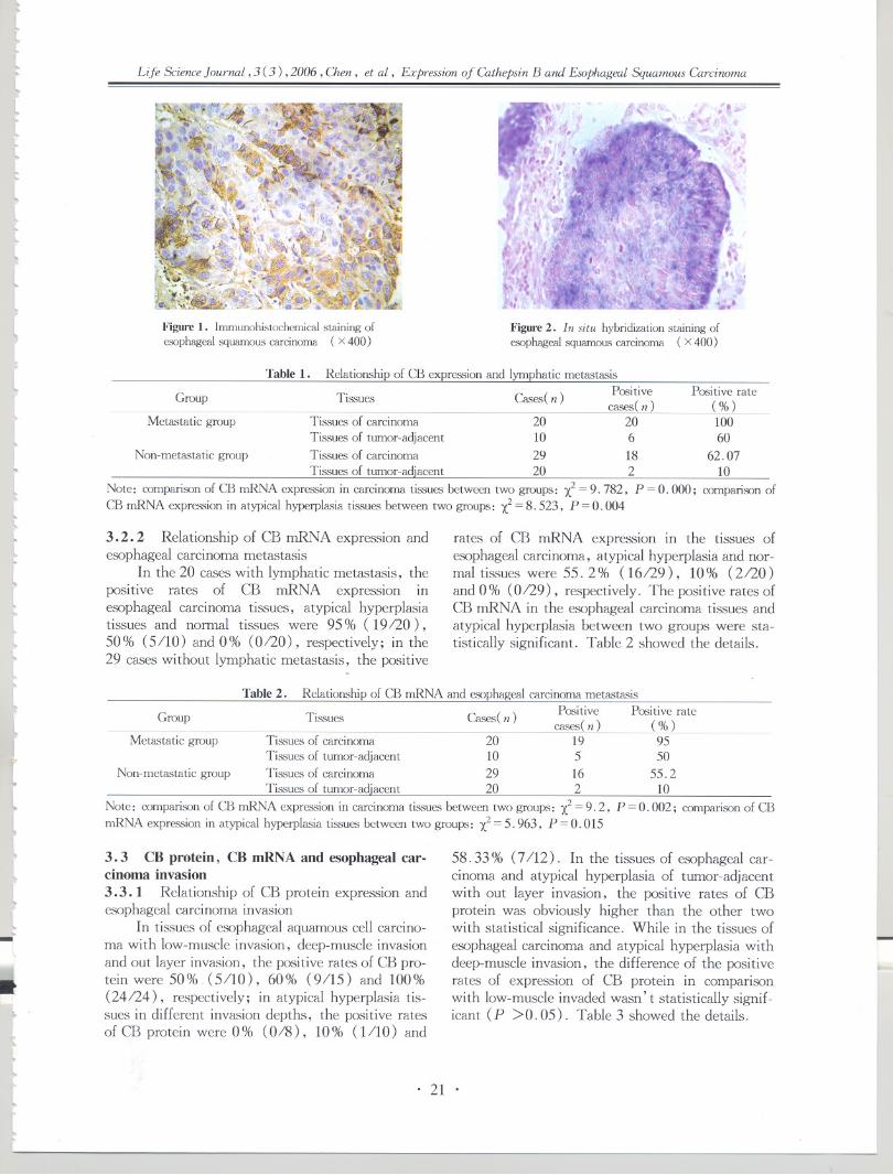
[l
llL
~
bl~l
tllll[
Life Science Journal, 3 (3) ,2006, Chen, et al, Expression of Cathepsin B and Esophageal Squamous Carcinoma
Figure 1. Immunohistochemical staining of
esophageal squamOtlc'i carcinoma (x 400 )
,
Figure 2. in situ hybridization staining ofesophageal squamous carcinoma (x 400 )
Table 1. Relationship of CB expression and lymphatic metastasisPositive
cases(n)Tissues of carcinoma 20 20Tissu€-,>of tumor-adjacent 10 6Tissues of carcinoma 29 18
Tissues of tumor-adjacent 20 2
Note: comparison of CB mRNA expression in carcinoma tissues between two groups: l = 9. 782, P = O.000 ;
CB mRNA expm,sion in atypical hyperplasia tissues between two groups: l = 8. 523, P = 0.004
Table 2. Relationship of CB mRNA and esophagealcarcinomametastasisPositive Positive ratecases(n) ( %)
Tissu€-,>of carcinoma 20 19 95Tissues of tumor-adjacent 10 5 50Tissues of carcinoma 29 16 55.2Tissues of tumor-adjacent 20 2 10
Note: comparison of CB mRNA expression in carcinoma tissues between two groups: l = 9. 2, P = O.002; comparisonof CBmRNA expression in atypical hyperplasia tissues between two groups: l = 5. 963, P = O. 015
Group
Metastatic group
Tissues
Il
L
lllll
tl'-lLl
~
l
Non-metastatic group
3.2.2 Relationship of CB mRNA expression andesophageal carcinoma metastasis
In the 20 caseswith lymphatic metastasis, thepositive rates of CB mRNA expression inesophageal carcinoma tissues, atypical hyperplasiatissues and normal tissues were 95 % (19/20),50% (5/10) and 0% (0/20), respectively; in the29 cases without lymphatic metastasis, the positive
Group--
Metastatic group
Tissu€-,>
Non-metastatic group
l
~
tII.-
3.3 CB protein, CB mRNA and esophageal car-cinoma invasion3.3.1 Relationship of CB protein expression andesophageal carcinoma invasion
In tissues of esophageal aquamous cell carcino-ma with low-muscle invasion, deep-muscle invasionand out layer invasion, the positive rates of CB pro-tein were 50% (5/10), 60% (9/15) and 100%(24/24), respectively; in atypical hyperplasia tis-sues in different invasion depths, the positive ratesof CB protein were 0 % (0/8), 10% (1/10) and
It~Ito"tL
tl
Positive rate(%)10060
62.0710
comparison of
Cases( n)
rates of CB mRNA expression in the tissues ofesophageal carcinoma, atypical hyperplasia and nor-mal tissues were 55.2% (16/29), 10% (2/20)and 0% (0/29), respectively. The positive rates ofCB mRNA in the esophageal carcinoma tissues andatypical hyperplasia between two groups were sta-tistically significant. Table 2 showed the details.
Cas€-,>(n )
58.33 % (7/12). In the tissues of esophageal car-cinoma and atypical hyperplasia of tumor-adjacentwith out layer invasion, the positive rates of CBprotein was obviously higher than the other twowith statistical significance. While in the tissues ofesophageal carcinoma and atypical hyperplasia withdeep-muscle invasion, the difference of the positiverates of expression of CB protein in comparisonwith low-muscle invaded wasn't statistically signif-icant (P >0.05). Table 3 showed the details.
i.,
. 21 .

r..}JI
1JJ~
Life ScienceJournal, 3 (3) ,2006, Dum, et al, Expression of Olthepsin B and EsophagealSquamous Olrcinoma
Table3. CB protein Expression and esophagealcarcinomainvasion
Tissues of carcinoma Tissues of atypical hyperplasia
Positive Negative Positive Positive Negative Positivecases(n ) cases(n ) rates( % ) cases(n ) cases(n ) rates( % )
Low-muscle layer" 5 5 50 0 8 0Deep-musclelaye~ 9 6 60 1 9 10
Out laver 24 0 100 7 5 58. 33
Note: tissuesof carcinoma: a vs.b: l=0.244, P=0.612; a vS.c: l=14.069, P=O; b vs.c: l=11.345, P=O.OOl;tissues of tumor-adjacent: a vs. b: l = O.847, P = O.357; a vs. c: l = 7.179, P=0.007; b vs. c: l =5. 507, P=0.019
Table4. CB mRNA and esophagealcarcinomainvasion
Tissues of carcinoma Tissues of tumor-adjacent
Positive Negative Positive Positive Negative Positivecases(n) cases(n ) rates( % ) cases(n ) cases(n) rates( % )
Low-muscle layer" 4 6 40 0 8 0Deep-musclelay~ 8 7 53.33 1 9 10
Outer laverc 23 1 95. 83 6 6 50
Note: tissues of carcinoma: a vs. b: l = O.427, P = O.513; a vs. c:X2= 13.459, P = 0; b vs. c: l = 10.226, P = O.001;tissues of tumor-adjacent: a vs. b: l=0.847, P=0.357; a vs. cl=5.714, P=0.017; b vs. cl=4.023, P=0.045
Depth ofmvaslon
3.3.2 Relationship of CB mRNA and esophageal. . .carCInoma InvaSion
In tissues of esophageal aquamous cell carcino-ma with low-muscle invasion, deep-muscle invasionand out layer invasion, the positive rates of CBmRNA were 30% (3/10), 53.33% (8/15) and95.83 % (23/24), respectively; in atypical hyper-plasia tissues in different invasion depths, the posi-tive rates of CB mRNA were 0% (0/8), 10% (1110) and 50 % (6/12), respectively. In tissues of
Depth ofmvaslon
3.4 Relationship of CB protein, CB mRNA andesophageal carcinoma development
Above-mentioned results showed, in the tis-sues of normal esophageal mucosa, CB protein ex-pression and CB mRNA expression were seldom de-tected. In the carcinoma tissues of metastaticgroup, the positive rates of CB protein expressionand CB mRNA expression were 100 % (20120),95 % (19120), respectively. In the matched tis-sues of tumor-adjacent atypical hyperplasia, thepositive rates of CB protein expression and CB mR-NA expression were 60 % (6/10), 50 % (5/10),
~...
Jj}
J
j~
esophageal carcinoma and atypical hyperplasia withdeep-muscle invasion, the CB mRNA expressionwas higher compared with low-muscle invasion,but the difference wasn't of statistical significance(p > O. 05). While CB mRNA expression in thetissues of esophageal carcinoma and atypical hyper-plasia with out layer invasion in comparison withthe former both, the difference of the positive rateswas statisticalLy significant. Table 4 showed thedetails.
J;
J~
IJ
~
-'
j.J..I
~~
".....
"
respectively; in the carcinoma tissues of non-metastatic group, the positive rates of CB proteinexpression and CB mRNA expression were 62. 07 %(18/29), 55.2% (16/29) respectively. In thematched tissues of tumor-adjacent atypical hyper-plasia, both of the positive rates of CB protein ex-pression and CB mRNA expression were 10% (2/20). The positive rates of CB protein expressionand CB mRNA expression were compared to thosein the matched tumor-adjacent atypical hyperplasia,with the statistically significant difference (P <0.01). Table 5 showed the details.
..I
....
J
J,
)...
JJ
JTable5. Correlations of CB protein expression, CB mRNA expression and esophageal carcinoma development
Tissues of carcinoma # Tissues of tumor-adjacent' /I
J...-J Group Expression ofprotein( % )
Metastatic groupa 100.00Non-metastatic grOUpb 62. 07
Note: #: correlation of a and CB mRNA expression,sion, r=3.649, P=O.Ol
Expression ofmRNA (%)
95.0055.20
r = 20.328, P = O.000;
Expression of Expressionprotein (%) of mRNA (%)
60.00 50.0010 10
* : correlation of b and CB mRNA expres-~
IJ
J~...
. 22 .
)
I<I
1'

~l~l[
t
rl~
~
lL
I...
l
l,ll(.
llL
llltll
1
llll
..}
Life Science Journal, 3 (3) ,2006, Chen, et al, Expression of Cathepsin B and Esophageal Squamous Carcinoma
3.5 Correlations of CB protein and CB mRNAIn the metastatic group and non-metastatic
group, the expression of CB protein and mRNA inthe tissues of carcinoma and atypical hyperplasia of
tumor-adjacent had positively relative tendency,the difference had statistical significance (carcino-ma tissues: P<O.Ol; atypical hyperplasia tissues:P < 0 . 01). Table 6 showed the details.
Table6. Correlations between CB protein expression and of CB rnRNA expression
Expre.'ision of protein Expression of rnRNA
Group Tissues Cases (n)
Metastatic group Ti&'iue.'iof carcinoma 20Ti&'iuesof tumor-adjacent 10
Non-metastaticgroup Ti&'iue.'iof carcinoma 29Tissues of tumor-adjacent 20
Note: correlation of carcinoma tissues and CB mRNA expre.'ision:sues and CB rnRNA expre.'ision: r = 3. 649,P<0. 01
4 Dissension
The mechanism of tumor is unclear, but it'sknown that metastasis would lead to worse progno-sis. Therefore index, which is relevant to metasta-
sis of tumor has been reseach hot-spot domesticallyand abroad. Recent years, CB was found to partici-pated in extracelluar matrix and facilitated the
metastasis of tumor. Miyake et ai[sJ discoveredthat in the serum of prostate carcinoma with lym-phatic metastasis, the level of CB protein was obvi-ously increased compared to those without lymphat-
ic metastasis; Yu et ai[6] discovered that the posi-tive rates of expression of CB protein in the tissuesof colorectal carcinoma with lymphatic metastasis
was also elevated. Xu et al [7J determined expres-sion of CB protein and mRNA of hepatoma tissuesand found that CB participated in the metastasis ofhepatoma.
Experiments may suggest that the highly ex-pressed CB have relationship with the metastasis ofprostate cancer, colorectal cancer, and liver cancer,but its relationship with the metastasis ofesophageal carcinoma hasn't been reported. Thisexperiment measured the CB protein expression andCB mRNA in the esophageal carcinoma tissues, tu-mor-adjacent atypical hyperplasia tissues and nor-mal esophageal tissues and explored relationship ofCB expression with esophageal carcinoma. Im-munohistochemical results demonstrated in 20 cases
with the lymphatic metastasis, the positive rates ofCB protein expression in the tissues of esophagealcarcinoma, tumor-adjacent atypical hyperplasia andnormal tissues were 100 %, 60 % and 0 %, respec-tively; in the 29 cases without lymphatic metasta-sis, the positive rates of expression of CB protein inthe tissues of esophageal carcinoma, tumor-adjacentatypical hyperplasia and normal tissues were
~lL..
l~~
~~.I"
Positive Positive Positive Positivecases(n) rates case.'i(n ) rates
20 100.00 19 95.006 60.00 5 50.0018 62.07 16 55.22 10 2 10
r = 20. 328, P < O. 01; correlation of atypical hyperplasia tis-
62.07 % , 10 % and 0 %, respectively. CB proteinexpression in metastasis group was significantly in-creased in tissues of carcinoma and tumor-adjacentatypical hyperplasia tissues compared with the non-matastasis group. In situ hybridization resultsdemonstrated in 20 cases with lymphatic metasta-sis, the positive rates of CB mRNA expression were95 %, 50 % and 0 %, respectively; while in non-lyphatic metastasis group, the positive rates of ex-pression of CB mRNA were 55.2 % , 6 % and 0 % ,respectively. The difference of the positive rates ofCB mRNA expression in the carcinoma tissues andtumor-adjacent atypical hyperplasia tissues betweenthe lymphatic group and non-lymphatic group wasstatistically significant. Above-mentioned resultsdemonstrated that both CB protein and mRNAhave close relationship with the metastasi.s ofesophageal carcinoma.
CB is a lysosomal cysteine proteinase that candegrade the extracellular matrix and facilitate to gothrough barrier of basement membrane and extra-cellular matrix, which results in the invasion andmetastasis of tumor cells. Accordingly, attentionswere paid to the relationship between CB and the
invasion of tumor, as well. Ejian et al[sJ analyzedthe tissues of transitional bladder carcinoma byWestern blot, and the results demonstrated that theexpression of CB protein was higher in invasivetype than that in superficial type, which suggestedthat CB protein expression was related to the inva-sion of bladder carcinoma; Dohchin et al[9J discov-ered that in the gastric carcinoma tissues with mus-cle layer invasion, CB protein expression was high-er than tissues with the mucosa layer invasion, andrelevant to the invasion of gastric carcinoma. Thisresearch results demonstrated: in the carcinoma tis-
sues and tumor-adjacent atypical hyperplasia tissueswith low-muscle invasion, deep-muscle invasion
..
. 23 .

secreted CB protein to peripheral matrix, whichcould degrade the matrix component and facilitatedthe invasion and metastasis of tumor.
1JJJ
JJJ...
j
jJJJjJJJ
JJjJJJJJJJJ
jJJJ.J
JJJI
....
J1
~
J
1..j-
Life Science Journal, 3 (3) ,2006 , Chen, et al, E.:cpression of Cathepsin B and Esophageal Squarrwus Carcinoma
~
and out layer invasion, the positive rates of CB pro-tein expression and CB rnRNA expression wereboth elevated. In the tissues of carcinoma and tu-
mor-adjacent atypical hyperplasia of the out layerinvasion, the positive rates of CB protein and CBmRNA were significantly highly expressed signifi-cantly comprared with the low-muscle invasion anddeep-muscle invasion both. So CB protein expres-sion and CB rnRNA expression was related not onlyto the metastasis but also to the invasion of
esophageal carcinoma. Therefore CB may be amarker of invasion and metastasis of tumor cells
and at the same time, provided important guidancefor clinical therapy to prevent the invasion andmetastasis.
Many abroad and domestic studies carried outwere on CB and its relationship with the cancer in-vasion and metastasis of tumor, but seldom on rela-
tionship of CB and cancer development. This re-search observed also explored CB and its relation-ship with esophageal carcinoma development. Re-sults demonstrated: CB protein and CB rnRNAcouldn't be detected in normal tissues, while in the
lymphatic and non-lymphatic group, the positiverates of expression of CB protein and CB rnRNA inthe esophageal carcinoma tissues were higher thanthose in the tumor-adjacent atypical hyperplasiawith statistical significance, which demonstratedthat CB participated in the development ofesophageal carcinoma.
Furthermore, this experiment results suggest-ed, and non-metastatic group, the positive rates ofCB protein expression and CB mRNA expression incarcinoma tissues and tumor-adjacent atypical hy-perplasia tissues in metastasis group was signifi-cantly higher than that in non-metastasis group.This results further showed that except non-metas-
tasis group, the positive rates of CB protein expres-sion was consistent with CB rnRNA expression, incarcinoma tissues and in the atypical hyperplasiatissues, but the positive rates of CB mRNA waslower than CB protein. To some extent, investiga-tion of CB by immunohistochemistry was more sen-sitive than that by in situ hybridization.
It was noteworthy of the different location ofCB protein positive staining. Positive staining inthe tumor-adjacent mucosa and normal mucosa wasonly in the cytoplasm, while positive staining in theesophageal carcinoma was scattered in the cyto-plasm, cellular membrane and peripheral cells.Weiss et al injected invasion tumor cell and non-in-vasion tumor cell to athmic mice respectively, andgot the similar results, suggesting that tumor cells
Acknowledgments
This work was supported by the BuildingFoundation for 211 Key Fields during the 15thFive-year-Plan Period of Miniatry of Education,No. 2002.
Corresponence toKuisheng ChenDepartment of PathologyThe First Affiliated Hospital of Zhengzhou Univer-sity; The Key Laboratory of Henan Tumor Pathol-ogyZhengzhou UniversityZhengzhou, Henan 450052, ChinaTelephone: 86-371-6691-2412Email: [email protected]
References
1. Li MN. Cathepsin B and tumor. Foreign Med Sci OnealSect 2005; 32(6): 429 - 32.
2. Anna M, Szpaderska AM, Frankfater A. An intracellu-lar fonn of Cathepsin B contributes to invasiveness inCancer. Cancer Res 2001; 61 (8): 3493 - 500.
3. Naoki F, Atsushi N, Yashitaka T, et al. Prognostic im-pact of Cathepsin B and matrix metalloproteinase-9 inpulmonary adenocarcinomas by immunohistochemicalstudy. Lung Can 2000; 27(1): 19 - 26.
4. Gong Q, Chan Sj , Bajkowski AS, et al. Characteriza-tion of the Cathepsin B gene and multiple mRNAs-in hu-man tissues: evidence for alternative splicing of CathepsinBpre-mRNA. DNA cell Bioi 1993; 12(4):299-309.
5. Miyake H, Hara I, Eto H. Serum level of Cathepsin Band its density in men with prostate cancer as novelmarkers of disease progression. Anticancer Res 2004; 24(4) :2573 -7.
6. Yu B, Li SY, An P, et al. Expression of Cathepsin Band its clinical importance in colorectal cancer. Chinec'ieJournal of Gastrointestinal Surgery 2005; 8(6): 507 - 9.
7. Xu CL, Huang ZM, Chen MX, et al. Expression and ef-fects of Cathepsin B and its mRNA in hepatocelluar carci-noma. Journal of Wenzhou Medical College 2004; 34(6): 414-6.
8. Ejian AM, Sandec'i EO, Riveros MD, et al. High ex-pression of Cathepsin B in transitional bladder carcinomacorrelates with tumor invasion. Cancer 2003; 98(2): 262-8.
9. Dohchin A, Suzuki ]I, Seki H, et al. lmmunostainedCathepsins Band L correlate with depth of invasion anddifferent metastatic pathways in early stage gastric carci-noma. Cancer 2000;89(3) :482 -7.
L
J
J
jJJ..J
JJj
Received March 1 , 2006
. 24 .

"llLtl"lttl
tlll
[
lll
tll...
lLL
1
t[ll
I
~
l[..-
Life Science Journal, 3 (3) ,2006, Liu, et al, Alteration of Telomere Length in Gastric Carcinoma
Alteration of Telomere Length in Gastric Carcinoma
Shuman Liul, Jie Fani', Wei Zhangl, Qinxian ZhangI
1. Department of Histology and Embryology, Basic Medical College, Zhengzhou University,
Zhengzhou, Henan 450052, China
2. Department of Internal Medicine, Huaxian People's Hospital, Huaxian, Henan 456400, China
Abstract: Objective. To evaluate the alteration of telomere length in gastric carcinoma. Methods. Southern blot wasused to detect the telomere length in gastric carcinoma, matched adjacent tumor tissue and nonnal gastric mucosa.Results. In 32 samples, the telomere length in gastric carcinoma, matched adjacent tumor tissue and correspondingnonnal gastric mucosa were 5.088 :t 1. 712 kb, 5. 969 :t 1. 659 kb and 6. 728 :t 1 . 707 kb respectively. There wasno significant correlation between the telomere shortening in gastric carcinoma and the pathological grades, invasiondepth, lymph node metastasis and tumor size. Conclusion. Telomere length in gastric carcinoma shortened obvious-ly than that in corresponding nonnal gastric mucosa and adjacent tumor tissue. Telomere shortening may not beused as a sensitive biomarker to judge the malignance of gastric carcinoma. [Life Science Journal. 2006; 3(3): 25-28J (ISSN: 1097-8135).
Keywords: gastric carcinoma; telomere length; Southern blot
Abbreviations: BCIP: 5-bromo-4-chloro-3-inddylphosphate; DIG: digoxin; NET: nitro-blue tetrazolium; TRF:tenninal restriction fragments; SSC: sodium saline citrate
1 Introduction
Telomeres are unique structures at the physicalends of linear eukaryotic chromosomes. In most eu-karyotes, telomeric DNA consists of simple repeti-tive sequences with G-rich 3' terminal. In humansomatic cells, telomeres have 500 - 3000 repeats ofTTAGGG, which gradually shorten with age in
vivo and in vitro[l]. It has been reported that
telomere shortening occurs in a subset of tumors[2] ,but the alteration of telomere length in gastric car-cinoma remained to be elucidated. About this there
are different opinions[3,4]. In this study, we exam-ined the telomere length in gastric carcinoma,matched adjacent tumor tissue and correspondingnormal gastric mucosa by using analysis of terminalrestriction fragments (TRF), with special refer-ence to their clinical features and histological find-ings. From the accumulated data, we determinedwhether the telomere length is associated with gas-tric carcinogenesis and the development of gastriccarcmoma.
2 Materials and Methods
IillL(ll
2. 1 MaterialsThirty- two samples from gastric carcinoma,
with matched adjacent tumor tissue and corre-sponding normal gastric mucosa, were studied. Ineach case, tumor tissue, matched adjacent tumor
tissue and corresponding normal mucosa, at least 5cm apart, were obtained from surgically dissectedstomach. The patients never received radiotherapyand chemotherapy, including twelve males andtwenty females. The age of the patients varied inthe range of 25 - 69 years old. All tissues werefrozen in liquid nitrogen. Considering the morpho-
logical characteristics, all of the samples from ,thegastric carcinoma were identified as ulcerating,papillary and infiltrating carcinoma. Histologicalexamination revealed 14 cases of well differentiated
adenocarcinoma, 16 cases of poorly differentiatedadenocarcinoma and 2 cases of mucous signet-ringcell adenocarcinoma.2.2 Genomic DNA isolation and Southern blot
Took out the fresh frozen samples from liquidnitrogen and rapidly ground into powder. High-molecula-weight DNA was prepared from each sam-ple by digestion with proteinase K and extractionwith phenol/chloroform. Deposited and condensedgenomic DNA with ethanol. Identified DNA purityand concentration with ultraviolet spectropho-tometer(HITACHI). 1 % agarose gel electrophore-sis revealed the genomic DNA integrity. Equivalentamounts of tumor and constitutional DNA (3 fJ-g)were digested overnight at 37°C with 5 fJ-IHinf I(TaKaRa, Kyoto, Japan). Thus, the terminal re-striction fragments (TRFs), containing both thesubtelomeric repetitive DNA and telomeric 5'-TTAGGG-3' repeats, were liberated. The TRF de-
~
. 25 .

cases of gastric carcinoma, the mean telomerelength slightly shortened, and was even a littlelonger than that of the normal mucosa(Figure 2).
~J~
})~
j.JJJ
)
~
J
JJJ
JJ
~
j.J
~
JJ...
J.J.J
J..IJ
JIJ
j1
JJJ
~-
Life Science Journal, 3 (3) ,2006, Liu, et al, Alteration of Telomere Length in Gastric Carcinoma
termining telomeric length were separated by O. 6 %agarose gel electrophoresis, denatured and neutral-ized, and then transferred by capillary transfer ontopositive nylon membranes (Osmonics) for Southernblotting. The filters were prehybridized in a hy-bridization buffer (Sangon) for 6 h at 42°C, then
hybridized with telomere probe 5' -( CCCTAA)r3'labeled with digoxin (DIG) random labeling anddetection kit (Boshide, Wuhan, China) in hy-bridization buffer overnight at 42°C. The filterswere washed twice in 2 X sodium saline citrate
(SSC)/O. 1 <16sodium dodecyl sulfate (SDS) for 5min at room temperature and then washed twice in0.2 X SSC/O. 1 % SDS for 10 min at 58°C. The
filters were blocked for 1 h at room temperaturewith blocking buffer (Pierce) and incubated for 1 hat 37°C in a Anti-Dig-Ap mixture that had beendiluted 1: 2500 in the blocking buffer. The filterswere washed twice in washing buffer for 30 min.Then incubated for 2 min at room temperature incoloring buffer. After mixing with NBT and BCIP(2: 1, Promega), the filters were kept still in shad-ed corner for color development until the ribbon ap-peared.2. 3 Densitometry and mean telomere lengthmeasurements
The telomeric lengths were quantified by den-sitometric analysis of the ribbon using Gel imaginganalysis system ( Gene Genius). The mean telomere
length in each sample was calculated as reported[S] .2.4 Statistical analysis
The analysis were conducted with SPSS 10. 0statistical software. Results were expressed iLSx:t s.
l tests, t and K-W tests were used, and a P value< O.05 was set statistically significant.
3 Results
.
3. 1 Mean telomere length in gastric carcinoma,matched adjacent tumor tissue and normal gastricmucosa
In 32 samples, the telomere length in normalgastric mucosavaried in the range of 4.0 kb - 11.0kb, the mean telomere length was 6. 728 :t 1. 707kb. The telomere length in matched adjacent tumortissue varied in the range of 3.0 kb ~ 10.0 kb, themean telomere length was 5. 969 :t 1. 659 kb. Thetelomere length in gastric carcinoma varied in therange of 2. 0 kb ~ 9. 0 kb, the mean telomerelength was 5.088 :t 1. 712 kb. There were signifi-cantly statistical differences between three groups(F = 7. 529, P = 0.01). The mean telomerelength in normal gastric mucosa, matched adjacenttumor tissue and gastric carcinoma shortened obvi-0usly in turn (Table 1, Figure 1). Otherwise, in 3
.".,-~'"
Table 1. The mean telomere length in gastric carcinoma,
adjacent tumor tissues and normal gastric mucosa
Lesions TRF(kb,x::l: s) Significance
Carcinoma 5. 088 ::I:1 . 712
Adjacent tumor tissues 5.969::1: 1.659
Normal tissues 6. 728 ::I:1. 707
P=O.O1
KbM 2 3 4 S 6
23
9.4
6.6
4.4 -2.32.0
Figure 1. Southern blottingM: Dig MW marker; Lane 1 and Lane 4: matched normal gastricmucosa; Lanes 2 and 5: adjacent tumor tissue; Lane 3 and Lane6: gastric carcinoma
M 2 3Kb
23
9.4
2.32.0
Figure 2.' Southern blotting
M: Dig MW marker; Lane 1: matched normal gastric mucosa;
Lane 2: adjacent tumor tissue; Lanes 3: gastric carcinoma
3.2 Correlation between telomere length shorten-
ing and clinical factors in gastric carcinomaSeveral variables, such as age, sex, tumor
size, histology, infiltrating depth and tumor stagewith lymph node metastasis, were examined forpotential links with telomere shortening in the gas-tric carcinoma group. Age was significantly associ-ated with telomere shortening in normal gastricmucosa. While the mean telomere length showed
shortening tendency with age in carcinoma group
)
J
jJJJJ
J
. 26 .
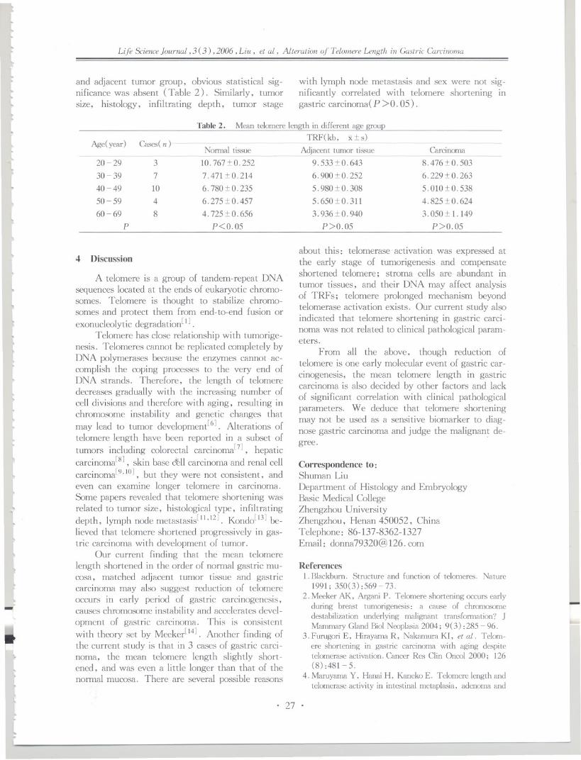
tI..I..~
r.
Life Science Journal, 3 (3) ,2006, Liu, et al, Alteration of Telomere Length in Gastric Carcinoma
It
t and adjacent tumor group, obvious statistical sig-nificance was absent ( Table 2). Similarly, tumorsize, histology, infiltrating depth, tumor stage
~10,I-
~
l
with lymph node metastasis and sex were not sig-nificantly correlated with telomere shortening ingastric carcinoma(P >0. 05).
Table2. Mean telomere length in different age group
TRF(kb, x:t s)~~...
Ll
Age( year )Normal tissue Carcinoma
Cases(n)
3
7
10
4
8
10. 767:t O.252
7.471:t0.214
6. 780 :t O.235
6.275:t O. 457
4.725:t O. 656
P<0.05
I,.
20 - 29
30 - 39
40 - 49
50 - 59
60 - 69
P~
l
Adjacent tumor tissue9.533:t O.643
6.900:t O.252
5.980:t O.308
5.650:t O.311
3.936:t o. 940
P>0.05
8. 476:t O.503
6.229:t O.263
5.01O:t O.538
4.825:t O.624
3.050:t 1. 149
P>0.05
,4 Discussion
to
A telomere is a group of tandem-repeat DNAsequences located at the ends of eukaryotic chromo-somes. Telomere is thought to stabilize chromo-somes and protect them from end-ta-end fusion or
exonucleolytic degradation[l].Telomere has close relationship with tumorige-
nesis. Telomeres cannot be replicated completely byDNA polymerases because the enzymes cannot ac-complish the coping processes to the very end ofDNA strands. Therefore, the length of telomeredecreases gradually with the increasing number ofcell divisions and therefore with aging, resulting inchromosome instability and genetic changes that
may lead to tumor development[6]. Alterations oftelomere length have been reported in a subset of
tumors including colorectal carcinoma[7], hepaticcarcinoma[8], skin base c~ll carcinoma and renal cell
carcinoma[9,1O], but they were not consistent, andeven can examine longer telomere in carcinoma.Some papers revealed that telomere shortening wasrelated to tumor size, histological type, infiltrating
depth, lymph node metastasis[1l,12]. Kondo[13] be-lieved that telomere shortened progressively in gas-tric carcinoma with development of tumor.
Our current finding that the mean telomerelength shortened in the order of normal gastric mu-cosa, matched adjacent tumor tissue and gastriccarcinoma may also suggest reduction of telomereoccurs in early period of gastric carcinogenesis,causes chromosome instability and accelerates devel-opment of gastric carcinoma. This is consistent
with theory set by Meeker[14]. Another finding ofthe current study is that in 3 cases of gastric carci-noma, the mean telomere length slightly short-ened, and was even a little longer than that of thenormal mucosa. There are several possible reasons
lI't
..
~"
tII.
~~
ll~
..~
~
l~
1I..
~
l:-
~~I."~
~r..
....
I.".
II..
about this: telomerase activation was expressed atthe early stage of tumorigenesis and compensateshortened telomere; stroma cells are abundant intumor tissues, and their DNA may affect analysisof TRFs; telomere prolonged mechanism beyondtelomerase activation exists. Our current study alsoindicated that telomere shortening in gastric carci-noma was not related to clinical pathological param-eters.
From all the above, though reduction oftelomere is one early molecular event of gastric car-cinogenesis, the mean telomere length in gastriccarcinoma is also decided by other factors and lackof significant correlation with clinical pathologicalparameters. We deduce that telomere shorteningmay not be used as a sensitive biomarker to diag-
nose gastric carcinoma and judge the malignan~ de-gree.
Correspondence to:Shuman LiuDepartment of Histology and EmbryologyBasic Medical CollegeZhengzhou UniversityZhengzhou, Henan 450052, ChinaTelephone: 86-137-8362-1327Email: [email protected]
References1. Blackburn. Structure and function of telomeres. Nature
1991; 350(3):569-73.
2. Meeker AK, Argani P. Telomere shortening occurs earlyduring breast tumorigenesis: a cause of chromosomedestabilization underlying malignant transformation? JMammaryGlandBioiNeoplasia2004; 9(3) :285- 96.
3. Furugori E, Hirayama R, Nakamura KI, et al. Telom-ere shortening in gastric carcinoma with aging despitetelomerase activation. Cancer Res Clin Oncol 2000; 126(8) :481 - 5.
4. Maruyama Y, Hanai H, Kaneko E. Telomere length andtelomerase activity in intestinal metaplasia, adenoma and
L
. 27 .

rI
..
~..I
}J
Life Science Journal, 3 (3) ,2006, Liu, et al, Alteration of Telamere Length in Gastric Carcinoma
well differentiated adenocarcinoma of the stomach. Nip-pon Rinsho 1998; 56(5): 1186-9.
5. Hastie ND, Dempster M, Dunlop MG, et al. Telomerereduction in human colorectal carcinoma and with aging.Nature 1990; 346(6287):866-8.
6. Shay JW. Molecular pathogenesis of aging and cancer:are telomeres and telomerase the connection? J ClinPatho11997; 50(10):799-800.
7. Kim HR, Kim YJ, Kim I-U, et al . Telomere lengthchanges in colorectal cancers and polyps. Korean Med Sci2002; 17 (3) :360 - 5 .
8 . Yokota T, Suda T, Igarashi M, et al . Telomere lengthvariation and maintenance in hepatocarcinogenesis. Can-cer2003; 98(1): 110-8.
9. Wainwright], Middleton PG, Rees JL. Changes inmean telomere length in basal cell carcinomas of the skin.Genes Chromosome Cancer 1995; 12(6) :45 - 9.
10. Dahse R, Fiedler W, Ernst G, et al. Changes in telom-
-~
ere lengths in renal cell carcinomas. Cell Mol Bioi 1996;42(9): 477-85.
11. Gordon KE, Ireland H, Roberts M, et al. High levels oftelomere dysfunction bestow a selective disadvantage dur-ing the progression of human oral squamous cell carcino-ma. Cancer Res 2003; 63(2): 458 - 67.
12. Frazil PA, Glickman J, Jiang S, et al. Differential im-pact of telomere dysfunction on initiation and progressionof hepatocellular carcinoma. Cancer Res 2003; 63 (16) :5021-7.
13. Kondo T, Que N , Yoshida K, et al. Expression ofPOTI is associated with tumor stage and telomere lengthin gastric carcinoma. Cancer Res 2004; 64(2): 523 - 9.
14. Meeker AK, De Marro AM. Recent advances in telom-ere biology: implications for human cancer. CUITOpinOnco12004; 16(1): 32 - 8.
.....
..~
~J
j~...
...
JJ''"
JJJReceived June 10, 2006
.01
J
JI..I..
...J.J
~
....
...I
..J,I...
,I
,I
'"
...
...
J
J
.01
J
J
~-
~
. 28 .
.J~Jj...I...~.J..

\:Ll
t[
tLll
[[{llll
~
lt!.ll~
lLtlL
L
tL
ll
l\
!-
Life Science Journal, 3 ( 3 ) ,2006 , Ba , et aI, Mage-a x , rnRNA Level in Lung Cancer by Coal Tar Pitch
Mage-ax mRNA Level in Lung Cancer of Mice Derived
by Coal Tar Pitch
Yue Ba, Huizhen Zhang, Qingtang Fan, Xiaoshan Zhou, Yiming Wu
Department of Labor and Environmental Health, College of Public Health,
Zhengzhou University, Henan 450052, China
Abstract: Objective. To investigate the expression of Mage-ax mRNA in lung cancer tissues of mice induced by coaltar pitch (CTP) fume and to discuss the pos",ibility of the lung cancer animal model induced by CTP as a model forlung cancer immunotherapy with MAGE-A. Methods. Tumor tissue samples of lung cancer and paired non-tumortissues of the lung were obtained from 8 lung cancer mice. Total RNA was extracted and cDNA was synthesized.Nested PCR amplification using Mage-ax specific primers was then performed to detect the expression of Mage-a.
The 2 clones of 1 sample of Mage-ax mRNA positive PCR products were DNA sequenced by using DNA" sequencer(PE-377). Results. Of 29 mice in the experimental group, 8 were induced to lung cancer. Of 8 lung cancers, 5(62.5%) expressed Mage-ax mRNA. The expression of Mage-ax gene was not recognized in adjacent lung tissuesat all. The DNA sequence confirmed that the target gene fragments in all 2 samples of PCR products were Mage-ax
cDNA. Conclusion. The Mage-a gene was expressed highly in tumor tissues with lung cancer in mice induced byCTP fume. This suggests that this kind of lung cancer mouse model may be an ideal animal model for lung cancertherapeutic experiment by MAGE-A. [Life Science Journal. 2006; 3(3) : 29 - 34] (ISSN: 1097 - 8135) .
Keywords: Mage-ax mRNA; mouse; CTP fume; lung cancer
Abbreviatious: CTP: coal tar pitch; CTL: cytotoxic T lymphocyte; TAs:tumor antigens
1 Introduction
Although the enormous manpower and materi-al resources have been spent, there are still no ef-fective methods developed to prevent and treat ma-lignant tumor. The incidence rate of malignant tu-mor is increasing and the onset age is tending to beyounger along with the changes of environment andlifestyle of human being. In China, the increasingmagnitude of lung cancer is in the first place in re-cent 20 years according to the infonnation derivedfrom a more recent national conference on oncologyin 2000[1].
Using a gene transfection approach to identifyantigens recognized by CTL (cytolytic T lympho-cytes) on a human melanoma cell line, Boon et al
isolated the gene MACE-A family that is located inthe Xq28 region and the gene family includes at
least 12 related genes[2-4J. MACE-B, including 4
genes, was identified in the Xp21. 3 region[5-7J.MACE-Cl is on band Xq26[8J. Most of theseMACE genes are expressed in a significant propor-tion of tumors of various histological origins,whereas no expression has been observed in normaltissues except on placenta and male genn cells.
The MACE-encoded antigens are recognizedby cytolytic T cells in the fonn of antigenic pep tides
~ll'"
l~~
t..
l.
presented by HLA class I molecules. Because malegenn cells do not express the HLA class I genes,they fail to present MAGE antigens even thoughthey express MACE genes. The MACE-encodedantigens are therefore strictly tumor-specific. Sev-eral immunogenic peptide epitopes from tumor-as-sociated antigens (such as MAGE-A3, MAGA-A1etc. ) have served as targets for cellular immune re-sponses in numerous clinical trials for therapeuticvaccinations[9, 1OJ.
In 1999, Boon et al found Mage-a, a newI ilmily of mouse genes homologous to the human
MACE-A genes[llJ. Mage-a genes were mappedon X chromosome. Like human MACE-A, Mage-a genes were transcribed in adult testis, but not inother tissues. Expression of some Mage-a geneswas also detected in tumor cell lines. Mage-a genesare higher degree homologous to the humanMACE-A genes. Like MACE-A genes, theyen-code acidic proteins. As the ideal tumor animalmodel, it is possible to research the immunotherapyby using MACE tumor antigens(TAs). There is,however, little information on their expression inlung carcinoma of mice. This experiment studiedthe expression of Mage-a gene in mice lung cancerand compared its sequence with that in GenBank,then discussed the possibility of the lung cancer ani-
II
~I
l
~
. 29 .
nal elongation cycle at 72 °C for 300 sec. Under thesame PCR condition, a second round of PCR was
performed using nested primer 5' -AGCGGATCC-CTCTCTCCCCAGGCC-3' (forward), 5' -A C G
bAGCTTCCAATTTCCGACGACACTCC-3' (re-verse) , and 1 t-tl of first-round PCR products astemplate. The nested primers were added appropri-ate BamHI and Hind III restricted site respective-
ly.2.3mids
For the construction of recombinant pUC18
plasmids, a PCR fragment of Mage-ax cDNA wasobtained with the nested primer. pUC18 plasmidsand the target fragments were digested by BamHIand Hind III respectively. Bands were isolated fromlow melting point agarose and purified by UNIQ-5column DNA gel reextraction kit (Sangon, Shang-hai, China) respectively, and then PCR productwas ligated directly into pUC18 plasmid with T4
DNA ligase. The mixture was transfered into E.coli cell, strain DH5a according to the method of
Molecular Cloning[12]. At least 3 positive cloneswere picked up and amplified. Then, the ligatedPCR products were isolated and sequenced bydideoxy method using DNAs sequencer (PE377).The sequencing results were analyzed using DNAsissoftware.
)JJ
))JJ.JJJJJ.d
J}JJJ
JJJJJ
JJ.JJ
j
)J.J
JJ.J
JJI
,tI
I.J
J
JJJ)J}
Life Science Journal, 3 (3) ,2006, Ba , et al, Mage-ax, mRNA Level in Lung Cancer by Coal Tar Pitch
mal model induced by CTP as a model for lung can-cer immunotherapy with the use of MAGE-A.
Mage-al' a2, a3, as, a6, as of Mage-a are ar-ranged in a cluster located in a region syntenic toXp22 and they share more than 93 % nucleotide identi-ty. The above 6 genes which were amplified in thisstudy were called as Mage-ax'
2 Materials and Methods
2.1 Sample collection and RNA extractionAnimal: 64 Kunming mice, 32 males and 32
females, were provided by Henan Animal Center(Zhengzhou, Henan, China). The mice were di-vided into experiment group and control group ran-domly. The experiment group was exposed to CTPfume 2 h per day for 12 weeks. The mice werekilled in the 12th week and the 24th week, respec-tively. The lung tissues were frozen in liquid nitro-gen. All the samples were confirmed by pathology.
Total cellular RNA was isolated using the flashcolumn total RNA preparation kit (QIAGEN, Ger-man) according to the manufacture's instructions.2.2 Nested RT-PCR
For nested RT-PCR analysis of Mage-ax tran-scripts, 5 t-tgof total RNA were reverse-transcribedwith the first round PCR specific primers: 5'-AATACCAAGTCCTCCCCAG-3' (forward), 5'-CTTGGGCCCCACAGGAACC-3' (reverse) in a30 t-tlreaction mixture containing reverse transcrip-tase buffer, 5 mmol dNTP, 25 pmol primer and10 U AMV reverse transcriptase (Promega, USA).The mixture was incubated at 42°C for 60 min,heated at 95°C for 5 min and then stored under -
20°C. 3 t-tlof RT reaction were used in one roundof PCR with 1 X Taq buffer, 5 mmol dNTPs, 25pmol first round PCR specific primers and 2 U ofTaq DNA polymerase (Promega, USA). PCR am-plification was 35 cycles at 94°C for 50 sec, 55°Cfor 50 sec and 72°C for 60 sec with an initial one-cycle predenaturation at 94°C for 120 sec and a fi-
Cloning and sequencing of recombinant plas-
3 Results
3. 1 The results of CTP induced cancerOf 29 mice in the experiment group, 8 were
induced to lung cancer, 7 were carcinoid and'l wasadenocarcinoma within 24 weeks. There were 5specimens with squamous metaplasia, 2 were hy-perplasia and the rest were normal. All the 32 micein control group were normal. The cancer incidencewas significantly different between two groups (P< 0.01). Detail results were shown in Table 1.
J
~
Group
Control group
Experiment groupP<O.Ol
C'AJmparisonof tumor induced in control group and experiment group
Cases( n) Mice with tumor( n) Mice without tumor( n)
TI 0 TI
29 8 21
Table 1.
3.2 Expression of Mage-ax gene at mRNA levelin mice lung cancer tissues
Of 8 mice with cancers, Mage-aT mRNA wasexpressed in 5 (62. 5% ). The expression of Mage-ax gene was not recognized in adjacent and normallung tissues at all. Mage-ax gene was also ex-
Ratio
0
27.6
pressed' in one squamous metaplasia. The PCRproducts were then digested by restriction endonu-cleas Sca I. The two fragments, 300 bp and 200bp, were shown respectively. Representative gelswere shown in Figure 1 and Figure 2. Results wereshown in Table 2.
,JI
;'JoJJJJJ
)
. 30 .

t
~
t~ll
t,L
lL
l
~
~
llll
~
Life Science Journal, 3 (3) ,2006, Ba ,et al, Mage-ax, rnRNA Level in Lung Cancer by Coal Tar Pitch
- 500 bp
Figure 1. Amplification of target gene
Amplified product of Mage-ax cDNA was 492 hp.
Lane 1,2,3,4: lung cancer tissues which expressed
MaKe-aT mRNA: Lane M: marker
-500bpI
~
l~~l.ll"L
l[
I..
-JOObp
-200bp
Figure 2. Identification of target geneLane 1: amplified target gene: Lane 2: fragments digested with
Sea I ; Lane M: marker
.ll
llllI,
f
Table2. Expression of MACE-ax gene in lung tumors inmice
Pathology
Carcinoid
Adenocarcinoma
Squamous metaplasia
Hyperplasia
Adjacent lung tissues
Nonnallung tissues
Proportion of positive samples
4/7 (57.1%)
1/1 (100%)
1/5 (20%)
0/2
0/8
0/32
3.3 Construction, identification and sequencingof mouse Mage-ax gene
The amplified DNA fragment was digestedwith BamHI and Hind ill. The target fragmentwas ligated into a predigested (with BamHI andHind ill) clone vector pUC18. Initial transforma-tion was carried out with E. coli DH5a hoststrain. Positive clone was identified via preparing
-
~llr"~
tt
the plasmid DNA from a number of clones and ana-lyzed by using amplification with universal primerof pUC18 on agarose gel electrophoresis, and wassequenced by using primer M13 (or pUC18 univer-sal primer). The target gene fragments in samplesof PCR products were Mage-ax cDNA. The resultswere shown in Figure 3 and Figure 4.
4 Discussion
The identification of TAs and their recognitionby tumor-specific CTL has fuelled the developmentof immunotherapeutic strategies in cancer[13]. Al-though numerous TAs and their epitopes have beenidentified, the majority of these are quite restrictedin expression and their clinical utility remains limit-ed. Therefore, it is imperative to evaluate the pos-sibilityof tumor immunotherapy by using TAs/epi-topes that are widely expressed in tumor. It hasmany benefit to research the MACE genes' func-tion in mice such as sample got easier, dynamic ob-servation and so on. It may, therefore, be the idealanimal model for the lung cancer's therapeutic ex-periment by using MACE.4. 1 Reliability, sensitivity and specificity of theexperiment
The authors took the following measures toensure the reliability of the experiment: (1) Re-move the necrosis tissues to refrain from RNAdegradation caused by necrosis. (2) RNA extractedwas all verified by ethidium bromide fluorescenceelectrophoresis and by ultraviolet radiomete (A260/A280>1. 9). (3) To ensure RNA was not degrad-ed, a PCR assay with primers specific for ~-actinwas carried out in each case. (4) To avoid the falsepositive results, all the samples of extracted RNAwere incubated with DNase. (5) Random samplingthe positive clones sequenced and confirmed theamplified products being Mage-ax (The sequence isthe same as that of GenBank).
Method of nested PCR can improve the sensi-tivity and specificity of the experiment. The firstround PCR will be carried out using the exo-primers firstly, and then the second round PCRwill amplify the smaller regions of the first roundPCR products by using the nested-primers. So thecontinuous twice enlargement may increase both thesensitivity and the specificity of PCR greatly. Forthe extreme-trace target fragments, it's very diffi-cult to get the better results for once amplification,but satisfactory results can be obtained using nestedPCR.
!-
. 31 .

3.2kb
1JJJ
)JJjJ
J.J
)J1
)JJ
JJJ
J)J~
JJJJjJ.)
JJ.J
J
Life Science ]aurnal,3 (3) ,2006,Ba, et al, Mage-an mRNA Level in Lung Cancer by OJal Tar Pitch
0.5 kb -
3.0 kb
Figure 3. Identification of recombinant-plasmid amplified by pUC18 universal primerLane 1,2,3,4,5,7: the inserted target fragments amplified by universal primer; Lane 6: plasmid without inserted the target gene;
Lane 8 : the negative control; Lane 9: plasmid inserted the target gene
Smageal :Smagea2 :Smagea3 :SmageaS :Smagea6 :Smagea8 :5MBY1
smagea1 :Smagea2 :Smagea3 :SmageaS :Smagea6 :Smagea8 :SHBY1
J
Smagea1 :smagea2 :
Smagea3 :
Smagea5 :Smagea6 :Smagea8 :SHBY1
879: 5285: 373: 7430: 2007: 2326
60
: 1215: 5621: 709: 7766: 2343: 2662
396 i
J
JI
JJJJJJ)
: 1271: 5677: 765: 7822: 2399: 2718
442
Figure 4. Comparing the sample sequence with Mage-ax sequence in GenBank (The underlined parts were the sample sequences)
4.2 Expression of Mage-ax mRNA of mouse lungcancer tissues
As the results of CfP fume inducing lung can-cer among 29 mice in the experiment group within24 weeks", 8 were induced to lung cancer, 7 werecarcinoid and 1 was adenocarcinoma, respectively.
PCR results showed that the majority of lung tu-mors in mice (5 of 8 tested) were positive for theMage-ax gene by gene-specific primers and thenested-PCR method. These results suggested thatmore than 60 % of lung cancers were expressedMage-ax gene, which was also expressed in one
LJjJ.J
JJ
j...
J,
~.
. 32 .

rt[[~
lLll
L
Ltt
Illlltllt\.
Life Science Journal, 3 (3) ,2006,Ba, et al, Mage-ux, mRNA Level in Lung Cancer by Coal Tar Pitch
squamous metaplasia, but not recognized in adja-cent and nonnallung tissues at all. Although thereare some mouse models used to evaluate human im-
mune responses to .MAGE-based tumor vaccine,there is no other infonnation about the expressionof Mage-a in lung carcinoma in mice. Gravekampet at. found high expression levels of Mage-b (an-other Mage family of mouse genes homologous tohuman .MAGE-B genes) in almost all metastases,regardless of age. The expression levels were 2- to3- fold higher in the metastases than in the primary
4TO7cg breast tumors[14]. It suggests that Mage-encoded tumor antigen will be used as model tostudy various anti-tumor immunization modalities. . .
In VlVO.
.MAGE-A Ags were detected in primary andmetastatic tumors of various histological types in-cluding melanoma, lung, bladder, ovarian, andbreast carcinomas. Individual .MAGE-A expressionvaries from one tumor type to another, but overallthe majority of tumors express at least one of the.MAGE-A family. Targeting epitopes shared by all.MAGE-A Ags would be of interest against a broadspectrum of cancers. At present, .MAGE peptide-based vaccines have been used in clinical trials with
tumorous patients, but with limited success[15,16].
A suitabl~ animal tumor model that would pennitthe optimization of .MAGE -encoding cancer vac-cines in mice is very necessary to immunotherapy oftumors using .MAGE gene products. Researchershave used different tumor animal models to evaluate
the possibility of tumor immunotherapy by using.MAGE Ags. Eggert et at demonstrated the im-
munogenicity of two Kb-restricted peptide epitopesderived from mouse MAGE proteins which mayserve as valuable tool for preclinical evaluation of
vaccination strategies[17]. Ni found that rSFV vac-cine could elicit human MAGE-3-specific antibodyand CTL response in the Trimera mice[18]. The re-sults of Gravekamp indicated that the metastaticand nonmetastatic breast tumor models could be
useful model systems to analyze how breast cancervaccines for humans[14].
However, a suitable mouse tumor model of
lung cancer that would pennit the optimization of.MAGE-encoding cancer vaccines in mice is cur-rently not available. In this study, it shows thatMage-ax gene was expressed highly in tumor tis-sues with lung cancer induced by CTP fume. Thissuggests that this lung tumor mice expressingMage-ax may be an ideal animal model for lungcancer therapeutic experiment by using .MAGE-A.
l~
t..I...
l\.
ll~l
(llllI
llI't
ll.-.~L
tLLI
t~
Correspondenceto:Yiming WuDepartment of Labor and Environmental HealthCollege of Public HealthZhengzhou UniversityZhengzhou, Henan 450051, ChinaTelephone : 86-371-6778-1797Email: [email protected]
AcknowledgmentsThis work was finished in Key Molecular Lab
of Henan Province. We are particularly grateful toProfessor Yi Zhang (Ludwig Institute for CancerResearch, Brussels Branch, Belgium) for his greathelp.
This work is supported by grants from theKey Project of Scientific Committee of HenanProvince (No. 524410067) and the Natural Sci-ence Research Fundation of Henan Educational Bu-reau(No. 2006330003).
References
1. Xu GW. The review and perspective for prevention andtreatment of tumor of China. Conference on Oncology,2000 (Educational Book).
2 . Van der Bruggem P, T raversari C, Chomez P, et al. A
gene encoding an antigen-recognized by cytolytic T lym-phocytes on a human melanoma. Science 1991; 254: 1643-7.
3. De Plaen, Arden K , Van der Bruggen P, et al. Structure,chroma.'iOmal, localization, and the expression of 12 genesof the MACE family. Immunogenetics 1994; 40 (5):360- 9.
4. Rogner UC, Wilke K, Steck E, et al. The melapomaantigen gene (MACE) family is clustered in the chromo-somalbandXq28. Genomics1995; 29(3) :725- 31.
5.Muscatelli F, Walker AP, De Plaen E, et al. Isolationand characterization of a MACE gene family in theXp21. 3 region. Proc Natl Acad Sci USA 1995; 92 (11) :4987 - 91.
6. Dabovic B, Zanaria E, Bardoni B. A family of rapidly e-volving genes from the sex reversal critical region inXp21. Mamm Genome 1995;6(9) :571- 80.
7. Lurquin C, De Smet C, Brasseur F. Two members of
the human MACE-B gene family located in Xp21. 3 areexpressed in tumors of various histological origins. Ge-nomic 1997; 46(3): 397 - 408.
8. Lucas S, De Smet C, Arden KC, et al. Identification of a
new MACE gene with tumor-specific expression by rep-resentational difference analysis. Cancer Res 1998; 58(4):743-52.
9. Ma JH, Sui YF, Ye], et al. Heat shock protein 70/MACE-3 fusion protein vaccine can enhance cellular andhumoral immune responses to MACE-3 in -pivo. CancerImmunol Immunother 2005; 54 (9) : 907 - 14.
10. Ye J, Chen GS, Song HP, et al. Heat shock protein 70/ MACE -1 tumor vaccine can enhance the potency ofMACE-I-specific cellular immune responses in vivo.Cancer Immunol Immunother 2004;53(9) :825 - 34.
i..
. 33 .

.I'4
(GI'L) -defined, melanocyte-specificantigen in metastaticmelanoma patients impacts on generation of peptide-spe-cific GI'Ls but does not overcometumor escape from im-mune surveillancein metastatic lesions. J ExpMed1999;190: 651- 68.
16. Marchand M, van Baren N, Weynants P, et at. Tumorregressionsobserved in patients with metastatic melanomatreated with an antigenic peptide encoded by geneMACE-3 and presented by HLA-A1. Int J Cancer 1999;80:219-30.
17. Eggert AO, Andersen MH, Voigt H, et al. Characteri-zation of mouse MACE-derived H-2Kb-restricted GI'Lepitopes. Eur J ImmunoI2004;34(11) :3285 - 90.
18. Ni B, Gao W, Zhu B, et al. Induction of specifichumanprimary immune responses to a Semliki Forest virus-basedtumor vaccine in a Trimera mouse model. Cancer Im-munol Immunother 2005; 54(5) :489 - 98.
.IJJ
1
/
J...
Life ScienceJournal, 3 (3) ,2006, Ba ,et al, Mage-ax' mRNA Level in Lung Cancerby OJal Tar Pitch
11. De Plaen E, De Backer D, Arnaud D, et al. A new fami-ly of mouse genes homologousto the MACE genes. Ge-nomicsl999; 55(2): 176-84.
12.£nnbrook 1. Fritsch EF, ManiatisT. MJlecularcloning: alaOOratorymanual. 2nd. Cold Spring Harix:Jr,NY: ColdSpring Harix:Jr Laboratory, 1989.
13. Graff-Duix:JisS, Faure 0, Gross DA, et al. Generationof GI'L recognizing an HLA-AOzol-restrictedepitopeshared by MACE-AI> -Az, -A3, -~, -~, -AIO, and-A12tumor antigens: Implication in a broad-spectrum tu-mor immunotherapy. J Immuno 2002; 169 ( 1) : 575 -80.
14. Gravekamp C,Sypniewska R, Gauntt S, et al. Behaviorof metastatic and nonmetastatic breast tumors in oldmice. Exp Bioi Med (Maywood) 2004; 229 (7) : 665 -
75.15. Anichini A, Molla A, Mortarini R, et al. An expanded
peripheral T cell population to a cytotoxic T lymphocyte
-~
I
~~
4
..4
...
.I...1J......
.!
JJ
JReceived April 2, 2006 .I
J
...
JI
.J~
...J.JJ
...
J~........"I4
iI
~
...
~
"
~I
~~
..I
)
J
J
. 34 .
.I041...IJ.J
.......
J..J

f
t(~lL.
ll"-
lL
tl"
Life Sci£nceJournal, 3 (3) ,2006 ,Zhang, et al, Effect of Folate and Vitamin B12 on Tau Plwsplwrylation
Effect of Folate and Vitamin B12 on Tau
Phosphorylation in Aged Rat Brain
Jiewen Zhangl, Fen Lul, Xu Li2, AiqinSuol
1 . Department of Neurology, Henan People's Hospital, Zhengzhou, Henan 450003, China
2. Department of Pathology, Henan Tumor Hospital, Zhengzhou, Henan 450008, China(All the authors contribute to the work equally)
llllllll~
L
t\~......
ll
~
ll
llIl~l~
Abstract:Alzheimer's disease (AD) is the cause of one of the most common types of dementia. In AD brain, abnor-
mal hyperphosphorylated tau composed the major protein of neurofibrillary tangles (NITs), one of the two neu-ropathological hallmarks of AD. To prevent and relieve the tau protein abnormal hyperphosphorylation in AD pa-tients' brain is thought to be the key point of therapy. Recent study suggested that there is some relationship be-tween folates, vitamin B12 and AD. In our study we aim to investigate the possible mechanism of AD especially thecorrelation between folates, vitamin B12 and tau phosphoiylation. We examined tau protein phosphorylation state inrats' hippocampus of different age stages: two and forty months old by phosphorylation dependent and independenttau antibodies. We found that tau phosphorylation in aged rats' brain showed significant high level than these twomonths old. And we also found that folates plus vitamin B12 can decrease the level of tau phosphorylation in agedrats' brain. It sugge.<;tsfolates and vitamin B12 may play an important role in preventing the neurodegenerativechange by influencing tau phosphorylation in brain. [Life Science Journal. 2006; 3 (3) : 35 - 40] (ISSN: 1097-8135) .
Keywords:Alzheimer's disease; aged; folate; vitamin El2
Abbreviations: AD: Alzheimer's disease; BCA: bicinchoninic acid; CSF: cerebrospinal fluid; DAB: diaminob-enizidine; NITs: neurofibrillarx tangles; PHF: paired helical filaments; SDS-PAGE: sodium dodecyl sulphate poly-acrylamide gel electrophoresis
1 Introduction
Dementia is a syndrome characterized by anacquired global impairment of memory and othercognitive functions sufficient to interfere with nor-mallife[ 1]. Alzheimer's disease (AD) is the causeof one of the most common types of dementia. TheWorld Health Organization has estimated that 25 -
29 million people in the world suffer from dementia.Approximately 6 % - 8% of all older people overthe age of 65 years have AD[2], and the prevalence
increases steeply with age[3]. AD is characterizedby the presence of two histopathological hallmarkscalled senile plaques and neurofibrillary tangles,which are involved in the process leading to pro-gressive neuronal degeneration and death. It is re-ported that neurofibrillary tangles (NFTs) arestructures present in the neuronal body and consistof paired helical filaments (PHF), mainly com-
posed of highly phosphorylated tau protein[4,5].Tau is a microtubule-associated protein expressedmostly, but not exclusively, in the nervous sys-tem, and its normal physiological function is tobind and stabilize microtubules[6]. In AD brain,
~~l...
L
l~II..~
~
tau is found aberrantly hyperphosphorylated[7].Abnormal phosphorylation of tau in brain seemed tobe an important pathogenesis of AD.
The possible involvement of nutritional faCtorsin the aetiology (causes) or pathogenesis (mecha-nisms of brain damage) of dementia has been wide-ly considered. In particular, dietary deficiency offolates has been postulated as contributing to theaetiology AD. Folates are vitamins essential to thedevelopment of the central nervous system. And re-searcher also found that vitamin Bl2 deficiency not
only produces anaemia but also causes irreversibledamage to the central and peripheral nervous sys-tems. In this study we aim to investigate the mech-anism of the relationship between AD and folatesand vitamin B12. We focus on the tau protein and tostudy whether folates plus vitamin Bl2 have effect onits phosphorylation status.
2 Materials and MethodsI.
2.1 AnimalMale Wistar rats were from Experimental Ani-
mal Central of Henan Medical College. All animalswere observed daily for clinical signs of disease. All
. 35 .

Dr. Chengxin Gong (New York State Institute forBasic Research, Staten Island, NY, USA). Bicin-choninic acid (BCA) protein detection kit, goat an-ti-rabbit peroxidase-conjugated secondary antibody,chemiluminescent substrate kit and phosphocellu-lose units were obtained from Pierce ChemicalCompany (Rockford, IL, USA). Detection kit( Histostain-SP) for immunohistochemistry. Goatanti-mouse and goat anti-rabbit alkaline phos-phatase-conjugated secondary antibodies, diaminob-enizidine (DAB) and other chemicals were pur-chased from Maixin Biotechnology Company (FuZhou, China). Folates were from Peking Universi-ty Pharmaceutical Co. Ltd. and vitamin El2 wasfrom YangzhouZhong Bao PharmaceuticalCo. Ltd.
tJ.J
J
~
~J~
J~JJ~
JJ
J
J
J
J
J
JJ-IJ.JJ
}JJ..I.JJ.,IJ
Life Science Journal, 3 (3) ,2006, Zhang, et al, Effect of Folate and Vitamin B12 on Tau Phosphorylation
Table 1. Tau antilxxlies employed and their properties
Antilxxly Dilution Type" SpecificityT au-1 1 :30000 Mono- unPPHF-1 1:500 Mono- P
R134d 1 :2000 Poly- P + unP
a Poly-, polyclonal; mono-, monoclonal; unP, unphosphorylated epitope; P, phosphorylated epitope
b Numbered according to the largest isoform of human brain tau
animal experiments were performed according toPolicies on the Use of Animals and Humans inNeuroscience Research, revised and approved by theSociety for Neuroscience in 1995. The subjectswere allocated into two groups: two months oldrats and forty months old rats. Group of fortymonths old rats were treated with folates (40 mg/kg diet) by the gut and vitamin B12 (20 figlkg) byintraperitoneal injection every day for one month.2. 2 Antibodies and reagents
Antibodies to tau are listed in Table 1. Rabbit
polyclonal antibody R134d against total tau, mono-clonal antibodies PHF-l against PHF-tau phospho-rylated at Ser396/404, and Tau-l against PHF-tauunphosphorylated at Serl99/202 were gifts from
2.3 Preparation of rat brain extractsThe rat is deeply anesthetized with sodium
pentobarbital (75 mglkg, i. p.) and then decapi-tated. Immediately remove the brain and separatethe brain sagittally into hemisphere and put intoice-chilled PBS. The left hemisphere was fixed forimmunohistochemistry. And the right hippocampuswas homogenated and supernatant was for We.<;temblot.2.4
J
Western blotWestern blots of hippocampus homogenate
were to determine the phosphorylation state of tauof different aged rats. The homogenizer containedcold homogenizing buffer containing 50 mM Tris-HCl, pH 7.0, 10 mM ~-mercaptoethanol, 1. 0mM EDTA, O. 1 mM phenylmethylsufonyl fluo-ride, and 2. 0 fig/ml each of aprotinin, leupeptin,and pepstatin A. Then they were homogenized inthe same buffer at a ratio of 9.0 ml of buffer/I. 0 gtissue with phosphatase inhibitor mixture contain-ing 20 mM ~-glycerophosphate, 1.0 mM Na3VO4,and 50 mM NaF, pH 7.0. The homogenates werespin at 15,000 rpm for 3 min at 4 "C for biochemi-cal analysis. The phosphorylation of tau in theabove samples was analyzed by Western blots using10% SDS-PAGE. The separated protein bandswere transferred into nitrocellulose membrane and
probed with specific anti-tau antibodies. Then allblots were probed with peroxidaseconjugated sec-ondary antibody and developed with chemilumines-cent substrate kit. The protein bands were quanti-
.
Phosphorylation sitesbSer-198/Ser-199/Ser-202
Ser-396/Ser-404
tatively analyzed, and the amount of protein wasexpressed as relative level of total optical density.2.5 Innnunohistochemistry
The hippocampus of the left hemisphere werefixed by 10% neutrality fonnaldehyde, 90% 0.01 MPBS solution at room temperature for 6 h, paraffinembedded, and cut into 5 fim-thick sections. Drythe slide.<;(processed by acetone and APES) withtissue sections in an 80°C oven for 30 min.. Sec-tions were blocked with O. 3% HzOz in absolutemethanol for 20 min and non-specific sites wereblockedwith instantcalfserumfor 60 min at 37 "C.
Then incubate sections overnight at room tempera-ture with primary antibodies as described. Theslides were developed by biotinylated secondary an-tibodies (1 : 200) and Avidin-peroxidase conjugate( 1 :200) / diaminobenzidine tetrachloride (0. 05 % )system.2.6 Statistical analysis
Data were expressed as x:t SD and analyzedusing SPSS 11. 0 statistical software (SPSS Inc,Chicago, Illinois, USA). The One-Way ANOVAprocedure followed by LSD's post hoc tests wasused to determine the different means amonggroups (P < O.05) .
~I
,-'I
JI
JJ..J
JJ)J
LJ
JJ~I
...
j..I
J.JJ~
3 Results
3.1 Tau phosphorylation at ser396/404 siteTo study whether folates and vitamin B12 af-
fect tau protein phosphorylation and their possible
. 36 .

rL
ll~
lLll
Life Science]aurnal,3 (3) ,2006 ,Zhang, et al, Effect of Folate and Vitamin B12 on Tau Phosphorylation
relationship, the status of tau phosphorylation ofdifferent ages in rats' hippocampus were analyzed.The status of tau of different age rat were carriedout by Western blot using three well characterizedphosphorylation-dependent and site-specific tau an-tibodies as listed in Table 1. Compared with twomonths old rats group, a remarkable increase ofphosphorylated tau for forty months old rats wasdetected by PHF-1 which recognize phosphorylatedtau at ser396/404 site. And tau-1 recognizes un-phosphorylated tau at serl99/ser202 sites. Tauphosphorylation in forty months old rats group havethe high expression (Figure 1 A and B). The levelof total tau was indicated by blot developed withR134d, a phosphorylation-independant antibody.Data showned that there was no remarkable differ-
ence between two months old group and fortymonths old group (Figure 1C). So the phosphory-lation of tau protein in rat's hippocampus is notablyincreased with. the aging process. And based thisresult we do the next step.
...I...
ll-"
lI"-
lI"-
llII,.
llllll
(I"-
~..
ll
l,llllllllL
~-
-_.- ._----A
J.. 2.5'":;:.2
0*
.~ 15
.~ 1..~
.~ 0 :..D.~...~2 months 40 months
-.---B
jl~[
0".. I *.::; 0.8 r~ 0.6 I:;; I: 0.4 i
~ O:l~_O.2 months 40 months
c
j 14
g 1. 2
.~'1
0 0g 0.8
'~.
" (
.
J.6
1: 0.4
.~ 0.2::1 0 "~._.
~l
l~l\
lt
2 months 40 months
Figure 1. Level of tau phosphorylation in different age rat brainby Western blot.15 f1iSof protein per lane was employed with antibody PHF.l,
tau. 1 and R134d. * P< 0.05, vs. 2 months old rat.
3. 2 Folates plus vitamin BI2 can decrease hyper-phosphorylation of tau in aged rat brain
After rats were treated with folates and vita-
min B12, the status of tau protein phosphorylationwere detected on different age stage groups in rats'brain. Gained the extract of rats' hippocampus wasfor immunoblot analysis. We found that bands ofthe drug treated group were sharply decreased com-pared to the group which was not treated withdrugs (Figure 2A). It indicates a large decrease inphosphorylation of tau at Ser396/404 sites. Thisresult was corroborated by the greatly diminishedstaining of tau bands by phosphorylation-indepen-dent anti-tau antibody tau-I, which has an optimalimmunoreactivity when tau is dephosphorylated atserl99/ser202 (Figure 2B). It demonstrated thatfolates and vitamin B12 have the effect on decreas-
ing the tau phosphorylation in aged rat brain. Wealso found that the level of total tau still had no re-
markable difference between groups(Figure 2C).
A """",",0.."--------
~. ::~r
0 0*
j ~:n.~ ~: ~ I ~ ".;:: 0 '-..-.---ro"
~ 40 months 40 months
(VBJ2+Folate)
B ~-, --
:::;;, L 6~ L 4.t';. L 2'U; 1;; 0.8-;:; 0.6
.: 0.4
.c:; 0.2<;; 0"
~
0 040 months 40 months
(VB12'Folate)
c 1 Jill. Hill.;iU...
;; L 4~ 12
~. ];'! 0.8
~ O. G'- 0.4.~ 0.2~ 0"~
D.m~mD40 months 40 months
(VB12+Folate) ~
Figure 2. Effect of folate plus vitamin E12 on tau phosphorylationin 40 months aged rat brain by Western blots.15 p.g of protein per lane was employed with antibody PHF.l,
tau. 1 and R134d. * P < 0.05, vs. aged rat that was not treatedwith folate and vitamin E12.
. 37 .

I I
-~
I
I
I
----
.-~...
~~
)Life ScienceJournal ,3(3) ,2006 ,Zhang, et al, Effect of Folateand Vitamin B12 on Tau Phosphorylation
PHF-'17'" -
w
.~
Q
qi
"
Ta(r1
'"
~.
,~.. ,r lJ/;'." 1J.?t~.L.--"
:J T',. 7
tt~. ~j~. ,tF,JI:f?0/l'..:~ .~
?",,-.; '..><. t!'
,/tf-J.) .
2 months
..
11 !I
~~
JJ.J
PHr;;~"1 ,#'i""-"--.' ,!,,' -
1'. II
" ,.,'il ""..
..,~,.I"
hiI ~
I r
11
....
if'.J~J
1. ..,I4
.J
I.;
~
J;;,I - ---- ..
-. JITau-1
..
.. ....JD~
;;!.{' "
....~.J
~<
,~ .,I
JJ
r
~." ...
of' ~
~
40 months
Figure 3. Level of tau phosphorylation in different age rat brain by immunohistochemistry
Paraffin embedded (5 I'm-thick) sections were immunostained with PHF-l and tau-I.
To learn the change of phosphorylation state oftau and its topographical distribution correlate withfolates and vitamin Bl2 treatment in the brain tis-sue, we immunostained the sections cut from theleft hemisphere with phosphorylation dependentand phosphorylation independent anti-tau antibod-ies. From these pictures we found that the im-munohistochemistry results were consistent withWestern blots results. The level of tau phosphory-lation in 40 months aged rat brain is higher thanthat of 2 months old rat (Figure 3). After treatedwith folates and vitamin Bl2 for one month we cansee that at ser396/404(PHF-l) the rat brain sec-tions were stained gradually weak. Oppositely,they were stained gradually increased when detect-
...
,.I
ed by antibodies tau-l (Figure 4). Immunohisto-chemistry results also demonstrated that folates andvitamin Bl2 could decrease the level of tau phos-phorlation in rats' hippocampus.
..
-I
J4 Discussion ~
JThis experiment studied the possible associa-
tions between vitamin Bl2/folate and tau proteinphosphorylation in aged rat brain. In the study wedetected the phosphorylation state of the aged andyoung adult rats. We found that the level of tauphosphorylation in aged rat brain was higher thanthat of young adult rat. The result suggests thatthe phosphorylation of protein tau changes dynami-cally according to the physiological state associated
;
~
-I
J.;JJ
. 38 ...I...
iJ

~
~
tt~
t
Life ScienceJournal, 3 (3) ,2006, Zhang, et al, Effect of Folate and Vitamin B12 on Tau Phosplwrylation
...l"
lL
tl"-
II
~
lllLIlL
~
l"LlllI
~lI..
~"-
I
L
ll
tl
tlll,-
--Control Treated
Figure 4. Effect of folate plus vitamin B12 on tau phosphorylation in 40 months aged rat brain by immunohistochemistry.Paraffin embedded (5 /lll-thick) sections were immunostained with PHF-l and tau-l.
with the aging process. In fact, recent studies hadreported that high incidence rate of AD in agedpopulation. And abno=al hyperphosphorylation oftau protein is closely related to AD. Biochemicaland anatomy also had proven these foundings. Butthe mechanism of the tau phosphorylation in agedpeople which probably cause AD is still not clear.Recently, researcher found that older people withlow levels of folate are twice as likely to develop ADas are those with no=allevels[sJ. In some observa-tional studies, researchers found that low serum fo-late levels have been associated with AD and with
all types of dementia[9, 10J. Red blood cell folate
~lt'-ll\
l{
and CSF folate levels are lower in patients with ADthan in controls[11J. It is known that low folate lev-
els can be the result of inadequate dietary intake,diminished absorption from the gastrointestinaltract or increased utilization. In older people folatemetabolism disturbance usually happened probably
because body regular function becomes declined. Sosupplement folate could be reasonable for these old-er people.
In our study we treated the aged rat with fo-late and vitamin B12 for one month the level of hy-
perphosphorylation of tau in rat brain extract de-creased. It suggested that folate and vitamin B12
..
. 39 .

-71.
3 . Jonn AF, Korten AE, HendersonAS. The prevalenceofdementia: a quantitative integration of the literature. Ac-ta PsychiatrScand1987 ;76:465- 79.
4.Grundke-Iqbal I, Iqbal K, Quinlan NM, et al. Micro-tubule-associatedprotein tau: a component of Alzheimerpaired helical filaments. J BioI Chem 1986; 261: 6084-9.
5.Godert M, Spillantini MG, Cairns NJ, et al. Tau pro-teins of Alzheimer paired helical filaments: abnormalphosphorylation of all six brain isofonns. Neuron 1992;8: 159- 68.
6. Drubin DG, Kirschner MW. Tau protein function in liv-ing cells. J Cell Biochem 1986; 103: 2739 - 46.
7. Kopke E, Tung YC, Shaikh S, et al. Microtubuleasso-ciated protein tau: abnonnal phosphorylation of a non-paired helical filament pool in Alzheimer disease. J BioiChem 1993;268:24374 - 84.
8. Wang HX, Wahlin A, Basun H, et al. Vitamin El2 andfolate in relation to the development of Alzheimer's dis-ease. Neurology 2001;56, 1188- 94.
9. Clarke R, Smith AD, Jobst KA, et al. Vitamin El2,and serum total homocysteine levels in confinnedAlzheimer disease. Arch Neuro11998;55:1449- 55.
10. Ebly EM, SchaeferJP, Campbell NR, et al. Folate sta-tus, vascular diseaseand cognition in elderly Canadians.Age Ageing 1998;27:485- 91.
l1.Abalan F, Zittoun J, Boutami C, et al. Plasma, redcell, and cerebrospinalfluid folate in Alzheimer's disease.Encephale 1996; 22: 430 - 4.
12. Kado DM, Karlamangla AS, Huang MH, et al. Homo-cysteine versus the vitamins folate, B6, and B12 as pre-dictors of cognitive function and decline in older high-functioning adults: MacArthur Studies of Successful Ag-ing. AmJ Med2005;118:161-7.
13. Hofman A, Ott A, Breteler MM, et al. Atherosclerosis,
apolipoprotein E, and prevalence of dementia andAlzheimer's disease in the Rotterdam Study. Lancet1997;349:151-4.
14. McCaddon A, Davies G, Hudson P, et al . Total serumhomocysteinein senile dementia of Alzheimer type. Int JGeriatr. Psychiatry 1998; 13: 235 - 9.
15. Seshadri S, Beiser A, Selhub J, et al. Plasma homocys-teine as a risk factor for dementia and Alzheimer's dis-
ease. New England Journal of Medicine2002;346 :476-83.
16. Kruman II, Kumaravel TS, Lohani A, et al. Folic aciddeficiency and homocysteine impair DNA repair in hip-pocampal neurons and sensitize them to amyloid toxicityin experimental models of Alzheimer's disease. J Neu-rosci 2002;22(5):1752-62.
3
~
J
~JjJ~
JJ<'
JjJ]J
J
JJJ)J'"
JJJ
)Jj)
Jj.JI
JJjJI
~
JJJJ
j
Life Science Journal, 3 (3) ,2006, Zhang, et al, Effect of Folate and Vitamin B12 on Tau Phosphorylation
could improve abnonnal hyperphosphorylation oftau, which composed NIT, one of the hallmarks ofAD. This result probably due to supplemental offolate and vitamin B12 decreaseing the levels ofblood homocysteine. It has been reported thatblood levels of homocysteine was elevated in pa-tients who were lack of folate and vitamin B12[12] .
High homocysteine levels are associated with de-
creased cognitive function and dementia[13]. Indi-viduals with AD have been found to have higher
plasma homocysteine levels than agematched con-trols[14], and it has been reported that elevation ofplasma homocysteine levels precedes clinical mani-festations of AD[15]. The underlying mechanisms ofhomocysteine as a risk factor for Alzheimer's de-mentia are still uncertain, but there are many waysin which homocysteine could damage neurons, in-cluding through endothelial dysfunction, cerebralmicroangiopathy and increased oxidative stress. Inrats, homocysteine induces apoptosis in hippocam-pal neurons, and in vivo it increases excitotoxicity
and oxidative damage[16]. The detail mechanismstill need further study.
In summary, this study demonstrated that fo-late and vitamin B12 could relieve the level of tau
protein hyperphosphorylation in aged rat brain.And this founding could be a reference for cliniciansand medicine researchers.
Correspondence to:Jiewen ZhangDepartment of NeurologyHenan People's HospitalZhengzhou, Henan 450003 , ChinaTelephone: 86-371-6558-0171Email: [email protected]
References
1. Gottfries CG, Lehmann W, Regland B. Early diagnosisof cognitive impainnent in the elderly with the focus onAlzheimer's disease. J Neural Transm 1998; 105: 773-86.
2. Small GW, Rabins PV, Barry PP, et al. Diagnosis andtreatment of Alzheimer disease and related disorders.Consensus Statement of the American A'isociation for
Geriatric Psychiatry, the Alzheimer's Association, andthe American Geriatrics Society. JAMA 1997; 278: 1363
~
Received June 20, 2006
-
. 40 .
~IJI)J~JJ~J,J.

a.,l~
~
lllIt
ll
Life Science Journal, 3 (3) ,2006, Zhang, et al, Peroxynitrite Mediated Oxidation Da71Ulge
Peroxynitrite Mediated Oxidation Damageand Cytotoxicity in Biological Systems'-
ll
f'tlIl
llLlllL-
LlllL
[i-
tl"-
L4.
lLl
~
lL
L
~.--
Xu Zhang, Dejia Li
Department of Biochemistry, Basic Medical College, Zhengzhau Univercity,
Zhengzhau, Henan 450052, China
Abstract: Peroxynitrite is the product of the diffusion-controlled tennination reaction between two radicals, nitricoxide and superoxide and is a strong oxidant and nitrating regeant. Critical biomolecules like proteins, lipids andDNA react with peroxynitrite via direct or radical-mediated mechanisms, resulting in alterations in enzyme activitie."and signaling pathways. The biological consequence." of peroxynitrite-mediated oxidative modifications depend onthe levels of oxidant achieved in vivo and its cellular site of production. In this article we overview multiple biologi-cal toxicity of peroxynitrite including the biological reactivity of peroxynitrite, peroxynitrite mediated oxidationdamage of biomacromolecule." such as proteins, lipids and DNA and the cytotoxic effects (apoptosis and necrosis) ofperoxynitrite. [Life Science Journal. 2006 :3(3) :41- 44] (ISSN: 1097 - 8135).
Keywords: peroxynitrite: toxicity: cytotoxicity; oxidation damage
Abbreviations: NOS: nitric oxide synthase; PARP-1: poly(ADP-ribose) polymerase-1
1 Production of peroxynitrite in biological sys-tems
ONOO- , fonned by the reaction of nitric ox-ide with superoxide (O~) which is a byproduct ofcellular respiration at near diffusion controlled
rates[l], is very likely to occur even in the presenceof physiological concentrations of SOD. Nitric ox-
ide is enzymatically produced from L-arginine bynitric oxide synthase (NOS). Three isoforms ofthis enzyme have been described: nNOS (neu-ronal), eNOS (endothelial) and iNOS (induced,inflammatory). On the other hand, superoxide canbe catalytically produced (for example, by xanthineoxidase or NADPH oxidase), and also formed bypartial reduction of oxygen in the mitochondrialmembrane or non-enzymatic monoelectron reduc-tion of oxygen (for example, hemoglobin autoxida-tion). Peroxynitrite has a short biological half-life(10 - 20 ms) but can cross biological membranesand diffuse one to two cell diameters[2]. In vivo
formation of peroxynitrite is supported by thegrowing experimental evidence[3].
2 Reactivity of Peroxynitrite
~~lL.ll,L
(
Peroxynitrite mediated oxidation damage by adecomposition intennediate with the biological ac-
tivityof hydroxyl radical[4]. The decomposition ofperoxynitrite to nitrate is intimately coupled with
the oxidation chemistry of this species, and both
reactions have been the subject of recent investiga-
tions. ONOO- is a relatively stable at alkaline pH,but at physiological pH it is capable of effecting oneand two electron reactions akin to those of HO + ,NOz, and nitrosonium cation. Oxidations of ascor-
bate[S], transition metal complexes, halide ions,
thiols, sulfides[6], olefins, benzenes, phenols[7]and other aromatics by peroxynitrite have been de-scribed. Peroxynitrite is a particularly effective-oxi-dant of aromatic molecules and organosulfur com-pounds that include free amino acids and peptideresidues. Cysteine and glutathione, which are sig-nificant components of antioxidant reservoirs, areconverted to disulfides. Methionine is converted to
sulfoxide or is fragmented to ethylene anddimethyldisulfide. Tyrosine and tryptophan under-go one-electron oxidations to radical cations, which
are competitively hydroxylated, nitrated and dimer-ized[8]. Purine nucleotides are vulnerable to oxida-
tion and to adduct fonnation[9]. Other reports havemore detailed reviews on the chemistry, decomposi-tion and reactivity of peroxynitrite, peroxynitrousacid and its activated isomer[lO]. The various reac-
tions of peroxynitrite when occurring during the re-action of peroxynitrite with enzymes, macro-molecules and lipids, have been shown to influencecellular functions.
L
. 41 .
II ,-.......J~
Life Science Journal, 3 (3) ,2006 ,Zhang, et al, Peroxynitrite Mediated Oxidation Damage I
J
J
3 Peroxynitrite Mediated Oxidation of Biomacro-Molecules
3. 1 Protein oxidationPeroxynitrite-induced protein modifications in-
clude protein oxidation (on methionine, cysteine,tryptophane or tyrosine residues) and nitration (oftyrosine or tryptophane residues). However, en-zymes containing a redox active transition metalcenter are the prime targets of the oxidant[ll]. Re-actions of peroxynitrite are affected by the local pHand the microenvironment with hydrophobic mem-brane compartments favoring nitration and aqueousenvironments favoring oxidation. Moreover, car-bon dioxide reacts with peroxynitrite resulting inthe formation of nitroso-peroxocarbonates[l]. Theubiquitous presence of CCh at high concentrationmay favor this reaction route. As nitroso-peroxo-carbonates divert peroxynitriteinduced protein mod-ifications toward nitration, CCh is now consideredas key determinant of peroxynitrite chemistry.
As just mentioned above, peroxynitrite can di-rectly oxidize the prosthetic group of a protein, forexample, hemoglobin, or directly react with thepeptide chain leading to conformational and func-tional changes with potential severe biological con-sequences. Enzymes with critical cysteine residuescan be inactivated by peroxynitrite[12]. In contrast,oxidation of a critical cysteine has been shown toactivate an enzyme, that is the case of matrix met-alloproteinases where the cysteine residue is in theautoinhibitory domain of the proenzyme[13].
In some cases, the oxidation of a cysteineresidue to disulfide (via sulfenic acid) is part of thecatalytic cycle, as is the case of peroxiredoxins,thiol-dependent peroxidases[14]. Critical methionineresidues can be oxidized by peroxynitrite to yieldmethionine sulfoxide with loss of protein functionas the case al-antiproteinase[15] which lose its abili-ty to inhibit proteases, in particular, elastase. Theoxidation of methionine is readily reversed by me-thionine sulfoxide reductase at the expenses ofthioredoxin. Peroxynitrite does not directly reactwith tyrosine residues[16] but can oxidize and ni-trate them. Nitration (i. e. addition of a N(hgroup) of protein tyrosines to 3-nitrotyrosine hasbeen interpreted as a footprint of peroxynitrite invivo and which can inactivate the enzymes[17] orthe proteins loss function after nitration[18].3.2 DNA oxidation
Peroxynitrite can mediate DNA damage suchas the oxidative modification of nitrogen bases andthe sugar moiety as well as strand breaks[19]. Themost reactive nitrogen base is guanine to yield 8-ox-
~
I
oguanine and 8-nitroguanine. The formation ofstrand breaks have been shown to activate poly-ADP ribose synthase (PARS) which catalyze thepoly-ADP ribosylation of histones, topoisomerases,DNA ligase II, triggering signaling towards cellcycle arrest[20]. Excessive PARS activation maylead to NAD consumption and energy depletion[21].3.3 Lipid peroxidation
Peroxynitrite can initiate oxidation of lipids(membranes, liposomes and lipoproteins) yieldinglipid hydroperoxides, conjugated dienes, aldehy-des, and even nitrated lipids have been detect-ed[22]. In contrast to the well-known oxygen radi-cal dependent lipid peroxidation that requires tran-sition metal ion catalysis, no iron is required to ini- .-tiate lipid peroxidation by peroxynitrite[l]. Oxida-tion of polyunsaturated fatty acids and cholesterol inthe process of lipoperoxidation causes membranepermeability and fluidity changes with biologicalconsequences. In addition, the intermediate prod-ucts of lipoperoxidation (lipid hydroperoxides, mal-ondialdehyde, 4-hydroxynonenal, isoprostanes) arenot inert and can initiate secondary oxidativeevents. A significant correlation has been found be-tween these products plasma concentration and sev-eral disorders like Alzheimer[23] or diabetes. Thereactivity and functions of novel nitrated derivativesfound after peroxynitrite-mediated lipoperoxidationare under study and their participation in cell sig-naling has been suggested[24].
I
;
~Jof~
JI1~
J~
J
))
I)
~
J....
J...~
J~~
/
""~
.J
4
~"
Cytotoxicity of Peroxynitrite
4.1 Peroxynitrite-induced apoptosisWhen peroxynitrite-induced cellular damage
reaches a level that cannot be handled by the repairmechanisms, cells will undergo one of the basic celldeath pathways, apoptosis or necrosis. Apoptosis isthe "default" death pathway characterized, amongother parameters, by a compact morphology, main-tenance of plasma membrane integrity, mitochon-drial depolarization, secondary oxidant production,activation of caspases (cysteinyl aspartate specificproteases) and oligonucleosomal DNA fragmenta-tion[25]. Pryor had the first report that peroxyni-trite can trigger apoptotic death. They have detect-ed DNA fragmentation in peroxynitrite treated thy-mocytes[26]. Later, activation of caspase-3, a keyplayer in the caspase cascade has also been detectedin thymocytes and HL-60 cells[27]. Prototypicalapoptosis models utilize apoptosis inducers such astumor necrosis factor acting upon cell surface deathreceptors. Channeling the death signal from thesereceptors to apoptotic effector machineries is well
~
~
...
.JI
:'
:I
~
I,j
~j
"
~
.J~~
. 42 .)~~~J~

..~l
tlll..ll
t~
~"
l[L
llLlL
l...
ll'"~
l('"Ll..
L..
L
L
L
ll
~
lll
Life Science JourrlilZ ,3 (3) ,2006, Zhang, et aZ, Peroxynitrite Mediated Oxidation Da1rl11ge
described[25]. However, it is not quite clear, howperoxynitrite triggers the apoptotic machinery. Mi-tochondria are likely sites for peroxynitrite inducedapoptosis initiation. Mitochondria are now recog-
nized as central organizators of apoptosis[25]. Acharacteristic sequence of events including openingof mitochondrial permeability transition pore, mi to-chondrial depolarization, secondary superoxide pro-duction, release of apoptotic mediators from the in-termembrane space to the cytoplasm, takes place in
apoptosing cells[25]. Furthermore, adenosine nu-cleotide translocator, a member of the permeability
pore is also targeted by peroxynitrite[28]. The roleof mitochondria in peroxynitrite-induced apoptosisis also supported by findings that bcl-2, a mito-chondrial antiapoptotic protein inhibits peroxyni-
trite-induced apoptosis[29]. The cellular energeticsmay become compromised by peroxynitrite also viaalternative mechanisms (e. g. inactivation of crea-tine kinase in cardiomyocytes) which may also con-
tribute to peroxynitrite cytotoxicity[30] .4.2 Peroxynitrite- induced necrosis
It has been found that low concentrations of
peroxynitrite trigger apoptosis, higher concentra-tions of the oxidant compromise the apoptotic ma-
chinery forcing the cells to die by necrosis[30]. Re-cently, a new method has emerged identifying anactive element in oxidative stress-induced necrosis.
According to method, degree of the activation ofpoly(ADP-ribose) polymerase-l (PARP-l) deter-mines the fate of the oxidatively-injured cells[31].PARP-l is activated by DNA strand breakage. Ac-tivated PARP-l cleaves NAD+ into nicotinamide
and ADP-ribose and polymerizes the latter on nu-clear acceptor proteins. Peroxynitrite-induced overactivation of PARP consumes NAD+ and conse-
quently ATP culminating in cell dysfunction, apop-tosis or necrosis. These findings indicate thatP ARP-l activation diverts the default apoptotic
process toward necrosis[31].Moreover, peroxynitrite-induced DNA break-
age activates PARP leading to NAD+ and ATP de-pletion and consequently to necrosis. The concertedaction of PARP-l and PARG maintains a highly ac-celerated ADP-ribose turnover in peroxynitrite
treated cells. As a result, NAD+ becomes depletedin the cells leading to malfunctioning glycolysis,Krebs cycle, mitochondrial electron transport and
eventually to ATP depletion[32]. The deteriorationof cellular energetic status may playa central role inthe "cell death switch" of PARP-l[33].
-
~~lr..l(...
~
lL
5 Conclusions
Peroxynitrite formed in vivo from superoxideand nitric oxide can mediate selective oxidation and
nitration of biomolecules via direct or radical-depen-dent pathways. Depending on the levels and cellu-lar sites of peroxynitrite produced, these oxidativemodifications can lead to conformational changes,impaired functions, enzyme inactivation, or signal-ing pathways alterations, apoptotic or necrotic celldeath and result in various diseases. Pharmacologi-cal approaches to ameliorate peroxynitrite-mediateddrug toxicity could be focused on diminishing theflux of precursor radicals (nitric oxide and superox-ide) or on scavenging the peroxynitrite formed.
Correspondence to:Xu ZhangDepartment of BiochemistryBasic Medical CollegeZhengzhou UnivercityZhengzhou, Henan 450052, ChinaEmail: [email protected]
References1. Radi R, PeluffoG, Alvarez MN, Naviliat M, Cayota A.
Unraveling peroxynitrite fonnation in biologicalsystem.Free Radic Bioi Moo 2001; 30: 463 - 88.
2. Denicola A, Souza JM, Radi R. Diffusion of peroxyni-trite across erythrocyte membrane.<;. Proc Natl Acad Sci1998; 95: 3566-71.
3. Frie.<;DM, Paxinou E, Themistocleous M, Swanberg E.Expression of inducible nitricoxide synthac,e and intracel-lular protein tyrosine nitration in vascular smooth musclecells: role of reactive oxygen species. J BioiChern 2003;278: 22901 - 7.
4. Pryor WA, Squadrito GL. The chemistry of peroxyni-trite: a product from the reaction of nitric oxide with su-peroxide. Am J Physiol1995; 268: L699 - L722.
5. Barlett D, Church DF, Bounds, PL, Koppenol WH.The kinetics of the oxidation of L-ascorbic acid by perox-ynitrite; Free Radic Bioi Moo 1995; 18: 85-90.
6. Padmaja S, Squadrito GL, Lemercier IN, Cueto R, Pry-, or WA. Rapid oxidation of DL-selenomethionineby per-
oxynitrite. Free Radic Bioi Moo 1996; 21: 317-24.7. Ischiropoulos H, Zhu L, Chen J, Tsai M, Martin J C,
Smith CD, Beckman JS. Peroxynitrite-mediatOO tyrosinenitration catalyzed by superoxide dismutase. ArchBiochem Biophys 1992; 298:431-7.
8. Ramezanian MS, Padmaja S, Koppenol WH. Nitrationand hydroxylation of phenolic compounds by peroxyni-trite. Chern Re.<;Toxico11996; 9: 232-7.
9.lliuki T, Cadet J. Peroxynitrite mediated oxidation ofpurine bases of nucleosides and isolated DNA. Free RadicRes 1996; 24: 369-80.
10. Groves JT. Peroxynitrite: reactive, invasive and enig-matic. Curr Opin Chern Bioi 1999; 3: 226 - 35.
11. Beckman JS, Koppenol WH. Nitric oxide, superoxide,
..
. 43 .

1372 - 9.24. Lim DG, Sweeney S, Bloodsworth A, White CR,
Chumley PH, Krishna NR, Schopfer F, O'Donnell VB,Eiserich JP, Freeman BA. Nitrolinoleate, a nitric oxide~derived mediator of cell function: synthesis, characteriza~tion, and vasomotor activity. Proc Natl Acad Sci 2002;99: 15941-6.
25. Green D, Kroemer G. The central executioners of apop~tosis: caspases or mitochondria ? Trends Cell Bioi 1998;8: 267-71.
26. Salgo MG, Squadrito GL, Pryor WA. Peroxynitritecauses apoptosis in rat thymocytes. Biochem Biophys Re..<;Commun 1995; 215: 1111-8.
27. Lin KT, Xue JY, Lin MC, Spokas EG, Sun FF, Wongpy. Peroxynitrite induces apoptosis of HL~60 cells by ac~tivation of a caspase~3 family protease. Am J Physiol1998; 274: C855-60.
28. Vieira HL, Belzacq AS, Haouzi D, Bernassola F, CohenI, Jacotot E, Ferri KF, El Hamel C, Bartle L M, Meli~no G, Brenner C, Goldmacher V, Kroemer G. The ade~nine nucleotide translocator: a target of nitric oxide, per~oxynitrite, and 4~hydroxynonenal. Oncogene 2001; 20:4305 - 16.
29. Virag L, Szabo C. Bcl~2 protects peroxynitrite~treatedthymocytes from poly(ADP~ribose) synthase (PARS) in~dependent apoptotic but not from PARS-mediated necrot~ic celldeath. Free RadicalBioiMed 2000; 29: 704 - 13.
30. Mihm MJ, Coyle CM, Schanbacher BL, Weinstein,DM, Bauer, JA. Peroxynitrite induced nitration and in~activation of myofibrillar creatine kinase in experimentalheart failure. CardiovascRes 2001a; 49: 798 - 807.
31. Virag L, Szabo C. The therapeutic potential of poly(ADP~ribose) polymerase inhibitors. Pharm Rev 2002;54: 375 - 429.
30. Virag L, Salzman AL, Szabo C. Poly(ADP~ribose) syn~thetase activation mediates mitochondrial injury duringoxidant~induced cell death. J Immunol1998; 161: 3753-9.
32. Berger SJ, Sudar DC, Berger NA. Metabolic conse~quences of DNA damage: DNA damage induce..<;alter~ations in glucose metabolism by activation of poly (ADP~Ribose) polymerase. Biochem Biophys Res Commun1986; 134: 227 - 32.
33. Ying W, Chen Y, Alano CC, Swanson RA. Tricar~boxylic acid cycle substrate..<; prevent PARP~mediateddeath of neurons and astrocytes. J Cereb Blood FlowMetab 2002; 22: 774 - 9.
jJ...
)J)J....
jJJ
J}jjJ
J
J
J
J
JJJ})J
J
JJ~JJ
Life Science Journal, 3 (3) ,2006, Zhang, et al, Peroxynitrite Mediated Oxidation Damage
and peroxynitrite: the good, the bad, and ugly. Am JPhysiol1996; 271: C1424 - 37.
12. Takakura K, Beckman JS, MacMillan~Crow LA, CrowJP. Rapid and irreversible inactivation of protein tyrosinephosphatases PTP1B, CD45, and LAR by peroxynitrite.Arch BiochemBiophys 1999; 369: 197 - 207.
13. Okamoto T, Akaike T, Nagano T, Miyajima S, SugaM, Ando M, lchimori K, Maeda H. Activation of hu~man neutrophil procollagenase by nitrogen dioxide andperoxynitrite: a novel mechanism for procollagenase acti~vation involving nitric oxide. Arch Biochem Biophys1997; 342: 261-74.
14. Bryk R, Griffin P, Nathan C. Peroxynitrite reductaseactivity of bacterial peroxiredoxins. Nature 2000; 407:211- 5.
15. Moreno JJ, Pryor WA. Inactivation of alpha l~proteinaseinhibi tor by peroxyni tri te. Chern Res T oxicol 1992; 5:425 - 31.
16. Alvarez B, Radi R. Peroxynitrite reactivity with aminoacids and proteins. Amino Acids 2003;25, 295 -311.
17. Greenacre SA, Ischiropoulos H. Tyrosine nitration: lo~calisation, quantification, consequences for protein func~tion and signal transduction. Free Radic Res 2001; 34:541 - 81.
18. Crow JP, Ye YZ, Strong M, Kirk M, Barnes S, Beck~man JS. Superoxide dismutase catalyzes nitration of ty~rosines by peroxynitrite in the rod and head domains ofneurofilament~L. J Neurochem 1997; 69: 1945-53.
19. Burney S, Caulfield JL, Niles JC, Wishnok JS, Tannen~baum SR. The chemistry of DNA damage from nitric ox~ide and peroxynitrite. Mutat Re..<;1999; 424: 37 - 49.
20. Satoh MS, Lindahl T. Role of poly(ADP~ribose) forma~tion in DNA repair. Nature 1992; 356: 356 - 8.
21. Szabo C, Zingarelli B, 0' Connor M, Salzman AL. DNAstrand breakage, activation of poly (ADP~ribose) syn~thetase, and cellular energy depletion are involved in thecytotoxicity of macrophages and smooth muscle cells ex~posed to peroxynitrite. Proc Natl Acad Sci 1996; 93:1753 - 8.
22.0' Donnell VB, Eiserich JP, Chumley PH, JablonskyMJ, Krishna NR, Kirk M, Barnes S, Darley~UsmarVM, Freeman BA. Nitration of unsaturated fatty acidsby nitric oxide~derived reactive nitrogen species peroxyni~trite, nitrous acid, nitrogen dioxide, and nitronium ion.Chern Res Toxico11999; 12: 83-92.
23. Cecchi C, Fiorillo C, Sorbi S, Latorraca S, Nacmias B,
Bagnoli S, Nassi P, Liguri G. Oxidative stre..ss and re~duced antioxidant defenses in peripheral cells from familialAlzheimer's patients. Free Radic Bioi Med 2002; 33:
J.
J,
Received March 14, 2006
),.I
I',
~I
J
)
J
J
J
JI
~J..-
. 44 .
~J
JJJJJ.JJ

tLl"~L
l~
~
tlll
tlllLlL
llI..
llL
l
lLL.
lLI..
L
L
L
lll
~
~
lL
Life Science Journal, 3 ( 3 ) , 2006 , Cherng, et ai, Arsenic Compounds in Carcinogenesis
Arsenic Compounds in Carcinogenesis: Cytotoxic Testingby Liver Stem Cells in Culture
Shen Cherng1, HongbaoMi, Jinlian Tsai3
1. Graduate Institute of Electrical Engineering, Chengshiu University,
Niausong, Kaohsiung, Taiwan 830, ROC
2. School of Medicine, Michigan State University East Lansing, MI 48823, USA
3. Graduate Institute of Occupational Safety and Health, Kaohsiung Medical University,
Kaohsiung City, Taiwan 801 , ROC
Abstract: Much of the work conducted on adult stem cells has focused on the application for carcinogenesis. By usingstem cell model and gap junctional intracellular communication (GnC) assay for studying the role of arsenic com-pounds in carcinogenesis including the cytotoxicity comparison of arsenic ( ill) oxide, arsenic (V) oxide, dimethylarsenic acid and disodium methyl arsenate are demonstrated in this article. From cell surviving curve of human liverstem cell (HLl), rat epithelial cell (WE) and liver cancer cell line (Malhava cells) in culture, the doses and timeperiod of the arsenic exposure for 50% cell surviving were obtained. Conclusively arsenite and arsenate significantlyaffected GnC within the cells in 50 % cell surviving dose dependent inhibition. [Life Science Journal. 2006; 3(3):45 - 48] (ISSN: 1097 - 8135).
Keywords: arsenate; carcinogenesis; cytotoxicity; gap junctional intracellular communication; stem cell
Abbreviations: BPE: bovine pituitary extract; DMA: dimethylarsinic acid; EGF: epidermal growth factor; FBS:fetal bovine serum; GnC: gap junctional intracellular communication; MMA: monomethylarsonic acid; PKC: pro-tein kinase C; SL/DT: scrape-Ioading/dye-transfer; TMAO: trimethylarsine oxide; WE: WB-F344 rat liver ep-ithelial cells
1 Introduction
Arsenic is a known human carcinogen and be-ing initiated different tumours in many sites of thebody organs, such as skin, lung, liver, urinarybladder, prostate, and many others[l]. The ad-verse effects of arsenic are dependent on its chemi-cal form and metabolism. Inorganic arsenicals werebasically more acutely toxic than organic speciessince the methylation of inorganic arsenic was in-
volved in the detoxification process[2J. However,more evidences indicate that the trivalent organicarsenicals being as metabolic products of inorganicarsenic can be more toxic than the parent com-
pound[3,4]. It is well known that As(V) can befirst reduced to As( III) and As( III) being producedby this reduction or from direct ingestion can bemethylated primarily to pentavalent organic arseni-cals including monomethylarsonic acid [MMA(V) ]and dimethylarsinic acid DMA ( V ). MMA andDMA are the predominant metabolites of inorganicarsenic. However, DMA may be further methylat-ed to. The forms of arsenic being exposed either di-rectly or via metabolism may complicate toxic and
~~
~ll
~
carcinogenic mechanisms of action. Drinkingground water was reported being arsenic contami-
nated by electroplating industry severely in regi.onsof west-south seashore of Taiwan fifteen years ago.At that time, people exposed into drinking water inhigh concentration of arsenic ( ~300 ppb) being af-fected to lung cancer probability was four times,bladder cancer probability was eight times, skincancer probability was twenty times, prostate can-cer probability was three times more than in low
concentration ( ~ O. 1 ppb) [5J. However, no publi-cation expresses that any cell or animal model can
successfully propose the mechanism of arsenic beinginitiated cancers.
In a cell, six connexin 43 subunits oligomerzein the Golgi apparatus into a connexon, called hemichannel and be transported to plasma membrane ofthe cell. Before pairing process, hemi channels areclosed to avoid leakage of cellular contents and en-try of extra-cellular materials. During the pairing ofconnexons and aggregation into plaques at the plas-ma membrane, connexin 43 is phosphorylated atleast twice and connexons are attracted to those lo-
cated on the adjacent cells. Two connexons join inan end- to-end manner to form a complete channel.
. 45 .

I I ~
..I
Life Science ]aurnal ,3 (3 ) .2006. Ozerng. et al, Arsenic Compounds in Carcinogenesis
J.,
medium (Formula No. 03-5045EF. Gibco,Rockville, MD, USA), supplemented with 5%FBS. The hepatoma cells (Malhava) were grownin Dulbecco' s modified Eagle medium supplement-ed with 10% FBS. All cell cultures were incubatedat 37 'C in incubators supplied with humidified airand 5% (Xh.2.3 Treatment of cells with arsenic compounds
HL-1, WE and Malhava cells were grown toapproximately 80 % confluence and then treatedwith arsenic (ill) oxide, arsenic (V) oxide, DMAand MMA for one day. The culture medium waschanged before treatment with arsenic compounds.Arsenic compounds were dissolved in distilled waterand then applied to the cells at carious concentra-tions in medium. The control cells were either nottreated with any arsenic chemicals.2.4 GJIC assay
GJIC was measured using scrape-loading/dye-transfer (SL/DT) [8J. The image pro plus software(Media Cybernetics, Georgia. USA) was used forscanning the size of the fluorescence area along thescrape line on monolayers to quantitate the levels ofGJIC.2.5 Statistical analysis
All data were presented as the mean group val-ue :t standard error of the mean (SEM). The datawere analyzed using one-way analysis of variance(ANOV A). Significant differences between controland arsenic compounds treatment were evaluatedusing Dunnett' s method. The level of statisticalsignificance was set at P < 0.05.
I
J)
The channel aggregate into large gap junctionplaques open to connect two cells for cell-to-cellcommunication and is called gap junctional intracel-lular communication (GJIC)[6J, which can be mod-ulated by environmental factors. such as effects ofarsenic compounds. Since the function of theGJIC, cultured cells coupled together in vitro.The scrape loading dye transfer technique can iden-tify the GJIC modulation by observing the diffusiverange of the fluorescence[7J. The varied diffuserange of Lucifer yellow fluorescence expresses thecellular response under the exposure of arsenic toxiccompounds. Since GJIC is affiliated with manypathological endpoints, GnC modulation can be agood factor to evaluate the cellular response to thereaction of chemical toxicities. In this article, a liv-er stem cell model is proposed to investigate the or-der of cell toxicity of arsenic compounds by theLucefer yellow dye mobility of the GJIC within thecells in a concentration and time dependent man-ner.
2 Materials and Methods
J~
I
II
I
2. 1 ReagentsKeratinocyte serum-free medium, Dulbecco's
modified Eagle medium, modified Eagle's mini-mum essential medium, recombinant human epi-dermal growth factor (EGF), bovine pituitary ex-tract (BPE), fetal bovine serum (FBS), peni-cillin. streptomycin, trypsin-EDT A and Trimlreagent were purchased from Invitrogen (GIBCO-Invitrogen Corporation, Carlsbad, CA, USA).Arsenic (ill) oxide, arsenic (V) oxide, dimethy-larsenic acid, disodium methyl arsenate, N-acetyl-L-cysteine, DMSO, L-ascorbic acid 2-phosphateand nicotinamide were obtained from Sigma Chemi-calCo. (St Louis, MO. USA). Anti-THY!. 1.AFP. albumin, and Oct4 monoclonal antibodieswere purchased from Santa Cruz (Santa Cruz, CA,USA).2. 2 Cell cnlture
The medium used to develop the putative hu-man liver stem/progenitor cell cultures (HL-l) is amodified MCDB 153 (Keratinocyte-SFM, GIBCO- Invitrogen Corporation, Carlsbad, CA, USA)supplemented with N-acetyl-L-cysteine (NAC) (2mM) and L-ascorbic acid 2-phosphate (Asc 2P)(0.2 mM) (referred to as K-NAC medium). WE-F344 rat liver epithelial cells (WE). originally iso-lated in the laboratory of Joe W. Grisham, Nation-al Cancer Institute, Bethesda, MD, USA, werekindly provided by Chia-Cheng Chang (MichiganState University, East Lansing, MI. USA). Cellswere grown in modified Eagle's minimum essential
~....
.J
..~
JI
i...Jj, .~
J..I
~
.;
'"
:I
J
.;;,J~.;
~I
-I
..,J..
3 Results
~.;I
.;
...
"3. 1 Effects of arsenic exposure to cell toxicity
Tests of different concentrations of the 24hours inorganic arsenic exposure to three differentliver cells, HL-l liver stem cell, WE cell line andMalhava cell line appear the results being depictedin Figure 1. Cell toxicity is to be in the order ofAs( ill) >As(V) > MMA> DMAfor all celllinesand arsenic-dose dependent. In the arsenic (ill)concentration of 5 ppb. no WE cells can be found insurviving but 50 % HL-1 cells was survived. Incomparison, the cell toxicity of As ( ill) is fortytimes more than As ( V) and two hundred timesmore than DMA or MMA. The cell line came fromdifferent sources has different sensitivity to the ar-senic toxicity. Normal liver cell (WE cell line) canonly be survived less in 50 % under concentration ofAs( ill) at 1. 25 ppb. However, under the samedosage. 83 % of the HL-l liver stem cell can besurvived and no effects to the cells of Malhava cellline.
~
.,}
I~
.-
I.,I
~
J/
J
J.,...
. 46 . 1....
)

..iii.
Ll
tllll
~
lbLlL.
llllllll[LlL
~
L
lL,Ll...
L
lL
l~
lll
~
lr.-
Life Science Journal, 3 (3),2006, Cherng, et ai, Arsenic Compounds in Corcinogenesis
100
80
60
40Ccncenlration V5 H 11
-0- Cooedll""' weCcncenfrllton "" MAl
20
00 10 20
ArsenicMoxide(ppb)
~ 40
(A)
100
80
60
40Ccncenlrallon vs HL 1
-<>- Ccncenlrallon "" WI!
C_enlntlion Y$ MAL
20
00 50 100 150
Dimethylarsinic acid (ppb)
200 ~
(C)
100
80
80
.0Ccncertral!cn Y$HLI
-<>- CQI\oent.aicn Y$WI!Ccnoentm", Y$MIlL
20
00 50 100 150 200
Disodiummethylarsenate(ppb)
250
(B)
100
80
60
40
20Coocenlrallon vs HL 1
-<>- Ccncenlralion WI!COOCeniralion Y$ MAL
"0 2 3
Arsenic (1'11)oxide conce~t<n (ppb) (D)
Figure I. Inhibition of the proliferation of HLl, WB and Malhare cells by arsenic compounds. The cells were treated with various con-centrationsof arsenic compounds for 24 h. (A) arsenic (V) oxide, (B) disodiurn acid-treated oxide, (C) methyl arsenate dimethy-larsenic, (D) arsenic (III), and untreated HLI, WE and Malhava cells from quaternary determinations.
3. 2 Effectsof arsenicexposureto cell lines byexpressionof GJIC
Under the arsenic As ( Ill) exposure of 60hours at 25 ~g/ml, the GJIC within WE cells wascompletely inhibited. In Figure 2, it depicts thestatistic difference(P<O. 05) of the GJIC inhabita-tion among As( Ill) arsenic exposure at concentra-tion of 50, 100 and 200 ~g/L for cell toxic test ofWB cells being exposed 24 hours. The arsenicAs( III )-dose dependent correlation is thus can beexpressed vs. the inhabitation of GJIC being ex-posed to 12, 24 and 60 hours. The arsenic As( V)-dose dependent GJIC correlation also is depictedunder the concentration of 5, 10, 30 and 50 ~g/mlfor WB cell being exposed to 10 hours. Under thetreatments repeated for DMA and MMA in con-trary, same GJIC inhabitation response requireddosage is about 500 to 700 times more thanAs( III ) .
~\lL..
ll...
l(
4 Discussion
The homeostasis is mediated by cell to cellGJIC being associated with cell differentiation,
proliferation and apoptosis[8] . .Thus, the factors af-fected connexin gene, such as the mutant, reducedexpression, degrading, changing of the transcri p-tion of connexin protein, can alter the GJIC fromnormal to block process and create the cancer pro-motion phase of carcinogenesis within the cells. Inaddition, gene mutant and epigenetic events mustbe happened in a multi-stage and multi-mechanismprocess in carcinogenesis. The metabolism of thearsenic compounds is in liver for the mammals. Thecatabolism of As(V) to As( Ill) will continue being
demethylated to MMA and then to DMAA. Thispathway is not reversible and poisoned to the or-gans. The observation of the inhibition of the GJICof the WB cells reflected the effect of consequence
of cell toxicity of the arsenic exposure at dose and
!::
. 47 .

be investigated later.
,..JJ...JJJ~jJJ
JjjJ)
J
j
J
J
J
J
JJJJJJJ
JJJI
Life ScienceJournal, 3 (3),2006, Cherng, et ai, Arsenic Compounds in Carcinogenesis
time dependent manner. The mediated connexinprotein has no doubt played an important role incarcinogenesis[9] under the arsenic exposure. Theearly research of the peroxisome proliferator acti-vated receptor (PP AR) revealed the interaction ofperoxisome proliferators being mediated by inhibi-tion of PPAR under As( ill) exposure[lO]. Howev-er, the connection of PPAR and GJIC'is still un-known. No published papers or reports suggestedthe mechanism or model for the study. Dengdemonstrated As ( ill) and As (V) can inhibit theGJIC within skin fiberblast cells through the inter-action of increasing of protein kinase C (PKC)[l1].Tsuchiya reported that both As ( ill) and As ( V)can inhibit the GJIC of the cells of V79 in dose de-pendent manner[12]. The toxicity concentration ofAs( ill) is about 10 times more than As(V) expo-sure. However, in our experiments, the DMA andMMA are not very dose sensitive to the inhibitionof GJIC within WE cells.
Figure 2. Gap junctional intercellular communication in WE cellsas measured by the SL/DT technique. Cells were untreated (A)and 25 flg/L arsenic (ill) oxide (B) for 60 hours.
I
I-5 Conclusion
~
GJIC assay revealed WE cells being cancerizedafter enough time and dose arsenic exposure. Thisreport depicted again that arsenic compound mustbe the carcinogen in carcinogenesis. Based upon thebasic theory of the GJIC, the possible mechanismin arsenic carcinogenesis is that arsenic compoundblocks the connexin gene expression and its phos-phorylation to inhibit the GJIC of the normal cells.Further and advance study of this mechanism will
Correspondenceto:Shen Cherng, P. E. , Ph. D. , M. D.Associate ProfessorGraduate Institute of Electrical EngineeringChengshiu UniversityNiausong, Kaohsuing, Taiwan, 833, ROCTelephone : 86-7732-0489Email: [email protected]
References1. IARC. Arsenic in drinking water. International Agency
for Research on Cancer Monographs on the Evaluation ofCarcinogenic Risk to Humans, Vol. 84. IARC Press,Lyon, 2004; 269-477.
2. Aposhian HV. Enzymatic methylation of arsenic speciesand other new approaches to arsenic toxicity. Annu RevPharmacol Toxico11997; 37: 397-419.
3. Petrick JS, Jagadish B, Mash EA, Aposhian HV.Monomethylarsonous acid (MMA( lll» and arsenite: LD(50) in hamsters and in vitro inhibition of pyruvate de-hydrogenase. Chern Res Toxicol2001; 14: 651 - 6.
4. Styblo M, Del Raw LM, Vega L. Comparative toxicityof trivalent and pentavalent inorganic and methylated ar-senicals in rat and human cells. Arch Toxicol2000; 74:289- 99.
5. Chiang HS, Guo HR, Hong CL, Lin SM, Lee EF. Theincidence of bladder cancer in the black foot disease en-
demic area in Taiwan. British Journal of Urology 1993;71:274- 8.
6. Trosko JE, Ruch RJ. Cell-cell communication in carcino-gencic. Frontiers in Bioscience 1998; 3: 208 - 36.
7. Upham BL, Weis LM, Trosko JE. Modulated gap junc-tional intercellular communication as a biomarker of PAH
epigenetic toxicity: Structure-function relationship..Envi-ron. Health Perspect 1998; 106(Suppl. 4): 975 - 81.
8. Loewenstein WR, Kanno Y. Intercellular communicationand the control of tissue growth: lack of communicationbetween cancer cells. Nature 1966; 209: 1248 - 9.
9 . Yamasaki H, Mesnil M, Omori Y, Mironov N, Kru-tovskikh V. Intercellular communication and carcinogen-esis. Mutat Res 1995; 333: 181 - 8.
10. Wauson EM, Langan AS, Vorce RL. Sodium arseniteinhibits and reverses expression of adipogenic and fat cell-specific genes during in vitro adipogene..'iis. ToxicologicalSciences2002; 65: 211- 9.
11. Deng F, Guo X. Effect of inorganic arsenic on gap junc-tional intercellular communication between human skin fi-
broblasts. Chinese Journal of Preventive Medicine 2001;35:51-4.
12. Tsuchiya T, Tanaka-Kagawa T, Jinno H, Tokunaga H,Sakimoto K, Ando M, Umeda M. Inorganic arseniccompounds and methylated metabolites induce morpho-logical transformation in two-stage BALB/c 3T3 cell as-say and inhibit metabolic cooperation in V79 cell assay.ToxicologicalSciences2005; 84: 344 - 51.
.J
J..
Jjj
)JJJJJJ
J
JI
I
jJJ}
J
J.
Received April 8, 2006
. 48 .

\0..
ll
tL
l
~ll~
l~LI-....
L
L
llLllll'I...
ll'"
~
llL-
ll....
lL
lL
ll
~
ll~--
Life Science Journal, 3 (3) ,2006, Zhao, et ai, Stable Expression of the hBDNF Gene in CHO Cells
Stable Expression of the hBDNF Gene in CHO Cells
Yaodong Zhao, Haifeng Zhang, Weihua Sheng, Yufeng Xie, licheng Yang,
Li Miao, S Sarode Bhushan, lingcheng Miao
Department of Cell and Molecular Biology ,Medical School of Suzhou University,
Suzhou Industry Park, Jiangsu 215123, China
Abstract: objective . To transfect the hBDNF (human brain-derived neurotrophic factor) gene into CHO cells, es-
tablish a stable expression system, and to detect the biological activity of the expressed hBDNF protein. Methods.Liposomes were used to mediate the transfection, and RT-PCR, Western-blotting and MTT method were to de-tect. Results. hBDNF mRNA was detected in the transfected CHO cells, and hBDNF protein, promoting PC12cells' growth, can be detected in the supernatant. Conclusion. The stable expression system of hBDNF-CHO wassuccessfully established, which could produce hBDNF protein with biologic activity. [Life Science Journal. 2006; 3(3):49-52J (ISSN: 1097-8135).
Keywords: hBDNF; eucaryon transfection; stable expression; function
Abbreviations: BDNF: brain derived neurotrophic factor; CGM: complete growth medium; CHO cell: Chinesehamster ovary cell; COS-7 cell: African green monkey SV40- transformed kidney fibroblast cell; CS: calf serum;SM: screening medium
1 Introduction
The brain derived neurotrophic factor (BD-
NF) belongs to the neurotrophin family[l], andplays important roles in the development and matu-ration process of nervous system. It is good for theregeneration, recovery and protection of neurocytesfrom degeneration after trauma. The most recentresearches show that BDNF has high biologic activ-ities upon the survival and development of manytypes of neurons, including the septal cholinergic
neuron[2], mesencephalic dopaminergic neuron[3],and motor neuron in cornu anterius medullae
spinalis[4]. They are potential in the treatment ofnervous system disease. Our department successful-ly introduced the hBDNF gene into Chinese ham-ster ovary cell (CHO) cells by gene-engineering tech-nology and cell-engineering technology; the hBDNFprotein secreted by the hBDNF-CHO cells has acertain biologic activity, which establishes the ex-perimental base of biologic hBDNF.
2 Materials and Methods
2. 1 Materials
The plasmid of pTracerTM-EV1V5-His-hBD-NF was constructed by our department, CHO cellsPC12 cells and E. coli DH5a all were from our de-partment. Calf serum (CS) and cation liposomewere purchased from GIBea ( USA); zeocin wasfrom Invitrogen (America); thiazolyl blue wasfrom Sigma (USA). Primers were synthesized by
~~L...ll...
l""
Shenggong Shanghai. Rabbit polyclonal antibodiesagainst hBDNF were purchased from Santa CruzBiotechnology, and the goat anti-rabbit antibodiestogether with its substrate were purchased fromShanjing, Shanghai.2.2 Methods2.2.1 hBDNF gene's introduction into CHOcells: The day before transfection, 2 X 10⁵ CHOcells were seeded per well of a 6-well plate in 2 mlcomplete growth medium (CGM) with serum andincubated at 37 'C in a 5 % COz incubator ~ntilcells were 40% - 60% confluent overnight. Solu-tion A: diluting 10 p.g DNA (plasmids pTracerTM-EV 1V5-His or plasmids pTracerTM-EV1V5-His) to100 p.l with medium DMEM without serum; solu-tion B : diluting 15 p.g cation liposomes to 100 p.lwith the same medium as above. Solution A and Bwere gently mixed and incubated at the room tem-perature for 30 min to form DNA-liposome com-plexes. For each transfection, 0..8 ml mediumwithout serum was added to the tube containing thecomplexes, then mixed gently and overlaid onto therinsed CHO cells. The cells with complexes subse-quently were incubated at 37 'C with 5% COz. Af-ter 18 - 24 hours the medium was replaced withfresh CGM containing 10% CS.2.2.2 Screening for positive clones: seventy-twohours after transfection we began to screening thepositive clones by replacing the CGM with screen-ing medium (SM), which was made up with 10%CS, DMEM and 800 p.g/ml zeocin. After mostcells were killed we changed the SM into main
L
. 49 .

I I
I
I
r~..J
J...Life ScienceJournal, 3 (3) ,2006 , Zhao, et al, Stable Expression of the hBDNF Genein CHO Cells .J
)
...I
medium, which was made up with CGM containing10% CS and 200 p.g/ml zeocin. Forty days laterwe got the two cloned lines i. e. CHO-pTracerTM-EV /V5-His and CHO-pTracerTM-EV/V5-His-hBDNF.2.2. 3 RT-PCR: The total RNA of the two
cloned lines of CHO-pTracerfM-EV /V5-His andCHO-pTracerTM-EV/V5-His-hBDNF were respec-tively extracted, then the first-strand cDNA wassynthesized from the mRNA template using reversetranscriptase and subsequently were amplified byPCR with above-mentioned primers as the follow-ing program: 95 'C for 5 min; degeneration 95 'Cfor 30 sec, primers annealing 52 'C for 40 sec, ex-tension 72 'C for 45 sec for 35 cycles and a final ex-tension 72 'C for 5 min. Finally 10 p.l PCR prod-ucts and DNA Marker were electrophoresed on1. 5% agarose gel.2.2.4 Concentration dialysis and filtration the su-pernatants of these CHO cells: The above two celllines were cultivated on large scale, and 72 hourslater their supernates were collected, and concen-trated 20 - 50 times by Polyethylene glycol 6000.Afterwards, the concentrated solution in bag filterswere dialyzed with PBS and filtrated sterilization.2.2.5 Western-blot analysis: 100 p.l concentratedsupernatants of the two kinds of CHO cells were re-spectively mixed with 100 p.l 2 X loading buffer,and the two mixtures were boiled for 5 min, thenfollowed with SDS-PAGE electrophoresis, incuba-tion of the blot with primary antibody in the anti-body binding buffer overnight at 4 'C , washing theblot 5 times in TBST buffer, incubate the blot withsecond antibody, washing the blot 5 times in TBSTbuffer again in order, at last the blot was developedfollowing DAB (p-dimethylaminoazobenzene) sub-strate instruction.2.2.6 Biological activity detection
Promote PC12 cells' growth: The density ofPC12 cells was adjusted to 4 X 1O51ml by 2 %DMEM, then 1 ml of such cells' suspension and1. 7 ml 2% DMEM were added to every smallsquare bottle, subsequently we added 0.3 ml con-centrated supernatant of pTracerTM-EV/V5-His-hBDNF-CHO cells into the experimental group,0.3 ml concentrated supernatant of pTracerTM-EVIV5-His-CHO cells into the control group and O. 3ml 2% DMEM into the blank group. All thesesmall bottles of cells were cultivated at 37 'C with5% COz. Seventy-two hours later the PC12 cellswere observed and counted.
Activity detection by MTT assay: A 96-well-plate was divided into a blank group, a controlgroup and an experiment group.. Every well in the
~
blank group contained 50 p.l PC12 cells at the den-sity of 1. 2 X 1O51ml and 50 p.l 2 % DMEM com-plete medium in each well; each well in the controlgroup contained 50 p.l PC12 cells at the density of1. 2 X 1O51ml and 50 p.l pTracerI'M-EV/V5-His-CHO cells' concentrated supernatant, which wasdiluted with 1: 2, 1: 4, 1: 8, 1: 16, 1: 32, 1: 64;and every well in the experiment group contained50 p.lPC12 cells also at the density of 1.2 X 1O51mland 50 p.l pTracerfM-EV /V5-His-hBDNF-CHOcells' concentrated supernatants, which was alsodiluted by 1 : 2, 1: 4, 1: 8, 1: 16, 1: 32, 1: 64.Then the plate was put into a incubator at 37 'Cwith 5 % CO2. 72 hours later, 10 p.l 5 mg/L thia-wlyl blue was added into each well, and 3 - 4 hourslater 100 p.l10% acidation SDS were added into allwells to tenninate reaction, then the A570 value ofeach well was measured after 12 - 14 hours. Final-ly, all these date were analyzed by SPSS statisticssoftware.
~
~
..IJ~
~
I
~~~~I I
i
J
~I
..I
..
"
~
...3 I
.J)
Results
3. 1 hBDNF gene's introduction into CRO cells72 hours after transfection CHO cells were ob-
served under fluorescence microscope, and sporadiccells with green fluorescence could be seen (Figure1). After 40 days' screening the CHO cells were a-gain observed under fluorescence microscope, allcells were found to emit green fluorescence (Figure2). This proved that the plasmids had been trans-fected into CHO cells and the GFP (green flures-cence protein) gene in the plasmid of pTracerTM-EV /V5-His could nonnally be expressed.3.2 RT-PCR analysis
The products of RT-PCR were electrophoresedon 1. 5 % agarose gel, and a band can be seen near750 bp. This demonstrated the gene hBDNF intro-duced into CHO cells could be effectively tran-scribed into mRNA (Figure 3) .
~
~J
~
~
..
J
~J
.,I
"
~
.j
i
-'I
I
i
)
1-
"~J~
Figure 1. CHO cells 72 h after transfection ...
. 50 .JJ
1
J

..~ll{,
lll~llI.-
ll..lL"-
lllllllll10.
Life Science JournaL, 3 ( 3 ) , 2006 , Zhao, et ai, StabLe Expression of the hBDNF Gene in CHO CeLLs
3.3 Westen-blot analysisA brown band appeared on the lane of experi-
ment group (EG), but no strap appeared on thecontrol group's (CG), which illustrated that the
target protein had been expressed in pTracerI'M-EV N5-His-hBDNF-CHO cells, but not in the
pTracerTM-EV N5-His-CHO cells (Figure 4).
Figure 2. CHO cells after 40 days' screening~
.......
~
lllLLL
L
[L
llLllllL
2
750bp
Figure 3. Lane 1 and 3 respectively showed the RT-PCR resultsof cells pTracerTM-EV/V5-His-CHO and pTracerTM-EV/V5-His-hBDNF-CHO; Lane 2 showed DNA Marker
CG EG
Figure 4. CG and EG respectively showed the Western-blot re-sults of enriched supernatants of the two kinds of CHO cells:pTracerTM-EV/V5-His-CHO and pTracer'I1vI-EV/V5-His-hBD-NF-CHO
3.4 Activity detection for the eukaryotic expres-sion product of hBDNF gene3. 4. 1 Promoting PC12 cells' growth: After in-cubation for 72 hours, cells in the blank group(BG) adhered and stretched, but in small number.Cells in vacant plasmid group adhered, stretchedwere a little more than blank group. Cells in exper-iment group adhered, stretched were in high densi-
.-..~lI..
..L..
L
(
ty (Figure 5A, B, C). The total number in eachbottle was 1. 8 X 10⁵ , 2. 5 X 10⁵ and 4. 0 X 105, re-
spectively.3.4.2 Activity detection by MTT assay: The
A57ovalue of the blank group was O. 137 :t O. 009,
the A57o values of the vacant plasmid group (VG)and the experiment group (EG) in different dilutestrength were shown in Table 1.3.4.3 Statistics analysis results: The t test of the
A57o value between two groups of EG and VGshows P < o. 01, (x:t s, n = 3) .
4 Discussion
BDNF, a kind of protein, which was firstfound and isolated from a pig's brain in 1982 byGerman neurobiologist Barde and his colleagues,can promote neurons' growth; generally its activeform exists as a dimeride combined by non-covalent
bonding. The binding of BDNF to its receptor ty-rosine kinase (TrkB) leads to the dimerization and
autophosphorylation of tyrosine residues in the in-tracellular domain of the receptor and subsequent
activation of cytoplasmic signal transmission[5 - 6J.At present, we get BDNF mainly from tissue's iso-lation and purification or recombined gene's ex-pression. Large-scale preparation of these naturalhBDNF proteins directly isolated from tissues isvery difficult, in spite of their better activities.
The BDNF proteins, expressed by prokary-ocytes through the technology of recombination invitro, have relatively lower activities because thesesynthetic polypeptides cannot properly fold. HQW-ever, proteins expressed in eukaryocytes are moreapproximate to the natural hBDNF protein andhave higher activities. There are some merits forthose proteins expressed in eukaryocytes than inprokaryocytes: first, acquiring more elaboration,e. g. a-helix and ~-pleated sheet, glycosylation andphosphorylation; second, acquiring mature mRNAby recognizing and eliminating the introns of exoge-nous genes; third, eukaryocytes transfected withtarget genes can stably express target proteins evenafter frezeeing and revivals.
We have constructed the plasmid pTracerTM-EV N5-His-hBDNF. By comparing the differenceof promoting PC12 cells' growth between the con-centrated supernatants of the two kinds of CHO
cells, pTracerTM-EV N5-His-hBDNF-CHO and
pTracerTM-EV N5-His-CHO, we concluded thatCS and hBDNF could promote PC12 cells' growthsynergistically, for CS in both kinds of enriched su-pernatants were in some higher concentration. AndPC12 cells grow better in VG than in BG, but worse.
~
. 51 .

0.493:1: O. 0286
0.363:1: O. 024
;)JJ
)JJ}JJ
jJJjJ
JJ
JJJ
J
)J})r.;
JJJJJJ...
JJI
JJJJJJJ
J
J-
Life Science ]aurnal , 3 (3) ,2006, Zhao, et ai, Stable Expression of the hBDNF Gene in CHO Cells
1164 (A)
EG
VG
Table 1.
1132(B)
O. 273:1: O. 009
0.163:1: O. 009
Comparisonbetween the EG and VG in different dilute strength
1116(C) 1I8(D) 1I4(E) 1I2(F)
0.27:1: O.037 0.283:1: O.021 0.310:1: 0 0.350:1: O.014
0.173:1:0.0050.207:1:0.0090.227:1:0.0090.247:1:0.025
r
I'IIIi
A lis
"
0Figure 5. A: cells in the blank group B: oells in the vacant plasmid group C: cells in the experiment group
than in EG. In addition, we selected CHO cells as
host cells but not African green monkey SV 40-transformed kidney fibroblast cell (COS-7) cells, itwas because that exogenous genes' expressions inCHO cells are stable and long-term after screeningwith G418, but not in COS-7 cells, which are onlyused to be the transient expression host cells. As aresult, gene-engineering pharmacy prefers CHOcells than COS- 7 cells.
Our subject establishes the experiment base forfurther research on developing these kind bioengi-neered medicines and treatments for some nervous
system problems.
Correspondence to:] ingcheng MiaoDepartment of Cell and Molecular BiologyMedical SchoolSuzhou UniversitySuzhou, Jiangsu 215123, ChinaTelephone: 86-512-6588-0107Email: [email protected]
~
References1. Maness LM, Kastin AT. The neurotrophins and their r=p-
tors: structure function and neuropathol~. Neuta3Cienoeand Bio-behavioral Reviews 1994; 18(1): 143 - 59.
2. Phillips HS, Hans JM, Laramee GR. Widespread expressionof BDNF but not NT-3 by target areas of basal forebraincholinergic neurons. &ience 1990;250 (4978) :290 - 4.
3.Q;tergaard K,Jones SA, Hyman C,et al. Effects of donorage and brain-derived neurotrophic factor on the survival ofdopaminergic neurons. Neuro Clin 1996; 142 (2) : 340 - 50.
4. Yan Q, Elliott J, Snider WD. Brain-derived neurotroph-ic factor rescues spinal motor neurons from axotomy in-duced cell death. Nature 1992; 360(6406) :753 -.s.
5. Barde YA, Edger D, Thoenen H. Purification of a newneurotrophic factor from mannnalian brain. EMBOJ1982; 1(5): 549 - 53.
6. Leibrock J, Lottspeich H, Hohn A, et al. Molecularcloning and expression of brain derived neurotrophic fac-tor. Nature 1989; 341:149-52.
Received May 13, 2006
. 52 .
LJ
J
~JJ.IJ
t:ll..
l
1
l..
llI."
L
~l...
l
~
lL
llll'"ll...
~
llL.lLL
lL
L
ll~
llll~-
Life Science Journal, 3 (3) ,2006, Zhao, et al, OL-PCR for Site-directed Mutagenesis
OL-PCR for Site-directed Mutagenesis of Full-lengthcDNA of DEN-2
Wei Zhaol, Beiguo Lon~, Zhuqiong Ht?, Hao Zhou2, Li Zhul, Hong Cao2
1. Institute of Virology School of Public Health and TroPical Medicine,
Southern Medical University, Guangzhou, Guangdong 510515, China
2. Department of Microbiology, School of Public Health and TroPical Medicine,
Southern Medical University, Guangzhou, Guangdong 510515, China
Abstract: Aim. To generate the oligonucleotide-directed mutants of the full length cDNA clone of dengue 2 virus.Methods. Two DNA fragments with single point mutation (E62 or E203) were amplified with four pairs ofoligonucleotide primers by OL-PCR and then cloned into pGEM-T vectors respectively. The recombinant T vec-tors, TB62 and TB203, were digested with ClaI + SphI and SphI + NheI, then ligated to pDVWS501 with T4DNA ligase respectively. The recombinant plasmids, TB62 and TB203, were sequenced. Results. The results ofDNA sequencing indicated that TB62 and TB203 with point mutation were obtained. Conclusion. OL-PCR couldbe applied to site-directed mutagenesisof large plasmid ( > 16 Kb). [Life ScienceJournal. 2006; 3 (3) : 53 - 57](ISSN: 1097 - 8135).
Keywords: dengue 2 virus; full-length cDNA clone; site-directed mutagenesis; OL-PCR .
1 Introduction
The site-directed mutagenesis in vitro to tar-get DNA sequence is a common-used way of molec-ular biology, but it is still a big problem to mutage-nesize a large plasmid at present. What researchersalways explore, under the operated conditions, isto maintain the sequences other than the sites ofmutation intact. In this research, by using thetechnique of OL-PCR, the site-directed mutagene-sis to a large plasmid of 16 kb has been establishedsuccessfully. According to the sequenced results,there has been a consistence between the sequencesof the mutagenesized plasmid and the designedones.
By comparing the amino acids sequences ofprotein E of 3 dengue virus strains in our laborato-ry, 2 loci discovered, which locate in site 62 and203 of protein E respectively, may be relevant tosuckling mice neurovirulence. When the E62 is Gluand the E203 is Asp, the virulence is decreased;While the E62 is Lys and the E203 is Trp, the vir-ulence is increased. pDVWS501 is a plasmid con-taining the full-length cDNA of D2-MON501, andRNA in virto transcripts from pDVWS501 couldbe recovered to infectious virus (MON501) uponelectroporation into BHK cells. Then, the infectedsuckling mice with MON501 appeared to be the en-cephalitis symptom. By OL-PCR, we want to mu-tagenesize the amino acids of E62 and E203 in
~~~~l{,.
l
pDVWSS01, and then mutated MON501 wouldnot show neurovirulence on suckling mice. In aword, we hope this research would lay a base forfurther study on the influence of the 2 amino acidsupon suckling mice neurovirulence.
2 Materials and Methods
2. 1 Strains and plasmidsThe pDVWS501 is a plasmid which contains
the full-length cDNA clone of DEN-2, and thisplasmid was kindly provided by Dr. Wright ofMonash University in Australia. AFO38403 is theAccession Number of complete genome sequence ofD2-MON501. The E. coli DH5a was stored in ourlaboratory and the clone vector pGEM-T was pur-chased from Promega Company.2.2 Enzymes and other reagents
The restriction enzymes were purchased fromBiolab Company. The T4-DNA ligase was pur-chased from Promega Company. The IPTG and X-gal were purchased from Huamei Biology Engineer-ing Company. The Pwo DNA polymerase, expandhigh fidelity PCR system and DNA recoverying kitwere provided by Boeringer Mannheim Company.QIAGEN plasmid midi kit was purchased fromQUIAGEN Company.2.3 Primers
The primers were designed with theDNAstar's Quickpri software package and synthe-sized by Sangon Co. Ltd. The New3 and New4
~
. 53 .

: I
I
I
1"...J..
Life Science Journal, 3 C3) ,2006, Zlwo, et ai, OL-PCR for Site-directed Mutagenesis ~
~ Ihold the £62 site with point mutation (GAG-AAA). The New5 and New6 carry the £203 sitewith point mutation (AAT-GAC). In Table 1,
the italics with underlines in the primers sequencesare the ones after point mutagenesis. J!
4
J
- -- ----
Table 1. Oligonucleotideprimers used for amplification
Genome position' Primer sequence(5'---+3')~-------
...J!
)I
~41J-I I
New-l 1638-1656 CGGGCGrCfTCGCfATIAC
New-3 2968-2992 GGTCAGCfTIGC TITIATACAGTAC
New-4 2968-2992 GTACTGTATAAAAGCAAAGCfGACC
New-7 3026-3042 GGAGMCCCAGCCTAAA
New-5 3391-3413 GCCMGCfTIGTCfTCCATTIGC
New-6 3391-3413 GCAAATGGMGACAAAGCTIGGC
R1566 3400-3423 CTGTGCACCAGCCMGCfTIATTI
New-2 4388-4411 TGMGCf AGCfTIGMGGGGATIC
* Genome positionsare given according to the sequenceof pDVWS501
Primer name
-
2.4 Plasmid PCRWith pDVWSK501 as templates and Newl,
New3, New4, R1566, New7, New5, New6,New2 as primers, we've amplified the segmentswhich were named as pml, pm2, pm4 and pm5 byPCR. These segments have been purified by DNArecovery kit.
PCR reaction system: 10 mmol/L dNTPs 0.6J-ll, upstream primer 1. 5 J-ll, downstream primer1. 5 J-ll,plasmid as template (about 10 ng/ J-ll)1 J-ll,10 X buffer 3 J-lland water 22 J-ll. After denatura-tion of 2 min at 94 "C , we added O.5 J-llPwo DNAPolymerase into the PCR reaction system, thenstarting PCR cycling. Reaction parameters are asfollowed: CD pml and pm4:94 "C for 15 sec, 55 "Cfor 30 sec, 72 "C for 1. 25 min, after 25 cycles,72 "C continuously lasted for 7 min; C2)pm2 andpm5 :94 "C fpr 15 sec, 55 "C for 30 sec, 72 "C for45 sec, after 25 cycles, 72 "C continuously lastedfor 7 min.2.5 OL-PCR
After making electrophoresis with 1% agarosegel and estimating the content of pml, pm2 pm4and pm5, we diluted the four segments to O.5 ng/J-ll,respectively and then mixed pml and pm2 to-gether, so did pm4 and pm5. PCR reaction sys-tem: 10 mmol/L dNTPs O. 6 J-ll, templates (pmland pm2 or pm4 and pm5) 2 J-ll, 10 X buffer 3 J-lland 21.9 J-llwater. After denaturation of 2 min at94 "C, O. 25 J-llexpand high fidelity DNA poly-merase was put into the above mixture, then fol-lowed PCRcycling. Reaction parameters: 94°Cfor 15 sec, 55 "C for 1 min, 72 "C for 2 min, after3 cycles, 72 "C lasted for 5 min. After the primerNewl 1 J-lland R1566 1 J-llor New7 1 J-lland New21 J-llwere respectively added into the PCR mixture
~I
i.1~.J
I.J
,j
Jabove and denatured of 2 min at 94 "C , 0.25 J-llex-pand high fidelity DNA polymerase was added intothe before system then running the second PCR cy-cles. Reaction parameters: 94 "C for 15 sec, 55 "Cfor 1 min, 72 "C lasting for 3 min, after 25 cycles,72 "C continuously kept for 7 min.2.6 Constitution of plasmid T-TB62 and T-TB203
After purifying the pm3 and pm6 fragmentsobtained by OL-PCR, according to the specificationof pG£M- T vector system kit, the two segmentswere ligated to pG£M- T vectors and transformedinto DH5a with these recombiant plasmids, and thepositive clones of T-TB62 and T-TB203 were se-lected. We identified the recombinant plasmids bydigesting them with endorestriction enzymes andran the PCR. The recombinant plasmids were se-quenced with automatic-sequenator of ABI377 ver-sion.2. 7 Constitution and clone of plasmids TB62 andTB203
T-TB62 was digested with Cla I + Sph I for2 h, therefore, we've got the segments tb62 about1463 bp through purifying. Another plasmid T-TB203 was digested with Sph I + Nhe I for 2 handthen tb203 about 1167 bp was purified. PlasmidpDVWSSOI was mono-digested by Cla I for 3 hand then being purified. The purified segmentswere mono-digested again with Sph I for 3 handthen the longer segments ZT-203 were purified.pDVWSSOI plasmid was digested with Sph I +Nhe I at the same time for 1.5 h. In order to digestthe pDVWS501 completely, we added Sph I +Nhe I into the previous mixture once again and last-ed for another 1.5 h. Consequently the longer frag-ment ZT-203 was acquired and purified. The puri-
1I
,.I
~J~
J.j
/..j
.j~
....j
J
"
...
,II
r'
l'
4
J;I
f-,j
fI
.~
-4-4
J.J. 54 .j
)

tL"l
l
l
l,(~
l
ElL
l
~
t
l
L
l
l
~
l...
ll~
llL'"
llL
l[llllLL
~
lL
Life Science Journal, 3 (3) ,2006, Zhao, et aI, OL-PCR for Site-directed Mutagenesis
fied segment ZT62 was amplified by PCR withNew7 and New5 as primers and another purifiedsegment ZT203 was amplified with New4 andR1566 as primers. If the results were negative, thefollowing proceure would be done.
Tb-62 and ZT-62 , tb-203 and ZT-203 wererespectively diluted to 3: 1 in moles and ligated withT4 DNA ligase. With the recombinant plasmids be-fore, DH5a was transformed again and the positiveclones were selected. TB62 and TB203, the full-length cDNA clone of DEN-2 with point mutationshould be tested for their correctness with digestionand plasmid PCR.
. 2.8 SequencingPlasmids TB62 and TB203 were extracted
with QIAGEN plasmid midi kit and sequenced withautomatic-sequenator of ABI377 version.
3 Results
The establishment of the method to dengue 2virus full-length cDNA site-directed mutagenesisbases on the facts that, in the plasmid pDVWS501with the cDNA, both ends of the E62 have mono-digestion sites of Cla I and Sph I; both ends of theE203 have mono-digestion sites of Sph I and Nhe I.3.1 Constitution and clone of plasmid T-TB62and T -TB203
Firstly with pDVWS501 as templates 2 groupsof short segments were amplified: pm1 and pm2,pm4 and pm5, which were 1355 bp, 456 bp, 1021bp; 388 bp in length, respectively and partly over-lapped one another. Then pm1 and pm2, pm4 andpm5 were mixed in equal quantity separately. Longtemplates were acquired by the first PCR and thelong segments pm3 and pm6 were obtained throughthe second PCR running, which were 1786 bp and1386 bp in length, respectively (Figure 1). Afterpm3 and pm6 were cloned into pGEM- T vectorsseparately, the recombinant plasmids T-TB62 andT-TB203 were obtained. The two plasmids weretransformed into DH5a. Running PCR to plasmidT-TB62 with primers New1 atld R1566, a segmentwas about 1786 bp in length. After the two plas-mids were digested with Cla I and Sph I, 2 seg-ments of 1463 bp and 3323 bp in length separatelywere got. Running PCR to the plasmid withprimers New7 and New2, the target segment about1386 bp in length. After the same plasmid was di-gested with SphI and NheI, other two target seg-ments of 1167 bp and 3219 bp in length respective-ly were got (Figure 2). The sequencing results in-dicated that there were expected shifts only in mu-tated loci and the other loci were the same as beforeby comparing T-TB62 and T-TB203 with
. 55 .
-
~~L!..llPo..
l,
pDVWS501.
bp 4 5 6 72 3
2027~1375~
Figure 1. DNA fragments with point mutationamplified by OL- PCR
Lane l: illNA/EcoRI + Hind III marker; Lane 2: pml; Lane 3:
pm2 ; Lane 4 : pOO ; Lane 5: pm4 ; Lane 6: prn5 ; Lane 7 : pm6
bp 2 3 4 5
3530-+
2027-
1375-
Figure 2. Identification of recombinant plasmidsT-TB62 and T-TB203
Lane 1 :illNA/EcoRI + Hind III marker; Lane 2: T-TB203 PCR
assay; Lane 3: T- TB62 digested with CIa I + Sph I; Lane 4: T-
TB62 PCR assay; Lane 5: T- TB203 digested with Sph I + Nhe I
3. 2 Constitution and clone of plasmids TB62 andTB203
After pDVWS501 and T-TB62 were respec-tively digested with Cla I and Sph I andpDVWS501 and T-TB203 were respectively digest-ed with Sph I and Nhe I, the same adhesive-ends ofvectors and inserting-segments were got. After theinserting-segments were ligated to the vectors invitro, the recombinant plasmids were transformedinto DH5a and later the positive clones werescreened with LB/Amp( + ) plates. Recombinantplasmids were extracted from the picked positiveclones and were mono-digested with Cla I, then asegment of 16151 bp in length were got. We'verun PCR to plasmid TB62 with primers New6 andNew2 and obtained another segment of 1356 bp.After TB203 was mono-digested with Nhe I, asegment was got, which was 16151 bp in length.PCR were run to TB203 with New6 and New2 asprimers, and a fragment of 1021 bp were acquired.All of the work above might show the success inconstituting the full-length cDNA clone TB62 andTB203 of dengue 2 virus with point mutation (Fig-ure 3) .

. I -~..
Life Science Journal, 3 (3) ,2006 , Zhao, et aI, OL-PCR for Site-directed Mutagenesis
~..
JJ
I
j
bp
21227-
2 3
5148-
2027-
4 5 . 6 7 8 9 ....~....
J
JJ
Figure 3. Identification of recombinant plasmids TB62 and TB203Lane 1: illNA/EcoRI + Hind ill marker; Lane 2: purified tb62; Lane 3: purified ZT62; Lane 4: TB62 digested with Cta I; Lane 5:TB62 PCR assay; Lane 6: purified tb203; Lane 7: purified ZTIO3; Lane 8: TB203 digested with Nhe I; Lane 9: TB203 PCR assay
~JJ
).3.3 Sequencing
The results of sequencing indicated that we'vesucceeded in constituting the full-length cDNAclones TB62 and TB203 with point mutation insites 62 and 203 of protein E in plasmidpDVWS501.
4 Discussion
-
As for site-directed mutagenesis on the DNAsegments larger than 1 kb, it is the most difficultproblem to keep high fidelity. Among the DNApolymerase, T4 DNA polymerase, owing to its~trcJng exonuclease activity of single and double-strand DNA 3'-5', is the most suitable candidatefor site-directed mutagengsis in vitro.
There are many kits for site-directed muta-gengsis on plasmids. But some of them need partic-ular vectors, which therefore depend on convenientrestriction endonuclease sites in the mutated DNAsegments; some of them utilize vectors designed byresearchers. All of these kits are designed by simi-1ar principles that after synthesizing all sequences ofthe target plasmid in vitro by T4 DNA polymeraseand ligating them together with T4 DNA ligase,the characteristic shifts of plasmid, which wereeasily picked out, are used as screening indicators.So the larger the plasmid is, the more difficult it isthat mutagenesized-plasmid is obtained and the fi-delity to those sequences except for mutation loci isguaranteed.
The aim of this research was to solve the prob-lem of site-directed mutagenesis to plasmidpDVWS501 which is about 16 kb in length. Bycomparing this plasmid's zymogram, we discoveredthat there separately was a mono-restriction enzymesite nearby the ends of the 2 mutagenesis loci,which were 1463 bp and 1167 bp in distance re-spectively. But all of these restriction enzyme siteswere not suitable for the point mutagenesis kits ofPromega Company and the plasmid itself was be-yond the applying confines of point mutagenesiskits. So, we selected 2 kinds of DNA polymerase
~-I
~
and adopted the method of OL-PCR in this re-search. Pwo DNA Polymerase, whose accuracy is10 times stronger than that of Taq DNA Poly-merases,is of strong3' - 5' exonucleaseactivityand more. than this, its PCR product has no "A" onits j' enci~- that are blunt ends, which avoids themismatch and disturbance to the reaction of OL-
PCR. ExpandTM High Fidelity Sys is of part 3'-5'exonuclease activity, therefore, the PCR product isthe mixture with the blunt ends and 3' ends with
" A ", which can be conveniently cloned intopGEM-T vectors. As is stated above, with the pe-culiarityof these 2 kinds of polymerase in our re-search, we've enhanced the fidelity to OL- PCR aspossible as we could, and hence paved the way forligating T-vectors. With expand high fidelity PCRsystem, if 20 effective cycles would be carried outto amplifying the segments of 1 kb, there were92 % segments identical to the templates in theory.Although the accuracy rate of these 2 kinds of DNA
polymerase was lower than that of T4 DNA Poly-merase, owing to the amplified segments beinggreatly shortened in vitro, it still relatively low-ered the error rate during replication. In this ex-periment, 2 clones were selected separately from T-TB62 and T-TB203 for sequencing and the expect-ed results were got.
In the course of site-directed mutagenesizingdesigned by this research, there was a biggestshortcoming that was, except for the directly se-quencing, no other simple ways for identifying theplasmid before and after its mutagenesizing. So,there may be false-positive results in the latter workwhen digesting the vectors were incompletely. Inorder to avoid the false-positive results, the restric-tion endonuclease was applied, intentionally ex-tended the time of digestion (3 h), then anothertime restriction endonuclease was added at the
midterm (1. 5 h) which could digest the plasmidcompletely. After digested and purified, the plas-mid was identified by PCR. The negative resultproved the complete digestion of plasmid, which
~
JI
,;
i
J
J..~~..j)4I~,,I
.JJ.J
.;
...
..
~
~
.)I
~
...
~
J
I'-
,
.-
..~.JJ~I
..j..,j
. 56 .
~

fl,lL
~
[~l
tl~l~~
lllL
~
lLI.,lL
tlL..llL
l(llllL
lL
ll
~
Life Science Journal, 3 (3) ,2006 , Zhao, et aI, OL- PCR for Site-directed Mutagenesis
would perfect the whole course of experiment andsecured the comparatively high mutation rate. Atlast, TB62 and TB203 were transformed intoDH5a and from this transformation 6 clones were
picked out for identification of PCR and digestion.Among which, there were 5 and 4 positive clonesrespectively, and from these positive ones, 2 cloneswere respectively chosen to determinate their muta-tion sites. As a result, expected mutation siteswere finally obtained.
The method of site-directed mutagenesis es-tablished by us suits for all successfully constitutedplasmids. But the only limitation of this method isthe 2 ends of the sites of point-mutagenesizing musthave mono-digestion sites. If the amplified seg-ments by OL-PCR were smaller than 2 kb, therewere relatively high fidelity and mutation efficiencyin the process of mutagenesizing.
AcknowledgmentsThis work was funded by Natural Science
Foundation of Guangdong Province of China, No.32833, Medical Science & Technology Foundationof Guangdong Province, No. B2003091 and Sci-
~~llllI.-
l,
ence & Technology Project of Guangzhou City,No. 200422-E0214.
Correspondenceto:Hong CaoDepartment of MicrobiologySchool of Public Health and Tropical MedicineSouthern Medical UniversityGuangzhou, Guangdong 510515, ChinaEmail: [email protected]
References
1. Ho SN, Hunt HD, Horton RM, et al. Site-directed mu-tagenesis by overlap extension using polymerase chain re-action. Gene 1989; 77: 51 - 9.
2. Zhao W, Hu ZJ, Yang J, et al. The genomic sequencedetermination and virulent gene analysis of the E proteinof three dengue 2 virus strains isolated in China. Chin JZoonoses 2001; 17: 10-4.
3. Gualano RC, Pryor MJ, Cauchi MR, et al. Identifica-tion of a major determinant of mouse neurovirulence ofdengue virus type 2 using stably cloned genomic-lengthcDNA. J Gen Viro11998: 79:437-46.
Received March 10, 2006
. 57 .

ease was found in the feral salmon in the same state
(Rucker et al, 1954). In 1955, BKD spread tothe Great Lakes basin with the introduction ofsalmonines and their products from the PacificNorthwest (Allison, 1958). Reports from Canadalinked the disease to mortalities in wild salmoninesfrom Nova Scotia (Pippy, 1969; Paterson et al,1979) to British Columbia (Evelyn et al, 1973;1981). By 1988, the disease became widespread inEurope (England, France, Finland, Germany,Iceland, Italy, Spain, Turkey and Yugoslavia),North America (USA and Canada), and Japan (re-viewed in Bullock and Herman, 1988; Fryer andLannan, 1993). The disease continued its spreadto Chile (Sanders and Barros, 1986) and there is acurrent consensus among fish health professionalsthat BKD is virtually prevalent in all parts of theworld where wild or cultured salmonines exist (Eu-ropean Commission, 1999).
~
J
jJJJj
jjJ
jJ
J
J
J
)JJJ
~
)J.,IJJ....
)JJI
)
)JJJ
JJ
J-
Life Science Journal, 3 (3) ,2006, Eissa AE, et al , An Overview on Bacterial Kidney Disease
An Overview on Bacterial Kidney Disease
Eissa AE1, Elsayed EE2
1 . Department of Fish Diseases and Management, Faculty of Veterinary Medicine,
Cairo University, Giza, Egypt.
2. Department of Pathobiology and Diagnostic Investigation, College of Veterinary Medicine,
Michigan State University, East Lansing, Michigan, 48824, USA
Abstract:Bacterial Kidney Disease (BKD) caused by a Gram-positive bacterium, Renibacterium salmoninarum(R. salmoninarum), is a systemic disease that threatens the expansion of both cultured and wild salmonids world-wide. BKD is virtually reported wherever salmonids are present, and continue to pose a threat to salmonids world-wide. Further, problems associated with BKD epizootics include high mortality rate, low growth rate, increasedsusceptibility to other diseases such as furunculosis and cold water disease (CWD) are another aspect of the prob-lem. Moreover, despite the expanding risk of BKD, the pathogenesis of R. salmoninarum infection has only beenpartially elucidated, hindering the progress of competent preventive and control measures to efficiently combat thisdisease. For all the above mentioned reasons, scientific work and current research need to be continually updated tobenefit the researchers, aquaculture sector and fisheries. The current review provides the most recent update of re-search work on BKD, discusses the agent and the disease it causes, with emphasis on the bacterium-host interac-tions in a trial for better understanding of the disease and its epizootiology. [Life Science Journal. 2006;3(3) :58-76] (ISSN: 1097 - 8135) .
Keywords: Renibacterium salrnoninarum; bacterial kidney disease; salmon
Abbreviations:BKD: bacterial kidney disease: CWD: cold water disease; ECP: extracellular products; FAT: fluo-r~~ence antibody tests; KDM: kidney disease medium; MKDM: modified KDM: SKDM: selective KDM
1 Historical Perspectives
~
Bacterial Kidney Disease (BKD), caused bythe Gram-positive bacterium Renibacteriumsalmoninarum (R. salmoninarum), is a systemicdisease that afflicts salmonid fish populationsworldwide. The condition was originally describedas the Dee Disease because it was first observed a-
mong Atlantic salmon (Salmo salar) from Ab-erdeenshire Dee and the River Spey in Scotland in1930 (Anonym, 1933; Smith, 1964). Othersyn-onyms of the disease include Kidney Disease,Corynebacterial Kidney Disease and Salmonid Kid-ney Disease (Fryer and Sanders, 1981). Few yearslater, Belding and Merrill (1935) described a verysimilar infection that caused losses in brook trout
(Salvelinus fontinalis), brown trout (Salmotrutta) and rainbow trout (Oncorhynchus mykiss)reared in a hatchery in Massachusetts, USA. By1953, due to serious outbreaks, BKD had become alimiting factor in rearing brook trout, brown trout,rainbow trout, chinook salmon (Oncorhynchus
tshawytscha ), coho salmon (Oncorhynchuskisutch) and sockeye salmon (Oncorhynchus ner-ka) in many hatcheries in the State of Washington(Earp et al, 1953). In the following year, the dis-
2 The PathogenI.,
JJJ)
JJJ
1
J,
2. 1 Nomenclature and current classification ofthe etiological agent
Based on Gram stain properties, morphology,the causative bacterium was suggested to be a mem-ber of the genus Corynebacterium by Ordal and
. 58 .

..f...
lL
(L
l~l.tl
~
~~
lLlL
L
L
L..
ll.-
ttlllL.lI.-
l[lLll~lL
lL
J
Life Science Journal, 3 (3) ,2006, Eissa AE, et al , An OuermeuJ on Bacterial Kidney Disease
Earp (1956) and subsequently by Smith (1964).Sanders and Fryer (1980) later refuted this classifi-cation based on the absence of mycolic acid, gua-nine plus cytosine (G + C) content of DNA, cellwall sugar and amino acid compositions of the pep-tidoglycan cell wall layer. The authors proposedthat this bacterium formed a single species in a newgenus Renibacterium and they identified the bac-terium as R. salmoninarum (Sanders and Fryer,1980). Sequencing of the 16S rRNA from R.salmoninarum (Gutenberger et al, 1991) and re-cent evaluation of G + C content (Banner et al ,1991) placed the organism in the Gram-positive eu-bacterial subdivision of actinomycetes. Arthrobac-ter and Micrococcus spp. are the closest relatives toR. salmoninarum.
2.2 Cell morphologyR. salmoninarum is a short rod (0. 3 - 1. 0
by 1 - 1.5 pm), Gram-positive, non-sporulated,non-capsulated, non-motile, and non acid-fast bac-terium that is arranged in pairs (diplobacilli) andrarely as short chains (Sanders and Fryer, 1980).R. salmoninarum consists of two regions; a cen-tral region filled with lightly stained filaments (rep-resent DNA) and a peripheral region filled withsmall, electron dense ribosomes ( Young and Chap-man, 1978).2.3 Isolation, culture and cultural characteris-tics
R. salmoninarum is a slow growing organ-ism (Sanders and Fryer, 1980). Earp et al (1953)cultured the bacterium on an artificial medium for
the first time from infected kidney tissues on amedium that consisted of fish extract, glucose,yeast extract and meat infusion in agar. The au-thors achieved limited growth with first appearanceof colonies after more than two weeks of incuba-tion. When the same authors used minced chick
embryo tissues embedded in 1 % agar or Dorset'sEgg medium, they achieved better growth. Addi-tion of O. 05 % to O. 1 % L-cysteine to the Dorset'sEgg medium has further enhanced the growth ofR. salmoninarum upon primary isolation (Ordaland Earp, 1956). The authors noted that trypti-caseblood agar could be used for secondary culturesand bacterial maintenance. Based on years of re-search, Ordal and Earp (1956) formulated theKidney Disease medium (KDM1) which consistedof: tryptose 1. 0 %, beef extract O.3%, NaCl0.5%, yeast extract 0.05%, cysteine-hydrochlo-ride 0.1 %, human blood 20 v/v and agar 1.5%.They designated this medium as "Cysteine BloodAgar Medium". While testing the in vitro sensi-tivity of R. salmoninarum to a large number oftherapeutics, Wolf and Dunbar (1959), achieved
~~lc...
l
tL
fair growth on cysteine supplemented Mueller-Hin-ton medium (MH). This modified MH mediumbecame the medium of choice for the growth of R.salmoninarum for several years (Bullock et al,1974) .
Evelyn (1977) modified Ordal and Earp'sKDM1 by replacing human blood, tryptose andbeef extract with 20 % fetal bovine serum and pep-tone and designated the modified medium asKDM2. To reduce the time needed for primary iso-lation, Evelyn et al (1989) added 25 ,ul of heavyinoculum of R. salmoninarum culture (commonlyknown as a nurse culture) to the center of KDM2plates. The authors reported that this modificationhas accelerated bacterial growth in primary cul-tures. Further, Evelyn et al (1990) were able toachieve more consistent growth of the primary cul-ture by replacing the nurse culture with 25 ,ul of fil-ter-sterilized R. salmoninarum spent medium.The major drawbacks of KDM2 medium, however,were the high cost and presence of serum proteins,which hampered the identification of proteins ofbacterial origin.
A number of serum-free media for R .
salmoninarum growth have also been formulated.For example, Embley et al (1982) described aserum-free, semi-defined growth medium that sup-ported secondary, but not primary, growth of R.salmoninarum. Daly and Stevenson (1985) for-
, mulated the Charcoal Agar Medium in which theysubstituted activated charcoal for serum. Starliperet al (1998) compared the perforr:lance of 13serum-free media and 1 serum-supplemented mediafor the growth of R. salmoninarum isolates andfound that there were no significant differences a-mong the 14 medium formulations used when meancell counts were compared after 10, 20, 30 days in-cubation.
To control growth of other bacteria from fishlesions, Austin et al (1983) incorporated four an-tibiotics (Cycloheximide, D-cycloserine, Oxolinicacid and Polymyxin B) to the KDM2 medium andreduced the volume of serum from 20 % to 10 %(designated selective KDM or SKDM). By thesemodifications, the authors significantly reducedbacterial contaminants, a matter that facilitated the
selected growth of R. salmoninarum from clinicaland environmental samples. Our current lab experi-ence (Eissa, 2005) suggested that the modificationof SKDM by incorporating 1% Spent medium intothe agar enhanceqthe growth of the R. salmoni-narum colonies;, shortened the period of incuba-tion, and minimized the growth of contaminatingbacteria.
R. salmoninarum colonies are creamy (non-
.~
. 59 .
J

II
--JI
~
Life Science Journal, 3 (3) ,2006, Eissa AE, et al , An Overview on Bacterial Kidney Disease
~~
which was advantageous for long-term preservation(Wellington and Williams, 1979). Preservation ofsmall inocula of R. salmoninarum in KDM2(Evelyn et al, 1977) or peptone saline (Starliperet al, 1997) and storage at - 80°C were success-fully used.2 .5 Biochemical characteristics
The organism is cytochrome oxidase negative,catalase positive, proteolytic and cysteine HCI is re-quired for its growth (Ordal and Earp, 1956;Smith, 1964; Sanders and Fryer, 1980). Interest-ingly, R. salmoninarum isolates from differentsources are identical in their biochemical character-istics (Austin et al , 1983; Goodfellow et al, 1985;Bruno and Munro, 1986a), but the result for agiven test can vary depending upon the testing sys-tem used. Thus, the organism is positive for thegelatinase and DNase reactions by standard methods(Bruno and Munro, 1986a), but it was negativefor these characters by the API-ZYM system(Goodfellow et al, 1985). The organism is ~-hemolytic on media supplemented with blood(Bruno and Munro, 1986a). The organism can liq-uefygelatin, degrade Tween (20 - 60), and hy-drolyze casein. The bacterium is negative for es-culin hydrolysis, DNase, urease, nitrate reduction,phosphatase, methyl red, indole test and carbohy-drate utilization test (Table 1).
~
JJpigmented), shiny, smooth, round, raised, en-
tire, and 1- 2 mm in diameter on KDM2 after in-cubation at 15°C for 20 days (Austin and Austin,1999). On cysteine supplemented solid media, oldcolonies (i. e. 12 weeks) appeared extremely gran-ular due to crystallization of cysteine, while in bothculture media; some R. salmoninarum strainsproduced a uniform turbidity whereas others floccu-lated out of suspension (Austin and Austin, 1999).The organism grows slowly at 5°C, 22°C and op-timally at 15°C but there was no growth at 37 °C(Smith, 1964).2.4 Preservation of cultures
Several methods have been used to preservedifferent species of actinomycetes including Strep-tomyces, Actinomyces and Renibacterium species.For long term preservation, methods such aslyophilization (Hopwood and Ferguson, 1969),storage under liquid nitrogen (Pridham and Hessel-tine, 1975) were successfully used. Bacterial cellscan also be preserved in diluted glycerol (10 % -20 % vIv) and frozen at - 20 °C, but thawing,and freezing cycles can affect cell stability and via-bility (Wellington and Williams, 1979). To over-come this disadvantage, Feltham et al (1978)stored bacteria on glass beads in 10% (v Iv) glyc-erol at - 76 'C. The glass beads allowed removal ofsmall samples without thawing the entire culture,
~-JJ)
J~"~l
J~
.I
J"~
)I
j
~
4~
JJ
I~
t~
~JJ---
Table 1. Summary of the morphologicaland biochemicalcharacteristicsof R. salmoninarumTest Criteria Notes
--
Gram stainPAS (Periodic Acid Schiff) stainZeihl-Nielsen (Acid Fast) stainArginine hydrolysisBile solubilityAgar hydrolysisAmylaseCarbohydrate utilizationCasein hydrolysisCatalaseCytochrome oxidaseDNase
Esculin hydrolysisEsteraseGelatin liquefactionHemolytic activityIndole test
Methyl RedNitrate reduction
PhosphataseTween-20, 40 and 60 HydrolysisTween-SO hydrolysisUrease
ABI - ZYM* is a bacterial enzymes based assay used for the specific identification of different bacteria.
-~
2.6 Antibiotic susceptibilityR. salmoninarum isolates. are sensitive to
++ .
Non acid fast
...
J...
++
J
JI
.,+ ( - ) By API - ZYM*
..+
~hemolytic(-) By API-ZYM*
Complete clearancewne around bacteria J
J~
+~
J
..
chloramphenicol, erythromycin, novobiocin, strep-tomycin, sulfamerazine, and tetracycline (Wolf and
~.....J.,I. 60 .~<
~

Life Science Journal, 3 (3) ,2006, Eissa AE, et al , An Overview on Bacterial Kidney Disease
Dunbar, 1959; Austin and Rodgers, 1980), car-benicillin, and cephaloridine (Goodfellow et al,1985). R. salmoninarum is also sensitive to en-rofloxacin (Hsu et al, 1994), tiamulin, cefawlin(Bandin et al, 1991) and azithromycin (Rathboneet al, 1999). Furthermore, the organism is resis-tant to D-cycloserine, oxolinic acid (4 Ilg/ml),polymyxin ~ and cycloheximide (Wolf and Dunbar,1959; Goodfellow et al, 1985).2. 7 Antigenic characteristics and virulence fac-tors
R. salmoninarum is an obligate intracellularpathogen that is able to invade all types of fish cellsparticularly phagocytic cells (Gutenberger et al,1997; Ellis, 1999). The ability of R. salmoni-narum to invade phagocytes or other cells dependsupon certain virulence determinants (Gutenbergeret al, 1997; Ellis 1999; Piganelli et al, 1999). Itwas demonstrated that R. salmoninarum secretes
a number of extracellular products (ECP) that pos-sess proteolytic, hemolytic and DNA degradationactivities in vitro (Austin and Rodgers, 1980;Bruno and Munro, 1986a). When crude or precipi-tated culture supernatants were injected into At-lantic salmon fingerlings, 80% -100% mortalitieswere reported (Shieh, 1988), but Bandin et al(1991) were unable to reproduce this finding usinguntreated culture supernatants. A 65-kDa R.salmoninarum zinc metalloprotease-like proteinhas been extracted from R. salmoninarum ECP
that possesses hemolytic activities against a numberof fish and mammalian erythrocytes. The encodinggene of the R. salmoninarum ECP with hemolyt-ic activity was designated as hly (Grayson et al,1995). R. salmoninarum secretes a water-solu-ble, heat stable, hydrophobic cell surface 57 kDaprotein (p57) that is believed to be the major viru-lence determinant of this bacterium (Getchell et
al, 1985). In vitro, purified p57 exhibited bothhemolytic (Daly and Stevenson, 1990) and leuco-agglutinating (Wiens and Kaattari, 1991) proper-ties. Hamel (2001) reported that R. salmoni-narum isolates differed in their pathogenicity tosalmonids, a finding that correlated positively withthe amount of surface associated p57.
Challenge of susceptible fish with non-auto-ag-glutinating strains of R. salmoninarum causedsignificantly lower mortality than auto-agglutinat-ing strains (Daly and Stevenson, 1990). SolubleR. salmoninarum surface proteins possess im-munosuppressive action against the salmonid specif-ic antibody response (Turaga et al, 1987), whichwas attributed not only to the p57 protein, but alsoto a 22-kDa surface protein (Fredriksen et al,1997). Starliper et al (1997) compared a number
of strains of R. salmoninarum isolated from chi-
nook and coho salmon from different regions inNorth America for virulence. The authors found
that virulence differed among the used isolates andconcluded that isolates retrieved from Michiganweirs in the late 1980s were the most virulent.
2.8 Molecular and genetic diversityAlthough the biochemical uniformity (Bruno
and Munro, 1986a) and phylogenetic homology ofR. salmoninarum strains (Gutenberger et al,1991), a minimal molecular diversity was detectedamong strains isolated from different parts in theworld (Alexander et al, 2001). Alexander et al(2001) succeeded in differentiation between isolatesof R. salmoninarum based on PCR amplificationand length polymorphism in the tRNA intergenicspacerregions (tRNA - ILPs). Moreover, a geneticdiversity was detected among 40 North Americanisolates by using the multilocus enzyme elec-trophoresis (MEE) assay with the highest geneticdiversity detected in strains isolated from chinookand coho salmon spawners returning to the LittleManistee' river weir in Michigan (Starliper, 1996).In particular, Michigan isolates showed a highervariation in succinate dehydrogenase and esteraseloci.
3 The Disease
3. 1 Disease courseDespite the fact that BKD develops slowly,
progress of the disease depends on environmentalfactors such as water temperature (Sanders et al ,1978; Fryer and Sanders, 1981; Bullock and Her-man, 1988), host factors (E venden et al, 1993),and R. salmoninarum strain virulence (Starliperet al, 1997).3. 1. 1 External signs
Affected fishes manifest a wide range of exter-nallesions as well as behavioral changes that mightvary according to the species, age of the fish affect-ed and the virulence of the R. salmoninarumstrain (Fryer and Sanders, 1981; Bullock and Her-man, 1988; Evenden et al , 1993). Erratic swim-ming behavior, exophthalmia, superficial blebs ofthe skin, cavitations in muscles and deep abscesses
all over the body surface have been reported in af-fecteafish (Belding and Merrill, 1935; Smith,1964; Fryer and Sanders, 1981; Bullock and Her-man, 1988). The blebs and cavitations might con-tain a white to yellowish or hemorrhagic fluid (Bul-lock and Herman, 1988). Ascitis and petichealhemorrhages in muscles and fins were also reported(Belding and Merrill, 1935; Earp et al, 1953;Evelyn, 1993). In very rare cases, the external
. 61 .

and brown trout exhibited microscopic lesions in thekidney, and to a lesser extent in the liver andspleen. Lesions were chronic in nature with multi-ple granulomas that resemble those noticed in mam-malian tuberculosis (Figure 2) (Snieszko and Grif-fin, 1955; Wood and Yasutake, 1956). Fibroticlesions were also noticed in kidneys, spleen, liverand intestines of the infected fish with proliferatingfibroblasts .forming distinct nodules that coalescedto form large masses of affected tissues (Woods andYasutake, 1956). The granulomatous lesions ap-parently arose in the connective tissue stroma be-tween the parenchymal cells of various organs(Woods and Yasutake, 1956 j Jansson, 2002). Itis believed that the granulomas are formed as a re-sult of macrophages activation (Secombes, 1985)followed by its adherence to each other forming ep-ithelioid appearance and then the fusion of a fewnumber of these activated macrophages to form gi-ant cells (Secombes, 1985). Both, giant cells andactivated macrophages release large amounts of lyticenzymes into the surrounding tissues leading tonecrosis at the central part of the granuloma(Bruno, 1986; Jansson, 2002). Interestingly,bacteria can occur intracellularly or extracellularlyin the granulomas or necrotic foci (Bruno, 1986;Bullock and Herman, 1988). The hematopoietictissue of the anterior kidney appeared to be affectedfirstly, followed by extensive damage to the excre-tory part of the kidneys ( Woods and Yasutake,1956; Jansson, 2002). Kidney pathology may con-tain eosinophilic granules in proximal tubules(Young and Chapman, 1978). Massive mYQcardi-tis (Wood and Yasutake, 1956), meningitis, andencephali tis (Speare, 1997) were recorded in somesalmonids. In the liver, histopathological changestake the form of granulomatous nodules in the con-nective tissue stroma between the cords of the hep-atic cells (Woods and Yasutake, 1956).3. 2 Susceptibility
There are a number of observations indicating
that salmonid species and even different strain ofthe same species can differ in their susceptibility toBKD. For example, coho salmon of three differenttransferrin genotypes (AA, AC and CC) differed inresistance to experimental infection with R.salmoninarum (Suzumoto et aI, 1977). Also,
three populations of chinook salmon from differentrivers, showed various mortality rates to experi-mental infection with R. salmoninarum. Winter
et al (1980) reported similar results in coho salmonand steelhead trout (Oncorhynchus mykiss). Fur-ther, Belding and Merrill (1935) reported thatbrook trout was more susceptible to R. salmoni-narum infection than the rainbow trout when ex-
)J
~
~
JJJ...
J~
Life Science] oamal , 3 ( 3 ) ,2006, Eissa AE, et al , An Overview on Bacterial Kidney Disease
signs of the disease in chinook and coho salmonmight only be manifested by exophthalmia with theaccumulation of infective fluid containing large
amount of the bacteria, pus and necrotic tissue inthe enlarged eyes (Bullock and Herman, 1988).3.1.2 Internal lesions
Kidneys of affected fishes are usually swollenand exhibit white foci that contain leucocytes, bac-
teria, and host cell debris (Figure 1) (Fryer andSanders, 1981). In advanced cases the kidneys ap-
pear mostly grayish in color, the spleen may in-crease in size and the liver appears very pale in color(Woods and Yasutake, 1956; Fryer and Sanders,1981). The most typical clinical lesions associatedwith BKD are the presence of scattered nodules ofvarious sizes over the surface of the kidneys, spleen
and liver (Belding and Merrill, 1935; Snieszko andGriffin, 1955; Klontz, 1983). In some cases,
peticheal hemorrhages were noticed in the muscleslining the peritoneum with ascitic fluid accumula-tion (Ferguson, 1989). An opaque membrane(pseudomembrane) that covers internal organs wasreported,. especially in fish maintained at a temper-ature below 9 'c (Snieszko and Griffin, 1955 ;
Bell, 1961 j Fryer and Sanders, 1981). The pseu-domembrane consists of fibrin and leucocytes(Smith, 1964). Similar membranes occur in troutat higher temperatures (12 - 13 'C) (Bullock andHerman, 1988). Hemorrhages with white or yel-low viscous fluid in the hindgut and peticheal hem-orrhages were often found in the peritoneum of in-fected Atlantic salmon (Smith, 1964).
- Figure 1. An Iron River brook trout fingerling with BKD. Thekidney is swollen with multiple creamy-whitish nodules (N). Theabove case is from an outbreak of BKD that killed thousands of
hatchery raised Iron River brook trout fingerlings in May 2003..I
3. 1.3 HistopathologyThe initial histopathological description by
Belding and Merrill (1935) indicated that the kid-ney as the major organ affected by the R.salmoninarum infection. All infected brook trout
J
iJJ
J
J
J)-4
J~J).JJ.,
JJJ-1"-'
j
JJ)JJJJJJJ....
~)
. 62 .
)
iJ.,)JJ.,

~,llL
L
ll
~
Life Science Journal, 3 (3) ,2006, Eissa AE, et al , All Overview on Bacterial Kidney Disease
perimentally infected. Mitchum and Sherman(1981) reported that brook trout were more sus-ceptible to natural BKD infection than rainbowtrout and brown trout. Eissa (2005) confirmedthat the brook trout is the most susceptible speciesamong all studied salmonids during a period of 4years study. Moreover, he indicated that the IronRiver strain of the brook trout species is more sus-ceptible than the Assinica strain of the samespecies.
r..L-
~
~
llll~l...
lI.,
Figure 2. Hematoxylin and Eosin stained slide of kidney showinga severe granulomatous reaction that is replacing kidney tissues ofa 3 years old Assinica brook trout. Notice the fibrous capsule(FC) surrounding the entire granuloma ( X 100). The case isfrom an outbreak of BKD that killed captive 3 years old A,,~inicabrook trout in mid September 2003.
"
lllL..ll
[lllllL
llll-
3.3 Pathogenesis and innnunity3.3. 1 Infection and pathogenesis
R. salmoninarum can induce uptake by non-phagocytic cells and can survive ingestion, whichprovides a means of entry into the host via the gillsand the gastrointestinal tract (Evelyn, 1996; Flaiioet al, 1996; Balfry et al, 1996), however a studydemonstrated that R. salmoninarum can not be
internalized by healthy rainbow trout gills in vitro(McIntosh et al, 2000). R. salmoninarum up-take by eggs is another possibility that result in ver-tical transmission of the organism from parent tooffspring (Evelyn et al, 1984; Evelyn et al,1986a, 1986b; Bruno and Munro, 1986b).
R . salmoninarum is believed to spreadthrough blood and also through intracellular habita-tion and replication in macrophages (Gutenbergeret al, 1997; Ellis, 1999). Although'R. salmoni-
narum is a slow growing organism, it can reachlevels of 109 cellslg in spleen and kidney tissues be-fore initiation of fish mortality (Evelyn, 1996).
Opsonization of the pathogen by antibody andlor complement increases the success of R.salmoninarum to survive and replicate withinphagocytes more willingly than limit its activity aswith most of other pathogens (Bandin et al,
~~lL
t~
lt
1995). To survive and replicate, R. salmon i-
narum must acquire nutrients from the host. In theabsence of iron, R. salmoninarum may produceiron reductase, which makes bound iron moreavailable for bacterial uptake (Grayson et al,1995).
R. salmoninarum produces large amounts ofthe p57 antigen (Wiens and Kaattari, 1989), bothin serum and intracellularly. The quantity can neu-tralize the vast majority of antibodies that may beevoked in response to infection. These antibody-p57 complexes may remain in tissue and contributeto tissue destructive hypersensitivity resulting ingranulomas (Bruno, 1986).
The p57 has immunosuppressive and tissue de-structive properties. The p57 agglutinates salmonleukocytes (Wiens et al, 1991) and suppresses an-tibody production against unrelated antigens in vit-ro (T uraga et al, 1987). The p57 is a potent in-hibitor of the phagocyte respiratory burst response(Campos-Perez et al, 1997) and could decrease thebactericidal activity of juvenile chinookmacrophages against Aeromonas salmonicida(Siegel and Congleton, 1997).
Senson and Stevenson (1999) suggested thatp57 and its breakdown products might form a pro-tective layer around R. salmoninarum cells. Bac-terial cells stripped of p57 induced stronger immuneresponse than those not stripped of p57 (Wood andKaattari, 1996). Cell surface associated p57 and itsbreakdown products may effectively block highlyimmunogenic areas of the bacterial cell surface fromdetection by host defenses (Wiens and Kaattari,1999) .3.3. 2 Effect of BKD on host immune response
Grayson et al (2002) studied the immunosup-pressive effect of R. salmoninarum in vitro andin vivo. Within an in vitro assay, macrophagesshowed a rapid inflammatory response in which theexpression of interleukin-l~, major histocompati-bility complex class II, inducible cyclooxygenase,
and inducible nitric oxide synthase (iNOS) wereenhanced, while tumor necrosis factor-a (TNF-a)expression was greatly reduced initially and then in-creased. In vivo study, intraperitoneal (i. p. ) in-jection of R. salmoninarum DNA vaccine con-structs (msa) reduced the expression of IL-l~,Cox-2, and MHC II but stimulated TNF-a. Inthis study, the authors concluded that p57 sup-presses the host immune response and hypothesizedthat the chronic granulomatous reaction is due toprolonged stimulation of TNF-a. The p57 possessedimmunosuppressive action against salmonid specificantibody response (Turaga et al, 1987), tissue de-structive properties (Bruno, 1986) and capable of
.
. 63 .

. declining water temperatures; however the greatestmortality was associated with periods of highestwater temperatures. Also, it was noted that duringperiods of low water temperatures the disease pro-duced a slow steady death rate.
Water salinityDespite the fact that BKD occurs mainly in
freshwater, significant infections also occur in salt-water (Banner et al, 1983). Reports demonstratedthat deaths continued in chinook, coho and pinksalmon stocks after movement to salt water-rearingponds (Earp etal, 1953; Bell, 1961). Frantsi etal (1975) reported that R. salmoninarum im-paired the ability of Atlantic salmon smolts to accli-mate to saltwater and caused a subsequent reductionin ocean survival. Ellis et al (1978) isolated the
organism from juvenile chinook salmon that hadspent two winters in the ocean. Fryer and Sanders(1981) indicated that BKD was thought to be themain cause of death among coho salmon smolts re-leased from Siletz hatchery in Oregon. The authorsreported that the majority of deaths occurred be-tween two and four months after the fish entered
saltwater. They also concluded that fish infectedwith BKD while in freshwater will continue to die
from this disease, but at an accelerated rate, after
migration to saltwater. BKD infection can impairacclimatization to seawater and cause death (Mesaet al, 1999) . Further, Price and Schreck (2003)experimentally assessed the effect of BKD on salt-water preference of juvenile spring chinook salmonand concluded that there is a significant negativerelationship between mean infection level and -salt-water preference.
~
JJJj
~
J...
~
)
j
Life Science ]aunw.l, 3 (3) ,2006, Eissa AE, et al , An Overview on Bacterial Kidney Disease
~I
agglutinating salmon leukocytes (Wiens and Kaat-tari, 1999).
Aside from its opsonizing action, antibodiesinteract directly with free antigen (p57), creatingimmune complexes that aggregate within the tissueand cause hypersensitivity reactions, resulting ingranulomas and tissue damage (Bruno, 1986).Macrophage activating factor (MAF )-activatedmacrophages can effectively kill R. salmoninarumcells (Hardie et al, 1996), but production of MAFin immature helper T-cells may be suppressed atlow temperature (Siegel and Congleton, 1997).The proliferation and action of T cells in activatingmacrophages may be the primary successful im-mune response against R. salmoninarum (Sec-ombes, 1985; Hardie et al , 1996) .3.3.3 Environmental factors
Effect of diet
Studies suggested that the prevalence andseverity of BKD might be partly related to certaindietary and environmental factors. Diets formulatedof gluten as opposed to cottonseed meal have result-ed in higher BKD prevalence in several hatcheries inWashington ( Wood, 1974). Wedemeyer and Ross(1973) demonstrated that BKD prevalence wassimilar in fish fed rations containing equivalentamounts.of either gluten or cottonseed, but thenon-specific stress of infection perhaps due to theincreased ascorbate depletion was more severe in thecorn gluten group. Sakai et al (1986) concludedthat vitamins had no effect on BKD prevalence.Woodall and LaRoche (1964) suggested that iodineinsufficiency was responsible for increased BKD in-cidence in juvenile chinook salmon. Paterson et al(1981) indicated that Vitamin A, zinc, and ironlevels are significantly reduced in BKD-infected fishand subsequent feeding trials provided a lower inci-dence of BKD in fish fed diets high in trace ele-ments (Fe, Cu, Mn, Co, I and F) or low in calci-um (0.2%).
Effects of temperatureSeveral authors reported that BKD could occur
over a wide range of water temperatures (Beldingand Merrill, 1935; Earp et al, 1953; Fryer andSanders, 1981; Bullock and Herman, 1988) . Forexample, at 15 - 20°C, experimentally infected ju-venile salmon and trout died 21 - 34 days after in-oculation, as opposedto 60 - 71 days post inocula-tion at 6.7 °C (Sanders et al, 1978). Also, Wood(1972, cited in Fryer and Sanders, 1981) reportedthat mortalities from BKD occurred after 30 - 35days post exposure at temperatures above 11 °C andtook 60 - 90 days at 7.2 -10 °C. Sandersand Fry-er (1981) indicated that most of epizootics occurredduring the autumn and winter, under conditions of
,JJ
JJ).jJ)A
JJJJJI
-"
4 Epizootiology
JI
)
JJJ))J
j)-
4. 1 Geographical distributionBKD has been reported wherever susceptible
salmonid populations are present (Fryer andSanders, 1981; Klontz, 1983). The disease iscommonly reported in cultured salmonid speciesfrom North America, Europe, Japan and SouthAmerica (Fryer and Sanders, 1981; Bullock andHerman, 1988). BKD has also been observed in awide range of wild (Pippy, 1969; Evelyn et al,1973; Ellis et al, 1978; Paterson et al, 1979;Mitchum and Sherman, 1981) and feral salmonidpopulations from North America (Elliot andPascho, 1991; Sanders et al, 1992; Holey et al,1998; Jonas et al, 2002). The geographic range ofBKD includes Canada, England, France, Finland,Germany, Iceland, Italy, Japan, Scotland, Spain,Turkey, United States, former Yugoslavia andChile (Bullock and Herman, 1988). BKD was pre-
)
)JJ)J
j
)
. 64 .

...
tllL
lL
tLI...
~
~
Life ScienceJournal, 3 (3 ) ,2006, Eissa AE, et al , An Overvie'UJon Bacterial Kidney Disease
sumptively diagnosed and reported in AustralianVictoria in the early 1970s in farmed chinooksalmon however further work identified the syn-drome to be nocardiosis (Humphrey et al, 1987).No evidence supported the presence of the diseasein New Zealand, Russia. BKD was recently report-ed in Denmark (Lorenzen et al, 1997) and Nor-way (Jansson et al, 2002).4. 2 Host range
BKD has been reported in salmonids of thegenera Oncorhynchus, Salmo and Salvelinus(Fryer and Sanders, 1981) yR. salmoninarumhas also been detected in chinook salmon (Holey etal, 1998), coho salmon (MacLean and Yoder,1970), brown trout, brook trout, rainbow trout(Belding and Merrill, 1935; Mitchum et al,1979), Pacific salmon, Atlantic salmon, lake trout(Bullock and Herman, 1988), pink salmon (Bell,1961), Kokanee salmon (Awakura, 1978),Grayling (Thymallus thymallus) (Kettler et al , .1986), Lake Michigan whitefish (0Jregonus clu-peformis) and bloater (0Jregonus hoyi) (Jonas etal, 2002) and whitefish (0Jregonus lavretus) inFinland (Rimaila-Parnanen, 2002) . The organismhas also been detected in absence of disease in fewnon-salmonid species such as greenling (Hera"grammos otaki), flathead (Platycephalus indicus)and Pacific herring (Glupea pallasi pallasi)(Traxler and Bell, 1988). R. salmoninarumantigen has also been detected in Japanese sculpin(0Jttus laponicus) and Japanese scallops(Patinopecten yessoensis) (Sakai and Kobayashi,1992). Recently, the organism has been isolatedfor the first time from clinically affected adult para-sitic stage of Lake Ontario Sea Lamprey (Petromy-zon marinus) (Eissa et al, 2006, In press).4 .3 Disease transmission4.3. 1 Source of infection
R. salmoninarum is excreted in the feces ofclinically diseased trout and can survive for up toone week and two weeks in the feces and sterile
seawater respectively (Balfry et al, 1996). Theorganism can also survive in non-sterile freshwaterand pond sediments for up to 21 days (Austin andRayment, 1985). Thus, the oro-fecal route of hor-izontal transmission may contribute significantly tothe increasing prevalence of BKD in salmonids.4.3.2 Horizontal transmission
R. salmoninarum possesses a powerful capa-bility of inducing uptake by tissue cells includingthe epithelial lining of the gastro-intestinal tract(Bruno, 1986; Evelyn, 1996; Flano et al,1996). Infection is thereby likely to occur wheresufficient numbers of bacteria are present withinthe immediate vicinity of aquatic environment. 0-
II.....
~llllll~Lk
ll'"~lLL.lL~
lllllllL
L
L
l-~
~lL.\.L...
L10.
L
ral-fecal route of infection can also, occur in netpens by ingestion of contaminated feces (with up to107 bacterial g of feces) during feeding (Balfry etal, 1996). Waterborne infection may occurthrough gills, eyes, lesions, wounds and ingestion(Evenden et al, 1993). The organism was alsotransmitted by feeding fish on infected or ineffi-ciently pasteurized fish offales or fish flesh (Wood,1974; Fryer and Sanders, 1981). Thus, uptake ofR. salmoninarum through the intestinal wall is alikely pathway of infection (Jansson, 2002). Hori-zontal transmission can also occur between wild andstocked hatchery trout in natural systems(Mitchum and Sherman, 1981). Long-term expo-sure (180 days) of healthy fish to highly infected ordying salmon resulted in the infection and death ofall exposed fish at an average water temperature of10 'C (Murray et al, 1992).4.3. 3 Vertical transmission
Numerous studies have been conducted in thelast two decades in order to study the possibility of
. . vertical transmission of R. salmoninarum frommother to offspring via eggs. Allison (1958) wasthe first to report the development of BKD in off-spring hatched from eggs transferred from a hatch-ery where the disease had been endemic for manyyears to another hatchery where it had never beendetected. Bullock et al (1978) demonstrated trans-mission of R. salmoninarum from the broodstocksto their progeny via the eggs. Interestingly, the or-ganism has been transmitted even after the surfacedisinfection of eggs which likely due to the fact thatthe pathogen was located within the perivittelinemembrane of the egg away from the reach of thedisinfectant (Evelyn, 1993). The intra-ovum routeof transmission has now been firmly established(Evelyn et al, 1986a, 1986b) where the pathogenis located in the yolk and is protected from surfacedisinfectants (Evelyn et al, 1986a, 1986b; Brunoand Munro, 1986c). Infected coelomic fluid hasbeen shown to be an important source of infectionfor the egg (Evelyn, 1993) where the organismfound its way to the yolk via the micropyle due tohigh bacterial counts in coelomic fluid. There aresome instances that intra-ovum infections can alsooccur prior to ovulation and directly from the ovari-an tissue (Evelyn, 1993). The pathogen has beenalso detected in the semen (milt) of infected Brooktrout brood stocks collected during spawning cyclesin .Michigan State hatcheries which suggests thatmale can playa possible role in transmission andspread of R. salmoninarum (Eissa, 2005).4.3.4 Fish as possible vectors and carriers
Although there are enough satisfactory dataindicating that R. salmoninarum is an obligate in-
. 65 .

r-J
f
Life Science laurnal ,3 (3) ,2006, Eissa AE, et al , An Overview on Bacterial Kidney Disease~
J
-
tracellular pathogen of salmonid fishes and that thereservoir and carrier of infection are other infectedsalmonid (Woods and Yasutake, 1956; Fryer andSanders, 1981; Klontz, 1983; Bullock and Her-man, 1988), yet there are few existing data aboutthe possibility that non-salmonids can act as a reser-voir or vector for the organism. Few non-salmonidspecies were able to contract the infection naturallyor experimentally and in turn they might becomeaccidental carriers and play an important role intransmission of the disease to salmonid species bycohabitation. For example, Pacific herring (Clu-pea harengus pallasi) living in net pens with R.salmoninarum infected coho salmon have been re-ported as infected (Paclibare et al, 1988). Also,Pacific herring (Traxler and Bell, 1988), sablefish(Anoplopoma fumbria) (Bell et al, 1990), Com-mon shiner (Notropis cornutus) (Hicks et al,1986), and the fathead minnow (Pimephalespromelas) (Hicks et al, 1986) were able to con-tract infection by i. p. injection of R. salmoni-narum. The organism was also detected in mori-bund Pacific hakes (Merluccius productus) (Kentet al, 1998). In addition, Greenlings (Hexa-grammos otakii) and flathead (Platycephalus indi-cus) were also reported as possible vectors for thedisease (Sakai and Kobayashi, 1992).4.3.5 Possible vectors other than fish
A limited number of studies have been con-ducted in the last two decades that have lead to theassumption that animals other than fish can act aspossible vectors for the transmission of R .salmoninarum to salmonids. For example, theJapanese scallop (Patinopecten yessoensis) has beenreported as a possible vector for R. salmoninarumtransmission to coho salmon pen-raised in theneighboring seawater (Sakai and Kobayashi,1992) .
Some blood-sucking ectoparasites, like salmonlice (Lepeophteirus salmon is ), can act as vectorsfor the pathogen. Although, salmon lice can occa-sionally harbor the pathogen, no record of activetransmission of R. salmoninarum between sea liceinfected and non-infected fish exists (Richards etal, 1985 ; Frerichs and Roberts, 1989).4.3.6 Reservoirs
Clinically infected, subclinically infected or la-tent carrier salmonids are the main reservoir of in-fection (Klontz, 1983; Richards et al, 1985, Bul-lock and Herman, 1988). Bacterial laden-feces andR. salmoninarum rich pond sediment can also actas a reservoir of infection (Balfry et al, 1996 ;Austin and Rayment, 1985). In addition, ineffi-ciently pasteurized infected salmon viscera are aconfirmed reservoir of infection (Wood, 1974).
~. I
~
5 Diagnosis of BKD ~
j;I
oJ
5. 1 Isolation and bacteriological identification ofthe agent
A number of culture media have been success-fully used for the primary isolation of R. salmoni-narum from clinically infected fish. Among thesemedia cysteine blood agar (Ordal and Earp, 1956),KDM2 (Evelyn et al, 1977), SKDM (Austin etal, 1983) and charcoal agar medium (Daly andStevenson, 1985) were used with varying degreesof success. The most common drawback of bacteri-al culture is the slow growing nature of R.salmoninarum, which requires up to 12 weeks toachieve bacterial growth. Most recently, Eissa( 2005) has suggested a modified KDM medium( MKDM) which enhanced R. salmoninarumgrowth, minimized other bacterial contaminantsand ultimately shortened the incubation time to 5-10 days.
The optimal incubation temperature for theisolation of R. salmoninarum on culture media is15°C (Sanders and Fryer, 1980). The organism isdifferentiated from other Gram-positive bacteria us-ing the morpho-chemotaxonomic features describedby Sanders and Fryer (1980).5.2 Antigen-antibody reactions5.2. 1 Agglutination test
Although easy and rapid to perform, the testrequires that bacteria are first cultured which con-veys no advantage if compared with that of otherdiagnostic methods. Kimura and Yoshimizu 0981)used Staphylococci specifically sensitized with anti-body against R. salmoninarum to develop a coag-glutination test to detect R. salmoninarum in kid-ney tissues with limited success.5.2.2 Immunofluorescence
Direct and indirect fluorescent antibody tests(FAT) have commonly been used to detect R.salmoninarum in infected tissues including fixedand paraffin embedded tissues. Bullock and Stuckey(1975) were first to describe the indirect fluores-cent antibody technique (IF AT) to visualize theR. salmoninarum cells in tissues of infected fish.They concluded that IFAT is more sensitive thanGram stain and can detect the bacteria in subclinicalinfections. Several methods to quantify R. salmoni-narum utilizing FAT have been used, including asubjective scoring of fluorescence intensity (1 + to4 + ) in tissue smears (Bullock et al, 1980). In alater procedure, bacteria are immobilized on filter-paper grids and titers expressed as cells per unit oftissue or ovarian fluid (Elliot and Barila, 1987).
Elliot and McKibben (1997) compared two
~~
1,~
,J
J
.,
."
J<'I
~J
.i'"..
J
J
,J
-'..J,-'.oj
...
...
It'
.-
,J
JJ~
)
JJ-"~
4~)
J..j
. 66 .1
f

~
lllltlL.
~
rtLL
L
L
llllLI..ll'"lllLl
lL
lL
L
ll~
L
L
L
ll-
Life Science Journal, 3 (3) ,2006, Eissa AE, et al , An Overview on Bacterial Kidney Disease
fluorescent antibody techniques (FATs) (mem-brane filtration FAT or MF-FAT and Smear-FAT
or S-FAT) for detection of R. salmoninarum inovarian fluid from naturally infected chinooksalmon. They reported greater sensitivity of MF-FAT compared to the S-FAT and concluded thatMF-FAT was preferable for detection of low num-bers of bacteria. Cross reactivity of other bacterialspecies with antisera prepared against R. salmoni-narum have been reported (Bullock et al, 1980;Austin et al, 1985; Brown et al, 1995), thus theinclusion of any FAT of control material from R.salmoninarum-positive fish is necessary for com-parison of cell morphology and staining propertiesof bacteria in test and control samples (Elliot andMcKibben, 1997). Inter-laboratory comparisonsrevealed that FAT reproducibility is poor whenused in detection of very low levels of infection(Armstrong et al, 1989).5. 2. 3 Enzyme linked immunosorbent assay( ELISA)
Hsu et al (1991) developed an improved mon-oclonal antibody based ELISA for detection of thep57 protein of R. salmoninarum. The assay wasboth specific and sensitive for detection of solubleR. salmoninarum antigen at concentrations as lowas 50 - 100 ng/ml. A double antibody sandwichELISA, also known as quantitative ELISA (Q-ELISA), provides accurate indication about the re-al prevalence of BKD in the tested fish populationbecause it determines both prevalence and intensityof the infection (Pascho et al, 1998). The proce-dures are fairly standardized by the studies ofPascho and Mulcahy (1987) and Pascho et al
(1991). A positive threshold has been computedand proposed for Q-ELISA results interpretation(Meyers et al, 1993; Pascho et al, 1998). Thepositive-negative cutoff absorbance for the kidneyhomogenate was determined as O. 10. Pascho et al(1998) assigned the following antigen level cate-gories for tested positive kidney samples: low(0.10 to 0.19), medium (0.20 - 0.99) and high( 1 . 00 or more) .5. 2.4 Immunohistochemistry (IHC)
Hoffmann et al (1989) compared variousstaining techniques (Gram, PAS, IF AT and indi-rect peroxidase procedures) for their ability to de-tect R. salmoninarum in the tissues of rainbow
trout fixed by various methods (Fresh frozen tis-sue, frozen formalin-fixed tissue, formalin orBauin's fixed paraffin-embedded tissue) and con-cluded that only the indirect peroxidase techniquegave satisfactory results regardless of the fixationmethod used. IHC has the advantage of visualizingR. salmoninarum and the tissue alteration they
~~lL..
llt;.
L
~
cause simultaneously (Jansson et al, 1991;Evensen et al, 1994). IHC has been used to detectBKD natural and experimental infections. For ex- .ample, using in situ IHC, Lorenzen et al (1997)reported the first demonstration of BKD in rainbowtrout in Denmark. Evensen et al (1994) detected
the organism in situ by using IHC in paraffin em-bedded tissue specimens from Atlantic salmon andthey reported the use of monoclonal antibodies spe-cific for the R. salmoninarum p57 protein. How-ever, it has been reported that prolonged preservation oftissue samples in formalin has very deleterious effect onthe antigen detection and retrieval in immunohistoche-mical assays (Evensen et al, 1994). A typical pictureof how bacteria and tissue look like after IHC adoptedon an infected kidney tissue is indicated in Figure 3 andFigure 4 (Eissa, 2005).
Figure 3. Kidney tissue of Iron River brook trout fingerling ex-
hibiting heavy R. salrrwnin£lrum infection. Kidney section wasstained using an anti-R. salmoninarum antibcdy based sW"pta-
vidin-irnmunoperoxidase irnrnmunolbeling ( X 400). Sections were
counterstained with Mayer's hematoxylin (Blue background)
Rs: R. salmonin£lrum soluble antigens with the red stainingaffinity. Tu: Non-affected kidney tubules with blue counterstain-
ing affinity.
5.3 Polymerase chain reaction (PCR)PCR has been successfully used to detect R.
salmoninarum DNA within individual chinooksalmon eggs with a detection sensitivity of 2 bacte-rial cells/egg (Brown et al, 1994). A nested PCR(nPCR) has been developed by Chase and Pascho(1998) to amplify a 320 bp fragment of the geneencoding the p57 protein and they recorded no spe-cific fragments amplification when other fish bacte-rial pathogens were used as templates for nPCR.The sensitivity of the method increased one hun-dred fold compared to a conventional PCR method(Pascho et al, 1998). The authors compared thesensitivities of nPCR, ELISA and FAT assays todetection R. salmoninarum in kidneys of infectedchinook salmon and concluded that nPCR showedthe highest sensitivity (61 % ), followed by ELISA
..
. 67 .

JI
...
Life Science Journal, 3 (3 ) ,2006, Eissa AE, et al , An Overview on Bacterial Kidney Disease J
J(47%) then FAT (43%). Pascho et al (1998)reported that nPCR detected R. salmoninarum in100 % of the tested ovarian fluid samples and there-by concluded that nPCR was the most accurate andsensitive method for detection of R. salmoni-narum. Hong et al (2002) designed a pair of spe-cific primer for nested amplification of 501 bp and314 bp DNA fragments of the sequence coding p57of R. salmoninarum respectively and they alsorecorded no specific fragments amplification whenother principal fish bacterial pathogens were used astemplates in PCR and nPCR tests. However, Miri-am et al (1997) have cautioned that PCR positivesamples may contain some proportion of dead R.salmoninarum with detectable level of DNA. Thismeans that kidney tissues containing non-culturableR. salmoninarum can be falsely positive whentested with nPCR. In a recent study, Eissa (2005)used three diagnostic technique namely culture, Q-ELISA and nPCR to detect R. salmoninarum inkidney tissues of returning spawners salmon andconcluded that nPCR is much more sensitive than theother two methods in discoveringthe very early infec-tion.
Figure 4. Kidney tissue of Iron River brook trout fingerling ex-hibiting heavy R. salmaninarum infection after enhanced antigenretrieval procedures using alkaline phosphatase red and goat anti-R. salmaninarum antibody. Sections were counterstained withMayer's Hematoxylin (Blue background) (x 400). This case isfrom an outbreak of BKD that killed thousands of hatchery raisedIron River brook trout fingerlings in May 2003.Rs: R. salmaninarum soluble antigens with the red stainingaffinity. Tu: Non-affected kidney tubules with blue counterstain-ing affinity.
...... 6 Differential Diagnosis
~External manifestations of BKD are non-
pathognomoriic, but the course of the disease andthe granulomatous nature of the kidney lesions mayprovide presumptive indications. The disease can bedifferentiated from other kidney diseases of chronicprogression including pseudo-kidney disease
I
~(Carnobacterium piscicola) (Ross and Toth,1974), nephrocalcinosis (calcium deposits) (Ped-die, 2004) and proliferative kidney disease (lym-phoid hyperplasia) in response to myxozoan parasiteTetracapsula bryosalmonae) (Clifton-Hadley et
al, 1984). Differentiation is mainly based on ob-servation and detection of the organism or its anti-gens using immunofluorescence, IHC, ELISA,PCR. In case of nephrocalcinosis, differentiation ismainly based on bacteriological assessment to ruleout the presence of the bacterium, however on-farm examination of lesion consistency can help todiscriminate between these conditions as BKD le-sions are soft whilst those caused by nephrocalci-nosis have a gritty texture (Peddie, 2004).
R. salmoninarum can be differentiated fromcoryneform group of bacteria, which includes thegenera of Listeria, Erysipelothrix, Corynebac-terium, Actinomyces, Celullomonas, Curtobac-terium, Arthrobacter and Brevibacterium by cellwall composition and G + C contents of DNA (Stu-art and Welshimer, 1974; Sanders and Fryer,1980) .
Although R. salmoninarum share certaincharacteristics with Actinomyces pyogenes (former-ly Corynebacterium pyogenes), they differ in anumber of other characteristics. A. pyogenes isfacultatively anaerobic, catalase negative and pro-duce acid from carbohydrates. The genus Renibac-terium can be separated from pathogenicCorynebacteria and genus Caseobacter by the pres-ence of lysine in the cell wall and the absence ofmycolic acids. The genus Caseobacter is furtherdifferentiated by a mol % G + C of 60 - 67(Crombach, 1978). Genus Celullomonas containsthe diamino acid ornithine in its cell wall peptido-glycan and has a mol % G + C ranging from 65 -72.
1~~)
J1
.I
J II
11.I
J"i
)I
J
~
..
j~
J~
)~
~J.;
~
'"
)
,;
Interestingly some of the coryneform groups ofbacteria have an overlapping characteristics andphylogenetic homology. Among this group of bac-teria a cell wall peptidoglycan containing lysine oc-curs primarily in Arthrobacter and Brevibacteri-um. DNA homology studies showed close relation-ship between several species in these two genera.However, these bacteria have usually been isolatedfrom the environment, are chemoorganotrophic,show a progression of morphological changes duringthe growth cycle and have a mol % G + C above60. Interestingly, all these characteristics are dis-tinctly different from that of Renibacterium.
:}
JI
,;
..
~
.~
~
J
7 Control
7.1 Chemotherapy
4~~J~I1
. 68 .
JI

~
tJ
llll'"Lt~~.l."-
~llLl~~L[r..
tIL.
l~
~ll..llL
~
lllll~-
Life Science]ounw.l , 3 (3 ) ,2006, Eissa AE, et al , An Overviecvon Bacterial Kidney Disease
Since the early 1950s a relatively large numberof chemotherapeutics have been intensively testedin vivo and in vitro for efficacy in treating BKD.Rucker et al (1951) was first to use antimicrobialagents against clinical BKD and their resultsshowed a definite decrease in mortalities when sul-
fadiazine was incorporated into fish diets. Althoughtreatment did not completely cure clinically sickfish, sulfamerazine reduced BKD mortalities aloneand combined with sulfaguanidine and sulfadiazine(Allison, 1958). Wolf and Dunbar (1959) tested
34 therapeutic agents including erythromycin thio-cyanate and sulfamerazine on 16 strains of R.
salmoninarum using the disk method for drug sen-sitivity screening followed by in vivo feeding trialswith experimentally infected fish. They concludedthat erythromycin fed at the rate of 100 mglkg offish for 5 consecutive days gave the best results.Generally, due to the occurrence of the bacteriumintracellularly as well as extracellularly, these treat-ments only suppressed the systemic spread of theorganism and induced partial relief (Amos 1977).Intramuscular (i. m. ) and i. p. administration ofsulfonamide drugs significantly reduced prespawn-ing mortality among chinook salmon broodstocksbeing hold prior to spawning (Amend and Fryer,1968). However, sulfonamides administered by i.m. or i. p. routes often producedsterile abscessesat the injection site in adults and induced mortali-ties and teratogenicity with their progeny (Amos,1977) .
In an attempt to reduce or prevent verticaltransmission of BKD, salmon eggs were waterhardened for 1 hour in 2 ppm erythromycin(Amos, 1977). However, erythromycin wasrapidly eliminated and dropped below detectablelevel within 24 hours after water hardening (Eve-lyn et al, 1986a). Monthly subcutaneous (S. c. )injection of adult female Pacific salmon with 11mglkg erythromycin reduced pre-spawning mortal-ity due to BKD (Klontz, 1983). Interestingly,erythromycin remains in the eggs of injected fe-males for up to 60 days before spawning (Evelyn etal, 1986; Moffitt, 1991). It is believed that ery-thromycin residues in the eggs assist in preventingvertical transmission of R. salmoninarum from
parents to their offspring (Lee and Evelyn, 1994).Detectable amounts of erythromycin often remain inthe perfused tissues of both juvenile and adultsalmon long after they are no longer detected in theplasma and muscle (Moffitt, 1991; Haukenes andMoffitt, 1999) and this possibly contributes to theefficacy of erythromycin against the slow growingR. salmoninarum. Feeding erythromycin can ef-ficiently reduce mortalities of infected hatchery
~'-l....
l~.
Lt
raised salmonids (Wolf and Dunbar, 1959; Austin,1985; Moffitt and Bjornn, 1989). A dose of 200mglkg body weight for 21 days was most effective(Moffitt, 1992). Erythromycin is only available asan Investigational New Animal Drug (INAD)through the Food and Drug Administration (FDA)(Moffitt, 1992).
Austin (1985) tested more than 70 antimicro-
bial compounds both in vivo and in vitro andfound that clindamycin, erythromycin, ki-tasamycin, penicillin G and spiramycin were usefulfor combating early clinical BKD cases whilecephradine, lincomycin and rifampicin were effec-tive prophylactically but had limited use therapeuti-cally. Hsu et al (1994) tested the efficacy of en-rofloxacin in treating BKD in vitro and in vivoand they concluded that low minimal inhibitionconcentrations (MICs), high bioavailability andlarge volume distribution of the antibiotic make itgood candidate for use as effective therapeutic a-gainst BKD.7.2 Adult segregation
Broodstock segregation is a more practicalmethod for reducing the prevalence and levels ofR. salmoninarum in hatchery-reared salmon(Pascho et al, 1991) and for increasing survivalduring their downriver migration and entry intoseawater (Pascho et al, 1993; Elliot et al, 1995).This procedure aims to interrupt vertical transmis-sion of R. salmoninarum by isolating or destroy-ing eggs from brood fish that exhibit clinical signsof BKD or test positive, with a high titer, againstR. salmoninarum antigens. The method is usedsuccessfully in a number of U. S states and Canadi-an provinces such as Washington, Idaho, Michi-gan, Wisconsin, and Ontario.7.3 Eradication
Due to the complicated nature of BKD and itsobvious threats to fisheries, Hoskins et al (1976)recommended complete destruction of the infectedstocks and disinfection of the holding facilities toachieve complete eradication of the disease. Howev-er, this procedure is considered by fisheries man-agers as impractical due to the widespread occur-rence of R. salmoninarum (Sanders and Fryer,1980) .
Eradication can be of value in single fish farmsor hatcheries that receive their water supply fromspecific pathogen free source (European Commis-sion, 1999). Eradication procedures should be fol-lowed by standard, cleaning and disinfection proce-dures. Although some trials have been made toeradicate BKD from fish farmed in open waters (e.g. sea and lake cages) or from farms and hatcherieswith water supply from rivers, results were very
r
. 69 .

r.;
Life Science Journal, 3 (3) ,2006, Eissa AE, et al , An Overview on Bacterial Kidney Disease JI
~
I
discouraging.After eradication procedures have been applied
in the fish farm and hatcheries, restocking shouldonly utilize certified BKD-free stocks. Restockingshould be followed by two inspections and laborato-ry examinations per year for a total period of twoyears before the facility can be designated as "BKD-free" (European Commission, 1999).7.4 Prophylaxis7.4. 1 Reducing the risk of BKD introduction
Special attention should be paid to prevent theintroduction of infected fish or their gametes (Eve-lyn et al, 1984; Yoshimizu, 1996). This can onlybe achieved through prior examination and quaran-tine. Special requirements of water supply, wildbirds and amphibians' control. In addition, restric-tion of movement of vehicle, visitors as well asutensils from infected into free areas is equally im-portant. Repopulation must be accompanied withcertificate issued by the competent authority certi-fying that the fish or eggs are specific pathogenfree.7.4. 2 Vaccination
In the last two decades, vaccination againstBKD has achieved different levels of success. Pater-son et al (1981) reported that an inactivated sus-pension of R. salmoninarum mixed 1: 1 with Fre-unds adjuvant (FCA) administered by i. p. injec-tion, reduced the level of infection of R. salmoni-narum in yearling salmon but, did not completelyeliminate the infection. Sakai et al (1993; 1995)found that although vaccination evoked specific an-tibodies, these antibodies did not endow with aprotection. Piganelli et al (1999) demonstratedthat oral administration of R. salmoninarum ex-pressing low levels of cell associated p57, resultedin an extension of the mean time to death afterchallenge and they concluded that the protectionwas not due to humoral antibody. This conclusionsupported earlier histopathological indications of aninvolvement of the cell mediated immune responsein recovery, due to intracellular survival and thecomposition of inflammatory cells in connectionwith signs of regression (Munro and Bruno,1988). Rhodes et al (2004) presented DNA adju-vants and whole bacterial cell vaccines against R.salmoninarum that were tested in chinook salmonfingerlings. These authors concluded that wholecell vaccines of either a nonpathogenic Arthrobacterspp. or an attenuated R. salmoninarum strainproduced limited protection against acute i. p.challenge with virulent R. salmoninarum. Theyalso concluded that the addition of either syntheticoligodeoxynucleotides or purified R. salmoni-narum genomic DNA as adjuvants did not increase
~I
-----
protection, however a combination of both wholecell vaccines significantly increased survival amongfish naturally infected with R. salmoninarum.Also, the surviving fish treated with the combina-tion vaccine exhibited reduced levels of bacterialantigens in the kidney.
~
J II
~~~
.;
JReferences
1. Alexander SM, Grayson TH, Chambers EM, CoopersLF, Barker GA, Gilpin ML. Variation in the spacer re-gions separating tRNA genes in R. salmoninarum dis-tinguishes recent clinical isolates from the same location.Journal of Clinical Microbiology 2001; 39: 119 - 28.
2. Allison LN. Multiple sulfa therapy of kidney disease a-mong brook trout. Progressive Fish Culturists 1958; 20:66-8.
3. Amend DF, Fryer JL. The administration of sulfonamidedrugs to adult salmon. Progressive Fish Culturists 1968;30: 168 -72.
4. Amos KH. The control of Bacterial Kidney Disease inspring chinook salmon. M. S. thesis, University of Ida-ho, Moscow 1977 :20.
5. Anonym. Second intrim report of the Furunculosis com-mittee. Edinburgh: H.M.S.O., 1933; 81.
6. Armstrong RD, Evelyn TPT, Martin SW, Dorwand W,Ferguson HW. Erythromycin levels in eggs and alevinsderived from spawning broodstock Chinook salmon (On-corhynchus tshawytscha) injected with the drug. Diseasesof Aquatic Organisms 1989; 6: 33 - 6.
7. Austin B. Evaluation of antimicrobial compounds for thecontrol of bacterial kidney disease in rainbow trout,Salmo gairdneri Richardson. Journal of Fish Diseases1985; 8: 209- 20.
8. Austin B, Austin DA. Bacterial Fish Pathogens, Diseasesin Fanned and Wild Fish. Third Edition. Springer-Prax-is, Chichester, UK. 1999.
9. Austin B, Bucke D, Feist S, Rayment IN. A false posi-tive reaction in the fluorescent antibody test for R.salmoninarum with a corynefonn organism. Bulletin ofthe European Association of Fish Pathologists 1985; 5: 8-9.
to. Austin B, Embley TM, Goodfellow M. Selective isola-tion of Renibacterium salmoninarum. FEMS Microbiol-
ogy Letters 1983; 17: 111 - 4.11. Austin B, Rayment IN. Epizootiology of Renibacterium
salmoninarum, the causal agent of Bacterial Kidney Dis-ease in salmonid fish. Journal of Fish Diseases 1985; 8:505- 9.
12. Austin B, Rodgers CJ. Diversity among strains causingbacterial kidney disease in salmonid fish. Current Micro-biology 1980; 3:231-5.
13. Awakura T. Bacterial Kidney Disease in mature salrmnid.Fish Pathology 1978; 13: 53 - 4.
14. Balfry SK, Albright LJ, Evelyn TPT. Horizontal trans-fer of Renibacterium salmoninarum among fannedsalmonids via the fecal-oral route. Diseases of AquaticOrganisms 1996; 25: 63 - 9.
15.Bandin I, Rivas C, Santos Y, Secombes CJ, Barja JL,Ellis AE. Effects of serum factors on the survival ofRenibacterium salmoninarum within rainbow trout
macrophages. Diseases of Aquatic Organisms 1995; 23:
~...
~ I
~
I~
.-
J.,.
JJJ
JJ~
J
J
~
J
J
.I
-'I
"
.-
;I
~
..
JI
J
J
J
...
)IiJ). 70 .i~
~J

~'"ll(l
ll~
llPo.
tll'"llLlll~ll'-lL.llllll
lllLl..~L
llLL.-
Life Science Journal, 3 (3) ,2006, Eissa AE, et al , An OvermevJ on Bacterial Kidney Disease
221 - 7.16. Bandin I, Santos Y, Bruno DW, Raynard RS, Toranw
AE, Barja JL. Lack of biological activities in the extra-cellular products of Renibacterium salmoninarum.Canandian Journal of Fisheries and Aquatic Science 1991;48:421-5.
17. Banner CR, Rohovec JS, Fryer JL. Renibacteriumsalmoninarum as a cause of mortality among Chinooksalmon in salt water. Journal of the World MaricultureSociety 1983; 14:236-9.
18. Banner CR, Rohovec JS, Fryer JL. A new value for molpercent guanine + cytosine of DNA for the salmonid fishpathogen Renibacterium salmoninarum. FEMS Micro-biology Letters 1991; 79: 57 - 9.
19. Belding DL, Merrill B. A preliminary report upon ahatchery disease of the salmonidae. Transactions of theAmerican Fisheries Society 1935; 65:76-84.
20. Bell GR. Two epidemics of apparent kidney disease incultured pink salmon (Oncorhynchus gorbuscha). Jour-nal of the Fisheries Research Board of Canada 1961; U!:559 - 62.
21. Bell GR, Hoffman RW, Brown LL. Pathology of experi-mental infections of the sablefish, Anoplopoma fimbria(Pallas), with Renibacterium salmoninarum, the agentof Bacterial Kidney Disease in salmonids. Journal of FishDiseases 1990; 13: 355 - 67.
22. Brown LL, Evelyn TPT, Iwama GK, Nelson WS,Levine RP. Bacterial species other than Renibacteriumsalmoninarum cross react with antisera against R.salmoninarum, but are negative for the p57 gene of R .salmoninarum as detected by the polymerase chain reac-tion (PCR). Diseases of Aquatic Organisms 1995; 21:227 - 31.
23. Brown LL, Iwama GK, Evelyn TPT, Nelson WS,Levine RP. Use of PCR to detect DNA from Renibac-
terium salmoninarum within individual salmonid eggs.Diseases of Aquatic Organisms 1994; 18: 165 - 71.
24. Bruno DW. Histopathology of Bacterial Kidney Disease inlaboratory infected rainbow trout, Salmo gairdneriRichardson, and Atlantic salmon, Salmo salar L. , withreference to naturally infected fish. Journal of Fish Dis-eases 1986; 9: 523 - 37.
25. Bruno DW. The relationship between auto-agglutination,cell surface hydrophobicity and virulence of fish pathogenRenibacterium salmoninarum . FEMS MicrobiologyLetters 1988; 51: 135 - 40.
26. Bruno DW, Munro ALS. Uniformity in the biochemicalproperties of Renibacterium salmoninarum isolates ob-tained from several sources. FEMS Microbiology Letters1986a; 33:247- 50.
27. Bruno DW, Munro ALS. Observation on Renibacterium
salmoninarum and the salmonid egg. Diseases of Aquat-ic Organisms 1986b; 1: 83 -7.
28. Bruno DW, Munro ALS. Haematologiqal assessment ofrainbow trout, Salmo gairdneri Richwrlson, and At-lantic Salmon, Salmo salar L. , infeet(jd with Renibac-terium salrnoninarum. Journal of Fish Diseases 1986c;9: 195-204.
29. Bullock GL, Griffin BR, Stuckey H!\1. Detection ofCorynebacterium salmoninus by direct; fluorescent anti-body tests. Canadian Journal of Fisheries and AquaticScience 1980; 37: 719-21.
~,lL.ll..
lt
30. Bullock GL, Herman RL. Bacterial Kidney Disease ofsalmonid fi~hes caused by Renibacterium salmoninarum .Fish Disease Leaflet 78, US Fish and Wildlife Service,Washington, DC, 1988.
31. Bullock GL, Stuckey HM. Fluorescent antibody identifi-cation and detection of the Corynebacterium causing theKidney Disease of salmonids. Journal of the Fisheries Re-search Board of Canada 1975; 32: 2224 - 7.
32. Bullock GL, Stuckey HM, Chen PK. CorynebacterialKidney Disease of salmonids: Growth and serologicalstudies on the causative bacterium. Applied Microbiology1974; 28: 811-4.
33. Bullock GL, Stuckey HM, Mulchay D. CorynebcaterialKidney Disease: Egg transmission following iodophoredisinfection. Fish Health News 1978; 7:51-2.
34. Campos- Perez JJ, Ellis AE, Secombes CJ. Investigationof factors influencing the ability of Renibacteriumsalmoninarum to stimulate rainbow trout macrophage
respiratory burst activity. Fish and Shellfish Immunology1997; 7: 555-66.
35. Chase DM, Pascho RJ. Development of a nested poly-merase chain reaction for amplification of a sequence ofthe p57 gene of Renibacterium salmoninarum that pro-vides a highly sensitive method for detection of the bac-terium in salmonid kidney. Diseases of Aquatic Organ-isms 1998; 34: 223 - 9.
36. Clifton-Hadley RS, Bucke D, Richards RH. ProliferativeKidney Disease of salmonid fish; a review. Journal ofFish Diseases 1984; 7: 363 - 77.
37. Crombach WI{J. Caseobacter polymorphus gen. nov.,sp. novo , a coryneform bacterium from chee.'ie. Interna-tional Journal of Systematic Bacteriology 1978; 28: 354-66.
38. Daly JG, Stevenson RM. Charcoal agar, a new growthmedium for the fish disease bacterium Renibacterium
salmoninarum. Applied and Environmental Microbiology1985; 50: 868 - 71. .
39. Daly JG, Stevenson RM. Characterization of the Reni-bacterium salmoninarum haemagglutinin. Journal ofGeneral Microbiology1990; 136: 949 - 53.
40. Earp BJ, Ellis CH, Ordal EJ. Kidney Disease in youngsalmon. Special Report Ser. No.1, Department of Fish-eries, Washington State, 1953;73.
41. Eissa A, Elsayed EE, McDonald R, Faisal M. Firstrecord of Renibacterium salmoninarum in the sea lam-
prey (Petromyzon marinus). Journal of Wildlife Dis-eases 2006; (in press) .
42. Eissa A. Bacterial kidney disease (BKD) in Michigansalmonids. Ph. D. Dissertation, Michigan State Univer-sity, East Lansing, Michigan 2005; 210.
43. Elliot DG, Barila TY. Membrane filtration-fluorescent
antibody staining procedures for detecting and quantifyingRenibacterium salmoninarum in coelomic fluid of Chi-
nook salmon (Oncorhynchus tshawytscha). CanadianJournal of Fisheries and Aquatic Science 1987; 44: 206-10.
44. Eliott DG, McKibben CL. Comparison of two fluorescentantibody techniques (FATs) for detection and quantifica-
tiop of Renib(lcterium salmoninarum in coelomic fluid ofsp~wning Chinook salmon Oncorhynchus tshawytscha.Diseasesof Aquatic Organisms 1997; 30: 37 - 43.
45. Elliott DG, Pascho RJ. Juvenile fish transportation: im-
10
. 71 .

197- 202.60. Evelyn TPT, Ketcheson JE, I,'rosperi-Porta L. Use of
erythromycin as a means of preventing vertical transmis-sion of Renibacterium salmoninarum. Diseases of
Aquatic Organisms 1986b; 2:7 ~ 11.61. Evelyn TPT, Prosperi-Porta L, Ketcheson JE. Two new
techniques for obtaining consistent results when growingRenibacterium salmoninarum on KDM2 culture medi-
um. Diseases of Aquatic Organisms 1990; 9: 209 - 12.62. Evenden AJ, Grayson TH, Gilpin ML, Munn CB.
Renibacterium salmoninarum and bacterial' kidney dis-ease - the unfinished jigsaw. Annual Review 'of FishDiseasc', 1993; 3:87-104.
63. Evensen 0, Dale OB, Nilsen A. Immunohistochemical i-dentification of Renibacterium salmoninarum by mono-clonal antibodies in paraffin-embedded tissues of Atlantic
salmon (Salmo salar L. ), using paired i!TI£l.)llloenzymeand paired immunofluorescence techniques. Journal ofVeterinary Diagnostic Investigation 1994; 6: 48 - 55.
64. Feltham RKA, Power AK, Pell PA, Sneath PHA. Asimple method for storage of bacteria at - 76 degree C.Journal of Applied Bacteriology 1978; 44: 313 - 6.
65. Ferguson HW. Systemic pathology of fish. Iowa StateUniversity Prc'is. Anles, United States 1989; 263.
66. Railo E, Kaattari SL, Razquin B, Villena AJ. Histopatholqsyof the thyumm of coho salm:JllOncmJrhymhus kisut~h experi-mentally infected with Renibacterium salmoninarum.Diseases of Aquatic Organisms 1996; 26: 11 - 8.
67. Frantsi CJ, Flewelling TC, Tidswell KG. Investigationson corynebacterial kidney disease and diplostomulum sp.(eye-fluke) at Margaree Hatchery, 1972 -1973, Tech-nical Seport Serial No. Mar/f -75 - 9. RC'iearch Devel-opment Branch, Fish and Marine Services, Departmentof Environment. Halifax, Nova Scotia, Canada 1975;30.
68. Fredriksen A, Endresen C, Wergeland HI. Immunosup-
pressive effect of a low molecular weight surface proteinfrom Renibacterium salmoninarum on lymphocytes fromAtlantic salmon (Salmo salar L.). Fish and ShellfishImmunology 1997; 7: 273 - 82.
69. Frerichs G, Roberts R Fish Patology (Roberts R, Sec-ond Edition) Bailliere Tindal, London 1989.
70. Fryer JL, Sanders JE. Bacterial Kidney Disease ofSalmonid Fish. Annual Review of Microbiology 1981;35: 273 - 98.
71. Fryer JL, Lannan CN. The history and current status ofRenibacterium salmoninarum, the causative agents ofBacterial Kidney Disease in Pacific salmon. Fisheries Re-search 1993; 17: 15 - 33.
72. Getchell RG, Rohovec JS, Fryer JL. Comparison ofRenibacterium salmoninarum isolates by antigenic anal-ysis. Fish Pathology 1985; 20: 149 - 59.
73. Goodfellow M, Embley TM, Austin B. Numerical tax-onomy and emended description of Renibacteriumsalmoninarum. Journal of General Microbiology 1985;131:2739 - 52.
.74. Grayson TH, Bruno DW, Evenden AJ, Gilpin ML,Munn CB. Iron acquisition by Renibacterium salmoni-narum; contribution by iron reductase. Diseases ofAquatic Organisms 1995; 22: 157 - 62.
75. Grayson TH, Coopers LF, Wrathmell AB, Roper J,Evenden AJ, Gilpin ML. Host responses to Renibacteri-
~
}J
t
j~}
J
JJ~JJ
JJJ
)JJ~
1)Jj
JJ~
JJJJJ
Life ScienceJournal, 3 (3) ,2006, Eissa AE, et al , An Overview on Bacterial Kidney Disease
J
pact of Bacterial Kidney Disease on survival of spring/summer chinook salmon stocks. Annual Report, 1989.Prepared by the U. S. Fish and Wildlife Service, Smttle,Wash. , for the U. S. Army Corps of Engineers 1991.
46. Elliott DC, Pascho RJ. Juvenile Fish Transportation:Impact of Bacterial Kidney Disease on survival of spring/summer chinook salmon stocks. Annual Report, 1993.U. S. Army Corps of Engineers 1995.
47. Elliott DC, Pascho RJ, Palmisano AN. Broodstock seg-regation for the control of Bacterial Kidney Disease canaffect mortality of progeny Chinook salmon ( On-
corhynchus tshawytscha) in seawater. Aquaculture 1995;132: 133- 44.
48. Ellis AE. Immunity to bacteria in fish. Fish and ShellfishImmunology 1999; 9: 291 - 308.
49. Ellis RW, Novotny AJ, Harrell LW. Case Report ofKidney Disease in wild Chinook salmon (Oncorhynchustshawytscha) in the sea. Journal of Wildlife Diseases1978; 14:121--3.
50. Embley TM, Goodfellow M, Austin B. A semi-definedgrowth medium for Renibacterium salmoninarum.FEMS Microbiology Letters 1982; 14: 299 - 301.
51. European Commission. Bacterial Kidney Disease. Reportof the Scientific Committee on Animal Health and Animal
Welfare. European Commission, Health and ConsumerProtection, Bmssels, 1999; 1 - 44 (also available at:www. europa. eu. int/comm/food/fs/sc/scah/out36-en.pdf)
52. Evelyn TPT. An improved growth medium for the kid-ney dismse bacterium and some notes on using the medi-um. Bulletin de L' Office International des Epizooties1977; 87: 511 - 3.
53. Evelyn TPT. Bacterial Kidney Disease - BKD. In: In-glis V, Roberts RJ, Bromage NR. (Eds). Bacterial Dis-eases of Fish. Blackwell Scientific Publications, Oxford,UK, 1993; 177-95.
54. Evelyn TPT. Infection and disease. In: Iwama G &Nakanishi T (Eds), The fish immune system. AcademicPres..'i, San Diego, United States 1996; 339-66.
55. Evelyn TPT, Bell GR, Prosperi-Porta L, Ketcheson JE.A simple technique for accelerating the growth of theKidney Disease bacterium Renibacterium salmoninarumon a commonly used culture medium (KDM2). Diseasesof Aquatic Organisms 1989; 7: 231 - 4.
56. Evelyn TPT, Hoskins GE, Bell GR. First Record ofBacterjal Kidney Disease in an apparently wild salmonidin British Columbia. Journal of the Fisheries ResearchBoard of Canada 1973; 30: 1578 - 80.
57. Evelyn TPT, Ketcheson JE, Prosperi-Porta L. The clin-ical significance of inmlUnofluorescence-based diagnoses ofbacterial kidney disease carrier. Fish Pathology 1981;15: 293-300.
58. Evelyn TPT, Ketcheson JE, Prosperi-Porta L. Furtherevidence for the presence of Renibacterium salmoni-narum in salmonid eggs and for the failure of providon-iodine to reduce the intra-ovum infection rate in the wa-
ter. Journal of Fish Disease 1984; 7: 173 - 82.59. Evelyn TPT, Ketcheson JE, Prosperi-Porta L. 'Experi-
mental intraovum infection of salmonideggs with Reni-bacterium salmoninarum and vertical transmission of the
pathogen with such eggs despite their treatment witherythromycin. Dismses of Aquatic Organims 1986a: 1:
~
JI,I,
JJ.,
}
j
J
~
J-
. 72 .
....
JJ
iJ
J

..~
tL
lL
~r..
t
t
Life Science Journal, 3 (3) ,2006, Eissa AE, et al , An Overview on Bacterial Kidney Disease
um salrnoninarurn and specific components of thepathogen reveal the mechanisms of immune suppressionand activation. Journal of Immunology 2002; 106: 273-83.
76. Gutenberger SK, Giovannoni SJ, Field KG, Fryer JL,Rohovec JS. A phylogenetic comparison of the 16SrRNA sequence of the fish pathogen, Renibacteriurnsalrnoninarurn, to Gram-positive bacteria. FEMS Mi-crobiology Letters 1991; 77: 151 - 6.
77. Gutenberger SK, Duimstra JR, Rohovec JS, Fryer JL.Intracellular survival of Renibacteriurn salrnoninarurn in
trout mononuclear phagocyte.~. Diseases of Aquatic Or-ganisms 1997; 28: 93 - 106.
78. Hamel O. The Dynamic and effects of Bacterial KidneyDisease in Snake River spring chinook salmon (On-corhynchus tshawytscha). Ph. D. Dissertation. Univer-sity of Washington. Washington OC, USA. 2001.
79. Hardie LJ, Ellis AE, Secombe.~ CJ. In vitro activationof rainbow trout macrophage.~ stimulates inhibition ofRenibacteriurn salrnoninarurn growth concomitant withaugmented generation of re.~piratory burst products. Dis-ease.~of Aquatic Organisms 1996; 27: 175 - 83.
80. Haukenes AH, Moffitt CM. Concentrations of ery-thromycin in maturing salmon following intraperitonealinjection of two drug formulations. Journal of AquaticAnimal Health 1999; 11: 61 - 7.
81. Hicks BD, Daly JG, Ostland VE. Experimental infectionof minnows with Bacterial Kidney Disease bacteriumRenibacteriurn salrnoninarurn. Abstract, Third AnnualMeeting, Aquaculture A~sociation of Canada, July 1986,Guelph, Ontario, 1986.
82. Hoffmann RW, Bell GR, Pfeil-Futzien C, Ogawa M.Detection of Renibacteriurn salrnoninariurn in tissue sec-
tions by different methods - a comparative study withspecial regard to the indirect, InmlUnohistochemical per-oxidase technique. Fish Path~logy 1989; 24:101-4.
83. Holey ME, Elliot RE, Marcqueneski SV, Hnath JG,Smith KD. Chinook salmon epiwotics in Lake Michigan:Possible contributing factors and management implica-tions. Journal of Aquatic Animal Health 1998; 10: 202-10.
84. Hong I, Longying G, Xiujie S, Yulin J. Detection ofRenibacteriurn salrnoninarurn by ne.~ted-PCR. Journalof Fisherie.~ of China / Shuichan Xuebao 2002; 26: 453-8.
85. Hopwood DA, Ferguson HM. A rapid method forlyophilizing Streptomyces culture. Journal of AppliedBacteriology1969; 32: 434 - 6.
86. Hoskins GE, Bell GR, Evelyn TPT. The occurrence,distribution and significance of infectious disease and neo-plasms observed in fish in the Pacific Region up to theend of 1974. Technical Report - Fisheries and MarineService Research Development 1976; 609: 37.
87. Hsu H, Bowser PR, Schachte JH. Development and e-valuation of a monoclonal-antibody-based enzyme-linkedimmunosorbent assay for the diagnosis of Renibacteriurn
salrnoninarurn infection. Journal of Aquatic AnimalHealth 1991; 3:168-75.
88. Hsu H, Wooster GA, Bowser PRo Efficacy of en-rofloxacin for the treatment of salmonids with bacterial
kidney disease, caused by Renibacteriurn salrnoni-narurn. Journal of Aquatic Animal Health 1994; 6: 220
L,.,.
~lI
lllllL
lPr,.
ll,
~
L
llLl...
llllllllllJ~~lL,l
tl...
~
-3.
89. Humphrey JD, Lancaster CE, Gudkovs N, Copland JW.The disease status in Australia of Australian salmonids:bacteria and bacterial diseases. Journal of Fish Disease1987; 10:403-10.
90. Jansson E. Bacterial Kidney Disease in salmonid fish:Development of methods to assess immune functions insalmonid fish during infection by Renibacteriurnsalrnoninarurn. Doctoral Thesis, University of Upsala,Sweden, 2002.
91. Jansson E, Hongslo T, Lindberg R, Ljungberg 0,Svensson 13M. Detection of Renibacteriurn salrnoni-
narurn and Yersinia ruckeri by the peroxidase-antiperoxi-dase immunohistochemicaltechnique in melanin-contain-ing cells of fish tissues. Journal of Fish Disease 1991;14: 689-92.
92. Johnson OC, Hnath JG. Lake Michigan Chinook salmonmortality-1988. Michigan Department of Natural Re-source.~. Fisheries Division Technical Report No. 91 - 4,1991.
93.Jonas JL, Schneeberger PJ, Clapp DF, Wolgamood M,Wright G, Lasee 13. Presence of the BKD-causing bac-terium Renibacteriurn salrnoninarum in lake whitefishand bloaters in the Laurentian Great Lakes. Archive of
Hydrobiology. Special Issue Advances Limnology 2002;57 :447 - 52.
94. Kent ML, Traxler GS, Kieser D, Richard J, Dawe SC,Shaw RW, Prosperi PL, Ketcheson J, Evelyn TPT.Survey of salmonid pathogens in ocean-caught fishes inBritish Columbia, Canada. Journal of Aquatic AnimalHealth 1998; 10: 211-9.
95. Kettler S, Pfeilputzien C, Hoffman R. Infection ofgrayling (Thymallus) with the agent of bacterial kidneydisease (BKD). Bulletin of the European Association ofFish Pathologists 1986; 6: 69 - 71.
96. Kimura T, Yoshimizu M. Rapid Methods for Detectionof bacterial kidney disease of salmonid (BKD) by coag-glutination of antibody sensitized protein A-contai~ingStaphylococci. Bulletin of the Japanese Society of Scien-tific Fisheries 1981; 47: 1173 - 83. In Japanese withEnglish summary. SFA 27 (0. (Lab. Microbiol. , Fac.Fish, Hokkaido Univ, Minato-3, Hakkodate 041,Japan) .
97. Klontz GW. Bacterial Kidney Disease in Salmonids: AnOverview. In: Anderson DP, Dorson M, Dubourget PH(Eds). Antigens of Fish Pathogens: Development andProduction for Vaccines and Serodiagnostics. CollectionFondation Marcel Merieux, Lyons, France, 1983; 177-200.
98. Lee E, Evelyn TPT. Prevention of vertical transmissionof the bacterial kidney disease agent Renibacteriumsalmoninarurn by brcx:xistock injection with ery-thromycin. Diseases of Aquatic Organisms 1994; 18: 1 - 4.
99. Lorenzen E, Olesen NJ, Korsholm H, Heuer OE,Evensen OE. First demonstration of Renibacteriurnsalmoninarurn /BKD in Denmark. Bulletin of the Euro-
pean Association of Fish Pathologists 1997; 17: 140 - 4.100. MacLean DG, Yoder WG. Kidney Disease among
Michigan salmon in 1967. Progressive Fish Culturists1970; 32:26 - 30.
101. McIntosh D, Austin 13, FlanD E, Villena JA, TarawnaMartinez-Pereda JV. Lack of uptake of Renibacteriurn
,
. 73 .

J
Life ScienceJournal, 3 (3) ,2006, Eissa AE, et al , An Overview on Bacterial Kidney Disease
.,
I' Investigation 1998; 10: 60 - 6.115. Pascho RJ, Elliot DG, Streufert JM. Broodstock segre-
gation of spring chinook salmon Oncorhynchustshawytscha by use of enzyme-linked immunosorbent as-say (ELISA) and the fluorescent antibody technique(FAT) affects the prevalence and levels of Renibocteri-urn salmoninarum infection in progeny. Diseases ofAquatic Organisms 1991; 12: 25 - 40.
116. Pascho RJ, Elliott DG, Achord S. Monitoring of the in-river migration of smolts from two groups of spring chi-nook salmon, Oncorhynchus tshawytscha (Walbaum),with different profiles of Renibocterium salmoninaruminfection. Aquaculture and Fisheries Management 1993;24: 163 - 9.
117. Pascho RJ, Mulcahy D. Enzyme-linked immunosorbentassay for a soluble antigen of Renibacterium salmoni-narum, the causative agent of salmonid bacterial kidneydisease. Canadian Journal of Fisheries and Aquatic Sci-ence 1987; 44: 183 - 91.
118. Paterson WD, Gallant C, Desautels D, Marshall L.Detection of Bacterial Kidney Disease in wild salmonids inthe Margaree River system and adjacent waters using anindirect fluorescent antibody technique. Journal of Fish-eries Research Board of Canada 1979; 36: 1464 - 8.
119. Paterson WD, Lall SP, Desantels D. Studies on bacteri-
al kidney disease in Atlantic salmon (Salmo salar) inCanada. Fish Pathology 1981; 15: 283 - 92.
120. Peddie S. Nephrocalcinosis. Fish farmers 2004; 27:37.
121. Piganelli JD, Wiens GD, Xhang JA, Christensen JM,Kaattari SL. Evaluation of a whole cell p57-vaccine a-gainst Renibocterium salmoninarum. Diseases of Aquat-ic Organisms 1999; 36:37-44.
122. Pippy JHC. Kidney disease in Juvenile Atlantic salmon(Salmo salar) in the Margaree River. Journal of Fish-eries Research Board of Canada 1969; 26: 2535 - 7.
123. Price CS, Schreck CB. Stress and saltwater-entry be-havior of juvenile chinook salmon (Oncorhynchustshawytscha ) : conflicts in physiological motivation.Canadian Journal of Fisheries and Aquatic Sciences 2003;60: 910 - 8.
124. Pridham TG, Hesseltine CWo Culture collections and
patent depositions. Advances in Applied Microbiology1975; 19:1-23.
125. Rathbone CK, Harrell LW, Peterson ME, Poysky FT,Strom MS. Preliminary Observations on the Efficacy ofAzithromycin for Chemotherapy of Bacterial Kidney Dis-ease. Western Fish Disease Workshop, Fish Health Sec-tion/ American Fisheries Society Annual Meeting , TwinFalls, Idaho, 1999, June 9 -11.
126. Rhodes LD, Rathbone CK, Corbett SC, Harrell LW,Strom MS. Efficacy of cellular vaccines and genetic adju-vants against Bacterial Kidney Disease in chinook salmon(Oncorhynchus tshawytscha). Fish and Shellfish Im-munology 2004; 16:461-74.
127. Richards RH, Roberts RJ, Schlotfeldt HJ. Bakterielle
Erkrankungen der Knochenfische. In: Roberts RJ andSchlotfeldt HJ (Eds. ), Grundlagen der Fischpathologie,Verlag Paul Parey, Hamburg, Gennany, 1985; 174-207.
128. Rirnaila-Parnanen. First case of Bacterial Kidney Dis-ease (BKD) in whitefish (Coregonus lavaretus) in Fin-
~
JI
~salmoninarum by gill epithelia of rainbow trout. Journalof FishBiology2000; 56:1053- 61.
102. Mesa MG, Maule AG, Poe TP, Schreck CB. Influence
of Bacterial Kidney Disease on smoltification insalmonids: is it a case of double jeopardy? Aquaculture1999; 174:25 - 41.
103. Meyers TR, Short S, Farrington C, Lipson K, GeigerHJ, Gates R. Establishment of negative-positive thresh-old optical density value for the enzyme-linked im-munosorbent assay (ELISA) to detect soluble antigen ofRenibacterium salmoninarum in Alaskan Pacific
salmon. Diseases of Aquatic Organisms 1993; 16: 191-7.
104. Miriam A, Griffiths SG, Lovely JE, Lynch WH. PCRand Probe-PCR assays to monitor broodstock Atlanticsalmon (Salmo salar L.) ovarian fluid and kidney tis-sues for the presence of DNA of the fish pathogen Reni-bocterium salmoninarum. Journal of Clinical Microbiolo-gy 1997; 35: 1322 - 6.
105. Mitchum DL, Sherman LE, Baxter GT. Bacterial kid-
ney disease in Feral Populations of Brook Trout (Salveli-nus fontinalis ), Brown Trout (Salmo trutta), andRainbow Trout (Salmo gairdneri). Journal of FisheriesResearch Board of Canada 1979; 36:1370-6.
106. Mitchum DL, Sherman I.E. Transmission of bacterial
kidney disease from wild to stocked hatchery trout.Canadian Journal of Fisheries and Aquatic Science 1981;36:547 - 51.
107. Moffitt CM. Oral and injectable applications of ery-thromycin in salmonid fish culture. Veterinary and Hu-man Toxicology 1991;3: 49-53.
108. Moffitt CM. Survival of juvenile chinook salmon chal-lenged with Renibacterium salmoninarum and adminis-tered oral doses of erythromycin thiocyanate for differentdurations. Journal of Aquatic Animal Health 1992; 4:119- 25.
109. Moffitt CM, Bjomn TC. Protection of chinook salmonsmolts with oral doses of erythromycin against acute chal-lenges of Renibacterium salmoninarum. Journal ofAquatic Animal Health 1989; 1: 227-32.
110. Murray CB, Evelyn TPT, Beacham TD, KetchesonI.W, Prosperi-Porta L. Experimental induction of bacte-rial kidney disease in chinook salmon by immersion andcohabitation challenges. Diseases of Aquatic Organisms1992; 12: 91- 9.
111. Munro ALS, Bruno DW. Vaccination against BacterialKidney Disease. In: Ellis, AE (Ed. ) Fish Vaccination,AcademicPress, London, England, 1988, 125 - 34.
112. Ordal EJ, Earp BJ. Cultivation and transmission of theetiological agent of Bacterial Kidney Disease. Proceedingsof the Society for Experimental Biology and Medicine1956; 92: 85-8.
113. Paclibare JO, Albright I.], Evelyn TPT. Investigationson the occurrence of the Kidney Disease Bacterium Reni-bacterium salmoninarum in non-salmonids on selected
farms in British Columbia. Bulletin of the AquacultureAssociation Canada 1988; 88: 113 - 5.
114. Pascho RJ, Chase D, McKibben CL. Comparison of themembrane-filtration fluorescent antibody test, the en-zyme-linked immunosorbent assay and the polymerasechain reaction to detect Renibacterium salmoninarum in
salmonid ovarian fluid. Journal of Veterinary Diagnostic
-~
4
..JJI
~
I
J
j I
4
..
J
'"
}I
J-<I
...J
J..
~
:~
...
JJ...
~
J
).;
I
JI
..
oJ
J
J
~
...
. 74 .
I~
~JJJ...JJ
I
. - --

[llLr
t~tl...
~L
lll~llLI..llL
~
lllll..llll~llL
L
l~--
Life Science Journal, 3 (3) ,2006, Eissa AE, et al , An Overvieu; on Bacterial Kidney Disease
land. Bulletin of the European A,<;ociation of FishPathologists 2002; 22: 403 - 4.
129. Ross AT, Toth RJ. Lactobacillus- a new fish pathogen?Progressive Fish Culturist 1974; 36: 191.
130. Rucker RR, Benner AF, Whipple W], Burrows RE.Sulfadiazine for Kidney Disease. Progressive Fish Cultur-ists 1951; 13:135-7.
131. Rucker RR, Earp B], Ordal EJ. Infectious diseases ofpacific salmon. Transaction of the American FisheriesSociety 1954; 83: 297 - 312.
132. Sakai M, Atsuta S, Kobayashi M. The immune re-sponse of Rainbow trout (Oncorhynchus mykiss) injectedwith five Renibacterium salmoninarum bacterins. Aqua-culture 1993; 113: 11-8.
133. Sakai. MS, Kobayashi M. Detection of Renibacterium
salmoninarum the causative agent of Bacterial KidneyDisease in salmonid fish, from pen-cultured coho salmon.Applied and Environmental Microbiology 1992; 58: 1061-3.
134. Sakai DK, Nagata M, Iwami T, Koide N, Tamiya Y,Ito Y, Atoda M. Attempt to control BKD by dietarymodification and erythromycin chemotherapy in hatchery-reared Masu salmon Oncorhynchus masou Brevoort. Bul-letin of the Japanese Society of Scientific Fisheries 1986;52: 1141-7.
135. Sakai M, T erutoyo Y, Masanori K. Influence of theimmuno-stimulant, EF203, on the immune responses ofrainbow trout (Oncorhynchus mykiss) to Renibacteriumsalmoninarum. Aquaculture 1995; 138: 61 - 7.
136. Sanders JE, Barros JR. Evidence by the fluorescent an-tibody test for the occurrence of Renibacterium salmoni-narum among salmonid fish in Chile. Journal of WildlifeDiseases 1986; 22: 255 - 7.
137. Sanders JE, Fryer JL. Renibacterium salmoninarumgen. novo , sp. novo , thecausative agent of bacterial kid-ney disease in salmonid fishes. International Journal ofSystematic Bacteriology 1980; 30: 496 - 502.
138. Sanders JE, Long JJ, Arakawa CK, Bartholomew JL,Rohovec JS. Prevalence of Renibacterium salmoninarum
among downstream-migrating salmonids in the ColumbiaRiver. Journal of Aquatic Animal Health 1992; 4: 72 -
5.
139. Sanders JE, Pilcher KS, Fryer JL. Rlation of watertemperature to Bacterial Kidney Disease in coho salmon( Onchorhynchus kisutch ), sockeye salmon (0. nerka),and steel head trout (Salmo gairdneri). Journal of Fish-eries Board Canada 1978; 35: 8 - 11.
140. Secombes CJ. The in vitro fonnation of teleost multin-ucleate giant cells. Journal of Fish Disease 1985; 8: 461-4.
141.Senson PR, Stevenson RM. Production of the 57 kDa
major surface antigen by a non-agglutinating strain of thefish pathogen Renibacterium salmoninarum. Diseases ofAquatic Organisms 1999; 38: 23 - 31.
142. Shieh HS. An extracellular toxin produced by fish Kid-ney Disease bacterium, Renibacterium salmoninarum.MicrobiologicalLetters 1988; 38: 27 - 30.
143. Siegel DC, Congleton JL. Bactericidal activity of juve-nile chinook salmon macrophages against Aeromonassalrnonicida after exposure to live or heat-killed Renibac-terium salmoninarum or to soluble proteins produced byR. salmoninarum. Journal of Aquatic Animal Health
..L.
ltlt~
1997; 9:180-9.
144. Smith IW. The occurrence and pathology of Dee Dis-ease. Freshwater Salmon Fisheries Research 1964; 34: 1-13.
145. Snieszko SF, Griffin PJ. Kidney Disease in brook troutand its treatment. Progressive Fish Culturists 1955; 17:3-13.
146. Speare DJ. Differences in patterns of meningoen-cephalitis due to Bacterial Kidney Disease in fanned At-lantic and chinook salmon. Research in Veterinary Sci-ence 1997; 62: 79-80.
147. Starliper CEo Genetic diversity of North American iso-lates of Renibacterium salmoninarum. Diseases of
Aquatic Organisms 1996; 27: 207 - 13.148. Starliper CE, Smith DR, Shatzer T. Virulence of
Renibacterium salmoninarum to salmonids. Journal ofAquatic Animal Health 1997; 9: 1 -7.
149. Starliper CE, William BS, Jay M. Perfonnance ofserum-free broth media for growth of Renibacteriumsalmoninarum. Diseases of Aquatic Organisms 1998;34:21-6.
150. Stuart SE, Welshimer HJ. Taxonomic reexamination of
Listeria pirie and transfer of Listeria grayi and Listeriamurrayi to a new genus Murraya. International Journalof Systematic Bacteriology 1974; 24: 177 - 85.
151. Suzumoto BK, Schreck CB, Mcintyre JD. Relative re-sistances of three transferring genotypes of coho salmon(Oncorhynchus kisutch) and their hematological respons-es to Bacterial Kidney Disease. Journal of Fisheries Re-search Board of Canada 1977; 34: 1 - 8.
152. Traxler G, Bell G. Pathogens associated with impound-ed Pacific herring Clupea harengus pallasi, with empha-sis on viral erythrocytic necrosis (YEN) and atypicalAeromonas salmonicida. Diseases of Aquatic Organisms1988; 5: 93-100.
153. Turaga PS, Weins GD, Kaattari SL. Analysis of Reni-bacterium salmoninarum antigen production in situ.Fish Pathology 1987; 22: 209 - 14.
154. Wedemeyer GA, Ross AT. Nutri tional factors in thebiochemical pathology of Corynebacterial Kidney Diseasein the coho salmon (Oncorhynchus kisutch). Journal ofFisheries Research Board of Canada 1973; 30: 296 - 8.
155. Wellington EMH, Williams ST. Preservation of actino-mycete inoculum in frozen glycerol. Microbios Letters1979; 6:151-7.
156. Wiens GD, Kaattari SL. Monoclonal antibody analysisof common surface protein ( s) of Renibacterium salmoni-narum. Fish Pathology 1989; 24: 1 - 7.
157. Wiens GD, Kaattari SL. Monoclonal antibody charac-terization of a leukoagglutinin produced by Renibacteri-um salmoninarum. Infection and Immunity 1991; 59:631 -7.
158. Wiens GD, Kaattari SL. Bacterial Kidney Disease(Renibacterium salmoninarum). In : Woo PTK andBruno DW (eds. ), Fish Diseases and Disorders, CAEIPublishing,NewYork, USA1999; 269- 302.
159. Wiens GD, Chein MS, Winton JR, Kaattari SL. Anti-
genic and functional characterization of p57 produced byRenibacterium salmoninarum. Diseases of Aquatic Or-ganisms 1999; 37: 43 - 52.
160. Winter GW, Schreck CB, Mcintyre JD. Resistance ofdifferent stocks and transferrin genotypes of coho salmon,
L
. 75 .

removal of the bacterial cell surface-a&'K>ciated57 kDa
protein. Diseases of Aquatic Organisms 1996; 25: 719.165. Woodall AN, LaRoche G. Nutrition of salmonid fishes.
11. Iodide requirements of chinook salmon. Journal ofNutrition 1964; 82: 475-82.
166. Young CL, Chapman GB. Ultrastructural aspects of thecausative agent and renal histopathology of Bacterial Kid-ney Disease in brook trout (Salvelinus fontinalis). Jour-nal of Fisheries Research Board Canada 1978; 35: 1234-48.
167. Yoshimizu M. Disease problems of salmonid fish inJapan caused by international trade. Bulletin de l' OfficeInternational des Epizooties 1996; 15: 533 - 49.
~
JJJ
j-JJ)
i;J
J
jJJ.J1
)
J
j.I
jJJJJ)J
J)~)JjJJJ-
Life Science Journal, 3 (3) ,2006, Eissa AE, et al , An Overview on Bacterial Kidney Disease
Oncorhynchus kisutch, and steel head trout, Salmo
gairdneri, to Bacterial Kidney Disease and vibriosis. U-nited States National Marine Fisheries Service FisheryBulletin 1980; 77: 795 - 802.
161. Wolf K, Dunbar CEo Tests of 34 therapeutic agents forcontrol of Kidney Disease in trout. Transactions of theAmerican Fisheries Society 1959; 88: 117- 24.
162. Wood JW. Diseases of Pacific Salmon, Their Preventionand Treatment. Olympia, Washington: Department ofFisheries, Hatchery Division, 2nd Edition. 1974, 82.
163. Wood EM, Yasutake WT. Histopathology of KidneyDisease in fish. American Journal of Pathology 1956;32: 845 - 57.
164. Wood PA, Kaattari SL. Enhanced immunogenicity ofRenibacterium salmoninarum in chinook salmon after
J~
Received June 5, 2006
. 76 .
~.,1IJ
~
-jJJ-I
)
..L
~?
lI\.Lll
ttll..l
llI~
tll(l'"tL
l~lLlI"'..
llll~lllLLl-.'
Life Science Journal, 3 (3) ,2006, Opara, et al, Effects of Breed and Weight on the Reproductive Status of Zebu Cows
Effects of Breed and Weight on the Reproductive Status
of Zebu, Cows Slaughtered in Imo State Nigeria
Maxwell Nwachukwu Oparal, Emelia Chioma Nwachukwul, Oluwatoyin Ajala2, Ifeanyi Charles Okoli1
1. Tropical Animal Health and Production Research Group, Department of
Animal Science and Technology, Federal U7;liversityof Technology,
P. M. B. 1526, Owerri, Nigeria
2. Department of Veterinary Surgery and Reproduction, University of Ibadan ,
Ibadan Oyo State, Nigeria
Abstract: Gross morphological studies of 200 zebu cows (Eos indicus) slaughtered in Owerri abattoir, southeasternNigeria was carried between the months of September and November, 2005 to determine the effects of breed andweight on the reproductive activities of such animals. Seventy seven (38.5 %) of these cows were White Fulani, 75(37.5%) and 48(24.0%) Sakoto Gudali and Cross-breeds. Among these cows examined, 77 (38.5%) of themweighed between 351 - 400 kg, 76 (38.0%) weighed 301- 350 kg while the cows that were within the 451- 500kg and 601 - 650 kg weight groups each recorded 1 (0. 5 %) against them. Macroscopic examination of the ovariesfor corpus luteum in the different breeds revealed that 130 (65.0 %) of the cows were undergoing active estrous cy-cle, with 53 (40.8%), 48 (36.9%) and 29 (22.3%) falling within the Sakoto Gudali, white Fulani and CroS.'i-breed cows rec"pectively. Pm;ence of corpus albicans on their ovaries showed that 178 (89.0 %) had calved before,again with Sakoto Gudali breed recording 70 (39.3%), while the White Fulani and Cross-breeds recorded 65
(36.5%) and 43 (24.2 %) number of corpus albicans. Mean ovarian measurements (pole to pole, border to bor-der) showed no gross difference among the breeds. However, the cross-breed had more follicles than the purebreeds. Weight of [he cows positively affected the ovarian measurements. A number of atrophied ovaries wererecorded which equally reduced the mean ovarian measurements and weight for cows in the 451 - 500 kg bodyweight range. It was concluded that breed and weight could be veritable tools to ascertain the reproductive status ofcows brought for slaughter in order to stop the indiscriminate slaughtering of reproductively active animals therebydepleting the Nigerian livec'itockpopulation. [Life Science Journal. 2006; 3(3): 77 - 81] (ISSN: 1097 - 8135).
Keywords: breed weight; reproductive status; cows; Imo state; Nigeria
Abbreviations: CA: corpus albicans; CH: corpus haemorrhagicum; CL: corpus luteum
1 Introduction
More than 80 % of livestock population in
Nigeria is in the hands of the illiterate Fulani no-
mads[I]. These livestock include cattle, sheep andgoats. Typical of their traditional, the Fulanis per-ceive ownership of livestock more as symbol of sta-
tus than as meat animals[2,3]. Proper economicmanagement of food animals demands that thosesold for slaughter should be males and females thatare reproductively inactive. Thus, information onthe reproductive status, breed and weight of ani-mals sent for slaughter should be continually evalu-ated to avoid wastage through the slaughtering ofreproductively active females. It is recommendedthat cattle be sold based on their weights, breedsand sometimes age, as consumers prefer very bigcattle to smaller ones because of their meat propor-
~~l'"ll'I.
~
~
tion[3] and palatability[ 4]. The total reliance onthese indices however may be misleading as somebreeds reproduce more than others[S] .
Research studies on data from abattoirs have
revealed high occurrence of fetal wastage among
cows slaughtered in Nigeria[6-9]. These studiesmay not reveal the exact magnitude of the problemsince they depict mainly the prevalence of indis-criminate slaughtering of pregnant cows in thecountry. Ovaries collected from slaughter houses
have been noted to contain evid~nce of the presentand past reproductive status of qj1ch animals in theform of follicles in varying degre~s of development,corpus haemorrhagicum ( CH) and corpus albicans
(CA) among others[2,1O]. .Studies on ovarian morphology could yield
valuable information on the present reproductiveconditions and history of cows cif different breedsand weights slaughtered within a locality.
..
. 77 .

I I ~
~
Life Science Journal, 3 (3) ,2006, Opara, et al, Effects of Breed and Weight on the Reproductive Status of Zebu CowsJJ
This study was thus designed to investigatethe influence of breed and weight on the reproduc-tive activities of cows brought in for slaughter atthe Owerri municipal abattoir of Imo state, south-eastern Nigeria.
2 Materials and Methods
Gross morphological studies of ovaries of 200Bas indicus cows slaughtered at Owerri municipalabattoir Imo state were carried out between themonths of September and November, 2005 to de-termine the effects of breed and weight on the re-productive activities of such slaughtered cows. Theabattoir was visited twice in a week and during eachvisit, the cows were identified before slaughter,their breeds and weights equally noted.
After the slaughter, pairs of ovaries from eachof the cows were harvested and put into properlylabeled clean glass dishes and taken to the laborato-ry where morphological examinations were carriedwithin 3 hours. In addition, the uterus of each cowwas excised and inspected for fetal materials. Theweights of the ovaries were determined in grams,using an electronic balance, while their dimensions
~(lengths of pole to pole and border to border) weremeasured in millimeter using a venier calipers. Thenumber of corpus luteum (CL), CA, and CH andother gross observations such as adhesions and atro-phy were equally recorded for each pair of ovaries.The data generated were analyzed, using simple av-erages, percentages and statistics.
J...
-4
IJJoj
~ I;I
;
3 Results
Breeds and weights of cows slaughtered in theabattoir at Owerri are reported in Table 1. Out of200 slaughter cows examined, 77 (38. 5 %) wereWhi te Fulani breed, while 75 (37. 5 %) and 48(24. 0 %) were Sakoto Gudali and Cross-breeds re-spectively. Across these breeds too, 77 (38.5%)of them weighed between 351 - 400 kg. Also, 41(54.7%) of the cows belonged to the Sakoto Gu-dali breed while 27 (35.1 %) and 9 (18.8%) wereof the White Fulani breed and Cross-breed respec-tively. Seventy-six (38.0 %) of the cows examinedbelonged to the 301- 350 kg weight range, where33 (42. 9%) of them were of White Fulani breed25 (33.3%) and 18 (37.5%) were Sakoto Gudaliand Cross-breed.
,J
j~.,.
....I
;~
.~,.IJ~-'
Weight and breeds of cows slaughtered at the abattoir in Owerri, Imo state
No. (%)WF No.(%)SG No. (%)CB
6(7.8)
8(10.4)
33(42.9)
27(35.1)
2(2.6)
1(1.3)
Total (%)
6(3.0)
36(18.0)
76(38.0)
77(38.5)
3(1.5) .
1(0.5)
I
JTable 1.
Weight(kg)
200- 250251- 300301- 350351- 400401- 450451- 500501 - 550
551 - 600
601 - 650
Total 77(38.5)WF= White Fulani, SG= Sakata Gudali, CB= Cross-Breed.
..J
The result of the effect of breed on the numberof CL from cows slaughtered at the abattoir in Ow-erri is reported in Table 2. Of the 200 cows exam-ined, 130 (65.0%) hadCL, while 70 (35.0%) ofthem had no CL. Among the cows whose ovarieshad CL, 53 (40. 8 %) were Sakoto Gudali followedby 48 (36.9%) and 29 (22.3%) of the White Fu-lani and Crc:w-br~ respectively. Nineteen (27.1 %)of the Cross-breed had no CL, while 29 (41.4 %)of White Fulani <tpd22(31. 4%) of Sakoto Gudalibreeds had no cI..:
The effect of breed on the number of CA fromcows slaughtered 13.1the abattoir in Owerri is shown
~~
l
~
7(9.3)
25(33.3)
41(54.7)
1(1. 3)
21(43.8)
18(37.5)
9(18.8)
.JI
r
"
~
I"1(1.3)
75(37.5) 48(24.0)
1(0.5)
200(100)
."
tin Table 3. Out of the total number of cows exam-ined, 178 (89. 0 %) of them had CA on their o-varies whereas 22 (11.0 %) had none. Of the cowswith CA, 70 (39. 3%) of them were Sakoto Gu-dali, while 65 (36. 5 %) and 43 (24. 2 %) wereWhite Fulani and Cross-breed respectively. Majori-ty, 12 (54. 5%) of the cows without CA were ofWhite Fulani breed, whereas Sakqto Gudali andCross-breed each was 5 (22. 7 % ) .
Table 4 shows the mean ovariaI].measurementsin cows of different breeds slaughtered at the abat-toir in Owerri. Out of the 70 cows examined here,28 of them were Sakoto Gudali with mean ovarian
oJ
JI
"I
.J",
)
-'
J
~..~
. 78 .JJ
.J
~-I
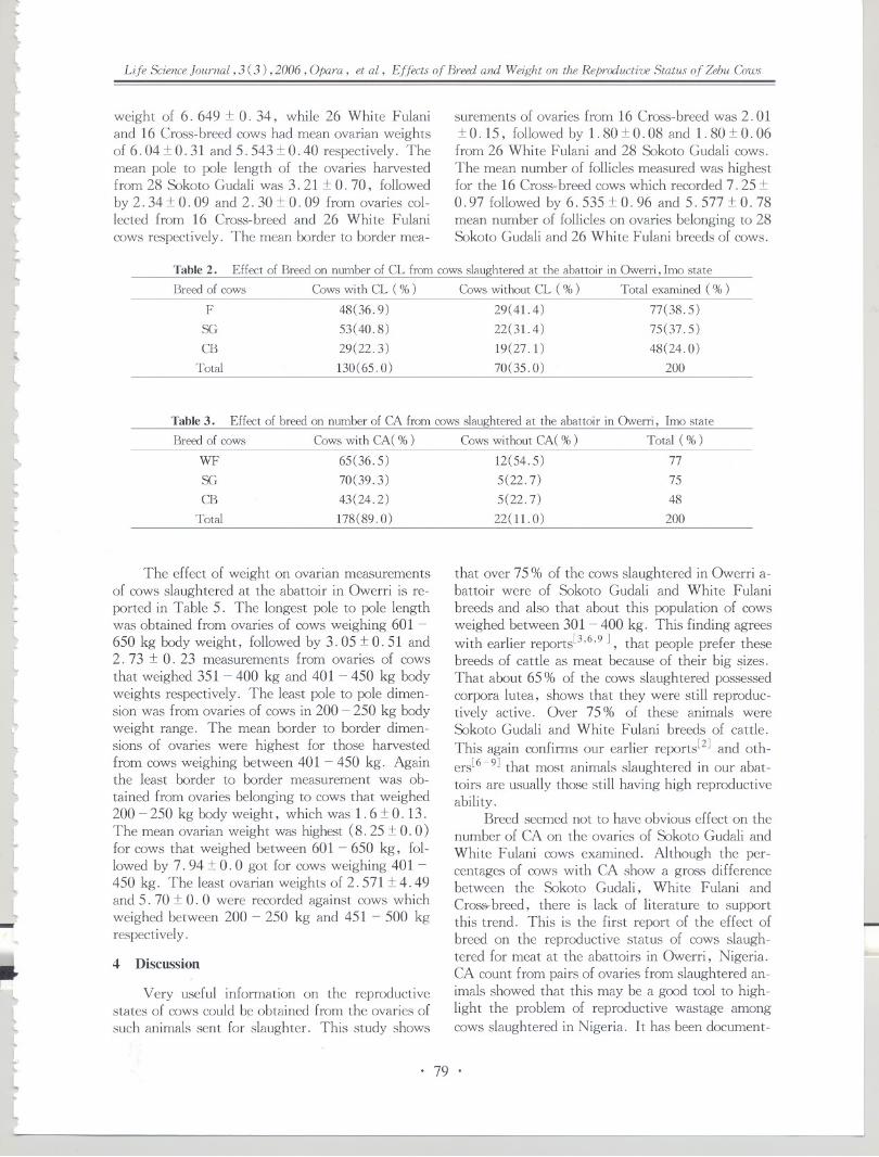
{
~
[
tL.l~\,~~....
llLlll\~lr",.
~l.tl('-lI"
Life Science JournaL, 3 (3) ,2006, Opara, et aL, Effects of Breed and Weight on the Reproductive Status of Zebu Cows
weight of 6.6491: O. 34, while 26 White Fulaniand 16 Cross-breed cows had mean ovarian weightsof 6. 04 1:O.31 and 5. 543 1:0.40 respectively. Themean pole to pole length of the ovaries harvestedfrom 28 Sakoto Gudali was 3. 21 1:O. 70, followedby 2.341: O. 09 and 2.301: O. 09 from ovaries col-lected from 16 Cross-breed and 26 White Fulanicows respectively. The mean border to border mea-
surements of ovaries from 16 Cross-breed was 2.011:O. 15, followed by 1. 80 1:0.08 and 1. 80 1:O.06from 26 White Fulani and 28 Sakoto Gudali cows.The mean number of follicles measured was highestfor the 16 Cross-breed cows which recorded 7.251:0.97 followed by 6. 535 1:O. 96 and 5. 577 1:O. 78mean number of follicles on ovaries belonging to 28Sakoto Gudali and 26 White Fulani breeds of cows.
Table 2. Effect of Breed on number of CL from cows slaughtered at the abattoir in Owerri, lmo state -
Breed of cows Cows with CL (%) Cows without CL (%) Total examined (%)
F 48(36.9) 29(41.4) 77(38.5)
SG 53(40.8) 22(31.4) 75(37.5)
CB 29(22.3) 19(27.1) 48(24.0)
Total 130(65.0) 70( 35.0) 200
Table3. Effect of breed on number of CA from cows slaughtered at the abattoir in Owerri, lmo state
Breed of cows Cows with CA( %) Cowswithout CA( %) Total (%)
VVF 65(36.5) 12(54.5) 77
SG 70(39.3) 5(22.7) 75
CB 43(24.2) 5(22.7) 48
Total 178(89.0) 22(11.0) 200
The effect of weight on ovarian measurementsof cows slaughtered at the abattoir in Owerri is re-ported in Table 5. The longest pole to pole lengthwas obtained from ovaries of cows weighing 601 -
650 kg body weight, followed by 3.05 1:0.51 and2. 73 1: O. 23 measurements from ovaries of cowsthat weighed351 - 400 kg and 401 - 450 kg bodyweights respectively. The least pole to pole dimen-sion was from ovaries of cows in 200 - 250 kg bodyweight range. The mean border to border dimen-sions of ovaries were highest for those harvestedfrom cows weighing between 401 - 450 kg. Againthe least border to border measurement was ob-
tained from ovaries belonging to cows that weighed200 - 250 kg bodyweight, which was 1.6 1:O.13.The mean ovarian weight was highest (8. 25 1:O.0 )for cows that weighed between 601 - 650 kg, fol-lowed by 7.94 :t 0.0 got for cows weighing401 -
450 kg. The least ovarian weights of 2.571 1:4. 49and 5. 70 1:o. 0 were recorded against cows whichweighedbetween 200 - 250 kg and 451 - 500 kgrespectively.
.lll~
~~
lllll-~
L.
l"l
l~II,.
L
4 Discussion
Very useful information on the reproductivestates of cows could be obtained from the ovaries of
such animals sent for slaughter. This study shows
that over 75 % of the cows slaughtered in Owerri a-battoir were of Sakoto Gudali and White Fulani
breeds and also that about this population of cowsweighed between 301 - 400 kg. This finding agreeswith earlier reports[3,6,9 ], that people prefer these
breeds of cattle as meat because of their big ~izes.That about 65 % of the cows slaughtered possessedcorpora lutea, shows that they were still reproduc-tively active. Over 75 % of these animals wereSakoto Gudali and White Fulani breeds of cattle.
This again confirms our earlier reports[2] and oth-
ers[6-9] that most animals slaughtered in our abat-toirs are usually those still having high reproductiveability.
Breed seemed not to have obvious effect on thenumber of CA on the ovaries of Sakoto Gudali and
White Fulani cows examined. Although the per-centages of cows with CA show a gross differencebetween the Sakoto Gudali, White Fulani andCross-breed, there is lack of literature to supportthis trend. This is the first report of the effect ofbreed on the reproductive status of cows slaugh-teredfor meat at the abattoirs in Owerri, Nigeria.CA count from pairs of ovaries from slaughtered an-imals showed that this may be a good tool to high-light the problem of reproductive wastage among
cows slaughtered in Nigeria. It has been document-
..
. 79 .

each animal. Our reports showed that 89 % of thecows had calved while 11 % had not, again depict-
ing the insufficiency of fetal wastage measurementsas a major tool in evaluating reproductive wastageamong slaughtered animals.
>
jJ
J)1
JtIJ
~JjJIj
jI~
J
)})JJ
j~I
jIJ
Life Science Journal, 3 (3) ,2006, Opara, et aI, Effects of Breed and Weight on the Reproductive Status of Zebu Cows
ed[10,11]that previous reproductive history in theform of fibrosed remains of CL of pregnancy (CA)persist for life in majority of cows and this preservesa record of the number of pregnancies undergone by
Table 4. Mean ovarian measurement in cows of different breeds slaughtered at the abattoir in Owerri, lmo state
Breeds of cows ( n = 70)Parameter
WF No. /mean SG No. /mean CB No. /mean
Table 5. Effeet of weight on ovarian measurement of cows slaughtered at the abattoir in Owerri, lmo state
Weight(kg) Pole to pole (em) Border to border (em) Weight of ovaries (g)~--- -- - -200 - 250 1. 86:1: 0.14 1. 60:1: 0.13 2.57:1: 4. 49
251 - 300 2.26:1: O.05 1. 73:1:O.04 6.68:1: O.52
301 - 350 2.44:1: O. 04 1. 87:1: O.04 6.01:1: O. 36
351- 400 3.05:1: O. 51 1. 88:1: O.05 6.05:1: O.29
401-450 2.73:1:0.23 2.20:1:0.32 7.94:1:0.0
451-500 2.50:1:0.0 1.70:1:0.0 5.70:1:0.0
501 - 550
551 - 600
601- 650 3.10:1:0.0 1.80:1:0.0 8.25:1:0.0 -)
J,JJ
j;J
~
Breed of cows slaughtered in Owerri abattoirseems to influence the ovarian measurements. Theovaries harvested from White Fulani and SakotoGudali were heavier than those of the Cross-breedcows. Also, Sakoto Gudali cows had longer pole to
pole measurement of their ovaries than the otherbreeds. The Cross-breed cows had wider border toborder measurements of their ovaries than the purebreeds. The Cross-breed equally had higher numberof follicles than the pure breeds. These findings arein agreement with a similar work carried out amongsmall ruminants in Ogun state, southwestern Nige-ria[S]. The difference that existed between the
Cross- breed and pure breeds might be revealing animprovement in the reproductive traits of the Cross-breed cows as a result of combined genetic make
ups.The ovarian measurements seem to increase as
the cows attained bigger body weights. In the poleto pole measurements, exceptions to this trendwere among the ovaries collected from cows withinthe weight of 401 - 500 kg. The cows seemed tohave ovaries which were wider than they were
long. But the ovaries belonging to cows thatweighed 451 - 500 kg had shorter border to bordermeasurements. The difference in these orders maybe attributed to the presence of some atrophied o-varies in the group encountered during this study.Weight also seemed to increase the weight of the o-varies. Exceptions here were ovaries of cows thatweighed451 - 500 kg, this again is explained bythe presence of atrophied ovaries as supported byArthur[lO] .
1
J
J
J-
5 Conclusion
Our studies revealed that about 65 % of differ-
ent breeds of cows slaughtered at Owerri abattoirfor meat, irrespective of their weights were still intheir active reproductive states but are being sentfor slaughter for reasons other than reproductive in-activity. Breeds seemed to positively influence thereproductive performance of our indigenous cows,however crCA"'"breeding also slightly improved thistrait. Ovarian measurements increased with the
body weight of the cows. Age[2] and body weight
.I...
J
J"J
jJ
J
. 80 .
-- -
Wt. of ovary(g) 26(6.04:1: o. 31) 28(6.649:1: o. 34) 16(5.543:1: O.40)
Pole to pole( em) 26(2.30:1: O. 09) 28(3. 2l:t O.70) 16(2.34:1: O. 09)
Border to border( em) 26 ( 1. 80 :I:O.08) 28(1.80:1: O. 06) 16(2.01:1: 0.15)
No. of follicles 26 (5. 577 :I:O. 78) 28(6.535:1: O. 96) 16(7.25:1: O.97)

klI
l'lr..
l~
~
tltL~
~lllt
Life ScienceJournal, 3 (3) ,2006 , Opara, et al, Effects of Breed and Weight on the Reproductive Status of Zebu GJws
of animals sent for slaughter could be a future reli-able index to ascertain the reproductive status ofsuch animals before they are finally passed forslaughter.
Correspondence to:Opara MNTropical Animal Health and Production ResearchGroupDepartment of Animal Science and TechnologyFederal University of TechnologyP. M. B. 1526, Owerri, Nigeria.Email: [email protected]
References
1. Ogunyemi G. Meat hygiene in Nigeria. The Vet. Sur-geon 1982: 7: 23-9.
2. Opara MN, Nwachukwu EC, Okoli IC. Ovarian activi-ties in cows of different ages slaughtered in Imo state,Nigeria. Asian Journal of Information Technology 2005:4(7): 640-3.
3. Aladi NO. Current trends in the production, handlingand sales of meat in Nigeria. B. Agric. Tech. Project Re-port, Federal University of Technology Owerri, Nigeria1999.
4.0keudo NJ. Emperical studies of the living conditions ofdomestic animals in Nigeria. In: Studies in sustainable a-griculture and Animal Science in sub-Saharan Africa.
I'"t
~l.l"-
t~
~l...lt,
lllL
~
tllL~-
~~l....
l
~
l..
I,
Malu UC, Gottwald F (Eds. ), Frankfurt, Peter Lang2004.
5. Abiola SS, Onwuka CFI. Reproductive performance ofWest African dwarf sheep and goats at villagelevelsin 0-gun state, Nigeria. Nig J Anim Prod 1998: 28: 79 - 82.
6. Matthew T, Adeola m, Matthew A. The recovery offetuses from slaughtered cattle in the abattoirs in Nigeriaand its economic implication to the nation: five yearstudy (1975 -1980). Nig Vet Med Assoc, 19th Ann.Conf. Book of abstracts, 1982, 45.
7.0yekunle MA, Olubanjo Fasina OF. Fetal wastage in a-battoirs and its implication: solution report for Ogunstate, Nigeria. Nig J Anim Prod 1992: 19:57 - 63.
8.Wekhe SN, Berepubo NA, Ekpenyong CEo Prevalenceand implication of inadvertent slaughtering of pregnantruminants in Port-Harcourt, Nigeria. Delta Agric 1992;1: 43 - 5.
9.0koli IC, Nwokeocha JR, Herbert U, Anyanwu GA.Analysis of meat inspection records for Imo state, Nigeria(11): Assessment of the magnitude of fetal wastage instate abattoirs. Trop Anim Prod Invest 2001; 4: 29 -
35.
10. Arthur GH, Moakes DE, Pearson H. Veterinary Repro-duction and Obstetrics. 5th edn, Bailliere Tindall, Lon-don 1982.
11. Dawson FLM. Observations on the corpora albicantes inthe ovaries of normal and infertile dairy cows. J Agric Sci1958; 50: 322-30.
Received March 16, 2006
..
. 81 .

J
ij
JJ
!
jJ
Li[e Sdence lournal, 3 (3) ,2006, Peng, el al, Gaseou.' f,mnaldehy:ie-induced DNA-prolein Crmslinks
Gaseous Formaldehyde-induced DNA-protein
Crosslinks in Liver, Kidney and Testicle,of Kunming Mice
Guangyin Peng, Xu Yang, Wei Zhao, ]unjun Sun, Yi Cao, Qian Xu, ]unlin Yuan, Shumao Ding
College of Life Science, Central China Normal University, Wuhan 430079, acina
Abstract,To explore the effect of distant-site toxicity, this study detected the amount of DNA-protein crosslinks(DPC) with KCI-SDS assay in liver, testide and kidney of the purebred Kunming mice treated with gaseousformaldehyde (FA). The ,,;sults showed that gaseous FA couldn't cause DPC or could cause few DPC at the lowerconcentration (0.5 mg/m3), while could cause significanc DPC at higher concentrations (1.0 mg/ms and 3.0 mg/m3) (P < 0.0 I). The results snggested that FA could induce DPC in the distant organs (liver, testicle and kidney)of mice at rdatively high concentrations, which indicated that FA might induce distant-sile toxicity. [Life ScienceJournal. 2006;3(3),82-87J ([&SN, 1097-8135).
I
1
Keywords, formaldehyde; dislant-site toxicity; DNA-protein cnv%links; KCI-SDS ",'ieY
Abbreviations, DPC, DNA-protein cro"linb; FA, formaldehyde; DSB,DNA stmnd breaks; lARC, InternationalAgency for Resea,eh; NO, nitric oxide; SDS,sodium dodccylsulfate
1 Introduction
-
Fonnaldehyde (FA) is a colorless, highly
flammable gas at mnbient temperature, which is
prescnt in the environment from both natural pro-cesses and manmade sources. As a major industrialchemical, it can be found in construction materials,
rcsins, textiles, leather goods, paper, and con-
sumer products. At the samc time, as a naturallyoccurring biological compound, it is present in tis-sucs, cells, and body fluids. In addition, some rc-
views have reported that FA is a genotoxic and mu-
tagenic compound and has been classificd as a hu-
man carcinogen (class AI) by IARC (International
Agency for Research) recently!]]. Due to its exten-sive sources, high level, long-tenn and high toxici-
ty, it is important to study FA toxic effect andmechanism.
Studies have shown that FA has extensive
genotoxicity, including DNA-protein crosshnks(DPC) and DNA strand breaks (DSB)[2,3,4]. DPC
is the primary genotoxic effect of FA, which isfonned through covalent bond where deoxyribonu-cleic acid is linked to an endogenous protein. Re-gions of DNA that are covalently linked with pro-tcin are typically considered to be non-functionaland can block normal functions of the nuclear ma-
trix, such as replication and transcription, and canfonn the foci for double strand breaks, which canlead to chromosomal aberrations and sister-chro-matid exchanges. Furthennore high amounts ofDPC can cause the expression of critical regulatory
~
genes change. For the significance of DPC, thecontent of it may be of value in the assessment ofFA-induced gcnotoxicity. Casanova et al haveproved that acutc FA inhalation could induce DPCin nasal mucosa of rats and monkeys by experi-
ments in -uivo[5,6.7] Kuykendall cxposed rat nasalepithclial cells to FA and found DPC content in-creased remarkably above the concentration of 100
!,mollL[4] In addition, in our laboratory, Liu etal exposed human peripheral blood lymphocytes toFA and found that there was no significant'differ-cnce in the DPC coefficient betwecn the groupstreated with 5 "M, 25 I'M FA and the controlgroup, while there was a significant difference be-tween the groups treated with 125 fJM, 625 fJMFA and thc control group (P < 0.01). The resultsshowed that FA could not induce DPC at low con-
centrations but could induce DPC significantly at
high concentrations[8].These studies above either chose organs located
at exposure site, or were conducted in vitro to es-timate FA toxicity. Few of them proved whetherFA could induce distant-site toxicity. At present,there are still controversies on whether inhaled FAcan induce distant-site toxicity, mainly because of
the rapid metabolism and removal of FA in vi-vo[9,1O] Recently, Franks developed a mathemati-cal model for estimating the absorption andmetabolism of FA by humans. This model was usedto analyze the increase of FA concentration in theblood after exposurc to FA, and results showedthat the increase was insignificant, indicating FA
!.J,JIJJ
J
J
J
)
rJJJJ
l
..~-!)
jJ
. 82 .

p~
l
~~\,l\l~l~l,lor
l~
~.l.
tl,l...
t~
tl
Life Science Journal, 3 (3) ,2006, Peng, et al, Gaseous Fornwldehyde-induced DNA-protein Crosslinks
could be removed rapidly in the blood[ll]. This isconsistent with previous reports declaring that in-
haled FA could be removed rapidly[9,1O]. However,some work also support FA has the distant-site tox-icity. For example, Shaham et al examined DPCin peripheral blood lymphocytes of workers exposedto FA, and they found a significantly positive cor-relation between FA and DPC concentrations. In
their early work, they also found a linear relation-ship between years of FA exposure and the amount
of DPC[12,13]. Epidemiological studies on workersalso suggested that the possible relations between
FA exposures and leukemia[14,15]. These authorsproposed that although inhaled FA was metabolizedrapidly at contact sites, FA might be transportedby unknown mechanism and cause cancers likeleukemia subsequently.
To investigate FA-induced genotoxicity in thedistant organs such as liver, kidney and testicle ofmice, in this study, the content of FA-inducedDPC in the organs of mice was detected by KCI-SDS assay. At the same time, the results will givemore information to understand whether FA has
distant-site toxicity. Additionally, this study maybe helpful to understand the genotoxicity and thecarcinogenicity of FA comprehensively and system-atically, providing a scientific basis for constitutingsecure professional concentration standard of FA.
"LL.L
L
~~~r(lL
ll
2 Materials and Methods
2. 1 Reagents and apparatus10% formalin, calf thymus DNA and fluores-
cence dye Hoechst 33258 were purchased from Sig-ma(USA). Sodium dodccyl sulfate (SDS) and pro-teinase K were purchased from Merck ( Germany) .PBS ( without ci+ and Mi+ ), trypan blue solu-tion of 0.4 % and other chemicals were of analyticalgrades.
A WH-2 type environmental chamber (WH-2,Yu-Xin Inc, China) as FA generator, a 4160 typedigital electrochemical FA analyzer (Interscan Inc,USA), glass low inhalation chamber, temperaturecentrifuge (Eppendof-5415R), fluorescence spec-trophotometer (RF -4500 , HITACHI, Japan) wereused in the experiments.2.2 Animal
24 male Kunming mice were supplied by theExperimental Animal Center of Hubei Province,China. Animals' weight were 19:t 1 g.2.3 Mice exposure to gaseous FA
24 male Kunming mice were divided randomlyinto 4 testing groups (n = 6 each) and were ex-posed to different concentrations of FA: 0, O. 5,1. 0 and 3. 0 mg/m3. The inhaled groups were ex-
-..
~~l""
~
llIt.
l
posed to gaseous FA for 72 hours continuously.During the exposure the mice were allowed to drinkand eat twice at fixed time each day . FA inhaledgroups were placed into glass inhalation chambers.The chamber temperature was 23°C :t O. 5 °c , andthe humidity was 45 % :t 0.5 %, and the gas fluxwas 1 :t O. 01 L/min. A 4160 type digital electro-chemical analyzer was used to measure the concen-trations of gaseous FA.2. 4 Preparation of cells
Mice were executed immediately after expo-sure and livers, kidneys and testicle were obtained.The tissues were minced with scissors and homoge-nized with seven to eight strokes in PBS (pH 7. 5) .The homogenate was filtered through four layers ofcheesecloth and then the cells were collected bycentrifugation at 1500 rpm for 5 min. After re-sus-pending, the cell density was regulated with PBSand cell viability was analyzed with the method oftrypan blue exclusion. Cell viability was above
95 % and density was 105 - 106 cells per m!.2.5 KCI-SDSassay
In this study the KCI-SDS assay was based onZhitkovich and Chakrabarti methods with somemodification to detect FA-induced DPC[16,17]. Cellswere harvested by centrifugation at 1500 rpm for 5min. The cells were resuspended in O.5 ml of PBS,pH 7.5, followed by lysis with 0.5 ml of 2 % SDSsolution with gentle vortexing. The lysate solutionwas heated at 65°C for 10 min and then 0.1 mlofpH 7. 4 and 10 mM Tris-HCI containing 2.5 MKCI was added, followed by passing the resultantmixture six times through a 1 ml polypropylenepipette tip to favor shearing of DNA for a uniformlength. Since SDS binds tightly to protein but notto DNA, the free protein and protein-DNA com-plexes are precipitated with added SDS while freeDNA is remained in the supernatant. The SDS-KCIprecipitate (containing the protein and DNA-pro-tein crosslinks complexes) was formed by placingthe samples in ice for 5 min and was then collectedby centrifugation at 10, 000 rpm for 5 min. Thesupernatants containing the unbound fraction ofDNA were collected in different labeled tubes. Thepellets (containing DPC) were washed three timesby resuspending in 1 ml washing buffer (0. 1 MKCl, 0.1 mM EDTA, and 20 mM Tris-HCI, pH7. 4) followed by heating at 65 ° C for 10 min,chilling in ice for 5 min, and centrifugating as de-scribed above. The latter supernatants from eachwash were added into the previous one with un-bound fractions of DNA. The final pellet was re-suspended in 1 ml proteinase K solution (0.2 mg/ml soluble in a wash buffer) and digested for 3 h at
..iI
. 83 .
I Ir-4
Life Science Journal, 3 (3) ,2006, Peng, et al, Gaseous Formaldehyde-induced DNA-protein Crosslinks
..
ing that as the inhaled gaseous FA concentrationsincreased, the DPC levels ascended.
I1~50 0 C. The resultant mixture was centrifuged at
12,000 rpm for 10 min and the supernatant wascollected (the supernatant contained the DNA pre-viously involved in DNA-protein crosslinks). 1 mlof either the supernatant containing the unboundfraction of DNA or the supernatant containing theDNA previously involved in DNA-protein crosslinkswas then mixed with 1 ml freshly prepared fluores-cent dye Hoechst 33258 (400 ng/ml soluble in 20mM Tris-HCl), and then the tubes were allowed tostand for 30 min in the dark[18]. The sample fluo-rescence was measured using a RF-4500 fluores-cence spectrofluorimeter with excitation wavelength350 nm and emission wavelength 450 nm. TheDNA contents of the samples were determinedquantitatively through a corresponding DNA stan-dard curve (as Figure 1 shows and regression equa-tion is y = 2. 4423 + O. 0028x, r2 = 0.9974) gen-erated from a set of calf thymus DNA. The DPCcoefficient was measured as a ratio of the percent-age of the DNA involved in DPC over the percent-age of the DNA involved in DPC plus unbound frac-tion of DNA.
1000 2000 3000 4000 5000
DN A (ng/ml)
Figure 1. The standard curve of DNA concentration
2. 6 Statistical analysis-Results were analyzedby softwareOrigin 6. O.
Student's t-test was applied to evaluate the signifi-cance of the differences in the results between treat-
ed and control groups. A level of P < 0.05 was de-fined to be statistically significant.
3 Results
3.1 Effect of FA-iuduced OPC iu the livers of- miceFigure 2 showed the effect of gaseous FA ex-
posure on DPC levels in livers of mice. There wasno significant difference in DPC coefficient between0.5 mg/m3 FA inhaled group and 0 mg/m3 controlgroup. However, the DPC levels at 1.0 mg/m3 and3.0 mg/m3 groups were significantly (P < O. 01 )higher than that in the control group, demonstrat-
~
I
..
0.20 **
I
I~ I.I; !I. .J I
1 It I
';:; 0.1'":~ 0.121:::
~ 0.08(J
~ I
II
J
CJ 0.0~ .0 0.00
a O.5 1. a 3. a
Formaldehyde concentration (mg/m3),;
Figure 2. DPC formation in mice liver cells at different FA con-centrations
* * :P < 0.01, compared with control group
iJt
J
J~3.2 Effect of FA-induced OPC in the kidneys of
mice
Figure 3 showed the effect of gaseous FA onDPC levels in kidneys of mice. There was signifi-cant difference in the DPC levels between controlgroup and FA inhaled groups ( P < O. 01). The re-sults indicated that the DPC levels ascended withthe increasing of inhaled gaseous FA concentra-tions.
4
,.I
~
~I
J
,J
J
oJ
J0.16
0.14
<;:; 0.12'".;; 0.106 0.08'"0 0.06(J
u 0.04P-O 0.02
0.00
**..
..
1(I~
-I
I~
I.J
1
of
0 0.5 1.0 3.0
Formaldehyde concentration (mg/m3)
Figure 3. DPC formation in mice kidney cells at different FA con-centrations
* * :P<O.Ol, compared with control group
,;
..
3.3 )~
I
Effect of FA-induced OPC in the testicles ofmice
Figure 4 showed that the DPC coefficient of0.5 mg/m3 FA-treated group was significantlyhigher than that of control group (P < 0.05), andthere were significant difference in DPC coefficientbetween 1. 0 mg/m3, 3. 0 mg/m3 FA-treatedgroups and the control group (P<O.O1). It indi-cated that there was a clearly dose-dependent rela-
i
~oJ...
J. 84 .I~.,/
~

..
~..
,\I.lr"10....
lL
~
~
\l~~
Life Science Journal, 3 (3) , 2{)O6, Peng, et al, Gaseous Formaldehyie-induced DNA-protein Crosslinks
tionship between the DPC coefficient and the con-centrations of FA.
**0.12
~ 0.10I=i(!)
'Q 0.08
S 6(!) 0.00() 0.04Up., 0.020
0.00
0 0.5 1.0 3.0 3Formaldehyde concentration (mg/m )
Figure 4. DPC fonnation in mice testicle cells at different FA con-centrations* :P<O.O5; * * : P<O.Ol, compared with control group
I.....
\I"\
4 Discussion
4. 1 Parameters of chamber and the concentra-
tions of gaseous FAThe concentrations of gaseous FA generated
by small environmental chamber were measured 3times per day for 3 days. The measurement results(0.03:t O. 03 mg/m3, 0.49:t O. 03 mg/m3, 1. 03
:t 0.04 mg/m3 and 3.03 :t 0.08 mg/m3) were veryclose to the anticipative concentrations (0 mg/m3,
0.5 mg/m3, 1. 0 mg/m3 and 3.0 mg/m3). It indi-cated that the gaseous FA from chamber emissionwas quite stable and reliable.
In the previous studies, gaseous FA was gen-erated from paraformaldehyde. Then high level ofFA was diluted with clean, filtered air to achieve
the desired gas concentrations[ 19J. In this way, itwas very difficult to control the temperature (T),the humidity (RH) and the gas flux (F) of gaseousFA. It is well-known that the alterations of tem-
perature and humidity could affect the experimentalresults remarkably. The gas flux of FA is also im-portant to the inhalational quality of mice. In thepresent study, different concentrations of gaseousFA were generated by small environmental chamberwith formalin. In this method, the temperature,the humidity and the gas flux could be set by thechamber before exposure. The parameters of thechamber will be very stable after 3 hours of opera-tion. The results were showed in Figure 5. Thus,this experimental method has great improvementfor better reliability and higher reproducibility.
4.2 Comparison of the effect of FA7induced DPCin the three different organs of mice
According to the effect of FA-induced DPC inthe three different organs of mice, we could con-clude that FA could induce DPC with a dose-depen-
'It......
l\.~....
tl
~lt'~..,'-I...
lll~.lc
~
l
ll-
~t
f~
r..
dent relationship between the DPC coefficient andthe concentrations of FA. We also found that the
DPC level of 3.0 mg/m3 FA inhaled groups of liverwas the highest and that of testicle was the lowest,which indicated that gaseous-FA-induced genotoxi-city in the liver of mice was the most severe, thenwas kidney and the least was testicle.
'I j !"l
._-
" """"""';00""""""""""
Figure 5. The parameters of the small environmental chamber
4.3 The mechanism of the FA-induced DPC
DPC is relatively permanent in the cells. Dueto the poor repair capacity, DNA-protein complexesmay be present during DNA replication and possi-bly cause a loss and inactivity of the importantgenes such as tumor suppressor genes, and may beresponsible for tumor formation. For the elec-trophilicity of carbonyl and smaller steric hin-drance , FA is apt to form DPC. Initially, a hy-droxymethyl intermediate is formed by the additionof FA to a primary amine of either DNA or protein.The hydroxymethyl group then condenses with asecond primary amine to produce a methylenebridge between DNA and protein. The form ofDPC can be expressed as histone-NH-CHz-NH-DNA. Additionally, as a inhibitor to antioxidases,FA could induce the. formation of DPC indirectly,for example, by increasing the content of DPC re-sulted from the depletion of GSH and the inhibitionof glutathioneperoxidase and superoxide dismutasewhich can clear hydroxy radical and oxygen-derivedfree radicals[6, 13,20,21] .4. 4 FA and distant-site toxicity
Whether the FA does have the distant-site
toxicity and what is the mechanism still is un-known. Thrasher proposed that rapid removal ofFA was insufficient to support that FA could notinduce distant -site toxicity, because FA might formadducts with amino acid or other biological moleculein blood, and the adducts could be sent to remote
..
. 85 .

inhaled fonnaldehyde to DNA in the nasal mucosa of Fis-cher 344 rats: analysis of fonnaldehyde and DNA byhigh-perfonnance liquid chromatography and provisionalphannacokinetic interpretation. Fundam Appl Toxicol1989; 12: 397-417.
6. Casanova M, Morgan KT, Gross EA. DNA-proteincross-links and cell replication at specific sites in the noseof F344 rats exposed subchronically to fonnaldehyde.FundamAppl Toxicol1994; 23: 525~36.
7. Casanova M, Morgan KT, Steinhagen WH, et al. Co-valent binding of inhaled fonnaldehyde to DNA in therespiratory tract of Rhesus monkeys: phannacokinetics,rat-to-monkey interspecies scaling, and extrapolation toman. FundamAppl ToxicoI1991;17: 403-28.
8. Liu YS, Lu ZS, Yang JW, et al. Quantification studyon the DNA-protein in crosslinks of human blood lym-phocytes induced by fonnaldehyde. Hubei journal of pre-ventive medicine 2004; 15: 4 - 7.
9. Heck HD, Casanova M. The implausibility of leukemiainduction by fonnaldehyde: a critical review of the bio-logical evidence on distant-site toxicity. Regul ToxicolPhannacol2004; 40: 92- 106.
10. Collins JJ, Ness R, Tyl RW. A review of adverse preg-nancy outcomes and fonnaldehyde exposure in humanand animal studies. Regul Toxicol Phannacol 2001; 34:17 - 34.
11. Franks SJ. A mathematical model for the absorption andmetabolism of fonnaldehyde vapour by humans. Toxicol-ogy and Applied Phannacology 2005; 206: 309 - 20.
12. Shaham ], Bomstein Y, Gurvich R. DNA-proteincrosslinks and p53 protein expression in relation to occu-pational exposure to fonnaldehyde. Occup Environ Med2003; 60: 403-9.
13. Shaham J, Bomstein Y, MeltzerA, et al. DNA-proteincrosslinks, a biomarker of exposure to fonnaldehyde-invitro and in vivo studies. Carcinogenesis 1996; 17: 121-5.
14. Coggon D, Harris EC, Poole J. Extended follow-up of acohort of British chemical workers exposed to fonnalde-hyde. J Natl Cancer Inst 2003; 95: 1608 - 15.
15. Hauptmann M, Lubin JH, Stewart PA. Mortality fromlymphohematopoietic malignancies among workers infonnaldehyde industries. J Natl Cancer Inst 2003; 95:1615 - 23.
16. Zhitkovich A, Costa M. A simple, sensitive assay to de-tect DNA-protein crosslinks in intact cells and in vivo.Carcinogenesis1992; 13: 1485 - 9.
17. Chakrabarti SK, Bai CJ, Subramanian KS. DNA- proteincrosslinks induced by nickel compounds inisolated rat re-nal cortical cells and its antagonism by specific aminoacids and magnesium ion. Toxicol Appl Phamacoll999;154: 245 - 55.
18. Cesarone CF, Bolognesi C, Santi L. Improved microfluo-rometric DNA detennination in biological materical using33258 Hoechst. Anal Biochem 1979; 100: 188 - 97.
19. Fujimaki H, Kurokawa Y, Kunugita N, et al. Differen-tial immunogenic and neurogenic inflammatory responsesin an allergic mouse model exposed to low levels offonnaldehyde. Toxicology 2004; 197: 1-13.
20. Speit G, Schutz P, Merk O. Induction and repair offonnaldehyde-induced DNA-protein crosslinks in repair-deficient human cell lines. Mutagenesis 2000; 15: 85-
J-iJ)JjJ)..II
i
JJ
Life Science Journal, 3 (3) ,2006 ,Peng, et al, Gaseous Formaldehyde-induced DNA-protein Crosslinks
sites by blood circular system and generate FA a-gain, so these adducts were responsible for tumors
caused by FA[22]. In fact, as biochemical active asnitric oxide (NO), it is also able to induce physio-logical and toxicological effects at distant sites,
probably by forming adducts[23,24]. NO is active tosulfhydryl and amino groups, and interestingly, re-cent work showed that it was also the cause for
FA[2S]. Therefore, it would be interesting to inves-tigate which kind of adduct formed in blood afterexposure to FA, since it had been proposed that theamount of free FA in blood was not altered after
exposure.In the present study, significant DPC could be
detected in the three organs of liver, kidney andtesticle when the mice were exposed to gaseous FAof higher concentrations, which evidently indicatedthat FA might induce distant-site toxicity.
5 Conclusions
In this study, KCl-SDS assay was applied todetect the amount of DPC in liver, kidney and tes-ticle of mice. According to the results, we couldconclude that FA could induce DPC significantly atthe higher concentrations, the results also indicatedthat FA might induce distant-site toxicity.
AcknowledgmentsThis work is supported by China National Sci-
ence Foundation (project coded 30570799).
Correspondence to:Xu YangCollege of Life SciencesHuazhong Normal UniversityNo. 152, Luo-Yu Road, Wuhan 430079,Email: [email protected]
China
~
References
1. International Agency for Research on Cancer. 2004.IARC classifies fonnaldehyde as carcinogenic to humans.2004- 06 - 15, http://www.iarc.fr/ENG/Press- Re-leases/archives/pr153a, html.
2. Bunde RL, Jarvi E], Rosentreter JJ. A piezoelectricmethod for monitoring fonnaldehyde induced crosslinkfonnation between poly-lysine and poly-deoxyguanosine.Talanta 2000; 51: 159 ~ 71.
3. Conaway ce, Whysner ], Vema LK. Fonnaldehydemechanistic data and risk assessment: endogenous pro-tection from DNA adduct fonnation. Phannacol Ther
1996; 71: 29-55.4. Kuykendall JR, Trela BA, Bogdanffy MS. DNA-protein
crosslink fonnation in rat nasal epithelial cells by hexam-ethylphosphoramide and its correlation with fonnalde-hyde production. Mutat Res 1995; 343: 209 - 18.
5. Casanova M, Deyo DF, Heck HD. Covalent binding of
Tt
~I
JI
,JI,J;,)
~I
()~
J.}JJJI
)II
.J
JJI
~
I,.I
I...I
JJ}..
1
t'"
JI
. 86 .
JJ
~
)
J

I~'"
10,
.\r...
...
t.
\.
.....\
I....
~
l~-
\-
...
r'10-..
..
.."-~
\.
"
..'"~
\.,
L
"..
~C
'"
l,
l\.--
~L.I.....L..
I\-
r
Life ScienceJournal, 3 (3) ,2006, Peng, et ai, GasoousFormaldehyde-inducedDNA-protein Crosslinks
90.21. Takashima H, BoerkoelCF, JohnJ. Mutation of IDP1,
encodinga topoiromerase I-dependent DNA damage re-pair enzyme, in spinocerebellarataxia with axonal neu-ropathy. Nature Genet 2002; 32: 267 -72.
22. Thrasher JD, Kilburn KH. Embryo toxicity and terato-genicity of formaldehyde. Arch Environ Health 2001;56: 300 -11.
23. Hogg N. The biochemistryand physiologyof S-nitroroth-iols. Annu Rev Pharmacol Toxicol 2002; 42: 585-
600.24. Weinberger B, Laskin DL, Heck DE, et al. The toxi-
cologyof inhaled nitric oxide. Toxicol Sci2001; 59: 5-16.
25. Metz B, Kersten GF, Hoogerhout P, et al. Identifica-tion of formaldehyde-inducedmodifications in proteins.BioiChem2004; 279: 6235- 43.
Received June 5, 2006
L
. 87 .
--

China
};~.1IJJ))
.~
j.~
-(I
1I
JI
~
;IJ1
))JJ
i~
()~
I
)
Life Science Journal, 3 (3) ,2006, Chen, et al, Microarchitecture Fabrication Process of the Artificial Bone
Microarchi tecture Fabrication Process
of the Artificial Bone
Zhongzhong Chen1, Cheng Li1, Zhiqiang Jiani, Zhiying Lu03
1. College of Mechanical Engineering, Zhengzhpu University, Zhengzhou, Henan 450001 ,
2. Department of Mechanical Engineering, Zhengzhou Institute of Aeronautics,
Zhengzhou, Henan 450015, China
3. The Second Internal Hospital of Zhengzhou University, Zhengzhou University,
Zhengzhou, Henan 450002, China
Abstract: By using bionic modeling and rapid prototyping technology, a novel system based on air-pressure jet solidi-fication (AJS) technique is developed to fabricate porous microarchitecture of the artificial bone. Acting as transi-tional carrier of the bone-implant, the porous scaffolds formed could provide proper porosity and interconnections forthe growth of bone tissue and nutrient transport. This approach is better than traditional fabrication processes, be-cause the latter methods cannot fabricate a microarchitecture with spatial bending micropores so as to satisfy biologi-cal requirements. [Life Science Journal. 2006 ;3(3) : 88 - 93] (ISSN: 1097 - 8135).
Keywords: rapid prototyping; scaffolds; air-pressure jet solidification
Abbreviations: AJS: air-pressure jet solidification; DS: denatured sucrose; FDM: fused deposition modeling; RP:rapid prototyping
1 Introduction
J
In the view of tissue engineering, the osteo- re-placement tissue must have proper aperture andporosity for bone repair so as to accelerate bone re-generation. Moreover, it must serve as three-di-mensional (3-D) template for initial cell attachment
and subsequent tissue regeneration[1-3]. Conven-tional wisdom states that scaffolds should be de-
signed to match healthy tissue morphological char-acteristics and have an interconnected pore networkfor cell migration and nutrient transport. Further-more, the simulation design of the inter-connectivearchitecture has a decisive effect on the activation ofbone substitute. The traditional methods to fabri-
cate scaffolds include polymer foaming technique,particulate-leaching, solid-liquid phase separation,textile technique and extrusion process, etc[ 4 - 10] .But with these methods, the bionic architecturesimilar in morphological characteristics to the inter-microstructure of the natural bone could not be en-sured, which is essential to vascularization and tis-
sue regeneration.Based on the building principle of fused deposi-
tion modeling (FDM) in rapid prototyping (RP)technology, a new forming technique - AJS systemis developed, which can build up the bone scaffoldswith a novel fabrication material. The formed scaf-
~
fold possesses an exterior mould exactly coincidentwith the replaced bone and interior porous architec-ture simulating the microstructure of the naturalbone tissue. By filling self-setting calcium phos-phate cement (CPC, a kind of biomaterial for bonesubstitute) and rhBMP (recombinant human bonemorphogentic protein, a kind of growth factor) in-to the scaffolds, the fabricated scaffolds are dis-solved with the solidification of CPC, and then thesimulated interior microstructure is formed.
~J
/I
pi
I~I
..I
;J
j))
j
J-
2 Materials and Methods
2. 1 Fabrication materialA kind of fabrication material- denatured su-
crose(DS) is developed to form the 3-D scaffolds.As a forming material, DS has proper plasticity,ductility and viscosity, so that the 3-D scaffolds canbe built up and will not distort after solidification.Equally important, as an implantable biomaterial,DS has some proper histological performance andcan be served as a biomaterial stabilizer of protein tomaintain the activation of rhEMP.
2.2 AJS systemAJS system is designed to fabricate the artifi-
cial bone based on the layer-by-layer manufacturing
principle of RP[11]. In the process, refined DS isfed into two controllable jets and melted into a se-mi-molten state by a heating system. Each jet has a
,)
)
j
j
1
j
. 88 .

"
~\
~Life Science Journal, 3 (3) ,2006, Chen, et al, Microarchitecture Fabrication Process of the Artificial Bone
r",\\~~
~J\."...,~
~l
small nozzle on the tip, the diameter of the nozzle is0.2 mm. Both jets are connected to an air compres-sor. Fine DS filament can be expressed through thenozzle by applying compressed air. Under the con-trol of a computer, the on-off operation of the pres-sure air can be controlled bv electromagnetic
I'P~,w
'1\\,
~
t.~
,~..
\'-{,f
tlt~
I
~
~l\:
('
valves, then a 3-D working platform moves accord-ing to the processed data generated from the bionicmodelling. So the filament is deposited layer-by-layer, and finally, a 3-D part is built in the areasdefined by CAD model (Figure 1) .
Figure 1. AJS system and its key part-two pressure jets
2.3 Diameter of filament
AJS system has many technical parameters,such as temperature of jet, squeezing and scanningvelocityetc, all of these are the important processvariables which determine the quality of the part.Due to the scaffolds fabricated, the key index is thediameter of filament in the fabrication process,which determine the configuration of the microar-chitecture in the interior of artificial bone.
A calculation modeling is built up according tothe matching relation of squeezing and scanning ve-locityof the jet. DS is a kind of thermoplastic ma-teriaL with a certain viscoelasticity. According to
L
~
)
~...
~
tJ\.
w
the viscoelastic and rheologic theory, it will keep ina semi-molten state in the whole fabrication pro-cess. Through double actions of air pressure andthe traction force of the solidified layer, the shear-ing stress in DS would be occurred. Therefore, thetensile process is belong to non-Newtonia fluid andstretching flow. According to the theory analysisand expriments result, after the filament expressedfrom jet, the cross-section of filament can be con-cluded as Figure 2 ( a) in the tensile region, whichis a rectangular CFGH (ill) in the center and twoconics at both ends.
B'lh
(b) L.
r
bK
(a)
According to the conic in Figure 2 ( b), line
Figure 2. Illustration of solidification filament model in cross-section
KC, KM are tangent to arc MBC, given MC= h,
. 89 .

A I =A sector(OFEC) - A triangle( OPC)
= ~ [J L2;h2r"2a-L~h
~llJL2+ h2 1
2"2 h - L"h2 2 VL2+h2 4
(since sin a~a, lal«O
=A(VL2+h2_L)4A section of filament = A rectangle ill + 2A I
= L " h + 2" A (V L2 + h 2 - L)24
=A(JL2+h2+L)2
J4!
JJ(
i?J~11
Life Science Journal, 3 (3) ,2006, aen, et al, Microarchitecture Fabrication Process of the Artificial Bone
~
KB = band BD = a, p is defined as a I( a + b ) ,from spline geometrics, it can be educed that thecontour of conic, namely the type of arc MBC canbe determined by p, and a serial of conclusionscould be derived as
0 0.05< p<O. 5, the conic is ellipse;2) p = O.5, the conic is parabola;3) O.5< p < O.95, the conic is hyperbola.For easy to analysis and calculation, Figure 2
is simplified as follows:a) While the squeezing velocity Vp is slow,
namely while p<O. 3, the cross-section contour offilament can be simplified as rectangle ill in Figure2(a). Due to the filament flow in unit time is equalto the forming volume required, an expressionshould be derived as
V "1!d2/4= V "L"hp s
W= L = 1!d2V/J.4hVs
Where h is defined as the height between the noz-zle and fabricated layer, d is the diameter of noz-zle, Vp is the squeezing velocity, Vs is the scan-ning velocity of jet, L is the width of rectangle ill,W is the diameter of filament.
b) The area of conic must be taken into ac-count while Vp is increased to a certainty. If thecontour of conic is difficult to describe, arc FECcan be used for approximate the conic (Figure 2(a». It belongs to the circle which include thefour border point of rectangle ill, where 0 is de-fined as center of circle, segment AC, AF are tan-gent to arc CEP. Given FD=DC= h12, OD= LI2, then the equation of p should be deduced as
VL2+h2_L2
h hL 2
=[1+~1+(£)2J-1 (0<£<0 (3)While h IL is taken values in the interval of (0,0, according to expression (3), it can be conclud-ed that variation range of p is in the interval of(0.414, O. 5). Due to in the fabrication process,hiL is taken values in the interval of (0, 0.5).Generally speaking, p is be in the interval of(0.472, 0.5), namely p is set in the middle ofvalue O. 05 and O. 95, therefore, the curvaturerange is between ellipse and hyperbola, which isapproach parabola. Therefore, arc FEC is served assubstitutional curve to calculate area I and II ap-------proximately.
Therefore, while Vp is)increased to a certain-ty, P is equal or greater than(? 3, the cross-sectionarea of filament can be expressed as:
(0
(2)
ED-P=BD
(4)
;)t
(J)
JJ..
I;J~r.J
)
(5)
From the principle of equal volume, it can bededuced that
Jl d2 "V - A " V4 p - sectionof filament s
= A (J L2 + h 2 + L) " Vs2
If 1;' = 1!d2 V P, it follows that2hVs
(2 - h 2
L= 21;'
(6)
(7)
In Figure 2 (a), by using the length of seg-ment AD as the distance of conic vertex to rectan-
gular border CF, the value L can be deduced ac-cording to the expression (7), consequently, theexpression of filament diameter can be derived
h2 L2+h2W=L+2"AD=L+2"-=-
2L L
c) Illustration and analysisIn the fabrication process of filaments, given
d=0.3 mm, h=0.2mm, Vp=15mm/s, Vs=20 mm/s, value W can be calculated from expres-sion (2) and (8), which is 0.265 mm and 0.403
mm respectively. Comparing with the actual mea-surement result, which is 0.4 mm, it can be con-cluded that the calculation modeling of expression(8) is feasible, so that the calculation value is closeto the actual result. Figure 3 shows the diametervariation of filaments in various processing parame-ter, for concreteness, T = 115 t , P = 0.95 Mpa,parameter hk, Vs is variable.2" 4 Integrated fabrication
According to the bionic CAD modelling[12],some anatomical characteristic can be conclude thatHaversian canals are connected by Volkmann'scanals with a constant angle, the average diameterof Haversian canal is approximately 300 11m, theaverage diameter of Volkmann's canal is approxi-mately 200 11m. In view of these considerations andthe fabrication characteristic of AJS system, a
(8)
J1
})IJ))..J
J1
1
i~
j)
)
i
" 90 "
~~
OJt'
\..~r.,10.
Life Science Journal, 3 (3) ,2006, Chen, et al, Microarchitecture Fabrication Process of the Artificial Bone
fonning process can be designed to fonn conductionof osteons in one cross section by expressing the fil-ament from point to point. Furthennore, in orderto ensure the interconnection of osteons between
upper and lower cross-sections, the jet should pausetemporarily at the point of Haversian canals so thatthe filament can be expressed downwards further.In this way, the integrated fabrication of Haversianand Volkman's canals can be realized (Figure 4) .
)'"
\
\.t
'-.~
t, 4> (mm)0.8
'\
0.7
'",10.
0.6
0.5
\.
l-.
...
t...
...
~
0.4
0.3
1.3
25
0.8 15V(mm/s)
.r'-.
lr...I
I~
Figure 3. Diameter variation of filaments in various processingparameter
."
I
Haversian canals
Volkmans' canals
II\.
lLl
lllll
J
Figure 4. Schematic illustration of the microarchitecture forming
process in vertical direction
From the mathematical model of the interior
microstructure, the integrated fabrication of Haver-sian and Volkman's canals can be realized in the
fabrication direction. After feeding DS into twojets, jet I and jet II are heated to 90 'C and 120 'Crespectively and kept unchanged during the wholefonning process. Jet I expresses fine filament withthe platfonn moving in X-Y directions according tothe processed data of exterior contour. After onelayer of exterior contour is fabricated, the platfonnmoves 0.2 mm downwards, and continues to fabri-
~l
~.~
lI\.
l..
cate the next layer, so the exterior mould can bebuilt up layer-by-Iayer. Until the required heightreaches to build the interior scaffolds, jet I is cutoff, and the platfonn moves horizonally to the Posi-tion under jet II , then jet II begins to express finefilament. Therefore, in the whole process, withthe platfonn moving according to the processed da-ta, the fabrication of exterior mould and interiorscaffolds can be built up by controlling the two jetscooperatively, so the 3-D scaffolds can be fabricated(Figure 5).
Figure 5. Simulated building of the interior 3-D scaffolds and cor-
responding scaffolds fabricated on AlS system
3 Results
3.1 Porous microstructure, porosity and pore di-mension
After filling CPC into the cavities of bone scaf-folds and solidifying process, the artificial bone canbe produced. It can be concluded that the compres-sive strength of the bone substitute, the degrada-tion rate and osteogenesis as well as osteogeneticquality are all associated closely with porosity. E-qually important, the pore dimension is also a keyindex in osteoconduction, which plays an importantrole in infiltration of tissue fluid and osteogenesis.In view of medichine, the porosity of 60% or moreand the pores dimension ranging from 200 to 500
pm in bone substitue are suitable for the bone re-
generation[13]. The CPC porosity is 40%, and theaverage micropore dimension is about 5 [J.m. So theporous architecture of the bone scaffolds must in-clude fabricated micropores and the inherent micro-pores of CPC. Therefore, the average diameters ofHaversian canals and Volkman's canals should be
controlled within 200 pm and 350 fJ.ffirespectively.
. 91 .

The porous morphologies of the scaffold areexamined by SEM at 20 KV (JEOL CompanyDJM -840, Japan) (Figure 7) . From the energyspectrum analysis of line scanning between two mi-cropores centers in a: cross-section, it can be seenthat there is no any chemical element in the centerof micropore, the carbon content (the main ele-ment of DS) is the highest in the border of microp-ores, and decreases gradually outwards; at thesame time, the content of calcium and phosphorus(the main elements of epe) increase gradually.These findings suggest that micropores can beformed with the 3-D scaffolds of DS dissolving gradual-
ly during the solidification of epc. So the validity ofusing filament scaffolds to fabricate the interior mi-cm'itructure of bone scaffolds is confirmed.
Ij
)JJiI;j3J
Life Science Journal, 3 ( 3 ) , 2006 , Chen, et al, Microarchitecture Fabrication Process of the Artificial Bone
The porosity can be adjusted by changing theheight (H). Experiments determine that if fabri-cating two layer filament scaffolds within 1 mm,the interference or destruction between two adja-cent filament scaffolds will occur, furthermore, thethermal field of the nozzle would melt the previousfilament scaffolds. On the other hand, if fabricat-
ing two layer filament scaffolds to 4 mm or more,the interior architecture will distort and could not
satisfy the necessary porosity. According to the ex-perimental analysis, the suitable porosity can bewell ensured if H is 2 mm, which can ensure the
simulation accuracy and give enough space for heatdissipation.
The porous morphologies of the bone scaffoldsare examined by gross observation, microscope andscanning electron microscopy (SEM). The bonescaffolds is cut off with a scalpel in both horizontaland vertical directions. Macroscopically, clear chan-nels and micropores can be seen in both vertical andhorizontal directions (Figure 6) .
Under microscope (Keyence Company VH-8000, Japan), the porous morphologies encompass-ing Haversian canals and Volkman's canals are visi-ble. There is no remarkable difference in shape orsize between the filament scaffolds and the porousarchitecture formed in the bone scaffolds. The
porosity of the bone scaffolds is 63. 2 % measuredby toluene infiltration displacement method, whichcan satisfy the histological criterion of carrier scaf-folds in bone tissue engineering.
....
.J
.~.J.JJJir)...J~Jj
~~JJJ
J1'),1JII
..;I
j
Figure 6. The fabricated bone scaffold
Calcium and
/ Phosphorus
,; A
I.J
JjJjJ
jt
jFigure 7. SEM image of the macropores in scaffolds
Gob..."".::. """"",.. '... ""d'-"""."'" ....
.I4 Conclusions
According to previous bionic eAD modellingfor artificial bone, bone scaffolds can be produced
through the fabrication system built and integrated
fabrication method, which has the exact external
contour of replaced bone and tissue-like 3-D scaf-folds simulating the interior of natural bone. In thisstudy, matching relation of processing parametersis determined through theory analysis and experi-ment, moreover, results show that some necessary
.J
I
)
J
J
J
. 92 .

l...
(
c,l\.f\.
ltr:
Life Science Journal, 3 (3) ,2006 , Chen, et al, Microarchitecture Fabrication Process of the Artificial Bone
indexes of biology, such as porous microstructure,porosity and pore dimension can be obtained accu-rately, which can provide an inherent network ofchannels for tissue fluid circulation and realize bonetransferma tion.
Correspondence to:Zhongzhong Chen, Ph. D.College of Mechanical EngineeringZhengzhou UniversityZhengzhou, Henan 450001, ChinaTelephone: 86-371-6778-1235Email: [email protected]
\.lI
~ References1. Sherwood]K, Riley SL, Palazwlo R, et al. A three-di-
mensional osteochondral composite scaffold for articularcartilage repair. Biomaterials 2002; 23(24): 4739 -'- 51.
2. Goldstein AS, Juarez TM, Helmke CD, et al. Effect ofconvection on osteoblastic cell growth and function inbiodegradable polymer foam scaffolds. Biomaterials 2001 ;22(11): 1279-88.
3. Deng Y, Lin XS, Zheng Z, et al. Poly (hydroxybu-tyrate-co-hydroxyhexanoate) promoted production of ex-tracellular matrix of articular cartilage chondrocytes invitro. Biomaterials 2003; 24(23): 4273 - 81.
4. Vaz CM, van Tuijl S, Bouten CVC, et al. Design ofscaffolds for blood vessel tissue engineering using a multi-layering electrospinning technique. Acta Biomaterialia2005; 1(5): 575-82.
5. Shum AWT, Li], Mak AFT. Fabrication and structural
characterization of porous biodegradable poly( dl-lactic-co-glycolic acid) scaffolds with controlled range of poresizes. Polymer Degradation and Stability 2005; ~7 (3) :487 - 93.
~r~
~.
UI
~
tI
"-
110,..
~tL,
l~I\.r....
.
L.,
..
lllt.I
I..
I....
~;..l~"-
t~
6. Ang TH, Sultana FSA, Hutmacher DW, et al. Fabrica-
tion of 3D chitosan-hydroxyapatite scaffolds using arobotic dispensing system. Materials Science and Engi-neering: C 2002; 20(1- 2): 35 - 42.
7. Ma ]B, Wang H], He BL, et al. A preliminary in vitrostudy on the fabrication and tissue engineering applica-tions of a novel chitosan bilayer material as a scaffold ofhuman neofetal dermal fibroblasts. Biomaterials 2001; 22(4):331-6.
8. Thomson RC, Mikos AG, Beahm E, et al. Guided tis-
sue fabrication from periosteum using preformedbiodegradable polymer scaffolds. Biomaterials 1999; 20(21): 2007 -18.
9. Lin ASP, Barrows TH, Cartmell SH, et al. Microarchi-tectural and mechanical characteri-zation of orientedporous polymer scaffolds. Biomaterials 2003; 24(3): 481-9.
10 . Gomes ME, Godinho] S, T chalamov D, et al. AItema-
tive tissue engineering scaffolds based on starch: process-ing methodologies, morphology, degradation and me-chanical properties. Materials Science and Engineering: C2002; 20(1-2):19-26.
11. Hutmacher DW, Sittinger M, Risbud MV. Scaffold-based tissue engineering: rationale for computer-aided de-sign and solid free-form fabrication systems. Trends inBiotechnology 2004; 22(7) :354 - 62.
12. Petwld R, Zeilhofer HF, Kalender WA. Rapid prototyp-ing technology in medicine-basics and applications. Com-puterized Medical Imaging and Graphics 1999; 23 (5) :277 - 84.
13. Nakagawa T, Sugiyama T, Kamei T, et al. An im-
muno-light- and electron-microscopic study of the expres-sion of bone morphogenetic protein-2 during the processof ectopic bone formation in the rat. Archives of Oral Bi-ology 2001; 46(5): 403 -11.
Received JUne 13, 2006
. 93 .
.,;
ISSN: 1097 - 8135
r
Pages13- 1829 - 3449 - 5282 - 8749 - 5282 - 8782 - 8'i49 - 5229 - 3435 - 4013 - 1825-2825-2841 - 44
6-121-5
13 - 1853 - 5782 - 8749 - 5219 - 2453 - 5729 - 3453 - 57
...~
..
..
,;
~ I
,Ij
l'
41
Subject Index
-
Keywords
agedair-pressure jet solidificationAlzheimer's diseasearsenatebacterial kidney diseasebreed weightcarcinogenesiscarcmoma
Cathepsin BC-erbB2
C-myccowsCfP fume
cytotoxicitydengue 2 virus;distant-site toxicityDNA-protein crosslinksesophageal carcinomaesophageal squamous cell carcinomaesophageal tumor
Pages Keywords35 euca.ryontransfection88 folate35 formaldehyde45 full-length cDNA clone58 function77 gap junctiooal intracellular rormur1irntion45 gastric carcinoma
1 gastric cardia adenocarcinoma19 hBDNF6 Henan Province6 lmo state
77 invasion29 KCI-SDSassay
41,45 lung cancer53 Mage-a:z:mRNA82 metastasis82 MMP-213 MMP-919 mouse
1 Nigeria
Pages Keywords Pages49 OL-PCR 5335 oxidation damage 4182 pathology 153 peroxynitrite 4149 precancerous lesion 645 rapid prototyping 8825 Reni.lxxteriumsalmnninarnm 586 reproductivestatus 77
49 Salmon 581 scaffolds 88
77 site-directed mutagenesis 5313,19 Southemblot 25
82 squamouscell carcinoma 629 stable expression 4929 stem cell 45
13, 19 telomere length 2513 toxicity 4113 vitamin B12 352977~
j
II
I..
~
.,;
"
'"
...
,J
"
..
}
.,J
.-
. 94 .
..01
.Joil
II
I
LifeScienceJournal,Volume3,Number3,September2006
Author IndexAuthors Pages Authors Pages Authors
Ajala Oluwatoyin 77 - 81 Li Cheng 88 - 93 Wen HongtaoBa Yue 29 - 34 Li Dejia 41 - 44 Wu YimingBai Yongmin 6-12 Li Jichang 13 - 18 Xie YufengBhushan S Sarode 49 - 52 Li Jilin 6-12 Xu QianCao Fengyu 1-5 LiXu 35 - 40 Yang JichengCao Hong 53 - 57 Liu Bin 6-12 Yang XuCaoYi 82 - 87 Liu Shuman 25 - 28 Yuan JunlinChang Fubao 6-12 Long Beiguo 53 - 57 Zhang HaifengChen Kuisheng 19-24 Lou Xin 19-24 Zhang HuizhenChen Zhongzhong 88 - 93 Lu Fen 35 - 40 Zhang JiewenCherng Shen 45 - 48 Lu Yongjie 6-12 Zhang LeiDing Shurnao 82-87 Luo Zhiying 88 - 93 Zhang QinxianEissa AE 58 - 76 Ma Hongbao 45 - 48 Zhang WeiElsayed EE 58 - 76 Miao Jingcheng 49 - 52 Zhang XuFan Qingtang 29 - 34 Miao Li 49 - 52 Zhang YanruiFan Zongmin 6-12 Nwachukwu Emelia Chioma 77 - 81 Zhang YunhanFang Jie 25-28 Okoli Ifeanyi Charles 77 - 81 Zhao QiuminFeng Changwei 6-12 Opara Maxwell Nwachukwu 77 - 81 Zhao WeiFeng Xiaoshan 6-12 Peng Guangyin 82 - 87 Zhao WeiGao Dangling 19 - 24 Sheng Weihua 49 "- 52 Zhao YaodongGao Shanshan 6-12 Sun Junjun 82-87 Zhao ZhihuaHe Xin 6-12 Sun Miaomiao 19 - 24 Zhou HaoHu Zhuqiong 53 - 57 Suo Aiqin 35 - 40 Zhou XiaoshanJiang Zhiqiang 88 - 93 Tsai Jinlian 45 - 48 Zhu LiJiao Xinying 6-12 Wang Lidong 6-12

f(lf
Instructions to Authors
i
1. General Infonnation( 1) Goals: A" an international journal published both inprint and on internet, Life Science Journal is dedicatedto the dissemination of fundamental knowledge in all ar-eas of life science. The main purpose of Life ScienceJournal is to enhance our knowledge spreading in theworld under the free publication principle. It publishesfull-length papers (original contributions), reviews,rapid communications, and any debates and opinions inall the fields of life science.(2) Categories of publication: Research articles, re-views, objective descriptions, research reports, opin-ions/debates, news, letters to editor, meeting report.(3) Cover feature: The covers of Life Science Journalfeature illustrations the Editor-in-Chief selects from thearticles scheduled for publication in that issue. Authorswhose articles are chosen for a cover feature will be askedto provide a high-quality version of the selected illustra-tion as well as a brief legend (four to five sentences) de-scribing the significance of the image. In light of therapid production schedule for journal covers, authors areexpected to provide the requested material within 3 days.(4) Publication costs: US $ 30 per printed page of anarticle to defray costs of the publication will be paid bythe authors when it is accepted. Extra expense for colorreproduction of figures will be paid by authors (estimateof cost will be provided by the publisher for the author'sapproval). For the starting, publication fee will bewaived for the current issues and they will be supportedby Zhengzhou University.(5)Journal copies to authors: Two hard copies of thejournal will be provided free of charge for each author.(6) Additional copies bought by authors: Additionalhard copies could be purchased with the price of US $ 4/issue (mailing and handling cost included).(7) Distributions: Web version of the journal is f,eelyopened to the world without payment or registration. Thejournal will be distributed to the selected libraries and in-stitutions for free. US $ 5/issue hard copy is charged forthe subscription of other readers.(8) Advertisements: The price will be calculated as US$ 400/page, i. e. US $ 200/a half page, US $ 1O0/aquarter page, etc. Any size of the advertisement is wel-come.2. Manuscripts Submission( 1) Submission methods: Electronic submission throughemail is encouraged and hard copies plus an IBM format-ted computer diskette would also be accepted.(2) Software: The Microsoft Word file will be pre-ferred.(3) Font: Normal, Times New Roman, 10 pt, singlespace.(4) Indent: Type 4 spaces in the beginning of each newparagraph.
l~
'.I
~,,\
)
(5) Manuscript: Do not use "Footnote" or "Header andFooter" .(6) Title: Use Title Case in the title and subtitles, e. g."Debt and Agency Costs" .(7) Figures and tables: Use full word of figure andtable, e. g. "Figure 1. Annul income of differentgroups", Table 1. Annual increase of investment" .(8) References: Number the references in the order oftheir first mention in the text. References should includeall the authors' \ last names and initials, title, journal,year, volume, issue, and pages etc.
Reference examples:Journal article: Hacker J, Hentschel U, DobrindtU. Prokaryotic chromosomes and disease. Science2003;301(34) :790 - 3.Book: Berkowitz BA, Katzung BG. Basic and clini-cal evaluation of new drugs. In: Katzung BG, ed.Basic and Clinical Pharmacology. Appleton &Lance Publisher. Norwalk, Connecticut, USA.1995:60-9.
( 9) Submission address: America: lifesciencej @ gmail.com, Marsland Press, P. O. Box 21126, Lansing,Michigan 48909, USA.China: lifesciencej@zzu. edu. en, Zhengzhou University,100 Science Road, Zhengzhou, Henan 450001 ,China.( 10) Reviewers: Authors are encouraged to recommand 2- 8 competent reviewers with their names and email ad-
dresses .
3 .Manuscript PreparationEach manuscript is suggested to include the followingcomponents but authors can do their own ways:(1) Title. Including each author's full name; institution(s) with which each author is affiliated, with' city,state/province, zip code, and country.(2) Abstract. Including Background, Materials andMethods, Results, and Discussion. r(3) Keywords.(4) Abbreviations. All abbreviations needed are listedhere, so that they could be used directly in the article.(5) Introduction.(6) Materials and Methods.(7) Results.(8) Discussion.(9) Acknowledgments.( 10) Correspondence. Correspondence author's fullname, institution, city, state/province, zip code, coun-try, telephone number, facsimile number (if available) ,and email address are listed here.(11) References.4. Copyright and PennissionsAll rights reserved. No part of this publication may bereproduced, stored in a retrieval system in any form orby any means for commercial use, without permission inwriting from the copying holder.
\@ 2006 Zhengzhou University / American Marsland Press