Level 3 guideline on the treatment of patients with severe ...
269
GUIDELINE Level 3 guideline on the treatment of patients with severe/multiple injuries AWMF Register-Nr. 012/019 Polytrauma Guideline Update Group Ó DGU (Deutsche Gesellschaft fu ¨ r Unfallchirurgie e.V.) 2018 Publisher: German Trauma Society (DGU) (lead) Office in Langenbeck-Virchow House Luisenstr. 58/59 10117 Berlin German Society of General and Visceral Surgery German Society of Anesthesiology and Intensive Care Medicine German Society of Endovascular and Vascular Surgery German Society of Hand Surgery German Society of Oto-Rhino-Laryngology, Head and Neck Surgery German Interdisciplinary Association for Emergency and Acute Care Medicine German Society of Oral and Maxillofacial Surgery German Society of Neurosurgery German Society of Thoracic surgery German Society of Urology German Radiology Society German Society of Plastic, Reconstructive and Aesthetic Surgeons German Society of Gynecology and Obstetrics German Society of Pediatric Surgery German Society for Transfusion Medicine and Immunohematology German Society for Burn Medicine German Interdisciplinary Association for Intensive and Emergency Medicine German Professional Association of Emergency Medical Services Society of Pediatric Radiology 123 European Journal of Trauma and Emergency Surgery (2018) 44 (Suppl 1):S3–S271 https://doi.org/10.1007/s00068-018-0922-y
-
Upload
khangminh22 -
Category
Documents
-
view
0 -
download
0
Transcript of Level 3 guideline on the treatment of patients with severe ...
GUIDELINE
Level 3 guideline on the treatment of patients with severe/multipleinjuries
AWMF Register-Nr. 012/019
Polytrauma Guideline Update Group
� DGU (Deutsche Gesellschaft fur Unfallchirurgie e.V.) 2018
Publisher:
German Trauma Society (DGU) (lead)
Office in Langenbeck-Virchow House
Luisenstr. 58/59
10117 Berlin
German Society of General and Visceral Surgery
German Society of Anesthesiology and Intensive Care
Medicine
German Society of Endovascular and Vascular Surgery
German Society of Hand Surgery
German Society of Oto-Rhino-Laryngology, Head and
Neck Surgery
German Interdisciplinary Association for Emergency and
Acute Care Medicine
German Society of Oral and Maxillofacial Surgery
German Society of Neurosurgery
German Society of Thoracic surgery
German Society of Urology
German Radiology Society
German Society of Plastic, Reconstructive and Aesthetic
Surgeons
German Society of Gynecology and Obstetrics
German Society of Pediatric Surgery
German Society for Transfusion Medicine and
Immunohematology
German Society for Burn Medicine
German Interdisciplinary Association for Intensive and
Emergency Medicine
German Professional Association of Emergency Medical
Services
Society of Pediatric Radiology
123
European Journal of Trauma and Emergency Surgery (2018) 44 (Suppl 1):S3–S271https://doi.org/10.1007/s00068-018-0922-y(0123456789().,-volV)(0123456789().,-volV)
Corresponding Address:
Prof. Dr. med. Bertil Bouillon
Department of Orthopedics, Traumatology and Sports
Traumatology
Chairman of the University Witten/Herdecke at Merheim
Hospital Cologne
Ostmerheimer Str. 200
51109 Cologne
Methods Consultant:
Dr. Dawid Pieper
IFOM-Institute for Research in Operational Medicine
Chairman of Surgical Research, Faculty of Health
University Witten/Herdecke
Ostmerheimer Str. 200, Haus 38
51109 Cologne
Overall Coordination:
IFOM-Institute for Research in Operational Medicine
Department: Evidence-Based Health Services Research
University Witten/Herdecke
Ostmerheimer Str. 200, Haus 38
51109 Cologne
Guideline Commission:
Prof. Dr. med. Sascha Flohe
Department of Traumatology, Orthopedics and Hand
Surgery
City Hospital Solingen gGMBH
Gotenstr. 1
42653 Solingen
Dr. med. Michaela Eikermann (until 12/2014)
Peggy Prengel (beginning 01/2015)
IFOM-Institute for Research in Operational Medicine
Department: Evidence-Based Health Services Research
University Witten/Herdecke
Ostmerheimer Str. 200, Haus 38
51109 Cologne
Prof. Dr. Steffen Ruchholtz
Clinic for Trauma, Hand and Reconstructive Surgery
University Hospital Gießen/Marburg
Baldingerstraße
35043 Marburg
Prof. Dr. med. Klaus M. Sturmer
Department of Trauma Surgery, Plastic and Reconstructive
Surgery
University Hospital Gottingen
Georg-August-University
Robert-Koch-Straße 40
37099 Gottingen
Coordination of Sections:
Pre-Hospital
Prof. Dr. med. Christian Waydhas
Surgical University Clinic and Polyclinic
BG University Hospital Bergmannsheil
Burkle-de-la-Camp-Platz 1
44789 Bochum
Dr.med. Heiko Trentzsch
Institute for Emergency Medicine and Medical Manage-
ment – INM
Hospital of the University of Munich
Ludwig Maximilians University
Schillerstr. 53
80336 Munich
Emergency Department
Prof. Dr. med. Sven Lendemans
Department of Orthopedics and Emergency Surgery
Alfried Krupp Hospital
Steele
Hellweg 100
45276 Essen
Prof. Dr. med. Stefan Huber-Wagner
Rechts der Isar Hospital
Clinic and Polyclinic for Traumatology
Technical University Munich
Ismaningerstr. 22
81675 Munich
Primary Operative Management
Prof. Dr. med. Dieter Rixen
University Witten/Herdecke
Member Faculty of Health
Alfred-Herrhausen-Straße 50
58448 Witten
Prof. Dr. med. Frank Hildebrand
University Hospital RWTH Aachen
Clinic for Accident and Reconstructive Surgery
Pauwelsstraße 30
52074 Aachen
Organization, Methods Consultingand Support for the First Version 2011
Dr. med. Michaela Eikermann (beginning 07/2010)
Institute for Research in Operational Medicine (IFOM)
University Witten/Herdecke
Ostmerheimer Str. 200, Haus 38
51109 Cologne
S4
123
Christoph Mosch
Institute for Research in Operational Medicine (IFOM)
University Witten/Herdecke
Ostmerheimer Str. 200, Haus 38
51109 Cologne
Ulrike Nienaber
Institute for Research in Operational Medicine (IFOM)
University Witten/Herdecke
Ostmerheimer Str. 200, Haus 38
51109 Cologne
PD Dr. med. Stefan Sauerland (bis 12/2009)
Institute for Research in Operational Medicine (IFOM)
University Witten/Herdecke
Ostmerheimer Str. 200, Haus 38
51109 Cologne
Dr. med. Martin Schenkel
Cologne Hospitals
Hospital Merheim
Department of Orthopedics, Traumatology and Sports
Traumatology
51058 Cologne
Maren Walgenbach
Institute for Research in Operational Medicine (IFOM)
University Witten/Herdecke
Ostmerheimer Str. 200, Haus 38
51109 Cologne
Organization, Methods und ProjectCoordination of the 2016 Update
The following employees of the IFOM actively contributed
to the Guideline Update:
Monika Becker
Institute for Research in Operational Medicine (IFOM)
University Witten/Herdecke
Ostmerheimer Str. 200, Haus 38
51109 Cologne
Stefanie Buhn
Institute for Research in Operational Medicine (IFOM)
University Witten/Herdecke
Ostmerheimer Str. 200, Haus 38
51109 Cologne
Dr. med. Michaela Eikermann (bis 12/2014)
Institute for Research in Operational Medicine (IFOM)
University Witten/Herdecke
Ostmerheimer Str. 200, Haus 38
51109 Cologne
Simone Heß
Institute for Research in Operational Medicine (IFOM)
University Witten/Herdecke
Ostmerheimer Str. 200, Haus 38
51109 Cologne
Thomas Jaschinski (until 06/2015)
Institute for Research in Operational Medicine (IFOM)
University Witten/Herdecke
Ostmerheimer Str. 200, Haus 38
51109 Cologne
Dr. Tim Mathes
Institute for Research in Operational Medicine (IFOM)
University Witten/Herdecke
Ostmerheimer Str. 200, Haus 38
51109 Cologne
Christoph Mosch (bis 03/2015)
Institute for Research in Operational Medicine (IFOM)
University Witten/Herdecke
Ostmerheimer Str. 200, Haus 38
51109 Cologne
Dr. Dawid Pieper
Institute for Research in Operational Medicine (IFOM)
University Witten/Herdecke
Ostmerheimer Str. 200, Haus 38
51109 Cologne
Peggy Prengel
Institute for Research in Operational Medicine (IFOM)
University Witten/Herdecke
Ostmerheimer Str. 200, Haus 38
51109 Cologne
Medical Societies and their delegatesparticipating in the First Version 2011
PD Dr. med. Michael Bernhard
(German Society of Anesthesiology and Intensive Care
Medicine)
Fulda Hospital gAG
Central Accident & Emergency
Pacelliallee 4
36043 Fulda
Prof. Dr. med. Bernd W. Bottiger
(German Society of Anesthesiology and Intensive Care
Medicine)
University Hospital Cologne
Clinic for Anesthesiology and Operative Intensive Care
Kerpener Str. 62
50937 Cologne
S3-guideline on treatment of patients with severe/multiple injuries S5
123
Prof. Dr. med. Thomas Burger
(German Society of Endovascular and Vascular Surgery)
Kurhessisches Diakonissenhaus
Department of Vascular Surgery
Goethestr. 85
34119 Kassel
Prof. Dr. med. Matthias Fischer
(German Society of Anesthesiology and Intensive Care
Medicine)
Klinik am Eichert Goppingen
Clinic for Anesthesiology and Operative Intensive Care,
Emergency Treatment & Pain Therapy
Eichertstr. 3
73035 Goppingen
Prof. Dr. med. Dr. med. dent. Ralf Gutwald
(German Society of Oral and Maxillofacial Surgery)
University Hospital Freiburg
Clinic for Oral and Maxillofacial Surgery
Hugstetterstr. 55
79106 Freiburg
Prof. Dr. med. Markus Hohenfellner
(German Society of Urology)
University Hospital Heidelberg
Urology Clinic
Im Neuenheimer Feld 110
69120 Heidelberg
Prof. Dr. med. Ernst Klar
(German Society of General and Visceral Surgery)
University Hospital Rostock
Department of General, Thoracic, Vascular & Transplan-
tation Surgery
Schillingallee 35
18055 Rostock
Prof. Dr. med. Eckhard Rickels
(German Society of Neurosurgery)
Celle General Hospital
Clinic for Trauma Surgery, Orthopedics &
Neurotraumatology
Siemensplatz 4
29223 Celle
Prof. Dr. med. Jurgen Schuttler
(German Society of Anesthesiology and Intensive Care
Medicine)
University Hospital Erlangen
Clinic for Anesthesiology
Krankenhausstr. 12
91054 Erlangen
Prof. Dr. med. Andreas Seekamp
(German Trauma Society)
University Hospital Schleswig-Holstein (Kiel Campus)
Clinic for Trauma Surgery
Arnold-Heller-Str. 7
24105 Kiel
Prof. Dr. med. Klaus Michael Sturmer
(German Trauma Society)
University Hospital Gottingen – Georg-August University
Department of Trauma Surgery, Plastic and Reconstructive
Surgery
Robert-Koch Str. 40
37075 Gottingen
Prof. Dr. med. Lothar Swoboda
German Society of Thoracic Surgery
Eißendorfer Pferdeweg 17a
21075 Hamburg
Prof. Dr. med. Thomas J. Vogl
(German Radiology Society)
University Hospital Frankfurt
Institute of Diagnostic & Interventional Radiology
Theodor-Stern-Kai 7
60590 Frankfurt/Main
Dr. med. Frank Waldfahrer
(German Society of Oto-Rhino-Laryngology, Head and
Neck Surgery)
University Hospital Erlangen
Oto-Rhino-Laryngology Clinic
Waldstrasse 1
91054 Erlangen
Prof. Dr. med. Margot Wustner-Hofmann
(German Society of Hand Surgery)
Klinik Rosengasse GmbH
Rosengasse 19
89073 Ulm/Donau
Medical Societies and their delegatesparticipating in the 2016 Update
Prof. Dr. med. Werner Bader
(German Society of Gynecology and Obstetrics)
Klinikum Bielefeld Mitte
Center for Gynecology and Obstetrics
Teutoburger Strasse 50
33604 Bielefeld
PD Dr. med. Michael Bernhard
(German Society of Anesthesiology and Intensive Care
Medicine)
Central Emergency Department
University Hospital Leipzig
Liebigstrasse 20
04103 Leipzig
S6
123
Prof. Dr. med. Bernd W. Bottiger
(German Society of Anesthesiology and Intensive Care
Medicine)
Department of Anesthesiology and Intensive Care
Medicine
Cologne University Hospital (AoR)
Kerpenerstraße 62
50937 Cologne
Prof. Dr. med. habil. Thomas Burger
(German Society of Endovascular and Vascular Surgery)
Agaplesion Deaconess Hospital Kassel
Vascular Surgery Department
Herkulesstraße 34
34119 Kassel
Dipl. med. Andreas Duran
(German Society of Gynecology and Obstetrics)
Hospital Nordwest
Gynecology and Obstetrics
Steinbacher Hohl 2-26
60488 Frankfurt am Main
Prof. Dr. med. Matthias Fischer
(German Society of Anesthesiology and Intensive Care
Medicine)
Department of Anesthesiology, Intensive Care Medicine,
Emergency Medicine and Pain Therapy
ALB FILS KLINIKEN GmbH
Hospital am Eichert
Postfach 660
73006 Goppingen
Prof. Dr. med. Birgit Gathof
(German Society for Transfusion Medicine and
Immunohematology)
Center for Transfusion Medicien
Cologne University Hospital (AoR)
Kerpenerstr. 62
50936 Cologne
Dr. med. Lucas Geyer
(German Radiology Society)
Institute for Clinical Radiology
Hospital of LMU Munich – Campus Innenstadt
Nussbaumst. 20
80336 Munich
Prof. Dr. Dr. med. Ralf Gutwald
(German Society of Oral and Maxillofacial Surgery)
Freiburg University Hospital
Department for Oral and Maxillofacial Surgery
Hugstetterstrasse 55
79106 Freiburg
David Haske, MSc MBA
(German Professional Association of Emergency Medical
Services)
Eberhard Karls University Tubingen
Medical Faculty
Geissweg 5
72076 Tubingen
Prof. Dr. med. Matthias Helm, OTA
(German Society of Anesthesiology and Intensive Care
Medicine)
Department of Anesthesia and Intensive Medicine
Section Emergency Medicine
Bundeswehrkrankenhaus Ulm
Oberer Eselsberg 40
89070 Ulm
Dr. med. Peter Hilbert-Carius
(German Society of Anesthesiology and Intensive Care
Medicine)
Department of Anesthesiology, Intensive and Emergency
Medicine
BG-Hospital Bergmannstrost Halle (Saale)
Merseburger Str. 165
06112 Halle an der Saale
Prof. Dr. med. Markus Hohenfellner
(German Society of Urology)
Department of Urology
Heidelberg University Hospital
Im Neuenheimer Feld 110
69120 Heidelberg
Prof. Dr. med. Karl-Georg Kanz
(German Interdisciplinary Association for Emergency and
Acute Care Medicine)
Department of Traumatology
Rechts der Isar Hospital of the Technical UniversityMunchen
Ismaninger Strasse 22
81675 Munich
Prof. Dr. med. Ernst Klar
(German Society of General and Visceral Surgery)
University Medicine Rostock
Department of Surgery
Schillingallee 35
18057 Rostock
Prof. Dr. med. Ulrich Kneser
(German Society of Plastic, Reconstructive and Aesthetic
Surgeons)
BG Trauma Hospital Ludwigshafen
Department of Hand, Plastic and Reconstructive Surgery –
Severe Burn Injury Center
Ludwig-Guttmann-Straße 13
67071 Ludwigshafen
S3-guideline on treatment of patients with severe/multiple injuries S7
123
Prof. Dr. med. Marcus Lehnhardt
(German Society for Burn Medicine)
Department of Plastic and Hand Surgery Severe Burn
Injury Center
BG-University Hospital Bergmannsheil Bochum
Burkle-de-la-Camp Platz 1
44789 Bochum
Dr. med. Heiko Lier
(German Society of Anesthesiology and Intensive Care
Medicine)
Department of Anesthesia and Surgical Intensive Medicine
Cologne University Hospital (AoR)
Kerpenerstraße 62
50937 Cologne
Dr. med. Carsten Lott
(German Society of Anesthesiology and Intensive Care
Medicine)
University Medicine
Johannes Gutenberg University
Department of Anesthesiology
Langenbeckstr. 1
55131 Mainz
PD Dr. med. Corinna Ludwig
(German Society of Thoracic Surgery)
Department of Thoracic Surgery
Florence-Nightingale Hospital
Kreuzbergstr. 79
40489 Dusseldorf
Prof. Dr. med. Ingo Marzi
(German Interdisciplinary Association for Emergency and
Acute Care Medicine)
Department of Trauma, Hand and Reconstructive Surgery
University Hospital Goethe University Frankfurt
Theodor-Stern-Kai 7
60590 Frankfurt am Main
Prof. Dr. med. Uwe Max Mauer
(German Society of Neurosurgery)
Bundeswehr Hospital Ulm
Dept. Neurosurgery
Oberer Eselsberg 40
89070 Ulm
Prof. Dr. med. Eckhard Rickels
(German Society of Neurosurgery)
General Hospital Celle
Department of Trauma Surgery, Orthopedics and Neuro-
Traumatology-Section of Neurotraumatology
Siemensplatz 4
29223 Celle
Prof. Dr. med. Jurgen Schafer
(Society of Pediatric Radiology)
Pediatric Radiology –Department of Diagnostic and Inter-
ventional Radiology
University Hospital Tubingen
Hoppe Seyler-Str. 3
72076 Tubingen
Prof. Dr. med. Robert Schwab
(German Society of General and Visceral Surgery)
Department of General, Visceral and Thoracic Surgery
Bundeswehr Central Hospital Koblenz
Rubenacher Straße 170
56072 Koblenz
PD Dr. med. Frank Siemers
(German Society of Hand Surgery)
BG Hospitals Bergmannstrost Halle
Department of Plastic and Hand Surgery, Burn Injury
Center
Merseburger Straße 165
06112 Halle
Prof. Dr. med. Erwin Strasser
(German Society for Transfusion Medicine and
Immunohematology)
Transfusion Medicine and Hemostaseology Department
University Hospital Erlangen
Krankenhausstrasse 12
91054 Erlangen
Dr. med. Frank Waldfahrer
(German Society of Oto-Rhino-Laryngology, Head and
Neck Surgery)
University Hospital Erlangen
Ear, Nose and Throat Department, Head and Neck Surgery
Waldstraße 1
91054 Erlangen
Prof. Dr. med. Lucas Wessel
(German Society of Pediatric Surgery)
University Hospital Mannheim
Pediatric Surgery Hospital
Theodor-Kutzer-Ufer 1-3
68135 Mannheim
PD Dr. rer. biol. Dr. med. Stefan Wirth
(German Radiology Society)
Institute for Clinical Radiology
Hospital of LMU Munich – Campus Innenstadt
Nussbaumst. 20
80336 Munich
Prof. Dr. med. Thomas Wurmb
(German Society of Anesthesiology and Intensive Care
Medicine)
S8
123

Section Emergency Medicine
Department of Anesthesiology
University Hospital Wurzburg
Oberdurrbacherstraße 6
97080 Wurzburg
S3-guideline on treatment of patients with severe/multiple injuries S9
123

Authors und Coauthors of Individual Chapters
Authors/Coauthors First Version 2011 Update 2016
Andruszkow, Dr. med. Hagen - 3.8
Arnscheidt, Dr. med. Christian - 2.4, 3.2
Aschenbrenner, Dr. med MSc. Ulf 1.9, 2.15 -
Bader, Prof. Dr. med. Werner - 2.5
Bail, Prof. Dr. med. Hermann 1.4, 1.7, 2.5 -
Banerjee, Dr. med. Marc 3.10 -
Bardenheuer, Dr. med. Mark 1.4 -
Bartl, Dr. med. Christoph 3.2 -
Bayeff-Filloff, Dr. med. Michael 1.4, 1.6, 2.10, 3.8 -
Beck, Prof. Dr. med. Alexander 1.4, 1.6, 1.10 -
Bernhard, PD Dr. med. Michael 1.2, 2.15, 2.16 1.2, 2.15, 2.16
Bieler, Dr. med. Dan - 1.9
Biewener, PD Dr. med. Achim 1.4, 1.9 -
Blum, Prof. Dr. med. Jochen 3.8 -
Bottiger, Prof. Dr. med. Bernd W. 1.2, 2.15, 2.16 1.2, 2.15
Bouillon, Prof. Dr. med. Bertil 1.4, 3.10 -
Braun, Dr. med. Jorg 1.9 -
Buhren, Prof. Dr. med. Volker 2.9, 3.7 -
Burger, Prof. Dr. med. Thomas - 2.10, 2.17
Burkhardt, PD Dr. med. Markus 2.7 -
Dahmen, Dr. med. Janosch - 3.10
Dresing, Prof. Dr. med. Klaus 2.2 2.3
Ekkernkamp, Prof. Dr. med. Axel 3.3, 3.4 -
Engelhardt, Dr. med. Michael - 1.7
Fiebig, Christian 2.17 -
Fischbacher, Dr. med. Marc 1.2, 1.4 -
Fischer, Prof. Dr. med. Markus 2.14 -
Fischer, Prof. Dr. med. Matthias 1.2, 2.15 1.2, 1.5
Flohe, Prof. Dr. med. Sascha - 2.3, 2.5, 3.4
Frank, Dr. med. Mark D. 1.9 -
Franke, PD Dr. med. Axel - 1.9
Friemert, Prof. Dr. med. Benedikt - 1.10
Frink, Prof. Dr. med. Michael - 3.8
Fritzemeier, Dr. med. Claus-Robin - 3.10
Gathof, Prof. Dr. med. Birgit - 1.3, 2.16
Gebhard, Prof. Dr. med. Florian 3.2 -
Geyer, Dr. med. Lucas - 2.18
Gliwitzky, Bernhard - 1.9, 2.15
Gonschorek, Dr. med. Oliver - 2.9, 3.7
Gumbel, Dr. med. Denis - 1.7, 3.10
Gutwald, Prof. Dr. med. Dr. med. dent. Ralf 2.13, 3.12 2.13, 3.12
Haas, Prof. Dr. med. Norbert P. 2.5 -
Hanschen, Dr. med. Marc - 2.16
Haske, David, MSc MBA - 1.6, 1.9, 1.10, 2.15
Helfen, Dr. med. Tobias - 1.6
Helm, Prof. Dr. med. Matthias - 1.2
Hentsch, Dr. med. Sebastian 1.4 -
Hilbert-Carius, Dr. med. Peter - 1.2, 1.3
S10
123

Authors/Coauthors First Version 2011 Update 2016
Hildebrand, Prof. Dr. med. Frank - 3.1, 3.8
Hinck, Dr. med. Daniel - 1.7
Hirche, PD Dr. med. Christoph - 3.14
Hogel, PD Dr. med. Florian - 2.9, 3.7
Hohenfellner, Prof. Dr. med. Markus 1.8, 2.8, 3.6 1.8, 2.8, 3.6
Hohlweg-Majert, PD Dr. med. Dr. med. dent. Bettina 2.13, 3.12 -
Hormann, Prof. Dr. med. Karl 2.14, 3.13 -
Huber-Wagner, Prof. Dr. med. Stefan - 2.1, 2.4, 2.5, 2.15, 2.18, 3.2
Huls, Dr. med. Ewald 1.4 -
Hußmann, Dr. med. Bjorn 2.10 1.3, 2.10, 3.10
Josten, Prof. Dr. med. Christoph 2.15 -
Kanz, Prof. Dr. med. Karl-Georg 1.2, 1.4 1.10, 2.15
Kinzl, Prof. Dr. med. Lothar 3.2 -
Klar, Prof. Dr. med. Ernst - 2.5, 3.3, 3.4
Kleber, Dr. med. Christian 1.7 1.4, 2.4, 2.15, 3.2
Kneser, Prof. Dr. med. Ulrich - 3.14
Knoferl, Prof. Dr. med. Markus W. 3.2 -
Kobbe, PD Dr. med. Philipp - 1.6
Kollig, PD Dr. med. Erwin - 1.3, 1.9
Kreinest, Dr. med. Dr. rer. nat. Michael - 1.6
Kuhne, Prof. Dr. med. Christian A. 2.2, 2.3 2.2, 2.3
Lackner, Prof. Dr. med. Christian K. 1.4 -
Lechler, PD Dr. med. Philipp - 3.8
Lehnhardt, Prof. Dr. med. Marcus - 3.14
Lendemans, PD Dr. med. Sven 2.1, 2.10 2.1, 2.10, 2.16
Liebehenschel, Dr. med. Dr. med. dent. Niels 2.13, 3.12 -
Liener, PD Dr. med. Ulrich C. 3.2 -
Lier, Dr. med. Heiko 2.16 1.2, 2.16
Lindner, Dr. med. Tobias 1.7, 2.5 -
Linsenmaier, PD Dr. med. Ulrich 2.17
Lott, Dr. med. Carsten - 1.2, 1.7, 1.10, 2.2, 2.3, 2.4, 2.15, 3.2
Ludwig, PD Dr. med. Corinna - 2.4, 3.2, 3.3
Lustenberger, Dr. med. Thomas - 3.8
Lynch, Thomas H. 1.8, 2.8, 3.6 -
Mack, Prof. Dr. med. Martin G. 2.17 -
Maegele, Prof. Dr. med. Marc - 2.16
Marintschev, Dipl.-Med. Ivan 1.4 -
Martınez-Pineiro, Luis 1.8, 2.8, 3.6 -
Marzi, Prof. Dr. med. Ingo - 2.16
Matthes, Prof. Dr. med. Gerrit 1.2, 1.4, 3.3, 3.4 1.2, 3.3, 3.4
Mauer, Prof. Dr. med. Uwe Max - 1.5, 2.6, 3.5
Maxien, Dr. med. Daniel - 2.17
Mayer, Dr. med. Hubert 1.4 -
Mor, Dr. med. Yoram 1.8, 2.8, 3.6 -
Morsdorf, Dr. med. Philipp - 2.7
Munzberg, Dr. med. Matthias - 1.6, 1.9
Mutschler, Dr. med. Manuel - 1.7, 2.10
Neubauer, Dr. med. Hubert - 3.10
Obertacke, Prof. Dr. med. Udo 2.4 -
Ochmann, PD Dr. med. Sabine - 2.12, 3.11
S3-guideline on treatment of patients with severe/multiple injuries S11
123
Authors/Coauthors First Version 2011 Update 2016
Oestern, Prof. Dr. med. Hans-Jorg 3.10 -
Perl, Prof. Dr. med. Mario - 2.4, 3.2
Pfitzenmaier, Prof. Dr. med. Jesco 1.8, 2.8, 3.6 -
Plas, Eugen 1.8, 2.8, 3.6 -
Pohlemann, Prof. Dr. med. Tim 2.7 2.7, 2.17
Probst, PD Dr. med. Christian - 1.7, 2.10, 3.10
Radtke, Dr. med. Jan Philipp - 1.8, 2.8, 3.6
Rammelt, PD Dr. med. Stefan 2.12, 3.11 2.12, 3.11
Raum, Dr. med. Marcus 1.3, 1.4 1.3
Regel, Prof. Dr. med. Gerd 2.10 -
Rennekampff, Prof. Dr. med. Oliver - 3.14
Reske, Dr. med. Alexander 2.15 -
Reske, Dr. med. Andreas 2.15 -
Rickels, Prof. Dr. med. Eckhard 1.5, 2.6, 3.5 1.5, 2.6, 3.5
Rixen, Prof. Dr. med. Dieter 3.1, 3.10 3.1, 3.10
Ruchholtz, Prof. Dr. med. Steffen 2.2 -
Ruppert, Dr. med. Matthias - 1.9
Santucci, Richard A. 1.8, 2.8, 3.6 -
Sauerland, PD Dr. med. Stefan 1.4, 1.8, 2.8, 2.15, 3.6, 3.10 -
Schachinger, Dr. med. Ulrich 1.4 -
Schadel-Hopfner, Prof. Dr. med. Michael 2.11, 3.9 2.11, 3.9
Schafer, Prof. Dr. med. Jurgen - 2.18
Schiffmann, Dr. med. Bodo 2.14, 3.13 -
Schiffmann, Mechthild 2.14, 3.13 -
Schildhauer, Prof. Dr. med. Thomas 1.4 -
Schmelzeisen, Prof. Dr. med. Dr. med. dent. Rainer 2.13, 3.12 -
Schonberg, Dr. med. Gita - 1.8, 2.8, 3.6
Schoneberg, Dr. med. Carsten - 2.18
Schreiter, Dr. med. Dierk 1.9, 2.15 -
Schulz-Drost, PD Dr. med. Stefan - 1.4, 2.4, 3.2
Schwab, Prof. Dr. med. Robert - 1.4, 2.4, 2.5, 2.17, 3.2, 3.3, 3.4
Schweigkofler, Dr. med. Uwe - 1.9, 2.7, 2.8
Schwerdtfeger, PD Dr. med. Karsten 1.5, 2.6, 3.5 1.5
Seekamp, Prof. Dr. med. Andreas 1.4, 2.7 -
Seifert, Prof. Dr. med. Julia 3.3, 3.4 -
Seitz, Dr. med. Daniel 3.2 -
Serafetinides, Efraim 1.8, 2.8, 3.6 -
Siebert, Prof. Dr. med. Hartmut 2.11, 3.9 -
Siemers, PD Dr. med. Frank - 3.9, 3.14
Simanski, PD Dr. med. Christian 3.10 -
Spering, Dr. med. Christopher - 2.2, 2.3
Stengel, Prof. Dr. med. Dirk 3.3, 3.4 3.3, 3.4
Stolpe, Dr. med. Erwin 1.4 -
Strasser, Prof. Dr. med. Erwin - 2.16
Sturm, Prof. Dr. med. Johannes 1.4 -
Sturmer, Prof. Dr. med. Klaus Michael 2.2 -
Swoboda, Prof. Dr. med. Lothar 3.2 -
Tager, Prof. Dr. med. Georg 2.10 -
Tjardes, Dr. med. Thorsten 3.10 -
Trentzsch, Dr. med. Heiko - 1.1, 1.2, 1.4, 2.15
S12
123

Authors/Coauthors First Version 2011 Update 2016
Turkeri, Levent 1.8, 2.8, 3.6 -
Voggenreiter, Prof. Dr. med. Gregor 2.4 -
Vogl, Prof. Dr. med. Thomas 2.17 -
Wafaisade, PD Dr. med. Arasch - 2.16
Wagner, Dr. med. Frithjof - 1.5, 2.6, 3.5
Walcher, PD Dr. med. Felix 1.4 -
Waldfahrer, Dr. med. Frank 2.14, 3.13 2.14, 3.13
Waydhas, Prof. Dr. med. Christian 1.1, 1.2, 1.4 1.1, 1.2, 1.4
Weinlich, Dr. med. Michael 1.4 -
Wessel, Prof. Dr. med. Lucas - 2.5, 2.17, 2.18, 3.4
Wirth, PD Dr. Dr. rer. biol. med. Stefan - 2.17, 2.18
Wolfl, Dr. med. Christoph Georg 1.4 1.9
Woltmann, Prof. Dr. med. Alexander 2.9, 3.7 2.9, 3.7
Wurmb, Prof. Dr. med. Thomas - 1.2, 1.10, 2.6, 2.18, 3.5
Yucel, Dr. med. Nedim 3.10 -
Zimmermann, Prof. Dr. med. Gerald 1.4 -
Zwipp, Prof. Dr. med. Hans 1.9, 2.12, 3.11 -
*The chapters marked in bold were chiefly coordinated for update by the corresponding author
S3-guideline on treatment of patients with severe/multiple injuries S13
123
Contents
List of Tables . . . . . . . . . . . . . . . . . . . . . . . . . . . . . . . . . . . . . . . . . . . . . . . . . . . . . . . . . . . . . . . . . . . . . . . S15
List of Figures . . . . . . . . . . . . . . . . . . . . . . . . . . . . . . . . . . . . . . . . . . . . . . . . . . . . . . . . . . . . . . . . . . . . . . S16
List of Abbreviations . . . . . . . . . . . . . . . . . . . . . . . . . . . . . . . . . . . . . . . . . . . . . . . . . . . . . . . . . . . . . . . . . S17
Foreward to the 2016 Update . . . . . . . . . . . . . . . . . . . . . . . . . . . . . . . . . . . . . . . . . . . . . . . . . . . . . . . . . . . S21
A Background and Goals . . . . . . . . . . . . . . . . . . . . . . . . . . . . . . . . . . . . . . . . . . . . . . . . . . . . . . . . . . . . . . S22
Introduction . . . . . . . . . . . . . . . . . . . . . . . . . . . . . . . . . . . . . . . . . . . . . . . . . . . . . . . . . . . . . . . . . . . . . . . . . . . . . . . . . . . . . . S22
A.1 Guideline Objectives . . . . . . . . . . . . . . . . . . . . . . . . . . . . . . . . . . . . . . . . . . . . . . . . . . . . . . . . . . . . . . . . . . . . . . . . . . . . S22
A.2 Publisher/Experts/Society Members/Authors . . . . . . . . . . . . . . . . . . . . . . . . . . . . . . . . . . . . . . . . . . . . . . . . . . . . . . . . . . . . S23
A.3 Target User Groups . . . . . . . . . . . . . . . . . . . . . . . . . . . . . . . . . . . . . . . . . . . . . . . . . . . . . . . . . . . . . . . . . . . . . . . . . . . . . S25
B Methods . . . . . . . . . . . . . . . . . . . . . . . . . . . . . . . . . . . . . . . . . . . . . . . . . . . . . . . . . . . . . . . . . . . . . . . . . S25
B.1 Methods 2016 Update . . . . . . . . . . . . . . . . . . . . . . . . . . . . . . . . . . . . . . . . . . . . . . . . . . . . . . . . . . . . . . . . . . . . . . . . . . . . S25
B.2 Methods of the Original 2011 Version . . . . . . . . . . . . . . . . . . . . . . . . . . . . . . . . . . . . . . . . . . . . . . . . . . . . . . . . . . . . . . . . S31
B.3 Distribution and Implementation . . . . . . . . . . . . . . . . . . . . . . . . . . . . . . . . . . . . . . . . . . . . . . . . . . . . . . . . . . . . . . . . . . . . S33
B.4 Guideline Validity and Updates . . . . . . . . . . . . . . . . . . . . . . . . . . . . . . . . . . . . . . . . . . . . . . . . . . . . . . . . . . . . . . . . . . . . . S34
1 Pre-Hospital Care . . . . . . . . . . . . . . . . . . . . . . . . . . . . . . . . . . . . . . . . . . . . . . . . . . . . . . . . . . . . . . . . . . S35
1.1 Introduction . . . . . . . . . . . . . . . . . . . . . . . . . . . . . . . . . . . . . . . . . . . . . . . . . . . . . . . . . . . . . . . . . . . . . . . . . . . . . . . . . . . S35
1.2 Airway Management, Ventilation and Emergency Anesthesia . . . . . . . . . . . . . . . . . . . . . . . . . . . . . . . . . . . . . . . . . . . . . . . . S37
1.3 Volume Replacement . . . . . . . . . . . . . . . . . . . . . . . . . . . . . . . . . . . . . . . . . . . . . . . . . . . . . . . . . . . . . . . . . . . . . . . . . . . . S57
1.4 Thorax . . . . . . . . . . . . . . . . . . . . . . . . . . . . . . . . . . . . . . . . . . . . . . . . . . . . . . . . . . . . . . . . . . . . . . . . . . . . . . . . . . . . . . . S62
1.5 Traumatic Brain Injury . . . . . . . . . . . . . . . . . . . . . . . . . . . . . . . . . . . . . . . . . . . . . . . . . . . . . . . . . . . . . . . . . . . . . . . . . . . S77
1.6 Spine . . . . . . . . . . . . . . . . . . . . . . . . . . . . . . . . . . . . . . . . . . . . . . . . . . . . . . . . . . . . . . . . . . . . . . . . . . . . . . . . . . . . . . . . S80
1.7 Extremities . . . . . . . . . . . . . . . . . . . . . . . . . . . . . . . . . . . . . . . . . . . . . . . . . . . . . . . . . . . . . . . . . . . . . . . . . . . . . . . . . . . . S83
1.8 Genitourinary Tract . . . . . . . . . . . . . . . . . . . . . . . . . . . . . . . . . . . . . . . . . . . . . . . . . . . . . . . . . . . . . . . . . . . . . . . . . . . . . . S91
1.9 Transport and Target Hospital . . . . . . . . . . . . . . . . . . . . . . . . . . . . . . . . . . . . . . . . . . . . . . . . . . . . . . . . . . . . . . . . . . . . . . S91
1.10 Massive Casualty Incident (MCI) . . . . . . . . . . . . . . . . . . . . . . . . . . . . . . . . . . . . . . . . . . . . . . . . . . . . . . . . . . . . . . . . . . . S97
2 Emergency Department . . . . . . . . . . . . . . . . . . . . . . . . . . . . . . . . . . . . . . . . . . . . . . . . . . . . . . . . . . . . . . S99
2.1 Introduction . . . . . . . . . . . . . . . . . . . . . . . . . . . . . . . . . . . . . . . . . . . . . . . . . . . . . . . . . . . . . . . . . . . . . . . . . . . . . . . . . . . S99
2.2 Emergency Department – Staffing and Equipment . . . . . . . . . . . . . . . . . . . . . . . . . . . . . . . . . . . . . . . . . . . . . . . . . . . . . . . S100
2.3 Emergency Department Trauma Team Activation . . . . . . . . . . . . . . . . . . . . . . . . . . . . . . . . . . . . . . . . . . . . . . . . . . . . . . . . S105
2.4 Thorax . . . . . . . . . . . . . . . . . . . . . . . . . . . . . . . . . . . . . . . . . . . . . . . . . . . . . . . . . . . . . . . . . . . . . . . . . . . . . . . . . . . . . . S108
2.5 Abdomen . . . . . . . . . . . . . . . . . . . . . . . . . . . . . . . . . . . . . . . . . . . . . . . . . . . . . . . . . . . . . . . . . . . . . . . . . . . . . . . . . . . . S120
2.6 Traumatic Brain Injury . . . . . . . . . . . . . . . . . . . . . . . . . . . . . . . . . . . . . . . . . . . . . . . . . . . . . . . . . . . . . . . . . . . . . . . . . . S127
2.7 Pelvis . . . . . . . . . . . . . . . . . . . . . . . . . . . . . . . . . . . . . . . . . . . . . . . . . . . . . . . . . . . . . . . . . . . . . . . . . . . . . . . . . . . . . . S130
2.8 Genitourinary Tract . . . . . . . . . . . . . . . . . . . . . . . . . . . . . . . . . . . . . . . . . . . . . . . . . . . . . . . . . . . . . . . . . . . . . . . . . . . . . S139
2.9 Spine . . . . . . . . . . . . . . . . . . . . . . . . . . . . . . . . . . . . . . . . . . . . . . . . . . . . . . . . . . . . . . . . . . . . . . . . . . . . . . . . . . . . . . . S146
2.10 Extremities . . . . . . . . . . . . . . . . . . . . . . . . . . . . . . . . . . . . . . . . . . . . . . . . . . . . . . . . . . . . . . . . . . . . . . . . . . . . . . . . . . S154
2.11 Hand . . . . . . . . . . . . . . . . . . . . . . . . . . . . . . . . . . . . . . . . . . . . . . . . . . . . . . . . . . . . . . . . . . . . . . . . . . . . . . . . . . . . . . S158
2.12 Foot . . . . . . . . . . . . . . . . . . . . . . . . . . . . . . . . . . . . . . . . . . . . . . . . . . . . . . . . . . . . . . . . . . . . . . . . . . . . . . . . . . . . . . S159
2.13 Mandible and Midface . . . . . . . . . . . . . . . . . . . . . . . . . . . . . . . . . . . . . . . . . . . . . . . . . . . . . . . . . . . . . . . . . . . . . . . . . . S160
2.14 Neck . . . . . . . . . . . . . . . . . . . . . . . . . . . . . . . . . . . . . . . . . . . . . . . . . . . . . . . . . . . . . . . . . . . . . . . . . . . . . . . . . . . . . . S162
2.15 Resuscitation . . . . . . . . . . . . . . . . . . . . . . . . . . . . . . . . . . . . . . . . . . . . . . . . . . . . . . . . . . . . . . . . . . . . . . . . . . . . . . . . S163
2.16 Coagulation System . . . . . . . . . . . . . . . . . . . . . . . . . . . . . . . . . . . . . . . . . . . . . . . . . . . . . . . . . . . . . . . . . . . . . . . . . . . . S171
2.17 Interventional Hemorrhage Control . . . . . . . . . . . . . . . . . . . . . . . . . . . . . . . . . . . . . . . . . . . . . . . . . . . . . . . . . . . . . . . . . S192
2.18 Imaging . . . . . . . . . . . . . . . . . . . . . . . . . . . . . . . . . . . . . . . . . . . . . . . . . . . . . . . . . . . . . . . . . . . . . . . . . . . . . . . . . . . . S195
3 Primary Operative Management . . . . . . . . . . . . . . . . . . . . . . . . . . . . . . . . . . . . . . . . . . . . . . . . . . . . . . S206
3.1 Introduction . . . . . . . . . . . . . . . . . . . . . . . . . . . . . . . . . . . . . . . . . . . . . . . . . . . . . . . . . . . . . . . . . . . . . . . . . . . . . . . . . . S206
3.2 Thorax . . . . . . . . . . . . . . . . . . . . . . . . . . . . . . . . . . . . . . . . . . . . . . . . . . . . . . . . . . . . . . . . . . . . . . . . . . . . . . . . . . . . . . S208
3.3 Diaphragm . . . . . . . . . . . . . . . . . . . . . . . . . . . . . . . . . . . . . . . . . . . . . . . . . . . . . . . . . . . . . . . . . . . . . . . . . . . . . . . . . . . S212
3.4 Abdomen . . . . . . . . . . . . . . . . . . . . . . . . . . . . . . . . . . . . . . . . . . . . . . . . . . . . . . . . . . . . . . . . . . . . . . . . . . . . . . . . . . . . S213
3.5 Traumatic Brain Injury . . . . . . . . . . . . . . . . . . . . . . . . . . . . . . . . . . . . . . . . . . . . . . . . . . . . . . . . . . . . . . . . . . . . . . . . . . S221
3.6 Genitourinary Tract . . . . . . . . . . . . . . . . . . . . . . . . . . . . . . . . . . . . . . . . . . . . . . . . . . . . . . . . . . . . . . . . . . . . . . . . . . . . . S223
3.7 Spine . . . . . . . . . . . . . . . . . . . . . . . . . . . . . . . . . . . . . . . . . . . . . . . . . . . . . . . . . . . . . . . . . . . . . . . . . . . . . . . . . . . . . . . S230
3.8 Upper Extremity . . . . . . . . . . . . . . . . . . . . . . . . . . . . . . . . . . . . . . . . . . . . . . . . . . . . . . . . . . . . . . . . . . . . . . . . . . . . . . . S235
S14
123

3.9 Hand . . . . . . . . . . . . . . . . . . . . . . . . . . . . . . . . . . . . . . . . . . . . . . . . . . . . . . . . . . . . . . . . . . . . . . . . . . . . . . . . . . . . . . . S237
3.10 Lower Extremity . . . . . . . . . . . . . . . . . . . . . . . . . . . . . . . . . . . . . . . . . . . . . . . . . . . . . . . . . . . . . . . . . . . . . . . . . . . . . . S244
3.11 Foot . . . . . . . . . . . . . . . . . . . . . . . . . . . . . . . . . . . . . . . . . . . . . . . . . . . . . . . . . . . . . . . . . . . . . . . . . . . . . . . . . . . . . . S257
3.12 Mandible and Midface . . . . . . . . . . . . . . . . . . . . . . . . . . . . . . . . . . . . . . . . . . . . . . . . . . . . . . . . . . . . . . . . . . . . . . . . . . S262
3.13 Neck . . . . . . . . . . . . . . . . . . . . . . . . . . . . . . . . . . . . . . . . . . . . . . . . . . . . . . . . . . . . . . . . . . . . . . . . . . . . . . . . . . . . . . S266
3.14 Thermal Skin Injury and Burns . . . . . . . . . . . . . . . . . . . . . . . . . . . . . . . . . . . . . . . . . . . . . . . . . . . . . . . . . . . . . . . . . . . . S268
Acknowledgements . . . . . . . . . . . . . . . . . . . . . . . . . . . . . . . . . . . . . . . . . . . . . . . . . . . . . . . . . . . . . . . . . . S269
S3-guideline on treatment of patients with severe/multiple injuries S15
123

List of Tables
Table 1: Levels of Guideline Development (AWMF) . . . . . . . . . . . . . . . . . . . . . . . . . . . . . . . . . . . . . . . . . .S22
Table 2: Inclusion Criteria for Preliminary Screening . . . . . . . . . . . . . . . . . . . . . . . . . . . . . . . . . . . . . . . . . .S25
Table 3: Inclusion and Exclusion Criteria for Guideline Searches . . . . . . . . . . . . . . . . . . . . . . . . . . . . . . . . . .S27
Table 4: Classification of Consensus Strength . . . . . . . . . . . . . . . . . . . . . . . . . . . . . . . . . . . . . . . . . . . . . . .S29
Table 5: CEBM Evidence Classification . . . . . . . . . . . . . . . . . . . . . . . . . . . . . . . . . . . . . . . . . . . . . . . . . . .S32
Table 6: Pre-Hospital Volume Replacement – Mortality . . . . . . . . . . . . . . . . . . . . . . . . . . . . . . . . . . . . . . . .S58
Table 7: Special Focus of the Physical Examination to Identify Relevant Thoracic Injuries . . . . . . . . . . . . . . . .S62
Table 8: Statistical Probability for Clinically Relevant Hemo/Pneumothorax with Various Combinations of Findings
After Blunt Chest Trauma (Basic Assumption: 10% Prevalence as Pre-Test Probability and Test
Independence) . . . . . . . . . . . . . . . . . . . . . . . . . . . . . . . . . . . . . . . . . . . . . . . . . . . . . . . . . . . . . . .S64
Table 9: Average Chest Wall Thickness according to CT chest in millimeters (range in brackets) of the 2nd intercostal
space in the mid-clavicular line (2nd ICS in the MCL) and of the 4th or 5th intercostal space in the anterior or
mid-axillary lines (4th-5th ICS in the A/MAL) . . . . . . . . . . . . . . . . . . . . . . . . . . . . . . . . . . . . . . . .S69
Table 10: Average Theoretical Failure Rate using CT Chest to reach the pleural space from the 2nd intercostal space of
the mid-clavicular line (2nd ICS in the MCL) and from the 4th or 5th intercostal space along the anterior or
mid-axillary lines (4th-5th ICS in the A/MAL) . . . . . . . . . . . . . . . . . . . . . . . . . . . . . . . . . . . . . . . .S70
Table 11: Composition and presence of specialist level physicians in the expanded Emergency Department trauma team
in relation to hospital care level . . . . . . . . . . . . . . . . . . . . . . . . . . . . . . . . . . . . . . . . . . . . . . . . .S103
Table 12: Grades of Renal Trauma according to Moore und Buckley . . . . . . . . . . . . . . . . . . . . . . . . . . . . . . .S141
Table 13: Standard Projections of the Foot . . . . . . . . . . . . . . . . . . . . . . . . . . . . . . . . . . . . . . . . . . . . . . . . .S159
Table 14: Glasgow Outcome Scale (GOS): . . . . . . . . . . . . . . . . . . . . . . . . . . . . . . . . . . . . . . . . . . . . . . . . .S164
Table 15: Escalating medical therapy options for coagulopathic bleeding (summary) . . . . . . . . . . . . . . . . . . . .S186
Table 16: NOAC Assessment in the ED (rule of thumb) . . . . . . . . . . . . . . . . . . . . . . . . . . . . . . . . . . . . . . . .S186
Table 17: Options to Oppose Common Antithrombotics . . . . . . . . . . . . . . . . . . . . . . . . . . . . . . . . . . . . . . . .S187
Table 18: Regions to be examined with ultrasound on the eFAST (extended Focused Assessment with Sonography in
Trauma) according to Brun et al. . . . . . . . . . . . . . . . . . . . . . . . . . . . . . . . . . . . . . . . . . . . . . . . . .S196
Table 19: NEXUS Criteria and Canadian C-Spine Rule (CCR) . . . . . . . . . . . . . . . . . . . . . . . . . . . . . . . . . . .S201
Table 20: Midline vs. Upper Abdominal Transverse Laparotomy in Abdominal Trauma . . . . . . . . . . . . . . . . . .S213
Table 21: Damage Control vs. Definitive Treatment . . . . . . . . . . . . . . . . . . . . . . . . . . . . . . . . . . . . . . . . . . .S214
Table 22: Second Look after Packing . . . . . . . . . . . . . . . . . . . . . . . . . . . . . . . . . . . . . . . . . . . . . . . . . . . . .S216
Table 23: Grading Classification of Renal Trauma according to Moore und Buckley . . . . . . . . . . . . . . . . . . . .S223
The evidence tables to this guideline can be found in the guideline report available at: http://www.awmf.org/leitlinien/
detail/ll/012-019.html
S16
123

List of Figures
Figure 1: Decision-Making Algorithm on Need for Update/Supplementation (according to Becker et al. 2014) S26
Figure 2: Flowchart Guideline Research . . . . . . . . . . . . . . . . . . . . . . . . . . . . . . . . . . . . . . . . . . . . . . . . . S27
Figure 3: From Evidence to Recommendation . . . . . . . . . . . . . . . . . . . . . . . . . . . . . . . . . . . . . . . . . . . . . S30
Figure 4: Treatment Algorithm of Complex Pelvic Trauma . . . . . . . . . . . . . . . . . . . . . . . . . . . . . . . . . . S137
Figure 5: ERC/GRC Sequence Algorithm for Traumatic Cardiac Arrest . . . . . . . . . . . . . . . . . . . . . . . . . . S169
Figure 6: Pathophysiology of Trauma-Induced Coagulopathy (TIC) . . . . . . . . . . . . . . . . . . . . . . . . . . . . . S173
Figure 7: RoTEM-based Algorithm for Coagulation Management in the ED . . . . . . . . . . . . . . . . . . . . . . S176
Figure 8: PECARN Decision Tree to identify children with very low risk for abdominal trauma requiring
intervention . . . . . . . . . . . . . . . . . . . . . . . . . . . . . . . . . . . . . . . . . . . . . . . . . . . . . . . . . . . . . S200
Figure 9: Diagnostic Recommendations for TBI in Children Younger than 2 Years . . . . . . . . . . . . . . . . . . S202
Figure 10: Diagnostic Recommendations for TBI in Children 2 Years and Older . . . . . . . . . . . . . . . . . . . . S203
Figure 11: Algorithm for the Diagnostic and Therapeutic Procedure for Suspected Kidney Injuries . . . . . . . S228
S3-guideline on treatment of patients with severe/multiple injuries S17
123
List of Abbreviations
A. Artery
AAST American Association for the Surgery of Trauma
ABC Assessment of blood consumption
ABCD Airway/Breathing/Circulation/Disability
ACS COT American College of Surgeons Committee on Trauma
ACTH Adrenocorticotropic Hormone
AZQ German Agency for Quality in Medicine (Arztliches Zentrum fur Qualitat in der Medizin)
AIS Abbreviated Injury Scale
ACS Abdominal Compartment Syndrome
ALI Acute Lung Injury
ALS Advanced Life Support
AP Apheresis Platelets
a.p. anterior-posterior
aPTT activated Partial Thromboplastin Time
ArbStattV Workplace Ordinance (Arbeitsstattenverordnung)
ARDS Acute Respiratory Distress Syndrome
ASIA-IMSOP American Spinal Injury Association – International Medical Society of Paraplegia
ASR Workplace Guideline (Arbeitsstatten-Richtlinie)
ASA Acetylsalicylic Acid (aspirin)
AT Antithrombin
ATLS� Advanced Trauma Life Support
AUC Area under the curve
AWMF Association of Scientific Medical Societies in Germany (Arbeitsgemeinschaft der Wissenschaftlichen
Medizinischen Fachgesellschaften)
BAK German Medical Association (Bundesarztekammer)
BE Base Excess
BGA Blood Gas Analysis
BLS Basic Life Support
BSA Body Surface Area
BW Body Weight
C1-7 Cervical Spine Vertebrae
Ca++ Calcium
CCT Cranial Computed Tomography
CEBM Oxford Centre for Evidenced Based Medicine
CI Confidence Interval
CK-MB Creatine Kinase-MB
CM Contrast Medium
COPD Chronic Obstructive Pulmonary Disease
CPAP Continuous Positive Airway Pressure
CPP Cerebral Perfusion Pressure
CPR Cardiopulmonary Resuscitation
CRASH Clinical Randomization of Antifibrinolytics in Significant Hemorrhage
C-Spine Cervical Spine
CST Cosyntropin-Stimulation Test
CT Computed Tomography
CTA CT Angiography
DC Damage Control
DDAVP Desmopressin
DGAI German Society of Anesthesiology and Intensive Care Medicine (Deutsche Gesellschaft fur
Anasthesiologie und Intensivmedizin)
DGNC German Society of Neurosurgery (Deutsche Gesellschaft fur Neurochirurgie)
S18
123

DGU German Trauma Society (Deutsche Gesellschaft fur Unfallchirurgie)
DIC Disseminated Intravascular Coagulation
DIVI German Interdisciplinary Association for Emergency and Acute Care Medicine (Deutsche
Interdisziplinare Vereinigung fur Intensiv- und Notfallmedizin)
DL Definitive Laparotomy
DO2I Oxygen Delivery Index
DPL Diagnostic Peritoneal Lavage
DSA Digital Subtractions Angiography
DSTC Definitive Surgical Trauma Care
EAES European Association for Endoscopic Surgery
EAST Eastern Association for the Surgery of Trauma
ECG Electrocardiogram
EL Evidence Level
EMS Emergency Medical System
EMT Emergency Medical Technician
ENT Ear Nose Throat (Otorhinolaryngology)
ERC European Resuscitation Council
ERG Electroretinogram
ETC European Trauma Course
FA/FA Attending Physician (Facharztin/Facharzt)
FAST Focused Assessment with Sonography for Trauma
FFP Fresh frozen plasma
FR French (equivalent to 1 Charriere [CH], thus 1/2 mm)
GCS Glasgow Coma Scale /Score
GoR Grade of Recommendation
GOS Glasgow Outcome Scale
HAES Hydroxyethyl Starch
Hb Hemoglobin
HFS Hannover Fracture Scale
ICP Intracranial pressure
ICU Intensive Care Unit
IU International Unit
IFOM Institute for Research in Operational Medicine (Institut fur Forschung in der Operativen Medizin)
INR International Normalized Ratio
INSECT Interrupted or continuous slowly absorbable Sutures – Evaluation of abdominal Closure Techniques
ISS Injury Severity Score
i.v. intravenous
IVP Intravenous Pyelography
L1-5 Lumbar Spine Vertebrae
LAK Regional Medical Association (Landesarztekammer)
LEAP Lower Extremity Assessment Project
LISS Less Invasive Stabilization System
LoE Level of Evidence
LSI Limb Salvage Index
L-Spine Lumbar Spine
MAL Median Axillary Line
MANDAT Minimum Enrollment Database
MCI Mass Casualty Incident
MCL Mid-Clavicular Line
MESS Mangled Extremity Severity Score
MILS Manual In-Line Stabilization
MPH Miles per hour
mRem Millirem (entspricht 0,01 Millisievert)
S3-guideline on treatment of patients with severe/multiple injuries S19
123

MRI Magnetic Resonance Imaging
MSCT Multislice Spiral CT
MTRA Medical-Technical Radiological Assistant
MVA Motor Vehicle Accident
NaCl Sodium Chloride
NASCIS National Acute Spinal Cord Injury Study
NASS CDS National Automotive Sampling System Crashworthiness Data System
NEF Emergency Physician Service Vehicle (Notarzteinsatzfahrzeug)
NISSSA Nerve injury, Ischemia, Soft-tissue injury, Skeletal injury, Shock and Age of patient
n. s. not significant
OMF Oral and Maxillofacial Surgery
PnS Paranasal Sinuses
OP Operation
OPSI Overwhelming Postsplenectomy Syndrome
OR Odds Ratio
OSG Ankle Joint (Oberes Sprunggelenk)
PASG Pneumatic Anti-Shock Garment
pAVD peripheral Arterial Vascular Disease
PHTLS� Pre-Hospital Trauma Life Support
PMMA Polymethylmethacrylate
POVATI Postsurgical Pain Outcome of Vertical and Transverse abdominal Incision
PPSB Prothrombin Concentrate
PPV Positive Predictive Value
pRBC Packed Red Blood Cells
PSI Predictive Salvage Index
PTFE Polytetrafluorethylene
PTS Polytrauma Score
PTT Partial Thromboplastin Time
QM Quality Management
RCT Randomized Controlled Trial
RISC Revised Injury Severity Classification
RR Relative Risk
RSI Rapid Sequence Induction
ROSC Return of Spontaneous Circulation
ROTEM Rotational Thromboelastometry
RoV X-ray Order (Rontgenverordnung)
RTH Rescue Helicopter (Rettungshubschrauber)
RTW Ambulance/Rescue Vehicle (Rettungswagen)
SAGES Society of American Gastrointestinal and Endoscopic Surgeons
SBP Systolic Blood Pressure
SCIWORA Spinal Cord Injury Without Radiographic Abnormality
TBI Traumatic Brain Injury
SIRS Systemic Inflammatory Response Syndrome
SR ED Trauma Bay (Schockraum)
STaRT Simple Triage and Rapid Treatment
STD Hour (Stunde)
TARN Trauma Audit and Research Network
TASH-Score Trauma Associated Severe Hemorrhage Score
TEE Trans-Esophageal Echocardiography
TEG Thromboelastography
T 1-12 Thoracic Vertebrae
TIK Trauma Induced Coagulopathy
PC Platelet Concentrate
S20
123

tPA Tissue-specific Plasminogen Activator
Trali Transfusion Associated Acute Lung Insufficiency
TRGS Technical Rules for Hazardous Substances (Technische Regeln fur Gefahrenstoffe)
TRIS Tris(hydroxymethyl)aminomethane
TRISS Trauma Injury Severity Score Method
T-spine Thoracic Spine
TTAC Trauma Team Activation Criteria
VEP Visual Evoked Potential
WBCT Whole Body Computed Tomography
WMD Weighted mean difference
WS Spine (Wirbelsaule)
XR Xray
S3-guideline on treatment of patients with severe/multiple injuries S21
123

Foreward to the 2016 Update
The first S3 Guideline on the Treatment of Patients with Severe and Multiple Injuries (AWMF Registry Number: 012-019)
was initially published in July 2011. With the active participation of eleven medical associations under leadership of the
German Trauma Society (Deutschen Gesellschaft fur Unfallchirurgie e.V., DGU), 264 recommendations for three main
topics based on the phase of care (Pre-Hospital, Emergency Department, Primary Operative Management) were adopted.
Because of the regular expiration of the recommendations’ validity, preparations for update and potential thematic
extension of the guideline were begun at the end of 2013. Auspiciously, the number of medical associations involved in the
update process increased to twenty. During the process, 17 chapters have been updated according to current evidence. Two
additional chapters have been added. In the chapters that were already present in the first version of the guideline, existing
recommendations were adapted, new recommendations were formulated, and out-of-date recommendations were deleted.
Authors checked the background text of each chapter for continued relevance, and revised if necessary.
S22
123
A Background and Goals
Introduction
Medical guidelines are systematically-developed decision-
aids for providers and patients regarding the appropriate
procedures for special health problems [7]. Guidelines are
important tools to make medical care decisions on a
rational and transparent basis [6]. The transfer of knowl-
edge they offer should lead to improvements in care [9].
The guideline creation process must be systematic, inde-
pendent and transparent [6]. The development of level 3
guidelines takes place according to the criteria of the
AWMF/AZQ (German Medical Center for Quality in
Medicine), with all elements for systematic creation [2].
Table 1: Levels of Guideline Development (AWMF) [2]
Level 1 Expert Group:
A representative group of experts from the respective
Medical Research Society creates a guideline by informal
consensus, which is approved by the board of the society.
Level 2 Formal Evidence Research or Formal ConsensusDevelopment:
Guidelines are developed from conclusions in the scientific
literature that have been formally evaluated, or debated
and adopted in an established formal consensus process.
Formal consensus processes are the nominal group
process, the Delphi method, and the consensus
conference.
Level 3 Guideline with all elements of systematic development:
Formal consensus attainment, systematic literature search
and evaluation of references, as well as classification of
studies and recommendations according to the criteria of
evidence-based medicine, clinical algorithms, outcome
analysis, decision analysis.
The current guideline is a Level 3 guideline
Background
Accidents are the most common cause of death in adoles-
cents and young adults aged 15-24 years. Almost every
third mortality in this group occurred because of an acci-
dent [11]. According to statistics of the Federal Institute for
Occupational Safety and Health, in 2013 8.58 million
people suffered accidental injuries and 21 930 people had
fatal accidents [5]. Typically, care of the seriously injured
is an interdisciplinary task. Due to the sudden occurrence
of the situation, the unpredictability of the number of
patients, and the heterogeneity of patient conditions, it is a
great challenge for care providers [4].
Initially, for treatment of polytraumatized and seriously
injured patients, there was the S1 Guideline of the German
Society of Trauma Surgery in 2002. Thus, a comprehen-
sive, interdisciplinary, current, and evidence-based guide-
line was lacking. This was the rationale behind the creation
of the first version of the interdisciplinary guideline for the
care of polytraumatized and/or seriously injured patients in
2011.
Requirements for the Guideline
The guideline must meet the following basic requirements:
• Guidelines for the management of polytrauma and
patients with severe injuries act as aids to decision-
making for specific situations, and are based on the
current state of scientific knowledge and on practically-
proven procedures.
• Due to the complexity of polytrauma and severe injuries,
there is no single ideal concept for management.
• Guidelines need to be constantly reviewed and adapted
according to the current state of knowledge.
• The recommendations in this guideline should enable
good management for the vast majority of severely
injured/polytrauma patients.
• Routine monitoring of treatment and the effects/out-
comes of treatment are necessary.
• Regular dialogue of all involved parties (physicians,
nursing staff, patients, relatives if possible) should make
the goals and methods of polytrauma treatment
transparent.
A.1 Guideline Objectives
This interdisciplinary S3 guideline is an evidence-based,
consensus-based instrument with the goal of improving
management of patients with multiple and severe injuries.
Implementation of the recommendations should contribute
to structural and procedural optimization in hospitals as
well as in prehospital care, and help improve outcomes,
measured by mortality rate or quality of life.
The guideline is intended to assist decision-making in
specific situations, based on the current state of scientific
knowledge and clinically-proven procedures. Thus, the
guideline can be used not only in acute treatment situa-
tions, but also during follow up and/or for discussions
regarding local protocols by quality circles of individual
hospitals. Legal (insurance) and accounting aspects are not
explicitly covered in this guideline. Regulations of the
social security code (SGB VII) apply.
The guideline should be an aid to decision making from an
interdisciplinary perspective. Thus, it is suitable to be used
to create new treatment protocols for individual hospitals
as well as to review existing protocols.
The guideline aims to provide support for the treatment of
the vast majority of severe injuries. It is possible that the
specific problems of individual patients with defined pre-
existing comorbidities or particular injury patterns may not
be adequately addressed.
S3-guideline on treatment of patients with severe/multiple injuries S23
123
The guideline is intended to stimulate further discussion
regarding care optimization for severely injured patients.
Thus, constructive criticism and suggestions are expressly
welcomed. Ideally, suggested changes should be briefly
summarized, referenced, and forwarded to the publisher.
This guideline is also intended to establish interdisciplinary
recommendations for the continued process management
of severely injured patients during the acute and post-acute
phases of care.
A.2 Publisher/Experts/Society Members/Authors
The German Society of Trauma Surgery (Deutschen
Gesellschaft fur Unfallchirurgie e.V. DGU) is responsible
for updates to the S3 Guideline to Treatment of Patients
with Multiple and Severe Injuries.
The following professional associations were involved in
the creation and update of the guideline:
Initial Version and Update
German Society of General and Visceral Surgery
(Deutsche Gesellschaft fur Allgemein- und Viszeral Chir-
urgie e.V.)
German Society of Anesthesiology and Intensive Care
Medicine (Deutsche Gesellschaft fur Anasthesiologie und
Intensivmedizin e. V.)
German Society of Endovascular and Vascular Surgery
(Deutsche Gesellschaft fur Gefaßchirurgie und
Gefaßmedizin e.V.)
German Society of Hand Surgery (Deutsche Gesellschaft
fur Handchirurgie e.V.)
German Society of Oto-Rhino-Laryngology, Head and
Neck Surgery (Deutsche Gesellschaft fur HNO-Heilkunde,
Kopf- und Hals-Chirurgie e.V.)
German Society of Oral and Maxillofacial Surgery
(Deutsche Gesellschaft fur Mund-, Kiefer- und Gesicht-
schirurgie e.V.)
German Society of Neurosurgery (Deutsche Gesellschaft
fur Neurochirurgie e.V.)
German Radiological Society (Deutsche Rontgenge-
sellschaft e.V.)
German Society of Thoracic Surgery (Deutsche Gesell-
schaft fur Thoraxchirurgie e.V.)
German Trauma Society (Deutsche Gesellschaft fur
Unfallchirurgie e.V.)
German Society of Urology (Deutsche Gesellschaft fur
Urologie e.V.)
Update
German Society of Gynecology and Obstetrics (Deutsche
Gesellschaft fur Gynakologie & Geburtshilfe e.V.)
German Interdisciplinary Association for Intensive and
Emergency Medicine (Deutsche Interdisziplinare Vereini-
gung fur Intensiv- und Notfallmedizin e.V.)
German Society of Pediatric Surgery (Deutsche Gesell-
schaft fur Kinderchirurgie e.V.)
German Interdisciplinary Association for Emergency and
Acute Care Medicine (Gesellschaft interdisziplinare Not-
fall- und Akutmedizin)
Society of Pediatric Radiology (Gesellschaft fur Padia-
trische Radiologie e.V.)
German Society of Plastic, Reconstructive and Aesthetic
Surgeons (Deutsche Gesellschaft der Plastischen, Rekon-
struktiven und Asthetischen Chirurgen e.V.)
German Professional Association for Emergency Medical
Services (Deutscher Berufsverband Rettungsdienst e.V.)
German Society of Transfusion Medicine and Immunohe-
matology (Deutsche Gesellschaft fur Transfusionsmedizin
und Immunhamatologie e.V.)
German Society for Burn Medicine (Deutsche Gesellschaft
fur Verbrennungsmedizin e.V.
Patient Participation
Patient representatives should be included in the update pro-
cess to give a patient-centered perspective in the S3Guideline
on Treatment of Patients with Severe and Multiple Injuries.
Through the Institute for Research in Operative Medicine
(IFOM), diverse patient initiatives and self-help groups were
queried. Unfortunately, no patient representative was able to
participate actively in the guideline update process.
Methodology, Coordination, and ProjectManagement of the 2016 Update
As the leading professional association, the German
Trauma Society transferred central coordination of this
guideline to the Institute for Research in Operative
Medicine.
The tasks of the IFOM were:
• Systematic collection of the areas requiring revision and
thematic supplementation for the update based on
preliminary research
• Implementation of a prioritization process to define and
prioritize the different subject areas
• Coordination of the project group
• Methods support and quality assurance
• Systematic literature review
• Literature search
• Extraction and systematic evaluation of the quality of
the included studies as well as the allocation of evidence
levels (LoE)
• Preparation of evidence reports
• Data management
• Structural and editorial standardization of the guideline
text
S24
123
• Coordination of the necessary discussions, meetings and
consensus conferences
Overriding Thematic Responsibilitiesfor the 2016 Update
The initial version of the guideline was divided into three
main sections according to the phase of care: Pre-Hospital
Care, Emergency Department, and Primary Operative Man-
agement, and this structure was maintained for the update.
Coordinators were assigned responsibility for each of
these treatment phases:
Pre-Hospital Care
Prof. Dr. med. Christian Waydhas
Department of Surgery
BG University Hospital Bergmannsheil
Burkle-de-la-Camp-Platz 1
44789 Bochum
Dr.med. Heiko Trentzsch
Institute of Emergency Medicine and Medical Manage-
ment - INM
Hospital of University of Munich
Ludwig-Maximilians-Universitat
Schillerstr. 53
80336 Munich
Emergency Department
Prof. Dr. med. Sven Lendemans
Department of Trauma Surgery and Orthopedics
Alfried Krupp Hospital
Steele
Hellweg 100
45276 Essen
Prof. Dr. med. Stefan Huber-Wagner
Rechts der Isar Hospital
Department of Trauma Surgery
Technical University of Munich
Ismaningerstr. 22
D-81675 Munich
Primary Operative Management
Prof. Dr. med. Dieter Rixen
University of Witten/Herdecke
Member Faculty of Health
Alfred-Herrhausen-Straße 50
58448 Witten
Prof. Dr. med. Frank Hildebrand
RWTH Aachen University Hospital
Department of Trauma and Reconstructive Surgery
Pauwelsstraße 30
52074 Aachen
Tasks of the 2016 Update coordinators were:
• Assignment of authors to topics needing update
• Specialty expertise in the prioritization of the topics
• Support to the authors for preparation of the approved
recommendations (including grade of recommendation)
and for the updates of the background text
• If necessary, update of the introductory background text
for the respective chapter sections
• Final review and control of the chapters created within a
thematic section
Moderation, Coordination and ProjectManagement of the Initial 2011 Version
As the leading professional association, the German
Trauma Society transferred central coordination of this
guideline to the Institute for Research in Operative Medi-
cine (Institut fur Forschung in der Operativen Medizin,
IFOM).
The tasks were:
• Coordination of the project group
• Methods support and quality assurance
• Systematic literature review
• Literature search
• Data management
• Structural and editorial standardization of the guideline
text
• Coordination of the necessary discussions, meetings and
consensus conferences
• Management of financial resources
Overall Thematic Responsibilitiesfor the Initial 2011 Version
The guideline was divided into three main sections: Pre-
hospital (now Pre-Hospital Care), Emergency Department,
and Emergency Surgery (now Primary Operative Man-
agement). Coordinators were assigned responsibility for
each of these treatment phases.
The tasks were:
• Establishing guideline contents
• Screening and evaluation of the literature for the
different treatment strategies for polytrauma and
severely injured patients, development and coordination
of the guideline text
S3-guideline on treatment of patients with severe/multiple injuries S25
123
The AWMF, represented by Professor I. Kopp, provided
methods guidance in developing the guideline.
A.3 Target User Groups
The primary target users of the guideline are the physicians
and other medical professionals treating patients with
multiple and severe injuries. The recommendations are for
adult patients. Recommendations for the care of pediatric
and adolescent patients are only occasionally specified in
the guideline.
B Methods
B.1 Methods 2016 Update
1. Determination of the Requirements for Updateand Supplementation
Prior to the actual update, the time from January until June
2014 was used to prioritize updated and newly introduced
topics and recommendations.
As a first step, preliminary screening was carried out. As
much as possible, these were based on the original searches
of the initial guideline, but were less comprehensive than
the final searches, and were limited in part to the relevant
core journals and particular study types. The preliminary
literature searches were performed within the MEDLINE
database (via PubMed) for the time period of 2009 till
January 14, 2014, using free text and subject headings
(Medical Subject Headings/MeSH).
The results of the preliminary searches were screened by
two independent reviewers according to predefined exclu-
sion criteria (see Table 2). The abstracts of studies identi-
fied as potentially relevant were then assigned to the
existing chapters of the guideline in a preliminary
overview.
In the next step, the overview of potentially relevant
studies was sent to the guideline group together with an
online survey. One goal of the survey was to identify rel-
evant literature in addition to results of the preliminary
screening as well as any newly relevant topics. Another
goal was to ask whether the new evidence warranted
update (e.g. revisions or deletion of existing
recommendations).
Based on the results of the preliminary screening and
expert surveys, decisions regarding priority for updates/
revision of thematic areas/chapters were made at a con-
stituent consensus conference held in Cologne on June 4,
2014.
Figure 1 gives an overview of the entire decision-making
process.
In addition, the steering committee later identified other
individual topics with high update requirements.
Another short survey was sent to all delegates in June 2015
regarding the need for updates in individual chapters that
had not yet been revised.
Some chapters identified as needing updates could not be
revised due to lack of time and budget. These have been
appropriately marked in the guideline and will be
accounted for in the next regularly scheduled update.
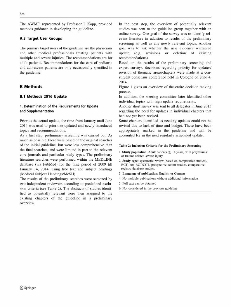
Table 2: Inclusion Criteria for the Preliminary Screening
1. Study population: Adult patients (C 14 years) with polytrauma
or trauma-related severe injury
2. Study type: systematic review (based on comparative studies),
RCT, non RCT/CCT, prospective cohort studies, comparative
registry database studies.
3. Language of publication: English or German
4. No multiple publications without additional information
5. Full text can be obtained
6. Not considered in the previous guideline
S26
123
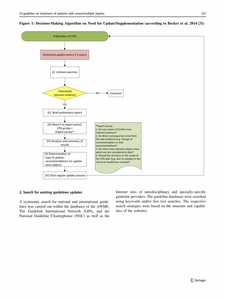
Figure 1: Decision-Making Algorithm on Need for Update/Supplementation (according to Becker et al. 2014 [3])
Scheduled update (every 3-5 years)
(I) Limited searches
ExclusionPoten�ally
relevant evidence NO
(II) Brief preliminary report
YES
(III) Report to expert panel/CPG-group +
Expert survey*
(IV) Analysis and summary of results
(V) Determina�on of- type of update- recommenda�ons for update- new subjects
(VI) Start regular update process
*Expert survey:1. Are you aware of further new relevant evidence?2. Do direct consequences arise from the new evidence (e.g. change of recommenda�on or new recommenda�on)?3. Are there new relevant subject areas which are not considered to date?4. Should the structure or the scope of the CPG alter (e.g. due to changes in the clinical or healthcare context)?
Publica�on of CPG
2. Search for existing guidelines updates
A systematic search for national and international guide-
lines was carried out within the databases of the AWMF,
The Guideline International Network (GIN), and the
National Guideline Clearinghouse (NGC) as well as the
Internet sites of interdisciplinary and specialty-specific
guideline providers. The guideline databases were searched
using keywords and/or free text searches. The respective
search strategies were based on the structure and capabil-
ities of the websites.
S3-guideline on treatment of patients with severe/multiple injuries S27
123
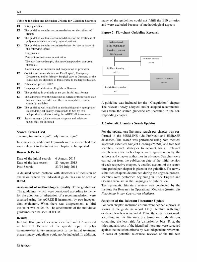
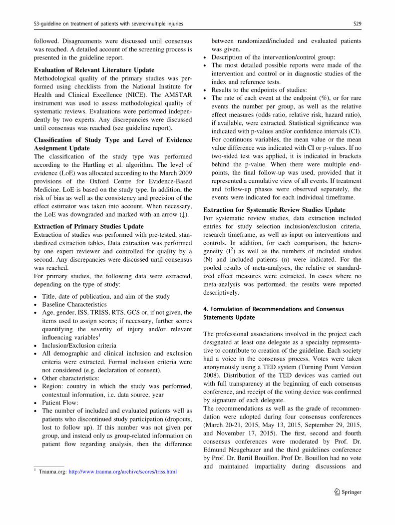
Table 3: Inclusion and Exclusion Criteria for Guideline Searches
E1 It is a guideline
E2 The guideline contains recommendations on the subject of
trauma
E3 The guideline contains recommendations for the treatment of
polytrauma and/or severely injured patients
E4 The guideline contains recommendations for one or more of
the following topics:
Diagnostics
Patient information/communication
Therapy (psychotherapy, pharmacotherapy/other non-drug
therapies)
Coordination of measures and cooperation of providers
E5 Contains recommendations on Pre-Hospital, Emergency
Department and/or Primary Surgical care in Germany or the
guidelines are classified as transferable to the target situation.
E6 Publication period: 2012
E7 Language of publication: English or German
E8 The guideline is available at no cost in full text format
E9 The authors refer to the guideline as current or the revision date
has not been exceeded and there is no updated version
currently available.
E10 The guideline was classified as methodologically appropriate
(methodological quality corresponds to S3) by two
independent evaluators using the AGREE-II instrument
E11 Search strategy (of the relevant chapter) and evidence
tables must be specified
Search Terms Used
Trauma, traumatic injur*, polytrauma, injur*
In some cases, additional keywords were also searched that
were relevant to the individual chapter to be updated.
Research Period
Date of the initial search: 6 August 2013
Date of the last search: 23 August 2013
Post-Search: 23/24 July 2014
A detailed search protocol with statements of inclusion or
exclusion criteria for individual guidelines can be seen at
IFOM.
Assessment of methodological quality of the guidelines
The guidelines, which were considered according to theme
for the adoption or adaptation of a recommendation, were
assessed using the AGREE-II instrument by two indepen-
dent evaluators. When there was disagreement, a third
evaluator was called in. The assessments of the individual
guidelines can be seen at IFOM.
Results
In total, 1040 guidelines were identified and 115 assessed
in full text. Because of the specific topic of poly-
trauma/severe injury management in the initial treatment
phases, many guidelines could not be included. In addition,
many of the guidelines could not fulfill the E10 criterion
and were excluded because of methodological aspects.
Figure 2: Flowchart Guideline Research
A guideline was included for the ‘‘Coagulation’’ chapter.
The relevant newly adopted and/or adapted recommenda-
tions from the source guideline are identified in the cor-
responding chapter.
3. Systematic Literature Search Updates
For the update, one literature search per chapter was per-
formed in the MEDLINE (via PubMed) and EMBASE
databases. The search was performed using both medical
keywords (Medical Subject Headings/MeSH) and free text
searches. Search strategies to account for all relevant
search terms for each chapter were agreed upon by the
authors and chapter authorities in advance. Searches were
carried out from the publication date of the initial version
of each respective chapter. A detailed account of the search
time period per chapter is given in the guideline. For newly
submitted chapters determined during the upgrade process,
searches were performed beginning in 1995. English and
German were set as the languages of publication.
The systematic literature review was conducted by the
Institute for Research in Operational Medicine (Institut fur
Forschung in der Operativen Medizin).
Selection of the Relevant Literature Update
For each chapter, inclusion criteria were defined a priori, as
shown in the guideline report. Only literature with high
evidence levels was included. Thus, the conclusions made
according to this literature are based on study designs
containing the least risk for distortion or bias. First, the
titles and abstracts of the identified literature were screened
against the inclusion criteria by two independent reviewers.
In cases of potential relevance, reviews of the full text
S28
123
followed. Disagreements were discussed until consensus
was reached. A detailed account of the screening process is
presented in the guideline report.
Evaluation of Relevant Literature Update
Methodological quality of the primary studies was per-
formed using checklists from the National Institute for
Health and Clinical Excellence (NICE). The AMSTAR
instrument was used to assess methodological quality of
systematic reviews. Evaluations were performed indepen-
dently by two experts. Any discrepancies were discussed
until consensus was reached (see guideline report).
Classification of Study Type and Level of Evidence
Assignment Update
The classification of the study type was performed
according to the Hartling et al. algorithm. The level of
evidence (LoE) was allocated according to the March 2009
provisions of the Oxford Centre for Evidence-Based
Medicine. LoE is based on the study type. In addition, the
risk of bias as well as the consistency and precision of the
effect estimator was taken into account. When necessary,
the LoE was downgraded and marked with an arrow (;).
Extraction of Primary Studies Update
Extraction of studies was performed with pre-tested, stan-
dardized extraction tables. Data extraction was performed
by one expert reviewer and controlled for quality by a
second. Any discrepancies were discussed until consensus
was reached.
For primary studies, the following data were extracted,
depending on the type of study:
• Title, date of publication, and aim of the study
• Baseline Characteristics
• Age, gender, ISS, TRISS, RTS, GCS or, if not given, the
items used to assign scores; if necessary, further scores
quantifying the severity of injury and/or relevant
influencing variables1
• Inclusion/Exclusion criteria
• All demographic and clinical inclusion and exclusion
criteria were extracted. Formal inclusion criteria were
not considered (e.g. declaration of consent).
• Other characteristics:
• Region: country in which the study was performed,
contextual information, i.e. data source, year
• Patient Flow:
• The number of included and evaluated patients well as
patients who discontinued study participation (dropouts,
lost to follow up). If this number was not given per
group, and instead only as group-related information on
patient flow regarding analysis, then the difference
between randomized/included and evaluated patients
was given.
• Description of the intervention/control group:
• The most detailed possible reports were made of the
intervention and control or in diagnostic studies of the
index and reference tests.
• Results to the endpoints of studies:
• The rate of each event at the endpoint (%), or for rare
events the number per group, as well as the relative
effect measures (odds ratio, relative risk, hazard ratio),
if available, were extracted. Statistical significance was
indicated with p-values and/or confidence intervals (CI).
For continuous variables, the mean value or the mean
value difference was indicated with CI or p-values. If no
two-sided test was applied, it is indicated in brackets
behind the p-value. When there were multiple end-
points, the final follow-up was used, provided that it
represented a cumulative view of all events. If treatment
and follow-up phases were observed separately, the
events were indicated for each individual timeframe.
Extraction for Systematic Review Studies Update
For systematic review studies, data extraction included
entries for study selection inclusion/exclusion criteria,
research timeframe, as well as input on interventions and
controls. In addition, for each comparison, the hetero-
geneity (I2) as well as the numbers of included studies
(N) and included patients (n) were indicated. For the
pooled results of meta-analyses, the relative or standard-
ized effect measures were extracted. In cases where no
meta-analysis was performed, the results were reported
descriptively.
4. Formulation of Recommendations and ConsensusStatements Update
The professional associations involved in the project each
designated at least one delegate as a specialty representa-
tive to contribute to creation of the guideline. Each society
had a voice in the consensus process. Votes were taken
anonymously using a TED system (Turning Point Version
2008). Distribution of the TED devices was carried out
with full transparency at the beginning of each consensus
conference, and receipt of the voting device was confirmed
by signature of each delegate.
The recommendations as well as the grade of recommen-
dation were adopted during four consensus conferences
(March 20-21, 2015, May 13, 2015, September 29, 2015,
and November 17, 2015). The first, second and fourth
consensus conferences were moderated by Prof. Dr.
Edmund Neugebauer and the third guidelines conference
by Prof. Dr. Bertil Bouillon. Prof Dr. Bouillon had no vote
and maintained impartiality during discussions and1 Trauma.org: http://www.trauma.org/archive/scores/triss.html
S3-guideline on treatment of patients with severe/multiple injuries S29
123
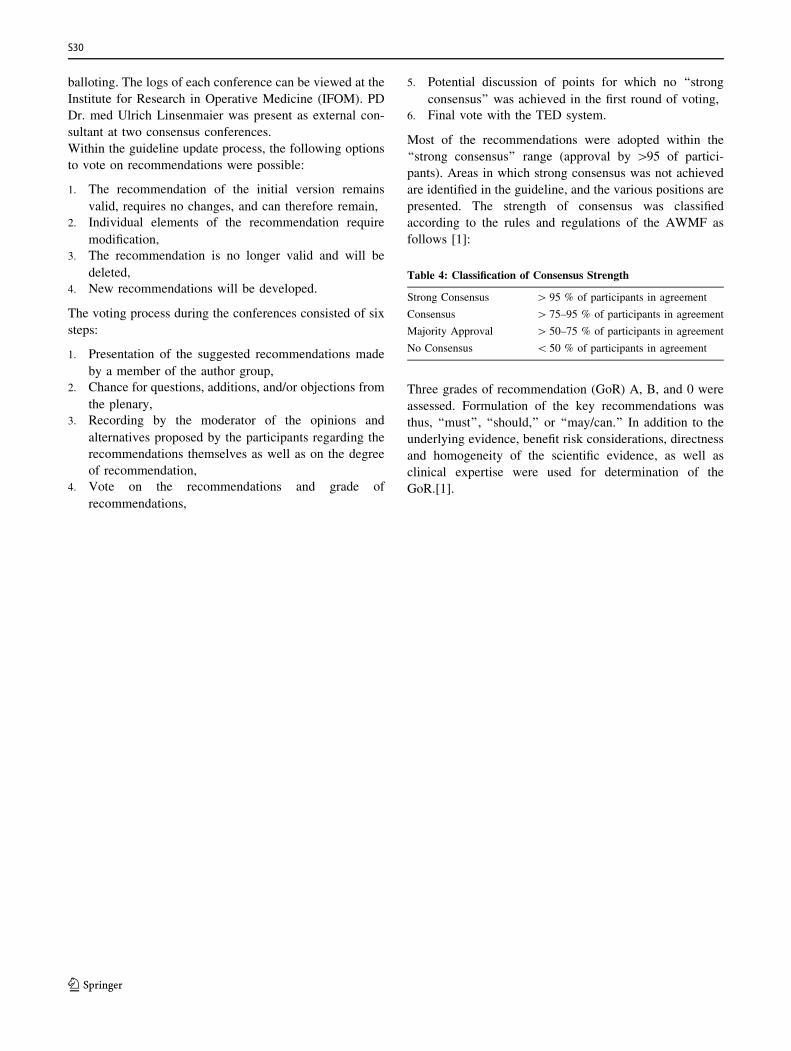
balloting. The logs of each conference can be viewed at the
Institute for Research in Operative Medicine (IFOM). PD
Dr. med Ulrich Linsenmaier was present as external con-
sultant at two consensus conferences.
Within the guideline update process, the following options
to vote on recommendations were possible:
1. The recommendation of the initial version remains
valid, requires no changes, and can therefore remain,
2. Individual elements of the recommendation require
modification,
3. The recommendation is no longer valid and will be
deleted,
4. New recommendations will be developed.
The voting process during the conferences consisted of six
steps:
1. Presentation of the suggested recommendations made
by a member of the author group,
2. Chance for questions, additions, and/or objections from
the plenary,
3. Recording by the moderator of the opinions and
alternatives proposed by the participants regarding the
recommendations themselves as well as on the degree
of recommendation,
4. Vote on the recommendations and grade of
recommendations,
5. Potential discussion of points for which no ‘‘strong
consensus’’ was achieved in the first round of voting,
6. Final vote with the TED system.
Most of the recommendations were adopted within the
‘‘strong consensus’’ range (approval by [95 of partici-
pants). Areas in which strong consensus was not achieved
are identified in the guideline, and the various positions are
presented. The strength of consensus was classified
according to the rules and regulations of the AWMF as
follows [1]:
Table 4: Classification of Consensus Strength
Strong Consensus [ 95 % of participants in agreement
Consensus [ 75–95 % of participants in agreement
Majority Approval [ 50–75 % of participants in agreement
No Consensus \ 50 % of participants in agreement
Three grades of recommendation (GoR) A, B, and 0 were
assessed. Formulation of the key recommendations was
thus, ‘‘must’’, ‘‘should,’’ or ‘‘may/can.’’ In addition to the
underlying evidence, benefit risk considerations, directness
and homogeneity of the scientific evidence, as well as
clinical expertise were used for determination of the
GoR.[1].
S30
123
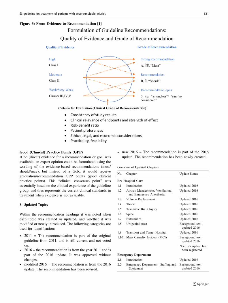
Figure 3: From Evidence to Recommendation [1]
Good (Clinical) Practice Points (GPP)
If no (direct) evidence for a recommendation or goal was
available, an expert opinion could be formulated using the
wording of the evidence-based recommendations (must/
should/may), but instead of a GoR, it would receive
graduation/recommendation GPP points (good clinical
practice points). This ‘‘clinical consensus point’’ was
essentially based on the clinical experience of the guideline
group, and thus represents the current clinical standards in
treatment when evidence is not available.
5. Updated Topics
Within the recommendation headings it was noted when
each topic was created or updated, and whether it was
modified or newly introduced. The following categories are
used for identification:
• 2011 = The recommendation is part of the original
guideline from 2011, and is still current and not voted
on.
• 2016 = the recommendation is from the year 2011 and is
part of the 2016 update. It was approved without
changes.
• modified 2016 = The recommendation is from the 2016
update. The recommendation has been revised.
• new 2016 = The recommendation is part of the 2016
update. The recommendation has been newly created.
Overview of Updated Chapters
No. Chapter Update Status
Pre-Hospital Care
1.1 Introduction Updated 2016
1.2 Airway Management, Ventilation,
and Emergency Anesthesia
Updated 2016
1.3 Volume Replacement Updated 2016
1.4 Thorax Updated 2016
1.5 Traumatic Brain Injury Updated 2016
1.6 Spine Updated 2016
1.7 Extremities Updated 2016
1.8 Urogenital tract Background text
updated 2016
1.9 Transport and Target Hospital Updated 2016
1.10 Mass Casualty Incident (MCI) Background text
updated 2016
Need for update has
been registered
Emergency Department
2.1 Introduction Updated 2016
2.2 Emergency Department - Staffing and
Equipment
Background text
updated 2016
S3-guideline on treatment of patients with severe/multiple injuries S31
123
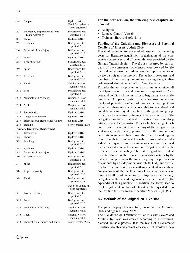
No. Chapter Update Status
Need for update has
been registered
2.3 Emergency Department Trauma
Team Activation
Background text
updated 2016
2.4 Thorax Updated 2016
2.5 Abdomen Background text
updated 2016
2.6 Traumatic Brain Injury Background text
updated 2016
2.7 Pelvis Updated 2016
2.8 Urogenital tract Background text
updated 2016
2.9 Spine Background text
updated 2016
2.10 Extremities Background text
updated 2016
2.11 Hand Original version
remains valid.
2.12 Foot Background text
updated 2016
2.13 Mandible and Midface Original version
remains valid.
2.14 Neck Original version
remains valid.
2.15 Resuscitation Updated 2016
2.16 Coagulation System Updated 2016
2.17 Interventional Hemorrhage Control Updated 2016
2.18 Imaging newly created 2016
Primary Operative Management
3.1 Introduction Updated 2016
3.2 Thorax Updated 2016
3.3 Diaphragm Background text
updated 2016
3.4 Abdomen Updated 2016
3.5 Traumatic Brain Injury Updated 2016
3.6 Urogenital tract Background text
updated 2016
3.7 Spine Background text
updated 2016
3.8 Upper Extremity Background text
updated 2016
3.9 Hand Background text
updated 2016
Need for update has
been registered
3.10 Lower Extremity Background text
updated 2016
3.11 Foot Background text
updated 2016
3.12 Mandible and Midface Original version
remains valid.
3.13 Neck Original version
remains valid.
3.14 Thermal Skin Injuries and Burns newly created 2016
For the next revision, the following new chapters are
planned:
• Analgesia
• Damage Control Vessels
• Training (Hard and soft skills)
Funding of the Guideline and Disclosure of Potential
Conflicts of Interest Update 2016
Financial resources for the methods support and covering
costs for literature acquisition, organization of the con-
sensus conferences, and of materials were provided by the
German Trauma Society. Travel costs incurred by partici-
pants of the consensus conferences were covered by the
medical societies/organizations sending representatives or
by the participants themselves. The authors, delegates, and
members of the steering committee creating the guideline
volunteered their time and effort free of charge.
To make the update process as transparent as possible, all
participants were requested to submit an explanation of any
potential conflicts of interest prior to beginning work on the
guideline. All participants of the consensus conference
disclosed potential conflicts of interest in writing. Once
submitted, these were always available to be updated and
could be accessed by all members of the guideline group.
Prior to each consensus conference, a current summary of the
delegates’ conflicts of interest declarations was sent along
with a request for evaluation. Prior to the beginning of each
conference, it was asked whether any of the delegates pre-
sent saw grounds for any person listed in the summary of
declarations to be excluded from the vote. Planned regula-
tion of conflicts of interest through exclusion of any indi-
vidual participant from discussions or votes was discussed
by the delegates in each session. No delegates needed to be
excluded from the voting. The risk of guideline content
distortion due to conflict of interest was also countered by the
balanced composition of the guideline group, the preparation
of evidence by an independent institute (IFOM), and the use
of a formal consensus process with independent moderation.
An overview of the declarations of potential conflicts of
interest by all coordinators, methodologists, medical society
delegates, authors, and organizers can be found in the
Appendix of this guideline. In addition, the forms used to
disclose potential conflicts of interest can be requested from
the Institute for Research in Operative Medicine (IFOM).
B.2 Methods of the Original 2011 Version
The guideline project was initially announced in December
2004 and again in May 2009.
The ‘‘Guideline on Treatment of Patients with Severe and
Multiple Injuries’’ was created according to a structured,
planned, reliable process. It is the result of a systematic
literature search and critical assessment of available data
S32
123
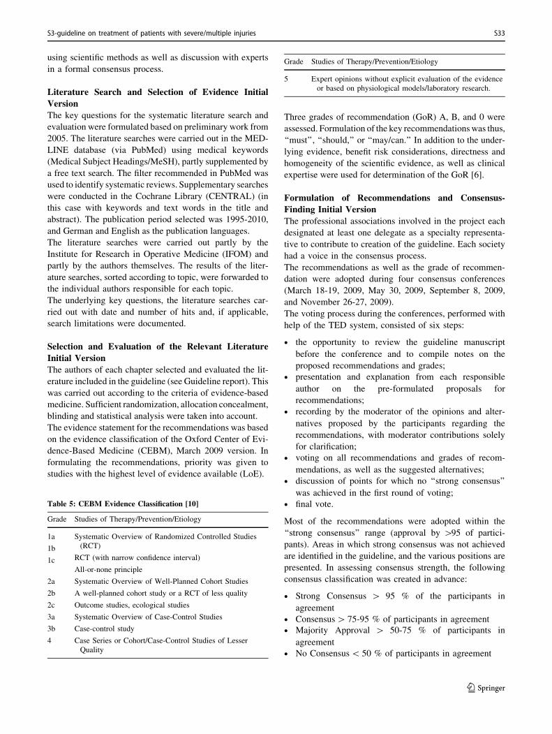
using scientific methods as well as discussion with experts
in a formal consensus process.
Literature Search and Selection of Evidence Initial
Version
The key questions for the systematic literature search and
evaluation were formulated based on preliminary work from
2005. The literature searches were carried out in the MED-
LINE database (via PubMed) using medical keywords
(Medical Subject Headings/MeSH), partly supplemented by
a free text search. The filter recommended in PubMed was
used to identify systematic reviews. Supplementary searches
were conducted in the Cochrane Library (CENTRAL) (in
this case with keywords and text words in the title and
abstract). The publication period selected was 1995-2010,
and German and English as the publication languages.
The literature searches were carried out partly by the
Institute for Research in Operative Medicine (IFOM) and
partly by the authors themselves. The results of the liter-
ature searches, sorted according to topic, were forwarded to
the individual authors responsible for each topic.
The underlying key questions, the literature searches car-
ried out with date and number of hits and, if applicable,
search limitations were documented.
Selection and Evaluation of the Relevant Literature
Initial Version
The authors of each chapter selected and evaluated the lit-
erature included in the guideline (see Guideline report). This
was carried out according to the criteria of evidence-based
medicine. Sufficient randomization, allocation concealment,
blinding and statistical analysis were taken into account.
The evidence statement for the recommendations was based
on the evidence classification of the Oxford Center of Evi-
dence-Based Medicine (CEBM), March 2009 version. In
formulating the recommendations, priority was given to
studies with the highest level of evidence available (LoE).
Table 5: CEBM Evidence Classification [10]
Grade Studies of Therapy/Prevention/Etiology
1a
1b
1c
Systematic Overview of Randomized Controlled Studies
(RCT)
RCT (with narrow confidence interval)
All-or-none principle
2a
2b
2c
Systematic Overview of Well-Planned Cohort Studies
A well-planned cohort study or a RCT of less quality
Outcome studies, ecological studies
3a
3b
Systematic Overview of Case-Control Studies
Case-control study
4 Case Series or Cohort/Case-Control Studies of Lesser
Quality
Grade Studies of Therapy/Prevention/Etiology
5 Expert opinions without explicit evaluation of the evidence
or based on physiological models/laboratory research.
Three grades of recommendation (GoR) A, B, and 0 were
assessed. Formulation of the key recommendationswas thus,
‘‘must’’, ‘‘should,’’ or ‘‘may/can.’’ In addition to the under-
lying evidence, benefit risk considerations, directness and
homogeneity of the scientific evidence, as well as clinical
expertise were used for determination of the GoR [6].
Formulation of Recommendations and Consensus-
Finding Initial Version
The professional associations involved in the project each
designated at least one delegate as a specialty representa-
tive to contribute to creation of the guideline. Each society
had a voice in the consensus process.
The recommendations as well as the grade of recommen-
dation were adopted during four consensus conferences
(March 18-19, 2009, May 30, 2009, September 8, 2009,
and November 26-27, 2009).
The voting process during the conferences, performed with
help of the TED system, consisted of six steps:
• the opportunity to review the guideline manuscript
before the conference and to compile notes on the
proposed recommendations and grades;
• presentation and explanation from each responsible
author on the pre-formulated proposals for
recommendations;
• recording by the moderator of the opinions and alter-
natives proposed by the participants regarding the
recommendations, with moderator contributions solely
for clarification;
• voting on all recommendations and grades of recom-
mendations, as well as the suggested alternatives;
• discussion of points for which no ‘‘strong consensus’’
was achieved in the first round of voting;
• final vote.
Most of the recommendations were adopted within the
‘‘strong consensus’’ range (approval by [95 of partici-
pants). Areas in which strong consensus was not achieved
are identified in the guideline, and the various positions are
presented. In assessing consensus strength, the following
consensus classification was created in advance:
• Strong Consensus [ 95 % of the participants in
agreement
• Consensus[ 75-95 % of participants in agreement
• Majority Approval [ 50-75 % of participants in
agreement
• No Consensus\ 50 % of participants in agreement
S3-guideline on treatment of patients with severe/multiple injuries S33
123
The logs of each conference can be viewed at the Institute
for Research in Operative Medicine (IFOM). The Delphi
method was then applied to recommendations for which no
consensus could be reached in the consensus conferences.
A detailed methods report is available for viewing on the
AWMF website and has been filed at the Institute for
Research in Operative Medicine (IFOM).
Funding of the Guideline and Disclosure of Potential
Conflicts of Interest Initial Version
Financial resources for the methods support and covering
costs for literature acquisition, organization of the consensus
conferences, and of materials were provided by the German
Trauma Society and the Institute for Research in Operative
Medicine of theUniversity ofWitten/Herdecke. Travel costs
incurred by participants of the consensus conferences were
covered by the medical societies/organizations sending
representatives or by the participants themselves.
All participants of the consensus conference disclosed
potential conflicts of interest in writing. A summary of the
declarations of potential conflicts of interest by all coor-
dinators, methodologists, medical society delegates,
authors, and organizers can be found in the Appendix of
this guideline. In addition, the forms used to disclose
potential conflicts of interest can be requested from the
Institute for Research in Operative Medicine (IFOM).
Warmest thanks are extended to the coordinators of the
individual subsections, the authors, and participants in the
consensus process for their completely voluntary work.
B.3 Distribution and Implementation
• Distribution of the guideline will be carried out as
follows:
• via the internet: AWMF website (http://www.awmf-
online.de) as well as the websites of the medical soci-
eties and professional organizations involved in the
guideline
• via printed media:
Publication of the guideline as a manual/book by the
DGU. Copies will be made available to all hospitals
involved in the DGU Trauma Network. In addition,
all hospitals involved will be notified in writing
regarding where and how the guideline can be viewed
on the AWMF homepage.Publication of excerpts of the guideline and implementation
strategies in the journals of the participating medical societies.
To simplify use of the guideline, a summary version of the
guideline containing the key recommendations will be pub-
lished in ‘‘Notfall- und Rettungsmedizin’’ [German medical
journal].
• via conferences, workshops, professional training
courses offered by the participating medical societies.
Various complementary measures are to be implemented in
this guideline. In addition to the presentation of the rec-
ommendations at conferences, a link to topic-specific
professional training courses is planned.
In addition, implementation at all the German DGU trauma
network hospitals will be evaluated approximately one year
after guideline publication. In particular, information
should be collected regarding guideline use and practical
suggestions for other users.
Quality Indicators and Evaluation
Audit filters were developed for the DGU Trauma Registry
as criteria for quality management. Based on available
audit filters, the following criteria were established for this
guideline:
Process quality for evaluation in the pre-hospital care
phase:
• duration of prehospital time from accident to hospital
admission for severely injured patients with ISS C 16
[min ± SD]
• intubation rate in patients with severe chest injury (AIS
4-5) by the emergency physician [%, n/total]
• intubation rate in patients with suspected traumatic
brain injury (unconscious, Glasgow Coma Scale [GCS]
B 8) [%, n/total]
Process quality for evaluation of the emergency
department phase:
• time from hospital admission to performance of chest
X-ray in severely injured patients (ISS C 16)
[min ± SD]
• time from hospital admission to abdominal/chest ultra-
sound in cases of severe trauma (ISS C 16) [min ± SD]
• time to computed tomography (CT) scan of the cranium
(CCT) in pre-hospital unconscious patients (GCS B 8)
[min ± SD]
• time to full-body CT scan on all patients, if carried out
[min ± SD]
• time from emergency arrival to completion of diagnos-
tic survey in severely injured persons, if this has been
completed normally (ISS C 16) [min ± SD]
• time from emergency arrival to completion of diagnos-
tic survey in severely injured persons, if interrupted due
to emergency (ISS C 16) [min ± SD]
Outcome Quality for Overall Evaluation:
• standardized mortality rate: observed mortality divided
by the expected prognosis based on RISC (Revised
Injury Severity Classification) in severely injured
patients (ISS C 16)
• standardized mortality rate: observed mortality divided
by the expected prognosis based on TRISS (Trauma
S34
123
Injury Severity Score Method) in severely injured
persons (ISS C 16)
The routine collection and evaluation of these data offer a
vital opportunity to monitor improvements in the quality of
management of patients with multiple and severe injuries.
From this it is not possible to ascertain which effects are
due to the guideline. Quality indicators should continue to
be developed based on the aforementioned criteria.
B.4 Guideline Validity and Updates
The present guideline is valid until June 2021. The German
Trauma Society is responsible for initiating the update
procedure. The next update is planned to address the topics
of analgesia, damage control vessels, and a special chap-
ter on training (hard and soft skills).
References
1. Arbeitsgemeinschaft der Wissenschaftlichen Medizinischen
Fachgesellschaften (AWMF) Standige Kommission Leitlinien.
AWMF-Regelwerk ,,Leitlinien‘‘. 1. Auflage 2012. http://www.
awmf.org/leitlinien/awmf-regelwerk.html.
2. Arbeitsgemeinschaft Wissenschaftlicher Medizinischer Fachge-
sellschaften (AWMF). 3-Stufen-Prozess der Leitlinien-Entwick-
lung: eine Klassifizierung. 2009. http://www.awmf.org/leitlinien/
awmf-regelwerk/ll-entwicklung.html.
3. Becker M, Neugebauer EA, Eikermann M. Partial updating of
clinical practice guidelines often makes more sense than full
updating: a systematic review on methods and the development
of an updating procedure. J Clin Epidemiol. 2014;67(1):33–45.
4. Bouillon, B., Weißbuch Schwerverletzten-Versorgung.
Empfehlungen zur Struktur, Organisation und Ausstattung sta-
tionarer Einrichtungen zur Schwerverletzten-Versorgung in der
Bundesrepublik Deutschland. 2006, Berlin: Dt. Gesellschaft fur
Unfallchirugie e.V. (DGU).
5. Bundesanstalt fur Arbeitsschutz und Arbeitsmedizin. Gesamtun-
fallgeschehen. 2013. http://www.baua.de/de/Informationen-fuer-
die-Praxis/Statistiken/Unfaelle/Gesamtunfallgeschehen/Gesam
tunfallgeschehen.html.
6. Council of Europe. Developing a Methodology for drawing up
Guidelines on Best Medical Practices: Recommendation Rec
(2001) 13 adopted by the Committee of Ministers of the Council
of Europe on 10 October 2001 and explanatory memorandum.
Council of Europe: Strasbourg Cedex; 2001.
7. Field MJ, Lohr KN (Eds). Clinical practice guidelines: directions
for a new program. 1990.
8. Hartling L, et al. Testing a tool for the classification of study
designs in systematic reviews of interventions and exposures
showed moderate reliability and low accuracy. J Clin Epidemiol.
2011;64(8):861–71.
9. Kopp IB. Perspectives in guideline development and implemen-
tation in Germany. Z Rheumatol. 2010.
10. Oxford Center for Evidence based Medicine (CEBM). Levels of
Evidence March 2009. 2009. https://www.cebm.net/index.
aspx?o=1025.
11. Robert Koch-Institut. Gesundheit in Deutschland. Gesundheits-
berichterstattung des Bundes. Gemeinsam getragen von RKI
und Destatis, Berlin. 2015.
12. Schmiegel W, et al. S3-Leitlinie ,,Kolorektales Karzinom‘‘.
2008.
S3-guideline on treatment of patients with severe/multiple injuries S35
123
1. Pre-Hospital Care
1.1 Introduction
Professional treatment of seriously injured patients begins
at the accident scene with a structured rescue service.
Already at this first phase of treatment, the introduction of
life-saving measures, a time-critical approach, and trans-
port to the appropriate target hospital set the overall tra-
jectory of the course to come.
Often before the emergency physician arrives, an
emergency rescue service without a physician is first at the
scene of the accident [1]. Thus, the ‘‘Pre-Hospital Care’’
section of this guideline is directed not only towards
physicians, but also to emergency medics, paramedics, and
other pre-hospital assisting personnel.
Five years ago, the first comprehensive AWMF guide-
line for the treatment of severely injured patients was
published, and is updated now for the first time. What has
changed since then - and what has not?
For the pre-hospital care section - but not only here - a
number of situations have been singled out and worked out
in more detail, situations in which pre-hospital measures
could be life-saving and in which full implementation of
these measures could prevent potentially avoidable deaths,
for example situations with problems securing the airway,
decompression of a tension pneumothorax, or insufficient
hemorrhage control [3].
In the chapter, ‘‘Airway Management, Ventilation, and
Emergency Anesthesia,’’ new recommendations regarding
the use of video laryngoscopy have been included. An
increasing collection of evidence shows clearly that use of
video laryngoscopy enables higher success rates of endo-
tracheal intubation for very experienced as well as less-
experienced users. However, since advantages in survival
with the use of video laryngoscopy have not yet been
proven, the recommendations have been formulated as
‘‘good clinical practice points’’ (GPP).
For the treatment of thoracic trauma, the importance of
rapid decompression for tension pneumothorax has been
more strongly emphasized. Several analyses have identified
tension pneumothorax as one of the most important pre-
ventable causes of death.
The importance of hemostasis outside of the operating
room has moved further into the foreground. A substantial
new section has been added to the ‘‘Extremities’’ chap-
ter dealing with profusely bleeding wounds and the use of
newer methods for hemorrhage control such as tourniquets
and hemostatic bandages. In particular, situations are
defined in which tourniquets can be considered a primary
management option. In this context, the reader should also
pay attention to the ‘‘Pelvis’’ chapter in the ‘‘Emergency
Department’’ section of the guideline, in which aspects of
examination and emergency stabilization are considered
that are also relevant to the pre-hospital care situation.
Since publication of the last S3 guideline, numerous
studies have addressed the use and performance of car-
diopulmonary resuscitation for cases of trauma-induced
cardiac arrest [11, 14, 18, 20]. Registry data show that in
traumatic cardiac arrest, even after blunt trauma, resusci-
tation measures can have positive results with good neu-
rological outcomes [9]. For this reason, the extensive and
reorganized ‘‘Resuscitation’’ chapter in the ‘‘Emergency
Department’’ section must be pointed out, as it is also
extremely relevant for pre-hospital management. Here, the
special measures for resuscitation after trauma have been
clearly prioritized and presented, and in particular, the
differences versus resuscitation for classic cardiac or pul-
monary induced arrest are highlighted. The algorithms and
recommendations presented are closely aligned with the
international guidelines of the ERC (European Resuscita-
tion Council).
The chapter ‘‘Thermal Skin Injuries and Burns’’ is
completely new and is dedicated mostly to the special
management of severely injured patients with concomitant
burn injuries. It is therefore considered a supplement to
existing guidelines [7, 17]. That chapter is found in the
‘‘Primary Operative Management’’ section of this guideline,
but also contains information relevant to pre-hospital care.
Another chapter that has undergone revision is the ‘‘Mass
Casualty Incident’’ chapter. A mass number of trauma
patients represents a rare, but very challenging situation.
Current events and the global political situation clearly
show that being prepared for a mass attack, e.g. a terrorist
strike, is more necessary than ever. In a modern and mobile
society, however, there are also other scenarios with large
numbers of severely injured persons, e.g. the recent train
accident at Bad Aibling, which are unavoidable despite high
safety standards. Prior to arrival of the executive emergency
physician on duty, the primary tasks focusing on triage and
allocation of scarce resources and treatment capacity must
be handled by the first emergency physician on the scene.
It cannot be stressed often enough that treatment of
severely injured patients is a time-critical enterprise.
Interestingly, the pre-hospital time, i.e. the time from the
accident until arrival in the Emergency Department, is
apparently not an independent risk factor, at least not for all
patients as a whole. Nevertheless, even data collection by
the best current risk prediction tool, the RISC II [13],
identifies pre-hospital time as an independent risk factor
predicting mortality. On the other hand, it is known that in
cases of severe intra-abdominal bleeding, a time delay of
one minute increases mortality by 3% [5]. Since the pre-
dictive power of recognizing these injuries in the pre-
hospital setting is low and the concerned patients can easily
S36
123
go unrecognized [2, 10, 16], it is advisable to transport all
patients with potentially severe injuries as quickly as pos-
sible to the hospital, where the full range of diagnostic and
targeted interventions can be performed. In the end, how-
ever, whether some time-consuming procedures are per-
formed at the accident scene or are postponed until the
early hospital phase must be weighed. It certainly plays a
role here as to how urgent the indication for an intervention
is, whether there are particular difficulties in carrying it out,
or whether individual skills are sufficient to do it safely and
correctly. In the meantime, it has also been found that the
time gained by skipping interventions in the pre-hospital
phase is lost again during the Emergency Department
phase. Thus, the balance between the ‘‘load and go’’ and
‘‘stay and play’’ approaches to pre-hospital management
must be based first and foremost on the urgency of the
required intervention [12]. Life-saving interventions should
not be foregone in favor of a shorter pre-hospital time.
Conversely, some measures, e.g. surgical hemostasis of
internal bleeding, must be performed in the hospital. As a
guideline, the Key Points Paper on Emergency Medical
Management of Patients recommends a maximum pre-
hospital time of 60 minutes [8]. This does not preclude the
fact that faster transport for certain patient groups would be
better.
Regarding the critical time period as well as rapid
injury-appropriate management, selection of the appropri-
ate hospital is of great importance. Germany now has a
national system of certified trauma centers in three levels of
care, resulting from a transparent catalog of available ser-
vices. This catalog is revised and published every four
years in the white paper Treatment of the Severely Injured
[6]. Recently, the benefits of air rescue in particular have
proliferated, because it seems to be associated with
improved survival after severe accident-related trauma.
One suspected reason for this advantage is the ability of
helicopters to reach an appropriate target hospital more
rapidly than ground-based rescue services, especially when
the hospital is far away. Particularly when it comes to
special requirements, e.g. a neurosurgical department in
cases of severe TBI, center-related effects could be a rea-
son for the benefits of air rescue. This topic is a focus of the
chapter ‘‘Transport and Target Hospital,’’ which has been
correspondingly expanded and made more detailed. Here
again, the Key Paper [8] calls for accessibility-oriented
treatment with a recourse to national treatment capacity
involving air rescue regardless of the time of day.
During registration in the target hospital, it’s important
that the treatment of the patient continues smoothly and
without unnecessary loss of time. As a benchmark, the Key
Paper calls for a time interval of 90 minutes from accident
until the start of lifesaving, operative interventions [8]. The
hub of continued hospital treatment is in the trauma bay of
the Emergency Department. Criteria for the requirements
there are listed in the ‘‘Trauma Team Activation’’ chap-
ter of the ‘‘Emergency Department’’ section of this
guideline. The guideline group placed low priority on the
revision of this chapter. However, questions surrounding
over- and under-triage based on the recommendations there
continue to present in daily practice as a stress test for
hospital resources. In 2012, the US Center for Disease
Control (CDC) published an update of its recommendations
regarding assignment of patients to a trauma center, which
has also been translated into German [19]. The criteria
given there are similar to the German trauma team acti-
vation criteria. The main problem, however, is that espe-
cially the criteria regarding accident mechanism, in the
absence of other criteria, might invite speculation that
trauma team management is not indicated. Leaders in the
emergency medical services (EMS) and the medical
director of the EMS should work together with the trauma
network to develop locally-adapted processes to meet their
respective needs. In view of the difficulties in making an
accurate diagnosis in the pre-hospital setting, however, the
pre-hospital team should accept higher rates of over-triage
over under-triage, for the benefit of patients. In cases of
doubt, the assessment by the emergency physician to
activate the trauma team should be generally accepted.
Recommendations for pain therapy were already lacking
in the last guideline. Unfortunately, due to the generous but
still limited resources, it was not possible to amend this
already detailed draft according to all methodological,
structural, and substantive requirements. It is envisaged,
however, that the draft text will first be published inde-
pendent of the guideline. This will provide a systematic
review, which can be both critically recognized and used as
a guideline for individual management decisions, and then
can be added to the consensus process of the update and
thus be added to the next S3 guideline version.
Because of the challenging conditions of the prehospital
emergency environment, the level of evidence is low, while
experience and expertise are considerable. Numerous dif-
ficulties contribute here:
• Compliance with the standards of good clinical practice
• As a rule, patients are incapable of consent
• Heterogeneity of the patient population
• The difficulty of correctly and completely identifying,
with limited diagnostic capabilities, the actual pattern of
injury or pathophysiological processes (e.g.
coagulopathy)
• Ethical concerns in omitting certain interventions in a
comparison group (e.g. decompression of a tension
pneumothorax)
• Doubts regarding transferability of results between
differently organized rescue services
S3-guideline on treatment of patients with severe/multiple injuries S37
123
Nevertheless, it is necessary to translate evidence-based
and expert recommendations into practical management
recommendations and priority-driven processes. With the
‘‘Trauma Care Bundle’’ for pre-hospital management of
severely injured patients, the German Trauma Society’s
section on Emergency, Intensive care, and Injuries (section
NIS) transferred key recommendations into a short and
practical treatment guide, thus attempting to create a basis
for improving quality of care [15]. There are also a variety
of commercially available course formats, such as Prehos-
pital Trauma Life Support� (PHTLS), International
Trauma Life Support� (ITLS) or TraumaManagement�,
which provide and distribute practical treatment protocols.
The individual steps in these protocols conform to the key
recommendations of the S3 guideline, but cannot be sci-
entifically backed up in detail, as explained above. Thus, the
present guideline did not aim to redevelop a similar con-
cept. It should be emphasized here that all of these man-
agement interventions must be physically practiced and
trained within the team. Within the ‘‘pre-hospital’’ section
of the guideline, the current state of knowledge was
accepted without much disagreement by the expert repre-
sentatives, and high levels of agreement were attained. At
the same time, the available knowledge remains incomplete
in many areas, which is why consensus is often shaped by
expert opinions. For this reason, the ‘‘new’’ recommenda-
tion grading system of GPP (Good Clinical Practice Points)
was introduced for recommendations for which there is not
evaluable evidence, but are considered important enough to
make a recommendation. We hope that knowledge deficits
and gaps, or recommendations that are not accepted uni-
versally, will inspire efforts to close these gaps, at least
partially, with solid scientific research leading up to the next
revision of the S3 guideline. We expressly encourage all
interested parties to take on this important task [4].
References
1. Aschenbrenner U, et al. Air rescue missions at night: Data
analysis of primary and secondary missions by the DRF air
rescue service in 2014. Unfallchirurg. 2015;118(6):549–63.
2. Blaivas M, Sierzenski P, Theodoro D. Significant hemoperi-
toneum in blunt trauma victims with normal vital signs and
clinical examination. Am J Emerg Med. 2002;20(3):218–21.
3. Buschmann C, et al. Vermeidbare Todesfalle nach Trauma -
Epidemiologie. Todesursachen und Managementfehler. Der
Notarzt. 2013;29(3):91–8.
4. Christ M. Notfallmedizin: Mehr Evidenz fur den Notfall. Dtsch
Arztebl. 2015;112(11):A 465–6.
5. Clarke JR, et al. Time to laparotomy for intra-abdominal bleeding
from trauma does affect survival for delays up to 90 minutes.
J Trauma. 2002;52(3):420–5.
6. Deutsche Gesellschaft fur Unfallchirurgie, Weißbuch Schwerver-
letztenversorgung, 2. erweiterte Auflage – Empfehlungen zur
Struktur, Organisation und Ausstattung der Schwerverletztenver-
sorgung in der Bundesrepublik Deutschland. Orthopadie und
Unfallchirurgie Mitteilung und Nachrichten, 2012(Supplement
1):4–34.
7. Ellerkamp V, et al. S2k-Leitlinie 006-128: Behandlung thermis-
cher Verletzungen im Kindesalter (Verbrennungen, Verbruhun-
gen). 2015. http://www.awmf.org/leitlinien/detail/ll/006-
128.html. Accessed 4 Mar 2016
8. Fischer M, et al. Eckpunktepapier zur notfallmedizinischen
Versorgung der Bevolkerung in der Prahospitalphase und in der
Klinik. Notfall Rettungsmedizin. 2016;19(5):387–95.
9. Grasner JT, et al. Cardiopulmonary resuscitation traumatic
cardiac arrest–there are survivors. An analysis of two national
emergency registries. Crit Care. 2011;15(6):R276.
10. Hasler RM, et al. Accuracy of prehospital diagnosis and triage of a
Swiss helicopter emergencymedical service. J TraumaAcuteCare
Surg. 2012;73(3):709–15.
11. Kleber C, et al. Requirement for a structured algorithm in cardiac
arrest following major trauma: epidemiology, management errors,
and preventability of traumatic deaths in Berlin. Resuscitation.
2014;85(3):405–10.
12. Kulla M, et al. Prehospital endotracheal intubation and chest
tubing does not prolong the overall resuscitation time of
severely injured patients: a retrospective, multicentre study of
the Trauma Registry of the German Society of Trauma Surgery.
Emerg Med J. 2012;29(6):497–501.
13. Lefering R, et al. Update of the trauma risk adjustment model of
the TraumaRegister DGU: the Revised Injury Severity Classi-
fication, version II. Crit Care. 2014;18(5):476.
14. Lockey DJ, Lyon RM, Davies GE. Development of a simple
algorithm to guide the effective management of traumatic
cardiac arrest. Resuscitation. 2013;84(6):738–42.
15. Matthes G, et al. Essential measures for prehospital treatment of
severely injured patients: The trauma care bundle. Unfallchir-
urg. 2015;118(8):652–6.
16. Muhm M, et al. Preclinical prediction of prehospital injury
severity by emergency physicians : approach to evaluate
validity. Anaesthesist. 2011;60(6):534–40.
17. Rennekampff O. Thermische und chemische Verletzungen
(Registernummer 044-001). 2011. http://www.awmf.org/leitlin-
ien/detail/ll/044-001.html. Accessed 2016 4 Mar 2016.
18. Sherren PB, et al. Algorithm for the resuscitation of traumatic
cardiac arrest patients in a physician-staffed helicopter emer-
gency medical service. Crit Care. 2013;17(2):308.
19. Trentzsch H, Urban B, Huber-Wagner S. Evidenzbasierte Triage
von verletzten Patienten am Unfallort. Notfall Rettungsmedizin.
2012;15(8):709–13.
20. Truhlar A, et al. European Resuscitation Council Guidelines for
Resuscitation 2015: Section 4. Cardiac arrest in special circum-
stances. Resuscitation. 2015;95:148–201.
1.2 Airway Management, Ventilation,and Emergency Anesthesia
Preamble
Endotracheal intubation and ventilation, and thus definitive
airway protection aimed at optimized oxygenation and
ventilation, are central therapeutic measures in emergency
medicine [146]. This is about securing the basic vital
functions directly associated with survival. In established
standards of trauma care, the ‘‘A’’ for airway and ‘‘B’’ for
breathing are given the highest priority and thus, are
S38
123
particularly important for both pre-hospital and early hos-
pital management [4, 131, 183].
One problem with evaluating the evidence available is
that information cannot be directly referred to the German
rescue and emergency physician system, due to the diver-
gent organization of emergency rescue services interna-
tionally and the resulting differences in experience and
routines used to secure the airway. This applies particularly
for negative results from paramedic systems [102, 157].
Although paramedics are often employed in the Anglo-
American region, the emergency physician system is
widely used in continental Europe. But even here there is
variation. In Germany, (specialist) physicians from all
disciplines can participate in emergency services after
acquiring the appropriate qualification, but in Scandinavian
countries, this is mainly reserved for anesthesiologists [14].
As a consequence, the evaluation of international studies
regarding pre-hospital securing of the airway reveals
emergency medical personnel with very different levels of
training. Depending on the personnel employed and how
commonly they perform intubation, high rates of unsuc-
cessful intubations are found in the literature, with 15 to
31%. Esophageal intubations occur in up to 12% of cases
[38, 174]. Within paramedic systems, there is a higher rate
of guideline non-compliant airway management [64]. For
the emergency physician system in Germany, the stipulated
minimum ‘‘Additional qualification in emergency medi-
cine’’ and the use of emergency anesthesia produce a dif-
ferent scenario than that in the Anglo-American paramedic
system, in which securing the airway is sometimes
attempted without medications.
For the key recommendations to come, the following
features of the pre-hospital care setting, which influence the
development of indications to perform emergency anes-
thesia, intubation and ventilation, must be considered:
• Level of experience and the routine training of the
emergency physician
• Circumstances of the medical emergency (e.g., patient
is trapped, rescue time)
• Type of transport (land-based versus air-supported)
• Transport time
• Concomitant injuries of the airway region and any
(recognizable) impediments to intubation
Depending on the individual case, the decision to perform
or not perform pre-hospital anesthesia, intubation/airway
management, and ventilation ranges between the extremes
of ‘‘advanced training level, long transport time, simple
airway,’’ and ‘‘minimal experience, shorter transport time,
predicted difficult airway.’’ In any event, sufficient oxy-
genation must be secured using appropriate measures.
The following recommendations cover the overall topics of
emergency anesthesia, airway management, and ventilation
in the pre-hospital phase and in the Emergency
Department.
Key Recommendations:
1.1 Recommendation 2016
GoR A For multiply injured patients with apnea or agonalbreathing (respiration rate <6), emergency anesthesia,endotracheal intubation and ventilation must beperformed in the pre-hospital setting.
1.2 Recommendation Modified 2016
GoR B For multiply injured patients, emergency anesthesia,endotracheal intubation and ventilation should beperformed in the pre-hospital setting for the followingindications:
• Hypoxia (SpO2 < 90 %) despite oxygenadministration and after tension pneumothorax isexcluded
• Severe TBI (GCS < 9)
• Trauma-associated persistent hemodynamicinstability (SBP < 90 mmHg, age adapted for children)
• Severe chest trauma with respiratory insufficiency(respiration rate > 29, age-adapted for children)
1.3 Recommendation 2016
GoR A Multiply injured patients must be pre-oxygenatedbefore anesthesia induction.
1.4 Recommendation Modified 2016
GoR A During in-hospital emergency anesthesia, endotrachealintubation and ventilation must be performed bytrained and experienced anesthesia staff.
When complicated in-hospital induction and/orendotracheal intubation is expected, an anesthesiologistmust perform the procedure.
Explanation:
Indications for Intubation
Severe polytrauma has serious effects on the integrity of
the human body. In addition to the acute consequences of
trauma on individual body regions, there is a mediator-
mediated whole-body reaction, i.e. Systemic Inflammatory
Response Syndrome (SIRS) [48, 93]. Tissue oxygenation
takes on special significance in this damage cascade. Tissue
oxygenation can only be achieved if oxygen uptake,
transport and release are maintained. Oxygen uptake is
only possible with a free airway. Direct consequences of
trauma (e.g. facial fractures, laryngeal injuries or obstruc-
tion due to blood, vomit and/or secretions), but also
inability to keep the airway open independently because of
loss of consciousness, can make securing the airway nec-
essary. Endotracheal intubation is the gold standard for
definitive airway protection according to current European
and non-European guidelines [56, 130, 131]. Treatment
recommendations of the German Society of Anesthesiol-
ogy and Intensive Care Medicine (Deutschen Gesellschaft
fur Anasthesiologie und Intensivmedizin) and the S1
S3-guideline on treatment of patients with severe/multiple injuries S39
123
Guideline (AWMF) also support the indications for airway
management listed above in the Key Recommendations
[19].
Severe impairment of consciousness with a Glasgow
Coma Score (GCS)\ 9 due to a traumatic brain injury is
regarded as an indication for intubation [12]. For the
trauma patient with impairedconsciousness GCS B 8,
endotracheal intubation in both pre-hospital and hospital
settings is also recommended in the guideline of the
Eastern Association for the Surgery of Trauma (EAST)
[56] and other training programs (e.g., ATLS� [4], ETC
[70]). Hypoxia and hypotension are the ‘‘lethal duo’’
inducing secondary damage particularly in polytrauma
patients with traumatic brain injury [36, 37, 90, 155, 158].
Abnormal brain computed tomography (38%) and
intracranial bleeding (28%) have been reported even in
patients with GCS of 13 or 14 who were endotracheally
intubated in the pre-hospital phase [61]. In a pre-hospital
cohort study, endotracheal intubation had a positive effect
on survival following severe traumatic brain injury [96]. A
comparative registry database study, which due to a num-
ber of factors is not comparable to the German emergency
physician system, found a higher mortality for patients with
a GCS of 3 who were intubated in the pre-hospital setting
(Odds Ratio [OR] 1.93; 95 % CI: 1.74–2.15, p\ 0.0001)
[88]. However, in the post-hoc subgroup analysis by the
Resuscitation Outcome Consortium (ROC) Hypertonic
Saline (HS) trials (RCT) [173], which looked at data from
two randomized clinical studies using hypertonic infusions,
there was no increased 28-day mortality in TBI patients
intubated in the pre-hospital setting versus those intubated
in the Emergency Department (OR 1.57; 95% CI:
0.93–2.64). This study is also not transferable to the Ger-
man emergency physician system for numerous reasons.
There was increased 28-day mortality for patients in shock
(SBP\70 mmHg or SBP 71-90 mmHg + HR[108 bpm)
who had been intubated in the pre-hospital setting (OR
5.14; 95% CI: 2.42–10.90). Limitations in the interpreta-
tions of the secondary analysis of the ROC HS trial [173]
include the pre-hospital paramedic system, the lack of data
regarding the type of airway/anesthetic management, and
the fact that muscle relaxants were used in only 70% of
cases. Another retrospective study showed reduced mor-
tality for children with severe TBI who were intubated by
emergency physicians in the pre-hospital phase versus
those receiving Basic Life Support (BLS) and delayed
intubation in regional trauma centers [159]. Limiting con-
sideration to the pediatric population, pre-hospital intuba-
tion was carried out by emergency medical physicians in
this study, meaning good transferability to the German
emergency physician system. Using the Trauma and Injury
Severity Score (TRISS), another study also confirmed that
pre-hospital intubation yields improved survival and
neurologic outcomes [63]. Another paper showed
improvements in the measured systolic blood pressure,
oxygen saturation, and end-tidal carbon dioxide (etCO2)
compared to baseline values prior to pre-hospital intubation
in patients with severe TBI [16]. A methodically weaker
retrospective analysis in a paramedic system showed no
survival advantage for patients with TBI [30]. Regarding
the time point of intubation in patients with traumatic brain
injury and decreased level of consciousness, important
results can be found in a prospective randomized controlled
trial (RCT) in which intubation was performed by para-
medics with a standard rapid sequence induction (RSI)
protocol (fentanyl 0.1 mg + midazolam 0.1 mg/kg + suc-
cinylcholine 1.5 mg/kg) in patients with GCS\ 10 (intu-
bation success rate of 97%) compared to cases intubated by
physicians on arrival to the hospital [17]. Using the
extended Glasgow Outcome Scale (eGOS), patients
undergoing pre-hospital intubation had better neurological
outcomes after six months [eGOS 5-8 (n = 157 vs. n =
142): 51% vs. 39% with RR 1.28; 95% CI: 1.00-1.64, p =
0.046] than those managed in-hospital [17]. However,
duration of intensive care and hospital admissions as well
as survival until discharge did not differ [17]. A compar-
ative registry database study also supports the recom-
mended indications for intubation in TBI listed above,
particularly for severely affected patients [47]. A retro-
spective cohort study of comparable patient groups (ISS 10
vs. 11) reported that delayed endotracheal intubation (n =
34, intubation 24 min) was associated with higher mortality
than earlier intubation (n = 56, intubation 10-24 min) after
admission to the Emergency Department (11.8 % vs.
1.8 %, p = 0.045) [112]. The authors calculated a relative
risk reduction of 85 % for mortality when the patient air-
way was secured early (i.e., within 10-24 min) on admis-
sion to the hospital.
Current review studies include heterogeneous patient
populations, various types of emergency medical services,
as well as differing levels of experience for the care pro-
viders and thus, do not always yield positive results for
intubation [14, 22, 45, 53, 102, 104, 124, 131, 172, 175]
The EAST guideline group also addressed this problem. In
the ‘‘Guidelines for Emergency Intubation Immediately
Following Traumatic Injury,’’ it was stated that there are no
randomized controlled trials on this research question. On
the other hand, however, the authors of the EAST Guide-
line also found no studies presenting a proven alternative
treatment strategy. In summary, endotracheal intubation
was assessed overall as such an established procedure in
hypoxia/apnea that, despite a lack of scientific evidence, a
Grade A recommendation was formulated [56].
Other conditions requiring a definitive airway are gas
exchange disturbances, even when the native airway is
open. A current investigation on airway protection in
S40
123
moderately injured patients supports apnea as an indication
for intubation [85]. Other indications for endotracheal
intubation (e.g. chest trauma) are disputed in the literature
[141]. Hypoxia and respiratory failure have been estab-
lished as consequences of severe chest trauma (multiple rib
fractures, lung contusion, flail chest). If hypoxia is refrac-
tory to oxygen administration, exclusion of tension pneu-
mothorax, and basic measures of airway support,
endotracheal intubation is recommended [56]. Pre-hospital
endotracheal intubation in patients with severe thoracic
trauma can prevent hypoxia and hypoventilation, which are
associated with secondary neurologic damage and severe
repercussions on the rest of the body. However, with dif-
ficult, prolonged attempts at intubation and the associated
hypoventilation and risk of hypoxia, endotracheal intuba-
tion itself can cause procedure-associated secondary dam-
age or even death. A database analysis of the Trauma
Registry of the German Trauma Society showed no
advantage in prehospital endotracheal intubation in patients
suffering chest trauma without respiratory insufficiency
[141]. Severe chest injury with respiratory insufficiency
does present an indication for prehospital endotracheal
intubation; however, the decision to intubate should be
made based on respiratory insufficiency and not the (sus-
pected) diagnosis of severe chest injury, which is associ-
ated with a degree of uncertainty [10].
Endotracheal intubation is included as an ‘‘Advanced
Life Support’’ procedure in the pre-hospital action algo-
rithms of various training programs (e.g., PHTLS�, ETC)
[70, 126]. In this context, studies have been conducted to
examine compliance with these recommendations. Using a
scoring system to evaluate management problems along
with the relevant autopsy reports, a series of fatal traffic
accidents were retrospectively analyzed to characterize the
effectiveness of pre-hospital care and potentially avoidable
mortality [136]. Here factors leading to avoidable death
included prolonged ‘‘pre-hospital and early in-hospital care
period’’ as well as ‘‘lack of airway protection with intu-
bation’’ [136].
Considering the cases presented in the Preamble, the
following aspects have particular relevance. A retrospec-
tive cohort study of 570 intubated patients versus 8137
non-intubated patients reported that patients intubated pre-
hospital had pre-hospital times lasting 5.2-10.7 minutes
longer than that of non-intubated patients [43]. A
prospective non-randomized study assessed the influence
of early intubation within two hours of trauma on subse-
quent organ failure [169]. Despite a significantly higher
degree of injury in the group of patients who were intu-
bated ‘‘early’’ (within 2 hours of trauma), the incidence of
organ failure and mortality were decreased compared to
those intubated ‘‘later.’’ A retrospective trauma registry
database study compared 3571 patients intubated pre-
hospital to 746 patients intubated in the Emergency
Department and 11 586 patients who were not intubated at
all [7]. Intubation performed in the Emergency Department
was associated with significantly higher mortality risk
compared to non-intubated patients (OR 3.1; 95% CI: 2.1-
4.5, p\ 0.0001) and patients intubated in the pre-hospital
setting (OR 3.0; 95 % CI: 1.9–4.9, p \ 0.0001) [7]. It
should also be noted that patients intubated in the pre-
hospital setting did not have a higher mortality risk than
patients who were not intubated in the Emergency
Department (OR 1.1; 95 % CI: 0.7–1.9; p = 0.6). The
authors concluded that patients who weren’t intubated
before arriving in the Emergency Department should have
been intubated in the pre-hospital setting [7]. Thus, when
choosing the optimal time point for anesthetic induction
and endotracheal intubation, one must consider the pattern
of injury, the personal experience of the emergency
physician/anesthesiologist, the ambient environment, the
time needed for transfer, the equipment available, and the
procedure-related complications. With these points in
mind, for definitive care, the polytrauma patient must
undergo emergency anesthesia with endotracheal intuba-
tion and ventilation. Endotracheal intubation must be car-
ried out for appropriate indications and corresponding level
of training in the pre-hospital setting, or at the latest in the
Emergency Department. According to an analysis of the
German Trauma Society’s trauma registry database, of 24
771 patients, 31% unconscious at the accident scene (GCS
\9), 19% showed severe hemodynamic instability (SBP\90 mmHg), and 55% were intubated by the emergency
physician in the pre-hospital setting [140]. According to
this analysis, 9% of polytrauma patients’ time in the
Emergency Department was cut short for a necessary
emergency intervention, 77% of the polytrauma patients
eventually underwent an operative intervention, and 87%
were in intensive care [140]. A large number of polytrauma
patients require intensive care ventilation and invasive
ventilation therapy because of traumatic brain injury and/or
chest trauma, and all require adequate pain therapy. In the
study mentioned above, the mean duration of ventilation
for polytrauma patients was nine days [140].
To prevent damaging effects like hypoxia and
hypoventilation, emergency anesthesia, intubation, and
ventilation must be performed in the pre-hospital setting or
at the latest in the Emergency Department when the cor-
responding indications are present and the provider has an
appropriate level of training. A large retrospective study
using the trauma registry of a Level I trauma center eval-
uated 6088 patients in whom endotracheal intubation had
been performed within the first hour of hospital admission
[156]. According to this trauma registry, an additional 26
000 trauma patients were intubated on the day of hospital
admission after the first hour of hospital care. As shown in
S3-guideline on treatment of patients with severe/multiple injuries S41
123
this hospital study, in the hands of experienced anesthesi-
ologists, rapid sequence induction is an effective and safe
procedure. No patients died during intubation. Of 6088
patients, 6008 were successfully intubated orotracheally
(98.7%), and a further 59 nasotracheally (0.97%). Only 17
patients (0.28%) needed cricothyroidotomy and 4 patients
(0.07%) underwent emergency tracheotomy. Three other
patients later required emergency tracheotomy after endo-
tracheal intubation [156]. Another retrospective study of a
monocenter trauma registry studied 1000 trauma patients
(9.9% of 10 137 patients) who had been endotracheally
intubated within 2 hours of admission to the trauma center
[153]. At\1%, the incidence of surgical airway placement
was also uncommon. Aspiration occurred in 1.1% of
intubations. Early intubation was also considered safe and
effective by the authors [153]. These data also confirm that
endotracheal intubation of trauma patients is a safe pro-
cedure in the hands of anesthesiologists. Another retro-
spective study from a paramedic-supported system showed
a success rate of 96.6% and a markedly higher cricothy-
roidotomy rate of 2.3% in 175 endotracheally intubated
patients [62]. In 1.1% of cases, patients were ventilated by
bag-valve-mask during transfer to hospital. There were five
cases of right endobronchial intubation (2.9%) and two
cases of tube displacement (1.1%). There were no docu-
mented cases of failed intubation. In pre-hospital emer-
gency medicine, intubation success varies among various
provider types and physicians of various specialties [72].
An observational study of 7259 trauma patients examining
failed intubation found significant differences between
anesthesiologists and non-anesthesiologists performing the
procedure (11/2587, 0.4 % vs. 41/4394, 0.9 %, p = 0.02)
[105]. In the pre-hospital as well as in-hospital settings, the
most experienced provider must secure the airway. In-
hospital, this is generally an anesthesiologist [38].
In a pre-hospital cohort study with comparable injury
severity (ISS 23 versus 24) and similar duration of care (27
versus 29 min, p = 0.41), 60 patients were treated by
emergency services personnel (emergency medical tech-
nician [EMT], intubation rate 3%) and 64 patients in
Advanced Life Support mode by emergency physicians
(intubation rate 100%). For the patients treated by emer-
gency physicians, oxygen saturation was significantly
improved upon hospital arrival (SaO2: 86% versus 96%;
p = 0.04) and systolic blood pressure was significantly
higher (105 versus 132 mmHg, p = 0.03). Overall mor-
tality did not vary between the groups (42 % vs. 40 %, p =
0.76). However, sub-group analysis showed a significant
survival advantage for those patients with GCS between 6
and 8 treated by an emergency physician (Mortality: 78
versus 24%, p\ 0.01; OR 3.85, 95% CI: 1.84–6.38,
p\ 0.001). The authors concluded that mortality is
reduced by a pre-hospital emergency physician system
offering rapid sequence induction, sufficient oxygenation,
and hemodynamic drug therapy, particularly for patients
with decreased levels of consciousness [96].
One point of criticism regarding pre-hospital endotra-
cheal intubation and the associated emergency anesthesia is
a theorized loss of time. In fact, an analysis of patients with
ISS C 16 in the German Trauma Society database found
that endotracheal intubation at the accident scene is asso-
ciated with an average pre-hospital time increase of 9 ± 1
min [184]. However, this does not necessarily reflect a
disadvantage regarding the entire treatment time for
patients from trauma to end of Emergency Department
care. Another comparative study of the German Trauma
Society’s trauma registry over the years 2002-2007 per-
formed by Kulla et al. [99] stratified the patients into three
groups: Group AA (n = 963, pre-hospital intubation and
chest tube placement), group AB (n = 1547; pre-hospital
intubation and in-hospital chest tube placement), and group
BB (n = 640; in-hospital intubation and chest tube place-
ment). While the pre-hospital care times of all groups
differed (time of trauma until hospital arrival: 80 ± 37 vs.
77 ± 44 vs. 64 ± 46 min), there was no difference in
overall treatment time (time of trauma until end of Emer-
gency Department care: 152 ± 59 vs. 151 ± 62 vs. 148 ±
68 min). This study makes clear that in cases where pre-
hospital (indicated) interventions are postponed, although
the time to Emergency Department arrival is decreased
(while life-threatening risks persist), the saved time is lost
again during the Emergency Department care phase. Thus,
no overall time benefit (pre-hospital phase + Emergency
Department phase) is produced. For this reason, indicated
pre-hospital and potentially life-saving interventions (e.g.,
endotracheal intubation, chest tube placement) should also
be performed in the pre-hospital phase [99].
In the German-speaking emergency physician system,
pediatric and adult emergency patients are endotracheally
intubated with very high success rates when the procedure
is carried out by experienced and trained personnel. In a
prospective study over a period of 8 years, 4% of all
pediatric emergency patients (82 of 2040 children) were
endotracheally intubated [60]. Pediatric calls accounted for
5.6% of all emergency calls (2040 out of 36 677 physician
ambulance calls). Anesthesiologists performed 58 of the
pediatric endotracheal intubations, with a success rate of
98.3%. Based on the incidence, the known number of
emergency physicians employed per year, and their abso-
lute number of ambulance calls, each emergency physician
in the emergency physician service has an average gap of 3
years between pediatric intubations and 13 years between
infant intubations. These results show that endotracheal
intubation in childhood is rare outside of the hospital set-
ting, and thus, special attention must be paid to maintaining
S42
123
expertise and appropriate training outside the emergency
services and emergency physician service.
A prospective study of 16 559 patients treated in the pre-
hospital setting included 2850 trauma patients of whom
259 (9.1%) were endotracheally intubated. More than two
attempts were required in 3.9% of cases before endotra-
cheal intubation was successful, and intubation failed in
3.9% of cases. A difficult airway was reported in 18.2% of
cases. In comparison, a difficult airway was reported in
only 16.7% of patients with cardiac arrest. In this study as
well, anesthesiologists working as emergency physicians
showed a success rate of 98.0% [165]. Another prospective
study of 598 patients (of these, 10% trauma patients) in an
emergency physician system showed a success rate of
98.5% [162]. Another prospective study reported a success
rate of 100% in a collective of 342 patients [n = 235
(68.7%) trauma patients] when anesthesiologists working
in the emergency services performed the intubations. In
this case, the first attempt was successful in 87.4% of cases,
the second attempt in 11.1% and the third attempt in 1.5%
[78]. Another study of the German emergency physician
system showed a prehospital endotracheal intubation suc-
cess rate of 97.9% in trauma patients [2].
In a retrospective cohort study of 194 patients with
traumatic brain injury, the mortality of patients treated with
basic life support (BLS) by the land-based emergency
services differed significantly from that of patients treated
with advanced life support (ALS) by anesthesiologists in
the air-supported emergency services (25 versus 21 %,
p\ 0.05). In this study, the survival rate of patients with
TBI treated with significantly more invasive measures by
the air rescue group (intubation 92 versus 36%, chest tubes
5 versus 0%) was better than that of patients treated by the
land-based emergency services (54 versus 44%, p\ 0.05)
[15].
Procedure-Related Complications
Regarding procedure-related complications, a retrospective
trauma registry database study found no higher risk of
pneumonia in 271 or 357 patients intubated in the respec-
tive pre-hospital or in-hospital settings [167]. Regarding
epidemiological data, patients intubated pre-hospital
showed lower GCS (4 versus 8, p\ 0.001) and higher
injury severity scores (ISS 25 versus 22, p\ 0.007), but
otherwise no differences. Nevertheless, although expected,
length of hospital stay for both patient collectives (15.7
versus 15.8 d), length of intensive care stay (7.6 versus 7.3
d), number of days on a ventilator (7.8 versus 7.2 d),
mortality rates (31.7 versus 28.2%), and resistant bacteria
rates (46% in each case) did not vary. On average, it took 3
days until the onset of pneumonia in both groups, and the
pneumonia rate was also comparable in both groups [167].
Another study did report a significantly increased rate of
pneumonia following pre-hospital versus in-hospital intu-
bation [154]. However, this had no influence on the 30-day
mortality rate and the number of days in intensive care.
Moreover, the group of patients intubated pre-hospital had
increased injury severity. In another study, frequency of
pulmonary complications was related to injury severity but
not to intubation mishaps [152]. A post hoc sub-group
analysis of data from the Resuscitation Outcomes Con-
sortium (ROC) Hypertonic Saline trial with 1676 patients
(inclusion criteria: age [ 14 years, SBP \ 70 mmHg or
71-91 mmHg with HR[ 107bpm or GCS\ 9; survival[24 hours) evaluated the association between intubation
timeframe and rate of pneumonia [6]. The overall rate of
pneumonia was 22% [6]. Compared to patients without an
invasive airway, patients intubated pre-hospital had a 6.8-
fold increased adjusted risk (95% CI: 2.0-23.0, p = 0.003)
for developing pneumonia after the 2nd-4th hospital day
(defined as the index time point for an external airway-
associated pneumonia); Patients receiving an invasive air-
way in-hospital had a 4.8-fold increased adjusted risk (95%
CI: 1.4-1.6, p = 0.01) [6]. Pre-hospital versus in-hospital
airway management showed no significant difference. The
authors concluded that invasive airway management itself
increases the risk for pneumonia. However, a correlation
between pre-hospital endotracheal intubation and the
occurrence of pulmonary complications could not be reli-
ably confirmed. Given the limited methodology of the
study and the low case number, the results must be inter-
preted with caution.
In a retrospective study of 244 patients intubated in the
prehospital phase by an emergency physician, desaturation
with an SpO2 \ 90% was documented in 18% of cases,
and hypotension with systolic blood pressure\ 90 mmHg
in 13% of cases. The two complications did not occur in
parallel for any of the cases [129].
Matched-pair analysis was used to evaluate the effects
of endotracheal intubation performed in the pre-hospital
setting, using the German Trauma Society’s trauma reg-
istry database over the years 2005 to 2008 [85]. In this
analysis, moderately injured patients were selected ([ 16
years, AIS\4, GCS 13-15, no pRBC transfusion). Owing
to the inclusion criteria, it appears, at least after the fact,
that the authors endorsed relatively minor needs for inva-
sive airway management. These patients were matched
with comparable cases who were treated without pre-hos-
pital endotracheal intubation. Comparison of the two
homogeneous groups showed that intubated patients differ
significantly from non-intubated regarding longer pre-
hospital time, increased volume replacement, and worse
coagulation parameters on admission to the Emergency
Department. In addition, intubated patients had higher rates
of sepsis (not intubated 1.5% vs. intubated 3.7%; p B 0.02)
and organ failure (not intubated 9.1% vs. intubated 23.4%;
S3-guideline on treatment of patients with severe/multiple injuries S43
123
p B 0.001). Regarding the harder treatment outcome
‘‘hospital mortality,’’ however, the groups did not differ
(intubated 0.5 vs. non-intubated 1.0%, p = 0.32). One
weakness of this study is the retrospective design based on
registry data, since it remains unclear what the pre-hospital
indications for intubation were. Accurate determination of
true severity of injury is often difficult in the pre-hospital
setting [75, 122], and it is possible that the indications for
intubation in the investigated cases later proved to be
unnecessary, distorting the end results of the study. In cases
of doubt, it is advisable to favor pre-hospital intubation if it
is indicated by the airway evaluation or the suspected
pattern of injury, since there is no difference in hospital
mortality. The basic prerequisite for this is that the provider
is proficient with the procedure. In any case, however, like
in any therapy, the potential complications and conse-
quences must be weighed against the potential benefits.
The available data speak in favor of intubation for the
indications given in the key recommendations, and of
critical consideration for others.
The key recommendations of the current S3 guideline on
indications for pre-hospital airway protection correspond to
those in the Treatment Recommendations for Pre-Hospital
Emergency Anesthesia in Adults by the German Society of
Anesthesiology and Intensive Care Medicine as well as of
the S1 Guideline (AWMF) by the same name [19].
Pre-Oxygenation
To avoid drops in oxygen saturation during anesthetic
induction and endotracheal intubation, the spontaneously
breathing polytrauma patient should, if feasible, be pre-
oxygenated for up to 4 minutes with 100% oxygen via a face
mask with reservoir [131]. In a non-randomized controlled
study of 34 intensive-care patients, the mean paO2 at the
onset of pre-oxygenation was (T0) 62 ± 15 mmHg, after 4
minutes (T4) 84 ± 52 mmHg, after 6 minutes (T6) 88
± 49 mmHg, and after 8 minutes (T8) 93± 55 mmHg. The
differences in paO2 were significantly different between T0
and T4–8, but not individually between T4, T6 and T8. In
24% of patients, there was even a PaO2 reduction between
T4 and T8. A longer period of pre-oxygenation for 4 to 8
minutes did not lead to any marked improvement in arterial
oxygen partial pressure and it delays securing the airway in
critical patients [119, 120]. Thus, appropriately performed
pre-oxygenation for 4 minutes has particular importance in
securing the airway of polytrauma patients. The recom-
mendations listed here correspond to those in the Treatment
Recommendations for Pre-Hospital Emergency Anesthesia
in Adults by the German Society of Anesthesiology and
Intensive Care Medicine (Deutschen Gesellschaft fur
Anasthesiologie und Intensivmedizin) as well as of the S1
Guideline (AWMF) by the same name [19]. Pre-oxygena-
tion by emergency personnel begins immediately after the
decision to anesthetize/intubate, while anesthetic and
emergency medications as well as equipment for airway
support and ventilation are being prepared. Pre-oxygenation
must be performed exclusively with 100% oxygen using a
face mask or a tight-fitting bag-valve-mask, each with an
oxygen reservoir (at least 12-15 L O2/min) or, even more
effective, through use of a demand valve or by non-invasive
ventilation (NIV) when contraindications are excluded [19].
Even with maximum oxygen flow, a facemask without a
reservoir is not sufficient. One approach that can facilitate
pre-oxygenation (particularly in non-cooperative patients) is
the use of dissociative anesthesia by administration of
ketamine (similar to ‘‘delayed sequence intubation) [182].
Education and Training
Key recommendation:
1.5 Recommendation 2016
GoR A Emergency personnel must be regularly trained inemergency anesthesia, endotracheal intubation, andalternative methods of airway protection (maskventilation, laryngeal tube, cricothyrotomy).
Explanation:
In a recent survey of emergency physicians working in the
emergency physician service, questions were posed
regarding knowledge of and experience in endotracheal
intubation and alternative methods for securing an airway
[163]. This survey received responses from 340 anesthe-
siologists (56.1%) and 266 non-anesthesiologists. It found
that all anesthesia-trained emergency physicians had per-
formed more than 100 in-hospital endotracheal intubations
compared to only 35% of non-anesthesiologists. A similar
picture emerged for alternative methods of securing an
airway. 97.8% of anesthesiologists-as-emergency physi-
cians had used alternative methods of securing an airway
on more than 20 occasions, while only 11.1% of non-
anesthesiologist emergency physicians had equivalent
experience (p\ 0.05). In addition, it came out that only
27% of emergency equipment included CO2 monitoring
(capnography). From this study, it can be concluded that
there is an urgent need for training of non-anesthesiologist
emergency physicians in endotracheal intubation, capnog-
raphy, and alternative airway methods [131].
Studies on first-year anesthesiology residents have
reported that more than 60 intubations are necessary to
achieve a success rate of 90% within the first two attempts
under standardized, optimum conditions in the operating
room [98]. Another proficiency study of 20 non-anesthe-
siologist physicians performing endotracheal intubation on
438 patients for elective anesthesia found an increasing
success rate and better vocal cord visualization up to the
S44
123
35th intubation. An 80% success rate was observed after
the 35th intubation, and 90% after the 47th [123]. The
largest prospective monocenter study to date on the
development of intubation proficiency of first year anes-
thesia residents (n = 21) showed progressive intubation
success rates from the 25th to the 200th intubation for first
pass intubation success (FPS: 67 vs. 83%, p = 0.0001) and
overall intubation success (OPS: 82 vs. 92%, p = 0.0001)
[20]. With an increasing number ofperformed intubations,
the attempts required for success decreased (1.6 ± 0.8 vs.
1.3 ± 0.5, p = 0.0001). The investigation found that a
trainee needs a range of approximately 100-150 intubation
procedures performed to reach an overall success rate
(OPS) of 95% [20]. There are no published studies to date
reporting on the development of proficiency for endotra-
cheal intubation under pre-hospital, Emergency Depart-
ment, polytrauma, or severely injured in emergency
situations. In clinical practice, only the most experienced
providers perform airway support during these situations,
and thus, the availability of such studies with inexperienced
trainees can hardly be expected in the future. The evidence
regarding proficiency during endotracheal intubation in
well-observed and safe elective situations led to the key
recommendation by the German Society of Anesthesia and
Intensive Care Medicine that endotracheal intubation
should only be carried out by providers who have per-
formed at least 100 endotracheal intubations overall and
perform 10 intubations per year [164]. Regarding profi-
ciency of securing the airway with alternative methods, a
prospective monocenter study of first year residents (n =
10) and 394 patients compared placement success rates for
the ProSeal laryngeal mask for the first five insertions and
the 40th insertion. The first pass success increased from 72
to 86% (p = 0.09) and overall success from 74 to 96% (p =
0.001) [113]. The evidence regarding proficiency during
alternative airway management in well-observed and safe
elective situations led to the key recommendation by the
German Society of Anesthesia and Intensive Care Medi-
cine that alternative airway placement should only be
carried out as a primary procedure when the indications for
endotracheal intubation listed above are not met or as a
supraglottic alternative for a difficult airway, and by pro-
viders who have performed at least ten alternative airway
placements overall and perform three alternative airway
placements per year [164]. However, since the success of
alternative methods for securing an airway (e.g., supra-
glottic airways: laryngeal mask, laryngeal tube) are only as
good as the corresponding level of training for the proce-
dure, and current evidence indicates that the appropriate
level of training is not available everywhere [163], endo-
tracheal intubation continues to be the gold standard. These
findings also illustrate that emergency medical personnel
must be regularly trained in endotracheal intubation as well
as alternative airway management [131]. This is particu-
larly important because the experience level of the person
securing the airway is negatively correlated with patient
mortality. A systematic review study and meta-analysis
showed higher mortality rates for patients with TBI intu-
bated by less experienced providers in the pre-hospital
setting (OR 2.33; 95 %-CI: 1.61-3.38, p\ 0.001). In cases
where experienced providers performed pre-hospital intu-
bation, there was no increased mortality (OR 0.75, 95% CI:
0.52-1.08, p = 0.126). Meta-regression identified experi-
ence in airway management as a significant predictor for
mortality of these patients (p = 0.009) [25]. Regarding
emergency crichothyrotomy, a retrospective cohort study
for the years of 2007 to 2013 found that of 493 airway
protection procedures performed by ‘‘ambulance nurses,’’ a
helicopter emergency physician detected and corrected
failed intubation and hypoxia in 42% (8.5%) of cases
[134]. For a further 1406 endotracheal intubations, there
was a success rate of 98.4%. Seven patients were ventilated
with bag-valve-mask ventilation (n = 2) and alternative
airway support (n = 2); in 30 cases, surgical airway pro-
cedures (emergency crichothyrotomy n = 28 and tra-
cheotomy n = 2) were performed; in three cases airway
management was abandoned for non-survivable injuries.
This data shows that a surgical airway is rarely necessary,
but to perform it, the appropriate training is necessary and
this measure can be life-saving [134].
In the literature, there are numerous references to stan-
dard operation procedures (SOPs) and checklists for
anesthetic induction and airway management [114, 149].
The Treatment Recommendations of the German Society
of Anesthesiology and Intensive Care Medicine offer a
national proposal for a structured approach to emergency
anesthesia [19].
Alternative Methods of Airway Management
Key Recommendations:
1.6 Recommendation 2016
GoR A A difficult airway must be anticipated when performingendotracheal intubation of the trauma patient.
1.7 Recommendation 2016
GoR A During anesthesia induction and endotrachealintubation of the polytrauma patient, alternativemethods to secure the airway must be available.
1.8 Recommendation Modified 2016
GoR A Fiberoptic equipment must be available for anesthesiainduction and endotracheal intubation performed in-hospital.
1.9 Recommendation Modified 2016
GoR A Alternative methods of ventilation and/or securing theairway must be considered after more than 2 attempts atintubation.
S3-guideline on treatment of patients with severe/multiple injuries S45
123
Explanation:
Because of the environmental factors, endotracheal intu-
bation of emergency patients in the pre-hospital setting is
significantly more difficult than in-hospital. Thus, a diffi-
cult airway must always be anticipated when endotra-
cheally intubating a trauma patient [131]. In a large study
of 6088 trauma patients, risk factors and impediments to
endotracheal intubation were foreign bodies in the pharynx
or larynx, direct injuries to the head or neck with loss of
normal upper airway anatomy, airway edema, pharyngeal
tumors, laryngospasm, and difficult pre-existing anatomy
[156]. In another study, a difficult airway was present more
frequently in trauma patients (18.2%) than e.g., patients
with cardiac arrest (16.7%) and particularly patients with
other diseases (9.8%). Reasons given for difficult airway
management were patient position (48.8% of cases), diffi-
cult laryngoscopy (42.7% of cases), secretions or aspiration
in the oropharynx (15.9% of cases), and traumatic injuries
(including bleeding/burns) in 13.4% of cases [165]. Tech-
nical problems occurred in 4.3% and other causes in 7.3%
of cases. Further studies show similar statistics for difficult
intubation (blood 19.9%, vomit 15.8%, hypersalivation
13.8%, anatomy 11.7%, trauma-induced anatomical chan-
ges 4.4%, patient position 9.4%, lighting conditions 9.1%,
technical problems 2.9%) [78]. In a prospective study of
598 patients, adverse events and complications occurred
significantly more often in patients with severe injuries
than non-traumatized patients (p = 0.001) [162]. At least
one event was documented in 31.1% of traumatized
patients. The number of attempts required for intubation
was also significantly increased in trauma patients (p =
0.007) [162]. Patients with severe maxillofacial trauma in
particular show increased risk for difficult intubation (OR
1.9, 95% CI: 1.0–3.9, p = 0.05) [41]. In fact, maxillofacial
trauma represents an independent risk factor for difficult
airway management (OR 2.1, 95% CI: 1.1–4.4, p = 0.038).
A retrospective analysis of a trauma registry over seven
years identified 90 patients with severe maxillofacial
injuries. Of these, 93% initially received definitive airway
protection, by means of endotracheal tube in 80% of cases
and through a surgical airway in 15% [39]. Based on this
data, the presence of blood, vomit or other fluids in the
oropharynx is to be expected, and with it, a difficult intu-
bation. The patient should also be assumed to be non-
fasting. A high-performance suction unit must therefore be
available as a matter of course. Because of structural and
procedural considerations of the pre-hospital setting, back-
up with an experienced anesthesiologist often isn’t possi-
ble. In-hospital, however, the standard is for an anesthesi-
ologist to participate in the anesthesia and intubation of
patients expected to be difficult. A prospective cohort study
found that the presence of an attending anesthesiologist at
in-hospital emergency intubations resulted in significantly
fewer complications (6.1 versus 21.7%, p\ 0.0001) [147].
However, there was no difference in the number of venti-
lator-free days or the 30-day mortality rate.
If endotracheal airway protection fails, oxygenation
must be ensured using an appropriate algorithm to revert to
bag-valve-mask ventilation and/or alternative methods of
airway management [5, 27, 79, 130, 131]. Treatment
Recommendations for Pre-hospital Airway Management
by the German Society of Anesthesiology and Intensive
Care Medicine and the Treatment Recommendations for
Pre-Hospital Emergency Anesthesia in Adults suggest
readiness and implementation of alternative methods for
airway protection [19] [164]. In a prospective study, intu-
bation success was evaluated in 598 patients in an emer-
gency physician system staffed solely by anesthesiologists.
Endotracheal intubation was successful at the first attempt
in 85.4% of all patients, and the second attempt in 10.4%.
Only 2.7% required more than two attempts; in 1.5% (n =
9), alternative methods such as the supralaryngeal com-
bitube (n = 7), laryngeal mask (n = 1) or an emergency
cricothyroidotomy (n = 1) were used after the third
unsuccessful intubation attempt [162]. The study illustrates
that alternative airway protection methods must be pro-
vided even in highly professional systems [94].
The success of endotracheal intubation on the first pass
(FPS) has relevant effects on patient morbidity [18]. When
multiple intubation attempts are necessary, the complica-
tion risk increases markedly (multiple attempts at intuba-
tion, MAI[ 1: 4-fold complication rate, MAI[ 2: 4.5 -
7.5-fold complication rate). The success of the first intu-
bation attempt and the occurrence of multiple attempts
depends on the experience of the provider [18]. In a ret-
rospective study of 2833 patients intubated in-hospital at a
Level I trauma center, the risk of airway-associated com-
plications was markedly increased with more than 2 intu-
bation attempts: hypoxemia 11.8 vs. 70%, regurgitation 1.9
vs. 22%, aspiration 0.8 vs. 13%, bradycardia 1.6 vs. 21%,
cardiac arrest 0.7 vs. 11% [117]. Another prospective,
multicenter study examined the number of intubation
attempts (through the oropharynx) necessary for successful
endotracheal intubation in emergency patients over an
18-month period [176]. Endotracheal intubation was car-
ried out by paramedics in 94% of cases and by nurses or
emergency physicians in the remaining 6%. Overall, 1941
intubations were carried out, of which 1272 (65.5%)
occurred in patients with cardiac arrest, 463 (23.9%) were
performed without drug administration in patients without
cardiac arrest, 126 (6.5%) occurred under sedation in
patients without cardiac arrest, and 80 (4.1%) took place by
rapid sequence induction using a hypnotic agent and a
muscle relaxant. Over 30% of patients required more than
one attempt to achieve successful endotracheal intubation.
S46
123
More than 6 intubation attempts were not reported in any
case. The cumulative success rates during the first, second
and third intubation attempts were 70%, 85% and 90% in
patients in cardiac arrest. This was markedly higher than in
the other 3 patient subgroups with intact circulatory func-
tion (intubation without drugs: 58%, 69% and 73%; intu-
bation under sedation: 44%, 63% and 75%; intubation with
rapid sequence induction: 56%, 81% and 91%). The
specific success rates were not further differentiated
according to provider type (paramedics, nurses and emer-
gency physicians). The results of this study [176] show that
the cumulative success rate of endotracheal intubation in a
paramedic system is markedly below that of emergency
physician systems staffed solely by anesthesiologists, with
rates of 97-100% [78, 162, 165]. In addition, the use of
medications, e.g. those used in rapid sequence induction
(including muscle relaxants), helps facilitate intubation in
patients without cardiac arrest and thus leads to a markedly
higher success rates. Both are often crucial to survival
during an emergency situation. According to the above-
cited study and other study results [18, 55, 73, 74, 95, 109,
117, 138], alternative methods must be considered to
secure an airway after two unsuccessful intubation attempts
[5, 117]. In particular, it must be considered that the
intubation success rate after the second attempt by inex-
perienced providers is \ 1% in elective anesthesia [20].
Thus, the current key recommendation of this S3 Guideline
corresponds to the S1 Guideline Airway Management by
the German Society of Anesthesiology and Intensive Care
Medicine, which states that intubation attempts with direct
laryngoscopy should be limited to a maximum of two
[135]. Although fiberoptic procedures are infrequently
available in the prehospital setting, fiberoptic intubation
must be available as part of hospital anesthesia equipment
according to the specifications of the S1 ‘‘Airway Man-
agement’’ Guideline of the German Society of Anesthesi-
ology and Intensive Care Medicine [135]. With appropriate
experience and conditions, fiberoptic (conscious) intuba-
tion, preserving spontaneous respiration, is considered an
alternative for emergency airway management by all
common guidelines and recommendations [57, 76, 79,
101].
In contrast, emergency cricothyroidotomy is simply the
last resort in a ‘‘cannot ventilate - cannot intubate’’ situa-
tion to secure emergency ventilation and oxygenation. In
national and international recommendations and guideli-
nes, emergency cricothyroidotomy has a firm place in the
prehospital and hospital settings and is indicated if alter-
native methods for securing an airway and bag-valve-mask
ventilation are not successful [14, 76, 79, 125].
Monitoring Emergency Anesthesia
Key recommendation:
1.10 Recommendation 2016
GoR A ECG, blood pressure, pulse oximetry, and capnographymust be monitored during anesthesia induction,endotracheal intubation, and emergency anesthesia.
Explanation:
The German Society of Anesthesiology and Intensive Care
Medicine (DGAI) specifies certain requirements for a
‘‘standard workplace’’ in its update to the guideline on
equipping anesthesia work areas [49, 135]. Special atten-
tion must be paid to the often difficult environment (e.g.,
confined space, unfavorable lighting, limited resources) in
the pre-hospital emergency setting and particularly in
trauma care. The complication rate in emergency anes-
thesia induction, airway insertion, and ventilation is not to
be underestimated and according to results of prospective
observational studies and prospective multicenter registry
studies is something between 11-13% [32, 127, 139].
Obesity, poor vocal cord visualization (Cormack/Lehane
III/IV), and difficult intubation situations are particularly
associated with complications in pre-hospital airway
management [32].
According to the S1 guideline and Treatment Recom-
mendations for Pre-hospital Emergency Anesthesia in
Adults of the German Society of Anesthesiology and
Intensive Care Medicine, the following equipment should
be available for the procedure and monitoring of emer-
gency anesthesia in the pre-hospital setting [19, 131]:
electrocardiogram (ECG), non-invasive blood pressure
monitoring, pulse oximetry, capnography/capnometry,
defibrillator, emergency respirator and suction unit.
Appropriate equipment must be provided based on the
guideline ‘‘Airway Management’’ of the German Society of
Anesthesiology and Intensive Care Medicine [27] as well
as the German DIN standards for emergency physician
vehicles (NEF) [52], rescue helicopter (RTH) [50] and
ambulance (RTW) [51].
In-hospital, the directives of the DGAI must be followed in
the emergency room and in the other hospital wards [49,
135].
Emergency Ventilation and Capnography
Key Recommendations:
1.11 Recommendation Modified 2016
GoR A Capnometry/capnography must be used pre-hospitaland in-hospital during endotracheal intubation tocontrol tube placement and afterwards, to monitordisplacement and/orventilation.
S3-guideline on treatment of patients with severe/multiple injuries S47
123
1.12 Recommendation 2016
GoR A Normoventilation must be carried out forendotracheally intubated and anesthetized traumapatients.
1.13 Recommendation 2016
GoR A Beginning in the Emergency Department, ventilationmust be monitored and controlled with frequent arterialblood gas analyses.
Explanation:
In the pre-hospital and in-hospital phases, capnome-
try/capnography must always be used during endotracheal
intubation for monitoring tube placement and afterwards to
reduce displacement and monitor ventilation. Capnography
is an essential component here in monitoring the intubated
and ventilated patient [131]. Normoventilation must be
carried out for endotracheally intubated and anesthetized
trauma patients. Beginning in the Emergency Department,
ventilation must be monitored and controlled with frequent
arterial blood gas analyses.
Capnography as Monitor of Tube Position/
Displacement
The most serious complication of endotracheal intubation
is an unrecognized esophageal intubation, which can lead
to patient death. For this reason, every alternative must be
used, in both pre-hospital and hospital settings, to recog-
nize esophageal intubation and remedy it immediately.
The range of esophageal intubation rates reported in the
literature begins at less than 1% [175, 181] goes through
2% [65] and 6% [133], and reaches almost 17% [92]. In
addition, high mortality was shown as a result of tube
misplacement in the hypopharynx (33%) or in the esoph-
agus (56%) [92]. Thus, esophageal intubation is not such a
rare event. Especially recently, various studies have
examined this catastrophic complication of endotracheal
intubation also in Germany. In a prospective observational
study, helicopter anesthesiologists-as-emergency physi-
cians identified esophageal tube placement in 6 (7.1%) and
endobronchial tube placement in 11 (13.1%) of 84 trauma
patients intubated by land-based emergency physicians
before helicopter arrival [166]. In this study, the mortality
rate for esophageally intubated patients was 80%. Another
prospective study of 598 patients within the German
emergency physician system found a rate of esophageal
intubations by non-medical personnel or non-emergency
physicians before arrival of the emergency physician sys-
tem of 3.2% [162]. One more prospective observational
study reported esophageal intubation in 5.1% of 58 patients
intubated by the land-based emergency service or emer-
gency physician before arrival of the helicopter emergency
physician (anesthesiologist) [68]. In another study, the
admitting trauma team in the Emergency Department
identified esophageal intubation in 4 out of 375 (1.1%)
patients intubated and ventilated in the pre-hospital setting
[66].
A prospective observational study of 153 patients found
no misplaced intubation in patients monitored with
capnography, but in 14 of 60 unmonitored (with capnog-
raphy) patients (23.3%) [151]. Capnography thus belongs
in the standard anesthesia equipment and has dramatically
increased anesthesia safety.
In a prospective observational study of 81 patients (n =
58 with severe TBI, n = 6 with maxillofacial trauma, n = 17
with multiple injuries), markedly greater sensitivity and
specificity were demonstrated by monitoring tube place-
ment with capnography versus auscultation alone (sensi-
tivity: 100 vs. 94%; specificity: 100 vs. 66%, p\ 0.01)
[69]. These data confirm that capnography must always be
used to monitor tube placement.
A survey reported that in Baden-Wurttemberg, only 66%
of 116 emergency physician sites had capnography avail-
able in 2005 [67]. There is an urgent need for optimization.
In addition, it is unknown how often capnography is actu-
ally used when available during pre-hospital endotracheal
intubation, for verification of tube position, and monitoring
of ventilation. The goal must be to reach a capnography rate
of 100% in the prehospital and in-hospital phases of care.
Based on the ‘‘Airway Management’’ guideline of the
German Society of Anesthesiology and Intensive Care
Medicine and the German DIN standards for emergency
physician vehicles (NEF) [52], rescue helicopters (RTH)
[50] and ambulances (RTW) [51], capnography equipment
is mandatory, and the lack of appropriate equipment already
constitutes organizational negligence [67].
Capnography for Normoventilation
Emergency anesthesia is used not only to maintain ade-
quate oxygenation but also effective ventilation and thus,
the elimination of carbon dioxide (CO2), which accumu-
lates in human metabolism. Both CO2 accumulation (hy-
percapnia and hypoventilation) and hyperventilation with
consecutive hypocapnia can cause damage, particularly in
patients with traumatic brain injury, and must be avoided in
the first 24 hours [26, 31]. This results in a vicious circle of
elevated intracranial pressure, hypercapnia, hypoxemia,
additional cellular swelling/edema and subsequent further
increases in intracranial pressure.
In a retrospective analysis of pre-hospital care data from
100 patients intubated and ventilated in the pre-hospital
setting, an etCO2[ 30 mmHg was measured in 65 patients
and etCO2 B 29 mmHg in 35 patients. There was a trend
towards a lower mortality in normoventilated patients
(mortality rate: 29 versus 46%; OR 0.49, 95% CI: 0.1–1.1,
p = 0.10) [33]. A prospective observational study of 74
trauma patients reported that abnormal etCO2 values
S48
123
compared to normal etCO2 vales on hospital admission
resulted in markedly increased mortality (RR 6.2; 95% CI:
1.5-26.5, p = 0.004) [82]. For patients with TBI, this study
found an even higher mortality risk when normoventilation
was lacking on hospital admission (RR 7.4; 95% CI: 1.0-
54.5, p = 0.02) [82]. The S1 Guideline and Treatment
Recommendations for Pre-hospital Emergency Anesthesia
in Adults by the German Society of Anesthesiology and
Intensive Care Medicine also suggests normoventilation as
well as the use of capnography to monitor tube position
and, indirectly, hemodynamics [19].
In a prospective observational study, only 155 of 492
patients intubated and ventilated in the pre-hospital setting
showed normoventilation according to paCO2 levels of 30
to 35 mmHg on the initial arterial blood gas analysis
(BGA) in the emergency department [177]. Eighty patients
(16.3%) were hypocapnic (paCO2 \ 30 mmHg), 188
patients (38.2%) were mildly hypercapnic (paCO2
36–45 mmHg), and 69 patients (14.0 %) were severely
hypercapnic (paCO2[ 45 mmHg). The injury severity of
the severely hypercapnic patients (paCO2[ 45 mmHg)
was markedly increased, and these patients also had
hypoxia, acidosis, or hypotension significantly more often
than the other 3 groups. The mortality of trauma patients
intubated/ventilated in the pre-hospital setting, both with
and without TBI, was specifically lowered by normoven-
tilation (OR: 0.57, 95% CI: 0.33–0.99). Patients with iso-
lated TBI benefitted the most from
normoventilation (OR: 0.31, 95% CI: 0.31–0.96).
According to the available results, hyperventilation with
consequent hypocapnia (paCO2\ 30 mmHg) appears to
be particularly harmful in severely injured patients. These
results make clear that beginning in the Emergency
Department, ventilation must be monitored and controlled
with frequent arterial blood gas analyses.
In a prospective study of 97 patients, patients monitored
with capnography had significantly higher rates of nor-
moventilation (63.2 versus 20%, p\ 0.0001) and signifi-
cantly less hypoventilation (5.3 versus 37.5%, p\0.0001)
compared to patients ventilated without capnography,
using the 10:10 rule [77]. Thus, capnography is an orien-
tation procedure for emergency ventilation. Nevertheless,
when capnography is used as ventilation monitoring, it
must be remembered that the correlation between etCO2
and paCO2 is weak (r = 0.277) [179]. A prospective
observational study of 180 patients found that 80% of
patients with etCO2 of 35–40 mmHg were actually
hypoventilated (paCO2[ 40 mmHg). A prospective study
of 66 intubated and ventilated trauma patients reported that
patients with high ISS, hypotension, severe chest injury,
and metabolic acidosis in particular showed larger differ-
ences in etCO2 and paCO2 [103]. The arterial CO2
(paCO2) therefore cannot always be directly inferred from
the end-tidal CO2 (etCO2) obtained by capnography [131].
Capnography serves primarily to evaluate tube place-
ment and to monitor on-going ventilation, with monitoring
of ventilation parameters a secondary use. This was also
briefly demonstrated in a retrospective cohort study of 547
trauma patients. All trauma patients, and particularly
patients with severe TBI, gained from paCO2-controlled
ventilation (OR: 0.33,95% CI: 0.16–0.75). There was a
significant survival advantage if paCO2 was already
between 30 and 39 mmHg on admission to the emergency
department (OR 0.32, 95% CI: 0.14–0.75). In patients
whose paCO2 was brought into the target range first in the
emergency department, there was a non-significant trend
towards lower mortality (OR 0.48, 95% CI: 0.21–1.09). A
markedly worse survival rate was evident in trauma
patients with initial paCO2 of 30–39 mmHg but were then
hypoventilated (paCO2[ 39 mmHg), hyperventilated
(paCO2\ 30 mmHg), or never attained the target paCO2
of 30–39 mmHg in the emergency department. This study
also shows that paCO2 may not be freely inferred from
etCO2 [178].
Using capnography to check tube placement and to
detect tube displacement is advisable and indispensable.
Beginning in the Emergency Department, ventilation
should be regulated as soon as possible according to blood
gas analyses.
Lung Protective Ventilation
A prospective randomized study reported that ventilation
with small tidal volumes (6 ml/kg BW) in patients with
acute respiratory distress syndrome (ARDS) led to signif-
icantly reduced mortality and lower incidence of baro-
trauma, and it improved oxygenation compared to
ventilation with high tidal volumes [3]. The multi-center
randomized, controlled trial conducted by the ARDS net-
work confirmed these results ventilating with low tidal
volumes and limiting plateau pressure to B 30 cm H2O in
patients with ARDS [128]. Chest injuries are observed in
approximately 60% of polytrauma patients, with corre-
sponding repercussions (e.g., pulmonary contusions,
ARDS), and the development of mild ARDS is an inde-
pendent associated factor for mortality (trauma patient
mortality with mild ARDS [n = 93]: 23.7 versus without
mild ARDS [n = 190]: 8.4%, p\ 0.01) [148]. Thus, lung
protective ventilation with tidal volume of 6 ml/kg BW and
with lowest possible peak pressures must be implemented
as soon as possible after endotracheal intubation [71].
S3-guideline on treatment of patients with severe/multiple injuries S49
123
Emergency Anesthesia
Key Recommendations:
1.14 Recommendation 2016
GoR A Emergency anesthesia for endotracheal intubation mustbe performed with rapid sequence induction due to thegeneral non-fasting state and aspiration risk ofpolytrauma patients.
1.15 Recommendation Modified 2016
GoR B Etomidate should be avoided as an induction agentbecause of the associated adrenal effects. Ketamine isgenerally a good alternative.
Explanation:
Emergency anesthesia is a frequently unavoidable com-
ponent of proper polytrauma patient care. Anesthesia
induction must be carried out in a structured way; if carried
out improperly, it is associated with increased risks of
morbidity and mortality [131]. In a retrospective study,
compared to non-emergency intubation (n = 2136), emer-
gency intubation (n = 241) was linked to markedly higher
risks of: severe hypoxemia (SpO2 \ 70%: 25 vs. 4.4%,
p\ 0.001), regurgitation (25 vs. 2.4%, p\ 0.001), aspi-
ration (12.8 vs. 0.8%), bradycardia (21.3 vs. 1.5%,
p\ 0.001), arrhythmia (23.4 vs. 4.1%, p\ 0.001) and
cardiac arrest (10.2 vs. 0.7%, p\ 0.001) [118].
In trauma patients, rapid sequence induction (RSI) (ileus
or crash induction) is performed to secure the airway in the
shortest possible time with the least aspiration risk. One
prospective study evaluated the number of intubation
attempts (laryngoscope through the oral cavity) necessary
for successful endotracheal intubation in 1941 emergency
patients over an 18-month period. The cumulative intuba-
tion success over the first three attempts in patients with
intact circulatory function differed greatly among patients
receiving no medications (58%, 69% and 73%), patients
receiving sedation alone (44%, 63% and 75%), and patients
undergoing rapid sequence induction (56%, 81% and 91%)
[176]. Analysis of the Resuscitation Outcome Consortium
Epistry - Trauma Registry found that endotracheal intu-
bation without muscle relaxation was associated with a
higher mortality in patients with GCS\ 9 (OR 2.78; 95%
CI: 2.03–3.80, p \ 0.01) [46]. Other studies have also
reported higher rates of failed intubation when conditions
are not optimized with muscle relaxation during anesthetic
induction [58, 106]. Medication-induced anesthesia like
rapid sequence induction is thus crucial to the success of
endotracheal intubation.
Depending on the hemodynamic state of the patient, the
injury pattern, and the personal experience of the physi-
cian, various hypnotic agents can be used for induction
(e.g., etomidate, ketamine, midazolam, propofol,
thiopental). Each of these drugs has its own pharmacologic
profile and associated side effects (e.g., etomidate: super-
ficial anesthesia, adrenal function effects; ketamine: arte-
rial hypertension; midazolam: slower onset of effect,
superficial anesthesia; propofol: arterial hypotension;
thiopental: histamine release and asthma trigger, necrosis
due to extravasation). Ketamine in particular can be used,
also in combination with midazolam or low-dose propofol,
for rapid sequence induction in patients with marked
hemodynamic instability, including patients with TBI [40,
89, 116, 131]. As analgesia, fentanyl or sufentanil are
suitable for hemodynamically stable, and ketamine for
unstable patients [89, 116, 131]. The recommendations
listed in the current S3 guideline correspond to those in the
S1 guideline and Treatment Recommendations for Pre-
Hospital Emergency Anesthesia in Adults by the German
Society of Anesthesiology and Intensive Care Medicine
[19].
Etomidate
Etomidate will be considered here in detail, because
important side effects have been recently discussed [168,
170]. A retrospective analysis of a trauma registry database
found potentially negative effects from using etomidate in
severe trauma [180]. Etomidate was given to 35 of 94
trauma patients (37%) during rapid sequence induction.
Patients treated with and without etomidate did not differ
according to demographic data (age: 36 vs. 41 years), cause
of trauma, and injury severity (ISS: 26 vs. 22). After
adjustment of the data (according to physiology, injury
severity, and transfusion), etomidate was linked to
increased risks of ARDS and multiple organ failure (ad-
justed OR: 3.9, 95% CI: 1.24–12.0). Trauma patients
anesthetized with a single dose of etomidate also had
longer hospital stays (19 versus 22 d, p\ 0.02), more
ventilation days (11 versus 14 d, p\ 0.04) and longer
intensive care stays (13 versus 16 d, p\ 0.02).
Another retrospective study of a US trauma registry
examined the results of the cosyntropin stimulation test
(CST) on 137 trauma patients in intensive care units [42].
61% of the trauma patients were non-responders. Respon-
ders and non-responders did not differ according to age
(51 ± 19 vs. 50 ± 19 years), sex (male: 38 vs. 57%), or
mechanism and severity of injury (ISS: 27 ± 10 vs.
31 ± 12, Revised Trauma Score: 6.5 ± 1.5 vs. 5.2 ± 1.8).
In addition, there was no difference in the rates of sepsis/
septic shock (20 vs. 34%, p = 0.12), need for mechanical
ventilation (98 vs. 94%, p = 0.38) and mortality (10 vs.
19%, p = 0.67). However, responders differed signifi-
cantly from non-responders regarding the incidence of
hemorrhagic shock (30 vs. 54%, p\ 0,005), need for
vasopressors (52 vs. 78%, p\ 0.002), incidence of coag-
ulopathies (13 vs. 41%, p\ 0.001), days in intensive care
S50
123
(13 ± 12 vs. 19 ± 14, p\ 0.007), days of mechanical
ventilation (12 ± 13 vs. 17 ± 17, p\ 0.006), and the use
of etomidate as an induction hypnotic (52 vs. 71%,
p\ 0.03). The authors concluded that etomidate adminis-
tration is one of the few modifiable risk factors for the
development of adrenocortical insufficiency in critically ill
trauma patients.
In another prospective, randomized study, trauma
patients received either etomidate and succinylcholine or
fentanyl, midazolam and succinylcholine for rapid
sequence induction after arriving in a Level I trauma center
[80]. The baseline serum cortisol concentration was
recorded before anesthetic induction and an ACTH
(adrenocorticotropic hormone) test carried out. Altogether,
30 patients were examined. Patients receiving etomidate (n
= 18) were comparable to those receiving fentanyl/mida-
zolam (n = 12) regarding the following patient character-
istics: age (42 ± 25 vs. 44 ± 20 years, p = 0.802); ISS
(27 ± 10 vs. 20 ± 11 years, p = 0.105); baseline serum
cortisol concentration (31 ± 12 vs. 27 ± 10 lg/dl,p = 0.321). The etomidatepatients showed a smaller rise in
serum cortisol concentration after the ACTH test than the
fentanyl/midazolam patients (4.2 ± 4.9 lg/dl versus
11.2 ± 6.1 lg/dl, p\ 0.001). The etomidate patients spent
more days in intensive care (8 versus 3 d, p = 0.011),
more days on mechanical ventilation (6.3 versus 1.5 d,
p = 0.007) and more days in hospital (14 versus 6 d,
p = 0.007). Two trauma patients died, and both had been
treated with etomidate. The authors concluded that other
induction hypnotics should be used rather than etomidate
for trauma patients.
One prospective cohort study evaluated patients treated
by physicians of an air-rescue system with either (Group 1)
etomidate (0.3 mg/kg bodyweight) + succinyl choline (1.5
mg/kg bodyweight) or (Group 2) with fentanyl (3 lg/kgbodyweight) + ketamine (2 mg/kg bodyweight) + rocuro-
nium (1 mg/kg bodyweight). A third group (Group 3)
received a reduced dose ratio (1:1:1). Overall, compared to
group 1, group 2 achieved better vocal cord visualization
(Cormack/Lehane) and better post-procedure vital param-
eters. The authors reported a comparable mortality rate,
each 19%, for etomidate/succinyl choline vs. fentanyl/ke-
tamine/rocuronium [107]. This study also documents the
expendability of etomidate under good intubation condi-
tions and stable hemodynamic parameters.
Overall, analysis of the currently available data in a
large survey, considering a large number of studies [1, 8, 9,
13, 34, 35, 44, 54, 59, 81, 87, 91, 97, 110, 115, 132, 137,
161], etomidate should only be used for emergency anes-
thesia and rapid sequence induction if no alternative
medication is available or the provider has insufficient
experience with these alternatives [168]. At the current
time, the body of evidence is not ready for final assessment.
In conclusion, the above-mentioned survey formulates its
conclusion that etomidate should be limited to use in well-
planned, randomized, controlled studies [168].
Endotracheal Intubation with Suspected Cervical Spine
Injury
Key recommendation:
1.16 Recommendation 2016
GoR B For endotracheal intubation, manual in-linestabilization should be performed with temporaryremoval of the cervical spine immobilizer.
Explanation:
Normally, trauma patients, particularly polytrauma
patients, are immobilized with a neck collar until cervical
spine fracture can be excluded with imaging. However, a
correctly positioned c-spine immobilization device restricts
the mouth opening and thus, the ability to insert a laryn-
goscope during an intubation maneuver. The c-spine
immobilizer prevents reclination of the head. Thus, a
prospective multi-center study reported cervical spine
immobilization as a cause of a difficult endotracheal intu-
bation [100]. For this reason, c-spine immobilizers are
replaced by manual in-line stabilization (MILS) during
endotracheal intubation. In such a case, the c-spine is
immobilized by an assistant using both hands to manually
immobilize the c-spine. The consequent direct laryn-
goscopy under MILS was the standard of care in emer-
gency situations for many years. However, MILS is not
without controversy, and some negative effects have been
reported [108, 145]. As an alternative to direct laryn-
goscopy, fiberoptic intubation by an experienced provider
is the gold standard of care for conscious and sponta-
neously breathing, hemodynamically stable patients in-
hospital [24, 131]. Current data show that video laryn-
goscopy allows good laryngeal visualization when the
c-spine immobilizer is in place, so that it can be used here
as an alternative procedure [29, 83, 84, 160].
Video Laryngoscopy
Key Recommendations:
1.17 Recommendation New 2016
GoR B Video laryngoscopy should be liberally considered pre-hospital and in-hospital given the better adjustability ofvocal cord level and optimal chance of primaryintubation success.
1.18 Recommendation New 2016
GPP Video laryngoscopy must be on hand pre-hospital andin-hospital and used as primary and reserve procedure.
S3-guideline on treatment of patients with severe/multiple injuries S51
123
Explanation:
There is increasing evidence for the use of video laryn-
goscopy during in-hospital airway management. Several
recently-published studies have shown that first pass intu-
bation success (FPS) can be significantly improved with the
primary and increasing use of video laryngoscopy (VL)
[28]. During endotracheal intubation of 619 patients in an
emergency department, the FPS with VL was twice as high
as with conventional direct laryngoscopy (DL) (85.0 vs.
81.5 %; 95% CI: 1.1-3.6) [171]. Multivariate regression
analysis showed an advantage for FPS using VL vs. DL
(OR 1.96; 95% CI: 1.10–3.49, p = 0.23). With very
experienced providers, VL-assisted intubation increased
FPS more than 2.5-fold compared to DL (OR 2.7; 95% CI:
1.03–7.09, p = 0.043). Another study of 3rd year emer-
gency medicine residents in the U.S. found that EPS
improved to 90% using VL vs. 73% for DL [144]. Similar
results were found in a cohort study with 313 patients each
in VL and DL groups, with comparable intubation condi-
tions in the operating room area (94 vs. 81 %) [86]. A
randomized, controlled trial (RCT) of in-hospital anesthe-
sia induction reported increased FPS with VL vs. DL (93
vs. 84%, p = 0.026), with concomitant better cord visual-
ization (Cormack/Lehane, C/L I/II: 93 vs. 81%, p\ 0.01)
[11]. Intubation success depends on cord visualization.
With VL, positive effects are seen because there is better
visualization. Although intubation time is somewhat longer
in VL vs. DL (46 vs. 33 seconds, p\ 0.001), the compli-
cation rate of the two procedures was comparable (20 vs.
13%, p = 0.146) [11]. In another prospective monocenter
observational study on intubation in trauma patients in the
Emergency Department found marked improvement of
FPS (76 vs. 71%, p = 0.17, OR 0.55; 95% CI: 0.35-0.87, p
= 0.01) and overall success rate (88 vs. 83 %, p = 0.05) in
VL vs. DL. In this study, the injury severity was higher in
the VL group (ISS: 2.4 vs. 20.5, p = 0.01) [111]. A ran-
domized controlled trial of 623 patients in the Emergency
Department with indications to intubate according to the
EAST trauma guideline (airway obstruction, hypoventila-
tion, severe hypoxemia, GCS\9, and hemorrhagic shock,
supplemented with altered level of consciousness, unco-
operativeness, severe pain), which induced anesthesia of
stable patients with thiopental (4 mg/kg bodyweight) and
succinyl choline (1.5 mg/kg bodyweight) and of hemody-
namically unstable patients with reduced doses of
thiopental or etomidate (0.2-0.4 mg/kg bodyweight), found
a minimal, non-relevant longer intubation time for VL vs.
DL (71 vs. 56.5 seconds, p = 0.002). In this study, FPS (80
vs. 81%, p = 0.46) and mortality (9.2 vs. 7.5%, p = 0.43)
were comparable for VL and DL [185]. In a prospective
observational study in an Emergency Department with
2677 patients (of these, 1173 trauma patients), VL
significantly reduced the frequency of short-term esopha-
geal intubation vs. DL (1.0 vs. 5.1%, p \ 0.001) [142].
There were significantly more complications after short-
term, recognized esophageal intubation compared to
patients intubated correctly from the beginning (aspiration:
8.6 vs. 1.4%, arrhythmia: 3.2 vs. 0.5%, hypotension: 2.2 vs.
0.7%, hypoxemia: 35.5 vs. 16.8%). Another study by
Sakles et al. [143] found that for 255 patients intubated
with VL versus 495 with DL, there were marked advan-
tages for VL regarding FPS (OR 2.2; 95% CI: 1.2–3.8) and
the overall success rate (OR 12.7; 95% CI: 4.1–38.8). A
prospective observational study in an intensive care unit
with 290 patients found for VL versus DL higher FPS (78.6
vs. 60.7%, p = 0.009), higher overall success rate of
endotracheal intubation (98.3 vs. 91.2 %, p = 0.04), and
higher success rate in patients with a difficult airway (76.3
vs. 57.7 %, p = 0.04) [121]. In this study, VL also showed
better cord visualization (CL1: 85.8 vs. 61.8%, p\0.001).
A randomized controlled study (RCT) in an intensive care
unit found higher FPS for VL (75 vs. 40%, p\0.01) and a
decreased rate of[ 2 intubation attempts (9 vs. 27%, p =
0.02) as well as decreased time for the intubation procedure
(120 vs. 218 seconds, p \ 0.01) compared to DL. VL
resulted in better visualization of the vocal cords compared
to DL (CL1: 93 vs. 57%, p\0.001) [150]. Additional pre-
hospital studies of trauma patients appear to support these
observations [84].
Currently, the high price of a video laryngoscope is
often cited as an argument against use in the pre-hospital
setting. However, the first studies in the pre-hospital setting
have given overwhelming evidence in favor of video
laryngoscopy. In a prospective monocenter observational
study in an air rescue system with a high trauma fraction of
71.5%, VL was successful in 227 of 228 patients (99.6%),
and supraglottic airway was necessary in only one case
(0.4%) [83]. In 57 of these patients, who were treated by
experienced anesthesiologists, vocal cord visualization was
improved from CL grade III/IV with DL to grade I/II with
VL (p \ 0.001). Similar positive results were achieved
during intubations by U.S. paramedics using VL in a ret-
rospective data analysis of an air rescue system over a nine-
year period with 790 emergency patients with a trauma
fraction of 60%. Here, VL showed a higher FPS than DL
(94.9 vs. 75.4%, p\ 0.0001) [23]. VL increased the suc-
cess rate of endotracheal intubation within two attempts
(97.4 vs. 89.2%, p = 0.0002). The overall intubation suc-
cess rate using VL was also significantly higher than with
DL (99.0 vs. 94.9%, p\ 0.011). In addition, VL reduced
the average number of intubation attempts until success
(1.08 vs. 1.33, p\0.0001) and the need to use supraglottic
airways by group (0.5 vs. 3.2%, p = 0.036) [23].
The advantage of VL is particularly evident with c-spine
immobilization. In the above-mentioned prospective,
S52
123
monocenter observational study by Michailidou et al.
[111], the subgroup of patients with c-spine immobilization
had significantly higher FPS with VL than DL (87 vs 80%,
p = 0.03) [111]. In this study as well, there were fewer
complications for VL-assisted intubations.
Limitations of the procedure are present in 14% of cases
because of the following: strong light causing reduced
contrast or contamination of the objective lense with blood
or secretions [21, 83, 84]. In this context, a VL blade
should be used, which allows DL in addition to indirect VL
(e.g., Macintosh blade). If the airway is particularly diffi-
cult, a specially curved laryngoscopy blade can be carried,
which allows only indirect laryngoscopy (e.g., D-blade).
The evidence table for this chapter is found on page 48 of
the guideline report.
References
1. Albert SG, Ariyan S, Rather A. The effect of etomidate on
adrenal function in critical illness: a systematic review.
Intensive Care Med. 2011;37(6):901–10.
2. Albrecht E, et al. Success Rate of Airway Management by
Residents in a Pre-hospital Emergency Setting: a Retrospective
Study Eur. J Trauma. 2006;32(6):516–22 [LoE 3b].3. Amato MB, et al. Beneficial effects of the ‘‘open lung
approach’’ with low distending pressures in acute respiratory
distress syndrome. A prospective randomized study on
mechanical ventilation. Am J Respir Crit Care Med.
1995;152(6 Pt 1):1835–46.
4. American College of Surgeons, American College of Surgeons
Committee on Trauma ATLS Student Course. Manual 9
Edition, 2014(Chicago)
5. American Society of Anesthesiologists Task Force on Man-
agement of the Difficult Airway. Practice guidelines for
management of the difficult airway: an updated report.
Anesthesiology. 2003;98(5):1269–77.
6. Andrusiek DL, et al. A Comparison of Invasive Airway
Management and Rates of Pneumonia in Prehospital and
Hospital Settings. Prehosp Emerg Care. 2015;19(4):475–81.
7. Arbabi S, et al. A comparison of prehospital and hospital data
in trauma patients. J Trauma. 2004;56(5):1029–32 [LoE 2b].
8. Archambault P, et al. Adrenal inhibition following a single
dose of etomidate in intubated traumatic brain injury victims.
CJEM. 2012;14(05):270–82.
9. Asehnoune K, et al. Etomidate increases susceptibility to
pneumonia in trauma patients. Intensive Care Med.
2012;38(10):1673–82.
10. Aufmkolk M, et al. The value of subjective estimation of the
severity of thoracic injuries by the emergency surgeon.
Unfallchirurg. 2003;106(9):746–53.
11. Aziz MF, et al. Comparative effectiveness of the C-MAC
video laryngoscope versus direct laryngoscopy in the setting of
the predicted difficult airway. Anesthesiology.
2012;116(3):629–36.
12. Badjatia N. Guidelines for prehospital management of trau-
matic brain injury 2nd edition. Prehosp Emerg Care. 2008;12
Suppl 1:S1–52 [Evidenzbasierte Leitlinie].13. Banh KV, et al. Single-dose etomidate for intubation in the
trauma patient. J Emerg Med. 2012;43(5):e277–82.
14. Berlac P, et al. Pre-hospital airway management: guidelines
from a task force from the Scandinavian Society for Anaes-
thesiology and Intensive Care Medicine. Acta Anaesthesiol
Scand. 2008;52(7):897–907 [Evidenzbasierte Leitlinie].
15. Berlot G, et al. Influence of prehospital treatment on the
outcome of patients with severe blunt traumatic brain injury: a
single-centre study. Eur J Emerg Med. 2009;16(6):312–7 [LoE4].
16. Bernard S, et al. The use of rapid sequence intubation by
ambulance paramedics for patients with severe head injury.
Emerg Med (Fremantle). 2002;14(4):406–11 [LoE 4].17. Bernard SA, et al. Prehospital rapid sequence intubation
improves functional outcome for patients with severe trau-
matic brain injury: a randomized controlled trial. Ann Surg.
2010;252(6):959–65.
18. Bernhard M, et al. The First Shot Is Often the Best Shot: First-
Pass Intubation Success in Emergency Airway Management.
Anesth Analg. 2015;121(5):1389–93.
19. Bernhard M, et al. Handlungsempfehlung: Prahospitale Not-
fallnarkose beim Erwachsenen. Anasth Intensivmed. 2015.
56(317–335).
20. Bernhard M, et al. Developing the skill of endotracheal
intubation: implication for emergency medicine. Acta Anaes-
thesiol Scand. 2012;56(2):164–71.
21. Bladt V, et al. Implementing videolaryngoscopy in anaes-
thetist-staffed pre-hospital critical care. Scandinavian Journal
of Trauma, Resuscitation and Emergency Medicine.
2015;23(Suppl 2):O8.
22. Bochicchio GV, et al. Endotracheal intubation in the field does
not improve outcome in trauma patients who present without
an acutely lethal traumatic brain injury. J Trauma.
2003;54(2):307–11 [LoE 5].23. Boehringer B, et al. Impact of Video Laryngoscopy on
Advanced Airway Management by Critical Care Transport
Paramedics and Nurses Using the CMAC Pocket Monitor.
Biomed Res Int. 2015;2015:821302.
24. Bonhomme V, Hans P. Management of the unstable cervical
spine: elective versus emergent cases. Curr Opin Anaesthesiol.
2009;22(5):579–85.
25. Bossers SM, et al. Experience in Prehospital Endotracheal
Intubation Significantly Influences Mortality of Patients with
Severe Traumatic Brain Injury: A Systematic Review and
Meta-Analysis. PLoS ONE. 2015;10(10):e0141034.
26. Brain Trauma Foundation, et al. Guidelines for the manage-
ment of severe traumatic brain injury XIV Hyperventilation.
J Neurotrauma. 2007;24 Suppl 1:S87–90.
27. Braun U, et al. Airway Management Leitlinien der Deutschen
Gesellschaft fur Anasthesiologie und Intensivmedizin. Anasth
Intensivmed. 2004;45:302–6 [LoE 5].
28. Brown CA, 3rd, et al. Techniques, success, and adverse events
of emergency department adult intubations. Ann Emerg Med.
2015. 65(4): 363–370 e1.
29. Bruck S, et al. Comparison of the C-MAC� and GlideScope�videolaryngoscopes in patients with cervical spine disorders
and immobilisation. Anaesthesia. 2015;70(2):160–5.
30. Bukur M, et al. Pre-hospital intubation is associated with
increased mortality after traumatic brain injury. J Surg Res.
2011;170(1):e117–21.
31. Bullock MR, Povlishock JT. Guidelines for the management of
severe traumatic brain injury. Editor’s Commentary. J Neuro-
trauma. 2007. 24 Suppl 1:2 p preceding S1. [Evidenzbasierte
Leitlinie].
S3-guideline on treatment of patients with severe/multiple injuries S53
123
32. Caruana E, et al. Tracheal intubation related complications in
the prehospital setting. Emerg Med J. 2015;32(11):882–7.
33. Caulfield EV, et al. Prehospital hypocapnia and poor outcome
after severe traumatic brain injury. J Trauma. 2009.
66(6):1577–82 (discussion 1583. [LoE 3b])34. Chan CM, Mitchell AL, Shorr AF. Etomidate is associated
with mortality and adrenal insufficiency in sepsis: a meta-
analysis*. Crit Care Med. 2012;40(11):2945–53.
35. Cherfan AJ, et al. Etomidate and mortality in cirrhotic patients
with septic shock. BMC Clin Pharmacol. 2011;11:22.
36. Chesnut RM. Avoidance of hypotension: conditio sine qua non
of successful severe head-injury management. J Trauma.
1997;42(5 Suppl):S4–9.
37. Chesnut RM, et al. The role of secondary brain injury in
determining outcome from severe head injury. J Trauma.
1993;34(2):216–22.
38. Cobas MA, et al. Prehospital intubations and mortality: a level
1 trauma center perspective. Anesth Analg.
2009;109(2):489–93.
39. Cogbill TH, et al., Management of maxillofacial injuries with
severe oronasal hemorrhage: a multicenter perspective.
J Trauma. 2008. 65(5):994–9 [LoE 4].
40. Cohen L, et al. The effect of ketamine on intracranial and
cerebral perfusion pressure and health outcomes: a systematic
review. Ann Emerg Med. 2015. 65(1):43–51 e2.
41. Combes X, et al., Prehospital standardization of medical
airway management: incidence and risk factors of difficult
airway. Acad Emerg Med. 2006. 13(8):828–34 [LoE 3b].
42. Cotton BA, et al. Increased risk of adrenal insufficiency
following etomidate exposure in critically injured patients.
Arch Surg. 2008. 143(1):62–7 (discussion 67 [LoE 2b])
43. Cudnik MT, et al. Endotracheal intubation increases out-of-
hospital time in trauma patients. Prehosp Emerg Care.
2007;11(2):224–9.
44. Cuthbertson BH, et al. The effects of etomidate on adrenal
responsiveness and mortality in patients with septic shock.
Intensive Care Med. 2009;35(11):1868–76.
45. Davis DP, et al., The effect of paramedic rapid sequence
intubation on outcome in patients with severe traumatic brain
injury. J Trauma. 2003. 54(3):444–53 [LoE 5].46. Davis DP, et al. The relationship between out-of-hospital
airway management and outcome among trauma patients with
Glasgow Coma Scale Scores of 8 or less. Prehosp Emerg Care.
2011;15(2):184–92.
47. Davis DP, et al. Prehospital airway and ventilation manage-
ment: A trauma score and injury severity score-based analysis.
Journal of Trauma Injury Infection and Critical Care.
2010;69(2):294–300.
48. Dewar D, et al. Postinjury multiple organ failure. Injury.
2009;40(9):912–8.
49. DGAI, Richtlinie der Deutschen Gesellschaft fur Anasthesi-
ologie und Intensivmedizin und des Berufsverbandes
Deutscher Anasthesisten. Ausstattung des anasthesiologischen
Arbeitsplatzes. Anasth Intensivmed. 1997. 39:470–474 [LoE5].
50. DIN. Patiententransportmittel in der Luft, auf dem Wasser und
in schwierigem Gelande - Teil 2: ed. D.I.f.N. e.V. Vol. 3.
2004, Berlin: Beuth.
51. DIN. Rettungsdienstfahrzeuge und deren Ausrustung -
Krankenkraftwagen, ed. D.I.f.N. e.V. Vol. 3. 2004, Berlin:
Beuth.
52. DIN. Notarzt-Einsatzfahrzeuge (NEF) - Begriffe, Anforderun-
gen, Prufung, ed. D.I.f.N. e.V. Vol. 2009-11. 2009, Berlin:
Beuth.
53. DiRusso SM, et al., Intubation of pediatric trauma patients in
the field: predictor of negative outcome despite risk stratifi-
cation. J Trauma. 2005. 59(1):84–90 (discussion 90–81 [LoE
5])54. Dmello D, et al. Outcomes of etomidate in severe sepsis and
septic shock. Chest. 2010;138(6):1327–32.
55. Duggan L, et al. Complications increase with greater than one
endotracheal intubation attempt: experience in a canadian
adult tertiary-care teaching center. J Clin Anesth. 2014;26:167.
56. Dunham CM, et al., Guidelines for emergency tracheal
intubation immediately after traumatic injury. J Trauma.
2003. 55(1):162–79. [Evidenzbasierte Leitlinie].57. Dupanovic M, Fox H, Kovac A. Management of the airway in
multitrauma. Curr Opin Anaesthesiol. 2010;23(2):276–82.
58. Eckstein M, et al. Effect of prehospital advanced life support
on outcomes of major trauma patients. J Trauma.
2000;48(4):643–8.
59. Edwin SB, Walker PL. Controversies surrounding the use of
etomidate for rapid sequence intubation in patients with
suspected sepsis. Ann Pharmacother. 2010;44(7–8):1307–13.
60. Eich C, et al., Characteristics and outcome of prehospital
paediatric tracheal intubation attended by anaesthesia-trained
emergency physicians. Resuscitation. 2009. 80(12):1371–7
[LoE 2a].61. Ellis DY, et al., Prehospital rapid-sequence intubation of
patients with trauma with a Glasgow Coma Score of 13 or 14
and the subsequent incidence of intracranial pathology. Emerg
Med J. 2007. 24(2):139–41 [LoE 2b].
62. Fakhry SM, et al. Prehospital rapid sequence intubation for
head trauma: conditions for a successful program. J Trauma.
2006;60(5):997–1001.
63. Frankel H, et al., The use of TRISS methodology to validate
prehospital intubation by urban EMS providers. Am J Emerg
Med. 1997. 15(7):630–2 [LoE 4].
64. Franschman G, et al., Prehospital endotracheal intubation in
patients with severe traumatic brain injury: guidelines versus
reality. Resuscitation. 2009. 80(10):1147–51 [Evi-denzbasierte Leitlinie].
65. Gausche M, et al. Effect of out-of-hospital pediatric endotra-
cheal intubation on survival and neurological outcome: a
controlled clinical trial. JAMA. 2000;283(6):783–90.
66. Genzwuerker HV, et al. Intubation by emergency physicians in
the field: Rate of unrecognized oesophageal intubations upon
admission to the trauma room, in The European Anaesthesi-
ology Congress. Copenhagen: Denmark; 2008.
67. Genzwurker H, et al. Infrastructure of emergency medical
services. Comparison of physician-staffed ambulance equip-
ment in the state of Baden-Wuerttemberg in 2001 and 2005.
Anaesthesist. 2007;56(7):665–72 [LoE 4].68. Gries A, et al. Time in care of trauma patients in the air rescue
service: implications for disposition? Anaesthesist.
2008;57(6):562–70 [LoE 2a].
69. Grmec S, Mally S. Prehospital determination of tracheal tube
placement in severe head injury. Emerg Med J.
2004;21(4):518–20 [LoE 3b].70. Gwinnutt C, et al. The Europen Trauma Course Manual. 2013.
71. Hager DN, et al. Tidal volume reduction in patients with acute
lung injury when plateau pressures are not high. Am J Respir
Crit Care Med. 2005;172(10):1241–5.
S54
123
72. Harris R, Lockey DJ. Success in physicians prehospital rapid
sequence intubation: what is the effect of base speciality and
lenght of anaesthetic training? EMJ. 2011;28:225–9.
73. Hasegawa K, et al. Increased incidence of hypotension in
elderly patients who underwent emergency airway manage-
ment: an analysis of a multi-centre prospective observational
study. Int J Emerg Med. 2013;6:12.
74. Hasegawa K, et al. Association between repeated intubation
attempts and adverse events in emergency departments: an
analysis of a multicenter prospective observational study. Ann
Emerg Med. 2012. 60(6):749–754 e2.
75. Hasler RM, et al. Accuracy of prehospital diagnosis and triage
of a Swiss helicopter emergency medical service. J Trauma
Acute Care Surg. 2012;73(3):709–15.
76. Heidegger T, Gerig HJ, Henderson JJ. Strategies and algo-
rithms for management of the difficult airway. Best Pract Res
Clin Anaesthesiol. 2005. 19(4):661–74 [LoE 5].77. Helm M, Hauke J, Lampl L. A prospective study of the quality
of pre-hospital emergency ventilation in patients with severe
head injury. Br J Anaesth. 2002. 88(3):345–9 [LoE 1a].
78. Helm M, et al. Factors influencing emergency intubation in the
pre-hospital setting–a multicentre study in the German Heli-
copter Emergency Medical Service. Br J Anaesth. 2006.
96(1):67–71 [LoE 2b].
79. Henderson JJ, et al. Difficult Airway Society guidelines for
management of the unanticipated difficult intubation. Anaes-
thesia. 2004. 59(7):675–94 [Evidenzbasierte Leitlinie].80. Hildreth AN, et al. Adrenal suppression following a single
dose of etomidate for rapid sequence induction: a prospective
randomized study. J Trauma. 2008. 65(3):573–9 [LoE 1b].81. Hohl CM, et al. The effect of a bolus dose of etomidate on
cortisol levels, mortality, and health services utilization: a
systematic review. Ann Emerg Med. 2010. 56(2):105–13 e5.
82. Holmes J, Peng J, Bair A. Abnormal end-tidal carbon dioxide
levels on emergency department arrival in adult and pediatric
intubated patients. Prehosp Emerg Care. 2012;16(2):210–6.
83. Hossfeld B, et al. Improvement in glottic visualisation by
using the C-MAC PM video laryngoscope as a first-line device
for out-of-hospital emergency tracheal intubation: An obser-
vational study. Eur J Anaesthesiol. 2015;32(6):425–31.
84. Hossfeld B, et al. [Out-of-hospital airway management in
trauma patients : Experiences with the C-MAC(R) video
laryngoscope.]. Unfallchirurg. 2014.
85. Hussmann B, et al. Prehospital intubation of the moderately
injured patient: a cause of morbidity? A matched-pairs
analysis of 1,200 patients from the DGU Trauma Registry.
Crit Care. 2011;15(5):R207.
86. Ibinson JW, et al. GlideScope Use improves intubation success
rates: an observational study using propensity score matching.
BMC Anesthesiol. 2014;14:101.
87. Iribarren JL, et al. Relative adrenal insufficiency and hemo-
dynamic status in cardiopulmonary bypass surgery patients. A
prospective cohort study. J Cardiothorac Surg. 2010;5:26.
88. Irvin CB, et al. Should trauma patients with a Glasgow Coma
Scale score of 3 be intubated prior to hospital arrival? Prehosp
Disaster Med. 2010;25(6):541–6.
89. Jabre P, et al. Etomidate versus ketamine for rapid sequence
intubation in acutely ill patients: a multicentre randomised
controlled trial. Lancet. 2009. 374(9686):293–300 [LoE 1b].
90. Jeremitsky E, et al. Harbingers of poor outcome the day after
severe brain injury: hypothermia, hypoxia, and hypoperfusion.
J Trauma. 2003;54(2):312–9.
91. Jung B, et al. Effects of etomidate on complications related to
intubation and on mortality in septic shock patients treated
with hydrocortisone: a propensity score analysis. Crit Care.
2012;16(6):R224.
92. Katz SH, Falk JL. Misplaced endotracheal tubes by para-
medics in an urban emergency medical services system. Ann
Emerg Med. 2001;37(1):32–7.
93. Keel M, et al. Influence of Injury Pattern on Incidence and
Severity of Posttraumatic Inflammatory Complications in
Severely Injured Patients. Eur J Trauma. 2006;32:387–95.
94. Keul W, et al. Methods of airway management in prehospital
emergency medicine. Anaesthesist. 2004;53(10):978–92.
95. Kim J, et al. The clinical significance of a failed initial
intubation attempt during emergency department resuscitation
of out-of-hospital cardiac arrest patients. Resuscitation.
2014;85(5):623–7.
96. Klemen P, Grmec S. Effect of pre-hospital advanced life
support with rapid sequence intubation on outcome of severe
traumatic brain injury. Acta Anaesthesiol Scand. 2006.
50(10):1250–4 [LoE 4].
97. Komatsu R, et al. Anesthetic induction with etomidate, rather
than propofol, is associated with increased 30-day mortality
and cardiovascular morbidity after noncardiac surgery. Anesth
Analg. 2013;117(6):1329–37.
98. Konrad C, et al. Learning manual skills in anesthesiology: Is
there a recommended number of cases for anesthetic proce-
dures? Anesth Analg. 1998. 86(3):635–9 [LoE 3b].99. Kulla M, et al. Prehospital endotracheal intubation and chest
tubing does not prolong the overall resuscitation time of
severely injured patients: A retrospective, multicentrestudy of
the Trauma Registry of the German Society of Trauma
Surgery. Emergency Medicine Journal. 2012;29(6):497–501.
100. Lackner CK, Reith MW, Ruppert M. Praklinische Intubation
und Verifikation der endotrachealen Tubuslage – Eine
prospektive multizentrische Studie zum Stellenwert der
Kapnografie. Notfall- und Rettungsmedizin. 2002;5:430–40.
101. Lavery GG, McCloskey BV. The difficult airway in adult
critical care. Crit Care Med. 2008;36(7):2163–73.
102. Lecky F, et al. Emergency intubation for acutely ill and injured
patients. Cochrane Database Syst Rev. 2008(2):CD001429
[LoE 5].
103. Lee SW, et al. Concordance of end-tidal carbon dioxide and
arterial carbon dioxide in severe traumatic brain injury.
J Trauma. 2009. 67(3):526–30 [LoE 2a].104. Liberman M, Roudsari BS. Prehospital trauma care: what do
we really know? Curr Opin Crit Care. 2007. 13(6):691–6 [LoE5].
105. Lockey D, et al. Observational study of the success rates of
intubation and failed intubation airway rescue techniques in
7256 attempted intubations of trauma patients by pre-hospital
physicians. Br J Anaesth. 2014;113(2):220–5.
106. Lockey DJ, et al. Advanced airway management is necessary
in prehospital trauma patients. Br J Anaesth.
2015;114(4):657–62.
107. Lyon RM, et al. Significant modification of traditional rapid
sequence induction improves safety and effectiveness of pre-
hospital trauma anaesthesia. Crit Care. 2015;19:134.
108. Manoach S, Paladino L. Manual in-line stabilization for acute
airway management of suspected cervical spine injury:
historical review and current questions. Ann Emerg Med.
2007. 50(3):236–45 [LoE 5].
S3-guideline on treatment of patients with severe/multiple injuries S55
123
109. Martin LD, et al. 3,423 emergency tracheal intubations at a
university hospital: airway outcomes and complications.
Anesthesiology. 2011;114(1):42–8.
110. McPhee LC, et al. Single-dose etomidate is not associated with
increased mortality in ICU patients with sepsis: analysis of a
large electronic ICU database. Crit Care Med.
2013;41(3):774–83.
111. Michailidou M, et al. A comparison of video laryngoscopy to
direct laryngoscopy for the emergency intubation of trauma
patients. World J Surg. 2015;39(3):782–8.
112. Miraflor E, et al. Timing is everything: delayed intubation is
associated with increased mortality in initially stable trauma
patients. J Surg Res. 2011;170(2):286–90.
113. Mohr S, et al. Developing the skill of laryngeal mask insertion:
prospective single center study. Anaesthesist.
2013;62(6):447–52.
114. Mommers L, Keogh S. SPEEDBOMB: a simple and rapid
checklist for Prehospital Rapid Sequence Induction. Emerg
Med Australas. 2015;27(2):165–8.
115. Morel J, et al. Haemodynamic consequences of etomidate
administration in elective cardiac surgery: a randomized
double-blinded study. Br J Anaesth. 2011;107(4):503–9.
116. Morris C, et al. Anaesthesia in haemodynamically compro-
mised emergency patients: does ketamine represent the best
choice of induction agent? Anaesthesia. 2009;64(5):532–9.
117. Mort TC. Emergency tracheal intubation: complications
associated with repeated laryngoscopic attempts. Anesth
Analg. 2004. 99(2):607–13, table of contents.
118. Mort TC. Esophageal intubation with indirect clinical tests
during emergency tracheal intubation: a report on patient
morbidity. J Clin Anesth. 2005. 17(4):255–62 [LoE 3b].
119. Mort TC. Preoxygenation in critically ill patients requiring
emergency tracheal intubation. Crit Care Med. 2005.
33(11):2672–5 [LoE 2b].120. Mort TC, Waberski BH, Clive J. Extending the preoxygena-
tion period from 4 to 8 mins in critically ill patients undergoing
emergency intubation. Crit Care Med. 2009. 37(1):68–71 [LoE
2b].121. Mosier JM, et al. Video laryngoscopy improves intubation
success and reduces esophageal intubations compared to direct
laryngoscopy in the medical intensive care unit. Crit Care.
2013;17(5):R237.
122. Muhm M, et al. Preclinical prediction of prehospital injury
severity by emergency physicians : approach to evaluate
validity. Anaesthesist. 2011;60(6):534–40.
123. Mulcaster JT, et al. Laryngoscopic intubation: learning and
performance. Anesthesiology. 2003;98(1):23–7.
124. Murray JA, et al. Prehospital intubation in patients with severe
head injury. J Trauma. 2000. 49(6):1065–70 [LoE 5].
125. Mutzbauer TS, et al. Die notfallmaßige Koniotomie. Notfall-
und Rettungsmedizin. 2008;11:310–6.
126. NAEMT. Praklinisches Traumamanagement: Das PHTLS-
Konzept, ed. NAEMT. Vol. 6. 2009, Munchen: Elsevier [LoE
5].127. Nakao S, et al. Trauma airway management in emergency
departments: a multicentre, prospective, observational study in
Japan. BMJ Open. 2015;5(2):e006623.
128. Network TARDS. Ventilation with lower tidal volumes as
compared with traditional tidal volumes for acute lung injury
and the acute respiratory distress syndrome. The Acute
Respiratory Distress Syndrome Network. N Engl J Med.
2000. 342(18):1301–8.
129. Newton A, Ratchford A, Khan I. Incidence of adverse events
during prehospital rapid sequence intubation: a review of one
year on the London Helicopter Emergency Medical Service.
J Trauma. 2008. 64(2):487–92 [LoE 4].
130. Nolan JP, et al. European Resuscitation Council guidelines for
resuscitation 2005. Section 4. Adult advanced life support.
Resuscitation. 2005. 67 Suppl 1:S39–86 [EvidenzbasierteLeitlinie].
131. Paal P, et al. Anaesthesia in prehospital emergencies and in the
emergency room. Resuscitation. 2010. 81(2):148–54 [LoE 5].
132. Payen JF, et al. Corticosteroid after etomidate in critically ill
patients: a randomized controlled trial. Crit Care Med.
2012;40(1):29–35.
133. Pelucio M, Halligan L, Dhindsa H. Out-of-hospital experience
with the syringe esophageal detector device. Acad Emerg Med.
1997;4(6):563–8.
134. Peters J, et al. Indications and results of emergency surgical
airways performed by a physician-staffed helicopter emer-
gency service. Injury. 2015;46(5):787–90.
135. Piepho T, et al. S1 Leitlinie: Atemwegsmanagement. Anasth
Intensivmed. 2015;56:505–23.
136. Rivara FP, et al. Evaluation of potentially preventable deaths
among pedestrian and bicyclist fatalities. JAMA.
1989;261(4):566–70.
137. Roberts DJ, et al. Sedation for critically ill adults with severe
traumatic brain injury: a systematic review of randomized
controlled trials. Crit Care Med. 2011;39(12):2743–51.
138. Rognas L, et al. Pre-hospital advanced airway management by
experienced anaesthesiologists: a prospective descriptive
study. Scand J Trauma Resusc Emerg Med. 2013;21:58.
139. Rognas L, et al. Anaesthesiologist-provided prehospital airway
management in patients with traumatic brain injury: an
observational study. Eur J Emerg Med. 2014;21(6):418–23.
140. Ruchholtz S, et al. Reduction in mortality of severely injured
patients in Germany. Dtsch Arztebl Int. 2008;105(13):225–31.
141. Ruchholtz S, et al. Prehospital intubation in severe thoracic
trauma without respiratory insufficiency: a matched-pair
analysis based on the Trauma Registry of the German Trauma
Society. J Trauma. 2002. 52(5):879–86 [LoE 3b].
142. Sakles JC, et al. The Use of a Video Laryngoscope by
Emergency Medicine Residents Is Associated With a Reduc-
tion in Esophageal Intubations in the Emergency Department.
Acad Emerg Med. 2015;22:700–7.
143. Sakles JC, et al. A comparison of the C-MAC video
laryngoscope to the Macintosh direct laryngoscope for intu-
bation in the emergency department. Ann Emerg Med.
2012;60(6):739–48.
144. Sakles JC, et al. Comparison of video laryngoscopy to direct
laryngoscopy for intubation of patients with difficult airway
characteristics in the emergency department. Intern Emerg
Med. 2014;9(1):93–8.
145. Santoni BG, et al. Manual in-line stabilization increases
pressures applied by the laryngoscope blade during direct
laryngoscopy and orotracheal intubation. Anesthesiology.
2009. 110(1):24–31 [LoE 3b].
146. Schlechtriemen T, et al. Uberprufung der korrekten Tubuslage
in der Notfallmedizin. Notfall- und Rettungsmedizin.
2004;7:231–6.
147. Schmidt UH, et al. Effects of supervision by attending
anesthesiologists on complications of emergency tracheal
intubation. Anesthesiology. 2008. 109(6):973–7 [LoE 4].
S56
123
148. Shah CV, et al. The impact of development of acute lung
injury on hospital mortality in critically ill trauma patients.
Crit Care Med. 2008;36(8):2309–15.
149. Sherren PB, Tricklebank S, Glover G. Development of a
standard operating procedure and checklist for rapid sequence
induction in the critically ill. Scand J Trauma Resusc Emerg
Med. 2014;22:41.
150. Silverberg MJ, et al. Comparison of video laryngoscopy versus
direct laryngoscopy during urgent endotracheal intubation: a
randomized controlled trial. Crit Care Med.
2015;43(3):636–41.
151. Silvestri S, et al. The effectiveness of out-of-hospital use of
continuous end-tidal carbon dioxide monitoring on the rate of
unrecognized misplaced intubation within a regional emer-
gency medical services system. Ann Emerg Med. 2005.
45(5):497–503 [LoE 3b].
152. Sing RF, et al. Rapid sequence induction for intubation by an
aeromedical transport team: a critical analysis. Am J Emerg
Med. 1998;16(6):598–602.
153. Sise MJ, et al. Early intubation in the management of trauma
patients: indications and outcomes in 1,000 consecutive
patients. J Trauma. 2009. 66(1):32–9 (discussion 39-40.
[LoE 2b])154. Sloane C, et al. Rapid sequence intubation in the field versus
hospital in trauma patients. J Emerg Med. 2000;19(3):259–64.
155. Stahel PF, Smith WR, Moore EE. Hypoxia and hypotension,
the ‘‘lethal duo’’ in traumatic brain injury: implications for
prehospital care. Intensive Care Med. 2008;34(3):402–4.
156. Stephens CT, Kahntroff S, Dutton RP. The success of
emergency endotracheal intubation in trauma patients: a
10-year experience at a major adult trauma referral center.
Anesth Analg. 2009. 109(3):866–72 [LoE 4].157. Stiell IG, et al. The OPALS Major Trauma Study: impact of
advanced life-support on survival and morbidity. CMAJ.
2008;178(9):1141–52.
158. Stocchetti N, Furlan A, Volta F. Hypoxemia and arterial
hypotension at the accident scene in head injury. J Trauma.
1996. 40(5):764–7 [LoE 4].159. Suominen P, et al. Intubation and survival in severe paediatric
blunt head injury. Eur J Emerg Med. 2000. 7(1):3–7 [LoE 4].160. Suppan L, et al. Alternative intubation techniques vs Macin-
tosh laryngoscopy in patients with cervical spine immobiliza-
tion: systematic review and meta-analysis of randomized
controlled trials. Br J Anaesth. 2016;116(1):27–36.
161. Tekwani KL, et al. A comparison of the effects of etomidate
and midazolam on hospital length of stay in patients with
suspected sepsis: a prospective, randomized study. Ann Emerg
Med. 2010;56(5):481–9.
162. Thierbach, A., et al., [Prehospital emergency airway manage-
ment procedures. Success rates and complications]. Anaesthe-
sist, 2004. 53(6): p. 543-50. [LoE 2a]
163. Timmermann A, et al. [Out-of-hospital airway management in
northern Germany. Physician-specific knowledge, procedures
and equipment]. Anaesthesist. 2007. 56(4):328–34 [LoE 4].164. Timmermann A, et al. Handlungsempfehlung fur das praklin-
ische Atemwegsmanagement. Fur Notarzte und Rettungsdien-
stpersonal. Anasth Intensivmed. 2012;53:294–308.
165. Timmermann, A., et al., Prehospital airway management: a
prospective evaluation of anaesthesia trained emergency
physicians. Resuscitation, 2006. 70(2): p. 179-85. [LoE 3b]
166. Timmermann A, et al. The out-of-hospital esophageal and
endobronchial intubations performed by emergency physi-
cians. Anesth Analg. 2007. 104(3):619–23 [LoE 3b].167. Tracy S, et al. Urgent airway intervention: does outcome
change with personnel performing the procedure? J Trauma.
2006. 61(5):1162–5 [LoE 2b].
168. Trentzsch H, et al. Etomidat zur ,,rapid sequence induction
‘‘bei schwerem Trauma. Notfall+ Rettungsmedizin. 2014.
17(6):521–535.
169. Trupka A, et al. Effect of early intubation on the reduction of
post-traumatic organ failure. Unfallchirurg. 1995;98(3):111–7.
170. van den Heuvel I, et al. Pros and cons of etomidate–more
discussion than evidence? Curr Opin Anaesthesiol.
2013;26(4):404–8.
171. Vassiliadis J, et al. Comparison of the C-MAC video
laryngoscope with direct Macintosh laryngoscopy in the
emergency department. Emerg Med Australas.
2015;27(2):119–25.
172. von Elm E, et al. Pre-hospital tracheal intubation in patients
with traumatic brain injury: systematic review of current
evidence. Br J Anaesth. 2009. 103(3):371–86 [LoE 5].173. Wang HE, et al. Association of out-of-hospital advanced
airway management with outcomes after traumatic brain injury
and hemorrhagic shock in the ROC hypertonic saline trial.
Emerg Med J. 2014;31(3):186–91.
174. Wang HE, et al. Outcomes after out-of-hospital endotracheal
intubation errors. Resuscitation. 2009;80(1):50–5.
175. Wang HE, et al. Failed prehospital intubations: an analysis of
emergency department courses and outcomes. Prehosp Emerg
Care. 2001. 5(2):134–41 [LoE 5].176. Wang, H.E. and D.M. Yealy, How many attempts are required
to accomplish out-of-hospital endotracheal intubation? Acad
Emerg Med, 2006. 13(4): p. 372-7. [LoE 1b]
177. Warner KJ, et al. The impact of prehospital ventilation on
outcome after severe traumatic brain injury. J Trauma, 2007.
62(6):1330–6 (discussion 1336-8. [LoE 2a])178. Warner KJ, et al. Emergency department ventilation effects
outcome in severe traumatic brain injury. J Trauma. 2008.
64(2):341–7 [LoE 4].
179. Warner KJ, et al. The utility of early end-tidal capnography in
monitoring ventilation status after severe injury. J Trauma.
2009. 66(1):26–31 [LoE 4].180. Warner KJ, et al. Single-dose etomidate for rapid sequence
intubation may impact outcome after severe injury. J Trauma.
2009;67(1):45–50.
181. Wayne MA, Friedland E. Prehospital use of succinylcholine: a
20-year review. Prehosp Emerg Care. 1999;3(2):107–9.
182. Weingart SD, et al. Delayed sequence intubation: a prospec-
tive observational study. Ann Emerg Med. 2015;65(4):349–55.
183. Wolfl CG, Gliwitzky B, Wentzensen A. Standardised primary
care of multiple trauma patients. Prehospital Trauma Life
Support und Advanced Trauma Life Support. Unfallchirurg.
2009;112(10):846–53.
184. Wyen H, et al. The golden hour of shock - how time is running
out: prehospital time intervals in Germany–a multivariate
analysis of 15, 103 patients from the TraumaRegister DGU(R).
Emerg Med J. 2013;30(12):1048–55.
185. Yeatts DJ, et al. Effect of video laryngoscopy on trauma
patient survival: a randomized controlled trial. Journal of
Trauma and Acute Care Surgery. 2013;75(2):212–9.
S3-guideline on treatment of patients with severe/multiple injuries S57
123
1.3 Volume Replacement
Key Recommendations:
1.19 Recommendation 2016
GoR B Volume replacement should be begun in severelyinjured patients. In cases of uncontrollable bleeding thisshould be done at a reduced level to maintain minimalhemodynamic stability while not increasing blood loss.
1.20 Recommendation 2016
GoR B In hypotensive patients with traumatic brain injury,volume replacement should be performed with the goalof maintaining normal blood pressure.
1.21 Recommendation Modified 2016
GoR A Intravenous access must be placed in trauma patients.
1.22 Recommendation Modified 2016
GoR 0 Volume replacement can be foregone when there is noevidence of volume depletion.
Explanation:
The hypoperfusion produced by traumatic hemorrhage and
consequent hemorrhagic shock results in an imbalance
between oxygen supply and tissue demand [45, 72]. This
disturbance in the microcirculation is blamed for secondary
damage that occurs after hemorrhagic shock. Thus, the goal
of volume replacement should be improvement of the
microcirculation and with it, organ perfusion. In the past,
expert opinion was that aggressive volume replacement has
favorable effects on the outcomes for acutely bleeding
patients [2, 46]. However, four current randomized con-
trolled trials have not confirmed this rationale for volume
replacement during the pre-hospital phase [12, 31, 51, 72]. A
study from Turner et al. [72] randomized patients to receive
or not receive volume replacement. 1309 patients were
included. The results of both groups did not differ regarding
mortality, morbidity, and long-term outcome [72].
In 2002, Dutton et al. [31] assigned 110 patients in
hemorrhagic shock to two different volume replacement
regimens. For one group, the target systolic blood pressure
(SBP) was over 100 mmHg, and for the other, 70 mmHg. No
differences were evident. Four patients of each group died.
Morisson et al. [51] used a very similar protocol. Here too,
patients with hemorrhagic shock were assigned to treatment
groups with different target blood pressures. In this case, the
mean arterial blood pressure (MAP) was used. The target
MAP for group 1 (n = 44) was 50 mmHg, and for group 2 (n
= 46) was 65 mmHg. Group 1 patients had significantly
decreased mortality in the first 24 hours (1 vs. 8 patients).
Another study from Bickell et al. [12] found a negative
survival effect from volume replacement after bleeding.
However, only patients with penetrating chest injuries were
included. There were 1069 patients in the study. In this
select population, volume replacement given in the pre-
hospital phase increased mortality from 30 to 38% and
increased post-operative complications from 23 to 30%.
The authors concluded that pre-hospital volume replace-
ment should not be administered and that surgical treat-
ment should be initiated as rapidly as possible.
A meta-analysis by Wang et al., referring essentially to
these four studies, concluded that forced volume replace-
ment leads to increased mortality [77]. On the other hand,
the heterogeneity of the patient population was pointed out.
Another large meta-analysis from Curry et al. assessed
the literature for the extent today’s volume replacement
therapies have been found to improve mortality, coagula-
tion, and need for transfusion and concluded that there are
no significant improvements [27].
In addition to the four controlled studies mentioned,
there are a number of publications that join in the con-
clusions [11, 42, 58, 67, 71]. However, the authors con-
tinually emphasize the situation of uncontrolled
intrathoracic or intraabdominal bleeding. In such cases,
surgical treatment should begin as soon as possible and not
be delayed by pre-hospital measures. Moderate volume
replacement with ‘‘controlled hypotension’’ and systolic
blood pressure of 90 mmHg should be the goal [31, 45, 60].
In patients with cardiac injury or TBI, this is also seen as
critical [30, 45, 69]. On the other hand, other authors
promote forced volume replacement, and often for different
patient cohorts e.g. with extremity injuries without
uncontrolled bleeding [46, 52, 62]. Other studies have not
confirmed the results of Bickell [39, 81].
A retrospective study by Balogh et al. [10] compared
156 patients in shock treated with supra-normal volume
replacement (resuscitation) with others receiving less
aggressive treatment that stopped at the oxygen delivery
index (DO2I). An oxygen delivery index (DO2I)[/= 500
ml/min per square meter body surface area (BSA) was set
for group 1, and DO2I[/= 600 ml/min/m2BSA for group
2. Increased intrabdominal pressure, associated with
increased organ failure, was observed in group 2.
Once the hospital is reached and surgery begun, or in
controlled bleeding situations, most studies recommend
initiating intensive volume replacement. Expert opinion
recommends a target hematocrit value of 25-30% as a
reference for the quantity of volume replacement [13, 34].
There are no controlled studies on this topic.
Catecholamine administration is disputed and is con-
sidered only as a last resort [1, 35].
In one study, the pre-hospital treatment time was
extended by 12-13 minutes because of volume replacement
interventions [72]. Some authors interpret this time loss as
less relevant [72], and others as a major detrimental factor
on mortality [63, 64]. However, it is unclear whether this
statement from North America is transferrable to the Ger-
man emergency physician system.
S58
123
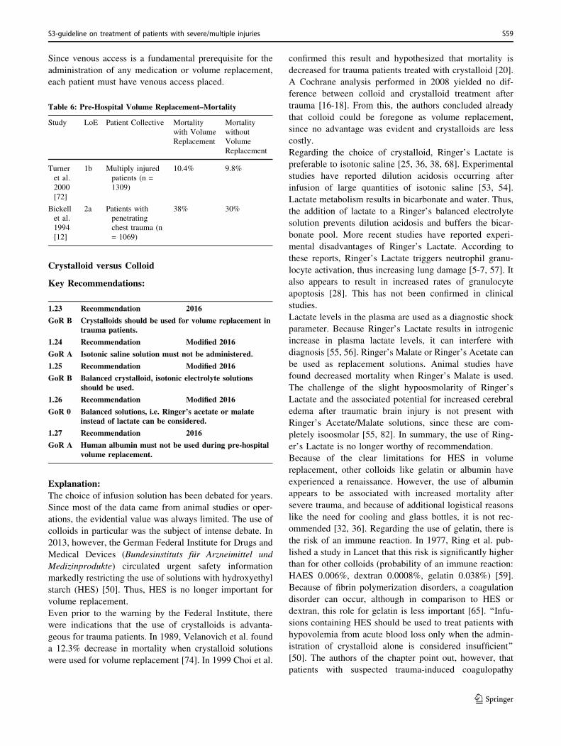
Since venous access is a fundamental prerequisite for the
administration of any medication or volume replacement,
each patient must have venous access placed.
Crystalloid versus Colloid
Key Recommendations:
1.23 Recommendation 2016
GoR B Crystalloids should be used for volume replacement intrauma patients.
1.24 Recommendation Modified 2016
GoR A Isotonic saline solution must not be administered.
1.25 Recommendation Modified 2016
GoR B Balanced crystalloid, isotonic electrolyte solutionsshould be used.
1.26 Recommendation Modified 2016
GoR 0 Balanced solutions, i.e. Ringer’s acetate or malateinstead of lactate can be considered.
1.27 Recommendation 2016
GoR A Human albumin must not be used during pre-hospitalvolume replacement.
Explanation:
The choice of infusion solution has been debated for years.
Since most of the data came from animal studies or oper-
ations, the evidential value was always limited. The use of
colloids in particular was the subject of intense debate. In
2013, however, the German Federal Institute for Drugs and
Medical Devices (Bundesinstituts fur Arzneimittel und
Medizinprodukte) circulated urgent safety information
markedly restricting the use of solutions with hydroxyethyl
starch (HES) [50]. Thus, HES is no longer important for
volume replacement.
Even prior to the warning by the Federal Institute, there
were indications that the use of crystalloids is advanta-
geous for trauma patients. In 1989, Velanovich et al. found
a 12.3% decrease in mortality when crystalloid solutions
were used for volume replacement [74]. In 1999 Choi et al.
confirmed this result and hypothesized that mortality is
decreased for trauma patients treated with crystalloid [20].
A Cochrane analysis performed in 2008 yielded no dif-
ference between colloid and crystalloid treatment after
trauma [16-18]. From this, the authors concluded already
that colloid could be foregone as volume replacement,
since no advantage was evident and crystalloids are less
costly.
Regarding the choice of crystalloid, Ringer’s Lactate is
preferable to isotonic saline [25, 36, 38, 68]. Experimental
studies have reported dilution acidosis occurring after
infusion of large quantities of isotonic saline [53, 54].
Lactate metabolism results in bicarbonate and water. Thus,
the addition of lactate to a Ringer’s balanced electrolyte
solution prevents dilution acidosis and buffers the bicar-
bonate pool. More recent studies have reported experi-
mental disadvantages of Ringer’s Lactate. According to
these reports, Ringer’s Lactate triggers neutrophil granu-
locyte activation, thus increasing lung damage [5-7, 57]. It
also appears to result in increased rates of granulocyte
apoptosis [28]. This has not been confirmed in clinical
studies.
Lactate levels in the plasma are used as a diagnostic shock
parameter. Because Ringer’s Lactate results in iatrogenic
increase in plasma lactate levels, it can interfere with
diagnosis [55, 56]. Ringer’s Malate or Ringer’s Acetate can
be used as replacement solutions. Animal studies have
found decreased mortality when Ringer’s Malate is used.
The challenge of the slight hypoosmolarity of Ringer’s
Lactate and the associated potential for increased cerebral
edema after traumatic brain injury is not present with
Ringer’s Acetate/Malate solutions, since these are com-
pletely isoosmolar [55, 82]. In summary, the use of Ring-
er’s Lactate is no longer worthy of recommendation.
Because of the clear limitations for HES in volume
replacement, other colloids like gelatin or albumin have
experienced a renaissance. However, the use of albumin
appears to be associated with increased mortality after
severe trauma, and because of additional logistical reasons
like the need for cooling and glass bottles, it is not rec-
ommended [32, 36]. Regarding the use of gelatin, there is
the risk of an immune reaction. In 1977, Ring et al. pub-
lished a study in Lancet that this risk is significantly higher
than for other colloids (probability of an immune reaction:
HAES 0.006%, dextran 0.0008%, gelatin 0.038%) [59].
Because of fibrin polymerization disorders, a coagulation
disorder can occur, although in comparison to HES or
dextran, this role for gelatin is less important [65]. ‘‘Infu-
sions containing HES should be used to treat patients with
hypovolemia from acute blood loss only when the admin-
istration of crystalloid alone is considered insufficient’’
[50]. The authors of the chapter point out, however, that
patients with suspected trauma-induced coagulopathy
Table 6: Pre-Hospital Volume Replacement–Mortality
Study LoE Patient Collective Mortality
with Volume
Replacement
Mortality
without
Volume
Replacement
Turner
et al.
2000
[72]
1b Multiply injured
patients (n =
1309)
10.4% 9.8%
Bickell
et al.
1994
[12]
2a Patients with
penetrating
chest trauma (n
= 1069)
38% 30%
S3-guideline on treatment of patients with severe/multiple injuries S59
123
should stay away from artificial colloids as volume
replacement.
Hypertonic Solutions
Key Recommendations:
1.28 Recommendation 2016
GoR 0 Hypertonic solutions can be used for hypotensivepatients with multiple injuries after blunt trauma.
1.29 Recommendation Modified 2016
GoR 0 In cases of penetrating trauma, hypertonic solutions canbe used provided pre-hospital volume therapy is beingperformed.
1.30 Recommendation 2016
GoR 0 Hypertonic solutions can be used in hypotensive patientswith severe traumatic brain injury.
Explanation:
In recent years, the use of hypertonic 7.5% saline solutions
has gained increasing importance, especially in the realm
of pre-hospital volume replacement. As described above,
disordered microcirculation appears to be the damaging
factor in cases of traumatic hemorrhagic shock.
Hypertonic solutions work by mobilizing intracellular
and interstitial fluid into the intravascular space, and thus,
improving microcirculation and the overall blood rheology.
Although most studies have infused pure 7.5% saline
solution at 4 ml/kg bodyweight, in Germany, only the
combination solutions with HES (Hyperhaes�) or 6%
dextran 70 (RescueFlow�) have been used. Because of the
problems discussed above regarding the use of HES, soon
neither of these solutions will be available in Germany.
Thus, the Southwest German Emergency Physicians’
Working Group (Arbeitsgemeinschaft Sudwestdeutsche
Notarzte e.V.) has recommended either importing Res-
cueFlow� (250 ml 7.5% NaCl solutions with 6% dextran
70) or using 10% NaCl solution. Neither recommendation
is unproblematic. In Germany, the import and use of non-
distributed pharmaceuticals requires individual prescription
with corresponding documentation, and the allergic
potential of dextran remains [59]. In addition, the dosage of
hypertonic solutions has been extensively studied by Rocha
e Silva et al., and is explicitly given as 7.5% saline. All
other dosages have increased mortality in dogs [61]. The
damaging factor is the potential hypernatremia. The use of
a 10% saline solution increases the quantity of NaCl from
18g to 20g. Thus, use must be considered critically and
should be first evaluated in studies.
Although there is much promising evidence for hyper-
tonic infusions in the literature, the poor availability of
appropriate hypertonic solutions allows no higher grade of
recommendation.
Disordered microcirculation is the essential factor
leading to complications from hemorrhage. Hypertonic
saline solutions work by rapidly mobilizing intracellular
and interstitial fluid into the intravascular space and thus,
improving blood rheology and with it, the microcirculation
[43]. Controlled studies have shown significant advantages
for hypertonic infusions. In 2004, Bunn et al. used a
Cochrane review to evaluate hypertonic versus isotonic
solutions [17]. The authors concluded that the available
data was not sufficient to make a final judgment regarding
use of hypertonic solutions. In 1991, in two controlled
randomized studies, Mattox et al. and Vassar et al. found
survival advantages for the use of hypertonic solutions,
particularly after traumatic brain injury [49, 73]. The work
of Alpar et al. from 2004 took the same direction in a study
of 180 patients. There were improved outcomes, particu-
larly in patients with TBI [3]. In 2009, Baker et al. also
reported a positive effect for hypertonic solutions given
after TBI [9]. Another controlled study of 229 patients
from 2004, however, found no significant difference in
long-term outcomes after TBI [24]. Positive effects for the
clinical treatment of TBI have been reported in other
studies. Wade et al. and Vassar et al. reported improved
mortality after TBI after initial therapy with hypertonic
solutions [73, 76]. Mortality decreased from 60 to 49% in
the Vassar et al. study, and from 37.0 to 26.9% in the Wade
et al. study with use of hypertonic solution. In the follow-
up treatment for increased intracranial pressure, the com-
bination of hypertonic solution/HES in particular showed
lowering effects [37, 41, 66, 78-80]. However, this effect
was not confirmed in a controlled clinical trial [67]. In
another current study by Bulger et al., no benefit from
hypertonic solutions was evident, so that the study was
discontinued after 1313 patients [67]. Wade et al. per-
formed a comparative investigation through a short meta-
analysis of 14 studies of hypertonic saline solutions with
and without dextran, and found no relevant benefit for
hypertonic solutions [76]. In 2003, the same author
reported a positive effect of hypertonic solutions for pen-
etrating trauma. In a double-blind study, 230 patients
received initial infusion of either hypertonic NaCl solution
or an isotonic solution. Mortality of patients receiving
hypertonic NaCl solution was 75.5%, significantly less than
patients receiving isotonic solution, with 82.5%. The rates
of surgery and bleeding were the same. The authors con-
cluded that hypertonic solutions improve survival after
penetrating trauma without increasing bleeding [75].
A current study of 209 polytrauma patients with blunt
trauma by Bulger et al. [15] compared Ringer’s Lactate to
hypertonic NaCl solution with dextran. The endpoint of the
study was ARDS-free survival. The study was discontinued
after intention-to-treat analysis, because there was no
apparent difference. In one subgroup analysis, an
S60
123
advantage for hypertonic solution with dextran was evident
only after massive transfusion. Even the most recent pub-
lication by this group reported no advantage for hypertonic
solutions after hemorrhagic shock [15]. In fact, patients not
requiring transfusion had even higher mortality rates after
administration of hypertonic solution (28-day mortality
hypertonic solution with dextran: 10%, isotonic solution:
4.8%, p\0.01) [15].
Immunological effects from hypertonic solutions have
also discussed. Experimental reports have described a
reduction in neutrophil activation and the pro-inflammatory
cascade [5-8, 22, 23, 26, 29, 57, 70]. Junger et al. also
attempted to demonstrate an inhibitory effect of pure
hypertonic solutions on post-traumatic inflammation in
trauma patients [40].
Hypertonic solutions lead to a rapid increase in blood
pressure and reduced volume needs [4, 14, 19, 21, 33, 44,
47, 48, 80]. The extent to which this affects treatment
outcomes cannot yet be conclusively answered by the
literature.
The evidence table for this chapter is found on page 80
of the guideline report.
References
1. American College of Surgeons Committee on Trauma ATLS
Student Course. Manual 9 Edition, 2012 (Chicago).
2. Adams HA, Piepenbrock S, Hempelmann G. Volume replace-
ment solutions–pharmacology and clinical use. Anasthesiol
Intensivmed Notfallmed Schmerzther. 1998;33(1):2–17 [LoE 5].3. Alpar EK, Killampalli VV. Effects of hypertonic dextran in
hypovolaemic shock: a prospective clinical trial. Injury.
2004;35(5):500–6 [LoE 1b].
4. Anderson JT, et al. Initial small-volume hypertonic resuscitation
of shock and brain injury: short- and long-term effects. J Trauma.
1997;42(4):592–600 (discussion 600-1. [LoE 5]).5. Angle N, et al. Hypertonic saline infusion: can it regulate human
neutrophil function? Shock. 2000;14(5):503–8 [LoE 2b].6. Angle N, et al. Hypertonic saline resuscitation reduces neutrophil
margination by suppressing neutrophil L selectin expression.
J Trauma. 1998a;45(1):7–12 (discussion 12-3. [LoE 5]).
7. Angle N, et al. Hypertonic saline resuscitation diminishes lung
injury by suppressing neutrophil activation after hemorrhagic
shock. Shock. 1998b;9(3):164–70 [LoE 5].8. Assalia A, et al. Influence of hypertonic saline on bacterial
translocation in controlled hemorrhagic shock. Shock.
2001;15(4):307–11 [LoE 5].9. Baker AJ, et al. Resuscitation with hypertonic saline-dextran
reduces serum biomarker levels and correlates with outcome in
severe traumatic brain injury patients. J Neurotrauma.
2009;26(8):1227–40.
10. Balogh Z, et al. Supranormal trauma resuscitation causes more
cases of abdominal compartment syndrome. Arch Surg.
2003;138(6):637–42 (discussion 642-3. [LoE 2a]).
11. Banerjee A, Jones R. Whither immediate fluid resuscitation?
Lancet. 1994;344(8935):1450–1 [LoE 5].
12. Bickell WH, et al. Immediate versus delayed fluid resuscitation
for hypotensive patients with penetrating torso injuries. N Engl J
Med. 1994;331(17):1105–9 [LoE 2b].
13. Brinkmeyer SD. Fluid resuscitation: an overview. J Am
Osteopath Assoc. 1983;82(5):326–30 [LoE 5].
14. Brock H, et al. Comparison of postoperative volume therapy in
heart surgery patients. Anaesthesist. 1995;44(7):486–92 [LoE
1b].15. Bulger EM, et al. Hypertonic resuscitation of hypovolemic
shock after blunt trauma: a randomized controlled trial. Arch
Surg. 2008;143(2):139–48 (discussion 149. [LoE 1b]).
16. Bunn F, et al. Hypertonic versus isotonic crystalloid for fluid
resuscitation in critically ill patients. Cochrane Database Syst
Rev. 2002;1:CD002045.
17. Bunn F, et al. Hypertonic versus near isotonic crystalloid for
fluid resuscitation in critically ill patients. Cochrane Database
Syst Rev. 2004;3:CD002045 [LoE 1a].
18. Bunn F, Trivedi D, Ashraf S. Colloid solutions for fluid
resuscitation. Cochrane Database Syst Rev. 2008;1:CD001319
[LoE 1a].19. Chiara O, et al. Resuscitation from hemorrhagic shock: exper-
imental model comparing normal saline, dextran, and hyper-
tonic saline solutions. Crit Care Med. 2003;31(7):1915–22 [LoE
5].20. Choi PT, et al. Crystalloids vs. colloids in fluid resuscitation: a
systematic review. Crit Care Med. 1999;27(1):200–10 [LoE1b].
21. Christ F, et al. Hyperosmotic-hyperoncotic solutions during
abdominal aortic aneurysm (AAA) resection. Acta Anaesthesiol
Scand. 1997;41(1 Pt 1):62–70.
22. Coimbra R, et al. Hypertonic saline resuscitation decreases
susceptibility to sepsis after hemorrhagic shock. J Trauma.
1997;42(4):602–6 (discussion 606-7. [LoE 5]).23. Coimbra R, et al. Role of hypertonic saline and pentoxifylline
on neutrophil activation and tumor necrosis factor-alpha
synthesis: a novel resuscitation strategy. J Trauma.
2005;59(2):257–64 (discussion 264-5. [LoE 5]).24. Cooper DJ, et al. Prehospital hypertonic saline resuscitation of
patients with hypotension and severe traumatic brain injury: a
randomized controlled trial. JAMA. 2004;291(11):1350–7 [LoE
1b].25. Coran AG, et al. The effect of crystalloid resuscitation in
hemorrhagic shock on acid-base balance: a comparison between
normal saline and Ringer’s lactate solutions. Surgery.
1971;69(6):874–80 [LoE 5].26. Corso CO, et al. Hypertonic saline dextran attenuates leukocyte
accumulation in the liver after hemorrhagic shock and resusci-
tation. J Trauma. 1999;46(3):417–23 [LoE 5].
27. Curry N, et al, The acute management of trauma hemorrhage: a
systematic review of randomized controlled trials. Critical Care.
2011;15(2).
28. Deb S, et al. Resuscitation with lactated Ringer’s solution in rats
with hemorrhagic shock induces immediate apoptosis.
J Trauma. 1999;46(4):582–8 (discussion 588-9. [LoE 5]).
29. Deitch EA, et al. Hypertonic saline resuscitation limits
neutrophil activation after trauma-hemorrhagic shock. Shock.
2003;19(4):328–33 [LoE 5].30. Dries DJ. Hypotensive resuscitation. Shock. 1996;6(5):311–6
[LoE 5].31. Dutton RP, Mackenzie CF, Scalea TM. Hypotensive resuscita-
tion during active hemorrhage: impact on in-hospital mortality.
J Trauma. 2002;52(6):1141–6.
32. Finfer S, et al. A comparison of albumin and saline for fluid
resuscitation in the intensive care unit. N Engl J Med.
2004;350(22):2247–56.
S3-guideline on treatment of patients with severe/multiple injuries S61
123
33. Fischer M, Hossmann KA. Volume expansion during cardiopul-
monary resuscitation reduces cerebral no-reflow. Resuscitation.
1996;32(3):227–40 [LoE 5].34. Giesecke AH Jr. C.M. Grande, and C.W. Whitten, Fluid therapy
and the resuscitation of traumatic shock. Crit Care Clin.
1990;6(1):61–72 [LoE 5].
35. Giraud R, et al. Impact of epinephrine and norepinephrine on
two dynamic indices in a porcine hemorrhagic shock model.
J Trauma Acute Care Surg. 2014;77(4):564–9 (quiz 650-1).36. Hankeln K, Lenz I, Hauser B. Hemodynamic effect of 6%
hydroxyethyl starch (HES 200,000/0.62). Anaesthesist.
1988;37(3):167–72 [LoE 2b].
37. Hartl R, et al. Hypertonic/hyperoncotic saline attenuates
microcirculatory disturbances after traumatic brain injury.
J Trauma. 1997;42(5 Suppl):S41–7 [LoE 2b].38. Healey MA, et al. Lactated ringer’s is superior to normal saline
in a model of massive hemorrhage and resuscitation. J Trauma.
1998;45(5):894–9 [LoE 5].
39. Holte K, et al. Liberal versus restrictive fluid management in
knee arthroplasty: a randomized, double-blind study. Anesth
Analg. 2007;105(2):465–74 [LoE 1b].40. Junger WG, et al. Resuscitation of traumatic hemorrhagic shock
patients with hypertonic saline-without dextran-inhibits neu-
trophil and endothelial cell activation. Shock.
2012;38(4):341–50.
41. Kempski O, et al. ‘‘Small volume resuscitation’’ as treatment of
cerebral blood flow disturbances and increased ICP in trauma
and ischemia. Acta Neurochir Suppl. 1996;66:114–7 [LoE 5].
42. Krausz MM, Bashenko Y, Hirsh M. Crystalloid and colloid
resuscitation of uncontrolled hemorrhagic shock following
massive splenic injury. Shock. 2001;16(5):383–8 [LoE 5].
43. Kreimeier U, et al. Small-volume resuscitation for hypovolemic
shock. Concept, experimental and clinical results. Anaesthesist.
1997;46(4):309–28 [LoE 5].44. Kreimeier U, Messmer K. Use of hypertonic NaCl solutions in
primary volume therapy. Zentralbl Chir. 1992;117(10):532–9
[LoE 5].
45. Kreimeier U, Prueckner S, Peter K. Permissive hypotension.
Schweiz Med Wochenschr. 2000;130(42):1516–24 [LoE 5].
46. Kroll W, et al. Prehospital fluid resuscitation. Acta Anaesthesiol
Scand Suppl. 1997;111:301–2 [LoE 5].
47. Matsuoka T, Hildreth J, Wisner DH. Uncontrolled hemorrhage
from parenchymal injury: is resuscitation helpful? J Trauma.
1996;40(6):915–21 (discussion 921-2. [LoE 5]).48. Matsuoka T, Wisner DH. Resuscitation of uncontrolled liver
hemorrhage: effects on bleeding, oxygen delivery, and oxygen
consumption. J Trauma. 1996;41(3):439–45.
49. Mattox KL, et al. Prehospital hypertonic saline/dextran infusion
for post-traumatic hypotension. The USA multicenter trial. Ann
Surg. 1991;213(5):482–91 [LoE 1b].50. Medizinprodukte, B.f.A.u., Rote-Hand-Brief zu HES-haltigen
Infusionslosungen: Neue Kontraindikationen und Anwendungs-
beschrankungen, in Rote-Hand-Brief. 2013, Bundesinstitut fur
Arzneimittel und Medizinprodukte.
51. Morrison CA, et al. Hypotensive resuscitation strategy reduces
transfusion requirements and severe postoperative coagulopathy
in trauma patients with hemorrhagic shock: preliminary results
of a randomized controlled trial. J Trauma. 2011;70(3):652–63.
52. Pargger H, Studer W, Ruttimann U. Volume therapy in
hypotensive trauma patients. Schweiz Med Wochenschr.
2000;130(42):1509–15 [LoE 5].
53. Pitts R, Lotspeich W. Bicarbonate and the renal regulation of
acid base balance. Am J Physiol. 1946;147(1):138–57.
54. Pitts RF, Ayer JL, Schiess WA. The renal regulation of acid-
base balance in man; the reabsorption and excretion of
bicarbonate. J Clin Invest. 1949;28(1):35–44.
55. Raum M, et al. Der Einfluß von exogenem Laktat auf gemessene
Plasmalaktatspiegel im hamorrhagischen Schock- eine kontrol-
lierte Studie am Schwein. Chir Forum. 2000;29:543–7.
56. Raum M, et al. Influence of lactate infusion solutions on the
plasma lactate concentration. Anasthesiol Intensivmed Not-
fallmed Schmerzther. 2002;37(6):356–8 [LoE 5].57. Rhee P, et al. Lactated Ringer’s solution resuscitation causes
neutrophil activation after hemorrhagic shock. J Trauma.
1998;44(2):313–9 [LoE 5].
58. Riddez L, Johnson L, Hahn RG. Central and regional hemody-
namics during crystalloid fluid therapy after uncontrolled intra-
abdominal bleeding. J Trauma. 1998;44(3):433–9 [LoE 5].59. Ring J, Messmer K. Incidence and severity of anaphylactoid
reactions to colloid volume substitutes. Lancet.
1977;1(8009):466–9.
60. Roberts K, et al. Hypotensive resuscitation in patients with
ruptured abdominal aortic aneurysm. Eur J Vasc Endovasc Surg.
2006;31(4):339–44 [LoE 1a].61. Rocha e Silva M, Velasco IT, Porfirio MF. Hypertonic saline
resuscitation: saturated salt-dextran solutions are equally effec-
tive, but induce hemolysis in dogs. Crit Care Med.
1990;18(2):203–7.
62. Rossi R. Early care or quick transport? The effectiveness of
preclinical treatment of emergency patients. Anaesthesist.
1997;46(2):126–32 [LoE 5].
63. Sampalis JS, et al. Direct transport to tertiary trauma centers
versus transfer from lower level facilities: impact on mortality
and morbidity among patients with major trauma. J Trauma.
1997a;43(2):288–95 (discussion 295-6).64. Sampalis JS, et al. Ineffectiveness of on-site intravenous lines: is
prehospital time the culprit? J Trauma. 1997b;43(4):608–15
(discussion 615-7).
65. Schlimp CJ, et al. The effect of fibrinogen concentrate and
factor XIII on thromboelastometry in 33% diluted blood with
albumin, gelatine, hydroxyethyl starch or saline in vitro. Blood
Transfus. 2013;11(4):510–7.
66. Schwarz S, et al. Effects of hypertonic saline hydroxyethyl
starch solution and mannitol in patients with increased intracra-
nial pressure after stroke. Stroke. 1998;29(8):1550–5 [LoE 2b].67. Shackford SR. Effect of small-volume resuscitation on intracra-
nial pressure and related cerebral variables. J Trauma.
1997;42(5 Suppl):S48–53 [LoE 5].
68. Shires GT. Pathophysiology and fluid replacement in hypov-
olemic shock. Ann Clin Res. 1977;9(3):144–50 [LoE 5].
69. Singbartl G. Significance of preclinical emergency treatment for
the prognosis of patients with severe craniocerebral trauma.
Anasth Intensivther Notfallmed. 1985;20(5):251–60 [LoE 2a].70. Tokyay R, et al. Effects of hypertonic saline dextran resusci-
tation on oxygen delivery, oxygen consumption, and lipid
peroxidation after burn injury. J Trauma. 1992;32(6):704–12
(discussion 712-3. [LoE 5]).71. Trunkey DD. Prehospital fluid resuscitation of the trauma
patient. An analysis and review. Emerg. Med Serv.
2001;30(5):93–5 (96, 98 passim. [LoE 5]).
72. Turner J, et al. A randomised controlled trial of prehospital
intravenous fluid replacement therapy in serious trauma. Health
Technol Assess. 2000;4(31):1–57 [LoE 1b].73. Vassar MJ, Perry CA, Holcroft JW. Prehospital resuscitation of
hypotensive trauma patients with 7.5% NaCl versus 7.5% NaCl
S62
123
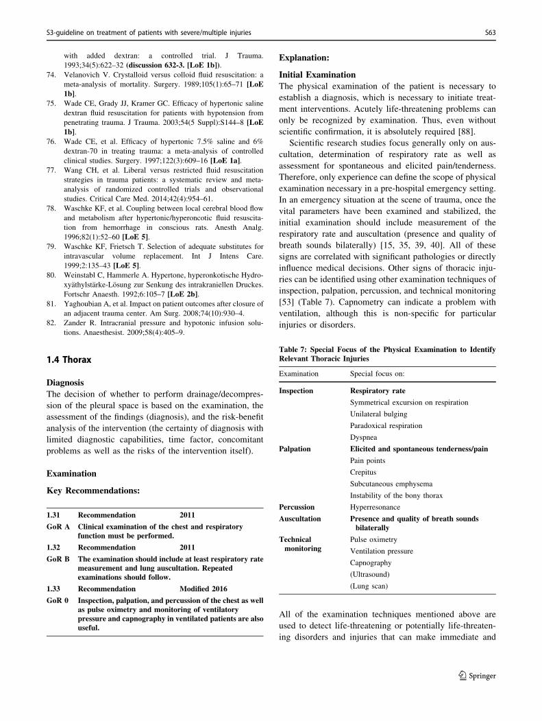
with added dextran: a controlled trial. J Trauma.
1993;34(5):622–32 (discussion 632-3. [LoE 1b]).
74. Velanovich V. Crystalloid versus colloid fluid resuscitation: a
meta-analysis of mortality. Surgery. 1989;105(1):65–71 [LoE
1b].75. Wade CE, Grady JJ, Kramer GC. Efficacy of hypertonic saline
dextran fluid resuscitation for patients with hypotension from
penetrating trauma. J Trauma. 2003;54(5 Suppl):S144–8 [LoE
1b].76. Wade CE, et al. Efficacy of hypertonic 7.5% saline and 6%
dextran-70 in treating trauma: a meta-analysis of controlled
clinical studies. Surgery. 1997;122(3):609–16 [LoE 1a].
77. Wang CH, et al. Liberal versus restricted fluid resuscitation
strategies in trauma patients: a systematic review and meta-
analysis of randomized controlled trials and observational
studies. Critical Care Med. 2014;42(4):954–61.
78. Waschke KF, et al. Coupling between local cerebral blood flow
and metabolism after hypertonic/hyperoncotic fluid resuscita-
tion from hemorrhage in conscious rats. Anesth Analg.
1996;82(1):52–60 [LoE 5].
79. Waschke KF, Frietsch T. Selection of adequate substitutes for
intravascular volume replacement. Int J Intens Care.
1999;2:135–43 [LoE 5].80. Weinstabl C, Hammerle A. Hypertone, hyperonkotische Hydro-
xyathylstarke-Losung zur Senkung des intrakraniellen Druckes.
Fortschr Anaesth. 1992;6:105–7 [LoE 2b].
81. Yaghoubian A, et al. Impact on patient outcomes after closure of
an adjacent trauma center. Am Surg. 2008;74(10):930–4.
82. Zander R. Intracranial pressure and hypotonic infusion solu-
tions. Anaesthesist. 2009;58(4):405–9.
1.4 Thorax
Diagnosis
The decision of whether to perform drainage/decompres-
sion of the pleural space is based on the examination, the
assessment of the findings (diagnosis), and the risk-benefit
analysis of the intervention (the certainty of diagnosis with
limited diagnostic capabilities, time factor, concomitant
problems as well as the risks of the intervention itself).
Examination
Key Recommendations:
1.31 Recommendation 2011
GoR A Clinical examination of the chest and respiratoryfunction must be performed.
1.32 Recommendation 2011
GoR B The examination should include at least respiratory ratemeasurement and lung auscultation. Repeatedexaminations should follow.
1.33 Recommendation Modified 2016
GoR 0 Inspection, palpation, and percussion of the chest as wellas pulse oximetry and monitoring of ventilatorypressure and capnography in ventilated patients are alsouseful.
Explanation:
Initial Examination
The physical examination of the patient is necessary to
establish a diagnosis, which is necessary to initiate treat-
ment interventions. Acutely life-threatening problems can
only be recognized by examination. Thus, even without
scientific confirmation, it is absolutely required [88].
Scientific research studies focus generally only on aus-
cultation, determination of respiratory rate as well as
assessment for spontaneous and elicited pain/tenderness.
Therefore, only experience can define the scope of physical
examination necessary in a pre-hospital emergency setting.
In an emergency situation at the scene of trauma, once the
vital parameters have been examined and stabilized, the
initial examination should include measurement of the
respiratory rate and auscultation (presence and quality of
breath sounds bilaterally) [15, 35, 39, 40]. All of these
signs are correlated with significant pathologies or directly
influence medical decisions. Other signs of thoracic inju-
ries can be identified using other examination techniques of
inspection, palpation, percussion, and technical monitoring
[53] (Table 7). Capnometry can indicate a problem with
ventilation, although this is non-specific for particular
injuries or disorders.
Table 7: Special Focus of the Physical Examination to IdentifyRelevant Thoracic Injuries
Examination Special focus on:
Inspection Respiratory rate
Symmetrical excursion on respiration
Unilateral bulging
Paradoxical respiration
Dyspnea
Palpation Elicited and spontaneous tenderness/pain
Pain points
Crepitus
Subcutaneous emphysema
Instability of the bony thorax
Percussion Hyperresonance
Auscultation Presence and quality of breath soundsbilaterally
Technicalmonitoring
Pulse oximetry
Ventilation pressure
Capnography
(Ultrasound)
(Lung scan)
All of the examination techniques mentioned above are
used to detect life-threatening or potentially life-threaten-
ing disorders and injuries that can make immediate and
S3-guideline on treatment of patients with severe/multiple injuries S63
123
specific interventions or logistic decisions necessary right
away. All of the diagnostic measures performed pre-hos-
pital are without specific risks. The disadvantage is loss of
time, which is generally minimal.
Some findings are strongly dependent on the examiner,
the patient, and the environment. Surrounding noise levels
can make auscultation difficult or impossible. Such cir-
cumstances need to be considered when selecting and
interpreting the primary diagnostic studies [35, 39, 72,
134].
Monitoring
Respiratory rate and auscultation as well as pulse oximetry
and ventilation pressure monitoring/capnography when
necessary should be continued, since airway problems, tube
displacement, tension pneumothorax, or acute respiratory
insufficiency can develop dynamically. Serial examinations
can serve as control of the interventions that have already
been performed.
Diagnosing Pneumothorax
Key Recommendations:
1.34 Recommendation 2011
GoR A A provisional diagnosis of pneumothorax and/orhemothorax must be made in cases of unilateral absenceor decrease in breath sounds (after controlling forcorrect tube placement). The lack of such findings onauscultation, particularly with normal respiratory rateand absence of thoracic discomfort, generally rules out alarge pneumothorax.
1.35 Recommendation 2011
GoR B Potential progression of a small, initially (in the pre-hospital stage) undetectable pneumothorax should bekept in mind.
Explanation:
At the present time, there are no evaluated methods
available in the pre-hospital setting to definitively detect or
exclude pneumothorax.
Ultrasound Examination
In hospital conditions, an ultrasound examination shows
evidence of pneumothorax and hemothorax (lung sliding,
seashore sign, B-lines/comet tail artifacts, etc.) with
extreme accuracy [5, 57], and is superior to both clinical
diagnosis and standard radiography. However, the esti-
mated magnitude of a confirmed pneumothorax is uncer-
tain and so far, poorly documented. Similarly, there has
been little study of the risk-benefit analysis including the
therapeutic consequences from the results of the investi-
gation. In addition to dependence on the examiner, the loss
of time and misdiagnosis by less experienced examiners
must be considered. In particular, there has been no reliable
experience with pre-hospital use, so a general recommen-
dation can’t be given. Nevertheless, pre-hospital ultrasound
performed by an experienced examiner can contribute to
confirmation of a provisional diagnosis of pneumothorax.
Increasing experience with ultrasound diagnosis of pneu-
mothorax in the pre-hospital setting could lead to more
prominence in the coming years. For these reasons, the lack
of expert consensus means that no recommendation for or
against the use of pre-hospital ultrasound for the diagnosis
of pneumothorax can be made. A new radar-based tech-
nology with a hand-held device has shown initial promis-
ing results for the detection of pneumothorax [4, 90].
Further studies must be performed to confirm the value and
practicality of widespread use before recommendations can
be given.
Auscultation, Dyspnea, Pain
A systematic review [137] analyzed studies regarding the
accuracy of clinical examination for the diagnosis of
pneumothorax.
Sensitivity was 90%. The specificity of a unilateral
decrease or lack of breath sounds, i.e. the probability that
patients without pneumothorax would not have these
findings, is very high, with 98%. The positive predictive
value, i.e. the probability that pneumothorax is actually
present when breath sounds are decreased, is also very high
with 86 - 97%. Pneumothorax not recognized on auscul-
tation had an average volume of 378 ml (max 800 ml), and
hemothorax of 277 ml (max 600 ml). In this study, thus, no
large, acutely life-threatening lesions were missed.
A requirement for this is that the appropriate position of
the endotracheal tube (when present) must be ensured, as
much as possible (capnography). The studies mentioned
here were performed in the emergency department of the
hospital, and not at the scene of the trauma. They appear to
be transferable, however, since there are also many com-
parable disruptions (e.g. increased noise, surrounding
activity) in the emergency department. False positive
findings occur occasionally (4.5% of cases in [87]) with
tube misplacement, diaphragmatic rupture [1, 6], or ven-
tilatory disruption (large atelectasis, movement on deep
respiration).
In cases of severe bilateral chest trauma, bilateral
pneumothorax needs to be considered. In such cases,
atypical findings might be present.
Data regarding differentiation between pneumothorax
and hemothorax or a mixed clinical picture are not avail-
able. Here percussion can be helpful, although this has only
limited relevance in the pre-hospital setting, since differ-
entiation between pneumothorax and hemothorax has no
verifiable effect on the need for therapy (see below).
Ultrasound examination can make the distinction (dis-
cussed above).
S64
123
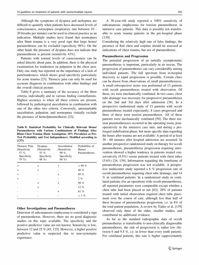
Although the symptoms of dyspnea and tachypnea are
difficult to quantify when patients have decreased levels of
consciousness, normopnea (respiratory rate between 10 -
20 breaths per minute) can be used in clinical practice as an
indication. Multiple studies have found that normopnea
after blunt trauma is a very good sign that large hemo/
pneumothorax can be excluded (specificity 98%). On the
other hand, the presence of dyspnea does not indicate that
pneumothorax is present (sensitivity 43%).
Patients with normal levels of consciousness can be
asked directly about pain. In addition, there is the physical
examination for tenderness on palpation in the chest area.
Only one study has reported on the importance of a lack of
pain/tenderness, which shows good specificity particularly
for acute trauma [23]. Thoracic pain can only be used for
accurate diagnosis in combination with other findings and
the overall clinical picture.
Table 8 gives a summary of the accuracy of the three
criteria, individually and in various finding constellations.
Highest accuracy is when all three criteria are present,
followed by pathological auscultation in combination with
one of the other two criteria. Conversely, unremarkable
auscultation, palpation, and normopnea virtually exclude
the presence of hemo/pneumothorax [23].
Other Investigations and Pneumothorax
Detection of subcutaneous emphysema is considered a sign
of pneumothorax. However, there are no good diagnostic
studies on this topic available. The specificity and the
positive predictive value are not known. Sensitivity is low,
between 12 and 25 % [45, 125]. However, a higher positive
predictive value is suspected due to non-systematic
experience.
A 30-year-old study reported a 100% sensitivity of
subcutaneous emphysema for tension pneumothorax in
intensive care patients. This data is possibly not transfer-
able to acute trauma patients in the pre-hospital phase
[130].
Considering the relatively high rate of false findings, the
presence of flail chest and crepitus should be assessed as
indications of chest trauma, but not of pneumothorax.
Pneumothorax and Progression
The potential progression of an initially asymptomatic
pneumothorax is important, particularly in air rescue. The
progression of pneumothorax can vary considerably among
individual patients. The full spectrum from in-hospital
discovery to rapid progression is possible. Certain clues
can be drawn from observations of small pneumothoraxes.
A small retrospective series was performed of 13 patients
with occult pneumothorax treated with observation. Of
these, six were mechanically ventilated. In two cases, chest
tube drainage was necessary for progressive pneumothorax
on the 2nd and 3rd days after admission [38]. In a
prospective randomized study of 21 patients with occult
pneumothorax treated expectantly, 8 cases progressed and
three of these were tension pneumothorax. All of these
patients were mechanically ventilated [59]. The three ten-
sion pneumothoraxes occurred in the operating room, post-
operatively in the intensive care unit, and during a pro-
longed stabilization phase, but more specific data regarding
the hours after trauma are not available. A period of at least
30 - 60 minutes after hospital admission are assumed. In
another prospective randomized study on therapy for occult
pneumothorax, pneumothorax progression requiring inter-
vention showed a higher tendency in patients treated con-
servatively (9.5%) versus patients treated with chest tubes
(5.6%) [24, 139]. Information regarding the timeframe of
pneumothorax progression was not available. A prospec-
tive multicenter study reported a 6 % progression rate of
occult pneumothorax requiring chest tube drainage, and 14
% in ventilated patients. In a randomized study in venti-
lated patients (for an operation) with occult pneumothorax,
all reported parameters were comparable except whether a
chest tube had been placed or not [83]. 20% of patients
treated with initial observation required chest tube place-
ment over the course of care, although less than half of
these because of pneumothorax progression, i.e. in 8% of
the total patient population. A review by Yadav et al. [139]
observed only three of the older, smaller studies, and
contributed no additional evidence.
As far as the standard radiographic data of occult
pneumothorax is transferable to non-clinically diagnosable
pneumothorax, the risk of progression is rather low (be-
tween 6 and 9.5 %, i.e. in fewer than every tenth patient).
For ventilated patients, this rate is higher (approximately
Table 8: Statistical Probability for Clinically Relevant Hemo/Pneumothorax with Various Combinations of Findings AfterBlunt Chest Trauma (Basic Assumption: 10% Prevalence as Pre-Test Probability and Test Independence). Modified according to[137]:
Thoracic Pain
(Sensitivity
57 %,
Specificity
79 %)
Dyspnea
(Sensitivity
43 %,
Specificity
98 %)
Auscultation
(Sensitivity
90 %,
Specificity
98 %)
Probability of
Hemo/
Pneumothorax
+ + + [ 99 %
+ + - 40 %
+ - + 89 %
+ - - 2 %
- + + 98 %
- + - 12 %
- - + 61 %
- - - \ 1 %
S3-guideline on treatment of patients with severe/multiple injuries S65
123
14 %), so that the classic situation of progressive pneu-
mothorax (up to tension pneumothorax) after intubation
actually occurs in one of seven patients receiving positive
pressure ventilation [106].
In summary, the data suggests that small, clinically non-
diagnosable pneumothorax, especially in non-ventilated
patients, progresses relatively seldom (and most only
slowly) as a rule, and thus, generally does not require
emergency decompression in the pre-hospital setting.
Nevertheless, progression of even an initially clinically
undetectable pneumothorax must be reckoned with.
Increased attention to monitoring, progress checks, and
readiness for decompression are necessary to recognize
cases of progression in a timely manner.
Diagnosis Tension Pneumothorax
Key Recommendations:
1.36 Recommendation Modified 2016
GoR B A provisional diagnosis of tension pneumothorax shouldbe made in cases of unilaterally absent breath sounds onlung auscultation (after controlling for correct tubeplacement) along with the presence of typical symptoms,particularly severe respiratory or hemodynamicinstability.
Explanation:
A number of systematic reviews have been published
regarding the diagnostic accuracy of examination findings
in the case of tension pneumothorax, based on cohort
studies, small case series, and case reports. However, there
is no uniform understanding of exactly what falls under the
definition of tension pneumothorax. Definitions range from
pneumothorax with life-threatening consequences to vital
functions, to a hiss of escaping air on needle decompres-
sion, to mediastinal shift on chest x-ray, to increased
ipsilateral intrapleural pressure, and on to hemodynamic
compromise [89].
Experimental investigations have found that the respi-
ratory changes and paralysis of the respiratory center as a
result of the hypoxia precede circulatory arrest. The
hypotension, which culminates in circulatory arrest, is a
late sign of tension pneumothorax [14, 122].
A review article from 2005 characterized ‘‘shortness of
breath,’’ and ‘‘tachycardia’’ as the typical and most com-
mon symptom/sign of tension pneumothorax in conscious
patients [89]. The same authors also found, however, that
the hemodynamic symptoms occur earlier in ventilated
patients, and that respiratory symptoms and the drop in
blood pressure often manifest at the same time. In
mechanically ventilated patients, extremely elevated or
increasing airway pressures are another important sign
found in some 20% of patients with hemo/pneumothorax
[15, 39]. A current systematic review reported significant
differences in the manifestation of tension pneumothorax
between spontaneously breathing patients and those being
mechanically ventilated [120].
Chest pain, tachypnea, and decreased breath sounds
occurred in more than 45 % of spontaneously breathing
patients. Dyspnea/respiratory distress, hypoxia with oxy-
gen demand, tachycardia, and hyperresonance were present
in 30 to 45 % of cases. Tracheal deviation (15-30 %) or
hypotension (when present, with a slow progression), dis-
tended neck veins, subcutaneous emphysema, cardiac
arrest (each under 15%) were much more seldom.
In contrast, in ventilated patients, the most common
signs ([ 45%) were decreased breath sounds as well as
hypotension (often with an acute beginning) and hypoxia.
Also common (30-45 %) were tachycardia, subcutaneous
emphysema, and cardiac arrest.
The diagnostic accuracy of the individual clinical signs
and findings has not yet been evaluated.
According to expert opinion, the combination of (uni-
lateral) absence of breath sounds (with verified tube posi-
tion) and changes of vital respiratory or hemodynamic
parameters makes tension pneumothorax so probable that
the diagnosis should be given and the necessary therapeutic
interventions should be performed. Further diagnostic
studies represent avoidable delay and should be foregone.
The consequences of a false positive tension pneumothorax
diagnosis are much less severe than those from the omis-
sion of required decompression.
Indications for Pleural Decompression
Key Recommendations:
1.37 Recommendation New 2016
GoR A Tension pneumothorax is the most frequent reversiblecause of traumatic cardiac arrest and must bedecompressed in the pre-hospital setting.
1.38 Recommendation 2011
GoR A Clinically suspected tension pneumothorax must bedecompressed immediately.
1.39 Recommendation 2011
GoR B Pneumothorax diagnosed on auscultation should bedecompressed in patients receiving positive pressureventilation.
1.40 Recommendation 2011
GoR B Pneumothorax diagnosed on auscultation should betreated expectantly with close observation in non-mechanically ventilated patients.
S66
123
Explanation:
There are no investigations comparing intervention and
expectant therapy. The treatment recommendations are
based on expert opinions and consideration of probabilities.
Tension pneumothorax
Tension pneumothorax is an acute life-threatening situation.
Left untreated, it generally leads to death. Death can occur
within minutes of the onset of signs of insufficient respira-
tory and hemodynamic function. There is no alternative to
decompression. Expert opinion is that particularly in cases of
circulatory or respiratory impairment, emergency decom-
pression must be performed immediately and that the time
lost in transport even to a hospital in the immediate vicinity
represents an unjustifiable delay. A study of 3500 autopsies
identified 39 cases of tension pneumothorax (incidence
1.1%), of which half were not diagnosed while the patient
was still living. Among soldiers of the Vietnam war, tension
pneumothorax occurred in 3.9 % of all patients with chest
injuries and in 33 % of soldiers with deadly chest injuries
[99]. An analysis of 20 patients categorized as unexpected
survivors according to TRISS prognosis reported that seven
of them underwent pre-hospital decompression for tension
pneumothorax [27]. In cases of pre-hospital resuscitation
post-trauma, spontaneous circulation was restored in 4 of 18
patients with decompression (once with needle, four with
mini-thoracotomy) [105]. An analysis of patients with
trauma-associated circulatory arrest identified decompres-
sion of tension pneumothorax as the most important factor
contributing to improved prognosis [76]. In a current anal-
ysis, untreated tension pneumothorax was identified as one
of the most frequent preventable causes of death [84].
Through a special training program for paramedics on the
detection and treatment of tension pneumothorax, the rate of
tension pneumothorax untreated by rescue services
decreased from 1.35% (10 of 740 patients) to 0.4% (4 of
1034) [30].
Diagnosed Pneumothorax
A large pneumothorax, which can be assumed with typical
auscultation findings, is a basic indication for evacuation of
the pleural cavity. Whether this needs to be done in the pre-
hospital setting or in the hospital is difficult to decide for
individual cases. The risk of progression from simple to
tension pneumothorax as well as the time this takes is
variable and difficult to estimate. The literature offers
neither general data on the topic nor risk factors. There is
evidence that tension pneumothorax is found on admission
to the Emergency Department more frequently in cases of
chest trauma that have been intubated versus non-intubated
patients.
Overall, it seems plausible to the experts that pneu-
mothorax diagnosed by auscultation in ventilated patients
has markedly higher risk to develop tension pneumothorax,
and thus an indication for pre-hospital decompression.
If a patient with pneumothorax diagnosed on ausculta-
tion is not ventilated, the risk for developing tension
pneumothorax appears to be much less. In a series of 54
cases of trauma-induced pneumothorax, 29 were treated
conservatively, i.e. without chest tube placement. These
patients were not mechanically ventilated, and most had no
accompanying injuries. Chest tube drainage was placed in
only two cases, as a result of radiologically progressing
pneumothorax six hours after hospital admission [81]. In
this case, pre-hospital decompression appears unnecessary
(over expectant therapy with close monitoring and clinical
controls). Due to the potential risks of pre-hospital
decompression, in such cases it should only be carried out
with strict consideration of risks versus benefits.
If appropriate monitoring and clinical controls are not
easily possible, e.g. during helicopter transport, there is a
certain, unquantifiable risk that tension pneumothorax
could develop and that this would not be noticed in time or
adequate therapy would not be possible due to space lim-
itations. In such situations, when appropriate clinical signs
are present and according to individual circumstances,
pneumothorax decompression may be required for non-
intubated patients prior to transport.
Chest Trauma Without Direct Diagnosis of
Pneumothorax
If there are equal breath sounds bilaterally, the presence of
clinically relevant pneumothorax is very improbable. In
this case, there is no indication for pre-hospital decom-
pression or pleural space evacuation, even when there is
other evidence of chest trauma (not specific for
pneumothorax).
A systematic review [137] found that the incidence of
pneumothorax is relatively low (10 to 50 %) even when
chest trauma is present. Thus, if an invasive intervention is
carried out for a diagnosis of chest trauma alone without
concrete evidence of pneumothorax, at least every second
patient and up to nine of ten patients would be treated
unnecessarily. Since cases of occult pneumothorax, only
detectable on CT, were also included in this study, the rate
of pneumothoraxes requiring evacuation was low. Even
when pneumothorax was suspected due to specific clinical
signs, the rate of unnecessary needle decompression and
chest tube evacuation was between 9 and 65 % [8, 15, 124].
Thus, for individual cases with grounds, in ventilated
patients with unmistakable signs of thoracic trauma but
unsuspicious auscultation findings, decompression can be
performed prior to longer transports or helicopter transport
with limited clinical monitoring or treatment options. The
high rate of false positive diagnoses for chest trauma by the
emergency physician must be considered.
S3-guideline on treatment of patients with severe/multiple injuries S67
123
Under these conditions, decompression is not indicated
in non-ventilated patients.
Hemothorax
The only general indications for pleural drainage/decom-
pression in the acute pre-hospital setting are tension
pneumothorax and massive hemothorax. The management
of pneumothorax has been characterized above. Although
hemothorax is a basic indication for pleural evacuation,
there is generally no direct danger of compression from this
blood and thus, no indication for pre-hospital drainage.
Only in cases of massive bleeding, possibly with the
development of a problem such as a tension pneumothorax,
would emergency drainage be indicated. However, such
situations would generally be associated with abnormal
auscultation and thus, would proceed as in the case of
pneumothorax. In the pre-hospital setting it is typically
difficult to differentiate hemothorax from hemopneumoth-
orax. Clinical signs of hemothorax versus pneumothorax
would be dullness versus hyperresonance to percussion,
provided the ambient conditions enable differentiation.
Ultrasound examination can differentiate the two.
Therapy
Methods
The goal of treatment is decompression of the positive
pressure in tension pneumothorax or tension hemothorax.
The second goal of treatment is to avoid development of a
tension pneumothorax from simple pneumothorax. Per-
manent and thorough evacuation of air and blood is not
important in the pre-hospital setting.
Key Recommendations:
1.41 Recommendation Modified 2016
GoR B Tension pneumothorax should be decompressed withsingle needle decompression. Surgical opening of thepleural cavity should follow, with or without chest tubeplacement.
1.42 Recommendation 2011
GoR B When indicated, pneumothorax should be treated withchest tube placement.
Explanation:
Because there are no suitable comparisons of data from the
three methods (needle decompression, surgical opening of
the pleural space alone, opening of the pleural space with
immediate chest tube drainage), there can be no evidence-
based recommendations for one method over another. For
all three methods, there are (predominantly retrospective)
data, case series, and case reports available that demon-
strate that successful decompression of tension pneu-
mothorax is possible.
Pathophysiologically, for sustained decompression it is
necessary that the amount of air expelled into the pleural
space with each inhalation also exits through the decom-
pression device (regardless of which method has been
chosen, thus needle or chest tube). The diameter with x4
then joins flow resistance. Thus, needle decompression
(and even single chest tube placement) could remain
ineffective, e.g. in tracheobronchial injury.
With the low level of evidence regarding the various
methods, and to directly compare the different methods
according to risk-benefits profile, the individual abilities of
the treating emergency provider should also be taken as a
practical consideration. One investigation reported a sig-
nificantly reduced complication rate after chest tube
placement by surgeons versus emergency physicians [61].
A more current study in North America also reported lower
complication rates in resident physicians training in sur-
gical versus non-surgical specialties [11]. The extent to
which these results are transferable to the German emer-
gency physician system cannot be evaluated due to the lack
of reliable data.
Chest Tube Drainage: Effectiveness and Complications
Chest tube insertion is an appropriate, highly effective ([85 %), but complication-ridden intervention to decompress
tension pneumothorax, which must be applied particularly
when alternative measures fail or prove inadequate. Typi-
cally, it offers definitive treatment and has the highest
success rate. In 79 to 95 % of cases, chest tube drainage
placed in the pre-hospital setting was the definitive and
successful intervention [10, 51, 115].
Conversely, chest tube drainage has a failure rate of 5.4
- 21 % (mean 11.2 %) due to misplacement or insufficient
effectiveness. Need for additional chest tube placement
occurred with the same frequency [10, 33, 44, 51, 61, 70,
115, 124]. Affected cases were roughly equally divided
between pneumo and hemothorax. Persisting tension
pneumothorax was also observed in individual cases of
chest tubes placed in the pre-hospital setting [10, 29, 98].
In addition to tube misplacement, this condition can occur
in rare cases of a highly productive bronchopulmonary air
fistula, which exceeds the discharge capacity of the
established chest tube (e.g. in cases of large parenchymal
tears or injuries to larger bronchi).
The pooled complication rates for chest tube drainage
placement for pre-hospital versus hospital placement show
increased complications in the former for subcutaneous
placement (2.53 vs. 0.39 %), intraparenchymal misplace-
ment (1.37 vs. 0.63 %), and intra-abdominal misplacement
(0.87 vs. 0.73 %). Conversely, the infection rates are
reversed (0.55 vs. 1.74 %) [52, 137]. Two studies directly
comparing the complication rates pre- and in-hospital at the
same institution [129, 141] found comparable infection
S68
123
rates (9.4 vs. 11.7 %) and misplacement (0 vs. 1.2 %).
Duration of drainage placement was comparable in both
groups. A current study of chest tubes placed in the
emergency department found that 70 % of patients suffered
acute and 40 % delayed complications [127]. In addition to
retroperitoneal misplacement (1.1 %), intercostal artery
injury (1.1 %), persisting pneumothorax (12.2 %) and over-
advancement of the drain (33.3 %), 38.9 % of patients
suffered pneumothorax recurrence with the chest tube in
place, and more seldom (each 2.2 %) pleural empyema or
local infection at the entry site. Another study observed a
complication rate of 22.1 % [102].
In addition, for anterior to mid-axillary line chest tube
drainage insertion, case studies have reported injury to the
intercostal arteries [31], lung perforation [63], perforation
of the right atrium [32, 100, 128], the right ventricle [117],
and the left ventricle [47], subclavian artery stenosis from
internal pressure of the drainage tip [107], ipsilateral
Horner syndrome from drain pressure on the apex of the
stellate ganglion [21, 29], intra-abdominal misplacement
[62], perforation of the liver [45], the stomach [6], and the
colon [1] in case of diaphragmatic hernia, a lesion of the
subclavian vein, perforation of the inferior vena cava [60],
and provocation of atrial fibrillation [13].
With insertion at the mid-clavicular line, there have
been reports of subclavian vessel fistula [42], cardiac wall
perforation [54], and perforation of the right atrium [100].
Other known complications include perforations of the
esophagus, the mediastinum with creation of contralateral
pneumothorax, phrenic nerve injury, etc.
Simple Surgical Opening: Effectiveness and
Complications
Simple surgical opening of the pleural space is an appro-
priate, effective, and relatively simple measure to decom-
press a tension pneumothorax. However, it is only
appropriate for patients receiving positive pressure venti-
lation, because only they have constant positive
intrapleural pressure. In spontaneously breathing patients,
negative intrapleural pressure is created, which can then
suck air through the thoracotomy into the thorax.
Clinical experience shows that air is released when the
pleural space is opened with a mini-thoracotomy for chest
tube placement to decompress pneumothorax or hemotho-
rax. Symptoms can improve dramatically in cases of a
tension pneumothorax with hemodynamic effects. This
technique was evaluated in the pre-hospital setting in a case
series of 45 patients and was found to be effective without
major complications [48]. In a prospective observational
study of an air rescue system over two years, 55 patients
with 59 suspected cases of pneumothorax were treated with
simple surgical opening. The average arterial oxygen sat-
uration increased from 86.4 % to 98.5 % as a result of the
procedure. On surgical opening, either pneumothorax or
hemothorax was found in 91.5 % of patients. Recurrent
pneumothorax was not observed by these authors, nor any
other serious complications (significant bleeding, lung
laceration, pleural empyema) [96].
However, another series found relevant complications in
9 % of patients, in whom almost half of the cases had to do
with non-decompressed or recurrent (e.g. through over-
lapping layers of soft tissue) tension pneumothorax [9].
Thus, insertion of chest tube drainage through the existing
mini-thoracotomy is indicated in hospital.
Needle Decompression: Effectiveness and
Complications
Needle decompression is an appropriate, frequently effec-
tive (approximately 32 - 53 %), simple, but not compli-
cation-free drainage procedure. If the effects are absent or
insufficient, surgical decompression and/or chest tube
insertion must be performed immediately.
The problem with clinical studies in the pre-hospital
setting is the lack of scientific certainty that tension
pneumothorax was actually present prior to needle
decompression, which makes it difficult to judge effec-
tiveness. With a pig model, the failure rate was 58 %, either
because within five minutes a secondary malfunction
occurred (bending, blockage, displacement) or because the
pressure release was not sufficient [94].
In pre-hospital studies, needle decompression yielded air
in 32 - 47 % of cases [15, 46]. Clinical improvements were
seen in 12 to 60 % of patients in whom needle decom-
pression was performed [15, 46, 58].
In contrast, one prospective series of 14 patients (five
further patients died in the Emergency Department and
were unsuitable for analysis) after needle decompression
found eight patients without evidence of pneumothorax,
two patients with occult pneumothorax, two patients with
persistent pneumothorax, one case of successfully decom-
pressed tension pneumothorax, and one case of persistent
tension pneumothorax [43], so that of 14 patients only one
clearly benefited.
In the Barton [15] study, needle decompression needed
to be supplemented with chest tube placement in 40 % of
cases (32 of 123) because of insufficient effectiveness. In
other pre-hospital studies [37, 46], chest tubes were needed
after needle decompression in 53 - 67 % of cases.
In one study, needle decompression did not work at all
in 4.1 % of verified cases of pneumothorax, because the
needle could not be placed deep enough. In 2.4 % of cases
there was secondary needle displacement and in 4.1 % it
was too difficult to place. No organ injuries were reported
[15]. Another study found that needle decompression was
unsuccessful in 2 % of patients because the needle was not
deep enough. In another 2 % there was no indication and
S3-guideline on treatment of patients with severe/multiple injuries S69
123
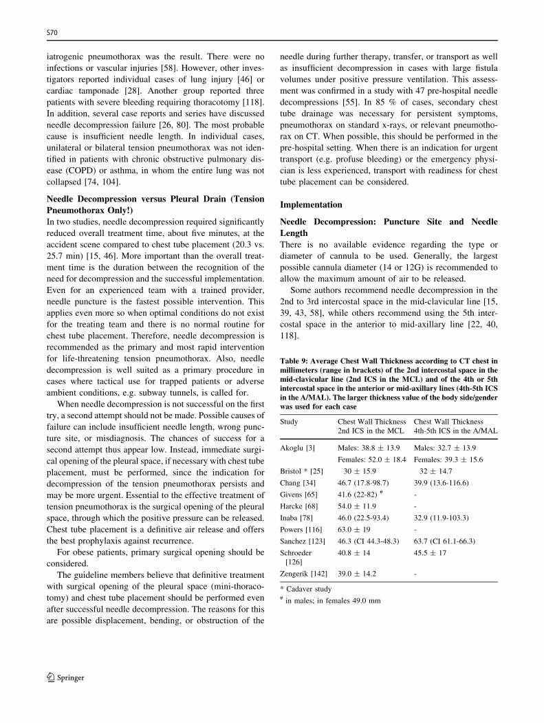
iatrogenic pneumothorax was the result. There were no
infections or vascular injuries [58]. However, other inves-
tigators reported individual cases of lung injury [46] or
cardiac tamponade [28]. Another group reported three
patients with severe bleeding requiring thoracotomy [118].
In addition, several case reports and series have discussed
needle decompression failure [26, 80]. The most probable
cause is insufficient needle length. In individual cases,
unilateral or bilateral tension pneumothorax was not iden-
tified in patients with chronic obstructive pulmonary dis-
ease (COPD) or asthma, in whom the entire lung was not
collapsed [74, 104].
Needle Decompression versus Pleural Drain (Tension
Pneumothorax Only!)
In two studies, needle decompression required significantly
reduced overall treatment time, about five minutes, at the
accident scene compared to chest tube placement (20.3 vs.
25.7 min) [15, 46]. More important than the overall treat-
ment time is the duration between the recognition of the
need for decompression and the successful implementation.
Even for an experienced team with a trained provider,
needle puncture is the fastest possible intervention. This
applies even more so when optimal conditions do not exist
for the treating team and there is no normal routine for
chest tube placement. Therefore, needle decompression is
recommended as the primary and most rapid intervention
for life-threatening tension pneumothorax. Also, needle
decompression is well suited as a primary procedure in
cases where tactical use for trapped patients or adverse
ambient conditions, e.g. subway tunnels, is called for.
When needle decompression is not successful on the first
try, a second attempt should not be made. Possible causes of
failure can include insufficient needle length, wrong punc-
ture site, or misdiagnosis. The chances of success for a
second attempt thus appear low. Instead, immediate surgi-
cal opening of the pleural space, if necessary with chest tube
placement, must be performed, since the indication for
decompression of the tension pneumothorax persists and
may be more urgent. Essential to the effective treatment of
tension pneumothorax is the surgical opening of the pleural
space, through which the positive pressure can be released.
Chest tube placement is a definitive air release and offers
the best prophylaxis against recurrence.
For obese patients, primary surgical opening should be
considered.
The guideline members believe that definitive treatment
with surgical opening of the pleural space (mini-thoraco-
tomy) and chest tube placement should be performed even
after successful needle decompression. The reasons for this
are possible displacement, bending, or obstruction of the
needle during further therapy, transfer, or transport as well
as insufficient decompression in cases with large fistula
volumes under positive pressure ventilation. This assess-
ment was confirmed in a study with 47 pre-hospital needle
decompressions [55]. In 85 % of cases, secondary chest
tube drainage was necessary for persistent symptoms,
pneumothorax on standard x-rays, or relevant pneumotho-
rax on CT. When possible, this should be performed in the
pre-hospital setting. When there is an indication for urgent
transport (e.g. profuse bleeding) or the emergency physi-
cian is less experienced, transport with readiness for chest
tube placement can be considered.
Implementation
Needle Decompression: Puncture Site and Needle
Length
There is no available evidence regarding the type or
diameter of cannula to be used. Generally, the largest
possible cannula diameter (14 or 12G) is recommended to
allow the maximum amount of air to be released.
Some authors recommend needle decompression in the
2nd to 3rd intercostal space in the mid-clavicular line [15,
39, 43, 58], while others recommend using the 5th inter-
costal space in the anterior to mid-axillary line [22, 40,
118].
Table 9: Average Chest Wall Thickness according to CT chest inmillimeters (range in brackets) of the 2nd intercostal space in themid-clavicular line (2nd ICS in the MCL) and of the 4th or 5thintercostal space in the anterior or mid-axillary lines (4th-5th ICSin the A/MAL). The larger thickness value of the body side/genderwas used for each case
Study Chest Wall Thickness
2nd ICS in the MCL
Chest Wall Thickness
4th-5th ICS in the A/MAL
Akoglu [3] Males: 38.8 ± 13.9
Females: 52.0 ± 18.4
Males: 32.7 ± 13.9
Females: 39.3 ± 15.6
Bristol * [25] 30 ± 15.9 32 ± 14.7
Chang [34] 46.7 (17.8-98.7) 39.9 (13.6-116.6)
Givens [65] 41.6 (22-82) # -
Harcke [68] 54.0 ± 11.9 -
Inaba [78] 46.0 (22.5-93.4) 32.9 (11.9-103.3)
Powers [116] 63.0 ± 19 -
Sanchez [123] 46.3 (CI 44.3-48.3) 63.7 (CI 61.1-66.3)
Schroeder
[126]
40.8 ± 14 45.5 ± 17
Zengerik [142] 39.0 ± 14.2 -
* Cadaver study# in males; in females 49.0 mm
S70
123
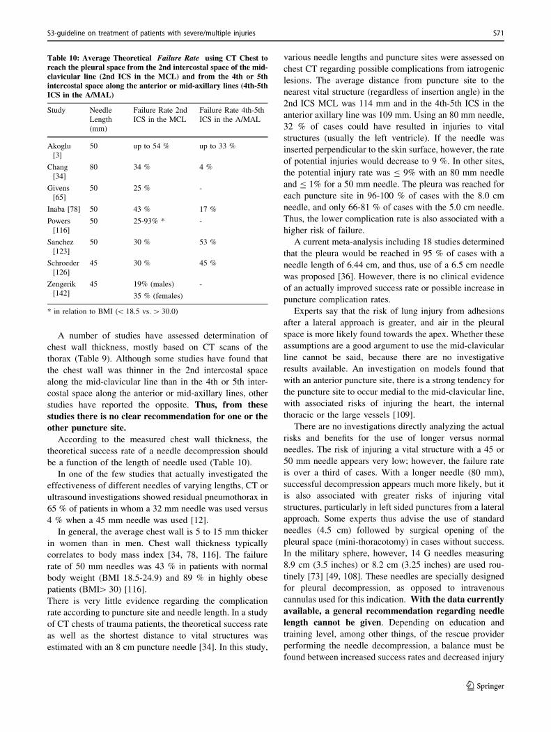
A number of studies have assessed determination of
chest wall thickness, mostly based on CT scans of the
thorax (Table 9). Although some studies have found that
the chest wall was thinner in the 2nd intercostal space
along the mid-clavicular line than in the 4th or 5th inter-
costal space along the anterior or mid-axillary lines, other
studies have reported the opposite. Thus, from these
studies there is no clear recommendation for one or the
other puncture site.
According to the measured chest wall thickness, the
theoretical success rate of a needle decompression should
be a function of the length of needle used (Table 10).
In one of the few studies that actually investigated the
effectiveness of different needles of varying lengths, CT or
ultrasound investigations showed residual pneumothorax in
65 % of patients in whom a 32 mm needle was used versus
4 % when a 45 mm needle was used [12].
In general, the average chest wall is 5 to 15 mm thicker
in women than in men. Chest wall thickness typically
correlates to body mass index [34, 78, 116]. The failure
rate of 50 mm needles was 43 % in patients with normal
body weight (BMI 18.5-24.9) and 89 % in highly obese
patients (BMI[ 30) [116].
There is very little evidence regarding the complication
rate according to puncture site and needle length. In a study
of CT chests of trauma patients, the theoretical success rate
as well as the shortest distance to vital structures was
estimated with an 8 cm puncture needle [34]. In this study,
various needle lengths and puncture sites were assessed on
chest CT regarding possible complications from iatrogenic
lesions. The average distance from puncture site to the
nearest vital structure (regardless of insertion angle) in the
2nd ICS MCL was 114 mm and in the 4th-5th ICS in the
anterior axillary line was 109 mm. Using an 80 mm needle,
32 % of cases could have resulted in injuries to vital
structures (usually the left ventricle). If the needle was
inserted perpendicular to the skin surface, however, the rate
of potential injuries would decrease to 9 %. In other sites,
the potential injury rate was B 9% with an 80 mm needle
and B 1% for a 50 mm needle. The pleura was reached for
each puncture site in 96-100 % of cases with the 8.0 cm
needle, and only 66-81 % of cases with the 5.0 cm needle.
Thus, the lower complication rate is also associated with a
higher risk of failure.
A current meta-analysis including 18 studies determined
that the pleura would be reached in 95 % of cases with a
needle length of 6.44 cm, and thus, use of a 6.5 cm needle
was proposed [36]. However, there is no clinical evidence
of an actually improved success rate or possible increase in
puncture complication rates.
Experts say that the risk of lung injury from adhesions
after a lateral approach is greater, and air in the pleural
space is more likely found towards the apex. Whether these
assumptions are a good argument to use the mid-clavicular
line cannot be said, because there are no investigative
results available. An investigation on models found that
with an anterior puncture site, there is a strong tendency for
the puncture site to occur medial to the mid-clavicular line,
with associated risks of injuring the heart, the internal
thoracic or the large vessels [109].
There are no investigations directly analyzing the actual
risks and benefits for the use of longer versus normal
needles. The risk of injuring a vital structure with a 45 or
50 mm needle appears very low; however, the failure rate
is over a third of cases. With a longer needle (80 mm),
successful decompression appears much more likely, but it
is also associated with greater risks of injuring vital
structures, particularly in left sided punctures from a lateral
approach. Some experts thus advise the use of standard
needles (4.5 cm) followed by surgical opening of the
pleural space (mini-thoracotomy) in cases without success.
In the military sphere, however, 14 G needles measuring
8.9 cm (3.5 inches) or 8.2 cm (3.25 inches) are used rou-
tinely [73] [49, 108]. These needles are specially designed
for pleural decompression, as opposed to intravenous
cannulas used for this indication. With the data currently
available, a general recommendation regarding needle
length cannot be given. Depending on education and
training level, among other things, of the rescue provider
performing the needle decompression, a balance must be
found between increased success rates and decreased injury
Table 10: Average Theoretical Failure Rate using CT Chest toreach the pleural space from the 2nd intercostal space of the mid-clavicular line (2nd ICS in the MCL) and from the 4th or 5thintercostal space along the anterior or mid-axillary lines (4th-5thICS in the A/MAL)
Study Needle
Length
(mm)
Failure Rate 2nd
ICS in the MCL
Failure Rate 4th-5th
ICS in the A/MAL
Akoglu
[3]
50 up to 54 % up to 33 %
Chang
[34]
80 34 % 4 %
Givens
[65]
50 25 % -
Inaba [78] 50 43 % 17 %
Powers
[116]
50 25-93% * -
Sanchez
[123]
50 30 % 53 %
Schroeder
[126]
45 30 % 45 %
Zengerik
[142]
45 19% (males)
35 % (females)
-
* in relation to BMI (\ 18.5 vs.[ 30.0)
S3-guideline on treatment of patients with severe/multiple injuries S71
123
rates. In cases of resuscitation (and similarly threatening
situations), it may be preferable to choose the longer nee-
dle, as the risk-benefit ratio is clearly shifted.
Chest Tube Drainage: Tube Placement and Size
Recommendations for placement for pleural evacuation
drains are either in the 4th to 6th intercostal space in the
anterior to mid-axillary line [39, 131, 135] or in the 2nd to
3rd intercostal space in the mid-clavicular line. The nipple
is an orientation point. Puncture should not occur beneath
this point, because below the nipple the risks for abdominal
misplacement and abdominal organ injury increases. In
women with larger breasts, the submammary fold (where
the wire of the bra usually lies) can be used as the orien-
tation point. Another method is one hand width below the
axilla, using the hand width of the patient. It is important to
note that the puncture site should always be between the
ribs. The skin incision can also be a bit inferior to the
intercostal space (see Implementation section).
Complications have been published for both puncture
sites in clinical case reports. One prospective study found
no influence of the level of puncture (2nd to 8th ICS) or the
lateral positioning (MCL or MAL) on the success rates
regarding decompression of pneumothorax or hemothorax
after penetrating trauma [56]. A cohort study analyzed the
complications for chest tubes placed in the 2nd to 3rd
intercostal space in the mid-clavicular line (n = 21) and in
the 4th to 6th intercostal space in the anterior axillary line
(n = 80) [75]. Although the rate of interlobar misplacement
is significantly higher with a lateral approach, rates of
functional misplacement from both sites were comparable
(6.3% vs. 4.5%). In contrast, a Japanese study [97] reported
significantly lower rates of residual pneumothorax (22 vs.
64 %) and functional misplacement (6 vs. 43 %) with an
anterior approach. A recommendation for a preferable
puncture site cannot be given, even if an anterior
approach is at least as favorable as the lateral.
Even a thin drain should suffice for pneumothorax
decompression. In cases of non-traumatic pneumothorax,
75-87 % of patients were successfully treated with size
8-14 French (Fr) chest tubes [41, 95]. A study of patients
with pneumothorax after isolated thoracic trauma found a
success rate of 75 % with thinner catheters (8 Fr). The
remaining 25 % required insertion of a large lumen chest
tube [50]. One case study reported the progression of
simple to tension pneumothorax despite an indwelling 8 Fr
tube. This was a mechanically ventilated patient with a
ruptured emphysematous bulla [17].
Because at least 30 % of trauma cases are combination
hemopneumothorax, it is feared that a thin chest tube can
be too easily blocked. For this reason, use of 24-32 Fr tubes
is suggested in adults [16, 77, 131, 135]. This recommen-
dation was confirmed with a current prospective
observational study [79]. In 353 cases overall, there was no
disadvantage regarding size of a retained hemothorax or
need for extra tube placement when thinner tubes (28-32
Fr) were inserted. A randomized study [140] treated
patients with isolated hemothorax (without pneumothorax,
unilateral, without coma or sedation, coagulopathy, etc.)
either with conventional chest tube drainage or with central
venous 16G catheter. Success and complication rates were
comparable. Another randomized study of patients with
traumatic uncomplicated pneumothorax by Kulvatunyou
et al. [85] had similar results. This study, however, inclu-
ded only patients who were conscious and cooperative,
without urgent need of decompression. Complication and
success rates were comparable, but the 14 Fr pigtail
catheter was less painful than the 28 Fr chest tube. The
same group reported similar good experiences with drai-
nage of hemothorax [86].
Because of general experience with blood clots blocking
small-lumen catheters, these studies should be considered
critically. Additional studies are needed before recom-
mendations regarding the use of small lumen tubes to
decompress hemothorax can be included or given, espe-
cially since the patients in these studies were stable, non-
ventilated patients. It’s possible that such patients can be
adequately treated with less invasive catheters.
Implementation (Needle Decompression)
Controlled studies have not investigated the best technique,
so this section is according to expert opinion. It is impor-
tant to choose the correct puncture site, because there is a
tendency to puncture medial to the mid-clavicular line
[109]. An in-dwelling venous cannula with an attached
syringe set to aspirate should be inserted in a straight path
until air is aspirated [39]. Once the pleural space has been
reached, the steel stylet should be left in place to prevent
kinking of the unprotected plastic cannula [43, 112]. Some
authors argue that the stylet should be withdrawn several
millimeters or removed completely, leaving only the
plastic cannula in place [58, 109]. However, kinks in the
cannula have been documented [109].
Implementation (Surgical Decompression and Chest
Tube Drainage)
Key Recommendations:
1.43 Recommendation 2011
GoR B The pleural space should be opened by mini-thoracotomy. Chest tubes should be placed without atrocar.
Explanation:
Controlled studies have never assessed the best technique.
Most experts recommend a standardized procedure as
S72
123
follows. Chest tubes must be placed using sterile technique.
After skin disinfection, local anesthesia will be applied to
the level of the parietal pleura to patients who are not
deeply unconscious. A horizontal skin incision approxi-
mately 4-5 cm in length is made with a scalpel along the
upper border of the rib below the intercostal space to be
punctured, or one rib deeper (for cosmetic reasons, this
should be done at the appropriate level in the sub-mam-
mary fold for women). The subcutaneous tissues and the
intercostal muscles on the upper rib border are opened with
blunt scissors or a clamp. The pleura can be separated by
blunt dissection or using a small cut with the scissors. Next,
a finger (sterile glove) is inserted into the pleural space to
verify the correct approach and to ensure a lack of adhe-
sions or release any adhesions present [16, 48, 103, 121,
131, 132, 135]. If a simple thoracic opening is desired, the
wound is then covered with a sterile dressing, left untaped
on one side (for venting).
If a chest tube drain is to be inserted, the procedure
continues as follows. A subcutaneous tunnel is not con-
sidered necessary by all experts [132]. Blind preparation of
the passage with a trocar should never be performed.
Serious complications have been reported with this, such as
perforation of the left ventricle, the right atrium in a patient
with kyphoscoliosis [100], or the lungs [63]. The compli-
cation rates in studies of the trocar technique are much
higher than those evaluating surgical technique (11.0 % vs.
1.6 %). A prospective cohort study (in intensive care
patients) reported that the use of a trocar was associated
with significantly higher rates of misplacement [119].
Some experts recommend that ventilation should be paused
briefly at the moment of pleural separation and tube
insertion to reduce the risk of parenchymal injury in the
inflated lung [63, 113, 114].
The chest tube is then inserted through the prepared
channel. A finger can be inserted as well to guide the
tube. The tip of the tube can also be held and inserted
with a clamp. Alternatively, a trocar can be used to guide
the tube (not for preparation or for perforation of the
thoracic wall!). When doing this, it is important that the
tip of the trocar does not protrude beyond the tip of the
drain, and that no force is applied while the tube is
advanced [135].
It is unclear how the tube tip should be positioned.
Generally, recommendations are that the tube tip should be
directed posterior/caudal for hemothorax and anterior/cra-
nial for pneumothorax. This doctrine was challenged in a
recent study, in which the drain position had no influence
on success rate (drainage of air and blood) [19].
To avoid displacement, the tube must be secured with
steri-strips or a suture. A self-locking plastic loop can also
be used for fixation [101].
Alternative Insertion Techniques
Alternative insertion techniques have not been used much
in the pre-hospital setting. There is a lack of studies for the
pre-hospital setting, particularly studies comparing to
standard technique. Two techniques (Seldinger technique,
Veres needle) must be briefly discussed.
Use of a laparoscopic trocar cannula has been well
studied compared to other alternative techniques, but has not
been directly compared to the standard surgical technique
[18, 64, 82, 92, 136]. A prospective cohort study including
112 patients, 39 of them trauma patients, compared tech-
niques and complications [136]. The only complication was
lung injury (0.89 %). In a pig model, the technique with a
Veres nail had a 100 % success rate within an average 70
seconds, versus a classic 14G needle with 21 % success
within 157 seconds. Puncture-related injuries to the internal
organs were not seen with either method [69, 91].
In 1988, Thal and Quick described a technique of
inserting a guidewire after direct puncture, then expanding
the canal with progressive dilators and eventual inserting
the chest tube (to 32 Fr) over the guidewire [111, 133]. The
patient had initial success in 24 pediatric patients (14
pneumothorax, three hemothorax, seven other). Kinking of
the catheters (10-20 Fr) occurred in five cases (approxi-
mately 20 %) [2]. A systematic review found no advantage
for the Seldinger technique over other techniques [7].
Drainage Systems
Suitable collection systems are also commercially available
for pre-hospital use. An ideal system, in which positive
pressure cannot develop, should have an appropriate valve,
an indicator for the presence of an air fistula, and a suffi-
ciently large reservoir to collect blood or secretions. Sys-
tems that operate only in upright or hanging positions seem
impractical, since this cannot be guaranteed at all times,
particularly during transport. For use in the pre-hospital
setting, systems not requiring the additional filling with
water are preferable. Systems using bags have the advan-
tage over bottles or containers, since they require less space.
Reliable data regarding the question of whether and
when chest drainage can be left open or not, and which
collection system should be used are not available. Thus, a
strong, evidence-based, uniform expert recommendation
cannot be given. However, it seems that the commercially
available thorax drainage bags with pressure-relief valves
are the most appropriate in fulfilling all of these require-
ments, even if there have not been published sustainable
clinical results.
No Closure
Theoretically, for patients receiving pressure ventilation,
the chest drainage could be left open. In such cases, there
would be a higher risk of contamination of the pleural
space with subsequent pleural empyema. A potentially
S3-guideline on treatment of patients with severe/multiple injuries S73
123
increased risk for the transmission of infectious diseases to
the emergency personnel and contamination would occur
with unprotected leakage of blood from the tube.
In spontaneously breathing patients, however, there
would be the danger that on inspiration, air could be sucked
into the pleural space and lead to lobar collapse. In this
situation, the tube cannot remain open to the outside, and
addition of a valve mechanism is necessary.
For these reasons, leaving the chest tubes open is not
recommended.
Heimlich Valve
One commercially available valve device is the Heimlich
valve. It was originally used to decompress spontaneous
pneumothorax in spontaneously breathing patients [20]. In
one of 18 cases, the valve stuck and no longer worked. In a
retrospective comparison, the 19 Heimlich valve patients
had shorter drainage and hospitalization times than 57
patients treated with a standard drainage system (one third
of the patients had traumatic pneumothorax). However,
patients with hemothorax were excluded and four patients
in the Heimlich valve group transferred to the control
group [110]. Thus, it’s unclear whether this experience is
transferable to the pre-hospital setting. Other case reports
have found that the valve stuck, causing an outflow
diversion and leading to recurrent tension pneumothorax
[71, 93]. Heimlich valves were used routinely during the
Falklands War, and it was reported that valves stuck fre-
quently from clots and needed repeated replacement,
without further quantification of the problems [138].
Experimental studies found that two of eight valves mal-
functioned, and after exceeding the expiration date, seven
of eight valves were defective [71]. In addition to material
fatigue, coagulated blood can also lead to malfunction.
Uncertainty regarding valve function leads to an incalcu-
lable risk potential, and close monitoring during its use is
necessary. The same concerns apply to all other valves
except for multi-bottle systems. Because of the risk of a
stuck valve from blood clots, contraindication of Heimlich
valves for hemothorax has been discussed [66]. Thus,
Heimlich valves can no longer be recommended.
Closed Bag or Chamber Systems
Although the attachment of a closed collection bag can
reduce the risk of contamination and infection, in cases
where there is a large enough air fistula, filling of the bag
with air or blood can lead to recreation of positive pressure
and tension developing in the pleural space. Here, contin-
uous observation and repeated bag emptying is necessary.
One must fear that failure of the chest tube drainage
through the reversed pressure ratios in a bulging collection
bag, particularly in the pre-hospital setting, could be easily
overlooked because the inserted chest tube suggests that
the tension pneumothorax has already been successfully
treated. Perforation of a primarily closed simple bag system
to avoid positive pressure is strongly discouraged for
hygienic reasons.
In hospital conditions, generally a derivative of a two or
three chamber system is used, with mainly closed com-
mercial collection systems in use. The advantages are the
good functionality and the protection against environmen-
tal contamination with blood. They are also the definitive
collection systems for further treatment in the hospital. In
prehospital use, problems arise because they are awkward
to handle when repositioning and during transport, and
there is a resulting risk of overturning. Overturning can
lead to uncontrolled displacement of the filling fluids
between the chambers, risking functionality [67]. One
major advantage of these systems, however, is the ability to
place suction and to quantify the collected fluids.
The evidence table for this chapter is found on page 113 of
the guideline report.
References
1. Ahmed-Nusrath A, Nusrath MA, Annamaneni R. Faecal
mediastinitis following decompression of suspected tension
pneumothorax. Emerg Med J. 2007;24(12):830 [LoE 5].
2. Ahmed MY, et al. The needle-wire-dilator technique for the
insertion of chest tubes in pediatric patients. Pediatr Emerg
Care. 1995;11(4):252–4 [LoE 4].3. Akoglu H, Coban E, Guneysel O. Tension pneumoperitoneum
complicated with tension pneumothorax in a patient with
diaphragmatic eventration. BMJ Case Rep. 2012.
4. Albers CE, et al. Can handheld micropower impulse radar
technology be used to detect pneumothorax? Initial experience
in a European trauma centre. Injury. 2013;44(5):650–4.
5. Alrajab S, et al. Pleural ultrasonography versus chest radio-
graphy for the diagnosis of pneumothorax: review of the
literature and meta-analysis. Crit Care. 2013;17(5):R208.
6. Andrabi SA, et al. An iatrogenic complication of closed tube
thoracostomy for penetrating chest trauma. N Z Med J.
2007;120(1264):U2784 [LoE 5].7. Argall J, Desmond J. Seldinger technique chest drains and
complication rate. Emerg Med J. 2003;20(2):169–70 [LoE 1].8. Aufmkolk M, et al. The value of subjective estimation of the
severity of thoracic injuries by the emergency surgeon.
Unfallchirurg. 2003;106(9):746–53 [LoE 4].
9. Aylwin CJ, et al. Pre-hospital and in-hospital thoracostomy:
indications and complications. Ann R Coll Surg Engl.
2008;90(1):54–7 [LoE 3].10. Baldt M, et al. Complications after emergency tube thoracos-
tomy: assessment with CT. Radiology. 1995;195:539–43 [LoE4].
11. Ball CG, et al. Chest tube complications: how well are we
training our residents? Can J Surg. 2007;50(6):450–8 [LoE 2].12. Ball CG, et al. Thoracic needle decompression for tension
pneumothorax: clinical correlation with catheter length. Can J
Surg. 2010;53(3):184–8.
13. Barak M, Iaroshevski D, Ziser A. Rapid atrial fibrillation
following tube thoracostomy insertion. Eur J Cardiothorac
Surg. 2003;24(3):461–2 [LoE5].14. Barton E. Tension pneumothorax. Curr Opin Pulm Med.
1999;5:269–74 [LoE 5].
S74
123
15. Barton E, et al. Prehospital needle aspiration and tube
thoracostomy in trauma victims: a six-year experience with
aeromedical crews. J Emerg Med. 1995;13:155–63 [LoE 4].16. Beall AC Jr, et al. Considerations in the management of
penetrating thoracic trauma. J Trauma. 1968;8(3):408–17
[LoE 4].
17. Behnia MM, Garrett K. Association of tension pneumothorax
with use of small-bore chest tubes in patients receiving
mechanical ventilation. Crit Care Nurse. 2004;24(1):64–5
[LoE 5].
18. Ben Ze’ev I. Percutaneous thoracostomy with plastic-shielded
locking trocar. J Thorac Cardiovasc Surg. 1995;110(1):273
[LoE 5].19. Benns MV, et al. Does chest tube location matter? An analysis
of chest tube position and the need for secondary interventions.
J Trauma Acute Care Surg. 2015;78(2):386–90.
20. Bernstein A, Waqaruddin M, Shah M. Management of
spontaneous pneumothorax using a Heimlich flutter valve.
Thorax. 1973;28(3):386–9 [LoE 4].21. Bertino RE, Wesbey GE, Johnson RJ. Horner syndrome
occurring as a complication of chest tube placement. Radiol-
ogy. 1987;164(3):745 [LoE 5].
22. Biffl WL. Needle thoracostomy: a cautionary note. Acad
Emerg Med. 2004;11(7):795–6 [LoE 5].
23. Bokhari F, et al. Prospective evaluation of the sensitivity of
physical examination in chest trauma. J Trauma.
2002;53:1135–8 [LoE 2].24. Brasel K, et al. Treatment of occult pneumothoraces from
blunt trauma. J Trauma. 1999;46:987–90 [LoE 1].
25. Bristol JB, Harvey JE. Safer insertion of pleural drains. Br
Med J (Clin Res Ed). 1983;286(6362):348–9 [LoE 5].
26. Britten S, Palmer SH, Snow TM. Needle thoracocentesis in
tension pneumothorax: insufficient cannula length and poten-
tial failure. Injury. 1996;27(5):321–2 [LoE 4].27. Bushby N, et al. Prehospital intubation and chest decompres-
sion is associated with unexpected survival in major thoracic
blunt trauma. Emerg Med Australas. 2005;17(5–6):443–9
[LoE 3].28. Butler KL, et al. Pulmonary artery injury and cardiac
tamponade after needle decompression of a suspected tension
pneumothorax. J Trauma. 2003;54(3):610–1 [LoE 5].
29. Campbell P, Neil T, Wake PN. Horner’s syndrome caused by
an intercostal chest drain. Thorax. 1989;44(4):305–6 [LoE 4].
30. Cantwell K, et al. Improvement in the prehospital recognition
of tension pneumothorax: the effect of a change to paramedic
guidelines and education. Injury. 2014;45(1):71–6.
31. Carney M, Ravin CE. Intercostal artery laceration during
thoracocentesis: increased risk in elderly patients. Chest.
1979;75(4):520–2 [LoE 4].
32. Casillas JA, de la Fuente A. Right atrium perforation by a
pleural drain. Report of a case with survival. Thorac Cardio-
vasc Surg. 1983;31(4):247–8 [LoE 4].33. Chan L, et al. Complication rates of tube thoracostomy. Am J
Emerg Med. 1997;15(4):368–70 [LoE 4].34. Chang SJ, et al. Evaluation of 8.0-cm needle at the fourth
anterior axillary line for needle chest decompression of tension
pneumothorax. J Trauma Acute Care Surg.
2014;76(4):1029–34.
35. Chen S, et al. Hemopneumothorax missed by auscultation in
penetrating chest injury. J Trauma. 1997;42:86–9 [LoE 3].36. Clemency BM, et al. Sufficient catheter length for pneumoth-
orax needle decompression: a meta-analysis. Prehosp Disaster
Med. 2015;1–5.
37. Coats T, Wilson A, Xeropotamous N. Pre-hospital manage-
ment of patients with severe thoracic injury. Injury.
1995;26:581–5 [LoE 4].38. Collins J, Levine G, Waxman K. Occult traumatic pneumoth-
orax: immediate tube thoracostomy versus expectant manage-
ment. Am Surg. 1992;58:743–6 [LoE 4].
39. Committee on Trauma. Advanved trauma life support
(ATLS)—reference manual. Chicago: American College of
Surgeons; 1997 [LoE 5].40. Committee on Trauma. Advanved trauma life support
(ATLS)—reference manual. Chicago: American College of
Surgeons; 2001.
41. Conces DJ Jr, et al. Treatment of pneumothoraces utilizing
small caliber chest tubes. Chest. 1988;94(1):55–7.
42. Cox PA, Keshishian JM, Blades BB. Traumatic arteriovenous
fistula of the chest wall and lung. Secondary to insertion of an
intercostal catheter. J Thorac Cardiovasc Surg.
1967;54(1):109–12 [LoE 4].
43. Cullinane D, et al. Needle thoracostomy may not be indicated
in the trauma patient. Injury. 2001;32:749–52 [LoE 4].
44. Curtin J, et al. Thoracostomy tubes after acute chest injury:
relationship between location in a pleural fissure and function.
AJR. 1994;163:1339–42 [LoE 4].45. David A. Thoraxsaugdrainagen bei der Erstversorgung von
Brustkorbverletzungen. Notfallmedizin. 1985;11:1481 [LoE4].
46. Davis DP, et al. The safety and efficacy of prehospital needle
and tube thoracostomy by aeromedical personnel. Prehosp
Emerg Care. 2005;9(2):191–7 [LoE 2].
47. de la Fuente A, et al. Left ventricular perforation by a pleural
drainage tube. Report of a case with survival. Tex Heart Inst J.
1994;21(2):175–6 [LoE 4].48. Deakin C, Davies G, Wilson A. Simple thoracostomy avoids
chest drain insertion in prehospital trauma. J Trauma.
1995;39:373–4 [LoE 4].
49. Defense Health Board. Needle Decompression of Tension
Pneumothorax Tactical Combat Casualty Care Guidelines
Recommendation 2012-05. 2012 [cited 2015 18.05]; Available
from: https://www.
google.de/url?sa=t&rct=j&q=&esrc=s
&source=web&cd=1&ved=0CCUQFjAA&am-
p;url=http%3A%2F%2Fwww.health.mil%2
FReference-Center%2FReports%2F2012%2F07%2F
06%2FNeedle-Decompression-of-Tension-Pneumoth
orax-Tactical-Combat-Casualty-Care-Guideline-Rec
ommendations&ei=HCpaVb7_HcSKsgGatIB
Q&usg=AFQjCNEix42ke3fEqEVqUI1wseP07
R7PpA&bvm=bv.93564037,d.bGg.
50. Delius R, et al. Catheter aspiration for simple pneumothorax.
Arch Surg. 1989;124:833–6 [LoE 3].51. Demartines N, et al. Thoracic drainage at the accident site].
Helv Chir Acta. 1990;57:273–7 [LoE 4].52. Deutsche Gesellschaft fur Unfallchirurgie (Federfuhrende
Fachgesellschaft), S3-Leitlinie Polytrauma/Schwerverletzten-
behandlung, AWMF-Register Nr. 012/019. 2011: AWMF
online.
53. Di Bartolomeo S, et al. A population-based study on
pneumothorax in severely traumatized patients. J Trauma.
2001;51(4):677–82 [LoE 4].
54. Dominguez EF, Neudeck F, Piotrowski J. Perforation of the
heart wall–a rare complication after thoracic drainage treat-
ment. Chirurg. 1995;66(9):920–2 [LoE 4].
S3-guideline on treatment of patients with severe/multiple injuries S75
123
55. Dominguez KM, et al. Is routine tube thoracostomy necessary
after prehospital needle decompression for tension pneumoth-
orax? Am J Surg. 2013;205(3):329–32.
56. Duponselle EF. The level of the intercostal drain and other
determinant factors in the conservative approach to penetrating
chest injuries. Cent Afr J Med. 1980;26(3):52–5 [LoE 4].
57. Ebrahimi A, et al. Diagnostic accuracy of chest ultrasonogra-
phy versus chest radiography for identification of pneumoth-
orax: a systematic review and meta-analysis. Tanaffos.
2014;13(4):29–40.
58. Eckstein M, Suyehara D. Needle thoracostomy in the prehos-
pital setting. Prehosp Emerg Care. 1998;2:132–5 [LoE 2].
59. Enderson B, et al. Tube thoracostomy for occult pneumoth-
orax: a prospective randomized study of its use. J Trauma.
1993;35:726–9 [LoE 1].60. Eriksson A. Fatal latrogenic exsanguination from pleural drain
insertion into the inferior cava. Thorac Cardiovasc Surg.
1982;30(3):191–3 [LoE 4].
61. Etoch S, et al. Tube thoracostomy. Factors related to
complications. Arch Surg. 1995;130:521–5 [LoE 4].
62. Foresti V, et al. Abdominal placement of tube thoracostomy
due to lack of recognition of paralysis of hemidiaphragm.
Chest. 1992;102(1):292–3 [LoE 5].63. Fraser RS. Lung perforation complicating tube thoracostomy:
pathologic description of three cases. Hum Pathol.
1988;19(5):518–23 [LoE 4].
64. Galloway P, et al. Use of Autosuture Surgiport for pleural
drain insertion. Injury. 1993;24:538–40 [LoE 4].
65. Givens M, Ayotte K, Manifoid C. Needle thoracostomy:
implications of computed tomography chest wall thickness.
Acad Emerg Med. 2004;11:211–3 [LoE 3].
66. Gogakos A, et al. Heimlich valve and pneumothorax. Ann
Transl Med. 2015;3(4):54.
67. Graham AN, et al. Randomised clinical trial of chest drainage
systems. Thorax. 1992;47(6):461–2 [LoE 2].
68. Harcke H, Pearse L, Levy A. Chest wall thickness in military
personal: implications for needle thoracocentesis in tension
pneumothorax. Mil Med. 2007;172:1260–3 [LoE 2].69. Hatch Q, et al. Standard laparoscopic trocars for the treatment
of tension pneumothorax: a superior alternative to needle
decompression. J Trauma Acute Care Surg. 2014;77(1):170–5.
70. Heim P, et al. Pleural drainage in acute thoracic trauma.
Comparison of the radiologic image and computer tomogra-
phy. Aktuelle Radiol. 1998;8(4):163–8 [LoE 4].71. Hiebl A. Problematik von Heimlich-Ventilen in der Notfal-
lversorgung. Munchen: Medizinischen Fakultat der Ludwig-
Maximilians-Universitat; 2001 [LoE 4].
72. Hirshberg A, Thomson S, Huizinga W. Reliability of physical
examination in penetrating chest injuries. Injury.
1988;19:407–9 [LoE 1].73. Holcomb JB, et al. Needle versus tube thoracostomy in a swine
model of traumatic tension hemopneumothorax. Prehosp
Emerg Care. 2009;13(1):18–27.
74. Hostetler MA, Davis CO. Bilateral localized tension pneu-
mothoraces refractory to needle decompression. Pediatr Emerg
Care. 1999;15(5):322–4 [LoE 4].75. Huber-Wagner S, et al. Emergency chest tube placement in
trauma care - which approach is preferable? Resuscitation.
2007a;72(2):226–33 [LoE 2].
76. Huber-Wagner S, et al. Outcome in 757 severely injured
patients with traumatic cardiorespiratory arrest. Resuscitation.
2007b;75:276–85.
77. Hyde J, Sykes T, Graham T. Reducing morbidity from chest
drains. Bmj. 1997;314(7085):914–5 [LoE 5].
78. Inaba K, et al. Radiologic evaluation of alternative sites for
needle decompression of tension pneumothorax. Arch Surg.
2012a;147(9):813–8.
79. Inaba K, et al. Does size matter? A prospective analysis of 28-32
versus 36-40 French chest tube size in trauma. J Trauma Acute
Care Surg. 2012b;72(2):422–7.
80. Jenkins C, Sudheer P. Needle thoracocentesis fails to diagnose
a large pneumothorax. Anaesthesia. 2000;55:925–6 [LoE 4].
81. Johnson G. Traumatic pneumothorax: is a chest drain always
necessary? J Accid Emerg Med. 1996;13(3):173–4 [LoE 4].
82. Kang SS. Use of the disposable laparoscopic trocar-cannula
for chest tube insertion. J Am Coll Surg. 1994;179(2):230
[LoE 5].83. Kirkpatrick AW, et al. Occult pneumothoraces in critical care:
a prospective multicenter randomized controlled trial of
pleural drainage for mechanically ventilated trauma patients
with occult pneumothoraces. J Trauma Acute Care Surg.
2013;74(3):747–54 (discussion 754-5).
84. Kleber C, et al. Trauma-related preventable deaths in Berlin
2010: need to change prehospital management strategies and
trauma management education. World J Surg.
2013;37(5):1154–61.
85. Kulvatunyou N, et al. Randomized clinical trial of pigtail
catheter versus chest tube in injured patients with uncompli-
cated traumatic pneumothorax. Br J Surg. 2014;101(2):17–22.
86. Kulvatunyou N, et al. 14 French pigtail catheters placed by
surgeons to drain blood on trauma patients: is 14-Fr too small?
J Trauma Acute Care Surg. 2012;73(6):1423–7.
87. Lechleuthner A, et al. Prehospital chest tubes—incidence of
iatrogenic injries in der emergency medical service Cologne.
Theor Surg. 1994;9:220–6 [LoE 4].
88. Lee C, et al. The prehospital management of chest injuries: a
consensus statement. Faculty of Pre-hospital Care, Royal
College of Surgeons of Edinburgh. Emerg Med J.
2007;24(3):220–4 [LoE 45].
89. Leigh-Smith S, Harris T. Tension pneumothorax–time for a re-
think? Emerg Med J. 2005;22(1):8–16 [LoE 1].
90. Lindner T, et al. Does radar technology support the diagnosis
of pneumothorax? PneumoScan-A diagnostic point-of-care
tool. Emerg Med Int. 2013;2013:489056.
91. Lubin D, et al. Modified Veress needle decompression of
tension pneumothorax: a randomized crossover animal study.
J Trauma Acute Care Surg. 2013;75(6):1071–5.
92. Lyass S, Simha M, Muggia-Sullam M. Use of a laparoscopic
trocar for tube thoracostomy–a rapid and safe method for chest
tube placement. Eur J Emerg Med. 1995;2(2):96 [LoE 5].93. Mainini SE, Johnson FE. Tension pneumothorax complicating
small-caliber chest tube insertion. Chest. 1990;97(3):759–60
[LoE 4].
94. MartinM, et al. Does needle thoracostomy provide adequate and
effective decompression of tension pneumothorax? J Trauma
Acute Care Surg. 2012;73(6):1412–7.
95. Martin T, et al. Use of pleural catheter for the management of
simple pneumothorax. Chest. 1996;110(5):1169–72 [LoE 5].96. Massarutti D, et al. Simple thoracostomy in prehospital trauma
management is safe and effective: a 2-year experience by
helicopter emergency medical crews. Eur J Emerg Med.
2006;13(5):276–80 [LoE 2].97. Matsumoto S, et al. Chest tube insertion direction: is it always
necessary to insert a chest tube posteriorly in primary trauma
care? Am J Emerg Med. 2015;33(1):88–91.
S76
123
98. McConaghy PM, Kennedy N. Tension pneumothorax due to
intrapulmonary placement of intercostal chest drain. Anaesth
Intensive Care. 1995;23(4):496–8 [LoE 4].99. McPherson JJ, Feigin DS, Bellamy RF. Prevalence of tension
pneumothorax in fatally wounded combat casualties.
J Trauma. 2006;60(3):573–8 [LoE 2].
100. Meisel S, et al. Another complication of thoracostomy -
perforation of the right atrium. Chest. 1990;98:772–3 [LoE 4].
101. Melamed E, Blumenfeld A, Lin G. Locking plastic tie–a
simple technique for securing a chest tube. Prehosp Disaster
Med. 2007;22(4):344–5 [LoE 5].102. Menger R, et al. Complications following thoracic trauma
managed with tube thoracostomy. Injury. 2012;43(1):46–50.
103. Millikan JS, et al. Complications of tube thoracostomy for
acute trauma. Am J Surg. 1980;140(6):738–41 [LoE 4].104. Mines D, Abbuhl S. Needle thoracostomy fails to detect a fatal
tension pneumothorax. Ann Emerg Med. 1993;22(5):863–6
[LoE 4].
105. Mistry N, Bleetman A, Roberts KJ. Chest decompression
during the resuscitation of patients in prehospital traumatic
cardiac arrest. Emerg Med J. 2009;26(10):738–40.
106. Moore FO, et al. Blunt traumatic occult pneumothorax: is
observation safe?–results of a prospective, AAST multicenter
study. J Trauma. 2011;70(5):1019–23 (discussion 1023-5).
107. Moskal T, Liscum K, Mattox K. Subclavian artery obstruction
by tube thoracostomy. J Trauma. 1997;43:368–9 [LoE 4].
108. Neitzel C, Ladehof K, Josse F. Leitlinien zur Verwundeten-
versorgung. In: Neitzel C, Ladehof K, editors. Taktische
Medizin. Berlin: Springer; 2012. p. 87–171.
109. Netto FA, et al. Are needle decompressions for tension
pneumothoraces being performed appropriately for appropriate
indications? Am J Emerg Med. 2008;26(5):597–602 [LoE 2].110. Niemi T, Hannukainen J, Aarnio P. Use of the Heimlich valve
for treating pneumothorax. Ann Chir Gynaecol.
1999;88(1):36–7 [LoE 2].
111. Nosher JL, Siegel R. Over-the-wire placement of large bore
thoracostomy tubes. Cardiovasc Intervent Radiol.
1993;16(3):195–7 [LoE 4].112. Pattison GT. Needle thoracocentesis in tension pneumothorax:
insufficient cannula length and potential failure. Injury.
1996;27(10):758 [LoE 4].
113. Peek G, Firmin R. Reducing morbidity from insertion of chest
drains. BMJ. 1997;315:313 [LoE 5].
114. Peek GJ, Firmin RK, Arsiwala S. Chest tube insertion in the
ventilated patient. Injury. 1995;26(6):425–6 [LoE 4].
115. Peters S, Wolter D, Schultz J. Dangers and risks of thoracic
drainage at the accident site. Unfallchirurg. 1996;99:953–7
[LoE 4].116. Powers WF, et al. Proper catheter selection for needle
thoracostomy: a height and weight-based criteria. Injury.
2014;45(1):107–11.
117. Rashid M, Acker A. Cardiac herniation with catheterization of
the heart, inferior vena cava, and hepatic vein vy a chest tube.
J Trauma. 1998;45:407–9 [LoE 4].118. Rawlins R, et al. Life threatening haemorrhage after anterior
needle aspiration of pneumothoraces. A role for lateral needle
aspiration in emergency decompression of spontaneous pneu-
mothorax. Emerg Med J. 2003;20(4):383–4 [LoE 5].119. Remerand F, et al. Incidence of chest tube malposition in the
critically ill: a prospective computed tomography study.
Anesthesiology. 2007;106(6):1112–9 [LoE 2].
120. Roberts DJ, et al. Clinical presentation of patients with tension
pneumothorax: a systematic review. Ann Surg. 2015.
121. Ruter A, Trentz O, Wagner M. Thorax-Akuttherapie. In: Ruter
A, Trentz O, Wagner M, editors. Minithorakotomie als
empfohlenes Vorgehen, in Unfallchirurgie. Baltimore: Urban
& Schwarzenberg; 1995. p. 315–6 [LoE 5].
122. Rutherford RB, et al. The pathophysiology of progressive,
tension pneumothorax. J Trauma. 1968;8(2):212–27 [LoE 5].
123. Sanchez LD, et al. Anterior versus lateral needle decompres-
sion of tension pneumothorax: comparison by computed
tomography chest wall measurement. Acad Emerg Med.
2011;18(10):1022–6.
124. Schmidt U, et al. Chest decompression of blunt chest injuries
by physician in the field: effectiveness and complications.
J Trauma. 1998;44:98–100 [LoE 4].125. Schochl H. Praklinische Versorgung des schweren Thorax-
traumas. Notfallmedizin. 1994;6:310 [LoE 4].126. Schroeder E, et al. Average chest wall thickness at two
anatomic locations in trauma patients. Injury.
2013;44(9):1183–5.
127. Sethuraman KN, et al. Complications of tube thoracostomy
placement in the emergency department. J Emerg Med.
2011;40(1):14–20.
128. Shih C, Chang Y, Lai S. Successful management of perforating
injury of right atrium by chest tube. Chung Hua I Hsueh Tsa
Chih. 1992;50:338–40 [LoE 4].
129. Spanjersberg WR, et al. Prehospital chest tube thoracostomy:
effective treatment or additional trauma? J Trauma.
2005;59(1):96–101 [LoE 2].130. Steier M, et al. Pneumothorax complicating continuous
ventilatory support. J Thorac Cardiovasc Surg.
1974;67(1):17–23.
131. Symbas P. Chest drainage tubes. Surg Clin N Am.
1989;69:41–6 [LoE 5].132. Tang A, Hooper T, Hasan R. A regional survey of chest drains:
evidence-based practice? Postgrad Med J. 1999;75:471–4
[LoE 5].
133. Thal AP, Quick KL. A guided chest tube for safe thoracos-
tomy. Surg Gynecol Obstet. 1988;167(6):517 [LoE 5].
134. Thomson S, Huizinga W, Hirshberg A. Prospective study of
the yield of physical examination compared with chest
radiography in penetrating thoracic trauma. Thorax.
1990;45:616–9 [LoE 1].
135. Tomlinson MA, Treasure T. Insertion of a chest drain: how to
do it. Br J Hosp Med. 1997;58(6):248–52 [LoE 5].
136. Waksman I, et al. Use of endoscopic trocar-cannula for chest
drain insertion in trauma patients and others. J Trauma.
1999;46:941–3 [LoE 4].137. Waydhas C, Sauerland S. Pre-hospital pleural decompression
and chest tube placement after blunt trauma: A systematic
review. Resuscitation. 2007;72(1):11–25.
138. Williams JG, Riley TR, Moody RA. Resuscitation experience
in the Falkland Islands campaign. Br Med J (Clin Res Ed).
1983;286(6367):775–7 [LoE 4].139. Yadav K, Jalili M, Zehtabchi S. Management of traumatic
occult pneumothorax. Resuscitation. 2010;81(9):1063–8.
140. Yi JH, et al. Management of traumatic hemothorax by closed
thoracic drainage using a central venous catheter. J Zhejiang
Univ Sci B. 2012;13(1):43–8.
141. York D, et al. A comparison study of chest tube thoracostomy:
air medical crew and in- hospital trauma service. Air Med J.
1993;12(7):227–9.
142. Zengerink I, et al. Needle thoracostomy in the treatment of a
tension pneumothorax in trauma patients: what size needle?
J Trauma. 2008;64(1):111–4 [LoE 2].
S3-guideline on treatment of patients with severe/multiple injuries S77
123
1.5 Traumatic Brain Injury
Interventions at the Accident Scene
Vital Functions
Key Recommendations:
1.44 Recommendation Modified 2016
GoR B In adults, the goal should be normal arterial pressurewith systolic blood pressure not falling below 90 mmHg(age-adapted in children).
1.45 Recommendation Modified 2016
GoR B Arterial oxygen saturation below 90% should beavoided.
Explanation:
Due to ethical considerations, prospective randomized
controlled trials examining the effects of hypotension and/
or hypoxia on treatment outcomes are not justifiable.
However, there are many retrospective studies [9, 20] that
provide evidence of markedly worse outcomes when
hypotension or hypoxia are present. The absolute priority
of diagnostic and therapeutic measures at the accident
scene are thus, the recognition and, if possible, immediate
elimination of all conditions associated with decreased
blood pressure or blood oxygen saturation. However,
aggressive therapy to increase blood pressure and oxygen
saturation has not always been supported, due to adverse
effects. The goals should be normal oxygen, carbon diox-
ide, and blood pressure levels.
Indications for intubation include insufficient sponta-
neous respiration and decreased level of consciousness with
or without adequate spontaneous breathing. The major
argument for intubation is the efficient prevention of sec-
ondary brain injury through hypoxia. This is a threat in
unconscious patients even when there is adequate sponta-
neous breathing, since impaired reflexes can result in
aspiration. The major argument against intubation is the
hypoxic injury that can occur during an unsuccessful intu-
bation. However, Bernard et al. reported a significantly
higher percentage of good neurological outcomes (defined
as 5 to 8 points on the extended Glasgow Outcome Scale,
GOSe) six months after traumatic brain injury in patients
with GCS B 9 who were intubated at the accident scene [4].
Interventions to support hemodynamic stability in mul-
tiply injured patients are described elsewhere in this
guideline (see Chapter 1.3). Specific recommendations
cannot be made regarding the infusion solution to be used
for volume replacement in multiply injured patients with
traumatic brain injury [9].
Neurologic Examination
Key Recommendations:
1.46 Recommendation Modified 2016
GoR A Repeated examinations and documentation of level ofconsciousness, pupillary function, and Glasgow ComaScale must be performed.
Explanation:
In the literature, the only clinical findings with prognostic
value are the presence of wide, fixed pupils [9, 19, 21] and
deteriorations in the GCS score [9,13,19,21], both of which
correlate with a poor treatment outcomes. There are no
prospective randomized controlled trials on guiding treat-
ment according to clinical findings. Since such studies are
not ethically justifiable, during the development of this
guideline, the importance of the clinical examination was
raised to grade of recommendation A under the assumption
(currently unconfirmable) that outcomes can be improved
with the earliest possible detection of life-threatening
conditions that have associated therapeutic consequences.
Despite various difficulties [3], the Glasgow coma scale
(GCS) has established itself internationally as the assess-
ment tool to estimate of the momentary severity of a brain
injury/impairment. It enables a standardized assessment of
eye opening, verbal response, and motor response. The
neurologic findings, documented according to the time, are
essential to the further course of treatment. Frequent checks
of neurologic findings must be performed to detect any
deterioration [9, 10].
However, the use of GCS alone risks a diagnostic gap,
particularly if only total values are considered. This applies
particularly to an early acute midbrain syndrome, which
can manifest as spontaneous decerebrate rigidity, which is
not recorded on GCS, or to associated injuries of the spinal
cord. Thus, motor function of the extremities must be
examined and recorded, with lateral differentiation of the
arms and legs - whether complete, incomplete, or no
paralysis is present. The presence of decorticate or decer-
ebrate rigidity should be noted. If voluntary movements are
not possible, all extremities should be examined for reac-
tions to painful stimuli.
If the patient is conscious, then orientation, cranial nerve
function, coordination, and speech must also be noted.
Neuroprotective Therapy
Key Recommendations:
1.47 Recommendation 2011
GoR A Glucocorticoid administration must be avoided.
S78
123
Explanation:
According to current scientific understanding, the goals of
interventions performed at the accident scene are to
achieve homeostasis (normoxia, normotension, prevention
of hyperthermia) and to prevent threatening complications.
Secondary brain injury must be limited and optimal con-
ditions must be provided for functional regeneration of
injured but intact brain cells. This applies equally when
multiple injuries are present.
To date, there has been no published evidence con-
firming the benefits derived from more extensive treatment
regimens focused solely on neuroprotection. At present, no
recommendation can be given for pre-hospital administra-
tion of 21-aminosteroids, calcium antagonists, glutamate
receptor antagonists, or tris-(tris[hydroxy methyl]amino-
methane) buffers [9,12,16,23].
Antiepileptic treatment prevents the incidence of
epileptic seizures in the first week after trauma. However,
the incidence of a seizure in the early phase does not lead
to worse clinical outcomes [18, 20].
The administration of glucocorticoids is no longer
indicated due to a significantly increased 14-day mortality
[1, 17] with no improvements in clinical outcome [8].
Therapy for Suspected Severely Elevated Intracranial
Pressure
Key Recommendations:
1.48 Recommendation Modified 2016
GoR 0 In suspected cases of severely increased intracranialpressure, particularly with symptoms of transtentorialherniation (pupillary dilation, extensor synergy, extensorreflex to pain stimulation, progressive disorientation),the following measures can be applied:
•Hyperventilation
•Hypertonic saline
•Mannitol
Explanation:
In cases where transtentorial herniation is suspected and
there are signs of acute midbrain syndrome (pupillary
dilation, decerebrate rigidity, extensor reaction to painful
stimuli, progressive loss of consciousness), hyperventila-
tion can be initiated in the early phase after trauma [9, 20].
The reference value is 20 breaths per minute in adults and
must be adapted for children according to age. Hyperven-
tilation was often used in the past because of its often
impressive effects in reducing intracranial pressure. How-
ever, because of associated vasoconstriction, it also reduces
cerebral perfusion. Thus, aggressive hyperventilation
involves a risk of cerebral ischemia and with it a worsening
in clinical outcomes [20].
Administration of mannitol can reduce intracranial
pressure (ICP) for a short time (up to one hour) [20]. When
transtentorial herniation is suspected, it can be given
without ICP measurement.
To date, there is little evidence that hypertonic saline
solutions are neuroprotective. Mortality appears somewhat
less than mannitol. However, this conclusion is based on a
small number of cases and is not statistically significant
[22].
In the time since the first version of this guideline was
published, there has been no new evidence that mannitol or
hypertonic saline solutions lead to better clinical outcomes
in severe TBI [6, 14]. Unfortunately, there are no mean-
ingful studies to date regarding this specific question
(measures to combat suspected transtentorial herniation).
One newer study [7] shows effects on ICP by both infusion
solutions. Methodological weaknesses (small population,
unclear statistics without confidence intervals, variation in
GCS between the groups) limit the relevance of the study.
Nevertheless, based on pathophysiological considerations,
an optional recommendation for these interventions
appears to be justified when there is clinical suspicion of
severely increased ICP also at the accident scene or during
transport. Since there are no clear differences in the
effectiveness of mannitol and hypertonic saline solutions,
the change in the order of recommendation is based prin-
cipally on the relative ease of storage for hypertonic saline
(see the ‘‘Hypertonic Solutions’’ section).
Although barbiturates have been recommended in pre-
vious guidelines for otherwise-uncontrollable increases in
ICP [11], there is insufficient evidence for their use [15].
When barbiturates are administered, attention must be
given to the negative inotropic effects, possible hypoten-
sion, and impaired neurological assessment.
Transport
Key Recommendations:
1.49 Recommendation 2011
GoR B In cases of penetrating injuries, the perforating objectshould be left in place, or removed if necessary.
Explanation:
It is essential that polytrauma patients with symptoms of
accompanying traumatic brain injury be admitted to a
hospital with adequate treatment facilities. In cases of TBI
with persistent unconsciousness (GCS B 8), increasing
confusion (deterioration of individual GCS values), pupil-
lary changes, paralysis, or seizures, the treating hospital
should definitely provide neurosurgical treatment for
intracranial injuries [9].
S3-guideline on treatment of patients with severe/multiple injuries S79
123
No clear recommendations can be given regarding
analgesia or relaxation for transport, as there is a lack of
studies showing positive effects for TBI. Cardiopulmonary
issues are much easier to manage with such measures; thus,
the decision must be left to the judgment of the treating
emergency physician. The disadvantage of this is the more
or less severe limitations to neurologic assessment [20].
In cases of penetrating injuries, the perforating object
should be left in place, or removed if necessary. Injured
intracranial vessels are often compressed by the foreign
body, so that removing it encourages the development of
intracranial bleeding. Thus, removal must be carried out
under surgical conditions with the ability for hemostasis in
the injured brain tissue. Although there are no prospective
randomized controlled studies on the optimal procedure for
penetrating injuries, this approach makes sense from a
pathophysiological perspective.
During transport, the possibility of accompanying
unstable spine fractures should be considered and appro-
priate positioning applied (see Chapter 1.6).
Teeth and Tooth Fragments:
Key Recommendations:
1.50 Recommendation New 2016
GPP Avulsed teeth and tooth fragments should be collected,stored in a moist environment, and brought to the traumacenter for re-implantation.
Explanation:
This can be carried out in a container with a specific cell-
friendly solution, Ringer’s, or UHT milk [2, 5].
The evidence table for this chapter is found on page 122 of
the guideline report.
References
1. Alderson P, Roberts I Corticosteroids for acute traumatic brain
injury. Cochrane Database Syst Rev. 2005; (1):CD000196.
2. Andersson L, et al. International Association of Dental Trauma-
tology guidelines for the management of traumatic dental
injuries: 2. Avulsion of permanent teeth. Dent Traumatol.
2012;28(2):88–96.
3. Balestreri M, et al. Predictive value of Glasgow Coma Scale after
brain trauma: change in trend over the past ten years. J Neurol
Neurosurg Psychiatry. 2004;75(1):161–2.
4. Bernard SA, et al. Prehospital rapid sequence intubation
improves functional outcome for patients with severe traumatic
brain injury: a randomized controlled trial. Ann Surg.
2010;252(6):959–65.
5. Blomlof L. Milk and saliva as possible storage media for
traumatically exarticulated teeth prior to replantation. Swed Dent
J Suppl. 1981;8:1–26.
6. Bulger EM, et al. Out-of-hospital hypertonic resuscitation
following severe traumatic brain injury: A randomized controlled
trial. JAMA J Am Med Assoc. 2010;304(13):1455–64.
7. Cottenceau V, et al. Comparison of effects of equiosmolar doses
of mannitol and hypertonic saline on cerebral blood flow and
metabolism in traumatic brain injury. J Neurotrauma.
2011;28(10):2003–12.
8. Edwards P, et al. Final results of MRC CRASH, a randomised
placebo-controlled trial of intravenous corticosteroid in adults
with head injury-outcomes at 6 months. Lancet.
2005;365(9475):1957–9 [LoE 1b].9. Gabriel EJ, et al. Guidelines for prehospital management of
traumatic brain injury. J Neurotrauma. 2002;19(1):111–74 [Ev-idenzbasierte Leitlinie].
10. Karimi A, Buchardi H. Deutsche Interdisziplinare Vereinigung
fur Intensiv-und Notfallmedizin (DIVI)—Stellungnahmen,
Empfehlungen zu Problemen der Intensiv-und Notfallmedizin,
ed. Aufl. Koln: asmuth druck+crossmedia; 2004.
11. Kraus JF, et al. The incidence of acute brain injury and serious
impairment in a defined population. Am J Epidemiol.
1984;119(2):186–201.
12. Langham J, et al. Calcium channel blockers for acute traumatic
brain injury (Cochrane Review). The Cochrane Library. vol.
I. Chichester, John Wiley & Sons, Ltd.; 2004.
13. Marmarou A, et al. Prognostic value of the Glasgow Coma
Scale and pupil reactivity in traumatic brain injury assessed pre-
hospital and on enrollment: an IMPACT analysis. J Neuro-
trauma. 2007;24(2):270–80 [LoE 3a].
14. Morrison LJ, et al. The Toronto prehospital hypertonic resus-
citation–head injury and multiorgan dysfunction trial: feasibility
study of a randomized controlled trial. J Crit Care.
2011;26(4):363–72.
15. Roberts I. Barbiturates for acute traumatic brain injury.
Cochrane Database Syst Rev. 2000;(2):CD000033.
16. Roberts I, et al. Colloids versus crystalloids for fluid resusci-
tation in critically ill patients. Cochrane Database Syst Rev.
2004;(4): CD000567.
17. Roberts I, et al. Effect of intravenous corticosteroids on death
within 14 days in 10008 adults with clinically significant head
injury (MRC CRASH trial): randomised placebo-controlled
trial. Lancet. 2004;364(9442):1321–8.
18. Schierhout G, Roberts I. Anti-epileptic drugs for preventing
seizures following acute traumatic brain injury. Cochrane Lib.
2004;1.
19. The Brain Trauma Foundation. The American Association of
Neurological Surgeons. The Joint Section on Neurotrauma and
Critical Care. Guidelines for the Management of Severe
Traumatic Brain Injury, 3rd ed; 2007.
20. The Brain Trauma Foundation. Guidelines of prehospital
management of traumatic brain injury, 2nd ed. Prehospital
Emergency Care, vol. 12(Suppl); 2007. p. S1–52.
21. Tien HC, et al. Do trauma patients with a Glasgow Coma Scale
score of 3 and bilateral fixed and dilated pupils have any chance
of survival? J Trauma. 2006;60(2):274–8 [LoE 3b].22. Wakai A, Roberts I, Schierhout G. Mannitol for acute traumatic
brain injury. Cochrane Database Syst Rev. 2007;(1): CD001049
[LoE 3b].
23. Willis C. Excitatory amino acid inhibitors for traumatic brain
injury (Cochrane Review). Cochrane library, vol. I. Chichester:
John Wiley & Sons, Ltd.; 2004.
S80
123
1.6 Spine
When should a spinal injury be presumed?
Which diagnostic interventions are necessary?
Key Recommendations:
1.51 Recommendation 2016
GoR A A targeted physical examination including the spine andrelated functions must be performed.
Explanation:
Evaluation of the accident mechanism can give indications
as to the probability of a spine injury [7]. Thus, the prob-
ability for these injuries increases with falls from a height
and high-speed accidents [18].
Once the vital functions have been checked and secured
in a conscious patient, examination of the spine at the
accident scene includes an orienting neurological exami-
nation with sensory and motor components. A neurological
deficit can indicate a spinal cord injury. Absence of back
pain is not an exclusion criterion for relevant injury to the
spine [17].
Inspection (signs of injury/deformity) and palpation (ten-
derness to pressure/palpation or palpable step deformity/
gaps between the spinous processes) complete the
examination.
Although there are no scientific studies regarding the
importance and required scope for physical examination of
the spine in pre-hospital emergency medicine, these
examinations remain indispensable for diagnosing potential
spinal injuries. All of the investigations listed above serve
to detect relevant, threatening or potentially threatening
disorders and injuries that taken together can necessitate
immediate and specific therapy or logistical decisions [6,
39].
Which associated injuries make a spinal injury more
probable?
Key Recommendations:
1.52 Recommendation 2016
GoR A In unconscious patients, spine injury must be assumeduntil there is evidence to exclude it.
Explanation:
There are particular injury patterns that are more com-
monly associated with spinal injuries. For example, there is
a high incidence of spinal injuries in patients with relevant
supraclavicular injuries or with severe injuries of other
body regions [41].
How is the diagnosis of spinal injury made and how
definite is it?
Explanation:
A number of groups have developed clinical decision-
making rules to simplify the diagnosis of spinal injury and
the indications for pre-hospital spine immobilization as
well as to give structure to the primary radiological
approach to potential spinal injuries after blunt trauma.
Some of these decision-making rules have to do with the
pre-hospital setting [13-15], and others with the emergency
department [3, 18-20, 38].
The NEXUS Study [14, 29] formulated five criteria
which, when lacking, make injury to the cervical spine
unlikely.
• Impaired level of consciousness
• Neurological deficit
• Spinal pain or muscle tension
• Intoxication
• Extremity trauma
Application of these five criteria yielded a sensitivity of 95
% with a negative predictive value of 99.5 %. Another
study concentrating on polytrauma patients with potential
c-spine injuries [33] found similar predictors. Thus, gen-
eralization of the NEXUS criteria seems appropriate and, in
our opinion, also applicable to the thoracic and lumbar
spine.
Prospective studies by Bandiera et al. and Stiell et al.
demonstrated that clinically significant injuries can be
unmasked in unconscious patients with a sensitivity of 100
% by applying the Canadian C-spine rule [1, 38].
Neurological deficits are crucial for the diagnosis of
spinal cord damage (sensory and motor). Whether it is a
complete or incomplete lesion and the segment height of
the injury can often be determined only to a limited extent
in the pre-hospital setting.
How should a spine injury be treated in the pre-hospital
setting?
What is the technical rescue procedure of a person with
spinal injury?
Key Recommendations:
1.53 Recommendation Modified 2016
GPP The cervical spine must be immobilized prior to the actualtechnical rescue during rapid and careful rescues. Anexception is the need for immediate rescue (e.g. fire or riskof explosion).
Explanation:
The indications for spine immobilization during the tech-
nical rescue are based on patient condition. For example, in
S3-guideline on treatment of patients with severe/multiple injuries S81
123
cases of acute life-threatening danger (fire, need for
resuscitation), immediate rescue (e.g., with a Rautek/fire-
fighter’s grip) can be performed without spinal immobi-
lization. In cases of a rapid rescue, manipulation of the
spine must be minimized; nevertheless, due to the patient’s
condition, short rescue time is the focus. For the cervical
spine, immobilization should be carried out with a cervical
collar, even though the research literature has not yet
substantiated this procedure to avoid secondary damage.
Depending on patient condition, slower, careful rescue
(e.g., with removal of a car roof) can be considered, during
which strict spinal immobilization should be carried out.
Treatment aids, such as scoop stretchers or backboards
facilitate rescue of patients with spinal injuries from
adverse locations.
How should a patient with spinal injuries be positioned/
immobilized?
Explanation:
The first pre-hospital measure for someone injured in an
accident should be c-spine immobilization manually or
with a cervical collar, even though there is no high level of
evidence for this. This places the c-spine in a neutral
position. If this causes pain or increased neurological
deficits, reposition to the neutral position should not be
carried out. Neutral position of the c-spine can also be
achieved with adults in the supine position by placing
padding underneath the head [37].
Using a cervical collar alone allows some residual
mobility of the c-spine. Better immobilization of the
c-spine can be achieved with additional positioning on a
vacuum mattress. This achieves the most effective immo-
bilization of the entire spine. Incorporating the head with
cushions or straps can further restrict possible residual
movement of the c-spine. To date, no randomized studies
have shown positive effects of spine immobilization [25].
Other aids such as a scoop stretcher provide only limited
immobilization.
When traumatic brain injury is present and c-spine
injury is suspected, the use of a rigid collar should be
weighed with the risk of a potential increase in intracranial
pressure [8, 11, 21, 23, 24, 31]. An alternative immobi-
lization method is fixation of the patient in a vacuum
mattress with upper body positioning and additional fixa-
tion of the head without the rigid collar.
How is a person with spinal injury transported?
Key Recommendations:
1.54 Recommendation Modified 2016
GPP Transport should be as gentle and pain-free as possible.
Explanation:
A patient with spinal injury should be transported as gently
as possible, i.e. without unnecessary external force, to
avoid pain and potential secondary injury. After appropri-
ate immobilization, analgesia can be administered for
transport. A helicopter offers the smoothest form of
transport. It also often offers a time advantage for patients
with spine and neurological injuries who must be trans-
ported to a specialized center that is farther away.
Are there specific interventions to be performed in the
pre-hospital phase for spinal injuries?
Explanation:
Currently, there is no evidence for meaningful treatment of
spinal injuries in the pre-hospital setting. Pre-hospital
administration of cortisone to patients with spine injuries
and neurological deficits cannot be recommended accord-
ing to the current body of evidence [2, 30]. In general, the
goals should be adequate perfusion and oxygenation for
patients with spinal injuries and neurological deficits. The
diagnosis of neurogenic shock requires that hemorrhagic
shock with hypovolemia has been excluded. The level of
evidence of specific therapy for neurogenic shock is not
high; however, volume replacement should be cautious
considering the normovolemia of the patient, and vaso-
pressors should then be favored.
Is it advantageous for a spinal injury patient to be
transported primarily to a spine trauma center?
Key Recommendations:
1.55 Recommendation Modified 2016
GoR B Patients with neurologic deficits and suspected spineinjury should be primarily transported to anappropriate trauma center.
Explanation:
Various studies have found that operative treatment of spine
injuries within 72 hours significantly reduces morbidity
(ventilation time, incidence of pneumonia and pulmonary
failure) as well as duration of intensive care and hospital
admission [4, 5, 9, 22, 35, 36]. The most severely injured
patients (ISS[33)with thoracic spine injuries seem tobenefit
particularly [34]. Current evidence is inconclusive as to the
extent that early decompression of spinal injuries with neu-
rological deficits positively influences neurological outcomes
[10, 12, 16, 28, 30, 32, 42]. However, several of these studies
do show a positive trend for early decompression (although
the definition of ‘‘early’’ varies according to study from 8 to
72 hours after trauma), particularly in cases where there are
symptoms of incomplete cord lesions, without increasing the
intraoperative complication rate [27, 43]. Neurological
S82
123
outcome was, however, correlated with the expertise of the
trauma center in the treatment of spinal injuries [26].
Thus, particularly in isolated spinal trauma and when the
patient’s condition is not acutely life-threatening, the
patient should be transported directly to an appropriate
spine trauma center [40, 42].
Notes:
The references underlying these recommendations gener-
ally refer to in-hospital situations that, when relevant, were
transferred to the pre-hospital setting. It should also be
considered that the organization of pre-hospital care in
certain countries varies considerably compared to the
German emergency physician service (e.g. paramedics),
which is why the results from the global literature are often
not completely relevant to the situation in Germany.
References
1. Bandiera G, et al. The Canadian C-spine rule performs better than
unstructured physician judgment. Ann Emerg Med.
2003;42(3):395–402.
2. Bernhard M, et al. Spinal cord injury (SCI)—prehospital
management. Resuscitation. 2005;66(2):127–39.
3. Blackmore CC, et al. Cervical spine imaging in patients with
trauma: determination of fracture risk to optimize use. Radiology.
1999;211(3):759–65.
4. Bliemel C, et al. Early or delayed stabilization in severely injured
patients with spinal fractures? Current surgical objectivity
according to the Trauma Registry of DGU: treatment of spine
injuries in polytrauma patients. J Trauma Acute Care Surg.
2014;76(2):366–73.
5. Cengiz SL, et al. Timing of thoracolomber spine stabilization in
trauma patients; impact on neurological outcome and clinical
course. A real prospective (rct) randomized controlled study.
Arch Orthop Trauma Surg. 2008;128(9):959–66.
6. Chen XY, et al. Corticospinal tract transection prevents operantly
conditioned H-reflex increase in rats. Exp Brain Res.
2002;144(1):88–94.
7. Cooper C, Dunham CM, Rodriguez A. Falls and major injuries
are risk factors for thoracolumbar fractures: cognitive impairment
and multiple injuries impede the detection of back pain and
tenderness. J Trauma. 1995;38(5):692–6.
8. Craig GR, Nielsen MS. Rigid cervical collars and intracranial
pressure. Intensive Care Med. 1991;17(8):504–5.
9. Croce MA, et al. Does optimal timing for spine fracture fixation
exist? Ann Surg. 2001;233(6):851–8.
10. Curt A, Ellaway PH. Clinical neurophysiology in the prognosis
and monitoring of traumatic spinal cord injury. Handb Clin
Neurol. 2012;109:63–75.
11. Davies G, Deakin C, Wilson A. The effect of a rigid collar on
intracranial pressure. Injury. 1996;27(9):647–9.
12. Dobran M, et al. Surgical treatment of cervical spine trauma:
Our experience and results. Asian J Neurosurg.
2015;10(3):207–11.
13. Domeier RM, et al. Prospective validation of out-of-hospital
spinal clearance criteria: a preliminary report. Acad Emerg Med.
1997;4(6):643–6 [LoE 1a].14. Domeier RM, et al. Multicenter prospective validation of
prehospital clinical spinal clearance criteria. J Trauma.
2002;53(4):744–50.
15. Ducker TB. Treatment of spinal-cord injury. N Engl J Med.
1990;322(20):1459–61.
16. Fehlings MG, Cadotte DW, Fehlings LN. A series of systematic
reviews on the treatment of acute spinal cord injury: a
foundation for best medical practice. J Neurotrauma.
2011;28(8):1329–33.
17. Frankel HL, et al. Indications for obtaining surveillance thoracic
and lumbar spine radiographs. J Trauma. 1994;37(4):673–6.
18. Hanson JA, et al. Cervical spine injury: a clinical decision rule
to identify high-risk patients for helical CT screening. AJR Am
J Roentgenol. 2000;174(3):713–7.
19. Hoffman JR, et al. Validity of a set of clinical criteria to rule out
injury to the cervical spine in patients with blunt trauma.
National Emergency X-Radiography Utilization Study Group.
N Engl J Med. 2000;343(2):94–9.
20. Holmes JF, et al. Prospective evaluation of criteria for obtaining
thoracolumbar radiographs in trauma patients. J Emerg Med.
2003;24(1):1–7.
21. Hunt K, Hallworth S, Smith M. The effects of rigid collar
placement on intracranial and cerebral perfusion pressures.
Anaesthesia. 2001;56(6):511–3.
22. Kerwin AJ, et al. The effect of early spine fixation on non-
neurologic outcome. J Trauma. 2005;58(1):15–21.
23. Kolb JC, Summers RL, Galli RL. Cervical collar-induced
changes in intracranial pressure. Am J Emerg Med.
1999;17(2):135–7.
24. Kuhnigk H, Bomke S, Sefrin P. Effect of external cervical spine
immobilization on intracranial pressure. Aktuelle Traumatol.
1993;23(8):350–3.
25. Kwan I, Bunn F, Roberts I. Spinal immobilisation for trauma
patients. Cochrane Database Syst Rev. 2001;2:CD002803.
26. Macias CA, et al. The effects of trauma center care, admission
volume, and surgical volume on paralysis after traumatic spinal
cord injury. Ann Surg. 2009;249(1):10–7.
27. McKinley W, et al. Outcomes of early surgical management
versus late or no surgical intervention after acute spinal cord
injury. Arch Phys Med Rehabil. 2004;85(11):1818–25.
28. Mirza SK, et al. Early versus delayed surgery for acute cervical
spinal cord injury. Clin Orthop Relat Res. 1999;359:104–14
[LoE 4].29. Muhr MD, Seabrook DL, Wittwer LK. Paramedic use of a
spinal injury clearance algorithm reduces spinal immobilization
in the out-of-hospital setting. Prehosp Emerg Care.
1999;3(1):1–6 [LoE 1a].30. Pointillart V, et al. Pharmacological therapy of spinal cord
injury during the acute phase. Spinal Cord. 2000;38(2):71–6.
31. Raphael JH, Chotai R. Effects of the cervical collar on
cerebrospinal fluid pressure. Anaesthesia. 1994;49(5):437–9.
32. Rosenfeld JF, et al. The benefits of early decompression in
cervical spinal cord injury. Am J Orthop. 1998;27(1):23–8.
33. Ross SE, et al. Clinical predictors of unstable cervical spinal
injury in multiply injured patients. Injury. 1992;23(5):317–9
[LoE 2b].
34. Schinkel C, Anastasiadis AP. The timing of spinal stabilization
in polytrauma and in patients with spinal cord injury. Curr Opin
Crit Care. 2008;14(6):685–9.
35. Schinkel C, et al. Timing of thoracic spine stabilization in
trauma patients: impact on clinical course and outcome.
J Trauma. 2006a;61(1):156–60 (discussion 160).
36. Schinkel C, et al. Does timing of thoracic spine stabilization
influence perioperative lung function after trauma? Orthopade.
2006b;35(3):331–6.
S3-guideline on treatment of patients with severe/multiple injuries S83
123
37. Schriger DL, et al. Spinal immobilization on a flat backboard:
does it result in neutral position of the cervical spine? Ann
Emerg Med. 1991;20(8):878–81.
38. Stiell IG, et al. The Canadian C-spine rule for radiography in
alert and stable trauma patients. JAMA. 2001;286(15):1841–8.
39. Surgeons ACo. Advanced Trauma Life Support for Doctors.
1997.
40. Tator CH, et al. Neurological recovery, mortality and length of
stay after acute spinal cord injury associated with changes in
management. Paraplegia. 1995;33(5):254–62.
41. Vandemark RM. Radiology of the cervical spine in trauma
patients: practice pitfalls and recommendations for improving
efficiency and communication. AJR Am J Roentgenol.
1990;155(3):465–72.
42. Walters BC, et al. Guidelines for the management of acute
cervical spine and spinal cord injuries: 2013 update. Neuro-
surgery. 2013;60(Suppl 1):82–91.
43. Waters RL, et al. Emergency, acute, and surgical management
of spine trauma. Arch Phys Med Rehabil. 1999;80(11):1383–90.
1.7 Extremities
Priority
Key Recommendations:
1.56 Recommendation 2011
GoR A Profusely bleeding extremity injuries that can impairvital functions must be given priority.
1.57 Recommendation 2011
GoR A The treatment of extremity injuries must avoid furtherdamage and not delay overall rescue time in cases whenother threatening injuries are present.
Explanation:
Securing vital signs and examination of the head and torso
should precede examination of the extremities. Particular
features nay be found in extremity injuries with severe
blood loss [41, 52].
Severe and immediately life-threatening bleeding must
be controlled immediately with appropriate positioning,
compression, or tourniquet placement. For example,
according to the ETC (European Trauma Course), this type
of bleeding can be recognized and treated during the 5
second check.
The recognition of major external but not immediately
life-threatening bleeding is important and is generally
performed with the ‘‘C’’ (circulation), while minor bleed-
ing is recorded during the secondary survey [41].
The highest priorities are to avoid further damage, to
restore and stabilize vital functions, as well as to transport
to the closest suitable hospital [20, 47]!
Management of extremity injuries (irrigation/wound care/
splinting) should not delay rescue time when additional
life-threatening injuries are present [44].
Diagnostic Studies
Medical History
An exact account of the circumstances of the accident (by
the patient or a witness) should be collected with as much
detail as possible to obtain sufficient information regarding
the forces at work and when applicable, the grade of
contamination for open wounds [5, 52].
In addition to the accident history and time of trauma, if
possible, information should be gathered regarding aller-
gies, medications, and previous medical history as well as
time of the last meal. Tetanus immunization status should
also be collected with the history [41, 60].
Examination
Key Recommendations:
1.58 Recommendation 2011
GoR B All extremities of an accident victim should be examinedin the pre-hospital setting.
Explanation:
Conscious patients should be asked first regarding com-
plaints and localization. If they are pain, adequate analgesia
should be given early [41]. A pre-hospital examination
should be performed [20]. At the accident scene, injuries
should be evaluated enough to assess injury severity
without unnecessary delay in overall rescue time [5]. It
should be an exploratory survey from head to feet and not
take longer than five minutes [60].
The examination should be performed in this order:
inspection (deformity, wounds, swelling, perfusion), test
for stability (crepitus, abnormal mobility, direct or indirect
signs of fracture), assessments of circulation, motor func-
tion, and sensation. The soft tissues should also be evalu-
ated (closed vs. open fractures, evidence of compartment
syndrome) [20, 41, 52]. Capillary reperfusion can be
compared bilaterally [41].
Leather clothing, e.g. motorcycle apparel, should be left
in place as much as possible, since it serves as a splint with
compression effects, especially for the pelvis and lower
extremities [25, 41].
Treatment
General
Key Recommendations:
1.59 Recommendation 2011
GoR B Even in cases of suspected injury, the extremity shouldbe immobilized prior to moving/transporting a patient.
S84
123
Explanation:
Immobilization of an injured extremity is an important pre-
hospital intervention. An injured extremity should be
immobilized against rough motion and prior to patient
transport. The reasons for this are to alleviate pain, to avoid
further soft-tissue injury or bleeding, as well as to mini-
mize the risk of fat emboli and neurological damage [41,
60].
Even suspected injuries should be immobilized [19, 60].
The joints proximal and distal to the injury should be
included in the immobilization [19, 20, 46, 60]. The injured
extremities should be supported in a flat position [10].
Particularly in cases of shortened femur fracture, traction/
immobilization should be carried out under traction to
minimize bleeding [5, 41]. Vacuum splints enable immo-
bilization in abnormal positions. Vacuum splints are rigid
and adapt to the shape of the extremity [41]. Air chamber
splints are suitable to splint upper extremity injuries except
for injuries around the shoulder joint. They are appropriate
to immobilize lower extremity injuries of the knee, lower
leg, and foot. Once applied, the air in the air chamber
splints and the peripheral circulation must be checked
regularly to recognize early any perfusion problems or
development of compartment syndrome [10]. The time of
splint application should be documented, for example
noted on the splint itself. One advantage of the air splint is
its low weight. The disadvantage is compression of the soft
tissues, which can cause secondary damage. Thus, vacuum
splints are preferred. Air and vacuum splints are both
unsuitable for immobilization of fractures near the shoulder
joint or femur fractures [11]. Cooling can reduce swelling
and alleviate pain [19]. Femoral injuries can be adequately
immobilized with a vacuum mattress or a rigid splint
without complications. Traction splints can be applied by
rescue workers.
A retrospective study with 4513 rescue calls by para-
medics in the American Emergency Medical Service
(EMS) identified 16 patients (0.35 %) with injuries of the
mid-femur. While only minor injuries were diagnosed in
eleven of these patients, five (0.11 %) were treated for
femoral fractures. Traction splints were applied in three of
these five patients. In one case, the traction splint needed to
be removed because of excessive pain, and rigid immobi-
lization was applied. One patient could not be treated with
traction because of a concomitant hip trauma injury. One
other patient was free of pain and transported in a com-
fortable position. The authors concluded that femoral
injuries and/or suspected fractures are uncommon and
easily treated on a backboard or with rigid immobilization.
Thus, traction splints do not necessarily need to be applied
by the rescue service [1]. Traction splints should not be
applied to patients with multiple injuries in particular, since
there are often contraindications to their use (pelvic/knee/
leg/ankle injuries) [59]. Because of the existing con-
traindications for use of traction splints, particularly in
critically injured patients, they are used only rarely. Trac-
tion splints are also contraindicated for use in displaced
proximal femoral fractures [13].
Traction splints are useful and depending on the model,
easy to apply for femoral fractures; thus, further studies are
necessary. Traction splints reduce muscle spasms and thus,
alleviate pain. Femoral shape is restored through traction
and with the reduced volume there is a corresponding
decrease in bleeding [14, 54, 55].
Photographs of wounds/open fractures can be taken for
documentation (polaroid/digital). Photographic documen-
tation of wounds, open fractures, or identified deformities
appears useful, since this can avoid the need for re-expo-
sure of wounds dressed or extremities immobilized in the
pre-hospital setting until it is time for definitive treatment.
Photographic documentation can help the subsequent
treating physician in injury assessments. Photographic
documentation may not extend the management/rescue
time and must comply with privacy protection require-
ments [5, 41].
Severity and extent of injuries must be documented in
the emergency physician protocol and the findings should
be submitted to the subsequent treating surgeon, personally
if possible [9].
Fractures
Key Recommendations:
1.60 Recommendation 2011
GoR B Grossly displaced fractures and dislocations should beapproximately reduced in the pre-hospital setting ifpossible, particularly in cases of accompanying limbischemia or longer rescue times.
Explanation:
The primary goal is to secure the local and peripheral
circulation and to avoid secondary damage. Anatomic
reduction is not the primary goal. More important is
appropriate axial alignment and stable positioning with
restoration of an adequate local and peripheral circulation
[9, 11]. If there is no neurovascular compromise of the
extremity distal to the injury, reduction can be foregone, in
principle [5]. Grossly displaced fractures and dislocations
should be approximately reduced to a more neutral position
with axial traction and manual correction in the pre-hos-
pital setting if possible, particularly in cases of accompa-
nying limb ischemia or longer rescue times. It is important
to perform assessments of peripheral perfusion as well as
motor and sensory function (as possible) before and after
S3-guideline on treatment of patients with severe/multiple injuries S85
123
reduction [9-11, 20, 41, 47]. Too much axial traction
should be avoided, as this increases compartment pressures
and decreases soft-tissue perfusion [9, 11].
Immediate attempts at reduction are called for when
there are neurological or vascular deficits distal to the
fracture. The same applies when the soft tissues or skin are
compromised [41]. After successful immobilization, the
peripheral perfusion, sensory and motor function should be
re-checked [5, 41]. If neurovascular status worsens after a
reduction attempt, the limb should be restored to the
original position and stabilized as well as possible [41]. It
is necessary to check whether reduced traction is necessary.
Reduction of ankle fracture/dislocations should only be
performed by experienced providers. Otherwise, immobi-
lization in the presenting position should be attempted [41].
In the case of common displaced ankle fractures with
obvious deformity of the joint, the reduction should be
performed at the accident scene. With adequate analgesia,
controlled and continuous longitudinal traction with both
hands on the calcaneus and dorsum of the foot can achieve
an approximate reduction, which can then be immobilized.
Neurovascular status of the limb should be documented
again after the reduction.
Obvious fractures of a long bone shaft should also be
treated in this manner. It is more difficult to estimate the
full extent of fractures closer to the joints. Once the frac-
ture is immobilized in a pain-free position, the patient
should be transferred as quickly as possible to the hospital
for further diagnostic studies [5, 60].
Excessive longitudinal traction should be avoided in
distal femur fractures as this can compromise the popliteal
vessels. The knee joint can be positioned in slight flexion
(30-50 degrees) [10].
Open Fractures
Key Recommendations:
1.61 Recommendation 2011
GoR B Each open fracture should be cleaned of coarsecontamination and given a sterile dressing.
Explanation:
Each open fracture should be identified and coarse con-
tamination should be removed immediately [41]. Active
bleeding should be controlled according to the following
levels of intervention. Open fractures can be irrigated with
normal saline solution [5, 41, 44, 51]. All open wounds
should be covered with sterile dressings [9, 10, 20, 41, 51,
60]. Sterile dressings should be applied to open wounds
without further measures for cleaning or disinfection.
Coarse contamination should be removed [9-11]. After-
wards, they should be immobilized like closed injuries [51,
60]. Similar to closed fractures, it is important to give
sufficient analgesia. The status of peripheral perfusion,
sensory and motor functions should be documented
immediately before and after application of an immobi-
lization aid and should be checked regularly over the
course of transport. When there is sufficient information
and documentation from the rescue services, the dressings
applied in the pre-hospital setting can be left in place until
primary surgical treatment, with the goal of preventing
further microbial contamination [41, 51].
Anti-microbial therapy should be initiated as soon as
possible. Without antibiotic prophylaxis, the risk of infec-
tion rises markedly after five hours [51]. If available,
intravenous antibiotics can be administered in the pre-
hospital setting, normally with a 2nd generation cepha-
losporin with good bone penetration [10]. If the rescue time
is prolonged, pre-hospital antibiotics should be given [44].
Key Recommendations:
1.62 Recommendation Modified 2016
GoR A Active bleeding must be treated according to thefollowing stepwise interventions:
1. Manual compression
2. Compression dressing
3. Tourniquet
1.63 Recommendation New 2016
GoR 0 If the foregoing measures are unsuccessful, hemostypticscan also be applied.
Explanation:
Measures to control bleeding must follow a stepwise
approach. A primary attempt must be made to control
active bleeding with manual compression and elevation of
the extremity. Afterwards a pressure dressing must be
placed. If this fails, a second pressure dressing must be
placed over the initial dressing. A pack of bandages can be
used as an aid to focused compression. If bleeding persists,
an attempt must be made to apply pressure to an artery
proximal to the injury. If possible, a tourniquet must be
applied. For exceptional cases, the vessel can be clamped
(cases of amputation, longer transport, neck vessels, ana-
tomic positioning making tourniquet placement impossi-
ble) [9, 10, 20 41, 56].
Key Recommendations:
1.64 Recommendation Modified 2016
GoR B •A tourniquet should be used immediately in cases of:
•Life-threatening bleeding/multiple sources of extremitybleeding
•No access to the actual injury
•Multiple bleeding patients
S86
123
•Profuse bleeding of the extremities with concomitantcritical A, B, or C problems
•Lack of hemostasis using other measures
•Profuse bleeding of the extremity with time pressurefrom a dangerous environment
Tourniquet
Use of a tourniquet requires appropriate analgesia [41].
Blood pressure cuffs can be applied with 250 mmHg to the
upper arm and 400 mmHg to the thigh [9, 11]. The time of
tourniquet application should be recorded [41, 42, 53]. The
tourniquet must completely interrupt arterial blood flow.
An incorrectly applied tourniquet can increase bleeding
(when only the low-pressure system is compressed) [42].
Effectiveness should be assessed according to bleeding
stoppage, and not by the disappearance of the distal pulse.
In cases of fracture, bleeding can continue from the bone
marrow [42].
If a tourniquet is ineffective, it should be re-applied with
more pressure, and only after that, a second tourniquet
should be considered, applied directly proximal to the first
[42]. Cooling the extremity with an applied tourniquet can
increase ischemia tolerance for longer rescue times [27].
There is insufficient evidence regarding the safe dura-
tion of tourniquet application. The general recommenda-
tion is 2 hours; however, this is based on evidence
collected from normovolemic patients with pneumatic
tourniquets [42]. If the transport time until operative
treatment is less than one hour, the tourniquet can be left in
place. For longer rescue times ([1 hour), attempts should
be made to release the tourniquet in a patient whose con-
dition has stabilized. If bleeding is renewed, the newly
applied tourniquet should remain in place until it is man-
aged in the operating room [42]. After 30 minutes, the
tourniquet should be checked to see whether it is still
necessary. This is not indicated if the patient is in shock or
if the attendant circumstances are adverse [21].
A retrospective case study of war casualties from the
British military database found that of 1375 patients treated
in English field hospitals over a particular time period,
tourniquets were applied in 70 (5.1%). There were 107
tourniquets applied overall; 17 patients (24 %) had two or
more tourniquets in place. Of these, five had double
tourniquets applied for the same injury, and twelve had
bilateral tourniquets (maximum number per patient: four,
two on each lower extremity). 106 of the tourniquets were
applied before reaching the field hospital. 61 of these 70
patients (87.1 %) survived. The mean ISS for survivors was
16, and for the mortalities (only six could undergo autopsy)
was 50.
In the period of time before tourniquet use became
standard (February 2003 to April 2006), only 9% of
patients (6 %) were treated with tourniquets. In the fol-
lowing period (April 2006 to February 2007), 64 patients
(91 %) received tourniquets. Without mentioning the total
number of casualties in this period, the authors reported a
20-fold increase in tourniquet use. There were three com-
plications directly caused by the tourniquets. There were
two compartment syndromes (one each of the upper and
lower leg, one after incorrect tourniquet application) as
well as an ulnar nerve injury (without further details of
follow up). Tourniquet application was assessed as life-
saving in four cases of patients with isolated extremity
injuries, hypovolemic shock and massive transfusion (in-
cluding Factor VIIa administration) [15].
A retrospective study by Beekley et al., including 165
patients with traumatic amputation or severe vascular
injury to an extremity, reported that pre-hospital applica-
tion of tourniquets resulted in better bleeding control; this
was particularly true in patients with multiple injuries (ISS
[ 15). Forty percent of soldiers (n = 67) received a
tourniquet. There was no reduced mortality. The average
tourniquet time was 70 minutes (minimum 5 minutes,
maximum 210 minutes); there were no complications
associated with use [12].
In a prospective cohort study of 232 patients with 428
applied tourniquets, Kragh et al. found no correlation
between tourniquet time (average 1.3 hours) and morbidity
(thromboses, number of fasciotomies, paresis, amputa-
tions). With tourniquet times over two hours there was a
trend towards increased morbidity regarding amputations
and fasciotomies.
A tourniquet should be applied as soon as possible. If
the distal pulse is still present after tourniquet application,
another tourniquet should be placed just proximal to the
first to increase effectiveness. No materials should be used
beneath the tourniquet, as these can lead to loosening.
Tourniquets should be placed directly proximal to the
wound. Tourniquets should be reevaluated for effective-
ness over the course of treatment [36]. The use of tourni-
quets has been associated with higher survival probability.
The use of tourniquets before the onset of shock has been
associated with a higher survival probability, also when
application occurs in the pre-hospital phase. Amputation
was not required as a result of tourniquet use. The time of
tourniquet application must be documented in the emer-
gency medical record and must be reported during patient
handover. In addition to the emergency medical record, the
time of application can be written on the patient’s skin with
a waterproof marker just proximal to the tourniquet.
A study of 2838 U.S. military casualties in Baghdad
with severe extremity injuries found that 232 patients (8.2
%) were treated with 428 tourniquets (on 309 injured
extremities). Of these, 13 % died. Matched pair analysis
including the parameters Abbreviated Injury Scale (AIS),
S3-guideline on treatment of patients with severe/multiple injuries S87
123
Injury Severity Score (ISS), gender (all male), and age, of
13 casualties receiving tourniquets (survival rate 77 %, 10
of 13) and five patients (more were not identified during the
time period in question) not receiving tourniquets (in
whom there was indication for tourniquet application, and
who all died in the pre-hospital phase, most after only
10-15 minutes!) found that early application of tourniquets
significantly increased the survival probability in severe
extremity injuries (p \ 0.0007). In ten patients, the
tourniquet was only applied after shock had manifested,
and of these, nine died (90 %). The tourniquet was applied
before the onset of shock in 222 patients, and only 22 of
these died (10 %, p \ 0.0001). 22 of the 194 patients
receiving tourniquets during the pre-hospital phase (11 %),
and 9 of 38 (24 %) receiving tourniquets first in the
‘‘emergency department,’’ died (p = 0.05). Transient nerve
paralysis occurred in ten cases without correlation to
tourniquet time [37].
The use of tourniquets is an effective and simple (for
medical and non-medical personnel) method to prevent
exsanguination in the military pre-hospital setting [40]. The
use of tourniquets is a safe, fast, and effective method to
control bleeding from open extremity injuries and should
be used routinely, and not only as a last resort (civilian
study) [29]. Application of a tourniquet is considered a
temporary measure to achieve rapid and effective
hemostasis.
The goal should always be conversion of a tourniquet.
This means that it should be replaced as soon as possible
with other bleeding control measures. Given the short pre-
hospital rescue times in civilian emergency services, con-
version should only occur during the pre-hospital setting in
exceptional cases (e.g. during long transport times during
mountain rescues). More often the conversion should be
delayed until early definitive surgical management occurs
in the emergency department or in the operating room.
Tourniquets can contribute to decreased mortality of war
casualties and have low complication rates (nerve paraly-
sis, compartment syndrome). The loss of an extremity
because of tourniquet application is a rarity [23]. As with
other emergency techniques, tourniquet application should
not be performed for the first time during an actual rescue;
rather, it should be learned under supervision and practiced
with regular training.
Hemostyptics
In areas where tourniquets cannot be applied (proximal
extremities), hemostyptic dressings can be used [23].
An analysis of autopsies for potential survivors of 982
soldiers from OIF (Operation Iraqi Freedom) and OEF
(Operation Enduring Freedom) found that of the group of
potential survivors (n = 232, 24 %), 85 % of the soldiers
died of treatable hemorrhage.
In 13-33 % of cases, extremity bleeding was actually
well or completely controlled; however, 20 % of cases had
difficult-to-access axillary, cervical, or inguinal bleeding
(junctional bleeding) [26, 30].
These numbers indicate the need to develop additional
local hemostyptics/hemostatic devices.
According to Pusateri et al. [49], the following properties
are desirable:
1. The product reliably stops strong arterial and venous
bleeding within two minutes
2. No necessary and time-consuming preparations prior to
product use
3. Simplicity of application, minimal training
requirements
4. Lightweight and robust product performance
5. Durable, with a long shelf-life under extreme climatic
conditions
6. No patient side effects
7. Biodegradable and resorbable
8. Inexpensive
As a result of this, a flood of new products has been
developed in the past 15 years to prove themselves in
various animal bleeding models, but to date, they have
found only partial success.
In the course of the truly excessive testing of these prod-
ucts, the following experiences have crystallized.
1. Pressure to the site of bleeding is indispensable,
regardless of the hemostyptic [43],
2. Not every product is suitable for every type of bleeding
[24], and
3. To date, none has fulfilled all of the criteria.
The most common products can be divided into two groups
based on hemostatic action.
1. Physically tissue-adherent and occluding the vessel
injury (muco-adhesive)
2. The acceleration and amplification of intrinsic blood
coagulation through two different mechanisms.
•Fluid absorbing and thus, concentration of the pro-
coagulant factors.
•Activation of intrinsic coagulation and platelets.
The various most common local hemostyptics will be
presented in the following and evaluated.
Chitin and Chitosan
The chitosan-based compress HemCon� (HemCon Medi-
cal Technologies Inc.�, Portland, OR, USA) can be called
the ‘‘mother’’ of local hemostyptics for the treatment of
bleeding in the pre-hospital setting.
Chitin and chitosan are polysaccharides in a group of
biopolymers, and chitosan is derived from chitin.
S88
123
In all likelihood, the hemostatic effect is based on
induced vasoconstriction and rapid activation of erythro-
cytes, platelets, and coagulation factors. Chitosan also
enhances platelet adhesion and aggregation along the
damaged tissues [17].
The HemCon� compress was approved by the Food and
Drug Administration/USA (FDA) for external hemostasis
in 2002 with financial support of the U.S. Army. In 2003,
the U.S. Army equipped its soldiers with this product, in
contrast to the U.S. Marine Corps. In addition to the simple
application and moderate cost of only 75 USD, storage
under extreme conditions is also unproblematic.
The application option for narrow wound canals was
improved with another FDA-approved chitosan dressing
(ChitoFlex�, North American Rescue Inc.�, Greer, SC,
USA) which, unlike the HemCon� dressing, can be shaped
and inserted into a wound cavity.
The problem with these products is that effectiveness
regarding survival is very inconsistent. Pusateri et al.
reported that survival increased significantly in pigs with a
grade V liver injury after HemCon� application [49].
However, the product showed varied levels of effectiveness
following this, even within a single batch. This problem
has also occurred with other chitin-based products (Rapid
Deployment Hemostat�, Marine Polymer Technologies�,
Danvers, MA, USA) [28, 50]. Even after a change to the
manufacturing process, only one study showed satisfactory
primary hemostasis for the product [31]. In further studies
with arterial or arterial/venous bleeding, the product failed
entirely [2, 22, 57] and has been removed from the armed
services inventory over the past few years.
Celox� (MedTrade Products Ltd�, Cheshire, UK) is a
chitosan-based powder. The mechanism of action is the
formation of a gel-like plaque on the damaged tissue that
remains there permanently. Hemostasis occurs as described
above for chitosan. Like the products mentioned above,
Celox� is not allergenic or exothermic. It is also in the
same price realm as the other chitin products. In two
studies, Celox� was more effective than Quikclot� and
HemCon�. Both Kheirabadi et al. and Kozen et al. found
decreased mortality and better primary hemostasis [32, 35].
The disadvantage is the powder form, which makes
application to the wound base more difficult, particularly in
cases of heavy bleeding. This was taken into account with a
new form of delivery. Celox-D� (medical products, Port-
land, OR, USA) packages the powder into water soluble
bags. However, this decreases the hemostatic effectiveness.
A new injection system (Celox-A�, SAM medical prod-
ucts, Portland, OR, USA) appears promising. In a modified
animal model that did not simulate a large wound, rather a
typical gunshot canal (3 cm diameter) with complete
transection of the femoral artery and vein, this product
form achieved 100 % primary hemostasis as well as an 88
% survival rate [43]. Now there is also a dressing based on
Celox� (Celox rapid Gauze�, MedTrade Celox-A Ltd�,
Cheshire, UK) on the market. However, this dressing
shows the positive results regarding rebleeding but not
survival rate like Combat Gauze� [38].
Zeolite Group
In 2002, almost simultaneously with the HemCon� pro-
duct, the Quikclot� product (Z-Medica�, Wallingford,
CT, USA) entered the American market. It was approved
by the FDA without clinical testing for moderate to severe
external bleeding and introduced to the American armed
forces, the U.S. Marine Corps, in 2003.
Quikclot� is based on a zeolite powder. It is a micro-
porous, crystalline aluminosilicate of volcanic rock. It is
distributed only as powder-filled pouches (Quikclot ACS+�).
Effectiveness is based on the extremely rapid with-
drawal of fluid and concentration of the cellular blood
components like platelets and clotting factors at the
bleeding site in an exothermic reaction. In addition, the
negative surface charge of the powder accelerates the
coagulation cascade [3, 45].
The advantage of this product is the low price (ap-
proximately 20 USD) and the fact that it is not an allogenic
or xenogenic preparation.
Because of the disadvantages regarding the temperature
of the exothermic reaction (42–140.4 �C), Quikclot� was
modified (Quikclot ACS +�). Nevertheless, increased
temperatures still occur [7, 8, 48].
The study results for effectiveness are varied. On the
one hand, in animal models with Grade V liver injuries and
femoral artery and venous injuries, Quikclot� was asso-
ciated with reduced blood loss as well as better survival
compared to simple dressings [4, 48].
However, with pure femoral artery damage, studies were
prematurely canceled due to decreased or no hemostasis [2,
32, 57].
There were also skin burns as well as nerve and tendon
injuries [18]. Until its replacement with Combat Gauze�,
however, it was used by official bodies according to the
‘‘life before wound’’ principle.
Kaolinite
Combat gauze� (Z-Medica�, Wallingford, CT, USA) is a
dressing based on Kaolinite aluminum silicate. Kaolinite
acts as an activator and accelerator of the intrinsic coagu-
lation cascade. It has historic value in the control of clinical
heparin therapy and for the treatment of diarrhea.
The prototype X-sponge, used as compress, showed a
survival rate of 84% in a pig model [6]. Other animal
studies achieved similar positive results with Combat
Gauze� for arterial and arteriovenous bleeding [32, 34].
Even in an experimental set-up with animals with
S3-guideline on treatment of patients with severe/multiple injuries S89
123
coagulopathy and acidosis, the kaolin-coated gauze was
convincing [16]. There are no side effects except for mild
endothelial swelling, and particularly none like those of
WoundStat� (TraumaCure�, Bethesda, MD, USA, no
longer on the market), known for embolization of
hemostyptic components [33, 34].
It is stable for storage, inexpensive (approximately 25
USD) and easy to apply to the wound, even for narrow
canals. However, the gauze is not biodegradable.
Since 2010, Combat Gauze� has been issued to various
armed forces and in 2009 was introduced to the recom-
mendations of the Committee on Tactical Combat Casualty
Care.
Assessment
An assessment of the use of local hemostyptics is extre-
mely difficult. Local hemostyptics fulfilling the criteria
published by Pusateri et al. (see above) are not yet in
existence. The hemostatic effectiveness of the products is
difficult to compare with the different bleeding models/
studies (arterial, venous, arterial/venous).
The study results are particularly sobering when simple,
cheap, bandaging gauze achieves the same positive results
as the hemostyptic to which it is compared [38, 43, 58].
These observations show all the more that the main focus
of the treatment of penetrating, bleeding wounds must be
on the trained packing of the gauze dressings or the
hemostyptic and the application of pressure.
Amputations
Key Recommendations:
1.65 Recommendation 2011
GoR B Amputated parts should be coarsely cleaned andwrapped in sterile, damp compresses. They should becooled indirectly during transport.
Explanation:
In addition to bleeding control, the amputation stump
should be splinted and a sterile dressing applied. Only
coarse contamination should be removed [9, 10]. The
amputated part must be preserved. Bony parts or amputated
digits should be taken from the accident scene or if nec-
essary, brought afterwards.
The amputated part should be wrapped in damp com-
presses and cooled for transport, when possible using the
‘‘double bag’’ method. For this, the amputated part is
placed in an inner plastic bag with sterile, damp com-
presses. This bag is then placed in another bag with ice
water (1/3 ice cubes, 2/3 water), which is then sealed. This
avoids secondary cold damage from direct tissue contact
with ice or cool packs [5, 9, 10, 39]. The carrier used for
transport should be marked with the patient’s name as well
as the time of cooling.
Amputations influence the choice of target hospital and
advance warning should be given [5, 9].
References
1. Abarbanell NR. Prehospital midthigh trauma and traction splint
use: recommendations for treatment protocols. Am J Emerg Med.
2001;19(2):137–40.
2. Acheson EM, et al. Comparison of hemorrhage control agents
applied to lethal extremity arterial hemorrhages in swine.
J Trauma. 2005;59(4):865–74 (discussion 874-5).
3. Ahuja N, et al. Testing of modified zeolite hemostatic dressings
in a large animal model of lethal groin injury. J Trauma.
2006;61(6):1312–20.
4. Alam HB, et al. Application of a zeolite hemostatic agent
achieves 100% survival in a lethal model of complex groin injury
in Swine. J Trauma. 2004;56(5):974–83.
5. Anonymous. Limb trauma. Clinical Practice Guidelines. For use
in U.K. Ambulance Services. Guidelines of the Joint Royal
Colleges Ambulance Liaison Committee and The Ambulance
Service Association. 2006 [cited 2008 31. October]; http://
www2.warwick.ac.uk/fac/med/research/hsri/emergencycare/
guidelines/limb_trauma_2006.pdf. http://www2.war-
wick.ac.uk/fac/med/research/hsri/emergencycare/guidelines/
limb_trauma_2006.pdf [Evidenzbasierte Leitlinie].
6. Arnaud F, et al. Comparison of 10 hemostatic dressings in a groin
puncture model in swine. J Vasc Surg. 2009;50(3):632–639 e1.
7. Arnaud F, et al. Exothermic reaction in zeolite hemostatic
dressings: QuikClot ACS and ACS+. Ann Biomed Eng.
2008;36(10):1708–13.
8. Arnaud F, et al. Comparative efficacy of granular and bagged
formulations of the hemostatic agent QuikClot. J Trauma.
2007;63(4):775–82.
9. Beck A. Notarztliche Versorgung des Traumapatienten. Notfall-
und Rettungsmedizin. 2002;1:57–61.
10. Beck A. Wunde- Fraktur- Luxation. Notfall- und Rettungsmedi-
zin. 2002;8:613–24.
11. Beck A, et al. Principles and techniques of primary trauma
surgery management at the site. Unfallchirurg.
2001;104(11):1082–96 (quiz 1097, 1099).
12. Beekley AC, et al. Prehospital tourniquet use in Operation Iraqi
Freedom: effect on hemorrhage control and outcomes.
J Trauma. 2008;64(2 Suppl):S28–S37 (discussion S37).13. Bledsoe B, Barnes D. Traction splint. An EMS relic? JEMS.
2004;29(8):64–9.
14. Borschneck AG. Traction splint: proper splint design &
application are the keys. JEMS. 2004;29(8):70–72-5.
15. Brodie S, et al. Tourniquet use in combat trauma: UK military
experience. J R Army Med Corps. 2007;153(4):310–3.
16. Causey MW, et al. The efficacy of Combat Gauze in extreme
physiologic conditions. J Surg Res. 2012;177(2):301–5.
17. Chou TC, et al. Chitosan enhances platelet adhesion and
aggregation. Biochem Biophys Res Commun.
2003;302(3):480–3.
18. Cox ED, et al. New hemostatic agents in the combat setting.
Transfusion. 2009;49(Suppl 5):248S–55S.
19. Cuske J. The lost art of splinting. How to properly immobilize
extremities & manage pain. JEMS. 2008;33(7):50–64 (quiz 66).
S90
123
20. DGU. Leitlinie Polytrauma. 2007; http://www.dgu-online.de/de/
leitlinien/polytrauma.jsp. http://www.dgu-online.de/de/leitlin-
ien/polytrauma.jsp.
21. Doyle GS, Taillac PP. Tourniquets: a review of current use with
proposals for expanded prehospital use. Prehosp Emerg Care.
2008;12(2):241–56.
22. Englehart MS, et al. A novel highly porous silica and chitosan-
based hemostatic dressing is superior to HemCon and gauze
sponges. J Trauma. 2008;65(4):884–90 (discussion 890-2).23. Ficke JR, Pollak AN. Extremity war injuries: development of
clinical treatment principles. J Am Acad Orthop Surg.
2007;15(10):590–5.
24. Granville-Chapman J, Jacobs N, Midwinter MJ. Pre-hospital
haemostatic dressings: a systematic review. Injury.
2011;42(5):447–59.
25. Hinds JD, Allen G, Morris CG. Trauma and motorcyclists: born
to be wild, bound to be injured? Injury. 2007;38(10):1131–8.
26. Holcomb JB, et al. Causes of death in U.S. Special Operations
Forces in the global war on terrorism: 2001–2004. Ann Surg.
2007;245(6):986–91.
27. Irving GA, Noakes TD. The protective role of local hypothermia
in tourniquet-induced ischaemia of muscle. J Bone Jt Surg Br.
1985;67(2):297–301.
28. Jewelewicz DD, et al. Modified rapid deployment hemostat
bandage reduces blood loss and mortality in coagulopathic pigs
with severe liver injury. J Trauma. 2003;55(2):275–80 (discus-
sion 280-1).29. Kalish J, et al. The return of tourniquets. Original research
evaluates the effectiveness of prehospital tourniquets for civilian
penetrating extremity injuries. JEMS. 2008;33(8):44–46, 49–50,
52, 54.
30. Kelly JF, et al. Injury severity and causes of death from
Operation Iraqi Freedom and Operation Enduring Freedom:
2003–2004 versus 2006. J Trauma. 2008;64(2 Suppl):S21–6
(discussion S26-7).
31. Kheirabadi BS, et al. Hemostatic efficacy of two advanced
dressings in an aortic hemorrhage model in Swine. J Trauma.
2005;59(1):25–34 (discussion 34-5).32. Kheirabadi BS, et al. Comparison of new hemostatic granules/
powders with currently deployed hemostatic products in a lethal
model of extremity arterial hemorrhage in swine. J Trauma.
2009;66(2):316–26 (discussion 327-8).33. Kheirabadi BS, et al. Safety evaluation of new hemostatic
agents, smectite granules, and kaolin-coated gauze in a vascular
injury wound model in swine. J Trauma. 2010;68(2):269–78.
34. Kheirabadi BS, et al. Determination of efficacy of new
hemostatic dressings in a model of extremity arterial hemor-
rhage in swine. J Trauma. 2009;67(3):450–9 (discussion459-60).
35. Kozen BG, et al. An alternative hemostatic dressing: compar-
ison of CELOX, HemCon, and QuikClot. Acad Emerg Med.
2008;15(1):74–81.
36. Kragh JF Jr, et al. Practical use of emergency tourniquets to stop
bleeding in major limb trauma. J Trauma. 2008;64(2
Suppl):S38–49 (discussion S49-50).
37. Kragh JF Jr, et al. Survival with emergency tourniquet use to
stop bleeding in major limb trauma. Ann Surg. 2009;249(1):1–7.
38. Kunio NR, et al. Chitosan based advanced hemostatic dressing
is associated with decreased blood loss in a swine uncontrolled
hemorrhage model. Am J Surg. 2013;205(5):505–10.
39. Lackner CK, et al. Praklinische Akutversorgung von Amputa-
tionsverletzungen. Notfall- und Rettungsmedizin.
1999;2:188–92.
40. Lakstein D, et al. Tourniquets for hemorrhage control on the
battlefield: a 4-year accumulated experience. J Trauma.
2003;54(5 Suppl):S221–5.
41. Lee C, Porter KM. Prehospital management of lower limb
fractures. Emerg Med J. 2005;22(9):660–3 [LoE 4].
42. Lee C, Porter KM, Hodgetts TJ. Tourniquet use in the civilian
prehospital setting. Emerg Med J. 2007;24(8):584–7.
43. Littlejohn LF, et al. Comparison of Celox-A, ChitoFlex,
WoundStat, and combat gauze hemostatic agents versus stan-
dard gauze dressing in control of hemorrhage in a swine model
of penetrating trauma. Acad Emerg Med. 2011;18(4):340–50.
44. Melamed E, et al. Prehospital care of orthopedic injuries.
Prehosp Disaster Med. 2007;22(1):22–5.
45. Ostomel TA, et al. Metal oxide surface charge mediated
hemostasis. Langmuir. 2007;23(22):11233–8.
46. Perkins TJ. Fracture management. Effective prehospital splint-
ing techniques. Emerg Med Serv. 2007;36(4):35–37, 39.
47. Probst C, et al. Prehospital treatment of severely injured patients
in the field: an update. Chirurg. 2007;78(10):875–84 [LoE 5].
48. Pusateri AE, et al. Application of a granular mineral-based
hemostatic agent (QuikClot) to reduce blood loss after grade V
liver injury in swine. J Trauma. 2004;57(3):555–62 (discussion562).
49. Pusateri AE, et al. Effect of a chitosan-based hemostatic
dressing on blood loss and survival in a model of severe venous
hemorrhage and hepatic injury in swine. J Trauma.
2003;54(1):177–82.
50. Pusateri AE, et al. Advanced hemostatic dressing development
program: animal model selection criteria and results of a study
of nine hemostatic dressings in a model of severe large venous
hemorrhage and hepatic injury in Swine. J Trauma.
2003;55(3):518–26.
51. Quinn RH, Macias DJ. The management of open fractures.
Wilderness Environ Med. 2006;17(1):41–8.
52. Regel G, Bayeff-Filloff M. Diagnosis and immediate therapeu-
tic management of limb injuries. A systematic review of the
literature. Unfallchirurg. 2004;107(10):919–26 [LoE 3a].53. Richey SL. Tourniquets for the control of traumatic hemor-
rhage: a review of the literature. World J Emerg Surg.
2007;2:28.
54. Scheinberg S. Traction splint: questioning commended. JEMS.
2004;29(8):78.
55. Slishman S. Traction splint: sins of commission vs. sins of
omission. JEMS. 2004;29(8):77–8.
56. Strohm PC, et al. Prehospital care of surgical emergencies.
MMW Fortschr Med. 2006;148(27–28):34, 36–38.
57. Ward KR, et al. Comparison of a new hemostatic agent to
current combat hemostatic agents in a Swine model of lethal
extremity arterial hemorrhage. J Trauma. 2007;63(2):276–83
(discussion 283-4).
58. Watters JM, et al. Advanced hemostatic dressings are not
superior to gauze for care under fire scenarios. J Trauma.
2011;70(6):1413–9.
59. Wood SP, Vrahas M, Wedel SK. Femur fracture immobilization
with traction splints in multisystem trauma patients. Prehosp
Emerg Care. 2003;7(2):241–3.
60. Worsing RA Jr. Principles of prehospital care of musculoskele-
tal injuries. Emerg Med Clin N Am. 1984;2(2):205–17.
S3-guideline on treatment of patients with severe/multiple injuries S91
123
1.8 Genitourinary Tract
Key recommendations:
1.66 Recommendation 2011
GoR B When urethral injury is suspected, pre-hospitalcatheterization should be omitted.
Explanation:
In contrast to other injuries, injuries to the ureters, bladder,
and urethra are not directly life-threatening [1, 2]. Kidney
rupture is potentially life-threatening, but cannot be
addressed directly in the pre-hospital setting except
through volume replacement as an anti-shock measure.
Thus, there are few specific pre-hospital measures for
diagnosis and therapy of urologic injuries. Only transure-
thral catheterization of the bladder has been assumed to be
valuable in this phase of care, since the presence and
severity of hematuria can be important for the choice of
target hospital as well as management on hospital arrival
[3, 5]. Because loss of time especially during pre-hospital
management is a relevant risk quoad vitam for multiply
injured patients, pre-hospital catheterization may be
advantageous in the pre-hospital setting when longer
transport times are expected, providing it does not cause
delay [4]. Insertion of a transurethral bladder catheter is a
commonly accepted procedure internationally in pre-hos-
pital treatment for polytrauma patients. There is a slight
risk that the bladder catheterization itself can cause addi-
tional injury by transforming an incomplete urethral rup-
ture into a complete rupture [3, 4]. In addition, the catheter
can create a false passage in cases of complete urethral
rupture [6-8]. Transurethral catheterization should thus be
avoided in patients with clinical signs of urethral injury,
until further diagnostic studies can be performed. Hema-
turia and blood at the urethral meatus are the leading
clinical criteria suggesting urethral injury. In addition,
dysuria, suspected pelvic fracture, local hematoma, and
general mechanism of injury provide diagnostic clues [3].
References
1. Corriere JN Jr, Harris JD. The management of urologic injuries in
blunt pelvic trauma. Radiol Clin North Am. 1981;19(1):187–93.
2. Corriere JN Jr, Sandler CM. Management of the ruptured
bladder: seven years of experience with 111 cases. J Trauma.
1986;26(9):830–3 [LoE 4].
3. Dguc, S3 Polytrauma/Schwerverletzten Behandlung. 2011(012).
4. Glass RE, et al. Urethral injury and fractured pelvis. Br J Urol.
1978;50(7):578–82 [LoE 4].5. Monstrey SJ, et al. Emergency management of lower urinary
tract injuries. Neth J Surg. 1987;39(6):179–84.
6. Morehouse DD. Emergency management of urethral trauma.
Urol Clin North Am. 1982;9(2):251–4.
7. Morehouse DD, Mackinnon KJ. Posterior urethral injury: etiol-
ogy, diagnosis, initial management. Urol Clin North Am.
1977;4(1):69–73 [LoE 5].8. Nagel R, Leistenschneider W. Urologic injuries in patients with
multiple injuries. Chirurg. 1978;49(12):731–6 [LoE 5].
1.9 Transport and Target Hospital
Key recommendations:
1.67 Recommendation Modified 2016
GoR B Air rescue operations should be used primarily for pre-hospital care of severely injured patients. Tacticalconsiderations and the time factor should be taken intoaccount.
Explanation:
For years, air rescue has been an important component of
emergency services care not only in Germany but also
internationally. In most European countries over the past
few decades, a comprehensive network of air rescue bases
has been constructed covering primary and secondary
management sectors. To date, numerous studies have
attempted to prove the effectiveness of air rescue. Potential
causes for improved outcomes in multiply injured patients
include shorter pre-hospital times (time of accident until
hospital admission) as well as more aggressive pre-hospital
management. Whether the use of air rescue services actu-
ally leads to decreased mortality, however, has remained
controversial. Newer studies, some based on data from the
DGU trauma registry database (TraumaRegister DGU�),
seem to have found recognizable positive effects from air
rescue, at least in Germany [2, 23]. More recent studies
focus less on the value of air rescue itself, and more on,
e.g., whether operations should be expanded to 24 hours.
Regarding pre-hospital management, up to 40% of
polytrauma patients are treated by ground and air rescue
teams together [48]. The importance of pre-hospital treat-
ment time may need to be reevaluated. On the other hand,
pre-hospital treatment time was also found to be approxi-
mately 16 minutes longer with air rescues than with ground
rescue alone [2]. The reasons for this are primarily logis-
tical aspects like the post-alarm of the air rescue service
instead of immediate parallel alarm, such as can be seen
e.g., in the BoLuS study (ground and air rescue interface,
multi-centric cross-system interface analysis) from Hessen.
The need for the sometimes enormous logistical costs for
trauma centers was also examined. In addition to more
complex technology, personnel resources in particular are
necessary for optimal logistical support of multiply injured
patients. To date, healthcare research has not yielded a
clear evidence-based need for organization of trauma
management within the DGU Trauma Network
S92
123
(TraumaNetzwerk DGU�, TNW) and the trauma centers
of that structure, but this is most likely because no struc-
tured data is available for comparison prior to the intro-
duction of the TNW. One current study from
Schweigkofler et al. reported improved survival for
severely injured patients when treated with air rescue and
hospital treatment [35, 36, 38, 39] in a national trauma
center [50]. However, it was not clear how much of that
was due to air rescue, and how much to management
within the corresponding national trauma center.
The results regarding pre-hospital management of mul-
tiply injured patients by air rescue versus ground rescue
services has been compared in many studies (evidence
level 2B [2, 7 to 10, 12, 18, 28, 34, 45, 47, 48, 50, 59]). The
primary endpoint for all cases was mortality. In most
studies, the primary target hospital was exclusively a Level
1 Trauma Center [3]. In an analysis of air rescue missions
for trauma in Germany (2005-2011), Schweigkofler et al.
found that 85 % of patients were flown to a national trauma
center, 14% to a regional trauma center, and only 1% to a
local trauma center [50].
Eleven studies reported significantly reduced mortality
(between -8.2 and -52 %) with air rescue.
For treatment of polytrauma patients, Schweigkofler
et al. found a mortality of 13.6 % in the ground rescue
emergency physician service group and 14.3 % in the air
rescue group.
This represents an expected mortality (mean of RISC
prognoses) of 15.6 % for ground rescue versus 18.0 % for
air rescues. Thus, the standardized mortality rates for both
groups are less than 1: ground rescue 0.874 and air rescue
0.793; this difference is significant (p\ 0.001).
In the subgroup of polytrauma patients with severe TBI,
there was improved survival for the 7 % of patients who
were flown to a national trauma center. In a multivariate
regression analysis for air rescue, Andruszkow et al.
reported decreased mortality for cases flown to national
trauma centers (OR 0.88; 95 CI 0.85–0.90) as well as to
regional trauma centers (OR 0.86; 95 CI 0.83-0.91) [2].
A retrospective study (2007-2009) of patients with ISS
[ 15 treated in American Level I and II trauma centers
found significantly improved survival for patients trans-
ported by air and significantly better quality of life after
discharge from the corresponding acute care hospital.
On the other hand, six studies found no benefits for
patients transported with air rescue, although they did show
the following features.
Phillips et al. 1999 [39]: Mortality was the same in both
groups, but the injury severity of the rescue helicopter
(RTH) group was significantly higher (p \ 0.0001);
adjusted mortality comparison was not performed. Schiller
et al. 1988 [45]: Patients in the RTH group had signifi-
cantly higher mortality and injury severity, adjusted
mortality comparison was not performed. Nicholl et al.
1995 [38]: Patients in both treatment groups were treated in
Level II and III hospitals as well as in trauma centers.
Cunningham et al. 1997 [18]: Patients in the RTH group
with moderate injury severity (ISS = 21-30) had signifi-
cantly reduced mortality; however, this result was not
confirmed in the logistical regression. Bartolomeo et al.
2001 [19]: Only patients with severe head injuries were
investigated (AIS C 4). The ground-based emergency
physician team performed invasive pre-hospital interven-
tions relatively often so that the treatment level ‘‘gap’’ with
the RTH group was minimal.
Comparability and Transferability of Study Results
As a result of the very different country-specific emergency
rescue service structures, comparability of the studies must
be debated. Particularly in the Anglo-American region, for
example, there is a paramedic-based system, which cannot
be compared structurally with the German emergency
physician system. The patterns of injury also vary mark-
edly among the studies. In Europe, trauma is predomi-
nantly blunt, while in the American region there is more
penetrating trauma. The studies also vary significantly in
terms of the transport route to be taken and the extent of
pre-hospital management. The majority of studies have
reported statistically significant decreases in the mortality
of multiply injured patients, particularly those with mod-
erate injury severity, with the use of air rescue. Other
studies without evidence of direct treatment benefits,
however, still show a trend for better results with RTH,
since increased injury severity yields identical mortality
rates.
In addition, all studies show a marked prolongation of
pre-hospital treatment time. This is due in part to the longer
transport distance, but also to a more comprehensive pre-
hospital management strategy. Thus, Schweigkofler et al.
found that an average 2.4 versus 1.8 of the 6 interventions
documented by the DGU trauma registry database were
carried out during air versus ground rescues. Invasive
measures such as intubation and/or chest tube placement
were more frequently implemented during air rescues. In
summary, the present evidence finds a trend for decreased
mortality of polytrauma patients with air rescue compared
to ground-based rescue. This is especially true for patients
with moderate injury severity, whose survival is very much
dependent on the treatment received. This is likely due to
better clinical diagnostic and management skills because of
the increased training and experience of the RTH team.
This conclusion is limited in its general validity and
transferability because of the systematic failures of the
cited studies, the heterogeneity of the regional emergency
services and hospital structures, as well as types of injury.
S3-guideline on treatment of patients with severe/multiple injuries S93
123

Comparison Trauma Center versus Level II and III
Hospitals
The importance of the duration of pre-hospital manage-
ment for polytrauma patients has been intensively dis-
cussed and the term ‘‘golden hour’’ created. The goal must
be to transport patients to hospitals with 24-hour acute
diagnostic and treatment units, with rapid availability of all
medical and surgical disciplines and of all corresponding
acute care capacity. In addition, hospitals with increased
numbers of critically injured patients had clearly better
outcomes than those with fewer patients per year [66].
According to the DGU trauma registry database, the cut-off
appears to be at a case number of 40 polytrauma patients
per year.
Key recommendations:
1.68 Recommendation Modified 2016
GoR B Severely injured patients should be primarilytransported to an appropriate trauma center.
Explanation:
When analyzing the studies, hospital levels I-III but also
sometimes I-IV are used. In this context, a Level 1 hospital
equates to a maximum care facility, generally a trauma
center, although the expression ‘‘Trauma Center’’ is not
internationally consistent.
A Level 2 hospital equates to a specialty hospital, and
Level 3 to a basic, general hospital.
With the development of the DGU trauma network,
three new categories or trauma care have been defined [42,
55]: ‘‘national trauma center,’’ ‘‘regional trauma center,’’
and ‘‘local trauma center.’’
Each treatment level is clearly defined according to a
certification process and there are obligations to maintain
the qualification. In addition to previous structures, these
facilities are linked via a ‘‘network.’’ This enables resource
sharing and integration of patient care, and it structures and
simplifies transfers between hospitals. Because of the
linking of the various care centers, the treatment of poly-
trauma patients is legitimized, according to the recom-
mendations of the ‘‘White Paper’’ [54] by the German
Trauma Society (DGU), even in local trauma centers.
The DGU’s ‘‘White Paper’’ was produced in connection
with the creation of the trauma network [54]. Among other
things, it summarizes data from relevant international and
national clinical studies, prospective data from the DGU
trauma registry database, and data and critical analysis of
the interdisciplinary S3 Guidelines for treatment of
severely injured patients by the DGU, to give recommen-
dations regarding structure, organization, and equipment
for care of severely injured patients.
The authors of the White Paper recommend that
severely injured patients be transported to the closest
regional or national trauma center when there is a need for
specialized trauma diagnosis/management in the emer-
gency department based on mechanism of injury, pattern of
injury, and vital parameters, and if it’s within 30 minutes
transport time. In cases where such a center cannot be
reached within that time frame, the patient must be trans-
ported to an appropriate and if necessary local trauma
center. From there, once the vital parameters have been
stabilized and if there is reason to do so, the patient can be
transported secondarily to a regional or national trauma
center. This assignment must consider local and regional
features of care as well as national treatment capacity,
including the need for air rescue services, ideally regardless
of the time of day.
Comparison of Level I Trauma Center vs. Level II/III
Hospitals
The research yielded seven studies from the USA (n = 3),
Canada (n = 2), Australia (n = 1), and Germany (n = 1)
directly comparing outcomes from trauma centers (maxi-
mal care facilities) with Level II/III hospitals (spe-
cialty/general hospitals) [9, 16, 17, 31, 41, 43, 44].
All of these studies conclude that mortality decreases
when primary treatment of blunt and penetrating trauma is
performed in a trauma center. This has also been confirmed
in current studies. Clement et al. (2013) compared out-
comes of treatment in hospitals with higher versus lower
case numbers for patients with TBI; there were signifi-
cantly worse outcomes when fewer than six cases per year
were treated [15]. Interestingly, hospitals treating more
than 60 cases per year also showed somewhat worse out-
comes than hospitals with moderate case numbers. Whe-
ther the explanation for this is increased injury severity in
the large centers is unclear because of the study design.
Traumatic brain injury in conjunction with polytrauma
has a worse prognosis when sent to an inappropriate center.
Thus, once primary management is completed at the
accident scene, these patients should be transported as soon
as possible to a hospital with diagnostic and treatment
capabilities for neurotrauma. Because of the considerable,
not completely controllable sources of bias [20, 31] and the
heterogeneity of the care systems investigated, this con-
clusion cannot be considered as definitive scientific evi-
dence. Some authors have reported that stabilization in a
regional hospital followed by transfer to a trauma center
did not negatively impact mortality compared to patients
brought there primarily [11, 27, 37, 40, 53, 55, 61]. Patients
who died prior to possible transfer were not included in the
studies. Thus, the ‘‘transfer’’ cohort has been positively
selected. This needs to be considered in the final analysis.
Concluding whether this treatment path is truly an
S94
123
equivalent alternative to direct admission to a trauma
center or a clinic with comparable levels of care is thus
impossible.
Even after implementation of the national trauma net-
work, the potential positive effects of the network on
outcomes cannot be clearly identified, especially because a
before and after comparison is lacking. Only with the
introduction of trauma network hospitals did the input of
data from patients treated at certified centers become
mandatory and available as a key source of data. An
appropriate trauma center is defined by the resources
offered, and also by the proven level of care within the
framework of this external quality control. Logistical and
especially regional conditions, however, must always be
taken into account.
Key recommendations:
1.69 Recommendation New 2016
GPP In cases of penetrating thoracic or abdominal trauma, thepatient should be transported as quickly as possible to thenearest trauma center.
Explanation:
Penetrating trauma to the chest and/or abdomen requires a
structured management approach. Depending on the
severity of injury and/or on the thoracic/abdominal struc-
tures involved, exsanguination is a life-threatening conse-
quence of trauma that can be treated only provisionally in
the pre-hospital setting.
Although chest tube placement is an intervention that
can be performed in the pre-hospital setting and that can
adequately treat some 80-90 % of penetrating thorax
trauma, there is currently no pre-hospital intervention that
is considered to be definitive treatment [24, 30, 33, 65].
Thus, 10-20 % of all penetrating trauma to the chest and
almost all penetrating trauma to the abdomen require
diagnostic studies and treatment in hospital.
For pre-hospital management of the severely injured, a
standardized and priority-oriented approach (e.g., accord-
ing to the PHTLS algorithm) has prevailed that is time-
sparing, effective, and safe for trauma patients [1, 25, 63].
In addition, several working groups have reported that
after this pre-hospital care, critically injured patients ben-
efit from rapid transport to an appropriate hospital and
long-term outcomes improve [32, 44, 51, 56, 58, 67].
Particularly in cases of penetrating trauma to the chest and/
or abdomen, the minimization of pre-hospital time is
considered essential to allow life-saving hemostasis via
surgery or other interventions that can only be carried out
in a hospital [32, 51, 58].
In Germany, in view of the shorter reaction times
between alert and arrival of the rescue service at the
accident site, there is no significant potential for time-re-
lated optimization. According to the work of several
research groups, the shortest possible on-scene time seems
to improve survival probability in cases of penetrating
trauma and when there is no chance for pre-hospital mea-
sures to prevent exsanguination [13, 14, 26, 52, 58].
Spaite et al. documented an average on-scene time of
8.1 minutes in a U.S. American urban setting (Tucson).
McCoy et al. reported 13 minutes in cases of blunt trauma
and 11 minutes for penetrating trauma in Orange County,
CA, and Ball et al. found an average on-scene time of 12
minutes in Atlanta, GA [5, 32, 57].
The short on-scene times are not directly due to the
‘‘scoop and run’’ approach (Basic Life Support, BLS).
Eckstein at al. found that the implementation of ALS
measures, such as intubation, does not necessarily prolong
on-scene time (average 12.8 minutes) [21].
Similar results with times under ten minutes ‘‘on-scene’’
in cases of penetrating trauma and an Advanced Life
Support (ALS) approach were evident in a 2011 study by
Funder et al. of 467 patients in a European, urban setting
(here: Copenhagen). Thus, in this patient group with pen-
etrating trauma, increased mortality was found with on-
scene times over 20 minutes. Similarly, more frequent
implementation of invasive measures in the pre-hospital
setting was associated with a significantly higher mortality
[22].
A single-center study by Ball et al. found that reductions
in pre-hospital on-scene times and transport times in an
urban setting increased overall in-hospital mortality.
However, the authors did not conclude that this outcome
resulted from minimal pre-hospital treatment. The reduc-
tion in pre-hospital time enabled 34% more patients to
reach the hospital alive compared to the control group.
These patients were also frequently hemodynamically
unstable. The authors concluded that despite increased
overall mortality, the only chance to positively affect out-
comes in patients with bleeding penetrating abdominal and
chest trauma is surgical hemostasis. They also found that
certain injuries are severe enough to appear un-survivable
despite this measure; however, this severity cannot be
clearly evaluated in the pre-hospital setting [5].
Band et al. reached a similar conclusion [6]. They
compared pre-hospital transport of patients with penetrat-
ing trauma by the police versus the Emergency Medical
Service (EMS) in a U.S. American urban setting (here:
Philadelphia). In their group of 2127 patients, they found
that the overall mortality in patients transported by the
police was higher, but that mortality adjusted according to
injury severity (TRISS) did not differ.
In a cohort of 91 132 patients, Johnson et al. even found
a survival advantage (TRISS adjusted) for patients with
penetrating trauma transported privately (9.6 %) [26]. They
S3-guideline on treatment of patients with severe/multiple injuries S95
123
also attribute this to reduced pre-hospital time and fewer
pre-hospital interventions, but also do not exclude other
confounders, e.g. urban versus rural environment, pre-
trauma health status, or unrecorded pre-hospital
mortalities.
Based on these results, prompt surgical (and/or inter-
ventional) hemostasis in hemodynamically unstable pa-
tients with penetrating trauma to the chest and/or abdomen
is the only therapy that has guaranteed benefits. Corre-
spondingly, for patients with penetrating chest and/or
abdominal trauma, the most rapid transport possible to the
closest trauma center or hospital equipped to perform
immediate surgical hemostasis is recommended to guar-
antee the fastest possible diagnostic and therapeutic
measures.
Key recommendations:
1.70 Recommendation New 2016
GPP To avoid transition problems during registration and/ortransfer of severely injured patients, appropriate andstandardized communication methods must be used.
Explanation:
In order to guarantee a smooth treatment course for
severely injured patients, and thus, also meet time-associ-
ated demands, for example according to the 2008 Key
Points Paper on Emergency Medical Management of
Patients in Hospital and Pre-Hospital, it is necessary to
establish appropriate documentation procedures using
resources available (e.g. IVENA [49]) and communica-
tion methods (e.g. establishment of a trauma mobile
phone, RescuetrackTM, MANDAT) [29, 62, 64].
During hand-over in the emergency department, appropri-
ate interdisciplinary and inter-professional training meth-
ods for the emergency physicians, the paramedical staff,
and the corresponding hospital personnel ensure standard-
ized transfers according to the ABCDE scheme. The use of
checklists for registration and transfers can also reduce
mistakes at the intersection of pre-hospital and hospital
care [4, 46].
Benefits for teamwork, technical competence, system
checks, and a culture of safety among other things are
evident in the areas of polytrauma and emergency depart-
ment care. However, scientific proof of the positive effects
of such interventions regarding increased survival of
affected patients is not available [60].
Conclusion
The studies comparing air with ground rescue reveal a
trend towards decreased mortality with the use of air res-
cue. If available, primary air rescue should be used for pre-
hospital management of severely injured patients, because
survival improves as a result, especially for moderate to
high injury severity. Logistical aspects and the time-factor
should be considered. Severely injured patients should be
primarily transported to an appropriate trauma center, since
this approach decreases mortality. If a regional or national
trauma center is not reachable within a reasonable time-
frame (recommendation according to the White Paper: 30
minutes), a closer hospital capable of implementing pri-
mary stabilization as well as life-saving emergency surgery
should be selected for transport. If transfer to a regional or
national trauma center is necessary, the patient can be
transferred secondarily when hemodynamically stable and
when other specific criteria are met.
In cases of penetrating thoracic or abdominal trauma,
the patient should be transported as quickly as possible to
the nearest trauma center. Prompt surgical (and/or inter-
ventional) hemostasis in this type of hemodynamically
unstable patients is the only therapy that has guaranteed
benefits.
To reduce errors as well as information gaps, a struc-
tured and thus, standardized handover process should be
performed. Corresponding education and training should
thus be carried out in an interdisciplinary and inter-pro-
fessional setting.
The evidence table for this chapter is found on page 135
of the guideline report.
References
1. Ali J, et al. Effect of the prehospital trauma life support program
(PHTLS) on prehospital trauma care. J Trauma.
1997;42(5):786–90.
2. Andruszkow H, et al. Ten years of helicopter emergency medical
services in Germany: do we still need the helicopter rescue in
multiple traumatised patients? Injury. 2014;45(Suppl 3):S53–8.
3. Anonymous. Hospital resources for optimal care of the injured
patient. Prepared by a Task force of the Committee on Trauma of
the American College of Surgeons. Bull Am Coll Surg.
1979;64(8):43–8.
4. Arnscheidt C, et al. High Fidelity Simulationstraining fur mehr
Sicherheit im Schockraum. Orthopadie und Unfallchirurgie-
Mitteilungen und Nachrichten. 2015;4(04):336–8.
5. Ball CG, et al. The impact of shorter prehospital transport times
on outcomes in patients with abdominal vascular injuries.
J Trauma Manag Outcomes. 2013;7(1):11.
6. Band RA, et al. Injury-adjusted mortality of patients transported
by police following penetrating trauma. Acad Emerg Med.
2011;18(1):32–7.
7. Bartolacci RA, et al. Air medical scene response to blunt trauma:
effect on early survival. Med J Aust. 1998;169(11–12):612–6
[LoE 2b].8. Baxt WG, Moody P. The impact of a rotorcraft aeromedical
emergency care service on trauma mortality. JAMA.
1983;249(22):3047–51 [LoE 2b].
9. Biewener A, et al. Impact of helicopter transport and hospital
level on mortality of polytrauma patients. J Trauma.
2004;56(1):94–8 [LoE 2b].
S96
123

10. Brathwaite CE, et al. A critical analysis of on-scene helicopter
transport on survival in a statewide trauma system. J Trauma.
1998;45(1):140–4 (discussion 144–6; [LoE 2b]).11. Bunn F, et al. Effectiveness of pre-hospital trauma care. Report
to the World Health Organisation Pre-Hospital Care Steering
Committee. 2001. http://www.cochrane-injuries.Ishtm.ac.uk.
12. Buntman AJ, Yeomans KA. The effect of air medical transport
on survival after trauma in Johannesburg. South Africa. S Afr
Med J. 2002;92(10):807–11 [LoE 2b].13. Busch J. Shots fired: when a police car becomes an ambulance.
In Philadelphia cops can transport penetrating-trauma patients;
will other systems follow suit? EMS World. 2013;42(5):I8.
14. Chaudery M, et al. Traumatic intra-abdominal hemorrhage
control: has current technology tipped the balance toward a role
for prehospital intervention? J Trauma Acute Care Surg.
2015;78(1):153–63.
15. Clement RC, et al. Volume-outcome relationship in neuro-
trauma care. J Neurosurg. 2013;118(3):687–93.
16. Clemmer TP, et al. Outcome of critically injured patients treated
at Level I trauma centers versus full-service community
hospitals. Crit Care Med. 1985;13(10):861–3.
17. Cooper DJ, et al. Quality assessment of the management of road
traffic fatalities at a level I trauma center compared with other
hospitals in Victoria, Australia. Consultative Committee on
Road Traffic Fatalities in Victoria. J Trauma. 1998;45(4):772–9.
18. Cunningham P, et al. A comparison of the association of
helicopter and ground ambulance transport with the outcome of
injury in trauma patients transported from the scene. J Trauma.
1997;43(6):940–6 [LoE 2b].
19. Di Bartolomeo S, et al. Effects of 2 patterns of prehospital care
on the outcome of patients with severe head injury. Arch Surg.
2001;136(11):1293–300.
20. DuBose JJ, et al. Effect of trauma center designation on
outcome in patients with severe traumatic brain injury. Arch
Surg. 2008;143(12):1213–7 (discussion 1217).
21. Eckstein M, et al. Effect of prehospital advanced life support on
outcomes of major trauma patients. J Trauma.
2000;48(4):643–8.
22. Funder KS, Petersen JA, Steinmetz J. On-scene time and
outcome after penetrating trauma: an observational study.
Emerg Med J. 2011;28(9):797–801.
23. Galvagno SM Jr, et al. Association between helicopter vs
ground emergency medical services and survival for adults with
major trauma. JAMA. 2012;307(15):1602–10.
24. Hunt PA, Greaves I, Owens WA. Emergency thoracotomy in
thoracic trauma-a review. Injury. 2006;37(1):1–19.
25. Johansson J, et al. Prehospital Trauma Life Support (PHTLS)
training of ambulance caregivers and impact on survival of
trauma victims. Resuscitation. 2012;83(10):1259–64.
26. Johnson NJ, et al. Characteristics and outcomes of injured
patients presenting by private vehicle in a state trauma system.
Am J Emerg Med. 2013;31(2):275–81.
27. Kearney PA, Terry L, Burney RE. Outcome of patients with
blunt trauma transferred after diagnostic or treatment procedures
or four-hour delay. Ann Emerg Med. 1991;20(8):882–6.
28. Kerr WA, Kerns TJ, Bissell RA. Differences in mortality rates
among trauma patients transported by helicopter and ambulance
in Maryland. Prehosp Disaster Med. 1999;14(3):159–64 [LoE2b].
29. Lichy G, et al. Schnittstellenoptimierung durch den Einsatz von
RescueTrack� in Notaufnahmebereichen und Intensivstationen.
Notfall + Rettungsmedizin. 2014;17(6):511–4.
30. Loogna P, et al. Emergency thoracic surgery for penetrating,
non-mediastinal trauma. ANZ J Surg. 2007;77(3):142–5.
31. MacKenzie EJ, et al. A national evaluation of the effect of
trauma-center care on mortality. N Engl J Med.
2006;354(4):366–78.
32. McCoy CE, et al. Emergency medical services out-of-hospital
scene and transport times and their association with mortality in
trauma patients presenting to an urban Level I trauma center.
Ann Emerg Med. 2013;61(2):167–74.
33. Mollberg NM, et al. Age-associated impact on presentation and
outcome for penetrating thoracic trauma in the adult and
pediatric patient populations. J Trauma Acute Care Surg.
2014;76(2):273–7 (discussion 277–8).34. Moront ML, Gotschall CS, Eichelberger MR. Helicopter
transport of injured children: system effectiveness and triage
criteria. J Pediatr Surg. 1996;31(8):1183–6 (discussion 1187–8;
[LoE 2b]).35. Moylan JA, et al. Factors improving survival in multisystem
trauma patients. Ann Surg. 1988;207(6):679–85 [LoE 2b].36. Nardi G, et al. Impact of emergency medical helicopter service
on mortality for trauma in north-east Italy. A regional prospec-
tive audit. Eur J Emerg Med. 1994;1(2):69–77 [LoE 2b].
37. Nathens AB, et al. The effect of interfacility transfer on
outcome in an urban trauma system. J Trauma.
2003;55(3):444–9.
38. Nicholl JP, Brazier JE, Snooks HA. Effects of London
helicopter emergency medical service on survival after trauma.
BMJ. 1995;311(6999):217–22 [LoE 2b].
39. Phillips RT, et al. One year’s trauma mortality experience at
Brooke Army Medical Center: is aeromedical transportation of
trauma patients necessary? Mil Med. 1999;164(5):361–5 [LoE
2b].40. Rogers FB, et al. Study of the outcome of patients transferred to
a level I hospital after stabilization at an outlying hospital in a
rural setting. J Trauma. 1999;46(2):328–33.
41. Rogers FB, et al. Population-based study of hospital trauma care
in a rural state without a formal trauma system. J Trauma.
2001;50(3):409–13 (discussion 414).42. Ruchholtz S, Kuhne CA, Siebert H. Trauma network of the
German Association of Trauma Surgery (DGU). Establishment,
organization, and quality assurance of a regional trauma
network of the DGU. Unfallchirurg. 2007;110(4):373–9.
43. Sampalis JS, et al. Trauma care regionalization: a process-
outcome evaluation. J Trauma. 1999;46(4):565–79 (discussion579–81).
44. Sampalis JS, et al. Impact of on-site care, prehospital time, and
level of in-hospital care on survival in severely injured patients.
J Trauma. 1993;34(2):252–61.
45. Schiller WR, et al. Effect of helicopter transport of trauma
victims on survival in an urban trauma center. J Trauma.
1988;28(8):1127–34 [LoE 2b].
46. Scholtz BG, Gliwitzky B, Boullion B, Lackner CK, Hauer T,
Wolfl CG. Mit einer Sprache sprechen. Die Bedeutung des Pre-
Hospital Trauma Life Support� (PHTLS�)-Konzeptes in der
praklinischen und des Advanced Trauma Life Support�
(ATLS�)-Konzeptes in der klinischen Notfallversorgung schw-
erverletzter Patienten. Notfall + Rettungsmedizin.
2010;13(1):58–64.
47. Schwartz RJ, Jacobs LM, Yaezel D. Impact of pre-trauma center
care on length of stay and hospital charges. J Trauma.
1989;29(12):1611–5 [LoE 2b].
S3-guideline on treatment of patients with severe/multiple injuries S97
123
48. Schweigkofler U, et al. Significance of helicopter emergency
medical service in prehospital trauma care. Z Orthop Unfall.
2015;153(4):387–91.
49. Schweigkofler U, et al. Web-based evidence of treatment
capacity. An instrument for optimizing the interface between
prehospital and hospital management. Unfallchirurg.
2011;114(10):928–37.
50. Schweigkofler U, et al. Importance of air ambulances for the
care of the severely injured. Unfallchirurg. 2015;118(3):240–4.
51. Seamon MJ, et al. Prehospital interventions for penetrating
trauma victims: a prospective comparison between Advanced
Life Support and Basic Life Support. Injury. 2013.
52. Seamon MJ, et al. Prehospital procedures before emergency
department thoracotomy: ‘‘scoop and run’’ saves lives.
J Trauma. 2007;63(1):113–20.
53. Sharar SR, et al. Air transport following surgical stabilization:
an extension of regionalized trauma care. J Trauma.
1988;28(6):794–8.
54. Siebert H. White book of severely injured-care of the DGU.
Recommendations on structure, organization and provision of
hospital equipment for care of severely injured in the Federal
Republic of Germany. Unfallchirurg. 2006;109(9):815–20.
55. Siebert HR, Ruchholtz S. Projekt TraumaNetzwerk DGU.
Trauma Berufskrankheit. 2007;9:265–70.
56. Smith RM, Conn AK. Prehospital care—scoop and run or stay
and play? Injury. 2009;40(Suppl 4):S23–6.
57. Spaite DW, et al. The impact of injury severity and prehospital
procedures on scene time in victims of major trauma. Ann
Emerg Med. 1991;20(12):1299–305.
58. Swaroop M, et al. Pre-hospital transport times and survival for
hypotensive patients with penetrating thoracic trauma. J Emerg
Trauma Shock. 2013;6(1):16–20.
59. Thomas SH, et al. Helicopter transport and blunt trauma
mortality: a multicenter trial. J Trauma. 2002;52(1):136–45
[LoE 2b].
60. Trentzsch H, et al. Does simulator-based team training improve
patient safety? Unfallchirurg. 2013;116(10):900–8.
61. Veenema KR, Rodewald LE. Stabilization of rural multiple-
trauma patients at level III emergency departments before
transfer to a level I regional trauma center. Ann Emerg Med.
1995;25(2):175–81.
62. Weichert O, et al. Optimierung der Patientenversorgung durch
strukturierte Anmeldung von kritisch kranken Patienten uber
den Rettungsdienst. Notf Rettungsmed. 2013;16:129–34.
63. Wolfl CG, Gliwitzky B, Wentzensen A. Standardised primary
care of multiple trauma patients. Prehospital Trauma Life
Support und Advanced Trauma Life Support. Unfallchirurg.
2009;112(10):846–53.
64. Wood K, et al. Clinical handovers between prehospital and
hospital staff: literature review. Emerg Med J.
2015;32(7):577–81.
65. Working Group, A.H.S.o.O.A.C.o.S.C.o.T. Practice manage-
ment guidelines for emergency department thoracotomy. Work-
ing Group, Ad Hoc Subcommittee on Outcomes, American
College of Surgeons-Committee on Trauma. J Am Coll Surg.
2001;193(3):303–9.
66. Zacher MT, et al. Association between volume of severely
injured patients and mortality in German trauma hospitals. Br J
Surg. 2015;102(10):1213–9.
67. Zafar SN, et al. Increased mortality associated with EMS
transport of gunshot wound victims when compared to private
vehicle transport. Injury. 2014;45(9):1320–6.
1.10 Mass Casualty Incident (MCI))
A mass casualty incident must be differentiated from a
disaster. Strategies for coping with a mass casualty incident
can be transferred to a disaster scenario only to a limited
extent.
One particular type of mass casualty incident is a ter-
rorist attack, which distinguishes itself from other events
strategically/tactically and according to required medical
treatment based on the patterns of injury, the time-related
development (multiple attacks at various times), and the
chance of a continuing threat also to rescue personnel.
Strategies for dealing with a mass casualty incident
should therefore include penetrating injuries from auto-
matic firearms and specific injuries from improvised
explosive devices that can be present during terrorist
attacks, in addition to ‘‘classic’’ patterns of injury. These
multidimensional injuries represent a particular qualitative
medical challenge, since there is limited expertise in
Germany.
A mass incident of severely injured presents an extreme
challenge for emergency rescue personnel at the site as
well as in the receiving hospitals. The human and material
resources standing by must be allocated based on triage for
the most efficient individual treatment possible. However,
it must be clear to all parties that it may be necessary to
abandon individual-based medicine for a certain time to
ensure survival of the greatest number of patients. This
conversion is a special challenge and also a burden for the
entire staff, and requires particular attention.
As a rule, triage performed by physicians or non-
physician staff is complicated by the fact that the severely
injured patients must be rapidly and reliably identified
among a large number of patients with minor injuries. In
general, there is less of a problem with ‘‘under-triage,’’ i.e.,
not recognizing the critically injured, than with ‘‘over-
triage,’’ in which non-critical injuries are overestimated.
The rate of over-triage correlates directly with mortality of
critically injured patients [4], since in this case, limited pre-
hospital and hospital resources that are urgently needed for
the critically injured will be expended inappropriately.
In 2008, Jenkins et al. analyzed various established
Anglo-American triage systems and found that no algo-
rithm was superior to the others, considering the evidence
available at the time. Therefore, the authors concluded that
there was a need to develop a universally workable algo-
rithm [8].
In 2011, representatives of U.S. medical societies/or-
ganizations approved the SALT triage concept (Sort,
Assess, Lifesaving Interventions, Treatment/Transport)
that had been presented by Lerner et al. in 2008 [11–13].
S98
123
In 2015, Streckbein et al. evaluated twelve international
and national triage systems using an evidence-based liter-
ature review [18]. The authors concluded that none of the
systems was superior to the others according to scientific
data, and that none of the various concepts is well-estab-
lished in Germany.
Because of higher priority assignments of other key
recommendations, an improved version of the previous
triage algorithm, which was based on a previous consensus
because of insufficient evidence, could not be approved in
this current version of the S3 Guidelines.
The basis for the current triage system was the STaRT
algorithm (Simple Triage and Rapid Treatment) used in
North America, which enables targeted sorting of injured
patients immediately by the first-responding emergency
personnel. The STaRT concept was originally developed
for California Fire Departments [2]. In addition to priorities
of the ATLS� and PHTLS guidelines [1, 15], the algo-
rithm for pre-hospital triage considers specific require-
ments of the German emergency physician service [16] in
terms of both the duties and the functions of the Chief
Emergency Physician or the Health Care Administration,
as well as the corresponding triage categories.
Patients in an acutely life-threatening condition of triage
category I/red are identified according to ABCD priority
(Airway, Breathing, Circulation, Disability) and treated as
quickly as possible. When an acute surgical indication is
present, such as thoracotomy/laparotomy to control
bleeding or decompression for traumatic brain injury, the
patient is released after a second screening by the Chief
Physician for immediate transport to the nearest appropri-
ate hospital. After the start of triage, all injured patients
who are able to walk are initially assigned to triage cate-
gory III/green and are referred to the meeting area for
slightly injured. This approach particularly considers the
problem that among the large number of patients, only a
small proportion have acutely life-threatening injuries and
require immediate treatment. In addition to life-saving
interventions that can be carried out at the scene, this also
includes rapid, resource-dependent transport for acute
surgical care.
National developments of the STaRT algorithm and the
previous triage algorithm are published in the mSTaRT
algorithm from 2006, which defines additional types and
extent of emergency treatment, the time point and conse-
quences of the second triage regarding the urgency of
emergency transport, as well as critical findings for the
detection of patients in triage category II/yellow [9].
The mSTaRT Trauma and Intox, introduced in 2013,
includes detection of potential toxic agents and the corre-
sponding procedures necessary such as decontamination
for each patient category [6]. In addition, critical findings
have been incorporated regarding subsequent triage in
terms of traumatic brain injury, inhalation injury with
stridor, and possible intoxication.
For military or police situations, Ladehof et al. intro-
duced the tacSTaRT in 2012. The tacSTaRT modifies the
early reaction to critical bleeding prior to triage and sup-
plements with further explanations [10]. This approach is
particularly relevant because, as seen in the ‘‘Resuscita-
tion’’ chapter, life-saving emergency measures such as
hemostasis with tourniquets and decompression of tension
pneumothorax, etc., must be performed as soon as possible
in acute life-threatening situations.
Future national triage algorithms based on STaRT and/
or ATLS/PHTLS should accordingly prioritize these
aspects of life-saving interventions, e.g. hemostasis with
tourniquets, decompression for tension pneumothorax,
freeing the airway (including 2x ventilation in children), as
well as antidote administration, to occur prior to triage.
First responders should therefore have the required equip-
ment as well as training in these interventions.
In 2014, the German Society of Disaster Medicine
(Deutschen Gesellschaft fur Katastrophenmedizin, DGKM)
and the Federal Office for Civil Protection and Disaster
Relief (Bundesamt fur Bevolkerungsschutz und Katastro-
phenhilfe, BBK) proposed a universal triage system for
surgical and medical-conservative clinical pictures [3].
PRIOR (Primary Ranking for Initial Orientation in Rescue
service), based partially on the established ABCDE algo-
rithm, references conditions instead of physiological
parameters, and is not completely compatible with
mSTaRT and tacSTaRT.
The use of triage systems should be included in local
considerations, which along with medical treatment basics
must coordinate emergency medical services with cooper-
ating services (e.g., fire department, police, THW, disaster
protection, military) [17]. Triage algorithms are adapted to
local considerations if necessary and should also be
adjusted to existing disaster contingency plans or similar.
One thing remains unaffected: appropriate preparation [5]
is the best prerequisite to confront this type of situation,
regardless of contingencies [7, 14]. This means consistent
processing and improvement of structure and process
quality of all participating bodies at the scene. In addition
to the analysis of previous disasters, simulation is an
appropriate method for evaluation and further
development.
References
1. American College of Surgeons. Advanced trauma life support for
doctors. Chicago: American College of Surgeons; 1997. p. 10.
2. Benson M, Koenig KL, Schultz CH. Disaster triage: START,
then SAVE—a new method of dynamic triage for victims of a
catastrophic earthquake. Prehosp Disaster Med.
1996;11(2):117–24.
S3-guideline on treatment of patients with severe/multiple injuries S99
123
3. Bubser F, Callies A, Schreiber J. PRIOR: Vorsichtungssystem fur
Rettungsassistenten und Notfallsanitater. Rettungsdienst.
2014;37(8):730–4.
4. Frykberg ER. Medical management of disasters and mass
casualties from terrorist bombings: how can we cope?
J Trauma. 2002;53(2):201–12.
5. Frykberg ER. Disaster and mass casualty management: a
commentary on the American College of Surgeons position
statement. J Am Coll Surg. 2003;197(5):857–9.
6. Hiereth K, et al. mSTaRT Trauma & Intox. Erweiterter
mSTaRT-Algorithmus fur einen Massenanfall von Verletzten
unter Berucksichtigung von chemischen Intoxikationen. Notfall+
Rettungsmedizin. 2013;16(8):627–36.
7. Jakobson J. What can we learn from the ferryboat accident in
Zeebrugge? To be well prepared is the best way to meet disasters.
Lakartidningen. 1990;87(11):827–9.
8. Jenkins JL, et al. Mass-casualty triage: time for an evidence-
based approach. Prehosp Disaster Med. 2008;23(1):3–8.
9. Kanz KG, et al. mSTaRT-Algorithmus fur Sichtung, Behandlung
und Transport bei einem Massenanfall von Verletzten. Notfall+
Rettungsmedizin. 2006;9(3):264–70.
10. Ladehof K. Triage und MASCAL/MANV. In: Neitzel C,
Ladehof K, editors. Taktische Medizin: Notfallmedizin und
Einsatzmedizin. Berlin: Springer; 2015. p. 221–48.
11. Lerner EB. Model uniform core criteria for mass casualty triage.
Disaster Med Public Health Prep. 2011;5(2):125–8.
12. Lerner EB, et al. Mass casualty triage: an evaluation of the
science and refinement of a national guideline. Disaster Med
Public Health Prep. 2011;5(02):129–37.
13. Lerner EB, et al. Mass casualty triage: an evaluation of the data
and development of a proposed national guideline. Disaster Med
Public Health Prep. 2008;2(S1):S25–34.
14. Maxwell CI, Strohl GR Jr. The best way to prepare for the
worst. Osteopath Hosp Leadersh. 1985;29(8):15–6, 29.
15. National Association of Emergency Medical Technicians
(NAEMT). PHTLS: Prehospital trauma life support. Burlington:
Jones & Bartlett Learning; 2014.
16. Sefrin P, Wilhelm Weidringer J, Weiss W. Katastrophenmedi-
zin-Sichtungskategorien und deren Dokumentation. Deutsches
Arzteblatt-Arztliche Mitteilungen-Ausgabe A.
2003;100(31–32):2057–8.
17. Stratmann D. Strategien des Rettungsdienstes–Konsequenzen
nach dem 11. September 2001. Notfall & Rettungsmedizin.
2003;6(2):102–6.
18. Streckbein S et al. Sichtungskonzepte bei Massenanfallen von
Verletzten und Erkrankten: Ein Uberblick 30 Jahre nach
START. Unfallchirurg. 2015; p. 1–11.
2 Emergency Department
2.1 Introduction
Because of the complexity of the individual processes, the
management of severe and/or multiply injured patients in
the Emergency Department presents a great challenge to
the treatment team. Treatment in the emergency depart-
ment should thus be definitively characterized by prede-
fined procedures and a common language. Here, the pre-
hospital, emergency department, and primary operative
management phases meet. Course concepts such as Pre-
Hospital Trauma Life Support� (PHTLS�) for pre-hospital
treatment and Advanced Trauma Life Support� (ATLS�)
or European Trauma Course� (ETC�) for in-hospital
treatment can be used to automate and improve this process
using a clear hierarchy of treatment levels and a common
language [9, 12]. Trauma care in the emergency depart-
ment is very relevant regarding survival probability of
severely injured patients. Mathematical models show that
about two thirds of the explainable variance of a model
including the patient-dependent factors of the pre-hospital
and emergency department phase are accounted for in the
emergency department phase [8].
The establishment of defined standard operating proce-
dures (SOPs) and the accompanying validation through the
DGU trauma registry (TraumaRegister DGU�) have led to
verified improvements in survival probability and quality
of care after severe trauma [4, 5, 9, 10, 12]. In addition,
there are numerous indications that the establishment of
treatment recommendations as outlined in the first version
of the S3 Guidelines on Treatment of Patients with Severe
and Multiple Injuries in the clinical SOPs can lead to
improvement in patient outcomes [11]. This also applies to
increased frequency of multilayer spiral computer tomog-
raphy (MSCT) in the early phase of emergency department
trauma care, which enables a rapid, highly precise diag-
nostic study also for unstable patients [6, 7]. It is important
that each hospital has a trauma algorithm developed by
interdisciplinary consensus, and that all potential partici-
pants are familiar with it.
As with all complex medical process, errors can occur.
In this case, not every error must adversely affect treatment
quality [10]. However, an accumulation of errors can have
fatal consequences for the patient. Thus, unemotional
review and processing of complications is the basis for
sensible quality management and should be permanently
established within the quality circle of hospitals treating
severely injured patients [5].
In many hospitals, task forces and quality circles have
been successfully initiated, which regularly evaluate and
improve their own trauma protocols using actual cases. The
management of such quality circles, like responsibility
within the trauma bay, remains a point of debate among
specialty societies. Since the diagnosis and treatment of
severe trauma is a core component of trauma surgery,
physicians of this specialty could be candidates to run both
quality circles and trauma care within the emergency
department [5]. However, it can’t be forgotten that there
are other functional treatment concepts for emergency
department trauma care [1,3,13]. In the guideline, this
sensitive territory is addressed in various places, since
treatment concepts focused on pure multidisciplinary
teamwork without a team leader can also be viable. In such
S100
123
cases, however, there should be a clear delineation of
responsibility for various situations in advance, particularly
in preparation for forensic issues [2].
This ‘‘Emergency Department’’ section has been com-
pletely revised and updated with the latest available evi-
dence during the re-issue of the S3 Guideline on Treatment
of Patients with Severe and Multiple Injuries. At the
repeated requests of specialist societies, a section dealing
with radiological diagnostic studies and imaging, and the
best use of these, was completely redone. Thus, the
Emergency Department section now includes its own
chapter on ‘‘Imaging in the context of severely injured care
in adults and children.’’
References
1. Bergs EA, et al. Communication during trauma resuscitation: do
we know what is happening? Injury. 2005;36(8):905–11.
2. Bouillon B. Brauchen wir wirklich keinen ,,trauma leader‘‘ im
Schockraum? Unfallchirurg. 2009;112(4):400–1.
3. Cummings GE, Mayes DC. A comparative study of designated
trauma team leaders on trauma patient survival and emergency
department length-of-stay. CJEM. 2007;9(2):105–10.
4. Ertel W, Trentz O. Neue diagnostische Strategien beim Poly-
trauma. Chirurg. 1997;68(11):1071–5.
5. Frink M, et al. Klinisches Polytrauma-Management im Schock-
raum-Was muss und kann der Unfallchirurg leisten? Zentralbl
Chir. 2007;132(1):49–53.
6. Huber-Wagner S, et al. Whole-body CT in haemodynamically
unstable severely injured patients—a retrospective, multicentre
study. PLoS One. 2013;8(7):e68880.
7. Huber-Wagner S, et al. Effect of whole-body CT during trauma
resuscitation on survival: a retrospective, multicentre study.
Lancet. 2009;373(9673):1455–61.
8. Huber-Wagner S, et al. The sequential trauma score—a new
instrument for the sequential mortality prediction in major
trauma. Eur J Med Res. 2010;15(5):185–95.
9. Kortbeek JB, et al. Advanced trauma life support, 8th edition, the
evidence for change. J Trauma. 2008;64(6):1638–50.
10. Ruchholtz S, et al. Fruhletalitat beim Polytrauma. Eine kritische
Analyse vermeidbarer Fehler. Unfallchirurg.
1994;97(6):285–91.
11. Schoeneberg C, et al. Traumanetzwerk, TraumaRegister der
DGU(R), Weissbuch, S3-Leitlinie Polytrauma—ein Versuch der
Validierung durch eine retrospektive Analyse von 2304 Patien-
ten (2002–2011) an einem uberregionalen (Level 1) Trau-
mazentrum. Zentralbl Chir. 2014.
12. Sturm JA, et al. ‘‘Advanced Trauma Life Support’’ (ATLS) und
‘‘Systematic Prehospital Life Support’’ (SPLS). Unfallchirurg.
2002;105(11):1027–32.
13. Wurmb T, et al. Polytrauma management in a period of change:
time analysis of new strategies for emergency room treatment.
Unfallchirurg. 2009;112(4):390–9.
2.2 Emergency Department—Staffingand Equipment
For the care of polytrauma patients, there are over 600
trauma centers available in Germany. With the
organization of these trauma centers into more than 50
trauma networks, almost comprehensive certification of the
entire nation has been achieved.
The auditing processes required to pass through
national, regional, and local trauma centers have enabled
implementation of a higher degree of standardization
throughout regarding personnel, structural conditions, and/
or professional competence for the treatment of multiply
injured patients.
To date, no investigations have yielded evidence that the
implementation of trauma networks significantly reduces
mortality for severely injured patients. However, Ruch-
holtz et al. found that triage required to distinguish mild
and severe injuries works very well already. Among other
things, the authors found that patients with severe injuries
and relevant vital function disturbances (GCS, hypoten-
sion) were treated more often in national trauma centers
than in regional and local trauma centers. Similarly, the
authors found that the secondary (additional) transfer rate
to national trauma centers, with 13.4 %, was significantly
higher than rates to regional or local trauma centers (4.6 %
and 2.3 %).
The Trauma Team
Key recommendations:
2.1 Recommendation 2011
GoR A For the care of polytrauma patients, a specific team (the‘‘Trauma Team’’) must work according to an organizedplan and/or have completed special training.
Explanation:
To achieve coordinated, balanced cooperation among var-
ious staff when managing polytrauma patients, it is com-
monplace over the world to assemble fixed teams for
trauma care within the emergency department, which work
according to pre-structured protocols and/or have com-
pleted special training (particularly ATLS�, ETC,
Definitive Surgical Trauma Care [DSTCTM]) [4, 33, 50, 52,
54, 59]. Various studies have found clinical benefits for this
trauma team concept [13, 31, 41, 57]. For example,
Ruchholtz et al. found that an interdisciplinary team,
integrated into a quality management (QM) system and
acting on the basis of internal hospital guidelines and dis-
cussions, works very efficiently under joint surgical and
anesthesia management [8, 58].
Key recommendations:
2.2 Recommendation 2011
GoR A The basic trauma team must consist of at least 3physicians (2 surgeons, 1 anesthesiologist), of whom at
S3-guideline on treatment of patients with severe/multiple injuries S101
123
least one anesthesiologist and one surgeon must haveattending status.
Explanation:
There are no validated studies on the composition of the
emergency department trauma team; thus, statements
regarding team composition can be based only on how
these are predominantly assembled worldwide. The ques-
tion as to which specialties should be primarily represented
in the emergency department trauma team often depends
on local conditions [6, 9, 11, 12, 14, 21-26, 28, 32, 34, 35,
40, 43, 48, 53]. Some studies from abroad have reported
that the majority of polytrauma patients can be effectively
managed with only two physicians [3, 7, 12]. Depending on
the pattern/severity of injury, however, the initial team of
at least 2-3 people can then be supplemented with addi-
tional colleagues [30, 42, 49]. Review of the international
literature indicates that almost all teams consist of either
specialized trauma surgeons with varying levels of expe-
rience or general surgeons with (long-term) experience in
trauma management, also with different levels of training.
The above constellation of at least three physicians can
serve only as a minimum number, and should be enlarged as
needed with 1-2 other physicians (e.g. radiologist, neuro-
surgeon) according to the size and treatment level of the
hospital aswell as patient numbers. In any case, management
of severely injured patients must be carried out by a qualified
surgeon. Minimal qualifications must be the level of spe-
cialist in either Orthopedics and Trauma Surgery or General
Surgery according to the regional medical association
(Landesarztekammer, LAK), Rules for Specialist Training
for Physicians, as at 07/2009. The treating anesthesiologist
must also have a minimum qualification of specialist.
In addition to the required medical competence of the
emergency department trauma team, medical/anesthesia/
technical support personnel are of course indispensable.
Regarding support personnel, the DGU White Paper (2nd
Edition, 2012) calls for two surgical, one anesthesia, and
one medical/technical/radiology (MRTA) nurse/techni-
cian(s) per individual care level.
The function and necessity for a ‘‘trauma leader’’ in the
Emergency Department is debated in the literature. Even in
the consensus conferences while developing the S3
guideline, the need for a ‘‘team leader,’’ his/her duties, and
assignment to a particular specialty were debated intensely.
A structured literature review of these topics was per-
formed during the guideline development process. In terms
of patient survival, there was no credible evidence for the
superiority of one particular management structure in the
emergency room (‘‘trauma leader’’ versus ‘‘interdisci-
plinary management group’’), or for the assignment of a
‘‘trauma leader’’ to one particular specialist area (trauma
surgery, surgery versus anesthesia).
Hoff et al. [21] found that bringing in a team leader
(‘‘command physician’’) improved the care and treatment
sequences [22]. Alberts et al. also found evidence of
improved treatment sequences and outcomes after intro-
duction of the ‘‘trauma leader’’ strategy [1]. Because of the
many tasks, including patient handover, patient examina-
tion, implementation and monitoring of therapeutic and
diagnostic measures, consultation with other specialist
disciplines, coordination of all medical and technical team
members, preparation for examinations to follow emer-
gency room care, communication with relatives after
completion, etc., that the ‘‘trauma leader,’’ in principle,
must oversee, this job and these duties must be performed
either on an interdisciplinary basis or by a ‘‘team leader’’
experienced in the management of multiply injured
patients. In interdisciplinary processes, it is even more
important to ensure that treatment sequences are agreed to
and consensual, to avoid any time delays [19, 22, 39, 53].
According to recommendations of the American College
of Surgeons Committee on Trauma (ACS COT), a qualified
surgeon must assume team leadership [8, 58]. A large
comparative study of over 1000 patients found almost
equal mortality and admission stays regardless of whether
one of four trauma surgery specialists or one of twelve
general surgeons were responsible for trauma care in the
emergency department, although the general surgeons had
trauma surgery experience [43]. Khetarpal reported shorter
management times and time to surgery when ‘‘Trauma
Surgeons’’ versus ‘‘Emergency Physicians’’ acted as leader,
but this had no apparent effect on treatment outcomes [8,
58]. Sugrue et al. confirmed that there is no critical dif-
ference regarding who leads the trauma team, as long as the
leader has sufficient experience, expertise, and training [8,
58]. Anesthesia leadership of the trauma team has also
been practiced effectively, cooperatively, and successfully
in many sites for years.
Interdisciplinary leadership models consider increased
specialization of the individual disciplines in particular.
Each specialty has predefined tasks and is responsible for
those tasks at defined time points during the emergency
department phase of care. The leadership group, comprised
of anesthesiology, surgery, radiology, and trauma surgery
(in alphabetical order), thus confers at strictly defined time
points and also, when the situation calls for it [60].
Nevertheless, experts are in favor of clear delineation of
responsibilities based on local conditions, agreements, and
skills. Team leadership should be encouraged, regardless of
specialty or whether coming from an individual or a group.
The duties of team leadership are to collect and inquire
about the findings from the individual specialty team
members and to make consequent decisions. Team
S102
123
leadership leads communication and, with team agreement,
establishes the next diagnostic and therapeutic steps. The
functions and qualifications of the ‘‘Team Leader’’ or the
‘‘Interdisciplinary Leadership Group’’ should be estab-
lished within the facility quality circle for the Emergency
Department. Ideally, after agreement, the ‘‘best’’ candidate
or candidates should be given the tasks of ‘‘Trauma Lea-
der’’ or ‘‘Interdisciplinary Leadership Group.’’ Rules must
be made in particular for the following points, which must
stand up to a ‘‘best practice jurisdiction’’:
• Responsibility,
• Leadership structure for coordination, communication,
and decision-making within the context of trauma care
in the Emergency Department,
• Monitoring and quality assurance (implementation of
quality circles; identification of quality and patient
safety indicators; continuous review of structure, pro-
cesses, and outcomes).
In addition to technical skills, non-technical skills (NTS)
play an important role in trauma team work. The most
important NTSs are
• Decision-making,
• Situational awareness,
• Teamwork,
• Task distribution,
• Communication.
Training for NTSs should be carried out regularly in an
interdisciplinary and inter-professional manner.
For organizational reasons, a multiply injured patient is
generally assigned to the Emergency Department to which
the Trauma Leader belongs. To enable optimal manage-
ment of the post-acute phase, it is recommended that all
involved specialties complete reevaluation of the injury
pattern within 24 hours. At this time, the discipline to take
primary responsibility for each polytrauma patient must be
defined, with consensus of all participants. This ensures
that the patient is appropriately assigned for further treat-
ment according to the main injury.
Key recommendation:
2.3 Recommendation 2011
GoR A Trauma centers must keep expanded trauma teams.
Explanation:
The size and composition of the expanded emergency
department team is determined by the care level of the
respective hospital and the corresponding expected level of
injury severity, as well as by the maximum number of
surgical interventions that can be performed on site if
necessary (White Paper). National trauma centers, having
the highest care levels, should thus involve principally all
specialties performing emergency care. An overview of
this is given in Table 11. A qualified specialist (attending)
from a consulted department should be present within
20-30 minutes (see below).
Key recommendation:
2.4 Recommendation 2011
GoR A Attending physicians needed for subsequent care mustarrive within 20-30 minutes of being alerted.
Explanation:
A comparative hospital study found that it was not abso-
lutely necessary for the trauma surgeon to be available in-
house at all hours, provided the distance to the hospital was
not greater than 15 minutes and a resident was already in the
hospital [11]. Allen et al. and Helling et al. report a limit of
20 minutes [3, 20]. In contrast, Luchette et al. and Cornwell
et al. found ‘‘in-house’’ readiness to be an advantage [9, 35],
although Luchette showed that only diagnosis and start of
surgery were more rapid when an attending physician was
initially present; both the duration of intensive care and
mortality for patients with severe thoraco-abdominal or
head injuries were unaffected [13, 31, 41, 57].
In a comparison over several years, calculations from
the English Trauma Audit and Research Network (TARN)
indicated significantly reduced mortality (60 % vs. 32 %)
with increased presence of a qualified specialist/attending
physician [29]. Wyatt et al. also reported that severely
injured patients in Scotland (n = 1427; ISS [ 15) were
treated more rapidly and were more likely to survive when
they were treated by an experienced specialist/department
chief than by a resident physician [61]. In the ACS COT
recommendations, the presence of an attending surgeon for
management of severely injured patients is not mandatory,
provided a senior surgical resident is directly involved [8,
58]. In a retrospective analysis over a period of 10 years,
Helling et al. found no relevant improvement in treatment
outcomes when the attending physician was present from
the outset [37, 41, 55]. For patients with penetrating inju-
ries, in shock, with a GCS\ 9 or ISS C 26, there was no
difference in care quality with regard to mortality, start of
surgery, complications, or intensive care unit stay duration
when the on-duty physician participated in subsequent care
within 20 minutes (‘‘on call’’). Only the initial care period
and the total hospital stay were decreased for blunt trauma
patients when the attending physician was in the emer-
gency room (‘‘in-house’’) from the outset. These outcomes
have been largely confirmed by Porter et al., Demarest
et al., as well as Fulda et al. [11, 17, 45].
Overall, it can be concluded from these results that an
attending physician does not need to be present at the
S3-guideline on treatment of patients with severe/multiple injuries S103
123
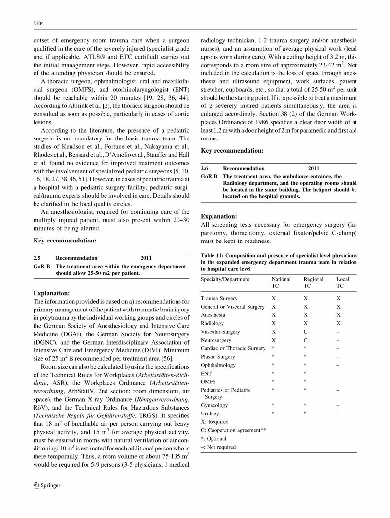
outset of emergency room trauma care when a surgeon
qualified in the care of the severely injured (specialist grade
and if applicable, ATLS� and ETC certified) carries out
the initial management steps. However, rapid accessibility
of the attending physician should be ensured.
A thoracic surgeon, ophthalmologist, oral and maxillofa-
cial surgeon (OMFS), and otorhinolaryngologist (ENT)
should be reachable within 20 minutes [19, 28, 36, 44].
According toAlbrink et al. [2], the thoracic surgeon should be
consulted as soon as possible, particularly in cases of aortic
lesions.
According to the literature, the presence of a pediatric
surgeon is not mandatory for the basic trauma team. The
studies of Knudson et al., Fortune et al., Nakayama et al.,
Rhodes et al.,Bensard et al.,D’Amelio et al., Stauffer andHall
et al. found no evidence for improved treatment outcomes
with the involvement of specialized pediatric surgeons [5, 10,
16, 18, 27, 38, 46, 51].However, in cases of pediatric traumaat
a hospital with a pediatric surgery facility, pediatric surgi-
cal/trauma experts should be involved in care. Details should
be clarified in the local quality circles.
An anesthesiologist, required for continuing care of the
multiply injured patient, must also present within 20–30
minutes of being alerted.
Key recommendation:
2.5 Recommendation 2011
GoR B The treatment area within the emergency departmentshould allow 25-50 m2 per patient.
Explanation:
The information provided is based on a) recommendations for
primarymanagement of the patientwith traumatic brain injury
in polytrauma by the individual working groups and circles of
the German Society of Anesthesiology and Intensive Care
Medicine (DGAI), the German Society for Neurosurgery
(DGNC), and the German Interdisciplinary Association of
Intensive Care and Emergency Medicine (DIVI). Minimum
size of 25 m2 is recommended per treatment area [56].
Roomsize can also be calculated b) using the specifications
of the Technical Rules for Workplaces (Arbeitsstatten-Rich-
tlinie, ASR), the Workplaces Ordinance (Arbeitsstatten-
verordnung, ArbStattV, 2nd section; room dimensions, air
space), the German X-ray Ordinance (Rontgenverordnung,
RoV), and the Technical Rules for Hazardous Substances
(Technische Regeln fur Gefahrenstoffe, TRGS). It specifies
that 18 m3 of breathable air per person carrying out heavy
physical activity, and 15 m3 for average physical activity,
must be ensured in rooms with natural ventilation or air con-
ditioning; 10m3 is estimated for each additional personwho is
there temporarily. Thus, a room volume of about 75-135 m3
would be required for 5-9 persons (3-5 physicians, 1 medical
radiology technician, 1-2 trauma surgery and/or anesthesia
nurses), and an assumption of average physical work (lead
aprons worn during care). With a ceiling height of 3.2 m, this
corresponds to a room size of approximately 23-42 m2. Not
included in the calculation is the loss of space through anes-
thesia and ultrasound equipment, work surfaces, patient
stretcher, cupboards, etc., so that a total of 25-50 m2 per unit
should be the starting point. If it is possible to treat amaximum
of 2 severely injured patients simultaneously, the area is
enlarged accordingly. Section 38 (2) of the German Work-
places Ordinance of 1986 specifies a clear door width of at
least 1.2mwith a door height of 2m for paramedic andfirst aid
rooms.
Key recommendation:
2.6 Recommendation 2011
GoR B The treatment area, the ambulance entrance, theRadiology department, and the operating rooms shouldbe located in the same building. The heliport should belocated on the hospital grounds.
Explanation:
All screening tests necessary for emergency surgery (la-
parotomy, thoracotomy, external fixator/pelvic C-clamp)
must be kept in readiness.
Table 11: Composition and presence of specialist level physiciansin the expanded emergency department trauma team in relationto hospital care level
Specialty/Department National
TC
Regional
TC
Local
TC
Trauma Surgery X X X
General or Visceral Surgery X X X
Anesthesia X X X
Radiology X X X
Vascular Surgery X C –
Neurosurgery X C –
Cardiac or Thoracic Surgery * * –
Plastic Surgery * * –
Ophthalmology * * –
ENT * * –
OMFS * * –
Pediatrics or Pediatric
Surgery
* * –
Gynecology * * –
Urology * * –
X: Required
C: Cooperation agreement**
*: Optional
–: Not required
S104
123
**C: Cooperation agreement
Treatment of vascular and/or neurosurgical injuries
require ‘‘cooperation agreements’’ in regional trauma cen-
ters. This rule - if no vascular surgeon and/or neurosurgeon
is available in-house, cooperation with a nearby depart-
ment/hospital for vascular and/or neurosurgery (e.g., pro-
vision of a consultant, transfer of the patient, surgical
readiness, etc.) can be agreed upon. 24 hours of acute care
must be ensured 365 days per year.
References
1. Alberts KA, Bellander BM, Modin G. Improved trauma care after
reorganisation: a retrospective analysis. Eur J Surg.
1999;165(5):426–30 (LoE 4).
2. Albrink MH, et al. Importance of designated thoracic trauma
surgeons in the management of traumatic aortic transection. South
Med J. 1994;87(4):497–501 (LoE 2b).3. Allen DM, Hicks G, Bota GW. Outcomes after severe trauma at a
northern Canadian regional trauma centre. Can J Surg.
1998;41(1):53–8 (LoE 2b).
4. Aprahamian C, et al. Characteristics of trauma centers and trauma
surgeons. J Trauma. 1993;35(4):562–7 (discussion 567–8).
5. Bensard DD, et al. A critical analysis of acutely injured children
managed in an adult level I trauma center. J Pediatr Surg.
1994;29(1):11–8.
6. Brennan R, et al. The OR suite as a unique trauma resuscitation
bay. AORN J. 1994;60(4):576–7, 580–4.
7. Carmody IC, Romero J, Velmahos GC. Day for night: should we
staff a trauma center like a nightclub? Am Surg.
2002;68(12):1048–51 (LoE 4).
8. Champion HR, Sacco WJ, Copes WS. Improvement in outcome
from trauma center care. Arch Surg. 1992;127(3):333–8 (discus-
sion 338).9. Cornwell EE, 3rd, et al. Enhanced trauma program commitment
at a level I trauma center: effect on the process and outcome of
care. Arch Surg. 2003;138(8):838–43 (LoE 4).
10. D’Amelio L, et al. ‘‘Adult’’ trauma surgeons with pediatric
commitment: a logical solution to the pediatric trauma man-
power problem. Am Surg. 1995;61(11):968–74.
11. Demarest GB, et al. In-house versus on-call attending trauma
surgeons at comparable level I trauma centers: a prospective
study. J Trauma. 1999;46(4):535–40 (discussion 540–2 LoE 4).12. Deo SD, Knottenbelt JD, Peden MM. Evaluation of a small trauma
team for major resuscitation. Injury. 1997;28(9–10):633–7 (LoE 4).13. Dodek P, Herrick R, Phang PT. Initial management of trauma
by a trauma team: effect on timeliness of care in a teaching
hospital. Am J Med Qual. 2000;15(1):3–8 (LoE 2b).
14. Eastes LS, et al. Outcomes of patients using a tiered trauma
response protocol. J Trauma. 2001;50(5):908–13.
15. Flohe S, Nast-Kolb D. Surgical management of life-threatening
injuries. Unfallchirurg. 2009;112(10):854–9.
16. Fortune JB, et al. A pediatric trauma center without a pediatric
surgeon: a four-year outcome analysis. J Trauma.
1992;33(1):130–7 (discussion 137–9).17. Fulda GJ, et al. In-house trauma surgeons do not decrease
mortality in a level I trauma center. J Trauma.
2002;53(3):494–500 (discussion 500–2).
18. Hall JR, et al. The outcome for children with blunt trauma is
best at a pediatric trauma center. J Pediatr Surg.
1996;31(1):72–6 (discussion 76–7).
19. Hartmann J, et al. A model for an integrated emergency
medicine/trauma service. Acad Emerg Med. 1996;3(12):1136–9
(LoE 5).20. Helling TS, et al. The presence of in-house attending trauma
surgeons does not improve management or outcome of critically
injured patients. J Trauma. 2003;55(1):20–5 (LoE 2b).
21. Highley DA. Review of the composition and use of trauma
teams within the Trent Region. J Accid Emerg Med.
1994;11(3):183–5.
22. Hoff WS, et al. The importance of the command-physician in
trauma resuscitation. J Trauma. 1997;43(5):772–7 (LoE 2b).23. Jacobs IA, et al. Cost savings associated with changes in routine
laboratory tests ordered for victims of trauma. Am Surg.
2000;66(6):579–84.
24. Kaplan LJ, et al. Improved emergency department efficiency
with a three-tier trauma triage system. Injury.
1997;28(7):449–53.
25. Kazemi AR, Nayeem N. The existence and composition of
Trauma Teams in the UK. Injury. 1997;28(2):119–21.
26. Khetarpal S, et al. Trauma faculty and trauma team activation:
impact on trauma system function and patient outcome.
J Trauma. 1999;47(3):576–81.
27. Knudson MM, Shagoury C, Lewis FR. Can adult trauma
surgeons care for injured children? J Trauma. 1992.
32(6):729–37 (discussion 737–9).28. Krotz M, et al. nterdisciplinary shock room management:
personnel, equipment and spatial logistics in 3 trauma centers in
Europe. Radiologe. 2002. 42(7):522–32 (LoE 5).
29. Lecky F, Woodford M, Yates DW. Trends in trauma care in
England and Wales 1989–97. UK Trauma Audit and Research
Network. Lancet. 2000;355(9217):1771–5 (LoE 4).
30. Lloyd DA, et al. A stratified response system for the emergency
management of the severely injured. Ann R Coll Surg Engl.
2001;83(1):15–20.
31. Lomas GA, Goodall O. Trauma teams vs non-trauma teams.
Accid Emerg Nurs. 1994; 2(4):205–10.
32. Lossius HM, et al. Efficiency of activation of the trauma team in
a Norwegian trauma referral centre. Eur J Surg.
2000;166(10):760–4.
33. Lott C, et al. The European Trauma Course (ETC) and the team
approach: past, present and future. Resuscitation.
2009;80(10):1192–6.
34. Lu WH, et al. An evaluation of trauma team response in a major
trauma hospital in 100 patients with predominantly minor
injuries. Aust N Z J Surg. 2000;70(5):329–32.
35. Luchette F, et al. Impact of the in-house trauma surgeon on
initial patient care, outcome, and cost. J Trauma.
1997;42(3):490–5 (discussion 495–7 LoE 2b).36. Mathiasen RA, et al. A dedicated craniofacial trauma team
improves efficiency and reduces cost. J Surg Res.
2001;97(2):138–43 (LoE 2b).
37. McLauchlan CA, Jones K, Guly HR. Interpretation of trauma
radiographs by junior doctors in accident and emergency
departments: a cause for concern? J Accid Emerg Med.
1997;14(5):295–8 (LoE 2b).
38. Nakayama DK, Copes WS, Sacco W. Differences in trauma care
among pediatric and nonpediatric trauma centers. J Pediatr Surg.
1992;27(4):427–31.
39. Nerlich M, Maghsudi M. Polytrauma management. Preclinical
and shock room management. Unfallchirurg.
1996;99(8):595–606.
S3-guideline on treatment of patients with severe/multiple injuries S105
123
40. Ochsner MG, et al. The evaluation of a two-tier trauma response
system at a major trauma center: is it cost effective and safe?
J Trauma. 1995;39(5):971–7.
41. Palmer SH, Maheson M. A radiological review of cervical spine
injuries from an accident and emergency department: has the
ATLS made a difference? J Accid Emerg Med.
1995;12(3):189–90 (LoE 2b).42. Plaisier BR, et al. Effectiveness of a 2-specialty, 2-tiered triage
and trauma team activation protocol. Ann Emerg Med.
1998;32(4):436–41.
43. Podnos YD, Wilson SE, Williams RA. Effect of surgical panel
composition on patient outcome at a level I trauma center. Arch
Surg. 1998;133(8):847–54 (LoE 2b).44. Poon A, McCluskey PJ, Hill DA. Eye injuries in patients with
major trauma. J Trauma. 1999;46(3):494–9 (LoE 4).45. Porter JM, Ursic C. Trauma attending in the resuscitation room:
does it affect outcome? Am Surg. 2001;67(7):611–4.
46. Rhodes M, Smith S, Boorse D. Pediatric trauma patients in an
‘adult’ trauma center. J Trauma. 1993;35(3):384–92 (discussion392–3).
47. Ruchholtz S, et al. Implementation of a nationwide trauma
network for the care of severely injured patients. J Trauma
Acute Care Surg. 2014;76(6):1456–61.
48. Ruchholtz S, et al. A multidisciplinary quality management
system for the early treatment of severely injured patients:
implementation and results in two trauma centers. Intensive
Care Med. 2002;28(10):1395–404.
49. Ryan JM, et al. Implementation of a two-tier trauma response.
Injury. 1998;29(9):677–83.
50. Sakellariou A, McDonald PJ, Lane RH. The trauma team
concept and its implementation in a district general hospital.
Ann R Coll Surg Engl. 1995;77(1):45–52.
51. Stauffer UG. Surgical and critical care management of children
with life-threatening injuries: the Swiss experience. J Pediatr
Surg. 1995;30(7):903–10.
52. Stoneham J, et al. Recent advances in trauma management.
Trauma. 2001;3(3):143–50.
53. Sugrue M, et al. A prospective study of the performance of the
trauma team leader. J Trauma. 1995;38(1):79–82.
54. Tscherne H, et al. Degree of severity and priorities in multiple
injuries. Chirurg. 1987;58(10):631–40.
55. Velmahos GC, et al. Around-the-clock attending radiology
coverage is essential to avoid mistakes in the care of trauma
patients. Am Surg. 2001;67(12):1175–7 (LoE 2b).56. Verlag S, Der Divi E. Empfehlungen zur Erstversorgung des
Patienten mit Schadel-Hirn-Trauma bei Mehrfachverletzung.
2000.
57. Vernon DD, et al. Effect of a pediatric trauma response team on
emergency department treatment time and mortality of pediatric
trauma victims. Pediatrics. 1999;103(1):20–4 (LoE 2b).58. Wenneker WW, Murray DH, Jr, Ledwich T. Improved trauma
care in a rural hospital after establishing a level II trauma center.
Am J Surg. 1990;160(6):655–7 (discussion 657–8).
59. Wong K, Petchell J. Trauma teams in Australia: a national
survey. ANZ J Surg. 2003;73(10):819–25.
60. Wurmb T, et al. Polytrauma management in a period of change:
time analysis of new strategies for emergency room treatment.
Unfallchirurg. 2009;112(4):390–9.
61. Wyatt JP, Henry J, Beard D. The association between seniority
of Accident and Emergency doctor and outcome following
trauma. Injury. 1999;30(3):165–8 (LoE 2b).
2.3 Emergency Department Trauma TeamActivation
Efficient trauma score systems or parameters should select
and/or identify patients precisely enough that the necessary
treatment is allotted to each patient according to injury
severity. The difficulty lies in adequate injury assessment.
Ideally, trauma team activation criteria should minimize
the rates of both undertriage and overtriage of severely
injured patients. While undertriage carries a risk to patient
safety, overtriage is associated with considerable costs as
well as disruptions to clinical routine. Thus, the effective-
ness of individual triage parameters leading to activation of
the emergency department trauma team should be evalu-
ated using measures such as sensitivity, specificity, positive
predictive value, and the calculation of overtriage and
undertriage rates. The American College of Surgeons
Committee on Trauma (ACS COT) [1] considers an
undertriage rate of 5 % with simultaneous overtriage rate of
25-30 % necessary for efficient trauma care in the emer-
gency department. Kane et al. concluded that the rate of
overtriage could not be brought under 70 % while
achieving a sensitivity greater than 80 %.
Activation Criteria
Key recommendation:
2.7 Recommendation Modified 2016
GoR A The Emergency Department Trauma Team must bemobilized for the following injuries:
•Systolic blood pressure below 90 mmHg (age adaptedfor children) after trauma
•Penetrating injuries of the torso/neck area
•Gunshot wounds to the torso/neck region
•GCS below 9 after trauma
•Respiratory impairment/need for intubation aftertrauma
•Fractures of more than two proximal long bones
•Flail Chest•Pelvic Fractures
•Amputation injury proximal to the hands/feet
•Spinal cord injury
•Open head injury
•Burns > 20% and grade ‡ 2b
Explanation:
Blood pressure/Respiratory rate
Individual studies have reported that hypotension with a
systolic blood pressure below 90 mmHg after trauma is a
good predictor/good criterion to activate the emergency
department trauma team. Franklin et al. [6] found that 50 %
of trauma patients with hypotension in the pre-hospital
S106
123
phase or on hospital arrival were sent for immediate sur-
gery or transferred to the intensive care unit. Overall, 75 %
of hypotensive patients underwent surgery during hospital
admission.
Tinkoff et al. [22] reported 24-fold increased mortality,
as well as 7-fold increased intensive care admission and 1.6-
fold increased emergency surgery rates in patients with
post-trauma hypotension as a sign of shock. In the recom-
mendations by the American College of Surgeons Com-
mittee on Trauma [1], hypotension is considered an
important criterion for admission to a trauma center.
According to Smith et al. [21], hypotension is used con-
sistently as a criterion for trauma team activation in all
hospitals within the state of New South Wales in Australia.
In a review of the New York State Trauma Registry, Henry
et al. [8] reported mortality rates of 32.9 % in trauma
patients with SBP (systolic blood pressure)\90 mmHg and
28.8% for those with a respiratory rate\10 or[29/min.
Gunshot Wounds
Sava et al. [20] found that a gunshot wound to the torso, as
sole activation criterion, has comparative predictive value
to the previously used TTAC (trauma team activation cri-
teria). In a subgroup with gunshot wounds to the abdomi-
nal/pelvic region, the frequency of severe injuries was the
same in groups with and without TTAC (74.1 % and 70.8
%, p = 0.61). Tinkoff et al. [22] found a significant cor-
relation between gunshot wounds to the torso or neck and
admissions to the intensive care unit (see below). This
criterion was also predictive for severe or fatal injuries and/
or for emergency surgery. In a retrospective analysis,
Velmahos et al. [6] reported an overall survival rate over
5.1 % in patients without vital signs in the emergency
department after penetrating gunshot and stab wounds. In a
review (25 years, 24 studies), Rhee et al. [18] identified a
survival rate of 8.8 % after emergency thoracotomy in
penetrating trauma.
In its last edition (2014), the ACS COT [1] listed vari-
ous, weighted triage criteria. The Step One and Step Two
criteria require transfer to a level 1 or 2 trauma center. Step
One criteria include a) GCS below 14, b) SBP below 90
mmHg, or c) respiratory rate (RR) below 10/min or over
29/min. Step two criteria are a) penetrating injuries to the
head, neck, torso, or proximal long bones, b) flail chest, c)
fracture of two or more proximal long bones, d) amputa-
tion(s) proximal to the hands/feet, e) unstable pelvic frac-
tures, f) open skull fractures, and g) spinal cord injuries. At
present, there is relatively little evidence for these criteria.
In a study of 1473 trauma patients, Knopp [9] found a
positive predictive value (PPV) of 100% for ISS[ 15 in
spinal cord and amputation injuries; however, fractures of
the long bones had a PPV of only 19.5%.
Tinkoff et al. [22] assessed several of these criteria for
accuracy in the identification of severely injured and/or
high-risk patients. Trauma patients fulfilling the ACS COT
criteria had more severe injuries, increased mortality, and
longer intensive care stays than control patients. Systolic
blood pressure under 90 mmHg, endotracheal intubation,
and gunshot wound to the torso/neck were predictive for
emergency surgery or intensive care admission. Mortality
was markedly increased in patients with SBP under 90
mmHg, endotracheal intubation, or GCS less than 9. Kohn
et al. [10] analyzed various trauma team activation criteria
(see Table 1), which are similar to those of the ACS COT.
Respiratory rate under 10 or over 29 breaths per minute
was the most predictive for presence of a severe injury.
Other highly predictive parameters were: a) burns with
over 20 % body surface area (BSA), b) spinal cord syn-
drome, c) systolic blood pressure under 90 mmHg, d)
tachycardia, and e) gunshot wounds to the head, neck, or
torso.
Open Head Injuries
With a lack of studies on the relevance of open head
injuries, this criterion is regarded by the ACS COT rather
as a significant indicator of severe injuries that require a
high level of specialist medical competence and is thus
assigned to the Step One criteria.
GCS
Kohn et al. [10] regard a GCS under 10 as an important
predictor of severe trauma. Of patients activating the
emergency department trauma team for low GCS, 44.2%
had confirmed severe injuries. The value of GCS as pre-
dictor of severe injury or as activation criterion for the
trauma team has been confirmed in studies by Tinkoff
et al., Norwood et al., and Kuhne et al. [11, 16, 22]. Nor-
wood and Kuhne both saw pathological intracerebral
findings and need for inpatient admission already at GCS
values under 14. At the same time, trauma team activation
does not appear absolutely necessary with these patients
(GCS B 14 and C 11). For GCS under 10, Engum [5]
found a 70 % sensitivity for the endpoints surgery, inten-
sive care unit (ICU), or death. The odds ratio (OR) was 3.5
(95% CI: 1.6-7.5). The authors found a PPV of 78 % for
severe injuries in children with a GCS\ 12.
Key recommendation:
2.8 Recommendation 2011
GoR B The emergency department trauma team should bemobilized for the following additional criteria:
•Falls from over 3 meters
•Motor vehicle accident (MVA) with
•Frontal collision with intrusion greater than 50-75 cm
•Vehicle speed change of delta > 30 km/h
S3-guideline on treatment of patients with severe/multiple injuries S107
123
•Pedestrian/bicycle collision
•Occupant mortality (driver or passenger)
•Occupant ejection (driver or passenger)
Explanation:
Accident-related/dependent criteria
In the literature, accident-related/dependent criteria are
evaluated very differently regarding predictive value for
severe trauma.
Norcross et al., Bond et al., and Santaniello et al. [2, 15,
19] report rates of overtriage up to 92%, sensitivity of
50-70 %, and PPV of 16.1% when accident-related
mechanisms are used as the sole criterion for predicting
injury severity. When physiological criteria are added,
sensitivity of 80 % and specificity of 90 % are reached [2].
Knopp et al. found poor positive predictive values for
the parameters of motor vehicle accident (MVA) with
ejection or death of an occupant (driver or passenger) and
MVAs involving a pedestrian [9]. Engum et al. also found
the lowest predictive values for MVAs involving a
pedestrian at 20 mph (miles per hour) and MVAs with
occupant death and trauma from vehicle rollover [5]. The
current ACS COT recommendations removed vehicle
rollover trauma from the criteria. Frontal impact with
intrusion greater than 20-30 inches, death of a car occu-
pant, MVA with pedestrian/bicycle collision with
C 20 mph, and ejection of an occupant were cited as Step
Three Criteria, i.e., there is no absolute necessity to
transport these patients to a maximum care level hospital,
provided these are isolated criteria. Kohn et al. [10] also
consider vehicle rollover trauma as inadequate. According
to Kohn et al., the same also applies to the criteria of MVA
with occupant ejection or death and MVA with pedestrian
involvement [10].
Champion et al. [3] consider vehicle rollover an
important indicator of severe injury. The average proba-
bility of suffering a lethal injury is much higher in an
overturned vehicle than in those not rolling over.
Nevertheless, the ACS COT removed the rollover
mechanism from its triage criteria because relevant injuries
after this type of accident should already be included in
Step One or Step Two.
Vehicular Body Damage
In a multivariate analysis of 621 patients, Palanca et al.
[17] found no significant association between vehicle
deformation (intrusion of[ 30 cm or[ 11.8 inches) and
the presence of relevant severe injury (OR: 1.5; 95% CI:
1.0–2.3; p = 0.05). Henry reported comparable results in
another multivariate analysis [8]. Using data from the
National Automotive Sampling System Crashworthiness
Data System (NASS CDS), Wang found a PPV of 20% for
ISS[ 15 [24].
Occupant mortality (driver or passenger)
Knopp et al. found increased risks for surgery or death
when a car occupant was fatally injured (OR: 39.0; 95%
CI: 2.7–569; PPV 21.4%) [9]. Palanca et al. [17] did not
confirm a significant association between occupant mor-
tality and severe injury, although the concurrence of the
two was 7 %.
Falls from a great height
In a prospective study by Kohn et al. [10], 9.4% of patients
falling from more than 6 meters had severe injuries -
defined as requiring intensive care admission or immediate
surgery. Yagmur et al. [25] reported 9 meters as the
average fall height from which patients died.
Burns
It is essential to determine whether burns are present
without concomitant injuries. In the case of combination
injuries where the non-thermal component is predominant,
the patient should be brought to a trauma center [1].
Age
A 2015 review study identified age cutoffs between 45 and
70 years. Patients older than the respective cutoff should be
treated in a trauma center by the trauma team. In addition
to extended monitoring, surgical management should be
adapted according to age-related pathophysiology. Treat-
ment in trauma centers with geriatric expertise can reduce
trauma-associated mortality of older patients.
Kohn et al. [10] evaluated various trauma team activation
criteria similar to those of the ACS COT. Of the evaluated
criteria, ‘‘age over 65’’ had the least informative value.
Thus, the authors recommended that this criterion be
removed from the ‘‘first-tier activations.’’ Demetriades et al.
[4] found markedly higher mortality (16 %), increased ICU
admission and need for surgery (19%) in patients over 70
years of age compared to younger patients. However, in this
study, all patients not requiring hospital admission were
excluded, so the cited percentages are probably overesti-
mations. In a retrospective study of over 5000 patients from
the DGU trauma registry, Kuhne et al. [12] found increased
mortality - irrespective of ISS - with increasing age. The
cutoff value for increased mortality was 56 years.
MacKenzie et al. [13] also found a marked increase in
(fatal) injuries with age[ 55 years. In a 13-year review,
Grossmann et al. [7] found that mortality increased 6.8 %
per year of life after 65. In a study by Morris et al. [14],
patients who died from the consequences of an accident had
lower ISS than younger patients in the control group.
Overall, the influence of age on trauma outcomes is a
controversial topic. The ACS COT has classified age as a
relatively low indication to transfer to Level 1 or 2 trauma
S108
123
center (Step Four criterion). Hildebrand considers age as a
relevant influencing variable; however, accompanying
medication, physiological reserves, and weak immune
systems should be taken into account when evaluating
geriatric patients (see above).
References
1. American College of Surgeons Committee on Trauma. Resources
for optimal care of the injured patient. Chicago: American
College of Surgeons; 2014.
2. Bond RJ, Kortbeek JB, Preshaw RM. Field trauma triage:
combining mechanism of injury with the prehospital index for an
improved trauma triage tool. J Trauma. 1997;43(2):283–7.
3. Champion HR, Lombardo LV, Shair EK. The importance of
vehicle rollover as a field triage criterion. J Trauma.
2009;67(2):350–7.
4. Demetriades D, et al. Old age as a criterion for trauma team
activation. J Trauma. 2001;51(4):754–6 (discussion 756–7).5. Engum SA, et al. Prehospital triage in the injured pediatric
patient. J Pediatr Surg. 2000;35(1):82–7.
6. Franklin GA, et al. Prehospital hypotension as a valid indicator of
trauma team activation. J Trauma. 2000;48(6):1034–7 (discus-sion 1037–9).
7. Grossman MD, et al. When is an elder old? Effect of preexisting
conditions on mortality in geriatric trauma. J Trauma.
2002;52(2):242–6.
8. Henry MC. Trauma triage: New York experience. Prehosp Emerg
Care. 2006;10(3):295–302.
9. Knopp R, et al. Mechanism of injury and anatomic injury as
criteria for prehospital trauma triage. Ann Emerg Med.
1988;17(9):895–902.
10. Kohn MA, et al. Trauma team activation criteria as predictors of
patient disposition from the emergency department. Acad
Emerg Med. 2004;11(1):1–9.
11. Kuhne CA, et al. Emergency room patients. Unfallchirurg.
2003;106(5):380–6.
12. Kuhne CA, et al. Mortality in severely injured elderly trauma
patients—when does age become a risk factor? World J Surg.
2005;29(11):1476–82.
13. MacKenzie EJ, et al. A national evaluation of the effect of
trauma-center care on mortality. N Engl J Med.
2006;354(4):366–78.
14. Morris JA Jr, MacKenzie EJ, Edelstein SL. The effect of
preexisting conditions on mortality in trauma patients. JAMA.
1990;263(14):1942–6.
15. Norcross ED, et al. Application of American College of
Surgeons’ field triage guidelines by pre-hospital personnel.
J Am Coll Surg. 1995;181(6):539–44 [Evidenzbasierte
Leitlinie].16. Norwood SH, et al. A prehospital glasgow coma scale score <
or = 14 accurately predicts the need for full trauma team
activation and patient hospitalization after motor vehicle
collisions. J Trauma. 2002;53(3):503–7.
17. Palanca S, et al. Mechanisms of motor vehicle accidents that
predict major injury. Emerg Med (Fremantle).
2003;15(5–6):423–8.
18. Rhee PM, et al. Survival after emergency department thoraco-
tomy: review of published data from the past 25 years. J Am
Coll Surg. 2000;190(3):288–98.
19. Santaniello JM, et al. Mechanism of injury does not predict
acuity or level of service need: field triage criteria revisited.
Surgery. 2003;134(4):698–703 (discussion 703–4).20. Sava J, et al. All patients with truncal gunshot wounds deserve
trauma team activation. J Trauma. 2002;52(2):276–9.
21. Smith J, Caldwell E, Sugrue M. Difference in trauma team
activation criteria between hospitals within the same region.
Emerg Med Australas. 2005;17(5–6):480–7.
22. Tinkoff GH, O’Connor RE. Validation of new trauma triage
rules for trauma attending response to the emergency depart-
ment. J Trauma. 2002;52(6):1153–8 (discussion 1158–9).23. Velmahos GC, et al. Outcome of a strict policy on emergency
department thoracotomies. Arch Surg. 1995;130(7):774–7.
24. Wang SW. Review of NASS CDS and CIREN data for
mechanism criteria for field triage. Presented at the National
Expert Panel on Field Triage meeting. 2005: Atlanta, Georgia.
25. Yagmur Y, et al. Falls from flat-roofed houses: a surgical
experience of 1643 patients. Injury. 2004;35(4):425–8.
2.4 Thorax
Importance of the Medical History
Key recommendation:
2.9 Recommendation 2016
GoR B An accurate medical history should be collected, ifnecessary from a third party.
2.10 Recommendation 2016
GoR B High-speed trauma and motor vehicle accidents withlateral impact should be considered evidence for chesttrauma/aortic rupture.
Explanation:
Although there are few studies on collecting the medical
history in regard to thoracic trauma, it remains an indis-
pensable requirement for the assessment of injury severity
and pattern of injury, and is used to establish whether or
not an accident actually occurred. Collection of the cir-
cumstantial details of the accident are essential for the
history. For motor vehicle accidents (MVAs) involving
passenger vehicles, questions regarding the speed of the
vehicle at the time of impact and the direction of impact are
particularly important. There are marked differences in the
occurrence, pattern, and severity of chest injuries as well as
overall injury severity depending on whether the impact is
lateral or frontal [113].
Horton et al. [64] reported a sensitivity of 100% and a
specificity of 34% for aortic rupture with a lateral collision
and/or change in velocity (delta V) C 30 km/h. In another
study [33], high velocity injuries at speeds of[ 100 km/h
were graded as suspicious for aortic rupture. Richter et al
[106] also found an increased risk of chest injury in lateral
collisions. In this study, delta V correlated with the AIS
(thorax), ISS, and clinical course. Ruchholtz et al. [110]
S3-guideline on treatment of patients with severe/multiple injuries S109
123
diagnosed chest injury in 8 of 10 passenger vehicle acci-
dents involving lateral collision.
In a study of 286 passenger vehicle occupants with ISS
C 16, the probability of aortic injury was twice as high
after lateral versus frontal collision [98]. Impact in the
region of the superior thoracic aperture and fractures of ribs
1-4 appear to be associated with an increased incidence of
aortic injuries [115].
Children as well have a 5-fold higher risk of severe chest
injury (AIS C 3) and significantly higher overall injury
severity when they are occupants of a passenger vehicle in
a lateral versus frontal collision [96].
The effect of seatbelts on chest injuries appears unclear.
In a retrospective study of 1124 patients with relatively
minor overall injury severity (ISS 11.6), Porter and Zaho
[100] did find increased incidence of sternum fractures (4
% vs. 0.7 %) in seat belted patients, but the proportion of
patients with thoracic injuries were identical in both groups
(21.8 vs. 19.1 %).
Importance of Physical Examination Findings
Key recommendation:
2.11 Recommendation 2016
GoR A Clinical examination of the chest must be performed.
2.12 Recommendation Modified 2016
GoR A Auscultation must be carried out with the physicalexamination.
Explanation:
Although there are barely any studies on the importance of
and required scope for physical examination, it remains an
indispensable requirement for the recognition of symptoms
and making (suspected) diagnoses. The physical exami-
nation serves to detect relevant, life-threatening, or
potentially fatal disorders or injuries that require immediate
and specific treatment. Even when an examination has
already been performed in the pre-hospital phase and even
when a chest tube has already been inserted, physical
examination in the emergency department is to be per-
formed, since a change in the constellation of findings
could have occurred.
The initial physical examination should address the
following objectives [81]:
• Examination and securing of the airway
• Respiratory rate/dyspnea
• Inspection of the chest (skin and soft-tissue injuries,
symmetry of the chest and respiratory excursion,
paradoxical respiration, congestion, belt marks)
• Palpation (subcutaneous emphysema, crepitus, points of
tenderness)
• Auscultation (presence and quality of breath sounds
bilaterally)
• Details regarding pain
The following immediately life-threatening injuries must
be checked during the initial examination [3]:
• Airway obstruction
• Tension pneumothorax
• Open pneumothorax
• Flail chest/Lung contusion
• Massive hemothorax
• Pericardial tamponade
Auscultation findings are the leading indicators in the
diagnosis of chest trauma. In addition, subcutaneous
emphysema, palpable instability, crepitus, pain, dyspnea,
and increased ventilatory pressures can indicate thoracic
injuries.
In a prospective study, Bokhari et al. [9] examined 676
patients with blunt or penetrating chest injury for clinical
signs and symptoms of hemopneumothorax. Of 523 patients
with blunt trauma, only 7 had hemopneumothorax. In this
group, auscultation had sensitivity and negative predictive
values of 100 %. Specificity was 99.8 % and the positive
predictive value was 87.5 %. In penetrating injuries, the
sensitivity of auscultation was 50 %, specificity and positive
predictive value were 100 %, and the negative predictive
value was 91.4 %. In both injury mechanisms, pain and
tachypnea are insufficient to diagnose hemopneumothorax.
In a retrospective study of 118 patients with penetrating
trauma, Chen et al. [21] also found a sensitivity of only
58%, specificity and positive predictive value of 98%, and
a negative predictive value of 61% for auscultation. In a
prospective study of 51 patients with penetrating trauma,
the combination of percussion and auscultation exhibited a
sensitivity of 96%, specificity of 93%, and positive pre-
dictive value of 83% [61].
These studies show that in penetrating trauma,
decreased or absent breath sounds generally indicate an
underlying pneumothorax, and a chest drain should be
inserted prior to x-ray.
In their search for a clinical decision-making tool to
identify children with chest injuries, Holmes et al. [63]
studied 986 patients, 80 of whom had a chest injury. This
yielded an odds ratio (OR) of 8.6 for positive auscultation
findings, OR of 3.6 for abnormal physical examination
(reddening, skin lesions, crepitus, tenderness), and OR of
2.9 for elevated respiratory rate.
S110
123
Importance of Diagnostic Equipment (chest radiograph,
ultrasound, CT, angiography, ECG, laboratory tests)
and Indications for Use
Key recommendations:
2.13 Recommendation Modified 2016
GoR A If thoracic trauma cannot be ruled out, radiologicaldiagnostic studies must be performed in the emergencydepartment.
2.14 Recommendation 2016
GoR B Spiral CT of the thorax with contrast medium should beperformed for any patient with clinical evidence orhistory suggestive of severe chest trauma.
Explanation:
As explained in points 1 and 2, both the mechanism of
injury and the findings from the physical examination
provide important information on the presence or absence
of a chest injury. For this reason, a chest radiograph can be
dispensed with if a trauma CT scan will be performed
immediately and if a chest injury can be excluded based on
the accident circumstances and at the same time there are
no findings from the physical examination that make an
intrathoracic injury probable.
Conversely, all patients with confirmed chest injury
should undergo chest x-ray once immediately life-threat-
ening injuries have been excluded and/or treated. The ini-
tial radiograph can enable diagnosis of pneumothorax and/
or hemothorax, rib fractures, tracheobronchial injuries,
pneumomediastinum, mediastinal hematoma, and pul-
monary contusion [53]. For detailed diagnosis of the pat-
tern of injury, computed tomography is the gold standard.
If there is not enough time available, chest x-ray can be
performed as primary diagnostic study, due to the low cost
and good availability. Nevertheless, there is little evidence
on sensitivity and specificity of the diagnosis of pulmonary
or thoracic injuries.
A prospective study of 100 patients found that the most
important thoracic injuries are evident on chest x-ray. The
sensitivity of images with the patient standing upright was
78.7 % and supine was 58.3 % [58]. In a series of 37
patients who died within 24 hours of admission, McLellan
et al. [83] identified 11 cases in which important injuries
found at autopsy were not evident on chest x-ray. Of these,
there were 11 cases of rib fractures, 3 sternum fractures, 2
diaphragmatic ruptures, and 1 intimal lesion of the aorta.
The chest x-ray gives sufficient accuracy, e.g. to estab-
lish a need for chest tube drainage. In a prospective study
of 400 polytrauma patients, Peytel et al. [99] reported that
all cases (n = 77) in which chest tubes were placed based
on chest x-ray findings were correct.
However, numerous studies have found that intratho-
racic injuries can be identified significantly more often on
CT than on chest radiographs alone. It is especially supe-
rior for the detection of pneumothorax, hemothorax, lung
contusion, and aortic injuries. Spiral CT with intravenous
(I.V.) contrast is preferable [94]. With the use of multi-
layer versus single-layer spiral CT, the average whole-body
examination time decreased from 28 to 16 minutes, and the
initial diagnostic information was taken from real-time
images on the monitor [76] The NEXUS criteria can be
helpful for the decision for or against radiological diag-
nostic studies [107].
In a series of 103 severely injured patients, Trupka et al.
[122] found that compared to radiographs, additional
information regarding the basic thoracic trauma was evi-
dent in 65 % of patients (lung contusion n = 33, pneu-
mothorax n = 34, hemothorax n = 21). There were direct
therapeutic consequences resulting from this extra infor-
mation for 63 % of these patients, the majority of whom
required new or correction of existing chest tube
placement.
In patients with relevant trauma (motor vehicle acci-
dents with accident speeds[15 km/h, falls from a height[1.5 m), Exadaktylos et al. [38] identified no thoracic
injuries on conventional radiographs in 25 of 93 patients.
However, CT showed some considerable thoracic injuries
in 13 of these 25 patients, including two aortic lacerations.
In a prospective study, Demetriades et al. [28] performed
spiral CT examinations of the chest in 112 patients with
deceleration trauma, of whom nine patients were diagnosed
with aortic rupture. Four of these patients had normal chest
x-rays. Eight cases of aortic rupture were identified on CT.
One patient had an injury to the brachiocephalic artery.
Local hematoma was evident on CT, but the vessel itself
was not visualized on the CT cuts. Even in patients without
clinical signs of thoracic injury and with negative radio-
graphic findings, chest injuries were evident on CT in 39%
of patients, and in 5% of cases this led to a change in
treatment [95].
Blostein et al. [8] concluded that routine CT is not
generally recommended in blunt chest trauma, since of 40
patients studied prospectively with defined chest injuries, 6
patients had changes in treatment (5x chest tubes, 1x aor-
togram with negative result). The authors admitted as well
that CT yields findings not visible on conventional radio-
graphs in patients who are intubated and ventilated. In
patients with an oxygenation index (PaO2/FiO2)\300, the
CT can help estimate the extent of pulmonary contusion
and to identify patients at risk for pulmonary failure.
Moreover, patients in whom an incompletely decom-
pressed hemothorax and/or pneumothorax could lead to
further decompensation can be identified. In a retrospective
study of 45 children [104] with 1) pathologic radiographic
S3-guideline on treatment of patients with severe/multiple injuries S111
123
findings (n = 27), 2) abnormal physical examinations (n =
8), and 3) substantial impact to the chest wall (n = 33), CT
identified additional injuries in 40%, leading to a change in
treatment of 18%.
Although the supplementary diagnostic information of
chest CT has become generally accepted for blunt thoracic
trauma in the more recent literature [54], its benefit
regarding clinical outcomes remains controversial and is
not yet clear. A prospective study by Guerrero-Lopez et al.
[56] found chest CT to be more sensitive in detecting
hemo/pneumothorax, lung contusion, vertebral fractures,
and chest tube misplacement, and led to changes in therapy
in 29 % of cases. On multivariate analysis, no therapeutic
correlation between CT and ventilation duration, intensive
care stay, or mortality was established. The authors con-
cluded that chest CT should be performed only for sus-
pected severe injuries that can be confirmed or excluded.
Current studies have identified clear benefits from multi-
layer CTs of the chest when there is a defined indication.
Brink et al. [15] evaluated routine versus selective use in
464 and 164 patients, respectively. Indications for routine
CT were high-energy trauma, threatening vital signs, and
severe injuries such as pelvic or vertebral fractures. Indi-
cations for selective CT were abnormal mediastinum, more
than three rib fractures, pulmonary shadowing, emphy-
sema, and thoracolumbar spine fractures. Injuries not evi-
dent on conventional radiographs were found in 43 % of
patients undergoing routine CT. This led to treatment
changes in 17 %. Of 7.9 % of patients with normal chest
x-rays, Salim et al. [111] found pneumothorax in 3.3 %,
suspected aortic rupture in 0.2 %, lung contusion in 3.3 %,
and rib fractures in 3.7 %.
Summarizing results in the literature, the following
criteria are indications for chest CT:
Indication criteria for chest CT (summarized according to
[15, 107, 111]):
• Motor vehicle accident (MVA) Vmax[ 50 km/h
• Fall from[ 3 m height
• Patient ejected from vehicle
• Rollover trauma
• Substantial vehicle damage
• Pedestrian hit at[ 10 km/h
• Bicyclist hit at[ 30 km/h
• Entrapment
• Pedestrian hit by vehicle and flung[ 3 m
• GCS\ 12
• Vital/hemodynamic abnormalities (respiratory rate [30/min, pulse[120/min, systolic blood pressure\100
mmHg, blood loss [ 500 ml, capillary refill [ 4
seconds)
• Severe concomitant injuries (pelvic ring fracture,
unstable vertebral fracture, or spinal cord compression)
A retrospective multicenter analysis using the DGU trauma
registry database found improved survival probability for
patients who had initially undergone a full-body CT scan
[65]. The use of full-body CT leads to a relative reduction
in mortality of 25 % in TRISS and of 13% in the RISC
score. CT was an independent predictor for survival on
multivariate analysis.
Key recommendation:
2.15 Recommendation Modified 2016
GoR B An initial ultrasound of the chest should be performed inany patient with clinical signs of thoracic trauma(according to the eFAST framework), unless an initialspiral CT chest with contrast has been performed.
Explanation:
In a prospective study of 27 patients, chest X-rays, ultra-
sound examinations, and CT were compared for diagnostic
accuracy for pneumothorax. The ultrasound examination of
the chest showed sensitivity and negative predictive value
of 100 % and specificity of 94 % [108]. In another study
comparing ultrasound with radiographs for the diagnosis of
pneumothorax, ultrasound yielded sensitivity and positive
predictive value of 95 % and a negative predictive value of
100 % [31]. However, emphysematous bullae, pleural
adhesions, or extensive subcutaneous emphysema can
distort ultrasound results.
As a retrospective study of 240 patients made clear,
ultrasound ranks equally with the X-ray in diagnosing
hemothorax. In 26 of these patients, hemothorax was
confirmed by either chest tube or chest CT. Ultrasound and
chest x-ray each showed sensitivity of 96%, specificity and
negative predictive value of 100%, and positive predictive
value of 99.5% [78].
In a prospective study of 261 patients with penetrating
injuries, chest ultrasound had a sensitivity of 100% and
specificity of 96.9% for detecting pericardial tamponade
[109]. False negative ultrasound results occur, however,
especially in patients with a larger hemothorax, which can
conceal smaller hematomas of the pericardium [86]. Thus,
ultrasound sensitivity was only 56% in this study.
A retrospective study of 37 patients with CT-confirmed
pulmonary contusion found ultrasound sensitivity of
94.6%, specificity of 96.1%, and positive and negative
predictive values of 94.6% and 96.1%, respectively [116].
Spiral CT chest with contrast excluded aortic injuries in
patients without detected mediastinal hematoma, so that
angiography was not necessary. Because of insufficient
sensitivity, conventional CT examinations are less suited
for exclusion of an aortic injury [32, 44, 87].
Aortogram is useful only in patients with inconclusive
CT or with a periaortal hematoma without direct signs of
S112
123
aortic injury. There is now general consensus that spiral CT
with contrast is suitable to exclude aortic rupture [18, 36,
85]. There is high probability that patients without
detectable mediastinal hematoma have no aortic injury.
Thus, the use of computed tomography can avoid a large
number of unnecessary aortogram procedures. However,
when brain CT scan is also necessary, it should be carried
out before the chest CT as the use of contrast agent com-
plicates the traumatic brain injury diagnosis.
As comparative studies on angiography have shown, CT
showing no evidence of mediastinal hematoma has a neg-
ative predictive value of 100% for the injury of large
intrathoracic vessels [103]. However, the study of Parker
et al. [97] finds the specificity only 89 % because of 14
false positive results. It is thus recommended that angiog-
raphy be performed for patients with CT scans showing
para-aortic hematoma or blood collections around aortic
branches as well as aortic contour irregularities. A negative
CT scan with contrast definitively excludes aortic rupture
[44, 91, 127].
In an analysis of 54 patients with surgically detected
aortic ruptures, Downing et al. [30] showed sensitivity of
100% and specificity of 96% for spiral CT. In a prospective
study of 1104 patients with blunt chest trauma, Mirvis et al.
[90] found mediastinal bleeding in 118 cases, of which 25
patients had aortic ruptures. For aortic rupture, spiral CT
yielded sensitivity and negative predictive value of 100%,
specificity of 99.7%, and positive predictive value of 89%.
In a retrospective study of chest CT, Bruckner et al. found a
negative predictive value of 99%, positive predictive value
of 15%, sensitivity of 95%, and specificity of 40%.
In another prospective study of 1009 patients, 10
patients had aortic injuries [34]. Spiral CT had sensitivity
and negative predictive value of 100 %, specificity of 96 %,
and positive predictive value of 40 % for direct signs of
aortic injury.
In contrast to the prospective studies mentioned above,
in a retrospective study of 242 patients, Collier et al. [25]
found a sensitivity of only 90% and a negative predictive
value of 99%; aortic injury was found during the autopsy of
one patient with a normal CT who had subsequently died
from the consequences of traumatic brain injury. In another
retrospective study, angiography did not detect any aortic
injury in 72 patients with intrathoracic hematoma but no
evidence of a direct aortic or other intrathoracic vessel
injury on CT scan [112].
Transesophageal echocardiography (TEE) is a sensitive
screening test [16, 24, 126], but angiography was often also
carried out afterwards [19, 89]. TEE requires an experi-
enced examiner [51] and is generally not as rapidly avail-
able as CT or angiography. The benefit of TEE may lie in
the visualization of small intimal tears [16] that might not
be visible on angiography or helical CT. However, TEE
cannot provide good images of the ascending aorta and the
aortic branches, which thus elude diagnostic evaluation
[92]. To date, only one prospective study compares spiral
CT to TEE in the diagnosis of aortic injury. CT and TEE
showed sensitivity of 73 and 93%, respectively, and neg-
ative predictive value of 95%.
Key recommendations:
2.16 Recommendation 2016
GoR A A 3-lead ECG must be performed to monitor vitalfunctions.
2.17 Recommendation Modified 2016
GoR A If blunt cardiac injury is suspected, a twelve lead ECGmust be performed.
Explanation:
An initial ECG is essential for every severely injured
patient. It is necessary particularly in the absence of pal-
pable pulses, to differentiate rhythms that can and can’t be
defibrillated in cardiac arrest. The ECG can also be used as
a screening test for potential cardiac complications from
blunt cardiac injury.
Patients with normal ECG, normal troponin-I value,
normal hemodynamics, and no other relevant injuries do
not require further diagnostic studies or treatment. Cardiac
enzymes are irrelevant in predicting complications from
blunt cardiac injury, although raised troponin I levels can
predict abnormalities on echocardiography. A 12-lead ECG
is performed for further diagnosis of patients with blunt
chest trauma [23].
Echocardiography should not be used in the emergency
department for the diagnosis of cardiac contusion, since
this entity is not correlated with clinical complications.
Echocardiography should be performed in hemodynami-
cally unstable patients to diagnose pericardial tamponade
or pericardial rupture. Transthoracic echocardiography
should be the method of choice, as there is no clear evi-
dence as yet that transesophageal echocardiography is
superior in diagnosing blunt cardiac injury.
The ECG is a rapid, cost-effective, non-invasive
examination that is always available in the emergency
department. Meta-analysis of 41 studies found that ECG
and creatine kinase MB (CK-MB) levels are more impor-
tant than radionuclide examinations and echocardiogram in
the diagnosis of clinically-relevant blunt cardiac injury
(defined as a treatment-requiring complication) [79].
Fildes et al. [43] reported prospectively on 74 hemo-
dynamically stable patients with normal initial ECG, no
existing heart disease, and no other injuries. None of these
patients developed cardiac complications. Another retro-
spective study of 184 children with blunt cardiac injury
reported that patients with a normal ECG in the emergency
S3-guideline on treatment of patients with severe/multiple injuries S113
123
department did not develop complications [29]. Meta-
analysis of 41 studies correlated abnormal admission ECG
with the development of treatment-requiring complications
[79]. Conversely, a prospective study from Biffl et al. [7]
identified contusion complications in 17 of 107 patients.
Only two of 17 patients had abnormal ECG on admission,
and three showed sinus tachycardia. In another retrospec-
tive study of 133 patients with clinical suspicion of
myocardial contusion at two institutions, 13 patients (9.7 %)
developed complications; however, no patients with normal
initial ECGs showed other abnormalities [41]. In a study by
Miller et al. [88] of 172 patients, four developed treatment-
requiring arrhythmias, with all four patients having abnor-
mal initial ECGs. Wisner et al. [134] studied 95 patients
with suspected cardiac contusion. Of these, four patients
developed clinically significant arrhythmias, and only one
of these had a normal ECG on admission. In summary, the
majority of authors recommend that asymptomatic, hemo-
dynamically stable patients with normal ECG do not require
any further diagnostic tests or treatment.
Key recommendation:
2.18 Recommendation Modified 2016
GoR 0 Serum levels of troponin can be measured as an adjunctfor the diagnosis of blunt cardiac injury.
Explanation:
The assessment of creatine kinase MB (CK-MB) levels is
not indicated for the diagnosis of myocardial contusion
[23]. In a retrospective study of 359 patients, of whom 217
were initially admitted to exclude myocardial contusion,
107 were diagnosed because of either abnormal ECG or
elevated CK-MB levels 16% of patients developed com-
plications requiring treatment (arrhythmias or cardiogenic
shock). All of these patients had abnormal ECG, but only
41% had elevated CK-MB levels. Patients with normal
ECG and increased CK-MB levels did not develop com-
plications [7]. In a prospective study of 92 patients
undergoing ECG, CK-MB analysis, and continuous moni-
toring, 23 patients developed arrhythmias requiring no
specific treatment. This shows that the number of
arrhythmias requiring therapy is low. 52% of patients with
arrhythmias had elevated CK-MB levels, whereas 19% of
patients without arrhythmias also had elevated CK-MK
levels [39]. Other studies have found no correlation
between elevated CK-MB levels and cardiac complications
[45, 57, 59, 71, 88, 117, 134].
Troponin I and T are sensitive markers in the diagnosis of
myocardial infarction and considerably more specific than
CK-MB, as they are not present in skeletal muscle. In a
study of 44 patients, the 6 patients with echocardiography-
confirmed myocardial injury showed simultaneous
elevations in CK-MB and troponin I. Of the 37 patients
without cardiac injury, CK-MB levels were elevated in 26
patients, but troponin I was elevated in none [1]. Another
study of 28 patients, 5 of whom with echocardiogram-
confirmed myocardial contusion, reported 100 % speci-
ficity and sensitivity for troponin I. In a study of 29
patients, troponin T showed higher sensitivity (31%) than
CK-MB (9%) in diagnosing myocardial injury. Troponin T
showed a sensitivity of 27 % and a specificity of 91 % for
predicting clinically significant ECG changes in 71 patients
[46].
In a more current prospective study of 94 patients, 26
patients were diagnosed with myocardial contusion by
either ECG or echocardiography. Troponin I and T showed
sensitivity of 23 and 12%, respectively, sensitivity of 97
and 100%, respectively, and negative predictive value of
76.5 and 74%, respectively. The authors describe an
unsatisfactory correlation between the two enzymes and
the occurrence of complications [6]. In another prospective
study, sensitivity, specificity, and positive and negative
predictive values of troponin I are given as 63, 98, 40, and
98%, respectively, for detecting myocardial contusion [35].
Velmahos et al. carried out ECG tests and troponin I
measurements prospectively in 333 patients with blunt
chest trauma [124]. In 44 diagnosed cardiac injuries, the
ECG and troponin I showed sensitivity of 89 and 73%,
respectively, and negative predictive values of 98% and
94%, respectively. The combination of ECG and troponin I
produced sensitivity and negative predictive values of
100% each. Rajan et al. [102] showed that a cTnI level
below 1.05 lg/l at admission and after six hours excludes
myocardial injury.
Patients with a normal ECG and Troponin I values do
not have myocardial contusion, and therefore need moni-
toring. It is different when the ECG is normal but the
troponin I value is elevated; monitored observation is then
recommended [23].
A transthoracic echocardiography (TTE) is often carried
out in the diagnosis of blunt cardiac injury but has hardly
any importance in hemodynamically stable patients. In a
prospective study, Beggs et al. carried out TTE in 40
patients with suspected blunt chest injury. Half of the
patients had at least one pathologic finding on ECG, TTE,
or in the cardiac enzyme levels. There was no correlation
between TTE, enzymes, or ECG findings, and TTE did not
predict the development of complications [5]. In another
prospective study of 73 patients undergoing TTE, CK-MB
measurements, and cardiac monitoring, 14 patients had
abnormalities on echocardiography. However, only 1
patient, who had a pathologic ECG on admission, devel-
oped a complication, in the form of a ventricular arrhyth-
mia [60]. A prospective study of 172 patients offered the
conclusion that only abnormal ECG or shock have
S114
123
predictive value with reference to monitoring or to specific
treatment. Patients with abnormalities on TTE or elevated
CK-MB levels without pathologic ECG developed no
complications requiring treatment [88]. Although there are
a number of studies showing the advantages of TTE in the
diagnosis of pericardial effusion or pericardial tamponade
in penetrating trauma, the value of this study for blunt
trauma is debatable [88, 93, 109].
There are a number of studies showing that the accuracy
of transesophageal echocardiography (TEE) is greater than
that of TTE in the diagnosis of cardiac injuries [16, 20, 22,
52, 130]. In addition, other cardiovascular changes such as
aortic injuries, for example, can be diagnosed by TEE. In a
prospective study of 95 patients with risk factors for aortic
injury, Vignon et al. [125] performed spiral CT and then
TEE in the intensive care unit. The sensitivity of TEE and
CT was 93 and 73%, respectively, the negative predictive
values were 99% and 95%, respectively, and the specificity
and the positive predictive values were 100% for both
examination methods. TEE proved superior in identifying
intimal tears, whereas an aortic branch lesion was missed.
In summary, echocardiography should be carried out if a
pericardial tamponade or pericardial rupture is suspected.
Additional Diagnostic Studies Available for Trauma
Patients in the Emergency Department
Fabian et al. [40] state that patients with mediastinal
hematoma and no direct evidence of aortic injury require
no further assessment. This also applies to intimal tears and
pseudo-aneurysms. However, patients with changes that
cannot be classified in more detail should undergo
angiography for further assessment. Gavant et al. [49] also
stated that in the absence of a mediastinal hematoma or if a
normal aorta is visualized despite mediastinal hematoma,
spiral CT with contrast agent is sufficient for diagnosis and
aortography is not necessary.
Mirvis et al. [90] and Dyer et al [34] suggest that aortic
injury or injury to the main branches and a mediastinal
hematoma detected on CT require either angiography or
direct thoracotomy, depending on the experience of the
treating establishment. Angiography is also necessary for
mediastinal hematoma in direct contact with the aorta or
the proximal great vessels without direct evidence of vas-
cular injury or for abnormal aortic contours [97].
Downing et al. [30] conclude that surgical treatment can
be carried out without further diagnostic tests if spiral CT
clearly detects an aortic rupture. In contrast to the study by
Dyer et al. [33] mentioned above, Fabian et al. [40] con-
clude that patients with mediastinal hematoma but no direct
evidence of aortic injury require no further work-up.
To date, there are no comparative studies evaluating the
need for angiography prior to a planned operation for an
aortic injury detected on CT scan. Thus, the
recommendations are based both on conclusions from
studies evaluating angiography and CT in the diagnosis of
aortic injury and on data from diagnostic tests performed
prior to endovascular treatment.
Gavant et al. [49], recommend that aortography be
carried out prior to surgical or endovascular treatment to
confirm the injury and define the extent of damage. Parker
et al. [97] also consider angiography necessary to confirm
positive CT findings.
In patients with direct evidence of aortic injury and
mediastinal hematoma, Mirvis et al. [90] and Dyer et al
[34] suggest either angiography or direct thoracotomy,
depending on the experience of the treating establishment.
Downing et al. [30] and Fabian et al. [40] hold the view
that thoracotomy can be carried out with a clear CT
diagnosis, also without additional angiography.
In a series of five patients with acute traumatic rupture
of the thoracic aorta, CT scans and angiography were
carried out on all patients prior to stent implantation [119].
Importance of Emergency Measures (chest tube drai-
nage, intubation, pericardiocentesis, thoracotomy)
Key recommendations:
2.19 Recommendation 2016
GoR A Clinically relevant or progressive pneumothorax mustbe primarily decompressed in ventilated patients.
2.20 Recommendation 2016
GoR B For non-ventilated patients, progressive pneumothoraxshould be decompressed.
2.21 Recommendation 2016
GoR A A chest tube must be inserted for this purpose.
2.22 Recommendation Modified 2016
GoR B Chest tubes sized 24 - 32 French should be favored.
Explanation:
A pneumothorax detected on x-ray is an indication to insert
a chest tube, particularly when mechanical ventilation is
necessary. This is general clinical practice, although there
are no comparative studies examining this in the literature
[2, 47, 50, 63, 129]. Because of the underlying patho-
physiology, it has been upgraded to Grade of Recommen-
dation A. Westaby and Brayley [132] recommend that a
chest tube should always be inserted for pneumothorax
larger than 1.5 cm at the level of the 3rd intercostal space.
If it is smaller than 1.5 cm, the chest tube should only be
placed if ventilation is necessary or if both sides are
affected. Chest tube placement can be omitted in a small
anterior pneumothorax detected on CT only, but close
clinical monitoring is required.
Chest tubes should be placed in the emergency depart-
ment since there is a risk of progression to tension
S3-guideline on treatment of patients with severe/multiple injuries S115
123
pneumothorax, and the rapidity of this development cannot
be estimated. The risk of a tension pneumothorax occurring
should be considered markedly higher in ventilated patients
versus non-ventilated patients. In non-ventilated patients, a
small pneumothorax less than 1-1.5 cm wide can initially
be treated conservatively with close clinical observation. If
this is not possible because of logistics, decompression of
the pneumothorax should also be performed here.
The benefits of a very large lumen chest tube 36-40 vs.
28-32 Fr have not been confirmed in polytrauma patients.
This was investigated in a study of 293 patients [67]. On
the contrary, smaller chest tubes (24 Fr) can be used
without complications.
The increasing use of abdominal and chest CT in the
diagnosis of blunt trauma has led to the detection of
pneumothorax cases that would not have been detected on
conventional supine radiographs. These cases of ‘‘occult
pneumothorax,’’ usually lying anteriorly, are found in
2-25% of patients after severe multiple injuries [8, 95, 105,
121, 122, 128]. Based on the available literature, the initial
insertion of a Bulau drain should be omitted in an occult
pneumothorax diagnosed by CT if:
• the patient is hemodynamically stable and has largely
normal pulmonary function,
• there are frequent clinical checks, with the possibility of
interval x-rays,
and
• a chest tube can be inserted at any time by a qualified
physician [72].
In a prospective randomized study, Brasel et al. [14] also
studied the need for chest tube insertion for an occult
traumatic pneumothorax. Chest tubes were inserted in 18
patients, and 21 patients were observed only. Ventilation
was necessary for nine patients of each group. In the chest
tube group, the pneumothorax increased in 4 patients; in
the control group without chest tubes, a Bulau drain was
inserted in 3 patients, of whom 2 were then also ventilated.
In a prospective study of 36 patients with 44 cases of
occult pneumothorax, cases were subdivided into minimal
(\1 cm visible on a maximum of 4 CT slices), anterior ([1 cm but not extending laterally into the dorsal half of the
chest), and anterolateral pneumothorax [135]. Fifteen cases
of minimal pneumothorax were monitored closely,
regardless of the need for ventilation. Two cases required
secondary insertion of a chest tube. In patients requiring
ventilation, anterior and anterolateral pneumothorax was
always treated with chest tube placement. In a prospective
study of children, Holmes et al. identified eleven patients
with occult pneumothorax, classified according to the
schema above [62]. Patients with minimal pneumothorax
were treated conservatively regardless of need for
ventilation.
In a retrospective study, 13 pneumothorax patients were
treated with and 13 without chest tube insertion [26]. Of
ten patients in whom mechanical ventilation was needed,
two patients required secondary chest tube placement.
However, there was no information regarding the size of
the initial pneumothorax. In another retrospective study,
the size of the occult pneumothorax was compared against
the need to insert a chest tube, and it was suggested that
pneumothorax less than 5 x 80 mm could be observed
regardless of the need for mechanical ventilation [48]. In
their retrospective study of 1199 patients (403 with trau-
matic pneumothorax), Weißberg et al. [131] stated that
clinical observation is possible for cases of pneumothorax
less than 20 % of the pleural space. There are no details
regarding the possible effects of mechanical ventilation.
A score was proposed by de Moya to better define a
‘‘small’’ pneumothorax, comprised of two parts: 1) the lar-
gest diameter of the pneumothorax, and 2) its relationship to
the pulmonary hilum. If the pneumothorax does not exceed
the pulmonary hilus, 10 is added to themillimetermeasure of
the pneumothorax; if the hilus is exceeded, 20 is added. The
score value is the sum of the individual values of each side.
The positive predictive value for a chest tube with a score[30 was around 78 % and the negative predictive value for a
score\20 was around 70 % [27]. In a randomized study of
21 ventilated patients, observation of occult pneumothorax
was shown to be safe. In 13 patients treated initially without
chest tube placement, emergency decompression was
required in none, although two patients required evacuation
of pleural effusion over the course of stay and one patient
required decompression of a progressive pneumothorax after
central venous catheter placement. A ‘‘wait and see’’ attitude
regarding chest tube insertion for occult pneumothorax
appears justified in ventilated and non-ventilated patients [4,
133].
In an analysis of patients with traumatic cardiac arrest,
decompression of tension pneumothorax was identified as
the most important factor leading to improved prognosis
[66]. In addition, non-decompressed tension pneumothorax
has been reported as the most frequent definitively avoid-
able cause of death after trauma [75]. For this reason,
resuscitation after trauma should not be ended until the
reversible cause ‘‘tension pneumothorax’’ is definitely
excluded [74].
Key recommendations:
2.23 Recommendation 2016
GoR B Pericardial decompression should be performed forconfirmed cardiac tamponade and acute deterioration ofvital parameters.
S116
123
2.24 Recommendation New 2016
GPP For hemodynamically unstable patients with thoracictrauma, an eFAST examination should be performed torule out pericardial tamponade.
Explanation:
Regardless of the patient’s condition, the diagnosis of
pericardial tamponade should be rapidly and reliably made
so that any necessary surgery can be carried out rapidly.
Although the diagnosis of tamponade can be confirmed by
the insertion of a pericardial window, this is an invasive
procedure, particularly if there is only mild suspicion of
cardiac injury. The ultrasound examination has proven
itself as a sensitive study to diagnose pericardial effusion
and is thus the current method of choice. In a prospective
multicenter study of 261 patients with penetrating peri-
cardial chest injuries, sensitivity was 100%, specificity
96.7%, and accuracy 97% [109]. There were no false
negative results. In another study, ultrasound identified
pericardial fluid in three of 34 patients. One patient was
hemodynamically unstable and underwent thoracotomy,
and the other two had negative pericardial windows [10].
Pericardiocentesis is now of lesser importance in the
diagnosis of pericardial tamponade, having been replaced
by ultrasound examination [23, 109, 120].
Key recommendation:
2.25 Recommendation 2016
GoR 0 Thoracotomy can be performed with initial blood loss >1500 mL from the chest tube or with continuing bloodloss > 250 mL/hour for more than 4 hours.
Explanation:
There was intensive debate within the guideline group
regarding indications for thoracotomy depending on the
initial or continuing blood loss through the chest tubes, not
least because of the inconsistent quantities reported in the
literature. These are almost exclusively cohort studies on
penetrating trauma; randomized studies on this topic are
not available. The evidence is much less clear for blunt
trauma; thoracotomy is indicated in far fewer cases and
generally later than for penetrating trauma. In certain
conditions with corresponding blood loss, thoracotomy can
be useful even in hemodynamically stable patients. There
is no data regarding coagulation status as criterion for
decision-making; however, body temperature can be taken
into account.
In the 1970s, based on experience with penetrating trauma
in the Vietnam War, McNamara et al. [84] reported
reduced mortality after early thoracotomy. The indication
criteria for thoracotomy were given as initial blood loss of
1000-1500 ml after chest tube insertion as well as blood
loss of 500 ml in the first hour after tube placement.
Kish et al. [73] evaluated 59 patients in whom thora-
cotomy was necessary. Thoracotomy was performed 6-36
hours post-trauma when there was continuous bleeding of
150 ml/h over more than 10 hours or 1500 ml over a shorter
time period in four of 44 cases of penetrating and two of 15
cases of blunt trauma. The strategy of thoracotomy for an
initial blood loss[ 1500 ml after chest tube placement or
with continuous blood loss[250 ml over 4 hours has been
accepted for penetrating injuries [80].
A multicenter study of 157 patients undergoing thora-
cotomy for thoracic bleeding found an association of
mortality with the level of thoracic blood loss [69]. Mor-
tality risk increased 3.2-fold with a blood loss of 1500 ml
versus 500 ml. The authors concluded that for patients with
penetrating and blunt trauma, thoracotomy should be
considered with a blood loss of 1500 ml in the first 24
hours after admission even when there are no signs of
hemorrhagic shock.
In the current version of the NATO Handbook [11],
initial blood loss of 1500 ml as well as drainage of 250 ml
or more per hour over more than four hours are given as
indications for thoracotomy. The various volumes reported
as threshold values were gone over by the guideline group.
Agreement was reached with the volume presented in the
recommendation of[1500 ml initially or[250 ml/h over
more than four hours.
Key recommendation:
2.26 Recommendation 2016
GoR B Emergency thoracotomy should not be performed in theemergency department for blunt trauma patients withabsence of vital signs at the accident scene.
Explanation:
When vital signs are absent at the accident scene in blunt
trauma patients, emergency thoracotomy in the emergency
department is not indicated. Vital signs include pupillary
reaction to light, any type of spontaneous breathing,
movement to pain stimuli, or supra-ventricular activity on
ECG [12]. However, if cardiac arrest develops on admis-
sion to the hospital, immediate thoracotomy should be
performed, especially in the case of penetrating trauma.
Boyd et al. carried out a retrospective study of 28 patients
undergoing thoracotomy in the emergency department for
resuscitation. Meta-analysis was also performed [12].
Survival rate was 2 of 11 patients with penetrating trauma
and 0 of 17 blunt trauma patients. Survival rate (2 of 3
patients) was highest when vital signs were present both at
the accident scene and in the emergency department. Meta-
analysis of 2294 patients yielded a survival rate of 11 %,
S3-guideline on treatment of patients with severe/multiple injuries S117
123
with survival significantly better in penetrating versus blunt
trauma (14 % vs. 2 %). There were no survivors in the
group of patients without vital signs at the accident scene,
and of blunt trauma patients without vital signs in the
emergency department, none survived without neurological
deficits.
Velhamos et al. [123] performed a retrospective analysis
of 846 patients who underwent emergency thoracotomy in
the emergency department. All patients presented without
vital signs at the time of admission or with cardiac arrest in
the emergency department. Of 162 patients who were
successfully resuscitated, 43 (5.1 %) were discharged from
the hospital, and 38 of these had no neurological deficit. Of
176 patients with blunt trauma, only one patient survived
(0.2 %) with significant neurological deficits.
Branney et al. [13] reported an overall survival rate of
4.4 % in 868 patients undergoing emergency thoracotomy.
Eight of 385 (2 %) blunt trauma cases survived. Of these,
four patients had no neurological deficits. Of blunt trauma
patients without vital signs at the accident scene, two
patients survived with severe neurological deficits. In
contrast, patients with penetrating trauma and without vital
signs at the accident scene fared markedly better with 12 of
355 surviving neurologically intact. This result is clearly
different from that obtained by the meta-analysis of Boyd
et al. [12] outlined above, and later studies by Esposito
et al. [37], Mazzorana et al. [82], Brown et al. [17] and
Lorenz et al. [77], who found no survivors among patients
with penetrating trauma without vital signs at the accident
scene.
Another retrospective study of 273 thoracotomies per-
formed in the emergency department yielded ten survivors
without neurological deficits [68]. They all had penetrating
injuries and vital signs were present either at the accident
scene or in the emergency department. Of 21 blunt trauma
patients, none survived. The authors conclude thus, that
thoracotomy should be performed in the emergency
department only for patients with penetrating trauma and
evidence of vital signs either at the accident scene or in the
emergency department. Grove et al. [55] also found no
survivors of 19 blunt trauma patients treated with emer-
gency thoracotomy. At the time of admission, five of these
patients had no vital signs and 14 patients had positive vital
signs. All patients died within four days. The survival rate
for penetrating trauma was 3 of 10 patients.
Based on a meta-analysis of 42 outcome studies with a
collection of 7035 emergency department thoracotomies,
the American College of Surgeons published a guideline on
the indications and implementation of emergency depart-
ment thoracotomy [118]. The resulting conclusions are
based primarily on the finding that with an overall survival
rate of 7.8 %, only 1.6 % of blunt trauma patients, but 11.2
% penetrating trauma patients survived. More recent
studies have also confirmed that emergency thoracotomy
performed during cardiopulmonary resuscitation (CPR) can
improve prognosis, particularly in penetrating trauma, and
appears to be particularly expedient when vital signs are
initially present [42, 70, 101, 114].
The evidence table for this chapter is found on page 169
of the guideline report.
References
1. Adams JE 3rd, et al. Improved detection of cardiac contusion
with cardiac troponin I. Am Heart J. 1996;131(2):308–12.
2. Adrales G, et al. A thoracostomy tube guideline improves
management efficiency in trauma patients. J Trauma.
2002;52(2):210–4 (discussion 214-6).3. American College of, S. and T. Committee. ATLS : advanced
trauma life support for doctors : student course manual.
Chicago: American College of Surgeons; 2008.
4. Barrios C, et al. Successful management of occult pneumoth-
orax without tube thoracostomy despite positive pressure
ventilation. Am Surg. 2008;74(10):958–61 (LoE 2b).5. Beggs CW, et al. Early evaluation of cardiac injury by two-
dimensional echocardiography in patients suffering blunt chest
trauma. Ann Emerg Med. 1987;16(5):542–5 (LoE 2b).6. Bertinchant JP, et al. Evaluation of incidence, clinical
significance, and prognostic value of circulating cardiac
troponin I and T elevation in hemodynamically stable patients
with suspected myocardial contusion after blunt chest trauma.
J Trauma. 2000;48(5):924–31 (LoE 2b).
7. BifflWL, et al. Cardiac enzymes are irrelevant in the patient with
suspected myocardial contusion. Am J Surg. 1994;168(6):523–7
(discussion 527-8. LoE 2b).8. Blostein PA, Hodgman CG. Computed tomography of the
chest in blunt thoracic trauma: results of a prospective study.
J Trauma. 1997;43(1):13–8 (LoE 2b).
9. Bokhari F, et al. Prospective evaluation of the sensitivity of
physical examination in chest trauma. J Trauma.
2002;53(6):1135–8 (LoE 2b).10. Boulanger BR, et al. The routine use of sonography in
penetrating torso injury is beneficial. J Trauma.
2001;51(2):320–5.
11. Bowen T, Bellamy R. Emergency war surgery: second United
States revision of the emergency war surgery NATO hand-
book. Virginia: US Department of Defense; 1988.
12. Boyd M, Vanek VW, Bourguet CC. Emergency room
resuscitative thoracotomy: when is it indicated? J Trauma.
1992;33(5):714–21 (LoE 1b).
13. Branney SW, et al. Critical analysis of two decades of
experience with postinjury emergency department thoraco-
tomy in a regional trauma center. J Trauma. 1998;45(1):87–94
(discussion 94-5. LoE 2b).
14. Brasel KJ, et al. Treatment of occult pneumothoraces from
blunt trauma. J Trauma. 1999;46(6):987–90 (discussion 990-1.
LoE 1b).15. Brink M, et al. Added value of routine chest MDCT after blunt
trauma: evaluation of additional findings and impact on patient
management. AJR Am J Roentgenol. 2008;190(6):1591–8
(LoE 2b).
16. Brooks SW, et al. The use of transesophageal echocardiogra-
phy in the evaluation of chest trauma. J Trauma.
1992;32(6):761–5 (discussion 765-8).
S118
123
17. Brown SE, et al. Penetrating chest trauma: should indications
for emergency room thoracotomy be limited? Am Surg.
1996;62(7):530–3 (discussion 533-4. LoE 2b).
18. Bruckner BA, et al. Critical evaluation of chest computed
tomography scans for blunt descending thoracic aortic injury.
Ann Thorac Surg. 2006;81(4):1339–46 (LoE 2b).
19. Buckmaster MJ, et al. Further experience with transesophageal
echocardiography in the evaluation of thoracic aortic injury.
J Trauma. 1994;37(6):989–95.
20. Catoire P, et al. Systematic transesophageal echocardiography
for detection of mediastinal lesions in patients with multiple
injuries. J Trauma. 1995;38(1):96–102.
21. Chen SC, et al. Hemopneumothorax missed by auscultation in
penetrating chest injury. J Trauma. 1997;42(1):86–9 (LoE 2b).
22. Chirillo F, et al. Usefulness of transthoracic and transoe-
sophageal echocardiography in recognition and management
of cardiovascular injuries after blunt chest trauma. Heart.
1996;75(3):301–6.
23. Clancy K, et al. Screening for blunt cardiac injury: an eastern
association for the surgery of trauma practice management
guideline. J Trauma Acute Care Surg. 2012;73(5 Suppl
4):S301–6.
24. Cohn SM, et al. Exclusion of aortic tear in the unstable trauma
patient: the utility of transesophageal echocardiography.
J Trauma. 1995;39(6):1087–90.
25. Collier B, et al. Is helical computed tomography effective for
diagnosis of blunt aortic injury? Am J Emerg Med.
2002;20(6):558–61 (LoE 2b).
26. Collins JC, Levine G, Waxman K. Occult traumatic pneu-
mothorax: immediate tube thoracostomy versus expectant
management. Am Surg. 1992;58(12):743–6 (LoE 2b).
27. de Moya MA, et al. Occult pneumothorax in trauma patients:
development of an objective scoring system. J Trauma.
2007;63(1):13–7 (LoE 2b).28. Demetriades D, et al. Routine helical computed tomographic
evaluation of the mediastinum in high-risk blunt trauma
patients. Arch Surg. 1998;133(10):1084–8 (LoE 2b).
29. Dowd MD, Krug S. Pediatric blunt cardiac injury: epidemi-
ology, clinical features, and diagnosis. Pediatric Emergency
Medicine Collaborative Research Committee: Working Group
on Blunt Cardiac Injury. J Trauma. 1996;40(1):61–7 (LoE 2b).
30. Downing SW, et al. Experience with spiral computed tomog-
raphy as the sole diagnostic method for traumatic aortic
rupture. Ann Thorac Surg. 2001;72(2):495–501 (discussion501-2. LoE 2b).
31. Dulchavsky SA, et al. Prospective evaluation of thoracic
ultrasound in the detection of pneumothorax. J Trauma.
2001;50(2):201–5 (LoE 2b).32. Durham RM, et al. Computed tomography as a screening exam
in patients with suspected blunt aortic injury. Ann Surg.
1994;220(5):699–704 (LoE 2b).
33. Dyer DS, et al. Thoracic aortic injury: how predictive is
mechanism and is chest computed tomography a reliable
screening tool? A prospective study of 1,561 patients.
J Trauma. 2000;48(4):673–82 (discussion 682-3. LoE 2b).
34. Dyer DS, et al. Can chest CT be used to exclude aortic injury?
Radiology. 1999;213(1):195–202 ([LoE 2b]).
35. Edouard AR, et al. Incidence and significance of cardiac
troponin I release in severe trauma patients. Anesthesiology.
2004;101(6):1262–8 (LoE 2b).36. Ellis JD, Mayo JR. Computed tomography evaluation of
traumatic rupture of the thoracic aorta: an outcome study. Can
Assoc Radiol J. 2007;58(1):22–6.
37. Esposito TJ, et al. Reappraisal of emergency room thoraco-
tomy in a changing environment. J Trauma. 1991;31(7):881–5
(discussion 885-7. LoE 2b).38. Exadaktylos AK, et al. Do we really need routine computed
tomographic scanning in the primary evaluation of blunt chest
trauma in patients with ‘‘normal’’ chest radiograph? J Trauma.
2001;51(6):1173–6.
39. Fabian TC, et al. A prospective evaluation of myocardial
contusion: correlation of significant arrhythmias and cardiac
output with CPK-MB measurements. J Trauma.
1991;31(5):653–9 (discussion 659-60. LoE 2b).40. Fabian TC, et al. Prospective study of blunt aortic injury:
helical CT is diagnostic and antihypertensive therapy reduces
rupture. Ann Surg. 1998;227(5):666–76 (discussion 676-7.
LoE 2b).41. Faller JP, et al. Ventilation in prone position in acute
respiratory distress syndrome of severe course. 3 cases. Presse
Med. 1988;17(22):1154 (LoE 4).
42. Fialka C, et al. Open-chest cardiopulmonary resuscitation after
cardiac arrest in cases of blunt chest or abdominal trauma: a
consecutive series of 38 cases. J Trauma. 2004;57(4):809–14.
43. Fildes JJ, et al. Limiting cardiac evaluation in patients with
suspected myocardial contusion. Am Surg. 1995;61(9):832–5.
44. Fisher RG, Chasen MH, Lamki N. Diagnosis of injuries of the
aorta and brachiocephalic arteries caused by blunt chest
trauma: CT vs aortography. AJR Am J Roentgenol.
1994;162(5):1047–52.
45. Frazee RC, et al. Objective evaluation of blunt cardiac trauma.
J Trauma. 1986;26(6):510–20.
46. Fulda GJ, et al. An evaluation of serum troponin T and signal-
averaged electrocardiography in predicting electrocardio-
graphic abnormalities after blunt chest trauma. J Trauma.
1997;43(2):304–10.
47. Gambazzi F, Schirren J. Thoracic drainage. What is evidence
based? Chirurg. 2003;74(2):99–107.
48. Garramone RR Jr. L.M. Jacobs, and P. Sahdev, An objective
method to measure and manage occult pneumothorax. Surg
Gynecol Obstet. 1991;173(4):257–61.
49. Gavant ML, et al. Blunt traumatic aortic rupture: detection
with helical CT of the chest. Radiology. 1995;197(1):125–33.
50. Gilbert TB, McGrath BJ, Soberman M. Chest tubes: indica-
tions, placement, management, and complications. J Intensive
Care Med. 1993;8(2):73–86.
51. Goarin JP, et al. Use of transesophageal echocardiography for
diagnosis of traumatic aortic injury. Chest. 1997;112(1):71–80.
52. Goldberg SP, Karalis DG, Ross JJ Jr. Severe right ventricular
contusion mimicking cardiac tamponade: the value of trans-
esophageal echocardiography in blunt chest trauma. Ann
Emerg Med. 1993;22(4):745–7.
53. Greenberg MD, Rosen CL. Evaluation of the patient with blunt
chest trauma: an evidence based approach. Emerg Med Clin
North Am. 1999;17(1):41–62.
54. Grieser T, et al. Significance of findings of chest X-rays and
thoracic CT routinely performed at the emergency unit: 102
patients with multiple trauma. A prospective study. Rofo.
2001;173(1):44–51.
55. Grove CA, et al. Emergency thoracotomy: appropriate use in
the resuscitation of trauma patients. Am Surg.
2002;68(4):313–6 (discussion 316-7).
56. Guerrero-Lopez F, et al. Evaluation of the utility of computed
tomography in the initial assessment of the critical care patient
with chest trauma. Crit Care Med. 2000;28(5):1370–5.
S3-guideline on treatment of patients with severe/multiple injuries S119
123
57. Gunnar WP, et al. The utility of cardiac evaluation in the
hemodynamically stable patient with suspected myocardial
contusion. Am Surg. 1991;57(6):373–7.
58. Hehir MD, Hollands MJ, Deane SA. The accuracy of the first
chest X-ray in the trauma patient. Aust N Z J Surg.
1990;60(7):529–32.
59. Helling TS, et al. A prospective evaluation of 68 patients
suffering blunt chest trauma for evidence of cardiac injury.
J Trauma. 1989;29(7):961–5.
60. Hiatt JR, Yeatman LA Jr, Child JS. The value of echocardio-
graphy in blunt chest trauma. J Trauma. 1988;28(7):914–22.
61. Hirshberg A, Thomson SR, Huizinga WK. Reliability of
physical examination in penetrating chest injuries. Injury.
1988;19(6):407–9.
62. Holmes JF, et al. Prevalence and importance of pneumotho-
races visualized on abdominal computed tomographic scan in
children with blunt trauma. J Trauma. 2001;50(3):516–20.
63. Holmes JF, et al. A clinical decision rule for identifying
children with thoracic injuries after blunt torso trauma. Ann
Emerg Med. 2002;39(5):492–9.
64. Horton TG, et al. Identification of trauma patients at risk of
thoracic aortic tear by mechanism of injury. J Trauma.
2000;48(6):1008–13 (discussion 1013-4).65. Huber-Wagner S, et al. Effect of whole-body CT during
trauma resuscitation on survival: a retrospective, multicentre
study. Lancet. 2009;373(9673):1455–61.
66. Huber-Wagner S, et al. Outcome in 757 severely injured
patients with traumatic cardiorespiratory arrest. Resuscitation.
2007;75(2):276–85.
67. Inaba K, et al. Does size matter? A prospective analysis of
28-32 versus 36-40 French chest tube size in trauma. J Trauma
Acute Care Surg. 2012;72(2):422–7.
68. Jahangiri, M., et al., Emergency thoracotomy for thoracic
trauma in the accident and emergency department: indications
and outcome. Ann R Coll Surg Engl, 1996. 78(3 (Pt 1)):
p. 221–4.
69. Karmy-Jones R, et al. Timing of urgent thoracotomy for
hemorrhage after trauma: a multicenter study. Arch Surg.
2001;136(5):513–8 ([LoE 2b]).
70. Karmy-Jones R, et al. Urgent and emergent thoracotomy for
penetrating chest trauma. J Trauma. 2004;56(3):664–8 (dis-
cussion 668-9).71. Keller KD, Shatney CH. Creatine phosphokinase-MB assays in
patients with suspected myocardial contusion: diagnostic test
or test of diagnosis? J Trauma. 1988;28(1):58–63.
72. Kirkpatrick AW, et al. Occult pneumothoraces in critical care:
a prospective multicenter randomized controlled trial of
pleural drainage for mechanically ventilated trauma patients
with occult pneumothoraces. J Trauma Acute Care Surg.
2013;74(3):747–54 (discussion 754-5).73. Kish G, et al. Indications for early thoracotomy in the
management of chest trauma. Ann Thorac Surg.
1976;22(1):23–8.
74. Kleber C, et al. Requirement for a structured algorithm in
cardiac arrest following major trauma: epidemiology, man-
agement errors, and preventability of traumatic deaths in
Berlin. Resuscitation. 2014;85(3):405–10.
75. Kleber C, et al. Trauma-related preventable deaths in Berlin
2010: need to change prehospital management strategies and
trauma management education. World journal of surgery.
2013;37(5):1154–61.
76. Kloppel R, et al. Early clinical management after polytrauma
with 1 and 4 slice spiral CT. Radiologe. 2002;42(7):541–6.
77. Lorenz HP, et al. Emergency thoracotomy: survival correlates
with physiologic status. J Trauma. 1992;32(6):780–5.
78. Ma OJ, Mateer JR. Trauma ultrasound examination versus
chest radiography in the detection of hemothorax. Ann Emerg
Med. 1997;29(3):312–5.
79. Maenza RL, Seaberg D, D’Amico F. A meta-analysis of blunt
cardiac trauma: ending myocardial confusion. Am J Emerg
Med. 1996;14(3):237–41.
80. Mansour MA, et al. Exigent postinjury thoracotomy analysis
of blunt versus penetrating trauma. Surg Gynecol Obstet.
1992;175(2):97–101.
81. Matthes G, et al. Essential measures for prehospital treatment
of severely injured patients: The trauma care bundle.
Unfallchirurg. 2015;118(8):652–6.
82. Mazzorana V, et al. Limited utility of emergency department
thoracotomy. Am Surg. 1994;60(7):516–20.
83. McLellan BA, et al. Role of the trauma-room chest x-ray film
in assessing the patient with severe blunt traumatic injury. Can
J Surg. 1996;39(1):36–41.
84. McNamara JJ, et al. Thoracic injuries in combat casualties in
Vietnam. Ann Thorac Surg. 1970;10(5):389–401.
85. Melton SM, et al. The evolution of chest computed tomog-
raphy for the definitive diagnosis of blunt aortic injury: a
single-center experience. J Trauma. 2004;56(2):243–50.
86. Meyer DM, Jessen ME, Grayburn PA. Use of echocardiogra-
phy to detect occult cardiac injury after penetrating thoracic
trauma: a prospective study. J Trauma. 1995;39(5):902–7.
87. Miller FB, et al. Role of CT in diagnosis of major arterial
injury after blunt thoracic trauma. Surgery.
1989;106(4):596–602.
88. Miller FB, Shumate CR, Richardson JD. Myocardial contu-
sion. When can the diagnosis be eliminated? Arch Surg.
1989;124(7):805–7 (89).
89. Minard G, et al. A prospective analysis of transesophageal
echocardiography in the diagnosis of traumatic disruption of
the aorta. J Trauma. 1996;40(2):225–30.
90. Mirvis SE, et al. Use of spiral computed tomography for the
assessment of blunt trauma patients with potential aortic
injury. J Trauma. 1998;45(5):922–30.
91. Mirvis SE, et al. Traumatic aortic injury: diagnosis with
contrast-enhanced thoracic CT–five-year experience at a major
trauma center. Radiology. 1996;200(2):413–22.
92. Mollod M, Felner JM. Transesophageal echocardiography in
the evaluation of cardiothoracic trauma. Am Heart J.
1996;132(4):841–9.
93. Nagy KK, et al. Role of echocardiography in the diagnosis of
occult penetrating cardiac injury. J Trauma.
1995;38(6):859–62.
94. Okamoto K, et al. Use of early-phase dynamic spiral computed
tomography for the primary screening of multiple trauma. Am
J Emerg Med. 2002;20(6):528–34.
95. Omert L, Yeaney WW, Protetch J. Efficacy of thoracic
computerized tomography in blunt chest trauma. Am Surg.
2001;67(7):660–4.
96. Orzechowski KM, et al. Patterns of injury to restrained
children in side impact motor vehicle crashes: the side impact
syndrome. J Trauma. 2003;54(6):1094–101.
97. Parker MS, et al. Making the transition: the role of helical CT
in the evaluation of potentially acute thoracic aortic injuries.
AJR Am J Roentgenol. 2001;176(5):1267–72.
98. Pattimore D, Thomas P, Dave SH. Torso injury patterns and
mechanisms in car crashes: an additional diagnostic tool.
Injury. 1992;23(2):123–6.
S120
123
99. Peytel E, et al. Initial imaging assessment of severe blunt
trauma. Intensive Care Med. 2001;27(11):1756–61.
100. Porter RS, Zhao N. Patterns of injury in belted and unbelted
individuals presenting to a trauma center after motor vehicle
crash: seat belt syndrome revisited. Ann Emerg Med.
1998;32(4):418–24.
101. Powell DW, et al. Is emergency department resuscitative
thoracotomy futile care for the critically injured patient
requiring prehospital cardiopulmonary resuscitation? J Am
Coll Surg. 2004;199(2):211–5.
102. Rajan GP, Zellweger R. Cardiac troponin I as a predictor of
arrhythmia and ventricular dysfunction in trauma patients with
myocardial contusion. J Trauma. 2004;57(4):801–8.
103. Raptopoulos V, et al. Traumatic aortic tear: screening with
chest CT. Radiology. 1992;182(3):667–73.
104. Renton J, Kincaid S, Ehrlich PF. Should helical CT scanning
of the thoracic cavity replace the conventional chest x-ray as a
primary assessment tool in pediatric trauma? An efficacy and
cost analysis. J Pediatr Surg. 2003;38(5):793–7.
105. Rhea JT, et al. The frequency and significance of thoracic
injuries detected on abdominal CT scans of multiple trauma
patients. J Trauma. 1989;29(4):502–5.
106. Richter M, et al. Correlation between crash severity, injury
severity, and clinical course in car occupants with thoracic
trauma: a technical and medical study. J Trauma.
2001;51(1):10–6.
107. Rodriguez RM, et al. NEXUS chest: validation of a decision
instrument for selective chest imaging in blunt trauma. JAMA
surgery. 2013;148(10):940–6.
108. Rowan KR, et al. Traumatic pneumothorax detection with
thoracic US: correlation with chest radiography and CT–initial
experience. Radiology. 2002;225(1):210–4.
109. Rozycki GS, et al. The role of ultrasound in patients with
possible penetrating cardiac wounds: a prospective multicenter
study. J Trauma. 1999;46(4):543–51.
110. Ruchholtz S, et al. The injury pattern in polytrauma. Value of
information regarding accident process in clinical acute
management. Unfallchirurg. 1996;99(9):633–41.
111. Salim A, et al. Whole body imaging in blunt multisystem
trauma patients without obvious signs of injury: results of a
prospective study. Arch Surg. 2006;141(5):468–73.
112. Sammer M, et al. Indeterminate CT angiography in blunt
thoracic trauma: is CT angiography enough? AJR Am J
Roentgenol. 2007;189(3):603–8.
113. Schulz-rost S, et al. Bony injuries of the thoracic cage in
multiple trauma : Incidence, concomitant injuries, course and
outcome. Berlin: Unfallchirurg; 2015.
114. Seamon MJ, et al. Emergency department thoracotomy:
survival of the least expected. World J Surg.
2008;32(4):604–12.
115. Siegel JH, Smith JA, Siddiqi SQ. Change in velocity and
energy dissipation on impact in motor vehicle crashes as a
function of the direction of crash: key factors in the production
of thoracic aortic injuries, their pattern of associated injuries
and patient survival. A Crash Injury Research Engineering
Network (CIREN) study. J Trauma. 2004;57(4):760–77.
116. Soldati G, et al. Chest ultrasonography in lung contusion.
Chest. 2006;130(2):533–8.
117. Soliman MH, Waxman K. Value of a conventional approach to
the diagnosis of traumatic cardiac contusion after chest injury.
Crit Care Med. 1987;15(3):218–20.
118. Surgeons ACo. Practice management guidelines for emer-
gency department thoracotomy. Working Group, Ad Hoc
Subcommittee on Outcomes, American College of Surgeons-
Committee on Trauma. J Am Coll Surg. 2001;193(3):303–9.
119. Thompson CS, et al. Acute traumatic rupture of the thoracic
aorta treated with endoluminal stent grafts. J Trauma.
2002;52(6):1173–7.
120. Thourani VH, et al. Penetrating cardiac trauma at an urban
trauma center: a 22-year perspective. Am Surg.
1999;65(9):811–6.
121. Tocino IM, et al. CT detection of occult pneumothorax in head
trauma. AJR Am J Roentgenol. 1984;143(5):987–90.
122. Trupka A, et al. Shock room diagnosis in polytrauma. Value of
thoracic CT. Unfallchirurg. 1997;100(6):469–76.
123. Velmahos GC, et al. Outcome of a strict policy on emergency
department thoracotomies. Arch Surg. 1995;130(7):774–7.
124. Velmahos GC, et al. Normal electrocardiography and serum
troponin I levels preclude the presence of clinically significant
blunt cardiac injury. J Trauma. 2003;54(1):45–50.
125. Vignon P, et al. Comparison of multiplane transesophageal
echocardiography and contrast-enhanced helical CT in the
diagnosis of blunt traumatic cardiovascular injuries. Anesthe-
siology. 2001;94(4):615–22.
126. Vignon P, et al. Routine transesophageal echocardiography for
the diagnosis of aortic disruption in trauma patients without
enlarged mediastinum. J Trauma. 1996;40(3):422–7.
127. von Segesser LK, et al. Diagnosis and management of blunt
great vessel trauma. J Card Surg. 1997;12(2 Suppl):181–6.
128. Wall SD, et al. CT diagnosis of unsuspected pneumothorax
after blunt abdominal trauma. AJR Am J Roentgenol.
1983;141(5):919–21.
129. Waydhas C. Thoracic trauma. Unfallchirurg.
2000;103(10):871–89 (quiz 890, 910).
130. Weiss RL, et al. The usefulness of transesophageal echocar-
diography in diagnosing cardiac contusions. Chest.
1996;109(1):73–7.
131. Weissberg D, Refaely Y. Pneumothorax: experience with
1,199 patients. Chest. 2000;117(5):1279–85.
132. Westaby S, Brayley N. ABC of major trauma. Thoracic
trauma–I. BMJ. 1990;300(6740):1639–43.
133. Wilson H, et al. Occult pneumothorax in the blunt trauma
patient: tube thoracostomy or observation? Injury.
2009;40(9):928–31.
134. Wisner DH, Reed WH, Riddick RS. Suspected myocardial
contusion. Triage and indications for monitoring. Ann Surg.
1990;212(1):82–6.
135. Wolfman NT, et al. Validity of CT classification on manage-
ment of occult pneumothorax: a prospective study. AJR Am J
Roentgenol. 1998;171(5):1317–20.
2.5 Abdomen
Key recommendation:
2.27 Recommendation 2011
GoR A The abdomen must be examined, although anunremarkable examination does not exclude a relevantintraabdominal injury, even in conscious patients.
S3-guideline on treatment of patients with severe/multiple injuries S121
123
Explanation:
In a prospective study of 372 hemodynamically stable pa-
tients after blunt abdominal trauma, Miller et al. reported
that intra-abdominal injury was detected on CT in only
25.5 % of the 157 patients complaining of painful abdomen
or pelvis. CT detected injury in only 20 % of patients with
the ‘‘seatbelt sign’’ [20].
In a multicenter prospective study of 2299 patients with
blunt abdominal trauma (exclusion criteria GCS B 14, age
B 16 years, emergency laparotomy undergone), Livingston
et al. [15] reported that a positive clinical examination
related to signs of external injury or pain was present in
1406 (61 %) of patients. Evidence of abdominal injury on
CT was present in only 26 % of these patients. Of patients
with evidence of abdominal injury on CT, 11 % of patients
had normal clinical examinations. Of 265 patients with free
intra-abdominal fluid on CT, 212 (80 %) had abnormal
clinical examinations. In this study, the sensitivity of the
clinical examination for free fluid on CT was 85 %, the
specificity 28 %, the positive predictive value 63 %, and
the negative predictive value 57 %.
In a prospective study of 350 patients, Ferrara et al.
investigated the predictive value of abdominal tenderness
in association with the presence of an intra-abdominal
lesion verified on CT or with diagnostic peritoneal lavage
(DPL) [5]. They calculated a sensitivity of 82 %, a speci-
ficity of 45 %, and a positive predictive value of 21 %, with
a negative predictive value of 93 %.
In a prospective study of 162 patients (2001-2003, Level
I Trauma Center) after blunt trauma with clear level of
consciousness (GCS C 14) and unremarkable clinical
abdominal examination (but requiring emergency extra-
abdominal surgical intervention, 88 % trauma surgery, and
CT scan of the abdomen), Gonzalez et al. [6] found that
these patients do not need diagnostic CT prior to surgery,
because the clinical examination is suitably reliable in this
population. The diagnostic CT study provided pathological
intraperitoneal findings in only two cases (1.2 %) that
needed no further interventions (spleen injury, mesenteric
hematoma).
Concomitant Injuries
In a study of 1096 patients with blunt abdominal trauma,
Grieshop et al. [7] tried clinical options to identify patients
not needing further diagnostic CT. Patients in shock with
GCS\ 11 or who had suffered spinal trauma were eval-
uated, but because of the limited clinical examination they
were not included in the statistical evaluation (n = 140).
The authors concluded that along with abnormal clinical
examination (abdominal tenderness, guarding or other
signs of peritonitis), the presence of macrohematuria or
thoracic trauma (fractures of ribs 1 or 2, multiple rib
fractures, sternum fracture, scapula fracture, widened
mediastinum, hemo/pneumothorax) must also be consid-
ered risk factors. The risk of intra-abdominal injury
increases by a factor of 7.6 with accompanying thoracic
trauma and a factor of 16.4 with accompanying macrohe-
maturia. All patients with relevant intra-abdominal injuries
(n = 44) belonged either to the group with abnormal
examination or to the group with one or both of the risk
factors mentioned above (n = 253), corresponding to a
sensitivity of 100 %. The authors continued that to exclude
organ injuries, these cases must undergo additional diag-
nostic studies, for example with computed tomography of
the abdomen. No abdominal injuries were evident in the
remaining 703 patients with normal examinations and no
risk factors. The calculated negative predictive value was
100 %, so that further diagnostic studies can be foregone in
these cases. According to this study, associated bony pelvic
injury, closed traumatic brain injury, spine injury, and
fractured long bones of the lower extremity are not sig-
nificant independent risk factors.
In contrast, in prospective studies, Ballard et al. and
Mackersie et al. found that pelvis fractures are associated
with increased risk of intra-abdominal organ injuries, so
that diagnostic CT is needed for several reasons [2, 17].
Schurink et al. [34] evaluated the importance of the
clinical examination in a retrospective study of 204 patients
subdivided into four groups: patients with isolated abdom-
inal trauma (n = 23), patients with lower rib fractures (ribs
7-12) (n = 30), patients with isolated head injury (n = 56),
and patients with multiple injuries (ISS C 18) (n = 95). All
patients underwent abdominal ultrasound examinations. For
the 20 patients with isolated abdominal trauma, clinical
examination yielded sensitivity of 95 %, negative predictive
value of 71 %, and positive predictive value of 84 % for the
presence of intra-abdominal injury. For patients with rib
fractures, the sensitivity and negative predictive value were
100 % with four abnormal clinical examinations.
Ultrasound
Key recommendations:
2.28 Recommendation 2011
GoR B Initial focused abdominal ultrasound screening for freefluid, ‘‘Focused Assessment with Sonography forTrauma’’ (FAST), should be performed.
2.29 Recommendation 2011
GoR B Ultrasound examinations should be repeated at intervalsif computed tomography (CT) cannot be done in atimely manner.
2.30 Recommendation 2011
GoR 0 If CT can’t be performed, ultrasound examinationfocused on diagnosing parenchymal injuries in additionto FAST can be used as an alternative.
S122
123
Explanation:
Stengel et al. performed a systematic review of four ran-
domized, controlled studies on the value of ultrasound-
based algorithms in the diagnosis of patients after blunt
abdominal trauma; they found no evidence as yet to rec-
ommend ultrasound-based algorithms [39]. The same
author previously carried out meta-analysis and systematic
review on the diagnostic value of ultrasound as a primary
investigative tool for detecting free fluid in the abdomen
(FAST) (19 studies) or intra-abdominal organ injuries (11
studies) after blunt abdominal trauma. The 30 studies
evaluated included investigations until July 2000 with 9047
patients and evidence levels IIb to IIIb [38]. One result was
that abdominal ultrasound is only minimally sensitive in
the diagnosis of free fluid and intra-abdominal organ
injury. Every tenth organ lesion was not detected on pri-
mary ultrasound. Thus, ultrasound is considered insuffi-
cient as a primary diagnostic study after abdominal trauma,
and other diagnostic studies (e.g. spiral CT) are recom-
mended for both negative and positive findings [38, 39].
FAST
In a prospective study of 359 hemodynamically stable pa-
tients, Miller et al. assessed the importance of FAST using
the hypothesis that a FAST examination leads to missed
detection of intra-abdominal injuries after abdominal
trauma [20]. As gold standard, abdominal CT was per-
formed within one hour of the ultrasound in all patients.
FAST was performed with four views and evidence of free
fluid was considered a positive result. The FAST exami-
nation yielded 313 true-negative, 16 true-positive, 22 false-
negative, and 8 false-positive results. This resulted in a
sensitivity of 42 %, a specificity of 98 %, a positive pre-
dictive value of 67 %, and a negative predictive value of 93
%. Of the 22 false-negative diagnosed patients, 16 had
parenchymal injuries to the liver or spleen, one each had
mesenteric injury and gall bladder rupture, two had
retroperitoneal injuries, and two other patients had free
fluid without recognizable injury on CT. Six patients of this
group required surgery and one underwent embolization
with angiography. Among the 313 patients with true-pos-
itive FAST findings, the CT examination identified 19
additional hepatic and spleen injuries as well as eleven
retroperitoneal injuries (including aortic wall hematoma,
bleeding of the pancreatic head, kidney contusion). None
of these patients required operative interventions. As a
consequence, the authors call for further assessment with
CT abdomen/pelvis for sufficiently hemodynamically
stable patients, regardless of FAST examination findings
[20].
In a systematic review by McGahan et al. of investiga-
tions assessing the importance of FAST as a diagnostic
study after abdominal trauma, there was wide variation in
the sensitivity of the examination for detection of free fluid,
ranging from 63 to 100 %. McGahan et al. are critical of
studies reporting high sensitivity and citing FAST as a
suitable initial screening method, in which there are sig-
nificant study design weaknesses (no standard reference, no
consecutive inclusion) [19].
Soyuncu et al. reported on a prospective case series of
442 patients who had sustained blunt abdominal trauma.
They found that FAST, performed by an examiner expe-
rienced in abdominal ultrasound (minimum one year
experience), had a sensitivity of 86 % and a specificity of
99 % with 0.95 % false-positive and 1.1 % false-negative
results (controls laparotomy, CT, autopsy) [37].
Ultrasound as Organ Diagnostic Study
In a prospective study of 55 hemodynamically stable pa-
tients, Liu et al. [14] compared the diagnostic value of
ultrasound (with screening for free fluid and organ injury),
computed tomography, and DPL on the same patients. DPL
was performed after the imaging procedures so as not to
distort the other studies. In the diagnosis of an intraab-
dominal injury (without differentiating between detection
of free fluid and direct detection of organ injury), the
authors found a sensitivity of 91.7 % and a specificity of
94.7 % for ultrasound, below the results of DPL and CT.
The disadvantages of ultrasound are: (1) technical diffi-
culty of ultrasound when subcutaneous emphysema is
present, (2) prior to surgery, free fluid may not flow into the
Douglas space and thus could elude diagnosis, (3) pancreas
and hollow organ injuries might not be well assessed, and
(4) poor ability to assess the retroperitoneal space. In
conclusion, the authors recommended ultrasound because
of its usability as a primary diagnostic tool in the exami-
nation of hemodynamically unstable patients. However,
due to the limitations cited, they warned against overesti-
mating its informative value.
In a study of 3264 patients, Richards et al. [29] evalu-
ated the quality of abdominal ultrasound for the diagnosis
of free fluid and organ parenchymal injuries after abdom-
inal trauma. Distinct from the FAST examination,
abdominal ultrasound in this study was explicitly focused
on detecting parenchymal injuries of the liver, kidneys, and
spleen. Results were verified using CT, laparotomy, DPL,
or clinical observation. Free fluid was detected in 288
patients on ultrasound and controlled with CT and
laparotomy. Sensitivity was 60 %, specificity 98 %, nega-
tive predictive value 95 %, and positive predictive value 82
% for the diagnosis of free fluid alone. Specific organ
injuries were detected in 76 cases, 45 with concomitant
free fluid. The contemporaneous targeted ultrasound for
organ parenchymal injuries increased the sensitivity for
diagnosis of intra-abdominal injury to 67 %.
S3-guideline on treatment of patients with severe/multiple injuries S123
123
Like Richards and Liu et al., Brown et al. [4] examined
2693 patients after abdominal trauma for free fluid and also
targeted for parenchymal injuries. Of these, 172 had intra-
abdominal injury verified with laparotomy, DPL, CT, the
clinical course, or on autopsy. Hemoperitoneum was not
detected on ultrasound in 44 patients (26 %), but organ
lesions were diagnosed in 19 (43 %) of these patients. The
authors conclude that by limiting an ultrasound to a short
examination focused on detecting free fluid (FAST), organ
injuries are overlooked. Thus, as an emergency diagnostic
study, an ultrasound examination should be focused on
detection of free fluid and injuries to the organ
parenchyma.
In a prospective study of 800 patients, Healey et al. [8]
found higher sensitivity (88 %) for the diagnosis of intra-
abdominal injury. This study also screened for both free
fluid and organ lesions, and the results were compared to
CT, DPL, laparotomy, repeat ultrasound, or the progressive
course.
Poletti et al. [28] used a comparable study design and
reported higher sensitivity. They evaluated 439 patients
after abdominal trauma. 222 of these patients underwent no
further investigations after initial normal screening and
were discharged with the instructions to return if they felt
there was deterioration. The remaining 217 patients were
assessed. Ultrasound showed sensitivity of 93 % (77 of 83
patients) for the detection of free fluid and sensitivity of 41
% (39 of 99 patients) for direct evidence of parenchymal
organ injury, although hepatic injuries were diagnosed well
compared to other organs. In a repeat examination in cases
of primary negative findings, these values could be
increased; however, pathology had already been identified
on CT and was known by the examiner. Overall, 205
patients underwent CT follow up examinations.
McElveen et al. [18] evaluated 82 consecutive patients
(for free fluid and organ lesions), performed controls for all
patients with reference examinations (71 with CT, six with
repeat ultrasound, three with DPL, and two with laparo-
tomy) and followed up for one week after trauma, either in-
hospital or as outpatient. With sensitivity of 88 % and
specificity of 98 %, along with negative predictive value of
97 % for the diagnosis of intra-abdominal injury, they
recommended ultrasound as the primary examination
method after abdominal trauma.
Poletti et al. compared the diagnostic value of ultra-
sound (with and without intravenous contrast enhance-
ment) with CT in a prospective study of 210 consecutive
hemodynamically stable patients after blunt abdominal
trauma. The patients first underwent conventional ultra-
sound (including targeted organ diagnosis) and then CT.
Patients with false-negative findings on primary ultrasound
initially underwent repeat conventional ultrasound and
with renewed negative findings, an ultrasound enhanced
with contrast agent. Poletti et al. [27] found that neither
repeat conventional ultrasound nor contrast-enhanced
ultrasound reached the quality of computed tomography for
the detection of organ injuries. With computed tomogra-
phy, 88 organ injuries (solid organs) were detected in 71
patients. Of 142 patients in whom CT did not detect free
fluid (intra-abdominal or retroperitoneal), organ injuries
(all organs) were evident in 33 (23 %). Four of these
patients (12 %) required intervention (laparotomy/inter-
ventional angiography). Primary ultrasound recognized 40
% (35 of 88), the control ultrasound 57 % (50 of 88), and
the contrast-enhanced ultrasound 80 % (70 of 88) of the
solid organ injuries. The authors concluded that even
contrast-enhanced ultrasound cannot replace CT in hemo-
dynamically stable patients.
Repeat Examinations
Regarding the importance of repeat ultrasound monitoring
of patients after abdominal trauma, Hoffmann et al. [10]
found that of 19 (18 %) of 105 patients with initially
unclear findings, free intra-abdominal fluid could only be
detected on repeat ultrasound in the emergency department
(after hemodynamic stabilization procedures). The authors
point out that if possible, both ultrasounds should be per-
formed by the same examiner to achieve optimal moni-
toring. The monitoring examination should be performed
about 10-15 minutes after the primary ultrasound in
patients with minimal evidence of fluid (1-2 mm border) or
unclear findings on the initial exam. Compared to DPL,
repeated ultrasound can document a possible increase in
free fluid, and can also be used to diagnose retroperitoneal
and intra-thoracic injuries.
In the study mentioned above, Richards et al. [29]
reported an increase in ultrasound sensitivity.
In a prospective study of 156 patients after blunt or pene-
trating abdominal trauma, Numes et al. [25] found that the
use of repeat ultrasound led to a 50 % reduction in the
false-negative rate for free intra-abdominal fluid and with
it, an increased sensitivity from 69 % (from a single scan)
to 85 %.
Examiners
Regarding who must perform the investigation, Hoffmann
et al. [10] believe that ultrasound screening for free
abdominal fluid can be easily learned and can then be
reliably carried out by a member of the emergency
department trauma team. However, the type and amount of
training necessary remains unclear.
A prospective study by Ma et al. [16] reported that a
10-hour theoretical introduction, coupled with implemen-
tation of 10-15 examinations on healthy subjects is suffi-
cient to achieve diagnostic proficiency in emergency
ultrasound of the abdomen, provided that this is restricted
to detection/exclusion of free fluid.
S124
123
McElveen et al. [18] make the same recommendation
although it is not based on a study. They stipulate 15
examinations of normal patients and 50 monitored exams
on trauma patients.
A retrospective study by Smith et al. [36] on the quality
of the ultrasound by trained, experienced surgeons showed
that extensive previous ultrasound experience is not
required and there is no learning curve.
Although a comparative study is lacking, Brown et al. [4]
call for screening to increase sensitivity of the ultrasound
by including focus on specific organ lesions, and thus,
recommend that the exam be carried out by an experienced
practitioner.
Diagnostic Peritoneal Lavage (DPL)
Key recommendation:
2.31 Recommendation 2011
GoR A Diagnostic peritoneal lavage (DPL) must only beperformed for exceptional cases.
Explanation:
With a sensitivity of 100 % and a specificity of 84.2 %,
DPL was the most sensitive method for detecting intra-
abdominal injury in the study by Liu et al. [14] comparing
it to CT and ultrasound. The authors argue, however, that
the high sensitivity (e.g. by detection of blood on catheter
insertion) leads to a relevant number of non-therapeutic
laparotomies. Liu et al. are also critical of DPL in cases of
retroperitoneal hematoma, since even small tears in the
peritoneum yielded positive results, which then led to
unnecessary laparotomies in half of the six patients with
retroperitoneal hematoma.
Hoffmann [10] considers the indications for DPL only in
exceptional cases of patients impossible to examine with
ultrasound (e.g. extreme obesity or abdominal wall
emphysema), since in comparison to ultrasound and CT,
DPL permits no conclusions regarding retroperitoneal
injuries. Waydhas states that DPL is contraindicated in
patients with previous laparotomy. In a prospective study
of 106 polytrauma patients, the authors found a markedly
lower sensitivity for ultrasound (88 %) versus DPL (95 %).
Despite the lower sensitivity, they recommend ultrasound
as a non-invasive, never contraindicated, and capable of
detecting specific organ injuries initial screening method
for which the sensitivity can be increased in cases of
hemodynamic instability with unclear or negative ultra-
sound findings by supplementing with DPL [41].
Primary use of DPL can be theoretically indicated in
hemodynamically unstable patients and if other diagnostic
tools (ultrasound) have failed.
Computed Tomography
Key recommendation:
2.32 Recommendation 2011
GoR A Multi-slice spiral CT (MSCT) has high sensitivity andthe highest specificity for detecting intra-abdominalinjuries and therefore must be performed afterabdominal trauma.
Explanation:
The prospective study from Liu et al. [14] compared the
diagnostic predictive value of ultrasound (with screening
for free fluid and organ injury), computed tomography, and
DPL on 55 hemodynamically stable patients. For CT, there
was a sensitivity of 97.2 % and a specificity of 94.7 %.
Correspondingly good results have also been reported in
more recent studies [12, 26] for the detection of hollow
organ injury with CT (after administration of oral and
intravenous contrast medium), which has been recognized
in the past as a diagnostic weakness of CT [35]. Liu et al.
also state the benefits of CT abdomen versus ultrasound
and DPL in terms of the ability to reliably image the
retroperitoneum. CT can easily distinguish hemoperi-
toneum versus fluid retention, and can localize fresh
hemorrhage using contrast medium. In addition, CT
abdomen (using bony windows) can also provide diag-
nostic imaging of the spine and pelvis (or a full body spiral
according to the pattern of injury) [24]. Due to similar
results previously reported above, Miller et al. and other
authors recommend CT abdomen for hemodynamically
stable patients regardless of the FAST ultrasound results,
because CT appears to be more sensitive for the diagnosis
of intra-abdominal injuries [20].
Regarding examination technicalities, Linsenmaier rec-
ommends a multi-layer spiral CT (MSCT) with regular
venous contrast medium for abdominal trauma. Cuts
should be at least 5-8 mm in the cranio-caudal scan
direction at a pitch of 1.5. If genitourinary injury is sus-
pected, a delayed scan (3-5 minutes after bolus adminis-
tration) should be performed [13]. If possible, oral contrast
can also be given for improved diagnosis of gut injuries
[13, 24]. Novelline describes the administration of gastro-
grafin via nasogastric tube first in the emergency depart-
ment after insertion, shortly before transfer, and in the
gantry. Normally this should allow visualization of the
stomach, duodenum, and jejunum. Contrast can also be
added to the rectum/sigmoid colon via rectal tube [24].
In a retrospective case-control study of 96 patients (54
consecutive patients with intestinal/mesenteric injury as
well as 42 matched pairs without injury) undergoing
laparotomy as well as preoperative CT (standardized with
S3-guideline on treatment of patients with severe/multiple injuries S125
123
oral contrast administration via nasogastric tube in the
emergency department) after abdominal trauma, Atri et al.
[1] found that multilayer CT reliably detects relevant
injuries to the intestines/mesentery and has a high negative
predictive value. Three radiologists at varying training
levels evaluated the CTs without knowledge of outcome.
38 (40 %) of the patients had surgically relevant injuries,
and 58 (60 %) had either no or negligible injuries of the
intestines or mesentery. Sensitivity ranged from 87-95 %
for the three examiners. Only ten CTs were performed
without oral contrast medium, because the patients were
transferred directly to CT.
Conversely, in a retrospective study of 1082 patients
(years 2001-2003), Stuhlfaut et al. concluded that multi-
layer CT abdomen/pelvis without contrast is sufficient to
detect intestinal and mesenteric injuries requiring surgical
intervention. After CT, 14 patients had suspected intestinal
or mesenteric injury. Of these patients, four CTs showed
pneumoperitoneum, two showed mesenteric hematoma and
intestinal wall changes, and four each showed isolated
mesenteric hematoma or intestinal wall thickening.
Intestinal/mesenteric injuries were surgically confirmed in
eleven of these patients. There were 1066 true-negative, 9
true-positive, 2 false-negative, and 5 false-positive results.
The sensitivity was 82 % and the specificity was 99 %. The
negative predictive value of the multilayer spiral CT
(MSCT) without contrast was 99 % [40].
Although the survival advantage offered by multilayer
spiral CT (MSCT) with regular venous contrast medium for
rapid and reliable diagnosis of the extent of injury is clearly
evident, the high radiation exposure should always be
considered. For children, a good 70 % of the radiation
exposure required to potentially induce thyroid malignancy
is reached [32]. There is no reliable data regarding this for
adult patients. Nevertheless, the potential for malignant
induction must always be weighed against the indication.
In ambiguous cases (non-specific radiological findings)
regarding possible intestinal/mesenteric injuries, Brofman
et al. [3] recommend clinical reevaluation and repeat
examination.
Experts are unanimous that the introduction of multi-
layer spiral CT is a step forward in spiral CT technology; in
addition to better resolution, the scanning duration can be
shortened considerably and motion artefacts are less dis-
ruptive [13, 24] [28, 30]. The same authors underscore the
importance of using pre-programmed protocols for CT
diagnosis of acute trauma (positioning, layer thickness,
table advance, time and manner of contrast administration,
bony/soft-tissue windows, reconstructions), since these can
considerably shorten examination time. When considering
concomitant injuries, some authors recommend full body
MSCT after stabilization (during which abdominal ultra-
sound to detect/exclude free fluid should be performed).
Full body MSCT enables diagnostic imaging of the skull,
chest, skeletal trunk, and the extremities in a single
investigation [30].
Computed tomography is the only diagnostic method for
which injury scores are in place [21], on which basis
treatment decisions can be made [33].
Hemodynamic status can restrict the implementation of
MSCT (see the section ‘‘Influence of Hemodynamic Status
on Diagnostic Studies’’).
Influence of Hemodynamic Status on Diagnostic Studies
Key recommendation:
2.33 Recommendation 2011
GoR B For patients who are hemodynamically unstable due anintra-abdominal lesion (free fluid), emergencylaparotomy should be initiated immediately. Thepossibility of shock from a non-abdominal cause shouldalso be kept in mind.
Explanation:
The diagnostic algorithm of a patient with blunt abdominal
trauma is fundamentally influenced by the vital parameters.
Thus, in the early phase of treatment, the immediate
evaluation and stabilization of the vital signs have the
highest priority. If adequate hemodynamic stability cannot
be restored despite immediate volume replacement or mass
transfusion, and there is positive history and suspicion for
intra-abdominal injury, Nast-Kolb et al. call for immediate
emergency laparotomy [23]. Even in cases of unstable vital
signs, the indication for emergency laparotomy should be
supported by an abdominal ultrasound that is performed
parallel to polytrauma management. This basic diagnostic
workup can be performed without further time delay [14,
28]. Nast-Kolb’s working group calls for early laparotomy
for patients in shock as well as in polytrauma patients (ISS
C 29) even when only a small quantity of fluid is detected.
The authors justify this with the fact that a retrospective
non-therapeutic laparotomy is much less traumatic and
risky than a secondary operation required for an organ
injury that was initially overlooked [23].
CT abdomen should only be performed when there is
sufficient hemodynamic stability [22, 23, 30, 31, 42], since
therapeutic interventions, for example those needed to
stabilize the patient, are limited within the CT gantry [23,
30, 31, 42]. This recommendation remains fundamentally
valid [11, 43, 44]; however, Hilbert et al. [9] discuss the
primary use of CT even in unstable patients.
References
1. Atri M, et al. Surgically important bowel and/or mesenteric injury
in blunt trauma: accuracy of multidetector CT for evaluation.
Radiology. 2008;249(2):524–33 (LoE 3b).
S126
123

2. Ballard RB, et al. An algorithm to reduce the incidence of false-
negative FAST examinations in patients at high risk for occult
injury. Focused Assessment for the Sonographic Examination of
the Trauma patient. J Am Coll Surg. 1999;189(2):145–50
(discussion 150-1 [LoE 2b]).3. Brofman N, et al. Evaluation of bowel and mesenteric blunt
trauma with multidetector CT. Radiographics.
2006;26(4):1119–31 (LoE 5).
4. Brown MA, et al. Importance of evaluating organ parenchyma
during screening abdominal ultrasonography after blunt trauma.
J Ultrasound Med. 2001;20(6):577–83 (quiz 585 [LoE 3b]).5. Ferrera PC, et al. Injuries distracting from intraabdominal injuries
after blunt trauma. Am J Emerg Med. 1998;16(2):145–9 (LoE3b).
6. Gonzalez RP, et al. Screening for abdominal injury prior to
emergent extra-abdominal trauma surgery: a prospective study.
J Trauma. 2004;57(4):739–41 (LoE 4).7. Grieshop NA, et al. Selective use of computed tomography and
diagnostic peritoneal lavage in blunt abdominal trauma.
J Trauma. 1995;38(5):727–31 (LoE 2b).
8. Healey MA, et al. A prospective evaluation of abdominal
ultrasound in blunt trauma: is it useful? J Trauma.
1996;40(6):875–83 [discussion 883–5 (LoE 2b)].9. Hilbert P, et al. New aspects in the emergency room management
of critically injured patients: a multi-slice CT-oriented care
algorithm. Injury. 2007;38(5):552–8 (LoE 4).
10. Hoffmann R, et al. Blunt abdominal trauma in cases of multiple
trauma evaluated by ultrasonography: a prospective analysis of
291 patients. J Trauma. 1992;32(4):452–8 (LoE 2b).
11. Kanz KG, et al. Priority-oriented shock trauma room manage-
ment with the integration of multiple-view spiral computed
tomography. Unfallchirurg. 2004;107(10):937–44 (LoE 4).12. Killeen KL, et al. Helical computed tomography of bowel and
mesenteric injuries. J Trauma. 2001;51(1):26–36 (LoE 3b).13. Linsenmaier U, et al. Structured radiologic diagnosis in
polytrauma. Radiologe. 2002;42(7):533–40 (LoE 4).14. Liu M, Lee CH, P’Eng KF. Prospective comparison of
diagnostic peritoneal lavage, computed tomographic scanning,
and ultrasonography for the diagnosis of blunt abdominal
trauma. J Trauma. 1993;35(2):267–70 (LoE 2b).15. Livingston DH, et al. Free fluid on abdominal computed
tomography without solid organ injury after blunt abdominal
injury does not mandate celiotomy. Am J Surg. 2001;182(1):6–9
(LoE 2b).16. Ma OJ, et al. Operative versus nonoperative management of
blunt abdominal trauma: role of ultrasound-measured intraperi-
toneal fluid levels. Am J Emerg Med. 2001;19(4):284–6 (LoE
2b).17. Mackersie RC, et al. Intra-abdominal injury following blunt
trauma. Identifying the high-risk patient using objective risk
factors. Arch Surg. 1989;124(7):809–13 (LoE 2b).
18. McElveen TS, Collin GR. The role of ultrasonography in blunt
abdominal trauma: a prospective study. Am Surg.
1997;63(2):184–8 (LoE 3b).19. McGahan JP, Richards J, Gillen M. The focused abdominal
sonography for trauma scan: pearls and pitfalls. J Ultrasound
Med. 2002;21(7):789–800 (LoE 2a).
20. Miller MT, et al. Not so FAST. J Trauma. 2003;54(1):52–9
(discussion 59-60 [LoE 2b]).
21. Moore EE, et al. Organ injury scaling: spleen and liver (1994
revision). J Trauma. 1995;38(3):323–4.
22. Nast-Kolb D, Bail HJ, Taeger G. Current diagnostics for intra-
abdominal trauma. Chirurg. 2005;76(10):919–26 (LoE 5).
23. Nast-Kolb D, et al. Abdominal trauma. Unfallchirurg.
1998;101(2):82–91 (LoE 5).
24. Novelline RA, et al. Helical CT in emergency radiology.
Radiology. 1999;213(2):321–39 (LoE 5).
25. Nunes LW, et al. Diagnostic performance of trauma US in
identifying abdominal or pelvic free fluid and serious abdominal
or pelvic injury. Acad Radiol. 2001; 8(2):128–36 (LoE 3b).26. Pal JD, Victorino GP. Defining the role of computed tomog-
raphy in blunt abdominal trauma: use in the hemodynamically
stable patient with a depressed level of consciousness. Arch
Surg. 2002;137(9):1029–32 (discussion 1032–3 [LoE 3b]).27. Poletti PA, et al. Blunt abdominal trauma: should US be used to
detect both free fluid and organ injuries? Radiology. 2003;
227(1):95–103 (LoE 4).
28. Poletti PA, et al. Traumatic injuries: role of imaging in the
management of the polytrauma victim (conservative expecta-
tion). Eur Radiol. 2002;12(5):969–78 (LoE 4).29. Richards JR, et al. Sonographic assessment of blunt abdominal
trauma: a 4-year prospective study. J Clin Ultrasound.
2002;30(2):59–67 (LoE 3b).
30. Rieger M, et al. Modern CT diagnosis of acute thoracic and
abdominal trauma. Anaesthesist. 2002;51(10):835–42 (LoE 4).
31. Ruchholtz S, et al. The value of computed tomography in the
early treatment of seriously injured patients. Chirurg.
2002;73(10):1005–12 (LoE 4).32. Schoneberg C, et al. Diagnostik des stumpfen Abdominaltrau-
mas des Kindes—ein systematisches Review mit Metaanalyse.
Zentralbl Chir. 2014;139(6):584–91.
33. Schueller G. Evidence-based diagnosis of abdominal trauma.
Radiologe. 2008;48(5):474–9 (LoE 5).34. Schurink GW, et al. The value of physical examination in the
diagnosis of patients with blunt abdominal trauma: a retrospec-
tive study. Injury. 1997;28(4):261–5 (LoE 4).
35. Sherck JP, Oakes DD. Intestinal injuries missed by computed
tomography. J Trauma. 1990;30(1):1–5 (discussion 5-7 [LoE
3b)].36. Smith RS, et al. Institutional learning curve of surgeon-
performed trauma ultrasound. Arch Surg. 1998;133(5):530–5
(discussion 535–6 [LoE 4]).
37. Soyuncu S, et al. Accuracy of physical and ultrasonographic
examinations by emergency physicians for the early diagnosis
of intraabdominal haemorrhage in blunt abdominal trauma.
Injury. 2007;38(5):564–9 (LoE 4).
38. Stengel D, et al. Systematic review and meta-analysis of
emergency ultrasonography for blunt abdominal trauma. Br J
Surg. 2001;88(7):901–12 (LoE 2a).39. Stengel D, et al. Emergency ultrasound-based algorithms for
diagnosing blunt abdominal trauma. Cochrane Database Syst
Rev. 2005; 2:CD004446 (LoE 1a).
40. Stuhlfaut JW, et al. Blunt abdominal trauma: performance of CT
without oral contrast material. Radiology. 2004;233(3):689–94
(LoE 3b).41. Waydhas C, et al. Abdominal sonography versus peritoneal
lavage in shock site diagnosis in polytrauma. Chirurg.
1991;62(11):789–92 (discussion 792–3 [LoE 3b]).
42. Wintermark M, et al. Traumatic injuries: organization and
ergonomics of imaging in the emergency environment. Eur
Radiol. 2002;12(5):959–68 (LoE 5).43. Wurmb T, et al. The Wurzburg polytrauma algorithm. Concept
and first results of a sliding-gantry-based computer tomography
diagnostic system. Anaesthesist. 2005;54(8):763–8, 770–2 (LoE
5).
S3-guideline on treatment of patients with severe/multiple injuries S127
123
44. Wurmb TE, et al. Whole-body multislice computed tomography
as the first line diagnostic tool in patients with multiple injuries:
the focus on time. J Trauma. 2009;66(3):658–65 (LoE 4).
2.6 Traumatic Brain Injury
Acute Management in the Emergency Department
Once clinical findings have been reviewed and vital func-
tions stabilized, the polytrauma patient with traumatic
brain injury generally requires diagnostic imaging, with an
overall body CT scan beginning with native (no contrast)
scan of the head. Because the immediate elimination of
intracranial bleeding can be life-saving, delay is not justi-
fied in cases where respiratory and hemodynamic functions
are stable. This applies also to the patient who was
responsive at the accident scene but sedated for intubation
and transport, because only CT will differentiate between a
developing intracranial bleed and medication-induced
unconsciousness.
Monitoring Clinical Findings
Key recommendation:
2.34 Recommendation 2011
GoR A Examinations of level of consciousness, with pupillaryfunction and Glasgow Coma Scale (bilateral motorfunction) must be repeated at regular intervals anddocumented.
Explanation:
According to the literature, the only clinical findings with
prognostic value are wide, fixed pupils [11, 23, 26] and
deterioration in GCS score [11, 15, 23], both of which are
correlated with poor outcomes. There are no prospective
randomized controlled trials on the use of clinical findings
to guide treatment. Since such studies would not be ethi-
cally justifiable, this guideline has upgraded the importance
of clinical examination to grade of recommendation A, on
the currently unconfirmed assumption that patient outcome
can be improved by detecting life-threatening conditions
with therapeutic consequences as soon as possible (see the
following recommendations).
Despite various difficulties [3], the Glasgow Coma Scale
(GCS) has established itself internationally as an assess-
ment of the momentary severity of brain dysfunction. It
enables standardized assessment of eye-opening, verbal
and motor response. The neurological findings, docu-
mented with the time of examination, are essential for the
course of further treatment. Frequent neurological checks
must be carried out to detect any deterioration [11, 13].
However, the use of GCS alone risks a diagnostic gap,
particularly if only total values are considered. This applies
to early onset of mid-brain syndromes, which can manifest
as spontaneous decerebrate rigidity, which is not recorded
on GCS, or to associated injuries of the spinal cord. Thus,
motor function of the extremities must be examined and
recorded, with lateral differentiation of the arms and legs -
whether complete, incomplete, or no paralysis is present.
The presence of decorticate or decerebrate rigidity should
be noted. If voluntary movements are not possible, all
extremities should be examined for reactions to painful
stimuli.
If the patient is conscious, then orientation, cranial nerve
function, coordination, and speech must also be noted.
Vital Functions
Key recommendations:
2.35 Recommendation 2011
GoR A The goals should be normal oxygen, carbon dioxide, andblood pressure levels. Arterial oxygen saturation fallingbelow 90% must be avoided.
2.36 Recommendation 2011
GoR A Unconscious patients (GCS £ 8) must be intubated withadequate ventilation (according to capnometry andblood gas analysis).
2.37 Recommendation Modified 2016
GoR B In adults, the goal should be normal arterial pressurewith systolic blood pressure not falling below 90 mmHg(age-adapted for children).
Explanation:
Due to ethical considerations, prospective randomized
controlled trials examining the effects of hypotension and/
or hypoxia on treatment outcomes are not justifiable.
However, there are many retrospective studies [11, 25] that
provide evidence of markedly worse outcomes when
hypotension or hypoxia are present. The absolute priority is
to eliminate all conditions associated with decreased blood
pressure or blood oxygen saturation. However, aggressive
therapy to increase blood pressure and oxygen saturation
has not always been supported, due to adverse effects. The
goals should be normal oxygen, carbon dioxide, and blood
pressure levels.
Intubation is indicated for insufficient spontaneous res-
piration but also for unconscious patients even with ade-
quate spontaneous breathing. Unfortunately, here as well
the literature does not offer high-quality evidence that
proves a clear benefit. The major argument for intubation is
the efficient prevention of secondary brain injury through
hypoxia. This is a threat in unconscious patients even when
there is adequate spontaneous breathing, since impaired
reflexes can result in aspiration. The major argument
against intubation is the hypoxic injury that can occur
during an unsuccessful intubation. However, it can be
S128
123
assumed that the conditions of the emergency department
will allow immediate detection of failed intubation that can
then be corrected. Ventilation is frequently required after
intubation, and this must be monitored for effectiveness
with capnography and blood gas analyses.
Interventions to support hemodynamic functions are the
elimination of obvious bleeding (if not yet done), moni-
toring of blood pressure and pulse, as well as volume
replacement, as described within this guideline. Specific
recommendations cannot be made regarding the infusion
solution to be used for volume replacement in multiply
injured patients with traumatic brain injury [11].
Diagnostic Imaging
Key recommendations:
2.38 Recommendation 2011
GoR A In cases of polytrauma with suspected traumatic braininjury, head CT must be performed.
2.39 Recommendation 2011
GoR A In cases of neurological deterioration, (control) CT mustbe performed.
2.40 Recommendation 2011
GoR B For unconscious patients and/or signs of injury on theinitial CT head, follow-up CT head should be performedwithin 8 hours.
Explanation:
High quality evidence regarding which situations call for
cranial imaging when traumatic brain injury is suspected is
not currently available in the literature. In isolated TBI, the
following findings are associated with increased risk of
intracranial bleed (absolute indication [16]):
• Coma,
• Decreased level of consciousness,
• Amnesia,
• Other neurological abnormalities,
• Vomiting, when it occurs soon after the impact,
• Seizure,
• Clinical signs or x-ray evidence of skull fracture,
• Suspected impression fracture and/or penetrating
injuries,
• Suspected cerebrospinal fluid fistula,
• Evidence of coagulation disorder (third-party medical
history, anticoagulant therapy, antiplatelet agents, inces-
sant bleeding from superficial injuries, etc.).
Noncompulsory indications that require close clinical
monitoring as an imaging alternative include:
• Unclear accident history,
• Severe headache,
• Drug or alcohol intoxication,
• Evidence of high-energy trauma. These include [1]
vehicle speed [ 60 km/h, severe vehicle damage,
intrusion[30 cm into the passenger compartment, time
required to rescue from vehicle[ 20 min, fall from[6m, vehicle roll-over trauma, pedestrian or motorcycle
collision with [ 30 km/h or rider thrown from
motorcycle.
Since great impact force can be assumed in patients with
multiple injuries, there was consensus during the devel-
opment of this guideline that head CT imaging must be
performed when there are symptoms of brain injury. If
symptoms first occur or increase in severity during the
course of treatment, control imaging is necessary, since
intracranial bleeding can have delayed onset and/or
increase in size. Detection of intracranial bleeding causing
compression (see Chapter 3.5) requires immediate surgery.
This recommendation is based on the clinical observa-
tion that compressive intracranial bleeding can develop in
patients with an initially unremarkable CT, and that ini-
tially smaller findings not requiring intervention can
increase markedly in size and thus require surgery. The
appearance of neurological symptoms can take several
hours and/or can be masked by the intensive care treatment
of unconscious patients. For this reason, there was agree-
ment that in such cases control CT head scans should be
performed at regular intervals.
Computed tomography is the gold standard of cranial
imaging because of its generally rapid availability and
easier examination procedure compared to magnetic reso-
nance imaging [28]. Magnetic resonance imaging has a
higher sensitivity for circumscribed tissue lesions [10]. For
this reason, it is recommended particularly for patients with
neurological abnormalities without pathologic CT findings.
Neuroprotective Therapy
Key recommendation:
2.41 Recommendation 2011
GoR A For treatment of TBI, glucocorticoid administrationmust be avoided.
Explanation:
Replacement of disordered functions (respiration, nutrient
intake [17, 25], etc.) is necessary in brain-injured patients.
According to current scientific understanding, the goals are
to achieve homeostasis (normoxia, normotension, preven-
tion of hyperthermia) and to avert threatening (e.g. infec-
tious) complications. Sepsis, pneumonia, and coagulation
disorders are independent predictors of poor clinical out-
comes [18]. The goal of these measures is to limit sec-
ondary brain injury and to provide optimal conditions for
S3-guideline on treatment of patients with severe/multiple injuries S129
123
functional regeneration of damaged but intact brain cells.
This applies equally when multiple injuries are present.
There is continued debate regarding the need for
antibiotic prophylaxis in frontobasal fractures with liquor-
rhea. However, there is no evidence for the administration
of antibiotics [5, 27].
Physical thromboprophylaxis measures (e.g. compres-
sion stockings) are undisputed as means to prevent sec-
ondary complications. Regarding administration of heparin
or heparin derivatives, the benefits must be weighed against
the risks of increased intracranial bleeding. These drugs are
not approved for brain injuries and thus, this ‘‘off-label’’
use must be approved by the patient or a legal
representative.
Anticonvulsant therapy prevents the manifestation of
epileptic seizures in the first week after trauma. However,
the incidence of seizures in the early phase does not lead to
worse clinical outcomes [22, 25]. Administration of anti-
convulsant therapy extended more than 1-2 weeks is not
associated with a reduction in late traumatic seizures [6, 22,
25].
To date, there has been no published evidence con-
firming the benefits derived from more extensive treatment
regimens focused solely on neuroprotection. At present, no
recommendation can be given for hyperbaric oxygen [4],
therapeutic hypothermia [12, 21], administration of
21-aminosteroids, calcium antagonists, glutamate receptor
antagonists, or tris-buffers [11,14,20,30].
The administration of glucocorticoids is no longer
indicated due to a significantly increased 14-day mortality
[2, 7] without improvements in clinical outcomes [8].
Therapy for Increased Intracranial Pressure
Key recommendation:
2.42 Recommendation 2011
GoR 0 In cases where severely increased intracranial pressureis suspected, particularly with symptoms oftranstentorial herniation (pupillary dilation, extensorsynergy, extensor reflex to pain stimulation, progressivedisorientation), the following measures can be applied:
•Hyperventilation
•Mannitol
•Hypertonic saline
Explanation:
In cases where transtentorial herniation is suspected and
there are signs of acute midbrain syndrome (pupillary
dilation, decerebrate rigidity, extensor reaction to painful
stimuli, progressive disorientation), hyperventilation can be
initiated in the early phase after trauma [11, 25]. The ref-
erence value is 20 breaths/min in adults. Hyperventilation,
used in the past because of its often impressive effects in
reducing intracranial pressure, has the consequence of
reduced cerebral perfusion because of induced vasocon-
striction. Thus, aggressive hyperventilation involves a risk
of cerebral ischemia and with it, worsening in clinical
outcomes [25].
Administration of mannitol can reduce intracranial
pressure (ICP) for a short time (up to one hour) [25].
Monitoring of therapy with ICP measurements is preferred
[29]. When transtentorial herniation is suspected, it can be
given without ICP measurement. Serum osmolarity and
renal functions must also be monitored.
To date, there is no evidence for the neuroprotective
effects of hypertonic saline solutions. Compared to man-
nitol, mortality appears somewhat less. However, the effect
is based on a small case number and is not statistically
significant [29].
Upper body elevation to 30� is often recommended,
although this does not affect the CPP. However, extremely
high ICP values are reduced.
(Analgesic) sedation itself has no ICP-lowering effect.
However, it can favorably influence agitation associated
with abnormal independent respiration, which can lead to
ICP increases. Improved ventilation also allows better
oxygenation. Although barbiturates have been recom-
mended in previous guidelines for otherwise-uncontrol-
lable increases in ICP [23], there is insufficient evidence
for their use [19]. When barbiturates are administered,
attention must be given to the negative inotropic effects,
possible hypotension, and impaired neurological
assessment.
References
1. Advanced Trauma Life Support (ATLS�) for Doctors. American
College of Surgeons Committee on Trauma, 7th edn. Chicago;
2004.
2. Alderson P, Roberts I. Corticosteroids for acute traumatic brain
injury. Cochrane Database Syst Rev. 2005;1:CD000196.pub2.
https://doi.org/10.1002/14651858.cd000196.pub2
3. Balestreri M, Czosnyka M, Chatfield DA, Steiner LA, Schmidt
EA, Smielewski P, Matta B, Pickard JD. Predictive value of
Glasgow Coma Scale after brain trauma: change in trend over the
past ten years. J Neurol Neurosurg Psychiatry. 2004;75:161–2.
4. Bennett M, Heard R. Hyperbaric oxygen therapy for multiple
sclerosis. Cochrane Database Syst Rev. 2004;1:CD003057.pub2.
https://doi.org/10.1002/14651858.cd003057.pub2
5. Brodie HA. Prophylactic antibiotics for posttraumatic cere-
brospinal fluid fistulae. A meta-analysis. Arch Otolaryngol Head
Neck Surg. 1997;123:749–52.
6. Chang BS, Lowenstein DH. Practice parameter: Antiepileptic
drug prophylaxis in traumatic brain injury. Neurology.
2003;60:10–6.
7. CRASH trial collaborators. Effect of intravenous corticosteroids
on death within 14 days in 10008 adults with clinically signif-
icant head injury (MRC CRASH trial): randomised placebo-
controlled trial. Lancet. 2004;364:1321–8.
S130
123
8. CRASH trial collaborators. Final results of MRC CRASH, a
randomised placebo-controlled trial of intravenous corticosteroid
in adults with head injury—outcomes at 6 months. Lancet.
2005;365:1957–59 (LoE 1b).
9. Firsching R, Messing-Junger M, Rickels E, Graber S und
Schwerdtfeger K. Leitlinie Schadelhirntrauma im Erwachse-
nenalter der Deutschen Gesellschaft fur Neurochirurgie. AWMF
online. 2007. http://www.uni-duesseldorf.de/AWMF/ll/008-001.
htm
10. Firsching R, Woischneck D, Klein S, Reissberg S, Dohring W,
Peters B. Classification of severe head injury based on magnetic
resonance imaging. Acta Neurochir (Wien). 2001;143:263–71.
11. Gabriel EJ, Ghajar J, Jagoda A, Pons PT, Scalea T, Walters BC,
Brain Trauma Foundation. Guidelines for prehospital manage-
ment of traumatic brain injury. J Neurotrauma. 2002;19:111–74
(Evidenzbasierte Leitlinie).
12. Harris OA, Colford JM Jr, Good MC, Matz PG. The role of
hypothermia in the management of severe brain injury: a meta-
analysis. Arch Neurol. 2002;59:1077–83.
13. Karimi A, Burchardi H, Deutsche Interdisziplinare Vereinigung
fur Intensiv- und Notfallmedizin (DIVI) Stellungnahmen,
Empfehlungen zu Problemen der Intensiv- und Notfallmedizin,
5. Auflage. Koln, asmuth druck + crossmedia. 2004.
14. Langham J, Goldfrad C, Teasdale G, Shaw D, Rowan K.
Calcium channel blockers for acute traumatic brain injury
(Cochrane Review). In: The Cochrane Library, issue 1. Chich-
ester: Wiley; 2004.
15. Marmarou A, Lu J, Butcher I, McHugh GS, Murray GD,
Steyerberg EW, Mushkudiani NA, Choi S, Maas AI. Prognostic
value of the Glasgow Coma Scale and pupil reactivity in
traumatic brain injury assessed pre-hospital and on enrollment:
an IMPACT analysis. J Neurotrauma. 2007;24(2):270–80 (LoE3a).
16. Mendelow AD, Teasdale G, Jennett B, Bryden J, Hessett C,
Murray G. Risks of intracranial haematoma in head injured
adults. Br Med J (Clin Res Ed). 1983;287:1173–6.
17. Perel P, Yanagawa T, Bunn F, Roberts IG, Wentz R. Nutritional
support for head-injured patients. Cochrane Database Syst Rev.
2006;4.
18. Piek J, Chesnut RM, Marshall LF, van Berkum-Clark M, Klauber
MR, Blunt BA, Eisenberg HM, Jane JA, Marmarou A, Foulkes
MA.Extracranial complications of severehead injury. JNeurosurg.
1992;77:901–7.
19. Roberts I. Barbiturates for acute traumatic brain injury
(Cochrane Review). In: The Cochrane Library, issue 1. Chich-
ester: Wiley; 2004.
20. Roberts I. Aminosteroids for acute traumatic brain injury
(Cochrane Review). In: The Cochrane Library, issue 1. Chich-
ester: Wiley; 2004.
21. Saxena M, Andrews PJD, Cheng A. Modest cooling therapies
(35 �C to 37.5 �C) for traumatic brain injury. Cochrane
Database Syst Rev. 2008;3.
22. Schierhout G, Roberts I. Anti-epileptic drugs for preventing
seizures following acute traumatic brain injury (Cochrane
Review). In: The Cochrane Library, issue 1. Chichester: Wiley;
2004.
23. The Brain Trauma Foundation. The American Association of
Neurological Surgeons. The joint section on neurotrauma and
critical care. Management and prognosis of severe traumatic
brain injury. 2000. http://www2.braintrauma.org/guidelines/
downloads/btf_guidelines_management.pdf (Evidenzbasierte
Leitlinie).
24. The Brain Trauma Foundation. The American Association of
Neurological Surgeons. The joint section on neurotrauma and
critical care. Management and prognosis of severe traumatic
brain injury. Update 2003. http://www2.braintrauma.org/guide
lines/downloads/btf_guidelines_cpp_u1.pdf (EvidenzbasierteLeitlinie).
25. The Brain Trauma Foundation. The American Association of
Neurological Surgeons. The Joint Section on Neurotrauma and
Critical Care. Guidelines for the management of severe
traumatic brain injury, 3rd edition. http://braintrauma.org/guide
lines/downloads/JON_24_Supp1.pdf (EvidenzbasierteLeitlinie).
26. Tien HC, Cunha JR, Wu SN, Chughtai T, Tremblay LN,
Brenneman FD, Rizoli SB. Do trauma patients with a Glasgow
Coma Scale score of 3 and bilateral fixed and dilated pupils
have any chance of survival? J Trauma. 2006;60(2):274–8 (LoE
3b).27. Villalobos T, Arango C, Kubilis P, Rathore M. Antibiotic
prophylaxis after basilar skull fractures: a meta-analysis. Clin
Infect Dis. 1998;27:364–9.
28. Vos PE, Alekseenko Y, Battistin L, Birbamer G, Gerstenbrand F,
PotapovA,PrevecT,StepanChA,TraubnerP,TwijnstraA,Vecsei
L, vonWildK.Ch16MildTraumatic Brain Injury. In: HughesRA,
Brainin M, Gilhus NE, editors. European Handbook of Neurolog-
ical Management, 1 ed. Blackwell Publishing; 2006.
29. Wakai A, Roberts IG, Schierhout G. Mannitol for acute
traumatic brain injury. Cochrane Database of Systematic
Reviews. 2007;1:CD001049. https://doi.org/10.1002/14651858.
cd001049.pub4 (LoE 3b).
30. Willis C, Lybrand S, Bellamy N. Excitatory amino acid
inhibitors for traumatic brain injury (Cochrane Review). In:
The Cochrane Library, issue 1. Chichester: Wiley; 2004.
2.7 Pelvis
Importance of the Initial Clinical Evaluation of the
Pelvis
Key recommendations:
2.43 Recommendation 2016
GoR A Acute life-threatening pelvic injury must be excludedupon patient arrival to the hospital.
2.44 Recommendation 2016
GoR A The pelvis must be physically examined for stability.
Explanation:
Massive pelvic bleeding in hemodynamically unsta-
ble polytrauma patient is an acute life-threatening situation.
There is no alternative to immediate surgical hemostasis
and accelerated blood replacement (expert opinion with
strong evidence from overall national and international
clinical experience). Thus, life-threatening pelvic injury
must be excluded or recognized and treated within the first
minutes of arrival to the emergency department [1].
Essential components of making the diagnosis include
clinical examination of the pelvis, inspection for external
S3-guideline on treatment of patients with severe/multiple injuries S131
123
signs of injury, abdominal ultrasound, as well as consid-
eration of mechanism of injury. Approximately 80 % of
roll-over trauma cases are associated with pelvic fractures
and often lead to significant soft-tissue injuries.
The following definitions are commonly used for the
most severe types of life-threatening pelvic fractures:
‘‘In extremis’’ pelvic injury: massive external bleeding,
e.g. in a traumatic hemipelvectomy or crush injuries after
severe roll-over trauma,
Complex trauma of the pelvis and/or acetabulum: frac-
tures/fracture-dislocations with additional peri-pelvic
injuries of the soft-tissue mantle, the genitourinary system,
the intestines, the great vessels and/or nerve bundles. The
modification according to Pohlemann et al. [45, 47]
includes bleeding from torn pelvic veins and/or the pre-
sacral plexus, which are the cause of bleeding in approxi-
mately 80 % of cases.
Traumatic hemipelvectomy: unilateral or bilateral
avulsion of the bony pelvis combined with tears of the
large intra-pelvic neurovascular pathways,
Pelvic-mediated hemodynamic instability (importance
of the initial blood loss, e.g.[2000 ml according to Bone
[6] or[ 150 ml/min according to Trunkey [65]).
If the clinical assessment suggests an ‘‘in extremis’’
complex pelvic trauma situation (complex trauma with
hemodynamic instability), the pelvic ring must be closed as
soon as possible, if not already performed in the pre-hos-
pital setting, if necessary in a non-invasive manner e.g.
with a ‘‘pelvic binder.’’
When multiple injuries are present, the priorities of each
individual injury should be weighed against each other. If
one or more injuries are equally life-threatening, only
emergency pelvic stabilization takes precedence.
Primary Diagnostic Studies for Suspected Pelvic
Injuries
Key recommendation:
2.45 Recommendation 2016
GoR A During the diagnostic survey, pelvic radiographs and/orcomputer tomography (CT) must be performed.
Explanation:
Clinical Examination:
If the patient is not ‘‘in extremis,’’ a more detailed physical
examination can be performed. This consists of a complete
external inspection and palpation of the pelvic area. It
includes inspection for external wounds, bruising, or
hematomas, as well as internal inspection and examination
of the vaginal and rectal cavities. Clinical testing of pelvic
stability is increasingly debated; however, current
publications have as yet insufficient evidence to change the
recommendations. Shlamovitz et al. reported low sensi-
tivity of clinical pelvic examination for the detection of a
mechanically unstable pelvic ring fracture as defined [54].
In a multicenter observational study, the working group of
Schweigkofler found a sensitivity of 31.6 % and specificity
of 92.2 % for the detection of unstable pelvic ring fractures.
The positive predictive value was 72 % and the negative
predictive value was 68 % [70].
In a study from Essen, the sensitivity and specificity of
the clinical pelvic exam were 44 % and 99 %, respectively.
However, approximately one fifth of unstable pelvic inju-
ries are first diagnosed using survey radiographs [40]. In
contrast to Kessel et al. [34] and Their et al. [60], who
question the need for emergency pelvic survey radiographs
when emergency CT is available, Pehle states that they
should remain part of the emergency department diagnostic
protocol for polytrauma [40]. This also corresponds with
the current recommendation of the ATLS� algorithm. The
hemodynamic situation must be given decision-making
priority. According to the data from Miller et al. [37],
blood pressure not responding to volume replacement has
30 % specificity for relevant intra-pelvic bleeding. Con-
versely, relevant bleeding can be excluded with a high
degree of certainty when blood pressure is over 90 mmHg
(negative predictive value 100 %).
During the clinical examination, special attention should
be given to women of child-bearing age. A pregnancy test
should be performed in these patients during the diagnostic
survey in the emergency department. At the same time, the
possibility of miscarriage/abortion induced by the trauma
should be considered in the case of vaginal bleeding and
pelvic trauma. Early consultation and close cooperationwith
the gynecology department is indispensable in this case.
Diagnostic Imaging:
Regarding diagnostic radiographs in the emergency
department, the use of whole-body CT (aka ‘‘trauma scan’’)
is considered in many modern emergency department
trauma care algorithms. Whole-body CT as the primary and
possibly only diagnostic imaging in the trauma care area of
the emergency department can be supplemented as needed
with a preceding or subsequent anterior-posterior (AP)
image. 28.6 % of surveyed DGU members reported that
they perform conventional pelvic survey radiographs
despite a planned trauma scan [69]. Further radiographs
such as inlet/outlet or Judet views are assigned according to
the particular case. Young et al. [71] state that 94 % of all
pelvic fractures are correctly classified with the AP pelvis
film alone. Edeiken-Monroe [19] found a success rate of 88
% for the AP pelvis x-ray. Several studies have compared
CT and x-ray for diagnosis of pelvic fractures. In one ret-
rospective study, Berg [4] detected 66 % of all pelvic
S132
123
fractures on the AP pelvis, and 86 % of all pelvic fractures
on CT with 10 mm axial cuts. Harley [30] also found
higher sensitivity for diagnostic CT, especially in detecting
fractures of the sacrum and acetabulum. Resnik [48] also
reported that plain x-ray misses 9 % of fractures, but noted
that these fractures had no clinical relevance. Stewart [58]
recommends that plain radiographs should be avoided
when CT is planned. Kessel et al. [34], Their et al. [60],
and Duane et al. [18] also question the need for emergency
pelvic survey films when emergency CT is planned. For
emergency CT, Stengel et al. [57] found a sensitivity of
86.2 % and specificity of 99.8 % for pelvic injuries after
blunt trauma. However, they fear that a negative CT might
have another explanation.
The increasing re-thinking of pre-hospital care as well as
already improved technical equipping of rescue services
(high availability of non-invasive stabilization tools such as
e.g. pelvic binders) have marked influence on the results of
diagnostic imaging. For example, when a pelvic binder is
correctly applied, a pelvic B1 injury (pure ligamentous
open book injury) can be so ‘‘concealed’’ that it is not
detected on CT. A working group in the Emergency,
Intensive, and Severely Injured Care section of the DGU
(NIS) is currently working on a ‘‘clear the pelvis’’ algo-
rithm with conventional radiographs/dynamic imaging
under image intensification with an open pelvic binder
(prior to and after CT) to address this problem.
When there is no fracture detected on x-ray, pelvic
bleeding can be excluded with higher probability. Indi-
vidual studies have examined the extent to which a fracture
classification using conventional diagnostic x-ray can be
used to assess vascular injury. Dalal et al. [15] found a
significantly higher volume requirement especially in the
most severe anterior-posterior pelvic fractures; however,
that can also be explained by intra-abdominal injuries.
In addition, there are numbers comparing CT and
angiography for the diagnosis of relevant pelvic bleeding.
Pereira [41] reported accurate detection of pelvic bleeding
by dynamic spiral CT in over 90 % of cases requiring
embolization. Similarly, Miller [37] found a sensitivity of
60 % and specificity of 92 %. Kamaoui also found the CT
pelvis examination with or without contrast helpful in
identifying patients who require angiography [33].
Brown et al. [9] found relevant bleeding on subsequent
angiography in 73 % of patients with pelvic fractures and
contrast evidence of bleeding on CT. Conversely, close to
70 % of patients with negative CT showed a source of
bleeding on angiography; thus, the relevance of bleeding
must be questioned. Brasel et al. reported contrast
extravasation on CT as a marker for pelvic injury severity;
however, it does not mean that angiography is required.
Like Brown, they found 33 % of cases with negative CT
had pelvic bleeding that benefited from angiography and
embolization [8].
Blackmore [5] suggested that contrast extravasation of
500 ml or more on CT indicates intra-pelvic bleeding.
Analysis of 759 patients yielded highly significant associ-
ation of these two with a RR of 4.8 (95% CI: 3.0-7.8). With
extravasation over 500ml, then, bleeding is present in
almost half of cases. When less than 200ml of extravasate
is evident, there is a 95 % certainty that there is no
bleeding. Sheridan [53] reports that bleeding can also be
estimated on native (non-contrast) CT, because there is a
correlation between hematoma development and bleeding
when the size is greater than 10cm2.
A study from 2007 [24] investigated the sensitivity and
specificity of the FAST (ultrasound) examination as an
alternative to CT in patients with pelvic-mediated frac-
tures, to help decide between emergency laparotomy and
emergency angiography. Sensitivity of the FAST was 26
%, and specificity was 96 %. However, a negative result
did not help with the decision regarding the need for
laparotomy and/or angiography in patients with pelvic
fractures [24]. CT abdomen was required for this decision,
because ultrasound alone, in the eFAST context, is not
considered adequate [8].
Classification of Injuries:
Bony pelvic injuries should be classified using diagnostic
imaging. An exact classification of pelvic fractures is the
basis for prioritized management [19]. This classification
should also be undertaken as soon as possible in patients
with compromised vital functions.
The AO (Arbeitsgemeinschaft Osteosynthese) classifi-
cation is generally used, which distinguishes three fracture
types:
• Stable A type injuries with osteoligamentous integrity
of the posterior pelvic ring, intact pelvic floor; the pelvis
will not displace with physiological forces,
• Rotationally unstable B type injuries with partially
retained stability of the posterior pelvic ring,
• Translationally unstable C type injuries with disruption
of all posterior osteoligamentous structures as well as
the pelvic floor. The direction of displacement (vertical,
posterior, distraction, excess rotation) plays a subordi-
nate role. The pelvic ring is disrupted anteriorly and
posteriorly, and the affected side is unstable.
The concept of a complex pelvic fracture applies for all
bony injuries of the pelvis with concomitant severe peri-
pelvic soft tissue injury e.g. hollow organ injury of the
pelvis, neurovascular injuries and/or urinary tract injuries.
It is also helpful to differentiate between open and closed
pelvic injuries. The following pelvic injury situations
would be deemed open:
S3-guideline on treatment of patients with severe/multiple injuries S133
123
• Primary open pelvic fracture: according to the typical
definition with communication between the fracture site
and the skin and/or the vaginal or anorectal mucosa,
• Closed pelvic fracture with packing inserted for
hemostasis,
• Closed pelvic lesions with documented contamination
of the retroperitoneum from an intra-abdominal injury
[32].
In contrast, pelvic fractures with isolated lesions of the
bladder or urethra should be considered complex injuries,
but not open. Open pelvic injuries have high mortality
(approximately 45 %) due to the concomitant abdominal
injuries with risks of acute exsanguination as well as
delayed sepsis [17].
Detection of Unstable Pelvic Fractures
Instability, particularly of the posterior pelvic ring, is
accompanied by a tendency for profuse bleeding from the
pre-sacral venous plexus. It is important here to distinguish
between isolated mechanical instability and hemodynamic
instability, although both can also occur together. Evidence
of instability should promote increased awareness of the
hemodynamic situation. Instability should be differentiated
according to iliac wing hinging inwards or outwards,
internal and external rotation. In cases of translational
instability, there can be craniocaudal instability in the
horizontal plane or anterior-posterior instability in the
sagittal plane. In addition to the increased bleeding risk,
instability can lead to further complications such as
thrombosis and secondary nerve, vascular, and organ
injuries. The latter can be primary injuries and must be
excluded during the primary diagnostic survey in unsta-
ble pelvic injuries. Pelvic instability should be managed
with early surgery. Depending on patient condition, this
can be a quick damage control procedure, or direct
definitive treatment (more time intensive). There is con-
sensus, however, that mechanical stabilization (regardless
of method) has highest priority and should ideally be per-
formed at the scene of the accident. Hemostasis has a
similarly high priority and should be implemented
according to the available alternatives.
Signs of pelvic instability can be identified on diagnostic
imaging. These include, for example, widening of the
public symphysis or the SI joints. Displacement of the iliac
wings horizontally or vertically should also be considered a
sign of instability. It should be kept in mind that dis-
placement is frequently much greater at the time of trauma
than at the time of diagnosis. Thus, a fracture of the
transverse process of the L5 vertebra should be evaluated
as a sign of instability if there is concomitant pelvic injury,
even if there is no iliac wing displacement. It should also
be kept in mind that a correctly placed pelvic girdle or
pelvic binder can mask pelvic instability.
The vector of pelvic instability is important for classi-
fication. If there is rotational instability only of the pelvis
over the vertical axis of the posterior pelvic ring, this is a
Group B injury. If there is translational instability in the
vertical or horizontal plane, it is a Group C injury.
Emergency Stabilization of the Pelvis
Key recommendation:
2.46 Recommendation Modified 2016
GoR A In cases of an unstable pelvis and hemodynamicinstability, emergency mechanical stabilization must becarried out.
Explanation:
Only simple and rapid procedures are suitable for emer-
gency stabilization of the pelvis. Use of a sheet or pneu-
matic or other industrial pelvic girdles is clearly inferior to
an anterior external fixator and the pelvic C-clamp in terms
of mechanical stability. Nevertheless, both procedures are
effective as emergency measures at least temporarily in an
urgent situation [16]. On the other hand, mechanical sta-
bility of the Ganz C-clamp or external fixator are depen-
dent on fracture type. The exact timeline of the injury must
be observed. Early stabilization with a cloth sling or a
pelvic girdle (pelvic binder) can be better for patient out-
come than a later stabilization with a supra-acetabular
fixator and pelvic clamp.
There is continued debate whether the anterior external
fixator (supra-acetabular) or the pelvic C-clamp should be
used. For unstable pelvic fractures type C (AO or CCF), the
pelvic C-clamp is preferred over the anterior fixator as
evidenced by biomechanical studies [46]. For unstable type
B injuries, there were no noteworthy differences between
the two. There have been no studies on the question of
which emergency stabilization technique has the best effect
on hemostasis [10, 14].
Overall, the pelvic C-clamp is used less often, since it is
a less definitive solution compared to the external fixator
and has special contraindications. For example, trans-iliac
fractures are a contraindication, because with compression,
the spine of the clamp could pass through the fracture and
result in lesser pelvic organ injury. On the other hand,
reliable stabilization with an external fixator is not always
possible when there is posterior instability. Siegmeth et al.
[55] theorize that an external fixator is sufficient for
instability of the anterior pelvic ring, but that injuries to the
posterior pelvic ring need additional compression in an
emergency. Trafton et al. called for the same already in the
late 1980s [64]. Studies of a commercial pelvic girdle (non-
S134
123
invasive pelvic stabilization) have yielded contradictory
results regarding reductions in mortality, pRBC transfu-
sions, and duration of trauma-related hospital admission.
Although Croce [13] reported benefits of the pelvic girdle
in his study, these findings were not confirmed by
Ghaemmaghami et al. [25]. One important change, how-
ever, is that in recent years, the use of pelvic girdles and
other non-invasive external stabilization techniques has
increasingly established itself in the pre-hospital setting.
The consequence of this is that effective emergency sta-
bilization is begun markedly earlier. The initiation of non-
invasive stabilization occurs generally based on accident
kinematics and can stabilize and manage the patient with
pelvic bleeding much sooner.
The widespread use of pelvic girdles has also changed
emergency stabilization within the trauma care area of the
emergency department. In a retrospective study, Pizanis
et al. [44] investigated the effects of three different emer-
gency stabilization techniques mainly on mortality. A time
advantage was identified for the use of cloth slings and pelvic
binders. The sequential procedure used in current practice,
with pre-hospital application of a pelvic girdle followed by
in-hospital change to a C-clamp when necessary was dis-
cussed, but the effects could not be analyzed based on the
study design. Commercial pelvic binders appear to have
benefits over cloth slings. This is because of the implemen-
tation, particularly in the positioning of the device. Similar to
Bonner et al. [6], the unpublished results of a retrospective
analysis of over 500 trauma scans with some 200 applied
pelvic girdles by Schweigkofler from Frankfurt found that a
correct position within a corridor ± 5 cm around the ideal
position was reached in only 62 % of cases.
The data regarding frequency of use of pelvic C-clamps,
pelvic binders, and cloth slings showed discrepancies. In a
survey on emergency management of the pelvis, Wohlrath
et al. [69] found that a pelvic C-clamp was used in only
47.7 % of cases as an emergency stabilization device, while
it was used in 69 % of cases in the Pizanis et al. [44] study.
Today, the pelvic C-clamp is used less often within the
emergency department, but increasingly in the ‘‘safe’’
operating room environment [44].
Procedures for Hemodynamic Instability Associated
with Pelvic Fractures
Key recommendation:
2.47 Recommendation Modified 2016
GoR B In cases of persistent bleeding, surgical hemostasis and/or selective angiography with angioembolization shouldbe performed.
Explanation:
Unstable pelvic fractures often lead to profuse bleeding,
depending on the degree of posterior pelvic ring displace-
ment. If an unstable pelvic fracture is diagnosed in com-
bination with hemodynamic instability, the pelvic fracture
should be considered a possible cause of the hemodynamic
instability. Except in cases of severe pelvic roll-over
trauma, emergency stabilization of the pelvis using the
methods already described here in combination with vol-
ume replacement infusion therapy can lead to persistent
hemodynamic stabilization, so that the need for surgical
hemostasis can be reevaluated. The ATLS�-compliant
classification as ‘‘responder’’ and ‘‘non-responder’’ can be
a useful decision aid in this case.
If the patient continues to be a ‘‘non-responder,’’ with
persistent hemodynamic instability despite these measures,
additional measures must be undertaken. There are basi-
cally two possibilities: surgical tamponade and emboliza-
tion. When selecting one of these procedures, it should be
noted that only arterial bleeding can be embolized, and that
this is estimated to account for only 10-20 % of cases of
bleeding in severe pelvic injuries. The remaining 80 % are
of venous origin [38].
In view of this, surgical tamponade of the lesser pelvis
appears reasonable and, in the German-speaking world at
least, is considered the method of first choice in such cases
([20], prospective study with 20 patients). Similarly, in a
prospective study of 150 patients, Cook [11] found an
advantage for rapid mechanical stabilization followed by
surgical hemostasis or tamponade. Pohlemann et al. [45]
made similar recommendations based on a prospective
study of 19 patients, and Bosch [7] after a retrospective
analysis of 132 patients. However, prior mechanical sta-
bilization of the pelvic ring is imperative.
At the same time, embolization must also be considered.
Miller [37] considers angiography and embolization even
more valuable than mechanical stabilization. Operative
stabilization merely delays efficient hemostasis and also
represents avoidable surgical trauma. According to Hagi-
wara, patients with hypotension and ‘‘partial responders’’
receiving two liters of fluid after blunt abdominal trauma
and injury to the pelvis and/or liver and/or spleen, etc.
benefit from angiography and subsequent embolization.
After embolization, volume requirements decreased sig-
nificantly and the shock index normalized [28, 29].
In a retrospective study for the years 2007-2012, Marzi
investigated 173 severely injured patients with pelvic ring
fractures. The following algorithm was applied: angioem-
bolization as hemostasis was used only in hemodynami-
cally stable ‘‘responder’’ patients. In contrast,
hemodynamically unstable patients underwent mechanical
S3-guideline on treatment of patients with severe/multiple injuries S135
123
and surgical hemostasis in the operating room as soon as
possible prior to angioembolization [36].
Agolini [2] writes that only a small percentage of
patients with pelvic fractures require embolization. When
applied, however, it can be close to 100 % effective.
Patient age, time of embolization, and the extent of initial
hemodynamic instability influence survival rate; for
example, angiography performed three hours after trauma
resulted in 75 % mortality versus 14 % when it was per-
formed less than three hours after the accident. Pieri et al.
found 100 % effectiveness of emergency angiography with
embolization in pelvic-mediated hemodynamic instability
and bleeding from the obturator as well as the gluteal
arteries [43]. Tottermann found significant arterial bleeding
from the internal iliac artery in 2.5 % of patients with
pelvic injuries. With an all-cause mortality of 16 % in the
patient population, he identified an inverse proportionality
between age and survival probability [62].
Panetta [39] postulated that early embolization with a
range of 1-5.5 hours (average 2.5 hours), but found no
correlation between time of procedure and mortality.
Outcome reports identified no benefits for embolization,
with success rates of approximately 50 % when performed
within six hours after the accident [41]. The groups of
Kimbrell [35] and Velmahos [66] are in favor of liberal use
of embolization for abdominal and pelvic injuries with
confirmed arterial bleeding even in patients without initial
signs of hemodynamic instability.
Gourlay et al. [26] describe angiography as the gold
standard for arterial bleeding in pelvic fractures. A special
subpopulation of approximately 7-8 % even needed follow
up angiography because of persistent hemodynamic insta-
bility. A study by Shapiro [52] gave indication for re-an-
giography as persistent shock symptoms (SBP \ 90
mmHg), lack of other intra-abdominal injuries, and sus-
tained base excess \ -10 for more than six hours after
admission. In the subsequent re-angiography, there was
pelvic-mediated bleeding in 97 % of cases.
In a study by Fangio, some 10 % of patients with pelvic
injuries were hemodynamically unstable. The subsequent
angiography was successful in 96 % of cases. Angiography
enabled the diagnosis and treatment of non-pelvic bleeding
in 15 % of cases. This decreased the rate of false positive
emergency laparotomy procedures [23]. Sadri et al. [49] also
discovered that a specific subgroup of pelvic injuries (ap-
proximately 9 %) with persistent volume requirements
benefited from emergencymechanical C-clamp stabilization
of the pelvic ring and subsequent angiography/embolization.
On the other hand, Perez [42] also considers embolization a
fundamentally reliable procedure, but sees a need for clarifi-
cation of the parameters defining indications and effective-
ness for use. Salim et al. reported the following parameters
with significant predictive values to identify the group of
patients who benefit from angioembolization: SI joint dis-
ruption, female gender, and persistent hypotension [50].
According to Euler [21], interventional radiological
procedures like embolization or balloon catheter occlusion
are more important in the later, post-primary treatment
phase and not during polytrauma management. Only 3-5 %
of hemodynamically unstable patients with pelvic injury
require and/or benefit from embolization [3, 22, 38].
As characterized above, there are divergent opinions in
the literature. These differences can be explained to some
extent by the considerable differences in patient collectives
and injury severity.
No definitive recommendation can be givendue to the lack
of high quality evidence for both packing and embolization.
Rather, it is crucial that a stabilizing intervention must be
applied, since unnecessary delayworsens patient outcome. In
the end, surgical hemostasis (packing and external stabi-
lization) and angioembolization are not competing, but
complementary procedures with different foci. The preferred
method will also certainly depend on local circumstances. In
addition to the availability of embolization, particular con-
sideration should be given to the fact that no other measures
can be performed in parallel for the patient during the pro-
cedure. Finally, there is the need for strict time management,
which must be adhered to in any case.
Data from the Pelvic Registry III of the DGU showed an
increase in emergency angiography procedures performed
in Germany from approximately 2 % to 4 %. In 2008,
Westhoff recommended early clinical integration of inter-
ventional emergency embolization for pelvic fractures in
cases when the appropriate infrastructure is in place [68].
Until 2007, the Anglo-American literature in particular
seemed to emphasize the angiography approach. The
results of the following two studies from 2007 might be
interpreted as the beginning of a paradigm shift.
Tottermann reported a significant blood pressure
increase after surgical packing. In the subsequent angiog-
raphy, there was still evidence of arterial bleeding in 80 %
of cases, so that he suggests a stepwise approach with
surgical packing followed by embolization [63]. The study
of Cothren found a significant reduction in the need for
packed red blood cell (pRBC) transfusions within 24 hours
of hospital admission for the patients receiving pelvic
packing only compared to the angiography group (ap-
proximately 12 versus 6 units pRBCs; [12]). Sufficient
training of operating personnel is essential to adequate
patient stabilization with surgical packing. Packing is a
sufficient method of hemostasis only when surgeons are
adequately trained in the method.
In 2008, Vorbeek called for the adaptation of current
treatment protocols in the treatment of severely injured
patients with pelvic fractures, to stop pelvic bleeding and
S136
123
particularly to avoid non-therapeutic and/or false positive
laparotomies [67].
A potential algorithm must account for hemodynamic
status and source of bleeding:
(a) Mechanical stabilization/packing (venous/bony
bleeding),
(b) Mechanical stabilization (also non-invasive) and
angioembolization (arterial bleeding),
(c) Mechanical stabilization/packing + secondary addi-
tion of angioembolization if needed (persistent
hemodynamic instability after packing/stabilization).
In the end, there are no clinical studies offering the cor-
responding data required for decision making for e.g. the
stepwise algorithm given above. The comparison of
angioembolization and surgical hemostasis alone is not
appropriate to the clinical picture (bleeding problems in
pelvic fractures).
In a study based on registry data of the ‘‘Pelvic Reg-
istry’’ from 2004 to 2009, Hauschild et al. [31] compared
angioembolization and packing. However, since angioem-
bolization was not available in all participating clinics,
group comparison is not quite sufficient. The data give no
information regarding the time course of therapy nor the
classification of arterial or venous sources of bleeding.
Specific Considerations for Pelvic Fractures in Geri-
atric or Pediatric Patients
Severe pelvic injuries are more life-threatening in children
and the elderly than for adults in middle age, and thus,
require even more rapid and effective management. The
physiologic compensatory mechanisms of circulation and
hemostasis are markedly less. The time pressure, under
which decisions must be made, grows. In children, the first
challenge is to recognize the threat to vital functions.
Circulatory decompensation does not emerge gradually,
but appears suddenly, because pediatric physiology offers
few compensation possibilities. Emergency stabilization of
the pelvis can be performed with simple compression
bilaterally, manually if needed. There are no large series of
studies on pediatric pelvic fractures available in the liter-
ature. The works of Torode [61], Silber [56], and Tarman
[59] all report that treatment guidelines do not differ sig-
nificantly from those of adults. There are no reports of the
use of a pelvic C-clamp in children. The need for volume
replacement and surgical hemostasis are applicable just as
in adults. Regarding diagnostic imaging, magnetic reso-
nance imaging (MRI) has the advantage versus CT that it
depicts non-ossified structures and thus, enables multipla-
nar characterization of a pelvic injury. Plain x-rays have a
significantly weaker predictive value than computed
tomography as a diagnostic study of bony pelvic structures
and can be used as a complement or completely omitted
according to Guillamondegui et al. [27]. Only in hemo-
dynamically unstable patients are the conventional pelvic
survey films still indicated, during the initial trauma sur-
vey. The elasticity of the pediatric pelvis requires special
consideration, because a complete re-assembly of the pel-
vic skeleton can occur despite severe roll-over trauma. In
20 % of pediatric complex pelvic injuries, a normal pelvic
skeleton is visualized on the plain x-rays as well as on CT.
S3-guideline on treatment of patients with severe/multiple injuries S137
123
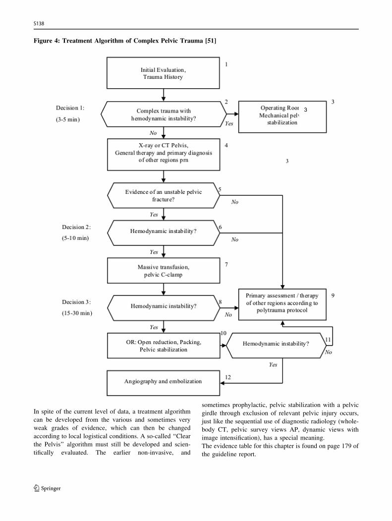
Figure 4: Treatment Algorithm of Complex Pelvic Trauma [51]
In spite of the current level of data, a treatment algorithm
can be developed from the various and sometimes very
weak grades of evidence, which can then be changed
according to local logistical conditions. A so-called ‘‘Clear
the Pelvis’’ algorithm must still be developed and scien-
tifically evaluated. The earlier non-invasive, and
sometimes prophylactic, pelvic stabilization with a pelvic
girdle through exclusion of relevant pelvic injury occurs,
just like the sequential use of diagnostic radiology (whole-
body CT, pelvic survey views AP, dynamic views with
image intensification), has a special meaning.
The evidence table for this chapter is found on page 179 of
the guideline report.
S138
123
References
1. Adams JE, et al. Pelvic trauma in rapidly fatal motor vehicle
accidents. J Orthop Trauma. 2003;17(6):406–10 [LoE 4].2. Agolini SF, et al. Arterial embolization is a rapid and effective
technique for controlling pelvic fracture hemorrhage. J Trauma.
1997;43(3):395–9 [LoE 4].
3. Ben-Menachem Y, et al. Hemorrhage associated with pelvic
fractures: causes, diagnosis, and emergent management. AJR Am
J Roentgenol. 1991;157(5):1005–14 [LoE 5].4. Berg EE, Chebuhar C, Bell RM. Pelvic trauma imaging: a
blinded comparison of computed tomography and roentgen-
ograms. J Trauma. 1996;41(6):994–8 [LoE 2].
5. Blackmore CC, et al. Assessment of volume of hemorrhage and
outcome from pelvic fracture. Arch Surg. 2003;138(5):504–8
(discussion 508–9) [LoE 3].6. Bonner TJ, et al. Accurate placement of a pelvic binder improves
reduction of unstable fractures of the pelvic ring. J Bone Joint
Surg Br. 2011;93(11):1524–8.
7. Bosch U, Pohlemann T, Tscherne H. Primary management of
pelvic injuries. Orthopade. 1992;21(6):385–92 [LoE 4].
8. Brasel KJ, et al. Significance of contrast extravasation in patients
with pelvic fracture. J Trauma. 2007;62(5):1149–52 [LoE 4].
9. Brown CV, et al. Does pelvic hematoma on admission computed
tomography predict active bleeding at angiography for pelvic
fracture? Am Surg. 2005;71(9):759–62 [LoE 4].
10. Burkhardt M, et al. Strategies for surgical treatment of multiple
trauma including pelvic fracture. Review of the literature.
Unfallchirurg 2005. 108(10): 812, 814–20 [LoE 3].11. Cook RE, Keating JF, Gillespie I. The role of angiography in the
management of haemorrhage from major fractures of the pelvis.
J Bone Joint Surg Br. 2002;84(2):178–82 [LoE 3].
12. Cothren CC, et al. Preperitonal pelvic packing for hemodynam-
ically unstable pelvic fractures: a paradigm shift. J Trauma.
2007;62(4):834–9 discussion 839–42 [LoE 3].13. Croce MA, et al. Emergent pelvic fixation in patients with
exsanguinating pelvic fractures. J Am Coll Surg.
2007;204(5):935–9 discussion 940–2 [LoE 4].
14. Culemann U, et al. Pelvic fracture. Diagnostics and current
treatment options. Chirurg. 2003;74(7):687–98 quiz 699–700
[LoE 4].15. Dalal SA, et al. Pelvic fracture in multiple trauma: classification
by mechanism is key to pattern of organ injury, resuscitative
requirements, and outcome. J Trauma. 1989;29(7):981–1000
discussion 1000–2 [LoE 4].16. DeAngelis NA, et al. Use of the trauma pelvic orthotic device
(T-POD) for provisional stabilisation of anterior-posterior
compression type pelvic fractures: a cadaveric study. Injury.
2008;39(8):903–6 [LoE 3].17. Dente CJ, et al. The outcome of open pelvic fractures in the
modern era. Am J Surg. 2005;190(6):830–5 [LoE 4].18. Duane TM, et al. Clinical examination is superior to plain films
to diagnose pelvic fractures compared to CT. Am Surg.
2008;74(6):476–9 (discussion 479–80) [LoE 3].
19. Edeiken-Monroe BS, Browner BD, Jackson H. The role of
standard roentgenograms in the evaluation of instability of
pelvic ring disruption. Clin Orthop Relat Res. 1989;240:63–76
[LoE 4].20. Ertel W, et al. Control of severe hemorrhage using C-clamp and
pelvic packing in multiply injured patients with pelvic ring
disruption. J Orthop Trauma. 2001;15(7):468–74 [LoE 3].
21. Euler E, Nast-Kolb D, Schweiberer L. Acetabular and pelvic
fractures in multiple trauma. Orthopade. 1997;26(4):354–9
[LoE 4].22. Failinger MS, McGanity PL. Unstable fractures of the pelvic
ring. J Bone Joint Surg Am. 1992;74(5):781–91 [LoE 5].23. Fangio P, et al. Early embolization and vasopressor adminis-
tration for management of life-threatening hemorrhage from
pelvic fracture. J Trauma. 2005;58(5):978–84 (discussion 984)
[LoE 4].24. Friese RS, et al. Abdominal ultrasound is an unreliable modality
for the detection of hemoperitoneum in patients with pelvic
fracture. J Trauma. 2007;63(1):97–102 [LoE 4].
25. Ghaemmaghami V, et al. Effects of early use of external pelvic
compression on transfusion requirements and mortality in pelvic
fractures. Am J Surg. 2007;194(6):720–3 (discussion 723) [LoE4].
26. Gourlay D, et al. Pelvic angiography for recurrent traumatic
pelvic arterial hemorrhage. J Trauma. 2005;59(5):1168–73
(discussion 1173–4) [LoE 4].27. Guillamondegui OD, et al. The utility of the pelvic radiograph
in the assessment of pediatric pelvic fractures. J Trauma.
2003;55(2):236–9 (discussion 239–40) [LoE 4].
28. Hagiwara A, et al. Predictors of death in patients with life-
threatening pelvic hemorrhage after successful transcatheter
arterial embolization. J Trauma. 2003;55(4):696–703 [LoE 4].29. Hagiwara A, et al. The usefulness of transcatheter arterial
embolization for patients with blunt polytrauma showing
transient response to fluid resuscitation. J Trauma.
2004;57(2):271–6 (discussion 276–7) [LoE 3].
30. Harley JD, Mack LA, Winquist RA. CT of acetabular fractures:
comparison with conventional radiography. AJR Am J Roent-
genol. 1982;138(3):413–7 [LoE 3].31. Hauschild O, et al. Angioembolization for pelvic hemorrhage
control: results from the German pelvic injury register.
J Trauma Acute Care Surg. 2012;73(3):679–84.
32. Holting T, et al. Diagnosis and treatment of retroperitoneal
hematoma in multiple trauma patients. Arch Orthop Trauma
Surg. 1992;111(6):323–6 [LoE 4].33. Kamaoui I, et al. Pelvic trauma: impact of iodinated contrast
material extravasation at MDCT on patient management.
J Radiol. 2008;89(11 Pt 1):1729–34 [LoE 4].
34. Kessel B, et al. Is routine portable pelvic X-ray in stable multiple
trauma patients always justified in a high technology era?
Injury. 2007;38(5):559–63 [LoE 4].35. Kimbrell BJ, et al. Angiographic embolization for pelvic
fractures in older patients. Arch Surg. 2004;139(7):728–32
(discussion 732–3) [LoE 4].
36. Lustenberger T, et al. The role of angio-embolization in the
acute treatment concept of severe pelvic ring injuries. Injury.
2015;46:S33–8.
37. Miller PR, et al. External fixation or arteriogram in bleeding
pelvic fracture: initial therapy guided by markers of arterial
hemorrhage. J Trauma. 2003;54(3):437–43 [LoE 4].
38. Mucha P Jr, Welch TJ. Hemorrhage in major pelvic fractures.
Surg Clin N Am. 1988;68(4):757–73 [LoE 4].
39. Panetta T, et al. Percutaneous transcatheter embolization for
massive bleeding from pelvic fractures. J Trauma.
1985;25(11):1021–9 [LoE 3].40. Pehle B, et al. Significance of physical examination and
radiography of the pelvis during treatment in the shock
emergency room. Unfallchirurg. 2003;106(8):642–8 [LoE 3].
S3-guideline on treatment of patients with severe/multiple injuries S139
123
41. Pereira SJ, et al. Dynamic helical computed tomography scan
accurately detects hemorrhage in patients with pelvic fracture.
Surgery. 2000;128(4):678–85 [LoE 4].42. Perez JV, Hughes TM, Bowers K. Angiographic embolisation in
pelvic fracture. Injury. 1998;29(3):187–91 [LoE 4].43. Pieri S, et al. Percutaneous management of hemorrhages in
pelvic fractures. Radiol Med. 2004;107(3):241–51 [LoE 4].44. Pizanis A, et al. Emergency stabilization of the pelvic ring:
clinical comparison between three different techniques. Injury.
2013;44(12):1760–4.
45. Pohlemann T, et al. Severe pelvic injury with pelvic mass
hemorrhage: determining severity of hemorrhage and clinical
experience with emergency stabilization. Unfallchirurg.
1996a;99(10):734–43 [LoE 2].
46. Pohlemann T, et al. Biomechanical comparison of various
emergency stabilization measures of the pelvic ring. Unfallchir-
urg. 1994;97(10):503–10 [LoE 4].47. Pohlemann T, et al. Traumatic hemipelvectomy. Experiences
with 11 cases. Unfallchirurg. 1996b;99(4):304–12 [LoE 3].48. Resnik CS, et al. Diagnosis of pelvic fractures in patients with
acute pelvic trauma: efficacy of plain radiographs. AJR Am J
Roentgenol. 1992;158(1):109–12 [LoE 3].
49. Sadri H, et al. Control of severe hemorrhage using C-clamp and
arterial embolization in hemodynamically unstable patients with
pelvic ring disruption. Arch Orthop Trauma Surg.
2005;125(7):443–7 [LoE 4].
50. Salim A, et al. Predictors of positive angiography in pelvic
fractures: a prospective study. J Am Coll Surg.
2008;207(5):656–62 [LoE 3].
51. Seekamp A, Burkhardt M, Pohlemann T. Schockraummanage-
ment bei Verletzungen des Beckens. Eine systematische Liter-
aturubersicht. Unfallchirurg. 2004;107(10):903–10.
52. Shapiro M, et al. The role of repeat angiography in the
management of pelvic fractures. J Trauma. 2005;58(2):227–31
[LoE 4].
53. Sheridan MK, et al. Can CT predict the source of arterial
hemorrhage in patients with pelvic fractures? Emerg Radiol.
2002;9(4):188–94 [LoE 3].54. Shlamovitz GZ, et al. How (un)useful is the pelvic ring stability
examination in diagnosing mechanically unstable pelvic frac-
tures in blunt trauma patients? J Trauma. 2009;66(3):815–20
[LoE 3].55. Siegmeth A, et al. Associated injuries in severe pelvic trauma.
Unfallchirurg. 2000;103(7):572–81 [LoE 4].56. Silber JS, et al. Analysis of the cause, classification, and
associated injuries of 166 consecutive pediatric pelvic fractures.
J Pediatr Orthop. 2001;21(4):446–50 [LoE 4].
57. Stengel D, et al. Accuracy of single-pass whole-body computed
tomography for detection of injuries in patients with major blunt
trauma. CMAJ. 2012;184(8):869–76.
58. Stewart BG, et al. Is the screening portable pelvis film clinically
useful in multiple trauma patients who will be examined by
abdominopelvic CT? Experience with 397 patients. Emerg
Radiol. 2002;9(5):266–71 [LoE 4].59. Tarman GJ, et al. Lower genitourinary injury and pelvic
fractures in pediatric patients. Urology. 2002;59(1):123–6
(discussion 126) [LoE 4].
60. Their ME, et al. Diagnostic value of pelvic radiography in the
initial trauma series in blunt trauma. Eur Radiol.
2005;15(8):1533–7 [LoE 4].
61. Torode I, Zieg D. Pelvic fractures in children. J Pediatr Orthop.
1985;5(1):76–84 [LoE 4].
62. Totterman A, et al. A protocol for angiographic embolization in
exsanguinating pelvic trauma: a report on 31 patients. Acta
Orthop. 2006;77(3):462–8 [LoE 3].63. Totterman A, et al. Extraperitoneal pelvic packing: a salvage
procedure to control massive traumatic pelvic hemorrhage.
J Trauma. 2007;62(4):843–52 [LoE 4].
64. Trafton PG. Pelvic ring injuries. Surg Clin N Am.
1990;70(3):655–69 [LoE 5].
65. Trunkey DD. Trauma. Accidental and intentional injuries
account for more years of life lost in the U.S. than cancer and
heart disease. Among the prescribed remedies are improved
preventive efforts, speedier surgery and further research. Sci
Am. 1983;249(2):28–35 [LoE 5].66. Velmahos GC, et al. A prospective study on the safety and
efficacy of angiographic embolization for pelvic and visceral
injuries. J Trauma. 2002;53(2):303–8 (discussion 308) [LoE 4].
67. Verbeek D, et al. Acute management of hemodynamically
unstable pelvic trauma patients: time for a change? Multicenter
review of recent practice. World J Surg. 2008;32(8):1874–82
[LoE 3].
68. Westhoff J, et al. Interventional emergency embolization for
severe pelvic ring fractures with arterial bleeding. Integration
into the early clinical treatment algorithm. Unfallchirurg.
2008;111(10):821–8 [LoE 4].
69. Wohlrath B, et al. Preclinical and clinical treatment of
instable pelvic injuries: Results of an online survey. Unfallchir-
urg 2014.
70. Wohlrath B, et al. Beobachtungsstudie zum Management
instabiler Beckenfrakturen. Anasth Intensivmed.
2015;56:S81–7.
71. Young JW, et al. Pelvic fractures: value of plain radiography in
early assessment and management. Radiology.
1986;160(2):445–51 [LoE 4].
2.8 Genitourinary Tract
Primary Clinical Assessment
Key recommendation:
2.48 Recommendation 2011
GoR B The primary diagnostic survey should include inspectionof the external urethral meatus and the transurethralcatheter (if already inserted) for blood.
Explanation:
Gross hematuria is the classic cardinal symptom for inju-
ries to the kidney, bladder, and/or urethra. In contrast,
ureter injuries are clinically undetectable in almost 50 % of
cases [11]. Thus, the urinary catheter and the urethral
meatus should be examined for blood during the primary
survey of the undressed patient [33]. Blood at the urethral
meatus should be distinguished from hematuria, as the two
have different diagnostic implications.
S140
123
Key recommendation:
2.49 Recommendation 2011
GoR B The flanks, abdomen, perineum, and external genitaliashould be examined for hematomas, ecchymoses, andexternal injuries.
Explanation:
Because the external physical examination can be rapidly
and easily performed, it should be carried out in all poly-
trauma patients even though it offers only low diagnostic
value [12]. The examination includes inspection for
external signs of injury (hematoma, abrasions, swelling,
etc.) of the flanks, perineum, groin, and external genitalia.
The value of a digital rectal exam is still debated, since it
seldom reveals pathology [33]. However, a digital rectal
exam should still be performed to assess sphincter tone in
spinal cord injuries as well as in cases where there is blood at
the urethral meatus or when a pelvic fracture suggests ure-
thral injury [33]. A prostate that is non-palpable, displaced or
surrounded by hematoma also offers clinically valuable
information, suggesting prostato-membranous tear [33].
Responsive patients can be questioned regarding details
of the accident and pain associated with genitourinary
injuries. Abdominal pain can give non-specific indications
of intra-abdominal injuries [13, 16, 18]. In addition, blad-
der rupture is specifically suggested when a patient
recounts an urge to urinate prior to the trauma that is no
longer present afterwards (without evidence of neurologic
lesions) [19], or when the patient tries to urinate post-
trauma without success [22].
The following external signs of injury can suggest asso-
ciated urologic injuries: hematoma, swelling, and abrasions
of the flanks, perineum, groin, and external genitalia.
In the studies of Cotton et al. and Allen et al., ecchymoses
and abrasions of the abdomen correlated with intra-ab-
dominal injuries [3, 29]. Hematoma of the penile shaft or
perineal butterfly hematoma suggests anterior urethral
injury [3, 29].
If the patient feels the urge to urinate prior to the trauma
and no longer senses it after the trauma, this suggests
bladder rupture, when there is no evidence of neurologic
lesions [33]. A failed attempt at micturition after trauma is
also suggestive of a bladder rupture [33].
Key recommendation:
2.50 Recommendation 2011
GoR B In cases of hemodynamic instability that precludesfurther primary diagnostic examination, and in caseswhen it is impossible to place a transurethral bladdercatheter, a suprapubic urinary catheter should beplaced percutaneously or during laparotomy (withconcomitant exploration).
Explanation:
In cases of complete urethral rupture, a false passage can
be created with the insertion of a transurethral catheter
[76]. An existing urethral injury can also be aggravated by
transurethral catheter insertion [76]. Thus, in patients with
clinical signs of urethral injury, transurethral catheteriza-
tion can be carried out in the emergency department with
x-ray control (e.g. retrograde urethrogram) to better mon-
itor elimination. This is only contraindicated in very
unstable patients in whom catheter insertion would repre-
sent an unnecessary delay, and for situations that are
unclear even with radiographic control [76].
In cases of hemodynamic instability, where further diag-
nostic assessment is not possible due to time concerns and
where laparotomy is to be performed for these reasons, a
suprapubic catheter should be placed during this procedure
that can later be used for diagnostic purposes [26]. Urinalysis
and serumcreatininemeasurement should be performed [33].
A dipstick urine test should be carried out to detect
hematuria. Compared to microscopic examination, the
dipstick has over 95 % sensitivity and specificity [23, 24,
37]. The advantage of the dipstick test is that results are
available in under 10 minutes. It is also helpful to provide
evidence of bacteriuria, something often present in the
elderly that can be particularly problematic in combination
with a urinary tract injury [33].
Need for Diagnostic Imaging
Key recommendation:
2.51 Recommendation 2011
GoR B All patients with hematuria, blood at the urethralmeatus, dysuria, failed catheterization, or otherhistorical evidence (local hematoma, concomitantinjuries, mechanism of injury) have increased risk ofurogenital injuries and should undergo a targeteddiagnostic work-up of the kidneys and/or the efferenturinary tract.
Explanation:
Even though simultaneous injuries of the upper and lower
urinary tracts occur in only 0.6 % of patients, a complete
urological workup is generally carried out in all patients
with corresponding signs, since this normally is performed
as CT for confirmed micro or gross hematuria and includes
the entire urinary system [55, 63, 67].
While gross hematuria is pathognomonic for genitourinary
injuries, microhematuria is borderline. Currently, however,
it is generally accepted that diagnostic assessment should
continue only if there is other evidence of injury [45, 58].
Renal trauma is classified according to mechanism of
injury, blunt or penetrating. While in rural areas, blunt
trauma accounts for 90-95 % of renal injuries (horse kicks,
S3-guideline on treatment of patients with severe/multiple injuries S141
123
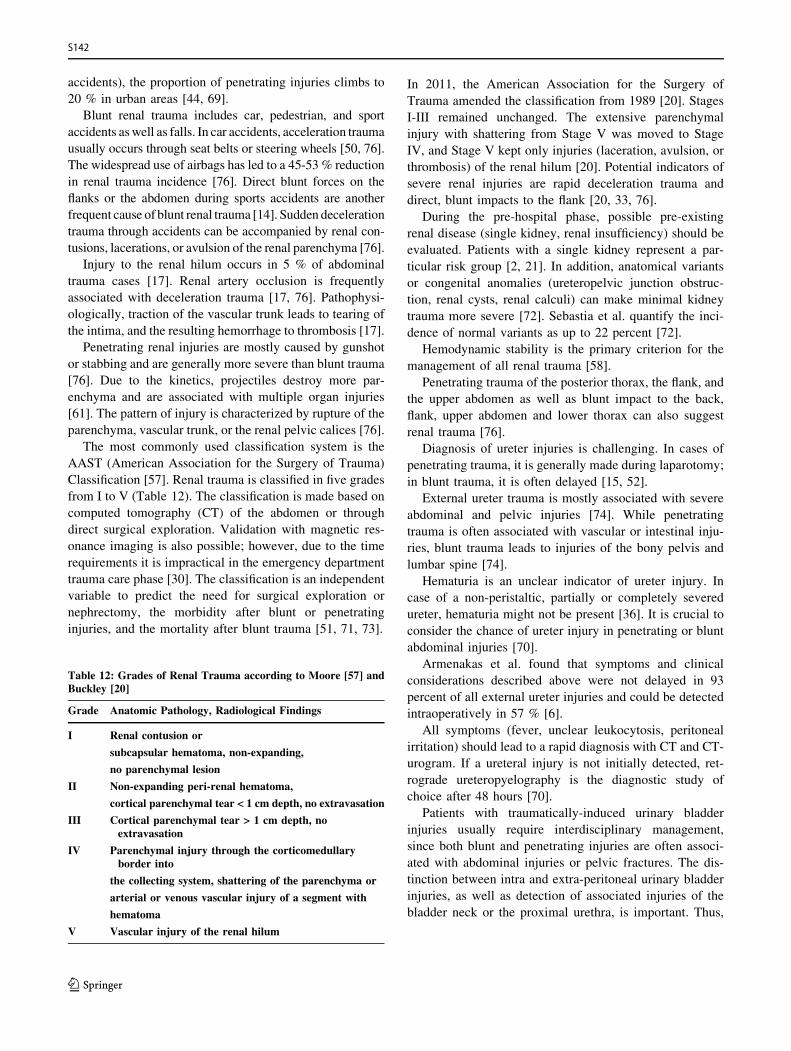
accidents), the proportion of penetrating injuries climbs to
20 % in urban areas [44, 69].
Blunt renal trauma includes car, pedestrian, and sport
accidents aswell as falls. In car accidents, acceleration trauma
usually occurs through seat belts or steering wheels [50, 76].
The widespread use of airbags has led to a 45-53 % reduction
in renal trauma incidence [76]. Direct blunt forces on the
flanks or the abdomen during sports accidents are another
frequent cause of blunt renal trauma [14]. Sudden deceleration
trauma through accidents can be accompanied by renal con-
tusions, lacerations, or avulsion of the renal parenchyma [76].
Injury to the renal hilum occurs in 5 % of abdominal
trauma cases [17]. Renal artery occlusion is frequently
associated with deceleration trauma [17, 76]. Pathophysi-
ologically, traction of the vascular trunk leads to tearing of
the intima, and the resulting hemorrhage to thrombosis [17].
Penetrating renal injuries are mostly caused by gunshot
or stabbing and are generally more severe than blunt trauma
[76]. Due to the kinetics, projectiles destroy more par-
enchyma and are associated with multiple organ injuries
[61]. The pattern of injury is characterized by rupture of the
parenchyma, vascular trunk, or the renal pelvic calices [76].
The most commonly used classification system is the
AAST (American Association for the Surgery of Trauma)
Classification [57]. Renal trauma is classified in five grades
from I to V (Table 12). The classification is made based on
computed tomography (CT) of the abdomen or through
direct surgical exploration. Validation with magnetic res-
onance imaging is also possible; however, due to the time
requirements it is impractical in the emergency department
trauma care phase [30]. The classification is an independent
variable to predict the need for surgical exploration or
nephrectomy, the morbidity after blunt or penetrating
injuries, and the mortality after blunt trauma [51, 71, 73].
Table 12: Grades of Renal Trauma according to Moore [57] andBuckley [20]
Grade Anatomic Pathology, Radiological Findings
I Renal contusion or
subcapsular hematoma, non-expanding,
no parenchymal lesion
II Non-expanding peri-renal hematoma,
cortical parenchymal tear < 1 cm depth, no extravasation
III Cortical parenchymal tear > 1 cm depth, noextravasation
IV Parenchymal injury through the corticomedullaryborder into
the collecting system, shattering of the parenchyma or
arterial or venous vascular injury of a segment with
hematoma
V Vascular injury of the renal hilum
In 2011, the American Association for the Surgery of
Trauma amended the classification from 1989 [20]. Stages
I-III remained unchanged. The extensive parenchymal
injury with shattering from Stage V was moved to Stage
IV, and Stage V kept only injuries (laceration, avulsion, or
thrombosis) of the renal hilum [20]. Potential indicators of
severe renal injuries are rapid deceleration trauma and
direct, blunt impacts to the flank [20, 33, 76].
During the pre-hospital phase, possible pre-existing
renal disease (single kidney, renal insufficiency) should be
evaluated. Patients with a single kidney represent a par-
ticular risk group [2, 21]. In addition, anatomical variants
or congenital anomalies (ureteropelvic junction obstruc-
tion, renal cysts, renal calculi) can make minimal kidney
trauma more severe [72]. Sebastia et al. quantify the inci-
dence of normal variants as up to 22 percent [72].
Hemodynamic stability is the primary criterion for the
management of all renal trauma [58].
Penetrating trauma of the posterior thorax, the flank, and
the upper abdomen as well as blunt impact to the back,
flank, upper abdomen and lower thorax can also suggest
renal trauma [76].
Diagnosis of ureter injuries is challenging. In cases of
penetrating trauma, it is generally made during laparotomy;
in blunt trauma, it is often delayed [15, 52].
External ureter trauma is mostly associated with severe
abdominal and pelvic injuries [74]. While penetrating
trauma is often associated with vascular or intestinal inju-
ries, blunt trauma leads to injuries of the bony pelvis and
lumbar spine [74].
Hematuria is an unclear indicator of ureter injury. In
case of a non-peristaltic, partially or completely severed
ureter, hematuria might not be present [36]. It is crucial to
consider the chance of ureter injury in penetrating or blunt
abdominal injuries [70].
Armenakas et al. found that symptoms and clinical
considerations described above were not delayed in 93
percent of all external ureter injuries and could be detected
intraoperatively in 57 % [6].
All symptoms (fever, unclear leukocytosis, peritoneal
irritation) should lead to a rapid diagnosis with CT and CT-
urogram. If a ureteral injury is not initially detected, ret-
rograde ureteropyelography is the diagnostic study of
choice after 48 hours [70].
Patients with traumatically-induced urinary bladder
injuries usually require interdisciplinary management,
since both blunt and penetrating injuries are often associ-
ated with abdominal injuries or pelvic fractures. The dis-
tinction between intra and extra-peritoneal urinary bladder
injuries, as well as detection of associated injuries of the
bladder neck or the proximal urethra, is important. Thus,
S142
123
the initial assessment for precise evaluation of injuries
plays an important role.
Motor vehicle accidents are the leading cause (90 %) for
blunt bladder injuries [9, 40]. Approximately 70 % of
patients with blunt bladder injuries have concomitant pel-
vic fractures (especially pelvic ring fractures, symphysis
ruptures, pelvic rami fractures) and approximately 44 % of
patients have at least one intra-abdominal injury [9, 32,
40]. Penetrating bladder injuries from gunshot wounds or
impalement injuries are rare [28]. Extra-peritoneal bladder
ruptures are more common than intraperitoneal, and they
are mostly associated with pelvic fractures, caused by
displacement of the pelvic ring. Intraperitoneal bladder
injuries are caused by suddenly increased intra-abdominal
pressure, kicks to the genitalia or in the lower abdomen. A
full bladder thus increases the risk for intraperitoneal
bladder rupture [40].
Injuries to the urethra have been classified by the
American Association for Surgery of Trauma in five grades
regarding the continuity of the urethral circumference
(modified by Moore et al. [56]) as follows:
I Contusion (incomplete tear of the urethral circum-
ference with evidence for blood at the urethral
meatus or evidence of extravasation on retrograde
urethrogram (RUG)
II Elongation injury of the urethra (elongation of the
urethra with intact urethral circumference, without
extravasation on RUG)
III Partial rupture of the urethra with complete disrup-
tion of the anterior/posterior urethral circumference
(with evidence of urethral extravasation and visual-
ization of the bladder)
IV Complete rupture of the urethra, \ 2 cm urethral
separation (with evidence of urethral extravasation
but without visualization of the bladder)
V Complete rupture of the urethra, C 2 cm urethral
separation or injury extending into the prostate or
vagina
90 % of urethral injuries occur with blunt external injury
patterns such as traffic accidents, impact injuries (straddle
trauma, e.g. impact against bicycle handlebars) or direct
injuries to the perineum [54]. They generally concern the
front aspect of the anterior urethra (compression of the
bulbar urethra between a blunt object and the pelvis) [54].
Pelvic fracture-associated urethral injuries (PFUI) are
usually caused by shearing injuries (distraction forces), in
which pelvic bone fragments contact the urethra and thus,
the anterior (80 %) section is more often affected than the
posterior section (proximal to the sphincter) [60].
Penetrating or open injuries resulting from gunshot
wounds, dog bites, or impalement injuries affect the
anterior urethra (penile and bulbar) more than the posterior
urethra [54].
PFUI concerns the anterior urethra (distal to the
sphincter) in up to 80 % of cases and is the most common
urethral injury after iatrogenic injury; 19 % of males and 6
% of females with pelvic fractures have associated urethral
injuries [76]. PFUI occurs most commonly in combination
with unstable pelvic fractures with diastasis of the
sacroiliac joints and/or the public symphysis [60].
Key recommendation:
2.52 Recommendation 2011
GoR B Further diagnostic imaging of the efferent urinary tractshould be performed when one or more of the followingcriteria are met: hematuria, bleeding from the urethralmeatus or the vagina, dysuria, and local hematoma.
Explanation:
Numerous studies have found that bladder ruptures are
associated with pelvic fractures in approximately 70 % of
cases [9, 40]. Hochberg and Stone found a direct correla-
tion between the number of fractured pubic rami (1, 2-3, 4)
and the frequency of bladder ruptures (4%, 12%, 40%)
[41]. Aihara et al. also found that disruption of the pubic
symphysis or sacroiliac joint is present in 75 % of bladder
ruptures after blunt trauma [1]. Nevertheless, bladder rup-
ture cannot be deduced from the presence of complex
pelvic fracture, as only 20 % (positive predictive value) of
patients with symphysis and sacroiliac joint disruption
have bladder rupture [1].
The close correlation between pelvic fractures and ure-
thral injuries is well documented. However, the severity of
the injury plays a big part [4, 5, 10, 75]. Koraitim et al. and
Aihara et al. have consistently found that pubic rami
fractures increase the risk of urethral injuries, but the risk
increases enormously in the presence of complex pelvic
fractures (type C) [1, 47, 48]. Aihara et al. emphasize that
fractures of the inferior pubic rami in particular suggest
urethral injury [1]. In a series of 200 patients with pelvic
fractures, Palmer et al. found that 26 of 27 patients with
urological lesions had fractures of the anterior and poste-
rior rings [62]. The association is less marked in women
because of the shorter urethra with less connective-tissue
tethering [10, 78]. Urethral injuries in women are most
often accompanied by bleeding vaginal injuries [78].
The classic combination of pelvic fracture and gross
hematuria enables a diagnosis of bladder and/or urethral
injury with a high degree of certainty [41, 62]. Of 719
patients with blunt pelvic/abdominal trauma, Rehm et al.
found all 21 cases of bladder injuries through hematuria, 17
of whom had gross hematuria [66]. Morey et al. also
reported that 85 % of their patients with pelvic fractures
S3-guideline on treatment of patients with severe/multiple injuries S143
123
and concomitant bladder rupture had gross hematuria [59].
In the study of Palmer et al., this number was ten of eleven
patients, and in Hsieh et al. it was 48 of 51 patients [42,
62]. A gap of the pubic symphysis and separation of the
sacroiliac joint doubled the risk for bladder injury in the
study by Aihara et al. [1]. However, even without evidence
of pelvic fracture, patients with gross hematuria or blood at
the urethral meatus must be assumed to have an injury of
the efferent urinary tract [58, 76].
The distinction between hematuria and blood at the
urethral meatus can be helpful to distinguish bladder and
urethral injuries. Morey et al. reported that all 53 patients
with bladder ruptures had hematuria, but also that blood at
the urethral meatus, present in all six cases, correctly
indicated an associated urethral injury [59].
The current body of evidence indicates clearly that a
lack of hematuria and corresponding exclusion of pelvic
injury exclude relevant injury of the bladder or urethra [58,
62]. Assessment in cases of pelvic fracture are somewhat
more difficult [41]. Hochberg and Stone found that here, a
urological injury remains unlikely as long as the pelvic
fracture does not involve the pubic rami [41].
Diagnostic Imaging of the Kidneys and Ureters
Key recommendation:
2.53 Recommendation 2011
GoR B CT with contrast should be performed for suspectedkidney injuries.
Explanation:
Computed tomography with contrast is available almost
nationwide and enables rapid and adequate detection and
staging of significant kidney injuries [58]. Even in pediatric
polytrauma patients, CT can be performed immediately
following the emergency department trauma care phase
[58]. Current developments of modern dual-source CT
scanners with minimal tube voltage (70-100 kV) enable
perfusion measurement and iterative image reconstruction
not only with lower radiation dose, but also decreased
amounts of contrast medium [27, 35, 80]. To date, this new
generation of dual-source CT scanners have mainly been
used for CT angiography and thoracic imaging, but because
of the short examination time, it is also useful for emer-
gency department diagnostic assessment of polytrauma
patients [27].
Although intravenous pyelography is a sensitive method
for the diagnosis of renal trauma, it has currently been
largely supplanted by the use of CT [58, 76]. Therefore,
current guidelines recommend its use only in centers
without available CT [8, 58, 76]. The main reasons for this
are time delay as well as the decreased image quality.
Validity is limited to extravasation and evidence of a mute
kidney, which is generally a sign of extensive renal trauma
or an injury to the renal vascular hilum [76]. Extravasation
suggests a high-grade renal injury with tearing of the
capsule as well as the parenchyma and involvement of the
renal pelvis [76].
Ultrasound offers a rapid, non-invasive, cheap, and
radiation-free option to detect retroperitoneal fluid collec-
tions [68]. The disadvantage is the high dependence on
examiner proficiency and patient positioning [68].
Renal lacerations can be detected on ultrasound, but
there is less accuracy regarding depth and expansion [58].
Statements regarding urinary extravasation or leakage are
also difficult to make [58]. On the other hand, ultrasound
monitoring of parenchymal lesions, retroperitoneal hema-
tomas, or urinomas are possible within the intensive care
setting [18]. Contrast-enhanced ultrasound (CEUS) has
greater sensitivity than conventional ultrasound and can be
useful in blunt trauma [38]. The patient must, however, be
hemodynamically stable [58].
Angiography is indicated in stable patients who are
determined as eligible for interventional bleeding control
after CT diagnosis [34]. Overall, angiography is less
specific, more time-intensive, and more invasive than CT;
however, it is more specific in terms of localization and
assessment of the severity of vascular injuries [76].
Diagnostic indications are lacerations, extravasation,
and assessment of renal hilar vessels as well as unclear
findings on CT [76]. In addition, causes of a non-visualized
kidney (complete avulsion of the renal hilar vessels, renal
artery thrombosis, vascular spasm after contusion) can be
evaluated on angiography [76].
Due to long examination time, cost effectiveness, and
the limited access to patients during the examination,
magnetic resonance imaging is suitable as a primary
diagnostic imaging procedure to assess renal trauma only
when there is a contrast allergy or when CT is not available
[53]. The domain for MRI is in monitoring after renal
trauma and particularly in the assessment of lacerations and
individual fragments [31, 76].
Thus, for primary trauma diagnostic assessments in the
emergency department, the importance of CT has gradually
increased over recent years [71]. Consequently, CT has
become the current standard investigation during the initial
emergency department trauma care phase [76].
S144
123
Diagnostic Imaging of the Lower Urinary Tract
Key recommendations:
2.54 Recommendation 2011
GoR B If the priority of care allows, retrograde urethrographyand a cystogram should be carried out in patients withclinical evidence suggesting a urethral lesion.
2.55 Recommendation 2011
GoR B If the priority of care allows, a retrograde cystogramshould be carried out in patients with clinical evidencesuggesting a bladder injury.
Explanation:
The main symptom of urethral injury is gross hematuria ([95 % of patients) [9, 40]. However, a lack of gross
hematuria does not exclude bladder injury [7].
Other symptoms suggesting bladder injury could be:
abdominal guarding (up to 97 %), inability to urinate, or
abdominal tension with urinary ascites [28, 40, 77]. Fur-
thermore, swelling of the perineum, the scrotum, or tension
along the anterior abdominal wall because of urine
extravasation are observed. Increased serum creatinine and
urea levels, caused by peritoneal resorption, can also be
present in intraperitoneal bladder rupture.
In cases of urinary retention and/or blood at the urethral
meatus, an associated injury of the urethra must always be
considered, which makes diagnostic clarification with ret-
rograde urethrography necessary prior to further manipu-
lation (e.g. catheterization) [39, 40].
In the acute situation with unstable patients, CT is the
main focus within the trauma care protocols of the emer-
gency department [43]. Conventional cystography is
comparable with CT cystography regarding sensitivity and
specificity; and plays a role more for stable patients or for
monitoring of catheter removal [64, 79]. Cystography,
whether conventional or with CT, should be performed
with at least 350 ml of diluted contrast medium through an
inserted catheter [65].
The diagnostic procedure in urethral injuries is depen-
dent on the hemodynamic situation and on accompanying
injuries as well as the pattern of injury of the patient.
Retrograde urethrography is a basic diagnostic study
that enables assessment of localization and extent of the
injury in stable patients [25]. If contrast medium is not
visible in the bladder or the posterior ureter is not visual-
ized, an additional antegrade urethrography (micturition
cystourethrography, MCU) can give further information
regarding the extent of injuries to the bladder neck, the
prostate and the membranous urethra as well as ureteral
distraction or stricture over longer the length [25].
Flexible cystoscopy can also be used (anteriorly over the
suprapubic access point) if retrograde urethrography of the
bladder neck or the posterior ureter cannot be assessed, or
an approach through the urethral meatus cannot be
observed or is not possible [25, 40, 46, 54].
Computed or magnetic resonance imaging with CT/
MRI-Urography and cystography are performed to evaluate
associated abdominal injuries, particularly in cases of
penetrating injury of the posterior ureter [25, 49].
Finally, in some 20 % of cases, it is not possible to
perform the cystogram in the initial emergency department
phase of treatment because of associated injuries [42]. This
may be unavoidable in individual cases; however, it must
be performed as soon as possible so that no injuries are
missed [42]. On the other hand, if the diagnosis for rup-
tured bladder is delayed, there are no severe disadvantages
for the patient [42].
References
1. Aihara R, et al. Fracture locations influence the likelihood of
rectal and lower urinary tract injuries in patients sustaining pelvic
fractures. J Trauma. 2002;52(2):205–9.
2. Akcay A, et al. Update on the diagnosis and management of acute
kidney injury. International Journal of Nephrology and Renovas-
cular Disease. 2010;3:129–40.
3. Allen GS, et al. Hollow visceral injury and blunt trauma.
J Trauma. 1998;45(1):68–9.
4. Andrich DE, Day AC, Mundy AR. Proposed mechanisms of
lower urinary tract injury in fractures of the pelvic ring. BJU
international. 2007;100(3):567–73.
5. Andrich DE, Mundy AR. The nature of urethral injury in cases of
pelvic fracture urethral trauma. J Urol. 2001;165(5):1492–5.
6. Armenakas NA. Current methods of diagnosis and management
of ureteral injuries. World J Urol. 1999;17(2):78–83.
7. Avey G, et al. Radiographic and clinical predictors of bladder
rupture in blunt trauma patients with pelvic fracture. Acad
Radiol. 2006;13(5):573–9.
8. Bent C, et al. Urological injuries following trauma. Clin Radiol.
2008;63:1361–71.
9. Bjurlin MA, et al. Genitourinary injuries in pelvic fracture
morbidity and mortality using the National Trauma Data Bank.
J Trauma. 2009;67(5):1033–9.
10. Black PC, et al. Urethral and bladder neck injury associated with
pelvic fracture in 25 female patients. J Urol.
2006;175(6):2140–4 (discussion 2144 [LoE 5]).11. Bozeman C, et al. Selective operative management of major
blunt renal trauma. J Trauma. 2004;57(2):305–9 ([LoE 4]).
12. Brandes S Jr, Borrelli J. Pelvic fracture and associated urologic
injuries. World J Surg. 2001;25(12):1578–87 (LoE 2).
13. Brandes S et al. Diagnosis and management of ureteric injury:
an evidence-based analysis. BJU international. 2004;94(3):277–
89 (LoE 2b).14. Brandes SB, McAninch JW. Urban free falls and patterns of
renal injury: a 20-year experience with 396 cases. J Trauma
Acute Care Surg. 1999;47(4):643–9 (LoE 4).
15. Brandes SB, McAninch JW. Renal Trauma : A Practical Guide
to Evaluation and Management. Sci World J. 2004;4:31–40.
16. Brosman SA, Paul JG. Trauma of the bladder. Surg Gynecol
Obstet. 1976;143(4):605–8 (LoE 2b).
17. Bruce LM, et al. Blunt renal artery injury: Incidence, diagnosis,
and management. American Surgeon. 2001;67(6):550–4.
S3-guideline on treatment of patients with severe/multiple injuries S145
123
18. Buchberger W et al. Diagnosis and staging of blunt kidney
trauma. A comparison of urinalysis, i.v. urography, sonography
and computed tomography. Rofo. 1993;158(6):507–12 (LoE2b).
19. Buckley JC, McAninch JW. Selective management of isolated
and nonisolated grade IV renal injuries. J Urol. 2006;176(6 Pt
1):2498–502 (discussion 2502 [LoE 4]).20. Buckley JC, McAninch JW. Revision of current American
Association for the Surgery of Trauma Renal Injury grading
system. J Trauma. 2011;70(35).
21. Cachecho R, Millham FH, Wedel SK. Management of the
trauma patient with pre-existing renal disease. Crit Care Clin.
1994;10(3):523–36.
22. Carroll PR, McAninch JW. Major bladder trauma: the accuracy
of cystography. J Urol. 1983;130(5):887–8 (LoE 5).23. Carroll PR, McAninch JW. Major bladder trauma: mechanisms
of injury and a unified method of diagnosis and repair. J Urol.
1984;132(2):254–57 (LoE 4).
24. Chandhoke PS, McAninch JW. Detection and significance of
microscopic hematuria in patients with blunt renal trauma.
J Urol. 1988;140(1):16–8 (LoE 5).25. Chapple C et al. Consensus statement on urethral trauma. BJU
Int. 2004;93(9):1195–202 (LoE 4).26. Chapple CR, Png D. Contemporary management of urethral
trauma and the post-traumatic stricture. Curr Opin Urol.
1999;9(3):253–60.
27. Christe A, et al. Accuracy of low-dose computed tomography
(CT) for detecting and characterizing the most common CT-
patterns of pulmonary disease. Euro J Radiol.
2013;82(3):e142–50.
28. Cinman NM et al. Gunshot wounds to the lower urinary tract: a
single-institution experience. J Trauma Acute Care Surg.
2013;74(3):725–30 (discussion 730-1).
29. Cotton BA et al. The utility of clinical and laboratory data for
predicting intraabdominal injury among children. J Trauma.
2004;56(5):1065–1068 (LoE 4).30. Dayal M. Imaging in renal trauma. World J Radiol.
2013;5(8):275.
31. Debus, F., et al., Rehabilitation von Schwerverletzten in
Deutschland. Der Unfallchirurg, 2014: p. 1–8.
32. Deibert CM, Spencer BA. The association between operative
repair of bladder injury and improved survival: results from the
National Trauma Data Bank. J Urol. 2011;186(1):151–5.
33. DGUC, S3 Polytrauma/Schwerverletzten Behandlung.
2011(012).
34. Eastham JA, Wilson TG, Ahlering TE. Radiographic evaluation
of adult patients with blunt renal trauma. J Urol. 1992;148(2 Pt
1):266–7 (LoE 3a).35. Ebner L et al. Feasible Dose Reduction in Routine Chest
Computed Tomography Maintaining Constant Image Quality
Using the Last Three Scanner Generations: From Filtered Back
Projection to Sinogram-affirmed Iterative Reconstruction and
Impact of the Novel Fully Integrated. J Clin Imaging Sci.
2014;4(38).
36. Elliott SP, McAninch JW. Ureteral injuries: External and iatro-
genic. Urol Clin North Am. 2006;33(1):55–66.
37. Flancbaum L, et al. Blunt bladder trauma: manifestation of
severe injury. Urology. 1988;31(3):220–2.
38. Gaitini D, et al. Sonographic Evaluation of Vascular Injuries.
Ultrasound Clin. 2008;3(1):33–48.
39. Gallentine ML, Morey AF. Imaging of the male urethra for
stricture disease. Urol Clin North Am. 2002;29(2):361–72.
40. Gomez RG, et al. SIU/ICUD Consultation on Urethral Stric-
tures: Pelvic fracture urethral injuries. Urology. 2014;83(3
Suppl):S48–58.
41. Hochberg E, Stone NN. Bladder rupture associated with pelvic
fracture due to blunt trauma. Urology. 1993;41(6):531–3 (LoE4).
42. Hsieh C-H et al. Diagnosis and management of bladder injury
by trauma surgeons. Am J Surg. 2002;184(2):143–7 (LoE 3).
43. Huber-Wagner S, et al. Effect of whole-body CT during trauma
resuscitation on survival: a retrospective, multicentre study.
Lancet. 2009;373(9673):1455–61.
44. Kansas BT, et al. Incidence and management of penetrating
renal trauma in patients with multiorgan injury: extended
experience at an inner city trauma center. J Urol. 2004;172(4 Pt
1):1355–60.
45. Kawashima A et al. CT Urography. Radiographics. 2004;35–54.
46. Kielb SJ, Voeltz ZL, Wolf JS. Evaluation and management of
traumatic posterior urethral disruption with flexible cys-
tourethroscopy. J Trauma. 2001;50(1):36–40.
47. Koraitim MM. Predictors of surgical approach to repair pelvic
fracture urethral distraction defects. J Urol.
2009;182(4):1435–9.
48. Koraitim MM et al. Risk factors and mechanism of urethral
injury in pelvic fractures. Br J Urol. 1996;77(6):876–880 (LoE
1b).49. Koraitim MM, Reda IS. Role of magnetic resonance imaging in
assessment of posterior urethral distraction defects. Urology.
2007;70(3):403–6.
50. Kuan JK et al. Renal injury mechanisms of motor vehicle
collisions: analysis of the crash injury research and engineering
network data set. J Urology. 2007;178(3 Pt 1):935–40 (discus-
sion 940).51. Kuan JK, et al. American Association for the Surgery of Trauma
Organ Injury Scale for kidney injuries predicts nephrectomy,
dialysis, and death in patients with blunt injury and nephrec-
tomy for penetrating injuries. J Trauma. 2006;60:351–6.
52. Kunkle DA et al. Delayed diagnosis of traumatic ureteral
injuries. J Urol. 2006;176(6):2503–7.
53. Leppaniemi A et al. Comparison of high-field magnetic
resonance imaging with computed tomography in the evaluation
of blunt renal trauma. J Trauma. 1995;38(3):420–427 (LoE 4).
54. Martinez-Pineiro L. Urethral Trauma. In Emergencies in
Urology. Springer. 2007;276–299.
55. Miller KS, McAninch JW. Radiographic assessment of renal
trauma: our 15-year experience. J Urol. 1995;154(2 Pt
1):352–355 (LoE 2).56. Moore EE, et al. Organ injury scaling. III: Chest wall,
abdominal vascular, ureter, bladder, and urethra. J Trauma.
1992;33(3):337–9.
57. Moore EE, et al. Organ injury scaling: spleen, liver, and kidney.
J Trauma. 1989;29(12):1664–6.
58. Morey AF et al. American Urological Association (AUA)
Guideline American Urological Association. AUA Clinical
Guidelines, 2014(April), pp 1–57.
59. Morey AF, et al. Bladder rupture after blunt trauma: guidelines
for diagnostic imaging. J Trauma. 2001;51(4):683–6.
60. Mouraviev VB, Santucci RA. Cadaveric anatomy of pelvic
fracture urethral distraction injury: most injuries are distal to the
external urinary sphincter. J Urol. 2005;173(3):869–72.
61. Najibi S, Tannast M, Latini JM. Civilian gunshot wounds to the
genitourinary tract: Incidence, anatomic distribution, associated
injuries, and outcomes. Urology. 2010;76(4):977–81.
S146
123
62. Palmer JK, Benson GS, Corriere JN. Diagnosis and initial
management of urological injuries associated with 200 consec-
utive pelvic fractures. J Urol. 1983;130(4):712–4.
63. Peterson NE, Schulze KA. Selective diagnostic uroradiography
for trauma. J Urol. 1987;137(3):449–51.
64. Quagliano PV, Delair SM, Malhotra AK. Diagnosis of blunt
bladder injury: a prospective comparative study of computed
tomography cystography and conventional retrograde cystogra-
phy. J Trauma. 2006;61(2):410–21 (discussion 421-2).65. Ramchandani P, Buckler PM. Imaging of genitourinary trauma.
Am J Roentgenol. 2009;192(6):1514–23.
66. Rehm CG, et al. Blunt traumatic bladder rupture: the role of
retrograde cystogram. Ann Emerg Med. 1991;20(8):845–7.
67. Richards JR, Derlet RW. Computed tomography for blunt
abdominal trauma in the ED: a prospective study. Am J Emerg
Med. 1998;16(4):338–42.
68. Rippey JCR, Royse AG. Ultrasound in trauma. Best Prac Res
Clin Anaesthesiol. 2009;23(3):343–62.
69. Sangthong B, et al. Management and Hospital Outcomes of
Blunt Renal Artery Injuries: Analysis of 517 Patients from the
National Trauma Data Bank. J Am Coll Surg.
2006;203(5):612–7.
70. Santucci RA, Doumanian L. Upper Urinary Tract Trauma, in
Campbell-walsh urology, A. Wein, Editor. 2012, Elsevier:
Philadelphia. pp 1169–1178.
71. Santucci RA, et al. Validation of the American Association for
the Surgery of Trauma organ injury severity scale for the
kidney. J Trauma. 2001;50(2):195–200.
72. Sebastia MC, et al. Renal trauma in occult ureteropelvic
junction obstruction: CT findings. European Radiology.
1999;9(4):611–5.
73. Shariat SF, et al. Evidence-based validation of the predictive
value of the American Association for the Surgery of Trauma
kidney injury scale. J Trauma. 2007;62(4):933–9.
74. Siram SM, et al. Ureteral trauma: patterns and mechanisms of
injury of an uncommon condition. American journal of surgery.
2010;199(4):566–70.
75. Spirnak JP. Pelvic fracture and injury to the lower urinary tract.
Surg Clin North Am. 1988;68(5):1057–69.
76. Summerton DJ, et al. EAU guidelines on iatrogenic trauma. Eur
Urol. 2012;62(4):628–39.
77. Tonkin JB, Tisdale BE, Jordan GH. Assessment and initial
management of urologic trauma. Med clin North Am.
2011;95(1):245–51.
78. Venn SN, Greenwell TJ, Mundy AR. Pelvic fracture injuries of
the female urethra. BJU Int. 1999;83(6):626–30.
79. Wirth GJ, et al. Advances in the management of blunt traumatic
bladder rupture: experience with 36 cases. BJU Int.
2010;106(9):1344–9.
80. Zheng M, et al. Image quality of ultra-low-dose dual-source CT
angiography using high-pitch spiral acquisition and iterative
reconstruction in young children with congenital heart disease.
J Cardiovasc Comput Tomogr. 2013;7(6):376–82.
2.9 Spine
As a rule, spine injury is suspected in all patients brought to
the hospital with potential multiple trauma. In our own
hospital population over the years 2000-2002, 34 % (245 of
720) polytrauma patients had spinal injuries.
Other studies have found a rate of 20 % [23]. Con-
versely, approximately one third of all spine injuries occur
with concomitant injuries [27, 78]. Overall, one can assume
approximately 10 000 patients per year with serious spinal
injuries in Germany, of whom 1/5 involve the cervical
spine and 4/5 involve the thoracic and/or lumbar spine
[26]. Of multiply injured patients, approximately 10 % will
have C-spine injuries [25]. With 1 to 27 injuries/million
children/year, pediatric spine injuries are relatively rare in
Western Europe/North America [8].
Spine injury as a component of multiple injuries has
important consequences regarding the diagnostic and
therapeutic approach. First, typical associated injuries, e.g.
thoracic or abdominal injuries, must be excluded. If sur-
gical stabilization is required, a comprehensive pre-opera-
tive CT evaluation of the injured region is necessary.
Positioning options for intensive care depend on the sta-
bility of a diagnosed spinal injury. Thus, if the patient’s
overall condition allows (hemodynamics, temperature,
coagulation, intracranial pressure, etc.), it is desirable to
assess spinal injury stability either in the emergency
department or in the operating room prior to transfer to the
intensive care unit.
Medical History
Key recommendation:
2.56 Recommendation 2011
GoR B The medical history is very important and should becollected.
Explanation:
Generally, in multiply injured patients, the history comes
from a third party. The mechanism of injury is very
important information, and should be passed on from the
pre-hospital team to the hospital team. Predisposing factors
for spinal injuries include polytrauma itself [3, 32, 40],
high-speed motor vehicle accidents [3, 14, 27, 85, 99, 132],
motor vehicle accidents of patients not secured by safety
belts or airbags [69, 85], pedestrians hit by a vehicle [14],
falls from a height [13, 32, 40, 111, 116], the influence of
alcohol or drugs [122], and increased age [14, 84, 118]. In
unconscious patients, the medical history should also
include information regarding movement of the extremities
and pain complaints prior to loss of consciousness or
intubation. Traumatic brain injury and facial injuries are
risk factors for cervical spine injury. According to multi-
variate analysis by Blackmore et al. [14], patients with
skull fractures or continuous unconsciousness have mark-
edly higher probability of cervical spine injuries (odds ratio
8.5), while the risk for those with milder injuries, e.g.
face/jaw fractures or short-term unconsciousness is much
S3-guideline on treatment of patients with severe/multiple injuries S147
123
less (OR 2.6). Similarly, Hills and Deane [62] found the
risk of C-spine injury in patients with TBI was approxi-
mately 4 times higher than in patients without. With GCS
less than 8, the risk is 7 times higher. Other studies on the
importance of traumatic brain injury [62, 71], loss of
consciousness [38, 65, 67, 115, 132], and facial skull
fractures [53, 62, 87, 107] confirm the association with
spine injuries. Only one study with large patient numbers
reported a tendency for a decreased risk of C-spine injuries
in patients with facial or head injuries [147], although GCS
was a significant predictor. The validity of clavicle frac-
tures as a predictor is doubtful [147].
In the context of demographic changes with increasingly
older people, however, spine injuries must be considered
even in patients with low-energy accidents. Nelson [96]
reported spine injuries in 43 % of ‘‘low-velocity’’ injuries
in patients with an average ISS of 15.
Clinical Examination
Key recommendation:
2.57 Recommendation 2011
GoR B In the emergency department, clinical examination ofspine injuries has high priority and should beperformed.
Explanation:
Due to its simple and rapid nature, clinical examination of
the spine is a valuable diagnostic tool in the emergency
department [40]. It is comprised of inspection and palpa-
tion of the spine, identifying contusions and hematomas as
well as deformities of the spinous processes or widening
between segments. Head or torso pain can suggest spine
injury. Tenderness on palpation, distraction, or movement
as well as involuntary deformities are further signs of spine
injury [20, 111]. As long as the patient is conscious, motor
and sensory functions should be tested. Neurological
examination should be performed and findings should be
precisely documented in a standardized fashion, if possible
according to the ASIA-IMSOP (American Spinal Injury
Association-International Medical Society of Paraplegia)
classification form [25, 26]. For monotrauma, there are
good, validated, clinical decision-making rules [10, 63,
133, 134], which enable exclusion of a spine injury and
prevent unnecessary radiation. However, these rules are not
transferable to polytrauma patients, because here, pre-
hospital measures (especially intubation) and concomitant
injuries (particularly head injuries) generally make reliable
history and examination impossible [29, 141], since the
patient cannot respond or can only respond in limited
fashion. Cooper et al. [32] found that pain from a spine
fracture was reported in only 63 % of severely injured
versus 91 % in less seriously injured patients; patients with
TBI were not included in the study. Similar numbers (58 %
vs. 93 %) were reported by Meldon and Moettus [89], so
that clinical examination was considered reliable only with
a GCS of 15. Mirvis et al. and Barba et al. observed that
some 10-20 % of all patients who appear to be severely
injured are in fact less seriously injured, and thus, can be
reliably assessed and spinal injury clinically excluded [11,
92]. From this follows that the clinical examination in
polytrauma patients depends largely on overall injury
severity. In patients brought to the hospital with suspected
polytrauma, then, radiological evaluation of the spine can
be potentially foregone only when injury severity is less
than expected (ISS \ 16). This, however, is outside the
focus of this guideline. In cases of polytrauma, clinical
diagnosis is not sufficiently reliable to exclude a suspected
spine injury. On the other hand, when specific signs of
spinal injury are present, the clinical examination can
confirm a suspected diagnosis [14, 50, 132]. Despite low
sensitivity, due to high positive predictive value ([66 %),
the following signs enable a suspected diagnosis in cases of
polytrauma [68]: palpable step formation in the mid-
sagittal plane, tenderness on palpation, peripheral neuro-
logical deficits, or hematoma over the spinal column. The
work of Holmes et al. [67], Gonzalez et al. [50], and Ross
et al. supports the value of clinical findings. For the clinical
examination, Gonzalez et al. and Holmes et al. report a
sensitivity over 90 % for the C-spine and up to 100 % for
the thoracic/lumbar spine, although patients with historical
risk factors (painful or consciousness-impairing concomi-
tant injuries) are set apart as a clinical risk group. Thus,
these studies are not transferable to polytrauma. In
unconscious trauma patients, slack muscle tone, particu-
larly of the anal sphincter, lack of pain response, purely
abdominal breathing, and priapism suggest spinal cord
injury. Thus, the value of clinical examination is overall
somewhat better than the history, even though some of the
studies were conducted with monotrauma or mixed patient
populations. The bottom line is that the presence of clinical
symptoms can predict spine injury. Their absence, how-
ever, does not reliably rule it out.
Diagnostic Imaging
Key recommendation:
2.58 Recommendation 2011
GoR B Spine injuries should be evaluated with imaging afterhemodynamic stabilization and before transfer to theintensive care unit.
S148
123
Explanation:
In principle, diagnostic assessment of the spine should be
completed as soon as possible, because continuing immo-
bilization makes medical and nursing measures (e.g. posi-
tioning, central venous access, intubation) more difficult,
and the immobilization itself may have unwanted effects
(e.g. pressure ulcers, infection) [94, 144]. The diagnostic
assessment of hemodynamically unstable patients presents
a challenge. Prioritization is used, so that life-threating
injuries (e.g. epidural hematoma, pneumothorax) are treated
and also operated first. Once this goal is attained, if there are
no other contraindications (e.g. hypothermia), the spine can
be assessed with imaging as described above prior to
transfer to the intensive care unit. If this is not advisable
because of the situation, for example, because there would
be no immediate consequences, the spine will be radio-
logically cleared once the overall condition has been sta-
bilized, generally on the following day [142]. In individual
cases, other injuries make it necessary to forego primary
imaging of the spine [142]. In this case, the usual precau-
tions must be applied until further notice: C-spine support,
positioning and turning en bloc, re-positioning with roll-
board, vacuum mattress, etc. [47, 120]. ‘‘Radiologic
exclusion’’ means that there is no observed displacement or
unstable spine fracture on spine x-rays or CT. Consistent
spine immobilization can be released only after diagnostic
imaging has been performed or when the patient has
recovered enough for an adequate clinical exam to exclude
spine fracture. Individual authors deliberately forego pri-
mary diagnostic imaging in cases of minor injuries when it
seems likely that the patient will be able to be evaluated
clinically within 24 hours, to circumvent unnecessary
studies [20]. However, this is seldom the case with multiple
injuries, so this approach is not recommended here.
Key recommendation:
2.59 Recommendation 2011
GoR B Depending on the hospital facilities, the following spineimaging should be performed on the hemodynamicallystable patient in the emergency department: preferablymulti-slice spiral CT from head to pelvis, oralternatively, conventional radiographs of the entirespine (AP and lateral, odontoid views).
Explanation:
Often in clinical practice, plain x-rays are used for diag-
nosis followed by focused CT for further workup [41, 93].
In contrast to the remainder of the spine, radiological
evaluation of the C-spine has highest priority. Assessment
can be done with CT and conventional radiographs (AP,
lateral, and odontoid views). The lateral C-spine view
alone is not sufficient to rule out bony injuries [30, 126,
135, 145, 149]. The following requirements must be ful-
filled: in the lateral view, all cervical vertebrae should be
visualized [46, 95]. On the AP projection, the C2-T1 spi-
nous processes should be visualized, and on the odontoid
view, the lateral masses of C1 and C2 must be easily
evaluated [39]. The 45� oblique views for C7/T1 align-
ment, swimmer’s, and similar projections have decreased
priority as they provide less informative value, waste time,
and give high radiation dose [43, 86, 110]. When neces-
sary, oblique views should take priority over swimmer’s
views [72]. Other authors have found that patients with
inadequate visualization of the C7-T1 junction on primary
imaging were better evaluated with oblique views than CT
[75], because CT could be avoided in more than 10 % of
cases. In unconscious patients, when there is reason to
suspect ligamentous injury, functional views of the C-spine
should be taken by the physician with an image intensifier
to exclude them [2, 37, 82, 125]. In conscious patients,
sensitivity is 92 % and specificity 99 % [20]. However, as
pathology is rarely detected on functional views, the rou-
tine and even targeted use of them is of questionable value
during primary diagnostic imaging [5, 52, 82, 105].
Overall, negative findings on functional images cannot yet
be considered as evidence to exclude instability. Computed
tomography or especially magnetic resonance imaging,
however, provide alternatives (see below). Overlooked
musculoskeletal injuries occur in approximately 12 % of
polytrauma cases [51]. The cervical spine is the most
common [4, 24, 117, 138]. The reasons for this are radio-
graphic studies that are inadequately or not at all per-
formed, or inconsistent follow up on necessary diagnostic
assessments [46, 88, 90, 117], which is why unconscious
patients without imaging of C0-C3 and/or C6/C7 should be
evaluated with CT [20, 139]. 20 % of spine injuries are
overlooked because diagnostic assessment is not complete
[16, 34]. This has been confirmed with data from 39
polytrauma patients, of whom nine had C-spine injuries,
only six of whom could be diagnosed with conventional
x-ray. In the remaining three patients, however, additional
studies (1x functional views and 2x CT) were required
[124]. The ‘‘polytrauma’’ diagnosis itself involves a con-
siderable risk that important injuries will be overlooked on
the primary survey [113]. Fifty percent of missed injuries
in polytrauma are spine injuries. One result of this is pro-
longed hospitalization time and need for additional surg-
eries [117]. Thus, in polytrauma it is recommended to
routinely image the entire spine [36, 100]. Particularly in
cases of blunt, high energy traumas and falls from a great
height, injuries with secondary fractures at other levels are
seen with a frequency of 10 %. Thus, thoracic and lumbar
spine must also undergo x-ray in two planes [26, 148].
S3-guideline on treatment of patients with severe/multiple injuries S149
123
Computed Tomography
Key recommendation:
2.60 Recommendation 2011
GoR B Regions with pathological, suspicious, and non-assessable findings on conventional radiographs shouldbe further evaluated with CT.
Explanation:
Due to its greater diagnostic accuracy for detecting spinal
injuries, preference should be given to diagnostic CT
imaging, if available [6]. Another practical advantage of CT
diagnosis is the markedly more rapid clearing of the spine
compared to conventional radiography [57, 60, 61], because
inadequate images, for example of the cervico-thoracic
junction, almost never occur. Generally, diagnostic CT is
performed with intravenous contrast medium. CT is also
considered beneficial in children, even though the radiation
dose, with approximately 400 mRem (Millirem), is higher
than that of conventional radiographs (150-300 mRem),
according to a pseudorandomized study [1]. Despite the
problem mentioned above, it is recommended that children
maximize their chances for clinical findings [54]. Basically,
the emergency department approach to trauma in children
does not differ from that in adults. Detected spine injuries
should not undergo surgery without CT [64], as assessment
and classification of the fracture often change significantly
after CT compared to the initial plain x-ray [57, 59]. Pre-
operative CT imaging and analysis is particularly important
in fractures with rotational instability [127]. The spiral CT
from head to pelvis, without conventional radiographs, is
particularly suitable for evaluation of the spine in poly-
trauma because it saves time, is more reliable than con-
ventional radiographs, requires fewer positioning
maneuvers, and is less expensive [97]. If the spine is normal
on CT, additional conventional radiographs are superfluous
[21, 22, 28, 108, 119], because the negative predictive value
is almost 100 %. With the current state of universal avail-
ability, diagnostic CT imaging seems to be the tool of choice
for the detection of spine injuries in polytrauma patients
during the emergency department phase of care [73].
Cervical Spine (C-spine)
In 2000, Harris et al. [55] reported conventional radiography
as unsatisfactory for the evaluation of C-spine injuries, so
that in polytrauma cases CT, or if necessary, magnetic res-
onance imaging, was recommended. CT is significantly
more accurate than conventional radiography for C-spine
injuries. In a study of 70 patients, conventional radiography
identified 38 with C-spine injuries, while CT identified 67
[123]. A current meta-analysis [66] and the review studies by
Crim et al. 2001 [33] and Link et al. 1994 [83] found similar
results. Conventional lateral x-ray views detected 60-80%of
C-spine injuries, and CT 97-100 % [103] (Table 2). Addi-
tional studies have found that CT layer thickness affects
diagnostic accuracy [59], which must be taken into account
when evaluating older studies using CT equipment that
would be obsolete by today’s standards. Based on figures in
the literature, Blackmore et al. also concluded that primary
diagnostic CT offers better clinical and cost-effective results
compared to conventional radiography in patients with
average to high risk of spine injuries [15]. CT examination of
the C-spine offers not only accurate assessment of bony
injuries, but also detects disco-ligamentous injuries, which
are associated with instability. Vanguri et al. [143] demon-
strated this, where disco-ligamentous instability was detec-
ted on CT in 38 % of cases. CT of the C-spine has high value
for observing indirect signs such as teardrop fractures,
widened segment gaps, and pre-vertebral hematomas.
Thoracic/Lumbar Spine (T-spine/L-spine)
Table 3 summarized the important studies on diagnostic
CT for T-spine/L-spine injuries of polytrauma patients in
the emergency department. Here there is also markedly
higher sensitivity of CT compared to conventional radio-
graphy. It must be noted that not all incidental findings
such as transverse process avulsions were clinically rele-
vant, but they could also suggest other relevant injuries
(abdominal injuries). There are also benefits regarding time
and operative planning. According to Hauser et al. 2003
[57], the time required to adequately clear the spine with
conventional radiography was 3 hours, and 1 hour for CT.
The rate of incorrect fracture classification was 1.4 % for
CT, and 12.6 % on conventional radiography.
Concomitant Injuries of the Head/Thorax/Abdomen
To assess the spine and associated injuries in polytrauma,
an initial standard CT from head to pelvis is recommended,
which takes approximately 20 minutes [81]. Computed
tomography on the day of admission is indicated particu-
larly in cases of C-spine injury combined with TBI [124].
For thoracic spine fractures, an emergency CT chest is
indicated due to the high rate of complicated thoraco-pul-
monary injuries [49]. The constellation of L-spine injuries
and abdominal trauma manifesting with internal bleeding
into the abdominal wall after seat belt injury also encour-
ages the use of CT to enable simultaneous assessment of
the abdomen [12]. Miller et al. 2000 [91] and Patten et al.
2000 [101] also refer to the importance of transverse pro-
cess fractures as important indicators of concomitant
abdominal injuries, for which CT is recommended. More-
over, assessment of the thoracolumbar spine with CT is
also recommended in cases of acetabular and pelvic frac-
tures [7, 59]. In conventional radiography, significant spine
injuries were overlooked in 11 % of cases with transverse
process fractures and only detected when CT was
S150
123
performed. For this reason, CT is considered a requirement
for assessment of these fractures [79].
Magnetic Resonance Imaging (MRI)
Overall, magnetic resonance imaging plays a quantitatively
subordinate role for polytrauma during the emergency
department phase of care [131]. For logistical reasons (ac-
cess, metal debris, time required, availability), MRI is gen-
erally not expedient in the acute phase of polytrauma
management. The main indication for MRI is in the evalu-
ation of unclear neurological deficits. In particular, lesions of
the spinal cord, the intervertebral discs, and the ligaments
can be visualized [33, 48, 76]. However, Patton et al. [102]
consider the search for ligamentous injuries with MRI
superfluous, considering the rarity of this injury. No studies
have directly compared conventional functional views and
MRI images; thus, both options seem to be worth recom-
mending. With sensitivity of only 12 % and specificity of 97
%, MRI is not suitable for the detection of fractures [77].
MRI is indicated over the course of follow up for neuro-
logical symptoms and has partially replaced functional
views for defined situations, for example in Hangman frac-
tures [74]. In general, there is no need to worry about false
negative results, but specificity is low [20]. If a neurological
deficit is present without correlating morphology on CT,
MRI of the corresponding section of the spine is urgent.
Additional indications arise occasionally in early post-op-
erative or post-traumatic follow up to evaluate for, for
example, intraspinal epidural hematomas, pre-vertebral
bleeding, or intervertebral disc injuries [35, 130, 146].
A retrospective analysis by Pourthaheri et al. [106] of
830 cases found a high value for MRI regarding pre-op-
erative planning for C-spine injuries. Here, information
obtained through the pre-operative MRI imaging impacted
the choice of operative approach to a significant degree. In
81 % of cases this was related to a residual spinal cord
compression, and in 9 % to occult instability. However, the
use of diagnostic MRI is feasible only in hemodynamically
stable patients, since it still is a time-consuming and per-
sonnel-heavy procedure.
Emergency Procedures such as Reduction and Corti-
sone Administration
Key recommendations:
2.61 Recommendation 2011
GoR B In the exceptional case of emergency closed reduction ofthe spine, this should only be performed after adequateCT imaging of the injury.
2.62 Recommendation 2011
GoR 0 Administration of methylprednisolone (NASCISprotocol) is no longer standard, but it can be initiatedwithin 8 hours of the injury in cases of neurologicaldeficit and confirmed injury.
Explanation:
Precise analysis of the spine injury must be undertaken
prior to each reduction, i.e., with careful analysis of the
imaging (CT). Despite a poor level of evidence, the rec-
ommendation has been upgraded due to the risks of com-
plications. Reduction is generally performed immediately
prior to surgery in the operating room or open during the
procedure, followed by operative stabilization of the
reduced injury. Caution is required during closed reduction
without operative stabilization and/or disc removal, as the
disc can herniate posteriorly, with detrimental neurological
effects [51]. On the basis of three randomized studies [18,
98, 104], a Cochrane review [17] found that methylpred-
nisolone improves neurological outcomes a year after
trauma compared to placebo, when it is given within 8
hours of trauma. The recommended dose (NASCIS proto-
col) is methylprednisolone 30 mg/kg body weight i.v. over
15 minutes within the first 8 hours after trauma, then 5.4
mg/kg BW per hour for 23 hours. The NASCIS-3 study
found that administration of methylprednisolone over 48
hours showed a tendency for improved results [19] and was
recommended there for patients in whom therapy was
begun 3 hours or more after trauma. In cases of verified
neurological symptoms with corresponding CT confirma-
tion of spinal cord narrowing, the NASCIS (National Acute
Spinal Cord Injury Study) protocol can be begun at an early
stage [9]. This can shorten rehabilitation time. However,
other analyses have found no effects with cortisone treat-
ment [128, 129] or do not recommend cortisone treatment
because no positive effects were evident [70]. In addition,
the validity of the NASCIS-2 study results has been
questioned [31]. The newest results on corticosteroid
administration in TBI [112] have also cast a shadow on the
effectiveness of steroids in spinal cord injury. Although
high-dose steroid administration to surgical/trauma patients
is considered safe overall and to some extent even advan-
tageous [114, 121, 137], the possible side effects are an
important argument against steroid administration accord-
ing to the NASCIS protocol [80, 136]. Known complica-
tions of steroid administration in patients with spinal cord
injuries include infections [44, 45], pancreatitis [58],
myopathy [109], psychological problems [140], and severe
lactic acidosis when high dose methylprednisolone is
combined with i.v. epinephrine [56].
References
1. Adelgais KM, et al. Use of helical computed tomography for
imaging the pediatric cervical spine. Acad Emerg Med.
2004;11(3):228–36 (LoE 2b).
2. Ajani AE, et al. Optimal assessment of cervical spine trauma
in critically ill patients: a prospective evaluation. Anaesth
Intensive Care. 1998;26(5):487–91 (LoE 4).3. Albrecht RM, et al. Severity of cervical spine ligamentous
injury correlates with mechanism of injury, not with severity
S3-guideline on treatment of patients with severe/multiple injuries S151
123
of blunt head trauma. Am Surg. 2003;69(3):261–5 (discussion265[LoE 4).
4. Alker GJ, et al. Postmortem radiology of head neck injuries in
fatal traffic accidents. Radiology. 1975;114(3):611–7 (LoE 4).
5. Anglen J, et al. Flexion and extension views are not cost-
effective in a cervical spine clearance protocol for obtunded
trauma patients. J Trauma. 2002;52(1):54–9 (LoE 4).6. Antevil JL, et al. Spiral computed tomography for the initial
evaluation of spine trauma: A new standard of care? J Trauma.
2006;61(2):382–7.
7. Archdeacon MT, et al. Concomitant fractures of the acetab-
ulum and spine: a retrospective review of over 300 patients.
J Trauma. 2006;60(3):609–12.
8. Augutis M, Levi R. Pediatric spinal cord injury in Sweden:
incidence, etiology and outcome. Spinal Cord.
2003;41(6):328–36.
9. Bagnall AM, et al. Effectiveness and cost-effectiveness of
acute hospital-based spinal cord injuries services: systematic
review. Health Technol Assess. 2003;7(19):iii, 1–92 (LoE 1a).10. Bandiera G, et al. The Canadian C-spine rule performs better
than unstructured physician judgment. Ann Emerg Med.
2003;42(3):395–402 (LoE 1b).
11. Barba CA, et al. A new cervical spine clearance protocol using
computed tomography. J Trauma. 2001;51(4):652–6 (discus-
sion 656-7 [LoE 4).12. Beaunoyer M, et al. Abdominal injuries associated with
thoraco-lumbar fractures after motor vehicle collision. J Pediatr
Surg. 2001;36(5):760–2 (LoE 4).
13. Bensch FV, et al. Spine fractures in falling accidents: analysis
of multidetector CT findings. Eur Radiol. 2004;14(4):618–24
[LoE 4].
14. Blackmore CC, et al. Cervical spine imaging in patients with
trauma: determination of fracture risk to optimize use.
Radiology. 1999a;211(3):759–65 (LoE 4).15. Blackmore CC, et al. Cervical spine screening with CT in
trauma patients: a cost-effectiveness analysis. Radiology.
1999b;212(1):117–25 (LoE 5).
16. Blauth M, et al. Complex injuries of the spine. Orthopade.
1998;27(1):17–31 (LoE 5).
17. Bracken MB. Steroids for acute spinal cord injury. Cochrane
Database Syst Rev. 2002;3:CD001046 (LoE 1a).
18. Bracken MB, et al. A randomized, controlled trial of
methylprednisolone or naloxone in the treatment of acute
spinal-cord injury. Results of the Second National Acute
Spinal Cord Injury Study. N Engl J Med.
1990;322(20):1405–11 (LoE 1b).19. Bracken MB, et al. Administration of methylprednisolone for
24 or 48 hours or tirilazad mesylate for 48 hours in the
treatment of acute spinal cord injury. Results of the Third
National Acute Spinal Cord Injury Randomized Controlled
Trial. National Acute Spinal Cord Injury Study. JAMA.
1997;277(20):1597–604 (LoE 1b).20. British Trauma Society. Guidelines for the initial management
and assessment of spinal injury. Injury. 2003;34(6):405–25
(Evidenzbasierte Leitlinie]).
21. Brohi K, et al. Helical computed tomographic scanning for the
evaluation of the cervical spine in the unconscious, intubated
trauma patient. J Trauma. 2005;58(5):897–901 (LoE 4).22. Brown CV, et al. Spiral computed tomography for the
diagnosis of cervical, thoracic, and lumbar spine fractures:
its time has come. J Trauma. 2005;58(5):890–5 (discussion
895-6 [LoE 4).
23. Buchinger W. Vertebromedullary injuries in polytrauma.
Anasthesiol Intensivmed Notfallmed Schmerzther.
1999;34(Suppl 1):S28–31.
24. Bucholz RW, et al. Occult cervical spine injuries in fatal traffic
accidents. J Trauma. 1979;19(10):768–71 (LoE 4).25. Buhren V. Fractures and instability of the cervical spine.
Unfallchirurg. 2002;105(11):1049–66.
26. Buhren V. Injuries to the thoracic and lumbar spine.
Unfallchirurg. 2003;106(1):55–68 (quiz 68-9 [LoE 5).27. Burney RE, et al. Incidence, characteristics, and outcome of
spinal cord injury at trauma centers in North America. Arch
Surg. 1993;128(5):596–9 (LoE 4).
28. Calendine CL, et al. Is there need for thoracic spine
radiographs following a negative chest CT in trauma patients?
Emerg Radiol. 2002;9(5):254–6 (LoE 4).29. Chang CH, et al. Distracting injuries in patients with vertebral
injuries. J Emerg Med. 2005;28(2):147–52 (LoE 4).30. Cohn SM, et al. Exclusion of cervical spine injury: a
prospective study. J Trauma. 1991;31(4):570–4 (LoE 4).31. Coleman WP, et al. A critical appraisal of the reporting of the
National Acute Spinal Cord Injury Studies (II and III) of
methylprednisolone in acute spinal cord injury. J Spinal
Disord. 2000;13(3):185–99 (LoE 5).32. Cooper C, Dunham CM, Rodriguez A. Falls and major injuries
are risk factors for thoracolumbar fractures: cognitive impair-
ment and multiple injuries impede the detection of back pain
and tenderness. J Trauma. 1995;38(5):692–6 (LoE 4).33. Crim JR, Moore K, Brodke D. Clearance of the cervical spine
in multitrauma patients: the role of advanced imaging. Semin
Ultrasound CT MR. 2001;22(4):283–305 (LoE 5).34. Cusmano F, et al. Upper cervical spine fracture: sources of
misdiagnosis. Radiol Med. 1999;98(4):230–5 (LoE 4).35. D’Alise MD, Benzel EC, Hart BL. Magnetic resonance
imaging evaluation of the cervical spine in the comatose or
obtunded trauma patient. J Neurosurg. 1999;91(1 Suppl):54–9
(LoE 4).36. Dai LY, et al. Thoracolumbar fractures in patients with
multiple injuries: diagnosis and treatment-a review of 147
cases. J Trauma. 2004;56(2):348–55 (LoE 4).
37. Davis JW, et al. Clearing the cervical spine in obtunded
patients: the use of dynamic fluoroscopy. J Trauma.
1995;39(3):435–8 (LoE 4).38. Demetriades D, et al. Nonskeletal cervical spine injuries:
epidemiology and diagnostic pitfalls. J Trauma.
2000;48(4):724–7 (LoE 4).
39. DiGiacomo JC, Frankel HL, Rotondo MF. Clearing the
cervical spine in victims of blunt trauma. Mil Med.
2002;167(5):398–401 (LoE 4).40. Durham RM, et al. Evaluation of the thoracic and lumbar spine
after blunt trauma. Am J Surg. 1995;170(6):681–4 (discussion684-5 [LoE 4]).
41. Eastern Association for Trauma (East) Practice Management
Guidelines Work Group, Practice management guidelines for
identifying cervical spine injuries following trauma. 2000:
http://www.east.org/tpg.html [Evidenzbasierte Leitlinie].
42. Enderson BL, et al. The tertiary trauma survey: a prospective
study of missed injury. J Trauma. 1990;30(6):666–9 (discus-
sion 669-70 [LoE 4]).43. Freemyer B, et al. Comparison of five-view and three-view
cervical spine series in the evaluation of patients with cervical
trauma. Ann Emerg Med. 1989;18(8):818–21 (LoE 2b).
S152
123
44. Galandiuk S, et al. The two-edged sword of large-dose steroids
for spinal cord trauma. Ann Surg. 1993;218(4):419–25 (dis-
cussion 425-7 [LoE 2b]).45. Gerndt SJ, et al. Consequences of high-dose steroid therapy for
acute spinal cord injury. J Trauma. 1997;42(2):279–84 (LoE2b).
46. Gerrelts BD, et al. Delayed diagnosis of cervical spine injuries.
J Trauma. 1991;31(12):1622–6 (LoE 4).
47. Gertzbein SD. Neurologic deterioration in patients with
thoracic and lumbar fractures after admission to the hospital.
Spine. 1994;19(15):1723–5 (LoE 4).48. Ghanta MK, et al. An analysis of Eastern Association for the
Surgery of Trauma practice guidelines for cervical spine
evaluation in a series of patients with multiple imaging
techniques. Am Surg. 2002;68(6):563–7 (discussion 567-8[Evidenzbasierte Leitlinie]).
49. Glaesener JJ, et al. Thoracopulmonary complications of fresh
fractures of the thoracic spine with neurologic damage.
Unfallchirurgie. 1992;18(5):274–9 (LoE 4).50. Gonzalez RP, et al. Role of clinical examination in screening
for blunt cervical spine injury. J Am Coll Surg.
1999;189(2):152–7 (LoE 4).
51. Grant GA, et al. Risk of early closed reduction in cervical
spine subluxation injuries. J Neurosurg. 1999;90(1
Suppl):13–8 (LoE 4).52. Griffiths HJ, et al. The use of forced flexion/extension views in
the obtunded trauma patient. Skeletal Radiol.
2002;31(10):587–91 (LoE 4).
53. Hackl W, et al. The incidence of combined facial and cervical
spine injuries. J Trauma. 2001;50(1):41–5 (LoE 4).54. Hanson JA, et al. Cervical spine injury: a clinical decision rule
to identify high-risk patients for helical CT screening. AJR Am
J Roentgenol. 2000;174(3):713–7 (LoE 1b).
55. Harris MB, et al. Evaluation of the cervical spine in the
polytrauma patient. Spine. 2000;25(22):2884–91 (discussion
2892).56. Hasse W, Weidtmann A, Voeltz P. Lactic acidosis: a
complication of spinal cord injury in multiple trauma.
Unfallchirurg. 2000;103(6):495–8 (LoE 4).
57. Hauser CJ, et al. Prospective validation of computed tomo-
graphic screening of the thoracolumbar spine in trauma.
J Trauma. 2003;55(2):228–34 (discussion 234-5 [LoE 3b]).58. Heary RF, et al. Steroids and gunshot wounds to the spine.
Neurosurgery. 1997;41(3):576–83 (discussion 583-4 [LoE2b]).
59. Herzog C, et al. Traumatic injuries of the pelvis and thoracic
and lumbar spine: does thin-slice multidetector-row CT
increase diagnostic accuracy? Eur Radiol.
2004;14(10):1751–60 (LoE 2b).
60. Heuchemer T, et al. The diagnosis of spinal trauma: the
indication for CT and myelo-CT on the day of the injury. Rofo.
1992;156(2):156–9 (LoE 4).61. Heyer CM, et al. Evaluation of multiple trauma victims with
16-row multidetector CT (MDCT): a time analysis. Rofo.
2005;177(12):1677–82 (LoE 4).
62. Hills MW, Deane SA. Head injury and facial injury: is there an
increased risk of cervical spine injury? J Trauma.
1993;34(4):549–53 (discussion 553-4 [LoE 4]).63. Hoffman JR, et al. Validity of a set of clinical criteria to rule
out injury to the cervical spine in patients with blunt trauma.
National Emergency X-Radiography Utilization Study Group.
N Engl J Med. 2000;343(2):94–9 (LoE 1b).
64. Hofmann D. Management of multiple traumas: possibilities
and limitations in a general hospital. Unfallchirurgie.
1992;18(2):105–10 (LoE 4).65. Holly LT, et al. Cervical spine trauma associated with
moderate and severe head injury: incidence, risk factors, and
injury characteristics. J Neurosurg. 2002;96(3 Suppl):285–91
(LoE 4).66. Holmes JF, Akkinepalli R. Computed tomography versus plain
radiography to screen for cervical spine injury: a meta-
analysis. J Trauma. 2005;58(5):902–5 (LoE 1a).
67. Holmes JF, et al. Prospective evaluation of criteria for
obtaining thoracolumbar radiographs in trauma patients.
J Emerg Med. 2003;24(1):1–7 (LoE 4).68. Hsu JM, Joseph T, Ellis AM. Thoracolumbar fracture in blunt
trauma patients: guidelines for diagnosis and imaging. Injury.
2003;34(6):426–33 (Evidenzbasierte Leitlinie).
69. Huelke DF, Mackay GM, Morris A. Vertebral column injuries
and lap-shoulder belts. J Trauma. 1995;38(4):547–56 (LoE 4).
70. Hurlbert RJ. Methylprednisolone for acute spinal cord injury:
an inappropriate standard of care. J Neurosurg. 2000;93(1
Suppl):1–7.
71. Iida H, et al. Association of head trauma with cervical spine
injury, spinal cord injury, or both. J Trauma. 1999;46(3):450–2
(LoE 4).
72. Ireland AJ, Britton I, Forrester AW. Do supine oblique views
provide better imaging of the cervicothoracic junction than
swimmer’s views? J Accid Emerg Med. 1998;15(3):151–4
(LoE 4).
73. Josten C, Katscher S. Radiological Diagnostics in Spinal
Injuries. Akt Traumatol. 2003;33(4):157–64 (LoE 5).74. Junge A, et al. Pathomorphology, diagnosis and treatment of
‘‘hangman’s fractures’’. Unfallchirurg. 2002;105(9):775–82
(LoE 4).
75. Kaneriya PP, et al. The cost-effectiveness of oblique radiog-
raphy in the exclusion of C7-T1 injury in trauma patients. AJR
Am J Roentgenol. 1998;171(4):959–62 (LoE 4).76. Katzberg RW, et al. Acute cervical spine injuries: prospective
MR imaging assessment at a level 1 trauma center. Radiology.
1999;213(1):203–12 (LoE 4).
77. Klein GR, et al. Efficacy of magnetic resonance imaging in the
evaluation of posterior cervical spine fractures. Spine.
1999;24(8):771–4 (LoE 2b).78. Knop C, et al. Surgical treatment of injuries of the thora-
columbar transition. 1: Epidemiology. Unfallchirurg.
1999;102(12):924–35.
79. Krueger MA, et al. Overlooked spine injuries associated with
lumbar transverse process fractures. Clin Orthop Relat Res.
1996;327:191–5.
80. Lee HC, et al. Pitfalls in treatment of acute cervical spinal cord
injury using high-dose methylprednisolone: a retrospect audit
of 111 patients. Surg Neurol. 2007;68(Suppl 1):S37–41
(discussion S41-2).81. Leidner B, et al. Standardized CT examination of the
multitraumatized patient. Eur Radiol. 1998;8(9):1630–8.
82. Lewis LM, et al. Flexion-extension views in the evaluation of
cervical-spine injuries. Ann Emerg Med. 1991;20(2):117–21
(LoE 4).
83. Link TM, et al. Fractures of the cervical spine. Diagnosis in
multiple trauma patients. Radiologe. 1994;34(12):721–7 (LoE
5).84. Lomoschitz FM, et al. Cervical spine injuries in patients 65
years old and older: epidemiologic analysis regarding the
effects of age and injury mechanism on distribution, type, and
S3-guideline on treatment of patients with severe/multiple injuries S153
123
stability of injuries. AJR Am J Roentgenol. 2002;178(3):573–7
(LoE 4).
85. Lund PJ, et al. Traumatic thoracolumbar facet instability:
characteristic imaging findings. Skeletal Radiol.
1997;26(6):360–5 (LoE 4).86. MacDonald RL, et al. Diagnosis of cervical spine injury in
motor vehicle crash victims: how many X-rays are enough?
J Trauma. 1990;30(4):392–7 (LoE 4).
87. Martin RC 2nd. D.A. Spain, and J.D. Richardson, Do facial
fractures protect the brain or are they a marker for severe head
injury? Am Surg. 2002;68(5):477–81 (LoE 4).88. McLain RF, Benson DR. Missed cervical dissociation–recog-
nizing and avoiding potential disaster. J Emerg Med.
1998;16(2):179–83 (LoE 4).
89. Meldon SW, Moettus LN. Thoracolumbar spine fractures:
clinical presentation and the effect of altered sensorium and
major injury. J Trauma. 1995;39(6):1110–4 (LoE 4).90. Metak G, Scherer MA, Dannohl C. Missed injuries of the
musculoskeletal system in multiple trauma-a retrospective
study. Zentralbl Chir. 1994;119(2):88–94 (LoE 4).
91. Miller CD, Blyth P, Civil ID. Lumbar transverse process
fractures–a sentinel marker of abdominal organ injuries.
Injury. 2000;31(10):773–6.
92. Mirvis SE, et al. Protocol-driven radiologic evaluation of
suspected cervical spine injury: efficacy study. Radiology.
1989;170(3 Pt 1):831–4 (LoE 4).
93. Morris CG, McCoy E. Clearing the cervical spine in uncon-
scious polytrauma victims, balancing risks and effective
screening. Anaesthesia. 2004;59(5):464–82 (LoE 5).
94. Morris CG, McCoy EP, Lavery GG. Spinal immobilisation for
unconscious patients with multiple injuries. BMJ.
2004;329(7464):495–9 (LoE 5).95. Mower WR, et al. Use of plain radiography to screen for
cervical spine injuries. Ann Emerg Med. 2001;38(1):1–7 (LoE4).
96. Nelson DW, et al. Evaluation of the risk of noncontiguous
fractures of the spine in blunt trauma. J Trauma Acute Care
Surg. 2013;75(1):135–9.
97. Novelline RA, et al. Helical CT in emergency radiology.
Radiology, 1999:213(2):321–39 (LoE 5).98. Otani K, et al. Beneficial effect of methylprednisolone sodium
succinate in the treatment of acute spinal cord injury [In
Japanese]. Spine and Spinal Cord. 1994;7:633–47 (LoE 1b).
99. Otte D, Sandor L, Zwipp H. Significance and mechanism of
thoracic and lumbar spine injuries in traffic accidents.
Unfallchirurg. 1990;93(9):418–25 (LoE 4).100. Pal JM, et al. Assessing multiple trauma: is the cervical spine
enough? J Trauma. 1988;28(8):1282–4 (LoE 4).101. Patten RM, Gunberg SR, Brandenburger DK. Frequency and
importance of transverse process fractures in the lumbar
vertebrae at helical abdominal CT in patients with trauma.
Radiology. 2000;215(3):831–4.
102. Patton JH, et al. Clearing the cervical spine in victims of blunt
assault to the head and neck: what is necessary? Am Surg.
2000;66(4):326–30 (discussion 330-1 [LoE 4]).
103. Platzer P, et al. Clearing the cervical spine in critically injured
patients: a comprehensive C-spine protocol to avoid unneces-
sary delays in diagnosis. Eur Spine J. 2006;15(12):1801–10.
104. Pointillart V, et al. Pharmacological therapy of spinal cord
injury during the acute phase. Spinal Cord. 2000;38(2):71–6
(LoE 1b).
105. Pollack CV Jr, et al. Use of flexion-extension radiographs of
the cervical spine in blunt trauma. Ann Emerg Med.
2001;38(1):8–11 (LoE 4).106. Pourtaheri S, et al. The role of magnetic resonance imaging in
acute cervical spine fractures. Spine J. 2014;14(11):2546–53.
107. Prasad VS, et al. Characteristics of injuries to the cervical
spine and spinal cord in polytrauma patient population:
experience from a regional trauma unit. Spinal Cord.
1999;37(8):560–8 (LoE 4).108. Ptak T, et al. Screening for cervical spine trauma with helical
CT: experience with 676 cases. Emergency Radiology.
2001;8(6):315–9 (LoE 4).
109. Qian T, et al. High-dose methylprednisolone may cause
myopathy in acute spinal cord injury patients. Spinal Cord.
2005;43(4):199–203.
110. Ralston ME, et al. Role of oblique radiographs in blunt
pediatric cervical spine injury. Pediatr Emerg Care.
2003;19(2):68–72 (LoE 4).
111. Roberge RJ, et al. Selective application of cervical spine
radiography in alert victims of blunt trauma: a prospective study.
J Trauma. 1988;28(6):784–8 (LoE 4).112. Roberts I, et al. Effect of intravenous corticosteroids on death
within 14 days in 10008 adults with clinically significant head
injury (MRC CRASH trial): randomised placebo-controlled
trial. Lancet. 2004;364(9442):1321–8.
113. Rogers LF, Hendrix RW. Evaluating the multiply injured
patient radiographically. Orthop Clin North Am.
1990;21(3):437–47 (LoE 4).
114. Rokkanen P, et al. The efficacy of corticosteroids in severe
trauma. Surg Gynecol Obstet. 1974;138(1):69–73 (LoE 1b).115. Ross SE, et al. Clinical predictors of unstable cervical spinal
injury in multiply injured patients. Injury. 1992;23(5):317–9
(LoE 4).
116. Ruchholtz S, et al. The injury pattern in polytrauma. Value of
information regarding accident process in clinical acute
management. Unfallchirurg. 1996;99(9):633–41 (LoE 4).117. Ryan M, Klein S, Bongard F. Missed injuries associated with
spinal cord trauma. Am Surg. 1993;59(6):371–4 (LoE 4).118. Ryan MD, Henderson JJ. The epidemiology of fractures and
fracture-dislocations of the cervical spine. Injury.
1992;23(1):38–40 (LoE 4).
119. Rybicki FJ, et al. Imaging of cervical spine trauma: are the
anteroposterior and odontoid radiographs needed when CT of
the entire cervical spine is routine? Emergency Radiology.
2000;7(6):352–5 (LoE 4).
120. Santora TA, Kaplan LJ, Trooskin SZ. Evaluation of the
cervical spine. In: Sing RF, Reilly PM, Messick WJ, editors.
Initial mangament of injuries: an evidence based approach.
London: BMJ Books; 2001. p. 20–30 (LoE 5).
121. Sauerland S, et al. Risks and benefits of preoperative high dose
methylprednisolone in surgical patients: a systematic review.
Drug Saf. 2000;23(5):449–61 (LoE 1a).122. Schandler SL, et al. Incidence and characteristics of spinal
cord injured patients with a family history of alcoholism.
J Stud Alcohol. 1995;56(5):522–7 (LoE 3).
123. Schenarts PJ, et al. Prospective comparison of admission
computed tomographic scan and plain films of the upper
cervical spine in trauma patients with altered mental status.
J Trauma. 2001;51(4):663–8 (discussion 668-9 [LoE 3b).
124. Schnarkowski P, et al. Cervical spinal injuries in craniocere-
bral trauma: their x-ray diagnosis on the day of the accident.
Rofo. 1991;154(6):605–9.
S154
123
125. Sees DW, et al. The use of bedside fluoroscopy to evaluate the
cervical spine in obtunded trauma patients. J Trauma.
1998;45(4):768–71.
126. Shaffer MA, Doris PE. Limitation of the cross table lateral
view in detecting cervical spine injuries: a retrospective
analysis. Ann Emerg Med. 1981;10(10):508–13 (LoE 4).
127. Shanmuganathan K, Mirvis SE, Levine AM. Rotational injury
of cervical facets: CT analysis of fracture patterns with
implications for management and neurologic outcome. AJR
Am J Roentgenol. 1994;163(5):1165–9 (LoE 4).
128. Short D. Is the role of steroids in acute spinal cord injury now
resolved? Curr Opin Neurol. 2001;14(6):759–63 (LoE 5).
129. Short DJ, El Masry WS, Jones PW. High dose methylpred-
nisolone in the management of acute spinal cord injury - a
systematic review from a clinical perspective. Spinal Cord.
2000;38(5):273–86 (LoE 1a).
130. Stabler A, et al. Cervical spine: postmortem assessment of
accident injuries–comparison of radiographic, MR imaging,
anatomic, and pathologic findings. Radiology.
2001;221(2):340–6 (LoE 4).
131. Staedele HG, et al. Primary Radiologic Diagnostic in Multiple
Trauma Patients. Akt Traumatol. 2003;33(4):148–56 (LoE 5).
132. Stanislas MJ, et al. A high risk group for thoracolumbar
fractures. Injury. 1998;29(1):15–8 (LoE 4).
133. Stiell IG, et al. The Canadian C-spine rule versus the NEXUS
low-risk criteria in patients with trauma. N Engl J Med.
2003;349(26):2510–8 (LoE 1b).134. Stiell IG, et al. The Canadian C-spine rule for radiography in
alert and stable trauma patients. JAMA. 2001;286(15):1841–8
(LoE 1b).135. Streitwieser DR, et al. Accuracy of standard radiographic
views in detecting cervical spine fractures. Ann Emerg Med.
1983;12(9):538–42 (LoE 3b).
136. Suberviola B, et al. Early complications of high-dose methyl-
prednisolone in acute spinal cord injury patients. Injury.
2008;39(7):748–52.
137. Svennevig JL, et al. Corticosteroids in the treatment of blunt
injury of the chest. Injury. 1984;16(2):80–4 (LoE 1b).138. Sweeney JF, et al. Is the cervical spine clear? Undetected
cervical fractures diagnosed only at autopsy. Ann Emerg Med.
1992;21(10):1288–90 (LoE 4).
139. Tehranzadeh J, et al. Efficacy of limited CT for nonvisualized
lower cervical spine in patients with blunt trauma. Skeletal
Radiol. 1994;23(5):349–52 (LoE 4).140. Travlos A, Hirsch G. Steroid psychosis: a cause of confusion
on the acute spinal cord injury unit. Arch Phys Med Rehabil.
1993;74(3):312–5 (LoE 4).
141. Ullrich A, et al. Distracting painful injuries associated with
cervical spinal injuries in blunt trauma. Acad Emerg Med.
2001;8(1):25–9 (LoE 4).142. van Beek EJ, et al. Upper thoracic spinal fractures in trauma
patients—a diagnostic pitfall. Injury. 2000;31(4):219–23 (LoE4).
143. Vanguri P, et al. Computed tomographic scan: it’s not just
about the fracture. J Trauma Acute Care Surg.
2014;77(4):604–7.
144. Vickery D. The use of the spinal board after the pre-hospital
phase of trauma management. Emerg Med J. 2001;18(1):51–4
(LoE 4).
145. West OC, et al. Acute cervical spine trauma: diagnostic
performance of single-view versus three-view radiographic
screening. Radiology. 1997;204(3):819–23 (LoE 4).
146. White P, Seymour R, Powell N. MRI assessment of the pre-
vertebral soft tissues in acute cervical spine trauma. Br J
Radiol. 1999;72(860):818–23 (LoE 4).147. Williams J, et al. Head, facial, and clavicular trauma as a
predictor of cervical-spine injury. Ann Emerg Med.
1992;21(6):719–22 (LoE 4).
148. Winslow JE 3rd, et al. Risk of thoracolumbar fractures doubled
in victims of motor vehicle collisions with cervical spine
fractures. J Trauma. 2006;61(3):686–7 (LoE 5).149. Zabel DD, et al. Adequacy and efficacy of lateral cervical
spine radiography in alert, high-risk blunt trauma patient.
J Trauma. 1997;43(6):952–6 (discussion 957-8 [LoE 4]).
2.10 Extremities
Importance of Evaluation and Examination
Although there are no scientific investigations verifying the
importance and required scope of physical examination in
the emergency department trauma care area, this exami-
nation remains an indispensable procedure to recognize
clinical signs and make (suspected) diagnoses. The sys-
tematic examination of the extremities of the undressed
patient ‘‘from head to toes’’ serves as the first line detection
of relevant, sometimes dire injuries that may require
diagnostic radiological studies, immediate and specific
therapy, and practical decisions to be made while still in
the ED [1,2]. It is used to estimate overall injury severity.
The examination of the extremities includes close inspec-
tion and manual examination of the extremities for any
type of external signs of injury such as swelling, hema-
toma, or wounds. Closed and/or open soft tissue injuries
are then classified. Clear signs of fracture are recorded.
Systematic examination of the extremities enables clinical
detection or narrowing down of fractures, dislocations, and/
or fracture-dislocations. The large and small joints should
be examined for stability.
Another goal of the primary examination is the rough
delineation of neurovascular injuries. Potential compart-
ment syndromes should be excluded. Complete neurolog-
ical exam of all extremities can be performed only in
conscious patients; otherwise, at least reflex status should
be checked. The distinction between central and peripheral
neurological etiology is essential to manage extremity
injuries.
Missed extremity injuries are found retrospectively,
particularly in unconscious and multiply injured patients.
Often, these injuries require operative management [3].
The incidence of missed injuries is independent of emer-
gency department assessments interrupted for emergency
surgery.
Examination of the extremities is sometimes neglected
when the patient is unstable, and injuries are overlooked [4,
5]. Another source of error is the examiner-dependent
S3-guideline on treatment of patients with severe/multiple injuries S155
123
evaluation of radiographs, which can result in misinter-
pretations [6-8].
In this case, process optimization and controlled training
[9] as well as the introduction of guidelines lead to
improved patient care [10]. Overlooked extremity injuries
are rarely life-threatening, and can often be diagnosed and
surgically managed secondarily once the multiply injured
patient has been stabilized [11].
Diagnostic Equipment
Key recommendations:
2.63 Recommendation 2011
GoR B If the overall condition of the patient allows, in cases ofdirect or indirect evidence of fractures, extremitiesshould be examined with the appropriate radiologicalstudy (radiographs in two planes or CT).
2.64 Recommendation 2011
GoR B Radiological studies should be done as soon as possible.
Explanation:
The duration of emergency department care affects the
results of treatment and the morbidity/mortality of severely
injured patients [10]. There is no absolute value to follow
such as, e.g. the ‘‘golden hour’’ [12].
In certain areas, the scope of diagnostic radiography can
be limited by performing a thorough clinical examination.
For example, in knee injuries (as monotrauma), a fracture
can be ruled out when there is no pain on weight-bearing,
no swelling, and no hematoma [13].
To screen particularly for a knee fracture, a lateral x-ray
is sufficient. It is 100 % sensitive [14].
If there is clinical suspicion for a bony extremity injury
in a stable patient, radiographs should be performed in at
least two planes. Deliberately omitting radiographic
assessment is only justifiable if emergency room manage-
ment is interrupted for emergency surgery [2].
There are no known studies of time in the ED and
extremity injury outcomes in particular. There are also no
studies examining whether deliberate postponement of
diagnostic radiography for extremity injuries to shorten
time in the ED influences treatment outcomes.
There are several studies showing poor outcomes after
delayed treatment of extremity injuries. However, they are
not considered in light of delays in the emergency
department assessment.
Assessment/Treatment
Reduction of Obvious Deformity
Key recommendations:
2.65 Recommendation 2011
GoR B Deformity and dislocations of the extremities should bereduced and immobilized.
2.66 Recommendation 2011
GoR B The reduction results should not be altered by otherinterventions.
Explanation:
An injured extremity that has been correctly immobilized
by emergency rescue services should be left alone in the
ED until definitive care. Any changes in immobilization at
the area of injury can lead to worsening soft tissue damage
and pain, especially in unstable bony injuries [15]. A
secure transfer from the emergency rescue services avoids
unnecessary re-positioning. There have been no scientific
studies to date exploring whether re-positioning maneuvers
in the emergency department affect extremity injuries.
With pre-hospital care of injured patients performed by
the emergency rescue service, it can be assumed that
extremity injuries have been immobilized in the neutral
position. If this has been performed correctly, transfers and
re-positioning maneuvers of the entire patient have almost
no effect on the individual extremity injuries. If immobi-
lization is performed correctly, it is unnecessary from a
medical perspective to remove or alter it until the patient is
in the pre-operative area.
In the literature, there have been no reported cases of
lack of pulse after a pre-hospital fracture reduction.
Open Fractures
Key recommendation:
2.67 Recommendation 2011
GoR B If sufficient reliable information has been provided bythe emergency rescue services, a sterile emergencydressing should be left in place until reaching theoperating room.
Explanation:
In the ED, open fractures are to be treated according to the
basic principles of aseptic wound care. In principle, open
fractures are a surgical emergency requiring immediate
S156
123
operative management. The most important factors
regarding potential infections are outside of the emergency
department, and thus: dressings should not be opened
repeatedly, because of the infection risk. Resistant hospital
pathogens are more dangerous than germs collected at the
accident scene. Merritt found a direct association between
frequency of infection and exposure [16, 17].
Pulseless Extremity
Key recommendations:
2.68 Recommendation 2011
GoR B If an extremity lacks a peripheral pulse(Doppler/palpation), further diagnostic studies shouldbe performed.
2.69 Recommendation 2011
GoR B Depending on the findings and patient condition,conventional arterial digital subtraction angiography(DSA), duplex sonography, or angio-CT (CTA) shouldbe performed.
2.70 Recommendation 2011
GoR B In the case of undiagnosed vascular injuries of theextremities, intraoperative angiography is preferable todiagnostic studies in the emergency department, toreduce ischemia time.
Explanation:
Compared to the sensitivity of other diagnostic equipment,
duplex ultrasound is at least equivalent to invasive arteri-
ography [18]. Good results from ultrasound are largely
dependent on the examiner [19, 20].
Ischemia time is crucial for the prognosis of the
extremity as well as the patient overall. Rapid diagnosis
with injury localization is essential to enable rapid surgical
management.
Vascular lesions cannot generally be diagnosed solely
on clinical findings. Injuries of the large vessels require
rapid and definite assessment in the ED. Depending on the
examiner, duplex ultrasound fulfills these requirements
best. If there is already a clear indication for surgery, intra-
operative angiography is preferable to diagnostic proce-
dures in the ED. Here, as in the ultrasound studies dis-
cussed above, hospital layout plays an important role, so
that a general recommendation must be limited.
More recent studies have found that in stable patients,
CT angiography is preferable to conventional arterial dig-
ital subtraction angiography (DSA). The procedure for
computed tomography angiography (CTA) is much less
time-consuming and also more cost-effective [21]. It is less
invasive than DSA and the rapid development of technol-
ogy today enables visualization of all arteries in shorter
time. However, its value is limited due to the large quantity
of iodine-containing contrast medium as well as high
radiation exposure. Calcified plaques also compromise
precise visualization, particularly for medium and small
arteries [22, 23]. The extent of ischemia in the peripheral
extremities depends on localization and the length of vas-
cular obstruction as well as the presence of existing col-
lateral circulation. In a healthy vascular system, even short
obstructions and/or isolated interruption of an artery to the
extremity can lead to necrosis of the dependent muscula-
ture and loss of limb.
In general, ‘‘the healthier the vascular system, the
shorter the tolerance for ischemia.’’
In polytrauma, there is the added challenge that
extremity injury triggers arterial vascular spams, which
themselves result in a marked decrease in circulation to the
extremities [24].
With prolonged ischemia time (after approximately 3-6
hours), the risk of compartment syndrome after successful
revascularization must be considered. Pronounced direct
soft tissue damage can further worsen the prognosis for
revascularization.
Compartment Syndrome
Key recommendation:
2.71 Recommendation 2011
GoR 0 In cases of suspected compartment syndrome, invasivecompartment pressure measurements can be performedin the emergency department.
Explanation:
Compartment syndrome is time-dependent and can develop
dynamically. It is produced by a rise in the intrafascial
pressure of the muscle compartments. It can affect all areas
of the extremities, primarily the lower leg. Aside from
fractures, other etiologies include burns and positioning
injury. During the clinical examination, there are many
compartment signs, which are not always relevant: pain,
increased by passive tension of the affected muscle com-
partment, swelling of the affected muscle compartment,
sensory disturbances of the muscle dermatome.
When the diagnosis is suspected based on these clinical
signs, objective measurement of the intrafascial pressure is
promptly carried out, if necessary with a starting value in
the ED. Continuous pressure monitoring is useful. The
compartment pressure measurement in mmHG is sub-
tracted from the diastolic blood pressure in mmHg; a value
less than 30 mmHg is considered pathological [25, 26].
Particularly in polytrauma, with massive infusion and
transfusions, the onset of compartment syndrome must be
S3-guideline on treatment of patients with severe/multiple injuries S157
123
considered. Clinical evaluations for threatened or manifest
compartment syndromes are often insufficient in sedated
patients, so that only bloody measurements of intrafascial
pressure offer data to assist clinical decision-making. It
must be noted here that the accuracy of compartment
pressure measurements is examiner-dependent and false
positives and negatives can occur.
Amputation Injuries
In multiply injured patients, the expedience of limb
preservation attempts needs to be discussed when soft tis-
sue injuries have reached grade 3 for closed and grade 4 for
open fractures. It is particularly important in polytrauma
patients to consider that a protracted attempt at preserva-
tion with long operative time can jeopardize vital
functions.
The decision to attempt limb salvage is advisable only
after the primary survey has been completed according to
ATLS� and ETC. Only then can an assessment including
the overall injury picture be made regarding patient stability
for an extensive time-consuming surgical intervention.
On the other hand, experience tells us that the indication
to attempt salvage should only be made by a competent
surgeon after detailed inspection of the injured soft tissue.
This can only take place in the operating room.
Thus, the question remains whether emergency subtotal
amputation should be performed in the ED for an unsta-
ble patient. This is a case-by-case decision, which depends
more on the pattern of other injuries than on the findings of
the extremity itself. The literature offers many case studies
of successful reconstructions or replantations of extremi-
ties. It is not possible to derive a recommendation. Carry-
ing out an investigation seems unrealistic.
For open extremity injuries in polytrauma patients, a
decision must be made in the ED regarding the patient’s
capacity to undergo surgery including the extensive oper-
ative time expected for a limb salvage procedure.
Emergency amputation in the ED remains an option for
unstable patients and the decision to carry it out is made by
the trauma leader according to individual factors. There is
the rule: ‘‘Life before limb.’’
Diagnostic CT
During emergency department trauma assessment, CT is
generally used as a first line tool for injuries of the torso,
including the pelvis. Because of structural measures as well
as software development, diagnostic CT is increasingly
used over conventional radiography for extremities during
emergency trauma management.
Whether conventional diagnostic radiography can be
forgone altogether is currently decided on a case by case
basis. A general recommendation cannot be made. In a
retrospective study of comparable patient groups, Wurmb
et al. found an average assessment time of 23 minutes for
82 patients receiving complete injury assessments with CT,
and 70 minutes for 79 patients first undergoing conven-
tional radiographs followed by targeted CT [27]. However,
Ruchholtz et al. highlight injuries overlooked on CT as
well as the increased radiation exposure [28].
For stable patients in the ED with suspected talus or
scaphoid fractures, diagnostic CT can be added to the
conventional diagnostic radiography for operative planning
and to avoid missing fractures in these regions [29, 30].
References
1. Tscherne H, Regel G. Care of the polytraumatised patient. J Bone
Jt Surg Br. 1996;78(5):840–5 [LoE 3a].2. American College of Surgeons, Advanced Trauma Life Support.
Chicago; 1997.
3. Enderson BL, et al. The tertiary trauma survey: a prospective
study of missed injury. J Trauma. 1990;30(6):666–9 (discussion669–70 [LoE 3b]).
4. McLaren CA, Robertson C, Little K. Missed orthopaedic injuries
in the resuscitation room. J R Coll Surg Edinb.
1983;28(6):399–401.
5. Born CT, et al. Delayed identification of skeletal injury in
multisystem trauma: the ‘missed’ fracture. J Trauma.
1989;29(12):1643–6 [LoE 3b].
6. Laasonen EM, Kivioja A. Delayed diagnosis of extremity injuries
in patients with multiple injuries. J Trauma. 1991;31(2):257–60
[LoE 3b].7. Metak G, Scherer MA, Dannohl C. Missed injuries of the
musculoskeletal system in multiple trauma—a retrospective
study. Zentralbl Chir. 1994;119(2):88–94 [LoE 3b].
8. Kremli MK. Missed musculoskeletal injuries in a University
Hospital in Riyadh: types of missed injuries and responsible
factors. Injury. 1996;27(7):503–6 [LoE 3b].9. Hoyt DB, et al. Video recording trauma resuscitations: an
effective teaching technique. J Trauma. 1988;28(4):435–40 [LoE3b].
10. Ruchholtz S, et al. Quality management in early clinical
polytrauma management. II. Optimizing therapy by treatment
guidelines. Unfallchirurg. 1997;100(11):859–66 [Evi-
denzbasierte Leitlinie].11. Pehle B, et al. The significance of delayed diagnosis of lesions
in multiply traumatised patients A study of 1,187 shock room
patients. Unfallchirurg. 2006;109(11):964–74 (discussion
975–6).12. Lerner EB, Moscati RM. The golden hour: scientific fact or
medical ‘‘urban legend’’? Acad Emerg Med. 2001;8(7):758–60
[LoE 3a].
13. Bauer SJ, et al. A clinical decision rule in the evaluation of acute
knee injuries. J Emerg Med. 1995;13(5):611–5 [LoE 1b].
14. Verma A, et al. A screening method for knee trauma. Acad
Radiol. 2001;8(5):392–7 [LoE 3b].
15. Beck A, et al. Principles and techniques of primary trauma
surgery management at the site. Unfallchirurg.
2001;104(11):1082–96 (quiz 1097, 1099 [LoE 3a]).16. Merritt K. Factors increasing the risk of infection in patients
with open fractures. J Trauma. 1988;28(6):823–7.
17. Rojczyk M. Bacteriological findings in open fractures (author’s
transl). Unfallheilkunde. 1981;84(11):458–62.
18. Ruppert V, et al. Vascular injuries in extremities. Chirurg.
2004;75(12):1229–38 (quiz 1239-40 [LoE 3a]).
S158
123
19. Panetta TF, et al. Duplex ultrasonography versus arteriography
in the diagnosis of arterial injury: an experimental study.
J Trauma. 1992;33(4):627–35 (discussion 635–6 [LoE 1b]).20. Kuzniec S, et al. Diagnosis of limbs and neck arterial trauma
using duplex ultrasonography. Cardiovasc Surg.
1998;6(4):358–66 [LoE 3b].
21. Seamon MJ, et al. A prospective validation of a current practice:
the detection of extremity vascular injury with CT angiography.
J Trauma. 2009;67(2):238–43 (discussion 243-4 [LoE 2b]).22. Jakobs TF, Wintersperger BJ, Becker CR. MDCT-imaging of
peripheral arterial disease. Semin Ultrasound CT MR.
2004;25(2):145–55.
23. Ota H, et al. MDCT compared with digital subtraction
angiography for assessment of lower extremity arterial occlu-
sive disease: importance of reviewing cross-sectional images.
AJR Am J Roentgenol. 2004;182(1):201–9.
24. Glass GE, Pearse MF, Nanchahal J. Improving lower limb
salvage following fractures with vascular injury: a systematic
review and new management algorithm. J Plast Reconstr
Aesthet Surg. 2009;62(5):571–9 [LoE 3a].
25. Elliott KG, Johnstone AJ. Diagnosing acute compartment
syndrome. J Bone Jt Surg Br. 2003;85(5):625–32 [LoE 3b].
26. Kosir R, et al. Acute lower extremity compartment syndrome
(ALECS) screening protocol in critically ill trauma patients.
J Trauma. 2007;63(2):268–75 [LoE 3b].27. Wurmb TE, et al. Whole-body multislice computed tomography
as the first line diagnostic tool in patients with multiple injuries:
the focus on time. J Trauma. 2009;66(3):658–65 [LoE 3b].
28. Ruchholtz S, et al. The value of computed tomography in the
early treatment of seriously injured patients. Chirurg.
2002;73(10):1005–12 [LoE 3b].
29. Blum A, et al. The diagnosis of recent scaphoid fractures:
review of the literature. J Radiol. 2007;88(5 Pt 2):741–59 [LoE
3a].30. Boack DH, Manegold S, Haas NP. Treatment strategy for talus
fractures. Unfallchirurg. 2004;107(6):499–514 (quiz 513-4(LoE 3a]).
2.11 Hand
For diagnosis and surgical management of hand injuries,
especially in polytrauma, there are no studies with level of
evidence greater than 4. The available literature describes
only injury frequency and combinations. Recommenda-
tions regarding assessment and care come only from expert
opinions. Thus, the following evidence-based recommen-
dations are derived more from studies investigating hand
injuries as a result of monotrauma.
Hand injuries, especially fractures, can occur in up to 25
% of patients with multiple injuries [1, 12, 15, 18]. The
most common injuries are hand fractures including the
distal radius, which occur in 2-16 % of all polytrauma
patients [1, 4, 10, 13, 19]. Tendon and nerve injuries are
less common with 2-11 % and 1.5 %, respectively [15].
Amputations at the hand occur only in 0.2-3 % of
polytrauma cases [3, 11]. Severe combination hand injuries
are also less common in polytrauma patients [17].
Primary Assessment
Key recommendation:
2.72 Recommendation 2011
GoR B Clinical evaluation of the hands should be performedwithin the basic survey, because it is crucial todetermine the need for further examinations withspecial equipment.
Explanation:
The probability of hand injury does not depend on the
severity of the polytrauma. It can also not be assumed that
the chance of missing a hand injury increases with
increasing injury severity [15]. However, hand injuries that
are initially missed and not treated can lead to considerable
functional impairment [8]. During the emergency assess-
ment, closed tendon injuries (tractus intermedius, distal
extensor tendons, avulsion of the deep flexor tendons),
carpal fractures, and dislocations are frequently missed [6,
9, 16]. The basic clinical assessment should include
inspection for skin damage, swelling, hematoma, defor-
mity, limited mobility, as well as perfusion (radial and
ulnar arteries, capillary refill of the fingertips) [17].
Key recommendation:
2.73 Recommendation 2011
GoR B In case of suspected hand injury, basic radiologicalwork-up should include radiographs of the hand and thewrist, each in two standard planes.
Explanation:
Unconscious patients with clinical evidence of hand injury
(see above) should have radiographs of the hand and the
wrist taken in two planes. Special attention should be paid
to potential carpal fractures and dislocations. When there
are clinical signs of phalangeal fractures that can’t be
excluded or well characterized on hand radiographs,
especially in cases of serial fractures of multiple fingers, it
is advised to obtain films of the individual finger(s) in two
planes as soon as possible [16, 17].
Key recommendation:
2.74 Recommendation 2011
GoR B Doppler or duplex ultrasonography should beperformed when there is clinical suspicion of an arterialinjury.
S3-guideline on treatment of patients with severe/multiple injuries S159
123
Explanation:
When arterial injury is suspected, Doppler or duplex
ultrasound can yield a rapid, accurate diagnosis [5, 7, 14].
Angiography is then indicated for the remainder of unclear
cases with urgent clinical suspicion of arterial injury, if the
overall condition of the patient prohibits operative explo-
ration [7] or when the location of the lesion is undefined
[2]. The Allen test permits reliable confirmation of patency
of the radioulnar anastomosis and both forearm arteries [5].
References
1. Aldrian S, Nau T, et al. Hand injury in polytrauma. Wien Med
Wochenschr. 2005;155(9–10):227–32 [LoE 4].2. Bongard FS, White GH, Klein SR. Management strategy of
complex extremity injuries. Am J Surg. 1989;158(2):151–5 [LoE4].
3. Brenner P, Reichert B, Berger A. Replantation bei Mehrfachver-
letzungen? Handchir Mikrochir Plast Chir. 1995;27(1):12–6
[LoE 4].4. Dittel KK, Weller S. Zur Problematik des polytraumatisierten
Patienten. Akt Traumatol. 1981;11:35–42 [LoE 4].5. Gelberman RH, Menon J, Fronek A. The peripheral pulse
following arterial injury. J Trauma. 1980;20(11):948–51 [LoE 4].6. Herzberg G, Comtet JJ, Linscheid RL, Amadio PC, Cooney WP,
Stalder J. Perilunate dislocations and fracture-dislocations: a
multicenter study. J Hand Surg [Am]. 1993;18(5):768–79 [LoE
2b].7. Koman LA, Ruch DS, Smith BP, Smith TL. Vascular disorders.
In: Green DP, Hotchkiss RN, Pederson WC, editors. Operative
Hand Surgery. Tokyo: Churchill Livingstone, New York, Edin-
burgh, London, Melbourne; 1999 [LoE 2a].8. Mark G. Das Schicksal des polytraumatisierten Patienten mit
einer ,,Bagatellverletzung‘‘ an der Hand. Handchir Mikrochir
Plast Chir. 1989;21(1):51–4 [LoE 5].
9. Moore MN. Orthopedic pitfalls in emergency medicine. South
Med J. 1988;81(3):371–8 [LoE 5].
10. Nast-Kolb D, Keßler S, Duswald KH, Betz A, Schweiberer L.
Extremtitatenverletzungen polytraumatisierter Patienten: stufen-
gerechte Behandlung. Unfallchirurg. 1986;89(1986):149–54
[LoE 4].11. Partington MT, Lineaweaver WC, O’Hara M, Kitzmiller J,
Valauri FA, Oliva A, Buncke GM, Alpert BS, Siko PP, Buncke
HJ. Unrecognized injuries in patients referred for emergency
microsurgery. J Trauma. 1993;34:238–41 [LoE 4].12. Regel G, Seekamp A, Takacs J, Bauch S, Sturm JA, Tscherne H.
Rehabilitation und Reintregration polytraumatisierter Patienten.
Unfallchirurg. 1993;96:341–9 [LoE 4].
13. Reynolds BM, Balsano NA, Reynolds FX. Fall from heights: A
surgical experience of 200 consecutive cases. Ann Surg.
1971;174:304–8 [LoE 4].14. Rothkopf DM, Chu B, Gonzalez F, Borah G, Ashmead D 4th,
Dunn R. Radial and ulnar artery repairs: assessing patency rates
with color Doppler ultrasonographic imaging. J Hand Surg.
1993;18(4):626–8 [LoE 4].15. Schaller P, Geldmacher J. Die Handverletzung beim Poly-
trauma. Eine retrospektive Studie an 728 Fallen. Handchir
Mikrochir Plast Chir. 1994;26:307–12 [LoE 4].
16. Skroudies B, Wening VJ, Jungbluth KH. Perilunare Luxationen
und Luxationsfrakturen beim Polytraumatisierten – Diagnostik
und Therapie. Unfallchirurgie. 1989;15:236–42 [LoE 4].
17. Spier W. Die Handverletzung bei Mehrfachverletzten. Med
Welt. 1971;22:169–72 [LoE 5].
18. Vossoughi F, Krantz B, et al. Hand injuries as an indicator of
other associated severe injuries. Am Surg. 2007;73(7):706–8
[LoE 4].19. Welkerling H, Wening JV, Langendorff HU, Jungbluth KH.
Computergestutzte Datenanalyse von Verletzten des knochernen
Bewegungsapparates beim polytraumatisierten Patienten. Zbl
Chir. 1991;116:1263–72 [LoE 4].
2.12 Foot
Assessment of Foot Injuries
In unconscious patients with multiple injuries, foot injuries
can be excluded by repeated clinical examinations. In
polytrauma patients, foot injuries are initially missed with
an above average frequency. The reasons for this are the
more eye-catching and life-threatening injuries, deficient
radiographic technique in the emergency situation, variable
clinic standards, lack of experience by the examiner,
sometimes in terms of case numbers of various foot inju-
ries, as well as communication deficits in the treatment of
polytrauma patients by several teams [2, 5-8, 11, 13, 14,
19]. Thus, in order to avoid missing foot injuries with
potentially serious long-term consequences in unconscious
patients, clinical examinations need to be repeated when
there are even subtle signs of injury [7, 20]. In a retro-
spective analysis, Metak et al. [11] reported that 50 % of all
overlooked injuries in the lower extremity are in the foot,
and recommended a thorough clinical examination every
24 hours. When there is clinical suspicion of foot injury,
first, standard radiographic views (see below) are indicated,
followed by stress views and/or foot CT if these don’t
provide sufficient information.
Table 13: Standard Projections of the Foot (Review in [19, 20]):
Pilon, ankle
joint
Ankle joint \
Talus Ankle joint \, Foot dorsoplantar (beam tilted 30�superior-inferior)
Calcaneus Calcaneus lateral, axial, Foot dorsoplantar (beam
tilted 30� superior-inferior)Chopart/
Lisfranc
Foot true lateral, Foot dorsoplantar (for Chopart, tube
tilted 30�, for Lisfranc 20� superior-inferior),Midfoot 45� oblique view
Midfoot/
Toes
Mid/Forefoot AP, 45� oblique views, true lateral
The appearance of tension blisters on the foot must be
considered an indicator of ischemic skin damage or com-
partment syndrome [12]. In addition to clinical criteria,
Doppler ultrasound is recommended for initial assessment
S160
123
of foot vascular status [3, 15]. Routine angiography for an
absent Doppler signal remains controversial [16], but is
indicated if the goal is complex reconstruction [9]. An
important indicator of skin perfusion is the ankle brachial
index. If the Doppler flow is at least 50 % of that of the
brachial artery, the rate of wound healing is 90 % [17]. The
same has been confirmed for a transcutaneous measure-
ment of oxygen tension over 30 mmHg [18]. Worse healing
rates after operative interventions can be expected in the
elderly (peripheral arterial occlusive disease, pAOD), in
diabetics, and in smokers [1, 4, 10].
References
1. Abidi NA, et al. Wound-healing risk factors after open reduction
and internal fixation of calcaneal fractures. Foot Ankle Int.
1998;19(12):856–61 [LoE 4].
2. Ahrberg AB, et al. Missed foot fractures in polytrauma patients: a
retrospective cohort study. Patient Saf Surg. 2014;8(1):10.
3. Attinger C. Use of skin grafting in the foot. J Am Podiatr Med
Assoc. 1995;85(1):49–56 [LoE 4].4. Folk JW, Starr AJ, Early JS. Early wound complications of
operative treatment of calcaneus fractures: analysis of 190
fractures. J Orthop Trauma. 1999;13(5):369–72 [LoE 4].
5. Haapamaki V, Kiuru M, Koskinen S. Lisfranc fracture-disloca-
tion in patients with multiple trauma: diagnosis with multidetec-
tor computed tomography. Foot Ankle Int. 2004;25(9):614–9
[LoE 4].
6. Hay-David AG, et al. The impact of the Major Trauma Network:
will trauma units continue to treat complex foot and ankle
injuries? Injury. 2014;45(12):2005–8.
7. Kotter A, et al. Die Chopart-Luxation: Eine haufig unterschatzte
Verletzung und ihre Folgen. Eine klinische Studie. Der
Unfallchirurg. 1997;100(9):737–41 [LoE 4].
8. Kremli MK. Missed musculoskeletal injuries in a University
Hospital in Riyadh: types of missed injuries and responsible
factors. Injury. 1996;27(7):503–6 [LoE 4].9. Levin LS, Nunley JA. The management of soft-tissue problems
associated with calcaneal fractures. Clin Orthop Relat Res.
1993;290:151–6 [LoE 4].
10. McCormack RG, Leith JM. Ankle fractures in diabetics.
Complications of surgical management. J Bone Jt Surg Br.
1998;80(4):689–92 [LoE 4].11. Metak G, Scherer MA, Dannohl C. Ubersehene Verletzungen
des Stutz- und Bewegungsapparats beim Polytrauma–eine
retrospektive Studie. Zentralbl Chir. 1994;119(2):88–94 [LoE
4].12. Peterson W, Sanders W. Principles of fractures and dislocations.
In: Rockwood C, Green D, Bucholz R, editors. Fractures in
adults. Philadelphia: J B Lippincott; 1996. p. 365–8 [LoE 5].
13. Probst C, et al. The economic challenges of polytrauma care.
Unfallchirurg. 2009;112(11):975–80.
14. Rammelt S, et al. Verletzungen des Fusses beim polytrauma-
tisierten Patienten. Unfallchirurg. 2005;108(10):858–65 [LoE5].
15. Sanders LJ. Amputations in the diabetic foot. Clin Podiatr Med
Surg. 1987;4(2):481–501 [LoE 4].
16. Shah DM, et al. Optimal management of tibial arterial trauma.
J Trauma. 1988;28(2):228–34 [LoE 4].
17. Wagner FW. Transcutaneous Doppler ultrasound in the predic-
tion of healing and the selection of surgical level for dysvascular
lesions of the toes and forefoot. Clin Orthop Relat Res.
1979;142:110–4 [LoE 3].
18. Wyss CR, et al. Transcutaneous oxygen tension as a predictor of
success after an amputation. J Bone Jt Surg Am.
1988;70(2):203–7 [LoE 3].19. Zwipp H. Chirurgie des Fußes. Wien, New York: Springer;
1994 [LoE 5].20. Zwipp H, Rammelt S. Frakturen und Luxationen. In: Wirth CJ,
Zichner L, editors. Orthopadie und orthopadische Chirurgie.
Stuttgart, New York: Georg Thieme Verlag; 2002. p. 531–618
[LoE 5].
2.13 Mandible and Midface
The incidence of injuries to the mandible and midface in
multiply injured patients is approximately 18 % [2, 19].
The most common concomitant injuries in craniofacial
fractures are cerebral hematoma, with over 40 %, followed
by lung contusion with over 30 % [1].
Examination
Key recommendation:
2.75 Recommendation 2011
GoR B Functional and aesthetic injuries of the head and neckregion should be ruled out during the clinicalexamination of polytrauma patients.
Explanation:
Consultation of qualified specialists (maxillofacial/ENT
surgeons according to availability or in-house arrange-
ments) is advisable for all patients with evidence of
mandibular and/or midface injuries, although this depends
on the specialty qualifications of the participating physi-
cians and space as well as organizational conditions of the
facility [12, 15, 22].
The examination should be carried out with thorough
inspection and palpation [3, 7]. Among other things, it
should confirm external and internal injuries (e.g., bruise
marks, hematoma, abrasions, soft tissue injuries, bleeding,
tooth injuries, eye injuries, intracranial fluid leaks, brain
extrusion, as well as mandibular and midface fractures).
Diagnostic Assessment
Key recommendation:
2.76 Recommendation 2011
GoR B Further diagnostic studies to complete the assessmentshould be performed when there are clinical indicationsof mandible and midface injuries.
S3-guideline on treatment of patients with severe/multiple injuries S161
123
Explanation:
Conventional radiography and/or CT are used to complete
the technical assessment [13]. Panoramic views
(orthopantomogram), paranasal sinuses views, specific
dental x-rays, and Clementschitsch (PA mandible) or lat-
eral skull films are performed. Computed tomography
enables visualization of signs of increasing intracranial
pressure, asymmetries, fractures, and larger maxillofacial
defects as well as the grade of displacement [9, 11, 23, 24].
Axial, sagittal, and coronal slices can be reconstructed [9,
18] (EL 3, EL 4). Preoperative planning can be made more
precisely with CT [4, 9]. This enables reduced and oper-
ating time and higher quality [9, 21].
For small deformities, lower radiation exposure can be
achieved with radiographs in two planes [17]. Pages et al.
point out that the effects of ionizing rays are increased by a
factor of 3 in children, and particular attention should be
paid regarding danger to the eyes [14].
The methods of diagnostic imaging (radiography or CT)
are generally determined according to the type and local-
ization of concomitant injuries and local equipment
availability.
In cases of orbital involvement, some authors recom-
mend visual evoked potentials (VEP) or electroretinogram
(ERG) to evaluate the optic nerve [5, 6, 8]. This can offer
objective evidence regarding optic nerve function and
enable early intervention especially in cases where func-
tional diagnostic testing is not possible or unreliable (as a
result of unconsciousness, sedation, massive swelling).
References
1. Alvi A, Doherty T, Lewen G. Facial fractures and concomitant
injuries in trauma patients. Laryngoscope. 2003;113:102–6.
2. Cannell H, Kc Silvester, O’regan MB. Early management of
multiply injured patients with maxillofacial injuries transferred to
hospital by helicopter. Br J Oral Maxillofac Surg.
1993;31:207–12.
3. Cantore GP, Delfini R, Gambacorta D, et al. Cranio-orbito-facial
injuries: technical suggestions. J Trauma. 1979;19:370–5 [LoE4].
4. Carls FR, Schuknecht B, Hf Sailer. Value of three-dimensional
computed tomography in craniomaxillofacial surgery. J Craniofac
Surg. 1994;5:282–8 [LoE 3].5. Coutin-Churchman P, Padron De Freytez A. Vector analysis of
visual evoked potentials in migraineurs with visual aura. Clin
Neurophysiol. 2003;114:2132–7 [LoE 4].6. Dempf R, Je Hausamen. Fractures of the facial skull. Unfallchir-
urg. 2000;103:301–13 [LoE 5].
7. Ellis E III, Scott K. Assessment of patients with facial fractures.
Emerg Med Clin North Am. 2000;18:411–48 vi [LoE 4].
8. Gellrich NC, Gellrich MM, Zerfowski M, et al. Clinical and
experimental study of traumatic optic nerve damage. Ophthal-
mologe. 1997;94:807–14 [LoE 5].9. Holmgren EP, Dierks EJ, Homer LD, et al. Facial computed
tomography use in trauma patients who require a head computed
tomogram. J Oral Maxillofac Surg. 2004;62:913–8 [LoE 3].
10. Larcan A, Bollaert PE, Audibert G et al. Polytraumatized
patient. First aid care, transport and resuscitation. Chirurgie.
1990;116:615–621 (discussion 622)11. Lewandowski RJ, Rhodes CA, Mccarroll K, et al. Role of
routine nonenhanced head computed tomography scan in
excluding orbital, maxillary, or zygomatic fractures secondary
to blunt head trauma. Emerg Radiol. 2004;10:173–5 [LoE 3].12. Mathiasen RA, Eby JB, Jarrahy R, et al. A dedicated cranio-
facial trauma team improves efficiency and reduces cost. J Surg
Res. 2001;97:138–43 [LoE 2].
13. Merville LC, Diner PA, Blomgren I. Craniofacial trauma. World
J Surg. 1989;13:419–39 [LoE 5].
14. Pages J, Buls N, Osteaux M. CT doses in children: a multicentre
study. Br J Radiol. 2003;76:803–11 [LoE 2].
15. Perry M. Advanced Trauma Life Support (ATLS�) and facial
trauma: can one size fit all? Part 1: dilemmas in the management
of the multiply injured patient with coexisting facial injuries. Int
J Oral Maxillofac Surg. 2008;37:209–14 [LoE 3].
16. Perry M, Morris C. Advanced trauma life support (ATLS�) and
facial trauma: can one size fit all? Part 2: ATLS�, maxillofacial
injuries and airway management dilemmas. Int J Oral Maxillo-
fac Surg. 2008;37:309–20.
17. Ploder O, Klug C, Backfrieder W, et al. 2D- and 3D-based
measurements of orbital floor fractures from CT scans. J Cran-
iomaxillofac Surg. 2002;30:153–9 [LoE 4].
18. Pozzi Mucelli RS, Stacul F, Smathers RI, et al. 3-dimensional
craniofacial computerized tomography. Radiol Med.
1986;72:399–404.
19 Regel G, Tscherne H. Fractures of the facial bones–second most
frequent concomitant injury in polytrauma. Unfallchirurg.
1997;100:329.
20. Dl Sackett, Ws Richardson, Rosenberg W, et al. Evidence-based
medicine: How to practice and teach EBM. London: Churchill
Livingstone; 1997.
21. Santler G, Karcher H, Ruda C. Indications and limitations of
three-dimensional models in cranio-maxillofacial surgery.
J Craniomaxillofac Surg. 1998;26:11–6 [LoE 3].
22. Schierle HP, Hausamen JE. Modern principles in treatment of
complex injuries of the facial bones. Unfallchirurg.
1997;100:330–7 [LoE 5].23. Thai KN, Hummel RP, Kitzmiller WJ III, et al. The role of
computed tomographic scanning in the management of facial
trauma. J Trauma. 1997;43:214–7 discussion 217-218 [LoE 4].
24. Treil J, Faure J, Braga J, et al. Three-dimensional imaging and
cephalometry of cranio-facial asymmetry. Orthod Fr.
2002;73:179–97 [LoE 4].
S162
123
2.14 Neck
Key recommendations:
2.77 Recommendation 2011
GoR A Securing the airway must take priority when treatingneck injuries.
2.78 Recommendation 2011
GoR B In case of tracheal rupture, tears, or open injuries,surgical exploration with tracheostomy placement ordirect reconstruction should be performed.
2.79 Recommendation 2011
GoR B For all neck injuries, early intubation or, in cases wherethis is not possible, tracheostomy should be considered.
Explanation:
Depending on the pattern of injury, intubation must be
considered at an early stage. This can be done with tran-
soral, transnasal, transvulnar approaches or via tra-
cheostomy. Even in a completely ruptured trachea, the
distal section can be bridged and endoscopically intubated.
If oral or transnasal intubation are not possible, tra-
cheotomy must be considered [2].
Tracheostomy is always an elective operation; in acute
emergencies, emergency tracheotomy should be performed
over a coniotomy approach [13]. In case of tracheal rup-
ture, tears, or open tracheal injuries, surgical exploration
with tracheostomy placement or direct reconstruction
should be performed. Conservative therapy can be
attempted in short-distance injuries not involving all layers
[6]. The same applies for laryngeal trauma [2, 3, 14].
Diagnostic Assessment
Key recommendations:
2.80 Recommendation 2011
GoR B To determine the type and severity of the injury, CT ofthe neck soft tissues should be carried out inhemodynamically stable patients.
2.81 Recommendation 2011
GoR B When there is clinical or CT-based suspicion for neckinjury, endoscopic examination of the traumatized areashould be performed.
Explanation:
See also the ‘‘Imaging’’ chapter, Key Recommendation
2.129 on page 301. Under certain conditions, CT can also
be performed in severely injured patients who are hemo-
dynamically unstable (2016). To avoid generating further
trauma with diagnostic or therapeutic measures, first,
injuries of the cervical spine should be sought or should be
minimized as much as possible with immobilization [10,
12, 14]. Although the consequences of tracheal tears or
ruptures van be visualized with diagnostic imaging (CT/
MRI/conventional radiography), the lesion itself or parts of
it are often difficult to see. For example, subcutaneous
emphysema indicates tracheal injury, but often the actual
lesion is not identified, or a pronounced neck hematoma is
present without detectable source of bleeding on conser-
vative imaging. In addition, endoscopic examination is
recommended for the diagnostic assessment of cervical
injuries [1]. For suspected vascular injuries, duplex ultra-
sound is a non-invasive alternative that is considered
equivalent to angiography [7, 8]; both procedures are
considered gold standard for neck vessel injuries. This
applies particularly in neck zones I and III according to
Roon and Christensen [12]. Supplementary surgical
exploration is recommended for zone II injuries. Although
this is still debated in the literature, there is no dispute that
this approach enables 100 % of defects to be detected and
treated if necessary [7, 12].
Therapy
Key recommendations:
2.82 Recommendation 2011
GoR B Open neck trauma with acute bleeding should beinitially treated with compression, followed by surgicalexploration.
2.83 Recommendation 2011
GoR B For closed neck trauma, vascular status should beevaluated.
Explanation:
Angiography or duplex ultrasound represent the gold
standard for injuries of the neck vessels, especially zones I
and III (Roon and Christensen) [12]. Supplementary sur-
gical exploration is recommended for zone II injuries.
The first line choice for functional and trauma evaluation is
Doppler ultrasound, because it is the least invasive, quickly
performed, and more economical examination method. In
terms of diagnostic predictive value, it is at least equivalent
to angiography and CT, with less invasiveness, lower costs,
and more rapid implementation [7, 8, 12].
References
1. Demetriades D, Velmahos GG, Asensio JA. Cervical pharyn-
goesophageal and laryngotracheal injuries. World J Surg.
2001;25(8):1044–8 [LoE 2b].2. Dienemann H, Hoffmann H. Tracheobronchial injuries and
fistulas. Chirurg. 2001;72(10):1131–6 [LoE 3a].3. Donald PJ. Trachealchirurgie: Kopf-und Hals Chirurgie. In:
Naumann HH, et al., editors. Georg Thieme Verlag; 1998. p. S.
243–57.
S3-guideline on treatment of patients with severe/multiple injuries S163
123
4. Erhart J, Mousavi M, Vecsei V. Penetrating injuries of the neck,
injury pattern and diagnostic algorithm. Chirurg.
2000;71(9):1138–43.
5. Etl S, Hafer G, Mundinger A. Cervical vascular penetrating
trauma. Unfallchirurg. 2000;103(1):64–7 (German).6. Gabor S, Renner H, Pinter H, Sankin O, Maier A, Tomaselli F,
Smolle Juttner FM. Indications for surgery in tracheobronchial
ruptures. Eur J Cardiothorac Surg 2001;20(2):399–404 [LoE 4].
7. Ginzburg E, Montalvo B, LeBlang S, Nunez D, Martin L. The use
of duplex ultrasonography in penetrating neck trauma. Arch Surg.
1996;131(7):691–3 [LoE 1a].8. LeBlang SD, Nunez DB Jr. Noninvasive imaging of cervical
vascular injuries. AJR Am J Roentgenol. 2000;174(5):1269–78
[LoE 4].
9. Munera F, Soto JA, Palacio D, Velez SM, Medina E. Diagnosis
of arterial injuries caused by penetrating trauma to the neck:
comparison of helical CT angiography and conventional angiog-
raphy. Radiology. 2000;216(2):356–62.
10. Oestreicher E, Koch O, Brucher B. Impalement injury of the
neck. HNO. 2003;51(10):829–32 [LoE 4].
11. Pitcock J. Traumatologie der Halsweichteile:Kopf-und Hals
Chirurgie. In: Naumann HH, et al., editors. Georg Thieme
Verlag; 1998. S. 459–75.
12. Roon AJ, Christensen N. Evaluation and treatment of penetrat-
ing cervical injuries. J Trauma. 1979;19(6):391–7 [LoE 2a].13. Walz MK. Tracheostomy: indications, methods, risks. Chirurg.
2001;72(10):1101–10 [LoE 2a].14. Welkoborsky HJ. Verletzungen der Halsregion und der Hal-
swirbelsaule: Praxis der HNO–Heilkunde, Kopf-und Halschir-
urgie. In: Strutz J, Mann W, editors. Georg Thieme Verlag;
2001. p. S. 843–6 [LoE 4]
2.15 Resuscitation
Treatment of Trauma-Induced Cardiac Arrest
Key recommendation:
2.84 Recommendation New 2016
GoR A For the treatment of trauma-induced cardiac arrest, itmust be underscored that the pathophysiology isdifferent from non-traumatic cardiac arrest, and thus,the procedure is fundamentally different.
Explanation:
Traumatic cardiac arrest (TCA) has a very high mortality.
For survivors, the neurological outcome is markedly better
than for survivors of cardiac arrest from other causes [35,
40, 71]. Zwingmann et al. reported long-term survival rates
between 3.3 % and 16.3 %, depending on patient age.
Adequate neurological outcomes were attained in 57.4 %
of survivors. Leis et al. reported a survival rate of 6.6 % of
patients with complete neurological recovery. Maron et al.
found a survival rate of 28 % after sudden cardiac death in
a subgroup of patients with ‘‘commotio cordis.’’ Immediate
treatment of potentially reversible pathologies, particularly
in TCA, is the foundation of the treatment concept [6, 31,
38, 57, 58, 65]. Several management algorithms and
guidelines highlight the need for a stand-alone pre-hospital
algorithm including thoracic and pericardial decompres-
sion as well as pelvic fracture stabilization and control of
external bleeding. In recent years, a series of algorithms
have been published taking the specifics of TCA into
account. The ERC guidelines for cardiopulmonary resus-
citation 2015 discuss the divergent approaches to TCA
versus non-traumatic cardiac arrest. In TCA, the response
time is critical, and outcome depends on a well-established
chain of survival, from advanced pre-hospital management
to trauma center care.
Chest compressions are the basic measure for cardiac
arrest of all kinds, regardless of etiology. In cases of
hypovolemia, tension pneumothorax, or pericardial tam-
ponade, however, chest compressions are not as effective
as they are in non-traumatic cardiac arrest. In cases of
hypovolemia or obstructive forms of shock like tension
pneumothorax or pericardial tamponade, the end diastolic
filling volume remains low despite correctly performed
external cardiac massage, even in normovolemic patients.
Because of this, chest compressions have lower priority in
TCA compared to immediate treatment of the above-
mentioned reversible causes (e.g., thoracotomy, hemor-
rhage control) [65]. However, this approach diverges sig-
nificantly from the universal ALS algorithm for non-
traumatic cardiac arrest, in which cardiac etiologies pre-
dominate (see also ‘‘non-traumatic etiology’’ section).
Criteria for Cardiac Arrest
Key recommendation:
2.85 Recommendation Modified 2016
GoR A When there are no vital signs, when there is uncertaintyregarding a pulse, or when there are other clinical signssuggesting likely cardiac arrest, cardiopulmonaryresuscitation procedures must begin immediately.
Explanation:
Cardiac arrest after trauma itself is not a hopeless situation
[31]. Based on retrospective analyses of patient collectives
mostly from the 1980s and 1990s, the ERC Guidelines for
Resuscitation 2005 found an average survival rate of
approximately 2 %, without serious neurological deficits in
only 0.8 %; there was a slightly better survival rate for
penetrating over blunt trauma [59]. In the meantime, fur-
ther evidence has been collected regarding outcomes after
treatment for traumatic cardiac arrest, and they are more
much more positive. In an evaluation of the DGU Trauma
Registry of 10 359 patients from 1993-2004, 17.2 % of
polytrauma patients were successfully resuscitated after
traumatic cardiac arrest, 9.7 % with moderate to good
S164
123
neurological outcomes (Glasgow Outcome Scale GOS C 4,
Table 14). 77 (10 %) of the resuscitated patients underwent
emergency thoracotomy with a survival rate of 13 % [28].
Other more recent studies have reported survival rates up to
29 % [27, 31, 36, 49, 54, 58, 64, 68]. A few studies have
even found comparable survival rates for traumatic and
non-traumatic cardiac arrest [15]. Now it must be assumed
that traumatic cardiac arrest can be survived more often
and with better neurological outcomes than was previously
believed.
Table 14: Glasgow Outcome Scale (GOS) [24]:
1. Died, as a result of acute brain hemorrhage
2. Apallic, permanent vegetative state
3. Severely disabled (mentally and/or physically), requiring
permanent care, no capacity for employment
4. Moderately disabled, mostly independent but marked
neurological and/or psychological impairments, greatly reduced
capacity for employment
5. Not/mildly disabled, normal life despite possible mild
impairments, slight or without restrictions to employment
capacity
The criteria for detecting cardiac arrest in trauma patients is
distinct from those detecting cardiac arrest from non-trau-
matic causes. There are differences in the recognition of
impending cardiac arrest [6, 48, 60]. Clinical signs of un-
survivable injury as well as potentially reversible causes of
death are unreliable. The ‘‘Casper sign,’’ known from legal
medicine, describes severe internal injuries without visible
external injuries. Before beginning resuscitation measures
in TCA, all potentially reversible causes of trauma-induced
cardiac arrest must be resolved or excluded [7, 9, 31, 32].
The decision regarding the need to resuscitate the trauma
patient must be made according to the most recent Guide-
lines of the European Resuscitation Council and initiated
and/or continued according to indications [65]. In
severely/multiply injured patients, the main criterion to
diagnose cardiac arrest is also pulseless apnea with or
without cardiac electrical activity. In previous resuscitation
guidelines, state of consciousness, breathing, and circula-
tion were checked with highest priority [23, 24]. In the
meantime, however, studies have found that both evaluation
for spontaneous breathing [14, 53] and especially checking
for a pulse [14, 17] are fraught with high error rates, even
when performed by trained personnel. The low sensitivity in
particular could result in a delay to resuscitation.
Medical personnel in the emergency department should
spend a maximum of 10 seconds searching for the presence
or absence of a central pulse. In case of doubt, or with other
clinical signs of probable cardiac arrest (e.g., absence of
breathing), resuscitation interventions should be initiated
immediately [29].
Unremarkable ECG does not exclude cardiac arrest.
Pulseless electrical activity (PEA) must be excluded by
pulse measurement for every potentially perfusing ECG
rhythm. The presence of an ECG rhythm cannot be used as
an indication of circulation [29]. Nevertheless, immediate
ECG is an essential component of monitoring and is always
included in cardiovascular assessment.
The ECG and observed changes also determine the use
and timing of defibrillation when there is suspicion or
evidence of cardiac arrest. Shockable rhythms are treated
by defibrillation also in cases of traumatic cardiac arrest
[29]. Pulse oximetry and especially capnography are
essential components of monitoring a polytrauma patient.
Both methods are able to detect cardiac arrest (absence of
pulse waves on pulse oximetry, quickly dropping etCO2 on
capnography). However, there are limitations to pulse
oximetry in shock, centralization, and hypothermia. The
lack of externally visible evidence of severe injuries (e.g.
excessive bruising) does not reliably exclude the presence
of severe internal injuries. The ‘‘Casper sign’’ occurs when
the patient experiences such a sudden loss of blood that
there is insufficient time to create peripheral hematomas
prior to cardiac arrest. In addition, because of its high
elasticity, the skin over the injury can remain intact despite
extensive internal organ lacerations [9]. The consequence
is that a patient who appears externally intact can still have
suffered a traumatic cardiac arrest.
Non-Traumatic Causes
Key recommendation:
2.86 Recommendation New 2016
GPP In (suspected) cases of non-trauma-induced cardiac arrestin trauma patients, the corresponding Europeanresuscitation guidelines must be applied.
Explanation:
Not every patient requiring resuscitation after avoidable
trauma suffered cardiac arrest of traumatic etiology. The
differential diagnosis should always include non-traumatic
causes (e.g. myocardial infarction or arrhythmia) as the
provocation of an accident from which a patient is found in
cardiac arrest but in whom there are no or only minor
injuries. 2.5 % of all out-of-hospital, non-traumatic cases of
cardiac arrest occur in motor vehicles. In these cases, there
is often a shockable rhythm (ventricular fibrillation, VF/
pulseless ventricular tachycardia, pVT) present. In an
analysis of 2194 patients in cardiac arrest, Engdahl et al.
found that 57 cases (3 %) occurred in a car [18, 31, 62].
Because these cases of non-traumatic cardiac arrest must
S3-guideline on treatment of patients with severe/multiple injuries S165
123
be treated according to the ALS universal algorithm, it is
vitally important that they not be misinterpreted as TCA at
the beginning of care [64].
Internal or neurological damage can cause secondary
trauma. Frequent causes are:
• Myocardial infarction
• Cardiac arrhythmia
• Hypoglycemia
• Stroke
• Seizure
The medical history, evidence of events immediately prior
to the accident, and a systematic evaluation (including
12-lead ECG) after successful resuscitation can help
identify the cause for the cardiac arrest.
Special Considerations when Resuscitating Trauma
Patients
Key recommendation:
2.87 Recommendation Modified 2016
GoR A During cardiopulmonary resuscitation, the trauma-specific, reversible causes of cardiac arrest (according tothe ABCDE protocol; e.g., airway obstruction,esophageal intubation, tension pneumothorax,pericardial tamponade, and hypovolemia) must bediagnosed, excluded, and/or treated.
Explanation:
In principle, establishment of the indication for resuscita-
tion must be done according to the ERC Guidelines also in
trauma patients [29, 59, 65]. Cardiopulmonary resuscita-
tion is generally only successful when the cause of cardiac
arrest can be resolved.
Reversible causes of traumatic cardiac arrest, classified
according to the ABCDE trauma protocol, are:
A (Airway):
• Hypoxemia from loss of patent airway as a result of
trauma and/or unconsciousness
• Misplaced Endotracheal Tube
B (Breathing):
• Hypoxemia
• Tension pneumothorax
C (Circulation):
• Hypovolemia
• Pericardial effusion/tamponade
Un-decompressed tension pneumothorax is the most com-
mon definitively preventable cause of death in traumatic
cardiac arrest [28, 31, 32, 57].
Trauma-specific reversible causes of cardiac arrest should
be detected and resolved with the following interventions:
Hypoxia
Hypoxemia (e.g., visible cyanosis, low oxygen saturation)
from airway obstruction and blunt chest compressive trauma
accounts for 13 % of all TCA [31]. Hypoxic cardiac arrest
must be resolved with effective airway management and
sufficient ventilation. The required interventions include
aspiration, visualization with direct laryngoscopy, inspec-
tion of the neck for external signs of injury, and tube
placement monitoring with auscultation and capnogra-
phy/capnometry. Studies from Germany have found that
esophageal intubation occurs in 7 % of cases. Thus, imme-
diate control of correct tube placement with auscultation and
capnometry/capnography right after intubation by the rescue
service and on admission to the ED is mandatory [63]. In
pronounced hypovolemia, it might be advisable to forego
ventilation with increased PEEP, to avoid interference with
pre-load and cardiac filling. Especially in hypotensive
patients, positive pressure ventilation can worsen hypoten-
sion by decreasing venous return. Low respiratory volumes
and ventilation frequency help to optimize cardiac preload.
Ventilation must be monitored with capnography and the
goal should be normocapnia [47, 61].
Tension pneumothorax
Decompression of increased pleural pressure with chest tube
placement (bilaterally when necessary) in cases of tension
pneumothorax is a positive predictor for survival after post-
traumatic cardiac arrest [8, 16, 28, 44].In TCA, bilateral
thoracostomy (mini-thoracotomy, surgical opening of the
pleura through the mini-thoracotomy) should be performed
for decompression in the 4th intercostal space, which can be
extended when necessary to a bilateral, transverse thoraco-
tomy (‘‘clamshell’’ approach). In cases of patients with
positive pressure ventilation, simple thoracotomy is more
effective than needle decompression and more rapidly per-
formed than chest tube placement [16,19,64].
Hypovolemia
In 48 % of all TCAs, uncontrolled blood loss is the cause of
cardiac arrest [31]. With emergency ultrasound (e.g.
eFAST) [22,46], and with the collection of hemoglobin,
lactate, and bicarbonate (arterial or venous) levels, blood
analysis should offer evidence of an abdominal or thoracic
source of massive bleeding. The treatment of severe
hypovolemic shock consists of several elements. The most
important principle is hemostasis without delay, which
usually happens through surgical measures, but can also be
achieved with radiological interventions if necessary. If
hypovolemia is the cause of cardiac arrest, the success rate
of cardiopulmonary resuscitation can be increased with
hemostasis and volume replacement [1, 26]. Temporary
S166
123
bleeding control should also be performed for TCA with
tourniquets, hemostatic dressings, and splinting (e.g., pel-
vic binders) [61,64]. Over the past ten years, the concept of
‘‘damage control resuscitation’’ (DCR) has become a
standard part of the treatment of uncontrolled, massive
bleeding. DCR combines permissive hypotension and
aggressive coagulation therapy with ‘‘damage control sur-
gery,’’ surgical damage control aimed at temporary stabi-
lization of injuries without additional surgical trauma. In
cases of unclear evidence, there is general consensus to
restrict intravenous fluids until bleeding is surgically con-
trolled. With permissive hypotension, just enough volume
is given to maintain a radial pulse [4, 25, 33, 64]. Although
the evidence regarding permissive hypotension is minimal,
especially in blunt trauma, this concept is used by both
military and civilian emergency services, and the goal is
systolic blood pressure of 80-90 mmHg. Permissive
hypotension is contraindicated in patients with traumatic
brain injury, because these patients need high cerebral
perfusion pressure due to increased ICP. Because of the
risk of irreversible organ damage, permissive hypotension
must not be continued for longer than 60 minutes duration.
Pericardial Tamponade and Emergency Thoracotomy
If cardiac arrest occurs after penetrating trauma to the chest
or upper abdomen, there is an indication under certain
conditions (4-E rule according to ERC 2015, see below) to
perform a pre-hospital emergency thoracotomy (clamshell
approach) if necessary [21, 65, 69]. According to the
guidelines published by Burlew et al., the following criteria
should be considered on admission to decide whether
emergency thoracotomy should be performed in the ED:
• Blunt trauma with less than ten minutes of pre-hospital
CPR,
• Penetrating torso trauma with less than 15 minutes CPR.
The survival rate after emergency thoracotomy is approxi-
mately 15 % for all patients and approximately 35 % for
patients with penetrating heart injuries. In contrast, survival
rates aremuch lower in patients with blunt trauma, with 0-2%
[6, 64]. In any case, appropriate training is important [11, 51].
Tension pneumothorax
Key recommendation:
2.88 Recommendation New 2016
GPP If tension pneumothorax is suspected in patients withtraumatic cardiac arrest, bilateral decompression must beperformed with mini-thoracotomy.
Explanation:
Tension pneumothorax is the most commonly treat-
able cause of traumatic cardiac arrest and must be excluded
or addressed during CPR. 13 % of severely injured patients
with tension pneumothorax develop TCA over the course
of management [3, 10, 31, 32, 34]. Findings include
symptoms of hemodynamic instability (hypotension, cir-
culatory arrest) and signs of pneumothorax (preceding
respiratory distress, hypoxia, unilateral absence of breath
sounds on auscultation, subcutaneous emphysema) or
mediastinal displacement (tracheal deviation and jugular
vein engorgement). During CPR, these signs are not always
classic [52]. In intubated patients, endotracheal tube posi-
tioning should be verified during the examination if nec-
essary, to avoid misinterpretation if the tube has been
advanced too far unilaterally.
Effective treatment of tension pneumothorax in TCA
includes endotracheal intubation, positive pressure venti-
lation, and some form of decompression. The incision and
rapid opening of the pleural space under positive pressure
ventilation correspond to the initial steps of chest tube
placement, which must be inserted only after primary
resuscitation, since it requires more equipment and time. In
addition, chest tubes can be obstructed (blood clots, lung
tissue) or can kink [16, 41].
Key recommendation:
2.89 Recommendation New 2016
GPP For diagnosis of trauma-specific, reversible causes ofcardiac arrest, eFAST can be used in the pre-hospitalsetting and must be used in the emergency department.
Although no studies have shown better treatment results
from the use of ultrasound during TCA, it is indis-
putable that ultrasound can exclude reversible causes of
cardiac arrest. The following pathologies can be detected
with the standardized protocol:
• Pneumothorax
• Hypovolemia
• Pericardial tamponade
• Pulmonary emboli
Pseudo PEA (organized myocardial activity without pal-
pable pulse) can be determined.
Chest compressions
Key recommendation:
2.90 Recommendation New 2016
GPP Chest compressions must not delay measures to correctreversible causes of blunt traumatic cardiac arrest.
S3-guideline on treatment of patients with severe/multiple injuries S167
123
Explanation:
Chest compressions are the basic measure for cardiac arrest
of all kinds, regardless of etiology. In cases of hypov-
olemia, tension pneumothorax, or pericardial tamponade,
however, chest compressions are not as effective as they
are in normovolemic situations [13, 36, 39, 64, 68].
Also, depending on the size of the treatment team, chest
compressions have lower priority than immediate treatment
of reversible causes (e.g., thoracotomy, hemorrhage con-
trol). If there are sufficient personnel available, chest
compressions must be continued in parallel to treatment of
reversible causes.
Key recommendation:
2.91 Recommendation Modified 2016
GoR B An intra-arterial catheter should be placed in theemergency department for invasive continuous bloodpressure measurement.
According to expert opinions of the guideline project
group, early placement of a catheter into the femoral artery
for invasive continuous blood pressure monitoring can
objectify the diagnosis of cardiac arrest and the efficiency
of resuscitative efforts in the ED. Thus, placement of the
intra-arterial catheter should lead neither to interruption nor
to delay of cardiopulmonary resuscitation.
Cessation of Resuscitation Measures
Key recommendations:
2.92 Recommendation New 2016
GPP Before cessation of resuscitation measures, allpotentially reversible causes of trauma-induced cardiacarrest must be excluded or treated.
2.93 Recommendation Modified 2016
GoR A When resuscitation has failed after eliminating allpossible trauma-specific, reversible causes of cardiacarrest, cardiopulmonary resuscitation must be ended.
2.94 Recommendation 2016
GoR A If there are clear signs of death, or injuries incompatiblewith life, cardiopulmonary resuscitation must not bestarted.
Explanation:
The success of cardiopulmonary resuscitation in TCA
depends on the elapsed time of cardiac arrest as well as the
ability to eliminate trauma-specific causes of cardiac arrest
during resuscitation. Cardiopulmonary resuscitation can
still fail despite the thorough implementation of all thera-
peutic measures (e.g. mini-thoracotomy) to resolve trauma-
specific causes of cardiac arrest. If no reversible causes can
be detected during CPR, or if treatment measures do not
lead to return of spontaneous circulation (ROSC), then
resuscitation must be called off.
Recognized signs of death indicate irreversible cellular
death in vital organs and are thus signs that CPR should be
foregone. If there are clear signs of death, or injuries
incompatible with life, cardiopulmonary resuscitation must
not be started. The decision to continue or to cease CPR is
the responsibility of the treating physicians and must be
reached by consensus. A specific amount to time to
determine failed resuscitation cannot be given. The
American College of Surgeons and the National Associa-
tion of EMS Physicians recommend the abandonment of
resuscitation measures in situations with detectable or
inevitable mortality and in apneic trauma patients without a
pulse and without organized cardiac rhythm [35, 43]. In the
pre-hospital setting, when clear signs of death or injuries
incompatible of life are not present, clinical estimates alone
are unreliable and when there is doubt, transport to the
closest suitable hospital should be attempted [7, 20, 32].
Importance of Emergency Thoracotomy in the Emer-
gency Department for Post-Traumatic Cardiac Arrest
Key recommendation:
2.95 Recommendation 2016
GoR B Emergency thoracotomy should be performed in casesof penetrating injures, particularly when cardiac arresthas recently occurred and vital signs were initiallypresent.
Explanation:
Emergency thoracotomy during cardiopulmonary resusci-
tation can improve prognosis especially in cases of pene-
trating trauma, and appears advisable particularly if vital
signs were initially present [30, 50, 70]. Corresponding
availability of logistical and personnel resources is obli-
gatory [56]. Conversely, emergency thoracotomy should be
performed with caution in patients with blunt trauma.
Emergency thoracotomy is a relatively simple procedure
[37, 57, 67, 69]. Lockey and Sherren describe its use within
the TCA algorithm also in the pre-hospital setting. Burlew
et al. observed an overall survival rate of approximately 15
% after emergency thoracotomy. In contrast to patients
with penetrating heart injuries, of whom some 35 % sur-
vived, a survival rate of only 0-2 % was found after
emergency thoracotomy for patients suffering blunt trauma
injuries [6, 42].
The conditions for successful emergency thoracotomy
(RT) can be summarized with the 4-E rule according to the
ERC Guideline [64, 65]:
• Expertise (Experience)
Teams performing RT must be led by a very well
S168
123
trained and competent medical specialist and work
under structured conditions.
• Equipment
Adequate equipment for the RT procedure and treat-
ment of conditions identified during the procedure must
be immediately available.
• Environment
Ideally, RT must be performed in an operating room. It
must not be performed where patient access is limited or
when the target hospital cannot be easily reached.
• Elapsed Time
The delay from occurrence of cardiac arrest to begin-
ning of RT must not be more than ten minutes.
If one of these four criteria are not fulfilled, RT is useless
and only endangers the team [55].
Extracorporeal Circulation
Key recommendation:
2.96 Recommendation New 2016
GoR 0 In individual cases of polytrauma patients with therapy-refractory cardiac arrest, extracorporeal circulation andmembrane oxygenation can be considered.
Explanation:
Bonacci and Tseng were successful using extracorporeal
support procedures (ECLS). Depending on the severity of
injury, the procedure can offer adequate hemodynamic
support to bridge refractory cardiac arrest and/or pul-
monary failure [5, 66]. A successful ECLS program
requires adequate structure and a multi-disciplinary team.
Equipment, personnel, and additional resources (e.g., suf-
ficient availability of blood products) must be guaranteed
around the clock. Combes et al. reported that canulization
requires four physicians plus assistants and cardio-tech-
nology. The decision to use ECLS should be made care-
fully, considering all facts. Patients with intracranial
hemorrhage (e.g. expanding lesion), or aortic dissection
(e.g. heart/vascular injuries) have higher risks during ECLS
therapy [2, 12, 45]. Bonacci et al. consider age over 70
years, prolonged hypoxemia, uncontrolled bleeding, and
potentially irreversible concomitant conditions as con-
traindications. Therapy-refractory post-traumatic cardiac
arrest was an inclusion criterion. Five of 18 patients sur-
vived the hospital admission. ECLS was assessed as a
reliable and effective procedure for defined individual
cases when resources are available [5].
The evidence table for this chapter is found on page 191 of
the guideline report.
S3-guideline on treatment of patients with severe/multiple injuries S169
123
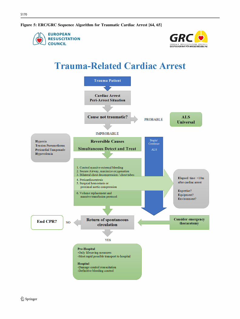
Figure 5: ERC/GRC Sequence Algorithm for Traumatic Cardiac Arrest [64, 65]
S170
123
References
1. Bansal V, et al. Hemorrhage is more prevalent than brain injury
in early trauma deaths: the golden six hours. Eur J Trauma Emerg
Surg. 2009;35(1):26–30.
2. Barbaro RP, et al. Association of hospital-level volume of
extracorporeal membrane oxygenation cases and mortality.
Analysis of the extracorporeal life support organization registry.
Am J Respir Crit Care Med. 2015;191(8):894–901.
3. Barton ED. Tension pneumothorax. Curr Opin Pulm Med.
1999;5(4):269–74.
4. Bickell WH, et al. Immediate versus delayed fluid resuscitation
for hypotensive patients with penetrating torso injuries. N Engl J
Med. 1994;331(17):1105–9.
5. Bonacchi M, et al. Extracorporeal life support in patients with
severe trauma: an advanced treatment strategy for refractory
clinical settings. J Thorac Cardiovasc Surg.
2013;145(6):1617–26.
6. Burlew CC, et al. Western Trauma Association critical decisions
in trauma: resuscitative thoracotomy. J Trauma Acute Care Surg.
2012;73(6):1359–63.
7. Buschmann C, et al. ,,Mit dem Leben nicht vereinbare Verlet-
zung‘‘—ein sicheres Todeszeichen. Rettungsdienst.
2013;36(6):32–4.
8. Bushby N, et al. Prehospital intubation and chest decompression
is associated with unexpected survival in major thoracic blunt
trauma. Emerg Med Australas. 2005;17(5–6):443–9 [LoE 2a].9. Byard RW. How reliable is external examination in identifying
internal injuries—Casper’s sign revisited. J Forensic Leg Med.
2012;19(7):419–21.
10. Chen KY, et al. Pneumothorax in the ICU: patient outcomes and
prognostic factors. Chest. 2002;122(2):678–83.
11. Chesters A, Davies G, Wilson A. Four years of pre-hospital
simple thoracostomy performed by a physician-paramedic
helicopter emergency medical service team: a description and
review of practice. Trauma. 2015.
12. Combes A, et al. Position paper for the organization of
extracorporeal membrane oxygenation programs for acute
respiratory failure in adult patients. Am J Respir Crit Care
Med. 2014;190(5):488–96.
13. Crewdson K, Lockey D, Davies G. Outcome from paediatric
cardiac arrest associated with trauma. Resuscitation.
2007;75(1):29–34.
14. Cummins RO, Hazinski MF. Cardiopulmonary resuscitation
techniques and instruction: when does evidence justify revision?
Ann Emerg Med. 1999;34(6):780–4 [LoE 1b].
15. David JS, et al. Does the prognosis of cardiac arrest differ in trauma
patients? Crit Care Med. 2007;35(10):2251–5.
16. Deakin CD, Davies G, Wilson A. Simple thoracostomy avoids
chest drain insertion in prehospital trauma. J Trauma.
1995;39(2):373–4 [LoE 2a].17. Eberle B, et al. Checking the carotid pulse check: diagnostic
accuracy of first responders in patients with and without a pulse.
Resuscitation. 1996;33(2):107–16 [LoE 1b].
18. Engdahl J, Herlitz J. Localization of out-of-hospital cardiac
arrest in Goteborg 1994–2002 and implications for public access
defibrillation. Resuscitation. 2005;64(2):171–5.
19. Escott ME, et al. Simple thoracostomy. Moving beyong needle
decompression in traumatic cardiac arrest. JEMS.
2014;39(4):26–32.
20. Esmer E, et al. Prehospital assessment of injury type and
severity in severely injured patients by emergency physicians:
an analysis of the TraumaRegister DGU(R). Unfallchirurg.
2016.
21. Flaris AN, et al. Clamshell incision versus left anterolateral
thoracotomy.Which one is faster when performing a resuscitative
thoracotomy? The tortoise and the hare revisited. World J Surg.
2015;39(5):1306–11.
22. Geeraedts LM Jr, et al. Exsanguination in trauma: a review of
diagnostics and treatment options. Injury. 2009;40(1):11–20
[LoE 1b].23. Handley AJ, et al. The 1998 European Resuscitation Council
guidelines for adult single rescuer basic life support: a statement
from the Working Group on Basic Life Support, and approved
by the executive committee. Resuscitation. 1998;37(2):67–80
[Evidenzbasierte Leitlinie].
24. Handley AJ, et al. Single rescuer adult basic life support. An
advisory statement from the Basic Life Support Working Group
of the International Liaison Committee on Resuscitation
(ILCOR). Resuscitation. 1997;34(2):101–8 [LoE 1c].
25. Harris T, Thomas GO, Brohi K. Early fluid resuscitation in
severe trauma. BMJ. 2012;345:e5752.
26. Herff H, et al. Ventilation strategies in the obstructed airway in
a bench model simulating a nonintubated respiratory arrest
patient. Anesth Analg. 2009;108(5):1585–8.
27. Hess EP, Campbell RL, White RD. Epidemiology, trends, and
outcome of out-of-hospital cardiac arrest of non-cardiac origin.
Resuscitation. 2007;72(2):200–6 [LoE 2c].
28. Huber-Wagner S, et al. Outcome in 757 severely injured
patients with traumatic cardiorespiratory arrest. Resuscitation.
2007;75(2):276–85 [LoE 2c].
29. International Liaison Committee on R. 2005 International
consensus on cardiopulmonary resuscitation and emergency
cardiovascular care science with treatment recommendations.
Part 1: introduction. Resuscitation. 2005;67(2–3):181–6 [LoE
1c].30. Karmy-Jones R, et al. Urgent and emergent thoracotomy for
penetrating chest trauma. J Trauma. 2004;56(3):664–8 (discus-sion 668–9; [LoE 2c]).
31. Kleber C, et al. Requirement for a structured algorithm in cardiac
arrest following major trauma: epidemiology, management errors,
and preventability of traumatic deaths in Berlin. Resuscitation.
2014;85(3):405–10.
32. Kleber C, et al. Trauma-related preventable deaths in Berlin
2010: need to change prehospital management strategies and
trauma management education. World J Surg.
2013;37(5):1154–61.
33. Kwan I, et al. Timing and volume of fluid administration for
patients with bleeding. Cochrane Database Syst Rev.
2014;3:CD002245.
34. Leigh-Smith S, Harris T. Tension pneumothorax—time for a re-
think? Emerg Med J. 2005;22(1):8–16.
35. Leis CC, et al. Traumatic cardiac arrest: should advanced life
support be initiated? J Trauma Acute Care Surg.
2013;74(2):634–8.
36. Lockey D, Crewdson K, Davies G. Traumatic cardiac arrest:
who are the survivors? Ann Emerg Med. 2006;48(3):240–4.
37. Lockey D, Lyon RM, Davies G. A simple algorythm for the
treatment of traumatic cardiac arrest. Scand J Trauma Resusc
Emerg Med. 2013;21(Suppl 1):S10.
38. Lockey DJ, Lyon RM, Davies GE. Development of a simple
algorithm to guide the effective management of traumatic
cardiac arrest. Resuscitation. 2013;84(6):738–42.
39. Luna GK, et al. Hemodynamic effects of external cardiac
massage in trauma shock. J Trauma. 1989;29(10):1430–3.
S3-guideline on treatment of patients with severe/multiple injuries S171
123
40. Maron BJ, et al. Increasing survival rate from commotio cordis.
Heart Rhythm. 2013;10(2):219–23.
41. Massarutti D, et al. Simple thoracostomy in prehospital trauma
management is safe and effective: a 2-year experiencebyhelicopter
emergency medical crews. Eur J Emerg Med. 2006;13(5):276–80.
42. Matsumoto H, et al. Role of resuscitative emergency field
thoracotomy in the Japanese helicopter emergency medical
service system. Resuscitation. 2009;80(11):1270–4.
43. Millin MG, et al. Withholding and termination of resuscitation
of adult cardiopulmonary arrest secondary to trauma: resource
document to the joint NAEMSP-ACSCOT position statements.
J Trauma Acute Care Surg. 2013;75(3):459–67.
44. Mistry N, Bleetman A, Roberts KJ. Chest decompression during
the resuscitation of patients in prehospital traumatic cardiac
arrest. Emerg Med J. 2009;26(10):738–40 [LoE 2a].45. Mosier JM, et al. Extracorporeal membrane oxygenation
(ECMO) for critically ill adults in the emergency department:
history, current applications, and future directions. Crit Care.
2015;19:431.
46. Ollerton JE, et al. Prospective study to evaluate the influence of
FAST on trauma patient management. J Trauma.
2006;60(4):785–91 [LoE 1b].
47. Pepe PE, Roppolo LP, Fowler RL. The detrimental effects of
ventilation during low-blood-flow states. Curr Opin Crit Care.
2005;11(3):212–8.
48. Perkins GD, et al. European Resuscitation Council guidelines
for resuscitation 2015: section 2. Adult basic life support and
automated external defibrillation. Resuscitation. 2015;95:81–99.
49. Pickens JJ, Copass MK, Bulger EM. Trauma patients receiving
CPR: predictors of survival. J Trauma. 2005;58(5):951–8 [LoE2c].
50. Powell DW, et al. Is emergency department resuscitative
thoracotomy futile care for the critically injured patient
requiring prehospital cardiopulmonary resuscitation? J Am Coll
Surg. 2004;199(2):211–5 [LoE 2c].
51. Puchwein P, et al. Clamshell thoracotomy and open heart
massage—a potential life-saving procedure can be taught to
emergency physicians: an educational cadaveric pilot study.
Injury. 2015;46(9):1738–42.
52. Roberts DJ, et al. Clinical presentation of patients with tension
pneumothorax: a systematic review.AnnSurg. 2015;261(6):1068–78.
53. Ruppert M, et al. Checking for breathing: evaluation of the
diagnostic capability of emergency medical services personnel,
physicians, medical students, and medical laypersons. Ann
Emerg Med. 1999;34(6):720–9 [LoE 1b].
54. Russell RJ, et al. The role of trauma scoring in developing
trauma clinical governance in the Defence Medical Services.
Philos Trans R Soc Lond B Biol Sci. 2011;366(1562):171–91.
55. Seamon MJ, et al. The use of emergency department thoracotomy
for traumatic cardiopulmonary arrest. Injury. 2012;43(9):1355–61.
56. Seamon MJ, et al. Emergency department thoracotomy: survival
of the least expected. World J Surg. 2008;32(4):604–12.
57. Sherren PB, et al. Algorithm for the resuscitation of traumatic
cardiac arrest patients in a physician-staffed helicopter emer-
gency medical service. Crit Care. 2013;17(2):308.
58. Smith JE, Rickard A, Wise D. Traumatic cardiac arrest. J R Soc
Med. 2015;108(1):11–6.
59. Soar J, et al. European Resuscitation Council guidelines for
resuscitation 2005. Section 7. Cardiac arrest in special circum-
stances. Resuscitation. 2005;67(Suppl 1):S135–70 [Evi-denzbasierte Leitlinie].
60. Soar J, et al. European Resuscitation Council guidelines for
resuscitation 2015: section 3. Adult advanced life support.
Resuscitation. 2015;95:100–47.
61. Spahn DR, et al. Management of bleeding and coagulopathy
following major trauma: an updated European guideline. Crit
Care. 2013;17(2):R76.
62. Stratton SJ, Brickett K, Crammer T. Prehospital pulseless,
unconscious penetrating trauma victims: field assessments
associated with survival. J Trauma. 1998;45(1):96–100.
63. Timmermann A, et al. The out-of-hospital esophageal and
endobronchial intubations performed by emergency physicians.
Anesth Analg. 2007;104(3):619–23.
64. Truhlar A, et al. Kreislaufstillstand in besonderen Situationen.
Notfall + Rettungsmedizin. 2015;18(8):833–903.
65. Truhlar A, et al. European Resuscitation Council guidelines for
resuscitation 2015: section 4. Cardiac arrest in special circum-
stances. Resuscitation. 2015;95:148–201.
66. Tseng YH, et al. Venoarterial extracorporeal life support in
post-traumatic shock and cardiac arrest: lessons learned. Scand J
Trauma Resusc Emerg Med. 2014;22:12.
67. Voiglio EJ, et al. Resuscitative transverse thoracotomy. Ann
Chir. 2003;128(10):728–33.
68. Willis CD, et al. Cardiopulmonary resuscitation after traumatic
cardiac arrest is not always futile. Injury. 2006;37(5):448–54
[LoE 2c].69. Wise D, et al. Emergency thoracotomy: ‘‘how to do it’’. Emerg
Med J. 2005;22(1):22–4.
70. Working Group, A.H.S.o.O., American College of Surgeons-
Committee on Trauma. Practice management guidelines for
emergency department thoracotomy. J Am Coll Surg.
2001;193(3):303–9 [Evidenzbasierte Leitlinie].
71. Zwingmann J, et al. Survival and neurologic outcome after
traumatic out-of-hospital cardiopulmonary arrest in a pediatric
and adult population: a systematic review. Crit Care.
2012;16(4):R117.
2.16 Coagulation System
Note: The following text represents the current state of evi-
dence-based therapy for patients with multiple and severe
injuries, which should be applicable to many situations.
Nevertheless, it is not possible to address each potential
individual case in such a guideline. Thus, in unique circum-
stances the clinical assessment by an experienced physician
can lead to options other than those listed here.
Trauma-Induced Coagulopathy
Key recommendations:
2.97 Recommendation* Modified 2016
GoR A Trauma-induced coagulopathy is an independentclinical picture with clear effects on survival. Thus,coagulation assessment and therapy must be begun atthe latest in the emergency department.
2.98 Recommendation* Modified 2016
GoR A Basic laboratory assessments of severe trauma mustinclude early and repeated measurement of blood gasanalysis, PT, aPTT, fibrinogen, and platelet count aswell as blood type determination.
S172
123
2.99 Recommendation* New 2016
GPP When treating severely injured patients in theemergency department, in addition to other diagnosticstudies and therapy for trauma-induced coagulopathy,early viscoelastic testing should be performed.
* modified according to Spahn et al. 2013
Explanation:
The term ‘‘polytrauma’’ covers a very heterogeneous
patient population. Early trauma-related mortality is mostly
a consequence of traumatic brain injury (40-50 % of
deaths) or massive bleeding (20-40 %). Concomitant
coagulopathy markedly increases bleeding [99,142]. The
additional coagulation disorder is often a result of the
bleeding itself, but is not uncommonly triggered by other
trauma and non-trauma-induced changes. Coagulopathy in
polytrauma patients (trauma-induced coagulopathy, TIC)
has been recognized for several decades [83]. Incidence
rates regarding existing coagulopathy at the time of ED
admission have been quoted as high as 60 % [43, 109].
Death due to uncontrolled bleeding generally occurs within
the first six to twelve hours after trauma, with major dis-
tribution within the first one to two hours [105]. Already
within a good hour of blunt trauma, there are statistically
significant reductions in fibrinogen (factor I) and calcium
(factor IV), as well as clotting factors II, V, VII, and X, and
a significant increase in factor VIII [21].
According to the current understanding of TIC, distinctions
are made between
• acute trauma-associated coagulopathy (ATC) and
• iatrogenic coagulopathy (IC) as well as the subsequent
• pro-thrombotic coagulopathy.
ATC occurs independently and prior to IC [92, 150].
The following points are important to the current under-
standing of the development of TIC (Figure 6):
S3-guideline on treatment of patients with severe/multiple injuries S173
123
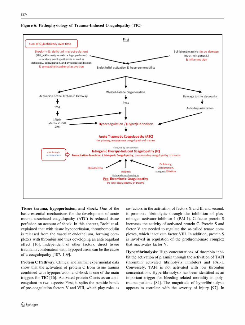
Figure 6: Pathophysiology of Trauma-Induced Coagulopathy (TIC)
Tissue trauma, hypoperfusion, and shock: One of the
basic essential mechanisms for the development of acute
trauma-associated coagulopathy (ATC) is reduced tissue
perfusion on account of shock. In this context, Brohi et al.
explained that with tissue hypoperfusion, thrombomodulin
is released from the vascular endothelium, forming com-
plexes with thrombin and thus developing an anticoagulant
effect [16]. Independent of other factors, direct tissue
trauma in combination with hypoperfusion can be the cause
of a coagulopathy [107, 109].
Protein C Pathway: Clinical and animal experimental data
show that the activation of protein C from tissue trauma
combined with hypoperfusion and shock is one of the main
triggers for TIC [16]. Activated protein C acts as an anti-
coagulant in two aspects: First, it splits the peptide bonds
of pro-coagulation factors V and VIII, which play roles as
co-factors in the activation of factors X and II, and second,
it promotes fibrinolysis through the inhibition of plas-
minogen activator-inhibitor 1 (PAI-1). Cofactor protein S
increases the activity of activated protein C. Protein S and
factor V are needed to regulate the so-called tenase com-
plexes, which inactivate factor VIII. In addition, protein S
is involved in regulation of the prothrombinase complex
that inactivates factor V.
Hyperfibrinolysis: High concentrations of thrombin inhi-
bit the activation of plasmin through the activation of TAFI
(thrombin activated fibrinolysis inhibitor) and PAI-1.
Conversely, TAFI is not activated with low thrombin
concentrations. Hyperfibrinolysis has been identified as an
important trigger for bleeding-related mortality in poly-
trauma patients [84]. The magnitude of hyperfibrinolysis
appears to correlate with the severity of injury [97]. In
S174
123
patients with verified fulminant hyperfibrinolysis, mortality
in the ED is 88 % [144].
Endothelial injury and auto-heparinization: The nega-
tively charged glycocalix covers the vascular endothelium as
an anti-adhesive and coagulation inhibiting layer, function-
ing as a barrier. Tissue trauma, inflammation, hypoperfusion,
and sympathetic adrenal activation lead to systemic damage
to the vascular endothelium, including the glycocalix, lead-
ing to increased vascular permeability. The systemic evi-
dence of various molecules reflects the damage of various
endothelial structures: glycocalix: syndecan-1; endothe-
lium: free thrombomodulin (sTM), endothelial growth factor
(VEGF); Weibel-Palade body molecules: tissue plasmino-
gen activator (tPA), angiopoietin-2 (Ang-2). The entire
endothelial glycocalix contains about a liter of non-circu-
lating plasma with significant quantities of a heparin-like
substance. Release into the systemic circulation after the
endothelial damage as described above leads to auto-hep-
arinization, as has been proven in trauma patients [127].
Iatrogenic coagulopathy (IC): IC usually occurs with
some time delay and can substantially enhance the above-
described primary and acute trauma-related coagulopathy.
Data from the DGU Trauma Registry (TR-DGU�) was
used to show that post-trauma coagulopathy is correlated
with administration of higher quantities of intravenous
fluids [163]. However, coagulopathy has been reported also
in cases where no pre-hospital fluids were given [109].
During hypotension, physiological dilution effects occur
through osmosis, with fluid transfer from the interstitial to
intravascular space until osmolar equivalence is reached. If
each protein is diluted in equal amounts, such as e.g. the
intrinsic tenase complex (composed of coagulation factors
IXa, VIIIa, and X), the individual factors are proportion-
ately reduced. In addition, coagulation factors are of course
also consumed, especially in the acute phase. However,
with the combination of consumption and dilution (con-
sumption and dilution coagulopathies), fibrinogen drops
out earlier as a substrate for clot formation during the
administration of packed red blood cells (pRBCs), as other
clotting disorders occur [68].
The definition of massive bleeding includes loss of C
100% of blood volume within 24 hours, C 50% within
three hours, and 150 ml/min or 1.5 ml/kgBW/min over 20
minutes [153].
Diagnostic Studies: Although the clinical picture of TIC is
characterized by non-surgical, diffuse bleeding from the
mucous membranes, serous membranes, and wound sur-
faces, as well as by bleeding from puncture sites of
intravascular catheters and from bladder catheters or
nasogastric tubes, there are no generally appropriate labo-
ratory parameters [101].
The ‘‘classic’’ standard coagulation tests (SLTs) are mea-
sured at 37 �C, buffered, and with excess calcium in the
serum and/or plasma. Thus, acidosis, hypothermia,
hypocalcemia, and anemia are not considered, although
these factors can have a tremendous impact [46, 102]. Data
from the Trauma Registry have shown that prolonged
prothrombin time in trauma patients is a predictor for
mortality [18, 106, 133]. Here, in addition to PT, docu-
mentation of INR is recommended because of better
comparability with the baseline values of the Trauma
Registry. Recommendation 12 of the third edition of the
‘‘European Trauma Guideline’’ (3rd ET) recommends
early, repeated, and simultaneous measurements of PT,
aPTT, fibrinogen, and platelets with a GoR 1C [150]. In
2015, Haas et al. published an analysis regarding the evi-
dence of SLTs for the management of coagulopathies.
They found only three prospective studies with 108
patients and concluded that there is no good evidence for
the use of SLTs for the diagnosis and choice of therapy for
coagulopathy. If no other diagnostic options are available,
however, the SLTs are preferable to no testing at all [51].
The European [91] and American [4] Associations of
Anesthesiology reached the same conclusion in 2013 and
2015, respectively. Thus, sufficient quantities of citrated
blood should be taken for SLTs during patient admission to
the ED.
Thromboelastography (TE) and Rotational Thromboe-
lastometry (RoTEM): Viscoelastic tests (VETs), i.e. TE
and RoTEM, have been increasingly studied for the mon-
itoring of polytrauma patients. In contrast to SLTs, these
tests detect not only time until clotting onset, but also the
speed of coagulation and the maximum clotting strength.
This test procedure can be carried out without delay in the
ED. Thus, management decisions can be made more
rapidly [131, 138]. Viscoelastic testing offers a time benefit
compared to conventional coagulation tests, especially for
fibrinogen determination, detection of possible fibrinolysis,
prediction of massive transfusions, as well as screening for
hemostatic competence and potential complications [30,
35, 53, 108, 143]. Several TEG/RoTEM based algorithms
for trauma management have already been published (e.g.,
[1, 46, 80, 108, 145]). In 2013, the 3rd ET gave a 1C
recommendation (strong recommendation, weak evidence)
for the use of VET (Rec. 12) and the earliest possible
initiation of coagulation assessment (Rec. 23) [150]. In
2014, a systematic review gave a 1B-level recommendation
for the trauma population [52]. In 2014, the group of da
Luz produced a descriptive review of 38 prospective cohort
studies, 15 retrospective cohort studies, and two before-
and-after studies of 12 489 patients. The found limited
evidence for the diagnosis of earlier TIC using TEG/
RoTEM as well as the prediction for blood transfusions and
S3-guideline on treatment of patients with severe/multiple injuries S175
123
mortality in trauma [33]. In 2015, the UK’s National
Institute for Health reviewed 39 studies in a ‘‘Health
Technology Assessment,’’ in which a cost advantage was
evident for VETs versus SLTs [168]. The time to occur-
rence of hyperfibrinolysis and the extent of hyperfibrinol-
ysis on the RoTEM correlated to increased mortality, with
a fulminant course having the worst prognosis [108]. A
prospective, international multicenter study of 808 patients
identified clot amplitude (MCF) at 5 minutes (CA 5) as a
valid marker to detect TIC and as a predictor for massive
transfusion, with the threshold for EXTEM (CA 5) B 40
mm and that of FIBTEM B 9 mm (sensitivity 72.7% and
77.5%, respectively). Fibrinogen concentration B 1.6 g/l
indicated TIC in 73.6 % of cases, and values B 1.9 g/l
indicated massive transfusion in 77.8 % [55]. In 2015, an
international expert group published the results of a formal
consensus including the Delphi method to develop a vis-
coelasticity-based transfusion guideline for the early man-
agement of trauma. In this, the recommended VET
threshold values were summarized for fibrinogen concen-
tration, platelet concentration, plasma, and PPSB [76, 108].
The RoTEM-based algorithm for the diagnosis and man-
agement of multiply injured patients in the ED is shown in
Figure 7, and its sources are the German language version
of the expert consensus [108] as well as the work cited
above by Hagamo et al. [55] (Figure 7).
S176
123

Figure 7: RoTEM-based Algorithm for Coagulation Management in the ED (modified according to [108]).
When it comes to therapy for coagulopathic bleeding,
platelet function is at least as important as the count;
despite adequate numbers, the function can be markedly
impaired [93]. VETs have only limited ability to assess
platelet function, primary hemostasis (inter-platelet and
platelet-endothelial interactions), and corresponding anti-
platelet medications. For this, impedance aggregometry
(e.g., Multiplate�, ROTEM�platelet) is more suitable [93,
149]. The evaluation of platelet function including
assessment of medication effects can be carried out with
other established laboratory methods (e.g., platelet aggre-
gation measurements with the Born method).
Blood type determination: Early determination of ABO
blood type (with antibody testing) on arrival to the ED
enables blood type-matched treatment of affected trauma
patients. Sufficient quantities of compatible blood prepa-
rations can be provided more rapidly when subsequently
needed. At the same time, the generally scarce stock
quantities of blood group O (Rh+/D+ and Rh-/D-) are not
used unnecessarily.
S3-guideline on treatment of patients with severe/multiple injuries S177
123
Damage Control Resuscitation
Key recommendations:
2.100 Recommendation* Modified 2016
GoR B In adult patients with active bleeding, the goal should befor permissive hypotension (mean arterial pressure ~ 65mmHg, systolic arterial pressure ~ 90 mmHg, age-adapted for children) until surgical hemostasis.
2.101 Recommendation* New 2016
GoR B For the combination of hemorrhagic shock andtraumatic brain injury (GCS <9) and/or spinal traumawith neurological symptoms, MAP should be 85-90mmHg.
2.102 Recommendation New 2016
GoR 0 When central nervous system injury and coronary heartdisease are excluded, a lower MAP (~ 50 mmHg) can beaimed for until surgical hemostasis.
2.103 Recommendation* New 2016
GoR A Treatment of shock must be controlled with repeatedmeasurements of base excess and/or lactate levels.
2.104 Recommendation* Modified 2016
GoR B Cooling of the patient should be avoided withappropriate measures and the goal should benormothermia.
2.105 Recommendation 2016
GoR B Acidemia should be avoided and treated withappropriate shock management.
2.106 Recommendation* Modified 2016
GoR B Hypocalcemia < 0.9 mmol/L should be avoided and thegoal should be for normocalcemia.
* modified according to Spahn et al. 2013
Explanation:
Similar to ‘‘damage control surgery,’’ where definitive
surgical management is temporarily deferred in favor of
vital process stabilization, the concept of ‘‘damage control
resuscitation’’ has been developed to avoid trauma-induced
coagulopathy [9]. Permissive hypotension, prevention of
acidemia, hypocalcemia, and hypothermia, as well as the
administration of coagulation-promoting medications [151]
are all parts of this strategy, which begins as soon as
possible [150]. Consistent invasive hemodynamic moni-
toring as well as the ability to perform rapid, repetitive
blood gas analyses is required for the determination of
these parameters.
Permissive Hypotension: This term describes two
approaches. The first is to tolerate or even strive for lower
than normal blood pressure to support thrombus formation.
The second is to infuse no more fluid than necessary to
ensure adequate end-organ perfusion, and thus, avoid
iatrogenic dilution. The correlation between ‘‘normal’’
blood pressure and tendency to bleed after trauma was
already recognized at the end of the First World War [22].
The idea of tolerating lower blood pressures according to
the palpable radial pulse until time of surgical hemostasis
evolved in the military sphere [9].
The basis for clinical application is a study by Bickell
et al. from 1994, in which patients with penetrating trauma
receiving pre-hospital volume replacement showed higher
mortality rates [11]. The accompanying editorial [79] and a
large number of readers’ responses mentioned flaws in the
study design, implementation, and interpretation. Using
data from the German Trauma Registry, Maegele et al.
found increased incidence of coagulopathy with increasing
pre-hospital volume therapy [109]. The effectiveness of
restrictive primary volume replacement therapy was first
shown in a prospective randomized study in 2011 [119]. In
2013, the 3rd ET gave a 1C level recommendation for a
target systolic blood pressure of 80-90 mmHg until surgical
hemostasis, when there is no TBI (Rec. 13) [150]. The
‘‘palpable radial pulse’’ was officially recommended for
pre-hospital care in 2009 [29], set as target in 2012 [60],
and is currently included in the 2015 Resuscitation Rec-
ommendations of the European Resuscitation Council
(ERC) [158].
Although there is a lack of evidence-based proof in
massively bleeding patients, because of pathophysiological
considerations and retrospective comparisons, a mean
pressure of 65 mmHg or SBP of 90 mmHg are recom-
mended in current review studies. This concept should be
considered carefully for older patients and those with
known arterial hypertension [150]. While it is indis-
putable that patients with a combination of hemorrhagic
shock with TBI (GCS B 8) require higher blood pressure to
maintain perfusion (MAP C 80 mmHg [150]; SBP C 110
mmHg [10]), the combination with spinal trauma and
neurology is less clear. The 3rd ET mentions ‘‘spinal
injury’’ only in the text of Rec. 13 [150]. Retrospective data
analysis [62, 78] and official recommendations [28, 54,
139] suggest MAP of 85-90 mmHg. Patients without CHD
and without central nervous system injury can benefit if
needed from a lower MAP (* 50 mmHg) [119].
To ensure adequate perfusion in spite of permissive
hypotension and restrictive volume replacement, repeated
measurements of lactate and/or base excess must be carried
out, which are prognostically significant [71]. The goal is
base excess less than -6 mEq/l [150]. The authors of the 3rd
ET give a 1B grade recommendation for the measurement
of lactate or base excess, urging caution for lactate mea-
surements in association with alcohol intoxication, and
suggest simultaneous measurements of both parameters
(text of Rec. 11) [150]. Permissive hypotension is a time-
limited option, that must only be maintained until surgical
hemostasis [150]. Important in this context is that there is
‘‘occult hypoperfusion’’ at the microcellular level [38], and
up to 72 hours (after restoration of the microcirculation, i.e.
S178
123
adequate macrovascular blood pressure) can pass before
the microvascular dynamics allow sufficient cellular per-
fusion [155].
Warming: HypothermiaB 34�Chas significant influence on
platelet function and the activity of clotting factors [102];
preservation of normothermia is optimal [150]. Tominimize
patient cooling, initial fluid therapymust be carried out using
warmed infusions [9, 156], and from the ED on, volume
replacement must be given exclusively with an infusion
warmer with temperatures set to 40-42� C [140, 156]. Both
passive (e.g. insulating foil covers, blankets, removal of wet
clothing) and active (e.g. replacement of non-warmed with
warmed infusions, constant use of heating pads, radiant
heaters, hot air blowers) measures are helpful. Both during
evaluation and later in the operating room, room temperature
should be as high as possible, optimally in the thermoneutral
zone, i.e., 28-29�C [140, 159].
The hypothermia-related platelet dysfunction can be
partially corrected with an infusion of desmopressin
(DDAVP) in the typical dose of 0.3 lg/kg [57, 123, 172].
Acidosis Correction: Acidosis with pH B 7.2 significantly
worsens coagulation [20, 102]. Because the main cause of
acidosis is hypoperfusion, it will persist until adequate
tissue perfusion is restored. Interventions that can enhance
acidosis, such as hypoventilation or NaCl infusion, should
be avoided [9]. Base excess (BE) also interferes with
coagulation [102]; critical thresholds for BE begin between
-6 and -10 [173].
Buffering of pH values to C 7.2 alone does not improve
the coagulopathy [15], and from a hemostaseologic point of
view, is advisable only in combination with administration
of coagulation preparations [91, 99]. The 3rd ET repeatedly
mentions ‘‘acidosis’’ or ‘‘acidemia’’ as unfavorable and/or
as a trigger for ‘‘damage control’’ (Rec. 21); however, it is
not further specified [150].
Calcium Replacement: Ionized calcium (Ca++), factor IV
of the coagulation cascade, is needed as a cofactor at
almost every step of plasma coagulation [102, 103]. The
reduction of ionized calcium (Ca++) after transfusion
depends on the citrate used as an anticoagulant by the
blood bank and is especially pronounced in fresh frozen
plasma (FFP). The reduction is even more significant
according to increased speed of transfusion, especially with
transfusions infused at[ 50 ml/min [20]. Calcium mono-
products available in Germany for intravenous use contain
variable amounts of calcium ions [100]. This must be
considered during replacement. Marked coagulation
impairment must be assumed when the ionized Ca++ con-
centration drops below 0.9 mmol/l [99, 102, 103, 150].
Replacement of Coagulation-Promoting Products
In recent years, various studies have explored optimization
of therapy with blood products and plasma derivatives in
polytrauma patients, and have led to markedly improved
survival rates.
Key recommendations:
2.107 Recommendation* Modified2016
GoR B A specific protocol for massive transfusions andcoagulation therapy should be established.
2.108 Recommendation* Modified2016
GoR B In an actively bleeding patient, the decision to transfuseis made individually according to clinical criteria, thedegree of injury, extent of blood loss, hemodynamicsituation, and oxygenation.
After stabilization, the goal should be normovolemiaand the Hb value should be raised to at least 7-9 g/dl[4.4-5.6 mmol/l].
2.109 Recommendation* New 2016
GoR B For cases of (expected) massive transfusions, theindications for FFP should be established andimplemented as soon as possible, and otherwise beconsidered restrictively.
2.110 Recommendation Modified2016
GoR B If coagulation therapy with FFP is carried out in cases ofmassive transfusions, the target ratio FFP : pRBC : PLTshould be 4 : 4 : 1.
2.111 Recommendation New 2016
GPP If coagulation therapy is done with factor concentratesfor massive transfusions, this should be controlled withappropriate procedures.
2.112 Recommendation* New 2016
GoR A For profusely bleeding patients, tranexamic acid (TxA)must be administered as soon as possible, with 1g over10 min and then followed as needed with an infusion of1g over 8 hours.
2.113 Recommendation* New 2016
GoR 0 For profusely bleeding patients, pre-hospitaladministration of tranexamic acid can be worthwhile.
2.114 Recommendation* New 2016
GoR B Administration of tranexamic acid should not beinitiated more than three hours after trauma (except incases of verified hyperfibrinolysis).
2.115 Recommendation with majorityapproval*
Modified2016
GoR B In cases of bleeding, fibrinogen substitution should beperformed with thromboelastometric signs of afunctional fibrinogen deficit or values of < 1.5 g/l (150mg/dl).
2.116 Recommendation* New 2016
GPP Within 24 hours of hemostasis, decisions must be maderegarding the type and beginning ofthromboprophylaxis.
S3-guideline on treatment of patients with severe/multiple injuries S179
123
* modified according to Spahn et al. 2013
Explanation:
Massive Transfusion (and Coagulation Therapy) Pro-
tocol (MTP): The term ‘‘massive transfusion’’ generally
includes the transfusion of C 10 units pRBCs per 24 hours
[151]. However, since the highest mortality of multiply
injured patients is within the first 2-6 hours, some authors
also recommend C 10 units pRBCs per 6 hours [82]. At the
European level, since 2010, MTP is a mandatory anesthe-
siology algorithm for the treatment of patients with life-
threatening bleeding, which is adapted to local conditions,
resources, etc. and must include pre-defined triggers to
intervention [115]. It is required by the 2013 perioperative
guidelines of the ESA, with a level 1B recommendation
(Rec. 5.1.7 [91]), and by the 3rd ET with level 1B and 1C
(Rec. 35-37 [150]) recommendations. The Trauma Asso-
ciated Severe Hemorrhage (TASH) score of the DGU
Trauma Registry [173] offers the chance to predict the need
for massive transfusion in the civilian sector. It includes the
factors systolic blood pressure, hemoglobin (Hb), BE, heart
rate, free intra-abdominal fluid, pelvic or femoral fracture,
and male gender (0-28 points). Increasing TASH point
values can be associated with higher probability for mas-
sive transfusion precisely and with good discrimination
(area under the curve, AUC, 0.89). Several authors have
published and analyzed MT scores, e.g. [14, 27, 122, 126,
164]. According to the 3rd ET, this evidence-based MTP
should be adapted to the personnel, logistical, and spatial
conditions of the individual hospital and verified in terms
of compliance with local protocols (Rec. 35 and 37, GoR
1C) [150]. MTPs can accelerate therapy with blood com-
ponents and avoid unnecessary discarding of preparations
[86]. Better MTP compliance can be associated with better
survival [8].
Packed Red Blood Cells (pRBCs): Red blood cells are an
essential component of coagulation (overview, for example
in [9] or [157]).
There remains no current data from randomized controlled
studies regarding the optimal hemoglobin and/or hemat-
ocrit levels for coagulation in polytrauma patients.
Accordingly, the German Medical Association (Bun-
desarztekammer, BAK) recommends that the goal should
be a hemoglobin concentration around 10 g/dl (6.2 mmol/
l), due to the favorable effects of higher hematocrit values
on primary hemostasis in cases of massive, acutely
bleeding hemorrhages until the time of hemostasis [20].
The 2004 review study by Hardy et al. [58] supports this
BAK recommendation with experimental evidence that
hematocrit levels up to 35 % are necessary to maintain
hemostasis in bleeding patients. The BAK emphasizes that
Hb value alone is not an indication for transfusion; rather,
the capability for physiologic compensation and signs of
anemic hypoxia should be considered [20]. Thus, it is
possible that in individual cases the goal should be Hb
values[ 10 g/dl [3, 152]. In cases of massively bleeding
patients, it can be necessary to begin transfusion of pRBCs
and FFP and plasma derivatives if needed before reaching
the Hb target range of 7-9 g/dl due to other causes (e.g.
hypoxemia, shock, etc.).
A Cochrane review from 2012 examined 19 studies of
6264 patients (a general patient population, not specific to
trauma patients with active bleeding!). The restrictive
transfusion trigger (7-9 g/dl) did not appear to affect the
rate of side effects (mortality, myocardial infarction,
stroke, pneumonia, arrhythmia, thromboemboli) compared
to liberal transfusion strategies (9-12 g/dl). The restrictive
strategy was linked to a significant reduction of hospital
mortality (RR 0.77; 95 CI: 0.62-0.95), but not 30-day
mortality (RR 0.85; 95 CI: 0.70 -1.03) [24]. The analysis of
391 985 inpatients found that reduction of average Hb
values to 7.6 g/dl prior to transfusion did not significantly
worsen the 30-day mortality [136]. Analysis of two studies
of 549 patients refusing transfusion showed that mortality
is extremely high in post-operative care without transfusion
for Hb under 5-6 g/dl. The probability of death increased in
patients with post-operative Hb B 8 g/dl, calculated around
2.5 times per gram of Hb decrease. There were no deaths in
the Hb range of 7-8 g/dl: however, there was a cardio-
vascular and/or infectious morbidity of 9.4 % [25, 147].
The authors of the 3rd ET offer a grade 1C recommenda-
tion (Rec. 17) for a target Hb of 7-9 g/dl after hemostasis;
however, the text specifies that the optimal Hb/Hct for
stabilization of coagulation in massively bleeding patients
is unclear [150]. In the case of TIC, a restrictive transfusion
trigger can be unfavorable [114], since significant impair-
ment of coagulation can develop before oxygenation is
affected [59]. Similarly, neurological outcomes can be
worse when there are lower Hb values after resuscitation
(e.g., Hb \ 10 g/dl after resuscitation for non-traumatic
cardiac arrest + poor cerebral saturation + poor central
venous saturation) [3].
A possible correlation between pRBC transfusion with
poorer outcomes is intensely debated, and better data
through prospective studies is needed [171]. Older publi-
cations from the fields of trauma and critical care medicine
reported a negative correlation of pRBC transfusion with
survival (overview, for example, in [9] or [157]). A current
meta-analysis reported that trauma patients appear to
benefit from the transfusion of fresh preserves [124]. A
current Cochrane analysis of three studies with 120 patients
in total yielded no clear benefits from the transfusion of
pRBCs collected less than 21 days previous [112]. Obser-
vations of earlier studies on the influence of pRBC age on
S180
123
survival may be attributed to the use of non-leukoreduced
blood (not permitted in Europe since 2001). The data of
multiple prospective randomized studies of seriously ill
children and adults (critical care/cardiac surgery) showed
no correlation between clinical outcomes and the storage
age of transfused packed red blood cells [36, 42, 94, 154].
Other potential consequences from the use of non-
leukoreduced blood as reported in international studies are
listed here, but cannot be directly correlated to German
blood products. In 2009, a retrospective analysis of trauma
patients identified an increased risk of 6 % per unit pRBCs
for ARDS (adjusted OR 1.06; 95% CI: 1.03-1.10); how-
ever, a confounding factor of severe pattern of injury in
ARDS patients was not excluded [26]. A retrospective
analysis of 1607 children with TBI not requiring surgery
found that a transfusion trigger of 8 g/dl can be beneficial
in this population [2]. Meta-analysis of observational
studies between 1947 and 2012 found a correlation
between pRBC transfusion and mortality and/or multi-or-
gan failure/ARDS/ALI and concluded that further inter-
ventional studies are needed to clarify how restricted
transfusion frequency influences mortality and other effects
[128]. A secondary analysis of CRAH-2 data (10 227
transfused of 20 127 patients; observational study, many
possible confounding factors) showed that the association
between transfusion and mortality in trauma seems to vary
according to mortality risk; transfusion seems to lower the
risk in high-risk patients, while increasing it in low-risk
patients [130]. A correlation between the transfusion of
leukoreduced blood products and the occurrence of rele-
vant, transfusion-associated immunomodulation has not
been scientifically proven. A meta-analysis of 31 ran-
domized controlled trials with 9813 patients found no
benefits or disadvantages regarding mortality, morbidity,
and risk of heart attack for liberal (Hb 9-13 g/dl) versus
restrictive (Hb 7.0-9.7 g/dl) transfusion strategies [74]. A
current study in cardiac surgery patients reported no sig-
nificant difference in morbidity or health costs between
restrictive (Hb \ 7.5 g/dl) versus liberal (Hb \ 9 g/dl)
transfusion triggers [121].
According to the hemovigilance report of the Paul-
Ehrlich-Institute (PEI) for Germany from 2013-2014, the
most common side effects of pRBCs per million transfused
units were: acute transfusion reactions (15.5), transfusion-
associated volume overload (8.5), hemolytic transfusion
reactions (6.3). Regarding virus transmission, in the years
2012-2014 there was one case of HEV (no cases of
Hepatitis A, B, C or HIV) recorded [47].
Fresh Frozen Plasma (FFP): According to the BAK,
plasma therapy is indicated in cases of complex coagu-
lopathy due to overt bleeding and threatened severe
bleeding (1C grade of recommendation), to prevent or treat
microvascular bleeding. They recommend a rapid initial
transfusion of 20 ml/kg BW, but emphasize that greater
quantities can be necessary [20]. According to BAK,
treatment of a coagulopathy with plasma alone is not
efficient enough because of the need to give large volumes
to increase plasma levels, the short half-life of some
coagulation factors, and the potential increase in
consumption.
In a systematic review of 80 randomized controlled
studies, Yang et al. found no consistent evidence for a
significant difference between prophylactic and therapeutic
administration of FFP; however, the study designs were
frequently problematic [169]. In a 2011 matched pair
analysis (18 patients each from the DGU and Innsbruck
Trauma Registries), Nienaber et al. reported that therapy
with FFP alone was associated with a higher rate of multi-
organ failure (p = 0.015) compared to treatment with factor
concentrates (fibrinogen, PPSB); there were also trends
towards shorter ventilation times and hospital stays. The
overall mortality in both groups was comparable [125]. In
2012, the working group of Mitra reported that a higher
ratio of FFP to pRBCs ([ 1:2 in the first four hours) in
existing TIC (INR[ 1.5 or aPTT[ 60 sec) is associated
with significantly reduced mortality (p = 0.03). Conversely,
when TIC is absent, high dose FFP has no positive effects
and can result in adverse effects and increased costs [117].
There is no good data regarding the effectiveness of other
therapeutic plasma products mentioned in the cross-sec-
tional guideline of the BAK (solvent detergent treated
plasma, SDP; methylene blue-treated plasma, MLP; lypo-
philized human plasma, LHP) in cases of massive trans-
fusions [20].
According to the hemovigilance report of the Paul-
Ehrlich-Institute (PEI) for Germany from 2013-2014, the
most common adverse effects per million transfused units
were: acute transfusion reactions (15.6), transfusion-asso-
ciated volume overload (3.6), hemolytic transfusion reac-
tions (6.3). Suspicion of viral transmission was not reported
in the years 2012-2014. With the reported figures of the
year 2014 for FFP, the following additional adverse effects
were reported: transfusion-associated cardiovascular vol-
ume overload (TACO) 2.41/million units, and transfusion-
associated lung insufficiency (TRALI) 1.2/million units
[47].
The PROPPR study [73] did not observe higher rates of
acute pulmonary failure in the arm with increased plasma
administration.
The authors of the 3rd ET recommend avoiding the use
of plasma in patients without severe bleeding (Rec. 26.3,
GoR 1B) [150].
Ratio of FFP to pRBC: A number of studies have dealt
with the FFP : pRBC ratio. Most of these studies had small
S3-guideline on treatment of patients with severe/multiple injuries S181
123
case numbers and were retrospective (at least for the con-
trol population). The idea that pRBCs, FFP, and platelet
concentrates (PC) should be substituted in a 1:1:1 ratio is
based on the intention to transfuse in as ‘‘whole blood’’ like
manner as possible. However, this attempt again only
succeeds to a limited extent. Preservatives must be added
to the actual components in the banked blood products
[49]. Thus, after reconstitution the coagulation activity
reaches approximately 65 % of whole blood [5, 49].
Hematocrit and platelet counts are significantly diluted, but
remain above the needs for transfusion; in contrast, fib-
rinogen reaches a concentration of only 1.5 to 1.9 g/l [132].
An increase of each component automatically dilutes the
two others. The resulting dilution coagulopathy is propor-
tional to the volume of transfused fluids [23]. In one study,
the 1:1 ratio did not correct hypoperfusion or coagulopathy
for trauma patients receiving massive transfusions [87].
In 2013, the first prospective multicenter study of this
question was published. PROMMTT studied 905 patients
receiving C 3 blood products (at least 1 unit pRBCs) and
analyzed 14 time intervals (minute 31 until day 30). In the
first six hours, the mortality of patients receiving FFP :
pRBC\ 1:2 was 3-4 times higher than those receiving C
1:1. After 24 hours, there was no more association. At the
median time of mortality probability (2.6 h), only
approximately 40 % of patients had received a FFP : pRBC
ratio C 1:2 [72]. The second prospective multicenter study
followed in 2015.PROPPR compared 680 patients expec-
ted to receive massive transfusion in FFP:PC:pRBC ratios
of 1:1:1 versus 1:1:2. The groups did not differ regarding
the primary objectives of the study, mortality within 24
hours (12.7 % in the 1:1:1 group vs. 17.0 % in the 1:1:2
group; difference -4.2 % [95% CI: -9.6 % – 1.1 %]) and
within 30 days (22.4 % vs. 26.1 %; difference -3.7 % [95%
CI: -10.2 % – 2.7 %]). However, the bleeding-related
mortality within 3 hours was significantly reduced in the
1:1:1 group (9.2 % vs. 14.6 %; Difference -5.4% [95% CI:
-10.4% --0.5%]; p = 0.03) [73]. The authors commented
that to achieve these effects, crystalloids and/or colloids
need to be restricted and blood components should be
available within 8 minutes [66]. A prospective study of
surgical patients compared the administration of 4 units
FFP (group A) versus 2 units FFP plus 2g fibrinogen (group
B). Hemostasis was comparably successful, with 21/22
patients in group A and 16/17 patients in group B. There
were differences in the coagulation parameters.
Prothrombin and clotting Factors VIII, IX, and X were
higher in group A; ROTEM parameter fibrin formation was
better in B, and thrombin formation was better in A [95].
To avoid dilution coagulopathy, whole blood for massive
transfusions (still allowed in many places in the USA but
no longer in Germany) has been discussed by individual
authors [170]. Fresh blood (preferably still warm) was used
in Germany until the 1980s, but this was eliminated due to
the lack of rapid infection testing.
Platelet Concentrates (PC): In acute blood loss, platelets
are initially released from the bone marrow and spleen;
thus, the platelet count drops later on initial hemorrhage.
After transfusion, the transferred vital platelets distribute
themselves in the blood and spleen, so that the recovery
rate in the peripheral blood is only around 60-70 %,
sometimes even lower [20]. When the patient is in acute
danger because of massive blood loss or because of the
localization (intracerebral bleeding), the BAK recommends
platelet replacement when the level falls below 100 000/ll[20]. With the same weak evidence, the 3rd ET also rec-
ommends keeping a platelet count[ 100,000/ll for mas-
sively bleeding patients and those with TBI (Rec. 28.2)
[150]. In acquired platelet dysfunction and bleeding ten-
dency, concomitant antifibrinolytic or desmopressin ther-
apies can be indicated [20]. According to BAK, PC in
Germany contains approximately 2 x 10¹¹ platelets. Such a
PC is available in two preparations: pooled from 4-6
individual donors or as PC apheresis [20]. A German PC
then correlates to approximately 4-6 American single-
donor PCs as used in the PROPPR study [73]; thus, in cases
of massive transfusions, one PC should be substituted after
each 4th to 6th pair of pRBC/FFP. Again, it should be
emphasized that the platelet count says nothing regarding
function [93].
According to the PEI hemovigilance report for
2013/2014, the most commonly reported adverse effects
per 1 million transfused PC were: acute transfusion reac-
tions (52.6), suspicion of bacterial contamination (6.2).
Regarding virus transmission, in the years 2013-2014 there
were two cases of HEV (no Hepatitis A, B, C or HIV)
recorded [47].
In summary, trauma patients must be transfused to
maintain an individual and sufficient Hb value, with only
what is necessary when it is necessary [91].
Note: The main risk in transfusion of blood products
remains oversight - more than half of reported cases are
due to human error during the transfusion process [13]. The
reported rate for mismatched transfusion was 10.19/million
units pRBCs, thus, approximately 1:100 000 in 2013/2014
[47]. Therefore, a bedside ABO test should be performed
prior to each transfusion, even in cases of emergency (4.3.9
‘‘emergency transfusion’’) [19]. In addition, batch docu-
mentation is mandatory for plasma derivatives; this is
required by law according to TFG § 14 (1) and (2) [19].
Fibrinogen: Fibrinogen is a glycoprotein synthesized in
the liver, with a long plasma half-life of approximately 3 to
5 days. As an acute phase protein, levels can increase up to
20 times during inflammation and thus, also in association
S182
123
with TIC [113]. Factor I (i.e., fibrinogen) is a clotting
substrate essential not only for the formation of the fibrin
network but also as a ligand for the GPIIb-IIIa receptor on
the platelet surface, and thus, responsible for platelet
aggregation. In the context of dilution or severe bleeding,
fibrinogen seems to be the most vulnerable of all coagu-
lation factors and is the first to reach its critical concen-
tration [20, 63, 68]. In cases of TIC, fibrinogen is reduced
not only by hyperfibrinolysis, but also by increased
degradation, acidosis, reduced synthesis as a result of
hypothermia as well as loss/dilution [6]. According to
BAK, the minimum plasma concentration following fib-
rinogen replacement must be 1.0 g/l. The critical threshold
where spontaneous bleeding can occur is\1.5 g/l in cases
of heavy bleeding (cross-sectional guideline 7.1.6.2). In
adults, generally a single dose of 3-6 g is required. The
administration of 3 g of fibrinogen in 3 liters of plasma
increases the measured fibrinogen concentration by
approximately 1 g/l. In the case of congenital deficiency,
the half-life of 96-120h should be considered. In cases of
shorter half-life, the fibrinogen concentrations are more
frequently controlled (Cross-Sectional Guideline 7.1.7)
[20].
Already in 1995, Hiippala [67] determined that in patients
receiving colloid infusions, the measurement of ‘‘derived’’
fibrinogens (determined with the PT/INR) as well as those
determined with the Clauss method were significantly
higher than the actual fibrinogen levels. The BAK recom-
mends that patients who have received hydroxyethyl starch
preparations or dextran and whose fibrinogen levels are
determined with the ‘‘derived fibrinogen method’’ should
be treated with an intervention threshold of 1.5 g/l instead
of 1.0 g/l. When hyperfibrinolysis is suspected, an
antifibrinolytic (e.g., 1 g tranexamic acid) should be given
beforehand [20].
The following presents studies regarding fibrinogen
replacement in acute bleeding and controls, grouped
according to size and profile and thus, enabling conclusions
based on the study results. Recently, Hagemo et al [41]
published a multicenter study (International Trauma
Research Network, INTRN) of 808 patients from four
trauma centers in England, Denmark, and Norway (2007-
2011) with the goal to reevaluate smaller cohort studies to
characterize TIC and to evaluate the threshold values for
fibrinogen concentrations (VET); they found that MT was
necessary in 6.1 % of cases, and that TIC was present in 11
%. The most important conclusions came in addition to the
results stated above (VET as a valid method for threshold
determination of fibrinogen concentration), which con-
firmed the results of earlier cohort studies that the Clauss
method determinations were comparable with the best
parameters of VET, and that future studies should be
planned to test coagulation therapy guided by VET in
trauma. Hagamo et al. published a multicenter study of
1133 patients treated within three hours in a trauma center.
They examined the initial fibrinogen concentrations
(prevalence of hypofibrinogenemia), as there is still little
evidence regarding the use of fibrinogen in the acute phase.
In 8.2 %, the fibrinogen concentration was B 1.5 g/l, and in
19.2 %, it was\2.0 g/l. With initial concentrations under
2.29 g/l, there was a marked increase in the 28-day mor-
tality. The average initial fibrinogen concentration of 28
day survivors was 2.68 g/l and it was 1.95 g/l in non-
survivors (p\0.001). With fibrinogen concentrations under
2.29 g/dl (95% CI: 1.93-2.64), there was a 90 % reduction
in mortality for each unit increase of fibrinogen concen-
tration, although the results must be interpreted cautiously
due to the large confidence intervals. The authors recom-
mended that these results be validated using interventional
studies with fibrinogen concentrates [56]. A larger
prospective cohort study of 517 patients from Rouke et al.
was devoted to the course of fibrinogen concentrations,
monitored with VET and Clauss method determination.
The blood samples were collected at admission and after
transfusion of four units pRBCs. Low fibrinogen levels on
admission were independently associated with ISS (p =
0.002), shock (p = 0.002), and the quantity of pre-hospital
volume given (p\ 0.001). Fibrinogen concentrations var-
ied significantly between non-survivors after 24 hours (1.1
g/l vs. 2.3 g/l, p\0.001) and after 28 days (1.4 g/l vs. 2.4
g/l, p\0.001) [137]. Other studies are mostly monocentric
cohort studies examining fibrinogen replacement in smaller
patient groups. When a concentration of\2 g/l is used as
indication for fibrinogen substitution (average 2 g, range
1-5 g), Fenger-Eriksen et al. found significant reductions in
the need for pRBCs (p\ 0.0001), FFP (p\ 0.0001), and
PC (p \ 0.001) as well as blood loss (p \ 0.05) in 35
heavily bleeding adult patients [40]. In a retrospective
analysis of 30 massively bleeding patients with hypofib-
rinogenemia (fibrinogen \ 1.5 g/l) of varying etiologies,
Weinkove et al. raised the level from 0.65 to 2.01 g/l
(Clauss method) with administration of 4 g fibrinogen
(median; range 2-14 g) [166]. In a retrospective analysis,
Farriols Danes et al. found that patients (n = 81) with acute,
bleeding-related hypofibrinogenemia (substitution below 1
g/l) react with a marked increase on fibrinogen replacement
and, in contrast to patients with chronic fibrinogen defi-
ciency (p = 0.314), they have a significantly better 7-day
survival rate (p = 0.014) [34]. In an in vitro study, the blood
of subjects (n = 6) was diluted 1:5 with NaCl 0.9 %
solution. To optimize the rate of clot formation, a fibrino-
gen concentration[2 g/l was necessary. The results of this
in vitro study cannot be directly transferred to a clinical
situation, and must be prospectively verified with clinical
studies [130]. Matched-pair analysis (registry study; n =
294 patients per group) with data from the DGU Trauma
S3-guideline on treatment of patients with severe/multiple injuries S183
123
Registry found that patients receiving fibrinogen concen-
trates between admission and the ICU showed significantly
decreased 6-hour mortality (10.5 % vs. 16.7 %, p = 0.03),
prolonged time until death, but also an increased rate of
multi-organ failure, although the overall mortality during
admission did not vary compared to patients receiving no
fibrinogen (28.6% vs. 25.5%, p = 0.40). These results
should be verified with prospective, randomized controlled
studies [161]. In a prospective cohort study of 144 patients
with ISS[15 by Innerhofer et al., 66 patients were treated
with fibrinogen and/or PPSB concentrate (CF group) and
78 patients received additional FFP (FFP group). Patients
treated with fibrinogen concentrates and/or PPSB received
significantly less pRBCs and PC than those receiving
additional FFP (pRBCs median 2, range 0-4 units vs.
median 9, range 5-12 units; PC median 0, range 0-0 units
vs. median 1, range 0-2 units; p \ 0.001). In addition,
patients of the CF group showed less multi-organ failure
(18.2% vs 37.2%, p 0.01) and sepsis (16.9% vs. 35.9 %,
p = 0.014) [77]. In a prospective in vitro study comparing
63 trauma versus 63 critically ill non-trauma patients,
thrombopenia \ 150 x109/l and fibrinogen concentration
\ 2 g/l were the only factors associated with enhanced
bleeding tendency, according to special measurements with
the TEG (MA) [70]. A retrospective analysis evaluated
fibrinogen concentrations of 290 patients on admission (3-
year period, level I trauma center). In this retrospective
analysis of 290 blunt trauma patients, fibrinogen concen-
tration\ 2 g/l on admission was an independent predictor
for 7-day mortality [88]. A retrospective analysis of 260
trauma patients (average ISS 26 ± 13) with massive
transfusions (C 10 units pRBCs within the first 24 hours)
divided patients into three groups according to fibrinogen
concentration on admission: normal (C180 mg/dL),
abnormal (C101 to\ 180 mg/dL), and critical (B100 mg/
dL). A value B 100 mg/dl led to significantly increased 24h
mortality (adjusted OR 3.97; adjusted p = 0.013;
R2 = 0.141), significantly increased hospital mortality, and
was the most important independent predictor for mortality
(p = 0.012). The impact of a fibrinogen replacement
strategy requires prospective evaluation [75]. There is
evidence that a fibrinogen concentration C 2 g/l can be
advisable in trauma patients with acute bleeding, but there
are no large prospective RCTs. Once the acute bleeding is
controlled, the fibrinogen concentration should not drop
below 1.5 g/l. The costs for fibrinogen concentrates are not
reimbursed by the state health insurance for every case,
especially in emergency therapy. Currently (DRG 2016),
fibrinogen cannot be billed through additional charges in
polytrauma patients (exceptions: congenital coagulation
deficiencies, exceeding a total sum of clotting factors). For
cost reasons and because there is a lack of randomized
multicenter studies proving a survival benefit at higher
target thresholds for fibrinogen replacement, the previous
threshold of 1.5 g/l for fibrinogen replacement was main-
tained, and the 1C recommendations of the 3rd ET (Rec. 27
[150]) and the ESA (Rec. 7.1.2 [91]) of 1.5-2 g/l were not
adopted.
There is very little data currently available regarding
adverse effects of fibrinogen (concentrates). In a systematic
review of studies from years 2000-2013, Aubron et al.
found no increased rates of adverse effects in trauma
patients [6]. The retrospective evaluation by the manufac-
turer, over a period of 27 years, yielded allergic reactions
(1 : 32 600 doses), possible thromboemboli (1 : 23 300
doses), and possible virus transmission (1 : 21 000 doses),
in addition to the problem of ‘‘underreporting’’ [37, 148].
Here the results of larger studies are awaited. Therefore,
when using fibrinogen, the instructions of the manufacturer
and the BAK [69] should be complied with and the initial
dosing of fibrinogen followed. Because fibrinogen has a
long half-life (approximately 3-4 days) and there is a risk
of accumulation at high or repeated doses, the risk of
thromboemboli can be increased. Especially in cases of
disseminated intravascular coagulation, which is associated
with TIC, it is important to ensure balanced treatment with
blood and plasma derivatives.
In addition to SLTs, e.g. viscoelastic tests are appro-
priate to guide the replacement of factor concentrates [76,
108, 116].
Prothrombin Concentrates (PPSB): Mixed preparations
of vitamin K dependent factors prothrombin (FII), pro-
convertin (FVII), Stuart factor (FX), and anti-hemophilia
factor B (FIX) as well as protein C, protein S, and protein Z
do not include fibrinogen, FV, or FVIII [141], and are only
standardized regarding factor IX content, so that the con-
tents of the other clotting factors above can fluctuate
widely [20]. The PPSB preparations available today con-
tain practically no activated clotting factors or activated
protein C or plasmin, so that undesirable effects such as
thromboembolic events, disseminated intravascular coag-
ulation, and/or hyperfibrinolytic bleeding are very
improbable, even when given in large amounts [20].
Thromboemboli reported in the past were most likely due
to a marked excess of prothrombin in some PPSB con-
centrates that are no longer available on the market today
[50]. Therefore, general replacement of anti-thrombin is no
longer necessary [20].
The main indication for PPSB is the elimination of
vitamin K antagonist activity. This indication has been
validated in many studies; for patients taking Vitamin K
antagonists, the BAK [20] recommends pre-operative
administration of PPSB for bleeding prophylaxis. In TIC,
the prothrombin complex deficiency can be so pronounced
that FFP administration must be supplemented with PPSB
S184
123
[20, 141]. In cases of severe bleeding, the BAK recom-
mends an initial bolus of 20-25 IE/kg BW; significant
individual variations in effectiveness must be considered
[20]. The weak 2C recommendation of the 3rd ET includes
a note that PPSB administration is coupled to the throm-
boelastometric signs of delayed initiation of coagulation
(Rec. 31.2 [150]). Thus, after PPSB administration, careful
laboratory monitoring of coagulation should be performed.
Joseph et al. compared the administration of FFP alone or
in combination with PPSB in 252 polytrauma patients
(median ISS 27) [81]. Supplemental PPSB resulted in
faster normalization of INR (394 vs. 1050 min; p = 0.001),
reduced consumption of pRBCs (6.6 vs. 10 units; p =
0.001) and FFP (2.8 vs. 3.9; p = 0.01) as well as reduced
mortality (23 vs. 28%; p = 0.04). The higher costs of PPSB
($1470 ± 845 vs. $1171 ± 949; p = 0.01) were offset by
markedly reduced transfusion costs ($7110 ± 1068 vs.
$9571 ± 1524; p = 0.01). In trauma patients, PPSB
administration leads to a sustained and pronounced
increase in endogenous thrombin potential as well as
reduced anti-thrombin levels; both can cause an increased
risk for thromboembolic events [146]. These adverse
effects have also been reported in association with the rapid
suspension of vitamin K antagonist activity during surgery.
In a retrospective review study with data from twelve
hospitals (n = 193), Hedges et al. reported that eight
patients (approximately 4 %) suffered acute venous
thromboemboli after PPSB [64]. The results of this study
are transferable to trauma patients only in a limited fashion.
Anti-fibrinolytics: Hyperfibrinolysis appears to be more
common in polytrauma patients than previously assumed
(just under 20 % [84, 142] up to 60 % [43, 109]); its extent
correlates with injury severity [97] and increasing mortality
[144]. In children with multiple injuries, hyperfibrinolysis
seems to be more commonly present on admission [104].
Prompt diagnosis of hyperfibrinolysis and also the effec-
tiveness of antifibrinolytic therapy is possible only with
thromboelastography/thromboelastometry [97, 144].
Administration of antifibrinolytics must be included in the
overall coagulopathy management concept, since in
hyperfibrinolysis there is often a heavy consumption of
fibrinogen up to and including complete defibrination of
the patient [17]. This fibrinogen deficiency must be bal-
anced after the hyperfibrinolysis is resolved; in other
words, in suspected hyperfibrinolysis, the anti-fibrinolytic
should be applied before fibrinogen [20]. Tranexamic acid
is a synthetic lysine analogue that inhibits the conversion of
plasminogen to plasmin by blocking the binding of plas-
minogens to the fibrin molecule.
CRASH-2 was a prospective randomized placebo-con-
trolled study performed in 274 hospitals in 40 countries to
examine the effects of early administration of TxA to
trauma patients on mortality, thromboembolic events and
the need for transfusion [32]. Adult trauma patients ([ 16
years) with significant hemorrhage (systolic blood pressure
\90 mmHg or pulse[110/min) were included according
to the ‘‘uncertainty principle,’’ i.e., when there was no clear
(contra-)indication for TxA. Accordingly, TxA (or pla-
cebo) was infused as an initial bolus (1 g over ten minutes)
followed by 1 g over 8 hours. After the inclusion of 10 060
patients (TxA group) and 10 067 patients (placebo), the
primary endpoint of mortality was reduced with TxA (14.5
% vs. 16.0 %, p = 0.0035), absolute mortality risk reduction
(AMRR) = 1.5 %: number needed to treat (NNT) 67.
Bleeding-related mortality was also reduced (4.9 % vs. 5.7
%, p = 0.0077, NNT 119). There was no significant vari-
ation in the secondary endpoints of thromboembolic events
(1.7% vs. 2.0%, p = 0.084), need for surgical intervention
(47.9% vs. 48.0%, p = 0.79), or transfusion of blood
products (50.4% vs. 51.3%, p = 0.21). Despite the immense
effort required for this study, no data regarding laboratory
parameters, injury severity (e.g., injury severity score), or
administration of other coagulation-active products/sub-
stances was documented. Although the study findings yield
a recommendation level GoR A, one major criticism is that
the majority of participating hospitals are located in
developing countries where one cannot assume modern
management of severely injured patients. In addition, the
mechanism of action for TxA remains unclear; hemorrhage
was not the main problem of the study population, since
only 50.4 % of patients received blood transfusions (me-
dian 3, range 2-6 units pRBCs) and since of 3076 mortal-
ities (15.3 %), only 1063 were bleeding-related.
MATTERs, a retrospective study in the military sphere,
found that treatment with 1 g TxA, followed as needed
with one further gram, yielded an AMRR of 6.5 % (NNT =
15) for the patients overall and an AMRR of 13.7 % (NNT
= 7) for massive transfusion patients [120]. Neither
CRASH-2 nor MATTER were able to demonstrate reduced
need for transfusions in trauma patients (in contrast to
studies of a perioperative setting, e.g. [85]). Based on the
CRASH-2 data, the 3rd ET gave a 1A recommendation for
the earliest possible administration of 1 G TxA over 10
min, followed by 1 g over 8 h, for all bleeding trauma
patients (Rec. 24.1 [150]). Smaller studies of trauma
patients with measurements of clotting parameters have not
been able to repeat the reduced mortality of CRASH-2 [61,
160].
A secondary exploratory analysis of the CRASH-2 data
revealed that the survival benefit was only evident when
the therapy was initiated in the first three hours after trauma
(Hazard Ratio HR B three hours = 0.78; 0.68-0.90; HR[three hours = 1.02; 0.76-1.36). The start of TxA adminis-
tration within the first three hours also reduced the risk of
bleeding-related death by 28 %, while administration after
S3-guideline on treatment of patients with severe/multiple injuries S185
123
three hours appears to correlate with higher mortality [31,
135]. From this comes the recommendation to forego later
administration (more than three hours after trauma), except
when there is laboratory evidence of hyperfibrinolysis. The
recommendation for pre-hospital administration has also
been extrapolated from the CRASH-2 data; sufficient
studies are not yet available for this. For these reasons, the
3rd ET gives a 1B recommendation for administration
within 3 hours (Rec. 24.2 [150]).
Desmopressin (DDAVP): Desmopressin (1-deamino-8-D
arginine vasopressin) is a synthetic vasopressin analogue.
Desmopressin (e.g. Minirin�) causes non-specific platelet
activation (increased expression of the platelet GpIb
receptor [129]), and releases von Willebrand factor as well
as FVIII from the liver sinusoidal endothelium, thus
improving primary hemostasis [44]. The main indication
for its use is for perioperative therapy of von Willebrand
disease. In TIC, von Willebrand disease can be acquired,
i.e. impaired platelet function as a result of particular
medications (e.g., ASS, heparin, ADP receptor antagonists/
thienopyridine derivatives), acidosis, or hypothermia [44,
150]. After i.v. administration, maximal effects occur only
after approximately 90 minutes [98]. Repeated adminis-
tration can lead to release of tissue plasminogen activator
(tPA) and thus, to hyperfibrinolysis. Thus, some authors
recommend simultaneous application of tranexamic acid
on repeat administration [98]. No controlled studies of
trauma patients are available.
From a pathophysiological perspective, a trial dose of
DDAVP can be considered in patients with suspected
platelet dysfunction as a result of acquired von Willebrand
disease, at a dosage of 0.3 lg/kg BW over 30 minutes (Rec.
30 [150]).
Factor XIII: In the presence of calcium ions, FXIII causes
covalent fibrin cross-linking. This produces a three-di-
mensional fibrin net that leads to definitive wound healing
[20]. Factor XIII is not detected with the general PT/INR
and aPTT (activated partial thromboplastin time) tests, as
these only measure the onset of fibrin formation, not fibrin
cross-linking [20].
An acquired deficiency is not uncommon and in TIC,
may arise from increased turnover (increased blood loss,
hyperfibrinolysis) and consumption (during large opera-
tions). In patients with existing clotting activation, e.g.
from a tumor, there may be a severe FXIII deficiency that
leads to massive hemorrhage as a consequence of trauma or
surgical intervention [89]. There are no randomized studies
of trauma patients.
When rapid FXIII testing is not available, especially in
cases of severe, acute bleeding, blind administration of
FXIII should be considered [20]. Prior to bleeding control,
a possible initial dose of 15-20 IE/kgBW is recommended.
Because the concentrate is produced from human plasma, a
residual risk for infection cannot be excluded. However, it
should be noted that Factor XIII, like fibrinogen, has a long
half-life and can accumulate with repeated doses.
Recombinant activated Factor VII (rFVIIa): The
approval of rFVIIa is limited to bleeding in cases of
hemophilia (antibodies against factors VIII or IX), Glanz-
mann’s thrombasthenia (congenital dysfunction of the
platelet GPIIb-IIIa receptor), and congenital factor VII
deficiency. The rFVIIa binds to activated platelets in a
supra-physiological dosage and causes a ‘‘thrombin burst’’
that leads to the formation of an extremely stable fibrin clot
[45]. The rFVIIa also enables thrombin generation on
activated platelets independent of tissue factors.
A number of clinical case studies have portrayed
unapproved uses. Undesired side effects in the form of
thromboembolic events in the arterial and venous vascu-
lature and/or in perioperative or traumatically damaged
vessels have been reported particularly in cases of ‘‘off-
label’’ use [20]. Registry data show an incidence of almost
9 % in trauma [174]. Matched-pair analysis with data of the
German Trauma Registry concluded that the administration
of rFVIIa in trauma patients does not reduce the need for
blood products or decrease mortality, but is associated with
cumulative multi-organ failure [162]. In 2005, Boffard
et al. [12] studied the effectiveness of 400 lg/kg BW
rFVIIa (after the 8th unit pRBCs, with a primary dose of
200 lg/kgBW followed by 100 lg/kgBW each at 1 and 3
hours) versus placebo as adjuvant therapy in 143 blunt and
134 penetrating trauma patients. In blunt trauma, there was
a significant reduction of transfused pRBC units (calcu-
lated decrease of 2.6 units, p = 0.02) and the need for
transfusion of C 20 units pRBCs (14 % vs. 33 %; p = 0.03).
In penetrating injuries, there was a trend in this direction
for both parameters. Neither decreased mortality nor
increased thromboembolic events were observed.
The effectiveness of rFVIIa is linked to a series of
requirements that should be met prior to application: a
fibrinogen value C 1.5 g/dl, Hb C 7 g/dl, platelet count C
50 000 /ll (better C 100 000/ll), ionized calcium C 0.9
mmol/l, core temperature C 34 �C, pH C 7.2 as well as
exclusion of hyperfibrinolysis or heparin effect [20, 45]. A
widespread standard dose for off-label use is 90 lg/kg BW
[39, 165].
In its guideline, the BAK refers to the review article by
Mannucci et al. [111]. Its conclusion is that rFVIIa is not a
panacea, but it is effective for patients with trauma and
excessive bleeding that do not respond to other therapeutic
options. One application, for use only after conventional
therapies have been unsuccessful, is also being promoted
[91, 150]. The current manufacturer’s information from the
company NovoNordisk (December 2013) recommends that
S186
123
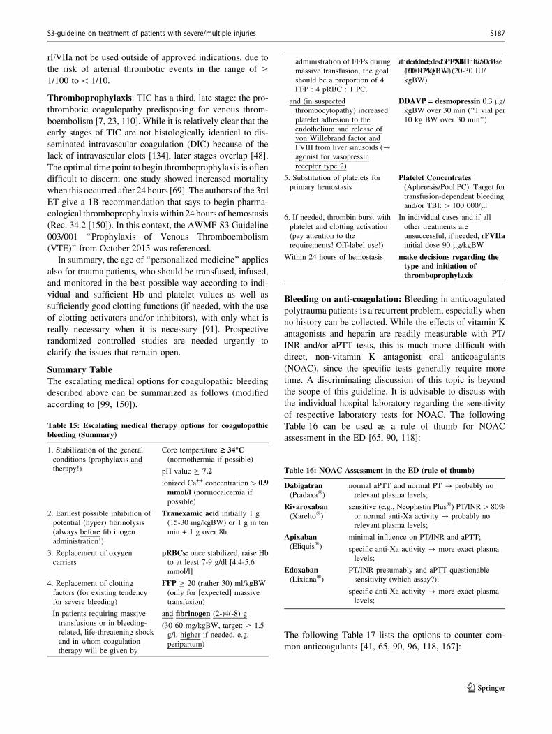
rFVIIa not be used outside of approved indications, due to
the risk of arterial thrombotic events in the range of C
1/100 to\ 1/10.
Thromboprophylaxis: TIC has a third, late stage: the pro-
thrombotic coagulopathy predisposing for venous throm-
boembolism [7, 23, 110]. While it is relatively clear that the
early stages of TIC are not histologically identical to dis-
seminated intravascular coagulation (DIC) because of the
lack of intravascular clots [134], later stages overlap [48].
The optimal time point to begin thromboprophylaxis is often
difficult to discern; one study showed increased mortality
when this occurred after 24 hours [69]. The authors of the 3rd
ET give a 1B recommendation that says to begin pharma-
cological thromboprophylaxis within 24 hours of hemostasis
(Rec. 34.2 [150]). In this context, the AWMF-S3 Guideline
003/001 ‘‘Prophylaxis of Venous Thromboembolism
(VTE)’’ from October 2015 was referenced.
In summary, the age of ‘‘personalized medicine’’ applies
also for trauma patients, who should be transfused, infused,
and monitored in the best possible way according to indi-
vidual and sufficient Hb and platelet values as well as
sufficiently good clotting functions (if needed, with the use
of clotting activators and/or inhibitors), with only what is
really necessary when it is necessary [91]. Prospective
randomized controlled studies are needed urgently to
clarify the issues that remain open.
Summary Table
The escalating medical options for coagulopathic bleeding
described above can be summarized as follows (modified
according to [99, 150]).
Table 15: Escalating medical therapy options for coagulopathicbleeding (Summary)
1. Stabilization of the general
conditions (prophylaxis and
therapy!)
Core temperature ‡ 34�C(normothermia if possible)
pH value C 7.2
ionized Ca++ concentration[0.9mmol/l (normocalcemia if
possible)
2. Earliest possible inhibition of
potential (hyper) fibrinolysis
(always before fibrinogen
administration!)
Tranexamic acid initially 1 g
(15-30 mg/kgBW) or 1 g in ten
min + 1 g over 8h
3. Replacement of oxygen
carriers
pRBCs: once stabilized, raise Hbto at least 7-9 g/dl [4.4-5.6
mmol/l]
4. Replacement of clotting
factors (for existing tendency
for severe bleeding)
FFP C 20 (rather 30) ml/kgBW
(only for [expected] massive
transfusion)
In patients requiring massive
transfusions or in bleeding-
related, life-threatening shock
and in whom coagulation
therapy will be given by
and fibrinogen (2-)4(-8) g
(30-60 mg/kgBW, target: C 1.5
g/l, higher if needed, e.g.
peripartum)
administration of FFPs during
massive transfusion, the goal
should be a proportion of 4
FFP : 4 pRBC : 1 PC.
and if needed PPSB initial dose
1000-2500 IU (20-30 IU/
kgBW)
if needed, 1-2x FXIII 1250 IU
(30 IU/kgBW)
and (in suspected
thrombocytopathy) increased
platelet adhesion to the
endothelium and release of
von Willebrand factor and
FVIII from liver sinusoids (?agonist for vasopressin
receptor type 2)
DDAVP = desmopressin 0.3 lg/kgBW over 30 min (‘‘1 vial per
10 kg BW over 30 min’’)
5. Substitution of platelets for
primary hemostasis
Platelet Concentrates(Apheresis/Pool PC): Target for
transfusion-dependent bleeding
and/or TBI:[ 100 000/ll
6. If needed, thrombin burst with
platelet and clotting activation
(pay attention to the
requirements! Off-label use!)
In individual cases and if all
other treatments are
unsuccessful, if needed, rFVIIainitial dose 90 lg/kgBW
Within 24 hours of hemostasis make decisions regarding thetype and initiation ofthromboprophylaxis
Bleeding on anti-coagulation: Bleeding in anticoagulated
polytrauma patients is a recurrent problem, especially when
no history can be collected. While the effects of vitamin K
antagonists and heparin are readily measurable with PT/
INR and/or aPTT tests, this is much more difficult with
direct, non-vitamin K antagonist oral anticoagulants
(NOAC), since the specific tests generally require more
time. A discriminating discussion of this topic is beyond
the scope of this guideline. It is advisable to discuss with
the individual hospital laboratory regarding the sensitivity
of respective laboratory tests for NOAC. The following
Table 16 can be used as a rule of thumb for NOAC
assessment in the ED [65, 90, 118]:
Table 16: NOAC Assessment in the ED (rule of thumb)
Dabigatran(Pradaxa�)
normal aPTT and normal PT ? probably no
relevant plasma levels;
Rivaroxaban(Xarelto�)
sensitive (e.g., Neoplastin Plus�) PT/INR[80%
or normal anti-Xa activity ? probably no
relevant plasma levels;
Apixaban(Eliquis�)
minimal influence on PT/INR and aPTT;
specific anti-Xa activity ? more exact plasma
levels;
Edoxaban(Lixiana�)
PT/INR presumably and aPTT questionable
sensitivity (which assay?);
specific anti-Xa activity ? more exact plasma
levels;
The following Table 17 lists the options to counter com-
mon anticoagulants [41, 65, 90, 96, 118, 167]:
S3-guideline on treatment of patients with severe/multiple injuries S187
123
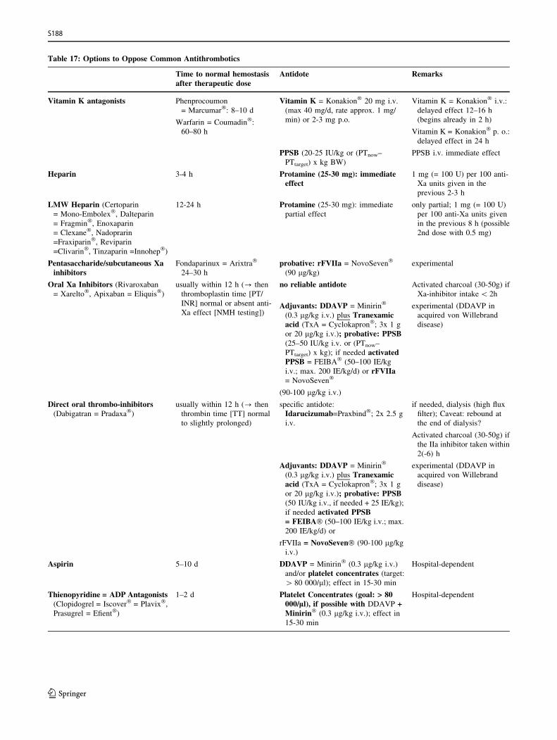
Table 17: Options to Oppose Common Antithrombotics
Time to normal hemostasisafter therapeutic dose
Antidote Remarks
Vitamin K antagonists Phenprocoumon
= Marcumar�: 8–10 d
Warfarin = Coumadin�:
60–80 h
Vitamin K = Konakion� 20 mg i.v.
(max 40 mg/d, rate approx. 1 mg/
min) or 2-3 mg p.o.
Vitamin K = Konakion� i.v.:
delayed effect 12–16 h
(begins already in 2 h)
Vitamin K = Konakion� p. o.:
delayed effect in 24 h
PPSB (20-25 IU/kg or (PTnow–
PTtarget) x kg BW)
PPSB i.v. immediate effect
Heparin 3-4 h Protamine (25-30 mg): immediateeffect
1 mg (= 100 U) per 100 anti-
Xa units given in the
previous 2-3 h
LMW Heparin (Certoparin
= Mono-Embolex�, Dalteparin
= Fragmin�, Enoxaparin
= Clexane�, Nadoprarin
=Fraxiparin�, Reviparin
=Clivarin�, Tinzaparin =Innohep�)
12-24 h Protamine (25-30 mg): immediate
partial effect
only partial; 1 mg (= 100 U)
per 100 anti-Xa units given
in the previous 8 h (possible
2nd dose with 0.5 mg)
Pentasaccharide/subcutaneous Xainhibitors
Fondaparinux = Arixtra�
24–30 h
probative: rFVIIa = NovoSeven�
(90 lg/kg)experimental
Oral Xa Inhibitors (Rivaroxaban= Xarelto�, Apixaban = Eliquis�)
usually within 12 h (? then
thromboplastin time [PT/
INR] normal or absent anti-
Xa effect [NMH testing])
no reliable antidote Activated charcoal (30-50g) if
Xa-inhibitor intake\ 2h
Adjuvants: DDAVP = Minirin�
(0.3 lg/kg i.v.) plus Tranexamicacid (TxA = Cyclokapron�; 3x 1 g
or 20 lg/kg i.v.); probative: PPSB(25–50 IU/kg i.v. or (PTnow–
PTtarget) x kg); if needed activatedPPSB = FEIBA� (50–100 IE/kg
i.v.; max. 200 IE/kg/d) or rFVIIa= NovoSeven�
(90-100 lg/kg i.v.)
experimental (DDAVP in
acquired von Willebrand
disease)
Direct oral thrombo-inhibitors(Dabigatran = Pradaxa�)
usually within 12 h (? then
thrombin time [TT] normal
to slightly prolonged)
specific antidote:
Idarucizumab=Praxbind�; 2x 2.5 g
i.v.
if needed, dialysis (high flux
filter); Caveat: rebound at
the end of dialysis?
Activated charcoal (30-50g) if
the IIa inhibitor taken within
2(-6) h
Adjuvants: DDAVP = Minirin�
(0.3 lg/kg i.v.) plus Tranexamicacid (TxA = Cyclokapron�; 3x 1 g
or 20 lg/kg i.v.); probative: PPSB(50 IU/kg i.v., if needed + 25 IE/kg);
if needed activated PPSB= FEIBA� (50–100 IE/kg i.v.; max.
200 IE/kg/d) or
rFVIIa = NovoSeven� (90-100 lg/kgi.v.)
experimental (DDAVP in
acquired von Willebrand
disease)
Aspirin 5–10 d DDAVP = Minirin� (0.3 lg/kg i.v.)
and/or platelet concentrates (target:[ 80 000/ll); effect in 15-30 min
Hospital-dependent
Thienopyridine = ADP Antagonists(Clopidogrel = Iscover� = Plavix�,
Prasugrel = Efient�)
1–2 d Platelet Concentrates (goal: > 80000/ll), if possible with DDAVP +Minirin� (0.3 lg/kg i.v.); effect in
15-30 min
Hospital-dependent
S188
123
Reversal of antithrombotics must be well considered, as it
inevitably increases the risks of thromboembolism!
The evidence table for this chapter is found on page 198 of
the guideline report.
References
1. Abdelfattah K, Cripps MW. Thromboelastography and rota-
tional thromboelastometry use in trauma. Int J Surg. 2015.
2. Acker SN, et al. Blood component transfusion increases the
risk of death in children with traumatic brain injury. J Trauma
Acute Care Surg. 2014;76(4):1082–7 (discussion 1087–8).3. Ameloot K, et al. Low hemoglobin levels are associated with
lower cerebral saturations and poor outcome after cardiac
arrest. Resuscitation. 2015;96:280–6.
4. American Society of Anesthesiologists Task Force on Periop-
erative Blood, M. Practice guidelines for perioperative blood
management: an updated report by the American Society of
Anesthesiologists Task Force on Perioperative Blood Man-
agement*. Anesthesiology. 2015;122(2):241–75.
5. Armand R, Hess JR. Treating coagulopathy in trauma patients.
Transfus Med Rev. 2003;17(3):223–31.
6. Aubron C, et al. Efficacy and safety of fibrinogen concentrate
in trauma patients—a systematic review. J Crit Care.
2014;29(3):471 e11–7.
7. Balvers K, et al. Risk factors for trauma-induced coagulopa-
thy- and transfusion-associated multiple organ failure in
severely injured trauma patients. Front Med (Lausanne).
2015;2:24.
8. Bawazeer M, et al. Compliance with a massive transfusion
protocol (MTP) impacts patient outcome. Injury.
2015;46(1):21–8.
9. Beekley AC. Damage control resuscitation: a sensible
approach to the exsanguinating surgical patient. Crit Care
Med. 2008;36(7 Suppl):S267–74.
10. Berry C, et al. Redefining hypotension in traumatic brain
injury. Injury. 2012;43(11):1833–7.
11. Bickell WH, et al. Immediate versus delayed fluid resuscita-
tion for hypotensive patients with penetrating torso injuries.
N Engl J Med. 1994;331(17):1105–9 (LoE2b).
12. Boffard KD, et al. Recombinant factor VIIa as adjunctive
therapy for bleeding control in severely injured trauma
patients: two parallel randomized, placebo-controlled, dou-
ble-blind clinical trials. J Trauma. 2005;59(1):8–15 (discus-
sion 15–8. LoE1b).13. Bolton-Maggs PH, Cohen H. Serious hazards of transfusion
(SHOT) haemovigilance and progress is improving transfusion
safety. Br J Haematol. 2013;163(3):303–14.
14. Boutefnouchet T, et al. Emergency red cells first: rapid
response or speed bump? The evolution of a massive
transfusion protocol for trauma in a single UK centre. Injury.
2015;46(9):1772–8.
15. Brohi K, Cohen MJ, Davenport RA. Acute coagulopathy of
trauma: mechanism, identification and effect. Curr Opin Crit
Care. 2007;13(6):680–5.
16. Brohi K, et al. Acute traumatic coagulopathy: initiated by
hypoperfusion: modulated through the protein C pathway?
Ann Surg. 2007;245(5):812–8.
17. Brohi K, et al. Acute coagulopathy of trauma: hypoperfusion
induces systemic anticoagulation and hyperfibrinolysis.
J Trauma. 2008;64(5):211–7 (discussion 1217. LoE2b).
18. Brohi K, et al. Acute traumatic coagulopathy. J Trauma.
2003;54(6):1127–30 (LoE3b).
19. Bundesarztekammer (BAK, German Medical Association).
Richtlinien zur Gewinnung von Blut und Blutbestandteilen
und zur Anwendung von Blutprodukten (Hamotherapie)—
Zweite Richtlinienanpassung 2010. 2010. http://www.bunde
saerztekammer.de/aerzte/medizin-ethik/wissenschaftlicher-
beirat/veroeffentlichungen/haemotherapie-transfusionsmedi
zin/richtlinie/.
20. Bundesarztekammer (BAK, German Medical Association).
Querschnittsleitlinien der BAK zur Therapie mit Blutkompo-
nenten und Plasmaderivaten. 4. uberarbeitete und aktualisierte
Auflage 2014. 2014. http://www.bundesaerztekammer.de/
fileadmin/user_upload/downloads/QLL_Haemotherapie_2014.
pdf.
21. Burggraf M, et al. Evaluation of clotting factor activities early
after severe multiple trauma and their correlation with
coagulation tests and clinical data. World J Emerg Surg.
2015;10:43.
22. Cannon W, Frawer J, Cowell E. The preventive treatment of
wound shock. JAMA. 1918;70:618–21.
23. Cap A, Hunt BJ. The pathogenesis of traumatic coagulopathy.
Anaesthesia. 2015;70(Suppl 1):96–101 (e32–4).24. Carson JL, Carless PA, Hebert PC. Transfusion thresholds and
other strategies for guiding allogeneic red blood cell transfu-
sion. Cochrane Database Syst Rev. 2012;4:CD002042.
25. Carson JL, et al. Mortality and morbidity in patients with very
low postoperative Hb levels who decline blood transfusion.
Transfusion. 2002;42(7):812–8.
26. Chaiwat O, et al. Early packed red blood cell transfusion and
acute respiratory distress syndrome after trauma. Anesthesiol-
ogy. 2009;110(2):351–60 (LoE2b).27. Chay J, et al. A national common massive transfusion protocol
(MTP) is a feasible and advantageous option for centralized
blood services and hospitals. Vox Sang. 2016;110(1):36–50.
28. Consortium for Spinal Cord, M. Early acute management in
adults with spinal cord injury: a clinical practice guideline for
health-care professionals. J Spinal Cord Med.
2008;31(4):403–79.
29. Cotton BA, et al. Guidelines for prehospital fluid resuscitation
in the injured patient. J Trauma. 2009;67(2):389–402.
30. Cotton BA, et al. Admission rapid thrombelastography
predicts development of pulmonary embolism in trauma
patients. J Trauma Acute Care Surg. 2012;72(6):1470–5
(discussion 1475–7).
31. Crash-collaborators, et al. The importance of early treatment
with tranexamic acid in bleeding trauma patients: an
exploratory analysis of the CRASH-2 randomised controlled
trial. Lancet. 2011;377(9771):1096–101 (1101 e1–2).
32. Crash-trial collaborators, et al. Effects of tranexamic acid on
death, vascular occlusive events, and blood transfusion in
trauma patients with significant haemorrhage (CRASH-2): a
randomised, placebo-controlled trial. Lancet.
2010;376(9734):23–32.
33. Da Luz LT, et al. Effect of thromboelastography (TEG(R)) and
rotational thromboelastometry (ROTEM(R)) on diagnosis of
coagulopathy, transfusion guidance and mortality in trauma:
descriptive systematic review. Crit Care. 2014;18(5):518.
34. Danes AF, et al. Efficacy and tolerability of human fibrinogen
concentrate administration to patients with acquired fibrinogen
deficiency and active or in high-risk severe bleeding. Vox
Sang. 2008;94(3):221–6.
S3-guideline on treatment of patients with severe/multiple injuries S189
123
35. Davenport R, et al. Functional definition and characterization
of acute traumatic coagulopathy. Crit Care Med.
2011;39(12):2652–8.
36. Dhabangi A, et al. Effect of transfusion of red blood cells with
longer vs shorter storage duration on elevated blood lactate
levels in children with severe anemia: the total randomized
clinical trial. JAMA. 2015;314(23):2514–23.
37. Dickneite G, et al. Animal model and clinical evidence
indicating low thrombogenic potential of fibrinogen concen-
trate (Haemocomplettan P). Blood Coagul Fibrinolysis.
2009;20(7):535–40.
38. Dutton RP. Haemostatic resuscitation. Br J Anaesth.
2012;109(Suppl 1):i39–46.
39. Dutton RP, Conti BM. The role of recombinant-activated
factor VII in bleeding trauma patients. Curr Opin Anaesthesiol.
2009;22(2):299–304.
40. Fenger-Eriksen C, et al. Fibrinogen concentrate substitution
therapy in patients with massive haemorrhage and low plasma
fibrinogen concentrations. Br J Anaesth. 2008;101(6):769–73
(LoE3b).
41. Fenger-Eriksen C, Munster AM, Grove EL. New oral antico-
agulants: clinical indications, monitoring and treatment of
acute bleeding complications. Acta Anaesthesiol Scand.
2014;58(6):651–9.
42. Fergusson DA, et al. Effect of fresh red blood cell transfusions
on clinical outcomes in premature, very low-birth-weight
infants: the ARIPI randomized trial. JAMA.
2012;308(14):1443–51.
43. Floccard B, et al. Early coagulopathy in trauma patients: an
on-scene and hospital admission study. Injury.
2012;43(1):26–32.
44. Franchini M. The use of desmopressin as a hemostatic agent: a
concise review. Am J Hematol. 2007;82(8):731–5.
45. Fries D, et al. Management of coagulation after multiple
trauma. Anaesthesist. 2005;54(2):137–44.
46. Fries D, Innerhofer P, Schobersberger W. Time for changing
coagulation management in trauma-related massive bleeding.
Curr Opin Anaesthesiol. 2009;22(2):267–74.
47. Funk M, et al. Hamovigilanz-Bericht des Paul-Ehrlich-Instituts
2013/14: Auswertung der Meldungen von schwerwiegenden
Transfusionsreaktionen nach § 63i. 2015.
48. Gando S, Otomo Y. Local hemostasis, immunothrombosis,
and systemic disseminated intravascular coagulation in trauma
and traumatic shock. Crit Care. 2015;19:72.
49. Gregory JA, et al. Optimizing transfusion ratios in massive
transfusion protocols: an argument against the 1:1:1 dogma
and approach to trauma resuscitation. Lab Med.
2015;46(2):e46–52.
50. Grundman C, et al. Prothrombin overload causes thromboem-
bolic complications in prothrombin complex concentrates:
in vitro and in vivo evidence. Thromb Haemost.
2005;94(6):1338–9.
51. Haas T, et al. Usefulness of standard plasma coagulation tests
in the management of perioperative coagulopathic bleeding: is
there any evidence? Br J Anaesth. 2015;114(2):217–24.
52. Haas T, et al. Thromboelastometry for guiding bleeding
management of the critically ill patient: a systematic review of
the literature. Minerva Anestesiol. 2014;80(12):1320–35.
53. Haas T, et al. Reproducibility of thrombelastometry
(ROTEM(R)): point-of-care versus hospital laboratory perfor-
mance. Scand J Clin Lab Investig. 2012;72(4):313–7.
54. Hadley M. Blood pressure management after acute spinal cord
injury. Neurosurgery. 2002;50(3 Suppl):S58–62.
55. Hagemo JS, et al. Detection of acute traumatic coagulopathy
and massive transfusion requirements by means of rotational
thromboelastometry: an international prospective validation
study. Crit Care. 2015;19:97.
56. Hagemo JS, et al. Prevalence, predictors and outcome of
hypofibrinogenaemia in trauma: a multicentre observational
study. Crit Care. 2014;18(2):R52.
57. Hanke AA, et al. Effects of desmopressin on platelet function
under conditions of hypothermia and acidosis: an in vitro study
using multiple electrode aggregometry*. Anaesthesia.
2010;65(7):688–91.
58. Hardy JF. Current status of transfusion triggers for red blood
cell concentrates. Transfus Apher Sci. 2004;31(1):55–66.
59. Hardy JF, et al. Massive transfusion and coagulopathy:
pathophysiology and implications for clinical management.
Can J Anaesth. 2004;51(4):293–310.
60. Harris T, Thomas GO, Brohi K. Early fluid resuscitation in
severe trauma. BMJ. 2012;345:e5752.
61. Harvin JA, et al. The impact of tranexamic acid on mortality in
injured patients with hyperfibrinolysis. J Trauma Acute Care
Surg. 2015;78(5):905–9 (discussion 909–11).62. Hawryluk G, et al. Mean arterial blood pressure correlates with
neurological recovery after human spinal cord injury: analysis
of high frequency physiologic data. J Neurotrauma.
2015;32(24):1958–67.
63. Hayakawa M, et al. Fibrinogen level deteriorates before other
routine coagulation parameters and massive transfusion in the
early phase of severe trauma: a retrospective observational
study. Semin Thromb Hemost. 2015;41(1):35–42.
64. Hedges A, et al. Clinical effectiveness and safety outcomes
associated with prothrombin complex concentrates. J Thromb
Thrombolysis. 2015.
65. Heidbuchel H, et al. Updated European Heart Rhythm
Association Practical Guide on the use of non-vitamin K
antagonist anticoagulants in patients with non-valvular atrial
fibrillation. Europace. 2015;17(10):1467–507.
66. Hess JR, Holcomb JB. Resuscitating PROPPRly. Transfusion.
2015;55(6):1362–4.
67. Hiippala ST. Dextran and hydroxyethyl starch interfere with
fibrinogen assays. Blood Coagul Fibrinolysis.
1995;6(8):743–6.
68. Hiippala ST, Myllyla GJ, Vahtera EM. Hemostatic factors and
replacement of major blood loss with plasma-poor red cell
concentrates. Anesth Analg. 1995;81(2):360–5.
69. Ho KM, Chavan S, Pilcher D. Omission of early thrombopro-
phylaxis and mortality in critically ill patients: a multicenter
registry study. Chest. 2011;140(6):1436–46.
70. Ho KM, Duff OC. Predictors of an increased in vitro
thrombotic and bleeding tendency in critically ill trauma and
non-trauma patients. Anaesth Intensive Care.
2015;43(3):317–22.
71. Hodgman EI, et al. Base deficit as a marker of survival after
traumatic injury: consistent across changing patient popula-
tions and resuscitation paradigms. J Trauma Acute Care Surg.
2012;72(4):844–51.
72. Holcomb JB, et al. The prospective observational multicenter
major trauma transfusion (PROMMTT) study. J Trauma Acute
Care Surg. 2013;75(1 Suppl 1):S1–2.
73. Holcomb JB, et al. Transfusion of plasma, platelets, and red
blood cells in a 1:1:1 vs a 1:1:2 ratio and mortality in patients
with severe trauma: the PROPPR randomized clinical trial.
JAMA. 2015;313(5):471–82.
S190
123
74. Holst LB, et al. Restrictive versus liberal transfusion strategy
for red blood cell transfusion: systematic review of randomised
trials with meta-analysis and trial sequential analysis. BMJ.
2015;350:h1354.
75. Inaba K, et al. Impact of fibrinogen levels on outcomes after
acute injury in patients requiring a massive transfusion. J Am
Coll Surg. 2013;216(2):290–7.
76. Inaba K, et al. 2014 consensus conference on viscoelastic test-
based transfusion guidelines for early trauma resuscitation:
Report of the panel. J Trauma Acute Care Surg.
2015;78(6):1220–9.
77. Innerhofer P, et al. The exclusive use of coagulation factor
concentrates enables reversal of coagulopathy and decreases
transfusion rates in patients with major blunt trauma. Injury.
2013;44(2):209–16.
78. Inoue T, et al. Medical and surgical management after spinal
cord injury: vasopressor usage, early surgerys, and complica-
tions. J Neurotrauma. 2014;31(3):284–91.
79. Jacobs LM. Timing of fluid resuscitation in trauma. N Engl J
Med. 1994;331(17):1153–4.
80. Jambor C, et al. Hemostasis management in multiple trauma
patients—value of near-patient diagnostic methods. Anasthe-
siol Intensivmed Notfallmed Schmerzther. 2009;44(3):200–9
(quiz 211).
81. Joseph B, et al. Prothrombin complex concentrate versus fresh-
frozen plasma for reversal of coagulopathy of trauma: is there
a difference? World J Surg. 2014;38(8):1875–81.
82. Kashuk JL, et al. Postinjury life threatening coagulopathy: is
1:1 fresh frozen plasma:packed red blood cells the answer?
J Trauma. 2008;65(2):261–70 (discussion 270–1, LoE2c).83. Kashuk JL, et al. Major abdominal vascular trauma—a unified
approach. J Trauma. 1982;22(8):672–9.
84. Kashuk JL, et al. Primary fibrinolysis is integral in the
pathogenesis of the acute coagulopathy of trauma. Ann Surg.
2010;252(3):434–42 (discussion 443–4).
85. Ker K, Prieto-Merino D, Roberts I. Systematic review, meta-
analysis and meta-regression of the effect of tranexamic acid
on surgical blood loss. Br J Surg. 2013;100(10):1271–9.
86. Khan S, et al. A major haemorrhage protocol improves the
delivery of blood component therapy and reduces waste in
trauma massive transfusion. Injury. 2013;44(5):587–92.
87. Khan S, et al. Hemostatic resuscitation is neither hemostatic
nor resuscitative in trauma hemorrhage. J Trauma Acute Care
Surg. 2014;76(3):561–7 (discussion 567–8).88. Kimura Y, et al. Predictors of hypofibrinogenemia in blunt
trauma patients on admission. J Anesth. 2015;29(2):242–8.
89. Korte W. Fibrin monomer and factor XIII: a new concept for
unexplained intraoperative coagulopathy. Hamostaseologie.
2006;26(3 Suppl 1):S30–5.
90. Koscielny J, Rutkauskaite E. Bleedings under NOAC (non
vitamin-K dependent oral anticoagulants). Evidence and
practical management. Hamostaseologie. 2015;35(Suppl
1):S43–53.
91. Kozek-Langenecker SA, et al. Management of severe periop-
erative bleeding: guidelines from the European Society of
Anaesthesiology. Eur J Anaesthesiol. 2013;30(6):270–382.
92. Kutcher ME, et al. Evolving beyond the vicious triad:
differential mediation of traumatic coagulopathy by injury,
shock, and resuscitation. J Trauma Acute Care Surg.
2015;78(3):516–23.
93. Kutcher ME, et al. Characterization of platelet dysfunction
after trauma. J Trauma Acute Care Surg. 2012;73(1):13–9.
94. Lacroix J, et al. Age of transfused blood in critically ill adults.
N Engl J Med. 2015;372(15):1410–8.
95. Lance MD, et al. Perioperative dilutional coagulopathy treated
with fresh frozen plasma and fibrinogen concentrate: a
prospective randomized intervention trial. Vox Sang.
2012;103(1):25–34.
96. Lance MD, et al. Perioperative dilutional coagulopathy treated
with fresh frozen plasma and fibrinogen concentrate: a
prospective randomized intervention trial. Vox Sang.
2012;103(1):25–34.
97. Levrat A, et al. Evaluation of rotation thrombelastography for
the diagnosis of hyperfibrinolysis in trauma patients. Br J
Anaesth. 2008;100(6):792–7 (LoE3b).98. Levy JH. Pharmacologic methods to reduce perioperative
bleeding. Transfusion. 2008;48(1 Suppl):31S–8S.
99. Lier H, et al. Coagulation management in multiple trauma: a
systematic review. Intensive Care Med. 2011;37(4):572–82.
100. Lier H, Kampe S, Schroder S. Prerequisites of a functional
haemostasis. What must be considered at the scene of an
accident, in the emergency room and during an operation?
Anaesthesist. 2007;56(3):239–51.
101. Lier H, Krep H, Schochl H. Coagulation management in the
treatment of multiple trauma. Anaesthesist.
2009;58(10):1010–26.
102. Lier H, et al. Preconditions of hemostasis in trauma: a review.
The influence of acidosis, hypocalcemia, anemia, and
hypothermia on functional hemostasis in trauma. J Trauma.
2008;65(4):951–60.
103. Lier H, Maegele M. Incidence and significance of reduced
ionised calcium in massive transfusion. Int J Intensive Care.
2012;130–133.
104. Liras IN, et al. Prevalence and impact of admission hyperfib-
rinolysis in severely injured pediatric trauma patients. Surgery.
2015;158(3):812–8.
105. MacLeod JB, et al. Trauma deaths in the first hour: are they all
unsalvageable injuries? Am J Surg. 2007;193(2):195–9.
106. MacLeod JB, et al. Early coagulopathy predicts mortality in
trauma. J Trauma. 2003;55(1):39–44 (LoE2b).107. MacLeod JB, et al. Early trauma induced coagulopathy
(ETIC): prevalence across the injury spectrum. Injury.
2014;45(5):910–5.
108. Maegele M, et al. Early viscoelasticity-based coagulation
therapy for severely injured bleeding patients: report of the
consensus group on the consensus conference 2014 for
formulation of S2k guidelines. Anaesthesist.
2015;64(10):778–94.
109. Maegele M, et al. Early coagulopathy in multiple injury: an
analysis from the German Trauma Registry on 8724 patients.
Injury. 2007;38(3):298–304 (LoE3b).
110. Mann KG, Freeman K. TACTIC: trans-agency consortium for
trauma-induced coagulopathy. J Thromb Haemost.
2015;13(Suppl 1):S63–71.
111. Mannucci PM, Levi M. Prevention and treatment of major
blood loss. N Engl J Med. 2007;356(22):2301–11.
112. Marti-Carvajal AJ, Simancas-Racines D, Pena-Gonzalez BS.
Prolonged storage of packed red blood cells for blood
transfusion. Cochrane Database Syst Rev. 2015;7:CD009330.
113. Maung AA, Kaplan LJ. Role of fibrinogen in massive injury.
Minerva Anestesiol. 2014;80(1):89–95.
114. McDonald V, Ryland K. Coagulopathy in trauma: optimising
haematological status. Trauma. 2008;10(2):109–23.
S3-guideline on treatment of patients with severe/multiple injuries S191
123
115. Mellin-Olsen J, et al. The Helsinki declaration on patient
safety in anaesthesiology. Eur J Anaesthesiol.
2010;27(7):592–7.
116. Meyer AS, et al. Thrombelastography and rotational throm-
boelastometry early amplitudes in 182 trauma patients with
clinical suspicion of severe injury. J Trauma Acute Care Surg.
2014;76(3):682–90.
117. Mitra B, Cameron PA, Gruen RL. Aggressive fresh frozen
plasma (FFP) with massive blood transfusion in the absence of
acute traumatic coagulopathy. Injury. 2012;43(1):33–7.
118. Moorman ML, Nash JE, Stabi KL. Emergency surgery and
trauma in patients treated with the new oral anticoagulants:
dabigatran, rivaroxaban, and apixaban. J Trauma Acute Care
Surg. 2014;77(3):486–94 (quiz 486–94).
119. Morrison CA, et al. Hypotensive resuscitation strategy reduces
transfusion requirements and severe postoperative coagulopa-
thy in trauma patients with hemorrhagic shock: preliminary
results of a randomized controlled trial. J Trauma.
2011;70(3):652–63.
120. Morrison JJ, et al. Military application of tranexamic acid in
trauma emergency resuscitation (MATTERs) study. Arch
Surg. 2012;147(2):113–9.
121. Murphy GJ, et al. Liberal or restrictive transfusion after
cardiac surgery. N Engl J Med. 2015;372(11):997–1008.
122. Neal MD, et al. Clinical assessment of trauma-induced
coagulopathy and its contribution to postinjury mortality: a
TACTIC proposal. J Trauma Acute Care Surg.
2015;79(3):490–2.
123. Ng KF, et al. Low-dose desmopressin improves hypothermia-
induced impairment of primary haemostasis in healthy volun-
teers. Anaesthesia. 2011;66(11):999–1005.
124. Ng MS, et al. Effects of packed red blood cell storage duration
on post-transfusion clinical outcomes: a meta-analysis and
systematic review. Intensive Care Med. 2015;41(12):2087–97.
125. Nienaber U, et al. The impact of fresh frozen plasma vs
coagulation factor concentrates on morbidity and mortality in
trauma-associated haemorrhage and massive transfusion.
Injury. 2011;42(7):697–701.
126. Nunez TC, et al. Early prediction of massive transfusion in
trauma: simple as ABC (assessment of blood consumption)?
J Trauma. 2009;66(2):346–52 (LoE2b).
127. Ostrowski SR, Johansson PI. Endothelial glycocalyx degrada-
tion induces endogenous heparinization in patients with severe
injury and early traumatic coagulopathy. J Trauma Acute Care
Surg. 2012;73(1):60–6.
128. Patel SV, et al. Risks associated with red blood cell transfusion
in the trauma population, a meta-analysis. Injury.
2014;45(10):1522–33.
129. Pereira A, del Valle M. Onorato, and C. Sanz, DDAVP
enhances the ability of blood monocytes to form rosettes with
activated platelets by increasing the expression of P-selectin
sialylated ligands on the monocyte surface. Br J Haematol.
2003;120(5):814–20.
130. Perel P, et al. Red blood cell transfusion and mortality in
trauma patients: risk-stratified analysis of an observational
study. PLoS Med. 2014;11(6):e1001664.
131. Plotkin AJ, et al. A reduction in clot formation rate and
strength assessed by thrombelastography is indicative of
transfusion requirements in patients with penetrating injuries.
J Trauma. 2008;64(2 Suppl):S64–8 (LoE3b)132. Ponschab M, et al. Haemostatic profile of reconstituted blood
in a proposed 1:1:1 ratio of packed red blood cells, platelet
concentrate and four different plasma preparations. Anaesthe-
sia. 2015;70(5):528–36.
133. Rixen D, et al. Predicting the outcome in severe injuries: an
analysis of 2069 patients from the trauma register of the
German Society of Traumatology (DGU). Unfallchirurg.
2001;104(3):230–9.
134. Rizoli S, et al. Disseminated intravascular coagulopathy in the
first 24 hours after trauma: the association between ISTH score
and anatomopathologic evidence. J Trauma. 2011;71(5 Suppl
1):S441–7.
135. Roberts I, Prieto-Merino D, Manno D. Mechanism of action of
tranexamic acid in bleeding trauma patients: an exploratory
analysis of data from the CRASH-2 trial. Crit Care.
2014;18(6):685.
136. Roubinian NH, et al. Trends in red blood cell transfusion and
30-day mortality among hospitalized patients. Transfusion.
2014;54(10 Pt 2):2678–86.
137. Rourke C, et al. Fibrinogen levels during trauma hemorrhage,
response to replacement therapy, and association with patient
outcomes. J Thromb Haemost. 2012;10(7):1342–51.
138. Rugeri L, et al. Diagnosis of early coagulation abnormalities in
trauma patients by rotation thrombelastography. J Thromb
Haemost. 2007;5(2):289–95 (LoE3b).139. Ryken TC, et al. The acute cardiopulmonary management of
patients with cervical spinal cord injuries. Neurosurgery.
2013;72(Suppl 2):84–92.
140. Sagraves SG, Toschlog EA, Rotondo MF. Damage control
surgery—the intensivist’s role. J Intensive Care Med.
2006;21(1):5–16.
141. Samama CM. Prothrombin complex concentrates: a brief
review. Eur J Anaesthesiol. 2008;25(10):784–9.
142. Schochl H. Coagulation management in major trauma.
Hamostaseologie. 2006;26(3 Suppl 1):S52–5.
143. Schochl H, et al. FIBTEM provides early prediction of massive
transfusion in trauma. Crit Care. 2011;15(6):R265.
144. Schochl H, et al. Hyperfibrinolysis after major trauma:
differential diagnosis of lysis patterns and prognostic value
of thrombelastometry. J Trauma. 2009;67(1):125–31 (LoE3b).145. Schochl H, et al. Practical application of point-of-care
coagulation testing to guide treatment decisions in trauma.
J Trauma Acute Care Surg. 2013;74(6):1587–98.
146. Schochl H, et al. Endogenous thrombin potential following
hemostatic therapy with 4-factor prothrombin complex con-
centrate: a 7-day observational study of trauma patients. Crit
Care. 2014;R147.
147. Shander A, et al. An update on mortality and morbidity in
patients with very low postoperative hemoglobin levels who
decline blood transfusion (CME). Transfusion. 2014;54(10 Pt
2):2688–95 (quiz 2687).
148. Solomon C, et al. Safety of fibrinogen concentrate: analysis of
more than 27 years of pharmacovigilance data. Thromb
Haemost. 2015;113(4):759–71.
149. Solomon C, et al. Platelet function following trauma. A
multiple electrode aggregometry study. Thromb Haemost.
2011;106(2):322–30.
150. Spahn DR, et al. Management of bleeding and coagulopathy
following major trauma: an updated European guideline. Crit
Care. 2013;17(2):R76.
151. Spinella PC, Holcomb JB. Resuscitation and transfusion
principles for traumatic hemorrhagic shock. Blood Rev.
2009;23(6):231–40.
152. Spolverato G, et al. Effect of relative decrease in blood
hemoglobin concentrations on postoperative morbidity in
S192
123

patients who undergo major gastrointestinal surgery. JAMA
Surg. 2015;150(10):949–56.
153. Stainsby D, MacLennan S, Hamilton PJ. Management of
massive blood loss: a template guideline. Br J Anaesth.
2000;85(3):487–91.
154. Steiner ME, et al. Effects of red-cell storage duration on
patients undergoing cardiac surgery. N Engl J Med.
2015;372(15):1419–29.
155. Tachon G, et al. Microcirculatory alterations in traumatic
hemorrhagic shock. Crit Care Med. 2014;42(6):1433–41.
156. Tieu BH, Holcomb JB, Schreiber MA. Coagulopathy: its
pathophysiology and treatment in the injured patient. World J
Surg. 2007;31(5):1055–64.
157. Tinmouth A, et al. Clinical consequences of red cell storage in
the critically ill. Transfusion. 2006;46(11):2014–27.
158. Truhlar A, et al. European Resuscitation Council Guidelines
for Resuscitation 2015: Section 4. Cardiac arrest in special
circumstances. Resuscitation. 2015;95:148–201.
159. Tsuei BJ, Kearney PA. Hypothermia in the trauma patient.
Injury. 2004;35(1):7–15.
160. Valle EJ, et al. Do all trauma patients benefit from tranexamic
acid? J Trauma Acute Care Surg. 2014;76(6):1373–8.
161. Wafaisade A, et al. Administration of fibrinogen concentrate in
exsanguinating trauma patients is associated with improved
survival at 6 hours but not at discharge. J Trauma Acute Care
Surg. 2013;74(2):387–3 (discussion 393–5).
162. Wafaisade A, et al. Recombinant factor VIIa for the treatment
of exsanguinating trauma patients: a matched-pair analysis
from the Trauma Registry of the German Society for Trauma
Surgery. Unfallchirurg. 2013;116(6):524–30.
163. Wafaisade A, et al. Drivers of acute coagulopathy after severe
trauma: a multivariate analysis of 1987 patients. Emerg Med J.
2010;27(12):934–9.
164. Waters JH. Role of the massive transfusion protocol in the
management of haemorrhagic shock. Br J Anaesth.
2014;113(Suppl 2):ii3–8.
165. Weeterings C, et al. The glycoprotein Ib-IX-V complex
contributes to tissue factor-independent thrombin generation
by recombinant factor VIIa on the activated platelet surface.
Blood. 2008;112(8):3227–33.
166. Weinkove R, Rangarajan S. Fibrinogen concentrate for
acquired hypofibrinogenaemic states. Transfus Med.
2008;18(3):151–7 (LoE2b).
167. Whalley D, Skappak C, Lang ES. The need to clot: a review of
current management strategies for adverse bleeding events
with new oral anticoagulants. Minerva Anestesiol.
2014;80(7):821–30.
168. Whiting P, et al. Viscoelastic point-of-care testing to assist
with the diagnosis, management and monitoring of haemosta-
sis: a systematic review and cost-effectiveness analysis. Health
Technol Assess. 2015;19(58):1–228 (v–vi).
169. Yang L, et al. Is fresh-frozen plasma clinically effective? An
update of a systematic review of randomized controlled trials.
Transfusion. 2012;52(8):1673–86 (quiz 1673).170. Yazer MH, et al. The effect of stationary versus rocked storage
of whole blood on red blood cell damage and platelet function.
Transfusion. 2016;56(3):596–604.
171. Yazer MH, Triulzi DJ. Things aren’t always as they seem:
what the randomized trials of red blood cell transfusion tell us
about adverse outcomes. Transfusion. 2014;54(12):3243–6
(quiz 3242).
172. Ying CL, Tsang SF, Ng KF. The potential use of desmopressin
to correct hypothermia-induced impairment of primary
haemostasis—an in vitro study using PFA-100. Resuscitation.
2008;76(1):129–33 (LoE5).
173. Yucel N, et al. Trauma associated severe hemorrhage (TASH)-
score: probability of mass transfusion as surrogate for life
threatening hemorrhage after multiple trauma. J Trauma.
2006;60(6):1228–36 (discussion 1236–7. LoE2b).
174. Zatta A, et al. The Australian and New Zealand Haemostasis
Registry: ten years of data on off-licence use of recombinant
activated factor VII. Blood Transfus. 2015;13(1):86–99.
2.17 Interventional Hemorrhage Control
Key recommendations:
2.117 Recommendation 2011
GoR B Embolization should be performed if possible onpatients who can be hemodynamically stabilized.
2.118 Recommendation Modified 2016
GoR A In cases of intimal dissection, vessel disruption, AVfistula, pseudoaneurysm or traumatic aortic rupture,the goal must be primary endovascular therapy.
2.119 Recommendation Modified 2016
GoR 0 In hemodynamically unstable patients in extremis, atemporary endovascular balloon occlusion can becarried out.
2.120 Recommendation 2011
GoR B If there is renewed bleeding after successfulembolization, further treatment should also beinterventional.
Explanation:
Requirements and Considerations for the Indications
Prior to interventional radiology to control a source of
bleeding, a multi-slice CT (MSCT) examination with
contrast should be performed to identify the source. In this
case, endovascular therapy should be considered only if the
MSCT shows evidence of active contrast leakage, because
only then will there be adequate chances for visualization
of the bleeding source as well as successful treatment
within the intervention [26]. In addition, the bleeding must
be in an area accessible to endovascular intervention.
It is also necessary that the patient can be sufficiently
stabilized for the procedure with transfusion and intensive
medical therapies. Sufficient personnel must be present
including physicians experienced with the procedure as
well as necessary material resources.
For the procedure to be indicated, other sources of
bleeding accounting for the relevant blood loss (e.g., dif-
fuse bleeding from multiple bony fractures) must be
excluded.
The rules stated above apply to adult and adolescent
patients. They are not directly transferable to children due
S3-guideline on treatment of patients with severe/multiple injuries S193
123
to the anatomic considerations with smaller vessels that are
still growing.
Materials and Techniques for Interventional Hemor-
rhage Control
There are diverse materials available to control bleeding
within interventional radiology. These include non-coated
and coated stents, balloon catheters, as well as solid and
liquid embolization agents (e.g., metal coils, microspheres,
or tissue adhesives).
The selection of the material to be used depends on the
bleeding as well as the experience of the interventional
radiologist, and requires a decision based on individual and
situational conditions.
Stents are used particularly for vessel perforations and
intimal injuries when blood flow of the vessel must/should
be maintained, e.g. in the case of aortic dissection.
In contrast, embolization agents are used to seal vessels
permanently (e.g. with metal coils) or at least temporarily
(e.g. with resorbable gelatin particles). Since the effects of
some embolization agents require intact clotting functions,
these should be managed as much as possible prior to the
intervention. As an alternative, non-clotting dependent
embolization agents (all liquid embolization agents and
PTFE-coated plugs) are used.
The appropriate selection of stent and particle sizes, etc.,
depends on the size of the target vessel; however, to avoid
unintentional embolization of non-target vessels, particu-
larly in the case of particle embolization, the presence of
potential physiological or trauma-induced arteriovenous
shunts should be considered. The goal of each embolization
must be to perform the procedure in the most tissue-sparing
manner possible. Care must be taken that there is residual
perfusion of organs downstream and that downstream tis-
sue damage is kept to a minimum.
Endovascular Therapy for Direct Injuries to the Med-
ium and Large Vessels
In cases of direct injury to the medium and larger vessels
with intimal dissection, vessel rupture, AV fistula forma-
tion, pseudoaneurysm formation, and traumatic rupture,
primary endovascular therapy must be the goal. The value
of endovascular therapy for traumatic aortic rupture in
particular has been widely studied [2, 8, 9, 14, 16, 17, 21,
22, 28]. Almost all of these studies have reported benefits
of endovascular therapy compared to open surgical pro-
cedures, which is why the endovascular method should be
preferred in acute situations.
When selecting the appropriate therapeutic approach,
not only must the technical feasibility and availability be
considered, but also the necessary time window (unsta-
ble patients) as well as concomitant injuries (access routes,
simultaneous operations).
Jonker et al. [14] reported significantly reduced in-hos-
pital mortality (6 % for endovascular therapy vs. 17 % for
open surgery; p = 0.024). Pulmonary complications, which
are the most commonly occurring complications overall,
occurred significantly less frequently in the group under-
going endovascular therapy (23.9% vs. 37.9%, p = 0.032).
No significant differences were observed for the incidence
of most other complications (cardiac complications, acute
renal insufficiency, cerebral events, and paraplegia).
Another advantage is the lack of potential early or late graft
infections that can occur after open procedures.
However, it must be mentioned that there are other
complications such as endoleaks or undesired distal
embolizations that are method-dependent and occur only
during endovascular intervention.
Embolization in Injuries to the Solid Organs of the
Upper Abdomen or Pelvic Bleeds as well as Recurrent
Hemorrhage
Depending on the severity of injury with bleeds of the
liver, spleen, or kidneys, endovascular embolization can be
performed as definitive treatment, as a supportive measure
for operative bleeding control, or as bleeding control in
cases of post-surgical bleeding after an open surgical pro-
cedure [6, 7, 10, 11, 13, 15, 24, 25]. Generally, selective to
super-selective embolization is the goal. For example,
embolization of the proximal splenic artery should be
avoided in splenic bleeds, since there is increased risk for
gastric wall ischemia or pancreatic infarction, and
embolizations in this area might not sufficiently reduce
perfusion pressure in the bleeding zone. In some cases,
however, non-selective embolization can also be per-
formed; for example, in cases of renal bleeding, when the
injuries are so severe that a nephrectomy is planned once
the patient’s condition has stabilized.
Pelvic bleeding resulting in patient instability is rela-
tively rare compared to the number of pelvic injuries, and
thus, embolization is seldom necessary [1, 12, 20]. In the
study of Hauschild et al., there was no reduction in overall
mortality for patients undergoing embolization versus
those without (17.6 % vs. 32.6 %, p = 0.27); however, there
was a significant decrease mortality related to blood loss (0
% vs. 23.7 %, p = 0.024) [12]. In cases of pelvic bleeding
where hemodynamics are affected and there is an indica-
tion for embolization, there is evidence that the
embolization should be performed early. Agolini et al.
found that early embolization reduced mortality (14 %
mortality when performed within three hours vs. 75 %
when performed later; p\ 0.05) [1]. It should be kept in
mind that often there is increased bleeding when operative
hemostasis is attempted around the iliac vessels, since
opening of the compartment can release the hematoma’s
S194
123
tamponade effect and thus, increase venous bleeding [4,
23].
The current literature shows an increased frequency of
embolization for injuries of the solid organs of the upper
abdomen and for pelvic bleeding [24].
Temporary Endovascular Balloon Occlusion
For hemodynamically unstable patients in extremis,
resuscitative endovascular balloon occlusion of the aorta
(REBOA) or other large vessels can be used to restore
central circulation and expand the time window to defini-
tive surgery or interventional therapy. The technical prin-
ciple and the procedure can vary. An overview of this is
found in Stannard et al. [27].
Because this is a last resort approach for the most sev-
ere, life-threatening bleeding and/or during ongoing
resuscitation efforts, the duration of occlusion should be
determined on an individual basis. Accordingly, there is no
defined time limit for the occlusion. This method is more
commonly applied during military operations, because the
specific injury patterns and the particular circumstances of
care lead to severe blood loss situations that can be treated
temporarily with balloon occlusion until definitive care [3,
19, 27]. There are also reports of use in the civil sector [3,
5]. Overall, however, the evidence is limited. In terms of
method, the procedure is suitable for massive bleeding
below the diaphragm (Zones II and III of the aorta) and is
contraindicated for bleeding in the upper mediastinum [3,
18].
The evidence table for this chapter is found on page 239
of the guideline report.
References
1. Agolini SF, et al. Arterial embolization is a rapid and effective
technique for controlling pelvic fracture hemorrhage. J Trauma.
1997;43(3):395–9.
2. Amabile P, et al. Surgical versus endovascular treatment of
traumatic thoracic aortic rupture. J Vasc Surg. 2004;40(5):873–9.
3. Belenkiy SM, et al. Resuscitative endovascular balloon occlusion
of the aorta for hemorrhage control: Past, present, and future.
J Trauma Acute Care Surg. 2015;79(4 Suppl 2):S236–42.
4. Ben-Menachem Y, et al. Hemorrhage associated with pelvic
fractures: causes, diagnosis, and emergent management. AJR Am
J Roentgenol. 1991;157(5):1005–14.
5. Brenner ML, et al. A clinical series of resuscitative endovascular
balloon occlusion of the aorta for hemorrhage control and
resuscitation. J Trauma Acute Care Surg. 2013;75(3):506–11.
6. Chuang VP, Reuter SR. Selective arterial embolization for the
control of traumatic splenic bleeding. Invest Radiol.
1975;10(1):18–24.
7. De Toma G, et al. The value of angiography and selective hepatic
artery embolization for continuous bleeding after surgery in liver
trauma: case reports. J Trauma. 1994;37(3):508–11.
8. Duncan IC, et al. Immediate endovascular stent-graft repair of an
acute traumatic rupture of the thoracic aorta: case report and
subject review. S Afr J Surg. 2004;42(2):47–50.
9. Dunham MB, et al. Endovascular stent grafts for acute blunt
aortic injury. J Trauma. 2004;56(6):1173–8.
10. Hagiwara A, et al. Nonsurgical management of patients with
blunt splenic injury: efficacy of transcatheter arterial emboliza-
tion. AJR Am J Roentgenol. 1996;167(1):159–66.
11. Hagiwara A, et al. Nonsurgical management of patients with
blunt hepatic injury: efficacy of transcatheter arterial emboliza-
tion. AJR Am J Roentgenol. 1997;169(4):1151–6.
12. Hauschild O, et al. Angioembolization for pelvic hemorrhage
control: results from the German pelvic injury register.
J Trauma Acute Care Surg. 2012;73(3):679–84.
13. Inoguchi H, et al. Intrahepatic pseudoaneurysm after surgical
hemostasis for a delayed hemorrhage due to blunt liver injury:
report of a case. Surg Today. 2001;31(4):367–70.
14. Jonker FH, et al. Trends and outcomes of endovascular and open
treatment for traumatic thoracic aortic injury. J Vasc Surg.
2010;51(3):565–71.
15. Kos X, et al. Radiologic damage control: evaluation of a
combined CT and angiography suite with a pivoting table.
Cardiovasc Intervent Radiol. 1999;22(2):124–9.
16. Lawlor DK, et al. Endovascular management of traumatic
thoracic aortic injuries. Can J Surg. 2005;48(4):293–7.
17. Lin PH, et al. Endovascular treatment of traumatic thoracic
aortic injury–should this be the new standard of treatment?
J Vasc Surg. 2006;43(Suppl A):22A–9A.
18. Morrison JJ, et al. Resuscitative endovascular balloon occlusion
of the aorta: a gap analysis of severely injured UK combat
casualties. Shock. 2014;41(5):388–93.
19. Morrison JJ, et al. Injury pattern and mortality of noncompress-
ible torso hemorrhage in UK combat casualties. J Trauma Acute
Care Surg. 2013;75(2 Suppl 2):S263–8.
20. Mucha P Jr, Welch TJ. Hemorrhage in major pelvic fractures.
Surg Clin North Am. 1988;68(4):757–73.
21. Orend KH, et al. Endovascular repair of aortic rupture due to
trauma and aneurysm. Eur J Vasc Endovasc Surg.
2002;23(1):61–7.
22. Ott MC, et al. Management of blunt thoracic aortic injuries:
endovascular stents versus open repair. J Trauma.
2004;56(3):565–70.
23. Panetta T, et al. Percutaneous transcatheter embolization for
massive bleeding from pelvic fractures. J Trauma.
1985;25(11):1021–9.
24. Roudsari BS, et al. Utilization of angiography and embolization
for abdominopelvic trauma: 14 years’ experience at a level I
trauma center. AJR Am J Roentgenol. 2014;202(6):W580–5.
25. Sclafani SJ, et al. Blunt splenic injuries: nonsurgical treatment
with CT, arteriography, and transcatheter arterial embolization
of the splenic artery. Radiology. 1991;181(1):189–96.
26. Shanmuganathan K, Mirvis SE, Sover ER. Value of contrast-
enhanced CT in detecting active hemorrhage in patients with
blunt abdominal or pelvic trauma. AJR Am J Roentgenol.
1993;161(1):65–9.
27. Stannard A, Eliason JL, Rasmussen TE. Resuscitative endovas-
cular balloon occlusion of the aorta (REBOA) as an adjunct for
hemorrhagic shock. J Trauma. 2011;71(6):1869–72.
28. Verdant A. Endovascular management of traumatic aortic
injuries. Can J Surg. 2006;49(3):217. (author reply 217-8).
S3-guideline on treatment of patients with severe/multiple injuries S195
123
2.18 Imaging
Introduction
In addition to other important factors, rapid and reliable
diagnostic imaging is a key component to the management
of severely injured patients in the emergency department
phase. It is of great importance for the treating team to
recognize the patient’s pattern of injury at an early stage in
order to implement a priority-driven approach for the
patient’s continued management.
The introduction of spiral computed tomography to
routine clinical practice in the early 1990s has revolu-
tionized diagnostic radiology [39]. In 1998, the introduc-
tion of multi-slice CT technology (MSCT) enabled an
eight-fold reduction of scanning times. This results from
halving the rotation time while quadrupling the volume
sampling with equal layer thickness, largely eliminating
motion artifacts. Development of detector technology has
also provided better resolution at decreased layer thickness
for the calculation of three-dimensional and multiplanar
reconstructions (MPR) [19, 42, 88].
Whole-body computer tomography (WBCT) has
become both conceivable and technically feasible since the
clinical introduction of multi-slice CT [19, 42]. This led
logically to the consideration of how WBCT could be
reasonably integrated as a diagnostic procedure within the
early management phase of severely injured patients [49,
64, 65, 68]. It is highly recommended during the initial
diagnostic assessment of polytrauma patients.
In 1997, Low (from Mainz) was the first to report the
use of whole body computed tomography within the con-
text of severely injured patients [53]. This was followed by
further reports from Scherer [74], Leidner [49], Ptak [65],
Kloppel [43], and Rieger [68]. Since that time, there have
been several approaches to integrate WBCT within emer-
gency department trauma algorithms and/or protocols [8,
23, 40, 46, 64, 93, 95, 96].
An ever-increasing number of trauma centers are now
routinely using WBCT for the assessment of polytrauma
patients during ED trauma management [40, 95]. According
to the 2014 annual report of the German Trauma Registry
from the German Trauma Society (DGU), 71 % of all par-
ticipating hospitals already use this type of assessment [91].
What is the actual evidence underlying diagnostic
radiology? Following are the evidence-based key recom-
mendations in the areas of ultrasound, conventional
radiographs (C-spine, thorax, and pelvis), and computed
tomography based on the systematic literature search
described in the Methods section. It begins with key rec-
ommendations for diagnostic imaging of severely injured
adults followed by a separate section with key recom-
mendations for diagnostic imaging of severely injured
children.
Imaging of Severely Injured Adults
Sonography/Ultrasound
Key recommendations:
2.121 Recommendation New 2016
GoR B For diagnosis of free fluid after blunt or penetratingabdominal trauma, an eFAST* examination should beperformed with the primary survey.
*(eFAST: extended focused assessment with sonographyin trauma; ultrasound examination of the abdomen,pericardium, and the pleura).
2.122 Recommendation New 2016
GoR B Serial ultrasound examinations should be performed ifCT chest cannot be performed in a timely manner.
2.123 Recommendation New 2016
GoR B For the diagnosis of pneumothorax or hemothorax,transthoracic ultrasound should be performed as part ofthe eFAST examination.
Explanation:
Emergency ultrasound examination of the abdomen, peri-
cardium, and the pleura is a routine screening examination
for severely injured or potentially severely injured patients.
In English-speaking areas, this is called the Focused
Assessment with Sonography in Trauma (FAST). In the
meantime, this examination has been expanded to include
the thorax and the pleura, and is thus referred to as the
extended Focused Assessment with Sonography in Trauma
(eFAST). In German-speaking areas, it is used during
trauma management for some 81 % of patients an average
six minutes after admission to the emergency department
[92]. Table 18 illustrates the regions examined on the
eFAST [13].
There are a number of studies that all essentially show
sufficient practicality and reliability of the examination
technique.
Becker et al. evaluated the diagnostic reliability of
FAST in 3181 patients divided into three groups according
to the Injury Severity Score (ISS). The authors conclude
that this is a reliable technique, but stress that the more
seriously injured the patient is, the less sensitive the FAST.
While the sensitivity is between 80-86 % for mildly injured
patients with ISS B 24, it is only 65 % for patients with ISS
[24. The specificity is high for all three groups with 97-99
% [7].
Comparable results were obtained by Ingemann et al.
[38] (sensitivity 75%, specificity 96%), Lentz et al. [50]
(sensitivity 87%, specificity 100%), Quinn et al. [66]
(sensitivity 28-100%, specificity 94-100%), Richards et al.
[67] (sensitivity 60-67%, specificity 98%), Schleder et al.
[75] (sensitivity 75%, specificity 100%), and Smith et al.
[79] (sensitivity 71%, specificity 100%).
S196
123
All of the cited authors emphasize that a negative result by
eFAST* does not in any way rule out the presence of intra-
abdominal injury. The higher the overall injury severity,
the less accurate the examination can be. In these cases,
according to the cited authors, at least one repeat ultra-
sound or a supplementary CT examination should be car-
ried out [7, 32, 35, 38, 41, 50, 66, 67, 75, 79, 85].
Table 18: Regions to be Examined with Ultrasound on the eFAST(extended Focused Assessment with Sonography in Trauma)according to Brun et al. [13]
Regions examined on ultrasound in eFAST
Hepatorenal Space (Morison)
Splenorenal Space (Koller)
Douglas Space (above the symphysis)
Pericardial Space (sub-xiphoid)
Right and Left Basal Pleural space
Right and Left Anterior-Superior Pleural space (midclavicular)
The following must be emphasized: Even if CT is avail-
able, routine assessment for free fluid with ultrasound of
the abdomen, pericardium, and the pleura (eFAST)should
be performed within the primary survey after blunt or
penetrating abdominal trauma.
These days, since computed tomography and/or whole
body computed tomography is commonly carried out dur-
ing management of severely injured patients, it might lead
some to consider the eFAST as superfluous and redundant.
However, since the procedure can be performed very
rapidly and without extra cost, and because a positive result
will accelerate the course of treatment, it should be carried
out even in cases when CT routinely follows. In the event
of a technical failure of the CT or a mass casualty incident
(MCI), it must be ensured that the eFAST technique is
mastered across the treating team.
With regard to the third key recommendation (trans-
thoracic ultrasound as a component of the eFAST), there is
the following evidence:
In an analysis of 146 patients, Abassi et al. found that
sensitivity for the detection of pneumothorax was 87 %
with ultrasound versus 49 % with conventional radio-
graphs. The specificity for both techniques was 100 %. The
authors stress, however, that this technique must be
learned. In the setting of their study, a two-hour training
course was sufficient [2]. In a study of 142 patients,
Abboud et al. found a relatively poor sensitivity for
hemothorax by thoracic ultrasound, with only 12.5 %
(specificity 98 %) [3]. Hyacinthe et al. found some better
values in a study of 119 patients. The authors calculated a
sensitivity of 53 % (pneumothorax) and 37 % (hemothorax)
as well as a specificity of 95 % (pneumothorax) and 96 %
(pneumothorax) for transthoracic ultrasound [37]. In an
analysis of 79 patients, Nagersheth et al. yielded a sensi-
tivity of 81 % and specificity of 100 % [61]. In a systematic
review of four relevant studies [10, 81, 82, 97] (n = 606
patients) by Wilkersen et al., the authors calculated sensi-
tivity values between 92 and 98 % and specificity values
between 97 and 100 % for transthoracic ultrasound diag-
nosis of pneumothorax [94].
A feasibility study for a new technique to diagnose
pneumothorax was published in 2013 by Lindner et al.
Based on 24 patients, a sensitivity of 75 % and a specificity
of 100 % were determined for a point-of-care radar tech-
nology, PneumoScan (micropower impulse radar, ultra-
short electromagnetic signals). The authors of this study
concluded that this technology needs further evaluation,
but has potential for the pre-hospital and/or mass casualty
settings [51].
Chest Xray
Key recommendation:
2.124 Recommendation New 2016
GoR B In case it is unclear whether there is a relevant thoracicinjury and chest CT is not immediately performed, chestxray should be performed.
Explanation:
Interestingly, there is little evidence regarding the diag-
nostic reliability of conventional chest xray (supine) in
severely injured patients. In an analysis of 79 patients,
Nagersheth et al. calculated sensitivity of 32 % and
specificity of 100 % for the detection of pneumothorax by
conventional chest (supine) radiographs [61].
In a systematic review of four relevant studies [10, 81,
82, 97] (n = 606 patients) by Wilkersen et al., the authors
calculated sensitivity values between 28 and 76 % and
specificity values of 100 % for the detection of pneu-
mothorax on conventional supine chest radiographs [94].
They also pointed out the possibility to collect essential
findings on the initial topogram of the computed tomog-
raphy (here the thorax region). In this way, serious
hemothorax and/or pneumothorax can be detected in
emergency conditions.
Pelvis Xray
Key recommendation:
2.125 Recommendation New 2016
GPP If it is unclear whether there is a relevant pelvic injuryand CT is not performed immediately, pelvic xrays shouldbe done.
S3-guideline on treatment of patients with severe/multiple injuries S197
123
Explanation:
There is no evidence known to us regarding the diagnostic
reliability of conventional pelvis xrays in severely injured
patients that fulfills the inclusion criteria of the S3
Guidelines. At the expert level, an analogous approach to
that of the chest xray was approved by consensus.
They also pointed out the possibility to collect essential
findings on the initial topogram of the computed tomog-
raphy (here the pelvis region). In this way, displaced pelvic
ring or acetabular fractures can be detected under emer-
gency conditions.
Computed Tomography (CT)/Whole-Body Computed
Tomography (WBCT)
Key recommendations:
2.126 Recommendation New 2016
GoR A In the context of diagnostic studies for severely injuredpatients, whole-body CT* with a trauma-specificprotocol must be performed in a timely manner.
* (Head to and including pelvis, CCT without contrast)
2.127 Recommendation New 2016
GoR B Indications for whole-body CT in adults
When the following criteria are met, whole-body CTshould be carried out:
• Vital parameter disturbance (circulation, breathing,consciousness)
• Relevant mechanism of injury
• At least two relevant body regions injured
2.128 Recommendation New 2016
GoR B The CT scanner should be in or near the EmergencyDepartment.
2.129 Recommendation New 2016
GoR 0 In hemodynamically unstable severely injured patients,immediate whole-body CT 1 with contrast can beperformed under certain circumstances*.1(head to and including pelvis, CCT without contrastmedium)
*Prerequisites for this are a high degree of trauma teamorganization and the appropriate infrastructure.
Explanation:
Regarding the first key recommendation, there is the fol-
lowing evidence. In a 2009 multi-center analysis of 4621
patients of the DGU Trauma Registry, Huber-Wagner and
Lefering et al. were the first to report a significantly posi-
tive effect on survival probability when whole body com-
puted tomography (WBCT) was performed during the
emergency department phase. The analysis was adjusted
according to severity. Thus, for example, the expected
mortality rate based on the RISC prognosis (Revised Injury
Severity Classification Score) was 23 % versus the actual
mortality of 20 % [47, 48]. This results in a number needed
to treat (NNT), more accurately a number needed to scan,
of at least 32. This means that every 32nd severely injured
patient in whom WBCT is performed will survive contrary
to the prognosis [34]. In another 2013 analysis of data from
16 719 DGU Trauma Registry patients, Huber-Wagner
et al. confirmed that severely injured patients undergoing
initial WBCT had an absolute mortality of 17.4 % com-
pared to 21.4 % for patients not undergoing WBCT
(p\ 0.001) [32]. In an analysis of 4814 patients, Kanz
et al. also identified a significant survival benefit for
patients with WBCT [41]. In a 2012 study of 982 patients,
Stengel et al. reported high sensitivity (85-92%), specificity
(95-99%), a high positive predictive value (95-99%) and a
high negative predictive value (86-97%) for WBCT in
severely injured patients [85]. Thus, Stengel et al. provided
proof of the high diagnostic reliability of WBCT. With
these results, WBCT has the highest diagnostic accuracy
and reliability of all other radiological procedures [85].
Regarding the second recommendation, there is the fol-
lowing evidence. In an analysis of 255 patients, Davies
et al. developed a score system to aid decision-making.
With a score[ 3, WBCT is recommended; with scores B
3, selective CT is advised. Injury of two or more injury
regions yields +2 points, hemodynamic instability +2,
respiratory insufficiency +3, Glasgow coma scale (GCS)\14 +3, fall B 5 meters -1, motor vehicle accident victim
within the vehicle +1, motor vehicle accident victim as
bicyclist or pedestrian +3, fall [ 5 meters +3 [17]. The
group of Hsiao considers multi-region injury as a particular
indication for WBCT. The examination is based on a
logistic regression model. Age over 65 years, male gender,
GCS \ 14, hemodynamic instability, full trauma team
activation, fall from more than 5-meter height, and accident
victim as bicycle rider are signs of multi-region injuries
[30]. Huber-Wagner et al. also developed a WBCT score to
aid decision-making using an analysis of 78 180 patients of
the DGU Trauma Registry. Propensity score analysis was
systematically performed. A score of 0-3 points indicates
moderate benefit of performing WBCT. A score of -16 to -
1 indicates no survival benefit for WBCT, of 4-16 shows
benefit, and a score of 17-35 shows high survival benefit of
WBCT. Scores are assigned as follows: intubation at
trauma scene (+8), suspicion of injuries to C 3 body
regions (+8), high energy trauma (+7), air rescue transport
(+5), GCS B 14 (+3), suspicion of injuries to 2 body
regions (+3), shock at trauma scene (+2), male gender (+2),
penetrating trauma (-7), low fall\3 m (-7), age\70 years
(-1), suspicion of injury to one body region (-1) [33].
Ultimately, these three studies can be broken down in the
three parameters disturbed vital signs, relevant mechanism
of injury, and presence of more than one injury region, as
meaningful indicators and/or predictors for the benefit of
WBCT [17, 30, 33].
S198
123
Regarding ideal location of the CT scanner with respect
to the Emergency Department, there is the following evi-
dence. Saltzherr et al. carried out a randomized study
(REACT trial) of trauma patients in Holland assigned by
lot either to a hospital performing initial conventional
imaging with ultrasound and radiographs and supplemental
CT evaluation in the radiology department if necessary, or
to a hospital performing primary CT examination in the
Emergency Department. Overall 1124 patients were eval-
uated, only 265 of them with multiple injuries. The entire
collective was mildly injured with an average ISS of 6.
Survival did not differ between the two groups; however,
there was a significant time advantage for the CT-in-the-
ED group. In the severely injured subgroup of 265 patients,
there was a trend to survival benefit for the CT-in-the-ED
group with 4.6 %, which was not significant because of the
small case number [70]. Riepl et al. also reported a sig-
nificant time advantage. Comparison of two collectives
before and after installation of a CT scanner in the Emer-
gency Department showed a gain of 22 minutes prior to CT
examination and a gain of ten minutes in the overall ED
phase for the CT-in-the-ED group [69]. With analysis of
8004 patients of the DGU Trauma Registry and the Trauma
Network database, Huber-Wagner et al. reported that the
location of the CT scanner significantly affected mortality
for polytrauma patients. The closer the CT is located to or
within the ED (\ 50m), the more beneficial the effect.
Location[ 50 m distant from the trauma care area of the
ED yielded significant adverse effects on outcome. The
authors conclude that in cases where the Emergency
Department is being built or renovated, the CT scanner
should be placed as close as possible (B 50 m) to the
trauma care area [36]. They also refer to the possibility and
positive experiences of the group around Gross et al., who
are able to carry out diagnostic assessments in a ‘‘multi-
functional image-guided therapy suite’’ [20].
Regarding the fourth key recommendation, there is the
following evidence. With an analysis of 16 719 patients
from the DGU Trauma Registry, Huber-Wagner et al.
found that WBCT can be safely carried out also in hemo-
dynamically unstable patients (patients in shock). The
survival rate for patients in severe circulatory shock with
blood pressure\ 90 mmHg on admission to the hospital
undergoing WBCT was 42.1 % compared to 54.9 %
without WBCT (p \ 0.001). The authors remark that
patients in shock might particularly benefit from WBCT,
since the cause of shock can be detected especially quickly
and comprehensively. It can be used to develop a rational
management plan, for example for or against a particular
emergency procedure. However, it should be explicitly
pointed out that this type of diagnostic assessment should
only be carried out on unstable patients in the presence of a
well-organized trauma team and correspondingly good
infrastructure with short travel distances [32].
Special Aspects for the Performance of Whole Body
Computed Tomography
It has been pointed out that whole body CT with arterial
contrast medium is particularly advisable for rapid detec-
tion of relevant bleeding. Unfortunately, however, there is
no uniform protocol available for WBCT in severely
injured patients. Also mentioned has been the possibility of
performing WBCT with a dual-bolus administration of
contrast medium. With this technique, rather than two
separate visualizations of arterial and venous phases with
two individual serial scans, one simultaneous (layered)
representation of the arterial and venous phases is produced
with a single scan [84, 89]. Also mentioned is the possi-
bility to use the initial topogram of the CT like conven-
tional chest and/or pelvis xrays.
It is unclear when CT-angiography of the neck vessels
should be performed in trauma patients. Practically
speaking, in cases of relevant trauma to the head/neck
region, once the native cranial CT is completed, the neck
region can be imaged with contrast so that relatively sel-
dom but highly relevant injuries to the vessels in this region
are not overlooked [16].
Radiation exposure is higher when the arms are posi-
tioned alongside the torso compared to when they are
positioned over the head. The effect is about 3 mSv [50]. In
practical terms, in hemodynamically stable patients, the
arms can be positioned above the head if the injury pattern
permits. In unstable patients, rapid diagnosis is of utmost
priority; thus, the arms of these patients can be left
alongside the torso to save time. Even positioning of a
single arm overhead achieves a relevant exposure reduction
[32, 52].
New generations of CT devices and the use of iterative
protocols can markedly reduce radiation exposure from
WBCT. A reduction of 30-80 % (!) is possible with this
[21, 55, 62]. For this reason, the effective dose for WBCT
under modern conditions should be less than 10 – 20 mSv
[12], and even values of 5 – 10 mSv have been reported
[21, 52, 55, 62]. In view of these values, the risk of radi-
ation-induced long-term complications are reduced and is
also outweighed by the attained positive survival effects
[31].
Imaging for Severely Injured Children
Introduction
The most common cause of death in children is trauma.
The proportion begins with 19.8 % in the age group 1 to 4
years and increases to 62.0 % between the 15th and 19th
years of life [83].
S3-guideline on treatment of patients with severe/multiple injuries S199
123
The choice of suitable imaging in children is debated,
since rapid and reliable diagnostic radiology is sought but
on the other hand, the risk for malignancy from ionizing
radiation is higher than in adults [56, 57, 63]. Generally,
but especially when using CT, the ALARA principle (as
low as reasonably possible) should be followed. Thus,
Mueller et al. found that non-selective use of CT and use of
non-adapted measurement protocols in children after blunt
trauma led to effective whole-body exposure that correlates
to increased risks for solid tumors and leukemias [59].
Schoeneberg et al. offer a current review of pediatric
imaging within the trauma bay of the ED [77].
Berrington de Gonzalez et al. gave the probability for
the development of a malignancy after whole body CT as
1:166 for a three-year-old girl and 1:333 for a boy of the
same age. For a 15-year-old girl, the risk remains 1:250 and
for boys, 1:500. For adults, the risk is 1:1500 [9].
Ultrasound (FAST) in Children
Key recommendations:
2.130 Recommendation New 2016
GoR B For diagnosis of free fluid after blunt or penetratingabdominal trauma in children, an eFAST* examinationshould be performed with the primary survey.
*(eFAST: extended focused assessment with sonography in
trauma, ultrasound examination of the abdomen,
pericardium, and the pleura).
A negative result does not exclude an intra-abdominalinjury. Close monitoring, and if needed, a detailedrepeat examination or CT examination should beperformed.
Explanation:
A 2007 meta-analysis by Holmes et al. included all pub-
lications through November 2005 on the subject of the
‘‘FAST’’ for identifying intra-abdominal injuries in chil-
dren. Here they calculated a sensitivity of 50 % (95% CI:
41-59 %), a specificity of 97 % (95% CI: 95-98 %), a
positive likelihood ratio of 14.8 (95% CI: 8.9-24.4), and a
negative likelihood ratio of 0.51 (95% CI: 0.43-0.61) for
FAST to detect evidence of intra-abdominal injuries in
children [25]. In 2013, this meta-analysis was supple-
mented with studies up to and including June 2012. This
meta-analysis included 1514 patients overall and yielded
sensitivity of 56.5% (95% CI: 51.62 - 61.26 %), specificity
of 94.68 % (95% CI: 93.4 -95.91%), positive likelihood
ratio of 10.63 (95% CI: 8.16 - 13.84), and a negative
likelihood ratio of 0.46 (95% CI: 0.41 - 0.51). Accuracy
was 84.02 % (95% CI: 82.05 - 85.81 %) [78].
These two meta-analyses show that FAST alone has a
low sensitivity for the detection of intra-abdominal injuries
in children.
In the literature, elevated liver transaminases, patho-
logical clinical examination findings, reduced systolic
blood pressure, decreased hematocrit, and microhematuria
have been reported as indicators for intra-abdominal injury
[27, 29]. Sola et al. found that the combination of FAST
with increased liver enzymes (AST or ALT[ 100 IU/L)
increases the sensitivity from 50 % to 88 %, the specificity
from 91 % to 98 %, the positive predictive value from 68 %
to 94 %, and the negative predictive value from 83 % to 96
% for the detection of intra-abdominal injury in children
[80]. In this study, the average ISS was 15.8.
However, most studies of FAST and other diagnostic
alternatives for pediatric abdominal trauma were not per-
formed in polytraumatized/severely injured patients, but in
pediatric monotrauma with unclear injury severity. This is
a fundamental constraint that limits the relevance of the
interpretation for severely injured children.
The Pediatric Emergency Care Applied Research Network
(PECARN) published a decision tree to identify children
with very low risk of intra-abdominal injuries (Figure 1)
[26]. This tree, developed in a prospective multi-center
study of 12 044 pediatric patients, enables identification of
children requiring immediate intervention with a sensitivity
of 97 %. A prospective validation of this decision tree is
still pending, so its use cannot be recommended without
limitations.
S200
123
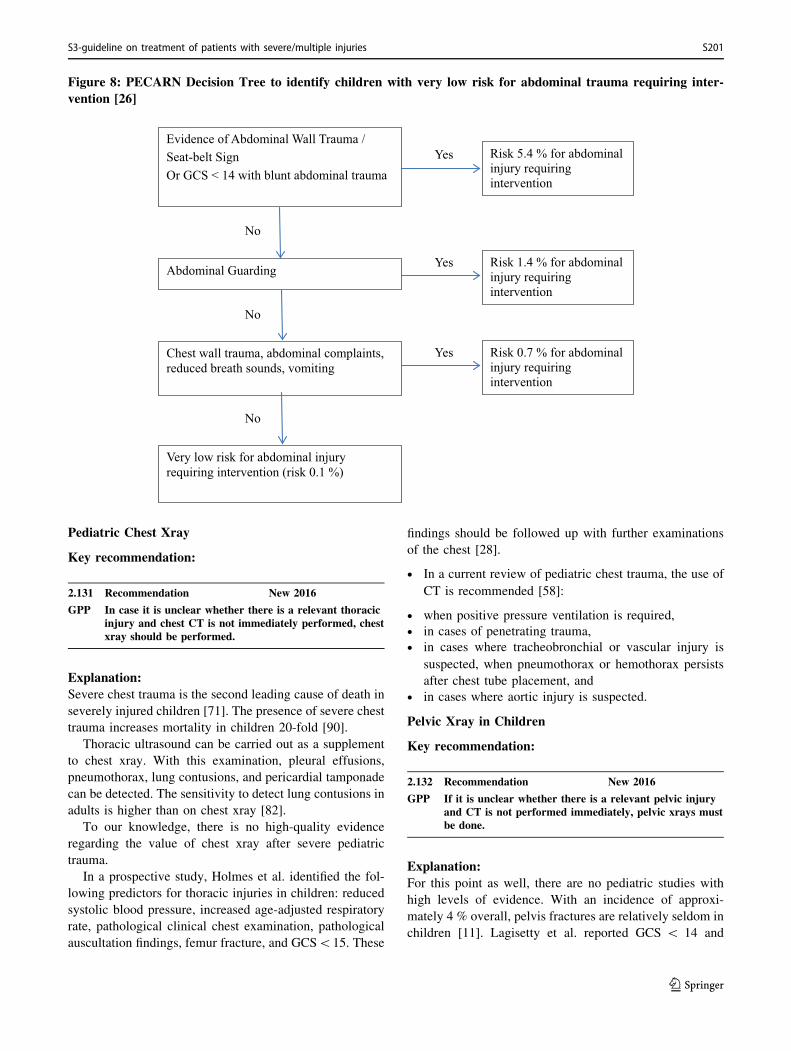
Figure 8: PECARN Decision Tree to identify children with very low risk for abdominal trauma requiring inter-
vention [26]
Yes
Yes
Yes
No
No
No
Risk 0.7 % for abdominal injury requiring intervention
Risk 1.4 % for abdominal injury requiring intervention
Risk 5.4 % for abdominal injury requiring intervention
Very low risk for abdominal injury requiring intervention (risk 0.1 %)
Chest wall trauma, abdominal complaints, reduced breath sounds, vomiting
Abdominal Guarding
Evidence of Abdominal Wall Trauma / Seat-belt SignOr GCS < 14 with blunt abdominal trauma
Pediatric Chest Xray
Key recommendation:
2.131 Recommendation New 2016
GPP In case it is unclear whether there is a relevant thoracicinjury and chest CT is not immediately performed, chestxray should be performed.
Explanation:
Severe chest trauma is the second leading cause of death in
severely injured children [71]. The presence of severe chest
trauma increases mortality in children 20-fold [90].
Thoracic ultrasound can be carried out as a supplement
to chest xray. With this examination, pleural effusions,
pneumothorax, lung contusions, and pericardial tamponade
can be detected. The sensitivity to detect lung contusions in
adults is higher than on chest xray [82].
To our knowledge, there is no high-quality evidence
regarding the value of chest xray after severe pediatric
trauma.
In a prospective study, Holmes et al. identified the fol-
lowing predictors for thoracic injuries in children: reduced
systolic blood pressure, increased age-adjusted respiratory
rate, pathological clinical chest examination, pathological
auscultation findings, femur fracture, and GCS\15. These
findings should be followed up with further examinations
of the chest [28].
• In a current review of pediatric chest trauma, the use of
CT is recommended [58]:
• when positive pressure ventilation is required,
• in cases of penetrating trauma,
• in cases where tracheobronchial or vascular injury is
suspected, when pneumothorax or hemothorax persists
after chest tube placement, and
• in cases where aortic injury is suspected.
Pelvic Xray in Children
Key recommendation:
2.132 Recommendation New 2016
GPP If it is unclear whether there is a relevant pelvic injuryand CT is not performed immediately, pelvic xrays mustbe done.
Explanation:
For this point as well, there are no pediatric studies with
high levels of evidence. With an incidence of approxi-
mately 4 % overall, pelvis fractures are relatively seldom in
children [11]. Lagisetty et al. reported GCS \ 14 and
S3-guideline on treatment of patients with severe/multiple injuries S201
123
tenderness to pelvic pressure as predictors of pelvic frac-
ture. If there is no hematuria, abdominal pain, pelvic
pressure tenderness, abrasions, hemorrhaging, or lacera-
tions, pelvic fracture is unlikely [45].
The high elasticity of the pediatric pelvis should be
considered. Because of this, some 20 % of complex pedi-
atric pelvic injuries appear normal on conventional radio-
graphs and CT. Thus, if pelvic injury is suspected clinically
but is not detected on xray or CT, MRI might be necessary.
When relevant pelvic bleeding is suspected, CT contrast
should be performed.
Cervical Spine Xrays in Children
Key recommendation:
2.133 Recommendation New 2016
GPP If it is unclear whether there is a relevant cervical spineinjury and CT is not performed immediately, cervicalspine radiographs should be done.
Explanation:
Cervical spine injuries are rare, with an incidence of about
1.4 %. However, when an injury occurs, it is associated
with high morbidity and mortality [15].
Here overall there is a lack of studies with high level of
evidence. A current review of diagnostic assessment of
pediatric C-spine injuries [76] yielded the following
remarks.
• C-spine injuries can be excluded without radiological
assessment in children who are conscious and able to be
clinically examined. For this, all NEXUS criteria and
Canadian C-spine rule (CCR) conditions must be
without pathological findings (Table 19). However,
sufficient verbal and cognitive development must be
present to use the NEXUS criteria and CCR. Thus, their
use is not sufficiently reliable in children under 3 years
of age [24, 54, 86, 87].
• When there is decreased level of consciousness or
absent verbal communication, whether it is related to
age, injury, or therapy, conventional C-spine xrays
should be performed in two planes.
• Additional radiological views, such as flexion-extension
images or a dens view, have almost no additional
relevance. At most, a dens view can be taken as a
supplement in children over 8 years of age.
• If conventional radiographs yield questionable findings,
CT should be performed in cases of suspected fracture.
• MRI offers the most sensitive assessment for suspected
soft-tissue injury. All children with neurological deficits
should receive MRI evaluation during the course of
management. In children who cannot be clinically
evaluated because of longer-lasting reduced levels of
consciousness (more than 72 hours), even when fracture
was initially excluded with xray, MRI should be
performed for definitive exclusion of injury.
Table 19: NEXUS Criteria and Canadian C-Spine Rule (CCR)[24, 54, 86, 87]
NEXUS Criteria CCR
Spinous process
tenderness
Dangerous mechanism of injury (Fall from a
height[ 1 m; motor vehicle accident with
rollover or occupant ejection)
Intoxication Spinous process tenderness
GCS\ 15 Inability to rotate head 45� bilaterallyFocal neurological
deficit
Other painful
injuries
Cranial Computed Tomography (CCT) in Children
Key recommendation:
2.134 Recommendation New 2016
GPP Cranial CT (without contrast) must be performed for achild with traumatic brain injury according to definedcriteria.
Explanation:
According to the interdisciplinary S2K Guidelines Trau-
matic Brain Injury in Childhood [1], there are mandatory
and optional indications for CCT examination. Mandatory
indications are:
• Coma with signs of transtentorial herniation such as
pupillary changes or decerebrate rigidity (for this, all
ED trauma assessments after A, B, and C problem
management should be aborted), persistent disorienta-
tion (GCS\ 15)
• focal neurological deficits such as cranial nerve deficits,
paresis, or seizures,
• Suspected impression fracture, basilar skull fracture,
and open injuries.
Optional indications are:
• severe mechanism of trauma, e.g. MVA, fall from[1.5
m (or [ 0.9 m in children under 2 years) or unclear
mechanism of trauma, strong or persistent headache,
• vomiting closely associated in time with the trauma
force and occurring repeatedly,
• Intoxication with alcohol or drugs,
• Evidence of coagulopathy,
• pre-existing cerebral conditions.
S202
123
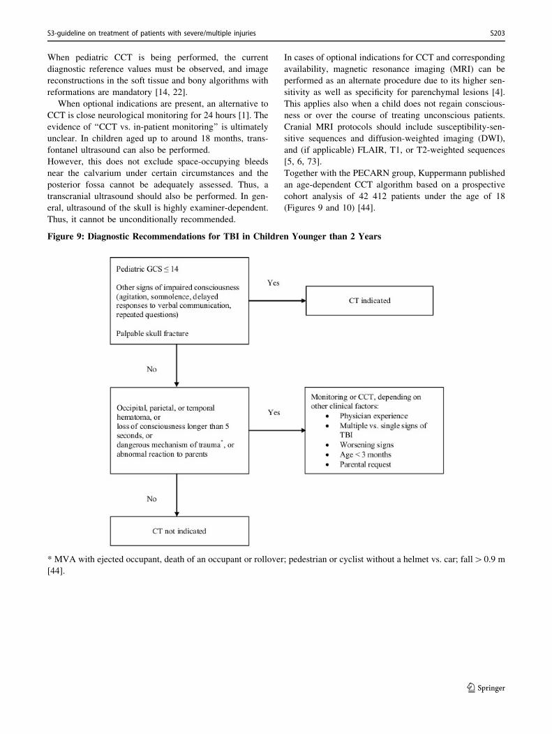
When pediatric CCT is being performed, the current
diagnostic reference values must be observed, and image
reconstructions in the soft tissue and bony algorithms with
reformations are mandatory [14, 22].
When optional indications are present, an alternative to
CCT is close neurological monitoring for 24 hours [1]. The
evidence of ‘‘CCT vs. in-patient monitoring’’ is ultimately
unclear. In children aged up to around 18 months, trans-
fontanel ultrasound can also be performed.
However, this does not exclude space-occupying bleeds
near the calvarium under certain circumstances and the
posterior fossa cannot be adequately assessed. Thus, a
transcranial ultrasound should also be performed. In gen-
eral, ultrasound of the skull is highly examiner-dependent.
Thus, it cannot be unconditionally recommended.
In cases of optional indications for CCT and corresponding
availability, magnetic resonance imaging (MRI) can be
performed as an alternate procedure due to its higher sen-
sitivity as well as specificity for parenchymal lesions [4].
This applies also when a child does not regain conscious-
ness or over the course of treating unconscious patients.
Cranial MRI protocols should include susceptibility-sen-
sitive sequences and diffusion-weighted imaging (DWI),
and (if applicable) FLAIR, T1, or T2-weighted sequences
[5, 6, 73].
Together with the PECARN group, Kuppermann published
an age-dependent CCT algorithm based on a prospective
cohort analysis of 42 412 patients under the age of 18
(Figures 9 and 10) [44].
Figure 9: Diagnostic Recommendations for TBI in Children Younger than 2 Years
* MVA with ejected occupant, death of an occupant or rollover; pedestrian or cyclist without a helmet vs. car; fall[0.9 m
[44].
S3-guideline on treatment of patients with severe/multiple injuries S203
123
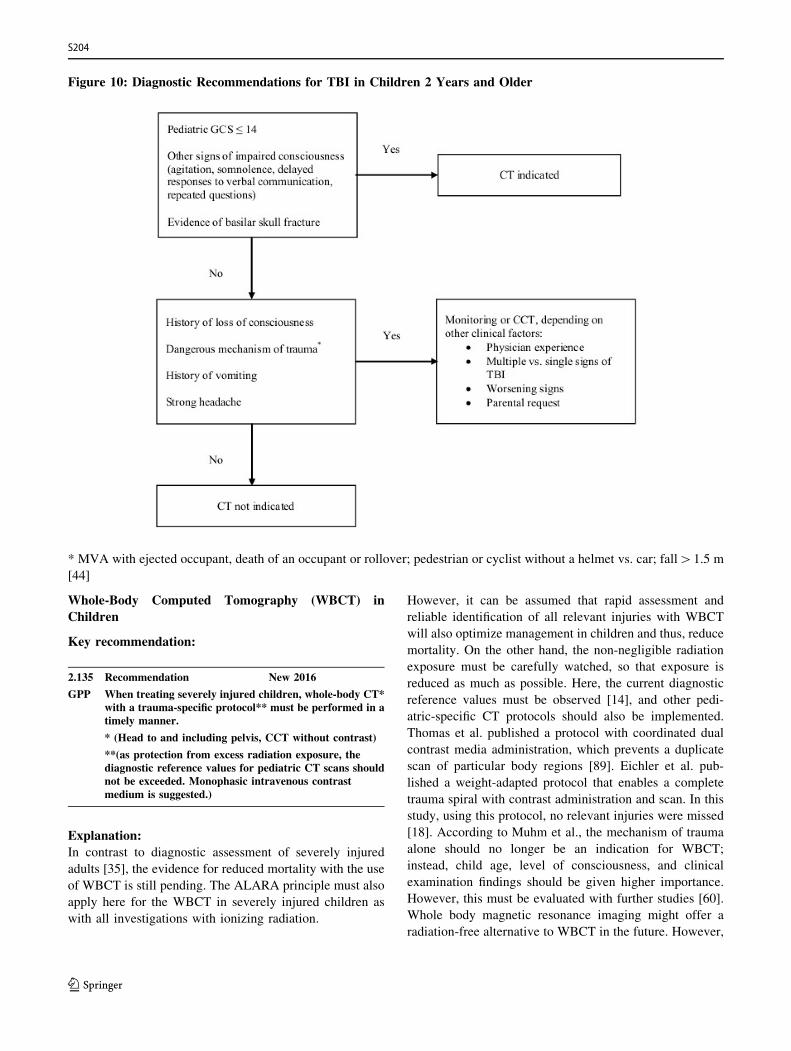
Figure 10: Diagnostic Recommendations for TBI in Children 2 Years and Older
* MVA with ejected occupant, death of an occupant or rollover; pedestrian or cyclist without a helmet vs. car; fall[1.5 m
[44]
Whole-Body Computed Tomography (WBCT) in
Children
Key recommendation:
2.135 Recommendation New 2016
GPP When treating severely injured children, whole-body CT*with a trauma-specific protocol** must be performed in atimely manner.
* (Head to and including pelvis, CCT without contrast)
**(as protection from excess radiation exposure, thediagnostic reference values for pediatric CT scans shouldnot be exceeded. Monophasic intravenous contrastmedium is suggested.)
Explanation:
In contrast to diagnostic assessment of severely injured
adults [35], the evidence for reduced mortality with the use
of WBCT is still pending. The ALARA principle must also
apply here for the WBCT in severely injured children as
with all investigations with ionizing radiation.
However, it can be assumed that rapid assessment and
reliable identification of all relevant injuries with WBCT
will also optimize management in children and thus, reduce
mortality. On the other hand, the non-negligible radiation
exposure must be carefully watched, so that exposure is
reduced as much as possible. Here, the current diagnostic
reference values must be observed [14], and other pedi-
atric-specific CT protocols should also be implemented.
Thomas et al. published a protocol with coordinated dual
contrast media administration, which prevents a duplicate
scan of particular body regions [89]. Eichler et al. pub-
lished a weight-adapted protocol that enables a complete
trauma spiral with contrast administration and scan. In this
study, using this protocol, no relevant injuries were missed
[18]. According to Muhm et al., the mechanism of trauma
alone should no longer be an indication for WBCT;
instead, child age, level of consciousness, and clinical
examination findings should be given higher importance.
However, this must be evaluated with further studies [60].
Whole body magnetic resonance imaging might offer a
radiation-free alternative to WBCT in the future. However,
S204
123
to this point the availability and the examination time,
which is too long for an acutely severely injured child,
remain obstacles [72, 77].
The evidence table for this chapter is found on page 243 of
the guideline report.
References
1. S2K Leitlinie Schadel-Hirn-Trauma im Kindesalter. 2011. http://
www.awmf.org/uploads/tx_szleitlinien/024-018l_S2k_Schaedel-
Hirn-Trauma_im_Kindesalter-2011-03.pdf. Accessed 16 Dec
2015.
2. Abbasi S, et al. Accuracy of emergency physician-performed
ultrasound in detecting traumatic pneumothorax after a 2-h
training course. Eur J Emerg Med. 2013;20(3):173–7.
3. Abboud PA, Kendall J. Emergency department ultrasound for
hemothorax after blunt traumatic injury. J Emerg Med.
2003;25(2):181–4.
4. Ashwal S, Holshouser BA, Tong KA. Use of advanced
neuroimaging techniques in the evaluation of pediatric traumatic
brain injury. Dev Neurosci. 2006;28(4–5):309–26.
5. Ashwal S, et al. Application of advanced neuroimaging modal-
ities in pediatric traumatic brain injury. J Child Neurol.
2014;29(12):1704–17.
6. Beauchamp MH, et al. Susceptibility weighted imaging and its
relationship to outcome after pediatric traumatic brain injury.
Cortex. 2013;49(2):591–8.
7. Becker A, et al. Is the FAST exam reliable in severely injured
patients? Injury. 2010;41(5):479–83.
8. Bernhard M, et al. Introduction of a treatment algorithm can
improve the early management of emergency patients in the
resuscitation room. Resuscitation. 2007;73(3):362–73.
9. Berrington de Gonzalez A, et al. Projected cancer risks from
computed tomographic scans performed in the United States in
2007. Arch Intern Med. 2009;169(22):2071–7.
10. Blaivas M, Lyon M, Duggal S. A prospective comparison of
supine chest radiography and bedside ultrasound for the
diagnosis of traumatic pneumothorax. Acad Emerg Med.
2005;12(9):844–9.
11. Bond SJ, Gotschall CS, Eichelberger MR. Predictors of
abdominal injury in children with pelvic fracture. J Trauma.
1991;31(8):1169–73.
12. Brenner DJ, Elliston CD. Estimated radiation risks potentially
associated with full-body CT screening. Radiology.
2004;232(3):735–8.
13. Brun PM, et al. Stay and play eFAST or scoop and run eFAST?
That is the question! Am J Emerg Med. 2014;32(2):166–70.
14. Bundesamt fur Strahlenschutz. Bekanntmachung der aktual-
isierten diagnostischen Referenzwerte fur diagnostische und
interventionelle Rontgenuntersuchungen. 2010. http://
www.bfs.de/SharedDocs/Downloads/BfS/DE/fachinfo/ion/drw-
roentgen.pdf. Accessed 16 Dec 2015.
15. Cirak B, et al. Spinal injuries in children. J Pediatr Surg.
2004;39(4):607–12.
16. Cronlein M, et al. Traumatic bilateral carotid artery dissection
following severe blunt trauma: a case report on the difficulties in
diagnosis and therapy of an often overlooked life-threatening
injury. Eur J Med Res. 2015;20:62.
17. Davies RM, et al. A decision tool for whole-body CT in major
trauma that safely reduces unnecessary scanning and associated
radiation risks: An initial exploratory analysis. Injury.
2016;47(1):43–9.
18. Eichler K, et al. Multidetector computed tomography (MDCT):
simple CT protocol for trauma patient. Clin Imaging.
2015;39(1):110–5.
19. Fox SH, et al. Future directions in CT technology. Neuroimag-
ing Clin N Am. 1998;8(3):497–513.
20. Gross T, et al. Impact of a multifunctional image-guided therapy
suite on emergency multiple trauma care. Br J Surg.
2010;97(1):118–27.
21. Hara AK, et al. Iterative reconstruction technique for reducing
body radiation dose at CT: feasibility study. AJR Am J
Roentgenol. 2009;193(3):764–71.
22. Hartin CW Jr, et al. Computed tomography scanning in pediatric
trauma: opportunities for performance improvement and radi-
ation safety. J Surg Res. 2013;180(2):226–31.
23. Hilbert P, et al. New aspects in the emergency room manage-
ment of critically injured patients: a multi-slice CT-oriented
care algorithm. Injury. 2007;38(5):552–8.
24. Hoffman JR, et al. Low-risk criteria for cervical-spine radiog-
raphy in blunt trauma: a prospective study. Ann Emerg Med.
1992;21(12):1454–60.
25. Holmes JF, Gladman A, Chang CH. Performance of abdominal
ultrasonography in pediatric blunt trauma patients: a meta-
analysis. J Pediatr Surg. 2007;42(9):1588–94.
26. Holmes JF, et al. Identifying children at very low risk of
clinically important blunt abdominal injuries. Ann Emerg Med.
2013;62(2):107–116 e2.
27. Holmes JF, et al. Validation of a prediction rule for the
identification of children with intra-abdominal injuries after
blunt torso trauma. Ann Emerg Med. 2009;54(4):528–33.
28. Holmes JF, et al. A clinical decision rule for identifying children
with thoracic injuries after blunt torso trauma. Ann Emerg Med.
2002a;39(5):492–9.
29. Holmes JF, et al. Identification of children with intra-abdominal
injuries after blunt trauma. Ann Emerg Med.
2002b;39(5):500–9.
30. Hsiao KH, et al. Whole-body computed tomography in the
initial assessment of trauma patients: is there optimal criteria for
patient selection? Emerg Med Australas. 2013;25(2):182–91.
31. Huber-Wagner S, et al. Whole-body CT in haemodynamically
unstable severely injured patients–a retrospective, multicentre
study. PLOS One. 2013;8(7):e68880.
32. Huber-Wagner S, et al. Whole-body CT in haemodynamically
unstable severely injured patients—a retrospective, multicentre
study. PLoS One. 2013;8(7).
33. Huber-Wagner S, et al. Whole-body CT Score—Kriterien zur
Durchfuhrung einer Ganzkorper-Computertomographie bei
potentiell schwerverletzten Patienten. In: Deutscher Kongress
fur Orthopadie und Unfallchirurgie (DKOU 2015). Berlin. 2015.
34. Huber-Wagner S, et al. Effect of whole-body CT during trauma
resuscitation on survival: a retrospective, multicentre study. The
Lancet. 2009a;373(9673):1455–61.
35. Huber-Wagner S, et al. Effect of whole-body CT during trauma
resuscitation on survival: a retrospective, multicentre study.
Lancet. 2009b;373(9673):1455–61.
36. Huber-Wagner S, et al. Effect of the localisation of the CT
scanner during trauma resuscitation on survival—a retrospec-
tive, multicentre study. Injury. 2014;45(Suppl 3):S76–82.
37. Hyacinthe AC, et al. Diagnostic accuracy of ultrasonography in
the acute assessment of common thoracic lesions after trauma.
Chest. 2012;141(5):1177–83.
38. Ingeman JE, et al. Emergency physician use of ultrasonography
in blunt abdominal trauma. Academic Emergency Medicine.
1996;3(10):931–7.
S3-guideline on treatment of patients with severe/multiple injuries S205
123
39. Kalender WA, et al. Spiral volumetric CT with single-breath-
hold technique, continuous transport, and continuous scanner
rotation. Radiology. 1990;176(1):181–3.
40. Kanz KG, et al. Priority-oriented shock trauma room manage-
ment with the integration of multiple-view spiral computed
tomography. Unfallchirurg. 2004;107(10):937–44.
41. Kanz KG, et al. Trauma management incorporating focused
assessment with computed tomography in trauma (FACTT)-
potential effect on survival. J Trauma Manag Outcomes.
2010;4(1):4.
42. Klingenbeck-Regn K, et al. Subsecond multi-slice computed
tomography: basics and applications. Eur J Radiol.
1999;31(2):110–24.
43. Kloppel R, et al. Early clinical management after polytrauma
with 1 and 4 slice spiral CT. Radiologe. 2002;42(7):541–6.
44. Kuppermann N, et al. Identification of children at very low risk
of clinically-important brain injuries after head trauma: a
prospective cohort study. Lancet. 2009;374(9696):1160–70.
45. Lagisetty J, et al. Are routine pelvic radiographs in major
pediatric blunt trauma necessary? Pediatr Radiol.
2012;42(7):853–8.
46. Lee KL, et al. Impact on trauma patient management of
installing a computed tomography scanner in the emergency
department. Injury. 2009;40(8):873–5.
47. Lefering R. Revised Injury Severity Classification (RISC)—
development and validation of a classification system for
severely injured patients based on a large trauma registry
(Habilitationsschrift). In: IFOM - Institut fur Forschung in der
Operativen Medizin. 2007, University Witten/Herdecke, Faculty
of Medicine, Germany: Cologne. pp. 1–131.
48. Lefering R. Development and validation of the Revised Injury
Severity Classification (RISC) score for severely injured
patients. Eur J Trauma Emerg Surg. 2009;35:437–47.
49. Leidner B, Beckman MO. Standardized whole-body computed
tomography as a screening tool in blunt multitrauma patients.
Emerg Radiol. 2001;8:20–8.
50. Lentz KA, et al. Evaluating blunt abdominal trauma: Role for
ultrasonography. J Ultrasound Med. 1996;15(6):447–51.
51. Lindner T, et al. Does radar technology support the diagnosis of
pneumothorax? Pneumoscan—a diagnostic point-of-care tool.
Emerg Med Int. 2013.
52. Loewenhardt B, et al. Radiation exposure in whole-body
computed tomography of multiple trauma patients: bearing
devices and patient positioning. Injury. 2012;43(1):67–72.
53. Low R, et al. Whole body spiral CT in primary diagnosis of
patients with multiple trauma in emergency situations. Rofo.
1997;166(5):382–8.
54. Mahadevan S, et al. Interrater reliability of cervical spine injury
criteria in patients with blunt trauma. Ann Emerg Med.
1998;31(2):197–201.
55. Martinsen AC, et al. Iterative reconstruction reduces abdominal
CT dose. Eur J Radiol. 2012;81(7):1483–7.
56. Mathews JD, et al. Cancer risk in 680,000 people exposed to
computed tomography scans in childhood or adolescence: data
linkage study of 11 million Australians. BMJ. 2013;346:f2360.
57. Miglioretti DL, et al. The use of computed tomography in
pediatrics and the associated radiation exposure and estimated
cancer risk. JAMA Pediatr. 2013;167(8):700–7.
58. Moore MA, Wallace EC, Westra SJ. The imaging of paediatric
thoracic trauma. Pediatr Radiol. 2009;39(5):485–96.
59. Mueller DL, et al. Pediatric radiation exposure during the initial
evaluation for blunt trauma. J Trauma. 2011;70(3):724–31.
60. Muhm M, et al. Pediatric trauma care with computed tomog-
raphy-criteria for CT scanning. Emerg Radiol.
2015;22(6):613–21.
61. Nagarsheth K, Kurek S. Ultrasound detection of pneumothorax
compared with chest X-ray and computed tomography scan. Am
Surg. 2011;77(4):480–3.
62. Noel PB, et al.Does iterative reconstruction lowerCT radiationdose:
evaluation of 15,000 examinations. PLoS One. 2013;8(11):e81141.
63. Pearce MS, et al. Radiation exposure from CT scans in
childhood and subsequent risk of leukaemia and brain tumours:
a retrospective cohort study. Lancet. 2012;380(9840):499–505.
64. Philipp MO, et al. Radiological emergency room management
with emphasis on multidetector-row CT. Eur J Radiol.
2003;48(1):2–4.
65. Ptak T, Rhea JT, Novelline RA. Experience with a continous,
single-pass whole-body multidetector CT protocol for trauma:
the three-minute multiple trauma CT scan. Emerg Radiol.
2001;8:250–6.
66. Quinn AC, Sinert R. What is the utility of the focused
assessment with sonography in trauma (FAST) exam in
penetrating torso trauma? Injury. 2011;42(5):482–7.
67. Richards JR, et al. Sonographic assessment of blunt abdominal
trauma: a 4-year prospective study. J Clin Ultrasound.
2002;30(2):59–67.
68. Rieger M, et al. Modern CT diagnosis of acute thoracic and
abdominal trauma. Anaesthesist. 2002;51(10):835–42.
69. Riepl C, et al. Reduced emergency room time for trauma patients
by integrated CT. Z Orthop Unfall. 2013;151(2):168–72.
70. Saltzherr TP, et al. Randomized clinical trial comparing the
effect of computed tomography in the trauma room versus the
radiology department on injury outcomes. Br J Surg.
2012;99(Suppl 1):105–13.
71. Sartorelli KH, Vane DW. The diagnosis and management of
children with blunt injury of the chest. Semin Pediatr Surg.
2004;13(2):98–105.
72. Schaefer JF, Kramer U. Whole-body MRI in children and
juveniles. Fortschr Rontgenstr. 2011;183(1):24–36.
73. Schaefer PW, et al. Diffusion-weighted MR imaging in closed
head injury: high correlation with initial glasgow coma scale
score and score on modified Rankin scale at discharge.
Radiology. 2004;233(1):58–66.
74. Scherer R, et al. Preclinical and clinical management of major
trauma patients. Rettungsdienst. 1999;12(22):36–43.
75. Schleder S, et al. Diagnostic value of a hand-carried ultrasound
device for free intra-abdominal fluid and organ lacerations in
major trauma patients. Emerg Med J. 2013;30(3):e20.
76. Schoneberg C, et al. Diagnosis of cervical spine injuries in
children: a systematic review. Eur J TraumaEmerg Surg.
2013;39(6):653–65.
77. Schoneberg C, et al. The injured child–diagnostic work-up in
the emergency room. Unfallchirurg. 2014a;117(9):829–41.
78. Schoneberg C, et al. Diagnostic management in paediatric blunt
abdominal trauma—a systematic review with metaanalysis.
Zentralbl Chir. 2014b;139(6):584–91.
79. Smith ZA, Postma N, Wood D. FAST scanning in the
developing world emergency department. S Afr Med J.
2010;100(2):105–8.
80. Sola JE, et al. Pediatric FAST and elevated liver transaminases:
An effective screening tool in blunt abdominal trauma. J Surg
Res. 2009;157(1):103–7.
81. Soldati G, et al. Occult traumatic pneumothorax: diagnostic
accuracy of lung ultrasonography in the emergency department.
Chest. 2008;133(1):204–11.
S206
123
82. Soldati G, et al. Chest ultrasonography in lung contusion. Chest.
2006;130(2):533–8.
83. Statistisches Bundesamt. Bevolkerung-Alter im Wandel-Altere
Menschen in Deutschland und der EU. 2012. https://www.desta-
tis.de/DE/Publikationen/Thematisch/Bevoelkerung/
Bevoelkerungsstand/AlterimWandel.html. Accessed 16Dec 2015.
84. Stedman JM, et al. Splenic parenchymal heterogeneity at dual-
bolus single-acquisition CT in polytrauma patients-6-months
experience fromOxford, UK. Emerg Radiol. 2014;21(3):257–60.
85. Stengel D, et al. Accuracy of single-pass whole-body computed
tomography for detection of injuries in patients with major blunt
trauma. CMAJ. 2012;184(8):869–76.
86. Stiell IG, et al. The Canadian C-spine rule versus the NEXUS
low-risk criteria in patients with trauma. N Engl J Med.
2003;349(26):2510–8.
87. Stiell IG, et al. The Canadian C-spine rule for radiography in
alert and stable trauma patients. JAMA. 2001;286(15):1841–8.
88. Taguchi K, Aradate H. Algorithm for image reconstruction in
multi-slice helical CT. Med Phys. 1998;25(4):550–61.
89. Thomas KE, et al. Dual bolus intravenous contrast injection
technique for multiregion paediatric body CT. Eur Radiol.
2015;25(4):1014–22.
90. Tovar JA. The lung and pediatric trauma. Semin Pediatr Surg.
2008;17(1):53–9.
91. TraumaRegister DGU, Annual Report of the Trauma Registry of
the German Trauma Society (DGU). Committee on Emergency
Medicine. Cologne: Intensive and Trauma Care (Sektion NIS)-
German Trauma Society (DGU); 2014.
92. TraumaRegister DGU, Annual Report of the Trauma Registry of
the German Trauma Society (DGU). Committee on Emergency
Medicine. Cologne: Intensive and Trauma Care (Sektion NIS)-
German Trauma Society (DGU); 2015.
93. Weninger P, et al. Emergency room management of patients
with blunt major trauma: evaluation of the multislice computed
tomography protocol exemplified by an urban trauma center.
J Trauma. 2007;62(3):584–91.
94. Wilkerson RG, Stone MB. Sensitivity of bedside ultrasound and
supine anteroposterior chest radiographs for the identification of
pneumothoraxafterblunt trauma.AcadEmergMed.2010;17(1):11–7.
95. Wurmb TE, et al. Whole-body multislice computed tomography
as the first line diagnostic tool in patients with multiple injuries:
the focus on time. J Trauma. 2009;66(3):658–65.
96. Wurmb TE, et al. Whole-body multislice computed tomography
as the primary and sole diagnostic tool in patients with blunt
trauma: searching for its appropriate indication. Am J Emerg
Med. 2007;25(9):1057–62.
97. Zhang M, et al. Rapid detection of pneumothorax by ultra-
sonography in patients with multiple trauma. Crit Care.
2006;10(4):R112.
3 Primary Operative Management
3.1 Introduction
How would you decide?
A 35-year-old bicyclist has an accident. The patient is
intubated and ventilated at the accident scene by the
emergency physician. Volume replacement therapy is
begun as hemodynamic support. The patient is brought to
you for primary management. After exclusion of relevant
intra-abdominal and intra-thoracic bleeding and after
thorough assessment, the following injury pattern is evi-
dent: traumatic brain injury grade 2 (small subdural hem-
orrhage, contusion, etc.), chest trauma with multiple rib
fractures and pronounced left pulmonary contusion, pri-
mary open left femoral shaft fracture, right distal tibia
fracture. Initial laboratory tests show a hemoglobin value
of 9.3 g/dl, INR of 0.77, and a base excess of -4.5 mmol/l.
You consider your options for primary surgical manage-
ment of this patient and weigh the pros and cons. The
longer you consider, the more questions arise. What is the
operative strategy of choice for the femur fracture? What is
the optimal treatment for the distal tibia? Must the fibula be
treated as well? Is it advisable to perform primary defini-
tive treatment or is temporary fixation better? What roles
do the traumatic brain injury and the thoracic injuries play
in these decisions? You recall the management of similar
cases in your department, the dogmatic repetitions of your
‘‘teacher’’ or other colleagues, the economic ‘‘restraints’’
placed by the hospital administration, and the perennial
lack of time to adequately consider the almost limitless
complex literature on the management of polytrauma
patients. Ultimately, you decide once again to carry out a
management strategy based on your own personal
experience.
How would other German providers decide?
According to the database of the German Trauma Society’s
Trauma Registry, more than 65 % of all polytrauma
patients have extremity and/or pelvic injuries (AIS[ 2).
This makes it even more astonishing that divergent oper-
ative strategies for femoral shaft fractures in a polytrauma
setting are practiced and published [1]. According to
analyses of the trauma registry, the primary treatment for
femoral shaft fractures in multiply injured patients by some
German hospitals is, almost dogmatically, always with
external fixators. In other hospitals, it is almost always with
intramedullary nails, and finally, in many hospitals, there is
every conceivable combination, sometimes with fixators
and sometimes with nails [1].
These differing approaches are based on two different
operative strategies, which can ultimately be used on var-
ious body regions. The strategy of ‘‘Easy Total Care’’
promotes primary definitive management of injuries.
Conversely, the ‘‘damage control’’ strategy for severely
injured patients has the goal of avoiding secondary injury
from surgery itself. For example, in the area of fracture
care, primary definitive care is foregone, and instead,
temporary stabilization is achieved with an external fixator.
The smaller intervention and shorter operative time should
keep the additional load, i.e. secondary damage, to a
minimum.
S3-guideline on treatment of patients with severe/multiple injuries S207
123
The Goal of the ‘‘Primary Operative Management’’
Guideline Section
During the assessment of the overall condition of a trauma
patient in the early post-trauma phase of care, there are a
number of individual (e.g., age), trauma-specific (e.g.,
overall injury severity, pattern of injuries), and physiolog-
ical (metabolism, coagulation, temperature, etc.) factors
that must be taken into account. Although this can make the
initial assessment more difficult, this assessment is the basis
for the choice of operative approach during the primary
operative management phase. It is generally accepted that
patients in an overall stable condition benefit from definitive
treatment and patients in less stable condition overall do
better with ‘‘damage control.’’ A recent analysis of the
German Trauma Society’s Trauma Registry shows that
approximately 15 % of patients are assigned to an unclear
overall condition (‘‘borderline’’). The optimal treatment
strategy in the primary operative phase remains a contro-
versial topic of debate particularly for these patients [1-12].
Thus, there is a continuing need for perspective
regarding levels of evidence and grades of recommenda-
tion for various management strategies. The goal of this
guideline section is to offer an overview of the evidence
level of various treatment strategies during the primary
operative phase after polytrauma and from this, to develop
clinical treatment pathways (in cases of sufficient evi-
dence) or document the need for further scientific review
(recommendation grade).
Special Instructions:
In the context of this guideline section, assessment of the
key issues is often complicated because ‘‘hard’’ scientific
data is lacking, or because results are related to isolated
injuries. This will be explicitly indicated in the corre-
sponding areas, and the greatest effort will be made to offer
the clearest possible individual recommendations for rou-
tine clinical practice despite the sometimes contradictory
information in the literature.
When discussing fractures, it should be assumed –unless
stated specifically otherwise – that the injury is closed,without
vascular involvement and without compartment syndrome.
Open fractures, vascular involvement, and compartment
syndromes are considered indications for emergency surgery
and may require different management strategies.
In addition, for certain fractures that require technically-
challenging surgery (e.g., complex distal femoral or hum-
eral condylar fractures), it is important for severely injured
patients in particular that primary definitive treatment
should only be considered if a) careful planning is made, if
necessary based on 3D CT imaging, b) the expected
operative time will not be too long, c) an experienced
surgeon is present, and d) an appropriate implant is avail-
able in-house. For this reason, in many German trauma
centers, such technically challenging fractures in poly-
trauma patients are initially stabilized temporarily, and
definitive reconstruction is undertaken secondarily.
References
1. Rixen D, Grass G, Sauerland S, Lefering R, Raum MR, Yucel N,
Bouillon B, Neugebauer EAM, ‘‘Polytrauma Study Group’’ of the
German Trauma Society. Evaluation of criteria for temporary
external fixation in risk-adapted Damage Control orthopaedic
surgery of femur shaft fractures in multiple trauma patients:
‘‘evidence based medicine’’ versus ‘‘reality’’ in the trauma
registry of the German Trauma Society. J Trauma.
2005;59:1375–95.
2. Giannoudis PV. Surgical priorities in Damage Control in poly-
trauma. J Bone Joint Surg (Br). 2003;85:478–83.
3. Pape H, Stalp M, Dahlweid M, Regel G, Tscherne H, Arbeits-
gemeinschaft ,,Polytrauma‘‘ der Deutschen Gesellschaft fur
Unfallchirurgie. Welche primare Operationsdauer ist hinsichtlich
eines ,,Borderline-Zustandes‘‘ polytraumatisierter Patienten
vertretbar? Unfallchirurg. 1999;102:861–69.
4. Pape HC, van Griensven M, Sott AH, Giannoudis P, Morley J,
Roise O, Ellingsen E, Hildebrand F, Wiese B, Krettek C, EPOFF
study group. Impact of intramedullary instrumentation versus
Damage Control for femoral fractures on immunoinflammatory
parameters: prospective randomized analysis by the EPOFF study
group. J Trauma. 2003;55:7–13.
5. Scalea TM, Boswell SA, Scott JD, Mitchell KA, Kramer ME,
Pollak AN. External fixation as a bridge to intramedullary nailing
for patients with multiple injuries and with femur fractures:
damage Control orthopedics. J Trauma. 2000;48:613–23.
6. BouillonB,RixenD,MaegeleM,SteinhausenE,TjardesT, Paffrath
T. Damage control orthopedics—was ist der aktuelle Stand?
Unfallchirurg. 2009;112:860–9.
7. Pape HC, Rixen D, Morley J, Husebye EE, Mueller M, Dumont
C, Gruner A, Oestern HJ, Bayeff-Filoff M, Garving C, Pardini D,
van Griensven M, Krettek C, Giannoudis P. The EPOFF study
group, Impact of the method of initial stabilization for femoral
shaft fractures in patients with multiple injuries at risk for
complications (borderline patients). Ann Surg.
2007;246:491–501.
8. Hildebrand F, Lefering R, Andruszkow H, Zelle BA, Barkatali B,
Pape HC. Development of a scoring system based on conven-
tional parameters to assess polytrauma patients: PolyTrauma
Grading Score (PTGS). Injury. 2015;46S4:S17–22.
9. O’Toole RV1, O’Brien M, Scalea TM, Habashi N, Pollak AN,
Turen CH. Resuscitation before stabilization of femoral fractures
limits acute respiratory distress syndrome in patients with
multiple traumatic injuries despite low use of damage control
orthopedics. J Trauma. 2009;67:1013–21.
10. Nahm NJ, Como JJ, Wilber JH, Vallier HA. Early appropriate
care: definitive stabilization of femoral fractures within 24 hours
of injury is safe in most patients with multiple injuries.
J Trauma. 2011;71:175–85.
11. Nahm NJ, Vallier HA. Timing of definitive treatment of femoral
shaft fractures in patients with multiple injuries: a systematic
review of randomized and nonrandomized trials. J Trauma
Acute Care Surg. 2012;73:1046–63.
12. Vallier HA, Moore TA, Como JJ, Wilczewski PA, Steinmetz
MP, Wagner KG, Smith CE, Wang XF, Dolenc AJ. J Orthop
Surg Res. 2015;10:155.
S208
123
3.2 Thorax
Operative Approach
Key recommendation:
3.1 Recommendation Modified 2016
GoR 0 Depending on the location of injury, anterolateralthoracotomy or sternotomy approaches can be used.When injury location is unclear, a clamshell approachcan be selected.
Explanation:
The standard approach for emergency thoracotomy is
anterolateral thoracotomy on the side of the injury at the
level of the 4th-6th intercostal space. However, anterolat-
eral thoracotomy seems to provide insufficient exposure to
the injured organs in up to 20 % of cases [20].
If the exact location of the injury can be identified
preoperatively, standard approaches can be used, e.g.
posterolateral approach for procedures on the thoracic aorta
or the intrathoracic trachea. A supraclavicular approach
with mobilization or division of the clavicle enables safe
visualization of the subclavian vessels in cases of injury
there [4, 14, 44, 45].
Median sternotomy is preferred for injuries to the
ascending aorta and the aortic arch as well as injuries of the
great vessels and the heart [20, 43, 44]. Injuries to the
posterior cardiac wall, however, are difficult to access
through a sternotomy and are better reached through an
anterolateral thoracotomy approach.
For trauma surgery, thoracoscopy is unsuitable for life-
threatening emergencies. However, video-assisted thora-
coscopy (VATS) can be performed for diagnostic and
therapeutic purposes in patients with compensated car-
diorespiratory function. For example, diaphragmatic inju-
ries, lung parenchymal injuries, or sources of bleeding can
be identified and treated. VATS is regularly used for
hemothorax evacuation, usually after the acute manage-
ment phase [14, 29, 44].
Penetrating Chest Injuries
Key recommendation:
3.2 Recommendation 2016
GoR B In the case of penetrating chest trauma, retained foreignbodies should only be removed under controlledconditions in the operating room after thoracotomy.
Explanation:
Once it is clear that there has been perforation of the chest
wall, a foreign body penetrating the thorax must not be
removed due to the possible tamponade effects or secondary
injuries caused by improper removal. Removal takes place
in the operating room or in the trauma care area of the ED
during exploratory thoracotomy. In certain cases of hemo-
dynamically stable patients, VATS can be considered for
foreign body removal. Airtight closure or binding of
puncture wounds is contraindicated, because they enable
decompression of the pleural cavity. If decompression is not
achieved with chest tube placement, the chest wound should
be treated with a sterile, semi-occlusive dressing.
Indication for Thoracotomy
Key recommendations:
3.3 Recommendation 2016
GoR A A penetrating chest injury that is the cause ofhemodynamic instability must undergo immediateexploratory thoracotomy.
3.4 Recommendation 2016
GoR 0 Thoracotomy can be performed for cases with initialblood loss > 1500 mL from the chest tube or withcontinuing blood loss > 250 mL/hour for more than fourhours.
Explanation:
In penetrating trauma, indications for immediate thoraco-
tomy include severe hemodynamic shock, signs of pericar-
dial tamponade, profuse bleeding, absence of peripheral
pulses, and cardiac arrest when vital signs were initially
observed and/or arrest began no longer than five minutes
previous [14, 20, 44]. After chest tube placement, hemody-
namically stable patients can be monitored and/or undergo
further diagnostic assessments like spiral CT with contrast.
Studies of predominantly penetrating injuries during the
Vietnam War found that mortality and complication rates
were reduced when thoracotomy was performed for initial
blood loss[1500 ml or more than 500 ml in the first hour
after chest tube placement [28].
A multi-center study also reported an association between
mortality and thoracic blood loss regardless of mechanism
of injury (blunt versus penetrating). Mortality rose by a
factor of 3.2 in the group with chest tube blood loss of
more than 1500 ml versus 500 ml in the first 24 hours.
Thoracotomy was performed an average 2.4 ± 5.4 hours
after admission [19]. Other authors [9, 19, 25, 28, 43]
follow the concept to perform thoracotomy in blunt or
penetrating trauma for an initial blood loss of 1500 ml or
continuous bleeding of 250 ml/h over 4 hours. If the
quantity of drainage per time unit is used as an indication
for thoracotomy, the chest tube(s) must be correctly placed
and draining reliably [44].
In case of combination chest injuries with great blood
loss and pronounced metabolic derangement, temporary
chest closure consistent with a damage control strategy can
S3-guideline on treatment of patients with severe/multiple injuries S209
123
be performed once acute management and bleeding control
has been completed. Once the vital parameters have been
stabilized and there is restoration of coagulation and end-
organ function, definitive surgical management and closure
of the chest can be performed [9, 15, 17, 43].
Lung Injuries
Key recommendation:
3.5 Recommendation 2016
GoR B If surgery is indicated because of lung injuries(persistent bleeding and/or air leakage), the interventionshould be parenchyma-sparing.
Explanation:
Parenchymal injuries of the lung after penetrating or blunt
chest trauma with persistent bleeding and/or air leakage
require surgical treatment [13]. One of the main indications
for exploratory thoracotomy is profuse or persistent bleeding
(1500 ml initially or 250 ml/h over 2-4 hours) [19]. In this
case, corresponding operative management for potential
lung parenchymal injuries might be indicated for bleeding
control. Compared to parenchyma-sparing procedures –
such as primary closure, pulmonary tractotomy, atypical
resection or segment resection – lobectomy and pneu-
monectomy are associated with higher complication and
mortality rates [9, 15, 17, 43]. At the same time, blunt trauma
injuries seem to be associated with worse prognosis in terms
of hospital duration, complications, and mortality [27].
Injuries of the Great Vessels
Key recommendations:
3.6 Recommendation 2016
GoR B In cases of thoracic aortic rupture, if technically andanatomically feasible, endovascular stent prosthesis ispreferable to open revascularization procedures.
3.7 Recommendation 2016
GoR B Until aortic reconstruction, or in cases of conservativemanagement, systolic blood pressure should be kept90-120 mmHg.
Explanation:
Traditional treatment for aortic rupture consists of aortic
reconstruction through direct suture with aortic clamping
and using various bypass procedures to perfuse the lower
half of the body and the spinal cord during clamping (left
heart bypass, Gott shunt, heart lung machine) [6, 31].
Current studies have identified acute stenting for aortic
ruptures as a minimally invasive, time-sparing therapeutic
option with minimal injury from the approach [33]. Com-
plications like reduced cerebral or spinal perfusion with the
associated consequences, such as paraplegia, occur less
frequently. Long term anticoagulant therapy, which is
necessary for most bypass procedures, can be avoided [41].
In a current meta-analysis comparing open aortic recon-
struction with endovascular stenting, the same technical
success rate yielded significantly lower mortality as well as
a significantly lower rate of postoperative neurological
deficits (paraplegia, stroke) for stenting [34]. However, to
date there is no long-term survival data after endovascular
aortic reconstruction [37]. Overall, according to the current
literature, endovascular stent prosthesis appears preferable
to the conventional procedure [12].
Complications such as paraplegia and acute renal failure
generally occur during open procedures due to operative-
dependent ischemia. Complication rates correlate with
clamping time of the aorta [18].
If perfusion is maintained during surgery with bypass
procedures, complication rates (paraplegia, renal failure)
decrease [5, 6, 13, 31].
The hemodynamic status of the patient at the time of
admission determines the timing of aortic rupture man-
agement. Patients who are hemodynamically unstable or in
extremis must undergo immediate surgery [5]. For patients
with concomitant traumatic brain injury, severe abdominal
or skeletal injuries requiring immediate operative inter-
vention, and in older patients with pronounced cardiac and
pulmonary comorbidities, aortic injuries can be treated
with delayed urgency after additional life-threatening
injuries and/or overall condition have been treated and
stabilized [43, 44]. In a series of 395 patients, Camp et al.
found that the mortality for hemodynamically stable pa-
tients undergoing non-emergent ([ 4 hours) or delayed
surgery ([ 24 hours) was not higher than in those under-
going emergent surgery (\ 4 hours) [10]. This opinion is
shared by other authors [12, 13]. Delays to surgery of up to
seven months may be tolerated in certain cases [35].
If surgery is not carried out emergently, strict pharma-
cological control of blood pressure (SBP between 90 and
120 mmHg and heart rate \ 100/min) is necessary with
beta blockers and vasodilators [12, 13].
Treatment of aortic dissection follows the same princi-
ples, with the difference that operative management always
occurs during a secondary phase since it is not acutely life
threatening.
Cardiac Injury
Life-threatening cardiac injuries occur predominantly
through penetrating trauma. Injuries to multiple chambers
are particularly associated with high mortality [14, 44].
Intra-pericardial injury of the inferior vena cava often
causes life-threatening pericardial tamponade that needs
immediate decompression. Operative treatment of the vena
cava follows pericardial decompression, through the right
S210
123
atrium with direct suture or patch closure under extracor-
poreal circulation [42-45].
The approach is made with highest urgency through a
left anterolateral thoracotomy or a median sternotomy.
After decompression of the pericardial tamponade, which
occurs in more than 50 % of cases, rapid control of
bleeding is performed with suture or stapler through a
longitudinal incision of the pericardium. After clamping of
the bleeding atrial wall (Satinsky clamp), direct suture can
follow here [30]. In difficult situations, a Foley catheter can
be used as a temporary solution with intraventricular
blocking. Ventricular lesions are closed with a pericardial
patch or with Teflon felt augmentation. If possible, care
should be taken of the coronary arteries while sewing to
avoid a secondary infarction. Finally, the pericardium is
left open or there is a loose approximation of the pericar-
dial incision to avoid recurrent tamponade [14, 44]. There
is no need for immediate thoracotomy for injuries not
affecting hemodynamics, isolated septum defects, valvular
injuries, or ventricular aneurysms [30]. These cases gen-
erally become evident after emergency stabilization of the
patient because of persistent instability. Transesophageal
echocardiography confirms their presence.
Proximal lesions of the coronary vessels must be
reconstructed or treated emergently with aorto-coronary
bypass, generally using a heart-lung machine [8]. Distal
coronary lesions can be sutured or ligated [14, 44].
The initial cardiac rhythm and cardiorespiratory func-
tion of the patient at the time of ED arrival have prognostic
relevance [1, 2]. Thus, attempts to maintain cardiac pump
function and treat arrhythmias must be made, as this
decreases mortality [1, 2]
Injuries of the Tracheo-Bronchial System
Key recommendations:
3.8 Recommendation Modified 2016
GoR B If there is clinical suspicion of injury to thetracheobronchial system, diagnostic tracheo-bronchoscopy should be performed to confirm thediagnosis.
3.9 Recommendation 2016
GoR B Traumatic injuries of the tracheobronchial systemshould be surgically treated as soon as possible afterdiagnosis.
3.10 Recommendation 2016
GoR 0 For localized injuries of the tracheobronchial system,conservative management can be attempted.
Explanation:
Injuries to the tracheo-bronchial system are rare and often a
delayed diagnosis [3, 21, 36]. Tracheo-bronchial injury can
also occur occasionally as a complication of orotracheal
intubation [38]. Penetrating injuries predominantly affect
the cervical trachea, while blunt trauma generally results in
intrathoracic injuries. The right main bronchus near the
carina is more frequently affected [3, 21, 36]. If there is
persistent pneumothorax despite correct positioning and
function of the chest tube, subcutaneous emphysema, or
atelectasis, suspected tracheo-bronchial injury should be
evaluated with tracheobronchoscopy [3, 21, 36]. Tracheal
ruptures occurring at the level of the endotracheal tube cuff
are initially covered and often missed because of posi-
tioning. Thus, during endoscopy, the endotracheal tube
should be withdrawn to examine for tracheal ruptures.
Fiberoptic intubation with cuff placement distal to the
defect can be performed immediately to secure the airway.
In a retrospective study, Kummer et al. determined that a
large number of patients required a definitive airway (tra-
cheostomy) [22]. The emphasis here was on penetrating
injuries. Operative treatment of the tracheo-bronchial sys-
tem should be performed as soon as possible after diag-
nosis, because delayed treatment is associated with higher
complication rates [30]. Surgical management of airway
injuries yields a significantly lower mortality than conser-
vative therapy [3, 21, 36]. Once bronchoscopy has been
completed, conservative therapy should be considered only
in patients with small bronchial tissue defects (defect
smaller than 1/3 of the bronchus circumference) and well
adapted bronchial margins [3, 11, 21 36]. A retrospective
study by Schneider and colleagues identified no difference
between conservative and operative management for cases
of iatrogenic tracheal injuries without ventilation distur-
bance and superficial or covered tracheal tears [38].
Injuries to the cervical trachea were treated through a
collar incision. For injuries to the intrathoracic trachea and
the main bronchus, a right posterolateral thoracotomy
approach should be used in the 3rd-4th intercostal space
[30]. For simple transverse tears, end-to-end anastomosis
of the bronchus should be performed after bronchus
mobilization and cartilage removal when necessary. If
direct suture is not possible in longitudinal tears with
defects of the parietal membrane, they can be closed with a
patch to avoid bronchial stenosis [3, 21, 36]. According to
the current literature, stent-assisted treatment of tracheo-
bronchial injuries appears irrelevant.
Injuries of the Bony Thorax (excluding Spine)
Explanation:
Bony injuries of the chest wall include fractures of the
sternum, the ribs, and combinations thereof. Nearly half of
all severely injured patients have rib fractures. Of these,
two thirds have fractures without relevant chest wall
instability and one third have an unstable thorax (flail
chest), of whom again nearly one third have bilateral
involvement. Sternum fractures without concomitant rib
S3-guideline on treatment of patients with severe/multiple injuries S211
123
fractures are infrequent, found in fewer than 4 % of all
severely injured patients. In combination with rib fractures,
sternum fractures are present in approximately 7 to 21 % of
severely injured patients [39].
The majority of uncomplicated fractures can be treated
conservatively. Consistent pulmonary hygiene and ade-
quate pain therapy are crucial. For many fracture patterns,
including flail chest, a prolonged need for artificial respi-
ration with positive pressure ventilation creates an ‘‘inter-
nal pneumatic splint,’’ yielding a favorable position for
consolidation [10]. This can be the case, e.g. in severe lung
contusion or severe traumatic brain injury [43].
However, if the unstable chest wall itself is the main
reason that ventilation is required, operative stabilization
can shorten the duration of ventilation as well intensive
care unit stay. A number of recent studies confirm this [7,
23, 24, 26, 40].
In addition, the following indications for operative
treatment are found in the literature:
Sternum fractures [16]
• Strong, persistent, or intractable pain
• Respiratory insufficiency or ventilator-dependence
• Overlapping or impressed fractures
• Sternum deformity or instability
• Non-union/pseudarthrosis
• Deformed posture due to rib deformity and/or pain
• Limited range of motion
Rib fractures [32]
• Unstable chest wall (flail chest) with weaning failure or
paradoxical chest excursions on weaning
• Prolonged pain
• Chest wall deformity
• Symptomatic non-union/pseudarthrosis
• Thoracotomy for another indication (rib fixation during
‘‘closure’’ of the thorax)
Various implants are available for chest wall stabilization
like metal brackets, rib clamping systems, and various
plates. Wire and suture are not unimportant. Stabilization
of the bony chest wall is considered for and performed on
stable patients; generally for severely injured patients, it is
not part of primary operative management.
The evidence table for this chapter is found on page 277
of the guideline report.
References
1. Asensio JA, et al. One hundred five penetrating cardiac injuries: a
2-year prospective evaluation. J Trauma. 1998a;44(6):1073–82
[LoE 2b].
2. Asensio JA, et al. Penetrating cardiac injuries: a prospective
study of variables predicting outcomes. J Am Coll Surg.
1998b;186(1):24–34 [LoE 2b].
3. Balci AE, et al. Surgical treatment of post-traumatic tracheo-
bronchial injuries: 14-year experience. Eur J Cardiothorac Surg.
2002;22(6):984–9 [LoE 4].4. Branney SW, et al. Critical analysis of two decades of experience
with postinjury emergency department thoracotomy in a regional
trauma center. J Trauma. 1998;45(1):87–94 (discussion 94–5)
[LoE 4].5. Camp PC, Shackford SR. Outcome after blunt traumatic thoracic
aortic laceration: identification of a high-risk cohort. Western
Trauma Association Multicenter Study Group. J Trauma.
1997;43(3):413–22 [LoE 3b].6. Cardarelli MG, et al. Management of traumatic aortic rupture: a
30-year experience. Ann Surg. 2002;236(4):465–9 (discussion469–70) [LoE 2b].
7. Cataneo AJ, et al. Surgical versus nonsurgical interventions for
flail chest. Cochrane Database Syst Rev. 2015;7:CD009919.
8. Chaudhry UA, et al. Beating-heart versus conventional on-pump
coronary artery bypass grafting: a meta-analysis of clinical
outcomes. Ann Thorac Surg 2015.
9. Cothren C, et al. Lung-sparing techniques are associated with
improved outcome compared with anatomic resection for severe
lung injuries. J Trauma. 2002;53(3):483–7 [LoE 4].
10. Dehghan N, et al. Flail chest injuries: a review of outcomes and
treatment practices from the National Trauma Data Bank.
J Trauma Acute Care Surg. 2014;76(2):462–8.
11. Dienemann H, Hoffmann H. Tracheobronchial injuries and
fistulas. Chirurg. 2001;72(10):1131–6 [LoE 4].12. Fabian TC, et al. Prospective study of blunt aortic injury: helical
CT is diagnostic and antihypertensive therapy reduces rupture.
Ann Surg. 1998;227(5):666–76 (discussion 676–7) [LoE 2b].13. Fabian TC, et al. Prospective study of blunt aortic injury:
Multicenter Trial of the American Association for the Surgery
of Trauma. J Trauma. 1997;42(3):374–80 discussion 380–3
[LoE 4].14. Feliciano DV, Rozycki GS. Advances in the diagnosis and
treatment of thoracic trauma. Surg Clin N Am.
1999;79(6):1417–29 [LoE 4].
15. Gasparri M, et al. Pulmonary tractotomy versus lung resection:
viable options in penetrating lung injury. J Trauma.
2001;51(6):1092–5 (discussion 1096–7) [LoE 4].16. Harston A, Roberts C. Fixation of sternal fractures: a systematic
review. J Trauma. 2011;71(6):1875–9.
17. Huh J, et al. Surgical management of traumatic pulmonary
injury. Am J Surg. 2003;186(6):620–4 [LoE 4].18. Jahromi AS, et al. Traumatic rupture of the thoracic aorta:
cohort study and systematic review. J Vasc Surg.
2001;34(6):1029–34 [LoE 3b].
19. Karmy-Jones R, et al. Timing of urgent thoracotomy for
hemorrhage after trauma: a multicenter study. Arch Surg.
2001;136(5):513–8 [LoE 3b].20. Karmy-Jones R, et al. Urgent and emergent thoracotomy for
penetrating chest trauma. J Trauma. 2004;56(3):664–8 (discus-sion 668–9) [LoE 4].
21. Kiser AC, O’Brien SM, Detterbeck FC. Blunt tracheobronchial
injuries: treatment and outcomes. Ann Thorac Surg.
2001;71(6):2059–65 [LoE 4].22. Kummer C, et al. A review of traumatic airway injuries:
potential implications for airway assessment and management.
Injury. 2007;38(1):27–33 [LoE 2b].
23. Lafferty PM, et al. Operative treatment of chest wall injuries:
indications, technique, and outcomes. J Bone Joint Surg Am.
2011;93(1):97–110.
S212
123
24. Leinicke JA, et al. Operative management of rib fractures in the
setting of flail chest: a systematic review and meta-analysis.
Ann Surg. 2013;258(6):914–21.
25. Mansour MA, et al. Exigent postinjury thoracotomy analysis of
blunt versus penetrating trauma. Surg Gynecol Obstet.
1992;175(2):97–101 [LoE 3b].
26. Marasco SF, et al. Prospective randomized controlled trial of
operative rib fixation in traumatic flail chest. J Am Coll Surg.
2013;216(5):924–32.
27. Martin MJ, et al. Operative management and outcomes of
traumatic lung resection. J Am Coll Surg. 2006;203(3):336–44
[LoE 2b].
28. McNamara JJ, et al. Thoracic injuries in combat casualties in
Vietnam. Ann Thorac Surg. 1970;10(5):389–401 [LoE 3b].
29. McSwain NE Jr. Blunt and penetrating chest injuries. World J
Surg. 1992;16(5):924–9 [LoE 4].
30. Meredith JW, Hoth JJ. Thoracic trauma: when and how to
intervene. Surg Clin North Am. 2007;87(1):95–118 vii.[LoE
2a].31. Miller PR, et al. Complex blunt aortic injury or repair: beneficial
effects of cardiopulmonary bypass use. Ann Surg.
2003;237(6):877–83 (discussion 883–4) [LoE 2b].
32. Nirula R, et al. Rib fracture repair: indications, technical issues,
and future directions. World J Surg. 2009;33(1):14–22.
33. Ott MC, et al. Management of blunt thoracic aortic injuries:
endovascular stents versus open repair. J Trauma.
2004;56(3):565–70 [LoE 4].34. Peterson BG, et al. Percutaneous endovascular repair of blunt
thoracic aortic transection. J Trauma. 2005;59(5):1062–5 [LoE
1].35. Reed AB, et al. Timing of endovascular repair of blunt
traumatic thoracic aortic transections. J Vasc Surg.
2006;43(4):684–8 [LoE 4].
36. Rossbach MM, et al. Management of major tracheobronchial
injuries: a 28-year experience. Ann Thorac Surg.
1998;65(1):182–6 [LoE 4].37. Rousseau H, et al. Acute traumatic aortic rupture: a comparison
of surgical and stent-graft repair. J Thorac Cardiovasc Surg.
2005;129(5):1050–5 [LoE 3b].
38. Schneider T, et al. Management of iatrogenic tracheobronchial
injuries: a retrospective analysis of 29 cases. Ann Thorac Surg.
2007;83(6):1960–4 [LoE 2b].39. Schulz-Drost S, et al. Bony injuries of the thoracic cage in
multiple trauma : Incidence, concomitant injuries, course and
outcome. Unfallchirurg. 2015.
40. Tanaka H, et al. Surgical stabilization of internal pneumatic
stabilization? A prospective randomized study of management
of severe flail chest patients. J Trauma. 2002;52(4):727–32
(discussion 732) [LoE 4].
41. Tang GL, et al. Reduced mortality, paraplegia, and stroke with
stent graft repair of blunt aortic transections: a modern meta-
analysis. J Vasc Surg. 2008;47(3):671–5 [LoE 2a].42. Voggenreiter G, et al. Operative chest wall stabilization in flail
chest–outcomes of patients with or without pulmonary contu-
sion. J Am Coll Surg. 1998;187(2):130–8 [LoE 4].
43. Wall MJ Jr, et al. Thoracic aortic and thoracic vascular injuries.
Surg Clin N Am. 2001;81(6):1375–93 [LoE 4].
44. Wall MJ Jr, Soltero E. Damage control for thoracic injuries.
Surg Clin N Am. 1997;77(4):863–78 [LoE 4].
45. Xenos ES, et al. Covered stents for injuries of subclavian and
axillary arteries. J Vasc Surg. 2003;38(3):451–4 [LoE 4].
3.3 Diaphragm
Key recommendation:
3.11 Recommendation 2011
GoR B Once recognized during the initial diagnostic surveyand/or intraoperatively, traumatic diaphragm ruptureshould be rapidly closed.
Explanation:
Diaphragm rupture is present in 1.6 % of blunt trauma
patients and mostly occurs during motor vehicle accidents
with side impact. It predominantly affects the left side [1,
3-6, 9, 11].
There is no valid data regarding the ideal time frame for
surgery in patients with multiple injuries. It seems clear
that in cases where abdominal organs have moved into the
thorax, closure of the rupture should be performed rapidly.
This also applies for cases in which the rupture is detected
incidentally during surgery where the space has been
opened for another injury.
However, there is no current clear evidence that delayed
closure increases mortality. A random effects meta-re-
gression of 22 studies (n = 980) from the years 1976-1992
[11] found that the overall mortality of 17 % showed no
association between the incidence of delayed treatment and
mortality (beta 0.013, 95% CI: -0.267 to -0.240). In a
current analysis of 4153 patients from the National Trauma
Database, pleural empyema was also not correlated with
the timing of operative interventions [2].
In an acute situation with a hemodynamically unsta-
ble patient without thoracic injury, the ideal surgical
approach is transabdominal. In cases of verified combina-
tion injuries, poor visualization, or technically difficult
suture, a thoraco-abdominal approach is used [2, 8, 11].
The diaphragmatic defect can usually be closed with
direct suture, and rarely is plastic defect coverage neces-
sary [1, 8, 9]. The data available offers no conclusions
regarding success rates of certain suture techniques (con-
tinuous versus interrupted) or material (monofil versus
braided, resorbable versus non-resorbable). Endoscopic
techniques are suitable for closing post-traumatic
diaphragmatic hernias [7, 10]; in the primary operative
phase of a polytrauma patient, the selection of laparoscopic
or thoracoscopic approach must be critically evaluated.
References
1. Athanassiadi K, et al. Blunt diaphragmatic rupture. Eur J
Cardiothorac Surg. 1999; 15(4):469–7 . [LoE 4].2. Barmparas G, et al. Risk factors for empyema after diaphragmatic
injury: results of a National Trauma Databank analysis. J Trauma.
2009;66(6):1672–6 [LoE 2b].
S3-guideline on treatment of patients with severe/multiple injuries S213
123
3. Bergeron E, et al. Impact of deferred treatment of blunt diaphrag-
matic rupture: a 15-year experience in six trauma centers inQuebec.
J Trauma. 2002;52(4):633–40 [LoE 4].4. Brasel KJ, et al. Predictors of outcome in blunt diaphragm
rupture. J Trauma. 1996;41(3):484–7 [LoE 4].5. Chughtai T, et al. Update on managing diaphragmatic rupture in
blunt trauma: a review of 208 consecutive cases. Can J Surg.
2009;52(3):177–81 [LoE 4].
6. Kearney PA, Rouhana SW, Burney RE, Blunt rupture of the
diaphragm: mechanism, diagnosis, and treatment. Ann Emerg
Med. 1989;18(12):1326–30 [LoE 4].7. Lomanto D, et al. Thoracolaparoscopic repair of traumatic
diaphragmatic rupture. Surg Endosc. 2001;15(3):323.
8. Matsevych OY, Blunt diaphragmatic rupture: four year’s expe-
rience. Hernia. 2008;12(1):73–8 [LoE 5].9. Mihos P, et al. Traumatic rupture of the diaphragm: experience
with 65 patients. Injury. 2003;34(3):169–72 [LoE 4].10. Ouazzani A, et al. A laparoscopic approach to left diaphrag-
matic rupture after blunt trauma. Acta Chir Belg.
2009;109(2):228–31.
11. Shah R, et al. Traumatic rupture of diaphragm. Ann Thorac
Surg. 1995;60(5):1444–9 [LoE 5].
3.4 Abdomen
Surgical Approach
Key recommendation:
3.12 Recommendation 2016
GoR B For trauma cases, midline laparotomy should be usedover other approaches.
Explanation:
Midline laparotomy is an anatomically based, universal
surgical approach to the injured abdomen. It provides good
visibility of all four quadrants, and can be performed more
quickly and with less bleeding than transverse incisions,
especially in trauma situations.
There is only one quasi-randomized study (assigned to
treatment groups according to even or odd admission
number), now over thirty years old, in which the midline
laparotomy was compared to an upper abdominal trans-
verse laparotomy in trauma patients [68]. The wound
infection rates for patients with negative and positive
findings on laparotomy were 2 % and 11 %, respectively,
regardless of approach. In positive laparotomy, the average
duration of anesthesia was 25 minutes shorter in patients
having undergone a midline versus transverse approach
(Table 20). According to the published data, this difference
was statistically significant (p = 0.02). However, no stan-
dard deviations were reported and operative times were not
further analyzed. This study cannot serve as evidence for
selecting one approach over the other, but supports the idea
of surgeon preference (‘‘Adequacy of organ exposure is
still a matter of personal preference’’).
Indirect evidence comes from randomized studies for
elective abdominal procedures. A Cochrane review presents
advantages of the transverse approach regarding postoper-
ative need for morphine equivalents, lung function, and
rates of incisional hernias [11]. There was no difference in
the rates of pulmonary complications or wound infections.
The multi-center randomized POVATI (Post-surgical pain
Outcome of Vertical and Transverse abdominal Incisions)
study of 2009 reported equivalent primary endpoint post-
operative analgesia requirements and no difference in the
secondary endpoints such as pulmonary complications,
mortality, and incisional hernias at one year [62].Here, the
authors also stress the possibility of a situation-dependent
approach to the abdomen (‘‘The decision about the incision
should be driven by surgeon preference with respect to the
patient’s disease and anatomy’’).
There are still no clinical studies available to enable
comparison of emergency laparoscopy and emergency
laparotomy in multiply injured patients. One should continue
to follow the consensus of the EAES that the current collec-
tion of evidence does not enable a clear recommendation in
favor of therapeutic laparoscopic procedures for polytrauma
patients with severe abdominal trauma (‘‘Nevertheless, the
scarceness of clinical data prohibits a clear recommendation
in favor of therapeutic laparoscopy for trauma’’) [37, 61].
Damage Control (DC): General Principles
Key recommendation:
3.13 Recommendation Modified 2016
GoR B In hemodynamically unstable patients with complexintra-abdominal damage, the damage control principle(hemostasis, packing, temporary abdominal closure/laparostoma) should be used over definitivereconstruction.
Table 20: Midline vs. Upper Abdominal Transverse Laparotomyin Abdominal Trauma
Study LoE Patients Results
Stone
et al.
1983
[68]
2b 339 patients
with blunt
or
penetrating
abdominal
trauma
Midline
Laparotomy
(n = 177)
Upper
Abdominal
Transverse
Laparotomy
(n = 162)
Mean duration
of anesthesia:
positive
laparotomy (n
= 66) 215 min,
negative
laparotomy (n
= 111) 126 min
Mean duration
of anesthesia:
positive
laparotomy (n
= 61) 240 min,
negative
laparotomy (n
= 101) 132
min
S214
123
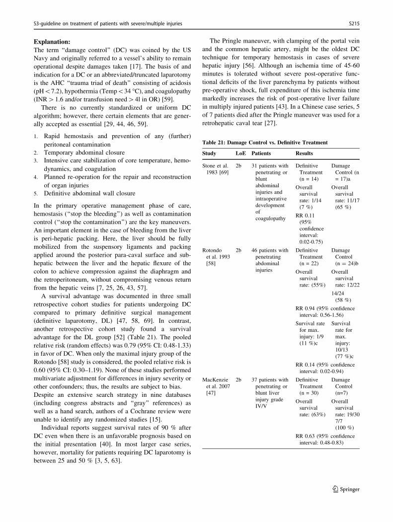
Explanation:
The term ‘‘damage control’’ (DC) was coined by the US
Navy and originally referred to a vessel’s ability to remain
operational despite damages taken [17]. The basis of and
indication for a DC or an abbreviated/truncated laparotomy
is the AHC ‘‘trauma triad of death’’ consisting of acidosis
(pH\7.2), hypothermia (Temp\34 �C), and coagulopathy(INR[1.6 and/or transfusion need[4l in OR) [59].
There is no currently standardized or uniform DC
algorithm; however, there certain elements that are gener-
ally accepted as essential [29, 44, 46, 59].
1. Rapid hemostasis and prevention of any (further)
peritoneal contamination
2. Temporary abdominal closure
3. Intensive care stabilization of core temperature, hemo-
dynamics, and coagulation
4. Planned re-operation for the repair and reconstruction
of organ injuries
5. Definitive abdominal wall closure
In the primary operative management phase of care,
hemostasis (‘‘stop the bleeding’’) as well as contamination
control (‘‘stop the contamination’’) are the key maneuvers.
An important element in the case of bleeding from the liver
is peri-hepatic packing. Here, the liver should be fully
mobilized from the suspensory ligaments and packing
applied around the posterior para-caval surface and sub-
hepatic between the liver and the hepatic flexure of the
colon to achieve compression against the diaphragm and
the retroperitoneum, without compromising venous return
from the hepatic veins [7, 25, 26, 43, 57].
A survival advantage was documented in three small
retrospective cohort studies for patients undergoing DC
compared to primary definitive surgical management
(definitive laparotomy, DL) [47, 58, 69]. In contrast,
another retrospective cohort study found a survival
advantage for the DL group [52] (Table 21). The pooled
relative risk (random effects) was 0.79 (95% CI: 0.48-1.33)
in favor of DC. When only the maximal injury group of the
Rotondo [58] study is considered, the pooled relative risk is
0.60 (95% CI: 0.30–1.19). None of these studies performed
multivariate adjustment for differences in injury severity or
other confounders; thus, the results are subject to bias.
Despite an extensive search strategy in nine databases
(including congress abstracts and ‘‘gray’’ references) as
well as a hand search, authors of a Cochrane review were
unable to identify any randomized studies [15].
Individual reports suggest survival rates of 90 % after
DC even when there is an unfavorable prognosis based on
the initial presentation [40]. In most larger case series,
however, mortality for patients requiring DC laparotomy is
between 25 and 50 % [3, 5, 63].
The Pringle maneuver, with clamping of the portal vein
and the common hepatic artery, might be the oldest DC
technique for temporary hemostasis in cases of severe
hepatic injury [56]. Although an ischemia time of 45-60
minutes is tolerated without severe post-operative func-
tional deficits of the liver parenchyma by patients without
pre-operative shock, full expenditure of this ischemia time
markedly increases the risk of post-operative liver failure
in multiply injured patients [43]. In a Chinese case series, 5
of 7 patients died after the Pringle maneuver was used for a
retrohepatic caval tear [27].
Table 21: Damage Control vs. Definitive Treatment
Study LoE Patients Results
Stone et al.
1983 [69]
2b 31 patients with
penetrating or
blunt
abdominal
injuries and
intraoperative
development
of
coagulopathy
Definitive
Treatment
(n = 14)
Damage
Control (n
= 17)a
Overall
survival
rate: 1/14
(7 %)
Overall
survival
rate: 11/17
(65 %)
RR 0.11
(95%
confidence
interval:
0.02-0.75)
Rotondo
et al. 1993
[58]
2b 46 patients with
penetrating
abdominal
injuries
Definitive
Treatment
(n = 22)
Damage
Control
(n = 24)b
Overall
survival
rate: (55%)
Overall
survival
rate: 12/22
14/24
(58 %)
RR 0.94 (95% confidence
interval: 0.56-1.56)
Survival rate
for max.
injury: 1/9
(11 %)c
Survival
rate for
max.
injury:
10/13
(77 %)c
RR 0.14 (95% confidence
interval: 0.02-0.94)
MacKenzie
et al. 2007
[47]
2b 37 patients with
penetrating or
blunt liver
injury grade
IV/V
Definitive
Treatment
(n = 30)
Damage
Control
(n=7)
Overall
survival
rate: (63%)
Overall
survival
rate: 19/30
7/7
(100 %)
RR 0.63 (95% confidence
interval: 0.48-0.83)
S3-guideline on treatment of patients with severe/multiple injuries S215
123
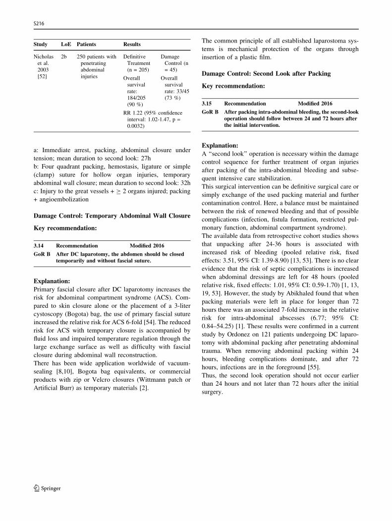
a: Immediate arrest, packing, abdominal closure under
tension; mean duration to second look: 27h
b: Four quadrant packing, hemostasis, ligature or simple
(clamp) suture for hollow organ injuries, temporary
abdominal wall closure; mean duration to second look: 32h
c: Injury to the great vessels + C 2 organs injured; packing
+ angioembolization
Damage Control: Temporary Abdominal Wall Closure
Key recommendation:
3.14 Recommendation Modified 2016
GoR B After DC laparotomy, the abdomen should be closedtemporarily and without fascial suture.
Explanation:
Primary fascial closure after DC laparotomy increases the
risk for abdominal compartment syndrome (ACS). Com-
pared to skin closure alone or the placement of a 3-liter
cystoscopy (Bogota) bag, the use of primary fascial suture
increased the relative risk for ACS 6-fold [54]. The reduced
risk for ACS with temporary closure is accompanied by
fluid loss and impaired temperature regulation through the
large exchange surface as well as difficulty with fascial
closure during abdominal wall reconstruction.
There has been wide application worldwide of vacuum-
sealing [8,10], Bogota bag equivalents, or commercial
products with zip or Velcro closures (Wittmann patch or
Artificial Burr) as temporary materials [2].
The common principle of all established laparostoma sys-
tems is mechanical protection of the organs through
insertion of a plastic film.
Damage Control: Second Look after Packing
Key recommendation:
3.15 Recommendation Modified 2016
GoR B After packing intra-abdominal bleeding, the second-lookoperation should follow between 24 and 72 hours afterthe initial intervention.
Explanation:
A ‘‘second look’’ operation is necessary within the damage
control sequence for further treatment of organ injuries
after packing of the intra-abdominal bleeding and subse-
quent intensive care stabilization.
This surgical intervention can be definitive surgical care or
simply exchange of the used packing material and further
contamination control. Here, a balance must be maintained
between the risk of renewed bleeding and that of possible
complications (infection, fistula formation, restricted pul-
monary function, abdominal compartment syndrome).
The available data from retrospective cohort studies shows
that unpacking after 24-36 hours is associated with
increased risk of bleeding (pooled relative risk, fixed
effects: 3.51, 95% CI: 1.39-8.90) [13, 53]. There is no clear
evidence that the risk of septic complications is increased
when abdominal dressings are left for 48 hours (pooled
relative risk, fixed effects: 1.01, 95% CI: 0.59-1.70) [1, 13,
19, 53]. However, the study by Abikhaled found that when
packing materials were left in place for longer than 72
hours there was an associated 7-fold increase in the relative
risk for intra-abdominal abscesses (6.77; 95% CI:
0.84–54.25) [1]. These results were confirmed in a current
study by Ordonez on 121 patients undergoing DC laparo-
tomy with abdominal packing after penetrating abdominal
trauma. When removing abdominal packing within 24
hours, bleeding complications dominate, and after 72
hours, infections are in the foreground [55].
Thus, the second look operation should not occur earlier
than 24 hours and not later than 72 hours after the initial
surgery.
Study LoE Patients Results
Nicholas
et al.
2003
[52]
2b 250 patients with
penetrating
abdominal
injuries
Definitive
Treatment
(n = 205)
Damage
Control (n
= 45)
Overall
survival
rate:
184/205
(90 %)
Overall
survival
rate: 33/45
(73 %)
RR 1.22 (95% confidence
interval: 1.02-1.47, p =
0.0032)
S216
123
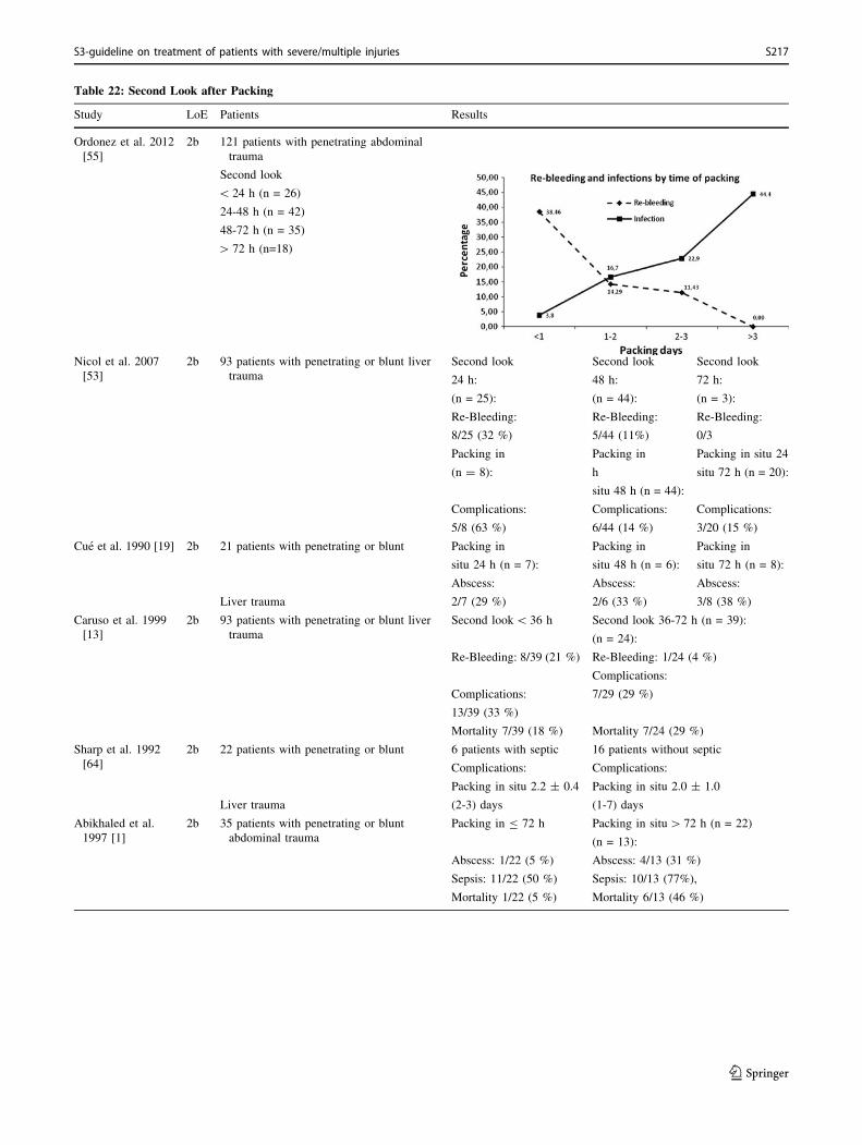
Table 22: Second Look after Packing
Study LoE Patients Results
Ordonez et al. 2012
[55]
2b 121 patients with penetrating abdominal
trauma
Second look
\ 24 h (n = 26)
24-48 h (n = 42)
48-72 h (n = 35)
[ 72 h (n=18)
Nicol et al. 2007
[53]
2b 93 patients with penetrating or blunt liver
trauma
Second look Second look Second look
24 h: 48 h: 72 h:
(n = 25): (n = 44): (n = 3):
Re-Bleeding:
8/25 (32 %)
Re-Bleeding:
5/44 (11%)
Re-Bleeding:
0/3
Packing in
(n = 8):
Packing in
h
situ 48 h (n = 44):
Packing in situ 24
situ 72 h (n = 20):
Complications:
5/8 (63 %)
Complications:
6/44 (14 %)
Complications:
3/20 (15 %)
Cue et al. 1990 [19] 2b 21 patients with penetrating or blunt Packing in Packing in Packing in
situ 24 h (n = 7): situ 48 h (n = 6): situ 72 h (n = 8):
Abscess: Abscess: Abscess:
Liver trauma 2/7 (29 %) 2/6 (33 %) 3/8 (38 %)
Caruso et al. 1999
[13]
2b 93 patients with penetrating or blunt liver
trauma
Second look\ 36 h Second look 36-72 h (n = 39):
(n = 24):
Re-Bleeding: 8/39 (21 %) Re-Bleeding: 1/24 (4 %)
Complications:
Complications:
13/39 (33 %)
7/29 (29 %)
Mortality 7/39 (18 %) Mortality 7/24 (29 %)
Sharp et al. 1992
[64]
2b 22 patients with penetrating or blunt 6 patients with septic 16 patients without septic
Complications: Complications:
Packing in situ 2.2 ± 0.4 Packing in situ 2.0 ± 1.0
Liver trauma (2-3) days (1-7) days
Abikhaled et al.
1997 [1]
2b 35 patients with penetrating or blunt
abdominal trauma
Packing in B 72 h Packing in situ[ 72 h (n = 22)
(n = 13):
Abscess: 1/22 (5 %) Abscess: 4/13 (31 %)
Sepsis: 11/22 (50 %) Sepsis: 10/13 (77%),
Mortality 1/22 (5 %) Mortality 6/13 (46 %)
S3-guideline on treatment of patients with severe/multiple injuries S217
123
Definitive Abdominal Wall Closure
Key recommendation:
3.16 Recommendation New 2016
GoR B When a laparostoma is placed, definitive closure shouldbe aimed for as soon as possible.
Explanation:
Hatch et al. [33, 34] performed a retrospective analysis of
the clinical histories of 925 consecutive trauma laparo-
tomies, of whom 282 (30 %) were performed within a DC
framework. Of these, 247 (88 %) survived until the second
look operation; fascial closure was carried out in 86 (35 %)
of these patients at this point. In 161 cases (65 %),
definitive fascial closure was performed or attained at a
later point. In the group with early fascial closure, there
was a decreased complication rate in the further clinical
course. Both groups were comparable, particularly
regarding ISS and early laboratory and vital parameters.
Thus, the authors concluded: ‘‘The current data demon-
strate quite convincingly that early fascial reapproximation
is associated with a significant decrease in complications
and organ failure. Therefore, we recommend DCL in only
the sickest subset of patients, optimization of open abdo-
men management, and fascial closure at the earliest pos-
sible time.’’
Multivariate analysis of both groups showed that the use of
a vacuum system already at the time of DC laparotomy was
an independent factor for successful early fascial closure
(odds ratio 3.1; 95% CI: 1.42-6.63; p = 0.004) [33].
In an older systematic review study, the results of case
series were summarized [10]. After that, the Wittmann
patch is associated with the highest rate of success for
abdominal wall closure. A retrospective cohort study
achieved similar results [74]. In a small randomized study,
there was no difference between temporary closure with
vacuum dressings and polyglactin 910 mesh [8].
The ideal technique for fascial closure after laparostoma is
not yet clear. Analysis of publications to date makes it
obvious that in addition to heterogeneity of the investigated
patient groups, there are also marked procedural differ-
ences among studies. There are almost no multi-center
studies. Most results come from retrospective, mono-center
studies. The current level of evidence is thus insufficient
and leaves many unanswered questions.
In order to improve the data and thus, the quality of care in
Germany, a laparotomy register has been implemented by
the German Society for General and Visceral Surgery
(DGAV).
Non-Operative Management of Blunt Liver or Spleen
Injuries
Key recommendation:
3.17 Recommendation New 2016
GoR B In hemodynamically stable patients with isolated bluntliver or spleen injury, the goal should be non-operativemanagement.
Explanation:
The undisputed standard of care for isolated liver and
spleen injuries in children after blunt trauma is non-oper-
ative management with intensive care supervision and
readiness for immediate interventional radiology and/or
surgical care.
In recent years, there has been a trend emerging to treat
adults conservatively as well in cases where the injury is
isolated blunt trauma and the patient is hemodynamically
stable; some authors consider this the standard of care [35,
36, 65].
According to Hommes et al. [36], the conditions for non-
operative management are hemodynamically stable pa-
tients with positive response to volume replacement as well
as CT excluding other intra-abdominal injuries requiring
surgery (e.g. bowel perforation).
After implementation of a damage control resuscitation
protocol, Shresta et al. [65] found an improved success rate
of operative management even of higher grade relevant
blunt liver injuries.
Angioembolization
Key recommendation:
3.18 Recommendation Modified 2016
GoR B If contrast-enhanced CT shows evidence for an arterialbleed in a hemodynamically stable patient with liverinjury, selective angioembolization when possible orlaparotomy should be performed.
Explanation:
Interventional radiology has a fixed place in polytrauma
management and is used both as definitive therapy for
organ and vessel injuries and in combination with operative
management (before, after, or hybrid surgery) [21, 45].
When there is evidence of active bleeding on the contrast-
enhanced CT that cannot or must not be addressed surgi-
cally, and there is good response to fluid and blood
replacement in the ED, angioembolization can contribute
to sustained hemodynamic stabilization and thus, can rep-
resent a key maneuver for definitive therapy [6, 31].
S218
123
There are still no randomized studies. The best available
evidence to date continues to suggest that angioemboliza-
tion for blunt and penetrating liver injuries as a supplement
to DC management decreases mortality compared to sur-
gery alone (common RR [fixed effects] 0.47, 95% CI: 0.28
– 0,78) [4, 6, 39, 49, 50, 73]. The lack of multivariate
adjustment and resulting bias must be considered. It is not
currently possible to answer the question of whether
angioembolization for liver injuries should be performed
prior to or after DC laparotomy. Two studies advocate
early neoadjuvant angioembolization because of lower
complication rates [49, 73]. In two other studies, however,
mortality was reduced when angioembolization was per-
formed after DC laparotomy [50, 71].
The indication for endovascular intervention, surgery, or a
combination of the two must be made carefully on a case-
by-case basis. The decision must be made according to the
rapid availability of an experienced interventional radiol-
ogist, the success of hemodynamic stabilizing procedures,
intraoperative findings, and post-operative hemodynamics.
Key recommendation:
3.19 Recommendation Modified 2016
GoR B For spleen injuries requiring intervention inhemodynamically stable patients, selectiveangioembolization can be performed instead of surgicalhemostasis.
Explanation:
Angioembolization of the spleen has similar considerations
to that of liver injuries.
The data available at the time that the previous version of
this guideline called for restraint [18, 22, 32, 66]. Publi-
cations from the years 2005-2008 comparing angioem-
bolization to conservative management found neither
decreased treatment failure rates (common RR [random
effects] 1.13; 95% CI: 0.86 – 1.48) nor reduced mortality
(common RR [fixed effects] 1.19; 95% CI: 0.66 – 1.15). In
the meantime, current data from Bhullar [9] and Miller [48]
have identified positive effects of selective angioem-
bolization, particularly in high-grade blunt spleen injuries.
Here as well, the indication for endovascular intervention
or for surgery must be carefully weighed on a case-by-case
basis. The decision must be made according to the rapid
availability of an experienced interventional radiologist
and the success of hemodynamic stabilizing procedures.
Spleen-Preserving Procedures
Key recommendations:
3.20 Recommendation Modified 2016
GoR B A spleen-preserving procedure should be aimed for inspleen injuries grade 1-3 severity on the AAST/Moorescale that require surgery.
3.21 Recommendation Modified 2016
GoR B For adult patients with spleen injuries of grades 4-5 onthe AAST/Moore scale that require surgery,splenectomy should be performed rather than anattempt at salvage.
Explanation:
The risk for ‘‘overwhelming post-splenectomy syndrome’’
(OPSI) is estimated at approximately 2.5 % [70]. Splenic
injuries in hemodynamically stable patients are only sel-
dom an indication for laparotomy. For the surgeon, the
question of whether an organ can and/or should be pre-
served comes only after it has been determined that the
patient needs surgery (e.g., for hemodynamic instability or
increased transfusion requirements).
Heuer et al. [35] analyzed the database of the DGU Trauma
Registry and found that patients treated with conservative
spleen-preservation had lower systemic infection rates than
patients undergoing splenectomy. The same analysis rec-
ommended that Grade 1-3 injuries should be preserved and
that indication for operation is a Grade 4 or 5 injury in an
adult together with hemodynamic instability and high
transfusion requirements [35].
As expected, direct comparison of the results after
splenectomy and salvage procedures is difficult to make
because of the various patient populations and injury
grades. An analysis of the North Carolina Trauma Registry
of the years between 1988 and 1993 reported a trend
toward primary non-operative management and abandon-
ment of splenectomy with a stable frequency of splenor-
rhaphy. Unsurprisingly, comparison between the methods
yielded decreased mortality after splenorrhaphy versus
splenectomy (RR 0.36, 95% CI: 0.18-0.73) in cases of
moderate ISS in the splenectomy group (25 ± 12 vs. 19 ±
11, p\ 0.001) [16]. In this cohort, there were ten patients
with an average ISS of 33 ± 15 in whom the salvage
procedure failed. Two patients died after splenectomy. In
another study of patients with comparable severity of
injury, there were significantly fewer infections after
splenorrhaphy (RR 0.30, 95% CI: 0.13-0.70) [28]. In
another study, there was a non-significant trend for overall
higher complication rates after splenorrhaphy (RR 1.81;
95% CI: 0.36-9.02) despite reduced injury severity [42].
In a series of 326 patients from the early 1980s, the rates of
spleen-preserving procedures for Moore I/II, III, and IV/V
S3-guideline on treatment of patients with severe/multiple injuries S219
123
injuries were 88.5%, 61.5%, and 7.7% [24]. A similar trend
in relation to ISS was also demonstrated in a more recent
study including 2258 adult patients [38]. The failure rate
(re-bleeding, secondary splenectomy) after salvage proce-
dures was 7 of 240 (2.9 %; 95% CI: 1.2-5.9 %). Splenec-
tomy was necessary for 66.4 % of all patients with ISS C
15. In a multivariate analysis of 546 patients over a 17-year
period, Carlin derived injuries of grade 4 and 5 in adult
patients as independent predictive variables for splenec-
tomy [12]. In children, preservation of the spleen is a goal
even in Grade 4 and 5 injuries.
Penetrating Hollow Organ Injuries
Key recommendation:
3.22 Recommendation Modified 2016
GoR A Penetrating colon injuries must be controlled bysuturing or resection to reduce the risk ofintraabdominal infections.
Explanation:
Penetrating colon injuries pose a potentially life-threaten-
ing situation because of contamination of the sterile peri-
toneal space with mixed anaerobic flora. Thus, patients
with abdominal gunshot wounds requiring immediate sur-
gical treatment have a 100-fold higher risk of death than
patients with injuries that can be treated non-surgically or
in a secondary procedure [72].
Since 1979, six published randomized controlled trials
(RCTs) have compared outcomes after primary continuity-
preserving procedures versus temporary ileostomy inser-
tion while leaving the injured intestinal segment [14, 23,
30, 41, 60, 67]. These studies were summarized in a 2009
Cochrane review [51]. The trends observed there were also
reproduced in the multi-center study of the American
Association for the Surgery of Trauma (AAST) [20].
Based on the best available evidence, there is a non-sig-
nificant trend for improved mortality when primary anas-
tomosis is performed (RR 0.67; 95% CI: 0.31-1.45), and a
marked reduction in complication rates (RR 0.73; 95% CI:
0.52-1.02). It is possible that the risk for intra-abdominal
infection is reduced by 23 % in cases of primary anasto-
mosis (RR 0.77; 95% CI: 0.55-1.06); however, there is no
clear evidence from an appropriately designed randomized
study.
During the initial trauma laparotomy of a DC sequence,
primary anastomosis is not indicated; here, the focus is on
bleeding control and contamination prevention.
The evidence table for this chapter is found on page 287 of
the guideline report.
References
1. Abikhaled JA, et al. Prolonged abdominal packing for trauma is
associated with increased morbidity and mortality. Am Surg.
1997;63(12):1109–12 (discussion 1112-3. [LoE 2b])
2. Aprahamian C, et al. Temporary abdominal closure (TAC) for
planned relaparotomy (etappenlavage) in trauma. J Trauma.
1990;30(6):719–23.
3. Arvieux C, et al. Damage control laparotomy for haemorragic
abdominal trauma. A retrospective multicentric study about 109
cases. Ann Chir. 2003;128(3):150–8 ([LoE 4]).
4. Asensio JA, et al. Multidisciplinary approach for the management
of complex hepatic injuries AAST-OIS grades IV-V: a prospec-
tive study. Scand J Surg. 2007;96(3):214–20 ([LoE 2b]).5. Asensio JA, et al. Has evolution in awareness of guidelines for
institution of damage control improved outcome in the manage-
ment of the posttraumatic open abdomen? Arch Surg.
2004;139(2):209–14 (discussion 215).6. Asensio JA, et al. Operative management and outcomes in 103
AAST-OIS grades IV and V complex hepatic injuries: trauma
surgeons still need to operate, but angioembolization helps.
J Trauma. 2003;54(4):647–53 (discussion 653-4. [LoE 2b]).7. Baracco-Gandolfo V, et al. Prolonged closed liver packing in
severe hepatic trauma: experience with 36 patients. J Trauma.
1986;26(8):754–6 ([LoE 4]).8. Bee TK, et al. Temporary abdominal closure techniques: a
prospective randomized trial comparing polyglactin 910 mesh
and vacuum-assisted closure. J Trauma. 2008;65(2):337–42
(discussion 342-4. [LoE 1b]).9. Bhullar IS, et al. Selective angiographic embolization of blunt
splenic traumatic injuries in adults decreases failure rate of
nonoperative management. J Trauma Acute Care Surg.
2012;72(5):1127–34.
10. Boele van Hensbroek P, et al. Temporary closure of the open
abdomen: a systematic review on delayed primary fascial
closure in patients with an open abdomen. World J Surg.
2009;33(2):199–207.
11. Brown SR, Goodfellow PB. Transverse verses midline incisions
for abdominal surgery. Cochrane Database Syst Rev.
2005;4:CD005199 ([LoE 1a]).
12. Carlin AM, et al. Factors affecting the outcome of patients with
splenic trauma. Am Surg. 2002;68(3):232–9 ([LoE 2b]).
13. Caruso DM, et al. Perihepatic packing of major liver injuries:
complications and mortality. Arch Surg. 1999;134(9):958–62
(discussion 962-3. [LoE 2b]).14. Chappuis CW, et al. Management of penetrating colon injuries.
A prospective randomized trial. Ann Surg. 1991;213(5):492–7
(discussion 497-8. [LoE 1b]).
15. Cirocchi R, et al. Damage control surgery for abdominal trauma.
Cochrane Database Syst Rev. 2010;1:CD007438.
16. Clancy TV, et al. Management outcomes in splenic injury: a
statewide trauma center review. Ann Surg. 1997;226(1):17–24
([LoE2b]).17. Committee on Technology for Future Naval Forces, Technology
for the United States Navy and Marine Corps, 2000-2035.
Becoming a 21st-Century Force. 1997.
18. Cooney R, et al. Limitations of splenic angioembolization in
treating blunt splenic injury. J Trauma. 2005;59(4):926–32
(discussion 932. [LoE 2b]).
19. Cue JI, et al. Packing and planned reexploration for hepatic and
retroperitoneal hemorrhage: critical refinements of a useful
technique. J Trauma. 1990;30(8):1007–11 (discussion 1011-3.[LoE 2b]).
S220
123
20. Demetriades D, et al. Penetrating colon injuries requiring
resection: diversion or primary anastomosis? An AAST
prospective multicenter study. J Trauma. 2001;50(5):765–75
([LoE 2b]).
21. Dondelinger RF, et al. Traumatic injuries: radiological hemo-
static intervention at admission. Eur Radiol. 2002;12(5):979–93.
22. Duchesne JC, et al. Proximal splenic angioembolization does
not improve outcomes in treating blunt splenic injuries
compared with splenectomy: a cohort analysis. J Trauma.
2008;65(6):1346–51 (discussion 1351-3. [LoE 2b]).
23. Falcone RE, et al. Colorectal trauma: primary repair or
anastomosis with intracolonic bypass vs. ostomy. Dis Colon
Rectum. 1992;35(10):957–63 ([LoE 1b]).24. Feliciano DV, et al. A four-year experience with splenectomy
versus splenorrhaphy. Ann Surg. 1985;201(5):568–75 ([LoE4]).
25. Feliciano DV, et al. Packing for control of hepatic hemorrhage.
J Trauma. 1986;26(8):738–43 ([LoE 4]).
26. Feliciano DV, Mattox KL, Jordan GL Jr. Intra-abdominal
packing for control of hepatic hemorrhage: a reappraisal.
J Trauma. 1981;21(4):285–90 ([LoE 4]).27. Gao JM, et al. Liver trauma: experience in 348 cases. World J
Surg. 2003;27(6):703–8 ([LoE 4]).28. Gauer JM, et al. Twenty years of splenic preservation in trauma:
lower early infection rate than in splenectomy. World J Surg.
2008;32(12):2730–5 ([LoE 2b]).
29. Germanos S, et al. Damage control surgery in the abdomen: an
approach for the management of severe injured patients. Int J
Surg. 2008;6(3):246–52.
30. Gonzalez RP, Falimirski ME, Holevar MR. Further evaluation
of colostomy in penetrating colon injury. Am Surg.
2000;66(4):342–6 (discussion 346-7. [LoE 1b]).31. Hagiwara A, et al. The usefulness of transcatheter arterial
embolization for patients with blunt polytrauma showing
transient response to fluid resuscitation. J Trauma.
2004;57(2):271–6 (discussion 276-7. [LoE 4]).32. Harbrecht BG, et al. Angiography for blunt splenic trauma does
not improve the success rate of nonoperative management.
J Trauma. 2007;63(1):44–9 ([LoE 2b]).
33. Hatch QM, et al. Current use of damage-control laparotomy,
closure rates, and predictors of early fascial closure at the first
take-back. J Trauma. 2011a;70(6):1429–36.
34. Hatch QM, et al. Impact of closure at the first take back:
complication burden and potential overutilization of damage
control laparotomy. J Trauma. 2011b;71(6):1503–11.
35. Heuer M, et al. No further incidence of sepsis after splenectomy
for severe trauma: a multi-institutional experience of The
trauma registry of the DGU with 1,630 patients. Eur J Med Res.
2010;15(6):258–65.
36. Hommes M, et al. Management of blunt liver trauma in 134
severely injured patients. Injury. 2015;46(5):837–42.
37. Hori Y. Diagnostic laparoscopy guidelines : This guideline was
prepared by the SAGES Guidelines Committee and reviewed
and approved by the Board of Governors of the Society of
American Gastrointestinal and Endoscopic Surgeons (SAGES),
November 2007. Surg Endosc. 2008;22(5):1353–83 ([Evi-denzbasierte Leitlinie]).
38. Hunt JP, et al. Management and outcome of splenic injury: the
results of a five-year statewide population-based study. Am
Surg. 1996;62(11):911–7 ([LoE 2b]).39. Johnson JW, et al. Hepatic angiography in patients undergoing
damage control laparotomy. J Trauma. 2002;52(6):1102–6
([LoE 2b]).
40. Johnson JW, et al. Evolution in damage control for exsan-
guinating penetrating abdominal injury. J Trauma.
2001;51(2):261–9 (discussion 269-71. [LoE 4]).41. Kamwendo NY, et al. Randomized clinical trial to determine if
delay from time of penetrating colonic injury precludes primary
repair. Br J Surg. 2002;89(8):993–8 ([LoE 1b]).
42. Kaseje N, et al. Short-term outcomes of splenectomy avoidance
in trauma patients. Am J Surg. 2008;196(2):213–7 ([LoE 2b]).
43. Kremer B, Henne-Bruns D. Value of various techniques in liver
rupture. Chirurg. 1993;64(11):852–9 ([LoE 4]).
44. Lee JC, Peitzman AB. Damage-control laparotomy. Curr Opin
Crit Care. 2006;12(4):346–50.
45. Lin BC, et al. Management of ongoing arterial haemorrhage
after damage control laparotomy: optimal timing and efficacy of
transarterial embolisation. Injury. 2010;41(1):44–9.
46. Loveland JA, Boffard KD. Damage control in the abdomen and
beyond. Br J Surg. 2004;91(9):1095–101.
47. MacKenzie S, et al. Recent experiences with a multidisciplinary
approach to complex hepatic trauma. Injury. 2004;35(9):869–77
([LoE 2b]).
48. Miller PR, et al. Prospective trial of angiography and emboliza-
tion for all grade III to V blunt splenic injuries: nonoperative
management success rate is significantly improved. J Am Coll
Surg. 2014;218(4):644–8.
49. Mohr AM, et al. Angiographic embolization for liver injuries:
low mortality, high morbidity. J Trauma. 2003;55(6):1077–81
(discussion 1081-2. [LoE 2b]).50. Monnin V, et al. Place of arterial embolization in severe blunt
hepatic trauma: a multidisciplinary approach. Cardiovasc Inter-
vent Radiol. 2008;31(5):875–82 ([LoE 2b]).51. Nelson, R. and M. Singer, Primary repair for penetrating colon
injuries. Cochrane Database Syst Rev, 2003(3): p. CD002247.
[LoE 1a]
52. Nicholas JM, et al. Changing patterns in the management of
penetrating abdominal trauma: the more things change, the more
they stay the same. J Trauma. 2003;55(6):1095–108 (discussion1108-10. [LoE 2b]).
53. Nicol AJ, et al. Packing for control of hemorrhage in major liver
trauma. World J Surg. 2007;31(3):569–74 ([LoE 2b]).
54. Offner PJ, et al. Avoidance of abdominal compartment
syndrome in damage-control laparotomy after trauma. Arch
Surg. 2001;136(6):676–81 ([LoE 2b]).55. Ordonez C, et al. The 1-2-3 approach to abdominal packing.
World J Surg. 2012;36(12):2761–6.
56. Pringle JH. V. Notes on the Arrest of Hepatic Hemorrhage Due
to Trauma. Ann Surg. 1908;48(4):541–9 ([LoE 5]).57. Reed RL 2nd, et al. Continuing evolution in the approach to
severe liver trauma. Ann Surg. 1992;216(5):524–38 [LoE 4].58. Rotondo, M.F., et al., ‘Damage control’: an approach for
improved survival in exsanguinating penetrating abdominal
injury. J Trauma, 1993. 35(3): p. 375-82
59. Rotondo MF, Zonies DH. The damage control sequence and
underlying logic. Surg Clin North Am. 1997;77(4):761–77.
60. Sasaki LS, et al. Primary repair of colon injuries: a prospective
randomized study. J Trauma. 1995;39(5):895–901 ([LoE 1b]).
61. Sauerland S, et al. Laparoscopy for abdominal emergencies:
evidence-based guidelines of the European Association for
Endoscopic Surgery. Surg Endosc. 2006;20(1):14–29 ([Evi-denzbasierte Leitlinie]).
62. Seiler CM, et al. Midline versus transverse incision in major
abdominal surgery: a randomized, double-blind equivalence
trial (POVATI: ISRCTN60734227). Ann Surg.
2009;249(6):913–20 ([LoE 1b]).
S3-guideline on treatment of patients with severe/multiple injuries S221
123
63. Shapiro MB, et al. Damage control: collective review. J Trauma.
2000;49(5):969–78 ([LoE 4]).
64. Sharp KW, Locicero RJ. Abdominal packing for surgically
uncontrollable hemorrhage. Ann Surg. 1992;215(5):467–74
(discussion 474-5).65. Shrestha B, et al. Damage-control resuscitation increases
successful nonoperative management rates and survival after
severe blunt liver injury. J Trauma Acute Care Surg.
2015;78(2):336–41.
66. Smith HE, et al. Splenic artery embolization: Have we gone too
far? J Trauma. 2006;61(3):541–4 (discussion 545-6. [LoE 2b]).67. Stone HH, Fabian TC. Management of perforating colon
trauma: randomization between primary closure and exterior-
ization. Ann Surg. 1979;190(4):430–6 [LoE 1b].
68. Stone HH, et al. Abdominal incisions: transverse vs vertical
placement and continuous vs interrupted closure. South Med J.
1983a;76(9):1106–8 ([LoE 2b]).69. Stone HH, Strom PR, Mullins RJ. Management of the major
coagulopathy with onset during laparotomy. Ann Surg.
1983b;197(5):532–5 ([LoE 2b]).
70. Velanovich V. Blunt splenic injury in adults: a decision analysis
comparing options for treatment. Eur J Surg.
1995;161(7):463–70.
71. Velmahos GC, et al. Angiographic embolization for intraperi-
toneal and retroperitoneal injuries. World J Surg.
2000;24(5):539–45 ([LoE 2b]).
72. Velmahos GC, et al. Selective nonoperative management in
1,856 patients with abdominal gunshot wounds: should routine
laparotomy still be the standard of care? Ann Surg.
2001;234(3):395–402 (discussion 402-3. [LoE 2b]).
73. Wahl WL, et al. The need for early angiographic embolization
in blunt liver injuries. J Trauma. 2002;52(6):1097–101 ([LoE2b]).
74. Weinberg JA, et al. Closing the open abdomen: improved
success with Wittmann Patch staged abdominal closure.
J Trauma. 2008;65(2):345–8 ([LoE 2b]).
3.5 Traumatic Brain Injury
Operative Management
Emergency Operative Management
Key recommendation:
3.23 Recommendation 2016
GoR A Space-occupying intracranial injuries must be treated asa surgical emergency.
Explanation:
The goal of TBI therapy is to limit the magnitude of sec-
ondary brain damage and to provide optimal conditions for
the functional regeneration of the neurons that have been
functionally injured but not destroyed. Injuries requiring
surgery must be treated promptly.
Indications for surgical decompression of a traumatic
intracranial space-occupying lesion has never been verified
in prospective randomized controlled studies. The benefits
of operative decompression can be derived from several
retrospective analyses [6-10, 17]. Due to decades of con-
sensual experience, the need for surgical intervention can
be considered a basic incontrovertible assumption of good
clinical practice [14].
Space-occupying intracranial injuries are an absolute,
urgent indication for surgery. This applies both for trau-
matic intracranial bleeds (epidural hematoma, subdural
hematoma, intracerebral hematoma/contusion) as well as
space-occupying impression fractures. The definition of
space-occupying refers to a shift of cerebral structures,
particularly the 3rd ventricle, which is normally located at
the midline. In addition to findings on computed tomog-
raphy (CT) (thickness, hematoma volume and location,
magnitude of midline shift), clinical findings are crucial to
the decision of whether and how rapidly operative inter-
vention will take place. When there are signs of transten-
torial herniation, minutes can determine the clinical
outcome. It is not considered relevant to state particular
volumes to determine whether a procedure is needed,
because the individual situation of the patient (age, pre-
existing cerebral atrophy, etc.) must be taken into account.
Operations with Deferred Urgency
Open or closed impression fractures without a shift of the
midline structures, penetrating injuries, and basal fractures
with CSF leaks are operations with deferred urgency.
These procedures require neurosurgical expertise. The
timing of the operative intervention is dependent on many
factors and must be determined individually.
Non-Operative Treatment of Intracranial Bleeding
In certain cases of non-space-occupying bleeding and
stable neurological findings, non-operative management
can be justified [8-10]. However, these patients must be
closely observed both clinically and with CT. In cases of
clinical deterioration or bleed expansion, immediate sur-
gical decompression must be possible.
Measurement of Intracranial Pressure
Key recommendation:
3.24 Recommendation Modified 2016
GoR B Measurement of intracranial pressure should beperformed in unconscious patients with brain injury.
Explanation:
In recent decades, measurement of intracranial pressure
(ICP) has found its way into acute management of
unconscious TBI patients internationally and has been
adopted by several international guidelines [2-4, 11, 14,
23]. This seems sensible for pathophysiological reasons,
since clinical monitoring of many cerebral functions is
S222
123
limited. In sedated patients, it can indicate imminent
midbrain compression secondary to progressive swelling or
expanding intracranial hematoma and thus, enable early
countermeasures to be implemented. Although to date there
have been no prospective randomized controlled studies
comparing clinical results related to use of ICP monitoring
[18], several prospective as well as registry cohort studies
[5, 16, 19, 22, 23, 29] in recent years have reported positive
effects of ICP measurements on the overall course and
outcomes. The much-discussed work of Chesnut et al. [12]
did not find this positive association compared to close
clinical monitoring and regular CT scans; however, in
addition to the diverse criticism regarding the study’s
methodology, there is a question as to how well these
results from Bolivia and Ecuador relate to our standard of
care. For example, fewer than half of the patients were
brought to the hospital in an official ambulance. The
introduction of guidelines, which include ICP monitoring,
also led to more favorable outcomes for TBI patients [15,
25].
ICP measurement is used for monitoring and as a guide to
the management of unconscious TBI patients, considering
the clinical course and the morphological findings on
imaging. However, it is not necessary for every uncon-
scious patient. ICP monitoring is also unnecessary in
patients with hopeless prognosis and no meaningful ther-
apeutic alternatives.
Adequate cerebral perfusion requires sufficient cerebral
perfusion pressure (CPP), which can be easily calculated as
the difference between the mean arterial blood pressure and
mean ICP. The literature offers different answers to the
question of whether the focus of treatment for increased
ICP should be to decrease ICP or to maintain CPP. Evi-
dence available to date suggests on the one hand that CPP
should not fall below 50 mmHg [1, 3], and on the other
hand that it should not be raised through aggressive therapy
to over 70 mmHg [1, 3].
Continuous CPP determination requires invasive ICP
measurement. As long as the ventricles are not completely
compressed, ICP monitoring via a ventricular drain offers
an alternative to lower ICP through cerebrospinal fluid
drainage.
The determination of the individual optimal CPP requires
understanding of cerebral perfusion, oxygen supply and
demand, and/or brain metabolism. Regional measurements
(with parenchymal sensors, transcranial Doppler exami-
nations, or perfusion-weighted imaging) to estimate this
value are the subject of scientific investigations [21, 28],
but are not yet part of routine clinical practice.
Decompressive Craniectomy
Key recommendation:
3.25 Recommendation New 2016
GoR 0 Operative decompression with craniectomy andexpansive duraplasty can be performed when there isincreased cerebral pressure.
Explanation:
An effective alternative for lowering elevated intracranial
pressure is surgical decompression by craniectomy and
expansive duraplasty, if necessary. The need for this gen-
erally arises from the development of pronounced (sec-
ondary) cerebral edema and thus, frequently appears after a
delay of several days. According to a prospective ran-
domized controlled study, the method has shown good
treatment success despite an increased complication rate
[26]. Further prospective studies [13, 20, 24] are ongoing,
with interpretation of clinical results the subject of current
debate, and thus, no conclusive recommendation can be
given [27].
For additional considerations, please refer to the updated
Guideline for Traumatic Brain Injury in adults [14].
The evidence table for this chapter is found on page 316 of
the guideline report.
References
1. The Brain Trauma Foundation. The American Association of
Neurological Surgeons. The Joint Section on Neurotrauma and
Critical Care. Guidelines for cerebral perfusion pressure. J Neu-
rotrauma. 2000a;17(6–7):507–11.
2. The Brain Trauma Foundation. The American Association of
Neurological Surgeons. The Joint Section on Neurotrauma and
Critical Care. Indications for intracranial pressure monitoring.
J Neurotrauma. 2000b;17(6–7):479–91.
3. The Brain Trauma Foundation. The American Association of
Neurological Surgeons. The Joint Section on Neurotrauma and
Critical Care. Intracranial pressure treatment threshold. J Neuro-
trauma. 2000c;17(6–7):493–5.
4. The Brain Trauma Foundation. The American Association of
Neurological Surgeons. The Joint Section on Neurotrauma and
Critical Care. Recommendations for intracranial pressure mon-
itoring technology. J Neurotrauma. 2000d;17(6–7):497–506.
5. Balestreri M, et al. Impact of intracranial pressure and cerebral
perfusion pressure on severe disability and mortality after head
injury. Neurocrit Care. 2006;4(1):8–13.
6. Bullock MR, et al. Surgical management of depressed cranial
fractures. Neurosurgery. 2006a;58(3 Suppl):S56–60 (discussionSi-iv. [Evidenzbasierte Leitlinie]).
7. Bullock MR, et al. Surgical management of posterior fossa mass
lesions. Neurosurgery. 2006b;58(3 Suppl):S47–55 (discussion
Si-iv. [Evidenzbasierte Leitlinie]).8. Bullock MR, et al. Surgical management of traumatic parenchy-
mal lesions. Neurosurgery. 2006c;58(3 Suppl):S25–46 (discus-
sion Si-iv. [Evidenzbasierte Leitlinie]).
S3-guideline on treatment of patients with severe/multiple injuries S223
123
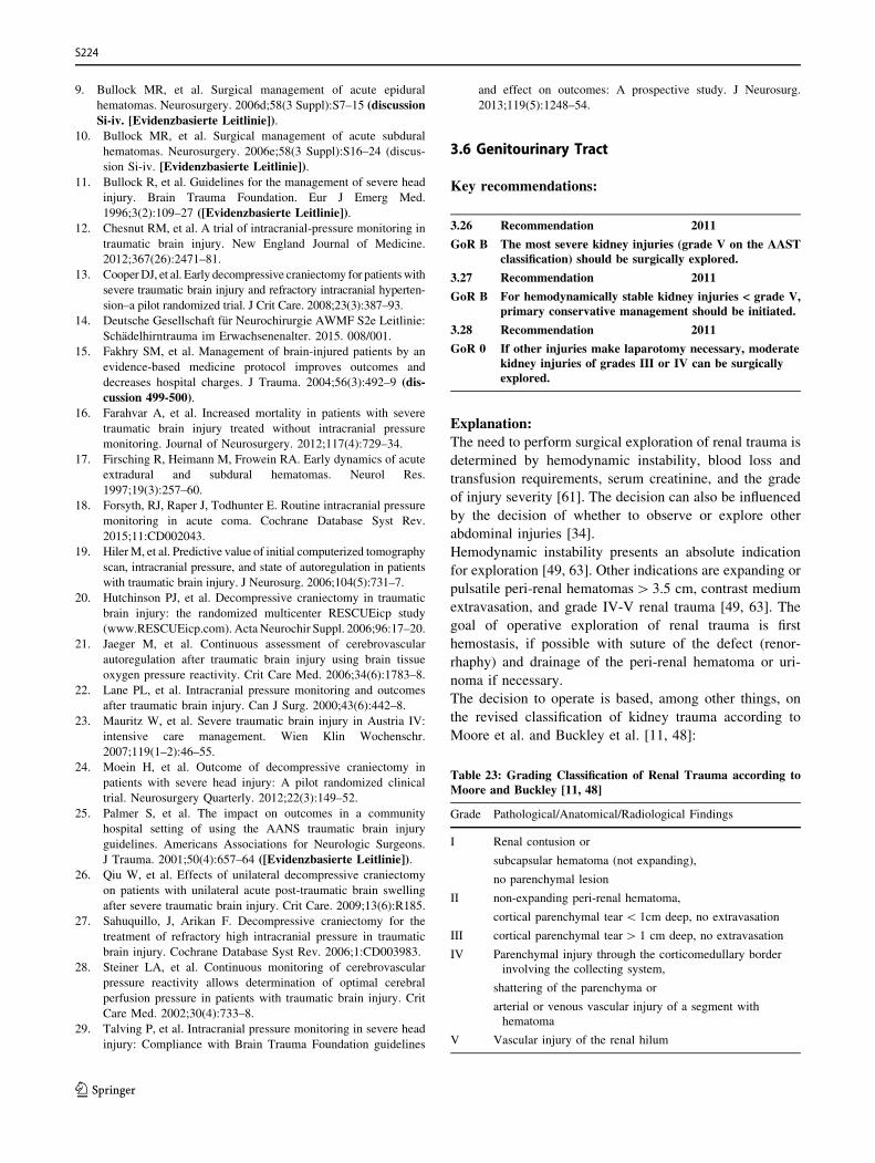
9. Bullock MR, et al. Surgical management of acute epidural
hematomas. Neurosurgery. 2006d;58(3 Suppl):S7–15 (discussion
Si-iv. [Evidenzbasierte Leitlinie]).10. Bullock MR, et al. Surgical management of acute subdural
hematomas. Neurosurgery. 2006e;58(3 Suppl):S16–24 (discus-
sion Si-iv. [Evidenzbasierte Leitlinie]).
11. Bullock R, et al. Guidelines for the management of severe head
injury. Brain Trauma Foundation. Eur J Emerg Med.
1996;3(2):109–27 ([Evidenzbasierte Leitlinie]).12. Chesnut RM, et al. A trial of intracranial-pressure monitoring in
traumatic brain injury. New England Journal of Medicine.
2012;367(26):2471–81.
13. CooperDJ, et al. Early decompressive craniectomy for patientswith
severe traumatic brain injury and refractory intracranial hyperten-
sion–a pilot randomized trial. J Crit Care. 2008;23(3):387–93.
14. Deutsche Gesellschaft fur Neurochirurgie AWMF S2e Leitlinie:
Schadelhirntrauma im Erwachsenenalter. 2015. 008/001.
15. Fakhry SM, et al. Management of brain-injured patients by an
evidence-based medicine protocol improves outcomes and
decreases hospital charges. J Trauma. 2004;56(3):492–9 (dis-
cussion 499-500).16. Farahvar A, et al. Increased mortality in patients with severe
traumatic brain injury treated without intracranial pressure
monitoring. Journal of Neurosurgery. 2012;117(4):729–34.
17. Firsching R, Heimann M, Frowein RA. Early dynamics of acute
extradural and subdural hematomas. Neurol Res.
1997;19(3):257–60.
18. Forsyth, RJ, Raper J, Todhunter E. Routine intracranial pressure
monitoring in acute coma. Cochrane Database Syst Rev.
2015;11:CD002043.
19. HilerM, et al. Predictive value of initial computerized tomography
scan, intracranial pressure, and state of autoregulation in patients
with traumatic brain injury. J Neurosurg. 2006;104(5):731–7.
20. Hutchinson PJ, et al. Decompressive craniectomy in traumatic
brain injury: the randomized multicenter RESCUEicp study
(www.RESCUEicp.com). ActaNeurochir Suppl. 2006;96:17–20.
21. Jaeger M, et al. Continuous assessment of cerebrovascular
autoregulation after traumatic brain injury using brain tissue
oxygen pressure reactivity. Crit Care Med. 2006;34(6):1783–8.
22. Lane PL, et al. Intracranial pressure monitoring and outcomes
after traumatic brain injury. Can J Surg. 2000;43(6):442–8.
23. Mauritz W, et al. Severe traumatic brain injury in Austria IV:
intensive care management. Wien Klin Wochenschr.
2007;119(1–2):46–55.
24. Moein H, et al. Outcome of decompressive craniectomy in
patients with severe head injury: A pilot randomized clinical
trial. Neurosurgery Quarterly. 2012;22(3):149–52.
25. Palmer S, et al. The impact on outcomes in a community
hospital setting of using the AANS traumatic brain injury
guidelines. Americans Associations for Neurologic Surgeons.
J Trauma. 2001;50(4):657–64 ([Evidenzbasierte Leitlinie]).
26. Qiu W, et al. Effects of unilateral decompressive craniectomy
on patients with unilateral acute post-traumatic brain swelling
after severe traumatic brain injury. Crit Care. 2009;13(6):R185.
27. Sahuquillo, J, Arikan F. Decompressive craniectomy for the
treatment of refractory high intracranial pressure in traumatic
brain injury. Cochrane Database Syst Rev. 2006;1:CD003983.
28. Steiner LA, et al. Continuous monitoring of cerebrovascular
pressure reactivity allows determination of optimal cerebral
perfusion pressure in patients with traumatic brain injury. Crit
Care Med. 2002;30(4):733–8.
29. Talving P, et al. Intracranial pressure monitoring in severe head
injury: Compliance with Brain Trauma Foundation guidelines
and effect on outcomes: A prospective study. J Neurosurg.
2013;119(5):1248–54.
3.6 Genitourinary Tract
Key recommendations:
3.26 Recommendation 2011
GoR B The most severe kidney injuries (grade V on the AASTclassification) should be surgically explored.
3.27 Recommendation 2011
GoR B For hemodynamically stable kidney injuries < grade V,primary conservative management should be initiated.
3.28 Recommendation 2011
GoR 0 If other injuries make laparotomy necessary, moderatekidney injuries of grades III or IV can be surgicallyexplored.
Explanation:
The need to perform surgical exploration of renal trauma is
determined by hemodynamic instability, blood loss and
transfusion requirements, serum creatinine, and the grade
of injury severity [61]. The decision can also be influenced
by the decision of whether to observe or explore other
abdominal injuries [34].
Hemodynamic instability presents an absolute indication
for exploration [49, 63]. Other indications are expanding or
pulsatile peri-renal hematomas[3.5 cm, contrast medium
extravasation, and grade IV-V renal trauma [49, 63]. The
goal of operative exploration of renal trauma is first
hemostasis, if possible with suture of the defect (renor-
rhaphy) and drainage of the peri-renal hematoma or uri-
noma if necessary.
The decision to operate is based, among other things, on
the revised classification of kidney trauma according to
Moore et al. and Buckley et al. [11, 48]:
Table 23: Grading Classification of Renal Trauma according toMoore and Buckley [11, 48]
Grade Pathological/Anatomical/Radiological Findings
I Renal contusion or
subcapsular hematoma (not expanding),
no parenchymal lesion
II non-expanding peri-renal hematoma,
cortical parenchymal tear\ 1cm deep, no extravasation
III cortical parenchymal tear[ 1 cm deep, no extravasation
IV Parenchymal injury through the corticomedullary border
involving the collecting system,
shattering of the parenchyma or
arterial or venous vascular injury of a segment with
hematoma
V Vascular injury of the renal hilum
S224
123
In 2011, the American Association for the Surgery of
Trauma amended the classification from 1989 [11]. Stages
I-III remained unchanged [11]. Extensive parenchymal
injury with shattering was moved from Stage V to Stage
IV, and Stage V kept only injuries (laceration, avulsion, or
thrombosis) of the renal hilum [11].
Single-shot pyelography can be performed intraoperatively
to document the function of the contralateral kidney [49].
The usefulness of prior surgical control of the hilar vessels
(with vascular clamp or vessel loop) is debated [3]. If
hemodynamically unstable patients undergo explorative
surgery, this generally leads to nephrectomy [4, 44].
Exploration rate is currently at approximately 10-15 % and
should continue to fall as more centers push conservative
measures for renal trauma [30, 43]. Most studies prefer a
transperitoneal approach for surgical exploration [58, 59].
The renal hilum is then reached through the posterior
parietal peritoneum, which is incised medial to the inferior
mesenteric vein [59].
Temporary clamping of the renal hilum prior to the open-
ing of the renal fascia and subsequent exploration and
reconstruction is safe and effective [13, 42]. Temporary
clamping ensures decreased intraoperative blood loss and
yields lower nephrectomy rates [59].
Key recommendation:
3.29 Recommendation 2011
GoR 0 Selective angioembolization of a renal vascular injurycan be attempted as therapy in hemodynamicallystable patients.
Explanation:
Advances in interventional radiology have significantly
influenced the management of renal trauma over the past
decade. Angiography with selective embolization is now
the most important alternative to surgical exploration,
provided there is no other indication for laparotomy [63].
Until now, the usefulness of angiography was limited by
the few documented case series and case reports and by
restricted use for only secondary or isolated trauma situa-
tions [23, 40]. Most of these had to do with branches of the
renal artery to be embolized. There is no dispute that the
selection of patients, technical equipment, and experience
of individual medical practitioners involved have a deci-
sive influence on the success rate. However, the above-
mentioned case series reported very promising rates of
successful hemostasis in over 82 % of cases, so that it is
currently recommended that hemodynamically stable pa-
tients with third or higher grades of renal trauma undergo
angiography with subsequent embolization [23, 29, 40].
The following findings on computed tomography are con-
sidered indications for angiography [63]:
• pronounced extravasation of contrast medium,
• large devascularized renal segments,
• grades IV-V renal trauma,
• arterial lacerations,
• avulsions,
• global or segmental hypoperfusion of the kidney,
• aneurysms,
• renal vein thrombosis,
• bleeding from a segmental or sub-segmental artery [63].
Peri-renal hematomas compressing the kidney and restrict-
ing perfusion are also indications for angiography [53].
Angiography is also important in the clinical course after
primary laparotomy, for example in cases of persistent
hematuria, with comparable success rates to the initial
intervention [33].
The most common injury of the renal artery is dissection and
subsequent occlusion [49]. Bleeding from the renal artery
often stops spontaneously because of a tamponade effect by
the hematoma within the renal fascia [49]. An injury to the
renal fascia produces a retroperitoneal hematoma. For
expanding hematomas, often the only alternatives are
endovascular embolization or laparotomy [26].
The success rate of embolization for renal trauma is cur-
rently at 65 %, and angiography in cases of blunt trauma
and hemodynamically stable patients at approximately 95
% [10, 15].
Although injuries to the renal hilum often require surgical
intervention, endovascular therapy options have increased
in importance. For example, renal artery embolization can
be performed in cases of severe polytrauma or increased
intraoperative mortality risk; on the one hand, it is a
definitive therapeutic alternative, and on the other hand,
nephrectomy can be performed after a delay during which
the patient’s condition is stabilized [63].
Caution is required in cases of fourth and fifth grade renal
trauma, because success rates are markedly lower, and they
may require more subsequent embolizations [63]. Neverthe-
less, embolization is safe and associated with few side effects
[15]. Patients who do not require immediate surgical explo-
ration can benefit fromangiography and embolizationwithout
the increased risk of nephropathy that occurs with the addi-
tional contrast medium applied for arteriography [60].
Key recommendations:
3.30 Recommendation 2011
GoR 0 According to the type and severity as well asconcomitant injuries, kidney injuries can be surgicallytreated with suturing and partial renal resection ifnecessary, and other salvage measures.
3.31 Recommendation 2011
GoR B Primary nephrectomy should be reserved for grade Vinjuries.
S3-guideline on treatment of patients with severe/multiple injuries S225
123
Explanation:
In hemodynamically stable patients, parenchymal recon-
struction is generally possible [63]. Nephrectomy is mainly
reserved for patients with penetrating injuries, with high
transfusion requirements, hemodynamic instability, and
higher injury severity scores (ISS) [19]. Mortality is usu-
ally associated more with the overall severity of the poly-
trauma than with the renal injury [63].
In the case of high-speed projectile injuries, reconstruction
is difficult and typically nephrectomy is the consequence
[63].
If reconstruction is possible, renorrhaphy is the most
common reconstructive technique [59]. In cases of
parenchymal necrosis, a secondary partial resection may be
necessary [63].
If the renal collecting system has been breached, either
from trauma or from iatrogenic causes, it can be surgically
closed when appropriate [45]. Alternatively, a shunt can be
placed with a single-J or double-J catheter. A capsule
defect can be covered with an omental flap-plasty, peri-
renal fat, or a hemostatic matrix [59]. A retroperitoneal
drain should always be placed to remove any possible urine
leakage [63].
Injuries of the renal hilum are generally associated with
extensive trauma and are accompanied by increased mor-
bidity and mortality rates [63]. It is extremely rare that
reconstruction of fifth grade injuries of the hilum is pos-
sible, but it should be attempted in patients with a single
kidney or in cases of bilateral injuries [63]. In these cases,
generally a primary nephrectomy is sought [63].
Ureter Injuries
Partial ureter injuries can be defined as first and second
grade injuries [47]. After initial diagnosis, minor grade
injuries should be treated with ureteral stents or percuta-
neous nephrostomy catheters [64]. Ureteral stents, placed
antegrade or retrograde, stabilize the leak and prevent
stricture formation. Ureteral stents should remain for three
weeks [64, 67].
Particularly in cases of delayed diagnosis, percutaneous
nephrostomy placement is recommended as primary man-
agement; through these, antegrade stent placement is pos-
sible either at the time of nephrostomy or after a delay of
2-7 days [67].
Retrospective comparative studies have certified that
antegrade treatment has a higher chance of success and is
more easily carried out [64]. This applies particularly in
higher grade injuries (grades II-III) [64]. When the stent is
successfully placed, open operative treatment is necessary
only in cases of persistent paravasation or stricture [64].
If grade II to III injuries are detected during open explo-
ration, primary suture can be performed after stent place-
ment [64]. Not even the smallest lesions created by gunshot
or stab wounds should be treated with primary suture, since
debridement is indispensable for stricture-free reconstruc-
tion [64].
Operative Therapy
The basic principles of operative care for ureteral injuries
are debridement of necrotic tissues, spatulation of the ends
with mucosa-to-mucosa anastomosis using absorbable
suture, ureteral stent placement, and external drainage [39].
In principle, there are differences in the treatment for
higher grade injuries, according to where the leak is located
[39, 49, 59, 63, 64].
In the upper third of the ureter, alternatives are [49, 59,
63]:
• ureteroureterostomy,
• transuretero-ureterostomy,
• pyeloplasty,
• transposition of the renal vein,
• intestinal ureteral replacement,
• ureterocalicostomy, and
• nephrectomy as a last resort.
Defects of themiddle and lower thirds of the ureter can be
treated as follows [6, 59, 63]:
• creation of a Boari bladder flap and ureteral
implantation,
• direct re-implantation of the ureter,
• ureteral implantation with psoas hitch,
• ureteral implantation with Boari bladder flap and psoas
hitch if necessary.
Complete ureteral avulsions require ureteral replacement
if ureteral re-implantation is not possible by the above-
mentioned procedures because alloplastic material is not
available. Here the kidney can be mobilized distally
through transposition of the renal vein or autotransplanted
to the iliac fossa [2, 7, 12].
Ureter replacement procedures can be performed using
the ileum, the colon, or in exceptional cases Meckel’s
diverticulum [1, 22, 55, 62, 63].
Urinary Bladder Injuries
Key recommendations:
3.32 Recommendation 2011
GoR B Intraperitoneal bladder ruptures should be surgicallyexplored.
3.33 Recommendation 2011
GoR 0 Extra-peritoneal bladder rupture without bladder neckinvolvement can be treated conservatively with asuprapubic urinary catheter.
S226
123
Explanation:
In most cases, the often numerous concomitant injuries in
multiply injured patients are treated with more priority than
bladder injuries. According to statistics, extraperitoneal
injuries occur about twice as often as intraperitoneal
bladder ruptures [17, 31]. Much more seldom are combi-
nation injuries with extra and intra-peritoneal ruptures [54].
In principle, management is differentiated according to
blunt and penetrating injuries as well as intra- and extra-
peritoneal bladder ruptures.
Blunt Trauma
Extraperitoneal Bladder Injury
Uncomplicated bladder injuries (grades I-III) can generally
be treated with a transurethral catheter, regardless of
whether there is large perineal or scrotal extravasate [28,
66]. Exceptions are injuries to the bladder neck, bone
fragments in the bladder wall, incarceration of the bladder
wall, or concomitant injury to the rectum (grade V), which
should be treated surgically [28, 66]. In cases of operative
exploration for other injuries, an extraperitoneal bladder
rupture should also be treated operatively, to minimize the
risk of infection [20, 69].
Intraperitoneal Bladder Injury
Intraperitoneal bladder injury (grades III-V) should receive
primary operative treatment due to the risk of peritonitis or
sepsis and the high associated mortality [28, 66, 69].
Penetrating Trauma
In case of penetrating bladder injury, immediate operative
exploration should be performed [16, 66]. The recom-
mended approach is a midline cystostomy, where the
bladder wall can be inspected, and the bladder neck as well
as the distal ureters can be examined for associated injuries
[16, 28].
Large Surface Bladder Injuries
In case of a large bladder wall defect, such as involvement
of the bladder when there is detachment of the lower
abdominal wall or the peritoneum, if necessary a bladder-
plasty, for example with a myocutaneous flap or another
option, can be performed [28, 71].
General Intra and Post-Operative Management
If possible, two-layered mucosa-detrusor closure with
resorbable suture is recommended for surgical treatment
[28, 70].
Post-operative bladder catheterization lowers intravesical
pressure and facilitates tension-free wound adaptation [70].
Depending on the type and scope of injury, a 7-14 day
duration of catheterization is recommended [28, 70].
Prior to removing the bladder catheter, retrograde cystog-
raphy should be carried out whenever possible [28, 70]. If
there is evidence of contrast medium extravasation, the
catheter can be kept for another seven days and re-cys-
tography performed [28, 70].
Urethral Injuries
Key recommendations:
3.34 Recommendation 2011
GoR B Complete rupture of the urethra should be treated in theprimary operative phase with a suprapubic catheter.
3.35 Recommendation 2011
GoR 0 The urinary catheter can be supplemented with aurethral stent.
3.36 Recommendation 2011
GoR B If pelvic fracture or other intraabdominal injuries makesurgery necessary, urethral ruptures should be treatedat the same time.
Explanation:
When treating urethral injuries, it is particularly worth
mentioning that the approach presented here refers
explicitly only to the primary operative phase, because
some other principles apply for further care.
To this point, it has not yet been sufficiently verified
whether primary, delayed, or secondary re-anastomosis is
preferable in cases of complete rupture of the posterior
urethra. Chapple et al. suggest primary and delayed stent-
ing of the urethra [14]. The main problems in the post-
traumatic course are urethral strictures, incontinence, and
impotence, so avoiding these is the goal of therapy.
In a literature review summarizing several case series and
comparative studies on the treatment for urethral ruptures,
Koraitim reported the following rates of stricture, inconti-
nence, and impotence. For suprapubic catheterization alone
they were 97 %, 4 %, and 19 %; for primary stent place-
ment rates were 53 %, 5 %, and 36 %; and for primary
closure they were 49 %, 21 %, and 56 % [18, 24, 25, 27,
35-37, 50]. Correspondingly, in cases of complete urethral
rupture in men, he recommends suprapubic catheterization
or stent placement for larger gaps between the urethral ends
[36]. In a more recent study by Ku et al., both alternatives
were also considered comparable [38]. The EAST guide-
line came to a similar conclusion that both primary stent
placement and suprapubic catheterization with secondary
surgery are equally worthy of recommendation [32].
In cases in which an operation is already required because
of adjacent injuries, it might be advisable to treat the ure-
thral rupture directly in certain situations, to avoid a second
procedure [8]. Especially when there is contamination of
the abdominal cavity from colon injuries, primary closure
of the urethra over a stenting catheter seems appropriate, to
avoid a complicating infection [9]. Even in cases when a
S3-guideline on treatment of patients with severe/multiple injuries S227
123
conservative approach appears possible, urethral injuries
can be treated with primary surgery if definitive
osteosynthesis of the bony pelvis cannot be performed
without it [41].
Rupture of the anterior urethra in males are somewhat less
common than of the posterior urethra [9]. Primary opera-
tive reconstruction can be necessary in cases of open
injuries. In the majority of cases, however, preference
should also be given here to suprapubic catheterization and
delayed reconstruction, since reconstruction of the anterior
urethra and the external male genitalia, which are often
concomitantly affected, is usually difficult and time-con-
suming [14]. However, in cases of penis fracture with
injury to the corpus spongiosum, it is recommended that
the urethral injury also undergo primary surgery [14, 51].
The severity of the urological injury and the severity of the
injuries overall are crucial to the selection of primary
operative versus conservative management [14, 46].
Urethral injuries are significantly less common in female
patients compared to men. When they do occur, however,
they are very pronounced and associated with bladder
injuries [14]. For this reason, primary treatment should
consist of suprapubic catheterization alone, assuming that
the patient is hemodynamically unstable and/or other
injuries require more urgent operative care [68]. On the
other hand, for women with less severe polytrauma, rup-
tures of the proximal urethra can undergo primary recon-
struction through a retropubic approach [5, 52, 65].
These recommendations generally apply to children, with
whom a similar distinction should be made according to
gender. In a series of 35 boys with posterior urethral tears,
Podesta et al. 1997 compared suprapubic catheterization
(with later urethroplasty), suprapubic catheterization with
catheter splinting of the urethra, and primary anastomosis
[57]. Since the continence rate after primary anastomosis
only reached 50 %, and ten patients in the catheter splinting
group eventually required urethroplasty, the authors rec-
ommended suprapubic catheterization with delayed sec-
ondary urethroplasty [57]. In a study of urethral injury in
girls with pelvic fractures and other concomitant injuries,
the same authors found the most advantage for delayed
treatment, since good results were observed despite
accompanying bladder and vaginal injuries [56].
S228
123
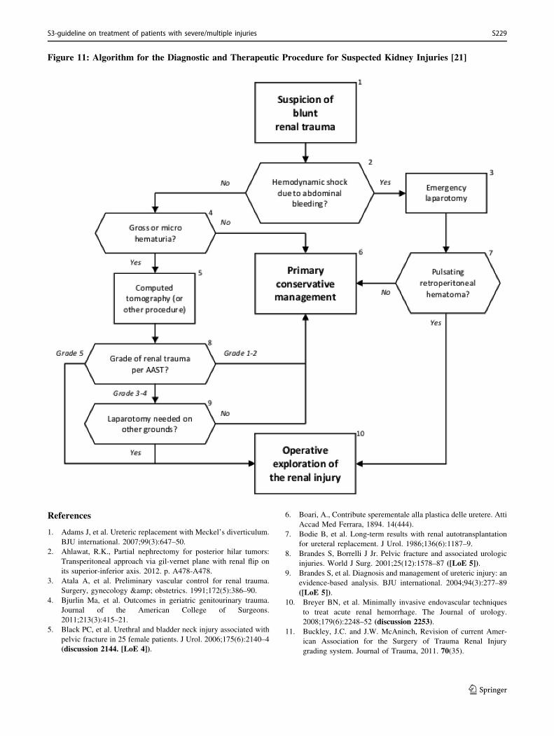
Figure 11: Algorithm for the Diagnostic and Therapeutic Procedure for Suspected Kidney Injuries [21]
References
1. Adams J, et al. Ureteric replacement with Meckel’s diverticulum.
BJU international. 2007;99(3):647–50.
2. Ahlawat, R.K., Partial nephrectomy for posterior hilar tumors:
Transperitoneal approach via gil-vernet plane with renal flip on
its superior-inferior axis. 2012. p. A478-A478.
3. Atala A, et al. Preliminary vascular control for renal trauma.
Surgery, gynecology & obstetrics. 1991;172(5):386–90.
4. Bjurlin Ma, et al. Outcomes in geriatric genitourinary trauma.
Journal of the American College of Surgeons.
2011;213(3):415–21.
5. Black PC, et al. Urethral and bladder neck injury associated with
pelvic fracture in 25 female patients. J Urol. 2006;175(6):2140–4
(discussion 2144. [LoE 4]).
6. Boari, A., Contribute sperementale alla plastica delle uretere. Atti
Accad Med Ferrara, 1894. 14(444).
7. Bodie B, et al. Long-term results with renal autotransplantation
for ureteral replacement. J Urol. 1986;136(6):1187–9.
8. Brandes S, Borrelli J Jr. Pelvic fracture and associated urologic
injuries. World J Surg. 2001;25(12):1578–87 ([LoE 5]).
9. Brandes S, et al. Diagnosis and management of ureteric injury: an
evidence-based analysis. BJU international. 2004;94(3):277–89
([LoE 5]).10. Breyer BN, et al. Minimally invasive endovascular techniques
to treat acute renal hemorrhage. The Journal of urology.
2008;179(6):2248–52 (discussion 2253).
11. Buckley, J.C. and J.W. McAninch, Revision of current Amer-
ican Association for the Surgery of Trauma Renal Injury
grading system. Journal of Trauma, 2011. 70(35).
S3-guideline on treatment of patients with severe/multiple injuries S229
123

12. Burks FN, Santucci RA. Management of iatrogenic ureteral
injury. Therapeutic Advances in Urology. 2014;6(3):115–24.
13. Carroll PR, Klosterman PW, McAninch JW. Early vascular
control for renal trauma: a critical review. J Urol.
1989;4(141):826–9.
14. Chapple C, et al. Consensus statement on urethral trauma. BJU
International. 2004;93(9):1195–202 ([LoE 5]).15. Chow SJ, et al. A 10-year review of blunt renal artery injuries at
an urban level I trauma centre. Injury. 2009;40(8):844–50.
16. Cinman NM, et al. Gunshot wounds to the lower urinary tract: a
single-institution experience. The journal of trauma and acute
care surgery. 2013;74(3):725–30 (discussion 730-1.).
17. Corriere JN Jr, Sandler CM. Management of extraperitoneal
bladder rupture. Urol Clin North Am. 1989;16(2):275–7 ([LoE
4]).18. Culty T, Boccon-Gibod L. Anastomotic urethroplasty for
posttraumatic urethral stricture: previous urethral manipulation
has a negative impact on the final outcome. J Urol.
2007;177(4):1374–7 ([LoE 4]).19. Davis KA, et al. Predictors of the need for nephrectomy after
renal trauma. The Journal of trauma. 2006;60(1):164–9 (dis-cussion 169-70).
20. Deibert, CM, Spencer Ba. The association between operative
repair of bladder injury and improved survival: results from the
National Trauma Data Bank. The Journal of urology.
2011;186(1):151–5.
21. DGUC, S3 Polytrauma/Schwerverletzten Behandlung.
2011(012).
22. Djakovic N, et al. Intestinal reconstruction of the lower urinary
tract as a prerequisite for renal transplantation. BJU Interna-
tional. 2009;103(11):1555–60.
23. Dunfee BL, Lucey BC, Soto JA. Development of renal scars on
CT after abdominal trauma: does grade of injury matter? AJR.
American journal of roentgenology. 2008;190(5):1174–9.
24. Elliott DS, Barrett DM. Long-term Followup and Evaluation of
Primary Realignment of Posterior Urethral Disruptions. The
Journal of Urology. 1997;157(3):814–6 ([LoE 2b]).
25. Follis HW, Koch MO, McDougal WS. Immediate management
of prostatomembranous urethral disruptions. The Journal of
urology. 1992;147(5):1259–62 ([LoE 2b]).26. Fu CY, et al. Angioembolization provides benefits in patients
with concomitant unstable pelvic fracture and unstable hemody-
namics. American Journal of Emergency Medicine.
2012;30(1):207–13.
27. Gheiler EL, Frontera JR. Immediate primary realignment of
prostatomembranous urethral disruptions using endourologic
techniques. Urology. 1997;49(4):596–9 ([LoE 4]).
28. Gomez RG, et al. SIU/ICUD Consultation on Urethral Stric-
tures: Pelvic fracture urethral injuries. Urology. 2014;83(3
Suppl):S48–58.
29. Hagiwara A, et al. The role of interventional radiology in the
management of blunt renal injury: a practical protocol.
J Trauma. 2001;51(3):526–31.
30. Hammer CC, Santucci RA. Effect of an institutional policy of
nonoperative treatment of grades I to IV renal injuries. J Urol.
2003;169(5):1751–3 ([LoE 4]).31. Hochberg E, Stone NN. Bladder rupture associated with pelvic
fracture due to blunt trauma. Urology. 1993;41(6):531–3 ([LoE2]).
32. Hoff WS, et al. Practice management guidelines for the
evaluation of blunt abdominal trauma: the East practice
management guidelines work group. The Journal of trauma.
2002;53(3):602–15.
33. Huber J, et al. Selective transarterial embolization for posttrau-
matic renal hemorrhage: a second try is worthwhile. The Journal
of Urology. 2011;185(5):1751–5.
34. Husmann DA, et al. Major renal lacerations with a devitalized
fragment following blunt abdominal trauma: a comparison
between nonoperative (expectant) versus surgical management.
J Urol. 1993;150(6):1774–7 ([LoE 2b]).35. Husmann DA, et al. Prostatomembranous urethral disruptions:
management by suprapubic cystostomy and delayed urethro-
plasty. J Urol. 1990;144(1):76–8 ([LoE 2b]).
36. Koraitim MM. Pelvic fracture urethral injuries: the unresolved
controversy. J Urol. 1999;161(5):1433–41 ([LoE 2b]).
37. Kotkin L, Koch MO. Impotence and incontinence after imme-
diate realignment of posterior urethral trauma: result of injury or
management? J Urol. 1996;155(5):1600–3 ([LoE 2b]).38. Ku JH, et al. Comparison of long-term results according to the
primary mode of management and type of injury for posterior
urethral injuries. Urol Int. 2002;69(3):227–32 ([LoE 2b]).
39. Lynch TH, et al. EAU guidelines on urological trauma. Eur
Urol. 2005;47(1):1–15 ([Evidenzbasierte Leitlinie]).
40. Matthews LA, Smith EM, Spirnak JP. Nonoperative treatment
of major blunt renal lacerations with urinary extravasation. The
Journal of urology. 1997;157(6):2056–8 ([LoE 4]).41. Mayher BE, Guyton JL, Gingrich JR. Impact of urethral injury
management on the treatment and outcome of concurrent pelvic
fractures. Urology. 2001;57(3):439–42 ([LoE 4]).
42. McAninch JW, Carroll PR. Renal trauma: kidney preservation
through improved vascular control-a refined approach. The
Journal of trauma. 1982;22(4):285–90.
43. McAninch JW, et al. Renal reconstruction after injury. The
Journal of urology. 1991;145(5):932–7.
44. McGuire J, et al. Predictors of outcome for blunt high grade
renal injury treated with conservative intent. The Journal of
urology. 2011;185(1):187–91.
45. Meng MV, Brandes SB, McAninch JW. Renal trauma: indica-
tions and techniques for surgical exploration. World journal of
urology. 1999;17(2):71–7.
46. Mohr AM, et al. Management of trauma to the male external
genitalia: the usefulness of American Association for the
Surgery of Trauma organ injury scales. J Urol. 2003;170(6 Pt
1):2311–5 ([LoE 4]).
47. Moore EE, et al. Organ injury scaling. III: Chest wall,
abdominal vascular, ureter, bladder, and urethra. Journal of
Trauma. 1992;33(3):337–9.
48. Moore EE, et al. Organ injury scaling: spleen, liver, and kidney.
J Trauma. 1989;29(12):1664–6.
49. Morey, A.F., et al., American Urological Association (AUA)
Guideline American Urological Association. AUA Clinical
Guidelines, 2014(April): p. 1-57.
50. Mouraviev VB, Coburn M, Santucci RA. The treatment of
posterior urethral disruption associated with pelvic fractures:
comparative experience of early realignment versus delayed
urethroplasty. J Urol. 2005;173(3):873–6 ([LoE 4]).
51. Nane I, et al. Penile fracture: emergency surgery for preserva-
tion of penile functions. Andrologia. 1991;23(4):309–11 ([LoE
4]).52. Netto Junior NR, Ikari O, Zuppo VP. Traumatic rupture of
female urethra. Urology. 1983;22(6):601–3 ([LoE 4]).53. Nuss GR, et al. Radiographic predictors of need for angio-
graphic embolization after traumatic renal injury. The Journal of
trauma. 2009;67(3):578–82 (discussion 582).
54. Palmer JK, Benson GS, Corriere JN. Diagnosis and initial
management of urological injuries associated with 200
S230
123
consecutive pelvic fractures. The Journal of urology.
1983;130(4):712–4.
55. Pfitzenmaier, J., et al., [Kidney trauma]. Urologe A, 2008.
47(6): p. 759-67; quiz 768.
56. Podesta ML, Jordan GH. Pelvic fracture urethral injuries in
girls. J Urol. 2001;165(5):1660–5.
57. Podesta ML, et al. Immediate management of posterior urethral
disruptions due to pelvic fracture: therapeutic alternatives.
J Urol. 1997;157(4):1444–8 ([LoE 2b]).58. Robert M, et al. Management of major blunt renal lacerations:
surgical or nonoperative approach? Eur Urol. 1996;30(3):335–9
([LoE 4]).
59. Santucci, R.A. and L. Doumanian, Upper Urinary Tract Trauma,
in Campbell-walsh urology, A. Wein, Editor. 2012, Elsevier:
Philadelphia. p. 1169 pp.-1169 pp.
60. Sarani B, et al. Contemporary comparison of surgical and
interventional arteriography management of blunt renal injury.
Journal of vascular and interventional radiology : JVIR.
2011;22(5):723–8.
61. Shariat SF, et al. Evidence-based validation of the predictive
value of the American Association for the Surgery of Trauma
kidney injury scale. J Trauma. 2007;62(4):933–9.
62. Steffens Ja, et al. [Ureteric reconstruction with reconfigured
ileal segments according to Yang-Monti. A 4-year prospective
report]. Der Urologe. Ausg. A. 2010;49(2):262–7.
63. Summerton DJ, et al. EAU guidelines on iatrogenic trauma. Eur
Urol. 2012;62(4):628–39.
64. Teber D, et al. Harnleitertrauma. Der Urologe.
2005;44(8):870–7.
65. Thambi Dorai CR, Boucaut HA, Dewan PA. Urethral injuries in
girls with pelvic trauma. Eur Urol. 1993;24(3):371–4 ([LoE 4]).
66. Tonkin JB, Tisdale BE, Jordan GH. Assessment and initial
management of urologic trauma. The Medical clinics of North
America. 2011;95(1):245–51.
67. Toporoff B, et al. Percutaneous antegrade ureteral stenting as an
adjunct for treatment of complicated ureteral injuries. J Trauma.
1992;4(32):534–8.
68. Venn SN, Greenwell TJ, Mundy AR. Pelvic fracture injuries of
the female urethra. BJU Int. 1999;83(6):626–30 ([LoE 4]).
69. Wirth GJ, et al. Advances in the management of blunt traumatic
bladder rupture: experience with 36 cases. BJU international.
2010;106(9):1344–9.
70. Wohlrab KJ, Sung VW, Rardin CR. Management of laparoscopic
bladder injuries. J Minim Invasive Gynecol. 2011;1(18):4–8.
71. Wu C-W, Lin C-H, Lin C-H. One-stage posttraumatic bladder
reconstruction and soft-tissue coverage of the lower abdomen or
perineum. Annals of plastic surgery. 2010;64(1):65–8.
3.7 Spine
Indications for Surgery
Key recommendation:
3.37 Recommendation 2011
GoR B Unstable spinal injuries with verified or assumedneurological deficits, with deformities, in whom thereduction, decompression and stabilization procedureappears to have halted or improved neurologicalsymptoms, should be treated surgically as soon aspossible (‘‘day 1 surgery’’).
Explanation:
Regarding the order of treatment priority, spine injuries
come in third place after life-threatening injuries to the
body cavities and the head as well as the large long bones,
or in second place in the presence of spinal cord injury
[36].
The indications for surgery include atlanto-occipital dis-
location, translational atlanto-axial dislocation, unsta-
ble Jefferson fracture, unstable dens fracture (especially
type II), Hangman’s fracture (pars interarticularis fracture
C2 and disc injury C2/C3), C3-C7 fractures (A3, B, and C
types) also in terms of dislocation, and T1 to L5 fractures
(A3, B, and C types) also in terms of dislocation.
According to prevailing opinion, there is also an absolute
indication for surgery when there is an open spine injury
[19, 51].
Also useful in establishing indications for primary man-
agement of spine injuries in polytrauma is to classify
according to a) complex spine injuries with injury to
essential neural pathways and organs such as spinal cord,
lungs, large vessels and abdominal organs, b) unsta-
ble spine injuries (types A3, B, and C), in whom functional
management can lead to severe deformity and neurologic
damage, and c) stable spine injuries according to Blauth
et al. 1998. If complex or unstable spine injury is present,
operative stabilization should be sought as soon as possi-
ble–on the day of the injury if none of the contraindications
listed below is present [8].
According to Blauth et al. 1998, a complex spinal injury is
a multi-level spinal injury or one accompanied by con-
comitant intra-thoracic or intra-abdominal injury or by
polytrauma. That polytrauma ‘‘creates’’ a complex injury is
substantiated by, among others, the studies of Hebert and
Burnham [26], who determined that hospital stays are
longer and the number of surgeries increased in these
patients, and that the combination of spine injury with
multiple trauma is associated with higher morbidity and
mortality as well as increased degree of disability. Never-
theless, according to a North American survey by Tator
et al. 1999, one third of spine injuries with neurologic
damage were still treated conservatively and only 60 % of
operations were performed before the 5th day, with 40 %
afterwards [60].
The goal of primary operative management in unsta-
ble spinal injuries with confirmed or assumed neurologic
deficits or with deformity is first, early spinal decompres-
sion while avoiding secondary neurologic damage and
second, to achieve stability for positioning maneuvers
during intensive care [36].
The surgical indication to avoid neurologic damage is
relatively clear in unstable fractures without a spinal cord
lesion. If there are unstable spinal injuries that could be
S3-guideline on treatment of patients with severe/multiple injuries S231
123
displaced during positioning maneuvers such as during
treatment for chest trauma, there is an indication for pri-
mary operative stabilization [8, 13, 29, 67]. If there is a
contraindication, the goal should be stabilization and
decompression at the earliest possible time. This was
confirmed by Fehlings et al. [23]. Here, the recommended
goal is decompression within the first 24 hours. Our data
shows significant improvements in neurological symptoms
according to the ASIA protocol when decompression and
stabilization are performed within the first six hours. Ani-
mal studies have found benefits regarding neurological
symptoms for the earliest possible spine stabilization [18,
20].
La Rosa et al. [39] and Fehlings et al. [23] found benefits
for early operative decompression versus later decom-
pression or conservative therapy. In the early group (17
hours) there was an improvement in neurological symp-
toms in 42 % of the patients with complete deficits, and 90
% of patients with incomplete deficits. Improvement rates
were 8 % and 59 % in the delayed stabilization group, and
25 % and 59 % in the conservative management group.
The only randomized controlled study on this topic inclu-
ded 62 patients with isolated cervical spine injury [59].
Although the authors found no difference in neurological
results for earlier (\ 72 hours) versus later ([ 5 days)
stabilization, they recommended earlier stabilization. Levi
et al. also reported indifferent results regarding earlier (\24 hours) and late ([ 24 hours) stabilization of cervical
spine injuries, but also ultimately recommended the earlier
procedure [62]. Wagner and Chehrazi also found no cor-
relation between time of surgery and neurological symp-
toms, and concluded that primary medullary damage
determines prognosis [65]. However, other studies related
to primary and secondary damage show that secondary
damage is not negligible and that patients benefit from
avoiding it [3, 70]. McKinley et al. draw a similar con-
clusion [44]. However, Papadopoulos et al. found better
neurological results after earlier surgery [47]. In 1999,
Mirza et al. also reported that early stabilization (\ 72
hours) had more favorable neurological outcomes than
later stabilization ([ 72 hours) [46]. All of these are data
from studies not exclusively investigating polytrauma
patients.
In addition to the studies focusing primarily on neurolog-
ical outcomes, there are a number of studies focusing on
non-neurological effects of earlier stabilization. A 2001
study by Croce et al. found evidence that early stabilization
(\3 days) of thoracic spine injuries in polytrauma (average
ISS 24) decreased intensive care duration, pneumonia rate,
costs, and time of mechanical ventilation compared to later
stabilization ([3 days) [13]. Johnson et al. [29] also found
a benefit for primary stabilization of unstable spine frac-
tures, with a decreased rate of ARDS in multiply injured
patients. Similarly, Dai et al. found reduced pulmonary
complications after early treatment [15]. According to the
results of Aebi et al. [1], immediate operative treatment of
cervical spine injuries is more important for neurological
outcomes than improved operative techniques. In a 2005
study, Kerwin et al. [30] found that primary stabilization of
the spine in severely injured patients (ISS[25) shortened
hospital stays from 29 to 20 days.
The indications listed above are based on an assumption
that the injury has undergone adequate assessment and
diagnosis in the emergency department. The patient should
be hemodynamically stable, and surgical causes of bleed-
ing should be excluded. Additional vital parameters such as
intracranial pressure, core body temperature, and coagula-
tion should be in the normal range. If there is reasonable
risk that primary reduction, decompression, and stabiliza-
tion of the spine would significantly worsen the patient’s
condition, a spinal stabilization procedure is relatively
contraindicated.
If their condition is stable according to intracranial pres-
sure, respiratory, and hemodynamic parameters, multiply-
injured patients benefit particularly from early management
of spine injuries; there is stability for positioning maneu-
vers, and secondary ‘‘hits’’ from subsequent procedures can
be avoided. In addition, the antigenic load created by an
unstable fracture in close proximity to the trunk can be
reduced. On the other hand, critical conditions such as
hypothermia, mass transfusion, coagulopathy, pulmonary
insufficiency, and higher catecholamine dependency are
relative contraindications for immediate spinal
stabilization.
In this context, McLain and Benson 1999 [45] found that
immediate (\24 hours after trauma) stabilization achieved
the same outcomes as early (24-72 hours after trauma)
stabilization of an unstable spine fracture in patients with
multiple injuries, neurological symptoms, and concomitant
thoracoabdominal injuries. Nevertheless, the authors rec-
ommend stabilization at the earliest possible opportunity.
Schlegel et al. [57] and Chipman et al. [12] reported that
operative stabilization of unstable spine fractures within 72
hours was associated with decreased morbidity (fewer
pulmonary complications, fewer urinary tract infections,
shorter hospital and intensive care stays) in polytrauma
patients. When an abdominal injury is present, which leads
to laparotomy in 38 % of patients with spine fracture [6],
the decision of whether an unstable spine fracture must or
can be stabilized during the same operative session must be
made after the abdomen has been treated.
In contrast, for cases of hemothorax, the presence of blood
in the thorax itself supports early stabilization of an
existing thoracic spine injury [16]. The results of Petitjean
et al. [48] also support early stabilization of thoracic spine
fractures for concomitant chest trauma with lung contusion
S232
123
among other things. If there is primary paraplegia or
unreducible dislocation, the operation can be postponed
until organ functions are stabilized with intensive care.
In conclusion, benefits for early surgery in polytrauma
patients have been reported, particularly in recent publi-
cations from 2006 to 2010 [2, 4, 5, 11, 21-24, 31, 32, 43,
52, 54-56]. Early fracture stabilization and decompression
enable favorable neurological shift, decrease general
complications, and shorten hospital stays. Because general
complications, particularly lung-related, occur frequently
in polytrauma patients, the result is a recommendation for
surgery to be performed as early as possible.
Key recommendations:
3.38 Recommendation 2011
GoR B Unstable thoracolumbar spine injuries withoutneurological symptoms should be treated operatively.
3.39 Recommendation 2011
GoR B Surgery should be performed on the day of the accidentor later in the course of treatment.
Explanation:
Apart from B and C type injuries, this recommendation
applies particularly to A2 and A3 type fractures of the
thoracolumbar spine, which are not displaced by posi-
tioning maneuvers during intensive care. In such cases,
there no reason for urgent stabilization on the day of
trauma. However, according to Jacobs et al. [28], patients
treated operatively for unstable thoracolumbar fractures
have better outcomes regarding reduction, neurological
symptoms, mobilization, rehabilitation times, and compli-
cation incidence than patients treated conservatively [69].
Key recommendation:
3.40 Recommendation 2011
GoR B Stable spine injuries without neurologic symptomsshould be treated conservatively.
Explanation:
Fractures of type A1, and sometimes A2, are considered
stable, and do not benefit from operative stabilization [50,
68], particularly when the adjacent intervertebral discs
remain intact. Surgical stabilization for such cases is not
recommended particularly in polytrauma [34].
Operative Technique
Key recommendation:
3.41 Recommendation 2011
GoR 0 For injuries of the cervical spine, the followingtechniques can be used as primary operative method: 1.Halo fixation, 2. ventral stabilization procedure.
Explanation:
Halo fixation is indicated when there is a contraindication
to necessary definitive internal fixation, and a soft cervical
collar is not sufficient as a temporary stabilizing measure
[10, 14, 25, 27, 33, 34, 37, 42, 50, 53, 64].
Ventral spondylodesis is indicated particularly in cases of
C3-C7 fracture dislocations. As a rule, corpectomy is the
procedure of choice, with removal of the disc, replacement
with iliac crest bone graft or a cage if necessary, and sta-
bilization with a plate, possibly with a fixed-angle [14].
Because of the bridging effect during fixation, the use of
chips and locking plates has not yielded better healing [53].
In polytrauma patients, ventral management of unsta-
ble cervical spine fractures is preferable to dorsal stabi-
lization procedures, particularly on the day of trauma [37].
According to Brodke et al. [10], there are no significant
differences regarding bony consolidation, reduction suc-
cess, neurological symptoms, or long-term complaints in
ventral versus dorsal approaches to the C-spine; however,
the latter is much more complex and time-consuming,
which is why it should not be recommended in patients
with multiple injuries. If there is an unstable dens fracture,
ventral screw placement is generally indicated. If the
patient is hemodynamically stable, a Magerl screw is
preferable due to the lower rate of screw displacement. In
cases of a Jefferson fracture, dorsal screws or occipito-
cervical fusion might be indicated. However, there is no
good indication to perform the latter as day-one surgery
and should be planned as an elective procedure at a later
time.
Key recommendation:
3.42 Recommendation 2011
GoR B A dorsal internal fixator should be used as primaryoperative method for injuries to the thoracolumbarspine.
Explanation:
Only dorsally-placed internal fixation can be recommended
as primary treatment of thoracolumbar spine fractures [35,
49, 58]. This procedure achieves good reduction, decom-
pression, and stabilization, sufficient for all positioning
maneuvers for intensive care. According to Kossmann
S3-guideline on treatment of patients with severe/multiple injuries S233
123
et al., these measures are considered the damage control
strategy for the spine in polytrauma [38]. When necessary,
ventral fusion can be performed electively during the sec-
ondary operative phase. In addition, according to Been and
Bouma, dorsal stabilization alone should be sufficient for
burst fractures of the thoracic/lumbar spine [7]. Both
logistical and technical effort as well as OR time must be
considered for the various spine surgery methods.
Laminectomy increases instability [1, 17, 41, 61, 71] and at
best can serve as access to slide posterior edge fragments
forward during dorsal decompression. It is debated whether
the removal of bony fragments from the spinal canal
(‘‘spinal clearance’’) is really clinically beneficial [9, 40,
66]. In this respect, the indication for laminectomy should
be very narrow and considered only if there are neuro-
logical symptoms and compression caused by bone and
disc fragments that cannot be removed from a ventral
approach.
Regarding blood loss and post-operative pain, percuta-
neous insertion of the internal fixator for dorsal stabiliza-
tion of the thoracic and lumbar vertebrae would be
preferable to an open procedure. A study by Vanek et al.
showed significant benefits for this. Screw malposition was
not more frequently observed [63]. In cases of neurological
deficits, however, an open approach is advisable, because
laminectomy for decompression of the spinal cord must be
performed.
References
1. Aebi M, et al. Indication, surgical technique, and results of 100
surgically-treated fractures and fracture-dislocations of the cer-
vical spine. Clin Orthop Relat Res. 1986;203:244–57 ([LoE 4]).
2. Albert TJ, Kim DH. Timing of surgical stabilization after cervical
and thoracic trauma. Invited submission from the Joint Sec-
tion Meeting on Disorders of the Spine and Peripheral Nerves,
March 2004. J Neurosurg Spine. 2005;3(3):182–90.
3. Asamoto S, et al. Hyperbaric oxygen (HBO) therapy for acute
traumatic cervical spinal cord injury. Spinal Cord.
2000;38(9):538–40.
4. Bagnall, A.M., et al., Spinal fixation surgery for acute traumatic
spinal cord injury. Cochrane Database Syst Rev, 2008(1): p.
CD004725.
5. Ball JR, Sekhon LH. Timing of decompression and fixation after
spinal cord injury–when is surgery optimal? Crit Care Resusc.
2006;8(1):56–63.
6. Beaunoyer M, et al. Abdominal injuries associated with thoraco-
lumbar fractures after motor vehicle collision. J Pediatr Surg.
2001;36(5):760–2 ([LoE 4]).
7. Been HD, Bouma GJ. Comparison of two types of surgery for
thoraco-lumbar burst fractures: combined anterior and posterior
stabilisation vs. posterior instrumentation only. Acta Neurochir
(Wien). 1999;141(4):349–57 ([LoE 2b]).
8. Blauth M, et al. Complex injuries of the spine. Orthopade.
1998;27(1):17–31 ([LoE 5]).
9. Boerger TO, Limb D, Dickson RA. Does ‘canal clearance’ affect
neurological outcome after thoracolumbar burst fractures? J Bone
Joint Surg Br. 2000;82(5):629–35 ([LoE 2a]).
10. Brodke DS, et al. Comparison of anterior and posterior
approaches in cervical spinal cord injuries. J Spinal Disord
Tech. 2003;16(3):229–35 ([LoE 1b]).
11. Cengiz SL, et al. Timing of thoracolomber spine stabilization in
trauma patients; impact on neurological outcome and clinical
course. A real prospective (rct) randomized controlled study.
Arch Orthop Trauma Surg. 2008;128(9):959–66.
12. Chipman JG, Deuser WE, Beilman GJ. Early surgery for
thoracolumbar spine injuries decreases complications.
J Trauma. 2004;56(1):52–7 ([LoE 2b]).
13. Croce MA, et al. Does optimal timing for spine fracture fixation
exist? Ann Surg. 2001;233(6):851–8 ([LoE 5]).
14. Daentzer D, Boker DK. Operative stabilization of traumatic
instabilities of the lower cervical spine. Experience with an
angle instable anterior plate-screw system in 95 patients.
Unfallchirurg. 2004;107(3):175–80 ([LoE 4]).
15. Dai LY, et al. Thoracolumbar fractures in patients with multiple
injuries: diagnosis and treatment-a review of 147 cases.
J Trauma. 2004;56(2):348–55 ([LoE 2b]).16. Dalvie SS, Burwell M, Noordeen MH. Haemothorax and
thoracic spinal fracture. A case for early stabilization. Injury.
2000;31(4):269–70 ([LoE 4]).
17. Degreif J, et al. Rotational stability of the thoracolumbar spine
after interlaminar ultrasound window, hemilaminectomy and
laminectomy. A comparative experimental study. Unfallchirurg.
1994;97(5):250–5 ([LoE 5]).
18. Delamarter RB, Sherman JE, Carr JB. 1991 Volvo Award in
experimental studies. Cauda equina syndrome: neurologic
recovery following immediate, early, or late decompression.
Spine. 1991;16(9):1022–9 ([LoE 5]).19. Deutsche Gesellschaft fur Unfallchirurgie, Verletzungen der
Halswirbelsaule, in Leitlinien Unfallchirurgie (2 Auflage), K.
Sturmer, Deutsche Gesellschaft fur Unfallchirurgie,, Editor.
1999, Thieme: Stuttgart. p. 34-54. [LoE 5]
20. Dolan EJ, Tator CH, Endrenyi L. The value of decompression
for acute experimental spinal cord compression injury. J Neuro-
surg. 1980;53(6):749–55 ([LoE 5]).
21. Fehlings MG, Perrin RG. The role and timing of early
decompression for cervical spinal cord injury: update with a
review of recent clinical evidence. Injury. 2005;36(Suppl
2):B13–26.
22. Fehlings MG, Perrin RG. The timing of surgical intervention in
the treatment of spinal cord injury: a systematic review of recent
clinical evidence. Spine. 2006;31(11 Suppl):S28–35 (discussionS36).
23. Fehlings MG, et al. Current practice in the timing of surgical
intervention in spinal cord injury. Spine (Phila Pa 1976).
2010;35(21 Suppl):S166–73.
24. Frangen TM, et al. Respiratory failure in thoracic spine injuries.
Does the timing of dorsal stabilization have any effect on the
clinical course in multiply injured patients? Orthopade.
2007;36(4):365–71.
25. Heary RF, et al. Acute stabilization of the cervical spine by
halo/vest application facilitates evaluation and treatment of
multiple trauma patients. J Trauma. 1992;33(3):445–51 ([LoE
4]).26. Hebert JS, Burnham RS. The effect of polytrauma in persons
with traumatic spine injury. A prospective database of spine
fractures. Spine. 2000;25(1):55–60.
27. Hertz H, Scharf W. Immobilization of the injured cervical spine
by the Halo-Fixateur-externe for transports by aircraft (author’s
transl). Wien Med Wochenschr. 1982;132(1):11–3 ([LoE 4]).
S234
123

28. Jacobs RR, Asher MA, Snider RK. Thoracolumbar spinal
injuries. A comparative study of recumbent and operative
treatment in 100 patients. Spine. 1980;5(5):463–77 ([LoE 2b]).29. Johnson KD, Cadambi A, Seibert GB. Incidence of adult
respiratory distress syndrome in patients with multiple muscu-
loskeletal injuries: effect of early operative stabilization of
fractures. J Trauma. 1985;25(5):375–84 ([LoE 5]).30. Kerwin AJ, et al. The effect of early spine fixation on non-
neurologic outcome. J Trauma. 2005;58(1):15–21 ([LoE 2b]).31. Kerwin, A.J., et al., Best practice determination of timing of
spinal fracture fixation as defined by analysis of the National
Trauma Data Bank. J Trauma, 2008. 65(4): p. 824-30;
discussion 830-1.32. Kishan S, Vives MJ, Reiter MF. Timing of surgery following
spinal cord injury. J Spinal Cord Med. 2005;28(1):11–9.
33. Kleinfeld F. Treatment of spine-injuries using halo-fixateur
(author’s transl). Unfallheilkunde. 1981;84(4):161–7 ([LoE 4]).34. Knight RQ, et al. Comparison of operative versus nonoperative
treatment of lumbar burst fractures. Clin Orthop Relat Res.
1993;293:112–21 ([LoE 2b]).
35. Knop C, et al. Surgical treatment of injuries of the thoracolum-
bar transition–3: Follow-up examination. Results of a prospec-
tive multi-center study by the ‘‘Spinal’’ Study Group of the
German Society of Trauma Surgery. Unfallchirurg.
2001;104(7):583–600 ([LoE 4]).36. Kohler A, et al. Patient management in polytrauma with injuries
of the cervical spine. Helv Chir Acta. 1994;60(4):547–50 ([LoE5]).
37. Koivikko MP, et al. Conservative and operative treatment in
cervical burst fractures. Arch Orthop Trauma Surg.
2000;120(7–8):448–51 ([LoE 4]).
38. Kossmann T, et al. Damage control surgery for spine trauma.
Injury. 2004;35(7):661–70 ([LoE 5]).
39. La Rosa G, et al. Does early decompression improve neurolog-
ical outcome of spinal cord injured patients? Appraisal of the
literature using a meta-analytical approach. Spinal Cord.
2004;42(9):503–12 ([LoE 2a]).
40. Limb D, Shaw DL, Dickson RA. Neurological injury in
thoracolumbar burst fractures. J Bone Joint Surg Br.
1995;77(5):774–7 ([LoE 4]).41. Lu WW, et al. Stability of the whole lumbar spine after
multilevel fenestration and discectomy. Spine.
1999;24(13):1277–82 ([LoE 5]).
42. Marshall LF, et al. Deterioration following spinal cord injury. A
multicenter study. J Neurosurg. 1987;66(3):400–4.
43. McHenry TP, et al. Risk factors for respiratory failure following
operative stabilization of thoracic and lumbar spine fractures.
J Bone Joint Surg Am. 2006;88(5):997–1005.
44. McKinley W, et al. Outcomes of early surgical management
versus late or no surgical intervention after acute spinal cord
injury. Arch Phys Med Rehabil. 2004;85(11):1818–25 ([LoE
2b]).45. McLain RF, Benson DR. Urgent surgical stabilization of spinal
fractures in polytrauma patients. Spine. 1999;24(16):1646–54
([LoE 2b]).
46. Mirza SK, et al. Early versus delayed surgery for acute cervical
spinal cord injury. Clin Orthop Relat Res. 1999;359:104–14
([LoE 2b]).47. Papadopoulos SM, et al. Immediate spinal cord decompression
for cervical spinal cord injury: feasibility and outcome.
J Trauma. 2002;52(2):323–32 ([LoE 2b]).
48. Petitjean ME, et al. Thoracic spinal trauma and associated
injuries: should early spinal decompression be considered?
J Trauma. 1995;39(2):368–72 ([LoE 4]).49. Pizanis A, Mutschler W. Dorsal stabilization of fractures of the
thoracic and lumbar spine by external fixator–technique and
outcome. Zentralbl Chir. 1998;123(8):936–43 ([LoE 4).
50. Resch H, et al. Surgical vs. conservative treatment of fractures
of the thoracolumbar transition. Unfallchirurg.
2000;103(4):281–8 ([LoE 2b]).51. Richter-Turtur, M., [Spinal injuries in polytrauma patients].
Langenbecks Arch Chir Suppl Kongressbd, 1992: p. 311-5.
([LoE 5])
52. Rutges JP, Oner FC, Leenen LP. Timing of thoracic and lumbar
fracture fixation in spinal injuries: a systematic review of
neurological and clinical outcome. Eur Spine J.
2007;16(5):579–87.
53. Saphier PS, et al. Stress-shielding compared with load-sharing
anterior cervical plate fixation: a clinical and radiographic
prospective analysis of 50 patients. J Neurosurg Spine.
2007;6(5):391–7.
54. Schinkel C, Anastasiadis AP. The timing of spinal stabilization
in polytrauma and in patients with spinal cord injury. Curr Opin
Crit Care. 2008;14(6):685–9.
55. Schinkel, C., et al., Timing of thoracic spine stabilization in
trauma patients: impact on clinical course and outcome.
J Trauma, 2006. 61(1): p. 156-60; (discussion 160.)
56. Schinkel C, et al. Spinal fractures in multiply injured patients:
an analysis of the German Trauma Society’s Trauma Register.
Unfallchirurg. 2007;110(11):946–52.
57. Schlegel J, et al. Timing of surgical decompression and fixation
of acute spinal fractures. J Orthop Trauma. 1996;10(5):323–30
([LoE 2b]).58. Schweighofer F, et al. Unstable fractures of the upper thoracic
spine. Langenbecks Arch Chir. 1997;382(1):25–8 ([LoE 4]).59. Silber JS, Vaccaro AR. Summary statement: the role and timing
of decompression in acute spinal cord injury: evidence-based
guidelines. Spine. 2001;26(24 Suppl):S110 ([Evidenzbasierte
Leitlinie]).60. Tator CH, et al. Current use and timing of spinal surgery for
management of acute spinal surgery for management of acute
spinal cord injury in North America: results of a retrospective
multicenter study. J Neurosurg. 1999;91(1 Suppl):12–8 ([LoE5]).
61. Tencer AF, Allen BL Jr, Ferguson RL. A biomechanical study
of thoracolumbar spinal fractures with bone in the canal. Part I.
The effect of laminectomy. Spine. 1985;10(6):580–5 ([LoE 5]).62. Vaccaro AR, et al. Neurologic outcome of early versus late
surgery for cervical spinal cord injury. Spine.
1997;22(22):2609–13 ([LoE 1b]).
63. Vanek P, et al. Treatment of thoracolumbar trauma by short-
segment percutaneous transpedicular screw instrumentation:
prospective comparative study with a minimum 2-year follow-
up. J Neurosurg Spine. 2014;20(2):150–6.
64. von Gumppenberg S, et al. Primary management of fresh
injuries of the thoracic and lumbar vertebrae. Aktuelle Trau-
matol. 1991;21(6):265–73 ([LoE 4]).65. Wagner FC Jr, Chehrazi B. Early decompression and neuro-
logical outcome in acute cervical spinal cord injuries. J Neuro-
surg. 1982;56(5):699–705 ([LoE 2b]).
66. Wessberg P, et al. The effect of surgery and remodelling on
spinal canal measurements after thoracolumbar burst fractures.
Eur Spine J. 2001;10(1):55–63 ([LoE 4]).
S3-guideline on treatment of patients with severe/multiple injuries S235
123
67. Wolter D, et al. Spinal and pelvic fractures within the scope of
multiple injury. Chirurg. 1987;58(10):648–55 ([LoE 5]).
68. Wood K, et al. Operative compared with nonoperative treatment
of a thoracolumbar burst fracture without neurological deficit. A
prospective, randomized study. J Bone Joint Surg Am. 2003;85-
A(5):773–81 ([LoE 1b]).
69. Wood KB, Bohn D, Mehbod A. Anterior versus posterior
treatment of stable thoracolumbar burst fractures without
neurologic deficit: a prospective, randomized study. J Spinal
Disord Tech. 2005;18(Suppl):S15–23 ([LoE 2b]).
70. Yeo JD. The use of hyperbaric oxygen to modify the effects of
recent contusion injury to the spinal cord. Cent Nerv Syst
Trauma. 1984;1(2):161–5.
71. Zander T, et al. Influence of graded facetectomy and laminec-
tomy on spinal biomechanics. Eur Spine J. 2003;12(4):427–34
([LoE 5]).
3.8 Upper Extremity
Key recommendation:
3.43 Recommendation 2011
GoR B Surgical management of upper extremity long bonefractures should be performed early.
Explanation:
Injuries of the upper extremity are common in multiply
injured patients [1, 26]. At the current time, there are no
prospective comparative studies that determine the optimal
time point for operative treatment of long bone fractures of
the upper extremity in polytrauma patients. Accordingly,
the following are based on studies that have either analyzed
shaft fractures of the lower extremity in polytrauma
patients or that have evaluated a subgroup of a polytrauma
patient population that has upper extremity long bone
fractures.
Severe extremity injuries lead to increased operative
interventions and prolonged duration of treatment [1].
Shaft fractures of the upper extremity must be treated
operatively at an early stage, if possible directly after res-
piratory and hemodynamic stabilization [24]. If there are
concerns regarding primary internal fixation, there are
alternatives such as external fixators as well as primary
casting and later approach change [32]. Even peri-articular
fractures can be treated with initial external fixator or
casting stabilization with a planned secondary procedure
according to fracture type and additional injuries [5].
Operative treatment of open fractures includes stabilization
measures on the one hand, and simultaneous soft tissue
debridement on the other. These measures should be car-
ried out within the first 6 hours if possible. The stabiliza-
tion procedure (temporary vs. definitive) depends on
overall patient condition and the severity of soft tissue
injury.
Within the hierarchy of urgency of care, however, the
existence of other fractures is also important. Thus, in
general, the priority of upper extremity fractures in poly-
trauma patients is after torso and lower extremity injuries,
if present, but before complex joint reconstructions, as well
as the definitive treatment of maxillofacial injuries and soft
tissue reconstructions. Specific concomitant factors (e.g.
open fractures) can also require modifications to treatment
priority [33].
There are no comparative studies specifically examining
the most suitable operative approach for fractures to the
upper extremity in patients with multiple injuries. How-
ever, because polytrauma patients are included together
with patients suffering isolated injuries in heterogeneous
groups of patients with long bone fractures of the upper
extremity, analogies are generally drawn from this overall
population. Large studies with corresponding high levels of
evidence are also not available [19].
The focus for management of upper extremity fractures in
polytrauma patients is on rapid and secure stabilization of
the fracture. Within this context, there is debate regarding
the importance of intramedullary nailing and plate fixation.
In these cases, the surgeon’s competence in a particular
method seems to be more important the procedure-specific
benefits and disadvantages [2, 5-8, 12, 23, 25, 34]. Specific
intramedullary procedures have now come into use for
metaphyseal fractures of the radius and ulna; there are no
studies yet available regarding their use in multiply injured
patients [10].
Key recommendations:
3.44 Recommendation 2011
GoR B The decision to amputate or to salvage a severelyinjured upper extremity should be made on a case-by-case basis. The local and overall condition of the patientplays a decisive role.
3.45 Recommendation 2011
GoR 0 In rare cases and with extremely severe injuries,amputation can be recommended.
Explanation:
In cases of subtotal amputation injuries, fracture stabi-
lization and reconstruction of nerves, vessels, and soft
tissue should be performed immediately after the resusci-
tation phase and treatment of vital injuries, if necessary
also while shortening the extremity. In cases of complete
amputation injuries, the availability and condition of the
lost extremity are key to deciding whether to perform
replantation or definitive amputation for vital stump cre-
ation. Even a heavily contaminated, high-grade open
fracture is not in itself an indication for primary amputation
in polytrauma patients [15]. For these, stabilization and
S236
123
debridement are the main concerns [15, 17]. Most publi-
cations available on this topic are case reports [13].
Key recommendation:
3.46 Recommendation 2011
GoR B As far as the overall severity of injury allows, surgicalmanagement of vascular injuries should be performedas soon as possible, i.e. directly after treatment of life-threatening injuries.
Explanation:
The restoration of adequate perfusion to injured extremities
has high treatment priority also for multiply injured
patients. This is based on the recognition that ischemia
duration has been verified as the decisive, surgery-influ-
encing factor regarding poor outcomes for the affected
extremity [14, 22, 28]. For injuries in which associated
vascular injuries eventually required secondary amputa-
tion, the ischemia time exceeded 6 hours in 51.8 % of
cases, there was a high degree of soft tissue damage in 81.4
%, and there was a third-degree open fracture in 85.2 %
[28].
In cases of life-threatening situations, an individual deci-
sion can be made to postpone a reconstructive procedure if
needed. Due to low case numbers, the only scientific evi-
dence is in isolated case series [14, 22, 28].
The lack of palpable pulse of the affected extremity can be
a crucial sign of fracture-associated but isolated vascular
injury [28]. Doppler and duplex ultrasound can supplement
other investigations; however, they have been reported as
not sensitive enough to reliably exclude vascular injury
[28]. Rather, the recommendation is for prompt pre-oper-
ative angiography in cases of even mild suspicion of vas-
cular injury [28].
Key recommendation:
3.47 Recommendation 2011
GoR B Injuries with nerve involvement should be managedtogether with stabilization, depending on the type ofnerve injury.
Explanation:
Adequate evaluation of potential nerve lesions is difficult,
since multiply injured patients are often intubated and
ventilated on arrival at the hospital. In addition, it is often
difficult to perform an adequate and dedicated examination
regarding the sensory and motor function of the fractured
upper extremity at the accident scene. For these reasons as
well, the incidence of peripheral, fracture-associated nerve
lesions of the upper extremity is reported as between 1 and
18 % in the literature [4, 27].
If it is not a simple decompression within the context of
fracture management, reconstruction of peripheral nerve
lesions around the long bones of the upper extremity is
considered time-consuming and complex. Thus, a planned
procedure carried out under stable conditions is of priority
[9, 16, 20, 21, 31]. This type of care should be integrated
into the primary management of polytrauma patients only
in exceptional cases. This holds true not only for injuries of
individual peripheral nerves, but also to injuries of the
brachial plexus [9, 16, 20, 21, 31]. Due to low case num-
bers, there are only isolated case series published that do
not focus on polytrauma patients. Regarding outcomes, it is
important to consider that these are multi-factorial and not
determined exclusively by the time of surgery [11].
Compartment syndromes occur rarely in association with
long bone fractures of the upper extremity. However,
because of the damaging consequences that occur within a
few hours, they require rapid decompression with fracture
stabilization. This applies equally to multiply injured
patients and isolated injuries, and should take place as soon
as possible following the trauma and/or diagnosis of the
compartment syndrome. In this case, prognosis depends on
the overall scope of injuries. Thus, in this context, better
outcomes have been demonstrated in isolated compartment
syndromes without fracture [29, 37]. Nevertheless, the
statement for rapid action comes more from experience of
trauma care for the lower extremity than from specific
studies examining compartment syndromes of the upper
extremity in polytrauma.
In cases of pediatric polytrauma, fractures involving the
epiphysis are an urgent indication for surgery once vital
functions have been stabilized. Long bone shaft fractures
are often treated with elastic intramedullary nails fixed
outside the epiphyses [35], and external fixator placement
is a recognized surgical alternative. Good healing results
have been achieved using external fixators particularly for
cases of open fractures as well as comminuted fractures [3].
However, it must still be considered that the scientifically
reported case numbers are very small [3, 30]. Thus, in
principle, it is recommended that the operative concept be
adapted to child age as well as the concomitant injuries [18,
36]. Again here, due to the small number of cases there are
only isolated case series available, which are not limited to
polytrauma.
References
1. Banerjee M, et al. Epidemiology of extremity injuries in multiple
trauma patients. Injury. 2013;44(8):1015–21.
2. Bell MJ, et al. The results of plating humeral shaft fractures in
patients with multiple injuries. The Sunnybrook experience.
J Bone Joint Surg Br. 1985;67(2):293–6.
3. Bennek J. The use of upper limb external fixation in paediatric
trauma. Injury. 2000;31(Suppl 1):21–6 ([LoE 4]).
S3-guideline on treatment of patients with severe/multiple injuries S237
123
4. Bercik MJ, Kingsbery J, Ilyas AM. Peripheral nerve injuries
following gunshot fracture of the humerus. Orthopedics.
2012;35(3):e349–52.
5. Bleeker WA, Nijsten MW, ten Duis HJ. Treatment of humeral
shaft fractures related to associated injuries. A retrospective study
of 237 patients. Acta Orthop Scand. 1991;62(2):148–53.
6. Blum J, et al. Retrograde nailing of humerus shaft fractures with
the unreamed humerus nail. An international multicenter study.
Unfallchirurg. 1998;101(5):342–52.
7. Bonnaire F, Seif El Nasr M. Indikation und Technik der
Plattenosteosynthese am Oberarmschaft. Aktuelle Traumatol.
1997;27:86–90.
8. Brumback RJ, et al. Intramedullary stabilization of humeral shaft
fractures in patients with multiple trauma. J Bone Joint Surg Am.
1986;68(7):960–70.
9. Dabezies EJ, et al. Plate fixation of the humeral shaft for acute
fractures, with and without radial nerve injuries. J Orthop
Trauma. 1992;6(1):10–3.
10. Gao H, et al. Internal fixation of diaphyseal fractures of the
forearm by interlocking intramedullary nail: short-term results
in eighteen patients. J Orthop Trauma. 2005;19(6):384–91.
11. Hundepool CA, et al. Prognostic factors for outcome after
median, ulnar, and combined median-ulnar nerve injuries: a
prospective study. J Plast Reconstr Aesthet Surg.
2015;68(1):1–8.
12. Idoine JD 3rd, et al. Plating of acute humeral diaphyseal
fractures through an anterior approach in multiple trauma
patients. J Orthop Trauma. 2012;26(1):9–18.
13. Kaleli T, Ozerdemoglu RA. Traumatic forearm amputation with
avulsions of the ulnar and median nerves from the brachial
plexus. Arch Orthop Trauma Surg. 1998;118(1–2):119–20.
14. Karas EH, Strauss E, Sohail S. Surgical stabilization of humeral
shaft fractures due to gunshot wounds. Orthop Clin North Am.
1995;26(1):65–73 ([LoE 4]).15. Kumar AR, et al. Lessons from the modern battlefield:
successful upper extremity injury reconstruction in the subacute
period. J Trauma. 2009;67(4):752–7.
16. Kwasny O, Maier R. The significance of nerve damage in upper
arm fractures. Unfallchirurg. 1991;94(9):461–7.
17. Levin LS, et al. Management of severe musculoskeletal injuries
of the upper extremity. J Orthop Trauma. 1990;4(4):432–40.
18. Machan FG, Vinz H. Humeral shaft fracture in childhood.
Unfallchirurgie. 1993;19(3):166–74 ([LoE 4]).
19. Nast-Kolb D, Knoefel WT, Schweiberer L. The treatment of
humeral shaft fractures. Results of a prospective AO multicenter
study. Unfallchirurg. 1991;94(9):447–54.
20. Nast-Kolb D, Ruchholtz S, Schweiberer L. Die Bedeutung der
Radialisparese fur die Wahl des Behandlungsverfahrens der
Humerusschaftfraktur. Aktuelle Traumatol. 1997;27:100–4.
21. Pollock FH, et al. Treatment of radial neuropathy associated
with fractures of the humerus. J Bone Joint Surg Am.
1981;63(2):239–43.
22. Richter A, et al. Peripheral vascular injuries in polytrauma.
Unfallchirurg. 1995;98(9):464–7 ([LoE 4]).23. Rommens PM, Blum J, Runkel M. Retrograde nailing of
humeral shaft fractures. Clin Orthop Relat Res.
1998;350:26–39.
24. Rommens PM, et al. Indications, dangers and results of surgical
treatment of humeral shaft fractures. Unfallchirurg.
1989;92(12):565–70 ([LoE 3b]).25. Rommens PM, Verbruggen J, Broos PL. Retrograde locked
nailing of humeral shaft fractures. A review of 39 patients.
J Bone Joint Surg Br. 1995;77(1):84–9.
26. Rubin G, et al. Upper extremity fractures among hospitalized
road traffic accident adults. Am J Emerg Med.
2015;33(2):250–3.
27. Saadat S, Eslami V, Rahimi-Movaghar V. The incidence of
peripheral nerve injury in trauma patients in Iran. Ulus Travma
Acil Cerrahi Derg. 2011;17(6):539–44.
28. Schlickewei W, et al. Upper and lower limb fractures with
concomitant arterial injury. J Bone Joint Surg Br.
1992;74(2):181–8 ([LoE 4]).29. Schmidt U, Tempka A, Nerlich M. Compartment syndrome of
the forearm. Unfallchirurg. 1991;94(5):236–9 ([LoE 4]).30. Schranz PJ, Gultekin C, Colton CL. External fixation of
fractures in children. Injury. 1992;23(2):80–2 ([LoE 4]).
31. Sonneveld GJ, et al. Treatment of fractures of the shaft of the
humerus accompanied by paralysis of the radial nerve. Injury.
1987;18(6):404–6.
32. Suzuki T, et al. Safety and efficacy of conversion from external
fixation to plate fixation in humeral shaft fractures. J Orthop
Trauma. 2010;24(7):414–9.
33. Tscherne H, et al. Internal fixation of multiple fractures in
patients with polytrauma. Clin Orthop Relat Res.
1998;347:62–78 ([LoE 2a]).
34. Vander Griend R, Tomasin J, Ward EF. Open reduction and
internal fixation of humeral shaft fractures. Results using AO
plating techniques. J Bone Joint Surg Am. 1986;68(3):430–3.
35. Verstreken L. Orthopedic treatment of the child with multiple
injuries and its current progress. Acta Chir Belg.
1990;90(4):177–84 ([LoE 4]).
36. Von Laer, L., Frakturen und Luxationen im Wachstumsalter.
1996, Stuttgart, New York: Thieme. [LoE 4]
37. Wippermann B, Schmidt U, Nerlich M. Results of treatment of
compartment syndrome of the upper arm. Unfallchirurg.
1991;94(5):231–5 ([LoE 4]).
3.9 Hand
Fractures and Dislocations of the Distal Forearm,
Carpus, Meta-Carpus, and Phalanges
Key recommendations:
3.48 Recommendation 2011
GoR B Closed fractures and dislocations should preferably betreated conservatively during the primary operativephase.
3.49 Recommendation 2011
GoR A During the primary operative phase, dislocations mustbe reduced and immobilized.
Explanation:
In multiply injured patients, 75 % of hand injuries are
closed fractures [1, 72]. In principle, closed fractures and
dislocations can be easily reduced according to clinical
criteria and immobilized by simple means (casts, splints).
However, in unstable, extremely displaced fractures of the
distal radius, metacarpus, and phalanges, primary stabi-
lization with external fixator and Kirschner wires is indi-
cated after closed reduction.
S238
123
In the secondary phase (5th to 12th day), the following
injuries should undergo definitive surgery: unstable frac-
tures and fractures with intolerable deformity, ligament
injuries and fractures treated temporarily during the pri-
mary operative phase.
Finger joint dislocations are important injuries regarding
the prognosis for hand function. In principle, reduction
should be performed immediately [18, 57]. If closed
reduction is not possible, open reduction must be per-
formed during the primary operative phase. After suc-
cessful primary reduction, stable closed finger dislocations
without articular fractures can be treated non-operatively
[3, 18, 34, 53, 55, 76, 81, 99, 105].
Key recommendation:
3.50 Recommendation 2011
GoR B For open fractures and dislocations, primarydebridement should be performed with subsequent wireor external fixator stabilization.
Explanation:
Open fractures and dislocations are to be treated during the
primary operative phase. The main approach corresponds
to the general procedure for open bony injuries (dressing
opened only in the OR, wound lavage, debridement, irri-
gation, fracture stabilization, soft tissue reconstruction).
Fracture stabilization with an external fixator or with
Kirschner wires is preferable to time-consuming primary
definitive plate or screw fixation [4, 11, 12, 29, 65, 78].
Wound irrigation and careful debridement are crucial to
prevent infection [38, 88]. The need for a second-look
procedure after 2-3 days depends on the primary local
pattern of injury and on the clinical situation [38].
Regarding antibiotic administration, see the ‘‘Medical
Therapy’’ section.
Key recommendation:
3.51 Recommendation 2011
GoR A For perilunar dislocation/fracture, reduction must beperformed during the primary operative phase, open ifnecessary.
Explanation:
Long-term outcomes after perilunar/lunar dislocations
depend on early diagnosis and appropriate treatment.
Reduction of the dislocated carpal bones is performed
closed early during the primary operative phase or if this is
not possible, open. After primary closed or open reduction,
stabilization with Kirschner wires and/or an external fixator
must be applied [31, 43, 67, 74].
Definitive open reduction, internal fixation with drill wires,
and/or reconstruction of the ruptured ligaments should be
undertaken during the secondary phase. Fractures accom-
panying perilunar dislocation injuries should be fixed with
screws or drill wires [30] [43, 46]. While injury morphol-
ogy itself (characteristics of the fracture and dislocation
lines, amount of displacement) is not important for the
long-term clinical and radiological outcomes, the time to
diagnosis and the accuracy and retention of the reduction
are relevant prognostic factors [31, 43].
Amputation Injuries
Key recommendations:
3.52 Recommendation 2011
GoR A For decisions regarding replantation, overall injuryseverity must be considered and the principle ‘‘lifebefore limb’’ applied.
3.53 Recommendation 2011
GoR B For this decision, local findings and patient-dependentfactors should also be taken into account.
Explanation:
Replantations around the hand in polytrauma patients are
possible and advisable, provided the severity grade is I-II
(polytrauma score, PTS) [10, 87]. However, the indication
for replantation should be kept very narrow in patients with
life-threatening injuries, because the operative time
increases significantly and with it, morbidity [9, 66].
Negative predictors are crush or avulsion injuries, severe
contamination, warm ischemia over 12 hours or cold
ischemia over 24 hours, arteriosclerosis, and smoking [2, 6,
9, 19, 25, 28, 37, 66, 73, 94, 95]. For replantations at the
level of the wrist and further proximal, the serum potas-
sium concentration measured 30 minutes after reperfusion
of the amputated part can be used as a prognostic indicator
(critical value 6.5 mmol/l) [100].
Key recommendation:
3.54 Recommendation 2011
GoR B As with isolated hand injuries, the goal should bereplantation particularly in cases with loss of thumb,multiple fingers, or in amputations at the level of themetacarpus/carpus/wrist as well as all amputationinjuries in children.
Explanation:
Injuries with priority for replantation are amputations of
the thumb, multiple digits, metacarpals, and the wrist [9,
25, 35, 37, 66, 73, 101, 106]. Revascularizations have a
somewhat more favorable prognosis, since the existing
tissue bridges often improve venous return [70, 77].
S3-guideline on treatment of patients with severe/multiple injuries S239
123
Provided that the patient’s overall condition allows it, the
indications for replantation should be more liberal in
children, because good functional results can be expected
[22, 37, 70, 93, 107]. Positive predictors here are smooth
amputation edges and body weight over 11 kg [5]. Fingers
in children tolerate significantly longer ischemia times than
adults [17].
Key recommendation:
3.55 Recommendation 2011
GoR B Individual finger amputations proximal to thesuperficial tendon insertion (base of the middle phalanx)should not be replanted.
Explanation:
The level of finger amputation is crucial to decide whether
replantation is indicated. Because of the expected poor
functional outcome as a result of severe movement
restriction, replantation is not indicated when the amputa-
tion of an individual finger is proximal to the superficial
tendon insertion [19, 94, 106]. In contrast, amputations that
occur further distal are advisable provided that the dorsal
veins can be reconstructed. Good results can be achieved
with the distal phalanx even without venous reconstruction
[16, 28, 36, 37, 50, 56, 89].
Complex Injuries of the Hand
Key recommendation:
3.56 Recommendation 2011
GoR A The decision to perform time-consuming hand salvageattempts is done on a case-by-case basis. The overallinjury severity and the severity of the hand injury mustbe considered.
Explanation:
When there are complex hand injuries with involvement of
bones, tendons, nerves, and skin, the additional strain on
the patient by reconstruction must be weighed against the
chances for successful outcomes and the expected func-
tional gains. Time consuming salvage attempts in the hand
region are only indicated in PTS severity grades of I and II
[87]. The decision for or against hand preservation must
always consider the individual circumstances of each
patient. The MESS (mangled extremity severity score),
originally developed for the lower limb, can serve as an
additional decision-making aid. In prospective and retro-
spective studies, MESS values of at least 7 points achieved
positive predictive value of 100 % for amputation, also for
the upper extremity [24, 42, 75].
Key recommendation:
3.57 Recommendation 2011
GoR B During the primary operative phase, debridement andbony stabilization should be performed.
Explanation:
Debridement and stabilization of the bony hand has priority
in an open injury, while nerve, tendon, and skin recon-
struction can take place at a later time [12, 27, 65, 79, 91].
Time-consuming definitive reconstructions of the soft tis-
sue structures should be undertaken during the secondary
operative phase. The benefits and disadvantages (time
required, operative trauma, mobilization) of drill wire fix-
ation should be weighed against those of stable plate and
screw fixation [14, 15, 27].
Skin and Soft Tissue Injuries Including Burns/Chemical
Damage
Explanation:
During primary operative management, debridement of
avital and contaminated tissue parts should be performed
[15, 78]. Keeping the wound surface and deeper structures
moist with appropriate dressing techniques is more
important than an attempt at soft tissue coverage during
primary operative management [12].
If wounds are clean and free of infection, definitive defect
coverage should be undertaken during the secondary phase
(5th to 12th day). In this case, the selected procedure
should be that offering the best chance of a stable defect
reconstruction with the least stress on the patient [33, 59].
Key recommendations:
3.58 Recommendation 2011
GoR B The initial treatment of extensive skin andsoft-tissue damage should include thoroughdebridement followed by moisture-retentionmeasures for areas of the wound that cannotbe closed primarily.
3.59 Recommendation 2011
GoR B Thermally/chemically damaged, completelyavital skin areas should undergo primarydebridement.
3.60 Recommendation 2011
GoR B In cases of deeper and larger areas ofthermal/chemical damage, an escharotomy,similar to a compartment syndromeprocedure, should be performed.
3.61 Recommendation 2016 invalid!
GoR B For the conservative treatment of superficial burns (Grades 1-2a), sulfadiazine silver cream or synthetic dressing material should be used, and for the temporary management of deeper burns (Grades 2b-3), hydrocolloid or vacuum-sealing dressings are preferable.
S240
123
Explanation:
Thermal injuries around the hand require initial debride-
ment and removal of all definitely avital areas. This can be
used to make an initial assessment of the depth of injury
(grades 1 to 3).
There is debate regarding whether blisters should be
removed from superficial dermal lesions (grade 2a) [68,
90]. Proponents of blister retention postulate that the blister
fluid creates a favorable wound-healing environment. In
addition, pain intensity should be less and processes like
cellular proliferation and angiogenesis are supported [64,
71]. In most German-speaking burn treatment centers, the
blisters are removed during the initial treatment on
admission. The reason is the high concentration of pro-
inflammatory cytokines within the blister, which can lead
to delayed wound healing. The protein-rich fluid within the
blister also offers a fertile breeding ground for bacterial
growth [41, 62].
In addition, after extensive initial debridement, circulatory
disorders can be better detected and treated appropriately.
When there are deep-grade, circular burn lesions around
the hands, perfusion problems should be suspected.
Immediate fasciotomy should be performed (for the tech-
nique, see the section on compartment syndrome).
After completion of the primary operative measures, an
antiseptic dressing should be applied. Moist dressings with
clear, antiseptic substances (e.g., octenisept, polyhexanide)
are preferred.
For clearly superficial dermal lesions (grade 2a), hydro-
colloid, PU foam, or membrane dressings can be applied
after the initial care measures. When these biosynthetic
dressings are used, re-epithelization of the area should
occur within two weeks, due to the vital skin integument,
among other things. There are other dressing arrangements
known to support the regeneration process [39].
However, the decision to place these dressing systems can
also be made during the reevaluation at 24 hours post-
initial treatment.
Silver sulfadiazine cream should not be used in superficial
(grade 2a) and deep dermal (grade 2b) burns. In addition to
impaired determination of injury depth, a meta-analysis
found worse wound healing outcomes when silver sulfa-
diazine cream was applied [102].
Definite third-degree areas, in other words, areas that will
need recurrent surgical interventions over the course of
treatment, can be treated with silver sulfadiazine cream.
Once the initial surgery is completed, it is advisable to
elevate the extremity to prevent edema.
Tendon Injuries (Flexor Tendons, Extensor Tendons)
Key recommendation:
3.62 Recommendation 2011
GoR B Time-consuming tendon repair should not be performedprimarily.
Explanation:
There is debate regarding whether a severed flexor tendon
should be managed by primary or delayed primary closure
[51, 52, 54, 82-86]. However, time-consuming tendon
repairs can be carried out during the secondary phase (5th
to 7th day) in multiply-injured patients without evident
disadvantages [15, 78-80, 84, 85, 103]. In contrast, the so-
called secondary flexor tendon reconstructions (after
weeks) are unfavorable [97].
In principle, the same recommendations apply to the
timetable for the reconstruction of extensor tendons as for
flexor tendons. However, the extent of soft tissue mantle
damage and open articular injuries can make primary
definitive management necessary [23, 98].
The choice of technique for flexor tendon repair depends
on surgeon preference, as individual experience and exe-
cution are more important than the choice of suture tech-
nique [85].
In cases when both flexor tendons have been severed,
reconstruction of both tendons is favored [51, 52, 59, 79,
82-86]. However, some authors prefer reconstruction of the
deep tendon alone in zone 2 because of better functional
results [20, 47, 54]. In addition, a prospective randomized
study found that in zone 2 (Tang’s subdivision 2C), par-
ticularly in cases of delayed primary repair, it is preferable
to resect the superficial tendon and reconstruct the deep
tendon alone [92]. Thus, in zone 2 and particularly in cases
of delayed primary repair, the deep tendon alone should be
reconstructed.
Routine administration of antibiotics is not indicated in
delayed primary flexor tendon repair. In a retrospective
cohort study, Stone and Davidson [80] reported that not
giving antibiotics in primary or delayed primary flexor
tendon reconstruction does not increase the risk of infec-
tion [80]. In polytrauma patients, antibiotic administration
S3-guideline on treatment of patients with severe/multiple injuries S241
123
depends more on the presence of other injuries or emer-
gence of infectious complications.
Nerve Injuries of the Hand
Key recommendation:
3.63 Recommendation 2011
GoR 0 For presumed closed nerve injuries, complex diagnosticmeasures or surgical release can be foregone during theprimary operative phase.
Explanation:
Closed nerve damage to the hand is the result of pressure or
traction forces. An interruption in nerve continuity is not
expected. Thus, primary operative revision is not indicated.
Exceptions to this are nerve lesions secondary to fractures
or dislocations, where the nerve can be exposed and
decompressed during surgical management of the bony
injury. Thus, there is no need to carry out time-consuming
diagnostic measures to detect suspected lesions while the
patient is still unconscious [15]. The development of
clinical symptoms and neurophysiological parameters can
be anticipated.
Key recommendation:
3.64 Recommendation 2011
GoR B The operative reconstruction of open nerve injuriesshould be delayed primary repair.
Explanation:
Open nerve injuries require time-consuming microsurgical
reconstruction. The best possible outcome must be
achieved through the initial nerve reconstruction [21].
Thus, these procedures should be undertaken as delayed
primary reconstruction during the secondary phase between
the 5th and 7th days [13, 78, 103]. Later secondary
reconstruction leads to worse outcomes [7, 48, 49, 58, 96].
It is helpful to identify and atraumatically mark the nerve
stumps during the first emergency procedure [15].
Compartment Syndrome
Key recommendation:
3.65 Recommendation 2011
GoR 0 A pressure measurement device can be used whencompartment syndrome of the hand is clinicallysuspected.
Explanation:
Early diagnosis of compartment syndrome is crucial, since
irreversible damage of muscle and nerves results within
eight hours [104]. The diagnosis is primarily made based
on clinical criteria [44, 45]. Normal coloration and tem-
perature of the fingers and the presence of distal pulses [8,
26, 40, 44, 61, 104] do not exclude compartment syndrome.
The cardinal symptoms of pain and pain provoked by
muscle extension and sensory tests are generally of no use
in unconscious or sedated polytrauma patients. Provided a
compartment syndrome has not already been clinically
diagnosed, a definitive diagnosis can be established using a
pressure measurement device [63, 69]. Compartment
pressures over 30 mmHg or, in cases of hypotension,
exceeding the difference BP(diastolic) - 30 mmHg, are
considered threshold values and are indications for fas-
ciotomy in an unconscious patient [40, 60, 61, 104].
Key recommendation:
3.66 Recommendation 2011
GoR A For manifest compartment syndrome of the hand,fasciotomy must be performed immediately.
Explanation:
Once the diagnosis of compartment syndrome has been
established, immediate fasciotomy is indicated. Early suf-
ficient dermato-fasciotomy prevents ischemic contractures
and is considered an emergency intervention [26, 40, 44,
61, 104].
If compartment syndrome has been detected clinically or
using a device, all ten compartments of the hand should be
decompressed via four incisions, whereas in the forearm a
single palmar fasciotomy is sufficient. In the forearm,
palmar fasciotomy is begun as a parathenar carpal tunnel
incision and continued to the elbow flexure by dividing the
bicipital aponeurosis, for which median arch-shaped and
palmar-ulnar incision lines are equally effective [32, 104].
If pressure is not sufficiently lowered in the dorsal com-
partment, additional decompression via a straight medial
incision of the dorsal forearm is necessary [32, 69]. The ten
compartments of the hand must be decompressed via sev-
eral incisions. The dorsal and palmar interosseous com-
partments can be accessed by dorsal incisions over the 2nd
and 4th metacarpals. The thenar and hypothenar compart-
ments are reached through incisions along the radial side of
the 1st and the ulnar side of the 5th metacarpals, respec-
tively [69].
The indication for fasciotomy of the fingers is made
according to clinical criteria. Because the pressure mea-
surement device is not practical for use in the fingers, the
degree of swelling is used to establish an indication for
S242
123
fasciotomy. The incision is made unilaterally, radially for
the thumb and little finger, and ulnar for the remaining
fingers. A mid-lateral incision from fingertip to interdigital
crease is preferred. The Cleland ligaments should be
divided bilaterally within the flexor tendon canal while
protecting the neurovascular bundle [69].
References
1. Aldrian S, et al. Hand injury in polytrauma. Wien Med
Wochenschr. 2005;155(9–10):227–32 ([LoE 4]).2. Arakaki A, Tsai TM. Thumb replantation: survival factors and
re-exploration in 122 cases. J Hand Surg Br. 1993;18(2):152–6
([LoE 2b]).
3. Arora R, et al. Dorsolateral dislocation of the proximal
interphalangeal joint: closed reduction and early active motion
or static splinting; a retrospective study. Arch Orthop Trauma
Surg. 2004;124(7):486–8 ([LoE 2b]).
4. Ashmead Dt, et al. Treatment of hand injuries by external
fixation. J Hand Surg Am. 1992;17(5):954–64 ([LoE 4]).
5. Baker GL, Kleinert JM. Digit replantation in infants and young
children: determinants of survival. Plast Reconstr Surg.
1994;94(1):139–45 ([LoE 4]).6. Betancourt FM, Mah ET, McCabe SJ. Timing of critical
thrombosis after replantation surgery of the digits. J Reconstr
Microsurg. 1998;14(5):313–6 ([LoE 4]).
7. Birch R, Raji AR. Repair of median and ulnar nerves. Primary
suture is best. J Bone Joint Surg Br. 1991;73(1):154–7 ([LoE
2b]).8. Blount WP. Volkmann’s ischemic contracture. Surg Gynecol
Obstet. 1950;90:244–6 ([LoE 5]).9. Boulas HJ. Amputations of the fingers and hand: indications
for replantation. J Am Acad Orthop Surg. 1998;6(2):100–5
([LoE 5]).10. Brenner P, Reichert B, Berger A. Replantation in multiple
injuries? Handchir Mikrochir Plast Chir. 1995;27(1):12–6
([LoE 4]).
11. Brown PW. War wounds of the hand revisited. J Hand Surg
Am. 1995;20(3 Pt 2):S61–7 ([LoE 5]).
12. Brown, P.W., Open injuries of the hand. Operative Hand
Surgery, 1999: p. p. 1607-1630 [LoE 2a]
13. Brushart, T.M., Nerve repair and grafting. Operative Hand
Surgery, 1999: p. p. 1381-1403. [LoE 2a]
14. Buchler U. Traumatic soft-tissue defects of the extremities.
Implications and treatment guidelines. Arch Orthop Trauma
Surg. 1990;109(6):321–9 ([Evidenzbasierte Leitlinie]).15. Buchler, U. and H. Hastings, Combined injuries. Operative
hand surgery, 1999: p. 1631-50. [LoE 2a]
16. Chen CT, et al. Distal phalanx replantation. Microsurgery.
1994;15(1):77–82 ([LoE 4]).17. Cheng GL, et al. Digital replantation in children. Ann Plast
Surg. 1985;15(4):325–31 ([LoE 4]).18. Chinchalkar SJ, Gan BS. Management of proximal interpha-
langeal joint fractures and dislocations. J Hand Ther.
2003;16(2):117–28 ([LoE 5]).
19. Chiu HY, Shieh SJ, Hsu HY. Multivariate analysis of factors
influencing the functional recovery after finger replantation or
revascularization. Microsurgery. 1995;16(10):713–7 ([LoE2b]).
20. Coenen L, Boeckx W, Gruwez JA. The treatment of flexor
tendon lesions of the fingers. Acta Chir Belg.
1981;80(4):195–204 ([LoE 4]).
21. de Medinaceli L, Seaber AV. Experimental nerve reconnec-
tion: importance of initial repair. Microsurgery.
1989;10(1):56–70 ([LoE 2b]).22. Demiri E, et al. Bone growth after replantation in children.
J Reconstr Microsurg. 1995;11(2):113–22 (discussion 122-3.[LoE 4]).
23. Doyle J. Extensor tendons-acute injuries. Operative. Hand
Surgery. 1999;2:1950–87 ([LoE 2a]).
24. Durham RM, et al. Outcome and utility of scoring systems in
the management of the mangled extremity. Am J Surg.
1996;172(5):569–73 (discussion 573-4.).25. Earley MJ, Watson JS. Twenty four thumb replantations.
J Hand Surg Br. 1984;9(1):98–102 ([LoE 4]).26. Eichler GR, Lipscomb PR. The changing treatment of
Volkmann’s ischemic contractures from 1955 to 1965 at the
Mayo Clinic. Clin Orthop Relat Res. 1967;50:215–23 ([LoE
4]).27. Elton RC, Bouzard WC. Gunshot and fragment wounds of the
metacarpus. South Med J. 1975;68(7):833–43.
28. Foucher G, Norris RW. Distal and very distal digital replan-
tations. Br J Plast Surg. 1992;45(3):199–203 ([LoE 4]).29. Freeland AE. External fixation for skeletal stabilization of
severe open fractures of the hand. Clin Orthop Relat Res.
1987;214:93–100 ([LoE 4]).
30. Garcia-Elias M, Carpal instabilities and dislocations. Opera-
tive Hand Surgery, 1999: p. p. 865-928. [LoE 2a]
31. Garcia-Elias M, et al. Perilunar dislocation of the carpus. A
diagnosis still often missed. Ann Chir Main. 1986;5(4):281–7
([LoE 2b]).
32. Gelberman RH, et al. Decompression of forearm compartment
syndromes. Clin Orthop Relat Res. 1978;134:225–9 ([LoE 4]).
33. Germann G, S.R., Levin LS. Decision-making in reconstruc-
tive surgery: upper extremity. 2000 [LoE 2a].
34. Glickel SZ, BOA, Eaton RG. Dislocations and ligament
injuries in the digits. Oper Hand Surg. 1999;772–808 [LoE
2a].35. Goldner RD, Nunley JA. Replantation proximal to the wrist.
Hand Clin. 1992;8(3):413–25 ([LoE 4]).36. Goldner RD, et al. Digital replantation at the level of the distal
interphalangeal joint and the distal phalanx. J Hand Surg Am.
1989;14(2 Pt 1):214–20 ([LoE 4]).
37. Goldner RD, Urbaniak JR. Replantation. Green’s operative
hand surgery. 2005;1:1139–55.
38. Gonzalez MH, Jablon M, Weinzweig N. Open fractures of the
hand. J South Orthop Assoc. 1999;8(3):193–202 ([LoE 4]).
39. Greenhalgh DG. Topical antimicrobial agents for burn
wounds. Clin Plast Surg. 2009;36(4):597–606.
40. Hargens AR, et al. Kappa Delta Award paper. Tissue fluid
pressures: from basic research tools to clinical applications.
J Orthop Res. 1989;7(6):902–9 ([LoE 4]).41. Heggers JP, et al. Evaluation of burn blister fluid. Plast
Reconstr Surg. 1980;65(6):798–804.
42. Helfet DL, et al. Limb salvage versus amputation. Preliminary
results of the Mangled Extremity Severity Score. Clin Orthop
Relat Res. 1990;256:80–6 ([LoE 2b]).
43. Herzberg G, et al. Perilunate dislocations and fracture-
dislocations: a multicenter study. J Hand Surg Am.
1993;18(5):768–79 ([LoE 2b]).44. Holden CE. Compartmental syndromes following trauma. Clin
Orthop Relat Res. 1975;113:95–102 ([LoE 4]).45. Holden CE. The pathology and prevention of Volkmann’s
ischaemic contracture. J Bone Joint Surg Br. 1979;61-
B(3):296–300 ([LoE 4]).
S3-guideline on treatment of patients with severe/multiple injuries S243
123
46. Inoue G, Tanaka Y, Nakamura R. Treatment of trans-scaphoid
perilunate dislocations by internal fixation with the Herbert
screw. J Hand Surg Br. 1990;15(4):449–54 ([LoE 4]).47. Jensen EG, Weilby A. Primary tendon suture in the thumb and
fingers. Hand. 1974;6(3):297–303 ([LoE 4]).48. Kallio PK, Vastamaki M. An analysis of the results of late
reconstruction of 132 median nerves. J Hand Surg Br.
1993;18(1):97–105 ([LoE 2b]).
49. Kallio PK, Vastamaki M, Solonen KA. The results of
secondary microsurgical repair of radial nerve in 33 patients.
J Hand Surg Br. 1993;18(3):320–2 ([LoE 2b]).50. Keller HP, Lanz U, Greulich M. Replantation of parts of the
distal phalanx without venous anastomosis. Handchir Mikro-
chir Plast Chir. 1984;16(1):28–30 ([LoE 4]).
51. Kleinert HE, et al. Primary repair of flexor tendons. Orthop
Clin North Am. 1973;4(4):865–76 ([LoE 4]).
52. Kleinert HE, Schepel S, Gill T. Flexor tendon injuries. Surg
Clin North Am. 1981;61(2):267–86 ([LoE 5]).
53. Liss FE, Green SM. Capsular injuries of the proximal
interphalangeal joint. Hand Clin. 1992;8(4):755–68 ([LoE 4]).
54. Lister GD, et al. Primary flexor tendon repair followed by
immediate controlled mobilization. J Hand Surg Am.
1977;2(6):441–51 ([LoE 2b]).55. Lutz M, et al. Dorsal dislocation of the proximal interpha-
langeal joints of the finger. Results after static and functional
treatment. Handchir Mikrochir Plast Chir. 2001;33(3):207–10
([LoE 2b]).56. Malizos KN, et al. Distal phalanx microsurgical replantation.
Microsurgery. 1994;15(7):464–8 ([LoE 4]).
57. Mark G. The fate of the polytraumatized patient with a ‘‘minor
injury’’ of the hand. Handchir Mikrochir Plast Chir.
1989;21(1):51–4 ([LoE 5]).58. Marsh D, Barton N. Does the use of the operating microscope
improve the results of peripheral nerve suture? J Bone Joint
Surg Br. 1987;69(4):625–30 ([LoE 2b]).
59. Massengill JB. Treatment of skin loss in the hand. Orthop Rev.
1987;16(6):386–93 ([LoE 5]).
60. McQueen MM, Court-Brown CM. Compartment monitoring in
tibial fractures. The pressure threshold for decompression.
J Bone Joint Surg Br. 1996;78(1):99–104 ([LoE 2b]).61. Mubarak SJ, Hargens AR. Acute compartment syndromes.
Surg Clin North Am. 1983;63(3):539–65 ([LoE 5]).62. Ono I, et al. A study of cytokines in burn blister fluid related to
wound healing. Burns. 1995;21(5):352–5.
63. Ortiz P, et al. Management of thoracic aorta traumatism in 5
multiple traumatized patients. Med Intensiva. 2008;32(4):194–7.
64. Pan SC, et al. Deep partial thickness burn blister fluid
promotes neovascularization in the early stage of burn wound
healing. Wound Repair Regen. 2010;18(3):311–8.
65. Peimer CA, Smith RJ, Leffert RD. Distraction-fixation in the
primary treatment of metacarpal bone loss. J Hand Surg Am.
1981;6(2):111–24 ([LoE 4]).66. Raskin KB, Weiland AJ. Current concepts of replantation. Ann
Acad Med Singapore. 1995;24(4 Suppl):131–4 ([LoE 5]).67. Rawlings ID. The management of dislocations of the carpal
lunate. Injury. 1981;12(4):319–30 ([LoE 2b]).68. Rockwell WB, Ehrlich HP. Should burn blister fluid be
evacuated? J Burn Care Rehabil. 1990;11(1):93–5.
69. Rowland SA. Fasciotomy: The treatment of compartment
syndrome. Oper Hand Surg. 1999;689–710 ([LoE 4]).70. Saies AD, et al. Results after replantation and revasculariza-
tion in the upper extremity in children. J Bone Joint Surg Am.
1994;76(12):1766–76 ([LoE 2b]).
71. Sargent RL. Management of blisters in the partial-thickness
burn: an integrative research review. J Burn Care Res.
2006;27(1):66–81.
72. Schaller P, Geldmacher J. Hand injury in polytrauma. A
retrospective study of 782 cases. Handchir Mikrochir Plast
Chir. 1994;26(6):307–12 ([LoE 4]).
73. Schlenker JD, Kleinert HE, Tsai TM. Methods and results of
replantation following traumatic amputation of the thumb in
sixty-four patients. J Hand Surg Am. 1980;5(1):63–70 ([LoE2b]).
74. Skruodies B, Wening VJ, Jungbluth KH. Perilunar dislocations
and dislocation fractures in polytrauma patients–diagnosis and
therapy. Unfallchirurgie. 1989;15(5):236–42 ([LoE 4]).75. Slauterbeck JR, et al. Mangled extremity severity score: an
accurate guide to treatment of the severely injured upper
extremity. J Orthop Trauma. 1994;8(4):282–5 ([LoE 2b]).
76. Soelberg M, Gebuhr P, Klareskov B. Interphalangeal disloca-
tions of the fingers treated by an elastic double-finger bandage.
J Hand Surg Br. 1990;15(1):66–7 ([LoE 4]).77. Soucacos PN, et al. Complete versus incomplete nonviable
amputations of the thumb. Comparison of the survival rate and
functional results. Acta Orthop Scand Suppl. 1995;264:16–8
([LoE 2b]).78. Spier W. Die Handverletzung bei Mehrfachverletzten. Med
Welt. 1971;22:169–72 ([LoE 4]).79. Steinberg DR. Acute flexor tendon injuries. Orthop Clin North
Am. 1992;23(1):125–40 ([LoE 4]).80. Stone JF, Davidson JS. The role of antibiotics and timing of
repair in flexor tendon injuries of the hand. Ann Plast Surg.
1998;40(1):7–13 ([LoE 2b]).81. Straub G, Orthner E. Die konservative Behandlung stabiler
Mittelgelenkluxationen mit der Stack’schen Schiene. Hand-
chirurgie Mikrochirurgie Plastische Chirurgie.
1996;28(5):246–8 ([LoE 4]).82. Strickland JW. Management of acute flexor tendon injuries.
Orthop Clin North Am. 1983;14(4):827–49 ([LoE 4]).83. Strickland JW. Flexor tendon repair. Hand Clin.
1985;1(1):55–68 ([LoE 4]).84. Strickland JW. Flexor tendon injuries. Part 2. Flexor tendon
repair. Orthop Rev. 1986;15(11):701–21 ([LoE 4]).85. Strickland JW. Flexor tendon surgery. Part 1: Primary flexor
tendon repair. J Hand Surg Br. 1989;14(3):261–72 ([LoE 4]).86. Strickland JW. Delayed treatment of flexor tendon injuries
including grafting. Hand Clin. 2005;21(2):219–43 ([LoE 2a]).87. Sudkamp N, et al. Criteria for amputation, reconstruction and
replantation of extremities in multiple trauma patients.
Chirurg. 1989;60(11):774–81 ([LoE 5]).
88. Suprock MD, Hood JM, Lubahn JD. Role of antibiotics in
open fractures of the finger. J Hand Surg Am.
1990;15(5):761–4 ([LoE 1b]).
89. Suzuki K, Matsuda M. Digital replantations distal to the distal
interphalangeal joint. J Reconstr Microsurg. 1987;3(4):291–5
([LoE 4]).
90. Swain AH, et al. Management of blisters in minor burns. Br
Med J (Clin Res Ed). 1987;295(6591):181.
91. Swanson TV, Szabo RM, Anderson DD. Open hand fractures:
prognosis and classification. J Hand Surg Am.
1991;16(1):101–7 ([LoE 2b]).92. Tang JB. Flexor tendon repair in zone 2C. J Hand Surg Br.
1994;19(1):72–5 ([LoE 1b]).93. Taras JS, et al. Replantation in children. Microsurgery.
1991;12(3):216–20 ([LoE 4]).
S244
123
94. Urbaniak JR, et al. The results of replantation after amputation
of a single finger. J Bone Joint Surg Am. 1985;67(4):611–9
([LoE 2b]).95. van Adrichem LN, et al. The acute effect of cigarette smoking
on the microcirculation of a replanted digit. J Hand Surg Am.
1992;17(2):230–4 ([LoE 2b]).
96. Vastamaki M, Kallio PK, Solonen KA. The results of
secondary microsurgical repair of ulnar nerve injury. J Hand
Surg Br. 1993;18(3):323–6 ([LoE 2b]).97. Verdan CE. Primary repair of flexor tendons. J Bone Joint Surg
Am. 1960;42-A:647–57 ([LoE 4]).98. Verdan CE. Primary and secondary repair of flexor and
extensor tendon injuries. Hand Surg. 1975;144–166 [LoE 2a].99. Vicar AJ. Proximal interphalangeal joint dislocations without
fractures. Hand Clin. 1988;4(1):5–13 ([LoE 5]).100. Waikakul S, Vanadurongwan V, Unnanuntana A. Prognostic
factors for major limb re-implantation at both immediate and
long-term follow-up. J Bone Joint Surg Br.
1998;80(6):1024–30 ([LoE 2b]).101. Ward WA, Tsai TM, Breidenbach W. Per Primam thumb
replantation for all patients with traumatic amputations. Clin
Orthop Relat Res. 1991;266:90–5 ([LoE 2b]).
102. Wasiak J, Cleland H, Campbell F, Dressings for superficial
and partial thickness burns. Cochrane Database Syst Rev.
2008;(4):CD002106.
103. Wehner W. Mittelhand- und Fingerfrakturen bei Mehrfach-
schwerverletzten. Hefte Unfallheilkd. 1980;141:59–64 ([LoE5]).
104. Whitesides TE, Heckman MM. Acute Compartment Syn-
drome: Update on Diagnosis and Treatment. J Am Acad
Orthop Surg. 1996;4(4):209–18 ([LoE 5]).
105. Wolff G, Dittmann M, Frede KE. Clinical management of the
patient with multiple injuries. Indications for priorities and
therapeutic plan. Chirurg. 1978;49(12):737–44 ([LoE 4]).106. Zhong-Wei C, et al. Present indications and contraindications
for replantation as reflected by long-term functional results.
Orthop Clin North Am. 1981;12(4):849–70 ([LoE 5]).
107. Zuker RM, Stevenson JH. Proximal upper limb replantation in
children. J Trauma. 1988;28(4):544–7 ([LoE 4]).
3.10 Lower Extremity
Key recommendations:
3.67 Recommendation 2011
GoR 0 In adult patients with multiple injuries, isolated andmultiple shaft fractures of the long bones of the lowerextremity can be treated either with primary definitiveor primary temporary and secondary definitiveosteosynthesis.
3.68 Recommendation 2011
GoR 0 As an exception, isolated closed tibial shaft fractures canalso undergo primary temporary stabilization in aplaster cast.
Explanation:
Regarding isolated long bone shaft fractures of the lower
extremities, there are two contradictory treatment strate-
gies: a) primary definitive internal fixation, and b) two-
stage fixation with secondary definitive care. Of all pub-
lished controlled studies examining femoral shaft fractures
in polytrauma patients, only a minority have prospective or
randomized study designs. The majority of papers were
based on retrospective hospital data. Along with the pri-
mary objective criterion of mortality, there are numerous
secondary outcomes: complication rates (from malunion to
sepsis and organ failure), intensive care duration, ventila-
tion parameters, cardiopulmonary changes, and hospital
stay. Only a few authors backed up their treatment regi-
mens with prospectively raised laboratory findings.
Approximately one third of the investigations favored later
treatment of the long bones, while two thirds considered
early treatment superior. Other authors were undecided
regarding the ideal timeframe. Most of the authors
emphasized that there are particular patient groups (chest
and/or brain injured patients) in whom one method is
particularly (contra)indicated. Specifically, controlled
studies on management strategies for isolated tibia frac-
tures in polytrauma were not identified. In summary, early
definitive operative stabilization is increasingly favored in
the literature; however, the type and timing for this stabi-
lization remain topics of debate. Thus, overall, it is best to
take a risk profile-adjusted approach. In this way, the
majority of severely injured patients who are hemody-
namically stable within 24 hours can be safely treated
primarily. Patients with hemodynamic instability, hemor-
rhagic shock, or a distinct combination of severe individual
injuries should be initially managed with an external fixator
as part of a damage control strategy. In borderline cases,
particularly in patients with concomitant traumatic brain
injury or lack of adequate improvement in hemodynamic
and respiratory parameters despite extensive, initial ED and
intensive care management (quantified by the lack of nor-
malization of lactate levels as well as improved ventilation
parameters), the decision to use external fixation as a
damage control procedure should be liberal [32, 39].
Management strategies for multiple femoral and lower leg
fractures in polytrauma patients have not yet been con-
clusively investigated. Although the reported incidence of
multiple femur and tibia fractures suggests its clinical
significance, with 2-7 %, it is rarely discussed in the lit-
erature. Most studies rely on retrospective hospital data (n
= 42, 4-222 patients) and case reports (n = 29). Along with
the primary objective criterion of mortality, there are
numerous secondary outcomes such as complication rates,
length of stay, and associated injuries. The vast majority of
authors see the advantages of early stabilization of frac-
tures; however, debate remains regarding the procedure
and the timing. In the only prospective study to date, a high
number of pulmonary complications were seen in the group
with multiple intramedullary nails (8.2 versus 62.5 %)
[209]. As a consequence, the author recommends a staged
S3-guideline on treatment of patients with severe/multiple injuries S245
123
treatment strategy. In other studies with retrospective data,
authors found no increased risk for pulmonary complica-
tions such as (fat) pulmonary emboli after multiple intra-
medullary nails. On the other hand, they found shorter
rehabilitation times and lower complication rates in oper-
atively stabilized (pediatric) patients, and advocate primary
definitive stabilization. In summary, early definitive oper-
ative stabilization is increasingly favored in the literature;
however, the type and timing for this stabilization remain
topics of debate. Thus, analogous to treatment of isolated
injuries of the lower extremities (see above), it is best to
take an approach adapted to the risk profile [32, 39, 181].
In this way, the majority of severely injured patients who
are hemodynamically stable within 24 hours can then be
safely treated primarily with (multiple) intramedullary
nails. Patients with hemodynamic instability, hemorrhagic
shock, or a distinct combination of severe individual
injuries should be initially managed with an external fixator
as part of a damage control strategy. In borderline cases,
particularly in patients with concomitant traumatic brain
injury or lack of adequate improvement in hemodynamic
and respiratory parameters despite extensive, initial ED and
intensive care management (quantified by the lack of nor-
malization of lactate levels as well as improved ventilation
parameters), the decision to use external fixation as a
damage control procedure should be liberal.
Since both isolated and multiple long bone fractures of the
lower extremities are clinically relevant, routine situations
in the management of multiply injured patients, there is a
somewhat urgent need for additional prospective studies
with an adequate study design to clarify therapy strategies.
Regarding risk assessment (damage control) of multiply
injured patients, please refer to the introductory chapter of
the Primary Operative Management section of this guide-
line for decision-making help with fracture care strategies.
Key recommendations:
3.69 Recommendation 2011
GoR 0 Proximal femoral fractures in multiply injured patientscan be stabilized with primary osteosynthesis.
3.70 Recommendation 2011
GoR 0 In particular cases warranting it, a joint-spanningexternal fixator can be used.
Explanation:
There are no controlled studies evaluating treatment of
proximal femur fractures specifically in multiply injured
patients. The following cites studies including both
monotrauma and polytrauma patients with proximal femur
fractures [36, 105, 106]. Proximal femur fractures are
classified according to location as intracapsular, extracap-
sular (trochanteric), and subtrochanteric fractures.
Femoral head fractures (Pipkin fractures) are rare and often
associated with hip dislocation and/or acetabular fractures.
Operative treatment ranges from the removal of small
osteochondral fragments to re-fixation up to femoral head
reconstruction. Femoral neck fractures are common among
the elderly after relatively trivial trauma; however, in
younger patients they are mostly caused by high velocity
trauma, often associated with multiple additional injuries.
As a femoral head preserving procedure, (cannulated)
screw fixation is favored [10, 94, 133, 134, 136, 137].
Prosthetic replacement is reported as equivalent [87, 133,
134, 136, 142, 156, 191]. While the meta-analyses of
Bhandari et al. [11] and Parker et al. [135, 138, 139] found
much higher revision rates after fixation for isolated
femoral neck fractures, the group with joint replacement
had increased infection rates, blood loss, operative time,
and tendency for mortality [11]. To date, there has been no
evidence that a bipolar prosthesis is superior to total joint
replacement in polytrauma patients [33, 38, 135, 138, 139].
For extracapsular fractures, treatment can be performed
with extramedullary fixed plate sliding screw fixation
(dynamic hip screw, Medoff sliding plate, etc.) or intra-
medullary procedures (proximal femoral nail, gamma nail,
etc.) [7, 27, 28, 37, 53, 64, 72, 73, 91, 102-104 [, 124,
132-136, 138, 139, 141, 147, 197]. Generally, surgical
management of proximal femur fractures is considered
standard treatment [7, 23, 43, 55, 62, 100, 135, 138-140,
202].
There is no evidence from randomized studies regarding
the timing of fracture management; and observational
studies have reached various conclusions [22, 45, 70, 140,
194]. Early operative management (within 24-36 hours)
after physiological stabilization is recommended for most
patients. Unnecessary delays of surgery can increase
complication rates (decubiti, pneumonia). Urgent indica-
tions for surgery include open fractures, fractures with
vascular injury, fractures with compartment syndrome. If
the operation must be significantly delayed ([48 hours), a
joint-spanning external fixator can be temporarily (or per-
manently, if necessary) applied. Potential complications
are: bleeding, infection, wound healing disturbances,
avascular necrosis of the femoral head, malunion, rota-
tional deformity, limited range of motion, prosthetic dis-
location, thrombosis, embolism [130].
Regarding risk assessment (damage control) of multiply
injured patients, please refer to the introductory chapter of
the Primary Operative Management section of this guide-
line for decision-making help with fracture care strategies.
S246
123
Key recommendation:
3.71 Recommendation 2011
GoR B For definitive treatment of femoral shaft fractures inpolytrauma patients, intramedullary locking nailfixation should be the operation of choice.
Explanation:
Surgical stabilization of femoral shaft fractures is consid-
ered standard treatment (see Key Recommendation 3.71).
Urgent indications for surgery include open fractures,
fractures with vascular injury, fractures with compartment
syndrome. In hemodynamically stable situations (see
Emergency Department management), early definitive
osteosynthesis is standard, with most authors considering
intramedullary nails as the gold standard [26, 31, 97, 201].
The central argument of proponents is the ability for early
weight-bearing.
Nevertheless, in a retrospective study of 255 patients with
femur fractures, Neudeck et al. [121] reported that only 29
% of these patients, considering injury severity, pattern of
injury, and clinical course, were able to benefit from early
weight-bearing after primary intramedullary nailing. Thus,
the choice of primary operative approach (nailing versus
plate fixation) in polytrauma patients remains a matter of
debate for a few authors [5, 16, 18, 84, 92, 128, 163, 170,
176]. Bone et al. [16] found that the incidence of pul-
monary complications is not dependent on the type of
stabilization (nail/plate), but solely on the lung injury. In a
retrospective study of 217 patients with reamed femoral
nail and 206 patients with plate fixation, Bosse et al. [18]
also found no difference in the incidence of respiratory
failure (ARDS) in multiply injured patients with and
without chest trauma. In a retrospective study of primary
plate fixation, Aufmkolk et al. [5] reported no increased
mortality and morbidity of patients with and without chest
trauma (AIS Thorax C 3). In support of this, several animal
models, including one by Wozasek et al. [203], found no
evidence of a significant pulmonary-hemodynamic effect
between medullary nailing and plate osteosynthesis. There
is no dispute that fat embolism occurs as a result of
increased intramedullary pressure during nailing and has
been proven in many animal studies especially with
echocardiography [148]. Whether this is clinically relevant
remains unclear and thus, also the question of whether un-
reamed intramedullary nails should be favored. Corre-
spondingly, several prospective randomized studies com-
paring reamed and un-reamed medullary nailing found no
differences in the ARDS rate, pulmonary complications, or
survival [4, 34].
Primary intramedullary nails are considered contraindi-
cated in hemodynamically stable patients with open grade
III femur fractures and vascular involvement [52, 121,
184]. In these cases, alternative procedures such as external
fixators are applied [168].
Femoral shaft fractures are characterized by good callus
formation and low risk of complications [25]. Ten to
twenty percent of femoral shaft fractures are associated
with ligamentous injuries in the knee joint. Potential
complications are: bleeding, infection, wound healing dis-
turbances, avascular necrosis of the femoral head, malu-
nion, rotational deformity, limited range of motion,
thrombosis, embolism.
Regarding the timing of surgery, Patel et al. found that the
earliest possible definitive osteosynthesis should be stan-
dard, and it makes no difference whether or not the patient
undergoes surgery within ‘‘normal’’ daytime operating
hours, in terms of quality of care [145].
Regarding risk assessment (damage control) of multiply
injured patients, please refer to the introductory chapter of
the Primary Operative Management section of this guide-
line for decision-making help with fracture care strategies.
Key recommendation:
3.72 Recommendation 2011
GoR 0 Unstable distal femoral fractures in polytrauma patientscan undergo primary operative stabilization.
Explanation:
There are no controlled studies evaluating treatment for
distal femur fractures specifically in multiply injured
patients. The following cites studies including both mono-
trauma and polytrauma patients with distal femur fractures.
Surgical treatment of distal femur fractures is considered
standard management. Urgent indications for surgery
include open fractures, fractures with vascular injury, frac-
tures with compartment syndrome. In hemodynamically
stable situations, early definitive osteosynthesis is standard.
Depending on the fracture type, both intra-articular fractures
and fractures with no articular involvement of the distal
femur can be treated with open or closed reduction and fix-
ation with plate (less invasive stabilization system [LISS],
angled plate, etc.) or retrograde nailing [66, 79, 89, 127, 171,
182, 210]. In hemodynamically unstable situations or within
a damage control framework, a joint-spanning external fix-
ator can also be temporarily placed.
Potential complications are: bleeding, infection, wound
healing disturbances, malunion, rotational deformity, lim-
ited range of motion, thrombosis, embolism, early arthritis.
Primary definitive fixation is considered contraindicated in
hemodynamically stable patients with unstable open grade
III distal femur fractures. In these cases, alternative pro-
cedures such as external fixators are applied [48].
S3-guideline on treatment of patients with severe/multiple injuries S247
123
Regarding risk assessment (damage control) of multiply
injured patients, please refer to the introductory chapter of
the Primary Operative Management section of this guide-
line for decision-making help with fracture care strategies.
Key recommendations:
3.73 Recommendation 2011
GoR A Knee dislocations must be reduced at the earliestopportunity.
3.74 Recommendation 2011
GoR B Knee dislocations should be immobilized at the earliestopportunity.
Explanation:
There are no controlled studies evaluating treatment for
knee dislocations specifically in multiply injured patients.
The following cites studies including both monotrauma and
polytrauma patients with knee dislocations. Highest man-
agement priority is given to vascular injuries (popliteal
artery) that need treatment. The study of 245 knee dislo-
cation patients by Green and Allen [57] reported vascular
injuries in 32 % of cases. 86 % of patients undergoing
surgery beyond the 8-hour treatment window required
amputation, and 2/3rds of the remaining patients retained
an ischemic contracture. If ischemia time exceeds the
6-hour window and there is a threatened compartment
syndrome, compartment release surgery is recommended.
In hemodynamically stable and unstable polytrauma
patients, knee dislocation should be reduced at the earliest
possible opportunity. If closed reduction is unsuccessful,
the dislocated joint is reduced open [77]. In planned con-
servative treatment and in planned early cruciate ligament
reconstruction, the reduction result can be stabilized with
an external fixator and Steinmann nail or with brace/cast.
According to expert opinion, external fixators have the
advantage over other methods [93]. MR-compatible
implants should be used to enable imaging after reduction
and temporary fixation.
Ligamentous injuries after knee dislocation can be treated
either operatively or conservatively. The meta-analysis of
Dedmond and Almekinders [40] evaluated the clinical
results from twelve retrospective and three prospective
studies of patients after knee dislocation, with 132 treated
operatively and 74 treated conservatively. Patients treated
operatively had significantly better results regarding range
of motion (123� vs. 108�), Lysholm score (85.2 vs. 66.5),
and decreased flexion contracture (0.5� vs 3.5�). The
groups were not randomized, and the indications for
operative or conservative therapy were not given. Two
additional retrospective studies have also reported
superiority for results of operative versus non-operative
therapy [115, 162].
For the surgical treatment of the cruciate ligaments after
knee dislocation, either direct repair or ligament replace-
ment are available. In a retrospective study with a small
case number, Mariani et al. [107] found that in the context
of knee dislocation, stability and range of motion were
superior when anterior and posterior cruciate ligament
reconstruction with patellar or semitendinosus tendon was
performed versus direct repair [107].
Key recommendation:
3.75 Recommendation 2011
GoR 0 Unstable proximal tibial fractures and tibial plateaufractures can undergo primary stabilization.
Explanation:
There are no controlled studies evaluating treatment for
proximal tibia fractures specifically in multiply injured
patients. The following cites studies including both
monotrauma and polytrauma patients with proximal tibia
fractures.
Primary management can be carried out with splint
immobilization. Non-displaced fractures are treated con-
servatively with non-weight-bearing and functional ther-
apy. If needed, operative fixation can be performed to
prevent secondary displacement. Surgical treatment of
displaced proximal tibia fractures is considered standard
care [75, 116]. Competing procedures are plate systems
(conventional, fixed angle ‘‘less invasive stabilization
system’’ - LISS, etc.), tibial nails, screws, and fixator sys-
tems [8, 85, 123, 157] that can be selected based on the
complexity and joint surface involvement of the fracture.
Requirements for internal fixation are the option for joint
surface reconstruction and permanent fracture retention as
well as stability during mobilization exercises while min-
imizing perioperative soft tissue injury. In cases of low-
grade displacement, percutaneous screw fixation with
arthroscopic and radiological reduction assistance can be
performed [60]. Urgent indications for surgery include
open fractures, fractures with vascular injury, fractures
with compartment syndrome. In these cases, an external
fixator can be placed if needed until the soft tissue condi-
tions allow definitive treatment. In hemodynamically
stable situations, early definitive elective osteosynthesis
once the initial swelling is reduced (e.g., after 3-5 days) is
standard. Up to 50 % of tibial plateau fractures have
associated meniscus injuries, and up to 25 % have associ-
ated ligamentous injuries [9].
Potential complications are [208]: bleeding, infection,
wound healing disturbances, malunion, rotational
S248
123
deformity, limited range of motion, thrombosis, embolism,
premature arthritis.
Regarding risk assessment (damage control) of multiply
injured patients, please refer to the introductory chapter of
the Primary Operative Management section of this guide-
line for decision-making help with fracture care strategies.
Key recommendation:
3.76 Recommendation 2011
GoR B Tibial shaft fractures should be operatively stabilized.
Explanation:
There are no controlled studies evaluating the optimal
treatment for tibial shaft fractures specifically in multiply
injured patients. The major requirement is management
adapted to overall patient condition. Because of the mar-
ginal soft-tissue coverage at the distal half of the tibia,
treatment strategies are often not dictated by the fracture
itself, but more based on the soft-tissue conditions.
Stable fractures with minimal displacement can be treated
conservatively with cast immobilization [167]. Surgical
treatment of unstable tibial shaft fractures is considered
standard care - generally with intramedullary nailing [163,
200, 204]. Urgent indications for surgery include open
fractures, fractures with vascular injury, fractures with
compartment syndrome. In hemodynamically stable situa-
tions, early definitive osteosynthesis is standard. If the
operation must be significantly delayed ([ 48 hours) or if
there is an extensive open injury with high-grade contam-
ination, an external fixator can also temporarily (or per-
manently, if necessary) be applied [80].
A meta-analysis by Bhandari et al. [12] evaluated the
treatment of open tibial shaft fractures. They reported that
un-reamed intramedullary nails reduced the risks of re-
operation, malunion, and superficial infection compared to
external fixators. Reamed nails showed a slightly smaller
risk for re-operation than un-reamed nails. A prospective
randomized study also found lower rates of re-operation
and malunion after reamed versus un-reamed intramedul-
lary nails [95]. Tibial shaft fractures are associated with
ligamentous injuries in up to 22 % of cases. Potential
complications are: bleeding, infection, wound healing dis-
turbances, soft tissue necrosis with the need for plastic
coverage (flap-plasty), malunion, rotational deformity,
limited range of motion, thrombosis, embolism. Regarding
risk assessment (damage control) of multiply injured
patients, please refer to the introductory chapter of the
Primary Operative Management section of this guideline
for decision-making help with fracture care strategies.
Key recommendation:
3.77 Recommendation 2011
GoR B Distal lower leg fractures including distal tibial articularfractures should be operatively stabilized.
Explanation:
There are no controlled studies evaluating treatment for
distal tibia fractures specifically in multiply injured
patients. The following cites studies including both
monotrauma and polytrauma patients with distal tibia
fractures.
Surgical treatment of distal tibia fractures is considered
standard management. Because of the marginal soft-tissue
coverage at the distal half of the tibia (and Pilon), treatment
strategies are often not dictated by the fracture itself, but
more based on the soft-tissue conditions. Urgent indica-
tions for surgery include open fractures, fractures with
vascular injury, fractures with compartment syndrome. In
hemodynamically stable situations, early definitive
osteosynthesis is standard. Distal tibial fractures without
Pilon involvement can be treated with intramedullary
nailing. Fixed angle plate fixation is an additional option,
particularly with plates inserted through a smaller incision.
In the case of additional fibular fracture, additional plate
fixation of the fibula is recommended (to construct a solid
frame and avoid distal axis deviation) [17, 41, 61, 99, 155,
159, 178, 190, 198]. When the Pilon is involved, open
reduction and internal fixation are considered standard
treatment [25, 68, 187, 205]. If the operation must be
significantly delayed ([ 48 hours), e.g. in the case of
marked swelling or open contamination, a joint-spanning
external fixator can be temporarily (or permanently, if
necessary) applied, if necessary with percutaneous fixation
of the joint surface (screws, K-wires). Potential complica-
tions are: bleeding, infection, wound healing disturbances,
soft tissue necrosis with the need for plastic coverage (flap-
plasty), malunion, rotational deformity, limited range of
motion, thrombosis, embolism, premature arthritis.
Regarding risk assessment (damage control) of multiply
injured patients, please refer to the introductory chapter of
the Primary Operative Management section of this guide-
line for decision-making help with fracture care strategies.
Key recommendation:
3.78 Recommendation 2011
GoR B Ankle fractures should undergo primary stabilization.
Explanation:
There are no controlled studies evaluating isolated treat-
ment for ankle fractures specifically in multiply injured
S3-guideline on treatment of patients with severe/multiple injuries S249
123
patients. The following cites studies including both
monotrauma and polytrauma patients with ankle fractures.
Operative treatment overall and the type of internal fixation
for fibula fractures depend on the overall pattern of injury
in the patient with multiple injuries. Some authors prefer
external fixation when the ISS [ 25 or 29 points and/or
there is chest trauma of AIS[3 [120, 3, 167]. In addition,
the fracture type determines the choice of fixation
materials.
Proximal Fibula: In Maisonneuve injuries, the distal fibula
should be operatively fixed to the tibia in the upper ankle
[47]. Two syndesmosis screws should be inserted. Because
they are tricortical, they offer 5-fold greater tear and
rotational strength than syndesmosis suture alone [56, 206].
Fibula Shaft: High fibular fractures such as those after a
pronation-eversion injury Lauge Hansen type III or IV
should be treated operatively (plate fixation). The complex
disruption mechanism may have resulted as well in other
bony (medial malleolar) and ligamentous (syndesmosis,
medial/lateral capsular ligament) injuries [161].
Distal Fibula: Isolated malleolar fractures must be distin-
guished as ‘‘stable’’ and ‘‘unstable’’ fractures. Stable frac-
tures are those at the level of the syndesmosis (Weber B1)
and supination-eversion fractures SE type II according to
Luage Hansen [24, 44, 160, 207]. A lateral malleolar
fracture is considered stable when there is no fibular
shortening, displacement is not[2 mm, there is no angular
deformity, and the posterior syndesmosis is intact [44,
160]. Stable lateral malleolar fractures can be treated
conservatively, e.g. in a plaster cast or a synthetic orthotic.
Other fractures must be treated operatively.
The type of internal fixation depends on the accompanying
soft-tissue injury (contusion, swelling, compartment syn-
drome) [150]. In higher grade soft-tissue injuries or com-
plex fracture types (e.g., fracture-dislocations), the initial
goal is external fixation regardless of the extent of
remaining injuries, to prevent neurovascular damage [21].
In cases of stable lateral malleolar fractures or lateral
malleolar fractures that have been operatively stabilized,
significant improvements of ankle mobility and shorter
rehabilitation times have been achieved with a follow up
treatment strategy including early functional therapy and
weight-bearing [152].
Regarding risk assessment (damage control) of multiply
injured patients, please refer to the introductory chapter of
the Primary Operative Management section of this guide-
line for decision-making help with fracture care strategies.
Key recommendation:
3.79 Recommendation 2011
GoR A Perioperative antibiotic prophylaxis must beadministered for both open and closed lower extremityfracture surgeries.
Explanation:
In open fractures, there is preoperative bacterial contami-
nation in 48-60% of all wounds and in 100% of severe
wounds (101].
Antibiotic Administration in Closed Fractures:
During operative management of closed fractures, admin-
istration of antibiotic prophylaxis (typically a single dose
of a long-acting 1st generation cephalosporin) is generally
recommended when implanting a foreign material [78,
149]. For the management of femoral neck fractures, there
is evidence level 1 data that perioperative antibiotic ther-
apy yields significantly fewer postoperative wound infec-
tions [19, 29, 30, 78]. A 2003 Cochrane review of data
from 8307 patients in 22 studies found a significant
reduction in post-operative wound infections as well as
genitourinary and respiratory infections through the use of
single-shot antibiotic administration during surgery for
fractures of the long bones of the extremities. Neither the
Cochrane review by Gillespie et al. [54] nor the meta-
analysis by Slobogean et al. [175] identified benefits of
multi-dose over single-shot antibiotic administration.
Antibiotic Administration in Open Fractures:
In cases of open fractures, there is sufficient evidence that
antimicrobial prophylaxis should be carried out. The EAST
(Eastern Association for the Surgery of Trauma) guidelines
recommend that in addition to careful wound debridement,
coverage for gram-positive pathogens should be begun as
soon as possible [59, 71]. For Gustilo grade III fractures,
additional therapy against gram-negative pathogens and
high dose penicillin for farm-related injuries should be
given as prophylaxis for clostridial infections. Treatment
should be continued until 24 hours after coverage of the
primary defect. In grade III fractures, antibiotic therapy
should be administered for up to 72 hours after trauma and
not more than 24 hours after soft-tissue coverage has been
performed [101]. Like a series of other studies [67], Del-
linger et al. [42] found no significant difference in infection
rates depending on the duration of antibiotic prophylaxis (1
vs. 5 days) in 248 patients [81, 188].
The use of antibiotic carriers, such as antibiotic impreg-
nated beads (PMMA), for local prophylaxis in pronounced
open injuries with serious contamination has been
increasingly favored in the literature to complement sys-
temic intravenous antibiotic treatment [35, 63, 81].
S250
123
However, the specific indications for this, the type, and the
timing of local application remain subjects of debate [67,
69, 126, 172].
Key recommendation:
3.80 Recommendation 2011
GoR B Provided the overall injury severity of the patientallows, operative treatment of vascular injuries of thelower extremity should be performed as soon aspossible, i.e. directly after treatment of life-threateninginjuries.
Explanation:
There is very little verified data regarding the incidence of
vascular injuries of the lower extremity in multiply injured
patients. There is wide variation among worldwide col-
lectives regarding the severity, mechanism of injury,
localization of vascular injury (and other injuries), and the
quality of preoperative evaluation and management [151,
179, 185, 189, 193]. The morphological damage to the
vessels depending on the mechanism of injury is specifi-
cally reported regarding its importance for the type of
management [195].
Treatment recommendations given here are based primar-
ily on the experience and recommendations of experts who
have published their results and conclusions for individual
patient collectives. Only one publication was based on a
controlled, randomized study [196]. However, the pub-
lished recommendations from various areas of trauma and
vascular surgery enable only limited conclusions regarding
the treatment for severe injuries of the lower extremity with
vascular involvement in multiply injured patients. Thus,
ultimately, an individual decision must be made for each
individual patient.
Provided the overall injury severity of the patient allows,
operative treatment of vascular injuries of the lower
extremity should be performed as soon as possible also in
polytrauma patients, i.e. directly after treatment of life-
threatening injuries. There is no consensus in the literature
whether the greatest advantage is offered by fracture sta-
bilization prior to vascular reconstruction or vice versa.
Interim solutions (such as primary shunt placement to
stabilize perfusion, fracture stabilization, and later defini-
tive vascular reconstruction, or providing a damage control
framework until physiological recompensation after severe
trauma) are also under discussion [82, 110, 119, 122, 125,
129, 146, 183]. In cases of complex trauma when there is a
high probability of vascular injury, primary vascular revi-
sion should be performed, with immediate vascular
reconstruction if needed [196]. The resources, operative
principles, and operative techniques correspond to those for
management of non-trauma-induced arterial and venous
reconstructions and sometimes extend beyond the indica-
tion spectrum.
Arterial injuries of the iliac and femoral vessel tracts
should be reconstructed and are usually technically easily
accessible. An isolated crural artery injury can be ligated if
there is confirmation that the major distal arteries are
patent. When there are injuries affecting at least two ves-
sels, there is almost always a critical perfusion disorder that
requires primary revascularization. The combination with
venous injuries increases the rate of amputation, which is
why venous reconstruction should be widely performed
when treating combination injuries [51, 179, 185]. Arterial
injuries of the lower extremity should be treated with (in
descending order) direct suture, insertion of a continuity-
preserving anastomosis, patch angioplasty (autologous,
synthetic), or bypass reconstruction (autologous, synthetic,
composite) [46, 193]. Venous injuries of the lower
extremities should be treated with (in descending order)
patch graft, autologous vein interposition graft, PTFE
(polytetrafluorethylene) interposition graft, or primary
ligature [1, 113, 131, 143, 144, 158, 186].
The indication for fasciotomy should be made early; if
necessary, it should be carried out before the vascular
reconstruction [50, 179].
Endovascular therapy is another alternative for the treat-
ment of lower extremity arterial injuries even in poly-
trauma patients. Established procedures (coiling, coated
stents) generally applied to the proximal extremity can also
be used peripherally in individual cases. Temporary
revascularization can also be sought prior to definitive
surgical management [108, 118, 153, 169].
Key recommendation:
3.81 Recommendation 2011
GoR A In compartment syndrome of the lower extremity,immediate compartment decompression and fixation ofa concomitant fracture must be performed.
Explanation:
Compartment syndromes associated with fractures of the
long bones in the lower extremity, and particularly the
tibia, are not uncommon. Because of the destructive con-
sequences occurring with a few hours, they require rapid
decompression (fasciotomy) during fracture stabilization.
Van den Brand et al. [20] even support prophylactic versus
therapeutic fasciotomy. Early diagnosis of compartment
syndrome is crucial, since irreversible damage of muscle
and nerves results within eight hours [199]. The diagnosis
is primarily based on clinical criteria [74]. Normal col-
oration and temperature of the skin as well as the presence
of distal pulses [65, 74, 117, 199] do not rule out com-
partment syndrome. The cardinal symptoms of pain and
S3-guideline on treatment of patients with severe/multiple injuries S251
123
pain provoked by muscle extension and sensory tests are
generally of no use in unconscious or sedated polytrauma
patients. Therefore, in unconscious patients, Rowland et al.
[165] suggest that definitive diagnosis should be made with
a pressure-measurement device. Compartment pressures
over 30 mmHg or, in cases of hypotension, exceeding the
difference BP(diastolic) - 30 mmHg, are considered
threshold values and are indications for fasciotomy in an
unconscious patient [65, 90, 112, 117, 199]. Once the
diagnosis of compartment syndrome is made, immediate
fasciotomy (emergency procedure) is indicated [65, 74,
117, 199]. All four muscle compartments of the lower leg
should be released. The prognosis depends on the overall
injury pattern and has the most favorable prognosis in cases
of isolated compartment syndrome without fracture. In
cases of concomitant fracture, stable bony fixation should
be performed with the fasciotomy. Preferred means of
stable fixation is intramedullary nailing [49, 192] over
other procedures because it leads to less soft-tissue irrita-
tion and avoids the need for pin transfixation of the tissues.
A meta-analysis by Bhandari et al. [13] evaluated reamed
versus un-reamed nails for the relative risk of compartment
syndrome. Although the difference was not significant
(relative risk 0.45; 95% CI: 0.13-1.56), the authors con-
cluded that reaming the intramedullary nail appears to
decrease the risk for compartment syndrome. Nevertheless,
the recommendation to act rapidly is based less on specific
studies and more on experience with compartment syn-
drome in patients with multiple injuries.
Key recommendation:
3.82 Recommendation 2011
GoR B The decision to amputate or to salvage the limb in casesof severe injury of the lower extremity should be madeon a case-by-case basis. The local and overall conditionof the patient plays a decisive role.
Explanation:
Critical injuries of the lower extremities can present a
complex problem in the treatment of patients with multiple
injuries. Often the critical decision between amputation
and limb preservation must be made. The literature has
shown that the loss of neurological function is correlated
with delayed amputation and increased morbidity and
mortality [2]. Early amputation should be considered if
there is loss of function and sensation of the foot/extremity.
Conversely, when function and sensation of the foot/ex-
tremity remain intact, the goal should be preservation [2].
Thus, amputation should be standard for all patients such as
those with a type IIIc fracture and completely severed
sciatic or tibial nerve. No study has shown benefits for
extremity salvage versus amputation when there is signif-
icant nerve separation [15, 111, 166].
Vascular integrity increases the probability for limb sal-
vage [164]. Perfusion disorders should be remedied as
quickly as possible. Ischemia lasting[6 hours is correlated
with irreversible nerve injury and loss of function [96,
180]. For practical reasons, necrotic (parts of the)
extremities should be amputated. Delayed amputation leads
to significant increases in sepsis, immobility, number of
required operative procedures, mortality, and costs [15,
111, 166].
Many reports have been published regarding the objective
criteria to decide between amputation and limb salvage
[58, 76, 86, 98]. However, to date none of these studies has
been able to define guaranteed prediction instruments for
this decision. Scoring systems (e.g. predictive salvage
index, mangled extremity severity score MESS, limb sal-
vage score or nerve injury, ischemia, soft-tissue injury,
skeletal injury, shock and age of patient NISSSA scoring
index) can serve as supplements to clinical assessment.
Thus, it is absolutely necessary that an individual decision
be made for each patient and each injury. The decision to
amputate or to salvage an extremity should never be made
purely based on the basis of a protocol or algorithm [14].
In summary, primary and secondary amputation rates for
injuries of the lower extremity (without being predictable,
e.g. through scoring systems) depend on the number and
location of concomitant arterial and venous injuries,
injured nerves, overall severity of injuries, and the mag-
nitude of accompanying soft-tissue damage [6, 50, 51, 83,
88, 109, 114, 125, 154, 173, 174, 177, 179, 185, 193].
References
1. Aitken RJ, Matley PJ, Immelman EJ. Lower limb vein trauma:
a long-term clinical and physiological assessment. Br J Surg.
1989;76(6):585–8.
2. Alexander JJ, et al. Outcome of complex vascular and
orthopedic injuries of the lower extremity. Am J Surg.
1991;162(2):111–6 ([LoE 3]).
3. Ankin NL. Surgical treatment of fractures of long bones in a
patient with multiple injuries. Klin Khir. 1998;7:41–4.
4. Anwar IA, et al. Femur fractures and lung complications: a
prospective randomized study of reaming. Clin Orthop Relat
Res. 2004;422:71–6 ([LoE 1]).5. Aufmkolk M, et al. Effect of primary femoral plate osteosyn-
thesis on the course of polytrauma patients with or without
thoracic trauma. Unfallchirurg. 1998;101(6):433–9 ([LoE 3]).
6. Ballard JL, Bunt TJ, Malone JM. Management of small artery
vascular trauma. Am J Surg. 1992;164(4):316–9.
7. Baumgaertner MR, Curtin SL, Lindskog DM. Intramedullary
versus extramedullary fixation for the treatment of inter-
trochanteric hip fractures. Clin Orthop Relat Res.
1998;348:87–94.
8. Beck M, et al. Treatment of complicated proximal segmental
tibia fractures with the less invasive stabilization locking plate
system. Unfallchirurg. 2008;111(7):493–8.
S252
123
9. Bennett WF, Browner B. Tibial plateau fractures: a study of
associated soft tissue injuries. J Orthop Trauma.
1994;8(3):183–8 ([LoE 2b]).10. Benterud J, et al. Fixation of displaced femoral neck fractures
with a sliding screw plate and a cancellous screw or two
Olmed screws. A prospective, randomized study of 225 elderly
patients with a 3-year follow-up. Annales chirurgiae et
gynaecologiae. 1996;86(4):338–42.
11. Bhandari M, et al. Internal fixation compared with arthroplasty
for displaced fractures of the femoral neck. The Journal of
Bone & Joint Surgery. 2003;85(9):1673–81.
12. Bhandari M, et al. Treatment of open fractures of the shaft of
the tibia. J Bone Joint Surg Br. 2001;83(1):62–8 ([LoE 1]).13. Bhandari M, et al. Reamed versus nonreamed intramedullary
nailing of lower extremity long bone fractures: a systematic
overview and meta-analysis. J Orthop Trauma. 2000;14(1):2–9
([LoE 1]).14. Bonanni F, Rhodes M, Lucke JF. The futility of predictive
scoring of mangled lower extremities. J Trauma.
1993;34(1):99–104 ([LoE 5]).
15. Bondurant FJ, et al. The medical and economic impact of
severely injured lower extremities. J Trauma.
1988;28(8):1270–3 ([LoE 3]).16. Bone LB, Babikian G, Stegemann PM. Femoral canal reaming
in the polytrauma patient with chest injury. A clinical
perspective. Clin Orthop Relat Res. 1995;318:91–4 ([LoE 3]).
17. Boos N, Bugyi I. Results of locking intramedullary nailing in
distal tibial shaft fractures. Unfallchirurg. 1989;92(9):453–8.
18. Bosse MJ, et al. Adult respiratory distress syndrome, pneu-
monia, and mortality following thoracic injury and a femoral
fracture treated either with intramedullary nailing with ream-
ing or with a plate. A comparative study. J Bone Joint Surg
Am. 1997;79(6):799–809 ([LoE 2a]).
19. Boyd RJ, Burke JF, Colton T. A double-blind clinical trial of
prophylactic antibiotics in hip fractures. J Bone Joint Surg Am.
1973;55(6):1251–8.
20. Brand, J.G.H., et al., Acute Compartment Syndrome after-
Lower Leg Fracture. European Journal of Trauma. 30(2):
p. 93-97.
21. Bray TJ, Endicott M, Capra SE. Treatment of open ankle
fractures. Immediate internal fixation versus closed immobi-
lization and delayed fixation. Clin Orthop Relat Res.
1989;240:47–52 ([LoE 3]).
22. Bredahl C, et al. Mortality after hip fracture: results of
operation within 12 h of admission. Injury. 1992;23(2):83–6.
23. Brien WW, et al. Subtrochanteric femur fractures: a compar-
ison of the Zickel nail, 95 degrees blade plate, and interlocking
nail. J Orthop Trauma. 1991;5(4):458–64.
24. Brink O, Staunstrup H, Sommer J. Stable lateral malleolar
fractures treated with aircast ankle brace and DonJoy R.O.M.-
Walker brace: a prospective randomized study. Foot Ankle Int.
1996;17(11):679–84 (LoE 1).25. Brumback RJ, McGarvey WC. Fractures of the tibial plafond.
Evolving treatment concepts for the pilon fracture. Orthop Clin
North Am. 1995;26(2):273–85 ([LoE 5]).
26. Brumback RJ, et al. Immediate weight-bearing after treatment
of a comminuted fracture of the femoral shaft with a statically
locked intramedullary nail. J Bone Joint Surg Am.
1999;81(11):1538–44 ([LoE 2b]).
27. Buciuto R, Hammer R. RAB-plate versus sliding hip screw for
unstable trochanteric hip fractures: stability of the fixation and
modes of failure–radiographic analysis of 218 fractures.
J Trauma. 2001;50(3):545–50.
28. Buciuto R, et al. RAB-plate vs Richards CHS plate for
unstable trochanteric hip fractures. A randomized study of 233
patients with 1-year follow-up. Acta Orthop Scand.
1998;69(1):25–8.
29. Buckley R, et al. Perioperative cefazolin prophylaxis in hip
fracture surgery. Can J Surg. 1990a;33(2):122–7.
30. Buckley S, et al. Open fractures of the tibia in children. J Bone
Joint Surg Am. 1990b;72(10):1462–9.
31. Butler MS, et al. Interlocking intramedullary nailing for
ipsilateral fractures of the femoral shaft and distal part of the
femur. J Bone Joint Surg Am. 1991;73(10):1492–502.
32. Caba-Doussoux P, et al. Damage control orthopaedics in
severe polytrauma with femur fracture. Injury. 2012;43(Suppl
2):S42–6.
33. Calder S, et al. Unipolar or bipolar prosthesis for displaced
intracapsular hip fracture in octogenarians. J Bone Joint Surg
Br. 1996;78:391–4.
34. Canadian Orthopaedic Trauma. S., Reamed versus unreamed
intramedullary nailing of the femur: comparison of the rate of
ARDS in multiple injured patients. J Orthop Trauma.
2006;20(6):384–7 ([LoE 1]).35. Cancienne JM, et al. Applications of Local Antibiotics in
Orthopedic Trauma. Orthop Clin North Am.
2015;46(4):495–510.
36. Chilov MN, Cameron ID, March LM. Evidence-based guide-
lines for fixing broken hips: an update. Medical journal of
Australia. 2003;179(9):489–94 ([Evidenzbasierte Leitlinie]).37. Chinoy MA, Parker MJ. Fixed nail plates versus sliding hip
systems for the treatment of trochanteric femoral fractures: a
meta analysis of 14 studies. Injury. 1999;30(3):157–63.
38. Cornell CN, et al. Unipolar versus bipolar hemiarthroplasty for
the treatment of femoral neck fractures in the elderly. Clin
Orthop Relat Res. 1998;348:67–71.
39. D’Alleyrand JC, O’Toole RV. The evolution of damage
control orthopedics: current evidence and practical applica-
tions of early appropriate care. Orthop Clin North Am.
2013;44(4):499–507.
40. Dedmond BT, Almekinders LC. Operative versus nonopera-
tive treatment of knee dislocations: a meta-analysis. Am J
Knee Surg. 2001;14(1):33–8 ([LoE 2c]).41. DeLee JC, Heckman JD, Lewis AG. Partial fibulectomy for
ununited fractures of the tibia. J Bone Joint Surg Am.
1981;63(9):1390–5.
42. Dellinger EP, et al. Duration of preventive antibiotic admin-
istration for open extremity fractures. Arch Surg.
1988;123(3):333–9 (LoE 1).43. Desjardins A, et al. Unstable intertrochanteric fracture of the
femur. A prospective randomised study comparing anatomical
reduction and medial displacement osteotomy. J Bone Joint
Surg Am. 1993;75(3):445–7.
44. Dietrich A, et al. Conservative functional treatment of ankle
fractures. Arch Orthop Trauma Surg. 2002;122(3):165–8 [LoE2b].
45. Dolk T. Operation in hip fracture patients–analysis of the time
factor. Injury. 1990;21(6):369–72 ([LoE 3]).
46. Dorweiler B, et al. Limb trauma with arterial injury: long-term
performance of venous interposition grafts. Thorac Cardiovasc
Surg. 2003;51(2):67–72.
47. Duchesneau S, Fallat LM. The Maisonneuve fracture. J Foot
Ankle Surg. 1995;34(5):422–8 ([LoE 5]).48. Dugan TR, et al. Open supracondylar femur fractures with
bone loss in the polytraumatized patient - Timing is every-
thing! Injury. 2013;44(12):1826–31.
S3-guideline on treatment of patients with severe/multiple injuries S253
123
49. Echtermeyer V. Compartment syndrome. Principles of ther-
apy. Unfallchirurg. 1991;94(5):225–30.
50. Fainzilber G, et al. Predictors of amputation for popliteal
artery injuries. Am J Surg. 1995;170(6):568–70 (discussion
570-1).51. Faris IB, Raptis S, Fitridge R. Arterial injury in the lower limb
from blunt trauma. Aust N Z J Surg. 1997;67(1):25–30.
52. Friedl H, Trentz O. Marknagelung im Rahmen der Versorgung
bei Polytraumatisierten. Op J. 1995;11:308–12.
53. Fritz T, et al. Prospective randomized comparison of gliding
nail and gamma nail in the therapy of trochanteric fractures.
Arch Orthop Trauma Surg. 1999;119(1–2):1–6.
54. Gillespie, W. and G. Walenkamp, Antibiotic prophylaxis for
surgery for proximal femoral and other closed long bone
fractures. Cochrane Database Syst Rev 2003(Issue 1). [LoE 1]
55. Goldhagen PR, et al. A prospective comparative study of the
compression hip screw and the gamma nail. J Orthop Trauma.
1994;8(5):367–72.
56. Grass R, et al. Injuries of the inferior tibiofibular syndesmosis.
Unfallchirurg. 2000;103(7):520–32 ([LoE 5]).
57. Green NE, Allen BL. Vascular injuries associated with
dislocation of the knee. J Bone Joint Surg Am.
1977;59(2):236–9 ([LoE 2c]).58. Gregory RT, et al. The mangled extremity syndrome (M.E.S.):
a severity grading system for multisystem injury of the
extremity. J Trauma. 1985;25(12):1147–50 ([LoE 3]).
59. Grote S, et al. Prevention of infection in the current treatment
of open fractures: an evidence-based systematic analysis.
Orthopade. 2012;41(1):32–42.
60. Guanche CA, Markman AW. Arthroscopic management of
tibial plateau fractures. Arthroscopy. 1993;9(4):467–71 ([LoE
3]).61. Habernek H, et al. Complications of interlocking nailing for
lower leg fractures: a 3-year follow up of 102 cases. J Trauma.
1992;33(6):863–9.
62. Haentjens P, et al. Treatment of unstable intertrochanteric and
subtrochanteric fractures in elderly patients. Primary bipolar
arthroplasty compared with internal fixation. J Bone Joint Surg
Am. 1989;71(8):1214–25.
63. Hake ME, et al. Local antibiotic therapy strategies in
orthopaedic trauma: Practical tips and tricks and review of
the literature. Injury. 2015;46(8):1447–56.
64. Hardy DC, et al. Use of an Intramedullary Hip-Screw
Compared with a Compression Hip-Screw with a Plate for
Intertrochanteric Femoral Fractures. A Prospective, Random-
ized Study of One Hundred Patients*. J Bone Joint Surg Am.
1998;80(5):618–30.
65. Hargens AR, et al. Kappa Delta Award paper. Tissue fluid
pressures: from basic research tools to clinical applications.
J Orthop Res. 1989;7(6):902–9.
66. Hartin NL, Harris I, Hazratwala K. Retrograde nailing versus
fixed-angle blade plating for supracondylar femoral fractures:
a randomized controlled trial. ANZ J Surg. 2006;76(5):290–4.
67. Hauser CJ, et al. Surgical Infection Society guideline:
prophylactic antibiotic use in open fractures: an evidence-
based guideline. Surg Infect (Larchmt). 2006;7(4):379–405
([Evidenzbasierte Leitlinie]).
68. Helfet DL, et al. Intraarticular ‘‘pilon’’ fracture of the tibia.
Clin Orthop Relat Res. 1994;298:221–8.
69. Henry SL, Ostermann PA, Seligson D. The prophylactic use of
antibiotic impregnated beads in open fractures. J Trauma.
1990;30(10):1231–8 ([LoE 3]).
70. Hoerer D, Volpin G, Stein H. Results of early and delayed
surgical fixation of hip fractures in the elderly: a comparative
retrospective study. Bull Hosp Jt Dis. 1993;53(1):29–33.
71. Hoff WS, et al. East Practice Management Guidelines Work
Group: update to practice management guidelines for prophy-
lactic antibiotic use in open fractures. J Trauma.
2011;70(3):751–4.
72. Hoffman CW, Lynskey TG. Intertrochanteric fractures of the
femur: a randomized prospective comparison of the Gamma
nail and the Ambi hip screw. Aust N Z J Surg.
1996;66(3):151–5.
73. Hoffmann R, et al. Classic nail versus DHS. A prospective
randomised study on operative fixation of trochanteric femur
fractures. Der Unfallchirurg. 1999;102(3):182–90.
74. Holden CE. Compartmental syndromes following trauma. Clin
Orthop Relat Res. 1975;113:95–102 ([LoE 5]).
75. Honkonen SE. Indications for surgical treatment of tibial
condyle fractures. Clin Orthop Relat Res. 1994;302:199–205
([LoE 2b]).76. Howe HR Jr, et al. Salvage of lower extremities following
combined orthopedic and vascular trauma. A predictive
salvage index. Am Surg. 1987;53(4):205–8 ([LoE 3]).
77. Huang FS, Simonian PT, Chansky HA. Irreducible postero-
lateral dislocation of the knee. Arthroscopy. 2000;16(3):323–7
([LoE 5]).78. Hunfeld K-P, et al. Perioperative Antibiotikaprophylaxe bei
aseptischen Eingriffen in der Orthopadie. Der Orthopade.
2003;32(12):1070–7 ([LoE 2]).
79. Iannacone WM, et al. Initial experience with the treatment of
supracondylar femoral fractures using the supracondylar
intramedullary nail: a preliminary report. J Orthop Trauma.
1994;8(4):322–7.
80. Inan M, et al. Treatment of type IIIA open fractures of tibial
shaft with Ilizarov external fixator versus unreamed tibial
nailing. Arch Orthop Trauma Surg. 2007;127(8):617–23.
81. Isaac, S.M., et al., Antibiotic Prophylaxis in Adults With Open
Tibial Fractures: What Is the Evidence for Duration of
Administration? A Systematic Review. J Foot Ankle Surg,
2015
82. Jaggers RC, et al. Injury to popliteal vessels. Arch Surg.
1982;117(5):657–61.
83. Jarde O, Abet D, Pietri J. Bone and vascular injuries of the
popliteal fossa. Apropos of 21 cases. Phlebologie.
1985;38(2):347–52.
84. Jekic IM, Jekic ML. The status of plate osteosynthesis in the
treatment of femur shaft fracture in polytrauma patients. Helv
Chir Acta. 1994;60(4):611–3.
85. Jiang R, et al. A comparative study of Less Invasive
Stabilization System (LISS) fixation and two-incision double
plating for the treatment of bicondylar tibial plateau fractures.
Knee. 2008;15(2):139–43.
86. Johansen K, et al. Objective criteria accurately predict
amputation following lower extremity trauma. J Trauma.
1990;30(5):568–72 (discussion 572-3. [LoE 3]).87. Johansson T, et al. Internal fixation versus total hip arthro-
plasty in the treatment of displaced femoral neck fractures: a
prospective randomized study of 100 hips. Acta orthopaedica
Scandinavica. 2000;71(6):597–602.
88. Katzman SS, Dickson K. Determining the prognosis for limb
salvage in major vascular injuries with associated open tibial
fractures. Orthop Rev. 1992;21(2):195–9.
89. Kayali C, Agus H, Turgut A. Successful results of minimally
invasive surgery for comminuted supracondylar femoral
S254
123
fractures with LISS: comparative study of multiply injured and
isolated femoral fractures. J Orthop Sci. 2007;12(5):458–65.
90. Kostler W, Strohm PC, Sudkamp NP. Acute compartment
syndrome of the limb. Injury. 2004;35(12):1221–7.
91. Koval KJ. Intramedullary nailing of proximal femur fractures.
Am J Orthop (Belle Mead NJ). 2007;36(4 Suppl):4–7.
92. Kregor PJ, et al. Plate fixation of femoral shaft fractures in
multiply injured children. J Bone Joint Surg Am.
1993;75(12):1774–80.
93. Krettek C, et al. Complex trauma of the knee joint. Diagnosis–
management–therapeutic principles. Unfallchirurg.
1996;99(9):616–27.
94. Lagerby M, Asplund S, Ringqvist I. Cannulated screws for
fixation of femoral neck fractures: no difference between
Uppsala screws and Richards screws in a randomized
prospective study of 268 cases. Acta orthopaedica Scandinav-
ica. 1998;69(4):387–91.
95. Larsen LB, et al. Should insertion of intramedullary nails for
tibial fractures be with or without reaming? A prospective,
randomized study with 3.8 years’ follow-up. J Orthop Trauma.
2004;18(3):144–9.
96. Lazarides MK, et al. Popliteal artery and trifurcation injuries:
is it possible to predict the outcome? Eur J Vasc Surg.
1994;8(2):226–30.
97. Lhowe DW, Hansen ST. Immediate nailing of open fractures
of the femoral shaft. J Bone Joint Surg Am.
1988;70(6):812–20.
98. Lin C-H, et al. The functional outcome of lower-extremity
fractures with vascular injury. Journal of Trauma and Acute
Care Surgery. 1997;43(3):480–5.
99. Lottes J. Intramedullary nailing of the tibia. American
Academy of Orthopaedic Surgeons: Instructional Course
Lectures, Ann Arbor: JW Edwards. 1958;15:65–77.
100. Lu-Yao GL, et al. Outcomes after displaced fractures of the
femoral neck. A meta-analysis of one hundred and six
published reports. J Bone Joint Surg Am. 1994;76(1):15–25.
101. Luchette, F.A., et al., East Practice Management Guidelines
Work Group: practice management guidelines for prophylactic
antibiotic use in open fractures. Eastern Association for the
Surgery of Trauma, 2000. [Evidenzbasierte Leitlinie]
102. Lunsjo K, et al. Extramedullary fixation of 569 unstable in-
tertrochanteric fractures: a randomized multicenter trial of the
Medoff sliding plate versus three other screw-plate systems.
Acta orthopaedica Scandinavica. 2001;72(2):133–40.
103. Lunsjo K, et al. Extramedullar fixation of 107 subtrochanteric
fractures: A randomized multicenter trial of the Medoff sliding
plate versus 3 other screw-plate systems. Acta orthopaedica
Scandinavica. 1999;70(5):459–66.
104. Madsen JE, et al. Dynamic hip screw with trochanteric
stabilizing plate in the treatment of unstable proximal femoral
fractures: a comparative study with the Gamma nail and
compression hip screw. Journal of orthopaedic trauma.
1998;12(4):241–8.
105. March LM, et al. Mortality and morbidity after hip fracture:
can evidence based clinical pathways make a difference? The
journal of rheumatology. 2000;27(9):2227–31.
106. March LM, et al. How best to fix a broken hip. Fractured Neck
of Femur Health Outcomes Project Team. The Medical journal
of Australia. 1999;170(10):489–94.
107. Mariani PP, et al. Comparison of surgical treatments for knee
dislocation. Am J Knee Surg. 1999;12(4):214–21.
108. Marin ML, et al. Initial experience with transluminally placed
endovascular grafts for the treatment of complex vascular
lesions. Ann Surg. 1995;222(4):449–65 (discussion 465-9).109. Martin LC, et al. Management of lower extremity arterial
trauma. J Trauma. 1994;37(4):591–8 (discussion 598-9).110. McHenry TP, et al. Fractures with major vascular injuries from
gunshot wounds: implications of surgical sequence. J Trauma.
2002;53(4):717–21.
111. McNamara MG, Heckman JD, Corley FG. Severe open
fractures of the lower extremity: a retrospective evaluation
of the Mangled Extremity Severity Score (MESS). J Orthop
Trauma. 1994;8(2):81–7 ([LoE 3]).
112. McQueen MM, Court-Brown CM. Compartment monitoring in
tibial fractures. The pressure threshold for decompression.
J Bone Joint Surg Br. 1996;78(1):99–104 ([LoE 2b]).113. Miranda JF, Francisco JJ, Burihan E. The management of
venous trauma: early and late results. International angiology:
a journal of the International Union of Angiology.
1990;10(3):146–51.
114. Moniz MP, et al. Concomitant orthopedic and vascular injuries
as predictors for limb loss in blunt lower extremity trauma. Am
Surg. 1997;63(1):24–8.
115. Montgomery TJ, et al. Orthopedic management of knee
dislocations. Comparison of surgical reconstruction and
immobilization. Am J Knee Surg. 1995;8(3):97–103.
116. Moore TM, Patzakis MJ, Harvey JP. Tibial plateau fractures:
definition, demographics, treatment rationale, and long-term
results of closed traction management or operative reduction.
J Orthop Trauma. 1987;1(2):97–119.
117. Mubarak SJ, Hargens AR. Acute compartment syndromes.
Surg Clin North Am. 1983;63(3):539–65 ([LoE 5]).
118. Naidoo NM, et al. Angiographic embolisation in arterial
trauma. Eur J Vasc Endovasc Surg. 2000;19(1):77–81.
119. Nanobashvili J, et al. War injuries of major extremity arteries.
World J Surg. 2003;27(2):134–9.
120. Nast-Kolb D. Intramedullary nailing in polytrauma. Pro and
contra early management. Unfallchirurg. 1997;100(1):80–4
([LoE 3]).121. Neudeck F, et al. How many severely injured multiple-trauma
patients can benefit from the biomechanical advantage of early
mobilization following femoral intramedullary nailing?
Unfallchirurg. 1998;101(10):769–74 ([LoE 3]).122. Niedermeier H. Gefaßverletzungen an den Extremitaten.
Gefasschirurgie. 2002;7(4):229–32.
123. Nork SE, et al. Intramedullary nailing of proximal quarter
tibial fractures. J Orthop Trauma. 2006;20(8):523–8.
124. Olsson O, Ceder L, Hauggaard A. Femoral shortening in
intertrochanteric fractures. A comparison between the Medoff
sliding plate and the compression hip screw. J Bone Joint Surg
Br. 2001;83(4):572–8.
125. Orcutt MB, et al. The continuing challenge of popliteal
vascular injuries. Am J Surg. 1983;146(6):758–61.
126. Ostermann PA, Henry SL, Seligson D. The role of local
antibiotic therapy in the management of compound fractures.
Clinical orthopaedics and related research. 1993;295:102–11.
127. Ostermann PA, et al. Long term results of unicondylar
fractures of the femur. J Orthop Trauma. 1994;8(2):142–6
([LoE 2b]).128. Paar O, Kasperk R, Schubert T. Plate osteosynthesis of the
femur in patients with multiple and mono-trauma. Aktuelle
Traumatol. 1990;20(4):171–5.
S3-guideline on treatment of patients with severe/multiple injuries S255
123
129. Padberg FT Jr, et al. Infrapopliteal arterial injury: prompt
revascularization affords optimal limb salvage. J Vasc Surg.
1992;16(6):877–85 (discussion 885-6).130. Pagnani MJ, Lyden JP. Postoperative femoral fracture after
intramedullary fixation with a Gamma nail: case report and
review of the literature. J Trauma. 1994;37(1):133–7.
131. Pappas PJ, et al. Outcome of complex venous reconstructions
in patients with trauma. J Vasc Surg. 1997;25(2):398–404.
132. Park SR, et al. Treatment of intertrochanteric fracture with the
Gamma AP locking nail or by a compression hip screw–a
randomised prospective trial. Int Orthop. 1998;22(3):157–60.
133. Parker, M. and H. Handol, Extramedullary fixation implants
and fixators for extracapsular hip fractures Cochrane Database
Syst Rev, 2002(CD000339). [LoE 1]
134. Parker, M. and H. Handoll, Gamma and other cephalocondylic
intramedullary nails versus extramedullary implants for extra-
capsular hip fractures Cochrane Database Syst Rev
2002(CD000093). [LoE 2]
135. Parker, M. and H. Handoll, Intramedullary nails for extracap-
sular hip fractures in adults. Cochrane Database Syst Rev,
2006(CD004961). [LoE 1]
136. Parker, M., et al., Condylocephalic nails versus extramedullary
implants for extracapsular hip fractures. Cochrane Database
Syst Rev, 2002(CD000338). [LoE 1]
137. Parker MJ, Blundell C. Choice of implant for internal fixation
of femoral neck fractures: meta-analysis of 25 randomised
trials including 4,925 patients. Acta Orthopaedica Scandinav-
ica. 1998;69(2):138–43 ([LoE 1a]).
138. Parker, M.J. and K. Gurusamy, Internal fixation versus
arthroplasty for intracapsular proximal femoral fractures in
adults. Cochrane Database Syst Rev, 2006(CD001708). [LoE
1]
139. Parker, M.J. and H. Handoll, Replacement arthroplasty versus
internal fixation for extracapsular hip fractures in adults.
Cochrane Database Syst Rev, 2006(CD000086). [LoE 1]
140. Parker MJ, et al. Cost-benefit analysis of hip fracture
treatment. J Bone Joint Surg Br. 1992;74(2):261–4 ([LoE 2b]).
141. Parker MJ, Pryor GA. Gamma versus DHS nailing for
extracapsular femoral fractures. Meta-analysis of ten ran-
domised trials. Int Orthop. 1996;20(3):163–8 ([LoE 2]).142. Parker MJ, Pryor GA. Internal fixation or arthroplasty for
displaced cervical hip fractures in the elderly: a randomised
controlled trial of 208 patients. Acta orthopaedica Scandinav-
ica. 2000;71(5):440–6 ([LoE 1b]).143. Parry NG, et al. Management and short-term patency of lower
extremity venous injuries with various repairs. Am J Surg.
2003;186(6):631–5.
144. Pasch AR, et al. Results of venous reconstruction after civilian
vascular trauma. Arch Surg. 1986;121(5):607–11.
145. Patel NM, et al. Timing of diaphyseal femur fracture nailing: is
the difference night and day? Injury. 2014;45(3):546–9.
146. Peck JJ, et al. Popliteal vascular trauma. A community
experience. Arch Surg. 1990;125(10):1339–43 (discussion
1343-4).147. Pelet, S, Arlettaz Y, Chevalley F. Osteosynthesis of per- and
subtrochanteric fractures by blade plate versus gamma nail. A
randomized prospective study. Swiss Surg. 2001;7(3):126–33.
148. Pell AC, et al. The detection of fat embolism by transoe-
sophageal echocardiography during reamed intramedullary
nailing. A study of 24 patients with femoral and tibial
fractures. J Bone Joint Surg Br. 1993;75(6):921–5 ([LoE 5]).
149. Pharmacists A.S.O.H.-S. ASHP therapeutic guidelines on
antimicrobial prophylaxis in surgery. Am J Health Syst Pharm.
1999;56(18):1839–88.
150. Phillips WA, et al. A prospective, randomized study of the
management of severe ankle fractures. J Bone Joint Surg Am.
1985;67(1):67–78 ([LoE 1b]).
151. Piatek S, et al. Arterial vascular injuries in fractures and
dislocations. Zentralbl Chir. 2001;126(5):379–84.
152. Port AM, et al. Comparison of two conservative methods of
treating an isolated fracture of the lateral malleolus. J Bone
Joint Surg Br. 1996;78(4):568–72 ([LoE 3]).153. Pretre R, et al. Lower limb trauma with injury to the popliteal
vessels. J Trauma. 1996;40(4):595–601.
154. Radonic V, et al. Military injuries to the popliteal vessels in
Croatia. J Cardiovasc Surg (Torino). 1994;35(1):27–32.
155. Rankin EA, Metz CW Jr. Management of delayed union in
early weight-bearing treatment of the fractured tibia. J Trauma.
1970;10(9):751–9.
156. Ravikumar KJ, Marsh G. Internal fixation versus hemiarthro-
plasty versus total hip arthroplasty for displaced subcapital
fractures of femur—13 year results of a prospective ran-
domised study. Injury. 2000;31(10):793–7.
157. Ricci WM, Rudzki JR, Borrelli J Jr. Treatment of complex
proximal tibia fractures with the less invasive skeletal
stabilization system. J Orthop Trauma. 2004;18(8):521–7.
158. Rich NM. Principles and indications for primary venous repair.
Surgery. 1982;91(5):492–6.
159. Richter D, et al. Ankle para-articular tibial fracture. Is
osteosynthesis with the unreamed intramedullary nail ade-
quate? Chirurg. 1998;69(5):563–70 ([LoE 2b]).160. Richter J, et al. Is functional conservative treatment of
stable lateral ankle fractures justified? Chirurg.
1996;67(12):1255–60 ([LoE 2b]).
161. Richter J, Muhr G. Ankle joint fractures in adults. Chirurg.
2000;71(4):489–502 ([LoE 5]).
162. Richter M, et al. Comparison of surgical repair or reconstruc-
tion of the cruciate ligaments versus nonsurgical treatment in
patients with traumatic knee dislocations. Am J Sports Med.
2002;30(5):718–27.
163. Riemer BL, et al. Immediate plate fixation of highly commin-
uted femoral diaphyseal fractures in blunt polytrauma patients.
Orthopedics. 1992;15(8):907–16 ([LoE 2b]).164. Roessler MS, Wisner DH, Holcroft JW. The mangled extrem-
ity. When to amputate? Arch Surg. 1991;126(10):1243–8
(discussion 1248-9).
165. Rowland SA. Fasciotomy: The treatment of compartment
syndrome, In: Green DP, Hotchkiss RN, Pederson WC,
editors. Operative Hand Surgery. Churchill Livingstone: New
York, Edinburgh, London, Melbourne, Tokyo; 1999. p. 689-
710.
166. Russell WL, et al. Limb salvage versus traumatic amputation.
A decision based on a seven-part predictive index. Ann Surg.
1991;213(5):473–80 (discussion 480-1. [LoE 3]).
167. Ruter A, Trentz O, Wagner M. Unfallchirurgie. 1st ed. Urban
Schwarzenberg: Munchen-Wien-Baltimore; 1995.
168. Scalea TM, et al. External fixation as a bridge to intramedul-
lary nailing for patients with multiple injuries and with femur
fractures: damage control orthopedics. J Trauma.
2000;48(4):613–21 (discussion 621-3. [LoE 2b]).
169. Scheinert D, et al. Percutaneous therapy of catheter-induced
traumatic vascular lesions with Dacron coated nitinol stents.
Zentralbl Chir. 2000;125(1):27–33.
S256
123
170. Schmidtmann U, et al. Results of elastic plate osteosynthesis
of simple femoral shaft fractures in polytraumatized patients.
An alternative procedure. Unfallchirurg. 1997;100(12):949–56
([LoE 2b]).
171. Schutz M, et al. Use of the less invasive stabilization system
(LISS) in patients with distal femoral (AO33) fractures: a
prospective multicenter study. Arch Orthop Trauma Surg.
2005;125(2):102–8.
172. Seligson D, et al. The management of open fractures
associated with arterial injury requiring vascular repair.
J Trauma. 1994;37(6):938–40.
173. Sfeir RE, et al. Injury to the popliteal vessels: the Lebanese
war experience. World J Surg. 1992;16(6):1156–9.
174. Shah PM, et al. Compartment syndrome in combined arterial
and venous injuries of the lower extremity. The American
journal of surgery. 1989;158(2):136–41.
175. Slobogean GP, et al. Single- versus multiple-dose antibiotic
prophylaxis in the surgical treatment of closed fractures: a
meta-analysis. J Orthop Trauma. 2008;22(4):264–9 ([LoE 1]).176. Smrke D, Princic J. Plate and screw osteosynthesis in femoral
shaft fractures. Retrospective study of 500 femur shaft
fractures. Unfallchirurg. 2000;103(2):110–4 ([LoE 3]).
177. Snyder WH 3rd. Popliteal and shank arterial injury. Surg Clin
North Am. 1988;68(4):787–807.
178. Sorensen KH. Treatment of delayed union and non-union of
the tibia by fibular resection. Acta Orthop Scand.
1969;40(1):92–104.
179. Sriussadaporn S. Arterial injuries of the lower extremity from
blunt trauma. J Med Assoc Thai. 1997;80(2):121–9.
180. Starr AJ, Hunt JL, Reinert CM. Treatment of femur fracture
with associated vascular injury. J Trauma. 1996;40(1):17–21
([LoE 3]).181. Steinhausen E, et al. A risk-adapted approach is beneficial in
the management of bilateral femoral shaft fractures in multiple
trauma patients: an analysis based on the trauma registry of the
German Trauma Society. J Trauma Acute Care Surg.
2014;76(5):1288–93.
182. Stocker R, Heinz T, Vecsei V. Results of surgical management
of distal femur fractures with joint involvement. Unfallchirurg.
1995;98(7):392–7 ([LoE 2b]).183. Subramanian A, et al. A decade’s experience with temporary
intravascular shunts at a civilian level I trauma center.
J Trauma. 2008;65(2):316–24 (discussion 324-6).
184. Sudkamp N, et al. Criteria for amputation, reconstruction and
replantation of extremities in multiple trauma patients.
Chirurg. 1989;60(11):774–81 ([LoE 5]).185. Sultanov DD, et al. Surgical management of traumatic injuries
to the tibial arteries. Angiol Sosud Khir. 2003;9(2):111–7.
186. Timberlake GA, Kerstein MD. Venous injury: to repair or
ligate, the dilemma revisited. Am Surg. 1995;61(2):139–45.
187. Tornetta P 3rd, et al. Pilon fractures: treatment with combined
internal and external fixation. J Orthop Trauma.
1993;7(6):489–96.
188. Uckay I, et al. Prevention of surgical site infections in
orthopaedic surgery and bone trauma: state-of-the-art update.
Journal of Hospital Infection. 2013;84(1):5–12.
189. Usmanov NU, et al. Surgical strategy in traumatic lesions of
the popliteal artery. Vestn Khir Im I I Grek. 2003;162(3):64–8.
190. Vallier HA, Le TT, Bedi A. Radiographic and clinical
comparisons of distal tibia shaft fractures (4 to 11 cm proximal
to the plafond): plating versus intramedullary nailing. J Orthop
Trauma. 2008;22(5):307–11.
191. Van Dortmont L, et al. Cannulated screws versus hemiarthro-
plasty for displaced intracapsular femoral neck fractures in
demented patients. Annales chirurgiae et gynaecologiae.
1999;89(2):132–7.
192. van Essen GJ, McQueen MM. Compartment syndrome in the
lower limb. Hosp Med. 1998;59(4):294–7.
193. Velinovic MM, et al. Complications of operative treatment of
injuries of peripheral arteries. Cardiovasc Surg.
2000;8(4):256–64.
194. Villar RN, Allen SM, Barnes SJ. Hip fractures in healthy
patients: operative delay versus prognosis. Br Med J (Clin Res
Ed). 1986;293(6556):1203–4 ([LoE 2b]).
195. Vollmar, J., Verletzungen der Arterien, in Rekonstruktive
Chirurgie der Arterien, J. Vollmer, Editor. 1995, Thieme:
Stuttgart. p. 70-95.
196. Waikakul S, Sakkarnkosol S, Vanadurongwan V. Vascular
injuries in compound fractures of the leg with initially
adequate circulation. J Bone Joint Surg Br. 1998;80(2):254–8.
197. Watson JT, et al. Comparison of the compression hip screw
with the Medoff sliding plate for intertrochanteric fractures.
Clin Orthop Relat Res. 1998;348:79–86.
198. Weber TG, et al. The role of fibular fixation in combined
fractures of the tibia and fibula: a biomechanical investigation.
J Orthop Trauma. 1997;11(3):206–11 ([LoE 3]).
199. Whitesides TE, Heckman MM. Acute Compartment Syn-
drome: Update on Diagnosis and Treatment. J Am Acad
Orthop Surg. 1996;4(4):209–18.
200. Whittle AP, et al. Treatment of open fractures of the tibial
shaft with the use of interlocking nailing without reaming.
J Bone Joint Surg Am. 1992;74(8):1162–71 ([LoE 2b]).201. Winquist RA, Hansen ST Jr, Clawson DK. Closed intrame-
dullary nailing of femoral fractures. A report of five hundred
and twenty cases. J Bone Joint Surg Am. 1984;66(4):529–39.
202. Wiss DA, Brien WW. Subtrochanteric fractures of the femur.
Results of treatment by interlocking nailing. Clin Orthop Relat
Res. 1992;283:231–6.
203. Wozasek GE, et al. Intramedullary pressure changes and fat
intravasation during intramedullary nailing: an experimental
study in sheep. J Trauma. 1994;36(2):202–7 ([LoE 5]).
204. Wu CC, Shih CH. Complicated open fractures of the distal
tibia treated by secondary interlocking nailing. J Trauma.
1993;34(6):792–6 ([LoE 2b]).205. Wyrsch B, et al. Operative treatment of fractures of the tibial
plafond. A randomized, prospective study. J Bone Joint Surg
Am. 1996;78(11):1646–57 ([LoE 1b]).
206. Xenos JS, et al. The tibiofibular syndesmosis. Evaluation of
the ligamentous structures, methods of fixation, and radio-
graphic assessment. J Bone Joint Surg Am.
1995;77(6):847–56.
207. Yde J, Kristensen KD. Ankle fractures: supination-eversion
fractures stage II primary and late results of operative and non-
operative treatment. Acta orthopaedica Scandinavica.
1980;51(1–6):695–701.
208. Young MJ, Barrack RL. Complications of internal fixation of
tibial plateau fractures. Orthop Rev. 1994;23(2):149–54 ([LoE
3]).209. Zalavras, C., et al., Multiple intramedullary nailing: A risk
factor for respiratory compromise following femur fractures,
in Annual Meeting of AAOS. 2003: New Orleans. [LoE 2b]
210. Zehntner MK, et al. Alignment of supracondylar/intercondylar
fractures of the femur after internal fixation by AO/ASIF
technique. J Orthop Trauma. 1992;6(3):318–26 ([LoE 2b]).
S3-guideline on treatment of patients with severe/multiple injuries S257
123
3.11 Foot
Often, despite a high level of personnel andmaterial resources
devoted to management of patients with multiple injuries,
there are residual complaints and restricted function in the
foot. Foot injuries are often overlooked or underestimated in
multiply injured patients because ofmore eye-catchingor life-
threatening injuries, deficient x-ray technique in the emer-
gency situation, highly variable clinical standards in patients
receiving analgesia and/or sedation, lack of experience by the
examiner in uncommon foot injuries, and communication
breakdowns in polytrauma management because of several
teams working together [1, 52, 67].
There are remarkably few studies with high grade evidence
on the management of foot injuries in polytrauma patients.
This is all the more worrisome considering that the pres-
ence of foot injuries significantly worsens the prognosis of
multiply injured patients [94]. For these reasons, there have
been repeated attempts to establish experience-based
treatment guidelines for this patient group [20, 57, 67, 87,
88, 101, 102]. In the absence of controlled studies, these
form the basis of the following outline. Thus, the goal of
this section of the guidelines is to provide support for the
timely and adequate treatment of foot injuries, based on the
extent of injury in the multiply injured patient.
Emergency Indications
The need for emergency management of open fractures,
neurovascular injuries, compartment syndromes, and
extreme soft tissue hazard is no different from the emergency
indications of all bony injuries [17, 87, 88]. Thus, reference
will be made to the corresponding guideline sections.
Topographic peculiarities of the foot result from the risk of
avascular necrosis even in closed fracture-dislocations of
the talus [16, 29, 41, 88], to a lesser degree the navicular
bone [81], as well as in Lisfranc fracture-dislocations and
calcaneal fractures, which include increased risk of com-
partment syndromes [53, 59, 65, 73, 104]. In addition,
closed reduction of fracture-dislocations of the talus as well
as the Chopart and Lisfranc joints is only possible in
exceptional cases. These injuries should be managed
immediately once the multiply injured patient has been
initially stabilized.
Compartment Syndrome of the Foot
Key recommendations:
3.83 Recommendation 2011
GoR A For a manifest compartment syndrome of the foot,fasciotomy must be performed immediately.
3.84 Recommendation 2011
GoR 0 If there is clinical suspicion of compartment syndromeof the foot, a pressure measurement device can be used.
Explanation:
Calcaneus fractures, Lisfranc fracture-dislocations, and
severe crush injuries in general are at particular risk for
compartment syndromes [47, 53, 59, 65, 79, 87]. Most
authors recommend fasciotomy for measurements above 30
mmHg [22, 55, 58, 59, 98]. Other authors recommend
compartment release already at 25 mmHg, in contrast to
the lower leg, because blisters develop more rapidly on the
foot and the tolerance of the small foot muscles as well as
the end branches of the nerves and vessels is less compared
to comparable pressure in the lower leg [ 102, 104].
In a series of eight cases, Manoli et al. [46] established that
there should be a higher level of suspicion for concomitant
compartment syndromes in the foot when compartment
syndromes of the lower leg are present. Multiple injuries
were present in 7 of 8 cases. Both dorsomedian and medial
fasciotomy (modified Henry approach) enable sufficient
decompression of all foot compartments [46, 58]. Also
reported are two parallel dorsal incisions as well as a
‘‘three-incision decompression’’ with additional medial or
plantar fasciotomies; however, there is no obvious advan-
tage to such an approach [79].
Open Injuries
Soft tissue damage of the foot has a crucial effect on
functional outcomes [30, 31, 39, 95] [39]. Aggressive
debridement of contaminated and hypoperfused tissue as
well as early soft tissue coverage are essential during the
treatment of open foot fractures to avoid a prolonged
course of infection [14, 17, 31, 42, 85, 86, 105].
Even when there is primary foot vitality, bone, articular
cartilage, and tendons are themselves at risk if there is
insufficient soft tissue coverage. Synthetic skin products
can be used temporarily if secondary skin closure is
expected once the swelling has decreased and the soft tis-
sues have consolidated, or when an additional second look
procedure is needed due to severe contamination (farm
injuries) [33]. Secondary split thickness skin grafts are
suitable for superficial defects of non-weight-bearing areas.
These require a clean (non-sterile) wound base without
exposed bone, cartilage, or tendons. In children, the
demands for the wound base are much less [2]. There are
still unresolved problems with marginal hyperkeratosis at
the borders between graft and local foot skin [13]. In
degloving injuries, the superficial layer (approximately 0.3
mm) of hypoperfused and potentially avital abraded skin
can be detached with the dermatome and used for coverage
of neighboring areas with vital wound base (split thickness
skin excision) [100]. In addition, the extent of bleeding
when the transplant is lifted is a reliable indication of the
borders of viability.
S258
123
Multilayer defects require local or free-flap transfers [42,
51, 78]. The choice of flap here depends on the defect size
and the pattern of blood supply and takes into account the
functional-anatomical foot zone divisions and the like with
like principle [2, 32, 51]. Due to their limited range of use,
local pedicled flaps are suitable for covering smaller lat-
eral, medial, or plantar defects [24]. Free flaps with
microvascular anastomosis require an intact point of
attachment, and in addition to the technical feasibility,
consideration must be given to the types of shoes worn and
cosmetic aspects [76]. Preoperative angiography (and
phlebography if necessary) should be routinely carried out
[42]. More extensive defects on the flat dorsum of the foot
do well with free fasciocutaneous flaps, while deep, con-
taminated defect cavities need to be filled with split-
thickness muscle flaps (e.g., latissimus dorsi). The latter are
less bulky than myocutaneous flaps [49]. When the main
vessels are insufficient, a salvage procedure can be per-
formed with a pedicle-rotated sural flap [7, 14, 45].
Even when there is successful limb preservation, there are
often considerable functional deficits, particularly after
Pilon, talus, and calcaneus fractures [31, 78, 84]. This is
explained partially by arthrogenic and tendogenic fibrosis
with corresponding mobility deficits after the longer
immobilization required. In grade II and III open lower leg
fractures, early defect coverage with free flaps has proven
superior to delayed coverage [12, 18, 23].
Experience with the foot is less due to smaller patient
numbers. In initial series, patients with larger, contami-
nated defects in open foot trauma reached good functional
outcomes with early flap coverage within 24-120 hours and
primary stable bony fixation [14, 56, 93] [62] [54 ].
However, this procedure is only possible for patients in
generally stable condition; for optimal functional results,
however, all reconstructive options should be attempted in
multiply injured patients as well [68].
As with open fractures in other extremity regions, single
shot administration of antibiotic prophylaxis is also rec-
ommended in the foot as a supplement to surgical
debridement; according to the expected predominantly
gram-positive pathogens, first or second generation
cephalosporins or another antibiotic with a similar spec-
trum of action should be used [11, 15, 28, 31, 48, 57, 64].
Complex Trauma of the Foot
Key recommendations:
3.85 Recommendation 2011
GoR B The decision to amputate the foot should be made on acase-by-case basis.
3.86 Recommendation 2011
GoR 0 Foot replantation in multiply injured patients cannot begenerally recommended.
Explanation:
The definition of complex foot trauma is based on both the
regional extent of the injury over the five anatomical-
functional levels of the foot and the extent of soft-tissue
damage [101]. Thus, one point is awarded for each injured
foot region and one point for the grade of soft-tissue
damage according to Tscherne and Oestern [91]; complex
foot trauma is defined by a score of five or more points.
The absolute score value also enables a prognostic state-
ment [101].
The criteria for amputation when complex foot trauma is
present in multiply injured patients are not well defined in
relation to overall injury severity. Tscherne [91] recom-
mends primary amputation by a PTS score (Hannover
Polytrauma Key [75]) of 3-4, and a case-by-case decision
with a PTS of 2. Additional help for the decision is offered
by the validated Hannover fracture scale (HFS [92]),
MESS (Mangled Extremity Severity Score [36]), NISSSA
score (Nerve injury, Ischemia, Soft tissue injury, Skeletal
injury, Shock and Age of patient [50]), Predictive Salvage
Index (PSI) [34], and Limb Salvage Index (LSI) [77]. In a
prospective multicenter study of 601 patients with complex
injuries of the lower extremity (Lower Extremity Assess-
ment Project, LEAP), all scores (HFS, MESS, NISSSA,
PSI, LSI) achieved high specificity but low to moderate
sensitivity [8]. This means that a low score can reliably
predict limb preservation, but a higher score is not pre-
dictive for amputation. Thus, the authors caution against
non-critical application of these scores when deciding in
favor of amputation [8]. Also, such scores cannot replace
the individual considerations of the overall polytrauma
course as well as the specific local pattern of foot injury
[103, 104].
In addition to general criteria such as age, concomitant
medical problems, and accompanying injuries, the fol-
lowing points regarding the foot are important for the
decision to amputate. Large portions of the foot sole, with
its unique chambered profile, cannot be replaced by
equivalent tissue and loss of these is potentially more
serious than defects on the dorsum of the foot. Vascular
injuries endanger the viability of distal foot sections and
severely impair the recovery of foot function [9, 27, 82,
S3-guideline on treatment of patients with severe/multiple injuries S259
123
104]. The loss of the protective sensitivity of the foot sole
from a traumatic tibial nerve lesion entails greater potential
for late soft-tissue related complications even though sen-
sation can be restored within two years for about half of the
cases of blunt tibial nerve injury [10].
Severe comminution of the bony framework and joint
destruction that requires primary arthrodesis to support
internal fixation will potentially lead to a rigid foot with
non-physiological pressure distribution on the foot sole,
which has often already been compromised by the trauma
itself. Traumatic loss of the talus or its joint surfaces with
the necessary tibio-talar, tibio-talo-calcaneal, or pan-talar
arthrodeses leads to a rigid foot with considerable func-
tional impairment even when there is unproblematic bone
and wound healing [25, 78, 81]. In all of these cases, the
indication for amputation should be considered early even
if there are no life-threatening concomitant injuries [27, 57,
78, 83]. In these cases, the Pirogoff amputation enables the
original sole of the foot to bear weight; it is also suitable in
cases of critical perfusion conditions [101].
In the LEAP Study at eight North American Level I trauma
centers, the most important criteria for amputation in sev-
ere high-energy injuries of the lower leg and foot were:
severe muscular injury (OR 8.74), severe venous injury
(OR 5.72), lack of plantar sensation (OR 5.26), open foot
fracture (OR 3.12), and absent foot pulses (OR 2.02).
Patient-related factors that influenced the decision in favor
of amputation were hemorrhagic shock and concomitant
diseases; in this series, the overall injury severity (ISS) had
no significant influence [88]. In the LEAP study subgroup
analysis of 174 severe open foot injuries, there were sig-
nificantly worse SIP values after limb preservation versus
lower leg amputation when flap coverage and/or ankle
arthrodesis were necessary [21]. In an analysis of 50
complex foot injuries, Kinner et al. (2011) reported a sig-
nificant correlation between amputation and ISS[ 16 as
well as the primary soft tissue damage [39].
In contrast to the vascular surgery principle of waiting for
demarcation of hypoperfused limb areas, in acute trauma,
an early decision regarding the need for eventual amputa-
tion is advisable to enable early definitive soft-tissue clo-
sure [101, 102]. In principle, tourniquets should be avoided
during surgery so that the viability of bones and muscu-
lature can be accurately assessed [57, 74].
Experience with replantation at the foot is disproportion-
ately less than that at the hand and are limited to case
reports and small case series [4, 6, 19, 38, 99] [61]. The
outlook for successful replantation is markedly higher in
children than in adults [5, 35]. Essentially, the attempt
should only be undertaken when a plantigrade, stable foot
with intact sole sensation is a realistic endpoint to be
achieved from surgery, without endangering the patient.
Important criteria for successful replantation are ischemia
time of less than 6 hours and high patient compliance with
the prospect of slow, difficult rehabilitation [19]. It is
almost impossible to estimate this criterion in multiply
injured patients, and a replantation that lasts several hours
within the critical ischemia period is generally not indi-
cated due to the overall condition of the patient [82].
Specific Injuries
Key recommendation:
3.87 Recommendation 2011
GoR B Dislocations and fracture dislocations of the tarsal andmetatarsal bones should be reduced and stabilized assoon as possible.
Explanation:
Central fracture-dislocations of the talus (‘‘aviator’s astra-
galus’’) are associated with polytrauma with above-average
frequency (in 52 % of cases according to the AO multi-
center study [41]). The relationship between talar avascular
necrosis and the initial extent of dislocation has been
demonstrated in several large clinical series [16, 29, 41,
97]. In fracture-dislocations of the talus, closed reduction is
rarely possible, and repeated attempts damage the already-
compromised soft tissues. For this reason, immediate open
reduction and (generally minimally invasive) fixation
should be the goal in cases of talar fracture-dislocation
(assuming the polytrauma patient’s general condition
allows), to avoid further compromise to the vitality of the
skin and talus itself [16, 29, 72, 89].In stable patients,
definitive care as well as osteosynthesis of mildly displaced
talar fractures can be performed after a delay without an
increased risk of developing talar avascular necrosis [43,
63, 95, 96].
Calcaneus fractures with open wounds, manifest compart-
ment syndrome, or incarcerated soft tissues should undergo
emergency surgery. In open injuries, once diagnostic
studies have been performed, the procedure should include
initial wound debridement with synthetic skin coverage if
needed, temporary percutaneous K-wire fixation or medial
transfixation (with one Schanz screw each in the distal
tibia, the calcaneus, and the first metatarsal) in order to
prevent soft-tissue retraction [31, 74, 103]. Insertion of
PMMA (polymethylmethacrylate) beads is recommended
in cases of extensive bony defects. A second look operation
must generally be carried out within 48-72 hours. The
decision for early flap coverage should be made liberally
[14, 56].
In patients with multiple injuries and closed grade III
fractures with manifest compartment syndrome, emergency
dermatofasciotomy is performed over an extensive dorso-
median approach with insertion of a triangular medial
S260
123
external fixator [74, 103]. Although the plantar calcaneal
compartment has been characterized in injection studies,
and isolated pressure increases have been measured within
it, its clinical relevance has not yet been definitively clar-
ified. The occurrence of hammer toe deformities after
isolated calcaneus fractures, however, indicate that this
problem exists [47, 65, 105].
In the vast majority of fractures (closed soft tissue injuries
grades I and II), definitive fixation is recommended after an
interval of 6-10 days once soft tissue swelling has subsided
[3, 66, 78, 80, 103, 107]. To avoid ischemia, elevating the
extremity more than 10 cm over the level of the heart is not
recommended [17]. A good indicator that it is time to
operate is the onset of skin creasing from the subsidence of
edematous swelling [78]. Surgery beyond the 14th day is
associated with increased risk of complications if there has
been no initial reduction and transfixation [66, 90]. Local
contraindications to fixation are critical soft tissue condi-
tions with high risk of infection such as tension blisters and
skin necrosis as well as advanced arterial or venous per-
fusion disturbances; general contraindications are lack of
compliance as well as manifest immunodeficiency [66,
101, 103]. Conservative treatment is indicated in these
cases because of the risk of wound healing disorders and/or
deep infections.
Injuries of the Chopart and Lisfranc joints occur at an
above average frequency (50-80 %) in multiply injured
patients [40, 73, 106]. They are some of the most com-
monly overlooked injuries overall, particularly in poly-
trauma patients [26, 40, 44, 69, 101].
Closed reduction of Chopart and Lisfranc fracture-dislo-
cations is generally not possible; thus, in most cases, there
is an indication for emergency surgery [44, 60]. Lisfranc
fracture-dislocations are associated with increased risk of
foot compartment syndromes [59, 73, 79]. If the patient’s
overall condition is not compatible with a definitive fixa-
tion procedure, Kirschner wire transfixation and/or the
application of a tibio-metatarsal external fixator should be
attempted; definitive treatment should be delayed [37, 69,
71, 74, 102].
Metatarsal and toe fractures can be managed with internal
fixation according to general fixation principles, performed
after a delay to stabilize the polytrauma patient’s general
condition; in open fractures, the general principles listed
above apply [70].
References
1. Ahrberg AB, et al. Missed foot fractures in polytrauma
patients: a retrospective cohort study. Patient Saf Surg.
2014;8(1):10.
2. Attinger CE, et al. Angiosomes of the foot and ankle and
clinical implications for limb salvage: reconstruction,
incisions, and revascularization. Plast Reconstr Surg.
2006;117(7 Suppl):261S–93S [LoE 5].
3. Benirschke SK, Sangeorzan BJ. Extensive intraarticular frac-
tures of the foot. Surgical management of calcaneal fractures.
Clin Orthop Relat Res. 1993;292:128–34 [LoE 4].4. Berger A, et al. Surgical replantation. Unfallchirurg.
1997;100(9):694–704 [LoE 4].5. Beris AE, et al. Major limb replantation in children. Micro-
surgery. 1994;15(7):474–8 [LoE 5].6. Betz AM, et al. Cross-over replantation after bilateral
traumatic lower-leg amputation: a case report with a six-year
follow-up. J Reconstr Microsurg. 1996;12(4):247–55 [LoE 4].
7. Boack DH, Bogutsch G, Mittlmeier T, Haas NP. Der
modifizirte erweiterte distal gestielte sapheno-fasziokutane
Suralislappen. Fuß Sprungg. 2005;3:9–18 [LoE 4].8. Bosse MJ, et al. A prospective evaluation of the clinical utility
of the lower-extremity injury-severity scores. J Bone Joint
Surg Am. 2001;83-A(1):3–14 [LoE 2].
9. Bosse MJ, et al. An analysis of outcomes of reconstruction or
amputation after leg-threatening injuries. N Engl J Med.
2002;347(24):1924–31 [LoE 2].10. Bosse MJ, et al. The insensate foot following severe lower
extremity trauma: an indication for amputation? J Bone Joint
Surg Am. 2005;87(12):2601–8 [LoE 2].
11. Braun R, Enzler MA, Rittmann WW. A double-blind clinical
trial of prophylactic cloxacillin in open fractures. J Orthop
Trauma. 1987;1(1):12–7 [LoE 1].12. Bray TJ, Endicott M, Capra SE. Treatment of open ankle
fractures. Immediate internal fixation versus closed immobi-
lization and delayed fixation. Clin Orthop Relat Res.
1989;240:47–52 [LoE 3].
13. Brenner P, Rammelt S. Abdominal wall and foot reconstruc-
tion after extensive desmoid tumor resection with free tissue
transfer. Langenbecks Arch Surg. 2002;386(8):592–7 [LoE 5].14. Brenner P, et al. Early soft tissue coverage after complex foot
trauma. World J Surg. 2001;25(5):603–9 [LoE 4].15. Brown PW. The prevention of infection in open wounds. Clin
Orthop Relat Res. 1973;96:42–50 [LoE 3].16. Canale ST, Kelly FB Jr. Fractures of the neck of the talus.
Long-term evaluation of seventy-one cases. J Bone Joint Surg
Am. 1978;60(2):143–56 [LoE 4].
17. Chapman MW, Olson SA. The treatment of open fractures. In:
Rockwood CA, Green DP, Bucholz RW, editors. Fractures in
adults. Philadelphia: J B Lippincott; 1996. p. 347 [LoE 5].18. Cierny G 3rd, Byrd HS, Jones RE. Primary versus delayed soft
tissue coverage for severe open tibial fractures. A comparison
of results. Clin Orthop Relat Res. 1983;178:54–63 [LoE 3].
19. Daigeler A, Fansa H, Schneider W. Orthotopic and heterotopic
lower leg reimplantation. Evaluation of seven patients. J Bone
Joint Surg Br. 2003;85(4):554–8 [LoE 4].20. Eberl R, et al. Treatment algorithm for complex injuries of the
foot in paediatric patients. Injury. 2011;42(10):1171–8.
21. Ellington JK, et al. The mangled foot and ankle: results from a
2-year prospective study. J Orthop Trauma. 2013;27(1):43–8.
22. Frink M, et al. Compartment syndrome of the lower leg and
foot. Clin Orthop Relat Res. 2010;468(4):940–50.
23. Godina M. Early microsurgical reconstruction of complex
trauma of the extremities. Plast Reconstr Surg.
1986;78(3):285–92 [LoE 3].
24. Gould JS. Reconstruction of soft tissue injuries of the foot and
ankle with microsurgical techniques. Orthopedics.
1987;10(1):151–7 [LoE 4].
S3-guideline on treatment of patients with severe/multiple injuries S261
123
25. Grob D, et al. Operative treatment of displaced talus fractures.
Clin Orthop Relat Res. 1985;199:88–96 [LoE 4].
26. Haapamaki V, Kiuru M, Koskinen S. Lisfranc fracture-
dislocation in patients with multiple trauma: diagnosis with
multidetector computed tomography. Foot Ankle Int.
2004;25(9):614–9 [LoE 4].
27. Hansen ST Jr. Salvage or amputation after complex foot and
ankle trauma. Orthop Clin North Am. 2001;32(1):181–6 [LoE
5].28. Hauser CJ, Adams CA Jr, Eachempati SR. Surgical Infection
Society guideline: prophylactic antibiotic use in open frac-
tures: an evidence-based guideline. Surg Infect (Larchmt).
2006;7(4):379–405 [Evidenzbasierte Leitlinie].29. Hawkins LG. Fractures of the neck of the talus. J Bone Joint
Surg Am. 1970;52(5):991–1002 [LoE 4].30. Heckman JD, Champine MJ. New techniques in the manage-
ment of foot trauma. Clin Orthop Relat Res. 1989;240:105–14
[LoE 5].
31. Heier KA, et al. Open fractures of the calcaneus: soft-tissue
injury determines outcome. J Bone Joint Surg Am. 2003;85-
A(12):2276–82 [LoE 3].32. Hidalgo DA, Shaw WW. Anatomic basis of plantar flap
design. Plast Reconstr Surg. 1986;78(5):627–36 [LoE 4].33. Horowitz JH, et al. Lawnmower injuries in children: lower
extremity reconstruction. J Trauma. 1985;25(12):1138–46
[LoE 4].
34. Howe HR Jr, et al. Salvage of lower extremities following
combined orthopedic and vascular trauma. A predictive
salvage index. Am Surg. 1987;53(4):205–8 [LoE 4].
35. Hsiao CW, Lin CH, Wei FC. Midfoot replantation: case report.
J Trauma. 1994;36(2):280–1 [LoE 5].
36. Johansen K, et al. Objective criteria accurately predict
amputation following lower extremity trauma. J Trauma.
1990;30(5):568–72 (discussion 572-3. [LoE 2]).37. Kadow TR, et al. Staged treatment of high energy midfoot
fracture dislocations. Foot Ankle Int. 2014;35(12):1287–91.
38. Kanaya K, et al. Replantation of severed foot at the
metatarsophalangeal joint: a case report. Microsurgery.
2012;32(5):415–7.
39. Kinner B, et al. Outcome after complex trauma of the foot.
J Trauma. 2011;70(1):159–68 (discussion 168).
40. Kotter A, et al. The Chopart dislocation. A frequently
underestimated injury and its sequelae. A clinical study.
Unfallchirurg. 1997;100(9):737–41 [LoE 4].41. Kuner EH, Lindenmaier HL. Treatment of talus fractures.
Control study of 262 treated cases. Unfallchirurgie.
1983;9(1):35–40 [LoE 4].
42. Levin LS, Nunley JA. The management of soft-tissue prob-
lems associated with calcaneal fractures. Clin Orthop Relat
Res. 1993;290:151–6 [LoE 4].43. Lindvall E, et al. Open reduction and stable fixation of
isolated, displaced talar neck and body fractures. J Bone Joint
Surg Am. 2004;86-A(10):2229–34.
44. Main BJ, Jowett RL. Injuries of the midtarsal joint. J Bone
Joint Surg Br. 1975;57(1):89–97 [LoE 4].
45. Mainard D, et al. Double use of sural fascio-cutaneous flap
with distal pedicle to cover loss of substance of ankle or heel.
Revue de chirurgie orthopedique et reparatrice de l’appareil
moteur. 1994;80(1):73–7 [LoE 4].
46. Manoli A 2nd, Fakhouri AJ, Weber TG. Concurrent compart-
ment syndromes of the foot and leg. Foot Ankle.
1993;14(6):339 [LoE 4].
47. Manoli A 2nd, Weber TG. Fasciotomy of the foot: an
anatomical study with special reference to release of the
calcaneal compartment. Foot Ankle. 1990;10(5):267–75 [LoE4].
48. Manway J, Highlander P. Open fractures of the foot and ankle:
an evidence-based review. Foot Ankle Spec. 2015;8(1):59–64.
49. May JW Jr, Rohrich RJ. Foot reconstruction using free
microvascular muscle flaps with skin grafts. Clin Plast Surg.
1986;13(4):681–9 [LoE 4].50. McNamara MG, Heckman JD, Corley FG. Severe open
fractures of the lower extremity: a retrospective evaluation
of the Mangled Extremity Severity Score (MESS). J Orthop
Trauma. 1994;8(2):81–7 [LoE 3].51. Menke H, et al. Therapeutic options in treatment of heel
defects. Presentation of an algorithm for therapeutic strategy.
Chirurg. 2000;71(3):311–8 [LoE 4].
52. Metak G, Scherer MA, Dannohl C. Missed injuries of the
musculoskeletal system in multiple trauma–a retrospective
study. Zentralbl Chir. 1994;119(2):88–94 [LoE 4].53. Mittlmeier T, et al. Compartment syndrome of the foot after
intraarticular calcaneal fracture. Clin Orthop Relat Res.
1991;269:241–8 [LoE 4].
54. Moucharafieh R, et al. Long-term follow-up on microsurgical
free-tissue transfer in foot and ankle reconstruction. Foot
Ankle Surg. 2008;14(2):82–8.
55. Mubarak S, Owen CA. Compartmental syndrome and its
relation to the crush syndrome: A spectrum of disease. A
review of 11 cases of prolonged limb compression. Clin
Orthop Relat Res. 1975;113:81–9 [LoE 4].
56. Musharrafieh R, et al. Microvascular composite tissue transfer
for the management of type IIIB and IIIC fractures of the distal
leg and compound foot fractures. J Reconstr Microsurg.
1999;15(7):501–7 [LoE 4].
57. Myerson M. Soft tissue trauma: acute and chronic manage-
ment. Surgery of the foot and ankle. 1999;2:1330–72 [LoE 5].
58. Myerson MS. Experimental decompression of the fascial
compartments of the foot–the basis for fasciotomy in acute
compartment syndromes. Foot Ankle. 1988;8(6):308–14 [LoE5].
59. Myerson MS. Management of compartment syndromes of the
foot. Clin Orthop Relat Res. 1991;271:239–48 [LoE 5].
60. Myerson MS, et al. Fracture dislocations of the tarsometatarsal
joints: end results correlated with pathology and treatment.
Foot Ankle. 1986;6(5):225–42 [LoE 4].61. Nagamatsu S, et al. Replantation of severed foot at the chopart
joint: a case report. Foot Ankle Int. 2009;30(12):1229–32.
62. Orr J, et al. Reverse sural artery flap for reconstruction of blast
injuries of the foot and ankle. Foot Ankle Int.
2010;31(1):59–64.
63. Patel R, Van Bergeyk A, Pinney S. Are displaced talar neck
fractures surgical emergencies? A survey of orthopaedic
trauma experts. Foot Ankle Int. 2005;26(5):378–81.
64. Patzakis MJ, Harvey JP Jr, Ivler D. The role of antibiotics in
the management of open fractures. J Bone Joint Surg Am.
1974;56(3):532–41 [LoE 1].
65. Pisan M, Klaue K. Compartment syndrome of the foot. Foot
and Ankle Surgery. 1994;1(1):29–36 [LoE 4].
66. Rammelt, et al. Kalkaneusfrakturen - offene Reposition und
interne Stabilisierung, vol. 128. Stuttgart: Thieme; 2003. p. 12
[LoE 4].67. Rammelt S, et al. Foot injuries in the polytraumatized patient.
Unfallchirurg. 2005;108(10):858–65 [LoE 5].
S262
123
68. Rammelt S, et al. Septische Talusnekrose nach drittgradig
offener Talusfraktur im Rahmen eines komplexen Fußtraumas
(floating talus). Trauma und Berufskrankheit.
2001;3(2):S230–5 [LoE 5].
69. Rammelt S, et al. Injuries of the Chopart joint. Unfallchirurg.
2002;105(4):371–83 (quiz 384-5). [LoE 5].
70. Rammelt S, Heineck J, Zwipp H. Metatarsal fractures. Injury.
2004;35(Suppl 2):SB77–86.
71. Rammelt S, et al. Primary open reduction and fixation
compared with delayed corrective arthrodesis in the treatment
of tarsometatarsal (Lisfranc) fracture dislocation. J Bone Joint
Surg Br. 2008;90(11):1499–506 [LoE 4].
72. Rammelt S, Zwipp H. Talar neck and body fractures. Injury.
2009;40(2):120–35.
73. Randt T, et al. Dislocation fractures in the area of the middle
foot–injuries of the Chopart and Lisfranc joint. Zentralbl Chir.
1998;123(11):1257–66 [LoE 4].74. Randt T, Zwipp H. Polytrauma: Indikation und Technik der
Osteosynthese am Fuß. OP-Journal. 1997;13:192–8 [LoE 5].75. Regel G, et al. Results of treatment of polytraumatized
patients. A comparative analysis of 3,406 cases between 1972
and 1991. Unfallchirurg. 1993;96(7):350–62 [LoE 5].
76. Reigstad A, et al. Free flaps in the reconstruction of foot injury.
4 (1-7) year follow-up of 24 cases. Acta Orthop Scand.
1994;65(1):103–6 [LoE 4].77. Russell WL, et al. Limb salvage versus traumatic amputation.
A decision based on a seven-part predictive index. Ann Surg.
1991;213(5):473–80 (discussion 480-1). [LoE 2].
78. Sanders R, et al. The salvage of open grade IIIB ankle and
talus fractures. J Orthop Trauma. 1992;6(2):201–8 [LoE 4].79. Sands AK, Rammelt S, Manoli A. Foot compartment syn-
drome – a clinical review. Fuß Sprungg. 2015;13:11–21.
80. Sangeorzan BJ, Benirschke SK, Carr JB. Surgical management
of fractures of the os calcis. Instr Course Lect. 1995;44:359–70
[LoE 5].
81. Sangeorzan BJ, et al. Displaced intra-articular fractures of the
tarsal navicular. J Bone Joint Surg Am. 1989;71(10):1504–10
[LoE 5].82. Seiler H, et al. Macro- and microreplantations of the lower leg
and foot. Langenbecks Arch Chir. 1986;369:625–7 [LoE 4].83. Shawen SB, et al. The mangled foot and leg: salvage versus
amputation. Foot Ankle Clin. 2010;15(1):63–75.
84. Siebert CH, Hansen M, Wolter D. Follow-up evaluation of
open intra-articular fractures of the calcaneus. Arch Orthop
Trauma Surg. 1998;117(8):442–7 [LoE 4].
85. Simpson AH, Deakin M, Latham JM. Chronic osteomyelitis.
The effect of the extent of surgical resection on infection-free
survival. J Bone Joint Surg Br. 2001;83(3):403–7 [LoE 3].86. Sirkin M, et al. A staged protocol for soft tissue management
in the treatment of complex pilon fractures. J Orthop Trauma.
1999;13(2):78–84 [LoE 4].
87. Stiegelmar R, et al. Outcome of foot injuries in multiply
injured patients. Orthop Clin North Am. 2001;32(1):193–204
x. [LoE 5].88. Swiontkowski MF. The multiply-injured patient with muscu-
loskeletal injuries. In: Rockwood CA, Green DP, Bucholz RW,
editors. Fractures in adults. Philadelphia: J B Lippincott; 1996.
p. 130–57 [LoE 5].89. Szyszkowitz R, Reschauer R, Seggl W. Eighty-five talus
fractures treated by ORIF with five to eight years of follow-up
study of 69 patients. Clin Orthop Relat Res. 1985;199:97–107.
90. Tennent TD, et al. The operative management of displaced
intra-articular fractures of the calcaneum: a two-centre study
using a defined protocol. Injury. 2001;32(6):491–6 [LoE 4].91. Tscherne H, Oestern HJ. A new classification of soft-tissue
damage in open and closed fractures (author’s transl). Unfall-
heilkunde. 1982;85(3):111–5.
92. Tscherne H, et al. Degree of severity and priorities in multiple
injuries. Chirurg. 1987;58(10):631–40 [LoE 3].
93. Tu YK, et al. Soft-tissue injury management and flap
reconstruction for mangled lower extremities. Injury.
2008;39(Suppl 4):75–95.
94. Turchin DC, et al. Do foot injuries significantly affect the
functional outcome of multiply injured patients? J Orthop
Trauma. 1999;13(1):1–4 [LoE 2a].
95. Vallier HA, et al. Talar neck fractures: results and outcomes.
J Bone Joint Surg Am. 2004;86-A(8):1616–24 [LoE 4].
96. Vallier HA, et al. Surgical treatment of talar body fractures.
J Bone Joint Surg Am. 2003;85-A(9):1716–24.
97. Vallier HA, et al. A new look at the Hawkins classification for
talar neck fractures: which features of injury and treatment are
predictive of osteonecrosis? J Bone Joint Surg Am.
2014;96(3):192–7.
98. Whitesides TE, et al. Tissue pressure measurements as a
determinant for the need of fasciotomy. Clin Orthop Relat Res.
1975;113:43–51 [LoE 3].99. Yuksel F, et al. Replantation of an avulsive amputation of a
foot after recovering the foot from the sea. Plast Reconstr
Surg. 2000;105(4):1435–7 [LoE 5].
100. Zeligowski AA, Ziv I. How to harvest skin graft from the
avulsed flap in degloving injuries. Ann Plast Surg.
1987;19(1):89–90 [LoE 4].
101. Zwipp H. Chirurgie des Fußes. Wien - New York: Springer-
Verlag; 1994 [LoE 5].
102. Zwipp H, et al. Complex trauma of the foot. Orthopade.
1997;26(12):1046–56 [LoE 5].
103. Zwipp H, Rammelt S, Barthel S. Fracture of the calcaneus.
Unfallchirurg. 2005;108(9):737–47 (quiz 748). [LoE 5].
104. Zwipp H, Rammelt S. Frakturen und Luxationen. In: Wirth CJ,
Zichner L, editors. Orthopadie und Orthopadische Chirurgie.
Stuttgart: Georg Thieme Verlag; 2002. p. 531–618 [LoE 5].105. Zwipp H, Sabauri G, Amlang M. Surgical management of
talipes equinovarus as sequelae of a compartment and/or
postischemic syndrome of the deep flexor compartment of the
lower leg. Unfallchirurg. 2008;111(10):785–95 [LoE 4].106. Zwipp H, et al. Verrenkungen der Sprunggelenke und der
Fußwurzel. Hefte Unfallheilkd. 1991;220:81–2 [LoE 5].107. Zwipp H, et al. Osteosynthesis of displaced intraarticular
fractures of the calcaneus. Results in 123 cases. Clin Orthop
Relat Res. 1993;290:76–86 [LoE 4].
3.12 Mandible and Midface
Securing the Airway, Bleeding
Key recommendation:
3.88 Recommendation 2011
GoR A In mandibular and maxillofacial injuries, primaryairway protection and hemostasis in the oral andmaxillofacial area must be carried out.
S3-guideline on treatment of patients with severe/multiple injuries S263
123
Explanation:
Immediate securing of the airways and management of
profuse bleeding are essential to life [44]. There is often
risk of suffocation from foreign bodies (e.g., dental pros-
theses, tooth and bone fragments, blood clots, mucus,
vomit). This risk should be eliminated by manually clear-
ing the oral cavity and the pharynx as well as suctioning the
deeper airways [2]. An unstable mandible as a result of
comminution or dispersal of the middle fragment can cause
the tongue to fall back and displace the airway. This dan-
gerous situation can be remedied through reduction and
stabilization of the mandible with wire ligatures attached to
available teeth [2]. If the airways in the head and neck area
are obstructed by heavy bleeding, tongue swelling, and
displacement, then depending on the urgency and feasi-
bility, intubation, tracheotomy, or crichothyroidotomy
(coniotomy) are necessary [3, 28].
If the larger vessels are involved (generally the origin of
the external carotid artery), surgical hemostasis is neces-
sary. Open surgical hemostasis with vascular ligation,
bipolar electrocoagulation, or embolization through
angiography is recommended [16, 18, 28]. The exact
source of bleeding should be localized for effective
hemostasis [38]. Epistaxis is one of the most common types
of bleeding. Most bleeding can be stopped through primary
compression with tamponade placement [26, 40]. When
there is persistent nasopharyngeal bleeding, it is necessary
to place Bellocq packing or a balloon catheter [15]. When
there is bleeding in the maxillofacial area, particularly from
the maxillary artery, an attempt can be made to arrest the
bleeding by compressing the maxilla dorsocranially against
the base of the skull (e.g., spatula head bandage, dental
impression tray with extra-oral brace) [2]. In the case of
sagittal maxillary fractures, compression might be neces-
sary, e.g. by a transverse wire suture from the molars on
one side to those on the contralateral side [2, 37]. Reduc-
tion and fixation of craniofacial fractures are often the best
causal therapy even for severe hemorrhages [15].
Facial Soft Tissue Injuries
Key recommendation:
3.89 Recommendation 2011
GoR B Soft-tissue injuries should be managed during theprimary operative phase.
Explanation:
Soft tissue injuries of the face are either isolated in the
form of abrasions, gashes, cuts, contusions, and defect
wounds, or in severe trauma, in combination with cranio-
facial fractures.
Gashes and contusions are the most common soft-tissue
injuries [43]. Soft tissue injuries, especially those e.g. with
exposed cartilage and/or bone surfaces, should be treated as
soon as possible. Ideally this can occur already in the
Emergency Department [20]. Among other things, rapid
management achieves better aesthetic and functional
results [5, 17, 27, 31, 41, 45].
The most important principles in the first hours after
trauma are adequate hemostasis and cerebral decompres-
sion when there is increased intracranial pressure [24].
Craniofacial and soft tissue injuries are treated as sec-
ondary concerns [42]. When there are combined soft tissue
injuries with craniofacial fractures, definitive soft tissue
care should be carried out after bony reconstruction if
possible (‘‘from inside to outside’’) [22]. Functional
structures such as eyelids, lips, the facial nerve, and parotid
duct should be reconstructed during primary wound man-
agement [39]. Gently cleansing of the wound and removal
of foreign bodies are to be performed prior to plastic
reconstruction to enable good aesthetic and functional
results [21]. Larger reconstructive measures or microvas-
cular reconstructions are generally undertaken in two
stages [32].
Tooth Injuries, Alveolar Process Fractures
Key recommendation:
3.90 Recommendation 2011
GoR B The goal for tooth-alveolar process trauma is immediateor if necessary rapid management.
Explanation:
The treatment goal for tooth injuries and alveolar process
fractures is the restoration of form and function (aesthetics,
occlusion, articulation, phonation). Attempts will be made
to salvage both tooth and alveolar process structure.
Treatment depends on the vitality and overall suitability for
tooth preservation [1].
The prognosis for long-term tooth preservation after avul-
sion depends on the duration and the storage of the tooth
(e.g. in cell culture medium/Dentosafe, cold milk, physio-
logic saline solution, oral cavity) until successful replan-
tation [9, 10]. The most favorable replantation results are
achieved within the first 30 minutes [46]. The most unfa-
vorable prognosis is for avulsed teeth that have been pre-
served in dry condition for several hours, although there
have been individual reports of successful replantations in
such cases. For this reason, a replantation attempt can be
justified in individual cases even after a longer interval
[13].
Treatment for alveolar process fractures should also be
initiated as soon as possible [6, 46].
S264
123
Acute treatment should be carried out within a few hours
for cases of extrusion, lateral dislocation, or avulsion of a
tooth, alveolar process fracture, or root fracture [1, 6].
Careful handling of the periodontal ligament and rapid
fixation with splints or supportive bandaging protect
against infections and permanent tooth loss [9, 10, 48].
Management of complicated crown fractures after 3 hours
and uncomplicated crown fractures with exposed dentin
after 48 hours worsen the prognosis of vital teeth [6].
Mandible and Midface
Key recommendation:
3.91 Recommendation 2011
GoR 0 Depending on overall injury severity, treatment ofmaxillofacial and mandibular fractures can be doneduring the primary operative phase or as a secondaryprocedure.
Explanation:
The goal of therapy is to restore form and function. Particular
value is placed on the restoration of occlusion, articulation,
joint function, and aesthetics, aswell as the function ofmotor
and/or sensory nerves. Treatment strategy, operative tech-
nique, and procedure are comparable in isolated and com-
bination injuries of the mandible and/or midface.
Ideally, early definitive primary treatment of maxillofacial
and mandible fractures can be performed [7, 36]. In max-
illofacial fractures, early treatment with anatomical reduc-
tion and fixation minimizes the development of edema and
enables better re-contouring of the facial soft tissues [12, 23,
34]. However, the timing of the procedure is very vaguely
reported, with ‘‘immediately’’ or ‘‘within the first few days.’’
Bos et al. [4] recommend surgical treatment of maxillofacial
fractures with open reduction and internal fixation within
48-72 hours to achieve good aesthetic and functional results
and to avoid the need for secondary corrections. In children
with maxillofacial fractures, better reduction of the bony
fragments and more rapid healing and thus, also better aes-
thetic results were observed when surgery was performed
within a week of trauma [19].
Regarding concomitant traumatic brain injury (TBI), the
Glasgow Coma Scale (GCS) gives valuable information
regarding the prognosis of the injured patient. However,
this does not mean that patients with low GCS must be
automatically excluded from the management of cranio-
facial fractures. Manson [23] reported that patients with
head injuries can undergo surgery without increased com-
plication rates, provided that intracranial pressure is kept
below 25 mmHg during the procedure. In a retrospective
study of 49 patients with mandibular and/or maxillofacial
fractures and concomitant traumatic brain injury, Derdyn
et al. [8] observed that patients with intracranial pressure
below 15 mmHg after early surgical management (0-3 days
after trauma) had comparable survival rates to patients
undergoing medium-term (4-7 days after trauma) and later
([ 7 days) procedures. Postoperative complications were
comparable between the patient cohorts undergoing early,
medium-term, and late surgery. In contrast, craniofacially-
injured patients with low GCS, intracranial bleeding, and
shifts in median brain structures after lateral and multi-
system trauma had significantly worse prognoses.
Due to improvements in functional and aesthetic outcomes
through the use of mini- and microplates and through less
invasive surgical techniques [14], there is increasing debate
regarding early management within 24-72 hours.
If higher priority is given to the overall condition or other
injuries, then definitive management of craniofacial inju-
ries can be postponed to 7-10 days after trauma, with pri-
mary management limited to soft tissue injuries and
temporary stabilization (e.g., with splint bandaging, wire
ligatures, splints) of fractures [7]. Ideally, soft-tissue injury
care and temporary stabilization can be performed in the
Emergency Department [20].
In a retrospective study of comparable groups with a total of
82 multiply injured patients with mandibular and/or max-
illofacial fractures, Weider et al. [47] found that delayed
management (C 48 hours) did not extend intensive care or
hospital admission times. The infection rate was negligible
and the complication rate comparable to patients undergoing
surgery within 48 hours. Schettler [35] reported no disad-
vantages when definitive management of maxillofacial
fractures occurred within 14 days. Neither infections nor
residual eye motility disorders were increased compared to
immediate treatment. On the other hand, once the initially
severe edema subsided, the complicated reduction of even
the smallest bone fragments was much easier to perform. He
considers the time between the 5th and 10th day post-trauma
as the most favorable period to perform definitive manage-
ment. Kuhne et al. [20] retrospectively analyzed 78 trauma
patients with mandibular and/or maxillofacial fractures who
underwent surgery. There was a practically identical rate of
postoperative complications in the patients treatedwith early
primary (within 72 hours) or delayed (after 72 hours) sur-
gery. The group of patients undergoing delayed surgery had
markedly higher overall injury severity than those under-
going early primary management.
Exceptions for delayed management are incessant bleeding
from fractures requiring immediate reduction and internal
fixation as well as intraorbital or intracranial injuries of the
optic nerve requiring therapeutic intervention within a few
hours [7]. Retrobulbar hematomas, elevated eye pressure,
or direct optic nerve compression with visual impairment
can require immediate administration of a cortisone
S3-guideline on treatment of patients with severe/multiple injuries S265
123
megadose over 48 hours (30 mg Urbason/kg BW i.v. as
bolus, and 5.4 mg Urbason/kg BW hourly over the fol-
lowing 47 hours) and/or immediate surgical decompression
of the optic nerve [7, 11, 30, 46].
In injuries involving multiple specialties, the appropriate
specialist disciplines must be involved in treatment plan-
ning as well as treatment itself [25]. Depending on injury
severity, the sequence of measures to be undertaken should
be established on an interdisciplinary basis [20, 25, 47].
References
1. Andreasen JO, Andreasen FM, Bakland LK, Flores MT. Trau-
matic dental injuries. Copenhagen: Blackwell Munksgaard; 2003
[LoE 4].
2. Austermann KH. Frakturen des Gesichtsschadels. In: Schwenzer
N, Ehrenfeld M, editors. Zahn-Mund-Kiefer-Heilkunde, vol. 2.,
Spezielle ChirurgieStuttgart: Georg Thieme Verlag; 2002.
p. 275–366 [LoE 5].
3. Bootz F. Immediate management of rupture and soft tissue
injuries the area of head and neck. HNO. 1993;41(11):542–52
[LoE 4].4. Bos RR, Jansma J, Vissink A. Fractures of the midface. Ned
Tijdschr Tandheelkd. 1997;104(11):440–3 [LoE 5].5. Bruce D. Craniofacial trauma in children. J Craniomaxillofac
Trauma. 1995;1(1):9–19.
6. Dale RA. Dentoalveolar trauma. Emerg Med Clin North Am.
2000;18(3):411–48 [LoE 4].7. Dempf HP, Hausamen JE. Gesichtsschadelfrakturen. Unfallchir-
urg. 2000;103:301–13 [LoE 5].8. Derydn C, Persing JA, Broaddus WC, Delashaw JB, Jane M,
Levine PA, Torner J. Craniofacial trauma: an assessment of risk
related to timing of surgery. Plast Reconstr Surg. 1990;86:238–45
[LoE 2].9. Flores MT, Andersson L, Andreasen JO, Bakland LK, Malmgren
B, Barnett F, Bourguignon C, DiAngelis A, Hicks L, Sigurdsson
A, Trope M, Tsukiboshi M, von Arx T. Guidelines for the
management of traumatic dental injuries. I. Fractures and
luxations of permanent teeth. Dent Traumatol.
2007;23(2):66–71 [Evidenzbasierte Leitlinie].
10. Flores MT, Andersson L, Andreasen JO, Bakland LK, Malm-
gren B, Barnett F, Bourguignon C, DiAngelis A, Hicks L,
Sigurdsson A, Trope M, Tsukiboshi M, von Arx T. Guidelines
for the management of traumatic dental injuries. II. Avulsion of
permanent teeth. Dent Traumatol. 2007;23(3):130–6 [Evi-denzbasierte Leitlinie].
11. Gellrich NC, Gellrich MM, Zerfowski M, Eufinger H, Eysel UT.
Clinial and experimental study on traumatic optic nerve
demage. Der Ophthalmologe. 1997;94(11):807–14 [LoE 4].12. Gruss JS. Complex craniomaxillofacial trauma: evolving con-
cepts in management: a trauma unit‘s experience – 1989 Fraser
B. Gurd Lecture. J Trauma. 1990;30:377–83 [LoE 5].
13. Gutwald R, Gellrich NC, Schmelzeisen R. Einfurung in die
zahnarztliche Chirurgie. Munchen: Urban&Fischer Verlag;
2003. p. 191–203 [LoE 4].14. Hartel J. Hoppe H (1991) Erfolgsrate in der Behandlung von
Mittelgesichtsfrakturen. Dtsch Z Mund Kiefer Gesichts Chir.
1991;15:111–5 [LoE 4].
15. Hausamen JE, Schmelzeisen R. Behandlung von Blutungen im
Kiefer- und Gesichtsbereich. Langenbecks Arch Chir.
1993;Suppl(Kongressbericht):333–7 [LoE 4].
16. Heymans O, Nelissen X, Gilon Y, Damme HV, Flandroy P.
Vascular complications after cranio-facial trauma. Rev Stomatol
Chir Maxillofac. 2002;103(5):281–7 [LoE 4].17. Jones WD 3rd, Whitaker LA, Murtagh F. Applications of
reconstructive craniofacial techniques to acute craniofacial
trauma. J Trauma. 1977;17(5):339–43.
18. Kirichenko MN, Mosunov AI, Bronnikov NM, Astakhov SI.
Surgical treatment of blood vessel injuries in a trauma center.
Vestn Khir Im I I Grek. 1983;131(10):88–91 [LoE 4].19. Kos M, Luczak K, Godzinski J, Rapala M, Klempours J.
Midfacial fractures in children. Eur J Pediatr Surg.
2002;12(4):218–25 [LoE 3].
20. Kuhne CA, Krueger C, Homann M, Mohr C, Ruchholtz S.
Mund Kiefer Gesichtschir. 2007;11(4):201–8 [LoE 3].
21. Leach J. Propper handling of soft tissue in the acute phase.
Facial Plast Surg. 2001;17(4):227–38 [LoE 4].
22. Lewandowski B, Brodowski R, Blajer P. Primary management
of facial skeleton injuries in patients treated at the maxillofacial
surgery ward. Pol Merkuriusz Lek. 2000;8(45):136–40 [LoE 2].23. Manson PN. Management of facial fractures. Perspect Plast
Surg. 1988;2:1 [LoE 5].24. Marzi I, Mutschler W. Strategie der operativen Versorgung des
Polytraumas. Zentralbl Chir. 1996;121(11):950–62 [LoE 4].25. Mathiasen RA, Eby JB, Jarrahy R, Shahinian HK, Margulies
DR. A dedicated craniofacial trauma team improves efficiency
and reduces cost. J Surg Res. 2001;97(2):138–43 [LoE 2].
26. McGarry GW, Moulton C. The first aid management of
epistaxis by accident and emergency department staff. Arch
Emerg Med. 1993;10(4):298–300 [LoE 4].
27. Merville LC, Diner PA, Blomgren I. Craniofacial trauma. World
J Surg. 1989;13(4):419–39 [LoE 4].
28. Perry M, O’Hare J, Porter G. Advanced Trauma Life Support
(ATLS�) and facial trauma: can one size fit all? Part 3:
Hypovolaemia and facial injuries in the multiply injured patient.
Int J Oral Maxillofac Surg. 2008;37(5):405–14 [LoE 3].
29. Perry M, Morris C. Advanced Trauma Life Support (ATLS�)
and facial trauma: can one size fit all? Part 2: ATLS�,
maxillofacial injuries and airway management dilemmas. Int J
Oral Maxillofac Surg. 2008;37(4):309–20.
30. Perry M, Moutray T. Advanced Trauma Life Support (ATLS�)
and facial trauma: can one size fit all? Part 4: ‘can the patient
see?’ Timely diagnosis, dilemmas and pitfalls in the multiply
injured, poorly responsive/unresponsive patient. Int J Oral
Maxillofac Surg. 2008;37(6):505–14 [LoE 3].31. Robotti E, Forcht Dagi T, Ravegnani M, Bocchiotti G. A new
prospect on the approach to open, complex, craniofacial trauma.
J Neurosurg Sci. 1992;36(2):86–99 [LoE 4].
32. Rodriguez ED, Martin M, Bluebond-Langner R, Khalifeh M,
Singh N, Manson PN. Microsurgical reconstruction of posttrau-
matic high-energy maxillary defects: establishing the effective-
ness of early reconstruction. Plast Reconstr Surg. 2007;120(7
Suppl 2):103S–17S [LoE 4].33. Sackett DL, Richardson WS, Rosenberg W, Haynes RB.
Evidence-based medicine: How to practice and teach EBM.
London: Churchill Livingstone; 1997.
34. Sargent LA, Rogers GF. Nasoethmoid orbital fractures: Diag-
nosis and management. J Craniomaxillofac Trauma.
1999;5(1):19–27 [LoE 5].35. Schettler D. Zeitpunkt der definitiven Versorgung schwerer
Orbita- und Mittelgesichtsverletzungen. Fortschr Kiefer
Gesichtschir. 1991;36:39–41 [LoE 4].
S266
123
36. Schierle HP, Hausamen JE. Moderne Prinzipien in der Behand-
lung komplexer Gesichtsschadelverletzungen. Unfallchirurg.
1997;100:330–7 [LoE 5].37. Schubert J. Wundlehre. In: Schwenzer N, Ehrenfeld M, editors.
Zahn-Mund-Kiefer-Heilkunde, vol. 1., Allgemeine ChirurgieS-
tuttgart: Georg Thieme Verlag; 2000. p. 1–26 [LoE 5].
38. Sparacino LL. Epistaxis management: whats new and whats
noteworthy. Lippincotts. Prim Care Pract. 2000;4(5):498–507
[LoE 4].39. Spauwen PH. Soft tissue injuries of the face. Ned Tijdschr
Tandheelkd. 1997;104(11):421–4 [LoE 4].40. Strachan D, England J. First-aid treatment of epistaxis-confir-
mation of widespread ignorance. Postgrad Med J.
1998;74(868):113–4 [LoE 4].
41. Stranc MF, Harrison DH. Primary treatment of craniofacial
injuries. Rev Stomatol Chir Maxillofac. 1978;79(5):363–71
[LoE 4].42. Tscherne H, Regel G, Pape HC, Pohlemann T, Krettek C.
Internal fixation of multiple fractures in patients with poly-
trauma. Clin Orthop. 1998;347:62–78 [LoE 4].
43. Tu Ah, Girotto JA, Singh N, Dufresne CR, Robertson BC,
Seyfer AE, Manson PN, Iliff N. Facial fractures from dog bite
injuries. Plast Reconstr Surg. 2002;109(4):1259–65 [LoE 4].44. Tung TC, Tseng WS, Chen CT, Lai JP, Chen YR. Acute life-
threatening injuries in facial fracture patients: a review of 1,025
patients. J Trauma. 2000;49(3):420–4 [LoE 4].
45. Vigneul JC, Le Flem P, Princ G. Craniofacial trauma. Value and
methods of early treatment 70 cases. Rev Stomatol Chir
Maxillofac. 1979;80(5):280–98 [LoE 3].
46. Ward Booth P, Eppley LB, Schmelzeisen R. Maxillofacial
traumaand esthetic facial reconstruction. Churchill Livingstone,
London: Elsevier Science; 2003 [LoE 4].47. Weider L, Hughes K, Ciarochi J, Dunn E. Early versus delayed
repair of facial fractures in the multiply injured patient. Am
Surg. 1993;65:790–3 [LoE 2].
48. Yang D, Shi Z. Clinical retrospect on autoimplantation of
traumatically dislocated teeth. Hua Xi Kou Qiang Yi Xue Za
Zhi. 2000.
3.13 Neck
Key recommendations:
3.92 Recommendation 2011
GoR A Provided that intubation or tracheotomy has not yetbeen performed, clinical findings relating to the airwaysmust be observed and evaluated prior to anestheticinduction for intubation.
3.93 Recommendation 2011
GoR A Intubation tools and a crichothyrotomy (coniotomy) setmust be kept available for immediate use. A ‘‘difficultairway’’ algorithm must be adhered to.
3.94 Recommendation 2011
GoR A A previously performed crichothyrotomy must be closedoperatively. When necessary, tracheotomy must beperformed.
3.95 Recommendation 2011
GoR B Penetrating trauma to the esophagus should undergoprimary reconstruction within 24 hours.
Explanation:
When the upper airways are involved in a polytrauma sit-
uation, difficulties with intubation are to be expected from
swelling, displacement, and/or secretions and blood.
In cases of tracheal tears or avulsion or open tracheal
injuries, surgical exploration is recommended with tra-
cheostomy placement or direct reconstruction [1]. The
same applies to trauma of the laryngeal region.
Conservative treatment of tracheal tears is a topic of
debate. Conservative therapy can be considered for non-
gaping, short segment lesions that can be bridged by the
endotracheal tube [3]. The majority of studies argue in
favor of surgical reconstruction at the earliest opportunity
via trans-cervical, thoracotomy, or as an exception, trans-
cervical/trans-tracheal approach. Resorbable materials and
single interrupted sutures are recommended [1, 2, 4-7]. The
decision for tracheotomy in the conventional sense, i.e.
epithelialized tracheostomy, versus puncture tracheotomy
must be made on a case-by-case basis. On the one hand,
there are considerations of the exclusion criteria for
puncture tracheotomy, and on the other hand, the risks of
iatrogenic damage to neighboring structures [5]. Ease of
cannula replacement is a major argument for epithelialized
tracheostomy. In cases of laryngeal trauma, early recon-
struction should be attempted. There are no references
focusing purely on conservative treatment of laryngeal
trauma [1, 2, 4-7], especially not considering the preven-
tion of stenosis and vocal disorders. In addition to the
removal of stenoses and coverage of cartilage defects, the
insertion of indwelling laryngeal stents for several weeks is
recommended to prevent stenoses and strictures [2, 4, 5].
Elective tracheotomy should be considered if prolonged
mechanical ventilation is expected. Historical studies have
reported irreversible damage to the laryngeal and tracheal
cartilage even after only 48 hours of orotracheal intubation,
for which blood pressure, tube material, and the use of
vasoactive substances are important influencing factors.
The most critical area is the cricoid cartilage; using modern
cuffs (low pressure, high volume), the risk for tracheal
stenosis can be lowered with simultaneous cuff pressure
monitoring. Early tracheotomy thus serves primarily to
prevent cricoid cartilage stenosis.
Damage to the recurrent laryngeal or the vagus nerves can
be most easily detected using laryngoscopy (direct and
indirect) or stroboscope to evaluate vocal cord mobility.
There is no evidence in the literature regarding emergency
surgical treatment for suspected recurrent paresis in mul-
tiply injured patients. Here, the focus is on confirming
airway stenosis possibly caused by post-traumatic vocal
cord paralysis. No studies are available examining trau-
matically induced laryngeal paralysis. The conclusions are
based on post-operative paresis after thyroid goiter surgery.
S3-guideline on treatment of patients with severe/multiple injuries S267
123
In these cases, conflicting results have been reported with
surgical decompression and reconstruction. Noticeable
improvements in the patient’s situation cannot be derived
from available studies. As supplements to endoscopic
functional assessments (laryngoscopy/stroboscopy), imag-
ing procedures such as computed tomography can provide
evidence regarding localization of the damage [9, 10].
As an alternative to surgery, conservative therapy with
antibiotic coverage can be considered for circumscribed
perforations in the cervical region of the esophagus [11].
According to case series, the best prognosis for the clinical
course is offered by direct repair of all layers within the
first 24 hours [12, 13]. The literature states that intratho-
racic esophageal injuries should always undergo surgical
treatment; there are no studies supporting conservative
therapy. In cases where esophageal perforation cannot be
directly repaired, partial resections, with interposition
grafts if necessary, are recommended [12–18]; alterna-
tively, endoluminal glue application with fibrin adhesive
can be considered. It should be noted that none of these
recommendations stem from clinical studies, rather from
case series and individual reports.
The procedure should be surgical reconstruction, if nec-
essary with arterial vessel interposition grafts. Various non-
lumen-occluding injuries can also be treated conservatively
(e.g., dissections). Venous reconstruction must not be
performed/is not indicated.
Angiography, computed tomography, and duplex/Doppler
ultrasound are the investigations of choice for injuries to
the neck vessels [21]; this applies unconditionally to Roon
and Christensen zones I and III [23]. In zone II, additional
surgical exploration is recommended. Although this
remains a subject of debate in the literature, it is undisputed
that this procedure enables detection and therapy for 100 %
of defects [21, 23]. The largest clinically controlled study
was performed by Weaver et al. [24] and concluded that
reconstruction of the arterial vessels offers the best out-
come in penetrating injuries. Reconstruction of arterial
vessels must be performed within a timeframe of 120
minutes [20]. However, injuries that don’t occlude the
lumen can be successfully treated conservatively with
monitoring by duplex ultrasound [24].
In cases of pseudoaneurysm or fistulas, neuroradiological
endovascular therapy is an alternative to surgical inter-
vention [19]. There are no studies supporting the recon-
struction of venous injuries [22].
References
1. Dienemann H, Hoffmann H. Tracheobronchial injuries and
fistulas. Chirurg. 2001;72:1131–6.
2. Donald PJ. Trachealchirurgie: Kopf—und Hals Chirurgie (H.
H. Naumann et al). Georg Thieme Verlag; 1998, p.S.243–57.
3. Gabor S, Renner H, Pinter H, Sankin O, Maier A, Tomaselli F,
Smolle Juttner FM. Indications for surgery in tracheobronchial
ruptures. Eur J Cardiothorac Surg. 2001;20:399–404.
4. Pitcock, J. Traumatologie der Halsweichteile: Kopf—und Hals
Chirurgie (H.H. Naumann et al), Georg Thieme Verlag; 1998, p.
S.459–75.
5. Welkoborsky HJ. Verletzungen der Halsregion und der Hal-
swirbelsaule. Praxis der HNO—Heilkunde, Kopf- und Halschir-
urgie (J. Strutz, W. Mann), Georg Thieme Verlag; 2010.
6. Hwang SY, Yeak SC. Management dilemmas in laryngeal
trauma. J Laryngol Otol. 2004;118:325–8.
7. Bell RB, Verschueren DS, Dierks EJ. Management of laryngeal
trauma. Oral Maxillofac Surg Clin N Am. 2008;20:415–30.
8. Robinson S, Juutilainen M, Suomalainen A. Multidetector row
computed tomography of the injured larynx after trauma. Semin
Ultrasound CT MR. 2009;30:188–94.
9. Thermann M, Feltkamp M, Elies W, Windhorst T. Recurrent
laryngeal nerve paralysis after thyroid gland operations. Etiology
and consequences. Chirurg. 1998;69(9):951–6.
10. Welkoborsky HJ. Verletzungen der Halsregion und der Hal-
swirbelsaule. Praxis der HNO—Heilkunde, Kopf—und
Halschirurgie (J. Strutz, W. Mann), Georg Thieme Verlag;
2010.
11. Demetriades D, Velmahos GG, Asensio JA. Cervical pharyn-
goesophageal and laryngotracheal injuries. World J Surg.
2001;25:1044–8.
12. Eroglu A, Can Kurkcuogu I, Karaoganogu N, Tekinbas C,
Yimaz O, Basog M. Esophageal perforation: the importance of
early diagnosis and primary repair. Dis Esophagus.
2004;17:91–4.
13. Kotsis L, Kostic S, Zubovits K. Multimodality treatment of
esophageal disruptions. Chest. 1997;112:1304–9.
14. Lamesch P, Dralle H, Blauth M, Hauss J, Meyer HJ. Perforation
of the cervical esophagus after ventral fusion of the cervical
spine. Defect coverage by muscle-plasty with the sternocleido-
mastoid muscle: case report and review of the literature.
Chirurg. 1997;68:543–7.
15. Mai C, Nagel M, Saeger HD. Surgical therapy of esophageal
perforation. A determination of current status based on 4
personal cases and the literature. Chirurg. 1997;68:389–94.
16. Pitcock, J. Traumatologie der Halsweichteile. Kopf—und Hals
Chirurgie (H. H. Naumann et al), Georg Thieme Verlag; 1998,
p. S.459–75.
17. Strohm PC, Muller CA, Jonas J, Bahr R. Esophageal perfora-
tion. Etiology, diagnosis, therapy. Chirurg. 2002;73:217–22.
18. Sung SW, Park JJ, Kim YT, Kim JH. Surgery in thoracic
esophageal perforation: primary repair is feasible. Dis Esoph-
agus. 2002;15:204–9.
19. Diaz-Daza O, Arraiza FJ, Barkley JM, Whigham CJ. Endovas-
cular therapy of traumatic vascular lesions of the head and neck.
Cardiovasc Intervent Radiol. 2003;26:213–21.
20. Etl S, Hafer G, Mundinger A. Cervical vascular penetrating
trauma. Unfallchirurg. 2000;103:64–7.
21. Ginzburg E, Montalvo B, LeBlang S, Nunez D, Martin L. The
use of duplex ultrasonography in penetrating neck trauma. Arch
Surg. 1996;131:691–3.
22. Pitcock J. Traumatologie der Halsweichteile Kopf—und Hals
Chirurgie (H.H. Naumann et al), Georg Thieme Verlag; 1998, p.
S.459–75.
23. Roon AJ, Christensen N. Evaluation and treatment of penetrat-
ing cervical injuries. J Trauma. 1979;19:391–7.
S268
123
24. Weaver FA, Yellin AE, Wagner WH, Brooks SH, Weaver AA,
Milford MA. The role of arterial reconstruction in penetrating
carotid injuries. Arch Surg. 1988;123:1106–11.
3.14 Thermal Skin Injuries and Burns
Foreword:
Epidemiological data show that additional burn injuries are
present in approximately 1-2 % of all patients with multiple
injuries. Conversely, approximately 5 % of all severely
burned patients have concomitant injuries of the head,
chest, abdomen, or skeletal system. In addition to explosion
injuries, one common mechanism of injury is entrapment
within a burning motor vehicle after an accident.
There is no evidence-based literature on combination
injuries ‘‘polytrauma ? burns’’ and there is very little data
regarding the concomitant injuries of severe burns ([20 %
body surface area) in multiply injured patients.
An additional burn injury approximately doubles mortality
in polytrauma patients.
Pre-hospital management does not change when burns are
present along with multiple injuries, since a burn itself
seldom has relevant effects on vital signs. Therefore, the
presence of burns does not significantly alter treatment
priorities.
In cases of severe burns, burned areas are not treated with
local cooling. On the contrary, severely injured patients are
especially prone to rapid cooling, which should be pre-
vented if possible, since it affects patient prognosis. The
common view of cooling with tap water does influence
pain intensity in smaller burns. It does not prevent heat
penetration into the tissues.
Severely injured patients with burns should be transported
to the closest trauma center. When there is equal accessi-
bility, a trauma center specializing in burn injuries is
preferable. In the Emergency Department as well, initial
management is no different than that for typical polytrauma
patients. In cases of circumferential burns of the chest,
respiratory mechanics must be checked and if necessary,
decompression escharotomy performed. Similarly, for cir-
cumferential burns of the extremities, the need for
escharotomy must be evaluated.
Once the vital signs are stabilized and the necessary pri-
mary operative management performed, the severely
burned patient must be transferred to a burn center asso-
ciated with a national trauma center.
Key recommendation:
3.96 Recommendation New 2016
GPP When burns are present in addition to other injuries in aseverely injured patient, pre-hospital treatment prioritiesremain the same.
Explanation:
Pre-hospital management does not change when burns are
present along with multiple injuries, since a burn itself
seldom has relevant effects on vital signs. Therefore, the
presence of burns does not significantly alter treatment
priorities [1, 7].
Key recommendation:
3.97 Recommendation New 2016
GPP In severely injured patients, burn injuries should not becooled.
Explanation:
In cases of severe burns, burned areas are not treated with
local cooling. On the contrary, severely injured patients are
especially prone to rapid cooling, which should be pre-
vented if possible, since it affects patient prognosis [8]. The
common view of cooling with tap water does influence
pain intensity in smaller burns. It probably does not prevent
heat penetration into the tissues [3].
Special protection against body cooling should be imple-
mented with warm infusions, warming blankets, and elec-
tric heaters [8].
Key recommendation:
3.98 Recommendation New 2016
GPP Severely injured patients with burns should be transportedto the closest trauma center. When there is equalaccessibility, a trauma center specializing in burn injuriesis preferable.
Explanation:
Maximal interdisciplinary cooperation between the Trauma
Team/Trauma Leader and the Burn Surgeon should begin
in the Emergency Department. The specialist Burn Surgeon
is present in the Emergency Department [2].
Key recommendation:
3.99 Recommendation New 2016
GPP When burns are present in addition to other injuries in aseverely injured patient, emergency department treatmentpriorities remain the same.
Explanation:
The burn surgeon participates in the secondary survey and
cooperates in the treatment plan/therapy concept as part of
the priority assessment [5, 6].
S3-guideline on treatment of patients with severe/multiple injuries S269
123
Key recommendation:
3.100 Recommendation New 2016
GPP In cases of burns to the torso affecting respiratorymechanics, escharotomy must be performed immediately.
Explanation:
In cases of deep and extensive burns to the skin of the
chest, emergency escharotomy must be considered to pre-
vent mechanical impairment of respiration. The decision
for this is made by an experienced burn surgeon. In contrast
to compartment syndrome, the swelling here is not in the
musculature, but in the subcutaneous tissues. Thus, incision
of the burn scab (eschar) is sufficient for release. The
muscle fascia must not necessarily be opened [4].
The same holds for circumferential burns of the abdominal
skin with increased intra-abdominal pressure.
Key recommendation:
3.101 Recommendation New 2016
GPP For burns of the extremities affecting perfusion, rapidescharotomy must be performed.
Explanation:
In cases of deep and extensive burns of the extremities,
emergency escharotomy must be considered to secure cir-
culation. The decision for this is made by an experienced
burn surgeon. In contrast to compartment syndrome, the
swelling here is not in the musculature, but in the subcu-
taneous tissues. Thus, incision of the burn scab (eschar) is
sufficient for release. The muscle fascia must not neces-
sarily be opened.
One area of concern is escharotomy of the hands. Here it is
especially important to take care that functional structures
are not injured. Thus, escharotomy of the thumb must not
be performed on the ulnar side, and on the index finger not
on the radial side, and the ulnar side of the hand overall
must be spared [4].
In principle, the area of patient care in the emergency
department should be equipped so that these types of sur-
gical procedures can be performed there.
Key recommendation:
3.102 Recommendation New 2016
GPP Once the vital signs are stabilized and the necessaryprimary operative management performed, the severelyburned patient must be transferred to a burn centerassociated with a national trauma center.
Explanation:
Direct ground-based transport from the trauma scene to a
burn injury center is generally possible only over short
distances. Most are coordinated through the Central
Facility for the Provision of Hospital Beds for Severely
Burned Patients (ZA-Schwerbrandverletzte) of Germany.
This has been carried out since 1999 by the Rescue
Coordination Center Hamburg (24 hours/7 days). All
involved centers report changes of occupancy and/or
available hospital bed capacity. The nearest suitable avail-
able facility can be named per telephone request. The
arrangements for transfer are then made autonomously by
the participating doctors or hospitals.
Currently in Germany, there are 120 designated burn injury
beds for adults and 45 beds for children in 38 centers. Only
Berlin keeps 12 beds overall for adults or children under
one roof.
Contact:
Telephone Number of the Central Facility for the Provi-
sion of Hospital Beds for Severely Burned Patients
(Hamburg Fire Department):
040/42851-3998
http://www.verbrennungsmedizin.de
References
1. Brandt CP, Yowler CJ, Fratianne RB. Burns with multiple
trauma. Am Surg. 2002;68(3):240–3 (discussion 243-4).2. Kramer PF. PA Grutzner, CG Wolfl. Care of burn victims.
Notfall Rettungsmedizin. 2010;13(1):23–30.
3. Lonnecker S, Schoder V. Hypothermia in patients with burn
injuries: influence of prehospital treatment. Chirurg.
2001;72(2):164–7.
4. Pallua N, Markowicz M. Primar plastisch-chirurgische Therapie.
F Wappler, Spilker G (ED) Verbrennungsmedizin. Vom Unfal-
lort Bis Zur Rehabilitation. Stuttgart: Georg Thieme Verlag;
2009. p. 41–51.
5. Pruitt BA Jr. Management of burns in the multiple inury patient.
Surg Clin North Am. 1970;50(6):1283–300.
6. Rosenkranz KM, Sheridan R. Management of the burned trauma
patient: balancing conflicting priorities. Burns.
2002;28(7):665–9.
7. Sheridan RL, et al. Case records of the Massachusetts General
Hospital. Case 36-2012. Recovery of a 16-year-old girl from
trauma and burns after a car accident. N Engl J Med.
2012;367(21):2027–37.
8. Singer AJ, et al. The association between hypothermia, prehos-
pital cooling, and mortality in burn victims. Acad Emerg Med.
2010;17(4):456–9.
Acknowledgements Polytrauma Guideline Update Group: Bertil
Bouillon, Dawid Pieper, Sascha Flohe, Michaela Eikermann, Peggy
Prengel, Steffen Ruchholtz, Klaus M. Sturmer, Christian Waydhas,
Heiko Trentzsch, Sven Lendemans, Stefan Huber-Wagner, Dieter
Rixen, Frank Hildebrand, Christoph Mosch, Ulrike Nienaber, Stefan
Sauerland, Martin Schenkel, Maren Walgenbach, Monika Becker,
Stefanie Buhn, Simone Heß, Thomas Jaschinski, Tim Mathes,
Michael Bernhard, Bernd W. Bottiger, Thomas Burger, Matthias
Fischer, Ralf Gutwald, Markus Hohenfellner, Ernst Klar, Eckhard
Rickels, Jurgen Schuttler, Andreas Seekamp, Lothar Swoboda, Tho-
mas J. Vogl, Frank Waldfahrer, Margot Wustner-Hofmann, Werner
S270
123

Bader, Andreas Duran, Birgit Gathof, Lucas Geyer, David Haske,
Matthias Helm, Peter Hilbert-Carius, Karl-Georg Kanz, Ulrich Kne-
ser, Marcus Lehnhardt, Heiko Lier, Carsten Lott, Corinna Ludwig,
Ingo Marzi, Uwe Max Mauer, Jurgen Schafer, Robert Schwab, Frank
Siemers, Erwin Strasser, Lucas Wessel, Stefan Wirth, Thomas
Wurmb.
S3-guideline on treatment of patients with severe/multiple injuries S271
123