Learning-induced plasticity in human audition: Objects, time, and space
15
Research paper Learning-induced plasticity in human audition: Objects, time, and space Lucas Spierer a,1 , Marzia De Lucia b, c,1 , Fosco Bernasconi a , Jeremy Grivel d , Nathalie M.-P. Bourquin a , Stephanie Clarke a , Micah M. Murray a, b, c, e, * a Neuropsychology and Neurorehabilitation Service, Department of Clinical Neuroscience, Vaudois University Hospital Center and University of Lausanne, Switzerland b Electroencephalography Brain Mapping Core, Center for Biomedical Imaging, Vaudois University Hospital Center and University of Lausanne, Switzerland c Radiology Department, Vaudois University Hospital Center and University of Lausanne, Switzerland d Community psychiatry Service, Vaudois University Hospital Center and University of Lausanne, Lausanne, Switzerland e Department of Hearing and Speech Sciences, Vanderbilt University, Nashville, TN, USA article info Article history: Received 23 December 2009 Received in revised form 16 February 2010 Accepted 3 March 2010 Available online 27 April 2010 abstract The human auditory system is comprised of specialized but interacting anatomic and functional path- ways encoding object, spatial, and temporal information. We review how learning-induced plasticity manifests along these pathways and to what extent there are common mechanisms subserving such plasticity. A first series of experiments establishes a temporal hierarchy along which sounds of objects are discriminated along basic to fine-grained categorical boundaries and learned representations. A widespread network of temporal and (pre)frontal brain regions contributes to object discrimination via recursive processing. Learning-induced plasticity typically manifested as repetition suppression within a common set of brain regions. A second series considered how the temporal sequence of sound sources is represented. We show that lateralized responsiveness during the initial encoding phase of pairs of auditory spatial stimuli is critical for their accurate ordered perception. Finally, we consider how spatial representations are formed and modified through training-induced learning. A population-based model of spatial processing is supported wherein temporal and parietal structures interact in the encoding of relative and absolute spatial information over the initial w300 ms post-stimulus onset. Collectively, these data provide insights into the functional organization of human audition and open directions for new developments in targeted diagnostic and neurorehabilitation strategies. Ó 2010 Elsevier B.V. All rights reserved. 1. Introduction Sounds convey information about what they signify/identify as well as about where they are located in space. This information is furthermore conveyed dynamically. Evidence based on anatomy, (neuro)psychology, and brain imaging suggest that these functions are likely mediated by specialized brain networks. The structural organization of auditory areas has been investigated both in humans (e.g. Rivier and Clarke, 1997; Morosan et al., 2001; Wallace et al., 2002) and non-human primates (e.g. Kaas and Hackett, 2000) using anatomical, cytoarchitectonic, and immunohistochemical methods. These data support a parallel and largely hierarchical organization wherein at least two interconnected pathways origi- nate in the primary or “core” auditory cortex (and perhaps also subcortically; Rauschecker et al., 1997; Kraus and Nicol, 2005). One of these pathways projects from primary auditory cortex caudally along the superior temporal cortex to parietal cortices as well as dorsal subdivisions of frontal and prefrontal cortices. A second pathway projects from primary auditory cortex rostrally along the superior temporal cortex into ventral subdivisions of frontal and prefrontal cortices (e.g. Hackett et al., 1999; Romanski et al., 1999; Kaas and Hackett, 2000). Auditory recognition and spatial functions appear to graft onto these anatomical pathways, and the ‘what’ and ‘where’ nomen- clature previously described for the visual system has been employed (Ungerleider and Mishkin, 1982). However, distinctions between the auditory and visual systems should be noted (e.g. Werner-Reiss and Groh, 2008). Anterior portions of lateral belt areas in non-human preferentially respond to conspecific Abbreviations: AEP, auditory evoked potential; BA, Brodmann’s Area; fMRI, functional magnetic resonance imaging; IID, inter-aural intensity difference; ITD, inter-aural temporal difference; MMN, mismatch negativity; PSR, posterior sylvian regions; STDP, spike timing dependent plasticity; TMS, transcranial magnetic stimulation; rTMS, repetitive transcranial magnetic stimulation; TOJ, temporal order judgment. * Corresponding author. CHUV, Radiodiagnostic, BH08.078, Rue du Bugnon 46, 1011 Lausanne Switzerland. E-mail address: [email protected] (M.M. Murray). 1 These authors contributed equally to this work. Contents lists available at ScienceDirect Hearing Research journal homepage: www.elsevier.com/locate/heares 0378-5955/$ e see front matter Ó 2010 Elsevier B.V. All rights reserved. doi:10.1016/j.heares.2010.03.086 Hearing Research 271 (2011) 88e102
Transcript of Learning-induced plasticity in human audition: Objects, time, and space
lable at ScienceDirect
Hearing Research 271 (2011) 88e102
Contents lists avai
Hearing Research
journal homepage: www.elsevier .com/locate/heares
Research paper
Learning-induced plasticity in human audition: Objects, time, and space
Lucas Spierer a,1, Marzia De Lucia b,c,1, Fosco Bernasconi a, Jeremy Grivel d, Nathalie M.-P. Bourquin a,Stephanie Clarke a, Micah M. Murray a,b,c,e,*
aNeuropsychology and Neurorehabilitation Service, Department of Clinical Neuroscience, Vaudois University Hospital Center and University of Lausanne, Switzerlandb Electroencephalography Brain Mapping Core, Center for Biomedical Imaging, Vaudois University Hospital Center and University of Lausanne, SwitzerlandcRadiology Department, Vaudois University Hospital Center and University of Lausanne, SwitzerlanddCommunity psychiatry Service, Vaudois University Hospital Center and University of Lausanne, Lausanne, SwitzerlandeDepartment of Hearing and Speech Sciences, Vanderbilt University, Nashville, TN, USA
a r t i c l e i n f o
Article history:Received 23 December 2009Received in revised form16 February 2010Accepted 3 March 2010Available online 27 April 2010
Abbreviations: AEP, auditory evoked potential;functional magnetic resonance imaging; IID, inter-auinter-aural temporal difference; MMN, mismatch negregions; STDP, spike timing dependent plasticity;stimulation; rTMS, repetitive transcranial magneticorder judgment.* Corresponding author. CHUV, Radiodiagnostic, B
1011 Lausanne Switzerland.E-mail address: [email protected] (M.M. Mu
1 These authors contributed equally to this work.
0378-5955/$ e see front matter � 2010 Elsevier B.V.doi:10.1016/j.heares.2010.03.086
a b s t r a c t
The human auditory system is comprised of specialized but interacting anatomic and functional path-ways encoding object, spatial, and temporal information. We review how learning-induced plasticitymanifests along these pathways and to what extent there are common mechanisms subserving suchplasticity. A first series of experiments establishes a temporal hierarchy along which sounds of objectsare discriminated along basic to fine-grained categorical boundaries and learned representations. Awidespread network of temporal and (pre)frontal brain regions contributes to object discrimination viarecursive processing. Learning-induced plasticity typically manifested as repetition suppression withina common set of brain regions. A second series considered how the temporal sequence of sound sourcesis represented. We show that lateralized responsiveness during the initial encoding phase of pairs ofauditory spatial stimuli is critical for their accurate ordered perception. Finally, we consider how spatialrepresentations are formed and modified through training-induced learning. A population-based modelof spatial processing is supported wherein temporal and parietal structures interact in the encoding ofrelative and absolute spatial information over the initial w300 ms post-stimulus onset. Collectively,these data provide insights into the functional organization of human audition and open directions fornew developments in targeted diagnostic and neurorehabilitation strategies.
� 2010 Elsevier B.V. All rights reserved.
1. Introduction
Sounds convey information about what they signify/identify aswell as about where they are located in space. This information isfurthermore conveyed dynamically. Evidence based on anatomy,(neuro)psychology, and brain imaging suggest that these functionsare likely mediated by specialized brain networks. The structuralorganization of auditory areas has been investigated both inhumans (e.g. Rivier and Clarke, 1997; Morosan et al., 2001; Wallace
BA, Brodmann’s Area; fMRI,ral intensity difference; ITD,ativity; PSR, posterior sylvianTMS, transcranial magneticstimulation; TOJ, temporal
H08.078, Rue du Bugnon 46,
rray).
All rights reserved.
et al., 2002) and non-human primates (e.g. Kaas and Hackett, 2000)using anatomical, cytoarchitectonic, and immunohistochemicalmethods. These data support a parallel and largely hierarchicalorganization wherein at least two interconnected pathways origi-nate in the primary or “core” auditory cortex (and perhaps alsosubcortically; Rauschecker et al., 1997; Kraus and Nicol, 2005). Oneof these pathways projects from primary auditory cortex caudallyalong the superior temporal cortex to parietal cortices as well asdorsal subdivisions of frontal and prefrontal cortices. A secondpathway projects from primary auditory cortex rostrally along thesuperior temporal cortex into ventral subdivisions of frontal andprefrontal cortices (e.g. Hackett et al., 1999; Romanski et al., 1999;Kaas and Hackett, 2000).
Auditory recognition and spatial functions appear to graft ontothese anatomical pathways, and the ‘what’ and ‘where’ nomen-clature previously described for the visual system has beenemployed (Ungerleider and Mishkin, 1982). However, distinctionsbetween the auditory and visual systems should be noted (e.g.Werner-Reiss and Groh, 2008). Anterior portions of lateral beltareas in non-human preferentially respond to conspecific
L. Spierer et al. / Hearing Research 271 (2011) 88e102 89
vocalizations independent of their azimuthal position, whereascaudal portions demonstrate preferentiality to the spatial positionof sound sources independent of vocalization (e.g. Tian et al., 2001;see also Rauschecker et al., 1997; Lomber and Malhotra, 2008;Bizley et al., 2009). Data from humans generally supportsa similar distinction (Clarke et al., 1998, 2000, 2002; Alain et al.,2001, 2009; Maeder et al., 2001; Warren and Griffiths, 2003;Arnott et al., 2004; De Santis et al., 2007), with some notableexceptions (e.g. Zatorre et al., 1999; Weeks et al., 1999;Middlebrooks, 2002). Others have supported a more nuancedmodel wherein the dorsal pathway is instead functionally orga-nized around action representations rather than spatial processingper se (e.g. Zatorre et al., 2002; Hickok and Poeppel, 2007). Morerecently, it has been proposed that a third functional (and perhapsleft-hemisphere lateralized) pathway, specialized for temporalprocessing of auditory information, may also exist (e.g. Zatorreet al., 2002; Zaehle et al., 2009; see also Spierer et al., 2009a).
Another important consideration for the functional organizationand putative plasticity within the auditory system is the propaga-tion of neural responses. Such temporal information constrainswhen stimulus driven brain activity can contribute to sensory and/or cognitive phenomena, including plasticity. Robust local fieldactivity within the human primary auditory cortices at approxi-mately 15e20 ms post-stimulus onset in response to rudimentarystimuli, including clicks and tone bursts, has been recorded intra-cranially (e.g. Liegeois-Chauvel et al., 1994). Additional studiesmeasuring postsynaptic potentials in humans have demonstratedwidespread auditory-driven cortical activity within the initial50e100 ms post-stimulus onset in regions including parietal andfrontal cortices in response to rudimentary stimuli (clicks, pips, andnoise bursts) (e.g. Giard et al., 2000; Inui et al., 2006; De Santiset al., 2007; Spierer et al., 2008a), complex environmental sounds(Murray et al., 2006; De Lucia et al., 2009a; also Romanski andGoldman-Rakic, 2002 for data in monkeys), and speech (Besleet al., 2008). Others suggest there to be responses in visualcortices, including primary visual cortex, at early latencies inresponse to rudimentary sounds (tones and noise bursts) in thecase of multisensory interactions (e.g. Giard and Peronnet, 1999;Molholm et al., 2002; Romei et al., 2007, 2009).
Given this rapid and diffuse propagation of auditory responses,plastic effects within the auditory system that are observed to onsetrelatively early in time post-stimulus presentation in humans neednot be, and likely are not, restricted to low-level cortices. Likewise,effects that are observed within low-level auditory cortices neednot be limited to purely sensory-driven, feedforward modulations.Instead, the effects can also follow from feedback modulations aswell as phase-resetting of ongoing activity; to name but a couple ofthe myriad alternatives for candidate mechanisms. Identifying,characterizing and mapping these effects will hopefully becomea focus of increased research and has certainly been one motivationfor our concentration on the use of non-invasive electrophysiologicmeasures in humans. Here we review some of our efforts in thesedomains that principally, though not exclusively, involved the useof electrical neuroimaging analyses of auditory evoked potentials(AEPs). Electrical neuroimaging refers to a set of analyses of scalp-recorded electroencephalographic data that permit the differenti-ation of modulations in response strength, topography, and latencywithin and between experimental conditions or populations(reviewed in Murray et al., 2008a). These analyses often also curtailthe application of source estimation methods (reviewed in Michelet al., 2004). One of the main benefits of electrical neuroimagingis its ability to provide statistically-based and neurophysiologicallyinterpretable results, which in turn facilitates translational researchacross imaging methods and/or species, thereby allowing forstronger models of sensory and cognitive processes.
This review focuses on the identification of spatio-temporalbrain mechanisms in humans that subserve learning-inducedplasticity during the processing of sounds of environmental objects,the temporal sequence of rudimentary stimuli, and the spatialposition of sounds. Throughout this text, we use several differentqualifiers of the term “plasticity”. As will be clear below, we are farfrom having a full understanding of precisely what in objet,temporal, and spatial processing is rendered plastic (though ourdata do provide some insights). Instead, we sought to use termi-nology that is descriptive of the task or experimental setting inwhich the plasticity effects are obtained. In discussing the plasticitywith sounds of environmental objects we use the moniker “repe-tition-induced plasticity” to refer to the fact that repeated exposureto the objects is sufficient to engender plasticity in performanceand neural activity. In discussing the plasticity in temporal andspatial processing of sounds, we use the moniker “training-inducedplasticity” to refer to the fact that the effects were all subsequent toa session of targeted practice on an explicit discrimination task.
2. Object discrimination and learning-induced plasticity
Recognizing relevant sounds is an essential survival skill. Werecognize people, objects or animals by (among other things) thesounds they produce. Moreover, this recognition often takes placein noisy contexts and in the absence of visual or other sensoryinformation (see Murray and Spierer, 2009 for a consideration ofthe impact of auditory object discrimination on multisensoryprocesses). How we process and discriminate sounds is currentlythe focus of a fertile field of research that aims at identifying whichbrain networks are specialized in different aspects of sound pro-cessing as well as unfolding the temporal stages that can lead toa coherent perceptual representation. Detailing such spatio-temporal dynamics is an essential first step in determining whichprocesses and types of object representations can in turn be subjectto learning-induced plasticity. Similarly, by providing insights onhow different categories of sounds of objects are represented onecan generate models for the kinds of impairments and potentialextent of rehabilitation in auditory object recognition one mightanticipate after focal brain damage. In the following we reviewrecent findings focusing on categorical discrimination of soundsand repetition-induced plasticity of sound representations.
2.1. Categorical discrimination
Human listeners can readily determine and discriminatebetween semantic categories of environmental sounds. This abilityhas been widely investigated in neuropsychological patients(Warrington and Shallice, 1984; Silveri et al., 1997) and morerecently in healthy participants with neuroimagingmethods (Lewiset al., 2005; Murray et al., 2006; Engel et al., 2009). This body ofwork provides evidence for the existence of specialized networksfor particular categories of environmental sounds within theputative ‘what’ pathway, including superior and middle temporalcortices bilaterally and extending into motor-related cortices of theso-called mirror neuron system (e.g. Rizzolatti et al., 2002). Indeed,functional magnetic resonance imaging (fMRI) results showed thatcorrectly categorizing animal vocalizations activated middleportions of the left and right superior temporal gyri, whereas toolsounds preferentially elicited a response within a wide left-later-alized network largely overlapping with the mirror system (Lewiset al., 2005). To determine the speed and likely neurophysiologicmechanism mediating basic-level semantic categorization weapplied electrical neuroimaging analyses to AEPs in response toacoustically and psychophysically controlled sounds.

L. Spierer et al. / Hearing Research 271 (2011) 88e10290
In a first study, we compared responses to sounds of living andman-made objects (Fig. 1a; Murray et al., 2006). Differential pro-cessing of these categories of complex environmental sounds beganwithin 70 ms post-stimulus onset throughmodulations in responsestrength within posterior middle temporal regions of the righthemisphere (Brodmann’s Areas (BA) 21/22), though a wider bilat-eral network of temporal and (pre)frontal regions was observed atthis latency in response to both categories (cf. Fig. 4 in Murray et al.,2006). Over this time period therewas no evidence for modulationsin response topography and by extension no evidence for a changein the configuration of active brain generators. As categorizationprocesses continued in time, distinct configurations of brainnetworks were active in response to sounds of living and man-
Fig. 1. Global field power (GFP) waveforms in response to different categories of sounds of epanel are shown group average waveforms as well as the results of millisecond-by-millisecoleast 10 consecutive data points are shown. Often effects persisted for longer durations thanshown were recorded from the same individuals who completed a living vs. man-made oddprovide an indication of the temporal hierarchy mediating the discrimination of sounds of eenvironmental objects. Effects began at 70 ms and were localized to the right middle temporserved as targets vs. when they served as distracters within the living vs. man-made oddbalthese categories. Effects began at 100 ms (modified from Murray et al., 2006). (c) Comparibegan at w170 ms and were localized to the right superior temporal sulcus and superior temof responses to sounds of man-made environmental objects that were the consequence ofpremotor and inferior (pre)frontal regions (modified from De Lucia et al., 2009a).
made objects. Specifically, our analyses revealed a latency shift overthe 155e257 ms post-stimulus period between sets of networksinvolving bilateral sources within the posterior portion of thesuperior and middle temporal cortices as well as pre-motorcortices. Responses to sounds of man-made objects exhibited anearlier shift between generator configurations than those to soundsof living objects. A final analysis in this study compared responseselicited by the same sounds when they served as distracters versuswhen they served as targets in order to ascertain the uppertemporal limit for the initiation of categorical brain processes whilealso controlling for any undetected differences in low-level acousticfeatures. Such task-related modulations were evident at 100 mspost-stimulus onset and provide an indication of when sufficient
nvironmental objects as well as loci of significant effects in source estimations. In eachnd paired t-tests (1� p-value shown). Only effects meeting the p< 0.05 criterion for atthis criterion. Where applicable, source estimation findings are also displayed. All databall detection paradigm (see Murray et al., 2006 for details). Results across these panelsnvironmental objects. (a) Comparison of responses to sounds of man-made and livingal cortex (modified fromMurray et al., 2006). (b) Comparison of responses when soundsl detection paradigm. Differences provide an upper limit on the brain discrimination ofson of responses to non-verbal human vocalizations and animal vocalizations. Effectsporal gyrrus (modified from De Lucia et al., submitted for publication). (d) Comparisoncontext-full or context-free actions. Effects began at w300 ms and were localized to
L. Spierer et al. / Hearing Research 271 (2011) 88e102 91
cerebral processing of the sounds has transpired to permit cate-gorical discrimination (Fig. 1b). Of interest, however, is the fact thatreaction times on the categorization task were on averagew900 ms. There would thus appear to be at least a partial disso-ciation between brain and behavioral indices of categorizationprocesses raising the possibility that learning and plasticity mightdifferentially affect these indices (see Section 2.2 below). In addi-tion, the delay between the latency of cortical and behavioralcategorization indices raises questions of what processes areintervening and how suchmay in turn be subject to learning and/orplasticity. An obvious candidate for these intervening processes ismore fine-grained categorical processing and semantic analyses,even if not explicitly required by task demands. We have to dateconsidered two examples: the discrimination of human fromanimal vocalizations and the discrimination of sounds with andwithout associated socio-functional action representations.Importantly, the effects we obtain with these other situations areobtained despite participants performing an orthogonal task. Assuch, this paradigmatic approach likely reveals intrinsic ‘tuning’properties of the auditory system in humans.
2.1.1. Vocalization discriminationThe ability to discriminate conspecific vocalizations within the
more general category of sounds of living objects is essential forcommunication and interactions, not only because it is at the basisof language but also because it carries information about thespeaker’s identity and intentions. In humans, deficits in voicerecognition can be reliably dissociated from both speech andenvironmental sound recognition; a syndrome termed pho-nagnosia (e.g. Assal et al., 1981). Circumscribed brain regionsexhibiting differential responses to human voices have been iden-tified within the middle and anterior superior temporal sulcus(reviewed in Belin, 2006). Evidence from intracranial recordings inmonkeys also supports the role of superior temporal structures invocalization processing, though the precise spatial distributionwithin and beyond the temporal lobes and extent of hemisphericspecialization remain disputed (Poremba et al., 2004; Petkov et al.,2008; Remedios et al., 2009). For example, differential responses toclasses of vocalizations have been observed in left ventralprefrontal cortices (Fecteau et al., 2005; Cohen et al., 2007).
Information on the spatio-temporal dynamics of theseprocesses can address such discrepancies by resolvingwhich region(s) exhibit vocalization-sensitive responses and in which sequence.To date, a limited set of studies have examined vocalizationdiscrimination. However, the precise latency and underlying basisfor effects remains ambiguous. Levy et al. (2001) reported there tobe a ‘Voice-Specific-Response’ (VSR), peaking at 320 ms post-stimulus onset that was more pronounced for voices than formusical instruments. Subsequent studies, however, disagree as towhether this effect depends on active listening and/or categoriza-tion (Levy et al., 2003; Gunji et al., 2003). Similarly problematic inthese previous works is that the principal contrast was betweensemantic categories of living and man-made objects, which asdiscussed above, has been shown to engage distinct brain networks(Lewis et al., 2005; Murray et al., 2006; Altmann et al., 2007;Staeren et al., 2009). More recently, Charest et al. (2009)compared AEPs in response to human vocalizations (both speechand non-speech) with those in response to either environmentalsounds or bird songs. While they observed human voice-relatedAEP waveform modulations beginning at 164 ms, this effect wasprimarily driven by the human speech sounds, making it difficult tospecifically ascribe these effects to vocalization processing.
In order to identify a voice-sensitive electrophysiologicalresponse and its precise mechanisms, latency, and location; weapplied electrical neuroimaging analyses to AEPs in response to
acoustically- and psychophysically-controlled human and animalnon-verbal vocalizations (De Lucia et al., submitted for publication).Three time periods of differential responses were identified(Fig. 1c); the earliest of which was over the 169e219 ms post-stimulus period and followed from strength modulations in theabsence of topographic differences. Source estimations identifiedstatistical effects within the right STS (BA22) and extending into theSTG (BA41), though we would emphasize that absolute differencesin source strength were more widely distributed and encompassedadditional functional regions (e.g. within the frontal cortex).Parsimony thus argues for common (or at least a statisticallyindistinguishable) network of brain regions varying in its strengthas a function of vocalization type.
More generally, our results argue against the conventionalnotion of functional selectivity as a mechanism mediating vocali-zation discrimination. The electrical neuroimaging analysesallowed us to demonstrate that the initial stages of vocalizationdiscrimination are based on modulations in response strength ofa common brain network. Second, the latency of our effects allowedus to situate voice discrimination along a more general timeline ofauditory object discrimination (Fig. 1). There was no evidence thatconspecific vocalizations are subject to facilitated processing overother types of objects, because the initial differential responsesoccurredw70e100 ms after discrimination alongmore basic levels.It is nonetheless noteworthy that the latency of the earliest vocal-ization discrimination is nearly synchronous with effects identifiedfor face discrimination (Bentin et al., 2007). Voice and faceprocesses likely unfold in parallel to mutually inform one another(Schroeder et al., 2008; Ghazanfar, 2009). Finally, we comparedresponses across a wider variety of sound object categories andshowed that at no point were those to human vocalizationsstronger than to all other categories (rather the converse at somelatencies). Stronger responses would be required to endorse theviewpoint that human vocalizations are subject to selectiveprocessing.
Our results support a paradigm shift in the conceptualization ofconspecific voice discrimination not only with regard to the timecourse attributed to these processes, but more generally to thenotion of whether specific brain regions or rather distributed brainnetworks mediate functional sensitivity. Future investigations willlikely focus on the interplay between this widespread pattern ofactivation and plasticity phenomena. In everyday lifewe commonlylearn how to recognize the voice of a new acquaintance (and otherrelated identity information). However, the fragility of this ability isevident not only in our capacity to forget voices, but also in theclinical observation that voice recognition can be selectivityimpaired in brain-lesioned patients. Understanding at which stageof vocalization processing these phenomena take place promises toimpact on our understanding of vocalization-related deficits andtheir rehabilitation.
2.1.2. The role of action representations in categoricaldiscrimination
Recent research indicates there to be strong links betweenrecognizing an object and the actions associated with that object(e.g. Rizzolatti et al., 2002). Through learning and plasticity, suchobject representations are thought to engender distinct neuronalresponse patterns or networks. In the case of sounds, thesenetworks can include (amongst elsewhere) premotor and (pre)frontal cortices often, but not exclusively, attributed to the so-calledaudio-visual mirror neuron system (e.g. Kohler et al., 2002; Keyserset al., 2003; Hauk et al., 2006). One possibility is that learnt actionrepresentations are operating in concert with and perhaps alsoguiding object recognition processes. We therefore determined thespatio-temporal dynamics wherein object and action-related

L. Spierer et al. / Hearing Research 271 (2011) 88e10292
effects transpire and situated one with respect to the other both intime and in terms of localization.
However, the kind(s) of actions driving differential activitywithin the auditory mirror system (and elsewhere) have yet to bespecified. A complication for generating a synthesis in terms of thenecessary conditions for observing response modulations withinthe human auditory mirror neuron system is that action-relateddifferences between stimuli are often confounded by semanticdifferences. For example, response differences between the soundof paper being ripped and a non-speech vocalization may eitherreflect action-related processes, man-made vs. living categoriza-tion, or a more fine-grained discrimination of the vocalization. Wecircumvented this confound by comparing AEPs to differentsubtypes of sounds of man-made environmental objects that eachhad an associated action (De Lucia et al., 2009a). Specifically, weconsidered two psychophysically-validated sub-groups of soundsof actions: those conveying a specific social and/or functionalcontext often cuing listeners to act in response (e.g. a ringingtelephone) and those sounds not forcibly linked to a specificcontext and not cuing a responsive action (e.g. notes on a piano).We use the terms ‘context-related’ and ‘context-free’, respectively,as shorthand to refer to this distinction.
Beginning w300 ms post-stimulus onset responses to context-related sounds significantly differed from context-free sounds bothin the strength and topography of the electric field at the scalp(Fig. 1d). Action representations appear to differentially affectobject discrimination only at relatively late stages. Additionally,such topographic differences indicate that sounds of differentaction sub-types engage distinct configurations of intracranialgenerators. Activity within premotor and inferior (pre)frontalregions (BA6, BA8, and BA45/46/47) was significantly stronger inresponse to sounds of actions that typically cue an action on thepart the listener. This localization is consistent with the role ofthese areas in the audio-visual mirror neuron system. It is essentialto note that the regions identified in this study are also involvedduring earlier stages of auditory object processing. These earlierstages include, but are not limited to, living vs. man-made cate-gorical discrimination. In this regard, it does not appear to be thecase that regions of themirror neuron system are only or selectivelyactive over a specific time period or in response to one and only onecategory of environmental sound.
2.2. Learning-induced plasticity in object representations
The studies reviewed above describe the time course of thecategorization of environmental sounds either at a relatively coarseor more fine-grained level. It is similarly important to ascertain theconditions under which and the mechanisms by which objectrepresentations can be rendered plastic via learning or repeatedexposure. One well-studied example of modifications in objectrepresentations is repetition priming, which refers to performanceenhancement on implicit memory tests following repeated expo-sure to stimuli (e.g. Tulving and Schacter, 1990). Two classes ofrepetition priming have been described (Schacter et al., 2004).Perceptual priming is linked to the physical features of the stim-ulus, such that changes to these features across initial and repeatedstimulus exposures reduces and in some cases eliminates thebehavioral facilitation. Conceptual or semantic priming occursdespite such changes and is instead linked to the underlyingreferent object itself. While both classes of priming have beendocumented using visual object as well as both visual and acousticlinguistic stimuli, it remains controversial as to whether semanticpriming can be elicited with sounds of environmental objects(Stuart and Jones, 1995; Chiu, 2000).
Mechanistically, repetition priming is often paralleled byreduced brain responses for repeated versus initial stimuluspresentations. This is commonly referred to as repetitionsuppression (e.g. Desimone, 1996). With regard to auditory stimuli,neuroimaging investigations have almost exclusively utilizedlinguistic stimuli and have obtained priming-related effects withinextrastriate visual and prefrontal cortices (Buckner et al., 2000;Badgaiyan et al., 2001). The predominant interpretation is thatsuch extrastriate visual regions mediate priming irrespective of thesensorymodality and also despite changes in the surface features ofthe stimuli (Badgaiyan et al., 2001). The implication is that commonregions and mechanisms are involved in both perceptual andsemantic priming of auditory and visual stimuli (Schacter et al.,2004). More recently, it has been shown that auditory cortices ofthe temporal lobe are involved in perceptual priming of sounds ofenvironmental objects (Bergerbest et al., 2004), suggesting thatpriming sounds of environmental objects might instead recruitdistinct networks from what has been previously observed witheither linguistic auditory or visual object stimuli. Specifically,repetition-induced plasticity in representations of sounds of envi-ronmental objects would appear to recruit temporal lobe structurestraditionally associated with auditory functions.
Work by our group first focused on determining the time courseand probable mechanism of perceptual repetition priming ofsounds of environmental objects (Murray et al., 2008b). We wereparticularly interested in determining whether repetitionsuppression is contemporaneous with or subsequent to the initialcategorical discrimination of sounds of environmental objects. Ourexperimental conditions included initial and repeated presenta-tions of acoustically identical stimuli from the above living vs. man-made categorization task (Murray et al., 2006). This study did notdifferentially examine perceptual and semantic contributions torepetition priming (though we return to this below). Behaviorally,repetition priming effects were observed as a significant speedingof reaction times (Fig. 2). This effect was robust to long interveningperiods between initial and repeated stimulus presentations. In ourcase, the average interval between the initial and repeatedpresentation of target sounds was approximately 7 min. Plus, whenwe included block of the experiment as a factor in our analyses,there was still only a main effect of initial vs. repeated exposure.Electrophysiologically, we observed a suppression of the strength ofresponses to repeated sound presentations over the 156e215 mspost-stimulus period (Fig. 2). Additional analyses indicated thatrepetition suppression of equivalent magnitude was observedduring both the first and the final blocks of trials of the experiment.These collective results suggest that repetition priming effects‘reset’ between blocks.
We are currently investigating the impact of high numbers ofstimulus repetitions on mechanisms of repetition-induced plas-ticity in order to determine whether priming effects with sounds ofobjects saturate as they do with visual stimuli (Hauptmann andKarni, 2002). We compared AEPs to initial and repeated stimulusrepetitions from the first of 25 experimental blocks with the sameconditions from the last of 25 experimental blocks, therebygenerating a 2� 2 within subject design (Bourquin et al., inpreparation). Repetition suppression effects differed betweenthese experimental blocks. The first block exhibited effects iden-tical to those reported by Murray et al. (2008b) within left middletemporal cortices, whereas the last block exhibited effects that nowalso included modulations within right temporo-parietal regions.Mechanisms of plasticity related to object representations canthemselves vary as a function of the exposure to the items.
We additionally sorted responses to living and man-madeobjects in the Murray et al. (2008b) study, in order to determine ifstimulus repetition differentially affected one or the other category

Fig. 2. Effects of auditory object repetition on behavior and brain activity. Throughoutthis figure INIT refers to the initial presentation of a stimulus, PSR to physical andsemantic repetitions of a stimulus (i.e. the same exemplar), and SR to semanticrepetitions (i.e. a different exemplar). Asterisks indicate significant differences withrespect to the INIT condition. a. Mean reaction times (s.e.m. indicated) during thecompletion of a living vs. man-made categorization task. The left side displays datafromMurray et al. (2008b), and the right side from De Lucia et al. (2009b). Both studiesdemonstrate there to be repetition priming (i.e. facilitated reaction times to repeatedstimuli). The data from De Lucia et al. (2009b) show that priming does not depend onrepeating the identical acoustic features of the stimuli. (b) Global Field Power (GFP)waveforms in response to INIT and PSR conditions in Murray et al. (2008b). These datashow that repetition suppression occurs starting at w170 ms post-stimulus onset,which is considerably after the initial categorization of the sounds (see Fig. 1). (c)Results of source estimations of AEP data (left side) and fMRI analyses (right side) bothdemonstrate there to be significantly weaker responses to repeated stimulus presen-tations within Brodmann’s Area 22. The bar graph plots scalar values of source esti-mations in the case of AEP data and beta values in the case of fMRI data from the node/voxel with the maximal difference.
L. Spierer et al. / Hearing Research 271 (2011) 88e102 93
of sounds. There was no evidence for differences in repetitionsuppression as a function of sound category. Finally, estimatedsources for these effects of plasticity were localized to the leftmiddle temporal gyrus and superior temporal sulcus (BA22), which
have been implicated in associating sounds with their abstractrepresentations and actions (Fig. 2). Repetition suppression effectswith sounds of objects are subsequent to and occur in differentbrain regions from what has been previously identified as theearliest discrimination of the same auditory object categories.
To address the relative contributions of acoustic and semanticfeatures more directly, we then conducted an event-related fMRIstudy where we repeated either identical or different exemplars ofthe initially presented object (De Lucia et al., 2009b). We reasonedthat identical exemplars share both physical and semantic features,whereas different exemplars share only semantic features. Reactiontimes were significantly faster for repeated than initial presenta-tions both when an identical exemplar was used and whendifferent exemplars of the same referent object were used (Fig. 2).There was no evidence that the magnitude of the reaction timefacilitation differed between perceptual and semantic priming. Norwas there a correlation between the magnitude of one and that ofthe other (cf. Fig. 1 in De Lucia et al., 2009b). Repetitions of acousticand/or semantic features produced equivalent suppression ofhemodynamic responses within overlapping brain regions thatincluded not only auditory association cortices but also premotor,prefrontal, and cingulate cortices. In this regard, there was noevidence of either a distinct mechanism or network of brain regionsmediating semantic priming. In contrast to Bergerbest et al. (2004),there was no evidence for a systematic relationship betweenbehavioral and neurophysiologic measures of priming in any of ourstudies. However, their use of a blocked design makes any corre-lation somewhat suspect due to uncontrolled modulations inattention and arousal. Additional studies will be necessary todetermine if, when, and where there is a direct causal relationshipbetween neurophysiologic and behavioral manifestations of repe-tition priming within the auditory modality. Such notwithstanding,our collective AEP and fMRI results suggest that repetition primingwith sounds of environmental objects involves at least minimalaccess to semantic attributes.
2.2.1. Mechanisms supporting repetition suppression as a form oflearning-induced plasticity
Our results support repetition suppression as an archetypicalmechanism, extending observations within extrastriate visualcortices with visual and linguistic stimuli to the auditory systemwith sounds of environmental objects. Grill-Spector et al. (2006)overviewed three putative neural mechanisms that could mediaterepetition suppression: fatigue, sharpening, and facilitation.Fatigue models propose there to be a proportionally equivalentreduction in neural responsiveness across initial and repeatedpresentations without any modulation in either their pattern ortemporal profile, such that all neurons responsive to a given object,including those most selective, exhibit repetition suppression.Sharpening models propose that repetition leads to a reduction inthe number of neurons responsive to a stimulus, with effectspredominantly impacting those neurons least selective for a givenobject. Facilitation models propose there to be a latency shift inresponse profiles following repeated exposure. Each of thesemodels would propose there to be a reorganization (either in thespatial distribution or timing) of neural activity and of neuralrepresentations, such that the spatio-temporal profile of activity inresponse to a given stimulus/object changes across initial andrepeated presentations.
Our electrical neuroimaging analyses indicate that repetitionsuppression following repeated exposure to sounds of objectsoccurs via a modulation in the strength of responses withinstatistically indistinguishable brain networks. In addition, both thesource estimation of AEPs in Murray et al. (2008b) and the event-related fMRI results in De Lucia et al. (2009b) independently

L. Spierer et al. / Hearing Research 271 (2011) 88e10294
support a mechanismwhereas a common network of brain regionswithin left middle temporal cortices modulates its strength ofresponsiveness. In the case of the AEP findings, there was noevidence of latency shifts across initial and repeated exposures,arguing against predictions based on facilitation. Additional studieswill be required to resolve between fatigue and sharpening modelsof repetition suppression.
Repetition suppression may be considered another example ofcortical plasticity that may reflect a similar underlying comparisonmechanism as the auditory mismatch negativity (MMN). TheauditoryMMN is a differential brain response between infrequentlypresented (also termed rare or deviant) stimuli within a stimulusseries (e.g., Näätänen et al., 2005, 2007). The MMN is considered tobe an index of the current stimulus’ access to and comparison witha perceptual or memory trace for the consistencies in the stimulusseries. TheMMN can be elicited on the one hand by changes in low-level acoustic features and on the other hand by alterations in morecomplex stimulus features, semantic attributes, and arbitrarypatterns (e.g., Näätänen et al., 2007). The MMN to such changes infeatures typically manifests as a signal increase, rather thansuppression, and cannot be fully explained by adaptation orhabituation of sensory components (e.g. Näätänen et al., 2005 fordiscussion; though see Ulanovsky et al., 2003; May and Tiitinen,2009 for alternative accounts). Javitt et al. (1996) dissociatedsensory responses and MMN generation in several ways. First, theirmulti-laminar recordings in primary auditory cortex of awakemacaques showed that sensory responses were concentrated ingranular cell layers, whereas MMN generation was focused insupra-granular layers. Second, application of the N-methyl-D-aspartate antagonist phencyclidine blocked generation of theMMN, while leaving sensory responses intact. More generally, therobust clinical observation of impaired MMN generation in patientswith schizophrenia despite intact sensory responses providesa further line of evidence for the dissociation of these processes(e.g. Javitt, 2009; also Lavoie et al., 2008). More recently, it has beenproposed that both the MMN and repetition suppression may bothfollow from cortical responses being based on predictive coding ofstimuli (Friston, 2005; Garrido et al., 2009). Establishing a moredirect neurophysiologic link between mechanisms of repetitionsuppression and MMN generation will require additional experi-mentation and modeling that will undoubtedly provide insightsconcerning auditory sensory processing, memory formation/retrieval, and decision-making.
3. Temporal discrimination and learning-induced plasticity
An accurate registration of the relative timing between theoccurrence of auditory events on a sub-second time scale isrequired for establishing coherent representations based on theongoing flow of auditory information. In turn, such temporal pro-cessing is believed to be involved in several high-level cognitivefunctions. Support for this perspective comes from evidence thattemporal processing impairments are implicated in a range ofneurological and psychiatric conditions (e.g. Mauk andBuonomano, 2004).
Investigations of temporal processing generally involve twotypes of relative timing tasks. Synchrony detection tasks requireparticipants to detect whether two sounds occur at the same time.Temporal order judgment (TOJ) tasks require the discrimination ofwhich of two sounds occurs first. The processing of the order ofstimuli’s occurrence is required when proper stimulus represen-tation depends on its intrinsic or relative position withina sequence. This is the case, for instance, in speech comprehensionwhere the order of phonemes determines word meaning (Hirsh,1959). Despite relative-timing paradigms being extensively
applied over the last decades, our understanding of the underlyingneural basis remains largely inferential as most studies involvedonly psychophysics.
3.1. Hemispheric specialization and inter-hemispheric interactionsin temporal processing
Lesion studies reveal that patients with damage to the lefttemporo-parietal cortices exhibit impaired TOJ performance (e.g.Wittmann et al., 2004). These results are in line with previousevidence for left hemisphere dominance for the processing ofauditory temporal features (e.g. Zatorre et al., 2002; Zaehle et al.,2009). Anatomical data similarly support a degree of hemisphericspecialization. A larger number of cells, as well as greater connec-tivity and more heavily myelinated neurons have been observedwithin left more than right supratemporal structures, which hasbeen interpreted as facilitating conditions for fast neural trans-mission (e.g. Anderson et al., 1999). Collectively, these resultssupport that the temporal stamping required for accurate stimuliordering depends on left hemispheric structures, putatively due totheir anatomo-functional advantages for fast temporal processing.
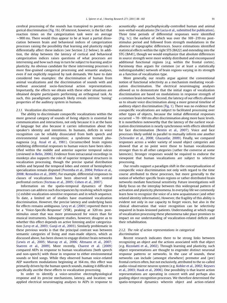
By comparing AEPs in response to accurate and inaccurate TOJperformance, we identified when during the course of stimulusprocessing a temporal ‘stamp’ is established to guide TOJ perception(Bernasconi et al., 2010a; Fig. 3a). Response modulations betweenaccurate and inaccurate TOJ manifested over the 33e77 ms post-stimulus period and were the result of changes in the topography ofthe electric field at the scalp (and by extension in the underlyingconfiguration of intracranial sources). Consistentwith the hypothesisfor a left hemispheric dominance in TOJ, source estimations per-formed over this time period revealed a modulation of left, but notright, superior temporal activity as a function of performance accu-racy, as well as a significant correlation between left, but not right,superior temporal cortex and behavioral sensitivity.
Our results further revealed that over the 33e77 ms interval, theactivity between left and right superior temporal cortices wascorrelated when performance was inaccurate but not when it wasaccurate, suggesting that the extent of functional connectivity, orcoupling, between posterior temporal homotopic areas impacts TOJaccuracy. This result suggests that in a near-threshold context,accurate TOJ can be achieved within the left superior temporalcortex only when temporal processing occurring within this regionis functionally released from the interfering influence of righthemisphere. Activity within right hemisphere structures couldhave interferedwith temporal integrationmechanisms occurring inthe contralateral (i.e. left) homotopic region. In agreement with thelikely importance of inter-hemispheric interactions in auditoryfunctions,Westerhausen et al. (2009) used diffusion tensor imagingto show that variability in fiber tracks interconnecting the superiortemporal lobes correlated with performance on an auditorydiscrimination task.
Several studies indicate that mechanisms supporting temporaldiscrimination are highly interactive with those mediating atten-tion (Eagleman, 2008). Consequently, right hemispheric structuresmight likewise play a role in TOJ performance. However, it iscontroversial as to whether attention impacts the amplitude(McDonald et al., 2005) and/or timing (Vibell et al., 2007) of brainactivity during temporal processing. These studies advanced thattemporal order perception could depend on gating and/or latencymechanisms, respectively reflected by increases in responseamplitude and/or decreases in the processing latency to attendedversus unattended stimuli (see Vibell et al., 2007 for discussion).
Support for the involvement of gatingmechanisms in TOJ comesfrom studies showing that the manipulation of exogenous atten-tional cues induced shifts in the point of subjective simultaneity
Fig. 3. Patterns of inter-hemispheric coupling during TOJ performance and acquisition of proficiency. (a) Source estimations during the 39e77 ms post-stimulus period whentopographic AEP modulations were observed between trials resulting in accurate vs. inaccurate TOJ performance are displayed. Prominent sources were observed within posteriorsylvian regions bilaterally, though the degree of bilateral responses varied between accurate and inaccurate trials. The cluster plot shows that responses between the hemisphereswere significantly coupled on inaccurate trials and decoupled on accurate trials. (b) Source estimations during the 43e76 ms post-stimulus period when topographic AEPmodulations were observed between trials at the end vs. beginning of a TOJ training session. Performance sensitivity (d0) significantly improved, and prominent sources were againobserved within posterior sylvian regions bilaterally. Again the extent of bilateral activation differed as a function of training. At the beginning of training when performance waspoor, responses between the hemispheres were significantly coupled. At the end of training when performance was improved, responses between the hemispheres were de-coupled.
L. Spierer et al. / Hearing Research 271 (2011) 88e102 95
accompanied by a gain in the amplitude of early visual evokedpotentials (e.g. Luck et al., 2000). Gating would likely rely on anactive inhibitory network, designed to reduce the flow of redun-dant sensory information associated with sensory overload (Kisleyet al., 2004). As accurate TOJs require unbiased perception of thefirst and/or second sound of the pair, they cannot be achieved if theprocessing of each of the two sounds interferes with each other. Anadequate gating of the first sound would therefore facilitate TOJ byinhibiting the response to the second sound. Auditory gatingmechanisms typically manifest around 50 ms post stimulus onset(e.g. Pelizzone et al., 1987), which is a time period corresponding tothe latency of the effects we have observed (Bernasconi et al.,2010a). However, we would exclude that a pure gating mecha-nism explains our results, because we found that accurate TOJ was
associated with a reduction of left superior temporal activity, ratherthan in an increase in response strength to the first sound as wouldbe expected according to the gating hypothesis. Additionally, puregating mechanisms would have likely manifested as a modulationin global field power in the absence of topographic modulations.Rather, we observed topographic modulations in the absence ofmodulations in global field power. That is, the mechanism oper-ating to influence TOJ accuracy appears to rely on changes in theconfiguration of active brain regions rather than simply changes intheir level of activity.
The prior-entry hypothesis is more consistent with our results. Itproposes that TOJ depends on the processing speed of sensorystimuli that in turn determines their order of arrival intoconsciousness (Titchener, 1908); though the precise
L. Spierer et al. / Hearing Research 271 (2011) 88e10296
neurophysiologic mechanism for such remains controversial. Thegeneral threshold model (Ulrich, 1987) postulates that TOJ mightdepend on the arrival time of the sensory information at a hypo-thetical “temporal comparator”; the arrival time depending onparameters including transduction time or transmission latenciesof the information from the receptor to a comparator (Pöppel, 1988;Stelmach and Herdman, 1991). In an electrophysiologic study usinga cross-modal visuo-tactile TOJ task, Vibell et al. (2007) providesupport for the prior-entry hypothesis by showing that attentionshifts the latency of visual evoked potentials, consistent witha speeding-up of sensory processing. Our pattern of results inBernasconi et al. 2010a likewise supports prior-entry as a putativemechanism for temporal order perception. The topographicmodulation could result from a rapid latency shift across condi-tions. That is, accurate TOJ might result in a faster transition fromone active configuration of brain areas to another, which in turnmight appear as a topographic difference in the electric field as thescalp. Differences in the prioritization of stimulus processing mighttherefore account for our effects. In this sense, inputs from rightsuperior temporal cortices might have perturbed the processinglatency, rather than or in addition to, the temporal stampingmechanisms occurring within left superior temporal cortices.Greater interference from right to left superior temporal areascould in turn have resulted from the higher level of functionalcoupling we found the inaccurate than accurate condition.
3.2. Learning-induced plasticity in temporal processing
To date, only a few studies have addressed behavioral plasticityof temporal representations, and none directly addressed its neuralunderpinnings. Some psychophysical studies document improve-ments in TOJ performance with experience (e.g. Hirsh, 1959), butonly a few report empirical data directly supporting training-induced improvements (e.g. Mossbridge et al., 2006, 2008).
We recently identified the spatio-temporal brain correlates oftraining-induced improvements in auditory TOJ (Bernasconi et al.,2010b). Thirty minutes of training significantly improved TOJperformance. AEPs recorded at the beginning vs. the end of trainingrevealed that over the 43e76 ms post-stimulus time periodresponses to trials when TOJ performance was accurate differedtopographically as a function of training blocks (Fig. 3b). This isevidence for the engagement of distinct configurations of brainnetworks at the beginning vs. at the end of the training session.Source estimations in turn revealed that TOJ improvement wasassociated with a change in the lateralization pattern of brainresponses from a bilateral pattern within posterior sylvian regions(PSR) at the beginning of training to a left-lateralized pattern at theend of training. Moreover, activity within the left but not right PSRcorrelated with discrimination performance (Fig. 3b). These resultsare in strong agreement with previous evidence of a left temporo-parietal dominance for TOJ (Davis et al., 2009; Wittmann et al.,2004) and more generally for the involvement of left supra-temporal plane in the processing of auditory temporal features(Zatorre et al., 2002; Zaehle et al., 2009; Foxton et al., 2009).Collectively, these results support that the temporal stampingrequired for accurate stimuli ordering depends on left PSR struc-tures, putatively due to their anatomo-functional advantages forfast temporal processing.
The plastic changes underlying TOJ improvement in Bernasconiet al. (2010b) impacted right PSR responses and their interactionwith left PSR, but not directly the activity within the left PSR. Weinterpreted these results as indicating that the TOJ improvementwas mediated by a release of task-relevant representationscomprised within the left PSR from temporally imprecise andinterfering activity and/or inputs from the right PSR. Left and right
PSR activity indeed correlated at the beginning but not at the end ofthe training session. One neurophysiologicmodel for such plasticityand improvements in TOJ performance is based on reductions insynaptic latency mediated by spike timing dependent plasticity(STDP; reviewed in Buonomano and Merzenich, 1998; Weinberger,2004).
STDP models assume that the temporal association betweenpre- and post-synaptic activation occurringwith repeated exposureto stimuli in turn modifies synaptic strength and reduces responselatencies (Song et al., 2000). In this way, the timing of the activatedneural circuitry would be sharpened and the ordering of inputfacilitated (e.g. Legenstein et al., 2005). According to STDP, neuralcircuits with sharp response latencies would in turn respondsynchronously to stimulus onsets andwould consequently see theiractivity preserved or reinforced, whereas responses within regionswith less synchronous response patterns would diminish. Due tothe anatomo-functional advantage of the left PSR for fast temporalprocessing reviewed above, repeated exposure to the sound pairswould be reinforced by STDP, whereas less sharply tuned regions,including right PSR would see their responses reduced. Finally, theinitial functional connection between left and right hemisphereswould be predicted to be suppressed due to the general differencesin levels of synchronicity. STDP (or a similar mechanism) couldtherefore account for both the decrease in right superior temporalsulcus response and the decoupling between left and right PSR, inturn sharpening the left posterior superior temporal sulcusresponse and facilitating the processing of temporal order.
The general time course of the effects we have observedprovides some insights on the likely basis upon which temporalprocessing and temporal discrimination are performed. Temporalstamping mechanisms and plastic changes accompanying behav-ioral improvements revealed in our studies (Bernasconi et al.,2010b, submitted) would appear to occur at similarly early pro-cessing stages and prior to stimulus feature integration. Priorstudies of spatial processing have only observed effects at latenciesbeyond approximately 100 ms post-stimulus onset (e.g. De Santiset al., 2007; Spierer et al., 2008a, b; Murray and Spierer, 2009).
4. Spatial discrimination and learning-induced plasticity
4.1. An overview of auditory spatial processing
While there is general consensus that accurate spatial process-ing of sounds relies on cortical activity (Jenkins and Masterton,1982; Heffner and Heffner, 1990; King et al., 2007), the precisemanner in which spatial positions are represented and accuratediscrimination achieved remain unresolved, particularly inhumans. Spatiotopic representations have been identified insubcortical but not cortical structures in animals (e.g. Palmer andKing, 1982; Woods et al., 2006). At the cortical level, recent inves-tigations suggest that sub-populations of location-sensitive corticalneurons over-sample and respond preferentially to the more lateralregions of either the ipsilateral or contralateral hemispace whilealso exhibiting their steepest tuning curves for positions thatstraddle the midline (Stecker et al., 2005). Based on these findings,Stecker and colleagues proposed an opponent-channel theory ofspatial coding wherein two sub-populations, or “spatial channels”,represent locations according to the slopes of their response areaand by graded changes in response rate. Such observations speak infavor a model of spatial representations based on the patternedactivity of population responses. Inaccurate encoding of spatialpositions could result from the activity of less specific neurons(Ghose, 2004; Ohl and Scheich, 2005), from noise-relatedresponses (Rainer et al., 2004), or from inaccurate perceptualtemplates (Li et al., 2004).
L. Spierer et al. / Hearing Research 271 (2011) 88e102 97
In humans, mechanisms of auditory spatial discrimination havecommonly been assessed using the MMN (e.g. Deouell et al., 2006,2007; Spierer et al., 2008b; also Salminen et al., 2009). Thesestudies showed that MMN amplitude correlated positively withdeviations in azimuthal eccentricity and with subjects’ discrimi-nation accuracy. Spatial comparisons underlying change detectionhave been proposed to rely on a separate memory trace thatencodes regularity in the auditory environment (“memory-tracemodel”; Näätänen et al., 2005) and which is thought to be localizedin the posterior superior temporal gyrus (Deouell et al., 2006) and/or regions along the putative spatial or “where” dorsalelateralprocessing stream (Deouell et al., 2006, 2007).
We recently provided evidence for the critical role of supra-temporal regions in spatial encoding accuracy by demonstratingthat responses to physically identical acoustic stimuli differedtopographically according to subsequent performance accuracy ona spatial discrimination task at approximately 100 ms post-stimulusonset, indicative of changes in the configuration of the underlyingintracranial sources preceding correct vs. incorrect spatial discrim-inations (Fig. 4a; see also Spierer et al., 2008a; as well as Ohl andScheich, 2005; Ohl et al., 2001 for similar findings in animalmodels). Analyses of distributed source estimations revealed largelysimilar sets of activated regions for both conditions, with strongeractivity within the contralateral (left) supratemporal plane andinferior parietal lobule preceding correct vs. incorrect discrimina-tions (Spierer et al., 2008a). A positive correlation was foundbetween discrimination sensitivity (d0) and the strength of sourceswithin the posterior supratemporal plane (BA41). No such correla-tion was observed within parietal cortices or elsewhere. Consistentwith a model based on population-based encoding of spatial posi-tions, we hypothesized that the activity within the supratemporalplanewas strongerwhen the spatial position of the stimulus ismorereliably encoded due to a larger differential response betweenneural populations constituting each opponent channel. On trialsleading to incorrect performance, responses within the supra-temporal plane would be smaller because of inaccurate and/orimprecise encoding of spatial information. Supporting this propo-sition are data from Deouell et al. (2007), who demonstrated thatresponses within the planum temporale (as well as anterior regionsalong the superior temporal gyrus) to different (supra-threshold; i.e.15�) spatial lateralizations increased as the number of stimulatedpositions increased within a block of trials.
The determinant role of supratemporal cortices in spatialencoding accuracy provides insights on the relative function ofregions involved in auditory spatial processing. First, our effects at75e117 ms were lateralized to the left (contralateral) superiortemporal plane. This finding is in agreement with previousevidence demonstrating the prominent role of the contralateralhemisphere in the processing of interaural temporal information(Krumbholz et al., 2005, 2007; see also Zatorre and Penhune, 2001for support for a left-hemisphere dominance in spatial discrimi-nation), at least at early latencies post-stimulus onset. While these“early” effects are left-lateralized, it is likely the case that bilateralnetworks are involved during subsequent time periods (e.g. Tardifet al., 2006). Electrophysiological recordings in the supratemporalplane of non-human primates suggest that the posterior part of thesupratemporal gyrus comprises an early representation of soundsources (Rauschecker, 1998; Recanzone et al., 2000; Woods et al.,2006). In humans, MMN studies also suggest that these represen-tations as well as spatial comparison mechanisms underlyingchange detection may reside within the planum temporale (Tataand Ward, 2005; Sonnadara et al., 2006; Deouell et al., 2006,2007; Salminen et al., 2009). By contrast, our data would insteadsuggest that more lateral regions of the superior temporal planeplay a particularly important role in spatial functions at early
latencies (see also Tardif et al., 2006), which are also anterior toregions of the planum temporale implicated in the above-mentioned prior studies (see also Zatorre and Penhune, 2001 forsimilar conclusions based on neuropsychological findings). Furtherinvestigations are required to disentangle the role of specific areaswithin the superior temporal regions in spatial processing.
Aside from superior temporal cortices, there is also evidence inboth humans and non-human primates for a prominent role ofparietal structures in spatial localization processes (e.g. Griffithset al., 1996; Mazzoni et al., 1996; Stricanne et al., 1996; Weekset al., 1999; Ducommun et al., 2002; Zatorre et al., 2002; Deouellet al., 2006, 2007; Tardif et al., 2006; De Santis et al., 2007;Spierer et al., 2008b). Neuropsychological studies of spatial func-tions have likewise shown that temporal and/or parietal lobelesions lead to impairments in sound localization (e.g. Ruff et al.,1981; Bisiach et al., 1984; Pinek et al., 1989; Vallar et al., 1995;Griffiths et al., 1997; Tanaka et al., 1999; Bellmann et al., 2001;Clarke et al., 2000, 2002; Zatorre and Penhune, 2001; Zimmeret al., 2003). However, it is likewise the case that temporal andparietal regions are differentially involved depending on whetherthe spatial task requires absolute or relative localization.
Following the initial analysis of auditory spatial informationwithin the supratemporal plane, higher-order processing of audi-tory spatial information has been proposed to occur along theparieto-frontal ‘where’ stream (Alain et al., 2001; Maeder et al.,2001; Ducommun et al., 2002; Arnott et al., 2004; Tardif et al.,2006; De Santis et al., 2007). However, we would emphasize thatdata concerning the temporal dynamics of activity in these regionsshows them to be responsive at early latencies, making it importantto distinguish between temporal hierarchies and processing hier-archies. Data from non-human primates suggest that the posteriorparietal cortex rather than the supratemporal plane is involved inhigh-level spatial processes (Rauschecker, 1998). A positron emis-sion tomography study in humans also demonstrated the absenceof activity in the supratemporal plane on an absolute localizationtask (Weeks et al., 1999). Consistently, Lewald et al. (2002, 2004a, b)reported that focal repetitive transcranial magnetic stimulation ofthe posterior parietal cortex induced a systematic shift in theperceived lateralization of a sound source position, whereas theacuity of position discrimination remained unaffected.
Several lines of evidence suggest that along the above-describedprocessing hierarchy of auditory spatial information, initial stagesinvolve temporal regions contralateral to the stimulation whileright parietal hemisphere dominates for higher-order spatialprocesses. Lesion data indeed reveal no differences between defi-cits associated with right and left temporal lesions (Sanchez-Longoand Forster, 1958; Efron et al., 1983), while studies includingpatients with parietal lesions suggest a right hemispheric domi-nance (Ruff et al., 1981; Bisiach et al., 1984; Tanaka et al., 1999). Ourown clinical data emphasize the role of the right hemisphere in theprocessing of binaural spatial cues (i.e. inter-aural intensity andtime differences; IID and ITD, respectively) in a large-scale neuro-psychological study including 25 right-hemisphere and 25 left-hemisphere brain damaged patients (Spierer et al., 2009b). Precisecomputation of contralateral spatial information involved the lefthemisphere, while the right hemisphere was involved in the pro-cessing of the whole of auditory space. On the other hand, thebuilding up of global auditory spatial representations relied onright temporo-parietal cortices. While numerous neuroimagingstudies speak in favor of right hemispheric dominance for auditoryspatial processing particularly at post-stimulus latencies >200 ms(Kaiser and Lutzenberger, 2001; Ducommun et al., 2002; Herrmannet al., 2002; Lewald et al., 2002; De Santis et al., 2007; Spierer et al.,2009c), initial processing stages would appear to involve more thecontralateral than the ipsilateral hemisphere.
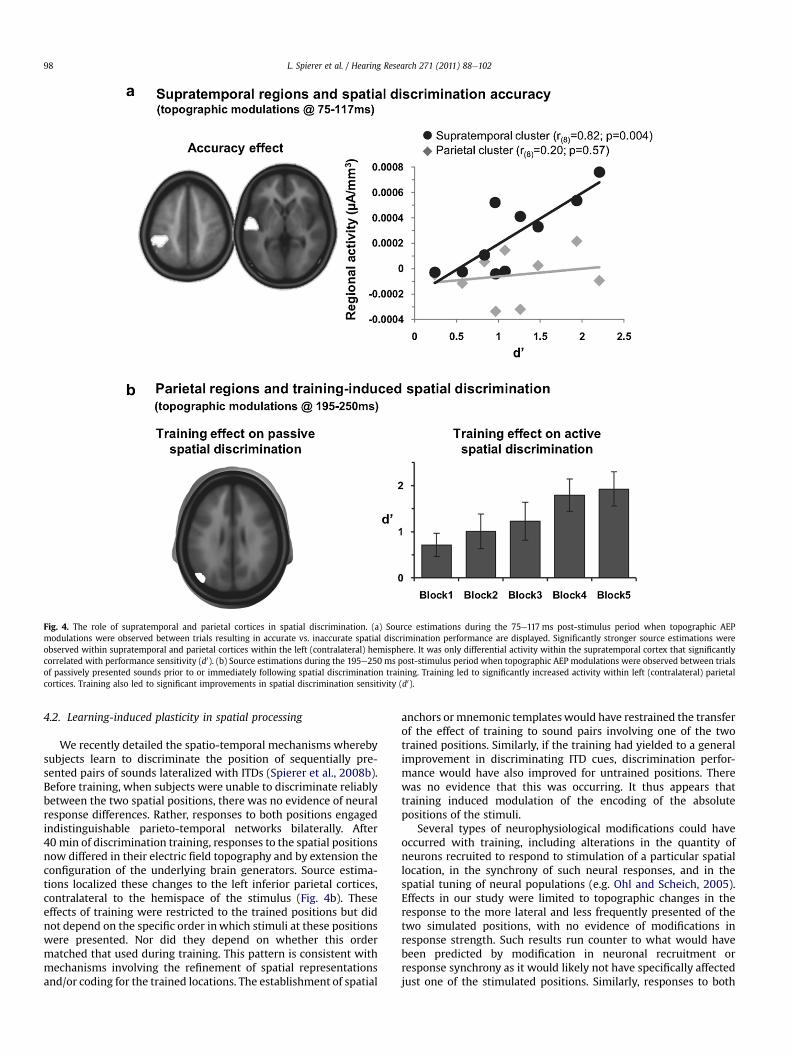
Fig. 4. The role of supratemporal and parietal cortices in spatial discrimination. (a) Source estimations during the 75e117 ms post-stimulus period when topographic AEPmodulations were observed between trials resulting in accurate vs. inaccurate spatial discrimination performance are displayed. Significantly stronger source estimations wereobserved within supratemporal and parietal cortices within the left (contralateral) hemisphere. It was only differential activity within the supratemporal cortex that significantlycorrelated with performance sensitivity (d0). (b) Source estimations during the 195e250 ms post-stimulus period when topographic AEP modulations were observed between trialsof passively presented sounds prior to or immediately following spatial discrimination training. Training led to significantly increased activity within left (contralateral) parietalcortices. Training also led to significant improvements in spatial discrimination sensitivity (d0).
L. Spierer et al. / Hearing Research 271 (2011) 88e10298
4.2. Learning-induced plasticity in spatial processing
We recently detailed the spatio-temporal mechanisms wherebysubjects learn to discriminate the position of sequentially pre-sented pairs of sounds lateralized with ITDs (Spierer et al., 2008b).Before training, when subjects were unable to discriminate reliablybetween the two spatial positions, there was no evidence of neuralresponse differences. Rather, responses to both positions engagedindistinguishable parieto-temporal networks bilaterally. After40 min of discrimination training, responses to the spatial positionsnow differed in their electric field topography and by extension theconfiguration of the underlying brain generators. Source estima-tions localized these changes to the left inferior parietal cortices,contralateral to the hemispace of the stimulus (Fig. 4b). Theseeffects of training were restricted to the trained positions but didnot depend on the specific order inwhich stimuli at these positionswere presented. Nor did they depend on whether this ordermatched that used during training. This pattern is consistent withmechanisms involving the refinement of spatial representationsand/or coding for the trained locations. The establishment of spatial
anchors ormnemonic templates would have restrained the transferof the effect of training to sound pairs involving one of the twotrained positions. Similarly, if the training had yielded to a generalimprovement in discriminating ITD cues, discrimination perfor-mance would have also improved for untrained positions. Therewas no evidence that this was occurring. It thus appears thattraining induced modulation of the encoding of the absolutepositions of the stimuli.
Several types of neurophysiological modifications could haveoccurred with training, including alterations in the quantity ofneurons recruited to respond to stimulation of a particular spatiallocation, in the synchrony of such neural responses, and in thespatial tuning of neural populations (e.g. Ohl and Scheich, 2005).Effects in our study were limited to topographic changes in theresponse to the more lateral and less frequently presented of thetwo simulated positions, with no evidence of modifications inresponse strength. Such results run counter to what would havebeen predicted by modification in neuronal recruitment orresponse synchrony as it would likely not have specifically affectedjust one of the stimulated positions. Similarly, responses to both

L. Spierer et al. / Hearing Research 271 (2011) 88e102 99
positions would be affected in the case of a general attention orarousal mechanism or a general learning-induced change in spatialcoding. Furthermore, these mechanisms would likely have resultedin a change in the strength of responses, rather than the configu-ration of underlying brain generators. Instead, our results areconsistent with the refinement of neuronal spatial tuning ata population level. Previous studies have shown that pitch trainingwas accompanied by an increase in neuronal selectivity anddecrease of the corresponding cortical representation (Edeline andWeinberger, 1993; Recanzone et al., 1993). In our study, trainingselectively changed the topography of the electric field anddecreased the activity of sources within left inferior parietalcortices (cf. Fig. 5 in Spierer et al., 2008b). A putative neuralmechanism may involve inhibitory processes in generating plas-ticity via the exclusion of the activity of less specific neurons(Ghose, 2004; Ohl and Scheich, 2005) or noise-related responses(Rainer et al., 2004). Li et al. (2004) further extended this notion interms of refining a perceptual template, wherein those neuronsthat respond most strongly might not convey the greatest amountof information regarding a learned discrimination. Rather, greaterdifferential responses to the spatial positions may instead occur inneurons exhibiting weaker response magnitude. In this case, theinhibition of such strongly responding neurons would producea more informative response profile (Ghose, 2004).
On the whole, our results lend additional support to the conceptof auditory spatial representations based on population coding.Together with the finding that this effect occurred at a relativelylate processing stage (around 250 ms), our findings would supporta multiphase spatial processing hierarchy that includes the trans-formation of spatial representations both along relative and abso-lute dimensions as well as along egocentric and allocentric framesof reference.
5. Conclusions
Current models of the organization of the auditory system positthat the processing of semantic, temporal or spatial auditoryinformation relies on partially segregated processing streams. Thestudies we reviewed above indicate that in addition to their ana-tomo-functional heterogeneity, distinct neurophysiological mech-anisms subserve training-induced plastic changes within each ofthese pathways.
Auditory object-related plasticity has been investigated byseveral experimental paradigms involving repetition priming.Based on our electrical neuroimaging and fMRI studies, weprovided evidence that repetition-induced plasticity involvesstrength modulations within a common network of brain regionsprincipally within the temporal lobe both when the initial andrepeated stimuli are identical exemplars as well as when they arephysically different but refer to a common referent object (and arethus semantically related). These findings challenge existingmodels wherein priming is mediated by extrastriate visual regionsand prefrontal cortices, irrespective of their surface features and inboth visual and auditory modalities. With respect to the timing ofthese effects, we have shown that perceptual priming took placeover the 156e215 ms post-stimulus period and therefore subse-quent to basic-level living vs. man-made categorical discrimination.This finding provides additional support to the notion thatperceptual priming can indeed imply access to some semanticfeatures of the auditory stimulus even when exposed to repetitionof identical sounds. Ongoing investigations are targeting mecha-nisms of learning to recognize individual sounds, such as whena sound is initially unrecognizable but is subsequently categorizedupon its repetition or further rendered identifiable at a finersemantic scale.
Learning-induced plasticity in temporal representations werefirst evident around 60 ms post-stimulus onset and involvedmodifications both in the lateralization pattern from bilateral to leftposterior sylvian regions and in the functional coupling betweenthe hemispheres. The latency of our effect is consistent withmodifications during the initial stages of auditory processing,rather than changes in higher-level functions or representations ofthe sound sources. Future investigations will focus on isolatingprocesses of temporal discrimination by introducing paradigmaticchanges such that the first and second sounds differ in otheracoustic features aside from their spatial location. Such will provideinsights on the extent to which temporal processing (and learningthereof) is independent of the acoustic features defining thetemporal separation between events.
Neuroplastic changes in auditory spatial representations or theability to accurately discriminate spatial representations occurredover several time periods throughout the 70e250 ms post-stimulusinterval. In all instances, however, modulations manifested aschanges in the topography of the electric field at the scalp and byextension the configuration of intracranial sources. This indicatesthat spatial representations at a population level are subject toplasticity, which is consistent within current models of spatialencoding of sounds. While we have emphasized population-levelmodulations, it is likely also the case that effects are occurring ata finer spatial (and mechanistic) scale that what is currentlypossible with non-invasive techniques in humans. In addition togeneral spatial encoding, we also demonstrated that relative andabsolute spatial representations of sound sources can be differen-tiated both in time and in terms of the principal brain regionsimplicated, including hemispheric dominance. Relative locationsappear to be differentiated within supratemporal regions at earlierlatencies than absolute locations within parietal structures.However, the reader should note that there is also clear evidencefor the (near) simultaneous responsiveness of these regions tosounds in general. As such, we by no means wish to give theimpression that parietal and supratemporal structures areresponding in a strictly serial manner. Still, differential involvementand encoding of relative and absolute spatial positions wouldappear to manifest at over multiple periods and brain regions.Additionally, the right hemisphere would appear to containa representation of the whole of auditory space, whereas therepresentation in the left hemisphere would appear to be largelyrestricted to the contralateral space. Determining how these sub-varieties of spatial representations are rendered plastic will be animportant direction for future research.
Acknowledgements
This work has been supported by grants from the Swiss NationalScience Foundation (K-33K1_122518/1 to MDL; 3100AO-103895 toSC; 3100AO-118419 to MMM), a grant from the Pierre MercierFoundation for Science (to LS and JG), and an inter-disciplinarygrant from the Faculty of Biology and Medicine of the CentreHospitalier Universitaire Vaudois and University of Lausanne (to LSand JG). Analyses of EEG data were performed using the Cartoolsoftware (http://brainmapping.unige.ch/cartool.htm) which isdeveloped by Denis Brunet from the Functional Brain MappingLaboratory, Geneva, Switzerland, and is supported by the EEG BrainMapping Core of the Center for Biomedical Imaging (CIBM) ofGeneva and Lausanne. MRI and fMRI data acquisition were sup-ported by the Center for Biomedical Imaging (http://www.cibm.ch),The University of Lausanne; The Swiss Federal Institute of Tech-nology, Lausanne; The University of Geneva; The UniversityHospital Center, Lausanne; The University Hospital of Geneva; andthe Leenaards and the Jeantet Foundations.
L. Spierer et al. / Hearing Research 271 (2011) 88e102100
References
Alain, C., Arnott, S.R., Hevenor, S., Graham, S., Grady, C.L., 2001. “What” and “where”in the human auditory system. Proc. Natl. Acad. Sci. USA 98, 12301e12306.
Alain, C., McDonald, K.L., Kovacevic, N., McIntosh, A.R., 2009. Spatiotemporalanalysis of auditory “what” and “where” working memory. Cereb. Cortex 19,305e314.
Altmann, C.F., Doehrmann, O., Kaiser, J., 2007. Selectivity for animal vocalizations inthe human auditory cortex. Cereb. Cortex 17, 2601e2608.
Anderson, B., Southern, B.D., Powers, R.E., 1999. Anatomic asymmetries of theposterior superior temporal lobes: a postmortem study. NeuropsychiatryNeuropsychol Behav Neurol 12, 247e254.
Arnott, S.R., Binns, M.A., Grady, C.L., Alain, C., 2004. Assessing the auditory dual-pathway model in humans. Neuroimage 22, 401e408.
Assal, G., Aubert, C., Buttet, J., 1981. [Cerebral asymmetry and voice recognition(author’s transl)]. Rev. Neurol. (Paris) 137, 255e268.
Badgaiyan, R.D., Schacter, D.L., Alpert, N.M., 2001. Priming within and acrossmodalities: exploring the nature of rCBF increases and decreases. Neuroimage13, 272e282.
Belin, P., 2006. Voice processing in human and non-human primates. Philos. Trans.R. Soc. Lond. B Biol. Sci. 361, 2091e2107.
Bellmann, A., Meuli, R., Clarke, S., 2001. Two types of auditory neglect. Brain 124,676e687.
Bentin, S., Taylor, M.J., Rousselet, G.A., Itier, R.J., Caldara, R., Schyns, P.G., Jacques, C.,Rossion, B., 2007. Controlling interstimulus perceptual variance does notabolish N170 face sensitivity. Nat. Neurosci. 10, 801e802.
Bergerbest, D., Ghahremani, D.G., Gabrieli, J.D., 2004. Neural correlates of auditoryrepetition priming: reduced fMRI activation in the auditory cortex. J. Cogn.Neurosci. 16, 966e977.
Besle, J., Fischer, C., Bidet-Caulet, A., Lecaignard, F., Bertrand, O., Giard, M.H., 2008.Visual activation and audiovisual interactions in the auditory cortex duringspeech perception: intracranial recordings in humans. J. Neurosci. 28,14301e14310.
Bernasconi, F., Grivel, J., Murray, M.M., Spierer, L., 2010a. Interhemispheric couplingbetween the posterior sylvian regions impacts successful auditory temporalorder judgment. Neuropsychologia, in press.
Bernasconi, F., Grivel, J., Murray, M.M., Spierer, L., 2010b. Plastic brain mechanismsfor attaining auditory temporal order judgment proficiency. Neuroimage 50,1271e1279.
Bisiach, E., Cornacchia, L., Sterzi, R., Vallar, G., 1984. Disorders of perceived auditorylateralization after lesions of the right hemisphere. Brain 107, 37e52.
Bizley, J.K., Walker, K.M., Silverman, B.W., King, A.J., Schnupp, J.W., 2009. Interde-pendent encoding of pitch, timbre, and spatial location in auditory cortex. J.Neurosci. 29, 2064e2075.
Bourquin, N., Spierer, L., Murray, M.M., Clarke, S. Distinct neural networks keeptrack of often versus recently heard sound objects, in preparation.
Buckner, R.L., Koutstaal, W., Schacter, D.L., Rosen, B.R., 2000. Functional MRIevidence for a role of frontal and inferior temporal cortex in amodal compo-nents of priming. Brain 123, 620e640.
Buonomano, D.V., Merzenich, M.M., 1998. Net interaction between different formsof short-term synaptic plasticity and slow-IPSPs in the hippocampus andauditory cortex. J. Neurophysiol. 80, 1765e1774.
Charest, I., Pernet, C.R., Rousselet, G.A., Quinones, I., Latinus, M., Fillion-Bilodeau, S.,et al., 2009. Electrophysiological evidence for an early processing of humanvoices. BMC Neurosci. 10, 127.
Chiu, C.Y., 2000. Specificity of auditory implicit and explicit memory: is perceptualpriming for environmental sounds exemplar specific? Mem. Cognit. 28,1126e1139.
Clarke, S., Adriani, M., Bellmann, A., 1998. Distinct short-term memory systems forsound content and sound localization. Neuroreport 9, 3433e3437.
Clarke, S., Bellmann Thiran, A., Maeder, P., Adriani, M., Vernet, O., Regli, L., et al.,2002. What and where in human audition: selective deficits following focalhemispheric lesions. Exp. Brain Res. 147, 8e15.
Clarke, S., Bellmann, A., Meuli, R.A., Assal, G., Steck, A.J., 2000. Auditory agnosia andauditory spatial deficits following left hemispheric lesions: evidence for distinctprocessing pathways. Neuropsychologia 38, 797e807.
Cohen, Y.E., Theunissen, F., Russ, B.E., Gill, P., 2007. Acoustic features of rhesusvocalizations and their representation in the ventrolateral prefrontal cortex. J.Neurophysiol. 97, 1470e1484.
Davis, B., Christie, J., Rorden, C., 2009. Temporal order judgments activate temporalparietal junction. J. Neurosci. 29, 3182e3188.
De Lucia, M., Camen, C., Clarke, S., Murray, M.M., 2009a. The role of actions inauditory object discrimination. Neuroimage 48, 475e485.
De Lucia, M., Cocchi, L., Martuzzi, R., Meuli, R.A., Clarke, S., Murray, M.M., 2009b.Perceptual and semantic contributions to repetition priming of environmentalsounds. Cereb Cortex.
De Lucia, M., Clarke, S., Murray, M.M., A temporal hierarchy for conspecific vocali-zation discrimination in humans, submitted for publication.
De Santis, L., Clarke, S., Murray, M.M., 2007. Automatic and intrinsic auditory “what”and “where” processing in humans revealed by electrical neuroimaging. Cereb.Cortex 17, 9e17.
Deouell, L.Y., Heller, A.S., Malach, R., D’Esposito, M., Knight, R.T., 2007. Cerebralresponses to change in spatial location of unattended sounds. Neuron 55,985e996.
Deouell, L.Y., Parnes, A., Pickard, N., Knight, R.T., 2006. Spatial location is accuratelytracked by human auditory sensory memory: evidence from the mismatchnegativity. Eur. J. Neurosci. 24, 1488e1494.
Desimone, R., 1996. Neural mechanisms for visual memory and their role inattention. Proc. Natl. Acad. Sci. USA 93, 13494e13499.
Ducommun, C.Y., Murray, M.M., Thut, G., Bellmann, A., Viaud-Delmon, I., Clarke, S.,et al., 2002. Segregated processing of auditory motion and auditory location: anERP mapping study. Neuroimage 16, 76e88.
Eagleman, D.M., 2008. Human time perception and its illusions. Curr. Opin. Neu-robiol. 18, 131e136.
Edeline, J.M., Weinberger, N.M., 1993. Receptive field plasticity in the auditorycortex during frequency discrimination training: selective retuning indepen-dent of task difficulty. Behav. Neurosci. 107, 82e103.
Efron, R., Koss, B., Yund, E.W., 1983. Central auditory processing. IV. Ear domi-nanceespatial and temporal complexity. Brain Lang. 19, 264e282.
Engel, L.R., Frum, C., Puce, A., Walker, N.A., Lewis, J.W., 2009. Different categories ofliving and non-living sound-sources activate distinct cortical networks. Neu-roimage 47, 1778e1791.
Fecteau, S., Armony, J.L., Joanette, Y., Belin, P., 2005. Sensitivity to voice in humanprefrontal cortex. J. Neurophysiol. 94, 2251e2254.
Foxton, J.M., Weisz, N., Bauchet-Lecaignard, F., Delpuech, C., Bertrand, O., 2009. Theneural bases underlying pitch processing difficulties. Neuroimage 45,1305e1313.
Friston, K., 2005. A theory of cortical responses. Philos. Trans. R. Soc. Lond. B Biol.Sci. 360, 815e836.
Garrido, M.I., Kilner, J.M., Kiebel, S.J., Stephan, K.E., Baldeweg, T., Friston, K.J., 2009.Repetition suppression and plasticity in the human brain. Neuroimage 48,269e279.
Ghazanfar, A.A., 2009. The multisensory roles for auditory cortex in primate vocalcommunication. Hear. Res. 258, 113e120.
Ghose, G.M., 2004. Learning in mammalian sensory cortex. Curr. Opin. Neurobiol.14, 513e518.
Giard, M.H., Peronnet, F., 1999. Auditory-visual integration during multimodalobject recognition in humans: a behavioral and electrophysiological study. J.Cogn. Neurosci. 11, 473e490.
Giard, M.H., Fort, A., Mouchetant-Rostaing, Y., Pernier, J., 2000. Neurophysiologicalmechanisms of auditory selective attention in humans. Front. Biosci. 5,D84eD94.
Griffiths, T.D., Rees, A., Witton, C., Cross, P.M., Shakir, R.A., Green, G.G., 1997. Spatialand temporal auditory processing deficits following right hemisphere infarc-tion. A psychophysical study. Brain 120, 785e794.
Griffiths, T.D., Rees, A., Witton, C., Shakir, R.A., Henning, G.B., Green, G.G., 1996.Evidence for a sound movement area in the human cerebral cortex. Nature 383,425e427.
Grill-Spector, K., Henson, R.N., Martin, A., 2006. Repetition and the brain, neuralmodels of stimulus-specific effects. Trends Cogn. Sci. 10, 14e23.
Gunji, A., Koyama, S., Ishii, R., Levy, D., Okamoto, H., Kakigi, R., et al., 2003. Mag-netoencephalographic study of the cortical activity elicited by human voice.Neurosci. Lett. 348, 13e16.
Hackett, T.A., Stepniewska, I., Kaas, J.H., 1999. Callosal connections of the parabeltauditory cortex in macaque monkeys. Eur. J. Neurosci. 11, 856e866.
Hauk, O., Shtyrov, Y., Pulvermuller, F., 2006. The sound of actions as reflected bymismatch negativity: rapid activation of cortical sensory-motor networks bysounds associated with finger and tongue movements. Eur. J. Neurosci. 23,811e821.
Hauptmann, B., Karni, A., 2002. From primed to learn: the saturation of repetitionpriming and the induction of long-term memory. Brain Res. Cogn. Brain Res. 13,313e322.
Heffner, H.E., Heffner, R.S., 1990. Effect of bilateral auditory cortex lesions on soundlocalization in Japanese macaques. J. Neurophysiol. 64, 915e931.
Herrmann, C.S., Senkowski, D., Maess, B., Friederici, A.D., 2002. Spatial versus objectfeature processing in human auditory cortex: a magnetoencephalographicstudy. Neurosci. Lett. 334, 37e40.
Hickok, G., Poeppel, D., 2007. The cortical organization of speech processing. Nat.Rev. Neurosci. 8, 393e402.
Hirsh, I.J.,1959. Auditoryperceptionof temporal order. J. Acoust. Soc. Am. 31, 759e767.Inui, K., Okamoto, H., Miki, K., Gunji, A., Kakigi, R., 2006. Serial and parallel pro-
cessing in the human auditory cortex: a magnetoencephalographic study.Cereb. Cortex 16, 18e30.
Javitt, D.C., Steinschneider, M., Schroeder, C.E., Arezzo, J.C., 1996. Role of cortical N-methyl-D-aspartate receptors in auditory sensory memory and mismatchnegativity generation: implications for schizophrenia. Proc. Natl. Acad. Sci. USA93, 11962e11967.
Javitt, D.C., 2009. Sensory processing in schizophrenia: neither simple nor intact.Schizophr Bull. 35, 1059e1064.
Jenkins, W.M., Masterton, R.B., 1982. Sound localization: effects of unilateral lesionsin central auditory system. J. Neurophysiol. 47, 987e1016.
Kaas, J.H., Hackett, T.A., 2000. Subdivisions of auditory cortex and processingstreams in primates. Proc. Natl. Acad. Sci. USA 97, 11793e11799.
Kaiser, J., Lutzenberger, W., 2001. Location changes enhance hemispheric asym-metry of magnetic fields evoked by lateralized sounds in humans. Neurosci.Lett. 314, 17e20.
Keysers, C., Kohler, E., Umilta, M.A., Nanetti, L., Fogassi, L., Gallese, V., 2003.Audiovisual mirror neurons and action recognition. Exp. Brain Res. 153,628e636.
L. Spierer et al. / Hearing Research 271 (2011) 88e102 101
King, A.J., Bajo, V.M., Bizley, J.K., Campbell, R.A., Nodal, F.R., Schulz, A.L., et al., 2007.Physiological and behavioral studies of spatial coding in the auditory cortex.Hear. Res. 229, 106e115.
Kisley, M.A., Noecker, T.L., Guinther, P.M., 2004. Comparison of sensory gating tomismatch negativity and self-reported perceptual phenomena in healthyadults. Psychophysiology 41, 604e612.
Kohler, E., Keysers, C., Umilta, M.A., Fogassi, L., Gallese, V., Rizzolatti, G., 2002.Hearing sounds, understanding actions: action representation in mirrorneurons. Science 297, 846e848.
Kraus, N., Nicol, T., 2005. Brainstem origins for cortical ‘what’ and ‘where’ pathwaysin the auditory system. Trends Neurosci. 28, 176e181.
Krumbholz, K., Hewson-Stoate, N., Schönwiesner, M., 2007. Cortical response toauditory motion suggests an asymmetry in the reliance on inter-hemisphericconnections between the left and right auditory cortices. J. Neurophysiol. 97,1649e1655.
Krumbholz, K., Schonwiesner, M., von Cramon, D.Y., Rubsamen, R., Shah, N.J.,Zilles, K., et al., 2005. Representation of interaural temporal information fromleft and right auditory space in the human planum temporale and inferiorparietal lobe. Cereb. Cortex 15, 317e324.
Lavoie, S., Murray, M.M., Deppen, P., Knyazeva, M.G., Berk, M., Boulat, O., Bovet, P.,Bush, A.I., Conus, P., Copolov, D., Fornari, E., Meuli, R., Solida, A., Vianin, P.,Cuénod, M., Buclin, T., Do, K.Q., 2008. Glutathione precursor, N-acetyl-cysteine,improves mismatch negativity in schizophrenia patients. Neuro-psychopharmacology 33, 2187e2199.
Legenstein, R., Naeger, C., Maass, W., 2005. What can a neuron learn with spike-timing-dependent plasticity? Neural Comput. 17, 2337e2382.
Levy, D.A., Granot, R., Bentin, S., 2001. Processing specificity for human voicestimuli: electrophysiological evidence. Neuroreport 12, 2653e2657.
Levy, D.A., Granot, R., Bentin, S., 2003. Neural sensitivity to human voices: ERPevidence of task and attentional influences. Psychophysiology 40, 291e305.
Lewald, J., Foltys, H., Topper, R., 2002. Role of the posterior parietal cortex in spatialhearing. J. Neurosci. 22, RC207.
Lewald, J., Meister, I.G.,Weidemann, J., Topper, R., 2004a. Involvement of the superiortemporal cortex and the occipital cortex in spatial hearing: evidence fromrepetitive transcranial magnetic stimulation. J. Cogn. Neurosci. 16, 828e838.
Lewald, J., Wienemann, M., Boroojerdi, B., 2004b. Shift in sound localizationinduced by rTMS of the posterior parietal lobe. Neuropsychologia 42,1598e1607.
Lewis, J.W., Brefczynski, J.A., Phinney, R.E., Janik, J.J., DeYoe, E.A., 2005. Distinct corticalpathways for processing tool versus animal sounds. J. Neurosci. 25, 5148e5158.
Li, R.W., Levi, D.M., Klein, S.A., 2004. Perceptual learning improves efficiency by re-tuning the decision ‘template’ for position discrimination. Nat. Neurosci. 7,178e183.
Liegeois-Chauvel, C., Musolino, A., Badier, J.M., Marquis, P., Chauvel, P., 1994. Evokedpotentials recorded from the auditory cortex in man: evaluation and topog-raphy of the middle latency components. Electroencephalogr. Clin. Neuro-physiol. 92, 204e214.
Lomber, S.G., Malhotra, S., 2008. Double dissociation of ‘what’ and ‘where’ pro-cessing in auditory cortex. Nat. Neurosci. 11, 609e616.
Luck, S.J., Woodman, G.F., Vogel, E.K., 2000. Event-related potential studies ofattention. Trends Cogn. Sci. 4, 432e440.
Maeder, P.P., Meuli, R.A., Adriani, M., Bellmann, A., Fornari, E., Thiran, J.P., et al., 2001.Distinct pathways involved in sound recognition and localization: a humanfMRI study. Neuroimage 14, 802e816.
Mauk, M.D., Buonomano, D.V., 2004. The neural basis of temporal processing. Ann.Rev. Neurosci. 27, 307e340.
May, P.J., Tiitinen, H., 2009. Mismatch negativity (MMN), the deviance-elicitedauditory deflection, explained. Psychophysiology. doi:10.1111/j.1469-8986.2009.00856.x.
Mazzoni, P., Bracewell, R.M., Barash, S., Andersen, R.A., 1996. Spatially tuned audi-tory responses in area LIP of macaques performing delayed memory saccades toacoustic targets. J. Neurophysiol. 75, 1233e1241.
McDonald, J.J., Teder-Salejarvi, W.A., Di Russo, F., Hillyard, S.A., 2005. Neural basis ofauditory-induced shifts in visual time-order perception. Nat. Neurosci. 8,1197e1202.
Michel, C.M., Murray, M.M., Lantz, G., Gonzalez, S., Spinelli, L., Grave de Peralta, R.,2004. EEG source imaging. Clin. Neurophysiol. 115, 2195e2222.
Middlebrooks, J.C., 2002. Auditory space processing: here, there or everywhere?Nat. Neurosci 5, 824e826.
Molholm, S., Ritter, W., Murray, M.M., Javitt, D.C., Schroeder, C.E., Foxe, J.J., 2002.Multisensory auditory-visual interactions during early sensory processing inhumans: a high-density electrical mapping study. Brain Res. Cogn. Brain Res. 14,115e128.
Morosan, P., Rademacher, J., Schleicher, A., Amunts, K., Schormann, T., Zilles, K.,2001. Human primary auditory cortex: cytoarchitectonic subdivisions andmapping into a spatial reference system. Neuroimage 13, 684e701.
Mossbridge, J.A., Fitzgerald, M.B., O’Connor, E.S., Wright, B.A., 2006. Perceptual-learning evidence for separate processing of asynchrony and order tasks. J.Neurosci. 26, 12,708e12,716.
Mossbridge, J.A., Scissors, B.N., Wright, B.A., 2008. Learning and generalization onasynchrony and order tasks at sound offset: implications for underlying neuralcircuitry. Learn Mem. 15, 13e20.
Murray, M.M., Camen, C., Gonzalez Andino, S.L., Bovet, P., Clarke, S., 2006. Rapidbrain discrimination of sounds of objects. J. Neurosci. 26, 1293e1302.
Murray, M.M., Brunet, D., Michel, C.M., 2008a. Topographic ERP analyses: a step-by-step tutorial review. Brain Topogr. 20, 249e264.
Murray, M.M., Camen, C., Spierer, L., Clarke, S., 2008b. Plasticity in representationsof environmental sounds revealed by electrical neuroimaging. Neuroimage 39,847e856.
Murray, M.M., Spierer, L., 2009. Auditory spatio-temporal brain dynamics and theirconsequences for multisensory interactions in humans. Hear Res. 258, 121e133.
Näätänen, R., Jacobsen, T., Winkler, I., 2005. Memory-based or afferent processes inmismatch negativity (MMN): a review of the evidence. Psychophysiology 42,25e32.
Näätänen, R., Paavilainen, P., Rinne, T., Alho, K., 2007. The mismatch negativity(MMN) in basic research of central auditory processing: a review. Clin. Neu-rophysiol. 118, 2544e2590.
Ohl, F.W., Scheich, H., Freeman, W.J., 2001. Change in pattern of ongoing corticalactivity with auditory category learning. Nature 412, 733e736.
Ohl, F.W., Scheich, H., 2005. Learning-induced plasticity in animal and humanauditory cortex. Curr. Opin. Neurobiol. 15, 470e477.
Palmer, A.R., King, A.J., 1982. The representation of auditory space in themammalian superior colliculus. Nature 299, 248e249.
Pelizzone, M., Hari, R., Makela, J.P., Huttunen, J., Ahlfors, S., Hamalainen, M., 1987.Cortical origin of middle-latency auditory evoked responses in man. Neurosci.Lett. 82, 303e307.
Petkov, C.I., Kayser, C., Steudel, T., Whittingstall, K., Augath, M., Logothetis, N.K.,2008. A voice region in the monkey brain. Nat. Neurosci. 11, 367e374.
Pinek, B., Duhamel, J.R., Cave, C., Brouchon, M., 1989. Audio-spatial deficits inhumans: differential effects associated with left versus right hemisphere pari-etal damage. Cortex 25, 175e186.
Pöppel, E., 1988. Mindworks. Time and Conscious Experience. Harcourt, Brace,Jovanovich, Boston.
Poremba, A., Malloy, M., Saunders, R.C., Carson, R.E., Herscovitch, P., Mishkin, M.,2004. Species-specific calls evoke asymmetric activity in the monkey’stemporal poles. Nature 427, 448e451.
Rainer, G., Lee, H., Logothetis, N.K., 2004. The effect of learning on the function ofmonkey extrastriate visual cortex. PLoS Biol. 2, E44.
Rauschecker, J.P., Tian, B., Pons, T., Mishkin, M., 1997. Serial and parallel processingin rhesus monkey auditory cortex. J. Comp. Neurol. 382, 89e103.
Rauschecker, J.P., 1998. Cortical processing of complex sounds. Curr. Opin. Neuro-biol. 8, 516e521.
Recanzone, G.H., Schreiner, C.E., Merzenich, M.M., 1993. Plasticity in the frequencyrepresentation of primary auditory cortex following discrimination training inadult owl monkeys. J. Neurosci. 13, 87e103.
Recanzone, G.H., Guard, D.C., Phan, M.L., Su, T.K., 2000. Correlation between theactivity of single auditory cortical neurons and sound-localization behavior inthe macaque monkey. J. Neurophysiol. 83, 2723e2739.
Remedios, R., Logothetis, N.K., Kayser, C., 2009. An auditory region in the primateinsular cortex responding preferentially to vocal communication sounds. J.Neurosci. 29, 1034e1045.
Rivier, F., Clarke, S., 1997. Cytochrome oxidase, acetylcholinesterase, and NADPH-diaphorase staining in human supratemporal and insular cortex: evidence formultiple auditory areas. Neuroimage 6, 288e304.
Rizzolatti, G., Fogassi, L., Gallese, V., 2002. Motor and cognitive functions of theventral premotor cortex. Curr. Opin. Neurobiol. 12, 149e154.
Romanski, L.M., Goldman-Rakic, P.S., 2002. An auditory domain in primateprefrontal cortex. Nat. Neurosci. 5, 15e16.
Romanski, L.M., Tian, B., Fritz, J.B., Mishkin, M., Goldman-Rakic, P.S.,Rauschecker, J.P., 1999. Dual streams of auditory afferents target multipledomains in the primate prefrontal cortex. Nat. Neurosci. 2, 1131e1136.
Romei, V., Murray, M.M., Merabet, L.B., Thut, G., 2007. Occipital transcranialmagnetic stimulation has opposing effects on visual and auditory stimulusdetection: implications for multisensory interactions. J. Neurosci. 27,11465e11472.
Romei, V., Murray, M.M., Cappe, C., Thut, G., 2009. Auditory spatial deficits in thepersonal and extrapersonal frames of reference due to cortical lesions. Curr.Biol. 19, 1799e1805.
Ruff, R.M., Hersh, N.A., Pribram, K.H., 1981. Auditory spatial deficits in the personaland extrapersonal frames of reference due to cortical lesions. Neuropsychologia19, 435e443.
Salminen, N.H., May, P.J., Alku, P., Tiitinen, H., 2009. A population rate code ofauditory space in the human cortex. PLoS One 4, e7600.
Sanchez-Longo, L.P., Forster, F.M., 1958. Clinical significance of impairment of soundlocalization. Neurology 8, 119e125.
Schacter, D.L., Dobbins, I.G., Schnyer, D.M., 2004. Specificity of priming: a cognitiveneuroscience perspective. Nat. Rev. Neurosci. 5, 853e862.
Schroeder, C.E., Lakatos, P., Kajikawa, Y., Partan, S., Puce, A., 2008. Neuronal oscil-lations and visual amplification of speech. Trends Cogn. Sci. 12, 106e113.
Silveri, M.C., Gainotti, G., Perani, D., Cappelletti, J.Y., Carbone, G., Fazio, F., 1997.Naming deficit for non-living items: neuropsychological and PET study. Neu-ropsychologia 35, 359e367.
Song, S., Miller, K.D., Abbott, L.F., 2000. Competitive Hebbian learning throughspike-timing-dependent synaptic plasticity. Nat. Neurosci. 3, 919e926.
Sonnadara, R.R., Alain, C., Trainor, L.J., 2006. Occasional changes in sound locationenhance middle latency evoked responses. Brain Res. 1076, 187e192.
Spierer, L., Bernasconi, F., Grivel, J., 2009a. The temporoparietal junction as a part ofthe “when” pathway. J. Neurosci. 29, 8630e8632.
L. Spierer et al. / Hearing Research 271 (2011) 88e102102
Spierer, L., Bellmann-Thiran, A., Maeder, P., Murray, M.M., Clarke, S., 2009b. Hemi-spheric competence for auditory spatial representation. Brain 132, 1953e1966.
Spierer, L., Bourquin, N.M.-P., Tardif, E., Murray, M.M., Clarke, S., 2009c. Righthemispheric dominance for echo suppression. Neuropsychologia 47, 465e472.
Spierer, L., Murray, M.M., Tardif, E., Clarke, S., 2008a. The path to success in auditoryspatial discrimination: electrical neuroimaging responses within the supra-temporal plane predict performance outcome. Neuroimage 41, 493e503.
Spierer, L., Tardif, E., Sperdin, H., Murray, M.M., Clarke, S., 2008b. Learning-inducedplasticity in auditory spatial representations revealed by electrical neuro-imaging. J. Neurosci. 27, 5474e5483.
Staeren, N., Renvall, H., De, M.F., Goebel, R., Formisano, E., 2009. Sound categoriesare represented as distributed patterns in the human auditory cortex. Curr. Biol.19, 498e502.
Stecker, G.C., Harrington, I.A., Middlebrooks, J.C., 2005. Location coding by opponentneural populations in the auditory cortex. PLoS Biol. 3, e78.
Stelmach, L.B., Herdman, C.M., 1991. Directed attention and perception of temporalorder. J. Exp. Psychol. Hum. Percept. Perform. 17, 539e550.
Stricanne, B., Andersen, R.A., Mazzoni, P., 1996. Eye-centered, head-centered, andintermediate coding of remembered sound locations in area LIP. J. Neuro-physiol. 76, 2071e2076.
Stuart, G.P., Jones, D.M., 1995. Priming the identification of environmental sounds.Q. J. Exp. Psychol. A 48, 741e761.
Tanaka, H., Hachisuka, K., Ogata, H., 1999. Sound lateralisation in patients with leftor right cerebral hemispheric lesions: relation with unilateral visuospatialneglect. J. Neurol. Neurosurg. Psychiatry 67, 481e486.
Tardif, E., Murray, M.M., Meylan, R., Spierer, L., Clarke, S., 2006. The spatio-temporalbrain dynamics of processing and integrating sound localization cues inhumans. Brain Res. 1092, 161e176.
Tata, M.S., Ward, L.M., 2005. Early phase of spatial mismatch negativity is localizedto a posterior “where” auditory pathway. Exp. Brain Res. 167, 481e486.
Tian, B., Reser, D., Durham, A., Kustov, A., Rauschecker, J.P., 2001. Functionalspecialization in rhesus monkey auditory cortex. Science 292, 290e293.
Titchener, E.B., 1908. Lectures on the Elementary Psychology of Feeling andAttention. Macmillan, New York.
Tulving, E., Schacter, D.L., 1990. Priming and human memory systems. Science 247,301e306.
Ulrich, R., 1987. Threshold models of temporal-order judgments evaluated bya ternary response task. Percept. Psychophys. 42, 224e239.
Ulanovsky, N., Las, L., Nelken, I., 2003. Processing of low-probability sounds bycortical neurons. Nat. Neurosci. 6, 391e398.
Ungerleider, L.G., Mishkin, M., 1982. Contribution of striate inputs to the visuospatialfunctions of parieto-preoccipital cortex in monkeys. Behav. Brain Res. 6, 57e77.
Vallar, G., Guariglia, C., Nico, D., Bisiach, E., 1995. Spatial hemineglect in back space.Brain 118, 467e472.
Vibell, J., Klinge, C., Zampini, M., Spence, C., Nobre, A.C., 2007. Temporal order iscoded temporally in the brain: early event-related potential latency shiftsunderlying prior entry in a cross-modal temporal order judgment task. J. Cogn.Neurosci. 19, 109e120.
Wallace, M.N., Johnston, P.W., Palmer, A.R., 2002. Histochemical identification ofcortical areas in the auditory region of the human brain. Exp. Brain Res. 143,499e508.
Warrington, E.K., Shallice, T., 1984. Category specific semantic impairments. Brain107, 829e854.
Warren, J.D., Griffiths, T.D., 2003. Distinct mechanisms for processing spatialsequences and pitch sequences in the human auditory brain. J. Neurosci. 23,5799e5804.
Weeks, R.A., Aziz-Sultan, A., Bushara, K.O., Tian, B., Wessinger, C.M., Dang, N., et al.,1999. A PET study of human auditory spatial processing. Neurosci. Lett. 262,155e158.
Weinberger, N.M., 2004. Specific long-term memory traces in primary auditorycortex. Nat. Rev. Neurosci. 5, 279e290.
Werner-Reiss, U., Groh, J.M., 2008. A rate code for sound azimuth in monkeyauditory cortex: implications for human neuroimaging studies. J. Neurosci. 28,3747e3758.
Westerhausen, R., Grüner, R., Specht, K., Hugdahl, K., 2009. Functional relevance ofinterindividual differences in temporal lobe callosal pathways: a DTI tractog-raphy study. Cereb. Cortex 19, 1322e1339.
Wittmann, M., Burtscher, A., Fries, W., von Steinbüchel, N., 2004. Effects of brain-lesion size and location on temporal-order judgment. Neuroreport 15,2401e2405.
Woods, T.M., Lopez, S.E., Long, J.H., Rahman, J.E., Recanzone, G.H., 2006. Effects ofstimulus azimuth and intensity on the single-neuron activity in the auditorycortex of the alert macaque monkey. J. Neurophysiol. 96, 3323e3337.
Zaehle, T., Jancke, L., Herrmann, C.S., Meyer, M., 2009. Pre-attentive spectro-temporal feature processing in the human auditory system. Brain Topogr. 22,97e108.
Zatorre, R.J., Mondor, T.A., Evans, A.C., 1999. Auditory attention to space andfrequency activates similar cerebral systems. Neuroimage 10, 544e554.
Zatorre, R.J., Penhune, V.B., 2001. Spatial localization after excision of humanauditory cortex. J. Neurosci. 21, 6321e6328.
Zatorre, R.J., Bouffard, M., Ahad, P., Belin, P., 2002. Where is ‘where’ in the humanauditory cortex? Nat. Neurosci. 5, 905e909.
Zimmer, U., Lewald, J., Karnath, H.O., 2003. Disturbed sound lateralization inpatients with spatial neglect. J. Cogn. Neurosci. 15, 694e703.