Latin American Association for the study ... - Repositorio UCHILE
17
Annals of Hepatology 19 (2020) 674–690 Contents lists available at ScienceDirect Annals of Hepatology jou rn al h om epa ge : www.elsevier.es/annalsofhepatology Clinical Practice Guidelines Latin American Association for the study of the liver (ALEH) practice guidance for the diagnosis and treatment of non-alcoholic fatty liver disease Juan Pablo Arab a,1 , Melisa Dirchwolf b,1 , Mário Reis Álvares-da-Silva c,d,e , Francisco Barrera a , Carlos Benítez a , Marlene Castellanos-Fernandez f , Graciela Castro-Narro g , Norberto Chavez-Tapia h , Daniela Chiodi i , Helma Cotrim j , Kenneth Cusi k , Claudia Pinto Marques Souza de Oliveira l , Javier Díaz m , Eduardo Fassio n , Solange Gerona o , Marcos Girala p , Nelia Hernandez i , Sebastián Marciano q , Walter Masson r , Nahum Méndez-Sánchez h , Nathalie Leite s , Adelina Lozano t,u , Martín Padilla v , Arturo Panduro w , Raymundo Paraná j , Edison Parise x , Marlene Perez y , Jaime Poniachik z , Juan Carlos Restrepo aa,bb , Andrés Ruf b , Marcelo Silva cc , Martín Tagle dd , Monica Tapias ee , Kenia Torres y , Eduardo Vilar-Gomez ff , José Eduardo Costa Gil gg , Adrian Gadano q,∗∗ , Marco Arrese a,∗ a Departamento de Gastroenterología, Escuela de Medicina, Pontificia Universidad Católica de Chile, Santiago, Chile b Unidad de Trasplante Hepático, Servicio de Hepatología, Hospital Privado de Rosario, Rosario, Argentina c Hepatology Division, Hospital de Clinicas de Porto Alegre, Brazil d School of Medicine, Universidade Federal do Rio Grande do Sul, Brazil e Graduate Program in Gastroenterology and Hepatology, Universidade Federal do Rio Grande do Sul, Porto Alegre, Brazil f Department of Research and Teaching, National Institute of Gastroenterology, Havana, Cuba g Gastroenterology Department, National Institute of Medical Sciences and Nutrition “Salvador Zubirán”, Mexico City, Mexico h Liver Research Unit, Medica Sur Clinic & Foundation, Mexico City, Mexico i Hospital de Clínicas, Facultad de Medicina, Universidad de la República, Montevideo, Uruguay j School of Medicine, Federal University of Bahia, Salvador, Bahia, Brazil k Division of Endocrinology, Diabetes and Metabolism, University of Florida, Gainesville, FL, USA l Department of Gastroenterology, Clinical Division, Hepatology Branch, University of São Paulo School of Medicine, São Paulo, Brazil m Departamento del Aparato Digestivo, Hospital Edgardo Rebagliati Martins, EsSalud, Lima, Peru n Sección Hígado, Vías Biliares y Páncreas, Servicio de Gastroenterología, Hospital Nacional Profesor Alejandro Posadas, El Palomar, Buenos Aires, Argentina o Liver Unit, Hospital de Fuerzas Armadas, Montevideo, Uruguay p Hospital de Clínicas, Asunción, Paraguay q Liver Unit, Hospital Italiano de Buenos Aires, Buenos Aires, Argentina r Hospital Italiano de Buenos Aires, Buenos Aires, Argentina s School of Medicine, Internal Medicine Department and Clementino Fraga Filho University Hospital, Federal University of Rio de Janeiro, Rio de Janeiro, Brazil t Unidad de Hígado, Servicio de Gastroenterología, Hospital Nacional Arzobispo Loayza, Lima, Peru u Universidad Peruana Cayetano Heredia, Lima, Peru v Hospital Guillermo Almenara, Lima, Peru w Department of Molecular Biology in Medicine, Civil Hospital of Guadalajara, Fray Antonio Alcalde, Guadalajara, Jalisco, Mexico x Department of Gastroenterology, Federal University of Sao Paulo, Sao Paulo, Brazil ∗ Corresponding author at: Pontificia Universidad Católica de Chile, Departamento de Gastroenterología, Facultad de Medicina, Marcoleta 367, 8330024, Santiago, Chile. ∗∗ Corresponding author at: Liver Unit, Hospital Italiano, Tte. Gral. Juan Domingo Perón 4190, C1199ABB, Buenos Aires, Argentina. E-mail addresses: [email protected] (J.P. Arab), [email protected] (M. Dirchwolf), [email protected] (M.R. Álvares-da-Silva), [email protected] (F. Barrera), [email protected] (C. Benítez), [email protected] (M. Castellanos-Fernandez), [email protected] (G. Castro-Narro), [email protected] (N. Chavez-Tapia), [email protected] (D. Chiodi), [email protected] (H. Cotrim), kcusi@ufl.edu, [email protected]fl.edu (K. Cusi), [email protected] (C.P.M.S. de Oliveira), [email protected] (J. Díaz), [email protected] (E. Fassio), [email protected] (S. Gerona), [email protected] (M. Girala), [email protected] (N. Hernandez), [email protected] (S. Marciano), [email protected] (W. Masson), [email protected] (N. Mendez-Sanchez), [email protected] (N. Leite), [email protected] (A. Lozano), [email protected] (M. Padilla), [email protected] (A. Panduro), [email protected] (R. Paraná), [email protected] (E. Parise), marleneperfi[email protected] (M. Perez), jaime [email protected] (J. Poniachik), [email protected] (J.C. Restrepo), [email protected] (A. Ruf), [email protected] (M. Silva), [email protected] (M. Tagle), [email protected] (M. Tapias), [email protected] (K. Torres), [email protected] (E. Vilar-Gomez), [email protected] (J.E. Costa Gil), [email protected], [email protected] (A. Gadano), [email protected], [email protected] (M. Arrese). 1 These two authors share first authorship. https://doi.org/10.1016/j.aohep.2020.09.006 1665-2681/© 2020 Published by Elsevier Espa ˜ na, S.L.U. on behalf of Fundaci ´ on Cl´ ınica M´ edica Sur, A.C. This is an open access article under the CC BY-NC-ND license (http:// creativecommons.org/licenses/by-nc-nd/4.0/).
-
Upload
khangminh22 -
Category
Documents
-
view
0 -
download
0
Transcript of Latin American Association for the study ... - Repositorio UCHILE

C
Lgl
JFGKSWMJMAa
b
c
d
e
f
g
h
i
j
k
l
m
n
o
p
q
r
s
Bt
u
v
w
x
bcj(((j(g
h1c
Annals of Hepatology 19 (2020) 674–690
Contents lists available at ScienceDirect
Annals of Hepatology
jou rn al h om epa ge : www.elsev ier .es /annalsofhepato logy
linical Practice Guidelines
atin American Association for the study of the liver (ALEH) practiceuidance for the diagnosis and treatment of non-alcoholic fattyiver disease
uan Pablo Araba,1, Melisa Dirchwolfb,1, Mário Reis Álvares-da-Silvac,d,e,rancisco Barreraa, Carlos Beníteza, Marlene Castellanos-Fernandezf,raciela Castro-Narrog, Norberto Chavez-Tapiah, Daniela Chiodi i, Helma Cotrimj,enneth Cusik, Claudia Pinto Marques Souza de Oliveira l, Javier Díazm, Eduardo Fassion,olange Geronao, Marcos Giralap, Nelia Hernandez i, Sebastián Marcianoq,alter Massonr, Nahum Méndez-Sánchezh, Nathalie Leites, Adelina Lozanot,u,artín Padillav, Arturo Pandurow, Raymundo Paranáj, Edison Parisex, Marlene Perezy,
aime Poniachikz, Juan Carlos Restrepoaa,bb, Andrés Rufb, Marcelo Silvacc, Martín Tagledd,onica Tapiasee, Kenia Torresy, Eduardo Vilar-Gomezff, José Eduardo Costa Gilgg,
drian Gadanoq,∗∗, Marco Arresea,∗
Departamento de Gastroenterología, Escuela de Medicina, Pontificia Universidad Católica de Chile, Santiago, ChileUnidad de Trasplante Hepático, Servicio de Hepatología, Hospital Privado de Rosario, Rosario, ArgentinaHepatology Division, Hospital de Clinicas de Porto Alegre, BrazilSchool of Medicine, Universidade Federal do Rio Grande do Sul, BrazilGraduate Program in Gastroenterology and Hepatology, Universidade Federal do Rio Grande do Sul, Porto Alegre, BrazilDepartment of Research and Teaching, National Institute of Gastroenterology, Havana, CubaGastroenterology Department, National Institute of Medical Sciences and Nutrition “Salvador Zubirán”, Mexico City, MexicoLiver Research Unit, Medica Sur Clinic & Foundation, Mexico City, MexicoHospital de Clínicas, Facultad de Medicina, Universidad de la República, Montevideo, UruguaySchool of Medicine, Federal University of Bahia, Salvador, Bahia, BrazilDivision of Endocrinology, Diabetes and Metabolism, University of Florida, Gainesville, FL, USADepartment of Gastroenterology, Clinical Division, Hepatology Branch, University of São Paulo School of Medicine, São Paulo, BrazilDepartamento del Aparato Digestivo, Hospital Edgardo Rebagliati Martins, EsSalud, Lima, PeruSección Hígado, Vías Biliares y Páncreas, Servicio de Gastroenterología, Hospital Nacional Profesor Alejandro Posadas, El Palomar, Buenos Aires, ArgentinaLiver Unit, Hospital de Fuerzas Armadas, Montevideo, UruguayHospital de Clínicas, Asunción, ParaguayLiver Unit, Hospital Italiano de Buenos Aires, Buenos Aires, Argentina
Hospital Italiano de Buenos Aires, Buenos Aires, ArgentinaSchool of Medicine, Internal Medicine Department and Clementino Fraga Filho University Hospital, Federal University of Rio de Janeiro, Rio de Janeiro,razilUnidad de Hígado, Servicio de Gastroenterología, Hospital Nacional Arzobispo Loayza, Lima, PeruUniversidad Peruana Cayetano Heredia, Lima, PeruHospital Guillermo Almenara, Lima, PeruDepartment of Molecular Biology in Medicine, Civil Hospital of Guadalajara, Fray Antonio Alcalde, Guadalajara, Jalisco, MexicoDepartment of Gastroenterology, Federal University of Sao Paulo, Sao Paulo, Brazil∗ Corresponding author at: Pontificia Universidad Católica de Chile, Departamento de Gastroenterología, Facultad de Medicina, Marcoleta 367, 8330024, Santiago, Chile.∗∗ Corresponding author at: Liver Unit, Hospital Italiano, Tte. Gral. Juan Domingo Perón 4190, C1199ABB, Buenos Aires, Argentina.
E-mail addresses: [email protected] (J.P. Arab), [email protected] (M. Dirchwolf), [email protected] (M.R. Álvares-da-Silva), [email protected] (F. Barrera),[email protected] (C. Benítez), [email protected] (M. Castellanos-Fernandez), [email protected] (G. Castro-Narro), [email protected] (N. Chavez-Tapia),[email protected] (D. Chiodi), [email protected] (H. Cotrim), [email protected], [email protected] (K. Cusi), [email protected] (C.P.M.S. de Oliveira),[email protected] (J. Díaz), [email protected] (E. Fassio), [email protected] (S. Gerona), [email protected] (M. Girala), [email protected]. Hernandez), [email protected] (S. Marciano), [email protected] (W. Masson), [email protected]. Mendez-Sanchez), [email protected] (N. Leite), [email protected] (A. Lozano), [email protected] (M. Padilla), [email protected]. Panduro), [email protected] (R. Paraná), [email protected] (E. Parise), [email protected] (M. Perez), jaime [email protected] (J. Poniachik),[email protected] (J.C. Restrepo), [email protected] (A. Ruf), [email protected] (M. Silva), [email protected] (M. Tagle), [email protected]. Tapias), [email protected] (K. Torres), [email protected] (E. Vilar-Gomez), [email protected] (J.E. Costa Gil), [email protected],[email protected] (A. Gadano), [email protected], [email protected] (M. Arrese).1 These two authors share first authorship.
ttps://doi.org/10.1016/j.aohep.2020.09.006665-2681/© 2020 Published by Elsevier Espana, S.L.U. on behalf of Fundacion Clınica Medica Sur, A.C. This is an open access article under the CC BY-NC-ND license (http://reativecommons.org/licenses/by-nc-nd/4.0/).
y
z
a
b
c
d
e
f
g
a
ARAA
KSNNFCNNMC
1
ac[toNocbflptFfmtipeeu
2
w
Hospital General de la Plaza de la Salud, Santo Domingo, Dominican RepublicSección de Gastroenterología, Hospital Clínico Universidad de Chile, Santiago, Chile
a Hepatobiliary and Liver Transplant Program, Hospital Pablo Tobon Uribe-Universidad de Antioquia, Medellín, Colombiab Grupo Gastrohepatologia, Facultad de Medicina, Universidad of Antioquía UdeA, Medellin, Colombiac Hepatology and Liver Transplant Unit, Hospital Universitario Austral, Pilar, Argentinad Facultad de Medicina Alberto Hurtado, Universidad Peruana Cayetano Heredia, Lima, Perue Hospital Universitario Fundación Santa Fe de Bogotá, Bogotá, Colombiaf Division of Gastroenterology and Hepatology, Indiana University School of Medicine, Indianapolis, IN, USAg Facultad de Ciencias Médicas, Universidad Favaloro, Buenos Aires, Argentina
r t i c l e i n f o
rticle history:eceived 16 September 2020ccepted 17 September 2020vailable online 5 October 2020
eywords:teatosisAFLDASHatty liverlinical practice guidanceonalcoholic fatty liver diseaseonalcoholic steatohepatitis
a b s t r a c t
Non-alcoholic fatty liver disease (NAFLD) currently represents an epidemic worldwide. NAFLD is themost frequently diagnosed chronic liver disease, affecting 20–30% of the general population. Further-more, its prevalence is predicted to increase exponentially in the next decades, concomitantly with theglobal epidemic of obesity, type 2 diabetes mellitus (T2DM), and sedentary lifestyle. NAFLD is a clinicalsyndrome that encompasses a wide spectrum of associated diseases and hepatic complications such ashepatocellular carcinoma (HCC). Moreover, this disease is believed to become the main indication for livertransplantation in the near future. Since NAFLD management represents a growing challenge for primarycare physicians, the Asociación Latinoamericana para el Estudio del Hígado (ALEH) has decided to orga-nize this Practice Guidance for the Diagnosis and Treatment of Non-Alcoholic Fatty Liver Disease, writtenby Latin-American specialists in different clinical areas, and destined to general practitioners, internalmedicine specialists, endocrinologists, diabetologists, gastroenterologists, and hepatologists. The main
AFLDirrhosis
purpose of this document is to improve patient care and awareness of NAFLD. The information providedin this guidance may also be useful in assisting stakeholders in the decision-making process related toNAFLD. Since new evidence is constantly emerging on different aspects of the disease, updates to thisguideline will be required in future.
© 2020 Published by Elsevier Espana, S.L.U. on behalf of Fundacion Clınica Medica Sur, A.C. This is anopen access article under the CC BY-NC-ND license (http://creativecommons.org/licenses/by-nc-nd/4.0/
).
. Introduction
Non-alcoholic fatty liver disease (NAFLD) currently representsn epidemic worldwide. NAFLD is the most frequently diagnosedhronic liver disease, affecting 20–30% of the general population1]. Furthermore, its prevalence is predicted to increase exponen-ially in the next decades, concomitantly with the global epidemicf obesity, type 2 diabetes mellitus (T2DM), and sedentary lifestyle.AFLD is a clinical syndrome that encompasses a wide spectrumf associated diseases and hepatic complications such as hepato-ellular carcinoma (HCC) [2]. Moreover, this disease is believed toecome the main indication for liver transplantation in the nearuture [3]. Since NAFLD management represents a growing chal-enge for primary care physicians, the Asociación Latinoamericanaara el Estudio del Hígado (ALEH) has decided to organize this Prac-ice Guidance for the Diagnosis and Treatment of Non-Alcoholicatty Liver Disease, written by Latin-American specialists in dif-erent clinical areas, and destined to general practitioners, internal
edicine specialists, endocrinologists, diabetologists, gastroen-erologists, and hepatologists. The main purpose of this documents to improve patient care and awareness of NAFLD. The informationrovided in this guidance may also be useful in assisting stakehold-rs in the decision-making process related to NAFLD. Since newvidence is constantly emerging on different aspects of the disease,pdates to this guideline will be required in future.
. Methodology
In order to rate the recommendations provided by authors,e used the Delphi method. Its objective is to obtain a degree
of consensus among the consulted experts regarding the avail-able recommendations on a given subject. Its rationale is based onthe premise that a collective group judgment should be superiorto individual expert opinion. This is a useful tool in controver-sial topics, where strong recommendations are not feasible. Forits implementation, each recommendation was submitted to avote by the expert reviewers. Reviewers had to select their degreeof concordance with each statement, considering the followingoptions: (a) complete agreement; (b) agreement with minor com-ments/doubts; (c) agreement with major comments/doubts; (d)rejection with some comments/doubts; (e) complete rejection.Consensus was defined when >70% of participants expressed com-plete agreement/agreeing with minor comments/doubts with astatement. When there was a lack of positive consensus on thefirst round, recommendations were reviewed with co-authors inconcordance with observations of reviewers, and they were re-written and re-submitted to expert revision. If consensus was notreached, the third round of revision was undertaken. Finally, in caseof a lack of consensus despite three rounds of revisions, this wasinformed in the corresponding recommendation in the final draftof the manuscript [4].
3. Definition and epidemiology
NAFLD is defined as the presence of hepatic steatosis, eitherby histology or imaging, in the absence of secondary causes of
hepatic fat accumulation, such as excessive alcohol consumption,prolonged use of steatogenic medications, hepatitis C (genotype 3),or monogenic hereditary disorders. Histologically, it is confirmedwhen macrovesicular steatosis is present in ≥5% of hepatocytes
6 Hepat
[daaan≥lptob
gap2wMHmsasmw
loeocSai
tiatpgtaptwh
3
thagtltktial
76 J.P. Arab et al. / Annals of
5]. NAFLD comprises two conditions with different prognosis andistinction between them can only be done by histology: (1) non-lcoholic fatty liver (NAFL), also called isolated steatosis, defineds the presence of ≥5% hepatic steatosis without evidence of hep-tocellular injury in the form of hepatocyte ballooning, and (2)on-alcoholic steatohepatitis (NASH), defined as the presence of5% hepatic steatosis, associated with hepatocyte ballooning and
obular inflammation, with or without fibrosis [6]. NASH is morerone to evolve to advanced fibrosis, cirrhosis, and its complica-ions than NAFL. Patients with NAFLD usually precede or followther features of metabolic syndrome, such as obesity, type 2 dia-etes, dyslipidemia, or arterial hypertension.
Regarding epidemiology, a recent meta-analysis estimated thelobal prevalence of NAFLD, including data from 86 studies with
sample size of 8,515,431 individuals from 22 countries. Theooled global prevalence of NAFLD in adults was estimated to be5.2% (95% CI, 22.1–28.6) [1]. The highest prevalence of NAFLDas reported in South America [30.4% (95% CI, 22.7–39.4)] and theiddle East, whereas Africa had the lowest rate of this disease.owever, only two South American studies were included in thiseta-analysis. Both studies included subjects undergoing health
creenings: the first was a Brazilian study that enrolled middle-ged patients [7], detecting NAFLD in 35.2% of them, whereas theecond study was performed in Colombia, including 263 youngales [8]: NAFLD was found in 26.6% of them and was associatedith higher insulin levels.
Only a few more Latin American studies assessing the preva-ence of NAFLD have been published [9]. In a retrospective analysisf 2503 records of individuals who underwent a pre-occupationalvaluation in Mexico City, NAFLD was detected in 14.3% [10]. Beingverweight or obese and dyslipidemia were the main factors asso-iated with NAFLD. Another population-based study performed inantiago, Chile showed NAFLD in 23.4% of 832 Hispanic subjects,ssociated with a body mass index (BMI) >26.9, abnormal AST,ncreased levels of HOMA, and C-reactive protein [11].
Most of these studies were performed over a decade ago, thushey may underestimate the current NAFLD prevalence. Other stud-es including more specific and sensitive non-invasive proceduresble to detect steatosis or fibrosis at earlier stages have been under-aken. In a recent Mexican study that investigated women witholycystic ovary syndrome, the prevalence of NAFLD in the controlroup determined by Fibroscan’s controlled attenuation parame-er (CAP) was 34.6%. In another Mexican study, 57% of 505 youngdults had at least one risk factor for NASH; 171 of these at-riskatients were assessed for fibrosis. In 106 patients that underwentransient elastography, abnormal liver stiffness was found in 54%,hereas in 65 obese patients that underwent liver biopsy, 90.8%ad NASH and liver fibrosis [12].
.1. Metabolic associated fatty liver disease (MAFLD)
Recently, a new proposal for modification of the NAFLD acronymo metabolic dysfunction-associated fatty liver disease (MAFLD)as been suggested to more accurately reflect its pathogenesisnd to avoid the use of a nosological definition based on negativerounds [13]. Suggested MAFLD diagnostic criteria are described inhe diagnosis section. In this new definition, the presence of otheriver diseases including excessive alcohol intake does not excludehe presence of MAFLD. This definition reflects a better pathogenicnowledge and dynamic characteristics of the disease, abandoning
he traditional need for exclusion of other liver disease and adopt-ng inclusion criteria, in concordance with its elevated prevalencend coexistence with other liver diseases. However, there is still aack of consensus regarding its widespread use of the new acronym,ology 19 (2020) 674–690
and consensus between the main scientific associations, includingALEH, needs to be reached.
3.2. Recommendations
- The prevalence of NAFLD ranges from 14.3% to 35.2% in LatinAmerican population-based studies, but most of them were per-formed more than 10 years ago.
- The variances in the prevalence of NAFLD/NASH may be relatedto differences in both genetic and environmental risk factors. Asthe frequency of obesity and metabolic syndrome continues toincrease, further studies on NAFLD/NASH prevalence should beperformed in the general population of Latin America.
Delphi consensus: Achieved on the first round of revision – allexperts expressed complete agreement or agreement with minorcomments.
4. Pathogenesis, genetics, and genetic testing
NAFL and NASH develop due to a complex interaction amongepigenetic, genetic, and environmental factors (i.e. Western diet)that result in disturbed lipid homeostasis and an excessive hepa-tocellular accumulation of triglycerides and other lipid species (i.e.diacylglycerol, saturated fatty acids, free cholesterol, and ceramidesamong others) [14]. Emergence and progressive worsening ofinsulin resistance (IR) is a central pathophysiological mechanismin NAFLD that is closely connected to its development and progres-sion [15]. The exact mechanisms of IR in NAFLD are unknown but anumber of phenomena, such as adipose tissue inflammation as wellas uninhibited lipolysis in this compartment, leading to increasedserum free fatty acids may be a root cause. Also, an increase inhepatic de novo lipogenesis and changes in gut microbiota with adysfunctional gut barrier may contribute to hepatic fat loading andmay trigger hepatic inflammation and hepatic stellate cell activa-tion, which in turn leads to progressive fibrogenesis and diseaseprogression first to NASH and eventually to cirrhosis [16].
Regarding genetic factors, initial studies of family aggrega-tion and heterogeneous prevalence of NAFLD among differentethnic groups have confirmed the genetic role of the disease.Genome-wide association study (GWAS) has identified power-ful and reproducible associations with NAFLD, including variantsin patatin-like phospholipase domain-containing 3 (PNPLA3),transmembrane 6 superfamily member 2 (TM6SF2), membrane-bound O-acyltransferase domain containing 7 (MBOAT7), andmore recently in the 17-beta hydroxysteroid dehydrogenase 13(HSD17B13) genes, contributing to a better understanding ofNAFLD pathogenesis [17]. PNPLA3 I148M variant (rs738409C > G)is the most studied genetic determinant of NAFLD. This variantmodifies the remodeling of triglycerides and phospholipids in hep-atic lipid deposits, which is associated with susceptibility to thewhole spectrum of NAFLD. This is the strongest genetic risk factorfor NAFLD and its severity, with odds ratios (ORs) ranging from 2.1to 18.2 in different populations. The frequency of the (G) risk alleleis 23.1% in the general population, with higher frequencies reportedin Hispanic/Latino populations (47.2%) than in European American(22.8%) and African American (13.7%) [18].
Due to the presence of greater Hispanic ancestry held by LatinAmerican countries, PNPLA3 can be an important genetic factor tobe considered in the evolution of NAFLD in Latin America. How-ever, it is important to consider the heterogeneous genetic makeup
among different countries, with a greater contribution of Africangenetic ancestry in the Brazilian population and of native Americanancestry in Bolivia, Peru, Mexico, Ecuador, Chile, and Colombia [19].In Chile, an elevated frequency of G allele has been reported, reach-
Hepato
isNeitIschsoweTpN
4
-
-
em
5l
5
iaspooisdit
5
setao6l
5
5
fi
J.P. Arab et al. / Annals of
ng 59% in a preliminary population-based study [20]. In a Braziliantudy, it was observed that PNPLA3 CG + GG increased the risk ofAFLD among subjects, and PNPLA3 GG was associated with livernzyme elevation and fibrosis in NASH patients [21]. In Brazilianndividuals with diabetes and NAFLD, the G allele was a marker forhe risk of significant liver fibrosis and cardiovascular disease [22].n a case-control association study from Argentina, the G allele wasignificantly associated with NAFLD, independent of other mainontributing factors [23]. Recently, in a study that used electronicealth records from a large multiethnic biobank suggested thattratifying risk within populations known to have an enhanced riskf liver disease, such as Hispanic carriers of the rs738409 variant,ould be effective in earlier identification of those who would ben-
fit most from NAFLD prevention and treatment strategies [18].hus, PNPLA3 genotyping might have a role in the Latin Americanopulation due to Hispanic ancestrality, which causes more risk toAFLD development and progression.
.1. Recommendations
Carriers of the PNPLA3 I148M and the TM6SF2 E167K variantshave a higher liver fat content and increased risk of NASH.
Genotyping might be considered in the Latin American popula-tion, however, it still has not proven to be able to guide therapy onan individual basis and is costly. Thus, its role in clinical practiceis yet to be established.
Delphi consensus: Achieved on the second round – all expertsxpressed complete agreement or agreement with minor com-ents.
. How should we diagnose NAFLD? (screening, clinic,aboratory, and images)
.1. Clinical features
NAFLD is usually diagnosed in three clinical scenarios: (a) afterncidental abnormal liver tests in a clinical check-up, (b) whenbdominal imaging is conducted for other reasons and it detectsteatosis or (c) when NAFLD screening is undertaken in high-riskopulations. Diagnosis is based on demonstrating the presencef liver steatosis of more than 5% of hepatocytes in the absencef other liver diseases, particularly excluding significant alcoholntake (males >30 g/day, females >20 g/day) [5]. NAFLD should beuspected in subjects with obesity or features of metabolic syn-rome such as T2DM, dyslipidemia, arterial hypertension, etc. This
s based on the growing evidence that supports a bidirectional rela-ionship between NAFLD and metabolic syndrome [24,25].
.2. Screening
There is currently no consensus for recommending NAFLDcreening in the general population, due to the low cost-ffectiveness of this practice and the associated risks of invasiveests [26]. Screening seems suitable for patients with increasingge (>50-year-old), one or more features of metabolic syndrome,r obesity (BMI > 30) due to their higher prevalence of NAFLD (over0%) and their higher risk of steatohepatitis, advanced fibrosis, and
iver-related mortality [27,28].
.3. Laboratory and imaging
.3.1. NAFLD diagnosisLiver ultrasound is the most recommended technique as the
rst approach because of its wide availability, low-cost, and safety.
logy 19 (2020) 674–690 677
However, reports assessing its accuracy vary greatly: its sensitiv-ity ranges from 53% to 100% and its specificity from 77% to 98%,with higher accuracy when evaluating only patients with mod-erate to severe hepatic steatosis (histologic grade 30%–33%) andlower values when all grades of hepatic steatosis are considered[29]. Additionally, its performance is less accurate when assessingobese patients [30]. Computer tomography has a similar steato-sis detection rate than ultrasound. Magnetic resonance imaging(MRI) spectroscopy and MRI proton density fat fraction (MRI-PDFF)have better sensitivity and can accurately quantify liver steato-sis but they are less accessible methods. Controlled attenuationparameters (CAP) is an ultrasound-based method for liver steatosisquantification that is associated with vibration-controlled tran-sient elastography (VCTE) with excellent accuracy for the detectionof liver steatosis [AUROC 0.87 (CI 95% 0.82–0.92] [31]. Serolog-ical scores such as the fatty liver index, NAFLD liver fat score(NAFLD-LFS), Hepatic Steatosis Index (HSI), and SteatoTest-2 havebeen proven useful tools for steatosis diagnosis in large cohorts orpopulation-based studies [32,33].
5.3.2. Concomitant or competing chronic liver disease diagnosisInitial workup should aim at the diagnosis of other etiologies
that may present with hepatic steatosis, such as significant alco-hol consumption, chronic viral hepatitis, autoimmune liver disease,or others. Some abnormalities that may be present do not alwaysreflect concomitant liver disease: elevated serum ferritin and/orelevation in serum autoantibodies are frequently found and shouldbe interpreted after the diagnosis algorithm for hemochromatosisor autoimmune liver disease are carried out.
5.3.3. MAFLD diagnostic criteriaThis syndrome includes the presence of liver steatosis associ-
ated with metabolic risk factors such as T2DM, obesity, or metabolicdysfunction. The latter is defined as the presence of at least2 of the following criteria: hypertriglyceridemia (≥150 mg/dL),low high-density lipoprotein (≤40 mg/dL), high blood pressure(≥130/85 mmHg), increased waist circumference, pre-diabetes,high HOMA-IR (≥2,5) or elevated high-sensitivity C reactive protein(≥2 mg/L) [13].
5.4. Recommendations
- NAFLD screening is not recommended in the general population.NAFLD screening is recommended for patients with repeatedlyaltered liver enzymes, features of metabolic syndrome, or obesity(BMI > 30). In patients with a high-risk profile (type 2 diabetes ormetabolic syndrome and >50 years old) NASH and liver fibrosisdiagnosis should be undertaken.
- The recommended method for initial screening for NAFLD in clin-ical practice is an abdominal ultrasound.
Delphi consensus: Achieved on the second round – all expertsexpressed complete agreement or agreement with minor com-ments.
6. How should we use non-invasive assessment and imagesin NAFLD? (risk stratification)
The estimation of the degree of liver inflammation and fibro-sis is key to establishing NAFLD prognosis since it predicts theprogression to cirrhosis, liver-related and all-cause mortality, and
even HCC [34,35]. The gold standard method is liver biopsy, aninvasive and expensive method, subjected to sampling error, inter-observer variability, and potential complications. Thus, severalnon-invasive tests have been analyzed for the assessment of liver
6 Hepat
fitcpue
6
waca
-
-
-
-
aNra>pi
4lad1yt
-
6
-
-
78 J.P. Arab et al. / Annals of
brosis in NAFLD, namely scores based on clinical and labora-ory parameters, as well as tests based on physical or imagingharacteristics of the liver. More sophisticated biomarkers such asroteomics, lipidomics, microRNAs, and microbiome-related prod-cts are beyond the scope of this document and are reviewedlsewhere [36].
.1. Scores based on clinical and laboratory parameters
The following scores can be calculated with inexpensive andidely available clinical and biochemical parameters. Their vari-
bles, Area Under the Receiver Operating Characteristic (AUROC)urve values and respective cutoffs for the detection (rule-in) ofdvanced fibrosis are shown in Table 1.
Aspartate aminotransferase (AST) to Platelet ratio (APRI): Origi-nally developed and validated in hepatitis C, its accuracy has beenquestioned in the context of NAFLD [36].
BARD score: this score was developed specifically for NAFLDfibrosis assessment. In its validation cohort, it reached a nega-tive predictive value (NPV) of 96% for advanced fibrosis with aresult <2 [37].
NAFLD fibrosis score (NFS): In the original work by Angulo et al.this index showed an accuracy of 82% and 88% to detect andexclude advanced fibrosis respectively [38].
FIB-4: with a threshold of 2.67 and 3.25, the sensitivities andspecificities of the FIB-4 score for advanced fibrosis were 26.6%and 96.5%, and 31.8% and 96.0%, respectively [39].
When these biomarkers were compared in a recent meta-nalysis assessing accuracy for the detection of advanced fibrosis inAFLD, both NFS and FIB-4 yielded an AUROC of 0.84; being supe-
ior to BARD and APRI score [40]. It has been found that both NFSnd FIB-4 scores have lower specificity when used in individuals65 years old (35% for FIB-4 and 20% for NFS). Mc Pherson et al.roposed and validated new cutoffs in this particular age group,
ncreasing specificity to 70% without affecting sensitivity [41].In a retrospective study that included diabetic patients, NFS, FIB-
, and APRI had lower accuracy for the prediction of cirrhosis andiver-related outcomes. Despite that FIB-4 yielded an acceptableccuracy for the detection of advanced fibrosis, authors found thatiabetic patients with a low NAFLD-FS, FIB4, or APRI score had a5%–21% 5-year rate of liver decompensation and a 10%–27% 5-ear rate of HCC, demonstrating that an isolated assessment withhese algorithms may not be ideal in this population [42].
Hepamet fibrosis score (HFS): This recently validated score [43]had an AUROC of 0.85 for advanced fibrosis, compared to 0.8obtained for NFS and FIB-4. It showed homogeneity across differ-ent ethnic populations without the need for age-adjusted cutoffs,with uniform performance across all BMI categories and amino-transferase levels. However, it has not been validated in LatinAmerica.
.2. Biomarkers based on physical properties of the liver
Vibration controlled transient elastography (Fibroscan®): themost commonly utilized point-of-care technique in clinical prac-tice for the evaluation of liver fibrosis. It can achieve a diagnosticaccuracy (AUROC) over 0.92 for advanced fibrosis in NAFLDpatients [39]. It is less precise for intermediate stages. Liver stiff-ness measurements can be overestimated by the ingestion of a
meal, inflammation, vascular congestion, or cholestasis [44].Acoustic radiation force impulse (ARFI) and shear wave elas-tography (SWE): These ultrasound-based techniques integrateultrasound imaging and liver stiffness information, they both
ology 19 (2020) 674–690
yield an accuracy comparable to Fibroscan [36]. However, there isstill insufficient data regarding their quality criteria, confounders,or accuracy during follow-up [39].
- Magnetic resonance elastography (MRE): this method has theadvantage of accurately assessing morphologic details and liverstiffness simultaneously, with an AUROC of 0.9 for cirrhosis. How-ever, it is not widely available, is costly, and not easy to performin claustrophobic patients [36].
6.3. Risk stratification strategy
In clinical practice, it has been proposed to use non-invasivetests for initial risk stratification. Due to their optimal NPV, serumbiomarkers such as FIB-4 and NFS could be used initially to rule outadvanced fibrosis. Patients with intermediate or high risk for fibro-sis should undergo a further assessment of an elastography-basedtechnique such as Fibroscan (other elastography techniques maybe considered according to local availability). Whenever advancedfibrosis cannot be excluded, patients should be considered for liverbiopsy [39] (Fig. 1).
6.4. Recommendations
- NAFLD Fibrosis Score and FIB-4 are useful tools for the initialassessment of fibrosis in NAFLD patients
- When NFS score and FIB-4 score are unable to exclude advancedfibrosis, an elastography based method is the suggested methodin the risk stratification algorithm. Vibration controlled transientelastography is the best validated and available tool; other elas-tography techniques should be considered upon availability.
- When non-invasive methods are unable to exclude significantfibrosis, liver biopsy should be considered.
Delphi consensus: Achieved on the first round of revision – allexperts expressed complete agreement or agreement with minorcomments.
7. When should we perform a liver biopsy in NAFLD?
Liver biopsy remains the gold standard for differentiate patientspresenting isolated steatosis from those with steatohepatitis(steatosis, lobular inflammation, hepatocytes ballooning) and forfibrosis staging. Although there have been advancements in nonin-vasive testing and methods, these tests are still unable to diagnoseNASH and to display the full range of findings provided by liverbiopsy. However, performing a liver biopsy in patients with sus-pected NAFLD is still controversial in daily practice due to severallimitations of this procedure: it requires expertise for accurateinterpretation, involves risk, and entails a high cost [45]. Addition-ally, the elevated NAFLD prevalence worldwide and lack of effectivemedical therapy makes the routine indication of liver biopsy unfea-sible, thus it requires careful indication.
The most frequent indications for performing a liver biopsy inpatients with NAFLD are to confirm or exclude NASH diagnosis andto stage the severity of the disease, as inclusion criteria of patientsin clinical trials or to perform a differential diagnosis when anotherliver disease is suspected.
Performing a liver biopsy can be useful to perform a differen-tial diagnosis or to detect concomitant liver disease and to stagethe severity of each condition. In a study that analyzed 354 biop-sied patients with unexplained abnormal liver tests, 66% were
found to have steatosis, one half of them presented with NASH,and 19% had other liver disease diagnosed by histologic evalua-tion including autoimmune hepatitis, primary biliary cholangitis,hereditary hemochromatosis and alcohol-associated liver disease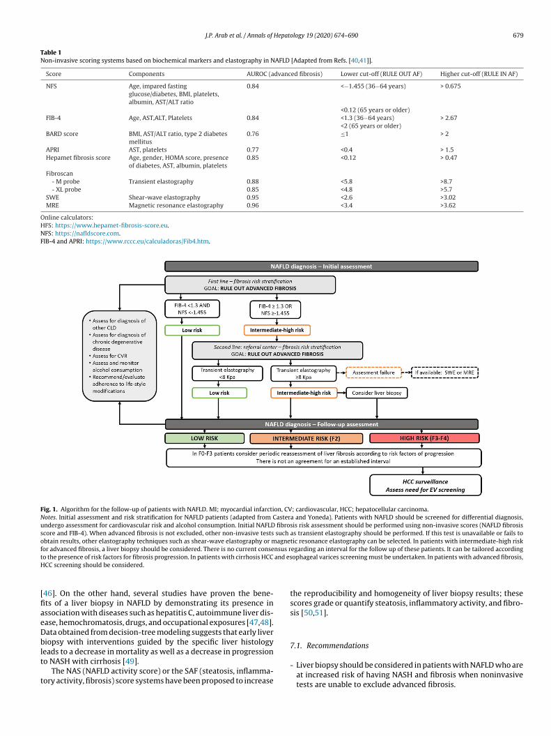
J.P. Arab et al. / Annals of Hepatology 19 (2020) 674–690 679
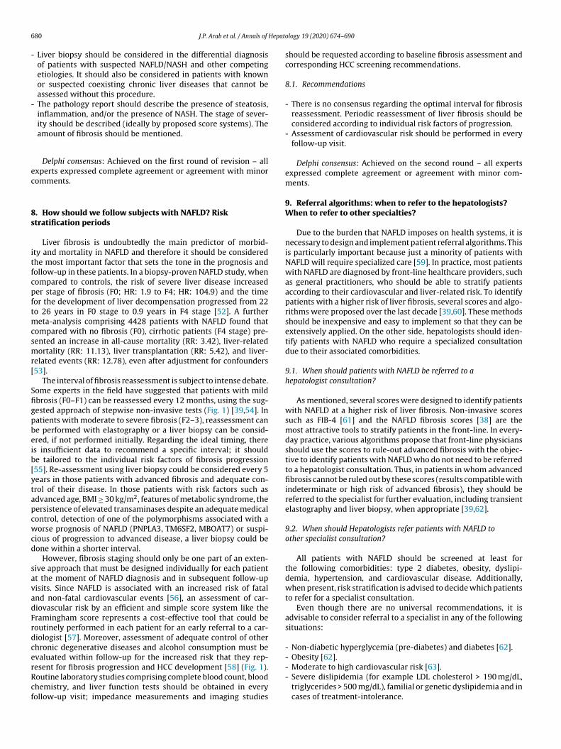
Table 1Non-invasive scoring systems based on biochemical markers and elastography in NAFLD [Adapted from Refs. [40,41]].
Score Components AUROC (advanced fibrosis) Lower cut-off (RULE OUT AF) Higher cut-off (RULE IN AF)
NFS Age, impared fastingglucose/diabetes, BMI, platelets,albumin, AST/ALT ratio
0.84 <−1.455 (36−64 years) > 0.675
<0.12 (65 years or older)FIB-4 Age, AST,ALT, Platelets 0.84 <1.3 (36−64 years) > 2.67
<2 (65 years or older)BARD score BMI, AST/ALT ratio, type 2 diabetes
mellitus0.76 ≤1 > 2
APRI AST, platelets 0.77 <0.4 > 1.5Hepamet fibrosis score Age, gender, HOMA score, presence
of diabetes, AST, albumin, platelets0.85 <0.12 > 0.47
Fibroscan- M probe Transient elastography 0.88 <5.8 >8.7- XL probe 0.85 <4.8 >5.7
SWE Shear-wave elastography 0.95 <2.6 >3.02MRE Magnetic resonance elastography 0.96 <3.4 >3.62
Online calculators:HFS: https://www.hepamet-fibrosis-score.eu.NFS: https://nafldscore.com.FIB-4 and APRI: https://www.rccc.eu/calculadoras/Fib4.htm.
Fig. 1. Algorithm for the follow-up of patients with NAFLD. MI; myocardial infarction, CV; cardiovascular, HCC; hepatocellular carcinoma.Notes. Initial assessment and risk stratification for NAFLD patients (adapted from Castera and Yoneda). Patients with NAFLD should be screened for differential diagnosis,undergo assessment for cardiovascular risk and alcohol consumption. Initial NAFLD fibrosis risk assessment should be performed using non-invasive scores (NAFLD fibrosisscore and FIB-4). When advanced fibrosis is not excluded, other non-invasive tests such as transient elastography should be performed. If this test is unavailable or fails toobtain results, other elastography techniques such as shear-wave elastography or magnetic resonance elastography can be selected. In patients with intermediate-high riskfor advanced fibrosis, a liver biopsy should be considered. There is no current consensus regarding an interval for the follow up of these patients. It can be tailored accordingt and esH
[fiaeDblt
t
o the presence of risk factors for fibrosis progression. In patients with cirrhosis HCC
CC screening should be considered.
46]. On the other hand, several studies have proven the bene-ts of a liver biopsy in NAFLD by demonstrating its presence inssociation with diseases such as hepatitis C, autoimmune liver dis-ase, hemochromatosis, drugs, and occupational exposures [47,48].ata obtained from decision-tree modeling suggests that early liveriopsy with interventions guided by the specific liver histology
eads to a decrease in mortality as well as a decrease in progressiono NASH with cirrhosis [49].
The NAS (NAFLD activity score) or the SAF (steatosis, inflamma-ory activity, fibrosis) score systems have been proposed to increase
ophageal varices screening must be undertaken. In patients with advanced fibrosis,
the reproducibility and homogeneity of liver biopsy results; thesescores grade or quantify steatosis, inflammatory activity, and fibro-sis [50,51].
7.1. Recommendations
- Liver biopsy should be considered in patients with NAFLD who areat increased risk of having NASH and fibrosis when noninvasivetests are unable to exclude advanced fibrosis.
6 Hepat
-
-
ec
8s
itfcpftmcsmr[
Sfigpbeib[ytapcwcd
savadFrdcerRcf
80 J.P. Arab et al. / Annals of
Liver biopsy should be considered in the differential diagnosisof patients with suspected NAFLD/NASH and other competingetiologies. It should also be considered in patients with knownor suspected coexisting chronic liver diseases that cannot beassessed without this procedure.
The pathology report should describe the presence of steatosis,inflammation, and/or the presence of NASH. The stage of sever-ity should be described (ideally by proposed score systems). Theamount of fibrosis should be mentioned.
Delphi consensus: Achieved on the first round of revision – allxperts expressed complete agreement or agreement with minoromments.
. How should we follow subjects with NAFLD? Risktratification periods
Liver fibrosis is undoubtedly the main predictor of morbid-ty and mortality in NAFLD and therefore it should be consideredhe most important factor that sets the tone in the prognosis andollow-up in these patients. In a biopsy-proven NAFLD study, whenompared to controls, the risk of severe liver disease increaseder stage of fibrosis (F0; HR: 1.9 to F4; HR: 104.9) and the timeor the development of liver decompensation progressed from 22o 26 years in F0 stage to 0.9 years in F4 stage [52]. A further
eta-analysis comprising 4428 patients with NAFLD found thatompared with no fibrosis (F0), cirrhotic patients (F4 stage) pre-ented an increase in all-cause mortality (RR: 3.42), liver-relatedortality (RR: 11.13), liver transplantation (RR: 5.42), and liver-
elated events (RR: 12.78), even after adjustment for confounders53].
The interval of fibrosis reassessment is subject to intense debate.ome experts in the field have suggested that patients with mildbrosis (F0–F1) can be reassessed every 12 months, using the sug-ested approach of stepwise non-invasive tests (Fig. 1) [39,54]. Inatients with moderate to severe fibrosis (F2–3), reassessment cane performed with elastography or a liver biopsy can be consid-red, if not performed initially. Regarding the ideal timing, theres insufficient data to recommend a specific interval; it shoulde tailored to the individual risk factors of fibrosis progression55]. Re-assessment using liver biopsy could be considered every 5ears in those patients with advanced fibrosis and adequate con-rol of their disease. In those patients with risk factors such asdvanced age, BMI ≥ 30 kg/m2, features of metabolic syndrome, theersistence of elevated transaminases despite an adequate medicalontrol, detection of one of the polymorphisms associated with aorse prognosis of NAFLD (PNPLA3, TM6SF2, MBOAT7) or suspi-
ious of progression to advanced disease, a liver biopsy could beone within a shorter interval.
However, fibrosis staging should only be one part of an exten-ive approach that must be designed individually for each patientt the moment of NAFLD diagnosis and in subsequent follow-upisits. Since NAFLD is associated with an increased risk of fatalnd non-fatal cardiovascular events [56], an assessment of car-iovascular risk by an efficient and simple score system like theramingham score represents a cost-effective tool that could beoutinely performed in each patient for an early referral to a car-iologist [57]. Moreover, assessment of adequate control of otherhronic degenerative diseases and alcohol consumption must bevaluated within follow-up for the increased risk that they rep-
esent for fibrosis progression and HCC development [58] (Fig. 1).outine laboratory studies comprising complete blood count, bloodhemistry, and liver function tests should be obtained in everyollow-up visit; impedance measurements and imaging studiesology 19 (2020) 674–690
should be requested according to baseline fibrosis assessment andcorresponding HCC screening recommendations.
8.1. Recommendations
- There is no consensus regarding the optimal interval for fibrosisreassessment. Periodic reassessment of liver fibrosis should beconsidered according to individual risk factors of progression.
- Assessment of cardiovascular risk should be performed in everyfollow-up visit.
Delphi consensus: Achieved on the second round – all expertsexpressed complete agreement or agreement with minor com-ments.
9. Referral algorithms: when to refer to the hepatologists?When to refer to other specialties?
Due to the burden that NAFLD imposes on health systems, it isnecessary to design and implement patient referral algorithms. Thisis particularly important because just a minority of patients withNAFLD will require specialized care [59]. In practice, most patientswith NAFLD are diagnosed by front-line healthcare providers, suchas general practitioners, who should be able to stratify patientsaccording to their cardiovascular and liver-related risk. To identifypatients with a higher risk of liver fibrosis, several scores and algo-rithms were proposed over the last decade [39,60]. These methodsshould be inexpensive and easy to implement so that they can beextensively applied. On the other side, hepatologists should iden-tify patients with NAFLD who require a specialized consultationdue to their associated comorbidities.
9.1. When should patients with NAFLD be referred to ahepatologist consultation?
As mentioned, several scores were designed to identify patientswith NAFLD at a higher risk of liver fibrosis. Non-invasive scoressuch as FIB-4 [61] and the NAFLD fibrosis scores [38] are themost attractive tools to stratify patients in the front-line. In every-day practice, various algorithms propose that front-line physiciansshould use the scores to rule-out advanced fibrosis with the objec-tive to identify patients with NAFLD who do not need to be referredto a hepatologist consultation. Thus, in patients in whom advancedfibrosis cannot be ruled out by these scores (results compatible withindeterminate or high risk of advanced fibrosis), they should bereferred to the specialist for further evaluation, including transientelastography and liver biopsy, when appropriate [39,62].
9.2. When should Hepatologists refer patients with NAFLD toother specialist consultation?
All patients with NAFLD should be screened at least forthe following comorbidities: type 2 diabetes, obesity, dyslipi-demia, hypertension, and cardiovascular disease. Additionally,when present, risk stratification is advised to decide which patientsto refer for a specialist consultation.
Even though there are no universal recommendations, it isadvisable to consider referral to a specialist in any of the followingsituations:
- Non-diabetic hyperglycemia (pre-diabetes) and diabetes [62].- Obesity [62].
- Moderate to high cardiovascular risk [63].- Severe dislipidemia (for example LDL cholesterol > 190 mg/dL,triglycerides > 500 mg/dL), familial or genetic dyslipidemia and incases of treatment-intolerance.

Hepato
--
9
-
-
-
-
em
1
idfmitroasmrdtIawse
tdrwpssbpatscNaaac
J.P. Arab et al. / Annals of
Secondary, refractory, occult, or white coat hypertension. Clinical or biochemical findings that are compatible with chronickidney disease (GFR < 60 ml/min/1.73 m2) or obstructive sleepapnea.
.3. Recommendations
FIB-4 and/or NAFLD fibrosis score (ideally both) should be calcu-lated in all patients with NAFLD at their initial assessment.
Patients with indeterminate scores should be referred if they por-tray a high-risk profile (type 2 diabetes or metabolic syndromeand >50 years old).
Patients with high results in at least one of the two scores shouldbe referred to a hepatologist for further evaluation.
Hepatologists should refer patients with significant or difficult-to-treat comorbidities to a specialized consultation.
Delphi consensus: Achieved on the second round – all expertsxpressed complete agreement or agreement with minor com-ents.
0. Alcohol consumption and thresholds in NAFLD
Currently, the NAFLD definition requires the exclusion of signif-cant alcohol consumption. This threshold has been set at 20 g/peray for women and 30 g/day in men, which can be simplifiedor patients as less than 2 and 3 daily drinks for women and
en, respectively. This cut-off point has been adopted for patientnclusion in NAFLD treatment trials [64] due to classic studieshat found that higher alcohol consumption over two years waselated to a higher risk for liver disease [65]. However, the effectf moderate amounts of alcohol consumption (below 30 g/daily)nd health-related risk in the general population is controver-ial: there is data that reflects an association with a lower risk foretabolic syndrome and cardiovascular disease, even though these
esults have geographical disparities. On the other hand, recentata suggests that any amount of alcohol consumption increaseshe risk of cancer-related mortality and all-cause mortality [66].n a recent longitudinal population-based cohort study, moderatelcohol intake (below the selected thresholds for NAFLD definition)as found to be related to incident liver disease, thus suggesting a
afe amount of alcohol consumption regarding liver risk may notxist [67].
In NAFLD patients the effect of moderate alcohol consump-ion is also controversial. There are several observational studiesated back from 2001 that suggest a possible beneficial effectegarding disease progression and liver fibrosis in NAFLD patientsith moderate-low alcohol intake in comparison with abstinentatients. However, the majority have a cross-sectional or retro-pective design and consider present alcohol consumption, withcarce data regarding their alcohol history. Furthermore, selectionias should be considered (completely abstinent patients are morerone to having severe comorbidities or prior heavy alcohol use)nd bias related to self-assessment methods to obtain consump-ion information [68]. Regarding the risk of hepatocarcinoma, aynergistic effect of alcohol and NAFLD has been suggested, indi-ating that alcohol may be an independent risk factor for HCC inAFLD/NASH-cirrhosis [69]. When analyzing cardiovascular risk
nd NAFLD patients, despite evidence suggesting a protective role,recent longitudinal cohort failed to find an association betweenlcohol and cardiovascular risk factors or subclinical markers ofardiovascular disease [70].
logy 19 (2020) 674–690 681
10.1. Recommendations
- Current data do not allow for recommendations of any amountof alcohol consumption to reduce cardiovascular or other health-related risks in NAFLD patients.
- The safest strategy in NAFLD patients is to restrain from anyalcohol consumption. However, if patients choose to consumealcohol, it should be recommended below the threshold of 14and 21 drinks per week for women and men, respectively.
- Patients with NASH and/or moderate to advanced fibrosis shouldabstain from alcohol consumption, due to their high risk for dis-ease progression.
Delphi consensus: Achieved on the first round of revision – allexperts expressed complete agreement or agreement with minorcomments.
11. Hepatocellular carcinoma screening
Although the incidence of hepatocellular carcinoma (HCC) inNAFLD patients has been increasing and it is the fastest-growingcause of HCC in liver transplant candidates [71], the surveillancestrategies are markedly suboptimal. A study showed that NAFLDpatients were less likely to receive HCC screening than chronic hep-atitis C patients (HR 0.44, 95%CI 0.19–0.99, p < 0.05) [72]. Thus, thereis a need to improve the coverage of this intervention in high-riskNAFLD patients.
NAFLD has been related to an increased risk of HCC in thepresence of cirrhosis. Although the annual incidence of HCC inNAFLD-related cirrhosis is lower than in that related to HCV, itis significantly higher than in controls. A study that followed 195patients with NAFLD related cirrhosis during a median of 3.2 yearsfound an annual incidence of HCC of 2.6% [58]. However, HCC hasalso been reported in NAFLD patients with liver fibrosis and even inthe absence of fibrosis [73]. Thus, the need to delimit a populationwhere HCC screening is cost-effective is warranted.
Based on cost-effectiveness studies it has been suggestedthat in a population with an annual incidence of ≥1.5% HCCscreening is recommended assuming the availability of therapiesand acceptable performance status and liver function. Therefore,HCC screening in NAFLD-related cirrhosis is mandatory. How-ever, screening should not be performed if the treatment ofHCC is not feasible or if it will not improve survival. Anotherstudy showed that, although higher than in controls, the risk ofHCC in non-cirrhotic NAFLD patients is markedly lower than incirrhotic patients (0.08/1000 person-years vs. 10.6/1000 person-years). Thus, the risk in non-cirrhotic NAFLD patients doesn’t reachthe minimum threshold to perform cost-effective HCC surveillance[74]. Notably, the authors found that the HCC risk in those patientswithout cirrhosis but with a high FIB-4 score result was 0.39/1000person-years compared to 0.04/1000 person-years on those with-out cirrhosis and a persistently low FIB-4 result.
Importantly, obesity, tobacco, and alcohol consumption have allbeen associated with an increased risk of HCC in NAFLD patients.Thus, the recommendation of weight loss, increase physical activ-ity, and to discontinue alcohol and tobacco consumption should bereinforced [73].
A proper strategy for effective HCC screening needs to be fol-lowed. An ultrasonography (US) performed every six months is thepreferred method considering that it was employed in most studies
and has an acceptable diagnostic yield [75]. Notably, increasing itsfrequency doesn’t improve the detection rate [76]. The use of alpha-fetoprotein in addition to the US does not add a clear benefit, thusits use is not mandatory [77].
6 Hepat
iosea
1
-
-
e
1
ade5loma
5lthtbl
iiatfc([hh
ipTir
82 J.P. Arab et al. / Annals of
A few observational studies have reported that the likelihood ofnadequate ultrasound quality is significantly higher in overweightr obese patients. Thus, to employ an alternative radiologic methoduch as CT or MRI is recommendable if the liver cannot be properlyvaluated using US [73]. Nonetheless, routinely use of CT or MRI as
screening method seems non-cost-effective [78].
1.1. Recommendations
Universal HCC screening is not recommended in NAFLD patients.HCC must be performed in all patients with cirrhosis and shouldbe considered in patients with advanced fibrosis due to NAFLD ifcurative or palliative therapies are available and without reducedshort-term survival.
HCC screening must be performed employing ultrasonographyevery six months with or without a simultaneous measurementof alpha-fetoprotein levels. Only if a proper liver visualization isnot achieved employing ultrasonography, a different radiologictest can be employed (CT scan or MRI).
Delphi consensus: Achieved on the first round of revision – allxperts expressed complete agreement.
Management:
2. Is diet effective for NAFLD treatment?
Diet, as part of lifestyle interventions, is key in NAFLD treatment,lthough the effect of these interventions is often difficult to assessue to the lack of standardization of the study designs. It has beenstablished that a decreased caloric intake and reductions of at least% of bodyweight achieve a significant reduction in intrahepatic
ipid content; furthermore, a decrease of 7−10% of body weightr greater is ideal, because it has been associated with improve-ent in all the histological parameters (steatosis, inflammation,
nd fibrosis) in a higher percentage of subjects [62,79–81].Experts recommendations suggest a caloric reduction between
00−1000 kcal/day, or total intake between 1200−1800 kcal/day,ow in fat and carbohydrates and rich in fiber, avoiding fruc-ose, especially if it derives from sugary drinks [62,79], which areighly consumed in Latin-America. Carbohydrates, especially fruc-ose and/or sucrose added to beverages, foods, and desserts, haveeen shown to be related to the development of NAFLD, increasing
iver triglyceride synthesis.Studies have shown that the Mediterranean diet is beneficial
ndependently of achieving weight loss. The Mediterranean diets characterized by reduced carbohydrate intake, especially sug-rs and refined carbohydrates (40% of the calories vs. 50–60% in aypical low-fat diet), and increased monounsaturated and omega-3atty acid intake (40% of the calories as fat vs. up-to 30% in a typi-al low-fat diet), and has been shown to improve insulin resistanceIR) and steatosis assessed by MRI spectroscopy or elastography81–84]. Polyunsaturated fatty acids, particularly omega-3 types,ave been shown to improve insulin sensitivity and reduce intra-epatic triglyceride content, thus improving steatohepatitis [83].
Finally, regular coffee consumption, mainly due to its antiox-dant effect, would have a beneficial role in both the general
opulation and in patients with NASH, improving liver fibrosis.hese effects on fibrosis are best seen in patients with low levels ofnsulin resistance [85,86]. Two or three cups of coffee per day areecommended.ology 19 (2020) 674–690
12.1. Recommendations
- A reduction in calorie intake to 1200−1800 kcal/day and theachievement of a weight reduction of 7−10% is beneficial inNAFLD management.
- A low-refined sugar diet based on the Mediterranean diet is ben-eficial in the management of NAFLD.
- The consumption of coffee in moderate amounts is recommendedin patients with NAFLD.
Delphi consensus: Achieved on the first round of revision – 4 outof 5 experts expressed complete agreement.
13. Is exercise effective for NAFLD treatment?
Exercise has shown to be an effective intervention to reduceintrahepatic triglyceride content, independent of weight loss inpatients with NAFLD [80,87]. A meta-analysis that included 28randomized controlled trials, showed that exercise, independentof changes in diet, is associated with a significant reduction inintrahepatic triglyceride (IHTG) content (p < 0.0001), and with areduction in ALT (p < 0.004) and AST (p = 0.01) values [88]. Regularexercise not only modifies the IHTG but improves cardiovas-cular comorbidities and insulin-resistance specifically associatedwith NAFLD [89]. The data is less clear regarding the type, fre-quency, and intensity of exercise. There is consensus amongexperts that exercise should be prescribed in 3–5 sessions perweek (150−200 min/week) of moderate-intensity aerobic exerciseassociating sessions of resistance exercise [62]. Both aerobic andresistance exercise are effective in reducing hepatic steatosis, insessions of at least three times a week for twelve weeks, althoughin resistance exercise, energy consumption and exercise intensitywere lower (MET 3.5 vs. 4.8 in aerobic exercise) [87]. A resistanceexercise program lasting 24 weeks showed similar benefits to thoseexperienced by aerobic exercise in terms of IHTG, ALT levels, andinsulin sensitivity [90].
A study compared the effect of 12 weeks of an exercise programon obese men with NAFLD, randomized into 3 groups (resistanceexercise (n = 20) vs. high-intensity interval aerobic exercise (n = 21)vs. moderate-intensity aerobic exercise (n = 20)). Besides clinicaland biochemical parameters, they used MRI to assess abdominalfat distribution, in addition to controlled-attenuation parameterand spectroscopy to assess intrahepatic lipid content. The authorsconcluded that intrahepatic lipid content was similar in all threegroups (−14.3% vs. −13.7% vs. −14.3%), with no specific changesin weight or visceral fat. Hepatic stiffness significantly improvedonly in the group randomized to high-intensity interval exercise(−16.8%) [91]. Recently, an 8-week high-intensity interval aero-bic exercise showed a beneficial effect on IHTG, visceral lipids, andquality of life in diabetic obese patients with NAFLD [92].
13.1. Recommendations
- Regular exercise (aerobic, resistance, or combined) should be partof the first line of non-pharmacological treatment of NAFLD.
Delphi consensus: Achieved on the first round of revision – allexperts expressed complete agreement.
14. Are antioxidants, insulin-sensitizers, and lipid-loweringdrugs effective for the treatment of NAFLD?
Despite the imperative need for effective pharmacotherapy forNASH patients, there is no standard-of-care treatment currentlyavailable. Several treatment strategies have been evaluated, with
Hepatology 19 (2020) 674–690 683
dh
1
spmPddio[tirnsiechhpt
1
dgsmialm
fmemiwaw[cc[
1
tArtsiim[
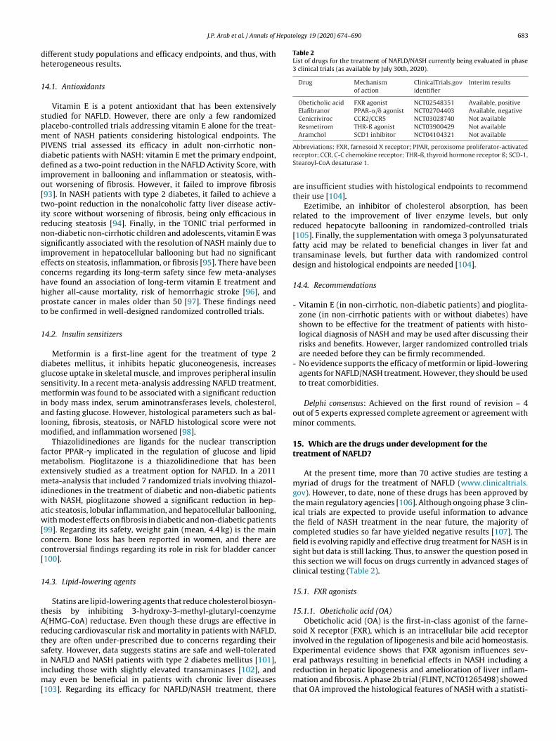
Table 2List of drugs for the treatment of NAFLD/NASH currently being evaluated in phase3 clinical trials (as available by July 30th, 2020).
Drug Mechanismof action
ClinicalTrials.govidentifier
Interim results
Obeticholic acid FXR agonist NCT02548351 Available, positiveElafibranor PPAR-�/� agonist NCT02704403 Available, negativeCenicriviroc CCR2/CCR5 NCT03028740 Not availableResmetirom THR-ß agonist NCT03900429 Not availableAramchol SCD1 inhibitor NCT04104321 Not available
J.P. Arab et al. / Annals of
ifferent study populations and efficacy endpoints, and thus, witheterogeneous results.
4.1. Antioxidants
Vitamin E is a potent antioxidant that has been extensivelytudied for NAFLD. However, there are only a few randomizedlacebo-controlled trials addressing vitamin E alone for the treat-ent of NASH patients considering histological endpoints. The
IVENS trial assessed its efficacy in adult non-cirrhotic non-iabetic patients with NASH: vitamin E met the primary endpoint,efined as a two-point reduction in the NAFLD Activity Score, with
mprovement in ballooning and inflammation or steatosis, with-ut worsening of fibrosis. However, it failed to improve fibrosis93]. In NASH patients with type 2 diabetes, it failed to achieve awo-point reduction in the nonalcoholic fatty liver disease activ-ty score without worsening of fibrosis, being only efficacious ineducing steatosis [94]. Finally, in the TONIC trial performed inon-diabetic non-cirrhotic children and adolescents, vitamin E wasignificantly associated with the resolution of NASH mainly due tomprovement in hepatocellular ballooning but had no significantffects on steatosis, inflammation, or fibrosis [95]. There have beenoncerns regarding its long-term safety since few meta-analysesave found an association of long-term vitamin E treatment andigher all-cause mortality, risk of hemorrhagic stroke [96], androstate cancer in males older than 50 [97]. These findings needo be confirmed in well-designed randomized controlled trials.
4.2. Insulin sensitizers
Metformin is a first-line agent for the treatment of type 2iabetes mellitus, it inhibits hepatic gluconeogenesis, increaseslucose uptake in skeletal muscle, and improves peripheral insulinensitivity. In a recent meta-analysis addressing NAFLD treatment,etformin was found to be associated with a significant reduction
n body mass index, serum aminotransferases levels, cholesterol,nd fasting glucose. However, histological parameters such as bal-ooning, fibrosis, steatosis, or NAFLD histological score were not
odified, and inflammation worsened [98].Thiazolidinediones are ligands for the nuclear transcription
actor PPAR-� implicated in the regulation of glucose and lipidetabolism. Pioglitazone is a thiazolidinedione that has been
xtensively studied as a treatment option for NAFLD. In a 2011eta-analysis that included 7 randomized trials involving thiazol-
dinediones in the treatment of diabetic and non-diabetic patientsith NASH, pioglitazone showed a significant reduction in hep-
tic steatosis, lobular inflammation, and hepatocellular ballooning,ith modest effects on fibrosis in diabetic and non-diabetic patients
99]. Regarding its safety, weight gain (mean, 4.4 kg) is the mainoncern. Bone loss has been reported in women, and there areontroversial findings regarding its role in risk for bladder cancer100].
4.3. Lipid-lowering agents
Statins are lipid-lowering agents that reduce cholesterol biosyn-hesis by inhibiting 3-hydroxy-3-methyl-glutaryl-coenzyme(HMG-CoA) reductase. Even though these drugs are effective ineducing cardiovascular risk and mortality in patients with NAFLD,hey are often under-prescribed due to concerns regarding theirafety. However, data suggests statins are safe and well-tolerated
n NAFLD and NASH patients with type 2 diabetes mellitus [101],ncluding those with slightly elevated transaminases [102], anday even be beneficial in patients with chronic liver diseases103]. Regarding its efficacy for NAFLD/NASH treatment, there
Abbreviations: FXR, farnesoid X receptor; PPAR, peroxisome proliferator-activatedreceptor; CCR, C-C chemokine receptor; THR-ß, thyroid hormone receptor ß; SCD-1,Stearoyl-CoA desaturase 1.
are insufficient studies with histological endpoints to recommendtheir use [104].
Ezetimibe, an inhibitor of cholesterol absorption, has beenrelated to the improvement of liver enzyme levels, but onlyreduced hepatocyte ballooning in randomized-controlled trials[105]. Finally, the supplementation with omega 3 polyunsaturatedfatty acid may be related to beneficial changes in liver fat andtransaminase levels, but further data with randomized controldesign and histological endpoints are needed [104].
14.4. Recommendations
- Vitamin E (in non-cirrhotic, non-diabetic patients) and pioglita-zone (in non-cirrhotic patients with or without diabetes) haveshown to be effective for the treatment of patients with histo-logical diagnosis of NASH and may be used after discussing theirrisks and benefits. However, larger randomized controlled trialsare needed before they can be firmly recommended.
- No evidence supports the efficacy of metformin or lipid-loweringagents for NAFLD/NASH treatment. However, they should be usedto treat comorbidities.
Delphi consensus: Achieved on the first round of revision – 4out of 5 experts expressed complete agreement or agreement withminor comments.
15. Which are the drugs under development for thetreatment of NAFLD?
At the present time, more than 70 active studies are testing amyriad of drugs for the treatment of NAFLD (www.clinicaltrials.gov). However, to date, none of these drugs has been approved bythe main regulatory agencies [106]. Although ongoing phase 3 clin-ical trials are expected to provide useful information to advancethe field of NASH treatment in the near future, the majority ofcompleted studies so far have yielded negative results [107]. Thefield is evolving rapidly and effective drug treatment for NASH is insight but data is still lacking. Thus, to answer the question posed inthis section we will focus on drugs currently in advanced stages ofclinical testing (Table 2).
15.1. FXR agonists
15.1.1. Obeticholic acid (OA)Obeticholic acid (OA) is the first-in-class agonist of the farne-
soid X receptor (FXR), which is an intracellular bile acid receptorinvolved in the regulation of lipogenesis and bile acid homeostasis.Experimental evidence shows that FXR agonism influences sev-
eral pathways resulting in beneficial effects in NASH including areduction in hepatic lipogenesis and amelioration of liver inflam-mation and fibrosis. A phase 2b trial (FLINT, NCT01265498) showedthat OA improved the histological features of NASH with a statisti-
6 Hepat
caAiFtoai52fsp3
1
I1rrrces2
1
nwart
1
reall(w
1
c1iiiswa≥2r
1
ts
84 J.P. Arab et al. / Annals of
ally significant improvement in fibrosis. More recent data, fromn interim analysis of an international phase 3 trial (REGENER-TE, NCT0254835) that evaluated two doses of OA 10−25 mg/day
n adult patients with definite NASH and fibrosis stages F2–F3, or1 with at least one accompanying comorbidity showed that whilehe co-primary endpoint of NASH resolution without worseningf fibrosis was not reached, all variables of the histologic NAFLDctivity score improved [108]. The endpoint for the improvementn fibrosis was achieved by 37 (12%) patients in the placebo group,5 (18%) in the OA 10-mg group (p = 0.045), and 71 (23%) in the OA5-mg group (p = 0.0002). Thus, OA is a potentially effective drugor NASH treatment pending the long-term follow-up phase of thistudy. Other FXR agonists (i.e. tropifexor) have also been tested inhase 2 trials with promising results [109] and are entering phase
testing.
5.1.2. ElafibranorElafibranor is a PPAR-�/� agonist being tested in the RESOLVE-
T phase 3 clinical trial. Results of an interim analysis of data from070 patients with NASH and F2 and F3 fibrosis stage were recentlyeleased and showed that the predefined primary endpoint of NASHesolution without worsening of fibrosis was not met [110]. Theesponse rate in those patients receiving the study drug was 19.2%ompared to 14.7% for patients in the placebo arm. The secondaryndpoint of at least one stage of liver fibrosis improvement wasimilar in patients receiving elafibranor and placebo (24.5% vs.2.4%).
5.1.3. Cenicriviroc (CVC)CVC is a CCR2/CCR5 receptor inhibitor that after a positive sig-
al in a phase 2b study showing improvement in fibrosis and noorsening of NASH compared with placebo [111] is being tested in
large multicenter phase 3 study (AURORA trial) [112]. The finalesults of the study are still pending at the time of the writing ofhis document.
5.1.4. ResmetiromResmetirom is an orally active, selective thyroid hormone
eceptor-� agonist (thyromimetic) that has shown beneficialffects in NASH in both preclinical and clinical studies. Of note,
phase 2b trial [113] showed that in addition to decreasingiver fat, the drug was associated with NASH histological reso-ution. Currently, resmetirom is being tested in a Phase 3 studyMAESTRO-NASH trial), which is expected to enroll 2000 patientsith biopsy-proven NASH.
5.1.5. AramcholAramchol is a fatty acid bile acid conjugate (arachidyl amido
holanoic acid) able to inhibit the stearoyl-CoA desaturase 1 (SCD-) enzyme, which is a key enzyme in fatty acid metabolism. SCD-1
nhibition determines a reduction in hepatic lipogenesis and anncrease in mitochondrial �-oxidation of fatty acids, thus decreas-ng in liver fat content. Preliminary results of a phase 2b trialhowed positive effects with NASH resolution without fibrosisorsening occurring more often (16.7%) in patients receiving the
ctive drug compared to those receiving placebo (5.0%). Also, a1 stage fibrosis reduction without NASH worsening was seen in9.5% of aramchol treated patients compared with 17.5% of patientseceiving placebo [114], a phase 3 trial is underway.
5.1.6. Combination therapiesThe use of drug combinations attempting to tailor NAFLD/NASH
reatment according to the predominant pathway involved, diseasetage, and associated conditions (e.g. presence of type 2 diabetes) is
ology 19 (2020) 674–690
being explored in several phase 2 trials. This approach is promisingbut more data is needed [115,116].
16. Is bariatric (metabolic) surgery effective for thetreatment of NAFLD?
Bariatric surgery (BS) is an effective treatment for obesity.Regarding NAFLD/NASH, there is clinical and experimental evi-dence that BS is cost-effective and has beneficial effects, inducinghistological resolution of NASH through weight loss-dependentand independent mechanisms, involving hormonal and bile acidmetabolism changes [117]. Indeed, steatosis, inflammation, andfibrosis appear to improve or completely resolve in the majorityof patients after BS–induced weight loss, as it was demonstrated in2008 by a meta-analysis comprising 15 studies, including 4 Latin-American cohorts [118]. However, most data evaluating NAFLDand BS are derived from retrospective studies. A recent Frenchstudy showed that BS provides a long-term resolution of NASHand regression of fibrosis. They observed the resolution of NASHin paired liver biopsies from 84% of patients 5 years after the pro-cedure. Additionally, they concluded that the reduction of fibrosis isprogressive, beginning during the first year and continuing through5 years. Even before this study, some experts considered that BSshould be offered as a treatment for NASH without the need forfurther clinical trials [117].
Some comprehensive or systematic reviews and meta-analysishave been published in the last few years [104,119–121]. The mostrecent one analyzed data from 32 studies comprising 3093 biopsyspecimens, with a biopsy-confirmed resolution of steatosis in 66%of patients, inflammation in 50%, ballooning degeneration in 76%,and fibrosis in 40%. Mean NAFLD activity score (NAS) also pre-sented a significant reduction (mean of −2.39 points). Moreover,the authors emphasized that the overall grading of recommen-dations and evidence quality was very low and concluded thatrandomized studies are still needed to better evaluate the effectof BS in this population [121], especially to consider NASH as a soleindication for metabolic surgery regardless of BMI.
Other issues deserve further discussion, such as concerns aboutBS in patients with advanced liver disease due to NAFLD, as wellas the choice of the type of procedure. Currently, patients withcompensated NASH cirrhosis (Child–Pugh A) may represent candi-dates for BS, especially those with MELD less than 10 and withoutsignificant portal hypertension. However, no clear recommenda-tions can be made, due to the lack of solid data [117]. There isdebate whether these patients should undergo Roux-en-Y gastricbypass (RYGB) or sleeve gastrectomy (SG). Liver decompensation ismore common after primarily malabsorptive procedures, such asjejunoileal bypass, that is no longer performed [117]. In a recentmeta-analysis that compared RYGB and SG on NAFLD patients con-sidering AST, ALT, NAS, and NAFLD fibrosis score as outcomes.The authors demonstrated that BS significantly improved thesebiochemical and histologic parameters but failed to indicate supe-riority between RYGB and SG in ameliorating NAFLD [120].
16.1. Recommendations
- Bariatric (metabolic) surgery should be considered in obesepatients with NAFLD unresponsive to clinical and pharmacolog-ical management, as it is related to improvement in histologicaloutcomes, including fibrosis. However, the recommendation
should be weighted with the risk of complications.- Additional randomized studies addressing BS in NAFLD areneeded, as well as to define the best surgical technique in thispopulation.

Hepato
om
1(
trmcmcfstprrpa
iFRhpTcddTItcsoasci
bscSatahhet
1
-
-
e
J.P. Arab et al. / Annals of
Delphi consensus: Achieved on the first round of revision – 4ut of 5 experts expressed complete agreement or agreement withinor comments.
7. How should we manage comorbidities in NAFLDhypertension, cardiovascular disease)?
During the last decade, it has been observed that NAFLD leadso an increased cardiovascular risk with an acceleration of arte-iosclerosis and events related to it, being the main cause of itsorbidity and mortality [122]. The higher incidence of cardiovas-
ular events in the NAFLD population could be explained by severalechanisms. Patients with NAFLD typically meet the diagnostic
riteria for metabolic syndrome and therefore have multiple riskactors for cardiovascular disease [123]. Likewise, the presence ofystemic inflammation in combination with metabolic abnormali-ies may act synergistically to increase cardiovascular risk in theseatients. Pre-hypertension and hypertension are both significantisk factors for the development of NAFLD independent of otherisk factors. Controlled blood pressure appears to be independentlyrotective of NAFLD and the absence of hypertension also protectsgainst moderate-to-severe hepatic fibrosis risk [124].
The estimation of cardiovascular risk is recommended and var-ous easy-to-use tools are available for this purpose, such as theramingham Score or the Atherosclerotic Cardiovascular Diseaseisk Estimator of the American College of Cardiology [57,63,125],owever, traditional scores have limitations. On the one hand, theseredictive tools were not specifically developed for NAFLD patients.he performance of these scores is suboptimal because traditionalardiovascular risk factors do not fully explain the increased car-iovascular risk in patients with NAFLD, and current risk functionso not represent other contributing factors (i.e. inflammation).hus, a multidisciplinary and step-wise approach is recommended.n an attempt to address these limitations, it has been proposedo add another predictive factor, such as the detection of sub-linical atheromatosis. Indicators of early vascular atherosclerosisuch as carotid atherosclerotic plaques diagnosed by ultrasoundr coronary calcium score quantified by computed tomographyre frequently altered in patients with NAFLD. Two meta-analyseshowed that the patients with NAFLD had higher coronary cal-ium score and carotid plaque prevalence compared with controls,ndependent of traditional risk factors [126,127].
A considerable amount of literature has shown the associationetween NAFLD and cardiovascular disease. A recent meta-analysishowed an elevated risk of cardiovascular events in NAFLD patientsompared to controls (RR 1.77; 95%CI 1.26–2.48, p < 0.001) [128].tatin utilization is the cornerstone of cardiovascular risk man-gement. To determine whether a patient is a candidate for statinherapy, clinicians must first determine the patient’s risk of having
future cardiovascular event. The statin use has historically beenampered, in individuals with liver disease, owing to the fear ofepatotoxicity. However, studies suggest that statins are not onlyffective in reducing cardiovascular events but may also exert mul-iple beneficial effects on the liver [129].
7.1. Recommendations
Risk scores should be used for the initial stratification of cardio-vascular risk.
NAFLD patients should get regular blood pressure measurementand lipids checks during clinical visits, with specific thresholds
for intervention according to current guidelines.Delphi consensus: Achieved on the first round of revision – allxperts expressed complete agreement.
logy 19 (2020) 674–690 685
18. How should we manage comorbidities in NAFLD(obesity, T2DM)?
NAFLD is considered the hepatic manifestation of metabolicsyndrome. Therapeutic principles for the management of patientswith NAFLD, obesity, and T2DM are mainly based on the imple-mentation of lifestyle optimization strategies aiming to achievesignificant weight loss, which ideally should be planned in thesetting of multidisciplinary teams. Currently, there is no particu-lar drug approved for the treatment of NAFLD/NASH in patientswith T2DM. Although, several approved diabetic medications holdpromise for NAFLD/NASH treatment and several NASH-specificdrugs are in evaluation in phase III RCTs.
The PIVENS trial (comparing vitamin E, pioglitazone, andplacebo) did not include diabetic subjects. Later publications sug-gested benefit of vitamin E in an observational study [130] butlimited benefit in a RCT in patients with diabetes [94]. Pioglitazoneefficacy in NAFLD therapy has been evaluated in five randomizedcontrolled trials (498 patients) with consistent results in resolutionof NASH and improvement in ballooning or inflammation, while theimprovement of fibrosis was reported in some of the studies [15].Most of them included diabetic subjects, showing improvement insteatosis and fibrosis [131]. However, some safety issues have beenraised with vitamin E and pioglitazone therapies.
From the standpoint of diabetes management, appropriateglycemic control is associated with a reduction in steatosis, adecrease in serum levels of aminotransferases, and eventuallyimprovement of fibrosis. Interestingly, there are medications forthe treatment of diabetes with potential efficacy for NAFLD/NASH.Metformin, although it hasn’t been shown to be associated with asignificant hepatic histological improvement, has shown to reducethe incidence of HCC of the diabetic population in basic stud-ies [132]. Other anti-diabetic drugs have been associated withpotentially beneficial effects in NAFLD. Among the most promis-ing therapies, Liraglutide, a GLP-1 agonist, is of special interest.Liraglutide has demonstrated to induce a significant improve-ment in metabolic control and a reduction in cardiovascularevents and deaths in diabetic subjects (HR 0.78 95%CI 0.66−0.93;N = 9340, follow-up 5 years) [133] and diabetes prevention insubjects with prediabetes (HR: 0.21 95%CI 0.13−0.34, N = 2254)[134]. Regarding NAFLD, a small (N = 52) phase 2 trial in non-diabetic subjects [135] demonstrated a significant increase in NASHresolution using Liraglutide 1.8 mg/d (39% vs. 9%, P = 0.019). Exe-natide, another GLP-1 analog, has demonstrated reducing weightand liver enzymes [136] (ongoing trial in NAFLD -NCT01208649-).Semaglutide (GLP-1 agonist administered once weekly) is cur-rently being assessed in a phase III trial in NASH. Data on theeffects of dipeptidyl peptidase 4 inhibitors (DPP4) in NAFLD shownconflicting results [137], thus further studies are needed. Sodium-glucose cotransporter2 (SGLT2) inhibitors have shown to havereno- and cardio-protective effects and are recommended as anadd-on treatment when glycemic control is not achieved in diabet-ics. Empagliflozin has been shown to reduce cardiovascular deaths(HR 0.62 95%CI 0.49−0.77), and hospitalizations for heart failure(HR 0.65 95% CI 0.5−0.85) [138,139]. The E-LIFT study addressedthe effect of the empagliflozin in T2DM patients with NAFLD show-ing that it was associated with a reduction in steatosis (assessedby MRI-PDFF) and in serum levels of aminotransferases [140]. Sim-ilar results were found with dapagliflozin [141] and other SGLT2[142,143]. Regarding histological outcomes, only one very smallcohort study (N = 5) have reported improvement in steatosis andNAS score with canagliflozin [144]. Thus, although some promising
data has been published, the effectiveness of this class of drugs forNASH treatment remains to be determined in future RCTs.Appropriate treatment of dyslipidemia in the T2DM populationis also very relevant and statins are effective and safe in subjects

6 Hepat
wdoi
1
-
-
-
om
1
pLbtu
1
Nhd≥Nuabt
1
hdaalsadd(HcTaotiwpe
A summary of the recommendations is presented in Table 3.
Conflict of interest
The authors have no conflicts of interest to declare.
Table 3Summary of recommendations.
Definition and epidemiologyThe prevalence of NAFLD ranges from 14.3% to 35.2% in Latin American
population-based studies, but most of them were performed more than 10years ago.
86 J.P. Arab et al. / Annals of
ith NAFLD [145]. Finally, a combination approach with multiplerug targeting different pathways will likely represent the futuref NASH therapeutics but more robust evidence for this approachs needed.
8.1. Recommendations
Therapeutic principles for the management of patients withNAFLD and T2DM are mainly based on the implementation oflifestyle optimization strategies aiming to achieve significantweight loss, which ideally should be planned in the setting ofmultidisciplinary teams.
Metformin should be considered to be a first-line option for thetreatment of T2DM but with minimal benefit for treating NASH.
Glucagon-like peptide-1 (GLP-1) agonists and sodium-glucosecotransporter 2 (SGLT2) inhibitors are promising agents for thetreatment of T2DM in NAFLD.
Delphi consensus: Achieved on the first round of revision – 4ut of 5 experts expressed complete agreement or agreement withinor comments.
9. When is liver transplantation indicated in NAFLD?
NASH-related cirrhosis is a growing indication for liver trans-lantation (LT) [146,147]. In western countries, the percentage ofT for hepatitis C has declined in the last decades, and NASH hasecome one of the most common indications. In a recent multicen-er Latin America study, NASH-cirrhosis was the third etiology ofnderlying disease in HCC candidates for LT [148].
9.1. Indication for LT in NAFLD patients
Indications for LT should be considered once a patient withASH-cirrhosis has developed a complication such as ascites,epatic encephalopathy, variceal hemorrhage, or hepatocellularysfunction that results in a MELD-Na score ≥15 or Child–Pugh7 points [146,147]. LT is recommended as the first-line option forASH-cirrhosis and HCC within Milan criteria when the tumor isnsuitable for resection, in the absence of tumor vascular invasionnd extrahepatic metastases. Patients beyond the Milan criteria cane considered for LT after successful downstaging with locoregionalherapy [148].
9.2. Comorbidities of NAFLD patients in the LT setting
Patients with NASH-cirrhosis on the waiting list for LT have aigh prevalence of metabolic comorbidities: obesity (53.4–68%),iabetes (49–72.9%), and arterial hypertension (37.5–75%) [149]. In
recent meta-analysis, the presence of NAFLD was associated with 65% increased risk of developing fatal and nonfatal cardiovascu-ar events over a median follow-up of almost 7 years. Based on thistrong link, most societies suggest that NAFLD should be consideredn independent risk factor for cardiovascular events. Unfortunately,efining the best approach to assess cardiovascular risk in LT can-idates is a focus of intense and moving debate [56]. Obesity aloneBMI ≥ 30 kg/m2) does not constitute a contraindication for LT.owever, in the presence of medical comorbidities, particularlyoncurrent diabetes, rigorous patient selection is recommended.he presence of obesity represents technical challenges and isssociated with post-transplant complications, but the impact ofbesity on long-term mortality after LT is debated [147]. Despitehe presence of obesity, sarcopenia is the most common finding
n NASH cirrhosis and is associated with higher mortality whileaiting for LT as well as increased hospitalization rates in theost-transplant setting [150]. The prevalence of cardiovascular dis-ases in patients transplanted by NASH is high: coronary artery
ology 19 (2020) 674–690
disease, 20%; congestive heart failure, 7%; stroke, 8% [151]. Cardio-vascular mortality is responsible for a higher proportion of deathsamong NASH patients compared to non-NASH transplant recip-ients. Despite this, NASH patients have shown to have similaroverall post-transplant survival rates compared to patients with-out NASH. These results could be explained in part by the lowerrisk of graft failure compared with other indications, particularlyhepatitis C in the pre-direct acting antiviral era [149]. NASH is anindependent risk factor for LT renal dysfunction before and afterLT, thus appropriate screening and management of kidney diseaseis recommended. In addition, NASH cirrhosis is the most rapidlygrowing indication for simultaneous liver-kidney transplantationin the United States and Europe [152].
19.3. Recurrent and de novo NAFLD/NASH after LT
Recurrent and de novo NAFLD occurs in over one-half of recip-ients as soon as 1 year after LT. NASH recurs in most patients afterLT (53% at 1 year), whereas de novo NASH is less frequent (13%at 1 year). NAFLD/NASH after LT is associated with metabolic riskfactors; in a recent meta-analysis, post-LT body mass index andhyperlipidemia were the most consistent independent predictorsof outcomes. The impact of recurrent or de novo NAFLD/NASH afterLT on allograft and patient survival is unclear. In patients trans-planted for NASH cirrhosis, immunosuppressive protocols withshort-term and low dose steroids and calcineurin inhibitor mini-mization should be preferred [153].
19.4. Recommendations
- Indication for liver transplantation should be considered oncea patient with NASH-related cirrhosis has developed an indexcomplication, hepatocellular dysfunction (MELD-Na Score ≥15 orChild–Pugh ≥7), and/or hepatocellular carcinoma within Milancriteria unsuitable for resection in the absence of tumor vascularinvasion and extrahepatic metastases.
- Liver transplant candidates with NASH-related cirrhosis shouldbe considered at higher risk of developing cardiovascular eventsbefore and after transplantation. There is not enough evidence tosupport a different approach to the pre-liver transplant cardio-vascular assessment.
- Liver transplantation in patients with NASH-related cirrhosis(w/o HCC) has a similar post-transplant survival as other livertransplant etiologies. NAFLD/NASH usually recurs after transplantbut the impact of the recurrence on allograft and patient survivalis unclear.
Delphi consensus: Achieved on the first round of revision – allexperts expressed complete agreement or agreement with minorcomments.
Summary
The variances in the prevalence of NAFLD/NASH may be related to differencesin both genetic and environmental risk factors. As the frequency of obesityand metabolic syndrome continues to increase, further studies onNAFLD/NASH prevalence should be performed in the general population ofLatin America.

J.P. Arab et al. / Annals of Hepatology 19 (2020) 674–690 687
Table 3 (Continued)
Pathogenesis, genetics, and genetic testingCarriers of the PNPLA3 I148M and the TM6SF2 E167K variants have a higher
liver fat content and increased risk of NASH.Genotyping might be considered in the Latin American population, however, it
still has not proven to be able to guide therapy on an individual basis and iscostly. Thus, its role in clinical practice is yet to be established.
How should we diagnose NAFLD? (screening, clinic, laboratory, and images)NAFLD screening is not recommended in the general population. NAFLD
screening is recommended for patients with repeatedly altered liverenzymes, features of metabolic syndrome, or obesity (BMI > 30). In patientswith a high-risk profile (type 2 diabetes or metabolic syndrome and >50years old) NASH and liver fibrosis diagnosis should be undertaken.
The recommended method for initial screening for NAFLD in clinical practice isan abdominal ultrasound.
How should we use non-invasive assessment and images in NAFLD? (riskstratification)
NAFLD Fibrosis Score and FIB-4 are useful tools for the initial assessment offibrosis in NAFLD patients
When NFS score and/or FIB-4 score are unable to exclude advanced fibrosis, anelastography based method is the suggested method in the risk stratificationalgorithm. Vibration controlled transient elastography is the best validatedand available tool; other elastography techniques should be consideredupon availability.
When non-invasive methods are unable to exclude significant fibrosis, liverbiopsy should be considered.
When should we perform a liver biopsy in NAFLD?Liver biopsy should be considered in patients with NAFLD who are at increased
risk of having NASH and fibrosis when noninvasive tests are unable toexclude advanced fibrosis.
Liver biopsy should be considered in the differential diagnosis of patients withsuspected NAFLD/NASH and other competing etiologies. It should also beconsidered in patients with known or suspected coexisting chronic liverdiseases that cannot be assessed without this procedure.
The pathology report should describe the presence of steatosis, inflammation,and/or the presence of NASH. The stage of severity should be described(ideally by proposed score systems). The amount of fibrosis should bementioned.
How should we follow subjects with NAFLD? Risk stratification periodsThere is no consensus regarding the optimal interval for fibrosis reassessment.
Periodic reassessment of liver fibrosis should be considered according toindividual risk factors of progression.
Assessment of cardiovascular risk should be performed in every follow-upvisit.
Referral algorithms: when to refer to the hepatologists? When to refer toother specialties?
FIB-4 and/or NAFLD fibrosis score (ideally both) should be calculated in allpatients with NAFLD at their initial assessment.
Patients with indeterminate scores should be referred if they portray ahigh-risk profile (type 2 diabetes or metabolic syndrome and >50 years old).
Patients with high results in at least one of the two scores should be referredto a hepatologist for further evaluation.
Hepatologists should refer patients with significant or difficult-to-treatcomorbidities to a specialized consultation.
Alcohol consumption and thresholds in NAFLDCurrent data do not allow for recommendations of any amount of alcohol
consumption to reduce cardiovascular or other health-related risks inNAFLD patients.
The safest strategy in NAFLD patients is to restrain from any alcoholconsumption. However, if patients choose to consume alcohol it should berecommended below the threshold of 14 and 21 drinks per week for womenand men, respectively.
Patients with NASH and/or moderate to advanced fibrosis should abstain fromalcohol consumption, due to their high risk for disease progression.
Hepatocellular carcinoma screeningUniversal HCC screening is not recommended in NAFLD patients. HCC must be
performed in all patients with cirrhosis and should be considered in patientswith advanced fibrosis due to NAFLD if curative or palliative therapies areavailable and without reduced short-term survival.
HCC screening must be performed employing ultrasonography every sixmonths with or without a simultaneous measurement of alpha-fetoproteinlevels. Only if a proper liver visualization is not achieved employingultrasonography a different radiologic test can be employed (CT scan or MRI).
Is Diet effective for NAFLD treatment?A reduction in calorie intake to 1200−1800 kcal/day and the achievement of a
weight reduction of 7−10% is beneficial in NAFLD management.A low-refined sugar diet based on the Mediterranean diet is beneficial in the
management of NAFLD.The consumption of coffee in moderate amounts is recommended in patients
with NAFLD.
Table 3 (Continued)
Is exercise effective for NAFLD treatment?Regular exercise (aerobic, resistance, or combined) should be part of the first
line of non-pharmacological treatment of NAFLD.Are antioxidants, insulin sensitizers, and lipid-lowering drugs effective for
the treatment of NAFLD?Vitamin E (in non-cirrhotic, non-diabetic patients) and pioglitazone (in
non-cirrhotic patients with or without diabetes) have shown to be effectivefor the treatment of patients with histological diagnosis of NASH and may beused after discussing their risks and benefits. However, larger randomizedcontrolled trials are needed before they can be firmly recommended.
No evidence supports the efficacy of metformin or lipid-lowering agents forNAFLD/NASH treatment. However, they should be used to treatcomorbidities.
Is bariatric (metabolic) surgery effective for the treatment of NAFLD?Bariatric (metabolic) surgery should be considered in obese patients with
NAFLD unresponsive to clinical and pharmacological management, as it isrelated to improvement in histological outcomes, including fibrosis.However, the recommendation should be weighted with the risk ofcomplications.
Additional randomized studies addressing BS in NAFLD are needed, as well asto define the best surgical technique in this population.
How should we manage comorbidities in NAFLD (hypertension,cardiovascular disease)?
Risk scores should be used for the initial stratification of cardiovascular risk.NAFLD patients should get regular blood pressure measurement and lipids
checks during clinical visits, with specific thresholds for interventionaccording to current guidelines.
How should we manage comorbidities in NAFLD (obesity, T2DM)?Therapeutic principles for the management of patients with NAFLD and T2DM
are mainly based on the implementation of lifestyle optimization strategiesaiming to achieve significant weight loss, which ideally should be planned inthe setting of multidisciplinary teams.
Metformin should be considered to be a first-line option for the treatment ofT2DM but with minimal benefit for treating NASH.
Glucagon-like peptide-1 (GLP-1) agonists and Sodium-glucose cotransporter 2(SGLT2) inhibitors are promising agents for the treatment of T2DM in NAFLD.
When is liver transplantation indicated in NAFLD?Indication for liver transplantation should be considered once a patient with
NASH-related cirrhosis has developed an index complication, hepatocellulardysfunction (MELD-Na Score≥15 or Child–Pugh ≥7), and/or hepatocellularcarcinoma within Milan criteria unsuitable for resection in the absence oftumor vascular invasion and extrahepatic metastases.
Liver transplant candidates with NASH-related cirrhosis should be consideredat higher risk of developing cardiovascular events before and aftertransplantation. There is not enough evidence to support a differentapproach to the pre-liver transplant cardiovascular assessment.
Liver transplantation in patients with NASH-related cirrhosis (w/o HCC) has asimilar post-transplant survival as other liver transplant etiologies.NAFLD/NASH usually recurs after transplant but the impact of the
recurrence on allograft and patient survival is unclear.References[1] Younossi ZM, Koenig AB, Abdelatif D, Fazel Y, Henry L, Wymer M. Global epi-
demiology of nonalcoholic fatty liver disease-meta-analytic assessment ofprevalence, incidence, and outcomes. Hepatology 2016;64(July (1)):73–84.
[2] Mahady SE, George J. Predicting the future burden of NAFLD and NASH. JHepatol 2018;69(October (4)):774–5.
[3] Younossi ZM, Marchesini G, Pinto-Cortez H, Petta S. Epidemiology of nonal-coholic fatty liver disease and nonalcoholic steatohepatitis: implications forliver transplantation. Transplantation 2019;103(1):22–7.
[4] Barkun A, Bardou M, Marshall JK, Nonvariceal Upper GI Bleeding Con-sensus Conference Group. Consensus recommendations for managingpatients with nonvariceal upper gastrointestinal bleeding. Ann Intern Med2003;139(November (10)):843–57.
[5] Ratziu V, Bellentani S, Cortez-Pinto H, Day C, Marchesini G. A position state-ment on NAFLD/NASH based on the EASL 2009 special conference. J Hepatol2010;53(August (2)):372–84.
[6] Kleiner DE, Brunt EM. Nonalcoholic fatty liver disease: pathologic patternsand biopsy evaluation in clinical research. Semin Liver Dis 2012;32(February(1)):3–13.
[7] Karnikowski M, Córdova C, Oliveira RJ de, Karnikowski MG de O, NóbregaO de T. Non-alcoholic fatty liver disease and metabolic syndrome in Brazil-ian middle-aged and older adults. Sao Paulo Med J 2007;125(November(6)):333–7.
[8] Perez M, Gonzáles L, Olarte R, Rodríguez NI, Tabares M, Salazar JP, et al. Non-alcoholic fatty liver disease is associated with insulin resistance in a youngHispanic population. Prev Med 2011;52(February (2)):174–7.
6 Hepat
88 J.P. Arab et al. / Annals of[9] López-Velázquez JA, Silva-Vidal KV, Ponciano-Rodríguez G, Chávez-Tapia NC,Arrese M, Uribe M, et al. The prevalence of nonalcoholic fatty liver disease inthe Americas. Ann Hepatol 2014;13(March (2)):166–78.
[10] Lizardi-Cervera J, Laparra IB, Chávez-Tapia NC, Ostos MER, Uribe EsquivelM. Prevalencia de hígado graso no alcohólico y síndrome metabólico enpoblación asintomática. Rev Gastroenterol México 2006;71(4):453–9.
[11] Riquelme A, Arrese M, Soza A, Morales A, Baudrand R, Pérez-Ayuso RM, et al.Non-alcoholic fatty liver disease and its association with obesity, insulin resis-tance and increased serum levels of C-reactive protein in Hispanics. Liver Int2009;29:82–8, http://dx.doi.org/10.1111/j.1478-3231.2008.01823.x.
[12] Sepulveda-Villegas M, Roman S, Rivera-Iniguez I, Ojeda-Granados C,Gonzalez-Aldaco K, Torres-Reyes LA, et al. High prevalence of nonalcoholicsteatohepatitis and abnormal liver stiffness in a young and obese Mexicanpopulation. PLoS One 2019;14(January (1)):e0208926.
[13] Eslam M, Sanyal AJ, George J, International Consensus Panel. MAFLD: aconsensus-driven proposed nomenclature for metabolic associated fatty liverdisease. Gastroenterology 2020;158(May (7)):1999–2014.e1.
[14] Manne V, Handa P, Kowdley KV. Pathophysiology of nonalcoholic fattyliver disease/nonalcoholic steatohepatitis. Clin Liver Dis 2018;22(February(1)):23–37.
[15] Khan RS, Bril F, Cusi K, Newsome PN. Modulation of insulin resistance innonalcoholic fatty liver disease. Hepatology 2019;70(August (2)):711–24.
[16] Arab JP, Arrese M, Trauner M. Recent insights into the pathogenesis of nonal-coholic fatty liver disease. Annu Rev Pathol 2018;13(January):321–50.
[17] Trépo E, Valenti L. Update on NAFLD genetics: from new variants to the clinic.J Hepatol 2020;72(June (6)):1196–209.
[18] Walker RW, Belbin GM, Sorokin EP, Van Vleck T, Wojcik GL, Moscati A, et al.A common variant in PNPLA3 is associated with age at diagnosis of NAFLD inpatients from a multi-ethnic biobank. J Hepatol 2020;72(June (6)):1070–81.
[19] de Oliveira CPMS, Cotrim HP, Arrese M. Clin Liver Dis 2019;13(May (Suppl1)):S5–8.
[20] Arrese M, Arab JP, Riquelme A, Benítez CE, Barrera F, Soza A, etal, Available from: High prevalence of PNPLA3 rs738409 (I148M)polymorphism in Chilean latino population and its association tonon-alcoholic fatty liver disease risk and histological disease sever-ity; 2015 http://repositorio.uchile.cl/bitstream/handle/2250/137175/High-prevalence-of-PNPLA3-RESTRINGIDO.pdf?sequence=1.
[21] Mazo DF, Malta FM, Stefano JT, Salles APM, Gomes-Gouvea MS, Nastri ACS,et al. Validation of PNPLA3 polymorphisms as risk factor for NAFLD and liverfibrosis in an admixed population. Ann Hepatol 2019;18(May (3)):466–71.
[22] Machado CM, Leite NC, Franc a PH, Cardoso CR, Salles GF, Villela-NogueiraCA. PNPLA3 gene polymorphism in Brazilian patients with type 2 dia-betes: a prognostic marker beyond liver disease? Nutr Metab Cardiovasc Dis2019;29(September (9)):965–71.
[23] Sookoian S, Castano GO, Burgueno AL, Gianotti TF, Rosselli MS, PirolaCJ. A nonsynonymous gene variant in the adiponutrin gene is associatedwith nonalcoholic fatty liver disease severity. J Lipid Res 2009;50(October(10)):2111–6.
[24] Byrne CD, Targher G. NAFLD: a multisystem disease. J Hepatol 2015;62(April(1 Suppl)):S47–64.
[25] Kim D, Touros A, Kim WR. Nonalcoholic fatty liver disease and metabolicsyndrome. Clin Liver Dis 2018;22(February (1)):133–40.
[26] European Association for the Study of the Liver (EASL), European Associationfor the Study of Diabetes (EASD), European Association for the Study of Obe-sity (EASO). EASL-EASD-EASO clinical practice guidelines for the managementof non-alcoholic fatty liver disease. Obes Facts 2016;9(April (2)):65–90.
[27] Golabi P, Otgonsuren M, de Avila L, Sayiner M, Rafiq N, Younossi ZM.Components of metabolic syndrome increase the risk of mortality innonalcoholic fatty liver disease (NAFLD). Medicine 2018;97(March (13)):e0214.
[28] Iqbal U, Perumpail BJ, Akhtar D, Kim D, Ahmed A. The epidemiology, risk pro-filing and diagnostic challenges of nonalcoholic fatty liver disease. Medicines(Basel) 2019;6(March (1)), http://dx.doi.org/10.3390/medicines6010041.
[29] Esterson YB, Grimaldi GM. Radiologic imaging in nonalcoholic fatty liverdisease and nonalcoholic steatohepatitis. Clin Liver Dis 2018;22(February(1)):93–108.
[30] Bril F, Ortiz-Lopez C, Lomonaco R, Orsak B, Freckleton M, Chintapalli K,et al. Clinical value of liver ultrasound for the diagnosis of nonalcoholic fattyliver disease in overweight and obese patients. Liver Int 2015;35(September(9)):2139–46.
[31] Eddowes PJ, Sasso M, Allison M, Tsochatzis E, Anstee QM, Sheridan D, et al.Accuracy of FibroScan controlled attenuation parameter and liver stiffnessmeasurement in assessing steatosis and fibrosis in patients with nonalcoholicfatty liver disease. Gastroenterology 2019;156(May (6)):1717–30.
[32] Fedchuk L, Nascimbeni F, Pais R, Charlotte F, Housset C, Ratziu V, et al.Performance and limitations of steatosis biomarkers in patients with nonal-coholic fatty liver disease. Aliment Pharmacol Ther 2014;40(November (10)):1209–22.
[33] Poynard T, Peta V, Munteanu M, Charlotte F, Ngo Y, Ngo A, et al. The diagnosticperformance of a simplified blood test (SteatoTest-2) for the prediction of liversteatosis. Eur J Gastroenterol Hepatol 2019;31(March (3)):393–402.
[34] White DL, Kanwal F, El-Serag HB. Association between nonalcoholic fatty liverdisease and risk for hepatocellular cancer, based on systematic review. ClinGastroenterol Hepatol 2012;10(December (12)):1342–59.e2.
[35] Younossi ZM, Stepanova M, Rafiq N, Makhlouf H, Younoszai Z, Agrawal R,et al. Pathologic criteria for nonalcoholic steatohepatitis: interprotocol agree-
ology 19 (2020) 674–690
ment and ability to predict liver-related mortality. Hepatology 2011;53(June(6)):1874–82.
[36] Albhaisi S, Sanyal AJ. Applying non-invasive fibrosis measurements inNAFLD/NASH: progress to date. Pharm Med 2019;33(December (6)):451–63.
[37] Harrison SA, Oliver D, Arnold HL, Gogia S, Neuschwander-Tetri BA. Develop-ment and validation of a simple NAFLD clinical scoring system for identifyingpatients without advanced disease. Gut 2008;57(October (10)): 1441–7.
[38] Angulo P, Hui JM, Marchesini G, Bugianesi E, George J, Farrell GC, et al.The NAFLD fibrosis score: a noninvasive system that identifies liver fibro-sis in patients with NAFLD. Hepatology 2007;45:846–54, http://dx.doi.org/10.1002/hep.21496.
[39] Castera L, Friedrich-Rust M, Loomba R. Noninvasive assessment of liverdisease in patients with nonalcoholic fatty liver disease. Gastroenterology2019;156:1264–81.e4, http://dx.doi.org/10.1053/j.gastro.2018.12.036.
[40] Xiao G, Zhu S, Xiao X, Yan L, Yang J, Wu G. Comparison of laboratorytests, ultrasound, or magnetic resonance elastography to detect fibrosis inpatients with nonalcoholic fatty liver disease: a meta-analysis. Hepatology2017;66(5):1486–501.
[41] McPherson S, Hardy T, Dufour J-F, Petta S, Romero-Gomez M, Allison M,et al. Age as a confounding factor for the accurate non-invasive diagnosisof advanced NAFLD fibrosis. Am J Gastroenterol 2017;112(May (5)):740–51.
[42] Bertot LC, Jeffrey GP, de Boer B, MacQuillan G, Garas G, Chin J, et al.Diabetes impacts prediction of cirrhosis and prognosis by non-invasive fibro-sis models in non-alcoholic fatty liver disease. Liver Int 2018;38(October(10)):1793–802.
[43] Ampuero J, Pais R, Aller R, Gallego-Durán R, Crespo J, García-Monzón C, et al.Development and validation of hepamet fibrosis scoring system–a simple,noninvasive test to identify patients with nonalcoholic fatty liver disease withadvanced fibrosis. Clin Gastroenterol Hepatol 2020;18:216–25.e5, http://dx.doi.org/10.1016/j.cgh.2019.05.051.
[44] Ferraioli G, Wong VW-S, Castera L, Berzigotti A, Sporea I, Dietrich CF, et al. Liverultrasound elastography: an update to the world federation for ultrasound inmedicine and biology guidelines and recommendations. Ultrasound Med Biol2018;44(December (12)):2419–40.
[45] Gunn NT, Shiffman ML. The use of liver biopsy in nonalcoholic fatty liverdisease: when to biopsy and in whom. Clin Liver Dis 2018;22(February(1)):109–19.
[46] Skelly MM, James PD, Ryder SD. Findings on liver biopsy to investigate abnor-mal liver function tests in the absence of diagnostic serology. J Hepatol2001;35(August (2)):195–9.
[47] Cotrim HP, De Freitas LAR, Freitas C, Braga L, Sousa R, Carvalho F, et al.Clinical and histopathological features of NASH in workers exposed to chemi-cals with or without associated metabolic conditions. Liver Int 2004;24(April(2)):131–5.
[48] Sanyal AJ, Contos MJ, Sterling RK, Luketic VA, Shiffman ML, Stravitz RT, et al.Nonalcoholic fatty liver disease in patients with hepatitis C is associated withfeatures of the metabolic syndrome. Am J Gastroenterol 2003;98(September(9)):2064–71.
[49] Gaidos JKJ, Hillner BE, Sanyal AJ. A decision analysis study of the value of a liverbiopsy in nonalcoholic steatohepatitis. Liver Int 2008;28(May (5)):650–8.
[50] Kleiner DE, Brunt EM, Van Natta M, Behling C, Contos MJ, Cummings OW,et al. Design and validation of a histological scoring system for nonalcoholicfatty liver disease. Hepatology 2005;41:1313–21, http://dx.doi.org/10.1002/hep.20701.
[51] Bedossa P, Poitou C, Veyrie N, Bouillot J-L, Basdevant A, Paradis V, et al.Histopathological algorithm and scoring system for evaluation of liver lesionsin morbidly obese patients. Hepatology 2012;56(November (5)):1751–9.
[52] Hagström H, Nasr P, Ekstedt M, Hammar U, Stål P, Hultcrantz R, et al. Fibrosisstage but not NASH predicts mortality and time to development of severe liverdisease in biopsy-proven NAFLD. J Hepatol 2017;67(December (6)):1265–73.
[53] Taylor RS, Taylor RJ, Bayliss S, Hagström H, Nasr P, Schatten-berg JM, et al. Association between fibrosis stage and outcomesof patients with nonalcoholic fatty liver disease: a systematicreview and meta-analysis. Gastroenterology 2020;158(May (6)):1611–25.e12.
[54] Pérez-Gutiérrez OZ, Hernández-Rocha C, Candia-Balboa RA, Arrese MA,Benítez C, Brizuela-Alcántara DC, et al. Validation study of systems for non-invasive diagnosis of fibrosis in nonalcoholic fatty liver disease in Latinpopulation. Ann Hepatol 2013;12(May (3)):416–24.
[55] Yoneda M, Imajo K, Takahashi H, Ogawa Y, Eguchi Y, Sumida Y, et al. Clin-ical strategy of diagnosing and following patients with nonalcoholic fattyliver disease based on invasive and noninvasive methods. J Gastroenterol2018;53(February (2)):181–96.
[56] Targher G, Byrne CD, Lonardo A, Zoppini G, Barbui C. Non-alcoholic fatty liverdisease and risk of incident cardiovascular disease: a meta-analysis. J Hepatol2016;65(September (3)):589–600.
[57] Méndez-Sánchez N, Chavez-Tapia NC, Almeda-Valdes P, Uribe M. The man-agement of incidental fatty liver found on imaging. What do we need to do?Am J Gastroenterol 2018;113(9):1274–6.
[58] Ascha MS, Hanouneh IA, Lopez R, Tamimi TA-R, Feldstein AF, Zein NN. Theincidence and risk factors of hepatocellular carcinoma in patients with non-
alcoholic steatohepatitis. Hepatology 2010;51(June (6)):1972–8.[59] Singh S, Allen AM, Wang Z, Prokop LJ, Murad MH, Loomba R. Fibrosis progres-sion in nonalcoholic fatty liver vs nonalcoholic steatohepatitis: a systematicreview and meta-analysis of paired-biopsy studies. Clin Gastroenterol Hepa-tol 2015;13:643–54.e9, http://dx.doi.org/10.1016/j.cgh.2014.04.014.
Hepato
J.P. Arab et al. / Annals of[60] Vilar-Gomez E, Chalasani N. Non-invasive assessment of non-alcoholic fattyliver disease: clinical prediction rules and blood-based biomarkers. J Hepatol2018;68(February (2)):305–15.
[61] Sterling RK, Lissen E, Clumeck N, Sola R, Correa MC, Montaner J, et al. Develop-ment of a simple noninvasive index to predict significant fibrosis in patientswith HIV/HCV coinfection. Hepatology 2006;43(June (6)):1317–25.
[62] European Association for the Study of the Liver (EASL), European Associa-tion for the Study of Diabetes (EASD), European Association for the Studyof Obesity (EASO). EASL-EASD-EASO Clinical Practice Guidelines for themanagement of non-alcoholic fatty liver disease. J Hepatol 2016;64(June(6)):1388–402.
[63] Arnett DK, Blumenthal RS, Albert MA, Buroker AB, Goldberger ZD, HahnEJ, et al. 2019 ACC/AHA guideline on the primary prevention of cardio-vascular disease: a report of the American College of Cardiology/AmericanHeart Association Task Force on clinical practice guidelines. J Am Coll Cardiol2019;74(September (10)):e177–232.
[64] Sanyal AJ, Brunt EM, Kleiner DE, Kowdley KV, Chalasani N, Lavine JE, et al. End-points and clinical trial design for nonalcoholic steatohepatitis. Hepatology2011;54(July (1)):344–53.
[65] Parrish KM, Dufour MC, Stinson FS, Harford TC. Average daily alcohol con-sumption during adult life among decedents with and without cirrhosis:the 1986 National Mortality Followback Survey. J Stud Alcohol 1993;54(July(4)):450–6.
[66] GBD 2016 Alcohol Collaborators. Alcohol use and burden for 195 countriesand territories, 1990-2016: a systematic analysis for the Global Burden ofDisease Study 2016. Lancet 2018;392(September (10152)):1015–35.
[67] Åberg F, Helenius-Hietala J, Puukka P, Färkkilä M, Jula A. Interaction betweenalcohol consumption and metabolic syndrome in predicting severe liver dis-ease in the general population. Hepatology 2018;67(June (6)):2141–9.
[68] Petroni ML, Brodosi L, Marchignoli F, Musio A, Marchesini G. Moderate alcoholintake in non-alcoholic fatty liver disease: to drink or not to drink? Nutrients2019;11(December (12)), http://dx.doi.org/10.3390/nu11123048.
[69] Ratziu V, Ghabril M, Romero-Gomez M, Svegliati-Baroni G. Recommendationsfor management and treatment of nonalcoholic steatohepatitis. Transplanta-tion 2019;103(January (1)):28–38.
[70] VanWagner LB, Ning H, Allen NB, Ajmera V, Lewis CE, Carr JJ, et al. Alcoholuse and cardiovascular disease risk in patients with nonalcoholic fatty liverdisease. Gastroenterology 2017;153(November (5)):1260–72.e3.
[71] Younossi Z, Stepanova M, Ong JP, Jacobson IM, Bugianesi E, Duseja A, et al.Nonalcoholic steatohepatitis is the fastest growing cause of hepatocellu-lar carcinoma in liver transplant candidates. Clin Gastroenterol Hepatol2019;17(March (4)):748–55.e3.
[72] Tavakoli H, Robinson A, Liu B, Bhuket T, Younossi Z, Saab S, et al. Cirrho-sis patients with nonalcoholic steatohepatitis are significantly less likely toreceive surveillance for hepatocellular carcinoma. Dig Dis Sci 2017;62(August(8)):2174–81.
[73] Loomba R, Lim JK, Patton H, El-Serag HB. AGA clinical practice update onscreening and surveillance for hepatocellular carcinoma in patients with non-alcoholic fatty liver disease: expert review. Gastroenterology 2020;158(May(6)):1822–30.
[74] Kanwal F, Kramer JR, Mapakshi S, Natarajan Y, Chayanupatkul M, RichardsonPA, et al. Risk of hepatocellular cancer in patients with non-alcoholic fattyliver disease. Gastroenterology 2018;155(December (6)):1828–37.e2.
[75] Chou R, Cuevas C, Fu R, Devine B, Wasson N, Ginsburg A, et al. Imaging tech-niques for the diagnosis of hepatocellular carcinoma: a systematic review andmeta-analysis. Ann Intern Med 2015;162(May (10)):697–711.
[76] Trinchet J-C, Chaffaut C, Bourcier V, Degos F, Henrion J, Fontaine H, et al.Ultrasonographic surveillance of hepatocellular carcinoma in cirrhosis: arandomized trial comparing 3- and 6-month periodicities. Hepatology2011;54(December (6)):1987–97.
[77] Aghoram R, Cai P, Dickinson JA. Alpha-foetoprotein and/or liver ultrasonog-raphy for screening of hepatocellular carcinoma in patients with chronichepatitis B. Cochrane Database Syst Rev 2012, http://dx.doi.org/10.1002/14651858.cd002799.pub2.
[78] Andersson KL, Salomon JA, Goldie SJ, Chung RT. Cost effectiveness of alter-native surveillance strategies for hepatocellular carcinoma in patients withcirrhosis. Clin Gastroenterol Hepatol 2008;6(December (12)):1418–24.
[79] Arab JP, Candia R, Zapata R, Munoz C, Arancibia JP, Poniachik J, et al. Manage-ment of nonalcoholic fatty liver disease: an evidence-based clinical practicereview. World J Gastroenterol 2014;20(September (34)):12182–201.
[80] Vilar-Gomez E, Martinez-Perez Y, Calzadilla-Bertot L, Torres-Gonzalez A, Gra-Oramas B, Gonzalez-Fabian L, et al. Weight loss through lifestyle modificationsignificantly reduces features of nonalcoholic steatohepatitis. Gastroenterol-ogy 2015;149(August (2)):367–78.e5, quiz e14–5.
[81] Romero-Gómez M, Zelber-Sagi S, Trenell M. Treatment of NAFLD with diet,physical activity and exercise. J Hepatol 2017;67(October (4)):829–46.
[82] Ryan MC, Itsiopoulos C, Thodis T, Ward G, Trost N, Hofferberth S, et al.The Mediterranean diet improves hepatic steatosis and insulin sensitivityin individuals with non-alcoholic fatty liver disease. J Hepatol 2013;59(July(1)):138–43.
[83] Gepner Y, Shelef I, Komy O, Cohen N, Schwarzfuchs D, Bril N, et al. The ben-
eficial effects of Mediterranean diet over low-fat diet may be mediated bydecreasing hepatic fat content. J Hepatol 2019;71(August (2)):379–88.[84] Pintó X, Fanlo-Maresma M, Corbella E, Corbella X, Mitjavila MT, MorenoJJ, et al. A Mediterranean diet rich in extra-virgin olive oil is asso-ciated with a reduced prevalence of nonalcoholic fatty liver disease
logy 19 (2020) 674–690 689
in older individuals at high cardiovascular risk. J Nutr 2019;149(11):1920–9.
[85] Bambha K, Wilson LA, Unalp A, Loomba R, Neuschwander-Tetri BA, Brunt EM,et al. Coffee consumption in NAFLD patients with lower insulin resistanceis associated with lower risk of severe fibrosis. Liver Int 2014;34(September(8)):1250–8.
[86] Alferink LJM, Fittipaldi J, Kiefte-de Jong JC, Taimr P, Hansen BE, Metselaar HJ,et al. Coffee and herbal tea consumption is associated with lower liver stiffnessin the general population: the Rotterdam study. J Hepatol 2017;67(August(2)):339–48.
[87] Keating SE, Hackett DA, George J, Johnson NA. Exercise and non-alcoholic fattyliver disease: a systematic review and meta-analysis. J Hepatol 2012;57(July(1)):157–66.
[88] Orci LA, Gariani K, Oldani G, Delaune V, Morel P, Toso C. Exercise-basedinterventions for nonalcoholic fatty liver disease: a meta-analysis and meta-regression. Clin Gastroenterol Hepatol 2016;14(October (10)):1398–411.
[89] Brouwers B, Hesselink MKC, Schrauwen P, Schrauwen-Hinderling VB. Effectsof exercise training on intrahepatic lipid content in humans. Diabetologia2016;59(October (10)):2068–79.
[90] Takahashi A, Imaizumi H, Hayashi M, Okai K, Abe K, Usami K, et al. Simpleresistance exercise for 24 weeks decreases alanine aminotransferase lev-els in patients with non-alcoholic fatty liver disease. Sports Med Int Open2017;1(January (1)):E2–7.
[91] Oh S, So R, Shida T, Matsuo T, Kim B, Akiyama K, et al. High-intensity aerobicexercise improves both hepatic fat content and stiffness in sedentary obesemen with nonalcoholic fatty liver disease. Sci Rep 2017;7(February):43029.
[92] Abdelbasset WK, Tantawy SA, Kamel DM, Alqahtani BA, Soliman GS.A randomized controlled trial on the effectiveness of 8-week high-intensity interval exercise on intrahepatic triglycerides, visceral lipids, andhealth-related quality of life in diabetic obese patients with nonalcoholicfatty liver disease. Medicine 2019;98:e14918, http://dx.doi.org/10.1097/md.0000000000014918.
[93] Sanyal AJ, Chalasani N, Kowdley KV, McCullough A, Diehl AM, Bass NM, et al.Pioglitazone, vitamin E, or placebo for nonalcoholic steatohepatitis. N Engl JMed 2010;362(May (18)):1675–85.
[94] Bril F, Biernacki DM, Kalavalapalli S, Lomonaco R, Subbarayan SK, Lai J,et al. Role of vitamin e for nonalcoholic steatohepatitis in patients withtype 2 diabetes: a randomized controlled trial. Diabetes Care 2019;42(August(8)):1481–8.
[95] Lavine JE, Schwimmer JB, Van Natta ML, Molleston JP, Murray KF, RosenthalP, et al. Effect of vitamin E or metformin for treatment of nonalcoholic fattyliver disease in children and adolescents: the TONIC randomized controlledtrial. JAMA 2011;305(April (16)):1659–68.
[96] Schurks M, Glynn RJ, Rist PM, Tzourio C, Kurth T. Effects of vitamin E on strokesubtypes: meta-analysis of randomised controlled trials. BMJ 2010;341,http://dx.doi.org/10.1136/bmj.c5702, c5702–c5702.
[97] Klein EA, Thompson Jr IM, Tangen CM, Crowley JJ, Lucia MS, Goodman PJ, et al.Vitamin e and the risk of prostate cancer: the selenium and vitamin E cancerprevention trial (SELECT). JAMA 2011;306(October (14)):1549–56.
[98] Said A, Akhter A. Meta-analysis of randomized controlled trials of pharma-cologic agents in non-alcoholic steatohepatitis. Ann Hepatol 2017;16(July(4)):538–47.
[99] Mahady SE, Webster AC, Walker S, Sanyal A, George J. The role of thiazol-idinediones in non-alcoholic steatohepatitis–a systematic review and metaanalysis. J Hepatol 2011;55:1383–90, http://dx.doi.org/10.1016/j.jhep.2011.03.016.
[100] Billington EO, Grey A, Bolland MJ. The effect of thiazolidinediones on bonemineral density and bone turnover: systematic review and meta-analysis.Diabetologia 2015;58(October (10)):2238–46.
[101] Bril F, Portillo Sanchez P, Lomonaco R, Orsak B, Hecht J, Tio F, et al. Liver safetyof statins in prediabetes or T2DM and nonalcoholic steatohepatitis: posthoc analysis of a randomized trial. J Clin Endocrinol Metab 2017;102(August(8)):2950–61.
[102] Tziomalos K, Athyros VG, Paschos P, Karagiannis A. Nonalcoholic fatty liverdisease and statins. Metabolism 2015;64(October (10)):1215–23.
[103] Gu Y, Yang X, Liang H, Li D. Comprehensive evaluation of effects and safetyof statin on the progression of liver cirrhosis: a systematic review and meta-analysis. BMC Gastroenterol 2019;19(December (1)):231.
[104] Younossi ZM, Reyes MJ, Mishra A, Mehta R, Henry L. Systematic review withmeta-analysis: non-alcoholic steatohepatitis–a case for personalised treat-ment based on pathogenic targets. Aliment Pharmacol Ther 2014;39:3–14,http://dx.doi.org/10.1111/apt.12543.
[105] Nakade Y, Murotani K, Inoue T, Kobayashi Y, Yamamoto T, Ishii N, et al. Eze-timibe for the treatment of non-alcoholic fatty liver disease: a meta-analysis.Hepatol Res 2017;47(13):1417–28.
[106] Neuschwander-Tetri BA. Therapeutic landscape for NAFLD in 2020. Gastroen-terology 2020;158(May (7)):1984–98.e3.
[107] Ratziu V, Friedman SL. Why do so many NASH trials fail? Gastroenterology2020;(May), http://dx.doi.org/10.1053/j.gastro.2020.05.046.
[108] Younossi ZM, Ratziu V, Loomba R, Rinella M, Anstee QM, Goodman Z, et al.Obeticholic acid for the treatment of non-alcoholic steatohepatitis: interim
analysis from a multicentre, randomised, placebo-controlled phase 3 trial.Lancet 2019;394(December (10215)):2184–96.[109] Lucas KJ, Lopez P, Lawitz E, Sheikh A, Aizenberg D, Hsia S, et al. Tropifexor, ahighly potent FXR agonist, produces robust and dose-dependent reductionsin hepatic fat and serum alanine aminotransferase in patients with fibrotic
6 Hepat
(3)):607–12.
90 J.P. Arab et al. / Annals of
NASH after 12 weeks of therapy: FLIGHT-FXR part C interim results. Dig LiverDis 2020;52:e38, http://dx.doi.org/10.1016/j.dld.2019.12.129.
[110] GENFIT: announces results from interim analysis of RESOLVE-IT phase 3 trialof elafibranor in adults with NASH and fibrosis | GENFIT [Internet]. GENFIT.[cited 9 July 2020]. Available from: https://ir.genfit.com/news-releases/news-release-details/genfit-announces-results-interim-analysis-resolve-it-phase-3.
[111] Friedman SL, Ratziu V, Harrison SA, Abdelmalek MF, Aithal GP, CaballeriaJ, et al. A randomized, placebo-controlled trial of cenicriviroc for treatmentof nonalcoholic steatohepatitis with fibrosis. Hepatology 2018;67:1754–67,http://dx.doi.org/10.1002/hep.29477.
[112] Anstee QM, Neuschwander-Tetri BA, Wong VW-S, Abdelmalek MF, YounossiZM, Yuan J, et al. Cenicriviroc for the treatment of liver fibrosis in adults withnonalcoholic steatohepatitis: AURORA Phase 3 study design. Contemp ClinTrials 2020;89(February):105922.
[113] Harrison SA, Bashir MR, Guy CD, Zhou R, Moylan CA, Frias JP, et al. Resmetirom(MGL-3196) for the treatment of non-alcoholic steatohepatitis: a multi-centre, randomised, double-blind, placebo-controlled, phase 2 trial. Lancet2019;394(November (10213)):2012–24.
[114] Ratziu V, Ladron-De-Guevara L, Safadi R, Poordad F, Fuster F, Flores-Figueroa J,et al. One-year results of the global phase 2b randomized placebo-controlledarrest trial of aramchol, a stearoyl CoA desaturase inhibitor, in patients withNASH. Hepatology 2018:1448A–9A. WILEY 111 RIVER ST, HOBOKEN 07030-5774, NJ USA.
[115] Johnston MP, Patel J, Byrne CD. Multi-drug approaches to NASH: what’s in thedevelopment pipeline? Expert Opin Investig Drugs 2020;29:143–50, http://dx.doi.org/10.1080/13543784.2020.1668926.
[116] Dufour J-F, Caussy C, Loomba R. Combination therapy for non-alcoholicsteatohepatitis: rationale, opportunities and challenges. Gut 2020;(May),http://dx.doi.org/10.1136/gutjnl-2019-319104.
[117] Perysinakis I, Pappis HC, Margaris E. Current controversies in metabolicsurgery for nonalcoholic fatty liver disease. Obes Surg 2019;29(March(3)):1058–67.
[118] Mummadi RR, Kasturi KS, Chennareddygari S, Sood GK. Effect of bariatricsurgery on nonalcoholic fatty liver disease: systematic review and meta-analysis. Clin Gastroenterol Hepatol 2008;6(December (12)):1396–402.
[119] Fakhry TK, Mhaskar R, Schwitalla T, Muradova E, Gonzalvo JP, Murr MM.Bariatric surgery improves nonalcoholic fatty liver disease: a contemporarysystematic review and meta-analysis. Surg Obes Relat Dis 2019;15:502–11,http://dx.doi.org/10.1016/j.soard.2018.12.002.
[120] Baldwin D, Chennakesavalu M, Gangemi A. Systematic review and meta-analysis of Roux-en-Y gastric bypass against laparoscopic sleeve gastrectomyfor amelioration of NAFLD using four criteria. Surg Obes Relat Dis2019;15(December (12)):2123–30.
[121] Lee Y, Doumouras AG, Yu J, Brar K, Banfield L, Gmora S, et al. Completeresolution of nonalcoholic fatty liver disease after bariatric surgery: a sys-tematic review and meta-analysis. Clin Gastroenterol Hepatol 2019;17(May(6)):1040–60.e11.
[122] Targher G, Day CP, Bonora E. Risk of cardiovascular disease in patientswith nonalcoholic fatty liver disease. N Engl J Med 2010;363(September(14)):1341–50.
[123] Brea Á, Pintó X, Ascaso JF, Blasco M, Díaz Á, González-Santos P, et al. Non-alcoholic fatty liver disease, association with cardiovascular disease andtreatment. (I). Nonalcoholic fatty liver disease and its association with car-diovascular disease. Clin Investig Arterioscler 2017;29(May (3)):141–8.
[124] Aneni EC, Oni ET, Martin SS, Blaha MJ, Agatston AS, Feldman T, et al.Blood pressure is associated with the presence and severity of nonalcoholicfatty liver disease across the spectrum of cardiometabolic risk. J Hypertens2015;33(June (6)):1207–14.
[125] ACC/AHA ASCVD risk calculator [Internet]. [Cited 31 May 2020]. Availablefrom: http://www.cvriskcalculator.com/.
[126] Jaruvongvanich V, Wirunsawanya K, Sanguankeo A, Upala S. Nonalcoholicfatty liver disease is associated with coronary artery calcification: a systematicreview and meta-analysis. Dig Liver Dis 2016;48:1410–7, http://dx.doi.org/10.1016/j.dld.2016.09.002.
[127] Sookoian S, Pirola CJ. Non-alcoholic fatty liver disease is strongly associatedwith carotid atherosclerosis: a systematic review. J Hepatol 2008;49(October(4)):600–7.
[128] Haddad TM, Hamdeh S, Kanmanthareddy A, Alla VM. Nonalcoholic fatty liverdisease and the risk of clinical cardiovascular events: a systematic review andmeta-analysis. Diabetes Metab Syndr Clin Res Rev 2017;11:S209–16, http://dx.doi.org/10.1016/j.dsx.2016.12.033.
[129] Nascimbeni F, Pellegrini E, Lugari S, Mondelli A, Bursi S, Onfiani G, et al. Statinsand nonalcoholic fatty liver disease in the era of precision medicine: morefriends than foes. Atherosclerosis 2019;284(May):66–74.
[130] Vilar-Gomez E, Vuppalanchi R, Gawrieh S, Ghabril M, Cummings OW, et al.Vitamin E improves transplant-free survival and hepatic decompensation
among patients with nonalcoholic steatohepatitis and advanced fibrosis. Hep-atology 2020;71(February (2)):495–509.[131] Musso G, Cassader M, Paschetta E, Gambino R. Thiazolidinediones andadvanced liver fibrosis in nonalcoholic steatohepatitis: a meta-analysis. JAMAIntern Med 2017;177(May (5)):633–40.
ology 19 (2020) 674–690
[132] Chen H-P, Shieh J-J, Chang C-C, Chen T-T, Lin J-T, Wu M-S, et al. Met-formin decreases hepatocellular carcinoma risk in a dose-dependent manner:population-based and in vitro studies. Gut 2013;62(April (4)):606–15.
[133] Marso SP, Daniels GH, Brown-Frandsen K, Kristensen P, Mann JFE, Nauck MA,et al. Liraglutide and cardiovascular outcomes in type 2 diabetes. N Engl J Med2016;375:311–22, http://dx.doi.org/10.1056/nejmoa1603827.
[134] le Roux CW, Astrup A, Fujioka K, Greenway F, Lau DCW, Van Gaal L, et al. 3 yearsof liraglutide versus placebo for type 2 diabetes risk reduction and weightmanagement in individuals with prediabetes: a randomised, double-blindtrial. Lancet 2017;389(April (10077)):1399–409.
[135] Armstrong MJ, Gaunt P, Aithal GP, Barton D, Hull D, Parker R, et al. Liraglu-tide safety and efficacy in patients with non-alcoholic steatohepatitis (LEAN):a multicentre, double-blind, randomised, placebo-controlled phase 2 study.Lancet 2016;387:679–90, http://dx.doi.org/10.1016/s0140-6736(15)00803-x.
[136] Shao N, Kuang HY, Hao M, Gao XY, Lin WJ, Zou W. Benefits of exenatide onobesity and non-alcoholic fatty liver disease with elevated liver enzymes inpatients with type 2 diabetes. Diabetes Metab Res Rev 2014;30:521–9, http://dx.doi.org/10.1002/dmrr.2561.
[137] Sumida Y, Seko Y, Yoneda M, Japan Study Group of NAFLD (JSG-NAFLD). Novelantidiabetic medications for non-alcoholic fatty liver disease with type 2 dia-betes mellitus. Hepatol Res 2017;47:266–80, http://dx.doi.org/10.1111/hepr.12856.
[138] Zinman B, Wanner C, Lachin JM, Fitchett D, Bluhmki E, Hantel S, et al.Empagliflozin, cardiovascular outcomes, and mortality in type 2 diabetes. NEngl J Med 2015;373:2117–28, http://dx.doi.org/10.1056/nejmoa1504720.
[139] Kahl S, Gancheva S, Straßburger K, Herder C, Machann J, Katsuyama H, et al.Empagliflozin effectively lowers liver fat content in well-controlled type 2diabetes: a randomized, double-blind, phase 4, placebo-controlled trial. Dia-betes Care 2020;43(February (2)):298–305.
[140] Kuchay MS, Krishan S, Mishra SK, Farooqui KJ, Singh MK, Wasir JS, et al. Effectof empagliflozin on liver fat in patients with type 2 diabetes and nonalcoholicfatty liver disease: a randomized controlled trial (E-LIFT trial). Diabetes Care2018;41(August (8)):1801–8.
[141] Eriksson JW, Lundkvist P, Jansson P-A, Johansson L, Kvarnström M, MorisL, et al. Effects of dapagliflozin and n-3 carboxylic acids on non-alcoholicfatty liver disease in people with type 2 diabetes: a double-blind randomisedplacebo-controlled study. Diabetologia 2018;61(September (9)):1923–34.
[142] Latva-Rasku A, Honka M-J, Kullberg J, Mononen N, Lehtimäki T, Saltevo J,et al. The SGLT2 inhibitor dapagliflozin reduces liver fat but does not affecttissue insulin sensitivity: a randomized, double-blind, placebo-controlledstudy with 8-week treatment in type 2 diabetes patients. Diabetes Care2019;42:931–7, http://dx.doi.org/10.2337/dc18-1569.
[143] Cusi K, Bril F, Barb D, Polidori D, Sha S, Ghosh A, et al. Effect of canagliflozintreatment on hepatic triglyceride content and glucose metabolism in patientswith type 2 diabetes. Diabetes Obes Metab 2019;21(April (4)):812–21.
[144] Akuta N, Watanabe C, Kawamura Y, Arase Y, Saitoh S, Fujiyama S, et al. Effectsof a sodium-glucose cotransporter 2 inhibitor in nonalcoholic fatty liver dis-ease complicated by diabetes mellitus: preliminary prospective study basedon serial liver biopsies. Hepatol Commun 2017;1(February (1)):46–52.
[145] Vargas JI, Arrese M, Shah VH, Arab JP. Use of statins in patients with chronicliver disease and cirrhosis: current views and prospects. Curr GastroenterolRep 2017;19, http://dx.doi.org/10.1007/s11894-017-0584-7.
[146] Haldar D, Kern B, Hodson J, Armstrong MJ, Adam R, Berlakovich G, et al. Out-comes of liver transplantation for non-alcoholic steatohepatitis: a EuropeanLiver Transplant Registry study. J Hepatol 2019;71(August (2)):313–22.
[147] Cholankeril G, Wong RJ, Hu M, Perumpail RB, Yoo ER, Puri P, et al. Liver trans-plantation for nonalcoholic steatohepatitis in the US: temporal trends andoutcomes. Dig Dis Sci 2017;62(October (10)):2915–22.
[148] Pinero F, Tisi Bana M, de Ataide EC, Hoyos Duque S, Marciano S, Varón A,et al. Liver transplantation for hepatocellular carcinoma: evaluation of thealpha-fetoprotein model in a multicenter cohort from Latin America. LiverInt 2016;36(11):1657–67.
[149] Wang X, Li J, Riaz DR, Shi G, Liu C, Dai Y. Outcomes of liver transplantationfor nonalcoholic steatohepatitis: a systematic review and meta-analysis. ClinGastroenterol Hepatol 2014;12(March (3)):394–402.e1.
[150] Montano-Loza AJ, Meza-Junco J, Baracos VE, Prado CMM, Ma M, MeebergG, et al. Severe muscle depletion predicts postoperative length of stay butis not associated with survival after liver transplantation. Liver Transpl2014;20(June (6)):640–8.
[151] Vanwagner LB, Bhave M, Te HS, Feinglass J, Alvarez L, Rinella ME. Patientstransplanted for nonalcoholic steatohepatitis are at increased risk for postop-erative cardiovascular events. Hepatology 2012;56(November (5)):1741–50.
[152] Singal AK, Hasanin M, Kaif M, Wiesner R, Kuo Y-F. Nonalcoholic steato-hepatitis is the most rapidly growing indication for simultaneous liverkidney transplantation in the United States. Transplantation 2016;100(March
[153] Saeed N, Glass L, Sharma P, Shannon C, Sonnenday CJ, Tincopa MA. Inci-dence and risks for nonalcoholic fatty liver disease and steatohepatitispost-liver transplant: systematic review and meta-analysis. Transplantation2019;103(November (11)):e345–54.