L. STUART SCOTT MB, Ch«M« - CORE
137
TEE SUBFEBTILE MAN A study of certain aspects by L. STUART SCOTT M.B., Ch«M« (Glas.), F.R.C.S. (Edin.), F.R.F.P.S. (Glas.). Presented to the University of Glasgow, in March 1960, as a thesis for the degree of Doctor of Medicine (M.D.) .
-
Upload
khangminh22 -
Category
Documents
-
view
1 -
download
0
Transcript of L. STUART SCOTT MB, Ch«M« - CORE

T E E S U B F E B T I L E M A N
A study of certain aspects
byL. STUART SCOTT
M.B., Ch«M« (Glas.), F.R.C.S. (Edin.), F.R.F.P.S. (Glas.).
Presented to the University of Glasgow, in March 1960, as a thesis for the degree of Doctor of Medicine (M.D.) .

ProQuest Number: 13850694
All rights reserved
INFORMATION TO ALL USERS The quality of this reproduction is dependent upon the quality of the copy submitted.
In the unlikely event that the author did not send a com p le te manuscript and there are missing pages, these will be noted. Also, if material had to be removed,
a note will indicate the deletion.
uestProQuest 13850694
Published by ProQuest LLC(2019). Copyright of the Dissertation is held by the Author.
All rights reserved.This work is protected against unauthorized copying under Title 17, United States C ode
Microform Edition © ProQuest LLC.
ProQuest LLC.789 East Eisenhower Parkway
P.O. Box 1346 Ann Arbor, Ml 48106- 1346

CONTENTS
Chapter Pag®
Introduction 1
Incidence of male subfertility 3
I Structure and functions of the male reproductive tract 4
II Investigations of male fertility 15
III Evaluation of Fertile and Subfertile standards 27
IV Factors affecting male fertility 35
V The effect of varicocele on fertility 41
VI The effect of cryptorchidism on fertility 73
VII The effect of mumps orchitis on fertility 94
VIII Obstructive azoospermia 103
Conclusions 117
Acknowledgements 123
References 124

INTRODUCTION — — 0O0— —
In spite of the fact that recent statistics show that the World population is increasing at a very rapid rate, the problem of the subfertile man creates an ever-increasing challenge to the medical profession. Experience has shown that the relatively simple investigation of male infertility should always precede the more complicated investigations that are required for his female partner, although it is the wife who is usually first to seek medical advice.
The grief of a woman who has failed to bear a live child knows no bounds. This has been appreciated since the beginning of time, and is first referred to in Genesis xvij-
Sarah's infertility was of long duration, it had not responded to treatment, and the joy which greeted her eventual conception, at the age of ninety, was paralleled only by the remarkable fact that the offspring in question was Isaac, the progenitor of the entire Jewish nation; such was the grief over her early infertility that she had even used her maid-servant to produce a child for her.This extract from the Bible emphasises some of the social and psychological aspects of hunan relationship that are upset by infertility; it also emphasises that one is dealing, not with a single patient, but with a family.
Erotic symbolism, sex and fertility played a very important part in the lives of the early Greeks and Romans. Greek mythology abounds with references to these subjects, and it is even said that Venus, the God of Love, was b o m from the foam that was raised when Kronos emasculated his father, Ouranos, and threw his genitals into the sea. To this day, the inability to bear a child is a stigma borne by many Eastern women, but although such stignata are not attached to infertile European women, a childless marriage

in any part of the World still gives rise to marital disharmony.
Fertility in man begins at puberty but, unlike his opposite number, it appears to continue indefinitely. If we can place any credence on what has been recorded, we find that Baron Baracivine de Ceyrelli died, aged 104, leaving his eighth wife pregnant; and Joseph Surrington, who died in Norway, at the age of 160, is said to have left several children, of whom the eldest was 105 and the youngest nine.
The increasing demand for advice on sexual problems has led to the creation of many subfertility clinics throughout the country, and it is from experience gained by the author in one of these clinics the Male Subfertility Clinic at the Western Infirmary, Glasgow - that the subject matter of this thesis was compiled.
Aims of this study.
(1) To determine the extent to which the husband is responsible for a ’barren' marriage.
(2) To set basic minimum standards of male fertility by comparing the seminal factors of men attending this clinic, whose wives are apparently fertile, with those of men whose fertility had already been proved by a successful conception.
(3) To study, in detail, some of the factors affecting male fertility that have been brought to light by the author's seven years' experience in this field of medical practice.
(4) To recommend, where possible, adequate prophylaxis or treatment of these factors.
To this end, the case records of all patients attending the Western Infirmary clinic have been carefully analysed, together with the findings of other authorities that have been collected by the author in an exhaustive review of the specialised literature on this subject. Most of these reviews have since been discarded but, where necessary, extracts of significant publications are included. A full list of references is given at the end of the thesis.
Diagnosis, prognosis and treatment will be considered together in each section, since they are all concerned with t he decision as to whether a man is fertile or, if not, as to how much of a chance hestands of producing a pregnancy in the future.

INCIDENCE OP MALE SUBFERTILITY 3.
Campbell (1958) defines a ’barren* marriage as 15 years of married life without a child and, even by this rigorous definition, 12 per cent of all marriages in this country are * sterile*.
For many years it was believed that as long as the husband was not impotent, a barren marriage implied sterility of his wife. Inow believe that the husband is more often at fault than his partner;of over 3,000 patients seen at my clinic, approximately 1 in 10 was sterile and 2*0 per cent were subfertile.
I have now collected the combined fertility studies of 380 couples, where both the husband and wife were investigated after a period of two or more years of 'barren* married life; the results are depicted in Table I.
TABLE IResults of combined fertility studies in
380 married couples.
Husband and wife both subfertile .... 12.7%Husband fertile, wife subfertile .... 16.3%Husband subfertile, wife fertile .... 34.2%Husband and wife both fertile ....... 36.2%
From these figures, it can be seen that the husbands* fertility was below par in 46*9 per cent of the couples, whereas the wife's fertility was substandard in only 29*0 per cent. Michelson and Michelson (1947) studied 287 couples and found that 2*4*2 per cent of the husbands were subfertile, compared with 26.4 per cent of the wives, which agrees very closely with my own findings and suggests that the husband is almost twice as often at fault as his wife. This ratio is inevitably weighted against the husband, as determination of fertility in the male is based on precise statistical concepts of seminal quality, whereas assessment of fertility in the female is essentially clinico-pathological, and minor deviations from the normal may pass unrecorded.
- CHAPTER I -
MODERN VIEWS ON THE STRUCTURE AND FUNCTIONS OF THE MALE REPRODUCTIVE TRACT. 0O0----
Without a knowledge of the normal, one cannot fully appreciate the problems set by the abnormal but, in order to marry clarity with brevity, I have chosen to describe only those structures that have a direct bearing on the problems under discussion in this thesis.
The ScrotumOur basic sperm-factories, the testes, are suspended in the
scrotum which, in addition to affecting a degree of protection to these organs, has an important thermo-regulatory function. The normal intra- scrotal temperature is about 3°C lower than body temperature, and maintenance of this hypothermia is essential to normal spermatogenesis. Thermo-regulation is carried out by the action of the dartos muscle - a thin layer of unstriped muscle which is adherent to the undersurface of the tenuous scrotal skin. Under the influence of warmth, the scrotal skin is elongated and flaccid, but the stimulus of cold produces a contraction of the dartos muscle, resulting in foreshortening of the scrotum and tight corrugation of the scrotal skin. At the same time, contraction of the cremasteric fibres in the spermatic cord pull the testicle up towards the warm abdomen and awa3»' from the cold stimulus.
The Testis
The fully developed adult testis measures 4 to 5 cm. in length and has a firm rubbery consistency. Adult testicular function is, however, dependent upon the successful transformation of the embryonic organ to a mature one and, in order to appreciate some of the abnormalities which will be discussed later in this thesis, a brief outline of this ”testicular metamorphosis” is given below:-
Prior to birth, the seminiferous tubules are small solid oords of cells surrounded by interstitial cells (Engle, 1955). Maternal hormones appear to exert a profound effect on the foetal testis and

Sniffen (1952) believes that the initial interstitial-cell stimulation comes from the luteinising-hoimone which is derived fran maternal gonadotrophins. At birth, sudden withdrawal of maternal hormones causes involution of these interstitial cells and, for the next twelve to sixteen years, androgenic activity is extremely low (Hamilton, 1954),The tubules slowly enlarge and there is some mitotic activity in the germinal epithelium but, according to Chamy et al., (1952), the testicle is more-or-less dormant for about nine or ten years.
Testicular maturity may occur at any time between nine and nineteen years of age (Albert et al., 1955) but, once started, full maturation requires about three years for its completion. Gonadotrophic hormones appear in the urine at about twelve years of age and, at the same time, the tubular diameter increases, spermatogonia are seen in large numbers and, finally, the various stages of spermatogenesis culminate in the production of mature spermatozoa. At puberty, the interstitial cells re-appear and androgen excretion - as determined by urinary 17-ketosteroids - becomes appreciable in amount. By the age of twenty-two, all normal males have completed this maturing process (Hotchkiss, 1956) and any retardation beyond this age must be regarded as abnormal and not due to delayed adolescence.
The adult testis is subdivided into approximately 250 lobules, each of which encloses from one to three seminiferous tubules. These tubules are supported by loose connective tissue in which lie the interstitial (Leydig) cells. Two types of Leydig cell have been identified (Mancini et al., 1952); one is immature and without steroids, while the mature type contain lipids, steroids and ascorbic acid. These lipids do not appear in appreciable amounts until puberty and are thought to be one of the precursors of testicular steroid.
It is now almost certain that the testis secretes one or more substances known as androgens, the chief one being testosterone, and the origin of this secretion appears to be the Leydig cells. There is some evidence that the testes also secrete oestrogens; Huggins and Moulder (1945)

6
believe that this function may be tubular in origin. Yet a third internal secretion was attributed to the testes by McCullagh (1932) - a pituitary-inhibiting hormone to which he gave the name Inhibin.
Lying in the interstitial tissue are the seminal tubules, each of which has a basement membrane containing elastic fibres, glycoproteins and alkaline phosphatase (Mancini, 1952), All the blood vessels in the testis are located within the interstitial tissue and it would therefore seem that the basement membrane must be permeable to nutritive elements for spermatogenesis; likewise it must permit an exodus of waste products from cell metabolism. Peri-tubular fibrosisis a common finding in the testes of sub-fertile men and Hotchkiss (1956) has conjectured that a thick basement membrane bars the interchange of nutrient elements, thus blockading spermatogenesis.
Immediately within the basement membrane are three irregular layers of epithelial germ cells which are the source of spermatogenesis. The various stages in the development of mature spermatozoa can generally be recognised in each normal tubule. Nearest to the basement membrane are the spermatogonia; these, in turn, give way to primary and secondary spermatocytes; thence to a layer of spermatids; and, finally, in the lumen of the tubule, are the mature spermatozoa. Radially disposed in the germinal epitheliun are the supportive Sertoli cells to which the majority of mature spermatozoa are attached. As spermatids contain no glycogen (Mancini, 1952) and spermatozoa which have detached themselves from the Sertoli cells are rich in this substance, it seems reasonable to assume that the Sertoli cells have a nutritive value.
In an interesting study based on volumetric analysis,Roosen-Runge (1956) has shown that the germ cells occupy approximately one-third of the total volune of the testis, Leydig cells only 1,5 per cent; interstitial tissue and basement membranes, combined, account for a further one-third.
The enzyme hyaluronidase is generally considered to be part of the testicular secretion although, in fact, it is actually associated with the sperm cell itself, its content being directly related to

tubular size and sperm density (perloff & Nodine, 1950) • It is absent in testes containing no mature spermatozoa and in testes with hyalinised tubules.
The physiological function of hyaluronidase remains obscure but McClean and Rowlands (1942) suggest that it acts by dispersing the cumulus cells of the ovum, thus allowing for better penetration. Thisaction is still doubted by many authorities (Austin, 1948; Leonard et al, 194-7), as it has been demonstrated, on many occasions, that sperms can penetrate the cumulus-covered ovum without any difficulty and, in spite of some enthusiastic claims, the addition of hyaluronidase to the semen of oligospermic men has not improved the rate of conception.
The Epididymis
Mature spermatozoa, after detaching themselves from the Sertoli cells, pass along straight tubules into a plexus of anastomosing channels (rete testis), thence out of the testis via the small coiled ducts of the vasa efferentia which perforate the tunica albuginea and enter the epididymis. These ducts now become elongated and exceedingly convoluted in the head of the epididymis, and further ducts lead from them to open into a single canal which, by its complex convolutions, constitutes the body and tail of the epididymis. Both the vasa efferentia and the epididymal duct have a muscular wall lined by ciliated epithelium and, although most standard textbooks state that the sperms are non-motile durinj this stage in their journey, repeated personal observations of the epididymal tubules, made during the operation of epididymo-vasostomy, have shown that actively motile sperms are readily identifiable.
Nicander (1954) demonstrated the presence of glycogen in the epididymal epithelium. He further demonstrated that, although glycogen was lacking in the secretion globules which were in close proximity to spermatozoa, it was present in the normal secretion globules of the epithelium lining the lumen of the tubules. He therefore postulated that glycogen secretion in the epididymis may represent a mode of sperm

nutrition. This theory is partly supported by MacLeod (1941) who showed that human sperms can utilise glycogen under anaerobic conditions although, in a later paper (MacLeod, 1956), he found that spermatozoa could not, by themselves, split glycogen if all traces of seminal fluid are removed from them; MacLeod now believes that the apparent utilisation of glycogen was due to a diastase in the seminal fluid.
The Vas Deferens
The vas deferens, which is merely a continuation of the epididymal canal, begins as a tortuous channel lined by ciliated epithelium, but it straightens out and becomes non-ciliated before joining the other structures in the spermatic cord. For its size, the vas deferens is the most highly muscularised tube in the body. At its ampulla, the narrow lumen widens out and the mucosa becomes greatly folded; most authorities now believe that it is the principle storage depot for the semen.
The accessory sex glands
The seminal vesicles are sacculated pouches which are very similar in structure to the ampulla of the vas deferens, and might profitably b e considered as diverticula of the ampullae. Their mucosal lining contains goblet cells which produce a secretion that forms the bulk of the seminal fluid. Its yellowish colour is thought to be mainly due to flavines. Mann (1954) has shown that potassium is present in much greater concentration than sodium, and its alkalinity is due to bicarbonates. Ascorbic acid is present in high concentration, but its significance is not understood. The most striking feature of vesicular secretion is its marked reducing properties, due to the presence of fructose (Mann, 1946). Tyler (1955) found that seminal fructose levels varied considerably in the same individual, but Davis and McCune (1950) believe that the rate of fructolysis is directly related to the quality of active motility; Bimberg et al (1952) believe that it is also related to speim density.

9.The prostate gland produces a secretion which, as it disappears
from the semen of castrated animals and re-appears with administration of androgens (Humphrey & Mann, 1949), must be considered a secondary sex characteristic. It is colourless, slightly acid (pH 6.5) and devoidof reducing sugars. Huggins et al (1941) found that it contains citricacid and acid phosphatase. Zinc occurs in a higher concentration than in any other human tissue yet examined (Mawson & Fischer, 1953). It is rich in proteolytic enzymes, the most prominent being fibrinolysin (Huggins & Neal, 1942), which participates in the liquefaction of semen after ejaculation. These authors also detected the presence of fibrinogen and thromboplastin in semen prior to liquefaction, but not afterwards; hence they postulated that, although not identical, seminal clotting resembles blood clotting.
Sperm physiology
Research work on the metabolic behaviour of human sperms is rather sparse, the literature being mainly concerned with the germ cells of other mammals. A recent approach to the physiology of human sperms is that of Leuchtenberger et al (1955) who measured the desoxyribosenucleic acid (ENA) content. This is a significant chemical entity in all animal and plant cells, and is thought to be an integral component of the chromosomes. They found that the DNA content was constant in the sperms of men of proven fertility, but that it varied greatly, within each individual, in sperms of subfertile men. It is, as yet, too early to assess the value or significance of this work.
A nuaber of observers believe that seminal fluid has functions in the process of conception, other than to serve as a mere transport medium for sperms; the case for and against this hypothesis has been extensively reviewed in an excellent paper by Mann and Lutwak-Mann (1951) •
Physiology of sperm transport
The physiological processes responsible for transportation of sperms from their inception in the seminal tubules until they reach the fallopian tubes are still largely a matter for conjecture, but a workingknowledge of these processes is essential in order to understand the
10.faults that will be discussed later. A review of present day knowledge on this subject is included below•-
In the testis of the male rabbit, slow rhythmic contractions of the tunica albuginea have been demonstrated by Cross (1955), who observed that the contractions occured alternately on the two sides, a contraction on one side being associated with relaxation and increasing concavity on the other.
Speim migration through the epididymis is also obscure, but recent evidence of oxytocin release in the female, during coitus, suggests that there might be a possible parallel in the male. Cross lias failed to demonstrate any effect of intravenous oxytocin on the accessory glands of male rabbits, but there appeared to be some stimulation of the contractile
. mechanism of the testis and epididymis. Peristaltic and pendular movements were seen in the tubules of the head and body of the epididymis, while segmentation contractions appeared in the tubules of the tail. It is presumed that such contractions continue throughout the thick muscular wall of the vas deferens.
The sperms appear to collect fluid from each of the above structures but, on reaching the ampulla of the vas deferens, semen proper is formed by the inclusion of vesicular and prostatic secretions. General agreement has now been reached that the vasal ampulla is the main storage depot for semen until it is required at the next ejaculation. When this occurs, the semen is liberated as a fluid that coagulates into a fplug' on standing. Huggins and Neal (1942) have demonstrated that dissolution of the clot is due to the fibrinolytic action of the prostatic component of semen. Liquefaction is normally completed within 15 minutes, but Ting et al (1956) believe that the semen of subfertile men may contain
- certain substances which act as inhibitors of fibrinolysis, and improved liquefaction can be brought about by adding alpha-amylase to the semen (Bunge & Sherman, 1954) • Immediately after ejaculation, the sperms lie quite iramotile but, within six to fifteen minutes, the peripheral sperms begin to gently move their tails; finally they detach themselves from the main mass and resume normal motility. This action can be readily

11.demonstrated in any masturbation specimen which is subjected to immediate microscopic examination.
Oettle (1954) has identified three fractions in the ejaculate?-
(1) A purely liquid fraction, derived from the prostatic secretions, which contains no sperms at all.
(2) The principal fraction, which is a mixture of fluid and gel, and contains most of the sperms.
(3) The terminal fraction; a homogeneous solid mass of fibres in which the sperms lie quite immotile. It is derived from the seminal vesicular secretions.
The seminal plug is deposited in the physiologically hostile environment of the vagina (pH 4.5) but, to a large extent, the destructive effect of the latter is offset by the buffering capacity of the semen, passage of sperms into the cervical canal is thought, by some, to be produced by a suction process, momentary opening of the cervical os being stimulated by the placement of the seminal plug.Suction due to negative abdominal pressure certainly seems to play some part, because Cary (1930) noted that "... while performing artificial insemination by placing the cannula against the cervix, a column of semen entered the cervical canal, against gravity, during inspiration." Some authorities believe that sperms migrate through the cervix under their own power but Parker (1930) found them in parts of the cervix and uterus far beyond the distance which even the most actively motile sperms could have reached under their own power, in the time available. Further evidence against this theory comes from Rubenstein (1951), "who found that bedside post-coital tests revealed a degree of cervical invasion which could not be explained by independent sperm migration, especially as sperm speed is considerably reduced by a capillary vaginal current flowing downwards towards the introitus (Van de Velde, 1947) •
Having passed into the cervical canal, the sperms are met by alkaline mucous (pH 7.5) which is much more favourable to sperm survival. Although occasional motile sperms have been found as late as seven days

12.after insemination, Stein and Cohen (1950) showed that, around ovulation time, sperm motility was maintained in the cervical secretions for approximately 72 hours whereas, pre- and post-menstrually, sperm survival appeared to be brief and inconstant.
We have some knowledge of the forces that may influence the passage of sperms through the uterine cavity: Rubenstein et al (1951) foundsperms in the fallopian tubes 30 minutes after insemination, and it seems unlikely that unassisted sperm motility could account for this degree of progress, which would only be possible if the sperms maintained a rate of 3 nan. per minute. Only the most vigorous can produce this rate of movement and they would have to travel in a straight line from the endocervix to the cornua. Such straight-line progression has not been observed either in saline (Sturgis, 1956) or in cervical mucous (Harvey, 1954) • In both these investigations, most of the vigorous sperms began to deviate in direction after 1 - 2 cm.; none of them progressed more than 3 am. in a straight line.
These considerations lend added importance to the work of Bickers (1951) who, using intra-uterine balloons, noted rapid, low- amplitude contractions of the uterine muscle in the pre-ovulatory phase in contrast to a slower rate and greater amplitude during the ’fertile period*. Van Demark (1952) has shown graphically that, in the cow, a crescendo of uterine contractions are produced by release of oxytocin from the posterior pituitary. He also showed that rapid transfer of speims could take place in an excised bovine uterus, provided the uterine tissues were transfused vascularly with a solution of oxytocin; without this, no sperms moved at all.
Hartman (1957) believes it is highly unlikely that sperm motility plays any part in ascending the female genital passages except in the passage of the utero-tubal junction. This seems to leave the chief function of the sperm’s flagellum to penetration of the corona radiata, the zona pellucida and vitelline membrane of the ovum, as was dramatically demonstrated by the cine-photography of Blandau (1952).

13.Comment
From this review of the literature on spera transport, it will be seen that there is considerable difference of opinion as to the forces that assist the passage of sperms throughout their hazardous journey. They can be summed-up as follows
(1) Movement from testis to epididymis is apparently produced by alternate contractions of each side of the testis (possibly due to the action of oxytocin)•
(2) Movement through the epididymal and vasal tubes is produced by peristaltic and segnental contractions of the muscle wall.
(3) Transfer of sperms from the vagina to the uterine cavity would appear to b e due to a suction effect, during inspiration.
(4) Migration from the cervix to the utero-tubal junction may be partly due to sperm motility, but this appears to b e assisted by rhythmic contractions of the uterine muscle.
(3) The principal actions of the sperm tail and its flagellatingaction may be passage of the utero-tubal junction and penetration of the coverings of the ovum.
-— oOo—

14.- m m m n ~
m m s m m
: " a ; , i t / r ' r f t a - u ’ 4 ■ : a..- . ;./■ .b;s o > l a /.f
■' 4'X a'" 2 lava .' X 4 laa:/• a a- - ..v -a ... a ■;* a '.a . . i :a a .a a :i f i . K d * X - iW - 'aaa - x * r M b , a;41,4
a a.;v - ; .X / a a i - , A b b a ’ TO Qit3at i 40: la-X . a A A; lO O i i *
■ ■> ■:. ■' ;v.J - ,.j.i )h.-y~-c ': '■ aiaor :■ ru' riilitxr $J* •. -:C IT O n s
-. ■ - lib*.V tc- 0 *0 .-A ft
P A R T O N E 1.. ■ : . ■■■.-' .•'*• b-7.4 J''?,'.1C',-■']'■.■'
INVESTIGATIONS OP FERTILITY IN THE MALE'' 4 ‘-'.r.V ~ A 4. :; ' " 4s.
> •’ ■'■A COO ■
aaac;-. o.o.oa/fA- i : ..'.oba: • 'a./- -a.
a■■■■ a a" iaa'-;.. a ■■ X a.-..a,a. a v ooaavA >c a Oa ao
c?.vl o :. > i.r;.vaa* y: rri ■ .a a a-a a::aaa-a'T-'; O:: .... V- ,1*. a-’ £ 4 ~ * • .tO.-'O

- CHAPTER II -
INVESTIGATIONS OP MALE FERTILITYo q o— —
Childless marriages require the complete investigation of both partners but, as assessment of the man* s fertility is less complicated, the husband should always be examined first. One of the problems confronting the physician is when to start fertility investigations, because it's difficult to estimate how much so-called infertility is due to contraception, and how much to a failure of gestation rather than a failure of conception. We know, however, that the stillbirth rate for England and Wales in 1951 was 2.3 per cent (Campbell, 1958); that between 7 and 11 per cent of all pregnancies terminate in abortion (Royal Commission on Population); and that, in a modem community, approximately 60 per cent of all couples resort to contraception at the start of their married lives (Pearl, 1939; Russell, 1946; Lewi3-Fanning, 1949)*
Tietze (1956) has shown that, .of 2423 couples not using birth-control 62.6 per cent had conceived within six months, and 79.7 per cent within one year; after two years, only 8.9 per cent had still failed to conceive From this study of a large, representative and widely scattered section of the married population of the United States, it would seem that investigations of fertility should not be delayed longer than two years after contraceptive practices have stopped.
Investigations of male fertility require full documentation of (1) past illnesses (2) marital history (3) clinical examination, and (4) seminal analysis: in certain cases, testicular biopsy may also beindicated. Details of each of these investigations are considered below.
On confronting the husband for the first time, one must remember that he has probably been coerced, against his wishes, into consulting you on what is, to him, a most delicate and private matter. The use of non-medical terns and a willingness to explain minor, as well as major, problems cannot be sufficiently stressed if the physician hopes to gain

16
the patient*s confidence; without this, any sexual history would be valueless.
It is essential that he should have a thorough examination to determine whether or not he has anything in his present clinical condition or in his past history which might have a bearing on the aetiology of his. barren marriage.
(1) MEDICAL HISTORY
With regard to the past history, all illnesses should be documented, but particular attention is paid to previous venereal disease or postadolescent mumps; to previous genital infections such as non-specific epididymo-orchitis and tuberculous epididymitis; to previous testicular trauma or operations in the groin and scrotum; and to any previous history of treated or untreated testicular maldescent.
(2) MARITAL HISTORY
By this stage in the examination the physician should have gained the patient1 s confidence, and the time is now ripe for discussing his marital habits such as powers of erection, desire for intercourse, use of contraceptives, and frequency of coitus; this last mentioned factor being the least well understood both by the patient and by his medical advisers. There appears to be a drop in the sperm count following daily intercourse for five successive days (Lampe and Masters, 1956) but increasing the period of continence to six or more days merely results in a more dilute, less active semen - due to increased ejaculate volume and a decreased percentage of actively motile sperms (MacLeod and Gold, 1952). The optimum interval appears to be three or four days.
(3) CLINICAL EXAMINATION
As will be brought out later in this thesis, absence or reduction in the nunber of sperms may be due to a variety of causes, and a constant watch should be kept for anything that suggests that the testes are

unhealthy or that a degree of hypogonadism exists. Many authorities state that the patient should be examined lying on a couch (Lane-Roberts et al., 1948) but I have found that such examinations will frequently result in a failure to detect the presence of a varicocele (Scott, 1958b) I prefer to have the patient standing upright and completely stripped.
A glance is generally sufficient to detect any departure from normal male configuration of trunk and limbs, deposit of fat over the breasts and hips, and distribution of chest and pubic hair. Any patient with feminine configuration, in any of the above, automatically has a buccal smear examined in order to detect the presence or absence of female chromatin in the nuclei: this investigation has led to the discovery ofa surprisingly large number of cases of 1 genetic-females*.
Attention should now be directed to the external genitalia, as follows*-
(a) The penis.From the point of view of fertility, the penis is less important
than other parts of the genital tract, although such abnormalities as hypospadias, epispadias and Peyronie's disease may prevent complete penile penetration, thereby resulting in an inability to place tne seminal plug close enough to the cervix during coitus.
(b) The testicles.
Gentle handling is essential in order to gauge their size and consistency without inflicting discomfort. The possessors of large testicles may, in general, be considered to b e in a more favourable position, as regards fertility, than those with small gonads: in myown cases, 65 per cent of patients with small testes were subfertile, and 20 per cent were sterile.
Post-traumatic and post-operative atrophy can be readily detected, and extra-careful examination is indicated in the presence of congenital maldescent, because, as will be shown later, the fertility of undescended and ectopic testes differs considerably.

18
Exclusion of a hydrocele is also important, as Hanley (1956) has shown that even minor degrees of this condition can result in depression of spermatogenesis.
(c) The epididymes.In a healthy man the epididymes are barely palpable; thickening
and tenderness are more likely to be felt at the lower pole, and this may persist long after the mildest attacks of previous infection. Calcified or grossly diseased epididymes will almost certainly indicate sterility, while distended or cystic epididymes frequently indicate an obstructive azoospermia, in which normal sperms are being held back by an obstruction in the lower conducting system.
(d) The spermatic cords.The presence or absence of a varicocele should be noted, together
with its size and any effect on the appropriate testicle. The full length of the scrotal pari; of the vas deferens is palpated between finger and thumb but, as will be shown later, clinical appraisal of the state of the vas is beset with errors which may be later revealed at surgical exploration.
(4) SEMINAL ANALYSIS
Seminal samples were collected by masturbation or coitus interruptus. As the former is more accurate and is a considerable time-saver when the husband is seen at his first examination, he was always given the choice of producing a masturbation specimen there and then: it has, however,been my experience that the vast majority are either unable to do so at short notice, or they dislike the practice so much that they request some alternative procedure; furthermore, religious principles may deny the right to practise masturbation. For these reasons, the overwhelming majority of my patients produced their seminal specimens by coitus interruptus. Typewritten instructions told them, in the simplest possible words, how to produce a sample of semen. They were instructed to use a clean glass receptacle with a wide neck and a well-fitted screw top which has no rubber connections. The older' practice of using a

condom specimen is to be deplored as motility is destroyed.
The exact length of the period of continence which should precede the production of a seminal sample for analysis is probably not very critical. Farris (1950) believes that a five-day period of continence has advantages over a shorter period, but my own observations and those of others (MacLeod and Gold, 1952; Swyer, 1953) do not support this view. It is appreciated that some men require five days to reach their optimum sperm counts while others may reach them in three but, as average coital frequency is of the order of twice a week, it can be argued that seminal fluid, as tested after a three-day period of continence, will give a better picture of the conditions in actual practice.
Although most authorities now accept a clean glass jar as the ideal receptacle, some seminologists do not appear to be aware - as are cattle inseminators - of the marked susceptibility of sperms to temperature shock. Strictly speaking, the glass jar should be at about 30°G before the semen touches it; it should then be slowly cooled to room temperature. In cold weather, my patients were instructed to warn the jar before having intercourse: they were also instructed to keep it in an inside pocketduring transit to the hospital, and to deliver it within two hour of ejaculation.
Examination of ejaculate volume, morphology and count can b e made many hours later, but assessment of sperm motility must be made as soon as possible - never later than five hours after emission.
(a) Ejaculate volume.
This was determined by emptying the container into a calibrated glass oylinder. There may be seme spilling of semen during the practice of coitus interruptus, so that estimation of total ejaculate volume is not always accurate; however, as minor deviations from the normal do not appear to have much significance, this loss of accuracy is not important.(b) Sperm motility.
There are various methods of determining motility, each of which

has its advocants. My own practice is to examine a thin, wet, unstained film which is gently warmed to body temperature before examination. I first count the total number of actively motile sperms in one-quarter of one high-power field and then count the number of sperms which are sluggishly motile; finally, I count the number of non-motile sperms.The two motile groups are then expressed as a percentage of the total number of sperms present in one-quarter of a high-power field, so that the final result might be shown on the patient's records as follows;-
Motility - 63# activeijft sluggish 20fa dormant
An interpretation of 'sluggish* motility is, of course, quite meaningless unless the sample is fresh and the temperature conducive to rapid movement. To facilitate estimation of one-quarter of a high-power field, the field can be conveniently narrowed by inserting into the eyepiece of the microscope a disc of black paper from which a sector had previously been excised. As many research workers have now shown that only the most actively motile sperms will reach the fallopian tubes, I assess motility on the basis of 'active' motility only. This means that, in the above example, this hypothetical patient would be assessed as having 65$ motility.
When the sperm count is very high, dilution of the semen simplifies the task of estimating motility, and the technique described by Farris (1949) can be used. The method described by Harvey (1945), although rather tedious, gives the most accurate assessment of motility, as it prevents the same motile spezm being counted twice in different parts of the same field. She charges a haemocytometer with semen (diluted if necessary) and counts the number of non-motile sperms. She then kills the remaining sperms by exposing them to osmic acid vapour, and the count is repeated; the second count minus the first count measures the nimber of motile sperms, and this figure is then expressed as a percentage of the total nunber.

(c) Sperm count.
This is determined in a haemocytometer of the Neubauer type. The sample of semen is thoroughly shaken to give an even admixture. Various diluents have been suggested, but I use 5 2 sodium bicarbonate to break up the mucus. Motility may still persist in occasional specimexis but this m i l cease if the specimen is allowed to dry before counting; it can also be overcome by adding formalin to the diluent, by placing the charged haemocytometer in a cabinet of formalin vapour, or by adding osmic acid.
By means of a glass pipette, the semen is diluted 1:10 in the sodium bicarbonate solution. The mixture is thoroughly shaken until all the mucus has been broken up, and a drop is then used to flood the counting chamber. The sperms are counted in five sets of sixteen small squares giving a total of eighty squares (each of which measures 2/20 x 2/20 x 2/10 mm.). This represents a total volume of 0.00002 ml. and the dilution is 1;10, so if 'N* represents the number of sperms in the eighty small squares, the number of sperms in one ml. can be calculated from the following equation:-
No. of sperms _ N x dilution _ N x 10 _ 1,000,000 x Nper ml. 0.00002 0.00002 2
In simpler terms, half the total number of sperms in eighty small squares represents the sperm count in millions per ml. of semen.
(d) Sperm morphology.This is estimated on the diluted semen. I prefer to use an
unstained preparation but, if a stain is considered necessary, it must be one that is not taken up by the seminal plasma. The method chosen must also permit differentiation of polymorphs, spermatozoa, testicular cells and crystals: one such staining technique is given below;-
A loopful of 2/ osmic acid is placed on a glass slide; into this is stirred a loopful of semen, a loopful of buffer solution (pH 6.8) and a loopful of Giemsa stain. This allows a rapid examination of a wet film, and is probably more accurate than formally stained dry films because the drying process may distort the spermatozoa.

Whichever technique is adopted, calculation of percentage morphology is made from the following equation; -
Percentage of No. of abnormal sperms in 80 small squares= ------------------------------------- 2----- x 100abnormal sperms Total No. of sperms in 80 small squares
(5) TESTICULAR BIOPSY
Direct histological evidence of the state of spermatogenesis can be readily obtained from the biopsied tissue, thus allowing for simple recognition of any local pathology in the seminal tubules which might be the cause of the man1s subfertility.
Although testicular biopsy has sprung into prominence through the recent excellent studies of Sniffen (1952), Chamy et al. (1952), and Nelson (1953), it was used by Huhner in 1913: his technique, underlocal anaesthesia, was to plunge a large bore needle through the entire length of the testis and epididymis, suction being applied on withdrawal. I have no personal experience of this method, and as it seems to be a rather unpleasant procedure, it is perhaps not surprising that it failed to become popular.
The more modem procedure is based on a technique whereby a small portion of the testicle is excised under direct vision; the portion removed is large enough to include a representative group of tubules, but small enough to produce little effect on the gland itself. Some observers have suggested that this technique may not produce a representative portion of the testicle as a whole, but my colleague,Dr. Ferguson-Smith, has correlated my biopsy findings with those of cut serial sections throughout the whole testis and, with few exceptions, the general pattern was consistent with the original biopsy.
Indications:Biopsy is a valuable diagnostic procedure but, although I have
practised it on several hundred occasions, experience has taught me that its true indications are somewhat limited. The usually accepted

indications for its use are:
(1) In azoospermia, to distinguish between obstructive and non-obstructive lesions.
(2) In extreme oligozoospe.rmia, to distinguish between arrested spermatogenesis and post-inflammatory conditions.
(3) In any given case, to assist with the prognosis by determining the capacity of the tubules to regenerate.
(4) In assessing the direct effect of various forms of treatment on the seminal tubules.
Anaesthesia:
This may take the form of a local infiltration of 5 oo. of ^ procaine into the spermatic cord, together with a further injection of 1 cc, into the scrotal skin at the site of incision; alternatively, the patient may have a general anaesthetic such as intravenous thiopentone.I have had considerable experience with both techniques and now use thiopentone for all my cases.
Technique;
An incision is made through the layers of the scrotum. The tunica albuginea is incised for 1 cm. or less and, by pressing the testicle between finger and thumb, some of the seminal tubules will prolapse (Pig. l). A piece approximately 5 mm. in diameter is removed, and the tunica albuginea closed with a fine catgut suture. The skin is also closed with catgut sutures, as this prevents the patient from having to return to the hospital to have his sutures removed.

24.
Fig. 1. Technique of testicular biopsy; showing prolapse of seminal tubules through a 1 cm. stab incision in the tunica albuginea.
There are virtually no complications from this procedure, other than a haematocele following careless haemostasis. Immediate fixation of the biopsy specimen is indicated and although, in common with most seminologists, I used Bouin’s Solution as the fixative for many years, my colleagues in the Histology Department found that less distortion and simpler interpretation was produced fey Davidson's solution, which is now used exclusively as our fixative. The constituents of Davidson's solution are shown below*
Formaldehyde 5 cc.Glacial acetic acid 5 cc.8Cfi Methylated spirit 90 cc.

Results:From the biopsy material, the Histologist can identify the
condition of the seminal tubules: as a result, many cases oftubular atrophy and severe maturation arrest have been detected at an early stage in the investigations, thus preventing years of worthless and costly drug therapy when the best advice would be early adoption.
On a happier and more practical note, biopsies have also revealed normal spermatogenesis in cases of azoospermia (Fig. 2), thereby indicating the presence of an obstructive lesion in the conducting system which may be amenable to surgical correction.
Fig. 2. Obstructive azoospermia; photomicrograph showing normal spermatogenesis.

26.
Comment;
In the studies presented later in this thesis, the following techniques were employed;
(1) Samples of semen were collected by coitus interruptus in a clean glass jar, following three days* abstinence from intercourse.
(2) Motility was estimated on a wanned slide within five hours of ejaculation, and was assessed on a basis of the percentage of actively motile sperms.
(3) The spenn count was determined by using an unstained solution of semen, diluted 1*10 in 5 per cent sodium bicarbonate.
(h) Morphology was calculated by comparing the number of malformed sperms in 80 small squares of the counting chamber with the total number of sperms present in the same volume of semen; staining techniques were not used in this study.
(3) Testicular biopsies were performed on practically all cases of azoospermia, many cases of extreme oligozoo- spermia, most cases of mumps orchitis, several of the varicocele cases, and on a wide variety of cases with testicular atrophy and maldescent.
I still make use of biopsies for the purpose of studying the tubular changes associated with atrophy and maldescent, but have now ceased to perform it on patients whose testicles are normal in size and consistency; such cases are submitted to formal surgical exploration in the hope of being able to remedy any defect that is encountered.
oOo-— -

CHAPTER III -
EVALUATION OP STANDARDS OP 'FERTILITY' AND 1 SUBFERTILITY' -0O0— —
No study of fertility in Man, be it the effect of extraneous factors or the results of treatment, can ever be complete without first defining certain standards of formality* consistent with probable fertility.
In the remarkably few studies of this problem that have so far been published, each of the authors seems to have chosen to compare the semen of fertile men with the semen of men attending Subfertility Clinics* most took no notice of the possible infertility of the man* s wife; all varied in their definition of the length of barren marriage consistent with subfertility. This variation in the definition of a Subfertile Man is, I believe, the principle reason for the wide variation in the accepted normal standards of fertility. With this in mind, I deemed it necessary to evaluate my own standards of Fertility and Subfertility, with particular regard to the most important factors, namely spera motility and sperm count.
METHODS OF EVALUATION
We have already seen that 91 Pe** cent of first conceptions occur within two years of 'trying for a family1 so, for the purposes of this study, I have defined a barren marriage as one in which no conception had taken place within two years of abandoning contraceptive practices.In approximately 30 per cent of these marriages, the wife's subfertility had contributed to the failure of conception, so the next logical step appeared to be the exclusion of those men whose wives* fertility had notbeen proven. This left me with two groups of men, defined below, whoseseminal characteristics were then compared in order to evaluate acceptable 'normal' values compatible with probable fertility.
Group I ; 122 Fertile Men whose fertility had already beenproven by the production of a successful conception within

two years of examination*
Group II: 22*j0 Subfertile Men who had failed to produce aconception for two or more years, without contraceptives, although their wives were fertile; female fertility being accepted on the basis of either a previous conception or complete sterility investigations by a competent gynaecologist .
RESULTS
Sperm motility.
Comparison of the percentage motility in the Fertile and Subfertile Groups is shown below*
Table IISperm motility in Fertile and Subfertile Men
Group Total No. of cases with motility ofcases Under 1C$ 11-49$ Over 5Q$
Fertile 122 3 21 'I/: 98 'v;:
Subfertile 240 49 47 144 (i y
The difference between the motility of these two groups is so striking that statistical analysis was considered unnecessary; only 19*6 per cent of the former had less than 50 per cent active motility compared with 40*0 per cent of the latter.
Sperm count*
Comparison of the sperm counts in the Fertile and Subfertile Groups is shown in Table III:

Table IIISperm counts of Fertile and Subfertile Men
Group Total No. of cases with counts ofUnder 20 20-60 Over 60cases millior/ml. milliorv/ml. millioVml.
Fertile 122 5 58 i 79 h.wSubfertile 22*0 52 2. ...? '
11*6
The difference between the counts of the Fertile and Subfertile Groups is again so striking that, as with motility, the statistician considered that analysis was unnecessary.
DISCUSSION
The studies of MacLeod and Gold (1951a) suggested that the dividing line between ’fertile* and ’subfertile' motility was above or below l+Q per cent active motility* this line of demarcation was also apparent when comparing those men attending MacLeod’s Clinic who subsequently conceived, without treatment, and those who had still failed to conceive within a period of four years from the first examination. The results of my own studies have, however, led me to accept 50 per cent active motility as my basic standard of 'fertile' motility.
MacLeod (1951) and Tyler (1955) have shown that the rate of conception is significantly lower when the sperm count is below 20 million per ml. and, although authorities still maintain that the dividing line between a 'fertile* and a 'subfertile' sperm count lies between h0 and 60 million per ml., my own observations have led me to adopt 20 million per ml. as my basic standard of 'normality' consistent with probable fertility.
In my earlier studies I accepted a single sample as being representative of a man's potential fertility, but it very soon became obvious that subsequent samples from the same patient showed a natural degree of

fluctuation in both the sperm count and the motility. In later studies I began to accept the average of two seminal samples as being adequate assessment but, after having a series of cases analysed by a statistician, I was advised that fluctuation could be partly overcome and greater accuracy of assessment attained by expressing the count and motility in a ranking system. Normal fluctuation of motility appears to be greater than that of the sperm count, so the former is expressed in three ranks and the latter in five;-
Method of ranking sperm counts Method of ranking motility
RankNo.
Sperm count in millions per ml.
RankNo.
Percentage active motility
1 60 + 1 50 +2 21 to 59 2 11 to 493 5 to 20 3 10 or less4 Under 55 Azoospermia
Applying these ranks to my own cases I found that approximately 80 per cent of those individuals whose sperm count fell into ranks 3 and 4 tended to repeat their initial rank in subsequent examinations: of thecounts in rank 5, 96 per cent remained in this rank, while 4 per cent increased to rank 4 after careful scrutiny of repeated centrifuged samples. This is in general agreement with the findings of MacLeod and Gold (1956).
With regard to motility, there appears to be a much wider range of normal fluctuation, although specimens in rank 1 seldom fell into rank 3 in subsequent specimens. Migration from one rank to another was much commoner in subfertile men than it was in fertile men; only 65 per cent of the former retained their original rank, compared with 85 per cent of the latter.
As the seminal samples of my cases were produced by coitus interrup- tus the volume of the sample was not always the entire ejaculate, but I found no significant difference between the ejaculate volume of fertile

and subfertile men; a distinct decrease was, however, evident in those men who were sterile. This agrees with the findings of MacLeod (1951) who found that the average ejaculate volume of 1000 fertile men was 3,4 ml,; the average of 800 subfertile men was 3.6 ml,; and the average of 300 azoospemic men was 2.7 ml.. Volumes greater than 5 ml. were occasionally encountered by me, but, in each case, it eventually transpired that the period of continence preceding ejaculation had been excessive. Persistently low ejaculate volumes were extremely rare, and are thought by McCullagh and Schaffenburg (1952) to indicate hypogonadism. On the basis of my own observations and those of MacLeod, I now accept an ejaculate volume of between 3 and 5 ml. as being adequate.
The accepted standards for the male appear to require the availability of hundreds of millions of motile sperms but, as only one sperm is required to fertilise an ovum, there is an apparent colossal wastage which has, so far, not been adequately explained. One must postulate as to whether any normally formed, actively motile sperm is capable of fertilising an ovum or whether, perhaps, there is a distinctive fertxlis- ing-type of sperm which might be called a "King-sperm". As yet such a sperm remains unidentified, but perhaps some of the current electron microscopy studies will reveal this elusive characteristic. The probability that abnormal sperms are unable to reach, let alone fertilise, an ovum are borne out by the studies of Cohen and Stein (1951) who found that the stained smears of cervical mucus showed a dramatic reduction in the percentage of abnormal sperms compared with stained smears of the husband's semen; endometrial aspirations from the same patients showed complete absence of abnormal forms.
These observations suggest that one must take notice of abnormal morphology in assessing fertility, but I seldom detected a high percentage of abnormal sperms in an otherwise acceptable semen. MacLeod and Gold (1951b) found that their fertile men had an average of 21 per cent abnormal morphology. Moench (1931) believes that if more than 25 per cent of the sperms are abnormally formed sterility is probable; other

authorities put the dividing line closer to 40 per cent. Basing my conclusions on my own observations and on an extensive review of the literature, I now accept a maximum of 30 per cent abnormally formed sperms as being the upper limit compatible with probable fertility.
Having ascertained basic standards of 'normality* for each of the seminal characteristics, one has still to translate them into an assessment of potential fertility in any given patient, and I have generally found a very close inter-relationship between the various aspects of seminal quality in that, as the sperm count decreases, both morphology and motility tend to show increasing abnormality.
If, in the presence of good morphology, one can accept a sperm count of over 20 million per ml. with an active motility of 50 per cent as indicating probable fertility, then the line of demarcation between Fertility and Subfertility must be around 10 million actively motile sperms per ml.. Translated into the ranking system, this minimum standard of Fertility would be attained by a count in rank 1 combined with motility in either rank 1 or rank 2; it could equally well be attained by any count in rank 2 provided the motility was in rank 1. A hopeful prognosis can, therefore, be confidently given to men whose combined count and motility rankings is not greater than 3.
These facts suggest that good motility can compensate for a low sperm count; this is further borne out by the fact that all the men in my Fertile Group who had sperm counts below 20 million per ml. had a high percentage of active motility, whereas 75 per cent of the Subfertile men with low sperm counts had poor motility as well.
In the premise that there is no "King-sperm", one would expect thatthe highest sperm counts would be associated with the greatest fertilityand, on this basis, many seminologists still assess a man's fertility onthis factor alone; this does not appear to be the case, as Hutt (1929),working on cocks, found that many subfertile cocks had very high sperm counts, and, according to Lagerloef (1934), there is no close correlation between sperm count and fertility in registered bulls. In Man, MacLeod

(1951) showed that the rate of conception was not significantly greaterwith very high counts, and, in view of the compensating nature of goodmotility, I now believe that motility is the most important singlefactor in the assessment of potential fertility.
CONCLUSIONS
(1) The following standards appear to indicate an acceptable level, consistent with probable fertility:-
Ejaculate volume 3 to 5 ml.Motility ............. . 50 per cent active motility
five hours after ejaculation.Morphology............. Less than 30 per cent abnormally
formed.Sperm count ............ Over 20 million per ml.
(2) Two or more seminal samples should be analysed before reaching a final assessment.
(3) Normal fluctuation of the seminal characteristics can be partly overcome by expressing motility and sperm count in 'ranks'.
(4) A combined count and motility ranking of 3 or less indicates probable fertility provided the volume and morphology are normal.
(5) Good motility can compensate for a low sperm count, and is probably the most important single factor in assessment of fertility.
(6) Careful application of the above standards will give a reliable assessment of probable fertility, but this does not imply that a successful conception will automatically result from mating with a fertile woman.
(7) In the absence of complete azoospermia, the label of 'sterility' must never be applied to a man; similarly, a diagnosis of Subfertility, based on seminal analysis alone, is only empirical, and any one of these men may represent the exception to the rule.

w '-iitt:? i?5 l$mh‘ the #is*|iX®2Si • s? &£*& ill
&rr?I-.
P A R T T W O
FACTORS AFFECTING FERTILITY IN THE MALE

CHAPTER IV
FACTORS AFFECTING- MALE FERTILITY — — 0O0----
A "wide variety of conditions are already known to have an adverse effect on fertility in Man: the simplest of these are discussed, briefly,below, while the remainder are considered, in detail, in subsequent chapters.
Contraception
This is, for domestic and financial reasons, used by approximately 60 per cent of couples in the early years of their marriage. Contrary to the views of many authorities, it does not lead to subsequent subfertility (Tietze, 1956).
Frequency of intercourseAs we have already seen, the speims, after ejaculation, have a
limited span of life consistent with powers of fertilising an ovum, so that timing of intercourse must play an important part in the probability of producing a successful conception. Excessive frequency will only lead to a reduction in the number of sperms available, and prolonged continence produces a more dilute, less active semen. I believe that the average coital frequency of twice a week is, in fact, the most likely to result in a pregnancy.
Inefficient use of the fertile periodThe so-called fertile period represents a few days in the middle
of the wife’s menstrual cycle corresponding to the period of ovulation and, while it is important to utilise this period for such procedures as artificial insemination, it should not, I believe, play an over- important role in normal marital relationships. One frequently encounters patients who have been advised to have prolonged abstinence from intercourse - to build up their sperms - followed by excessive

intercourse during the fertile period but, as was shown in the preceding paragraph, this advice is physiologically unsound and, in my experience, frequently results in a neurotic wife - who lives only for her periods - and an impotent husband who is unable to ’perform’ at the appropriate time.
PenetrationApart from the congenital abnormalities that were discussed on
page 17, ignorance of the coital art often results in penetration of the penis being far from complete; several of the wives of these men were found, by my gynaecological colleagues, to have retained their virginity after many years of so-called regular ’intercourse*.
Positional faults
These are rare but, occasionally, bizarre coital positions are encountered in which conception would be mechanically impossible. For further information on this subject the reader is referred to Van De Velde (1947) •
Genital tuberculosis
This was present in the histories of five cases* four were sterile, the other grossly subfertile. One patient was unaware, until after my clinical examination, that the ”scraping of his testicles carried out many years previously for tuberculosis” had been, in fact, a bilateral epididymectomy. This man had been married for seven years without having a family, and I believe that much misery and worry could have been prevented by a simple explanation at the time of his original treatment.
Traumatic atrophy of the testiclesThis had been produced, in fourteen cases, by herniotomy in
infancy; 14 per cent of the cases were sterile, and 57 per cent were subfertile. The need for especial care in avoiding the vas and testicular vessels is again amply demonstrated.

Radiotherapy
Irradiation therapy, given after orchidectomy for seminoma, resulted in sterility in one of my cases, and could, I believe, have been prevented by adequate shielding of the opposite testicle. Sterility following treatment of ankylosing spondylitis has also been reported, and must be a frequent complication if the testes are not adequately screened.
HydroceleHanley (1956) has shown, by means of a thermocouple, that even
small hydroceles may raise the intra-scrotal temperature sufficiently to cause depression of spermatogenesis. I was unable to confirm this from my seminal studies, and found that a post-operative hydrocele following varicocele ligation had no apparent effect on sperm count or motility.
Hypogonadism and Klinefelter's SyndromeAs a result of testicular biopsy studies and the histological
investigation of buccal smears on patients attending the Male Subfertility Clinic at the Western Infirmary, Glasgow, these conditions are now more fully understood but, as they are the subject of theses by my colleagues, the reader is referred to their publications for further information. (Lennox et al„ 1958; Stewart et al., 1958)
Errors of testicular temperature regulationUnder this heading we have to consider two of the most important
of the known causes of subfertility in the male, namely varicocele and testicular maldescent. In 1924, Moore and Oslund showed that characteristic seminal degeneration occured in rams following insulation of the scrotum. Gunn et al (1942) confirmed this work but believed that seminal changes only began after five days' continuous insulation; more recently, Glover (1955), working in

Cambridge, carried out the following experiment into the effect of scrotal insulation on spermatogenesis; -
A series of rams underwent scrotal insulation by rubberised cloth packs, lined with kapok and suspended by a webbing harness. At the end of the experiment, the ram semen was studied microscopically, after collection in an artificial vagina. It was found that twenty-four hours of insulation produced a sudden, but transitory, appearance of tail-less sperms, together with coiling of the remaining sperm tails.By keeping up the insulation for a week, the sperms shed their tails altogether and became immobilised.
The effects of heat exposure are now known to vary in different animals because, although it takes a full day to initiate these changes in rams, the same effects were produced by Moore (1951) within ten minutes in a guinea-pig, but the mechanism of the damage produced by overheating the testes still remains obscure; even the cytological changes, produced by heat, remain controversial. Some investigators (Young, 1927) believe that spermatocytes degenerate first; some maintain that it begins at the spermatid level (Asdell & Salisbury,1941); while Payne (1956) concludes that the earliest changes appear in mature spermatozoa. More recently, Steinberger and Dixon (1959) have shown that, in rats, exposure to high temperatures produce their earliest effects on the spermatids, while lower temperatures primarily affected the spermatocytes, and they concluded that heat produces a specific type of germinal epithelium damage which can be masked if an excessive amount of heat is applied.
These harmful effects on spermatogenesis take place both in the permanently low-slung testis of the varicocele patient (Scott, 1958,b) and in the permanently high-lying testis of the cryptorchid (Nelson, 1951)*
Their effects on fertility in Man are discussed in Chapters V and VI where it is seen that the normal abdominal/scrotal temperature differential (Badenoch, 1945) is considerably reduced by varicoceles (Hanley, 1956), so that the testes, in both varicocele and cryptorchidism, are subjected to abnormally high temperatures.

39.
Mumps orchitisThe pre-pubertal testis appears to be practically immune to the
mumps virus (Scott, 1960,b) but many cases of post-pubertal mumps are complicated by orchitis in one or both testes. Some authorities believe that mumps orchitis is never followed by sterility (Benard,1927), although it is a common lay belief that sterility is inevitable after this disease. In Chapter VII, I have discussed the pros and cons of these arguments in the light of personal experience, and I now believe that it is quite possible to have bilateral orchitis without any subsequent impairment of fertility although, unfortunately, sterility is a very common sequel.
Defects in the Genital Conducting Tract
In Chapter VIII, the reader will find that many cases of azoospermia result from an obstruction in the seminal tract which prevents sperms that are being formed in the testes from reaching the ejaoulate, These obstructions may occur anywhere between the rete testis and the ejaculatory ducts, and may be either congenital or acquired; some of them are amenable to surgical correction.
— — 0O0— -—

40.
k •',/ VA.ra:,cc :■'• r, .3: ■:.:::/’i.:. ■•■
^ *’ \ : -' ; 3 ■ V-' .1 -i- '■'■i.' . Vi . .3 , ■ ■ . . .... ■
.■•, v t * ’,‘ -3 3 r \ v-,-.- r \ ” .
.. j/f Hr. -.Ui s'- U& ? 'W;• 3 ;,*??>; \ f ? v- . v ? -isr-rtXi&t-ly - o n l y ^ . L v f ? a ? ; <$* v / r w
a * ? •'- ':3 I’ e v?..»£k h *
' ■.- - r K■ -, .. :.: / ■•- 3, ' ■■.; ! M *?0' 3
.. :■ .■• ' . . - : ' ' ■ ' ■ > ' " > . 1 \.v- C i l ' i - ,
' i 13 ■ 33-0- ...• f< U'-v s3.-«3 3. ‘ ■ t ..• ;i*i •’;•• ••.*-V v . * *'.;•• ' /t*11 i V !.,«s fc!- ,L’ . >, ,v ■.
r . fr.-i " ■ •■ 3 ' i .< f '■ *3 y sx ; ■'3 -.-^ +t l R^I O t J U & L S ■■■'■•• w'-’ ' ^
T \ - ; ’ v- ' ^ v ? .'
3 > 3 ~ i,:L •• .v ' '; ' - •.<*} - i : ' ? jr ■
1 V .■.-■■■:. J..i V -
, •> -'3 4
• w . ;. z\’. 1 ? v--?>r 4.* .
;3 3 •’•’ ' ■'./ : -3 ■ i'/’ ;. •-■/ to• i - . - ::• v/hl/-;. •■• v.r
.-O X 3 " : 3 -''d i.;kr ti.-; ixC iXjt'.iiU.t
c »1; of u-v-.n 33 ‘sy 3v-- ir • • •>« r
:. >
i 3 "
>yr.~ -< ■■i: 33,-; 3f ‘.. 1;::; '3 3
■ .i ■ ! '»• ' i- ’ ' . • ■ r 3,;- r.‘ ..
3SC:.y.U‘A:
' ..I

41.- CHAPTER V -
THE EFFECT OF VARICOCELE ON FERTILITY 0O0----
In 1952, Tulloch reported a dramatic return of fertility followingligation of a varicocele in a man who was previously azoospermic,although eight years previously, Hotchkiss had written ..."the surgical correction of a varicocele is probably only effective as a prophylactic measure against damage which might ensue with passing years".
In 1954, Russell found that the incidence of varicocele in men with one or more children was only 2 per cent, whereas it was over 9 per cent in men attending his Subfertility Clinic.
In 1957* I delivered a paper on this subject to the West ofScotland Gynaecological Society (Scott, 1958,a) in which I pointed out that 66 per cent of my varicocele cases were subfertile, compared with only 59 per cent of those men attending my Clinic who had no varicocele.
There are four ways in which a varicocele may affect spermatogenesis:-(1) As in varicose veins of the leg, the circulation in the
dilated pampiniform veins is sluggish; and just as leg varicosities lead to local skin destruction so, in a less dramatic way, do varicoceles damage the germinal epithelium.
(2) The testicle is held in one position by the bulkiness of the varicocele, thereby preventing efficient use of the normal physiological cooling mechanism that was described on page 4*
(5) The large volume of slowly circulating blood acts as a kind of radiator surrounding the testicle which has an effect similar to that which is produced in rams after insulation of the scrotum.
(4) Most of these men are advised by their doctors to wear a scrotal support to overcome the dragging sensation; this, in turn, further insulates the scrotum.
Confirmation of this suspected increase in the intra-scrotal temperature was produced by Hanley (1956) who demonstrated, by means

of a thermocouple, that varicoceles and hydroceles can raise the intra-scrotal temperature by as much as 2.8°C; and although varicoceles are generally unilateral, this temperature increase can be detected in both compartments of the scrotum.
My own personal contribution to this field of medical research has included three separately conducted clinical studies carried out over the past six years. The overall results of these researchs can best be appreciated if the three studies are presented separately, with constructive comment at the end. The studies were as follows:-
(1) A study of the effect on spermatogenesis produced by different sizes of varicocele, designed to allow more accurate selection of cases for operation. (Scott, 1958»b).
(2) A study of the results produced by ligating a series of varicoceles of different sizes, and a correlation of the results of operation with those that might have been anticipated by the conclusions drawn from the first study. (Scott, i960,a).
(3) A final study in which 54 varicoceles were selected for operation by the criteria indicated in the two previous studies, thereby putting these criteria to the supreme test of accuracy. (Scott, 1960,d)
Each of these studies will now be presented in detail:-

I: THE EFFECT OF VARICOCELE SIZE CN SPERMATOGENESIS— — 0O0 — —
Of 598 husbands seen personally by the author at the Male Subfertility Clinic of this hospital between 1955 and 1958, 197 were found to have varicoceles of varying sizes. This high incidence is attributed to the inclusion of 62 small varicoceles which, as they are not visible and cannot be detected with the patient lying supine on a couch, have been excluded from studies like that of Russell (1957). Twenty-one of these varicocele cases had recognisable factors in their past history or clinical examination other than a varicocele which might have caused their subfertility, so they were excluded from the series. Final comparison was made between 176 varicocele cases and 357 ’control* cases who had no varicocele and no other known cause of subfertility.
METHOD OF STUDY
In order to compare the effect on spezmatogenesis of the different sizes of varicocele with that of the ‘controls', all the varicocele cases were graded by the method shown below;
GRADING OF THE STUDY GROUTS(Patient in the upright position)
Group Name1
Diagnostic features No. o f cases
I Controls No varicocele present and no other known cause of subfertility
357
II Smallvaricoceles
Palpable dilatation of the pampiniform plexus up to the diameter of the indexfinger. Not visible
62
III i Moderate I varicoceles
1
The degree of dilatation and tortuosity of the plexus is greater than the diameter of the index finger but, although finequentty visible, there is no elongation of the scrotum
56
IV Largevaricoceles
Dilatation and tortuosity of the plexus is readily visible. The scrotum is elongated on the affected side and the dilated veins course down below the lower pole of the testis
5S

44.
Uniformity of grading was achieved by studying only those cases whose clinical examination and seminal analyses were performed personally by the author. For the purposes of this study, the dividing line between a 'normal* sperm count and a 'low' one was taken to mean above or below 20 million per ml. Sperm motility was considered 'normal* provided 50 per cent were still active five hours after ejaculation. Necrospermia was taken to mean a complete absence of sperm motility in two or more seminal specimens, and azoospermia was only diagnosed after a prolonged microscopic examination of two centrifuged seminal specimens•
RESULTS1. Overall effect on fertility
No significant effect on sperm morphology or seminal volume was noted in any of the varicocele cases so, for the purpose of this study, a diagnosis of subfertility was made solely on a combination of sperm count and motility.
ioo
so
60%
4 0
27 SO14720
i n m n
Frequency of subfertility in each of the study groups. I—Controls, II—Small, III—Moderate and IV—Large varicoceles.
Fig. 3.

Pig. 3 represents the relative frequency of subfertility ineach of the four study groups, and clearly shows that the size of thevaricocele is directly proportional to the frequency of subfertility, as gauged by these two factors alone.
2. Effect on the sperm count
Pigs. 2fA — G show the relative frequencies of sperm counts in the four groups;
lOO-i
6 0
4 025179
20
6C 10
i a z sLOWN O R M A L
Frequency of (A) normal sperm counts, (B) low sperm counts, and (Q azoospermia in each of the study groups.
Fig. 4.Prom these histograms, it can be seen that the occurrence of
azoospermia (Pig. 4C) is seemingly independent of the presence or absence of a varicocele. Pigs. 4A and B suggest that the relative frequencies of normal and low counts in the small and moderate varicocele groups do not differ significantly from those of the control group; on the other hand, histograms 4A and B indicate that, in the case of large varicoceles, the relative frequencies of normal and low counts differ greatly from those of the control group. This difference is statistically significant (P = <0.0003).

3. Effect on sperm motility
Figs. 5 A - C show the relative frequencies of good, poor sad no motility in each of the study groups;
IOO
8 0 -
6 0 -
7 o - 40
20 -
i n m n rGOOD
M O TIL IT Y
in»Iffw
p o o h
M O TILITY
• r la11 Iff s
NCCHOSPEPM IA
Frequency of (A) good motility, (B) poor motility, and (C) necrospermia in each of the study groups.
Fig. 5.
The quality of spern motility appears, at first sight, to be inversely proportional to the size of the varicocele and, although Figs. 5A and 5B demonstrate this apparent effect in a step-ladder fashion with almost geometric precision, only in the large and moderate varicocele groups is this effect statistically significant. The three contingency tables gave the followingX values; (Large 56.96, P^O.0005; Moderate 8.88, p = 0.0001 to 0.0005; Small 1.41, P = 0.7 to 0.8).
All three grades of varicocele had a significant effect on the production of necrospermia (Fig. 5C), the most marked being again produced by the large varicoceles. The three contingency tables gave the followingX*values: (Large 63.15, P =^0.0005; Moderate 7.15, P = 0.0005 to 0.0101; Small 8.88, p = 0.0001 to 0.0005).

4. Effect on spezm count and motility combined
Fig. 6 shows the frequency with which normal and low sperm counts were combined with poor motility:
too
•o
6 0 - —
4 0 - ---- ----
2 0 -
O .61 17 16 16 40 4 • 14
i n m n rNORMAL COUNT
i n m eLOW COUNT
Frequency of association of poor motility with (A) normal and (B) low sperm counts in each of the study groups.
Fig. 6.
Fig. 6a shows that a normal count was more frequently associated with poor motility in the large and moderate varicoceles than it was in the controls ( X = 20 and 9*3 respectively) but the frequency of this combination in the small varicoceles was not significant (X = 3.67).
The combination of a low count with poor motility was, if anything, less frequently associated with varicoceles than with the controls (Fig. 6b).
Fig. 7 represents the 40 cases of necrospermia, and shows the frequency of its association with normal and low sperm counts in each group;

0i n m uL O W COUNT
t B ll
r n i aNORMAL COUNT
Frequency of association of necrospermia with (A) normal and (B) low sperm counts in each of the study groups.
Fig. 7.
The association of necrospermia with a normal sperm count is rare (10 cases in all) and its frequency was not significantly greater in the varicocele cases than in the controls (Fig. 7A), but the combination of necrospermia and a low sperm count was significantly commoner in all the varicocele groups than it was in the controls.
5. Effect on testicular histology
This study was carried out in co-operation with Dr. Ferguson-anith of the Pathology Department, University of Glasgow. Testicular biopsy specimens were removed during the operation of high ligation, and were immediately immersed in Davidson's solution. As the scrotal skin and tunica vaginalis are more congested and vascular than normal, careful haemostasis is required in order to prevent the formation of a haematocele. In the small series that we have, so far, studied (17 cases), the histological picture was nearly always one of hypospermatogenesis, as revealed by a decrease in the thickness of the germinal epithelium with, occasionally, spermatogenic arrest at the secondary spermatocyte or

49
spermatid stage; three examples are quoted below by way of illustration:
Case 1, R.M., aged 23 years, had a large left-sided varicocele;Both testes were normal in size, but the left one was softer than the right. Sperm count - 20 million per ml,; motility = nil. The histological picture on the two sides was identical. Therewas moderately severe depression of spermatogenesis, with reduced numbers of mature sperms and spermatids; spermatogonia and spermatocytes were present in normal numbers, Leydig cells were normal, and there was no peritubular sclerosis.
Case 2. J.Y., aged 29 years, had a large left-sided varicocele.Both testes were normal in size, but the left one was softer than the right. Sperm count - 7 million per ml.; motility = lCfi.The histological picture was one of severe hypospermatogenesis with marked reduction in thickness of the germinal epithelium.The appearances were more marked on the left side. Leydig cells were normal, and there was no peritubular sclerosis.
Case 3. D.S., aged 30 years, had a moderate left-sided varicocele.The left testis was one-half normal size; the right one was three-quarters normal size. The left was softer than the right.The histological picture was one of depressed spermatogenesis in the majority of the tubules on the right side, while, on the left side, only one tubule showed any evidence of spermatogenesis; the remainder of the tubules contained no germ cells and showed slight peritubular sclerosis. The Leydig cells were normal.
We have not yet examined a sufficient number of biopsies in each grade of varicocele to draw any significant conclusions about the effect produced by each individual size of varicocele, but it was evident that the changes in the affected testis are mirrored, to some extent, in its opposite number; furthermore, there is a distinct increase in the volume of fluid between the layers of the tunica vaginalis which may produce an additional * hydrocele-eff ect*, like that noted by Hanley (1955)*
As there is still some doubt as to whether or not a testicular biopsy may cause permanent damage to the germinal epithelium, by interfering with its blood supply, we have not had the courage to submit any of our successful cases to a post-operative biopsy.
oOo— —

50.
COTCLUSICKTS FROM STUDY No. I
There is a definite overall reduction in fertility when varicocele patients are compared with the ’controls*, and the frequency of subfertility appears to be directly proportional to the size of the varicocele.
The brief glimpse that has so far been obtained of the underlying testicular histology associated with a varicocele has revealed the bilateral nature of the changes. This may be explained by a reflex alteration in the calibre of the vessels concerned, as suggested by Harrison (1958); alternatively, it may be due to transference of heat across the septum scroti from a testis affected by a unilateral varicocele, since Hanley has found that the intra-scrotal temperature is increased in both compartments of the scrotum. This suggests that any derangement, by a varicocele, of the normal thermo-regulatory mechanism that was described by Harrison and Weiner (1949) is manifested bilaterally through local transference of heat.
From the results of the seminal studies, it would appear that all three grades play some part in the production of male subfertility, but the effect produced by small varicoceles is not statistically significant. Only large varicoceles have a significant effect on the spezm count, but motility is significantly affected by both large and moderate varicoceles.
It therefore seems reasonable to postulate that, by raising intrascrotal temperature, the earliest effect of a varicocele is one of reduced speim motility; the higher temperatures produced by the larger varicoceles eventually lead to tubular damage, as evidenced by varying degrees of hypospeimatogenesis and a greater frequency of low sperm counts.
oOo---

II: SEMINAL CHANGES AFTER LIGATION OF DIFFERENT SIZED VARICOCELES
0O0---
If the conclusions of the previous study are accurate, one would anticipate that a significant improvement in the sperm count would follow ligation of large varicoceles, while ligation of large and moderate varicoceles might be expected to improve motility.
To put this to the test, the second study consisted of 59 consecutive cases in which the author carried out both the operation and the clinical assessment of the size of the varicocele. Of these59 cases, 53 had left-sided varicoceles of varying sizes, 2 had large right-sided varicoceles and, by way of control, 4 had no varicocele at all.
METHOD OF STUDY
In order to compare the effect of ligation of the different sizes of varicocele with that of the * controls* and with cases that had complicating factors, all varicoceles were assessed by the method previously described, and the same standards of normality in the speim count and motility were observed.
All the patients had high ligation and division of the varicocele carried out at the deep inguinal ring, and all had post-operative seminal analyses performed at intervals ranging from one to twenty-four months afterwards. In each case, the figure representing the postoperative sperm count and motility was the one pertaining six months after operation.

52.
RESULTS
For the convenience of analysis the cases have been divided into five study groups;-
Group 1: Cases with Low Sperm Counts. This group consisted oftwenty-three cases (Table IV), of which fourteen had large varicoceles, four were moderate and five were small.
TABLE IVEFFECT OF U O A T IO N ON A LOW SPERM COUNT
Before operation After operationSize of Count in No. of Sperm count in millions/ml
varicocele millions/ml cases Under 1 1 to 10 11 to 20 21 to 40 41 t o 60 61 to 100 Over 100Under 1 4 2 1 1 _ _
Large 1 to 10 6 1 1 2 1 111 to 20 4 _ 1 3
Under 1 1 1 _ _ — _
Moderate 1 to 10 1 - _ l -11 to 20 2 - 1 - 1
Under t I 1 - _ _ _ _ _
Small 1 to 10 2* - 1 - - - -11 to 20 2 — “ a
* O n r patient, whose wife became pregnant after his operation, refused to produce • poat-operative specimen.
Group 2; Cases with Poor Motility and Noiraal Sperm Counts. This group consisted of twenty-six cases (Table V), of which eight had large varicoceles, ten were moderate and eight were small.
TABLE VEFFECT OF UO A T IO N ON SPERM MOTII.ITY IN TH E PRESENCE OF AN ALREADY ADEQUATE VERM OOUMT
Before operation Site of Sperm
varicocele motilityNo. of
After operation Sperm motility
Nil i to io% 11 to 40% 50 to 75% Over 75%
Large
Moderate
Small
Nil 1 to 10
M to 4 0
Nil 1 to 10
I I to 4 0
Nil I to 10
11 to 40

53
Group 3: Bight-sided varicoceles. This group consisted of only two cases (Table VI), both of which had large varicoceles.
TABLE VIBBMNAL CHANOES FOLLOWING UOATION OP RIOHT-SIDRD
VARIOOCRLXS
Before operation Sperm count in Motility
millionsjml %
After operation Sperm count in Motility
milliontlml %
at 30 30 10
ao 30 11 10
Group 4: Varicoceles with Complicating Factors. This group consistedof four cases (Table VII), three of which were large varicoceles and one was moderate.
TABLE VII
SEMINAL CHANOES FOLLOW ING U O A T IO N OP VARIOOGELKS COM PLICATED BY
O TH ER FACTORS
Before operation Sperm count in Motility
millionsjml %
After operation Sperm count in Motility
millions j ml %Complicating
factor
? AUnder 1 10 Under 1 50
I 508 90 Under 1 10 Under 1 Nil
Maldescent M sldacent Maldescent Mumps orchitis
Group 51 Oases with no varicocele. This small group of four volunteers was included to act as a form of control (Table VIII); two of the oases had normal sperm counts associated with poor motility, and two had faults in both factors.
TABLE VIIISEMINAL CHANOES FOLLOW ING U O A T IO N O P A NORMAL
PAMPINIFORM PLEXUS
Btfort operation Sperm count in Motility
millions I ml %
After operation Sperm count in Motility
mUliont/ml %
68 Nil 7* NU50 Nil 48 Nil11 10 11 10
Under 1 Nil a 50

DISCUSSION
Although all sizes of varicocele appear to contribute to the general improvement, appreciably greater increases in the spera count were demonstrated after ligation of large and moderate varicocelescompared with the small ones.
By subtracting the pre-operative from the post-operative counts in all the uncomplicated cases, a considerable number of positive differences are obtained, whereas, if the operation had no effect, one would expect to find an equal number of positive and negative results. By making allowance for a nomal fluctuation in the spem count of million per ml., the P values for Large, Moderate and Small varicoceles are 0.22^, 0.8^2 and 7*2Q$ respectively. This means that ligation of Large and Moderate varicoceles has a statistically significant effect on the speim count, but the effect on the Small varicoceles is not significant.
Six cases started with a virtual azoospermia (Table IV). Two of these had maturation arrest proved by biopsy, and one had severe bilateral hypospermatogenesis. None of these three cases showed any improvement but, of the remaining three cases, one improved to 5 million one to 13 million and one to 35 million per ml. Of nine cases with spem counts between 1 million and 10 million, one showed no improvement one improved to 13 million, and six increased above the 20 million mark; one further case achieved a pregnancy within three months of operation but declined further seminal analysis. Seven of the remaining eight cases with pre-operative counts between 11 million and 20 million, became *fertile1 within six months of operation, and six of these cases more than trebled their spem counts; one case improved from 13 million to 20 million.
Prom the prolonged follow-up which continued, in some cases, for as long as two years after ligation, there appears to be a general pattern whereby the count drops below pre-operative levels for four to six weeks after ligation, and then continues to rise for the next twelve to twenty-four months. Several cases had higher counts at twelve months

than they did at six, but none showed any further increase or decrease after the twenty-fourth month; two such cases are cited below as an illustration of this effect;-
Case 1; A man, aged 27 years, had a large left-sided varicocele with a pre-operative count of 7 million per ml. One month after operation, the count had fallen to 5 million but, six months later, it had risen to 12 million and, two years later, it was 22 million per ml.Case 2; A man, aged 29 years, had a large left-sided varicocele with a pre-operative count of 3 million per ml. Nine months after operation, the count had risen to 21 million and, by thirteen months, it had risen to 126 million per ml.
In nine cases the operation was performed because an adequate sperm count was associated with persistent necrospermia (Table V); of those, two increased to 5$, one to 25;£ and five of the remaining six to above the fertile* level of 5Q$. Seventeen had a normal sperm count associated with varying degrees of poor motility; of these, two showed no improvement and one increased to 3Q*a. Fourteen cases showed such dramatic improvement - both qualitatively and quantitatively - that the pre-operative sluggishness was transformed into teeming activity in the overwhelming majority of the sperms.
By subtracting the pre-operative from the post-operative motility percentages in all the uncomplicated cases, a considerable number of positive differences is again obtained. As with the spem count, I have made allowance for a degree of normal fluctuation of _+ 5 per cent in the motility of each case and find that for Large, Moderate and Small varicoceles, the p value is 0,22fi, 0.85fe and 7.2^ respectively. This means that ligation of Large and Moderate varicoceles has a statistically significant effect on spem motility, but the effect on Small varicoceles is again not significant.
Both the right-sided operations were unsuccessful and both developed a post-operative hydrocele. No anatomical explanation for these failures has been forthcoming.

Three cases were associated with maldescent of the opposite testis. Biopsies in each case revealed tubules lined by Sertoli cells in the maldescended organs, together with severe hypospematogenesis on the side of the varicocele; none of these cases could reasonably have been expected to improve. The remaining case in this group of 'complicated varicoceles' had a previous history of left-sided mumps orchitis, and had histological evidence of widespread tubular atrophy. There was no change in the spem count or the motility in any of these cases after operation, and such cases, together with right-sided varicoceles, would no longer be accepted for operation.
In the absence of a varicocele, ligation of the vein at the deep inguinal ring produced no change in either the spem count or motility of any of my four volunteer 'control' cases, who were siibfertile but had no varicocele.
CONCLUSIONS FROM STUDY No. II
Seminal changes following 53 high ligations of different sized varicoceles show that although the operation appears to improve both spem count and motility in all sizes of varicocele, the effect produced is only statistically significant after ligation of Large and Moderate varicoceles.
An initial depression of the spem count is generally followed by a steady increase for as long as two years after operation. Depressed spem motility associated with an adequate spem count seems to be a strong indication for operation, but good results can also be anticipated when there is complete necrospemia.
Congenital or pathological changes in the testes, other than those produced by the varicocele, are a contra-indication to operation.

Ill: RESULTS QP VARICOCEIE LIGATION IN PROPERLY SELECTED CASES
— — 0O0-— -
Each of the first two studies are, in a sense, complimentary, and a glance at their respective conclusions shows that they are in considerable agreement.
Combined results of first and second studies
StudyNo.
Effect of varicocele on spem count
Effect of varicocele on motility
Large Moderate Small Large | Moderate Small
I S NS NS1
S | S NSII S S NS
...... . ...s s_____ NS
S = statistically significant NS = not statistically significant
The only difference between these results is the fact that significant increases in the sperm count followed ligation of moderate varicoceles whereas this was not anticipated by the conclusions of the first study. Both studies showed that ligation of small varicoceles is not worth while.
The method of grading varicoceles into different sizes was devised solely for the purpose of these studies, but is only of value if interpretation of grading is carried out by one clinician. Its practical value was later put to the test by inviting my urological colleagues to assess the size of various varicoceles, and comparing their findings with each others and with my own. It immediately became apparent that while there was no difficulty in distinguishing a small varicocele from the others, there was considerable difference of opinion regarding the differentiation of large and moderate varicoceles; fortunately it

58.
appears that, from a purely practical aspect, there is nothing to be gained by differentiating large from moderate varicoceles, as both have the same effect on spezmatogenesis. As a result of these deliberations, I finally decided to grade all varicoceles as 'small* or 'large' depending on ■whether they were smaller or larger than the diameter of the index finger.
Using this new nomenclature, there is apparently no indication for high ligation of 'small' varicoceles, but if the conclusions of the first two studies are accurate, there would appear to be a strong indication for ligating all 'large* varicoceles, providing they are left-sided and not associated with any other testicular pathology.
Aimed with this knowledge, the third study of varicooele-effect was carried out. At the time of going to press, over 100 'large' varicoceles have now been ligated but, due to an inadequate lapse of time since operation, only 54 cases could be analysed for this study.
METHOD OF STUDY
All the patients had high ligation and division of the varicocele carried out immediately below the deep inguinal ring, using identical techniques, and all had seminal analyses perfomed six months after operation. To allow for normal fluctuation in speim count and motility, both these factors are expressed by the ranking method that was described under "Assessment of Fertility". Only those cases with a combined count and motility ranking of 3 or more were submitted to operation. All the pre- and post-operative findings were statistically analysed by Dr. Sven Iversen of the Research Department, Royal Beat son Memorial Hospital, Glasgow; the post-operative figures were then statistically compared with those of my 'fertile' group.

59
ANATOMICAL FACTORS INFLUENCING THE SITE OF LIGATION
Comer and Nitch (1906), in a follow-up of 100 cases of high ligation for varicocele where a segpaent of the pampinifoim plexus was excised, observed changes in testicular consistency in 90 per cent of of cases. Wells (1948) pointed out that the testicular artery lies in close relationship to the veins and may be damaged if the operation for varicocele is done carelessly; he believes that the results of Comer and Nitch were probably due to inclusion of the testicular artery in the ligature, and that subsequent atrophy of the testis is inevitable after mass division of the pampiniform plexus.
The dangers implicit in ligation of the testicular artery have long been recognised. Cooper (1830) observed that division of the testicular artery and veins in a dog was followed by gangrene of the testis. Miflet (1879) embolised the testicular artery and, after observing the effects for a period of six months, concluded that the testicular artery had, for the testis, the significance of an end-artery. Enderlen (I896) claimed that atrophic changes only took place after more than 16 hours ligation of the spermatic cord. Griffiths (I896) found that, in dogs, ligation of the testicular artery alone produced rapid degeneration of the seminal tubules but, three months after operation, the tubules were normal; he concluded that regeneration had taken place. Ligation of the testicular veins, alone, caused haemorrhagic infarction of the testis, while ligation of both artery and veins led to complete atrophy. Capurro (1902) claimed that there is a profuse anastomosis between the testicular and vasal arteries (fundamental arteries) and the cremasteric and internal pudendal arteries (accessory arteries); he also believed that the effects of ligation of vessels on one side could eventually be overcome by anastomosis with vessels of the opposite side.
All this experimental work was carried out in dogs or rats, since it was assigned that the blood supply of the testis in these animals was similar to that in Man. Harrison (1949) has now shown that thereare marked differences in the manner of anastomosis of the testicular

60artery with other vessels in Man, compared with the dog and rat, hence unconditional application of the results of division of the vessels in experimental animals to Man is not valid*
The vasal artery in the rat is only one-half the diameter of the testicular artery, and can play but a small part in delaying degeneration.In Man, the vasal artery is much larger, proportionately; it has a more efficient anastomosis with the testicular artery; and it has the assistance of the cremasteric artery in nourishment of the testis.
In Man, the sun of the diameters of the cremasteric and vasal arteries is at least equal to the diameter of the testicular artery in one-third of all cases, and the cremasteric artery does not anastomose with the vasal or testicular arteries at all in one-third of cases.
On the venous side of the picture, various considerations have to be taken into account; Gray (1938) states that "... below the superficial ring the pampinifoim veins unite to form three or four veins which pass along the inguinal canal and, entering the abdomen through the deep inguinal ring, coalesce to form two veins .... which open into the left renal vein at a right angle.” Prom autopsy dissections, venogram studies and observations made at operation, I have found that the veins nearly always coalesce into one vein before reaching the deep inguinal ring (Fig. 8), and they occasionally unite at the superficial inguinal ring (Fig. 9) •The aetiology of varicocele is still not understood, but the usual explanation is an absence of valves and a right-angled opening into the left renal vein. My venogram studies have never yet revealed a right- angled junction; in each case, the vein entered at an acute angle (Fig. 9). This has also been demonstrated by Lane (1955), using a similar radiographic technique.
Rivington (1873) made an interesting anatomical study of the valves in the spermatic and renal veins, in which he showed that a single or double valve was generally present at the junction of the left spermatic vein with the left renal vein; on the right side, there were always two valves at the junction of the spermatic vein with the inferior vena

61.
Fig. 8j Venogram showing union of the tortuous pampiniform veins into one single vein in the inguinal canal; the point of section and ligation is indicated by the arrow. One collateral vein will also require ligation in this case.

62.
Pig. 9: Venogram showing union of the pampiniform veinsat the level of the superficial inguinal ring.Entry of the spermatic vein into the left renalvein is at an acute angle, not a right angle (top right).

cava* Most of these valves were formed by two crescentic folds of lining membrane, leaving a slit-like orifice between them. When no valves existed at the junction of the left spermatic with the left renal vein, other valves were generally present in the renal vein itself, within one-quarter of an inch of the orifice of the spermatic vein.
Anatomical dissection by El-Sadr and Mina (1950) showed that dye injected into the left spermatic vein at the deep inguinal ring may be recovered from any or all of the following: -
(1) The long saphenous veins on both aides, via the superficial external pudendal veins.
(2) The femoral veins on both sides, via the deep external pudendal veins.
(5) The dorsal vein of the penis, via transverse channels.
(4) The prostatic and vesical plexus of veins, via the dorsal vein of the penis.
(5) The obturator veins on both sides, via retropubic channels.
On some occasions, they were able to demonstrate ana atomic channels connecting the pampinifoim plexus on each side so that, all- in-all, there is an extravagant collateral venous circulation which may require to be ligated, in addition to the spermatic veins, if recurrence of the varicocele is to be prevented; this widespread anastomosis is clearly shown in Pig. 10.

64.
Pig. 10; Venogram of the dorsal vein of penis showing its extensive ramifications.
OPERATIVE TECHNIQUE
The older operation of scrotal excision of a varicocele is sc fraught with danger of damage to the collateral blood supply that it is now seldom performed. Hanley (1956) still uses a scrotal approach, but he ligates the veins separately and not ' en bloc’. Using this technique, he believes that there is better access to ligation of anastomotic veins, when these are present.
Basing their assumptions on the experimental work that has just been described, most surgeons now agree with Javert and Clark (1944^, who maintain that the optimum point of ligation for varicocele is at

65.
the deep inguinal ring, as this will not impair the collateral circulation through the vasal and cremasteric vessels. Unfortunately, there is still no agreement as to the exact optimum site for ligation, Ivanissevich (1918) recommended high retroperitoneal ligation to permit isolation of the testicular artery before ligating the veins, and the same technique, with slight modifications, has been more recently described by Paloma (1949) in the United States, and by Robb (1954) in Edinburgh.
The principal disadvantages of going above the deep inguinal ring are that one is very liable to ligate and divide the deep epigastric vessels instead of the spermatics, and, through the higher exposure, it is quite impossible to ligate any of the anastomotic veins, A simple clinical test, based on the Trendelenberg test for saphenous incompetence, will demonstrate pre-operatively, whether or not such anastomotic veins are present. This test, as far as Iknow, has not been previously described; its technique is as follows:-
The patient lies supine on the couch and the veins are emptied by elevating the scrotum. The spezmatic cord is then fiimly pressed between finger and thumb at the point where it emerges from the superficial inguinal ring, and the patient stands up.If the veins refill, incompetent anastomotic channels are present.
It is extremely difficult to be dogmatic about the number of veins requiring ligation in any given case, as I have found that failure to ligate the anastomotic veins may result in recurrence of the varicocele, while over-enthusiasm in ligation merely results in the development of a hydrocele; this is particularly liable to happen if the vasal or cremasteric veins are ligated.
After trial and error, using all the different techniques that have been described, my present practice is to ligate and divide the single vein that is foimed by the union of the dilated pampinifoim veins (Pig. 8 ), at a point immediately below the deep inguinal ring, through the usual inguinal hernia exposure.

66.The patients are allowed out of bed twenty-four hours after operation,
and are able to resume light work as soon as their stitohes are removed on the seventh post-operative day. A scrotal support is worn for six weeks, as the bulky thrombosing veins are often very uncomfortable, at this stage, if not adequately supported.
RESULTS(1) Overall effect on fertility
In an earlier Chapter, I pointed out that a combined count and motility ranking of 3 or less was considered to indicate probable fertility.If we apply this test to these 34 patients with "large” varicoceles, only 9*2 per cent of them might have been considered ‘fertile' before operation compared with 50.0 per cent after operation. This increase is even more striking when one considers that 53*7 per oent of the cases were either sterile or very severely impaired before having their varicoceles ligated.
(2) Effect on the sperm countThe effect of high ligation on the spexm count of 54 cases is shown below* -
Table IXEffect of ligation on the spexm count
BEFORE OPERATION AFTER OPERATION
Countrank
Number of cases
Number of cases in ranks
1 2 3 4 5
12
3
4
5
1316
1311
........J _
12 1
9 5 2
5 7 1 1 4 5 1
1r\rrw-trr-jcxra.n vr-* :v.-m •**■.*? iu mi

From this Table, it can be seen that of the 54 oases, 31 improved to a higher ranking count within six months of operation. By oomparing the pre- and post-operative count rankings of these 'laj ge' varicoceles, it is apparent that, as P = 0*3$, the difference between the two sets of count ranks is statistically significant (Table X).
Table XCount rankings before and after operation
Before operation After operation
Percentage of cases in each rank
2450
30
31
3
24
15
202
When, however, one compares the post-operative count rankings with those of a known fertile population (Table XI), there is evidence that, while there has been a general 'ranking up* of the varicocele cases, there is still a statistically significant difference between these two groups of cases (P = 0.5 to 3$).
Table XIComparison between the count rankings of the post-operative varicocele
population and those of a known 'fertile' population
Population
Post-operative varicoceles
'Fertile' men
Percentage of cases in each rank
50
653131
15
4

68
(3) Effect on motility
The effect of high ligation on 33 cases is shown below; one case is omitted (the one with azoospermia) as there were no sperms present before or after operation*
Table XII Effect of high ligation on motility
BEFORE OPERATION
Motilityrank
Number of cases
8 12 33
AFTER OPERATION Number of cases in ranks
1
8!
7 I 524 3
From Table XII it can be seen that of the 33 cases submitted to operation, 34 improved to a higher motility ranking within six months.By comparing the pre- and post-operative motility rankings (Table XIII), the difference is so striking that there is no need to apply statistical analysis*
Table XIIIMotility rankings before and after operation
Before operation After operation
Percentage of cases in each rank
1 I 2 I 31574
2315
6211
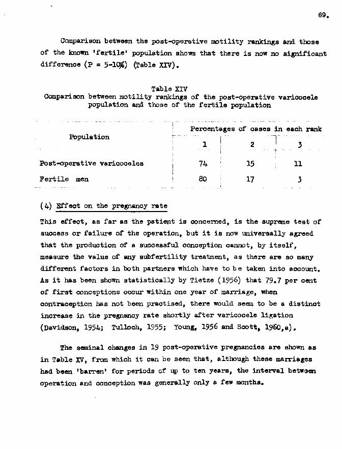
Comparison between the post-operative motility rankings and those of the known ’fertile* population shows that there is now no significant difference (p = 5-lCpJ) (Table XIV).
Table XIVComparison between motility rankings of the post-operative varicocele
population and those of the fertile population
Percentages of oases in each rankPopulation
i ;. I
2 ... 3
Post-operative varicoceles1 74 !
15i
11Fertile men 80 1 17 3
(4) Effect on the pregnancy rate
This effect, as far as the patient is concerned, is the supreme test of success or failure of the operation, but it is now universally agreed that the production of a successful conception cannot, by itself, measure the value of any subfertility treatment, as there are so many different factors in both partners which have to be taken into account. As it has been shown statistically by Tietze (1956) that 79*7 per cent of first conceptions occur within one year of marriage, when contraception has not been practised, there would seem to be a distinct increase in the pregnancy rate shortly after varicocele ligation (Davidson, 1954; Tulloch, 1955; Young, 1956 and Scott, i960,a).
The seminal changes in 19 post-operative pregnancies are shown as in Table XV, from which it can be seen that, although these marriages had been ‘barren* for periods of up to ten years, the interval between operation and conception was generally only a few months.

lI*£•ri
.0
.0
65554444433322222
70.
Table XVin 19 cases who achieved a post-operative pregnancy
BEFORE OPERATION
Countrank
3233 2 2 2 1
2 24 4 2 3 1
3 2 3 3
AFTER OPERATION
Motility; Count j Motility Months between rank x*ank ! rank i ligation and conception
Not received
Not received11
31
Not received
246211518434 4
44 3 3 85 3 912

71.Before operation, seven of these cases were virtually sterile
due to persistent necrospermia; nine had combined count and motility rankings of 4 or more - representing gross impairment of fertility. After operation, all but four had a combined ranking of 3 or less - representing my own standard of ‘fertility*.
SUMMARY AND CONCLUSIONS
Although American and Continental seminologists have not yet accepted varicocele as a factor in the aetiology of male subfertility, in the author's opinion the results of these investigations show conclusively that varicoceles do impair spermatogenesis, and that the degree of impairment is roughly proportional to the size of the varicocele
The exact mechanism of this effect is not yet clearly understood. There is considerable evidence to suggest that the seminal changes are produced by an increased intra-scrotal temperature, but as similar high intra-scrotal temperatures accompany lar*ge hydroceles, and as I have been unable to demonstrate any adverse effect on fertility in such cases, the high temperature may be only a "red-herring". An alternative explanation may be that the damage is trophic in origin, due to a reversible type of reflex alteration in the calibre of the nutritional vessels of the testes.
Whatever the explanation, a dramatic improvement in the sperm count and sperm motility can be anticipated in the majority of cases within a few months of ligation and division of the varicocele.
A considerable increase in the pregnancy rate can also be anticipated shortly after varicocele ligation, provided the patient's wife is fertile; if she is not fertile, there is no indication for operating on the husband
Ligation of the varicocele may be carried out through the scrotum, at the deep inguinal ring, or retro-peritoneally according to the preference of the surgeon. The last-mentioned route of exposure is the least upsetting to the patient but gives the poorest exposure of any anastomotic branches and, consequently, may result in recurrence or failure.
oOo-------

72,
M A L D E S C E N T O F
— oOo-T H E T E S T E S

- CHAPTER V I -
THE EFFECT CP CRYPT ORCHIDISM ON FERTILITY IN MAN
— — 0O0— —
M uch h a s a l r e a d y b e e n w r i t t e n o n t h e e f f e c t o f c r y p to r c h id i s m o n
f e r t i l i t y i n Man a n d i n o t h e r s p e c i e s , b u t c o n s i d e r a b l e c o n f u s io n s t i l l
e x i s t s i n i n t e r p r e t a t i o n o f t h e h i s t o l o g i c a l c h a n g e s an d i n t h e i r
r e l a t i o n t o a e t i o l o g y an d t r e a t m e n t . I n t h i s c h a p t e r , I h a v e t r i e d ,
b y m ean s o f s e m in a l an d h i s t o l o g i c a l s t u d i e s , t o a n a ly s e t h e c a u s e s o f
o f t h i s c o n f u s io n an d t o s u g g e s t l o g i c a l s t e p s i n t h e t r e a t m e n t a im ed
a t p r o d u c in g a g o n ad t h a t i s f u n c t i o n a l l y a s w e l l a s a n a to m ic a l ly
’n o r m a l1 .
T he human m a le i s t h e o n ly m em ber o f t h e a n im a l k ingdom t h a t h a s
t e s t i c l e s i n t h e s c ro tu m b e f o r e b i r t h , w h i le te m p o ra ry c r y p to r c h id i s m
d u r i n g l i f e i s a f e a t u r e common t o many o t h e r s p e c i e s . I n h i b e r n a t i n g
a n im a ls l i k e t h e w oodohuck , t h e h ed g eh o g a n d t h e m o le t h e t e s t i c l e s a r e
w ith d ra w n i n t o t h e abdom en i n c o l d w e a th e r , a n d a r e o n ly p r e s e n t i n t h e
s c ro tu m d u r in g t h e m a t in g s e a s o n . C e r t a i n o t h e r a n im a ls l i k e t h e
g o p h e r r e t a i n t h e t e s t i c l e s i n t h e abdom en e x c e p t d u r in g c o p u l a t i o n .
As we a s c e n d t h e s c a l e we f i n d t h a t i n t h e M acaque m onkey t h e t e s t i o l e
r e m a in s a t t h e u p p e r en d o f t h e i n g u i n a l c a n a l u n t i l t h e s i x t h y e a r o f
l i f e a n d t h e n d e s c e n d s i n t o t h e s c ro tu m , w h i le i n t h e c h im p an z ee th e
t e s t i c l e e n t e r s th e s c ro tu m so o n a f t e r b i r t h an d re m a in s t h e r e
p e r m a n e n t ly .
HISTORICAL
S in c e t h e b e g in n in g o f m e d ic a l r e c o r d s , a r e l a t i o n s h i p h a s b e e n
know n t o e x i s t b e tw e e n c r y p to r c h id i s m a n d s t e r i l i t y , b u t t h e c o n t r o v e r s y
o f t h i s r e l a t i o n s h i p c a n b e s e e n fro m th e f o l l o w i n g tw o q u o ta t io n s * ■■
"W hen o n e o r b o th t e s t i c l e s re m a in th r o u g h l i f e i n t h e b e l l y , Ib e l i e v e t h a t th e y a r e e x c e e d in g ly im p e r f e c t an d p r o b a b ly in c a p a b le

o f p e r fo rm in g t h e i r n a t u r a l f u n c t i o n . "J o h n H u n te r
HI t i s com m only b e l i e v e d t h a t t h e im p e r f e c t i o n o f a n u n d e a o e n d e d t e s t i c l e i s du e t o i t s f a i l u r e t o r e a c h t h e s c ro tu m . T h i s I b e l i e v e t o b e a n e r r o r . An u n d e sc e n d e d t e s t i c l e f a i l s t o r e a c h t h e s c ro tu m b e c a u s e o f i t s i m p e r f e c t i o n ."
B la n d - S u t to n .
As lo n g ago a s 1 8 9 1 , P ia n a fo u n d t h a t e x p e r im e n ta l c r y p to r c h id i s m
i n r a t s r e s u l t e d i n t h e t e s t e s com ing t o re s e m b le t h o s e o f c o n g e n i t a l
c r y p to r c h id i s m i n h o r s e s . I n 1 9 2 4 , Moor® an d O a lu n d c o n f ir m e d t h e s e
h i s t o l o g i c a l c h a n g e s i n s h e e p , an d i n 1 9 2 7 , W a n g en steen show ed t h a t ,
i n H a n , r e t e n t i o n o f t h e t e s t i c l e s i n t h e i n g u i n a l c a n a l f o r a few y e a r s
a f t e r p u b e r ty r e s u l t e d i n a n a b s e n c e o f spezm a to g e n e s i s , t h e t u b u l e s
b e in g l i n e d b y S e r t o l i c e l l s an d s p e m a t o g o n i a . T h e re i s , h o w e v e r,
s t i l l no f i r m p r o o f a s t o w h e th e r t h e s e v a r io u s f i n d i n g s a r e t h e c a u s e
o r r e s u l t o f m a ld e s c e n t .
T he b u lk o f t h e p u b l i s h e d w ork o n t e s t i c u l a r m a ld e s c e n t i s c o n c e rn e d
w i t h h i s t o l o g i c a l c h a n g e s b u t ; i n th e p a s t d e c a d e o r s o , t h e r e h av e
a p p e a re d a few p a p e r s d e a l i n g w i th th e s e m in a l c h a n g e s . Some c o n c e p t
o f t h e a p p a r e n t o v e r a l l e f f e c t o n f e r t i l i t y i n Man c a n b e g au g ed fro m
T a b le X V I, w h ic h I h av e c o m p ile d fro m 231 p u b l i s h e d c a s e s (H a n s e n , 1949 ;
M ack, 1 9 5 3 ; N e ls o n , 1 9 5 3 ; an d H and, 1 9 5 5 ) .
T a b le XVIO v e r a l l e f f e c t o f c r y p to r c h id i s m on f e r t i l i t y
( a s g au g ed fro m r e c e n t p u b l i c a t i o n s )
N um ber o f c a s e s
T ype o f c a s e F e r t i l e 1S u b f e r t i l e: j
S e v e r e ly j S t e r i l e T o ta .im p a ir e d I
t
B i l a t e r a l , u n t r e a t e d - - 7 35 ! 42B i l a t e r a l , t r e a t e d 5 4 7 39 1 55U n i l a t e r a l , u n t r e a t e d 31 11 19 11 72U n i l a t e r a l , t r e a t e d 30 16 9 7 62

U n f o r t u n a t e l y , many o f t h e c o n c lu s io n s t h a t w e re r e a c h e d b y t h e s e
a n d o t h e r a u t h o r s w e re i n v a l i d a t e d b y a f a i l u r e t o d i s t i n g u i s h b e tw e e n
t h e v a r i o u s t y p e s o f m a ld e s c e n t ; t o a p p r e c i a t e t h i s d i s t i n c t i o n o n e
m u s t h a v e a c l e a r u n d e r s ta n d in g o f t h e a n a to m ic a l f a c t o r s i n v o l v e d .
ANATOMICAL FACTORS AFFECTING NORMAL DESCENT
T he r e c o g n i t i o n o f a s u p e r f i c i a l i n g u i n a l p o u ch by D e n is Browne
(1933) h a s r e v o l u t i o n i s e d o u r i d e a s o f l o g i c a l t r e a t m e n t . The r o o f
o f t h i s p o u c h i s fo rm e d b y S c a r p a 's f a s c i a , an d t h e f l o o r b y t h e
e x t e r n a l o b l iq u e a p o n e u r o s i s an d t h e s u p e r f i c i a l i n g u i n a l r i n g . T h is
m ean s t h a t t h e e m e rg in g t e s t i c l e c a n e i t h e r p a s s i n t o t h e s c ro tu m p r o p e r
o r t u r n u p w ard s an d l a t e r a l l y t o o cc u p y t h i s a b d o m in a l e x t e n s i o n o f th e
s c r o tu m . I f t h e S c a r p a 's f a s c i a h a s n o t b e e n f u s e d t o t h e sy m p h y s is
p u b i s , c o n t r a c t i o n o f t h e c r e m a s te r a n d d a r t o s m u s c le s c a n draw t h e
t e s t i c l e up a s f a r a s t h e m id - in g u in a l p o i n t . T h is i s t h e s t a t e o f
a f f a i r s i n t h e now w e ll-k n o w n r e t r a c t i l e t e s t i s . I f , o n t h e o t h e r
h a n d , S c a r p a 's f a s c i a i s f u s e d t o t h e sy m p h y s is p u b i s , t h e n t h e e m e rg in g
t e s t i c l e c a n n o t r e a c h t h e s c r o tu a , an d m u st come to l i e i n o n e o f t h e
e c t o p i c p o s i t i o n s . I t i s now g e n e r a l l y b e l i e v e d t h a t t h e s u p e r f i c i a l
i n g u i n a l p o u c h r e p r e s e n t s a s t a g e i n e v o l u t i o n fro m e a r l i e r mammals,
a n d p e r s i s t s a s e v id e n c e t h a t th e g o n ad c o u ld , a t o n e t im e , b e w ith d ra w n
to w a r d s t h e abdom en w hen n o t i n u s e .
W ith t h i s k n o w led g e i n m in d , I d e f in e t h e d i f f e r e n t t y p e s o f
m a ld e s c e n t a s f o l l o w s j -
R e t r a c t i l e t e s t e s a r e n o rm a l g o n ad s t h a t sp e n d m uch o f t h e i r l i f e i n t h e u p p e r s c ro tu m o r i n t h e s u p e r f i c i a l i n g u i n a l p o u c h , b u t w h ic h c a n b e m a n u a lly r e p la c e d i n t h e lo w e r p o l e o f t h e s c ro tu m .
E c t o p i c t e s t e s a r e g o n ad s t h a t h a v e p r o g r e s s e d n o rm a l ly th ro u g h t h e i n g u i n a l c a n a l b u t , a f t e r e m e rg in g fro m t h e e x t e r n a l r i n g , h a v e b e e n d i r e c t e d away fro m t h e s c ro tu m by a f u s i o n o f S c a r p a 's f a s c i a w i t h t h e sy m p h y s is , an d g e n e r a l l y assum e a p o s i t i o n i n t h e s u p e r f i c i a l i n g u i n a l p o u c h . T h ese t e s t e s c a n n o t b e m a n ip u la te d i n t o t h e s c ro tu m .
R e ta in e d t e s t e s a r e ■immat u r e g o n ad s t h a t l i e i n t r a - a b d o m in a l l y o r

in the inguinal canal; they are frequently associated withcongenital hemiae and other congenital abnormalities of the▼as and epididymis.
INCIDENCE OF MALDESCENT
N o rm al d e s c e n t o f t h e t e s t e s o c c u r s a t a b o u t t h e e i g h t h m o n th
o f i n t r a - u t e r i n e l i f e , b u t a r e v ie w o f t h e l i t e r a t u r e r e v e a l s a w id e
d iv e r g e n c e o f o p i n i o n a s r e g a r d s t h e i n c id e n c e o f m a ld e s c e n t a t b i r t h .
Dom ing (1952) p u t s t h e f i g u r e a t 1 p e r c e n t , C o u n s e l lo r (1933) a t 10 p e r c e n t , an d S c o r e r (1956) a t 3.4 p e r c e n t . T h e re seem s t o b e l i t t l e
d o u b t t h a t t h e i n c id e n c e i s c o n s i d e r a b l y h i g h e r i n p re m a tu re b i r t h s a s
H o f s t a t t e r (1912) fo u n d c r y p to r c h id i s m i n 32 p e r c e n t o f p re m a tu re m a le
b i r t h s co m p ared w i th o n ly 4 p e r c e n t o f f u l l - t e x m b i r t h s .
S c o r e r (1955) fo u n d t h a t 50 p e r c e n t o f o r y p t o r c h id t e s t e s h ad
d e s c e n d e d w i t h i n t h e f i r s t m onth o f l i f e , b u t h e b e l i e v e s t h a t c o m p le te
d e s c e n t w i l l n o t o c c u r a f t e r t h e t h i r d m o n th . A i r d (1949) c o n s i d e r s
t h a t s p o n ta n e o u s d e s c e n t i s u n l i k e l y a f t e r o n e y e a r , an d G rey T u rn e r
(1 9 3 7 ) p u t s t h e 'd e a d l in e * a t t h r e e y e a r s ; o n t h e o t h e r h a n d ,
Williams (1936), observing the records of 2,104 schoolboys, found that between the ages of 11 and 17 years, spontaneous descent occured in 87 per cent of both unilateral and bilateral maldescended testes.
The i n c i d e n c e o f m a ld e s c e n t i n a d u l t l i f e c a n b e g au g e d fro m
co m b in ed p a p e r s r e l a t i n g t o t h e e x a m in a tio n o f A r a y r e c r u i t s (M cNab,
1 9 5 5 ) , an d w o u ld a p p e a r t o b e i n t h e r e g io n o f 0 .2 3 p e r c e n t .
CLINICAL MATERIAL
The p a t i e n t s i n t h i s s tu d y c o n s i s t e d o f f i f t y - e i g h t c r y p to r c h i d s
a t t e n d i n g t h e M ale S u b f e r t i l i t y C l i n i c o f t h e W e s te rn I n f i r m a r y , G lasg o w ,
f o r s e m in a l e x a m in a t io n ; t o g e t h e r w i th a f u r t h e r tw e n t y - t h r e e c r y p t
o r c h i d s a t t e n d i n g t h e U r o lo g ic a l D e p a rtm e n t o f th e I n f i r m a r y f o r
o rc h id o p e x y .

RESULTS CP SEMINAL STUDIES
F o r c o n v e n ie n c e o f a n a l y s i s t h e c a s e s h a v e b e e n d i v i d e d i n t o f i v e
s tu d y g ro u p s , a s f o l l o w s • -
G ro u p 1 : T r e a t e d b i l a t e r a l c r y p t o r c h i d s .
T h i s g ro u p c o n s i s t s o f f i v e c a s e s t h a t h a d r e c e i v e d s u r g i c a l o r h o rm o n a l
t h e r a p y b e f o r e p u b e r ty ( T a b le X V T l), o f w h ic h tw o h a d a p r e v io u s b i l a t e r a l
o rc h id o p e x y , o n e a u n i l a t e r a l o r c h id o p e x y , an d tw o h a d r e c e i v e d a c o u r s e
o f g o n a d o t r o p h in s .
T a b le X VIIS e m in a l f i n d i n g s i n t r e a t e d b i l a t e r a l c r y p to r c h i d s
C asen u m b er
12
T re a tm e n t
B i l a t e r a l o rc h id o p e x y a e t 10M It it i i
U n i l a t e r a l o rc h id o p e x y a e t 11
D e s c e n t a f t e r horm ones a e t 10it it it n 1 2
S e m in a l f i n d i n g s
M o t i l i t yC ount i n
m i l l i o n / m l .
U n d er 1 U n d e r 1
U n d er 1
A zo o sp e rm ia2
N i lN i l
N i l
P o o r
G ro u p 2 ; U n t r e a te d b i l a t e r a l c r y p t o r c h i d s .
T h i s g ro u p c o n s i s t s o f e le v e n c a s e s t h a t r e c e iv e d no t r e a tm e n t b e f o r e
p u b e r ty ( T a b le X V I I I ) , o f w h ic h two h a d l a t e s p o n ta n e o u s d e s c e n t ,
t h r e e s t i l l h a d b i l a t e r a l e c to p i c t e s t e s a t t h e t im e o f e x a m in a tio n ,
a n d s i x h a d b i l a t e r a l r e t a i n e d t e s t e s .

Table XVIIISeminal findings in untreated bilateral cryptorchids
Casen u n b e r T ype o f o a s e
Late descent aet 8 years
Seminal findings
MotilityCount in millions/ml
23 Good7 n h 11 14 years Under 1
iNil
8 Bilateral ectopic testes Azoospermia -
9 i t i t i t Azoospermia -10 t i n it Azoospermia
11 Bilateral retained testes Under 1 Nil12 n i t it Under 1 Nil13 N it n Under 1 Nil14 It i t it Under 1 Nil15 It i t n Azoospermia -16 II n it Azoospermia -
Group 3s Treated unilateral cryptorchids
This group consisted of five cases that had received surgical treatment before puberty (Table XIX) but, of these five cases, only one had had an orchidopexy; the remaining four cases had an orchideotomy performed because of an inability to bring the testis down into the scrotum.
Table XIXSeminal findings in treated unilateral cryptorchids
Casen u m b er
17181920 21
Treatment
Orchidopexy aet 16 yearsOrchideotomy aet 3 years
w " 6 years" w 8 yearsw " 1 5 years
Seminal findings Count in
millions/ml
60112
Under 1 Azoospermia
Motility
GoodGoodGoodNil

Group 4: Untreated unilateral cryptorchids
This group consisted of thirty-one cases that received no treatment before puberty (Table XX), of ‘which fifteen were ectopic, fifteen were retained and one had a late spontaneous descent*
Table XXSeminal findings in untreated unilateral cryptorchids
Casenum ber
22
Type of caseSeminal findings
MotilityCount in millions/ml
Late descent aet 14 years 51 Good
23 Unilateral ectopic testis 152 Good24 n n it 81 Good25 t t i t t t 56 Good26 II n t t 35 Poor27 It i t n 17 Poor28 It i t i t 16 Good29 I t n •t 11 Poor30 It i t i t 10 Poor31 I t n t t 6 I Poor32 It n n 3 Poor33 I t i t •t Under 1 Nil34 I t i t i t Under 1 Nil35 I t it n Under 1 Nil36 n n n Azoospermia -37 i t n n Azoospexmia -
38 U n i l a t e r a l r e t a i n e d t e s t i s 204 Good39 n It t t 65 Good40 i t It t t 63 Good41 i t t t t t 55 Good42 n it tt 37 Good43 ; It It t t 36 Good44 It i t 5 P o o r45 i It It t t U n d e r 1 N i l46 t t It i t U n d er 1 N i l47 I t It t t U n d e r 1 N i l48 t t II t t U n d e r 1 N i l49 t t It t t A z o o sp e rm ia -50 It It i t A zo o sp e rm ia -51 It t t i t A zo o sp e rm ia -52 II tt n A zo o sp e rm ia -

Group 5: Bilateral retractile testes
T h is g ro u p c o n s i s t s o f s i x c a s e s ( T a b le X X I), a l l o f w h ic h w e re r e f e r r e d
t o t h e S u b f e r t i l i t y C l i n i c a s u n d e so e n d e d t e s t e s ; i n e a c h c a s e t h e
t e s t i s c o u ld b e m a n u a l ly r e p l a c e d i n t h e lo w e r p o le o f t h e s c ro tu m ,
a l t h o u g h many o f t h e s e p a t i e n t s s t a t e d t h a t t h e i r g p n ad s w e re p r a c t i c a l l y
a lw a y s a b s e n t fro m t h e s c r o t u a .
T a b le XXIS e m in a l f i n d i n g s i n men w i t h r e t r a c t i l e t e s t e s
C a sen u m b er
535455565758
T ype o f c a s e
B i l a t e r a l r e t r a c t i l e
S e m in a l f i n d i n g s
M o t i l i t yC o un t i n m i l l i o n s / m l
1456149482221
P o o rGoodGoodGoodGoodP o o r
RESULTS CEP HISTOLOGICAL STUDIES
F o r c o n v e n ie n c e o f a n a l y s i s , t h e t e s t e s h a v e b e e n d i v i d e d i n t o
f i v e g ro u p s , a s f o l lo w s * -
G roup I ; R e t r a c t i l e t e s t e s
T h i s g ro u p i s r e p r e s e n t e d b y a s i n g l e c a s e i n w h ic h b o th t e s t e s w e re
b i o p s i e d ; n o rm a l s p e rm a to g e n e s is w as p r e s e n t o n b o th s i d e s .

G ro u p 2* E c to p ic t e s t e s
T h i s g ro u p c o n s i s t e d o f tw e lv e t e s t e s , o f w h ic h n in e r e p r e s e n t t h e
e c t o p i c t e s t i s i n t h e p u b e r t a l p h a s e ( T a b le X X II) ; t h r e e r e p r e s e n t
t h e h i s t o l o g i c a l p i c t u r e o f u n t r e a t e d e c t o p i c t e s t e s i n t h e a d u l t
p h a se *
T a b le XXH B io p sy f i n d i n g s i n 12 e c to p i c t e s t e s
Age F re q u e n c yi n o f germy e a r s i c e l l s__ _ _ _ _ _ _ j
P r im a ry jS e c o n d a ry S p e rm a to - s p e r m a to - s p e rm a to
g o n ia c y t e s
10 Few + 111 None
12 N one
13 None
13 63# + j
13 10# + i| +
14 5q# + +
16 Few + | +
17 4 i# + +
19 N one
31 N one
66 N one
c y t e s S p e rm a tid sM a tu resp e rm s
The t u b u l e s i n t h e t h r e e c a s e s a g e d 10 t o 12 w e re m a in ly p r e - p u b e r t a l
i n t y p e , some w i t h no lum en a t a l l . T he m a j o r i t y o f t h e t u b u l e s i n t h e
r e m a in in g c a s e s w e re l i n e d o n ly b y S e r t o l i c e l l s (F ig * 1 1 ) , w h i l e , i n t h e
66 y e a r o l d m an, t h e t u b u l e s w e re c o m p le te ly a t r o p h i e d a n d h y a l i n i s e d .
L e y d ig c e l l s w e re e i t h e r a b s e n t o r r a r e i n f o u r o f t h e s i x t e s t e s i n t h e
10 t o 13 y e a r s a g e g ro u p , b u t w e re d i f f u s e l y i n c r e a s e d i n e a c h o f th e
t h r e e a d u l t t e s t e s .

Group 3: Retained testes
T h is g ro u p c o n s i s t e d o f tw e lv e t e s t e s ( T a b le X X I I I ) , o f w h ic h s i x
r e p r e s e n t t h e c h a n g e s fo u n d i n t h e p u b e r t a l p h a s e ; s i x r e p r e s e n t t h e
h i s t o l o g i c a l p i c t u r e o f r e t a i n e d t e s t e s i n t h e a d u l t p h a s e .
T a b le X X III B io p sy f i n d i n g s i n 12 r e t a i n e d t e s t e s
- .... j ;Ageinyears
Frequency s of germ 1 cells
Spermatogonia
Primary spezmato-
i cytesi
Secondary spermato- j cytes
10i
Nonei i13 | Pew + :13 Pew + i14 I None j i; t14
i! None i ■ i ; !
14 I NoneI
>j
: ij
16 \ Pew + ;26 Pew + j i28 None28 Pew + + +32 Pew + + +
44 Pew + + +
S p e rm a tid sMaturesp e rm s
The t u b u l e s i n e a c h c a s e w e re l i n e d m a in ly by S e r t o l i c e l l s a l th o u g h ,
i n f o u r o f t h e p u b e r t a l t e s t e s , s p e rm a to g o n ia w e re o c c a s i o n a l l y d e t e c t e d .
I n t h r e e o f t h e s i x a d u l t r e t a i n e d t e s t e s , s p e r m a to g e n e s is c o u ld b e
d e t e c t e d i n o c c a s i o n a l t u b u l e s , b u t sp e rm a to g e n ic a r r e s t w as g e n e r a l l y
p r e s e n t a t t h e s p e rm a to c y te s t a g e ( P i g . 1 2 ) . L e y d ig c e l l s w e re a b s e n t
o r r a r e i n a l l t h e t e s t e s u n d e r 14 y e a r s o f a g e , b u t w e re n o t i c e a b l y
i n c r e a s e d i n a l l t h e a d u l t t e s t e s (P ig * 13) •

Group 4: The opposite testis in unilateral cryptorchidism
T h i s g ro u p c o n s i s t e d o f e i g h t c a s e s ( T a b le XXIV), w h ic h r e p r e s e n t t h e
h i s t o l o g i c a l p i c t u r e a s s o c i a t e d w i th t h e n o rm a lly d e s c e n d e d s c r o t a l t e s t i s
i n c a s e s w h e re t h e o p p o s i t e t e s t i s w as r e t a i n e d o r e c t o p i c .
T a b le XXIVB io p s y f i n d i n g s i n t h e o p p o s i t e t e s t i s o f u n i l a t e r a l c r y p to r c h i d s
Age F re q u e n c y P r im a ry S e c o n d a ryi n o f germ S p e rm a to - s p e r m a to - s p e rm a to - M a tu rey e a r s c e l l s g o n ia c y t e s c y t e s S p e rm a tid s sp e rm s
u R a re +
16 R a re + +
26 N o rm al + + + +
27 Few + + +
30 N o rm al + + + +
3 1 None
32 N o rm al + + + +
32 N orm al + + | +
The t u b u l e s o f t h e p u b e r t a l t e s t e s w e re m a in ly l i n e d by S e r t o l i
c e l l s w i t h o c c a s i o n a l s p e rm a to g o n ia . The t u b u l e s i n t h e re m a in in g s i x
c a s e s w e re a d u l t i n f o u r , m a in ly l i n e d b y S e r t o l i c e l l s i n o n e an d
e n t i r e l y l i n e d b y S e r t o l i c e l l s i n th e o t h e r . L e y d ig c e l l s w e re n o rm a l
i n a l l t h e t e s t e s e x a m in e d . S e v e re h y p o s p e rm a to g e n e s is w as p r e s e n t i n
m o s t o f t h e t u b u l e s o f b o th t h e 32- y e a r o l d t e s t e s .

Group 5: Testes treated by previous orchidopexy (Table XXV)
Table XXV Biopsy findings after orchidopexy
Ageinyears
Age of orchidopexy
Spermatogonia
Primaryspermatocytes
Secondaryspermatocytes Spermatids
Maturesperms
36 11 +27 11 + + + + +30 19 + + + + +
In the first two cases there were few geim cells present; one had occasional spermatogonia, and the other had complete spermatogenesis in occasional tubules with severe hypo spermato gene sis in the remainder.The third case had germ cells in 80 per cent of the tubules, with no evidence of impaired spermatogenesis at all. Peritubular fibrosis was detected in all of the cases.
Testicular biopsy of an ectopic testis (Age 16 yrs.) showing tubules lined almost entirely by Sertoli cells alone. (H & E x 140).

85
Pig, 12, Testicular biopsy of a retained testis (Age bh yrs.)showing spennatogenic arrest at the spermatocyte level. (H & E x 490).
Pig. 13. Testicular biopsy of a retained testis (Age bUyrs.)showing increased Leydig cells appearing in clunps. (H & E x 200).

OBSERVATIONS
Reference to Table XVIII shows clearly that all the nine untreated bilateral cryptorchids were almost certainly sterile; five had complete azoospermia, and the remaining four had only very occasional non-motile sperms. This is in keeping with the general belief that sterility isalmost inevitable if bilateral cryptorchidism is not treated beforepuberty.
Table XVII shows that although there is a slight improvement following pre-pubertal surgery, the incidence of sterility is stillextremely high. This confirms the findings of Mack (1953), and is inkeeping with those of the combined published cases in Table XVI, where 83 per cent were either sterile or very severely impaired. It is, however, a complete contradiction of the observations of McCollum (1935) who reported paternities in 82 per cent of cases treated by bilateral orchidopexy. Most of McCollum’s 'fertilities' were judged on a resultant pregnancy; very few had seminal findings to support his claims, and I have been unable to trace any other publication with such exceptional results.
Turning now to untreated unilateral cryptorchids, we find from Table XX that ten (34 per cent) were fertile, four (13 per cent) were subfertile, three (10 per cent) were severely impaired, six (20 per cent) were sterile due to complete azoospermia, and the remaining seven (23 per cent) were also almost certainly sterile as they had only occasional non-motile sperms in their semen. This high incidence of subfertility associated with post-pubertal unilateral cryptorchidism is not generally appreciated (Chamy & Wolgin, 1956), but is more than substantiated by Mack (1953) who found that, of thirty-seven untreated cases, only 19 per cent were fertile while 59*4 per cent were either sterile or severely impaired; in the larger series of published cases in Table XVI, 43 per cent were fertile and 41 per cent were sterile or severely impaired.If, now, we add my thirty cases to the seventy-two published cases, we

can. compare the fertility of 102 untreated unilateral cryptorchids with that of the 122 men in my fertile population, and we find that whereas 83 per cent of the latter group had an acceptably 'fertile* semen, only 39 per cent of the unilateral cryptorchids had similar seminal findings.
The solitary case of unilateral cryptorchidism treated by orchidopexy before puberty was fertile (Table XIX). In a similar group of twenty-three cases, Mack (1953) found that 17 per cent were fertile and 34 per cent either sterile or severely impaired; in the combined series in Table XVI, 48 per cent were fertile and only 25 per cent were sterile or severely impaired.
Only two of my bilateral cryptorchids had experienced descent after gonadotrophic therapy; one was sterile, the other severely impaired.The number of cases is too small to draw any definite conclusions, and I have been unable to find a similar group of cases in the literature for comparison; I did, however, note that Chamy and Wolgin (1956) biopsied eight testes that descended after 40,000 units of gonadotrophin, and they found that three of them had speraa to genic arrest at the spermatocyte level, while five had small tubules filled with spermatogonia; none had advanced stages of spermatogenesis.
Two cases had bilateral late spontaneous descent (Table XVTIl).The testes that came down at the age of eight were fertile, but the ones that only came down at puberty showed evidence of severe impairment;Hand (1955) reported one similar case with complete azoospermia.Charny and Wolgin (1956) reported three cases with biopsies; spermato- genic arrest was present in a man whose testes descended at the age of twelve, while normal spermatogenesis was present in two men whose testes had descended at four and six years of age respectively. These findings, taken in consideration with my own, would suggest that treatment should not be delayed until puberty, as suggested by Smith (1939), because even although spontaneous descent may still occur after puberty, there appears to be some evidence that spermatogenesis may be retarded.

In the remaining group of six cases that were referred to me as bilateral undescended testes11 but which, on closer examination,
proved to be bilateral high retractile testes (Table XXI), five were fertile and one was mildly subfertile, so it becomes obvious that great care must be taken to distinguish these cases from the truly retained testes in which sterility would have been almost inevitable*
In a most interesting histological study of the testes from birth to maturity, Chamy, Conston and Meranze (1952) found that there are three distinct phases: (1) a static phase which goes on from birth tothe age of four years, (2) a growth phase between the ages of four and ten years, and (3) a maturation phase, starting at the age of ten and continuing right up to puberty. In the pubertal phase, active spermatogenesis was present up to, and including, mature spermatozoa.
All my cases should, from the age point of view, have represented the maturation or adult phases of spermatogenesis, and it is at once apparent that both the retained and ectopic testes show histological evidence of retarded onset of spermatogenesis; none had mature spermatozoa. I have been struck by the fact that the majority of the ectopic testes were well developed anatomically, in striking contrast to the high incidence of abnormalities encountered with the retained testes. In the latter, there were numerous examples of mal-union between the testes and epididymes, ranging from a loose attachment by a mesentery to complete non-union in which the vas and epididymis were in the scrotum or inguinal canal while the testis was still intra- abdominal.
The difference between these two types of maldescent is also borne out by the histological findings during the pubertal phase (Tables XXII & XXIIl) . Six of the nine pubertal ectopic testes had some evidence of spermatogenesis, four of which went as far as the spermatocyte stage, whereas only two of the seven pubertal retained testes had any evidence of spermatogenesis, and none went beyond the spermatogpnia stage.

Furthermore, geim cells were either scanty or completely absent in all the retained testes although they were frequently present in good numbers in the ectopic testes. This apparent difference might be explained by the fact that the retained testes are subjected to the high temperature of the abdomen while the ectopic testes are extra- abdominal and lie in the superficial inguinal pouch. This, as we have already seen, merely represents an abdominal extension of the scrotum.
I believe that the cause of this high incidence of subfertility in unilateral cryptorchidism is explained by the fact that the opposite testis is also frequently abnormal histologically, although, clinically, it may appear to be normal. In Table XIX, we have four examples of cryptorchid men (cases 18, 19, 20 and 21) whose fertility depended entirely on the opposite testis as the maldescended gonad had been removed; two were almost certainly sterile, one was severely impaired and one was distinctly subfertile. This abnormality of the opposite testis was also brought out in Table XXIV which showed that mature spermatozoa were detected in only three of the eight testes examined; spermato genic arrest at various levels was present in four testes, and one of the so-called 'normal' opposite testes showed no evidence of spermatogenesis at all.
Germ cells were present in each of the three testes examined some years after orchidopexy (Table XXV), and mature spermatozoa were detected in two of them; in all of them, spermatogenesis was present in only a proportion of the tubules. This'focal spermatogenesis' was also noted by Hand (1955) . Chamy and Wolgin (1956) found no evidence of advanced spermatogenesis in any of their seventy-three post-operative biopsies while, in thirty-eight, spermatogenesis was completely absent.
DISCUSSION CN TREATMENT
There is a remarkable similarity between ray own seminal findings at the Western Infirmary, Glasgow, and those of Mack (1953) at the Royal

Infirmary, Glasgow, which is not, in my opinion, due to any chance geographical distribution, but to a strict conformity between the two series in interpretation of the types of maldescent.
The presence of a voluminous literature purporting to deal with the treatment of maldescent is, in itself, a monument to the fact that there is, as yet, no clear-cut and uniformly acceptable regime. This, I believe, is partly due to the fact that some authors, having failed to differentiate between the different types of maldescent, have not appreciated that no single treatment is applicable to all cases. It is also partly due to a lack of uniformity in the aims of treatment; some are satisfied so long as their treatment produces a good aesthetic result, while those of us who believe that subsequent fertility is the all-important factor, cannot be wholly content unless the gpnad is treated in such a way and at such a stage in its development that normal spermatogenesis will eventually take place.
We have already seen that the maturation phase of spermatogenesis begins around the tenth year of life, so it is reasonable to assume that the appropriate treatment should not be delayed longer than this. Furthermore, Rea (1942) has noted that experimental evidence suggests that the farther the pre-pubertal testis has descended, the more closely it corresponds to the normally located gonad at the same age.
Retractile testes require no treatment as we have already seen that spermato gene sis is not impaired by this abnormality. Hormonal therapy will frequently accelerate the descent of retractile andretained testes, but most, if not all, of these testes would have
descended spontaneously if left alone. There is, however, some evidence to suggest that testes associated with late spontaneous descent and those brought down by gonadotrophins are frequently already abnormal histologically.
Turning now to the question of orchidopexy, all the evidence seems to point to the fact that operative treatment should be carried out by the tenth year, although I can find no evidence to suggest that it should be done in infancy, as suggested by some authorities. The ectopic testis cannot descend without surgical assistance, and I believe, from the results of my studies, that fertility can frequently be anticipated if these testes are brought down into the scrotum before puberty. Retained testes, on the
other hand, are frequently associated with other local abnormalities and, histologically, appear to be more severely retarded than the ectopic ones. There is, therefore, some doubt in my mind as to whether gpod results can be anticipated following any form of treatment for the retained testis. It should be remembered, in this context, that anatomically good results and functionally good
results are not synonymous.

92.
CONCLUSIONS
Three distinct types of cryptorchidism occur, and careful assessment of the individual case is important because of the difference in potential fertility of each type.
Retractile testes are much commoner than is frequently appreciated. They do not require any form of treatment and suffer no impairment of spermatogene si s.
Ectopic testes cannot descend without surgical assistance, but there is histological evidence to suggest that their state of spermatogenesis is more advanced than that of retained testes, and timely orchidopexy before the onset of the maturation phase may result in subsequent fertility.
Retained testes are frequently abnormal in other respects, and it is doubtful if any form of treatment will alter their 'fertility*. Orchidopexy should be carried out around the tenth year of life to prevent torsion, pain and tumour formation, but neither this nor gonadotrophins can be relied upon to improve the already retarded abnormal spermatogenesis.
Late spontaneous descent may occur up to the age of seventeen years, but there is considerable evidence to suggest that many of these testes are already abnormal before entering the scrotum.
The high incidence of subfertility and sterility associated with bilateral cryptorchidism is an already accepted fact, but it is now suggested that the incidence of subfertility associated with unilateral maldescent is considerably greater than is generally appreciated. This,in the writer* s opinion, is due to the high incidence of impaired spermatogenesis in the opposite normally descended testis.
0O0-- -

~ v w s 'm v'ju ~
■ m v s m cf ‘i m m z m n u n
•—*— otJO— *”*
rx b lty ijd.UF Fm ys b: .. Fy-tbd,.' t i i f t F < -S y & y.-A. '■ b v t!:^ : J . ' i x , { ,‘ ) j.
j • > . . ; b O i . i , s e l d o m J r t n b ; ; b *
^..y O F v i u . . t t f ? I s F v ^ y o n K ' . h , ;. -:- '\.- •■■-■
'•: ob :: bbOWii . b 4.<; , tbe-'.vF
M U M P S O H O H I T I S
I .•' 5 ■ , -V. . i <, **r •■•■-,.
X '- t ’j c’.X .FV '...;i'.r F ;/ • i.y ; ■•%>• - .1 ' •«
■v- j.ii ' , ■ ■ ; • ■ '■- •• 'F’ f '. ’ f;V -i
f ' i '- ■■ : b'F- ' / . ■ • V ;■
.b. F.: b: . x . - U : : .;k > ' 1 &0h-f m &.ci,nt\X fas*-
% l ‘iu x^c'i^^s bmbztis If. bb»"bO .MX m~0, ( !dld) ZO,i:\ii •:•h> nfii v?,c '$£¥y>. 3®2it -3# Zfxiy

94.
- CHAPTER VII -
THE EFFECT OF MUMPS ORCHITIS ON FERTILITY— 0O0— —
1 Sterility following adult mumps is a myth*. This statement, based on the title of a paper by Benard (1927), is still often quoted but, in my opinion, seldom substantiated. in this Chapter, I hope to show that although mumps orchitis is responsible for only a small proportion of the known causes of subfertility, there is a definite association between bilateral mumps orchitis and subsequent subfertility*
Incidence
As mumps is not a notifiable disease in this country, it is difficult to gauge the true incidence of post-pubertal mumps but, of the husbands attending my Subfertility Clinic, 21.4 per cent had a history of mumps in childhood, and 1.8 per cent had mumps after puberty. During the 1914-18 and 1939-45 wars there were several large epidemics of adult mumps reported from Army camps (MacLeod, 1919; Eagles, 1947; Candel, 1951). Weigert (1916) found that 30 per cent of cavalrymen developed orchitis, compared with 2 .5 per cent of infantrymen, and, as a result, recommended dismount drill during mumps epidemics: one might as well advise shoreduty for sailors in an epidemic because of the 100 per cent incidence of orchitis in the affected members of the crew of the ill-fatedH.M.S. "Ardent” (Noble, 1808), whereas, in actual fact, Manine (1913) found that the incidence of orchitis in sea-going French naval personnel was only 10 per cent, and Clement (1918) found an incidence of 28 per cent in the United States Navy, most of whom were doing shore duty.
From an extensive review of the world literature on adult mumps,I have collected over 25*000 cases and I found, from these studies, that approximately one case in five is complicated by orchitis, 15 per cent of which have bilateral involvement of the testes*

95
Clinical pathology
Orchitis generally appears about one week after the onset of the parotitis, but it may occasionally precede the parotid swelling (Torpey, 1911; Feiling, 1915) and, in epidemics, it had been reported in cases whose parotid glands were not involved at all (Wesselhoeft, 1920; Bieberbach & Vibber, 1933) • It has been described in an undescended testis (Boss, 1912) and in an ectopic testis (Mitchell, 1911). The pre-pubertal testis appears to have almost complete immunity to the mumps virus, but at least eight cases in this age group are on record (Wesselhoeft, 1920; Connolly, 1953; Ballew & Masters, 1954); to this can now be added one further case of my own which is detailed below*
J. McA., aged 25, developed mumps at the age of 12. About a week after the onset of the left parotitis his left testicle became swollen and painful, and as it did not subside with conservative treatment and the doctor was unaware of this complication, he was admitted to hospital as an emergency. Left orchideotomy was performed, and histological examination apparently revealed acute mumps orchitis. At the time he attended rny Clinic, seminal analysis revealed a sperm count of 65 million per ml., 8C{# active motility, and 20# abnormally formed sperms.
Serial testicular biopsy studies covering the early stages in the histopathology of mumps orchitis were carried out by Gall (1947) and by Chamy & Meranze (1948). Gall* s studies covered the first two to five days and included 75 testes. The earliest changes were interstitial oedema with normal spermatogenesis. This was followed by vascular dilatation and engorgement, particularly in the deep layers of the tunica albuginea. The third phase consisted of oedema, during which the germinal epithelium exhibited progressive degeneration; ultimately, all the epi the linn became dislodged, leaving only a ring of Sertoli cells. At the next, and final, stage of Gall's studies, the lymphocytic reaction gave way to a polymorph one; interstitial oedema became pronounced; germinal cells were completely absent; and the tubules were distended and plugged with dense masses of polymorphs. The leydig cells were not involved.

96.
Chamy and Meranze studied only two cases but, in both of them, biopsies of the testes were carried out on the 3rd, 6th, 14th and 21st days following the onset of orchitis, so that the histopathological changes were followed into a later stage of the disease. Their studies suggest that spermatogenic destruction begins around the second or third day, and is severely reduced by the sixth day. By then, some of the tubules were completely degenerated and acellular; some were normal; and some showed degeneration of mature spermatozoa, spermatids and secondary spermatocytes. By the fourteenth day, the tubules showed irregularity of size, and degeneration had spread to the primary spermatocytes. Like Gall, they noticed the focal nature of these changes in that, even at this advanced stage of the infection, some tubules and all the Leydig cells were normal.
I have t aken this investigation of histological changes one stage further by carrying out testicular biopsy studies of the late efi ects of mumps orchitis on spermatogenesis.
Case 1. D. McK*, aged 32, had mumps with right-sided orchitis at the age of 16. On clinical examination, his left testis was normal in size and consistency, but the right one was soft and atrophic. Seminal analysis revealed a sperm count of 11 million per ml., with good morphology and poor motility.The left testicular biopsy showed normal spermatogenesis.Biopsy of the atrophic right testis (Pig.14) revealed a large area of completely hyalinised tubules with peritubular fibrosis; immediately adjacent to this was an area of less complete atrophy, in which some tubules showed scanty spermatogenesis, while others were lined exclusively by Sertoli cells. (Fig*15)«
Case 2; J. M., aged 33, had mumps with bilateral orchitis at the age of 32. On clinical examination, both testes were soft and atrophic, while seminal analysis revealed a sperm count of0.5 million per ml., with poor motility and a high percentage of abnormal sperms. Testicular histology showed completely hyalinised ghost tubules with considerable peritubular fibrosi3 in all the tubules of the left biopsy* Most of the tubules in the right biopsy were similarly hyalinised, but a few showed scanty spermatogenesis going on up to the level of mature spermatozoa.(Fig.16).

Fig, 14: Case 1 (right testicular biopsy), showing completelyhyalinised ghost tubules with peritubular fibrosis. (H & E x 140).
97.
Fig. 15* Case 1 (adjacent area to the one depicted above) showingtubules lined exclusively by Sertoli cells. (H & E x 140).

98.
Fig. 16; Case 2 (right testicular biopsy) showing complete tubular atrophy with hyalinisation of the basement membranes. Leydig cells are condensed into small clumps. (Masson x 140).
Case 3: L.S., aged 32, had mumps with severe bilateral orchitisat the age of 29. On clinical examination, both testes were normal in size and consistency. Seminal analysis revealed a sperm count of 110 million per ml., with good motility and morphology. After 10 years of * barren marriage1, he requested testicular biopsy to make certain that his testes were normal, and the histological picture showed normal spermatogenesis in all the tubules of the biopsy specimen. In view of the known focal nature of this condition, one might argue that biopsies from other parts of the testicles would have revealed areas of tubular atrophy, but the appearance of the testicles, at exploration, and the presence of a normal semen suggest that, for some unknown reason, no permanent damage had ensued.

Subsequent effect on fertility
Of the twenty-two testes that had been previously infected by the mumps virus, sixteen (73 per cent) were smaller or softer than normal. Gandel (1951) found that, on discharge from hospital after acute nnmps orchitis, 44*9 per cent of the involved testes were softer than normal, and Benard (1927) found that 50 per cent showed late atrophy.
The seminal findings of fifteen cases with a previous history of mumps orchitis are shown below in Table XXVI •
Table XXVI Effect of mumps orchitis on fertility
TesticularInvolvement
Casa Presentage
Age ofjumpaorohitii
annul FINDINGSAssessedfertilityCount
rankMotilityrank
1 30 24 4 2 Severelyimpaired
2 32 15 3 2 Subfertile
Unilateral 3 28 27 1 2 Fertile
with 4 38 31 2 1atrophy 5 28 20 2 1
6 a 38 2 17 31 28 1 18 25 12 1 1
1 1 FertileBilateral 9 50 22without 10 32 2S 1 1atrophy 11 35 17 1 1
4 1 Severely12 35 impaired
Bilateral 13 35 30 A zoospermia Sterile
with 14 34 17 Azoospermia "atrophy 15 39 35 Azoospermia
1-----

160.In only one case was the morphology below the accepted ‘fertile1
standards. Those cases that were not followed by subsequent changes in testicular size or consistency suffered no ill-effects as regards fertility, but 75 per cent of the bilateral cases associated with atrophy were sterile; the remaining 25 per cent had very severely impaired fertility amounting almost to complete sterility. The number of cases in this series is too small to allow do^natic conclusions to be drawn, but a further thirteen cases of bilateral orchitis were reported by Miehelson and Michelson (1947), of which 6l per cent were sterile and 59 per cent were grossly subfertile; unfortunately they failed to differentiate between cases with and without testicular atrophy.
Of the unilateral cases, 75 per cent were fertile, 12.5 per cent were subfertile, and 12.5 per cent were severely impaired. This compares favourably with the seminal findings of my 120 cases whose fertility had already been proven; allowing for the difference in the number of cases under study, the two ‘populations* are not statistically different. It can therefore be confidently stated that, provided the opposite testis is healthy, unilateral mumps orchitis has no significant effect on subsequent fertility.
prophylaxis and treatment
The discovery by Smith (1912) of oedema in the underlying testis led to a voluminous literature advocating various surgical procedures which purported to release the tubules from ‘pressure atrophy*, This line of treatment is now thought to have little, if any, value in preventing subsequent atrophy. Since then, attempts have been made to control the infection by various antibiotics (Homer & Davidson, 1952; Sutcliff & Barnes, 1953) but none of them appear to have any effect on the prevention of orchitis or on its duration in established cases.
Although diethylstilboestrol appeared to be an effective prophylactic against orchitis in the cases studied by Savran (1946), this was not confirmed by Norton (1950) J both noted a reduction in morbidity in established cases, as did Hoyne, Diamond and Christian (1949).
161.Cortisone also has its advocants (Solem, 1954; Risman, 1956;
Zeluff & Fatheree, 1957) > all of 'whom noted a prompt symptomatic relief of symptoms in established orchitis, but no prophylactic effect against its development.
Convalescent serum seems to offer protection against munps in exposed children (Hess, 1915) and the most effective prophylactic against the development of orchitis in the adult appears to be gammaglobulin prepared from convalescent serum. Gellis, McGuiness and Peters (1945) found that this reduced the incidence of orchitis to 7*8 per cent compared with 27.4 per cent in an untreated control series.
SUMMARY AND CCNCLUSIONS
The subsequent effects on fertility of fifteen cases of mumps orchitis are noted and I find that unilateral orchitis has no effect on subsequent fertility provided the opposite testis is normal.Bilateral orchitis associated with testicular atrophy generally results in sterility or severe impairment of fertility, but bilateral infection without atrophy does not appear to impair fertility.
The incidence and histopathology, both early and late, have been discussed, and the focal nature of the degenerative changes has been well substantiated. The severity of the initial infection appears to have no exact correlation with late histological changes, and there are examples of severe bilateral orchitis without subsequent clinical or histological evidence of atrophy.
Cortisone appears to give symptomatic relief in established cases of mumps orchitis, but convalescent garama-globulin seems to be the only efficient prophylactic against this complication.
oOo----

102.
O B S T R U C T I V E A Z O O S P E R M I A

CHAPTER V I H -
163
OBSTRUCTIVE AZOOSPERMIA
Persistent absence of sperms from the ejaculate in repeated seminal analyses can only result from one of two basic reasons: either the testicles are not producing spezms or the sperms that are being produced are not getting through into the semen because of a
blockage in the seminal conducting system. This latter condition is known as obstructive azoospermia, and is readily diagnosed by an absence of sperms in the semen coupled with normal spermatogenesis on testicular biopsy.
In Table 27 you can see the histological findings of 90 consecutive testicular biopsies in cases of azoospermia; 33 of them had atrophic testes, and 37 were normal in size and consistency.
Table 27
Biopsy findings in azoospermia
Histological findings
Normal spermatogenesis Maturation arrest Sertoli cells only Klinefelter* s syndrome Post-pubertal orchitis
TOTAL
Number of cases
Normal testes ! Atrophic testes
291710
1
57
18213
33

104.Hanley (1955), attempting to justify his belief that testicular
biopsy has little to recommend it, stated that 11 ... if a testicle feels clinically normal it is nearly always capable of producing spermatozoa, even if they are reduced in number ...". The histological findings in the aforementioned Table tend to refute these sentiments, as 29.8 per cent of the azoospermic males with normal testes had complete maturation arrest, generally at the spermatocyte level; and 17.5 per cent had germinal aplasia with complete absence of spermatogenesis, the tubules being lined only by Sertoli cells. None of the atrophic testicles showed normal spermatogenesis, but this was present in 50 per cent of the normal testicles that were examined by biopsy, thus indicating obstructive azoospermia.
Any of the following lesions might, in theory, cause ductal obstruction:
(1) Incomplete development of the ductal system.(2) Post-inflammatory obstruction in the epididymes, vasa or ejaculatory
ducts.(3) Inadvertent or purposeful ligation or division of the vasa.(4) Tumour growth in the ductal system.
In this Chapter, I hope to show that, although the last-mentioned must be extremely rare, all the other lesions to which I have referred can, and do, produce obstructive azoospermia.
CLINICAL MATERIAL
Twenty-nine men with proven obstructive azoospermia were offered surgical exploration of their testes in the hope that the lesion might prove amenable to surgical correction: in those cases where the biopsyshowed that the testicles were not producing spermatozoa, no known form of surgery would have altered the situation. In all, twenty-four men agreed to submit to exploration, and the following results are based on the findings, at operation, on thirty-eight testicles; ten of the men refusing permission to explore the opposite side.

105METHOD
Prior to operation, in each of the 38 cases, the azoospermia was confirmed by repeated analyses of centrifuged seminal specimens, and normal spermatogenesis was confirmed by histological examination of a testicular biopsy. At operation, the testes and epididymes were dissected clear of all coverings, and the vasa deferentia were traced from the epididymal tails to the neck of the scrotum. The epididymal tubules were exposed by a small incision through the glistening capsule, and, by gently squeezing the epididymis between finger and thumb, a yellowish fluid exuded: smears of this fluid were taken on a sterileglass slide and were immediately examined, in a fresh state, under high power magnification. The lumen of the vas was exposed by making a small nick in its anterior wall, using sharp-pointed ophthalmic scissors.A blunt-pointed needle, attached to a syringe, was inserted into this opening, and any contents were aspirated for microscopic examination.The needle was then directed down the lumen of the vas towards the neck of the scrotum, and vasal patency was tested by slow injection of 2 ml. sterile saline. In certain cases biopsies were taken from the epididymal tubules to allow for detailed histological study.
RESULTS
Pig. 17 shows, in diagrammatic form, the readily identifiable congenital abnormalities that were detected in 11 of the cases; the remaining cases had obstructive lesions in the rete testis or epididymal tubules which were invisible to the naked eye.
The results of these surgical explorations have been divided into three distinct groups, and are given separately under the appropriate headings.

OBSTRUCTIVE AZOOSPERMIA DUE TO VISIBLE CONGENITAL ABNORMALITIES OF THE EPIDIDYMIS AND VAS
A D
B E
\C u.
Fig. 17: A. Vas represented by fibrous cord.B. Vas with narrow or absent lumen.C. Congenital absence of the vas.D. Terminal vas replaced by fibrous cord.E. Epididymis detached from testis by mesentery.F. Developmental failure of body and tail of epididymis.

167.Group Is Incomplete development of the ductal system
(A) Vas represented as a fibrous cord (1 example)J.B., aged 28 years, had been married for 5 years without issue.No relevant previous history. Both testes and vasa were palpably normal. Exploration of the right testis showed a normal-looking organ. The epididymal tubules were distended with actively motile sperms, but the epididymal tail was aplasic, and tailed off into a fibrous cord representing the vas deferens.
(B) Vas with narrow or absent lumen (1 example)S.K., aged 35 years. Married 6 years. Right-sided hydrocele excised several years previously. Right testis and vas were palpably normal. Exploration of the right testis showed a normal-looking organ, but the epididymal head contained dilated tubulesfilled with actively motile sperms. The vas deferens looked normal on inspection, but the lumen was minute and failed to allow saline to be injected along it.
(C) Congenital absence of the vas (4 examples)D.R., aged 32 years. Married 3 years. No relevant previous history. Both testes felt normal, and both vasa were palpated*. Exploration of both testes revealed the same congenital lesion.The testes looked normal. The epididymes ended blindly, and no trace of a vestigeal vas could be found in the scrotum. The epididymal tubules were dilated and filled with actively motile sperms.
I.L., aged 27. Married 4 years. No relevant previous history.Both testes felt normal and the vasa were described as 'indefinite' on clinical examination. Exploration of both testes revealed the same congenital lesion. The testes looked normal. The epididymal head contained dilated tubules filled with motile sperms, but the body of the epididymis terminated half-way down, and there was no epididymal tail. No trace of a vestigeal vas was found in the scrotum.
C.N., aged 34. Married 10 years. No relevant previous history.Both testes and vasa were palpably 'normal* on clinical examination. Exploration of both testes revealed the same congenital lesion.The testes looked normal. The epididymes ended blindly, and no trace of a vas deferens was found in the scrotum on either side.The tubules in the head of the epididymis were dilated and contained numerous motile sperms.
J.B., aged 30 years. Married 4 years. No relevant previous history. Both*testes and vasa were thought to be normal on clinical examination. Exploration of both testes revealed the same congenital lesion. The

108.
testes looked normal. The epididymes ended blindly, and no trace of a vas deferens was found in either side of the scrotum. The tubules in the head of the epididymis were dilated and contained numerous motile sperms. In this case, an epididymal biopsy revealed numerous epithelial-lined tubules packed with mature spermatozoa: further sperms were also seen in stromal spacesthat had no epithelial lining.
(D) Terminal vas replaced by a fibrous cord (2 examples)G.M., aged 33 years. Married 4 years. No relevant previous history.Both testes and vasa were palpably normal. Exploration of the right testis revealed a normal-looking organ. The tubules in the headof the epididymis were dilated and filled with motile sperms. Thebody and tail of the epididymis were normal. The terminal 1 - inches of the vas deferens was represented by a fibrous cord but, below this the lumen of a normal-looking vas was patent.
J.M., aged 26 years. Married 5 years. No relevant previous history.Both testes and vasa were palpably normal. Exploration of both testes revealed the same congenital lesion. The testes looked normal. The tubules in the head of the epididymes were dilated and contained motile sperms. The terminal vas deferens was represented by a fibrous cord for about 2 inches but, below this, the vasal lumen was patent. The junction of normal vas with atresic vas was represented by tremendous tortuosity and dilatation. In this case, an epididymal biopsy showed dilated tubules containing mature spermatozoa.
(E) Hour-glass epididymis (1 example)A.F., aged 41 years. Married 8 years. No relevant previous history. Both testes and vasa were palpably normal. Exploration of the right testis revealed a normal organ. The tubules in the head of the epididymis were dilated and contained motile sperms. The tail of the epididymis looked normal, the body was aplasic and the organ gave the appearance of an hour-glass constriction. The vas was patent, but aspirations of the contents in its terminal part failed to reveal sperms.
(p) Epididymis detached from testis by a mesentery (2 examples)N.L., aged 29 years. Married 8 years. No relevant previous history. Right-sided hydrocele excised five years previously. Exploration of the rigftit testis revealed a normal organ, but the epididymis and the vas were hypoplastic. The epididymal tubules were not dilated and contained no sperms. The vas was not patent.

169.S.K., aged 35 years. Married 6 years. No previous history.Exploration revealed a normal left testis, but the epididymis wasdetached from it by a mesentery. The epididymal tubules were notdilated, but the vas was patent.
All the cases previously described had some gross congenital abnormality which was readily identifiable at operation, but a further 19 cases had no demonstrable macroscopic lesion to account for the obstruction. These cases had normal testes and epididymes, and the vasa were patent, thus indicating some obstruction or physiological failure of development in the conducting tubules between the testes and the vasa. Such obstructions appeared to vary in site from the vasa efferentia down to the epididymal tail, the cases falling into two main groups; -
(a) those with sperms in the epididymal tubules, and(b) those without sperms in the epididymal tubules.
The former group, in theory, should be suitable cases for surgical anastomosis between the vas and the epididymal head, but the obstruction in the second group must lie in the region of the rete testis and, to date, no successful anastomosis has been described between the vas and the testicular tubules.
Ten cases had sperms in their epididymal tubules. Each of them had normal testes, but distended tubules were readily detected beneath the tense capsule of the epididymal head. When the capsule was incised, a yellowish fluid - teeming with motile sperms - exuded under pressure. Frequently, the body and tail of the epididymis were collapsed. In each case, the lumen of the vas was patent throughout. These cases were diagramatically represented in Fig. 17 F.
A further nine cases were readily distinguished from the above group by the fact that the tubules in the head of the epididymes were not dilated. When these epididymes were incised the tubules appeared dry; even on gentle pressure no yellowish fluid exuded from them. Smears taken from these tubules never revealed motile sperms, but, in a few cases, biopsies of the tubules did subsequently reveal occasional mature spermatozoa.

Group 2; Post-inflammatory obstructions
One case of post-inflammatory obstruction of the vas deferens was encountered in this study; details are given below*-
A.S., aged 35 years, had a history of gonococcal urethritis 12 years previously, and, although he did not remember any episode of genital swelling at the time, his right epididymis was thickened and tender on clinical examination. At exploration of the right side, the testis looked normal, but the vas was completely blocked with thick pus, and multiple strictures were present some distance from the epididymis.
Group 3: Previous interruption of vasal continuity
Two cases fell into this group; both were Indians who had been sterilised many years previously. The considerable chance of a successful re-anastomosis was explained to them but, after further consideration, one man declined operation because of the loss of working-time which hospitalisation would have entailed; the other had his vasa re-anastomosed four weeks agp, and it is too early to know the final result.
DISCUSSION
The testes arise from genital ridges on the medial aspect of the mesonephros. Remnants of the caudal tubules of the mesonephric duct pei«sist ^0 fonn the vasa efferentia of the testis. The part of the duct below this becomes elongated and convoluted to fozm the epididymal tubules while, at a later stage in development, the remainder of the duct

111.developes a mesodeimal coat and becomes the vas deferens.
If development is disturbed before the 60 mm. stage, it is possible to have a fully developed testis and epididymis with an underdeveloped vas deferens, ejaculatory duct and seminal vesicle.If, on the other hand, development is disturbed at a very early stage, the ureteric bud would not develop, and there would be congenital absence of the kidney and ureter on the same side* this abnormality has, in fact, been demonstrated at autopsy in seven cases (Demel, 1926).
Until a decade ago, aplasia and atresia of the vas were seldom, if ever, considered as a possible cause of male sterility. The advent of testicular biopsy has now led to routine exploration of the testes in cases of obstructive azoospermia, and the early recognition of this defect by O'Connor (1948) triggered off a spate of papers on the subject (Young, 1949; Michelson, 1949; Sandler, 1950; Foss & Miller, 1950; Sniffen et al, 1950). In 1945, at a conference on the diagnosis of sterility at Springfield, Illinois, both Simmons and Charny put the incidence of this abnormality at about 5 per cent and, out of a total of 589 published cases of obstructive azoospermia, I was able to collect 39 examples of bilateral atresia or congenital absence of the vasa (6 per cent), which conforms very closely with the previously suggested incidence. It should be stressed that, in many of my own cases with this lesion, a 'palpable vas' was detected on clinical examination!
Congenital lesions of the seminal vesicles and ejaculatory ducts were not observed in any of my cases, and I can find few references to such cases in the literature. Hanley (1555) reported a case of bilateral absence of vasa and vesicles, and demonstrated, by vesiculography, a cystic lesion in the seminal vesicle of a further case. Congenital absence, fusion, re-duplication and cyst formation have all been detected at autopsy (Dockerty, 1951; Pereira, 1953; Rolnick, 1954).
Post-inflammatory strictures of the vas were only demonstrated in two of my cases* Bayle (1952) velieves that gonococcal lesions are commonest, but Hanley (1955) found them to be a rarity because of the

112.partial disappearance of bilateral gonococcal epididymitis with modem treatment. It is not yet known whether the multiple strictures that are found on microdissection of the epididymal tubules (Hanley & Hodges, 1959) are congenital or post-inflammatory in origin* such strictures give rise to the clinical picture that was described in those cases who had sperms present in dilated epididymal tubules.
Inadvertent ligation or damage to the vasa may occur during operations in the pelvis, inguinal canal and scrotum, and it has been reported following herniorrhaphy, varicocele ligation, orohidopexy and hydrocele operations. It is notable that 14*0 per cent of my cases who had a history of bilateral herniotomy in infancy were subsequently sterile.
Purposeful ligation of the vasa to produce sterility frequently leads to subsequent psychogenic impotence; both my patients came to the Subfertility Clinic for this reason. The Nazi habit of sterilising the Jews, while thoroughly to be condemned, has actually provided a unique opportunity for studying the vasa as many of these men have, since the war, been submitted to surgical exploration in an effort to restore fertility. Rosenbloom reported 11 such cases from Israel, with a 36 per cent success rate; Hanley (1955) reported 4 successes out of 5 cases, with one pregnancy.
In recent years several papers have been published on this subject (Dorsey, 1953; O'Connor, 1953) and it would appear that vasal patency was frequently restored although re-anastomosis of the vas following traumatic or accidental vasectomy gives very poor results. It has been the author's experience that the vas has exceptional powers of re-canalisation - as he has found when trying to sterilise patients for medical reasons - so it is perhaps not surprising that a high success rate follows operation on cases where the original ligation or division was intentional.
Tumour growth is such a rarity that it can be almost dismissed as a cause of obstructive azoospeimia. According to Graham & O'Connor (1954),

113
very few tumours of the vas deferens are on record although they were able to produce one unilateral case from a Subfertility Clinic. I have been unable to find any published case in which the condition was bilateral, as it would have to be in order to produce obstructive azoospermia.
EPIDIDYMO-VASOSTOMY
Anastomosis between a patent vas deferens and the tubules in the epididymal head should, in theory, overcome any obstruction that might be present in the epididymal tail or terminal vas; in practive, this procedure is only feasible when the epididymal tubules are dilated and contain motile sperms.
The operation was first popularised by Hagner (1931), and has since been modified by O ’Connor,(1953) and Hanley (1955). In principle, a small eliptical opening in the wall of the vas is anastomosed to a similar opening in the epididymis at a site where live sperms have been identified by aspiration. Various anastomotic techniques can be adopted (Pig. 18) including end-to-side, side-to-side and implantation of the vasal stoma into the head of the cut epididymis. I have tried all of them at one time or another but, as yet, have had disappointing results.At the Western Infirmary Clinic we have now carried out epididymo-vasostomy twelve times. On three occasions the tubules of the epididymal head were not dilated and, as would be expected, none of these cases subsequently had sperms in their ejaculates. In eight cases the epididymal tubules were distended, the vas was patent, and aspiration revealed abundant motile sperms. Five of these operations failed to produce sperms in subsequent post-operative seminal smaples but, in four cases, sperms were eventually recovered from the ejaculate in good numbers for varying periods after operation: brief details of these cases are given below: -
E.H. had a left epididymo-vasostomy into an epididymal cyst.Pour years later, seminal analysis revealed a count of 4 millionper ml., with good motility and morphology.
J. P. had a right epididymostomy. Two years later, his sperm

114.
Fig. 18* Techniques of epididymo-vasostcmy; end-to-side; side-to-side; and implantation of the vasal stoma into the head of the cut epididymis (the last-mentioned is shown in some detail).

115.count was 7 million per ml., but by three year’s, the sperms were again absent from the ejaculate.
A.S. had a left epididymestomy. Six months later, a few non-mo tile sperms appeared in the ejaculate, but subsequent analyses showed a return to azoospermia.
J.S. had a bilateral epididymostomy. Ten months later hissperm count was 15 million per ml., with good motility and morphology. Eight months after that, the count had dropped to 1 million per ml., and two years after operation, he had azoospermia once again.
Hanley (1955) managed to obtain sperms post-operatively in the semen of 66 per cent of his post-gonococcal cases, and reported four subsequent pregnancies. O'Connor (1948) sent a questionnaire to 750 Urologists in the United States and found that 135 of them had performed the operation A20 times with •success* in 160 cases. Successful results have been reported from time to time (Massey & Nation, 1949), but close personal observation of these published results has led me to believe that, although patency is frequently restored, few of these men were returned to satisfactory levels of * fertility*. O'Connor (1953) reported 6l cases with return of patency in 14 (23 per cent) and return of 'fertility* in 10 per cent. In cases where there was terminal occlusion of both vasa, he devised an operation whereby the free cut ends of the vas were brought into the midline of the scrotum and allowed to empty continuously into an inverted skin pouch, thus creating a reservoir from which semen could be collected for artificial insemination. Hanley (1955) created artificial spermatoceles in the epididymal head by inserting a loosely-folded ball of amnion under the capsule of the epididymis; from these cysts he was subsequently able to aspirate fluid laden with motile spermatozoa for use in insemination.
The cases which I have detailed above suggest that a prolonged follow- up may frequently reveal that in some cases, patency is only temporarily restored; the anastomostic stoma presumably becoming stenosed at a later date.

116.
SUMMARY AND CCNCLUSICNS
Contrary to current belief, an appreciable number of azoospermic males with normal-sized testes have histological evidence of complete germinal failure or maturation arrest; the remainder have normal spermatogenesis indicating an obstruction between the testes and the ejaculate.
Obstructive azoospermia may result from a congenital defect in the seminal conducting tubules; from previous inflammation; from inadvertent or purposeful damage to the vasa; or, rarely, from tumour growth in the spermatic cord.
Aplasia, atresia and congenital absence of the vasa are not as rare as the literature would suggest.
Many cases of obstructive azoospermia have no demonstrable obstruction. Some have a block in the tubules of the epididymal tail revealed by dilatation of the tubules in the head; others have a block between the testes and the epididymes, and have no dilated tubules.The former cases are suitable for epididymo-vasostomy, but no known treatment can improve the latter.
Patency can frequently be restored to a vas that has been surgically divided, but is less likely to follow re-anastomosis of a vas that has been inadvertently damaged at a previous operation.
Epididymo-vasostomy can overcome an obstruction in the terminal vas or epididymal tail, but return to acceptable ‘fertility* is uncommon. Many anastomoses will eventually stenose, and a more prolonged follow-up of these cases is recommended before labelling them as ’successes*.
— — oOo

CONCLUSIONS 0O0----
This study at its inception, some six years ago, set out to define seminal standards which might be considered as compatible with probable fertility and, by means of a series of clinical investigations, it was also hoped to determine the effect on spermatogenesis of various extraneous factors. Certain other aspects of the problem of the subfertile man were dealt with by careful and judicious appraisal of the views of other authorities, as expressed by them in previous publications concerning this very wide and important subject.
Throughout the thesis, I have attempted to analyse the results of my experiments and observations, and the discussion and conclusions that were drawn from each were presented at the end of the appropriate chapters. In order to complete the thesis and, at the same time, facilitate interpretation by the reader, my overall conclusions are listed below;-
(1) Based on the findings at seminal analysis, the husband appears to be at fault in almost 50 per cent of the cases attending a Subfertility Clinic.
(2) The husband is more often at fault than his wife, but the exact ratio is difficult to determine, as his fertility is based on precise statistical concepts, while his wife’s fertility is based on clinico- pathological findings in which minor deviations from normal might pass un-noticed.
(5) Speims are formed in the testicular tubules and pass from there to the epididymal tubules as a result of alternate contractions of each side of the testis; these contractions are possibly produced by oxytocin release from the pituitary.
(4) Movement of the sperms through the epididymal and vasal tubes appears to be produced by peristaltic and segmental contractions of the muscle wall.
(5) On ejaculation,- the speims have already been freely mixed with secretions from the epididymes, prostate and vesicles to form semen which appears in three fractions; (a) a purely liquid fraction, derived from the prostatic secretions and containing no speims at all,(b) the principal fraction, which is a mixture of fluid and gel,

containing most of the sperms, and (c) a terminal fraction, derived from the seminal vesicular secretions, which is a homogeneous mass in which the sperms lie immotile.
(6) After a period of about 10 to 15 minutes, the seminal plug liquefies under the influence of the fibrinolytic action of the prostatic component.
(7) On reaching the vagina at intercourse, the sperms are transferred to the uterine cavity by what appears to be a suction effect taking place during inspiration.
(8) Migration from the cervix to the utero-tubal junction may be partly due to sperm motility, but this appears to be materially assisted by rhythmic contractions of the uterine muscle.
(9) The principal actions of the sperm tail and its flagellating movement seem to be passage through the utero-tubal junction and possibly penetration of the coverings of the ovum.
(10) Seminal analysis is the most important single investigation of fertility in the male.
(11) Testicular biopsy is a useful adjuvant in cases with azoospermia, as it allows distinction between obstructive and non-obstructive lesions. It is occasionally of value in assessing the direct effect of various forms of treatment on the seminal tubules and, in some cases, may assist with the prognosis b}7 determining the potential capacity of the tubules t o r e g e n e r a t e . Demonstration of anirreversible lesion allows an early decision to adopt a child rather than continue with prolonged, expensive and possibly worthless t h e r a p y .
(12) Before reaching any final decision on assessment of a man’s fertility potential, two or more seminal samples should be analysed.
(15) The current practice of assessing results of treatment based onchanges in the sperm count and motility (expressed numerically) is subject to a considerable margin of error, as a degree of normal fluctuation occurs in both these factors in every patient. This fluctuation can, in pari at least, be overcome by using a ranking system similar to the one suggested in this thesis.
(14) A combined count and motility ranking of 3 or less can be taken to indicate probable fertility, as good motility can undoubtedly compensate for a low sperm count.
(15) The following standards appear to indicate an acceptable level, in

seminal analysis, compatible with probable fertility*
Ejaculate volume ........ 3 to 5 ml.Motility ................. 50 per cent active motility
five hours after ejaculation.Morphology ....... . Less than 30 per cent abnormally
formed.Spera count ............. Over 20 million per ml.
(16) Careful application of the above standards will give a reliable assessment of a man’s probable fertility, but this does not imply that a successful conception will automatically result from mating with a fertile woman.
(17) There is a definite increase in the incidence of subfertility when varicocele patients are compared with men who have no varicocele.
(18) All sizes of varicocele play some part in the production of subfertility, but the effect produced by small varicoceles is not statistically significant. Only large varicoceles have a significant effect on the sperm count, but sperm motility is significantly affected by both large and moderate varicoceles*
(19) Seminal analyses after operation on different sized varicoceles shows that a significant improvement in the sperm count and motility can be anticipated in most cases of large and moderate sized varicoceles, but some improvement may also follow ligation of the smaller ones.
(20) An initial depression of the sperm count is generally followed by a steady increase for as long as two years after operation.
(21) Depressed sperm motility associated with an already adequate sperm count s e e m s to be a strong indication for operation, but good results can also be anticipated when there is complete necrospermia. A return to acceptable fertility may occasionally follow varicocele ligation even when there is a pre-operative azoospermia.
(22) Congenital or pathological changes in the testes, other than those produced by t h e varicocele, are a contradiction t o operation.
(23) Histological studies of the testicular changes associated with varicocele show that both testes are involved, although the changes are more pronounced on the side of the varicocele.
(24) The earliest effect of a varicocele appears to be one of reduced sperm motility, but larger varicoceles seem to eventually damage the seminal tubules.

(25) The exact mechanism of this effect is not clearly understood.There is some evidence to suggest that the changes may be produced by an increased intra-scrotal temperature due to derangement of the normal thermoregulatory mechanism coupled with local transference of heat through the septum scroti. As similar high intra-scrotal temperatures are also produced by large hydroceles, and as I have been unable to demonstrate any adverse effect on fertility in such cases, it may be that the damage is caused by some reflex alteration in the calibre of the nutritional vessels of the testes resulting in a trophic lesion of the tubules.
(26) Ligation of the varicocele may be carried out through the scrotum, at the deep inguinal ring, or retro-peritoneally according to the preference of the surgeon.
(27) A considerable increase in the pregnancy rate can be anticipated after ligation of a varicocele, even in cases where there has been a prolonged barren marriage beforehand.
(28) A high rate of subfertility and sterility is associated with all forms of true cryptorchidism.
(29) Three distinct types of maldescent occur, and careful assessment of each individual case is important because of the difference in the potential fertility of each type.
(30) Retractile testes are much commoner than is frequently appreciated.They do not require any form of treatment, and suffer no impairment of spermatogenesis.
(31) Ectopic testes cannot descend without surgical assistance, but thereis histological evidence to suggest that their state of spermatogenesis is more advanced than that of retained testes. Timely orchidopexy before the onset of the maturation phase may result in subsequent fertility.
(32) Retained testes are frequently abnormal in other respect, and it is doubtful if any form of treatment will alter their fertility. Orchidopexy should be carried out around the tenth year of life to prevent torsion, pain and tumour formation, but neither this nor gonadotrophins can be relied upon to improve the already retarded abnormal spermatogenesis.
(33) Late spontaneous descent may occur up to the age of seventeen years, but there is ample evidence to suggest that many of these testes are already abnormal before entering the scrotum. They should, therefore, not be left untreated until after puberty has started.
Ou) The high incidence of subfertility and sterility associated with

unilateral cryptorchidism appears to be due to the high incidence of impaired spermatogenesis in the opposite normally descended testis.
(35) Aesthetic normality of the testis following orchidopexy does not necessarily mean functional normality, and histological studies of these testes have shown that many of them are still seriously affected as regards spermatogenesis.
(36) Bilateral mumps orchitis after puberty, associated with subsequent testicular atrophy, generally results in sterility or severe impairment of fertility, but bilateral post-pubertal infection without subsequent atrophy does not appear to impair fertility.
(37) Unilateral post-pubertal mumps orchitis has no subsequent effect on fertility.
(38) The histological changes in the testis, produced by post-pubertal mumps orchitis, are focal in nature, and the severity of the initial infection has no exact correlation with the late histological changes.
(39) Cortisone appears to give symptomatic relief in established cases of mumps orchitis, but convalescent gamma-globulin seems to be the only efficient prophylactic against this complication. No antibiotic or other form of therapy has been found to reduce the incidence of testicular atrophy in established cases.
(40) An appreciable number of azoospermic males will give histological evidence of complete germinal failure or maturation arrest even although clinical examination reveals apparently normal testes.
(41) Obstructive azoospermia may result from congenital defects in the conducting tubules, from previous inflammation, from inadvertent or purposeful damage to the vasa, or, rarely, from tumour growth in the spermatic cord.
(42) Aplasia, atresia and congenital absence of the vasa are not as rare as the literature would suggest. Any or all of these congenital deformities can easily be missed at clinical examination.
(43) Many cases of obstructive azoospermia have no demonstrable obstruction Some have an obstruction in the tubules of the epididymal body or tail as revealed by dilatation of the tubules in the head; others have an obstruction between the testes and epididymes and have no dilated epididymal tubules at all. The former cases are suitable for epididymo-vasostomy, but no known treatment can improve the latter.
(44) Epididymo-vasostomy can frequently overcome an obstruction in the terminal vas or in the body or tail of the epididymis, but a return

122.
to acceptable fertility* is uncommon. Many anastomoses will eventually stenose and a more prolonged follow-up of these cases is recommended before labelling them as ”successes”.
(45) Patency can frequently be restored to a vas that has been surgically divided, but it is less likely to follow re-anastomosis of a vas that has been inadvertently damaged at a previous operation.
— — 0O0---

ACKNOWIBDGEaaBNTS
— — 0O0----
First and foremost, I 'would like to express my sincere thanks to Mr. Wm. S. Mack for stimulating my interest in this perplexing problem, and for constant freely-given advice and comment throughout these studies; I am also deeply indebted to Mr. Spencer Breslin of the Photographic Department, Royal Beatson Memorial Hospital, Glasgow, for the illustrations; to Mr. Gabriel Donald and his Staff at the Department of Medical Illustration, Western Infimaxy, Glasgow, for the line drawings; to Dr. Malcolm Ferguson-Smith of the University Department of Pathology for his co-operation in the varicocele studies; to numerous histologists for their reports on my testicular biopsies; to Dr. Sven Iversen of the Research Department, Royal Beatson Memorial Hospital, for his statistical analyses; and lastly, to Miss Myra Ogilvie for the numerous hours of her leisure-time which she has foregone in order to type and re-type this manuscript.
oOo-------
REFERENCES
AIKD, I. (1949) In "A Companion of Surgical Studies", Edinburgh; Livingstone*
ALBERT, A., UNDERDAHL, L.O., GREENE, L.F. & LORENZ, A. (1955)Male Hypogonadism* Proc. Mayo Clin,* 30*31*
ASDELL, S.A. & SALISBURY, G.W. (1941) The rate at which spermatogenesis occurs in the rabbit. Anat.Rec* * 80.145.
AUSTIN, C.R. (1948) Function of hyaluronidase in fertilization.Nature. Lond* 162.63.
BADENOCH, A.W. (1945) Descent of the testis in relation to temperature. Brit.med. J. 2*601*
BALLEW, J.W. & MASTERS, W.H. (1954) Mumps: a cause of infertility.Fertil. & Steril. 5.536*
BAYLE, H. (1952) Azoospermia of excretory origin.Proc. Soc. Study Fertil. 4.30.
BENAED, R. (1927) Sterility following adult mumps is a myth*Medicine. 9.184.
BICKERS, W. (1951) Patterns of uterine motility in relation to spennigration. Fertil. & Steril. 2.342.
BIEBERBACH, W.D. & VIBBER, F. (1933) Orchitis due to mumps withoutinvolvement of parotid glands. J.Amer.med.Ass.. 100.1092.
BIRNBERG, C.H., SHERBER, D.A. & KURZROK, H.L. (1952) Fructose and fructolysis in semen. Amer.J.Obatet.Gynec.. 63.877,
BLANDAU, R.J. & ODOR, D.L. (1952) Sperm penetration into ooplasm.Fertil. & Steril. 3.13.
BLAND-SUTTON, J. cited by HOBDAY, F.T.G. (1914) in "Castration and Ovariotomy".
BUNGE, R.G. & SHEHMAN, J.K. (1954) Liquefaction of semen by alpha-amylase. Fertil. & Steril. 5.353.
CAMPBELL, H. (1958) Infertility: its incidence and hope of cure.Brit.med.J. 1,429.
CANDEL, S. (l95l) Epididymo-orchitis in mumps.Ann.intern.Med. 34.20.

CA PUHRQ, M.A. (1902) Sulla circolazione sanguigna normale et di compenso del testiculo. Anat.Anaz., 20.577.
CARY, W.H. (1930) Study of sperm cell migration in female secretions. N.Y. State J.Med. 30.131.
CHAHNY, C.W. & MERANZE, D.R. (1948) Pathology of mumps orchitis.J. Urol. 60.140.
CHAHNY, C.W., CONSTON, A.S. & MERANZE, D.R. (1952) Development of the testis from birth to maturity. Fertil. & Steril. 3.461.
CHAHNY, C.W. & WOLGIN, W. (1956) The management of cryptorchidism. Surg.Gynec.Obstet. 102.177.
CLEMENT, R. (1918) Cited by WESSELHOEFT. C. (1920) Loc.cit.
COHEN, M.R. & STEIN, I.F. (l95l) Sperm survival at ovulation time.Fertil. 8c Steril. 2.20.
CONNOLLY, N.K. (1953) Mumps orchitis without parotitis in infants.Lancet. 1.69,
COOPER, A. (1830) In "Observations on the Structure and Diseases of the Testis"., Pt.l., pp.1-55. London; Longman.
CORNER, E.M. & NITCH, C.A.R. (1906) Immediate and late results of the high operation for varicocele, Brit.med.J. 1.191.
COUNSELLOR, V.S. (1933) Cryptorchidism; treatment and results in 100 cases. J. Urol. 30.327.
CROSS, B.A. (1955) Posterior pituitary in relation to reproduction and lactation. Brit.med.Bull. 11.151.
DAVIDSON, H.A. (1954) Testicular temperature and varicocele.Practitioner, 173.703.
DAVIS, M.E. & McCUNE, W.W. (1950) Fructolysis of humen spermatozoa.Fertil. & Steril. 1.362,
DEMEL, R. (1926) Cited by FOSS, G.L. & MILLER, A. (1950) loc.cit.
DEMING, C.L. (1952) Evaluation of hormonal therapy in cryptorchidism.J. Urol. 68.354.
DENIS BROWNE (1933) Some anatomical points in operation for undescended testicle. Lancet, 1.460.

DENIS BROWNE (1949) Treatment of undescended testicle. Proc.R.Soc.Med. 42.643.
DOCKERTY, M.B. (1951) Proc. Mayo Clin. 26.413. cited by Hanley (1955) loc.cit
DORSEY, J.W. (1953) Anastomosis of vas deferens to correct post-vasectomy sterility. J. Urol. 70.515.
EAGLES, A.Y. (1947) Analysis of a four-year epidemic of mumps.Arch.intern.Med. 80.374.
EL-SADR, A.R. & MINA, E. (1950) Anatomical and surgical concepts in the management of varicocele. Urol.Cutan.Rev. 54.257.
ENDERLEN, H. (1896) Klinische und experimentelle Studien zur Frage der Torsion des Hodens. Dtsch.z.Chir. 43.177.
ENGLE, E.T. (1955) The life history of the human testis.J. Urol. 74.379.
FARRIS, E.J. (1955) Test of motility in Locke's solution.J. Urol. 61.1019.
FARRIS, E.J. (1950) In "Human Fertility". New York.
FEILING, A, (1915) Mumps: a critical review.Quart.J.Med. 8.262.
FOSS, G.L. & MILLER, A. (1950) Aplasia of the vas.Lancet.2.737.
GALL, E.A. (1947) The histopathology of acute mumps orchitis.Amer.J.Path. 23.637.
GELLIS, S.S., McGUINESS,A.C. & PETERS, M. (1945) Study of mumps orchitis prevention by gamma-globulin. Amer.J.med.Sci. 210.661.
GLOVER, T.D. (1955) Some effects of scrotal insulation on the semen of rams. Proc.Soc.Study Fertil. 7.66.
GRAHAM, J.B. & O'CONNOR, V.J. (1954) Spermatic cord tumour presenting an infertility problem. J. Urol. 72.946.
GRAY, H. (1938) In "Textbook of Anatomy", 27th Ed. pp. 1387. London; Longman.
GRIFFITHS, J. (1896) The effects upon the testes of ligature of spermatic artery and veins. J. Anat.Lond. 30.81.

HAGNER, F.R. (l93l) Sterility in the male.Surg.Gynec.Obstet. 52.330.
HAMILTON, J.B. (1954) Androgenic activity in the urine.J.Clin.Endocrinol. 14.452.
HAND, J.R. (1955) Undescended testes: results of follow-up to 33 years.Tr.Amer.Ass.G-U.Surg. 47.9.
HANLEY, H.G. (1955) The surgery of male subfertility.Ann.R.Coll.Surg. 17.159.
HANLEY, H.G. (1956) Surgical correction of errors of testicular temperature regulation. Proc.2nd World Cong.Fertil.Steril. p.953.
HANLEY, H.G. & HODGES, R.D. (1959) The epididymis in male sterility: microdissection studies. J. Urol. 82.508.
HANSEN, T.S. (1949) Fertility in operatively treated and untreated cryptorchidism.Proc.R.Soc.Med. 42.645.
HARRISON, R.G. (1949) Distribution of vasal and cremasteric arttries to the testis. J.Anat.Lond. 83.267.
HARRISON, R.G. (1958) In "La Fonction Spermatogenetique du Testicule Humain". p.63. Paris; Masson et Cie.
HARRISON, R.G. & WEINER, J.S. (1949) Vascular patterns of mammalian testis and their functional significance. J.Exper.Biol. 26.304.
HARTMAN, C.G. (1957) How do sperms get into the uterus ?Fertil. & Steril. 8.403.
HARVEY, C. (1954) Experimental study of the penetration of cervical mucous by sperms. Amer.J.Obstet.Gynec. 61.480.
HARVEY, C. & JACKSON, M.H. (1945) Assessment of male fertility; attempt to standardise methods. Lancet.2.99 and 2.134.
HESS, A.F. (1915) A protective therapy for mumps.Amer.J.Pis.Child. 10.99.
HOFSTATTER, R. (1912) Uber Kryptorchismus und Anomalien des Descensus Testiculi. Klin.Jahrb. 26.155.
HOMER, L. & DONOVAN, W.N. (1952) Aureomycin in mumps.J.Amer.med.Ass. 150.465.

HOTCHKISS, U.S. (1944)"Fertility in Man". Philadelphia; Lippincott.
HOTCHKISS, U.S. (1956) The human testis.Fertil. & Steril. 7.284.
HOYNE, A.L., DIAMOND, J.H. & CHRISTIAN, J.R. (1949) Diethylstilhoestrol in mumps orchitis. J.Amer.med.Ass. 140.662.
HUHNER, J. (1913) cited by TULLOCH.W.S. (1953) ProcR.Soc.Med. 46.838.
HUNTER, J.(l837) In "The Works of John Hunter". Vol.4. p.15. Ed.Palmer.J.F. London; Longman.
HUGGINS, C., SCOTT, W.W. & HODGES, C.V. (1941) Studies on prostatic cancer. J. Urol. 46.997.
HUGGINS, C. & NBAL, W. (1942) Coagulation and liquefaction of semen. J.Exper.Med. 76.527.
HUGGINS, C. & MOULDER, P.V. (1945) Estrogen production by Sertoli cell tumours of the testis. Cancer Res. 5.510.
HUMPHREY, G.F. & MANN, T. (1949) Studies of citric acid in semen.Biochem.J. 44.97.
HUTT, F.B. (1929) Relation of fertility in fowls to density of sperm suspension. Proc.R.Soc.Edin. 49.102.
rVANISSEVICH, 0. & GHEGORINI, H. (1918) Uno nueva operacion para curar el varicocele. Semana.med. 25.575.
JAVERT, C.T. & CLARK, R.L. (1944) Combined operation for varicocele and inguinal hernia. Surg.Gynec.Obstet. 79.644.
LAGEHLOEF, N. (1934) Morphologische im Spermabild und in den Hoden bei Bullen. Acta.path.microbiol.Scand.(supp). 19.1 - 254.
LAMPE, E.H. & MASTERS, W.H. (1956) Problems of male fertility : II.Effect of frequent ejaculation. Fertil. & Steril. 7.123.
LANE, J.W. (1955) Radiographic studies in varicocele.U.S. Armed Forces med.J. 6.1589.
LANE-ROBERTS, C., SHAHMAN, A., WALKER, K., WEISNER, B.P. & BARTON, M. (1948) In "Sterility and Impaired Fertility". London; Hamilton.
LEONARD, S.L., PERLMAN, P.L. & KURZBOK, R. (1947) Effect of prostate and vesicles on production of hyalase. Endocrinology. 40.199.

LENNOX,B., FERGUSON-SMITH, M.A., MACK, W.S. & STEWART, J.S.S. (1958)Frequency of Klinefelter's Syndrome. Ciba Symposium on Nuclear Sex.
LEUCHTENBERGER, C., WEIR, D.R., SCHRADER, F. & MURMANIS, L. (1955)D.N.A. content in spermatozoa. J.Lab.Clin.Med. 45.851.
LEWIS-FANING, E. (1949) Family limitation. In Papers of Royal Commission on Population, Vol. 1.; London.
MACK, W.S. (1953) Discussion on male infertility.Proc.R.Soc.Med. 46.835.
McCLEAN, D. & ROWLANDS, I.W. (1942) Role of hyalase in fertilization.Nature. London. 150.627.
McCOLLUM, D.W. (1935) Clinical study of the spermatogenesis of undescended testicles. Arch.Surg. 31.290.
McCULLAGH, D.R. (1932) Dual endocrine activity of the testes.Science. 76.19.
McCULLAGH, E.P. & SCHAFFENBURG, C.A. (1952) Testicular androgen deficiency and semen volume. J.Amer.med.Ass. 149,1214.
G. (1919) Mumps in adults.Brit.med.J. 2.742.
J. (l94l) Metabolism of human spermatozoa.Amer.J.Physiol. 132.193.
J. (1951) Semen quality in fertile and subfertile men,Fertil. & Steril. 2.115.
J. (1956) Human semen.Fertil. & Steril. 7.368.
J. & GOLD, R.Z. (1951a) Sperm motility in fertile and infertile marriages. Fertil. & Steril. 2.187.
J. & GOLD, R.Z. (1951b) Sperm morphology in fertile and infertile marriages. Fertil. & Steril. 2.394.
J. & GOLD, R.Z. (1952) Effect of continence on semen quality. Fertil. & Steril. 3.297.
J. & GOLD, R.Z. (1956) A study of variation in semen quality. Fertil. & Steril. 7.387.
MacNAB, G.H.(l955) Maldescent of the testicle.J.Roy.Coll.Surg.Edin. 1.126.
MacLEOD,
MacLEOD,
MacLEOD,
MacLEOD,
MacLEOD,
MacLEOD,
MacLEOD,
MacLEOD,

MANCINI, R.E., NOLAZCO, J. & de la BALZE, F.A. (1952) Histochemical study of the normal adult testis. Anat.Rec. 114.127.
MANINE, J. (1913) Note sur les oreillons.Bull.Soc.med.d.hop.da Par., 35.1139.
MANN, T. (1946) Fructose as a normal constituent of seminal plasma. Biochem.J. 40.481.
MANN, T. (1954) In "Biochemistry of Semen", New York; Wiley.
MANN, T. & LUTWAK-MANN, C. (l95l) Secretory function of male accessory organs of reproduction. Physiol.Rev. 31.27.
MASSEY, B.D. & NATION, E.F. (1949) Vas deferens anastomosis.J.Urol. 61.391.
MAWSON, C.A. & FISCHER, M.I. (1953) Zinc and carbonic anhydrase in human semen. Biochem.J. 55.696.
MICHELSON, L. (1949) Congenital abnormalities of the vas and epididymis. J. Urol. 61.384.
MICHELSON, L. & MICHELSON, R. (1947) Fertility studies of the male in barren marriages. J.Amer.med.Ass. 134.941.
MIFLET, J. (1879) Uber die pathologischen Verenderungen des Hodens.Arch.klin.Chir. 24.399.
MITCHELL, W.S. (1911) A case of acute orchitis occuring in an undescended testis. Lancet.1.23.
MOENCH, G.L. (1931) cited by LANE-ROBERTS et at. (1948). loc. cit.
MOORE, C.R. (1922) Cryptorchidism experimentally produced.Anat.Rec. 24.383.
MOORE, C.R. (1951) Experimental studies on the male reproductive system.J. Urol. 65.497.
MOORE, C.R. & OSLUND, R. (1924) Experiments on sheep testes; cryptorchidism, vasectomy & scrotal insulation. Amer.J.Physiol. 67.595.
NELSON, W.O. (1951) Effects of cryptorchidism in the rat, and non-descent of the testis in man. Rec.Progr. Hormone Res. 6.29.
NELSON, W.O. (1953) Some problems of testicular function.J. Urol. 69.325.

NICANDER, L. (1954) Glycogen secretion in the epididymis.Nature. London. 174.700.
NOBLE, A. (1808) History of endemic cyanache parotidea on board HMS "Ardent" Edin.med. surg.J. p.304.
NORTON, R.J. (1950) Use of diethylstilbowstrol in orchitis due to mumps.J.Amer.med.Ass. 143.172.
O'CONNOR, V.J. (1948) Anastomosis of vas deferens after purposeful division for sterility. J. Urol. 59.229.
O'CONNOR, V.J. (1953) Mechanical aspects and surgical management of sterility in man. J.Amer.med.Asa. 153.532.
OETTLE, A.G. (1954) Morphologic changes in human semen after ejaculation. Fertil. & Steril. 5.227.
PALOMO, A. (1949) Radical cure of varicocele by new technique.J. Urol. 61.604.
PARKER, G.H. (1930) The passage of spermatozoa through the oviduct of the rabbit. Proc.Soc.Exper.Biol.med. 27.826.
PAYNE, J.M. (1956) Degenerative changes in the adult mouse testis returned to the abdominal cavity. J.Path.Bact. 71.117.
PEARL, R. (1939) In "Natural History of Population", London.
PEREIRA, A. (1953) Roengten interpretation of vesiculograms.Amer.J.Roengtenol. 69.361.
PEELOFF, W.H. & NODINE, J.H. (1950) Extraction of hyalase from testicles and seminal fluid. Fertil. & Steril. 1.373.
PIANA, G.P. (1891) La clinica veterinaria, 14.51.
REA, C.E. (1942) Histologic character of undescended testes after puberty. Arch. Surg. 44.27.
REA, C.E. (1951) Fertility in cryptorchids.Minnesota med. 34.216.
RISMAN, G.C. (1956) Effect of cortisone in orchitis of epidemic parotitis. J.Amer.med. Ass. 162.875.
RIVINGTON, W. (1873) Valves in the renal veins.J.Anat.Physiol. 7.163.

ROBB, W.A.T. (1954) Operative treatment of varicocele.^rit.med.J. 2.355.
BOLNICK, H.C. (1954) Some observations on the seminal ducts.J. Urol. 72.911.
ROOSEN-RUNGE, E.C. (1956) Quantitative investigation on human testicular biopsies. Fertil. & Steril. 7.251.
ROSENBLOOM, D. (1956) Reversal of sterility due to vasectomy,Fertil. & Steril. 7.540.
ROSS, S.J. (1912) A case of mumps orchitis occuring in a retained testicle. Med.Press & Circ. 94.164.
RUBENSTEIN, B.B., STRAUSS? H., LAZARUS, M.L. & RANKIN, H.(l95l)Sperm survival in women. Fertil. & Steril. 2.15.
RUSSELL, C.S. (1946) Human fertility.Lancet. 1.300.
RUSSELL, J.K. (1954) Varicocele in groups of fertile and subfertile men. Brit.med.J. 1.1231.
RUSSELL, J.K. (1957) Varicocele; age and fertility.Lancet. 2.222.
SANDLER, B. (1950) Sterility due to congenital absence of the vas.Lancet. 2.736.
SCORER, C.G. (1955) Descent of the testicle in the first year of life,Brit.J.Urol. 27.374.
SCORER, C.G. (1956) The incidence of incomplete descent of the testis at birth. Arch. Pis. Child. 31.198.
SAVRAN, J. (1946) Diethylstilboestrol in prevention of orchitis following mumps. Rhode I.med.J. 29.662.
SCOTT, L.S. (1958a) The effect of varicocele on spermatogenesis.Proc.Soc.Study Fertil. 10.33.
SCOTT, L.S. (1958b) The effect of varicocele on male fertility.J.Obst.Gynaec.Brit.Emp. 65.504.
SCOTT, L.S. (1959) Male subfertility and the barren marriage.6cot.med.J. 4.354.
SCOTT,L.S. (1960A) Varicocele ligation with improved fertility.J.Fertil.Reprod. (in the press)

SCOTT, L.S. (1960b) Mumps and male fertility.Brit.J.Urol. (in the press).
SCOTT, L.S. (1960c) Fertility in cryptorchids.J.Fertil.Reprod. (in the press)
SCOTT, L.S. (I960d) Varicocele: a treatable cause of subfertility.Brit.med.J. (awaiting publication)
SCOTT, L.S. (l960e) Male Subfertility, (abstract form).J.Amer.med.Ass.172,107.
SMITH, G.G. (1912) Two cases of orchitis due to mumps treated by operation. Boston.med.surg.J. 167.323.
SMITH, R.E. (1939) Observations on the descent of the testicle at puberty. Arch.Pis.ChiId. 14.1.
SNIFFEN, R.C. (1952) Histology of normal and abnormal testes at puberty. Ann.N.Y.Acad.Sci. 55.609.
SNIFFEN, R.C., HOWARD, R.P. & SIMMONS, F.A. (1950) Atresia of the excretory ducts. Arch.Path. 50.285.
SOLEM, J.H. (1954) Effect of corticotrophin in orchitis of mumps.Acta med. Scand. 149.341.
SOHVAL, A.R. (1954) Testicular dysgenesis as an etiologic factor in cryptorchidism. J. Urol. 72,693.
STEIN, I.F. & COHEN, M.R. (1950) Sperm survival at estimated ovulation time. Fertil. 8c Steril. 1,169.
STEINBEBGER, E. & DIXON, W.J. (1959) Some observations on effect of heaton testicular germinal epithelium. Fertil. & Steril. 10.578.
STEWART, J.S.S., FERGU50N-SMITH, M.A., LENNOX, B. & MACK, W.S. (1958) Klinefelter's Syndrome; genetic studies. Lancet, 2.117.
STURGIS, S.H. (1956) The cervix and uterus in sterility.Fertil. & Steril. 7.468.
SUTCLIFF, W.D. & BARNES, Z.B. (1953) Aureomycin, chloramphenicol and terramycin in mumps. Arch.internMed. 91,68.
SWYER, G.I.M. (1953) Discussion on male infertility.Proc.R.Soc.Med. 46.835.
TIETZE, C. (1956) Statistical contributions to study of human fertility. Fertil. & Steril. 7.88.

TORPEY, J.H. (1911) Primary orchitis with secondary parotitis.J.Amer.med.Ass. 56.742.
TULLOCH, W.S. (1952) Consideration of sterility: snbfertility in the male. Trans.Edin.Obst.Soc. p.29. in Edin.med.J. Vol. 59.
TULLOCH. W.S. (1955) Varicocele in subfertility: results of treatment. Brit.med.J. 2.356.
TURNER, G.G. (1937) Imperfect migration of testicle: a surgical problem. Proc.R.Soc.Med. 30.1319.
TYLER, E.T. (1953) Physiological and clinical aspects of conception. J.Amer.med.Ass. 153.1351.
TYLER, E.T. (1955) Seminal fructose studies in infertility.Fertil. & Steril. 6.247.
VAN DEMARK, N.L. (1952) In "Mammalian Germ Cells", p.159. Boston; Little.
VAN DE VELDE Th.H. (1947) In "Ideal Marriage" p.186. London; Heinemann.
WANGENSTEEN, O.H. (1927) Undescended testis; experimental and clinical study. Arch.Surg. 14.663.
WEIGERT, R. (1916) Epidemie d'oreillons au 13e regiment de chasseurs a cheval. Lyon med. 125.165.
WELLS, C. (1948) In "Textbook of Genito-Urinary Surgery" Ed. Winsbury-White. pp. 535 and 586. Edinburgh; Livingstone.
WESSELHOEFT, C. (1920) Orchitis in mumps.Boston med.surg.J. pp. 183, pp. 425, 458, 491 & 520.
WILLIAMS, P (1936) Imperfectly migrated testes; some statistical data.Lancet. 1. 426.
YING, S.H., DAY, E., WHITMORE, W.F. & TAGNON, H.J. (1956) Fibrinolyticactivity in prostatic fluid and semen. Fertil. & Steril.7.80.
YOUNG, D.H. (1949) Bilateral aplasia of vas deferens.Brit.J.Surg. 36.417.
YOUNG, D.H. (1956) Influence of varicocele on spermatogenesis.Brit.J.Urol. 28.426.
YOUNG, W.C. (1927) Influence of high temperatures on the guinea-pig testis. J.Exper.Zool. 99.459.
ZELUFF, G.W. & FATHEREE, T.J. (1957) Steroid therapy in mumps orchitis.Ann.internMed. 46.852.