Karen Ann Patterson BMedSc BSc (Hons) - FLEX
194
1 THE UTILITY OF AUTOANTIBODIES AS BIOMARKERS IN A WELL CHARACTERISED AUSTRALIAN SYSTEMIC SCLEROSIS (SCLERODERMA) COHORT Karen Ann Patterson BMedSc BSc (Hons) Department of Immunology, Allergy and Arthritis School of Medicine Faculty of Health Medicine, Nursing and Health Sciences Flinders University of South Australia A thesis submitted for the degree of Doctor of Philosophy July 2017
-
Upload
khangminh22 -
Category
Documents
-
view
0 -
download
0
Transcript of Karen Ann Patterson BMedSc BSc (Hons) - FLEX

1
THE UTILITY OF AUTOANTIBODIES AS BIOMARKERS IN A WELL CHARACTERISED AUSTRALIAN SYSTEMIC SCLEROSIS
(SCLERODERMA) COHORT
Karen Ann Patterson BMedSc BSc (Hons)
Department of Immunology, Allergy and Arthritis
School of Medicine
Faculty of Health Medicine, Nursing and Health Sciences
Flinders University of South Australia
A thesis submitted for the degree of Doctor of Philosophy
July 2017

2
TABLE OF CONTENTS
The Utility of Autoantibodies as Biomarkers in a Well Characterised Australian Systemic Sclerosis (Scleroderma) Cohort 1
Table of Contents 2
Table of Figures 5
Glossary 8
Summary/Abstract 10
Utility of Autoantibodies as Biomarkers in a Well Characterised Australian Systemic
Sclerosis (Scleroderma) Cohort 10 Background 10 Aim 10 Hypothesis 10 Method 10 Statistical Analyses 11 Results 11 Conclusion 11
Declaration 12
Acknowledgement 13
Chapter 1 15
Literature Review; The Utility of Autoantibodies as Biomarkers in Systemic sclerosis, (Scleroderma). 15
Clinical Course 15 Disease Outcomes - Prognosis 18 Causes of Mortality 21 Strategies to improve outcome in Systemic Sclerosis 22 Updating Diagnostic Criteria, and Classification in SSc 23 The Role of Sub-classification in SSc 24 Biomarkers and SSc 26 Autoantibodies 28 Primary SSc specific and SSc associated autoantibodies 33 Rarer Autoantibodies Specific to SSc 43 Unresolved Issues in Systemic Sclerosis 58 Conclusion 61
3
Aims & hypotheses for this study 62
Chapter 2 64
Methodology 64
The Australian Scleroderma Interest Group (ASIG) 64 Study Design and Ethical Approval 64 Patient Population 64 Autoantibody analysis 67 Statistical analysis 69
Chapter 3 77
Interpretation of an Extended Autoantibody Profile in a Well Characterised Australian Systemic Sclerosis (Scleroderma) Cohort Utilising Principal Component Analysis. 77
Introduction 77 Results 78 Other SSc associated autoantibodies 85 Discussion 87
Chapter 4 92
Cluster 4, ‘Other’, Exploring the Clinical Utility of Systemic Sclerosis Primary and Associated Autoantibodies 92
Introduction 92 Methods 93 Results 95 Discussion 119 Conclusion 127
Chapter 5 128
Autoantibody Negative Systemic Sclerosis 128
Introduction 128 Methods 128 Results 128 Discussion 135 Conclusion 139
4
Chapter 6 140
Comparison of Methods 140
Introduction 140 Comparison of results 141 Results 142 Discussion 148
Chapter 7 151
Conclusions and Future Research 151
Conclusion 151 Future Directions 154 Final Comments 158
Appendices 159
Thesis publication 159 Contributions to other papers from work in this thesis 159
Australian Scleroderma Interest Group Terms of Reference 161 1. Mission Statement 161 2. Background 162 3. Scope 163 4. Specific issues to be addressed 163 5. Desired outcomes/outputs 165 6. Persons involved 166 7. Project Administration 167 8. ASIG Committees 168 9. Resources 169 10. Intellectual property and ownership of data 169
REFERENCE 177

5
TABLE OF FIGURES
Figure 1-1: Skin involvement in systemic sclerosis, limited vs diffuse disease. .................... 16 Table 1-1: Clinical summary of the four major SSc sub types ............................................... 16 Figure 1-3 Raynaud’s Phenomenon ...................................................................................... 17 Figure 1-2: dcSSc presentation ............................................................................................. 17 Figure 1-5 Vasculature ........................................................................................................... 17 Figure 1-4 Sclerodactyly ........................................................................................................ 17 Figure 1-6: Diffuse nailfold capillaries .................................................................................... 17 Figure 1-8 ILD in SSc. ............................................................................................................ 17 Figure 1-7: End stage PAH. ................................................................................................... 17 Table1-2: Survival in SSc, a comparison of international cohorts. ....................................... 20 Figure 1-9 Kaplan‐Meier cumulative survival curve of Scleroderma patients in the South
Australian Scleroderma Register with pulmonary arterial hypertension ................................ 21 Figure 1-10: Kaplan-Meier cumulative survival curve of SSc patients in the SASR with ILD 21 Figure 1-11: Kaplan-Meier cumulative survival of Scleroderma patients in the SASR with
scleroderma renal crisis ......................................................................................................... 22 Table 1-3 The American College of Rheumatology/European League Against Rheumatism
criteria for the classification of systemic sclerosis. ................................................................ 24 Table 1-4. LeRoy and Medsger 2001 SSc ............................................................................. 25 Figure 1-12: IIF Centromere pattern. Image .......................................................................... 29 Figure 1-13: Immunoblot work station ................................................................................... 29 Figure 1-14: Results - line blot assay .................................................................................... 30 Figure 1-15 ELISA formats .................................................................................................... 31 Figure 1-16: ENA/IP ............................................................................................................... 32 Table 1-5: Summary of AAs on the Euroimmun LIA examined in this study ......................... 34 Table 1-6 Geographic and ethnic differences in the prevalence of RNA Polymerase III ...... 41 Table 2-1: ASCS Clinical and serological variables ............................................................... 65 Figure 2-1: ANA Speckled Pattern ......................................................................................... 69 Figure 2-2: Counter-immuno-electrophoresis (CIEP) positive and negative Antibody/antigen
interaction ............................................................................................................................... 69 Table 2-2: Entire ASCS PCA. Correlation between autoantibody score and each PCA
dimension. .............................................................................................................................. 71 Figure 2-3: Cluster 4 Scree plot associated with the Supplementary PCA of Cluster 4 ....... 72 Table 2-2: Extraction Communalties for Cluster 4 PCA ........................................................ 73 Table 2-4: Pattern matrix correlation between autoantibody score and each PCA component.
............................................................................................................................................... 74 Figure 2-4: Hierarchical clustering of Cluster 4 autoantibodies ............................................. 74 Table 2-4: The Kappa statistic and strength of agreement .................................................... 76 Table 3-1: Demographic, clinical, and serologic characteristics of the 505 SSc patients from
the ASCS ............................................................................................................................... 78 Table 3-2: Autoantibody counts and combinations in ASCS patients. .................................. 79 Figure 3-2: Percentage of patients with co expression of AA in ASCS ................................. 80
6
Figure3-3: Principal components analysis and hierarchical clustering of immunoblot assay
autoantibody scores (range 0–3) in 505 patients with systemic sclerosis ............................. 81 Figure 3-4: Heat map of the immunoblot assay autoantibody scores in 505 patients with
systemic sclerosis (SSc). ....................................................................................................... 82 Table 3-3: Clinical associations of the 5 AA clusters ............................................................. 83 Figure 3-4: Mean disease duration (years) in ASCS by cluster ............................................ 84 Figure 3-5: Modified Rodnan Skin Scores for each cluster.. ................................................. 84 Table 3-4. Clinical characteristics of the rarer SSc-associated autoantibodies ................. 85 Table 4-1: Autoantibody frequencies and combinations in cluster 4 patients ....................... 95 Figure 4-1: Individual autoantibody counts in Cluster 4 ASCS patients positive for at least
one AA ................................................................................................................................... 96 Figure 4-2: Cluster 4, Autoantibody expression per patient (%) ............................................ 96 Table 4-2: Summary; Primary SSc autoantibodies and co-expressed AAs in Cluster
4,’Other’, and their associated staining intensity. .................................................................. 99 Table 4-3: Summary comparison of data Cluster 4 primary autoantibody data .................. 102 Table 4-4: CENP/Topo1 double positive patients ................................................................ 103 Figure 4-3: Age onset Raynaud’s Comparison; Topo1/CENP double positive vs Topo1
patients. ................................................................................................................................ 104 Table 4-5: IQR for double positive Topo1/CENP vs Topo1 patients for Age of Onset
Raynaud’s ............................................................................................................................ 104 Figure 4-4: Age comparison disease onset Topo1/CENP double positive vs CENP patients
............................................................................................................................................. 105 Table 4-6: IQR for double positive Topo1/CENP vs CENP patients; Age Disease Onset .. 105 Table 4-7: Summary comparison co-expression of Topo1/CENP data ............................... 106 Table 4-8: Comparison TRIM21/Ro52 monospecific and TRIM21/Ro52 negative patients 108 Table 4-9: Comparison; PmScl monospecific and negative patients .................................. 109 Table 4-10: Summary, Cluster 4 U1RNP blot testing criteria and results ........................... 111 Table 4-11: Results of retesting selected patients for U1RNP Autoantibodies compared with
the results of the initial ASIG autoantibody testing .............................................................. 113 Table 4-12: Classificaton criteria and relevant clinical details for 11 U1RNP SSc patients 114 Table 4-13: Clinical manifestations for all blot positive U1RNP SSc patients ..................... 115 Table 4-14: Comparison U1RNP positive and negative patients, whole cohort and Cluster 4
............................................................................................................................................. 117 Figure 4-8: MRSS comparison U1RNP positive SSc .......................................................... 118 Figure 4-9: Gangrene in SSc ............................................................................................... 126 Table 5-1 Summary gender and disease classification ....................................................... 129 Table 5-2: Demographic analysis, AA/ANA neg vs AA pos ................................................. 129 Figure 5-1: AA negative, Age onset Raynaud’s ................................................................... 129 Figure 5-2: Onset first non-Raynaud’s symptom ................................................................. 130 Figure 5-3: Disease duration from first non-Raynaud’s symptom ....................................... 130 Table 5-3: Demographic variability of AA negative and AA positive cohorts ....................... 131 Table 5-4: Summary, clinical and demographical data, SSc AA negative patients ............. 132 Table 5-5: Significant clinical associations and trends for SSc AA negative patients ......... 134
7
Figure 5-5: Data variation and median in AA negative and AA positive groups. ................. 134 Table 5-7: Summary of Malignancy in AA/ANA negative patients ....................................... 135 Figure 5-6: SSc male ........................................................................................................... 135 Figure 5-7: Raynaud's phenomenon in SSc. ....................................................................... 136 Figure 5-9: Telangiectasia in SSc. ....................................................................................... 137 Figure 5-8: Bundle branch block .......................................................................................... 137 Table 6-4: Summary ASIG ANA by IIF ................................................................................ 142 Table 6-5: Summary ASIG IIF Centromere ......................................................................... 142 Table 6-6: Summary ASIG ENA Topo1 ............................................................................... 142 Table 6-7: Summary ASIG RNAP3 (ELISA) ........................................................................ 142 Table 6-5: Comparison CENP LIA and ASIG IIF centromere independent laboratory results
(% agreement) ..................................................................................................................... 143 Table 6-6: Summary blot CENP positive, ASIG IIF Centromere negative .......................... 144 Table 6-7: Comparison Topo1 LIA and ASIG ENA independent laboratory results. ........... 145 Table 6-8: Summary, Blot positive Topo1 and ASIG Topo1 ENA negative results ............. 146 Table 6-9: Comparison LIA RNAP3 and ASIG ELISA RNAP3 results ................................ 146 Table 6-10: Summary blot RNAP3 positive, ASIG ELISA RNAP3 negative ....................... 147 Table 6-11: Summary RNAP3 ASIG positive blot RNAP3 negative .................................... 147 Figure 7-1: Paul Klee 1879-1940: Capture c1935 ............................................................... 158
8
GLOSSARY
Acronym/abbreviation Meaning
AA/s autoantibody/autoantibodies
ACA anti-centromere antibodies
ACE angiotensin-converting enzyme
ACR American College of Rheumatology
Ag antigen
AIM autoimmune myositis
ALBIA addressable laser bead immunoassays
ANA antinuclear antibody
ANOVA analysis of variance
ASCS Australian Scleroderma Cohort Study
ASIG Australian Scleroderma Interest Group
Blot immunoblot
C.I. confidence interval
cDNA complimentary deoxyribonucleic acid (DNA)
CENP Centromere protein (A or B)
CIE Counter immuno-electrophoresis
CK creatine kinase
CLIA chemiluminescent immunoassay
CTD connective tissue disease
dcSSc diffuse cutaneous systemic sclerosis
DID double immunodiffusion
DLCO diffusing capacity of the lungs for carbon monoxide
DM dermatomyositis
DNA deoxyribonucleic acid
ELISA enzyme linked immunosorbent assay
ENA extractable nuclear antigen
EULAR European League Against Rheumatism
FEIA fluoro-enzyme immunoassay
GAVE gastric antral vascular ectasia
GWAS genome wide association studies
He-La HeLa cell line, originally derived from a tumour from Henrietta Lack, a cancer patient.
HE-p2 cells human epithelial type 2 cells
HLA human leukocyte antigen
HSCT Autologous hematopoietic stem cell transplant
hUBF/NOR90 human upstream binding factor
IB immunoblotting
IgG Immunoglobulin G
IIF indirect immunofluorescence
IIM idiopathic inflammatory myopathies
ILD Interstitial lung disease
IP immunoprecipitation
IPF idiopathic pulmonary fibrosis
kDa Kilo Dalton
lcSSc limited cutaneous systemic sclerosis
LIA line immunoassay
LSSc Localised systemic sclerosis/morphea
9
Acronym/abbreviation Meaning
mRNA messenger ribonucleic acid
MCTD mixed connective tissue disease
mRSS modified Rodnan Skin Score
NHC normal healthy control
PAH pulmonary arterial hypertension
PBC primary biliary cirrhosis
PCA principal component analysis
PDGF/R platelet derived growth factor/receptor
PF pulmonary fibrosis
PmScl polymyositis/scleroderma
RA rheumatoid arthritis
rDNA ribosomal deoxyribonucleic acid
RNA ribonucleic acid
RNAP3 RNA Polymerase III
RNase MRP RNA Mitochondrial RNA processing complex
RNase P Ribonuclease P
ROS reactive oxygen species
RP Raynaud’s Phenomenon
RR relative risk
rRNA ribosomal ribonucleic acid
SARD systematic autoimmune rheumatic disease
SASR South Australian Scleroderma Register
SDS-PAGE sodium dodecyl sulphate polyacrylamide gel electrophoresis
SjS Sjogren's syndrome
SLE systemic lupus erythematosus
SMR standardised mortality ratio
snoRNP small nucleolar ribonucleoprotein
snRNP small nuclear ribonucleoprotein
SRC scleroderma renal crisis
SSc systemic sclerosis
ssSSc scleroderma sine scleroderma
TGF-β transforming growth factor β
Topo1 topoisomerase 1/DNA topoisomerase 1
TRIM21/Ro52 tripartite motif-containing protein 21
tRNA transfer ribonucleic acid
U1RNP U1 ribonucleoprotein
USA/US United States of America
WB western blot
10
SUMMARY/ABSTRACT
Utility of Autoantibodies as Biomarkers in a Well Characterised Australian Systemic Sclerosis (Scleroderma) Cohort
Background
Systemic Sclerosis is a clinically heterogeneous systemic autoimmune disease of
unknown aetiology. Autoantibodies (AAs) are present in >95% of patients. Three
AAs were originally considered to be highly associated with SSc; Centromere
protein (CENP A or CENP B), Topoisomerase1 (Topo1) and RNA Polymerase III
(RNAP3) and all were closely linked with distinct clinical manifestations. Initially it
was thought that AAs were mutually exclusive and patients expressed only a single
AA, however more recent technologies have demonstrated that multiple AAs can be
expressed in a single patient and that other serum AAs are associated with SSc.
Some of these later AAs were only available in a research setting, with their clinical
associations and frequencies obscure.
Further uncertainties regarding AA’s in scleroderma include the relevance of
multiple AA positivity, and that of AA negative SSc. Lastly, the 2013 ACR/EULAR
classification criteria for SSc showed improved diagnostic validity, but did not
encompass sub-classification nor provide prognostication. Improved biomarkers for
SSc subsets are sorely needed.
Aim
To determine the relationships between SSc related autoantibodies including their
clinical associations in a large and well-characterized Australian patient cohort using
a single diagnostic platform to detect multiple AAs.
Hypothesis
Important relationships between AAs and their clinical associations will identify and
stratify AAs into clinically homogeneous subgroups.
Method
The (Euroimmun) line immunoblot assay (LIA) was used to characterise antibodies
to CENP-A, CENP-B, RNAP3; epitopes 11 and 155, Topo I, NOR-90, Fibrillarin,
Th/To, PM/Scl-75, PM/Scl-100, Ku, TRIM21/Ro52, and PDGFR in 505 Australian
SSc sera. Supplementary LIA testing of U1RNP was also performed in selected
patients.
11
Statistical Analyses
Patient subgroups were identified by hierarchical clustering in a principal
components analysis (PCA) of quantitative autoantibody scores. Results were
compared with detailed clinical data.
Results
A total of 449/505 patients were positive for at least 1 AA by LIA. Heatmap
visualization of AA scores, along with PCA clustering, demonstrated strong, mutually
exclusive relationships between CENP, Topo I and RNAP3. Five patient clusters
were identified: CENP, RNAP3 strong, RNAP3 weak, Topo I, and ‘Other’. Clinical
features associated with CENP, RNAP3, and Topo I were consistent with previously
published reports concerning lcSSc and dcSSc. A novel finding was the statistical
separation of RNAP3 into two clusters. Patients in RNAP3 strong cluster had an
increased risk of gastric antral vascular ectasia, but a lower risk of oesophageal
dysmotility. Additional PCA of Cluster 4 revealed that Topo1 and CENP maintained
their clinical influence even at reduced LIA staining intensity with co-expressed
CENP/Topo1 patient phenotype resembling Topo1 with minimal CENP influence. A
statistically significant presence of males in this and the AA negative subgroup. The
AA negative subgroup phenotype was more fibrotic and less vasculopathic. Clinical
associations for TRIM21 included older age at disease onset and a tendency
towards ILD. SSc positive U1RNP patients and U1RNP MCTD were different, the
latter having a milder phenotype.
Conclusion
Five major autoantibody clusters with specific clinical and serologic associations
were identified in Australian SSc patients. Sub-classification and disease
stratification using autoantibodies may have clinical utility, particularly in early
disease.

12
DECLARATION
I certify that this thesis does not incorporate without acknowledgment any material
previously submitted for a degree or diploma in any university; and that to the best
of my knowledge and belief it does not contain any material previously published or
written by another person except where due reference is made in the text.
Karen Patterson

13
ACKNOWLEDGEMENT
I would like to express my deep gratitude to Emeritus Professor Peter Roberts-
Thomson for his supervision, support, knowledge, guidance, advice and friendship
with whom I have explored the autoimmune condition scleroderma, since 2008. I
am also extremely grateful to Associate Professor Jenny Walker, my principal
supervisor, for her supervision, her meticulous attention to detail, her guidance,
support and comprehensive knowledge of autoantibodies. My gratitude and thanks
to Professor Michael Fenech, my co-supervisor at the CSIRO, for his knowledge,
support and guidance and offering new pathways to investigate autoimmune
disease.
I would like to thank Ms Sue Lester for her statistical brilliance and using principal
component analysis in the exploration of this data. I would also like to thank her for
the many, many hours of discussion about the interpretation of results.
My thanks also to the Australian Scleroderma Interest Group (ASIG), specifically
Associate Professor Susanna Proudman, Dr Mandana Nikpour and Dr Wendy
Stevens for allowing me access to patient sera from the Australian Scleroderma
Cohort Study (ASCS) Serum Repository, located at The Queen Elizabeth Hospital,
Woodville South Australia operated by Dr Maureen Rischmueller and Ms Sue
Lester, and access to the ASIG database.
My thanks to the scientific staff at the Department of Immunology, Allergy and
Arthritis; Mr Tony Nikoloutsopoulos, Mrs Dimitra Beroukas and Ms Olja Saran for
their assistance in testing AA and ANA negative sera for me. I am also grateful to
the Department of Immunology, Allergy and Arthritis at Flinders Medical Centre,
Bedford Park, South Australia for the use of their facilities and the everyday
camaraderie I shared with the wonderful staff who work there.
I am grateful to Euroimmun (Australia and Germany) for donating the line
immunoblot instrument, SSc profile kits, and flatbed scanner.
It is also with thanks that I acknowledge the support from the Flinders Foundation
and the CSIRO Office of the Chief Executive in providing a financial scholarship,
allowing me to work full time on this project for three and half years.

14
Thank you to Ms Ashleigh Merriel, Deputy Manager and Senior Academic
Administrative Co-Ordinator (Research Higher Degrees, Faculty Medicine, Nursing
and Health Sciences) and for her advice and support throughout my PhD
candidature. Thanks also to Associate Professor Malcolm Bond for his support of
my application for the Flinders University Overseas Travelling Fellowship
undertaken during the course of my PhD candidature.
My thanks and gratitude to the hundreds of people living with systemic sclerosis who
donated sera to the ASCS allowing me to undertake this study. I hope that in some
small way this study has assisted in the understanding of this awful condition and
contributes to improving the both quality of care and quality of life for sufferers.
Finally and by no means least I would like to thank my partner Anthony Patterson,
and my children Jack, Lara, Kirra and Connor Patterson who have supported me
unconditionally throughout not only this PhD study, but for the last 16 years from
when I commenced the Flinders University Foundation Course and throughout my
undergraduate and honours years.
‘I can’t change the direction of the wind…but I can adjust my sails to always reach
my destination…’ Jimmy Dean
15
CHAPTER 1 LITERATURE REVIEW; THE UTILITY OF AUTOANTIBODIES AS BIOMARKERS IN SYSTEMIC SCLEROSIS, (SCLERODERMA).
Systemic sclerosis (SSc) is a heterogeneous autoimmune rheumatic disorder
bearing the hallmarks of fibrosis of the skin and visceral organs, a widespread
micro-vasculopathy and dysregulation of the innate and adaptive immune systems
(1). Despite rigorous examination, no one unifying aetiology or pathogenesis has
been found. The likely contributors to developing SSc include genetic susceptibility
(2-5) and environmental interaction (6-8) with unknown and/or stochastic factors
(including the effects of epigenetics) (9-11), playing a pivotal role.
This review begins by summarising the clinical course and disease outcomes of
systemic sclerosis. Strategies to improve disease outcome are discussed with a
focus on recent advances in disease classification as a means to increase early
detection of disease. There follows a detailed discussion of scleroderma related
autoantibodies and their clinical associations. It concludes with hypotheses to
investigate the role of an extended panel of disease-associated autoantibodies
(AAs) in disease sub classification and prognostication.
Clinical Course
Systemic sclerosis is a rare disorder, affecting approximately 23 individuals per 105
(10) of the Australian population. Like most autoimmune diseases it occurs more
frequently in women and presents most commonly in the 4th- 5th decade of life (12).
The majority of patients present with a history of Raynaud’s phenomenon, skin
changes, characteristic AA findings and nailfold capillaroscopy changes, in addition
to a varying degree of major organ involvement (13).
Generally, two major clinical presentations (limited and diffuse SSc) are recognised,
and these have been characterised according to the degree of skin involvement
(Figure 1-1). These broad subtypes have been linked with classic AA expression.
An archetypal patient with limited cutaneous SSc (lcSSc) would present with a
history of Raynaud’s phenomenon, predating their skin changes by some years.
Sclerodactyly is limited to the peripheries, and frequently accompanied by
telangiectasia, calcinosis and oesophagitis. In a small percentage, pulmonary
arterial hypertension (PAH) may occur, usually some years after the onset of
disease (13).

16
Figure 1-1: Skin involvement in systemic sclerosis, limited vs diffuse disease.
In contrast, patients with diffuse cutaneous SSc (dcSSc) present with rapid
progressive skin change involving the chest wall and proximal limbs as well as the
peripheries. Raynaud’s phenomenon occurs simultaneously with the onset of skin
fibrosis, or precedes fibrosis by only a short time period. Patients with diffuse
disease are more likely to develop the feared complications of pulmonary fibrosis or
renal crisis. Occasionally patients are also seen with localised scleroderma (LSSc or
morphea) and scleroderma sine scleroderma (ssSSc) where typical AA and other
skin changes are identified in the former but the skin is spared in the latter (Table 1-
1). Figures 1-2 to 1-8 show the clinical manifestation of various common symptoms
in dcSSc and lcSSc (14) .
Table 1-1: Clinical summary of the four major SSc sub types
Subtype Clinical presentation Diffuse cutaneous SSc (13) (dcSSc) Raynaud’s phenomenon onset simultaneously or within 1 year
of skin changes Proximal skin fibrosis up to elbows and knees including trunk Rapidly progressing skin fibrosis Nailfold capillary dilatation and destruction Characteristic AAs (Anti topoisomerase I, RNA Polymerase III) Interstitial lung disease, renal complications (renal crisis), diffuse
gastrointestinal disease, myocardial involvement Tendon friction rubs may be present Limited cutaneous SSc (13) (lcSSc) Raynaud’s phenomenon onset for years before skin involvement Skin involvement limited to hands, face, feet Nailfold capillary dilatation with less destruction Significant (10-12%) late onset PAH Gastrointestinal complications Sclerodactyly, telangiectasia and calcinosis SSc Sine Scleroderma (13) No detectable skin involvement Raynaud’s phenomenon Nailfold capillary abnormalities PAH Localised scleroderma (LSSc) (Morphea) (15)
Five subtypes; plaque, localised, linear, bullous and deep.
No serious systemic manifestations (with exceptions) Neurological and ocular manifestations (possible) Fibrosis limited to skin and subcutaneous tissues

17
Figure 1-3 Raynaud’s Phenomenon (14)
Figure 1-2: dcSSc presentation, sclerotic
skin, microstomia (14)
Figure 1-5 Vasculature, normal (left) vs
SSc (right) kidney. Source: ACR
Figure 1-4 Sclerodactyly, skin pigmentation
and digital ulcers (14)
Figure 1-6: Diffuse nailfold capillaries; dilatation, dropout, leakage, disordered appearance. Image: K.A. Patterson
Figure 1-8 ILD in SSc.
Figure 1-7: End stage PAH. Arrows indicate dilatation right atrium (A) and right ventricle (B).
18
Disease Outcomes - Prognosis
Among the systemic autoimmune rheumatic diseases (SARD), SSc has the worst
outcome with dcSSc having the highest standardised mortality ratio (SMR) of all
systemic rheumatic disorders of 4.73 (95%CI 3.69–6.07) (16, 17). In addition SSc
also has a profound effect on quality of life, although the societal, emotional and
economic costs to sufferers and their families are difficult to accurately determine
because of the multifactorial comorbidities (18). The predominant cause of death
has changed in the past few decades from renal crisis to the pulmonary
complications of interstitial lung disease (ILD) and PAH (17).
In a meta-analysis of mortality and survival in SSc by Rubio-Rivas et al (17) it was
reported that 47.6% of all deaths were SSc related with 73% of those deaths
attributed to cardiopulmonary involvement. They also reported that renal and
gastrointestinal related deaths had fallen over the past 20 years with these
complications now representing 18% of SSc related deaths. They have attributed
the decline in deaths from renal complications to the introduction of angiotensin
converting enzyme (ACE) inhibitors. They estimated that dcSSc patients had a SMR
of 4.73 (95%CI 3.69–6.07) and lcSSc SMR was estimated at 2.04 (95%CI 1.55–
2.68). There were also differences between genders with the male SMR estimated
at 3.14 (95%CI 2.62–3.76) and females at 2.93 (95%CI 2.36–3.64).
The Pittsburgh Scleroderma Databank (n=1432) compared patient survival by
disease classification as well as AA subset and found that patient survival is closely
related to both the subset, (limited or diffuse), and the antibody present (19). In this
study, AAs associated with lcSSc or dcSSc classifications were investigated within
their disease classification subsets to control for bias in disease duration. Table 1-2
shows the results from this study in which Steen states that most lcSSc patients
have either anti-centromere antibodies (ACA), Th/To, U1RNP or PmScl AAs. In
contrast, dcSSc patients have Topoisomerase 1 (Topo1), RNA Polymerase III
(RNAP3) and fibrillarin AAs. Others have also explored the relationship between
survival, disease subset and antinuclear antibody (ANA) status. A Japanese study
of 275 patients by Kuwana et al (20) (Table 1-2) compared survival between both
disease classification and AA subgroups. They reported ‘…clear differences in
survival rates among the ANA based patient groups...it is reasonable to state that
each ANA was associated with prognosis because of the associations of the ANA
with the fatal complications. These data suggest that the survival rates are
associated more strongly with serum ANA than with the disease classification…’ A
separate study by Hashimoto et al (21) (n=405) found that dcSSc and male gender
were associated with a poor prognosis while ACA patients had a better prognosis.
19
In an Australian study by Graf et al (22), survival from the first symptom onset was
examined in 285/331 patients in a deceased cohort where age at disease onset,
classification and AA status were available. Patients with Topo1, RNAP3 and
U1RNP were associated with significantly reduced survival compared to ACA. The
authors noted that most deceased U1RNP were young women with mixed
connective tissue disease (MCTD). Th/To, fibrillarin and Ku were infrequently found
and any association with survival did not reach statistical significance. The
conclusion from this study was that SSc specific AAs are associated with clinical
phenotype and survival. Another study by Hissaria et al (23) found that diffuse skin
involvement and male gender along with Topo1 and U1RNP were associated with a
poor prognosis.
The Belgian Systemic Sclerosis Cohort study (24) (n=438) investigated 5 year
survival of patients by skin classification and found that 39 (9%) of their patients had
died. This high figure is perhaps due to patients with long standing disease being
included in this study as it was made up of consecutive patients with SSc who were
examined at Belgian teaching hospitals between 2006 and 2011. This study
compared survival between LSSc, lcSSc and dcSSc. Time to death by Kaplan-
Meier analysis was shorter for dcSSc patients compared to lcSSc and LSSc
patients. Of those that died, 3, 9 and 10 patients had RNAP3, ACA or Topo1 AAs
respectively with the remaining patient’s AA positivity not stated.
Lastly, Nihtyanova and Denton (25) (n=234) compared AA subgroups with survival
and found that patients positive for ACA, U1RNP and RNAP3 had a favourable
outcome, while patients’ positive for Topo1, Th/To and fibrillarin predicted a worse
outcome. The finding of increased survival for RNAP3 in this cohort contrasts
findings in other international cohorts. The authors attribute the improved survival to
aggressive treatment with angiotensin-converting enzyme (ACE) inhibitors, prior to
which there was an extremely poor survival. Table 1-2 also demonstrates survival in
this cohort.
20
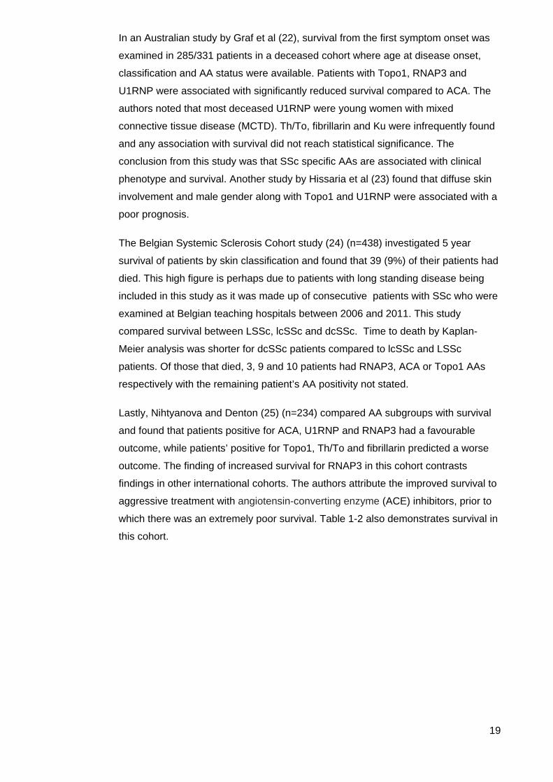
Table1-2: Survival in SSc, a comparison of international cohorts, AA status and disease (skin) classification. N/A= Data not available.
Overall, autoantibodies associated with a poor prognosis are Topo1 and RNAP3
with ACA, Pm/Scl and Ku having a more favourable prognosis (25). Survival
outcomes are varied for U1RNP, Th/To and fibrillarin. The difference in survival
outcome for the latter three AAs in various geographic locations may lie in genetic
background, and indeterminate environmental influences.
The extent of skin thickening or fibrosis associated with diffuse disease also has a
major influence on survival outcomes. Some studies have explored differences both
within and between the major AA groups (ACA and Topo1) for fibrotic involvement
(26), however it is clear that further studies are warranted to investigate the
subtleties within AA subgroups for underlying fibrotic mechanisms to improve not
only quality of life, but survival.
Autoantibody /Skin subset
10 year survival (%) Pittsburgh(19)
10 year survival (%) Japan(20)
15 year survival (%) United Kingdom (25)
lcSSc N/A 83 N/A U1RNP 88 72 78
Centromere 76 93 78
PmScl 72 Not found Japanese pts N/A Th/To 65 N/A N/A dcSSc N/A
71 N/A RNA Polymerase III 75 30 93
Topo1 64 62 57
Fibrillarin (U3RNP) 61 N/A N/A
21
Causes of Mortality
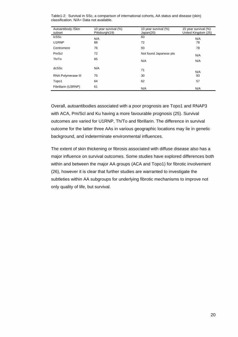
Pulmonary Arterial Hypertension Pulmonary arterial hypertension is prevalent in ~10% of SSc patients (27) and
represents ~30% of deaths (28). It is reported to have a worse outcome than either
idiopathic PAH or other connective tissue disease (CTD) related PAH with a recent
meta-analysis concluding that the three year survival rate for SSc-PAH was only
56% (95%CI 51-61) (27).The reasons for the difference in PAH mortality/survival
between the CTDs are unknown.
Figure 1-9 Kaplan‐Meier cumulative survival curve of Scleroderma patients in the South Australian Scleroderma Register with pulmonary arterial hypertension. (Reproduced from (23))
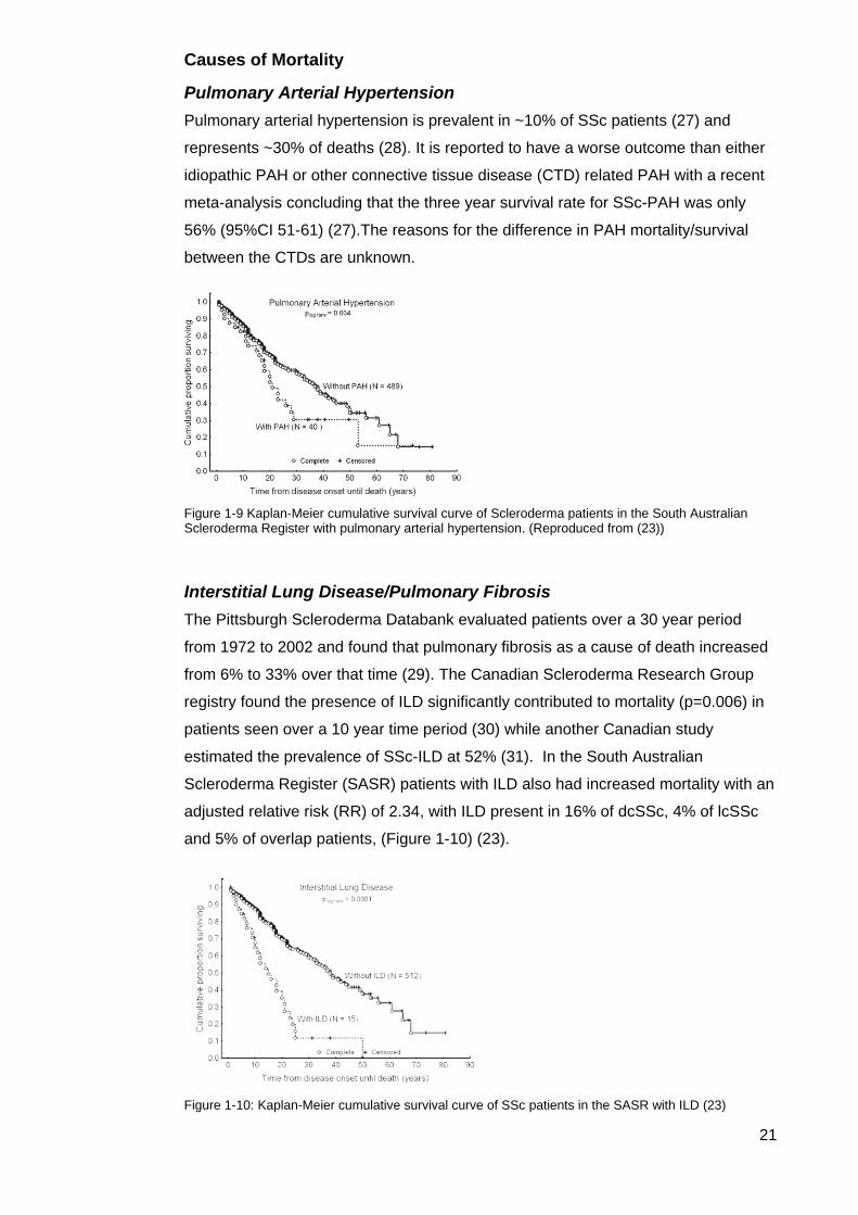
Interstitial Lung Disease/Pulmonary Fibrosis The Pittsburgh Scleroderma Databank evaluated patients over a 30 year period
from 1972 to 2002 and found that pulmonary fibrosis as a cause of death increased
from 6% to 33% over that time (29). The Canadian Scleroderma Research Group
registry found the presence of ILD significantly contributed to mortality (p=0.006) in
patients seen over a 10 year time period (30) while another Canadian study
estimated the prevalence of SSc-ILD at 52% (31). In the South Australian
Scleroderma Register (SASR) patients with ILD also had increased mortality with an
adjusted relative risk (RR) of 2.34, with ILD present in 16% of dcSSc, 4% of lcSSc
and 5% of overlap patients, (Figure 1-10) (23).
Figure 1-10: Kaplan-Meier cumulative survival curve of SSc patients in the SASR with ILD (23)
22
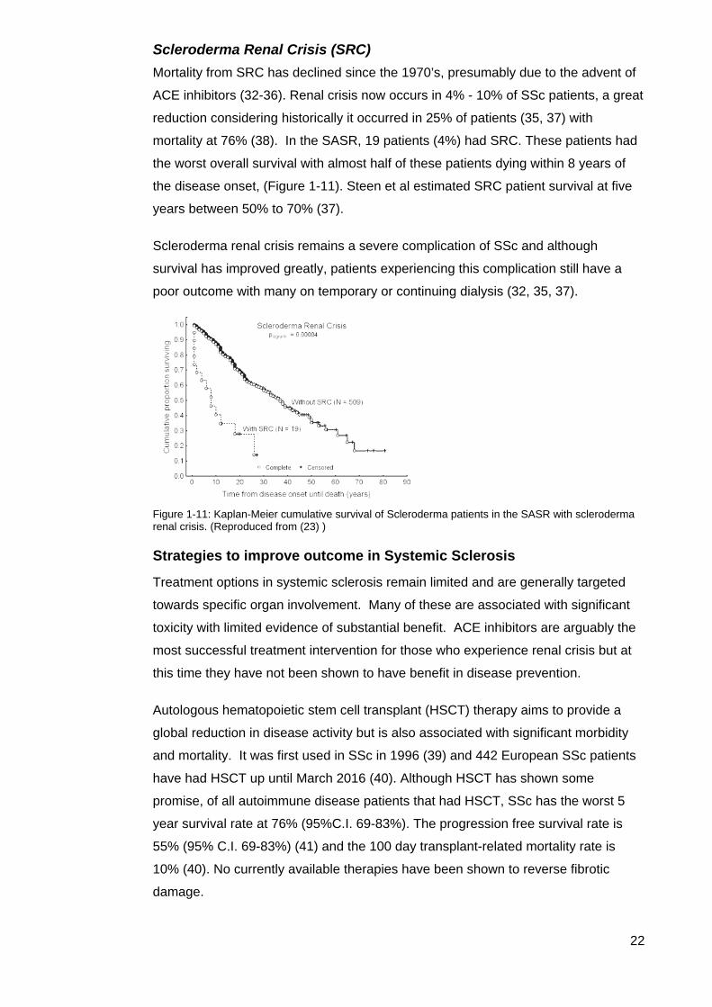
Scleroderma Renal Crisis (SRC) Mortality from SRC has declined since the 1970’s, presumably due to the advent of
ACE inhibitors (32-36). Renal crisis now occurs in 4% - 10% of SSc patients, a great
reduction considering historically it occurred in 25% of patients (35, 37) with
mortality at 76% (38). In the SASR, 19 patients (4%) had SRC. These patients had
the worst overall survival with almost half of these patients dying within 8 years of
the disease onset, (Figure 1-11). Steen et al estimated SRC patient survival at five
years between 50% to 70% (37).
Scleroderma renal crisis remains a severe complication of SSc and although
survival has improved greatly, patients experiencing this complication still have a
poor outcome with many on temporary or continuing dialysis (32, 35, 37).
Figure 1-11: Kaplan-Meier cumulative survival of Scleroderma patients in the SASR with scleroderma renal crisis. (Reproduced from (23) )
Strategies to improve outcome in Systemic Sclerosis
Treatment options in systemic sclerosis remain limited and are generally targeted
towards specific organ involvement. Many of these are associated with significant
toxicity with limited evidence of substantial benefit. ACE inhibitors are arguably the
most successful treatment intervention for those who experience renal crisis but at
this time they have not been shown to have benefit in disease prevention.
Autologous hematopoietic stem cell transplant (HSCT) therapy aims to provide a
global reduction in disease activity but is also associated with significant morbidity
and mortality. It was first used in SSc in 1996 (39) and 442 European SSc patients
have had HSCT up until March 2016 (40). Although HSCT has shown some
promise, of all autoimmune disease patients that had HSCT, SSc has the worst 5
year survival rate at 76% (95%C.I. 69-83%). The progression free survival rate is
55% (95% C.I. 69-83%) (41) and the 100 day transplant-related mortality rate is
10% (40). No currently available therapies have been shown to reverse fibrotic
damage.
23
Significant improvements in disease outcome are likely to rely on accurate, early
disease detection before irreversible damage has occurred. Early diagnosis and
prognostication of patients has the potential to improve outcome by identifying those
patients at greater risk of complications and allowing the institution of treatments in a
timely fashion, either as standard therapy, or in the setting of a clinical trial.
Updating Diagnostic Criteria, and Classification in SSc
Early diagnosis of patients with SSc is critical to allow timely intervention of
therapies, both in the setting of clinical trials and during routine management. In
2013, the American College of Rheumatology/European League Against
Rheumatism (ACR/EULAR) published updated classification criteria for SSc (42) as
the 1980 ACR classification lacked sensitivity and missed patients with early
disease or with limited skin involvement (43). Underpinning the need for the
reclassification was the recognised heterogeneity of SSc. The new classification
criteria as described by Van den Hoogen et al, are:
…intended to be used by rheumatologists, researchers, national and international
drug agencies, pharmaceutical companies, or any others involved in studies of SSc.
Our objective was to develop a set of criteria that would enable identification of
individuals with SSc for inclusion in clinical studies, being more sensitive and
specific than previous criteria (43).
Ideally there should be no difference between classification criteria and diagnostic
criteria, but in reality diagnostic criteria will tend to have a higher sensitivity and
lower specificity, whereas classification criteria will maximize specificity. Three major
AAs (ACA, anti Topo1 and anti RNAP3) are recognised to be almost exclusively
associated with SSc and have been included in the ACR/EULAR revised criteria
(43), but allowance is also made for the small number of patients (5-10%) who have
SSc in the absence these autoantibodies (44) (Table1-3).
24
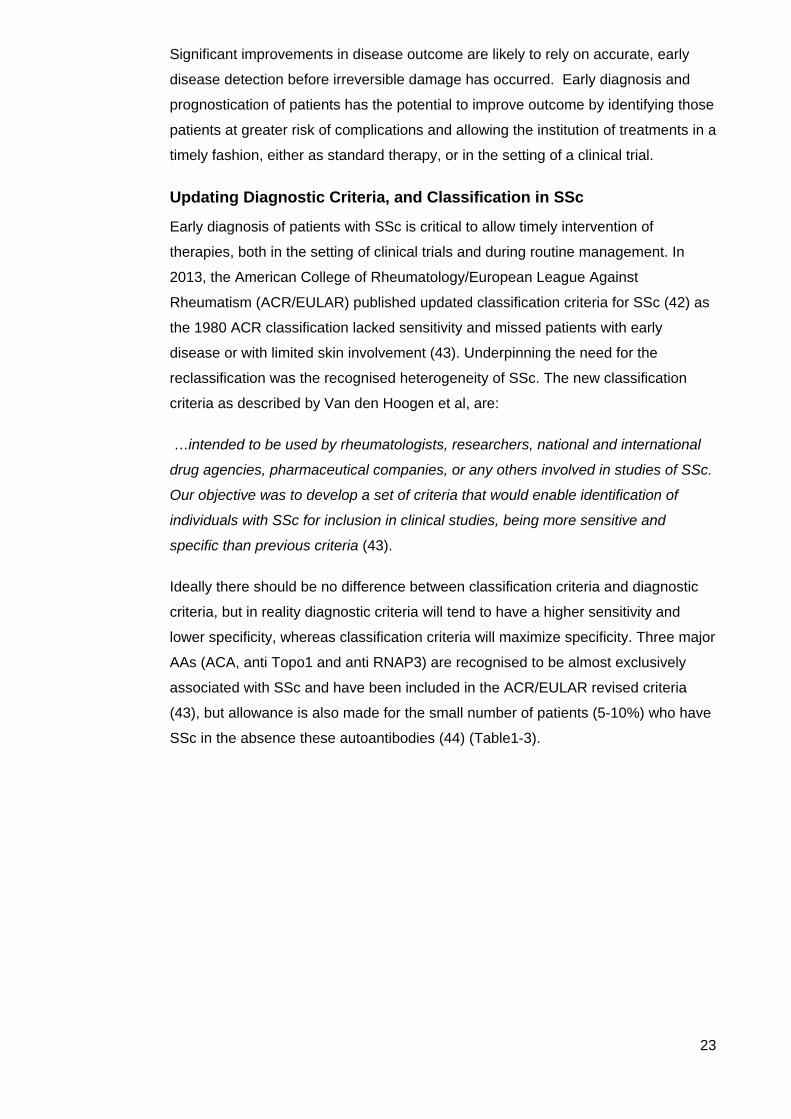
Table 1-3 The American College of Rheumatology/European League Against Rheumatism criteria for the classification of systemic sclerosis* (reproduced from (43)) * These criteria are applicable to any patient considered for inclusion in a systemic sclerosis study. The criteria are not applicable to patients with skin thickening sparing the fingers or to patients who have a scleroderma-like disorder that better explains their manifestations (e.g. nephrogenic fibrosis, generalised morphea, eosinophilic fasciitis, scleroderma diabeticorum, scleromyxedema, erythromyalgia, porphyria, lichen sclerosis graft-versus-host disease, diabetic cheiroarthropathy).
† The total score is determined by adding the maximum weight (score) in each category. Patients with a total score of ≥9 are classified as having definite systemic sclerosis.
It is hoped that these updated classification criteria will allow a greater and more
accurate capture of those patients who have systemic sclerosis but they are not
designed to provide any form of sub-classification or stratification.
The Role of Sub-classification in SSc
SSc is a heterogeneous disease and the differing clinical presentations, AA
associations and genetics have led some to propose that it encompasses more than
one condition (45). Therefore, accurate stratification of the disease is critical to
identify and separate patients into groups that contain a similar clinical course and
prognostic outlook.
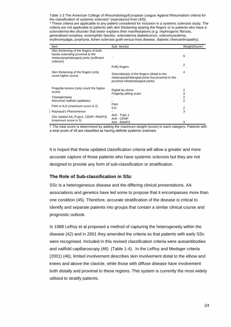
In 1988 LeRoy et al proposed a method of capturing the heterogeneity within the
disease (42) and in 2001 they amended the criteria so that patients with early SSc
were recognised. Included in this revised classification criteria were autoantibodies
and nailfold capillaroscopy (46) (Table 1-4). In the LeRoy and Medsger criteria
(2001) (46), limited involvement describes skin involvement distal to the elbow and
knees and above the clavicle, while those with diffuse disease have involvement
both distally and proximal to these regions. This system is currently the most widely
utilised to stratify patients.
Item Sub- Item(s) Weight/Score† Skin thickening of the fingers of both hands extending proximal to the metacarpophalangeal joints (sufficient criterion)
- 9
Skin thickening of the fingers (only count higher score)
Puffy fingers Sclerodactyly of the fingers (distal to the metacarpophalangeal joints but proximal to the proximal interphalangeal joints)
2 4
Fingertip lesions (only count the higher score)
Digital tip ulcers Fingertip pitting scars
2 3
Telangiectasia - 2 Abnormal nailfold capillaries - 2
PAH or ILD (maximum score is 2) PAH ILD 2
Raynaud’s Phenomenon - 3
SSc related AA (Topo1, CENP, RNAP3) (maximum score is 3)
Anti - Topo 1 Anti - CENP Anti - RNAP3 3
25
Table 1-4. LeRoy and Medsger 2001 SSc
Category Features Limited SSc Raynaud’s Phenomenon (objective documentation)
plus any one: SSc-type nailfold capillary pattern or SSc selective autoantibodies OR Raynaud’s phenomenon (subjective) plus both: SSc-type nailfold capillary pattern and SSc selective antibodies
Limited Cutaneous SSc Criteria for limited SSc plus distal cutaneous changes
Diffuse Cutaneous SSc Criteria for limited SSc plus proximal cutaneous changes
Diffuse fasciitis with eosinophilia
Proximal cutaneous changes without criteria for limited SSc or limited cutaneous SSc
However there is recognition that these subsets are an over simplification of SSc
(47-51) and others have proposed further modifications (45, 52-54), so that a
precision medicine approach can be pursued (55-59). Given the considerable
variation in SSc pathologies and the desire to improve treatments and interventions,
stratification is vital when comparing outcomes and when designing clinical trials.
The challenge now is to identify SSc patients that are more likely to develop severe
organ involvement and also to determine the appropriate time to intervene before
irreversible fibrotic or vascular damage occurs.
At present it can be difficult to stratify patients and compare research outcomes
across varying geographic locations and ethnic (genetic) backgrounds merely using
the broad lcSSc and dcSSc subsets. In addition to AA profiling to enhance patient
stratification (56), utilising a precision medicine approach linked with HLA and non-
HLA susceptibility genes identified in genome-wide association studies (GWAS)
may lead to a better understanding of inheritance patterns, susceptibility, various
pathogenic pathways and therapeutic targets (3, 60-62).
Finally, the interpretation and comparison of research outcomes and the
stratification of patients would be enhanced if clinical associations were more closely
aligned with focused subsets. One recent finding by Srivastava et al found that
stratifying patients by skin involvement as well as AAs may predict clinical outcomes
better than skin or serology alone in SSc (26). These findings can inform ongoing
efforts to define more robust SSc subsets and perhaps it may become recognisable
that skin in AA subsets may be due to biological variation within the AA subset
rather than a function of an arbitrary title of lcSSc or dcSSc.

26
Biomarkers and SSc
‘Biological markers’ or ‘biomarkers’ are defined by the National Institute of Health
Working Group on Biomarkers as a ‘characteristic that is objectively measured and
evaluated as an indicator of normal biological processes, pathological processes or
pharmacologic responses to a therapeutic intervention’ (63).
Biomarkers should be readily obtainable, quantifiable, objective, reproducible and
able to act as a surrogate clinical endpoint with predictive power in different
populations remaining at a similarly high degree of specificity and/or sensitivity.
They should be available broadly and not only in an isolated research capacity. It is
vital that there is an understanding between an assessable biological process and
the clinical outcome they represent because biomarker outcomes may be the
principal measure in drug development and other biomedical research enterprises
(64). The utility of biomarkers includes prevention, diagnosis and early detection so
that appropriate treatments can be developed and implemented before irreversible
damage to the patients’ health and wellbeing occurs. Further utility includes
response to treatment, progression or cessation of disease.
Scleroderma, as with most autoimmune conditions, has a suite of biomarkers
associated with various pathologies within the condition (51). Serum
autoantibodies, found in >95% (65) of patients, are correlated with distinct clinical
manifestations and have the potential, particularly in early disease, to aid in
predicting disease course. There is potential to utilise AAs in conjunction with other
biomarkers to predict fibrotic, vascular and organ manifestations and response to
treatment. For example, a panel of SSc associated AAs could be interpreted in
combination with information obtained from transcriptomics, proteomics,
metabolomics, genomics and epigenomics to provide a detailed and individually
personalised assessment of disease course (13). Theoretically this will be highly
beneficial for patient outcomes as biomarkers based on precision medicine will
refine treatments and therapies for which the patient is most likely to respond (66).
At present, sub classification for SSc is based upon a dichotomised skin based
system (42) which is applied across a continuous spectrum of disease (45, 53). It is
recognised that the current system means that important features of the disease
process including serologic biomarkers and other organ involvement will be missed
(53, 67). Furthermore, the skin changes that allow identification of disease subtypes
may take some time to evolve and therefore have limited use in guiding therapy.
Identification of a biomarker that can be reliably assessed before damage occurs, to
both predict clinical outcomes and to determine appropriate intervention, has been
arduous. To date, the most universal biomarkers remain the SSc associated

27
autoantibodies, found in 95% people with SSc (65). The following section of this
review will describe the most relevant knowledge to date on autoantibodies as
biomarkers in SSc including their association with clinical phenotypes and their utility
in the sub classification of this perplexing condition.

28
Autoantibodies
Scleroderma associated autoantibodies are widely recognised to have distinct
clinical associations but the validity of these findings will vary according to the
diagnostic platform used. In addition, findings will be influenced by environmental
and genetic factors (68).
Diagnostic Platforms In recent years, numerous diagnostic platforms have been released to the
commercial market, several without sufficient validation compared to conventional
and standard methods (69). Because AAs recognise a variety of epitopes, it is vital
to validate each test’s sensitivity and specificity. Mahler et al explains (70):
…The diagnostic sensitivity is a statistical measure of how accurately a test correctly
identifies diseased individuals...the diagnostic specificity is a statistical measure of
how well a test correctly identifies absence of the disease in question…
It is also important to establish cut-offs for each assay that are based on the results
from a range of local patients with SSc, other SARDs and healthy controls and that
the cut-offs have been validated in other cohorts with differing demographic,
geographic, environmental and genetic factors (71).
Indirect Immunofluorescence (IIF) on HEp-2 cells. Screening for anti-cellular antibodies in subjects with suspected autoimmune
disease using indirect immunofluorescence on HEp-2 cells test is one of the most
common screening tests. The pattern obtained can provide information to assist in
the diagnosis and classification of SSc and some other autoimmune diseases (72)
with the exception of autoimmune myopathies. IIF is considered by some to be the
‘gold standard’ of anti-cellular antibody detection (73). The advantages of IIF on HE-
p2 cells are that it is able to detect more than 100 different antigens including some
that can be identified without a further confirmatory test (centromere). The
disadvantages are that it is dependent on the experience of the reader, although
there are automated systems available that can somewhat limit inter observer
variation (74). It also has a low sensitivity for certain clinically important AAs (i.e., Jo-
1 and other synthetase autoantigens, ribosomal P, SS-A/Ro60, Ro52/TRIM21) and,
depending on the screening serum dilution, a low specificity (high false positive rate)
(70). Further limitations include the nomenclature of patterns for nuclear (true ANA),
cytoplasmic and mitotic staining, alternative platforms with different antigen profiles,
standardisation, automation and incongruent results (75).
A negative IIF test does not exclude the presence of all connective tissue disease
associated AAs and so where clinical suspicion is high, further testing should be
29
undertaken even in the presence of a negative IIF result (76) (77, 78).
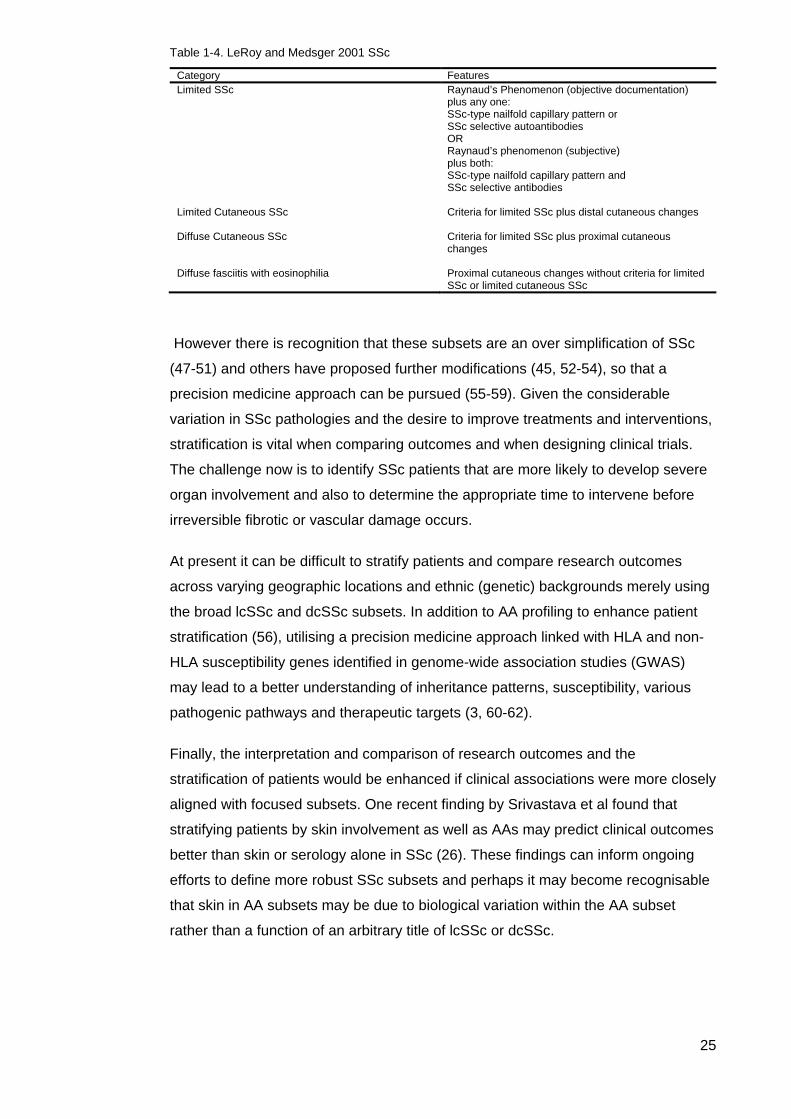
Figure 1-12: IIF Centromere pattern. Image ' ANA PATTERNS’. www.anapatterns.org
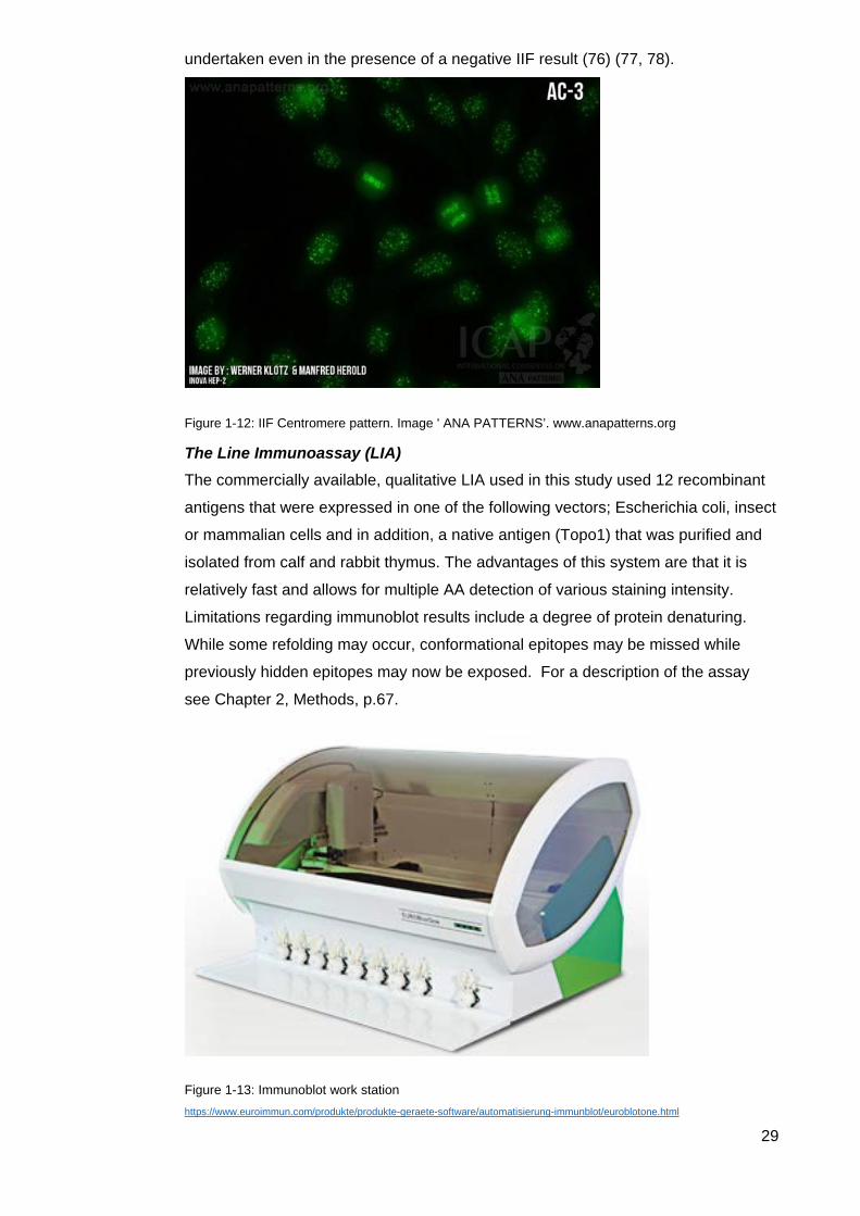
The Line Immunoassay (LIA) The commercially available, qualitative LIA used in this study used 12 recombinant
antigens that were expressed in one of the following vectors; Escherichia coli, insect
or mammalian cells and in addition, a native antigen (Topo1) that was purified and
isolated from calf and rabbit thymus. The advantages of this system are that it is
relatively fast and allows for multiple AA detection of various staining intensity.
Limitations regarding immunoblot results include a degree of protein denaturing.
While some refolding may occur, conformational epitopes may be missed while
previously hidden epitopes may now be exposed. For a description of the assay
see Chapter 2, Methods, p.67.
Figure 1-13: Immunoblot work station https://www.euroimmun.com/produkte/produkte-geraete-software/automatisierung-immunblot/euroblotone.html

30
Figure 1-14: results - line blot assay Original Image: Karen Patterson
Enzyme linked immunosorbent assay (ELISA) The original ELISA was a plate-based assay (‘sandwich’) technique designed for
detecting and quantifying substances such as antibodies. It is routinely used in
diagnostic laboratories with a variety of AAs available for testing in suspected
autoimmune disease patients. There are also multiplex ELISA formats that adopt
chemiluminescent/fluorescent reporter systems that use micro-bead based
suspensions. The various ELISA formats (Figure 1-15), have been found to be
reliable methods of AA detection (73, 79, 80). During the period of this study when
sera were tested by the Australian Scleroderma Interest Group independent
laboratories, the ELISA was the most utilised method for detecting RNA Polymerase
III.
31
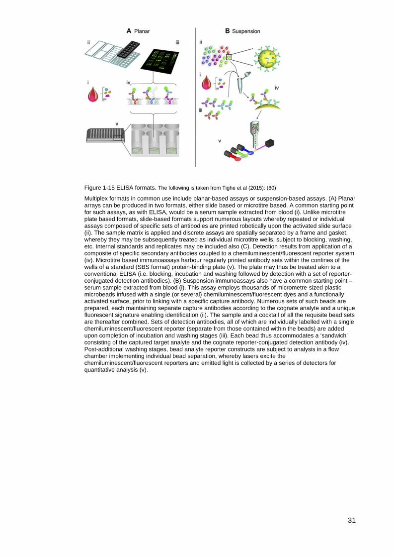
Figure 1-15 ELISA formats. The following is taken from Tighe et al (2015): (80)
Multiplex formats in common use include planar-based assays or suspension-based assays. (A) Planar arrays can be produced in two formats, either slide based or microtitre based. A common starting point for such assays, as with ELISA, would be a serum sample extracted from blood (i). Unlike microtitre plate based formats, slide-based formats support numerous layouts whereby repeated or individual assays composed of specific sets of antibodies are printed robotically upon the activated slide surface (ii). The sample matrix is applied and discrete assays are spatially separated by a frame and gasket, whereby they may be subsequently treated as individual microtitre wells, subject to blocking, washing, etc. Internal standards and replicates may be included also (C). Detection results from application of a composite of specific secondary antibodies coupled to a chemiluminescent/fluorescent reporter system (iv). Microtitre based immunoassays harbour regularly printed antibody sets within the confines of the wells of a standard (SBS format) protein-binding plate (v). The plate may thus be treated akin to a conventional ELISA (i.e. blocking, incubation and washing followed by detection with a set of reporter-conjugated detection antibodies). (B) Suspension immunoassays also have a common starting point – serum sample extracted from blood (i). This assay employs thousands of micrometre-sized plastic microbeads infused with a single (or several) chemiluminescent/fluorescent dyes and a functionally activated surface, prior to linking with a specific capture antibody. Numerous sets of such beads are prepared, each maintaining separate capture antibodies according to the cognate analyte and a unique fluorescent signature enabling identification (ii). The sample and a cocktail of all the requisite bead sets are thereafter combined. Sets of detection antibodies, all of which are individually labelled with a single chemiluminescent/fluorescent reporter (separate from those contained within the beads) are added upon completion of incubation and washing stages (iii). Each bead thus accommodates a ‘sandwich’ consisting of the captured target analyte and the cognate reporter-conjugated detection antibody (iv). Post-additional washing stages, bead analyte reporter constructs are subject to analysis in a flow chamber implementing individual bead separation, whereby lasers excite the chemiluminescent/fluorescent reporters and emitted light is collected by a series of detectors for quantitative analysis (v).

32
Extractable nuclear Antigen (ENA)/Immunoprecipitation (IP) The simplest form of IP isolates a single target antigen to investigate the identity,
structure, expression or activation of a protein that is immobilised on a solid support
such as magnetic beads or agarose gel. It is one of the most widely used methods
of protein isolation from cell or tissue lysates for the purpose of detection by other
assay techniques and was commonly used by ASIG independent laboratories
particularly for the AAs in Figure 1-16.
Figure 1-16: ENA/IP extract of rabbit or bovine tissue extracts that form precipitins with autoantibodies
http://www.immunovision.com/ena-1001/
The source and characteristics of the autoantigen used in the various platforms
must be considered as results may vary between recombinant, native peptide and
full-length antigens. In particular, short polypeptide epitopes may give different
results to an assay that detects reactivity to native antigens such as those
represented in IIF cell based assays, immunoblotting or immunoprecipitation (76).
In addition there are inter-manufacturer variations in reagents, in the standardisation
of antigen (substrate) and the fixation process, nor is it a reliable method to detect
antibodies to nucleic acids. Finally, there are the inter-laboratory variations in
methods used, expertise, equipment and subjectivity in interpreting results.
Varying advantages and limitations apply to all platforms and it is most important to
be aware of these considerations. Other techniques used to detect AAs include
chemiluminescent immunoassays (CLIA), addressable laser bead immunoassays
(ALBIA) and other microbead based assays, and nanobarcode arrays on planar
surfaces (71). The latter are not widely available in diagnostic laboratories in
Australia (70).
In short, no test is without its challenges and these should be considered when
interpreting any AA result.
33
Primary SSc specific and SSc associated autoantibodies
There are three primary autoantibodies that are conventionally associated with SSc
and considered to be highly specific for the disease; Anti Centromere antibodies
(CENP), anti-Topoisomerase 1 (Topo1, formerly known as Scl-70) and anti-RNA
Polymerase III. Earlier research hypothesised that these three autoantibodies were
mutually exclusive and expressed in isolation, however with evolving diagnostic
platforms and improved AA detection, it is now recognised that it is not uncommon
for SSc patients to express other AA of varying titres (22, 76, 81), and even though
it is a rare occurrence, co-expression in various combinations of the three central
AAs has been found (22, 81). Less frequent but also considered relatively specific
for SSc are anti-U3-RNP/fibrillarin, anti-RNA polymerase I, and anti-U11/U12 RNP.
As a consequence of improved AA detection methods and international
collaboration efforts to combine cohorts, Anti-Th/To is now also considered to be
relatively (but not exclusively) SSc specific (82). This AA is not included in routine
diagnostic laboratory testing and it has been suggested that current methods may
miss patients positive for Th/To due to the reliance on one epitope, hPop1 (83).
Additional important epitopes for this AA are currently being investigated (82) and
are described later.
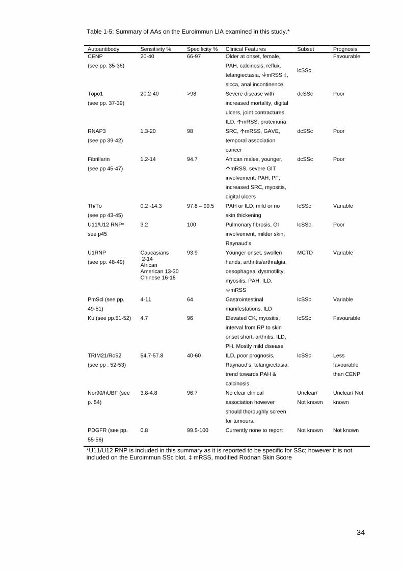
Other AAs that may be associated with SSc but are not specific to the disease
include anti-PmScl (75 and 100 epitopes), anti-Ro52/TRIM21, anti-Ku, anti- U1-
ribonucleoprotein (U1-RNP) and anti-NOR 90. A summary table (Table 1-5) of all
AAs on the Euroimmun Line Immunoassay that were tested in this study is below.
34
Table 1-5: Summary of AAs on the Euroimmun LIA examined in this study.*
*U11/U12 RNP is included in this summary as it is reported to be specific for SSc; however it is not included on the Euroimmun SSc blot. ‡ mRSS, modified Rodnan Skin Score
Autoantibody Sensitivity % Specificity % Clinical Features Subset Prognosis CENP
(see pp. 35-36)
20-40 66-97 Older at onset, female,
PAH, calcinosis, reflux,
telangiectasia, mRSS ‡,
sicca, anal incontinence.
lcSSc
Favourable
Topo1
(see pp. 37-39)
20.2-40 >98 Severe disease with
increased mortality, digital
ulcers, joint contractures,
ILD, mRSS, proteinuria
dcSSc Poor
RNAP3
(see pp 39-42)
1.3-20 98 SRC, mRSS, GAVE,
temporal association
cancer
dcSSc Poor
Fibrillarin
(see pp 45-47)
1.2-14 94.7 African males, younger,
mRSS, severe GIT
involvement, PAH, PF,
increased SRC, myositis,
digital ulcers
dcSSc Poor
Th/To
(see pp 43-45)
0.2 -14.3 97.8 – 99.5 PAH or ILD, mild or no
skin thickening
lcSSc Variable
U11/U12 RNP*
see p45
3.2 100 Pulmonary fibrosis, GI
involvement, milder skin,
Raynaud’s
lcSSc Poor
U1RNP
(see pp. 48-49)
Caucasians 2-14 African American 13-30 Chinese 16-18
93.9 Younger onset, swollen
hands, arthritis/arthralgia,
oesophageal dysmotility,
myositis, PAH, ILD,
mRSS
MCTD Variable
PmScl (see pp.
49-51)
4-11 64 Gastrointestinal
manifestations, ILD
lcSSc Variable
Ku (see pp.51-52) 4.7 96 Elevated CK, myositis,
interval from RP to skin
onset short, arthritis, ILD,
PH. Mostly mild disease
lcSSc Favourable
TRIM21/Ro52
(see pp . 52-53)
54.7-57.8 40-60 ILD, poor prognosis,
Raynaud’s, telangiectasia,
trend towards PAH &
calcinosis
lcSSc Less
favourable
than CENP
Nor90/hUBF (see
p. 54)
3.8-4.8 96.7 No clear clinical
association however
should thoroughly screen
for tumours.
Unclear/
Not known
Unclear/ Not
known
PDGFR (see pp.
55-56)
0.8 99.5-100 Currently none to report Not known Not known

35
Anti-Centromere Proteins A and B (CENP A and CENP B)
Description Cell replication and division occurs in a dedicated region of the chromosome called
the centromere. Without centromere integrity, erroneous cell replication and division
can have disastrous consequences such as genomic instability with abnormal cell
division as found in autoimmune disease (84, 85) and cancer (86, 87). Within this
centromere region specialised chromatin provides the basis for kinetochore
assembly where sister chromatids attach. It is within this region that the centromere
proteins, CENP A and CENP B, necessary for a functional kinetochore, are located.
The first description of anti-centromere proteins was in 1980 (88). CENP A is a 17
kDa histone H3 variant essential for epigenetically marking centromere location (89)
to which other proteins dock in the replication process. The anti-centromere immune
response is directed against two domains in the N-terminus containing a linear motif
(G/A-PR/S-R-R) (90). CENP B is an 80 kDa protein localised in the heterochromatin
under the kinetochore (91) where it binds to a 17bp DNA sequence, the CENP B
box (92). The major epitope of CENP B maps to the C-terminal part of the protein
(amino acids 535–599) (93). The exact function of CENP B is unknown, however it
seems to have a role in regulating the formation and action of heterochromatin in
centromeres (94) and interestingly unlike CENP A, which is highly evolutionarily
conserved, mice can live without CENP B (95).
While autoantibodies to CENP A-F and CENP O have been detected in SSc sera
(93) , the primary autoantigens in SSc are to CENP A and CENP B. In CENP B, the
first AA to be cloned, there were three independent epitopes identified and these
epitopes were recognised by ≥90% of SSc sera containing anti-centromere
antibodies (96). CENP B was therefore thought to be the major autoantigen but
Mahler et al found that CENP A antibodies (as detected by ELISA) are a more
specific biomarker for SSc than antibodies to CENP B (97). Most SSc patients with
ACA have antibodies to both CENPs (97-99).
Detection Serological detection of ACA can precede clinical manifestations of SSc by a
number of years and they continue to be expressed throughout the life of the patient
(100) with stable titres over time (101). Historically, ACA patterns were detected by
indirect immunofluorescence on HEp-2 cells and this is still considered ‘gold
standard’ methodology. (73). Further assays have been developed for both
research and commercial use. These include ELISA and the LIA with both methods
using recombinant proteins expressed in insect cells utilising a baculovirus system
(102-104). Alternatively, the commercial synthesis of synthetic peptides (97) and

36
originally, a cloned fusion protein of the CENP B antigen were used (103). Other
detection methods include multiplexed assays such as addressable laser bead
immunoassay, planar assays (capturing ligands on a two dimensional array) and the
forerunner of the LIA, the dot blot (105). Hanke et al noted when using the LIA that
both CENP A and CENP B shared significant associations to clinical manifestations,
but were not completely identical and surmised that detection of both antibodies in
parallel may slightly increase the diagnostic sensitivity for SSc (104) and Mahler et
al found good qualitative agreement and ‘remarkably good’ quantitative correlation
between the CENP ELISAs and IIF on HEp-2 cells (73).
Sensitivity and Specificity The sensitivity of anti CENP antibodies for SSc is between 20% - 40% with
specificity ranging between 66%-97% (97, 106-108). CENPs are occasionally found
in other SARDs such as Systemic Lupus Erythematosus (SLE) (2-5%) Sjögren’s
syndrome (SjS) (5-10%), idiopathic inflammatory myopathies
(polymyositis/dermatomyositis) (IIM/PM/DM) (1-3%), MCTD (2-5%), 30% primary
biliary cirrhosis (PBC) patients (93, 109) and in less than 3% of healthy individuals
(105). Prevalence varies between geographic locations and ethnic groups with
Caucasians having the highest representation of this AA (19, 110-112) and African
Americans and Asians having the lowest representation (110, 112-115).
Clinical Associations Patients’ positive for CENPs are more likely to be female and are older at disease
onset compared to other SSc related AA (19, 111, 116); the exception to this finding
lies with Mexican Mestizo patients where no differences in age of onset between the
AA subsets are found (117). CENPs are present more often in Caucasians and
Mexican Mestizos’ than they are in African Americans (19, 110, 116). Prevalence
varies throughout Asia with Chinese (113), West Malaysian (118) and Thai patients
(119) having a lower prevalence compared to Japan where the prevalence of CENP
is similar to Caucasian cohorts (120). In India, CENP positivity accounts for 22.7%
of SSc patients (121). The clinical correlates of CENPs include pulmonary arterial
hypertension, calcinosis, reflux oesophagitis, telangiectasia and milder skin
involvement (19, 111, 116, 117, 122). Other significant clinical associations are
sicca symptoms (123, 124) and anal incontinence (125). Although the prevalence
rates differ across geographic locations and ethnic groups, the clinical associations
remain (126).
37
Topoisomerase 1
Description A hierarchical arrangement of tight coils and loops (helical winding or supercoiling)
enables chromatin, a combination of DNA and proteins, to be packed into cells. This
complex organisation is comprised of DNA wound around specific proteins called
histones to form the nucleosome and its appearance is often likened to ‘beads on a
string’. This hierarchical organisation also allows the binding of enzymes, such as
topoisomerases, that are required for DNA replication, transcription and repair.
Topoisomerases introduce temporary single or double-strand breaks in the DNA. A
specific type of topoisomerase, Topo1, transiently breaks one strand of the DNA,
allowing for adjustments in helical winding. Topo1 is primarily responsible for
removing torsional stress generated by processes that leave the DNA overwound or
under wound (127). It is a 765 amino acid long enzyme (105-kDa) that contains five
distinct regions: the N-terminal domain (amino acids 1–215), core subdomains I–II
(amino acids 216–435), core subdomain III (amino acids 436–636), the linker
domain (amino acids 637–713), and the C-terminal domain (amino acids 714–765)
(128). Several studies have demonstrated that antibodies against Topo1 recognize
multiple epitopes on the molecule (129).
In characterising what was thought to be the 70 kDa ‘Scl-70’ antigen, originally
identified by Douvas et al in 1979 in sera from SSc patients (130), Shero et al
discovered that the antigen was actually the 100kDa nuclear enzyme
Topoisomerase 1 and suggested that the smaller 70kDa protein and another 86kDa
protein also detected, were degradation products of the larger enzyme (131). In a
separate study, Guldner et al probed purified DNA topoisomerase I isolated from
calf thymus directly with the autoantibodies from a SSc patient and from the cross
reaction patterns observed with the different antigens and antibodies concluded that
DNA topoisomerase I is one of the antigenic components against which
autoantibodies are formed in scleroderma patients (132). Thus, the Scl-70 antigen
was identified as anti-Topoisomerase 1, however many still refer to it as Scl-70.
Pathogenicity of this AA has remained elusive. Henault el al (133) and Seneccal et
al (134) demonstrated that Topo1 complex binding can trigger the adhesion and
activation of monocytes which provides a plausible model for a fibrotic cascade, but
the complete functional attribute of Topo1 remains unexplained.(135). Kuwana et al
took serial measurements of Topo1 levels and has linked these increased or
decreased levels with disease activity and have found that if the AA becomes
undetectable, prognosis improves (136)
38
Detection SSc sera are usually screened on HE-p2 cells by IIF followed by a second test to
detect the associated specificities. IIF on HE-p2 cells produce a fine granular to
homogeneous staining of the nucleoplasm with or without staining of the nucleoli
and chromatin of mitotic cells (137). Topo1 is a precipitating AA and Douvas’ original
detection of Scl-70 was by immunoprecipitation (130). Methods of detection include
immunodiffusion using the Ouchterlony technique (double diffusion) or counter
immuno-electrophoresis (CIE) using calf thymus extract, ELISA, LIA or ALBIA using
purified native or recombinant topoisomerase 1 fusion protein as the antigen (137).
A study conducted by Tamby et al, found that using a combination of IIF, ELISA and
an immunoblot increased the sensitivity for the detection of Topo1 from 24.3% to
36.9% using IIF and ELISA (138). In an Asian population, Low et al found good
agreement with LIA and ELISA (kappa = 0.97). Another study by Bonroy et al
evaluated a fluoro-enzyme immunoassay (FEIA) as an alternative for the combined
conventional techniques (IIF on HEp-2000, western blotting (WB), protein radio
immunoprecipitation and a LIA) and reported a good overall agreement between
combined conventional techniques and FEIA reactivity (kappa>0.800) (139). Shero
et al concluded that immunoblotting or solid phase immunoassays were
substantially more sensitive for detection than the Ouchterlony test (131) .
Sensitivity and Specificity Topo1 AA is found in found in 10%–40% of SSc sera (128). Sensitivity for SSc is
dependent on the assay used and also the comparative control group (e.g. healthy
controls, other CTDs, Raynaud’s Phenomenon (RP) and non-SSc relatives).
Sensitivity ranges between 20.2% and 40% with a reported high specificity of >98%
(108). While high titre Topo1 AA are highly specific for SSc (128), they have been
detected in other CTD such as SLE. An early study found that 25% of SLE patients
were positive for Topo1 (140) when detected using ELISA, WB and double
immunodiffusion (DID). A later comprehensive study using a variety of platforms
(ELISA, ALBIA, LIA) found that <5% of SLE sera were positive for Topo1 (128) .
This discrepancy is perplexing but no other study has been able to replicate the
findings that 25% of SLE patients are positive for Topo1. An answer may lie in the
one of five the domains of Topo1 because it seems that the region between amino
acid 450 and 600 is a common epitope for the autoantibodies of patients with SSc
and SLE, while epitopes localized in the N-terminal domain are recognized mainly
by dcSSc sera and those that are found in core subdomains I–II are specific for SLE
sera (128). Topo1 are associated with HLA-DRB1, DQB1 and DPB1 and among
these markers, DRB1*11 was associated with Topo1 in all ethnic groups, while HLA-
DRB1*1101 was found in Caucasians and African-Americans. HLA-DRB1*1104
39
was found in Japanese and HLA-DRB1*1502 was found in Caucasians and
Hispanics (76).
Clinical Associations The frequency of Topo1 varies according to geographic location and ethnicity.
Germany (81) and France (141) have higher frequencies of Topo1 compared to
Australia (22), New Zealand (116), Canada (142), the United States of America
(USA) (19) and Belgium (24). The most remarkable ethnic association with Topo1
lies with full-blooded Choctaw Native Americans living in south eastern Oklahoma
who have the highest prevalence (469/105) of SSc yet found in any population. A
major risk factor for disease is a uniquely Amerindian HLA haplotype (143). Despite
these variations, characteristic clinical associations remain (22, 24, 81, 110, 122,
144, 145). Topo1 patients have more severe disease and increased mortality. They
develop more digital ulcers, joint contractures, ILD, a high mRSS (19, 22, 76) and
proteinuria (76). Topo1 was an independent predictor of pulmonary fibrosis in the
EULAR Scleroderma Trials and Research Group database (n=3656) with pulmonary
fibrosis present in 60.2% of Topo1 patients.
RNA Polymerase III (RNAP3)
Description RNAP3 consists of two proteins; a 155kDa protein, IIIA, and a 138kDa protein, IIIB.
Together they form a multi protein complex localised in the nucleoplasm (137).
RNAP3 assists in the translation of a number of noncoding RNA genes whose
products are involved in fundamental cellular processes such as protein synthesis,
RNA processing, transcription and chromatin regulation (146). Therefore, the genes
transcribed by RNAP3 fall in the category of "housekeeping" genes whose
expression is required in all cell types and most environmental conditions. RNAP3
transcription is regulated with cell growth and proliferation and in response to stress.
Deregulation of RNAP3 is linked to diseases such as Alzheimer's disease (147),
fragile X syndrome (146), and cancer (148, 149). The anti-RNAP3 autoantibody is
also one of the three major autoantibodies associated with SSc.
Detection In 1993 Kuwana et al reported a novel antibody that reacted with RNA Polymerases
(I, II and III) (150). Incubation of SSc sera was carried out with (35S) methionine
labelled He-La cell extracts and 14/275 SSc sera reacted, precipitating 12 -14
proteins. Purified IgG from these sera then inhibited RNA transcription catalysed by
RNA polymerase I, II and III. Immunoblot analysis was carried out and the majority
of these sera reacted with a 42kDa or 25kDa protein. In 1998 Chang et al developed
an ELISA to detect antibodies against RNAP I, II, and III. In validation studies both
40
the ELISA and immunoprecipitation of (35S) labelled HeLa cells were used to
analyse sera from a large cohort of well-characterized Caucasian SSc patients.
They found excellent concordance for the presence of anti-RNAP antibodies
between both methods (151). Later in 1998 Satoh et al found that 2% of SLE and
Overlap sera also had RNAP activity; this was subsequently revealed to be RNAP2
which is found in patients with SLE or Overlap disease and that RNAPI and RNAP3
are specific for SSc (152).
In 2002 Kuwana et al characterised the immunodominant epitopes on RNAP3
subunits. RPC62 and RPC155 were generated in a bacterial expression system as
a series of recombinant fragments. Reactivity fragments were examined by
immunoblots and/or ELISA in SSc sera, other CTD disease and in normal healthy
control (NHC) individuals. RNAP3 positive SSc sera recognised several distinct
epitopes on RPC62 and RPC155 in various combinations, but the fragment
encoding amino acids at positions 732-1166 of RPC155 was recognized by all
11 RNAP3 positive SSc sera (153). RNAP3 is now commonly detected using ELISA
and immunoblot technologies.
Lastly, RNAP3 titres are known to change over time (154).
Sensitivity and Specificity After classifying 735 SSc patients for their ANA specificities (fine speckled
nucleoplasmic staining with or without nucleolar staining) followed by
immunoprecipitation using (35S) labelled HeLa cell antigen, Bunn et al concluded
that RNAP1 and RNAP3 AAs represented a discrete serological subgroup (11.7%)
(155). A later study found that antibodies to RNAP3 were not consistently associated
with such an IIF pattern on conventional HEp-2 cell substrates (156). A major
epitope commonly recognized by SSc sera containing RNAP3 autoantibodies was
identified on RPC155 when 16/16 SSc RNAP3 positive sera, but not SSc RNAP3
negative sera, were tested using a purified recombinant fragment in an ELISA (153).
This epitope was considered by Kuwana as having 100% sensitivity and specificity.
In 2005, a new ELISA was developed for detection of RNAP3 antibodies, using a
recombinant fragment containing the immunodominant epitope as the antigen
source and was compared to the (then gold standard) immunoprecipitation method.
This cohort consisted of 522 SSc patients and 516 controls including patients with
other connective tissue diseases and blood bank donors. The results of the ELISA
showed analytical sensitivity of 91% and analytical specificity of 99% compared with
the immunoprecipitation assay. The clinical analysis of the ELISA (with respect to
diagnosis) demonstrated that clinical sensitivity was 17% and the specificity was
98% (157) and these results are consistent with other studies (158) (79).
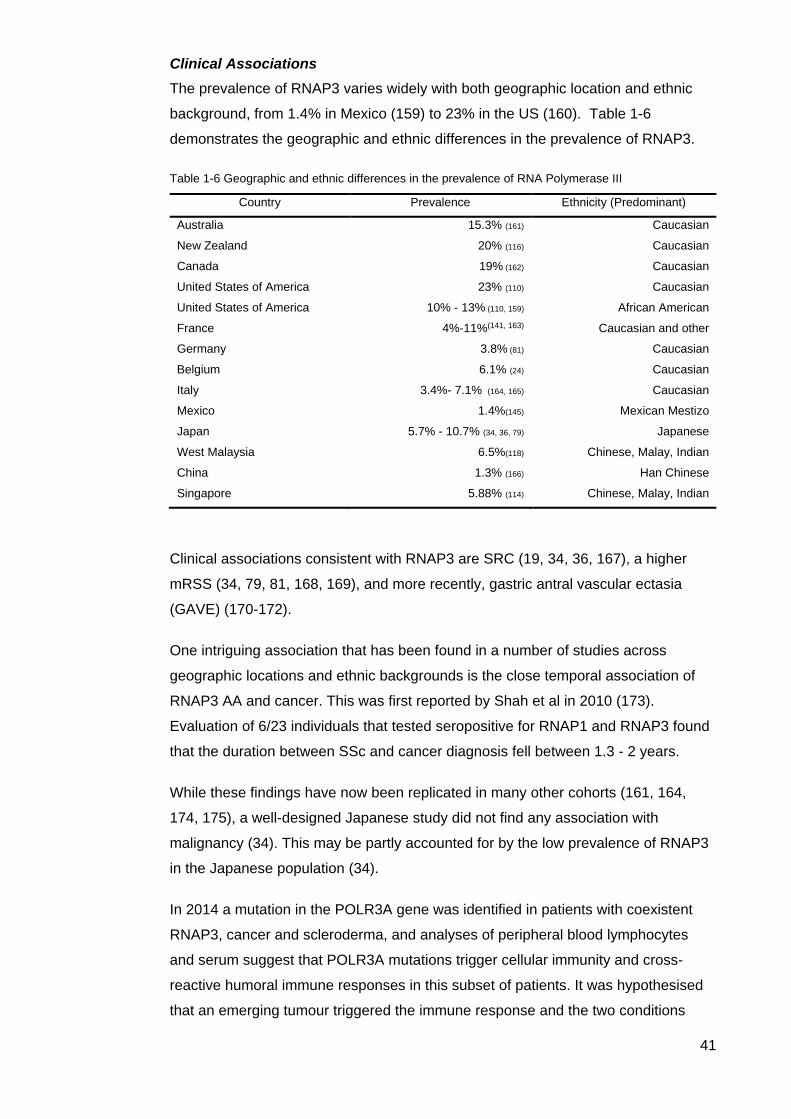
41
Clinical Associations The prevalence of RNAP3 varies widely with both geographic location and ethnic
background, from 1.4% in Mexico (159) to 23% in the US (160). Table 1-6
demonstrates the geographic and ethnic differences in the prevalence of RNAP3.
Table 1-6 Geographic and ethnic differences in the prevalence of RNA Polymerase III
Country Prevalence Ethnicity (Predominant)
Australia 15.3% (161) Caucasian
New Zealand 20% (116) Caucasian
Canada 19% (162) Caucasian
United States of America 23% (110) Caucasian
United States of America 10% - 13% (110, 159) African American
France 4%-11%(141, 163) Caucasian and other
Germany 3.8% (81) Caucasian
Belgium 6.1% (24) Caucasian
Italy 3.4%- 7.1% (164, 165) Caucasian
Mexico 1.4%(145) Mexican Mestizo
Japan 5.7% - 10.7% (34, 36, 79) Japanese
West Malaysia 6.5%(118) Chinese, Malay, Indian
China 1.3% (166) Han Chinese
Singapore 5.88% (114) Chinese, Malay, Indian
Clinical associations consistent with RNAP3 are SRC (19, 34, 36, 167), a higher
mRSS (34, 79, 81, 168, 169), and more recently, gastric antral vascular ectasia
(GAVE) (170-172).
One intriguing association that has been found in a number of studies across
geographic locations and ethnic backgrounds is the close temporal association of
RNAP3 AA and cancer. This was first reported by Shah et al in 2010 (173).
Evaluation of 6/23 individuals that tested seropositive for RNAP1 and RNAP3 found
that the duration between SSc and cancer diagnosis fell between 1.3 - 2 years.
While these findings have now been replicated in many other cohorts (161, 164,
174, 175), a well-designed Japanese study did not find any association with
malignancy (34). This may be partly accounted for by the low prevalence of RNAP3
in the Japanese population (34).
In 2014 a mutation in the POLR3A gene was identified in patients with coexistent
RNAP3, cancer and scleroderma, and analyses of peripheral blood lymphocytes
and serum suggest that POLR3A mutations trigger cellular immunity and cross-
reactive humoral immune responses in this subset of patients. It was hypothesised
that an emerging tumour triggered the immune response and the two conditions
42
developed synchronously (176), this finding was supported in a study by Airo et al in
Italian SSc patients (164). Further to these studies Shah et al sought to determine if
autoantibody status and other characteristics are associated with cancer and, a
clustering of cancer with SSc onset. (177) They confirmed RNAP3 positivity was
associated with a short cancer-scleroderma interval independent of age at SSc
onset and that the cancer-scleroderma interval shortened with older age
at SSc onset in other antibody groups, particularly among patients with Topo1.
As a result of these findings, it has been suggested that SSc is a paraneoplastic
syndrome in some subsets and that these patients should be meticulously screened
for malignancies. However it is not known if treating the cancer early would improve
the associated SSc (178).
43
Rarer Autoantibodies Specific to SSc
Th/To
Description Th/To autoantibodies react with several protein components of the RNA
Mitochondrial RNA Processing complex (RNase MRP) and the evolutionarily related
Ribonuclease P (RNase P) complex (179). The RNase MRP complex specifically
cleaves a number of RNAs including ribosomal mRNAs for the processing of the 5’
end of the short form of 5.8S RNA and also mitochondrial RNAs and mRNAs
involved in cell cycle control, allowing the cell to exit mitosis (180). The vast majority
of RNase MRP is located in the nucleolus (181). The 9 protein components of
RNase MRP are Rpp14, Rpp20, Rpp21, Rpp29 (hPop4), Rpp25, Rpp30, Rpp38/40,
hPop1 and hPop5 (82). SSc reactivity has been determined with hPop1, Rpp25,
Rpp30 and Rpp38 (179, 182-184) with Rpp25, Rpp38 and hPop1 considered as the
main autoantigens(82).
Detection Detection of Th/To was initially difficult due to the lack of commercial assays and
although assays are now available, they are still rarely used routinely in diagnostic
laboratories. Th/To antibodies show homogenous nucleolar staining in conventional
IIF ANA tests, but this is not specific for this antibody. They are conventionally
detected by immunoprecipitation of metabolically labelled cell lysates after screening
by IIF for ANAs (82, 185) but this is a labour-intensive assay generally only utilised
in specialised laboratory settings. A number of other assays have now been
developed including a LIA, CLIA, ELISA and IP real time PCR based on either
serological cohorts or serum samples identified by IIF staining patterns (82).
Identifying both epitope distribution and a reliable method of detection has become a
focus recently as very little is known about the Th/To antigen and some newer
assays may not identify patients who have Th/To antibodies demonstrable by IP (82,
184). More recently, assays have been developed based on the Rpp25 and Rpp38
antigens and it has been suggested these may provide greater information with
regards to clinical associations (82).
Sensitivity and Specificity The prevalence of Th/To varies between cohorts, ranging from 0.2% to 14.3% with
most between 2.1% - 6.2% (82). The method of detection and the autoantigen
utilised has influenced these figures but in addition, there are likely differences in the
prevalence within ethnic backgrounds or geographic locations (116, 145, 182, 186).
Differences in specificity were also found between methods of detection with LIA at
44
97.8% and 98.7% (106) using hPop1, CLIA at 99.5% (184) using Rpp25. While the
commercial LIA (which detects AA to hPop1) is easy to use and has been validated
in a number of cohorts worldwide (22, 106, 166, 187) further studies are required to
determine the sensitivity to Rpp25 as a number of studies have shown that this is
also a major autoantigen (183, 184) along with Rpp38. Th/To is rarely found in other
SARDs and is considered specific for SSc (82, 183).
Clinical Associations An early study of the Th/To antibody found a decreased frequency of
gastrointestinal involvement (188). In 2001, Grunduz et al in an analysis of the
University of Pittsburgh Scleroderma Database, found that among 2459 SSc
patients, 4/11 patients who were positive for Th/To had PAH and SRC with SRC
occurring prior to PAH. Those patients that survived SRC had an increased risk of
developing PAH (189). Mitri et al compared Th/To (87 pts) and ACA (306 pts) SSc
patients and observed that Th/To patients were younger, had a shorter disease
duration at first evaluation, more subtle skin changes, less severe vascular
involvement and less distal oesophageal hypomotility. They found the same high
frequency of pulmonary arterial hypertension as ACA patients, increased
radiographic evidence of pulmonary fibrosis (PF) compared with ACA patients and
importantly, reduced survival of Th/To positive patients (190). Some of these
findings were replicated by Steen who also found pulmonary hypertension, PF and
decreased survival compared to ACA patients (19).
Fisher et al carried out further studies on patients with idiopathic pulmonary fibrosis
(IPF) and found 13/25 of IPF patients were positive for Th/To. Furthermore, the
Th/To positive patients all had worsening dyspnea; 4/13 had three or more
symptoms of CREST and 9/13 met with SSc sine scleroderma criteria (ssSSc)
(191). Their survival appeared similar to other patients positive for Th/To without
IPF. Fisher et al then reviewed known ssSSc patients with ILD and found 5/6
patients positive for Th/To in their cohort. Common clinical manifestations included
scattered telangiectasias, four had RP with abnormal nailfold capillaries, all had
reflux, three had oesophageal dysmotility, all had ILD with reduced diffusing
capacity of the lungs for carbon monoxide (DLCO), and 5 had pericardial effusion
with an elevated pulmonary arterial pressure. An Italian study found similar clinical
manifestations, however prognosis was described as ‘excellent’ and pulmonary
function impairment appeared mild (186).
The most common clinical associations with Th/To appear to be lung involvement,
with mild or no skin thickening and survival yet to be determined. Perhaps
stratification of patients can be advanced when larger cohorts are analysed with a
45
range of sub specificities (anti-Rpp25, anti-Rpp38 and anti-hPop1 antibodies) in an
easy to use specific assay.
Anti-U11/U12 RNP
Description U11and U12 belong to the family of Sm small nuclear ribonucleoproteins (snRNPs),
and via protein mediation, they interact to form an 18S complex (192). In contrast to
the highly abundant U1, U2, U4/U6 and U5 particles, U11 and U12 are in low-
abundance and like the former, are involved in the splicing of pre-mRNAs (192). This
snRNP family are also described below under the heading ‘U1 Ribonucleoprotein
(U1RNP)’. The exact function of the U11/U12 molecule is unknown however it is
suspected that they play a role in polyadenylation signalling both upstream and
downstream of the AAUAAA sequence. Polyadenylation adds a poly (A) tail to
mRNA, which is part of the process that produces mature mRNA for translation.
Detection Immunoprecipitation of RNP complexes utilising unlabelled K562 or HeLa cells is
followed by cross linking of antibodies using human serum IgG to agarose beads.
The complex bound beads are washed and resolved by SDS-PAGE and finally,
identification of U11 and U12 snRNA is by reverse transcriptase–polymerase chain
reaction (193).
Sensitivity and Specificity In a 2009 study, Fertig et al (193) identified 3.2% consecutive patients over a ten
year period had U11/U12 AAs and found them to be 100% specific for SSc.
Clinical Associations The most serious clinical association with U11/U12 AAs is pulmonary fibrosis. In the
study by Fertig et al, 79% of the U11/U12 RNP AA positive patients had lung fibrosis
and a 2.25-fold greater risk of death than U11/U12 RNP negative patients (193).
None had intrinsic PAH, all had Raynaud’s phenomenon, skin involvement was mild
and 82% had gastrointestinal involvement.
Fibrillarin (Fib, U3RNP)
Description Nucleoli are specific subdomains of the nucleus and are present in virtually all
eukaryotic cells. They are not a static structure; instead they assemble at the end of
mitosis, are active during interphase and disassemble at the beginning of mitosis. It
is in the nucleoli that transcription of ribosomal genes (rDNAs),
maturation/processing of ribosomal RNAs (rRNAs) and assembly of rRNAs with
46
ribosomal proteins occur (194). The nucleolus is a multifunctional domain where not
only ribosome production occurs but also other functions of the nucleolus, such as
cell proliferation control, stress sensing and tumour surveillance, apoptosis,
telomere formation, transfer RNA modification and viral life-cycle control. (194).
There are three functional components within the nucleolus as identified by electron
microscopy, the fibrillar centre, the dense fibrillar component and the granular
component. The nucleolar proteins that participate in the early stages of rRNA
processing, such as fibrillarin, localise in the dense fibrillar component (195). SSc
patient sera that react with this region of the nucleolus and are called ‘anti-fibrillarin
antibodies’ in recognition of this reactivity (196). There is, however, an antigenic
complexity to detecting fibrillarin whereby fibrillarin is a component of the many
small nucleolar ribonucleic proteins (snoRNPs) that contain box C/D RNAs (197)
and although a number of studies have attempted to map the protein for epitopes of
the exact binding site of anti-fibrillarin AA, it remains unknown (198) and it is
possible that box C/D snoRNPs contain antigenic components other than fibrillarin
(197).
Detection Fibrillarin is a 34kDa basic protein and is an S-adenosylmethionine-dependent
methyltransferase of rRNA (197). On IIF anti-fibrillarin AA may show a “clumpy”
pattern with bright dots labelling Cajal or coiled bodies in the nucleoplasm that also
contain fibrillarin (197). The use of IIF is not always definitive and so further
characterisation is required. Over time various methods have been utilised;
immunoblotting (IB) (using purified nucleoli) or IP (using radio labelled
35Smethionine cell extracts) and even then a positive reaction must be treated with
caution as there may be some cross reactivity with other snoRNPs (197). More
reliable methods include the use of the cognate fibrillarin cDNA which can be used
in an in vitro transcription and translation procedure to generate the full-length
protein, which is homologous to that produced in vivo. This recombinant protein can
then be used in an IP assay to detect anti-fibrillarin AA. IB using native or
bacterially–expressed recombinant protein and purified to minimise nonspecific
reactions can also be used but one must be mindful that this commonly leads to
poor immunoreactivity, possibly due to a loss of conformational epitopes (197).
This is important as it is thought that the conformation of the fibrillarin epitope is
highly conserved (198). For diagnostic laboratories, there are commercially available
kits, including LIA by Euroimmun, (Germany) and an EliA by Phadia, (Sweden)
(197).
Sensitivity and Specificity Fibrillarin AA are most often reported to be specific for SSc, however they have
47
been detected in SLE, Rheumatoid Arthritis (RA), PM, DM and primary RP patients
(199). In SSc they have been found between 0.48% - 14% of patients (199, 200)
using a recombinant fibrillarin line blot (201), or an in vitro transcribed and translated
immunoprecipitation (201, 202) or a HeLa nucleoli immunoblot (203). In an
American cohort of 1000 SSc patients, the LIA was recently compared with
immunoprecipitation and overall agreement between the two was excellent
(κ = 0.966) with analytic sensitivity and specificity of the U3-RNP LIA 100% and
94.7 %, respectively (196). There is an antigenic complexity to the C/D box
snoRNPs and it is important to appreciate that the platform used to detect fibrillarin
may not capture all patients as the sera maybe reacting to other proteins within the
C/D box (i.e. Nop56 and Nop5/58) (199). Male gender and African descent also
greatly influence the presence of this AA (204).
Clinical Associations Males of African descent have the highest prevalence of fibrillarin compared to
females and Caucasians (110, 159, 203, 204). Patients are younger with diffuse skin
involvement and high mRSS scores as well as more severe gastrointestinal
involvement, heart and lung manifestations including PAH and pulmonary fibrosis
(110, 204, 205) and an increased prevalence of renal crisis (196). Amongst patients
positive for fibrillarin, PAH was the most common cause of death (205) although one
study found no difference in survival between fibrillarin positive versus fibrillarin
negative patients (203). Patients positive for fibrillarin also have more muscle
disease (205) and experience more digital ulcers (204).
48
SSc Associated Autoantibodies
U1 Ribonucleoprotein (U1RNP)
Description Transcription is carried out on pre-mRNA that contains both introns (intervening
sequences) and exons (expressed sequences). Introns are removed during pre
mRNA splicing, a process that joins exons together to produce mature mRNA (206).
The cellular machinery that carries out this process is called a spliceosome and is
made up of a set of small nuclear RNAs (snRNAs) and associated proteins. The
procedure is energetically expensive requiring hydrolysis of a large quantity of
adenosine triphosphate in a two-step process (206). The spliceosome is composed
of five different RNP subunits (along with many associated protein cofactors), which
can be subdivided into two major classes: Sm and Sm-like snRNAs. U1RNP is an
Sm snRNP and is involved early in the exon definition and interacts with U2 to pair
the splice sites across an exon (206). U1RNP is not specific for SSc and it is found
in other CTDs such as SLE, RA, PM and DM (207). It is considered to be a marker
for MCTD where 75%-90% of patients are positive for U1RNP.
Detection The U1RNP antigen target is a 68kDa (A and C) protein that is detectable by IIF
showing a medium granular (137) to coarse speckled (75) pattern. Numerous
methods have been utilised over time to detect autoantibodies to U1RNP. These
methods include DID (Ouchterlony technique) and CIE using calf thymus extracts,
WB using HeLa or MOLT4 tumour cell extracts, EIA,CLIA, LIA or ALBIA using
purified native RNP complexes or recombinant protein 68kDa A and C (137) (208),
radio IP using 32S-methionine labelled cell extracts, ELISA (209), RNA IP and a
protein chip array (210).
Sensitivity and Specificity U1RNP antibodies are found in 2% - 14% of SSc patients and in about 90% of
MCTD patients (76). When comparing CIE, ELISA, IB (using extracts of rabbit
thymus and human placenta) with an automated LIA that simultaneously detects
nine different AA, the LIA either equalled or surpassed the former assays in
identifying U1RNP with a sensitivity and specificity of 87.5% and 93.9% respectively
(209). In a separate study of 100 autoimmune myositis patients that compared an
ALBIA (native U1RNP and other antigens) with DID and IP (35S methionine-labelled
HeLa cell extracts) it was found that DID, IP and ALBIA were equally able to detect
U1RNP (211). Kuwana et al examined the role of HLA class II genes in the
development of SSc along with a clinical and serological profile in 105 Japanese
SSc patients and 104 race-matched NHCs using IIF, DID and IP and found that
49
U1RNP was associated with DRB1*0401/*0802 and DQB1*0302 and in Han
Chinese SSc patients there was an increased frequency of the DQB1*03:03 gene
(212). The prevalence of U1RNP varies with ethnicity and this is demonstrated by
both Steen (19) and Krzyszczak et al (110) where U1RNP is more common in
African Americans (13% and 30% respectively). In Han Chinese SSc patients,
U1RNP is present in 18% of patients (166) and 16% of Singaporean Chinese SSc
patients (114). In Australia U1RNP is present in 5.5% (76) - 7% (22) of SSc patients
and this figure is similar with other predominantly Caucasian cohorts (19, 81).
Clinical Associations U1RNP patients are younger with the most common clinical manifestations being
Raynaud’s (211, 213, 214), swollen hands (213, 214), arthritis/arthralgia (211, 213,
214), sclerodactyly (211), oesophageal dysmotility (213), myositis, PAH (213, 215,
216), pulmonary hypertension (22, 214), ILD (213, 214) and milder skin changes
(217). Co-expression of other AA maybe a contributing factor to severe disease and
U1RNP maybe protective against developing an aggressive disease course (218).
This may account somewhat for the great variance in pathogenesis and outcome of
patients positive for this AA.
Pm/Scl (PM75 and PM100)
Description The exosome complex is evolutionarily conserved in most eukaryotes and is
involved in many functions including rRNA processing and mRNA degradation (219,
220). A core complex of nine proteins and several other proteins that associate with
the exosome in specific subcellular locations or during certain processes constitute
the human exosome (219). This autoantigen was first described by Wolfe et al in
1977 when a precipitate was formed from PM patient sera using calf thymus
extracts in an immunodiffusion assay and was termed PM-1 (221). It was first
labelled Pm/Scl in 1984 when it was found that this autoantigen was present in
patients that exhibited features of both polymyositis and SSc. In 1990, two proteins
were identified, PM75 and PM100 (based on their molecular weights), that were
found to be the main antigenic targets of this macromolecular complex (219). It is
not exclusive to SSc and is also detected in patients with polymyositis and
dermatomyositis though their highest occurrence are in SSc overlap syndromes
(219).
50
Detection Screening on IIF (HEp2 cells with calf thymus extract) shows a typical nucleolar
staining pattern with a fine specking of the nucleoplasm (76) requiring confirmation
with IB (using extractable nuclear antigens), IP (with radioactively labelled cell
extracts), ELISA, LIA (using recombinant proteins) and protein chips (recombinant
proteins) or ALBIA (219). Most ELISA and LIA use recombinant proteins expressed
in E.coli or insect cells and are the predominant method of detection (222).
Sensitivity and Specificity Pm/Scl autoantibodies are found in 4-11% of SSc patients (76) with the majority of
Pm/Scl positive patients having antibodies to PM75 (most common target overall) or
PM100 (with the primary target being PM1-alpha) (76). Approximately 64% of
Pm/Scl positive patients are also positive for Rrp4, another of the core components
of the exosome (220).
Clinical Associations A comprehensive tri-nation study (Australia, Canada and USA) on 1574 SSc
patients focusing on the clinical features associated with monospecific reactivity to
PM75 and PM100 using LIA (baculovirus system using insect cells) (222).
In this study 16 (1%) patients were monospecific for PM75, 11 (0.7%) were
monospecific for PM100 and 22 (1.4%) were positive for both to the exclusion of all
other AA. Of note there were 26 patients in the entire cohort that expressed
positivity to PM75 and PM100 as well as other AAs. Of interest was the low
prevalence of inflammatory myositis in either monospecific group, PM75 (7.7%) or
PM100 (0%), whereas the highest prevalence of inflammatory myositis (36%) was
among the subjects expressing PM75 and PM100 antibodies and another AA and/or
AAs. The analysis showed that both monospecific PM75 and PM100 had more
calcinosis than other SSc AAs but clinical differences were then found in isolation
between each of these AA and other SSc AAs. PM75 was frequently associated
with gastrointestinal manifestations (gastroesophageal reflux disease 87.5%,
dysphagia 68.8%, requiring antibiotics for bacterial overgrowth 14.3% and anal
incontinence 28.6%), ILD was common (50.0%), second only to the Topo1 group
(56.1%) and pulmonary hypertension (21.4%) was most frequent in PM75. In
unadjusted survival analysis, PM100 was associated with a significantly better
survival compared to RNAP3 and there was a trend toward better survival compared
to Topo1 (222). As these authors noted, conceivably there is an interactive effect
with individual AAs, with each additional AA contributing to or protecting against a
particular clinical characteristic because all other studies on PmScl bar one (223) did
not exclude other AA positivity or investigate monospecificity of the PmScl AAs (81,
51
200, 224-227) and although generally there are similarities with clinical
manifestations, they are all subtly different. Monospecificity is rare, and so ongoing
international collaborations with different ethnicities are required to confirm these
findings and to determine differences between geographic location and genetic
background.
Ku
Description The Ku antigen is a nuclear, non-histone, heterodimeric multifunctional protein made
up of two subunits, Ku70 and Ku80 with specific binding affinity for DNA and less so
for RNA (228). Its roles include repair of DNA double stand (dsDNA) breaks, V(J)D
recombination of immunoglobulins and T-cell receptor genes, DNA replication,
transcription regulation, structural regulation of telomeric ends, regulation of heat
shock induced responses and a role in the G2 and M phases of the cell cycle (228).
The functionality of the Ku antigen lies with its ring like structure that encircles the
DNA duplex providing a docking platform for DNA dependent protein kinase and a
ligase complex that facilitates end processing and ligation of DNA broken ends
(229). Ku is involved in non-homologous end joining (NHEJ) where breaks are
repaired due to DNA damage (ionising radiation, oxidative stress or chemical
exposure) but it also has the potential to incorrectly join DNA sequences resulting in
chromosomal translocations, deletions and insertions. In telomeres, an absence of
Ku results in telomere shortening and increased rates of telomere end fusions and
ultimately overall genomic instability (230).
Detection IIF on HEp-2 and other tissue culture cells typically show a fine speckled staining
pattern of interphase nuclei and nucleoli (228). Ku antibodies react with natural or
recombinant Ku70 and Ku80 and were initially defined by DID or CIE and more
recently by IP, ELISA (229) CLIA and LIA (228). Until recently, anti-Ku was rarely
tested in commercial laboratories and little is known about its clinical associations
with the newer platforms (228). It has been reported that the LIA has problems with
quantitation of autoantibody reactivity and analytical sensitivity (70). Historically, the
‘gold standard’ for detection is IP, although ELISA has been reported to have
comparable sensitivity (229).
Sensitivity and Specificity Anti Ku is not specific for SSc although it is often found in people with CTDs. A
cohort of 484 SARD patients were tested for Ku compared to NHCs and disease
controls using a CLIA platform (research use only assay). The results demonstrated
that SLE had a higher prevalence (9.8%) of Ku than SSc patients (4.3%) who had
52
SSc/SLE/AIM overlap disease (228). Sensitivity and specificity for Ku in
scleroderma appears to vary with the type of platform used to detect the AA as well
as the patient’s genetic background with reactivity to either/both subunits (228, 231,
232). In Italian cohort of 210 patients using LIA the sensitivity and specificity for Ku
AA were 4.7% and 96% (200), while same LIA in an Australian cohort of 129
patients identified 5% were Ku positive. While it appears that the prevalence of Ku in
SSc is similar in some cohorts it is evident that further research is required to
develop an assay that is able to detect various epitopes of Ku in patients of different
genetic backgrounds.
Clinical Associations Ku is more often associated with SLE and myositis related conditions (PM, DM,
AIM) than with SSc (233). Patients that have SSc and are Ku positive are often
classified with an overlap condition and their disease progression is different to that
of patients with traditional SSc AAs (233, 234). Anti-Ku positive patients in a
German cohort experienced myositis and an elevated creatine kinase and the
interval between RP onset and skin onset was found to be the shortest of all the
SSc associated AAs (0.7 ± 0.7 years) (234). Further clinical associations (depending
on genetic background and detection methodology) include arthritis, ILD and PAH
with a recent finding that there are neurological manifestations in connection with
CTDs (137). Overall, those positive for Ku appear to have a more benign disease
course and a good response to immunosuppressive therapy (137).
TRIM21/Ro52
Description TRIM21/Ro52 belongs to the TRIM superfamily of proteins. characterised by their
highly conserved domain, the RING/Bbox/coiled-coil tripartite motif (235).
TRIM21/Ro52 is involved in regulation of the innate immune response specifically as
an E3 ubiquitin ligase by recognising, binding and labelling the target molecule for
degradation in the ubiquitination process (235). TRIM21/Ro52 has the ability to
regulate downstream signalling of various pattern recognition receptors such as the
NF-κB, TGF-β and interferon response (236). Interestingly, recent research has
revealed that TRIM21/Ro52 antibody-mediated protection extends to the cytosolic
compartment of cells, a process that was thought to only occur in the extracellular
environment (237). TRIM21/Ro52 is found mostly in the cytoplasm, however it is
translocated from the cytoplasm into the nucleus on interferon alpha stimulation and
it has also been reported to be translocated to the cell surface in apoptotic or
stressed cells (235). As an autoantibody in SARD, TRIM21/Ro52 is primarily found
in SLE and SjS, but also in SSc, PM, DM, IIM, MCTD and RA (137).
53
Detection TRIM21/Ro52 is a 52 kDa protein identified by Chan et al by isolating cDNA clones
from human HepG2 and MOLT-4 cell cDNA libraries. In addition, they identified that
TRIM21/Ro52 (or as it was then known, SSa-Ro52) was a discrete protein and not
in a complex with another protein SS-A Ro60 (238) The identity of this protein was
established in three ways; by the specificity of the antibody affinity purified from the
recombinant protein, the reactivity of the purified recombinant protein with prototype
SS-A/Ro sera in immunoblot and ELISA, and two-dimensional gel co-migration of
MOLT-4 cell 52-kD protein and the recombinant protein (238). The TRIM21/Ro52
autoantigen can be detected on a variety of platforms including EIA, LIA, CLIA,
ALBIA (137) or ELISA (239) with purified native or recombinant antigen. False
negatives and positives may occur if using an SS-A ELISA that uses a mixture of
both antigens in a single assay (240).
Sensitivity and Specificity In one cohort of 89 ENA positive and 90 randomly selected ENA negative patients, a
CLIA and an ELISA were evaluated with both platforms using an antigen
composition of recombinant insect cells. In addition (and using the same methods),
64 SSc patients, 363 other SARD patients and 605 disease controls were evaluated.
The results showed that 39.3% of the ENA positive group were positive for
TRIM21/Ro52 and 0% in the ENA negative group. In the SSc, SARD and disease
comparator group sensitivity for SSc patients using the CLIA and ELISA were 54.7%
and 57.8% with specificities of 95% and 97.8% (239). A study in an Asian SSc and
SLE population compared a LIA with an ELISA. In the LIA when comparing SSc with
SLE, the specificity was 63% with a positive predictive value of 60% and a negative
predictive value of 40% (144). This is a potential problem when comparing SSc with
SLE in this (and possibly other) population(s). TRIM21/Ro52 is the most common
co-expressed AA in SSc and occasionally it is expressed monospecifically (162). In
a large tri-nation study (Australia, Canada and USA), of 1574 subjects, 6.5% were
monospecific for TRIM21/Ro52 and 20.6% had TRIM21/Ro52 co-expressed with
other SSc AA (241). In conclusion, TRIM21/Ro52 can be expressed both
monospecifically and in combination with other AAs, and is not specific for SSc.
Clinical Associations In the tri-nation study, TRIM21/Ro52 was significantly associated with ILD and
overall had a poor prognosis (241) with the ILD result mirroring that of a Canadian
study (162). The association with lung fibrosis was also found in a Norwegian cohort
of MCTD patients (242).

54
Human Upstream Binding Factor (hUBF /NOR-90)
Description In 1934 while studying the genetics of Zea mays, Barbara McClintock proposed the
‘nucleolus is organized in the telophase through the activity of ... the nucleolar-
organizing (NOR) body’ (243). In 1987 Rodriguez-Sanchez et al identified an SSc
patient whose serum contained a high titre of IgG antibodies that stained the
nucleoli in a pattern of independent tiny spots. Immunoblots were then performed
with serum from this patient on isolated nucleolar substrates and they identified a
protein of approximately 90 kDa (244). Then in 1991, Chan et al reported that by
using a cDNA clone of the NOR-90 antigen this probe identified an alternative form
of the human upstream binding factor (hUBF) (245). Subsequent results from
immunoprecipitation assays demonstrated that NOR-90 antibodies recognised
hUBF/NOR-90 (245).
Detection IIF on HEp-2 cells shows granular or speckled staining of the nucleoli and some
dots in the chromatin of mitotic cells (137). Other methods of detection include WB
using HeLa, HEp-2 or MOLT4 cell extracts with well characterised NOR-90 antibody
or serum, IP of recombinant protein expressed in Sf9 cells or LIA with recombinant
hUBF(137).
Sensitivity and Specificity In one cohort of 210 Italian SSc patients the sensitivity and specificity for NOR-90
was 4.8% and 96.7% respectively (200). This surprising finding in specificity
occurred because nine of their patients were positive only for this AA. Generally,
hUBF/NOR-90 is considered less specific for SSc, found in < 5% of SSc patients
(246). In a cohort of Japanese patients with rheumatic diseases, anti-NOR-90/hUBF
was investigated using an IB of recombinant fusion proteins expressed from several
cloned cDNAs encoding the NOR-90/hUBF antigen. Of the 91 sera tested, 9 were
positive for NOR-90/hUBF (9.9%). Seven of the patients had SjS, 4 had concomitant
RA, 1 had concomitant SSc and 2 (2.2%) had SSc in isolation (247). NOR-90 are
seen in a variety of inflammatory rheumatic diseases with about one third of these
patients in the SSc spectrum but they are also found in other conditions such as
alcoholic liver cirrhosis and hepatocellular carcinoma (137).
Clinical Associations NOR-90 is a rare autoantibody and there are no clear clinical associations. If NOR-
90 AAs are detected, then other clinical symptoms should be thoroughly
investigated to determine if a SARD or tumour is present.
55
Other Autoantibodies A number of other AAs have been reported in SSc which at this time are felt to have
limited wider clinical significance, either due to lack of specificity or there may be
difficulty in obtaining a commercial diagnostic assay and they have been extensively
reviewed elsewhere (65, 76). A summary of those AAs that are not included in this
study but have been detected in SSc are in Table 1-7. Lastly, the platelet derived
growth factor receptor AA which is included in this study, is discussed in greater
detail below.
Platelet derived growth factor receptor (PDGFR)
Description Platelet derived growth factor (PDGF) is a growth factor involved in the regulation of
cell growth and repair, particularly in angiogenesis, but also in other developmental
processes (248). PDGFRs are high affinity cell surface tyrosine kinase receptors for
the PDGF family. In SSc, PDGFR autoantibodies were hypothesized to have a
pathogenic role because PDGFR expression is increased by TGF-β signalling and
binding of PDGFR AA to the PDGFR ligand results in amplification of the Ras-extra-
cellular signal-regulated kinase 1/2-reactive oxygen species (ROS) cascade, leading
to enhanced collagen production (76). To date, there is conflicting data regarding
the existence, stimulatory activity and SSc specificity of PDGFR AAs (249-251)
Detection Perhaps some of the controversial findings from PDGFR AAs may be explained by
the use of varied assays, their complexity and the difficulty in reproducing results
from initial experiments by Baroni et al (252) who used mouse embryonic fibroblasts
with or without PDGFR inhibitors and characterised the AA by IP, IB, and absorption
experiments. They found that PDGFR antibodies induced tyrosine phosphorylation
in normal fibroblasts with an increase in reactive oxygen species and production of
α-smooth muscle actin and type I collagen production which supported a pathogenic
role of PDGFR (253). Subsequently, Loizos et al (250) used electro-
chemiluminescence binding assays to detect binding of purified immunoglobulins to
PDGFR and found that it wasn’t specific to SSc and was found in only 33% of SSc
sera. They also found PDGFR was present in 34% of NHCs.
Classen et al (249) generated a 32D mouse cell line transfected with human
PDGFRα and PDGFRβ to assess the PDGFR agonistic activity of purified IgG.
PDGFR activation was tested using 4 different sensitive bioassays, i.e., cell
proliferation, ROS production, signal transduction, and receptor phosphorylation and
found that neither PDGFRα nor PDGFRβ was specifically activated in any of the
56
tests. Classen concluded that the results from their study raised questions in
regards to the existence of agonistic autoantibodies to PDGFR in SSc. A recent
study by Moroncini et al (254) concluded that epitope specificity determines
pathogenicity and detectability of anti-PDGFRα AAs after generating different
recombinant human PDGFRα AAs from B cells derived from an SSc patient then
using a direct or competitive ELISA to detect all serum PDGFRα AAs. From this
study, Günther et al proposed that there is a heterogeneous set of anti-PDGFRα AA
involved in SSc pathogenesis that shows distinctive functional properties (135).
Lastly in a complex experiment to demonstrate that PDGFR AAs induced skin
fibrosis in vivo, Luchetti et al (255) bio engineered skin from different SSc clinical
subsets and NHCs and then grafted the skin onto mice and injected SSc IgG into
the NHCs. In addition, nilotinib (a tyrosine kinase receptor inhibitor of PDGFR) was
administered to some NHC mice. The experiment generated significant dermal
collagen accumulation in the mice without nilotinib, and the nilotinib mice resembled
the NHC mice in that they had less skin thickness, collagen deposition and dermal
vessel rarefaction. Finally, they injected PDGFR stimulatory and non-stimulatory
monoclonal antibodies into the NHC mice as nilotinib has been shown to inhibit
other fibrotic molecules such as TGFβ. The mice with stimulated PDGFR antibodies
developed a SSc phenotype measured by histologic features and vessel rarefaction.
Fritzler and Choi comment on this experiment and explain that there are many
factors involved in such vascular changes such as ‘ligand–mediated dimerization
and other requirements for cell signalling which were largely unaddressed…’ (253).
Much work is still to be done to clarify the position of anti-PDGFR as a pathogenic
AA in SSc.
A commercial LIA is available using recombinant PDGFR expressed in mammalian
cells (256), but to date, as seen in the section below, there are few positive anti-
PDGFR sera in the cohorts tested by this assay.
Sensitivity and Specificity This LIA has been used in a number of cohorts varying in geographic locations and
ethnic background and the results are as follows; in an Asian Singaporean
population no patients had this AA (144), in unpublished data, less than 6% of a
Canadian SSc cohort of 800 patients were positive (76), in a New Zealand cohort of
60 SSc patients none had PDGFR AAs (116) and in an Italian cohort of 210 patients
the sensitivity was 0.95% (2/210) (200). PDGFR AAs have been found in NHCs
(249) as well as SLE patients (251) and appear to be a very rare AA in SSc.
Clinical Associations: At present there are no clinical associations to report.
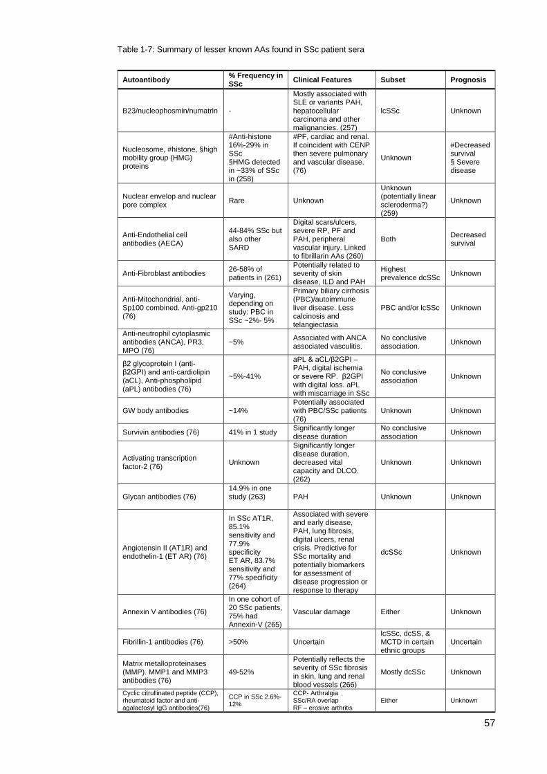
57
Table 1-7: Summary of lesser known AAs found in SSc patient sera
Autoantibody % Frequency in SSc Clinical Features Subset Prognosis
B23/nucleophosmin/numatrin -
Mostly associated with SLE or variants PAH, hepatocellular carcinoma and other malignancies. (257)
lcSSc Unknown
Nucleosome, #histone, §high mobility group (HMG) proteins
#Anti-histone 16%-29% in SSc §HMG detected in ~33% of SSc in (258)
#PF, cardiac and renal. If coincident with CENP then severe pulmonary and vascular disease. (76)
Unknown
#Decreased survival § Severe disease
Nuclear envelop and nuclear pore complex Rare Unknown
Unknown (potentially linear scleroderma?) (259)
Unknown
Anti-Endothelial cell antibodies (AECA)
44-84% SSc but also other SARD
Digital scars/ulcers, severe RP, PF and PAH, peripheral vascular injury. Linked to fibrillarin AAs (260)
Both Decreased survival
Anti-Fibroblast antibodies 26-58% of patients in (261)
Potentially related to severity of skin disease, ILD and PAH
Highest prevalence dcSSc Unknown
Anti-Mitochondrial, anti-Sp100 combined. Anti-gp210 (76)
Varying, depending on study: PBC in SSc ~2%- 5%
Primary biliary cirrhosis (PBC)/autoimmune liver disease. Less calcinosis and telangiectasia
PBC and/or lcSSc Unknown
Anti-neutrophil cytoplasmic antibodies (ANCA), PR3, MPO (76)
~5% Associated with ANCA associated vasculitis.
No conclusive association. Unknown
β2 glycoprotein I (anti-β2GPI) and anti-cardiolipin (aCL), Anti-phospholipid (aPL) antibodies (76)
~5%-41%
aPL & aCL/β2GPI –PAH, digital ischemia or severe RP. β2GPI with digital loss. aPL with miscarriage in SSc
No conclusive association Unknown
GW body antibodies ~14% Potentially associated with PBC/SSc patients (76)
Unknown Unknown
Survivin antibodies (76) 41% in 1 study Significantly longer disease duration
No conclusive association Unknown
Activating transcription factor-2 (76) Unknown
Significantly longer disease duration, decreased vital capacity and DLCO. (262)
Unknown Unknown
Glycan antibodies (76) 14.9% in one study (263)
PAH Unknown Unknown
Angiotensin II (AT1R) and endothelin-1 (ET AR) (76)
In SSc AT1R, 85.1% sensitivity and 77.9% specificity ET AR, 83.7% sensitivity and 77% specificity (264)
Associated with severe and early disease, PAH, lung fibrosis, digital ulcers, renal crisis. Predictive for SSc mortality and potentially biomarkers for assessment of disease progression or response to therapy
dcSSc Unknown
Annexin V antibodies (76)
In one cohort of 20 SSc patients, 75% had Annexin-V (265)
Vascular damage Either Unknown
Fibrillin-1 antibodies (76) >50% Uncertain lcSSc, dcSS, & MCTD in certain ethnic groups
Uncertain
Matrix metalloproteinases (MMP). MMP1 and MMP3 antibodies (76)
49-52%
Potentially reflects the severity of SSc fibrosis in skin, lung and renal blood vessels (266)
Mostly dcSSc Unknown
Cyclic citrullinated peptide (CCP), rheumatoid factor and anti-agalactosyl IgG antibodies(76)
CCP in SSc 2.6%-12%
CCP- Arthralgia SSc/RA overlap RF – erosive arthritis
Either Unknown
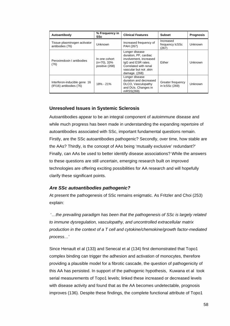
58
Autoantibody % Frequency in SSc Clinical Features Subset Prognosis
Tissue plasminogen activator antibodies (76) Unknown Increased frequency of
PAH (267)
Increased frequency lcSSc (267)
Unknown
Peroxiredoxin I antibodies (76)
In one cohort (n=70), 33% positive (268)
Longer disease duration, PF, cardiac involvement, increased IgG and ESR rates. Correlated with renal vascular but not .skin damage. (268)
Either Unknown
Interferon-inducible gene 16 (IFI16) antibodies (76) 18% - 21%
Longer disease duration and decreased DLCO, Vasculopathy and DUs. Changes in mRSS(269)
Greater frequency in lcSSc (269) Unknown
Unresolved Issues in Systemic Sclerosis
Autoantibodies appear to be an integral component of autoimmune disease and
while much progress has been made in understanding the expanding repertoire of
autoantibodies associated with SSc, important fundamental questions remain.
Firstly, are the SSc autoantibodies pathogenic? Secondly, over time, how stable are
the AAs? Thirdly, is the concept of AAs being ‘mutually exclusive’ redundant?’
Finally, can AAs be used to better identify disease associations? While the answers
to these questions are still uncertain, emerging research built on improved
technologies are offering exciting possibilities for AA research and will hopefully
clarify these significant points.
Are SSc autoantibodies pathogenic? At present the pathogenesis of SSc remains enigmatic. As Fritzler and Choi (253)
explain:
‘…the prevailing paradigm has been that the pathogenesis of SSc is largely related
to immune dysregulation, vasculopathy, and uncontrolled extracellular matrix
production in the context of a T cell and cytokine/chemokine/growth factor-mediated
process…’
Since Henault el al (133) and Senecal et al (134) first demonstrated that Topo1
complex binding can trigger the adhesion and activation of monocytes, therefore
providing a plausible model for a fibrotic cascade, the question of pathogenicity of
this AA has persisted. In support of the pathogenic hypothesis, Kuwana et al took
serial measurements of Topo1 levels; linked these increased or decreased levels
with disease activity and found that as the AA becomes undetectable, prognosis
improves (136). Despite these findings, the complete functional attribute of Topo1
59
remains unexplained (135).
Anti-PDGFR antibodies are controversially also reported in some experiments to be
pathogenic (254, 255, 270) but not in others (249, 250).
For patients that are in an early disease phase and are yet to be diagnosed and
classified as having SSc, the strongest predictor of disease progression is the
presence of an AA (271, 272). If patients who are not AA positive do not progress to
SSc or another CTD, this would suggest that either treatment/s were effective or the
absence of an AA impeded disease progression. It is hypothesised that other AAs
may be protective as is the case with U1RNP (273), although further work is
required as not all subjects with this AA do well (23).
Many more recently identified AAs are thought to play a pathogenic role in both the
initiation and development of SSc and in particular in the progression of
vasculopathy and fibrosis (218). These include several of the newly discovered AAs
in SSc in Table 1-7 (pp.51-52) (AT1R, ETAR, IFI16, Anti-Fibroblast antibodies, MMP1
and MMP3) and others such as intracellular adhesion molecule, oestrogen receptor
α, methionine sulfoxide reductase, type 3 muscarinic acetylcholine receptor and
anti-PDGFR (253). Future studies on AAs that are considered pathogenic or
‘functional’ need to capture patients early in their disease course to clarify
pathogenic pathways so that therapeutic targets can be identified and interventions
can be developed. However, critical for early disease detection is a greater
understanding of how very early SSc presents, not only clinically and serologically,
but also at the molecular and importantly the proteomic level.
Although pathogenicity of scleroderma AAs remains uncertain, there is much scope
for the development of better AA detection and for the development of assays to
determine functionality (253). With the advent of new technologies and further
understanding of the repertoire of AAs and their role they play in SSc, the question
of pathogenicity may become clearer.
How stable are AAs? AA titres are thought to be stable over the course of disease (246); however
experiments measuring Topo1 titres have shown fluctuation occurs over time in
association with an changes in disease activity as measured by the mRSS (274),
lung involvement and survival rates (136, 275).
Fewer studies are available on the stability over time of RNAP3. One small study
(n= 6) showed that RNAP3 levels (as detected by ELISA) increased early in the
disease course and then decreased with the fluctuation of RNAP3 AA levels closely
60
correlating with skin score (157). Two patients developed renal crisis early in the
disease with a synchronous rise in their RNAP3 levels.
CENP appears to be relatively stable throughout the disease (101) and the stability
of other AAs is unknown. To determine the stability of AAs and if pathogenicity is
related to the level of AAs in patient sera, further longitudinal studies in multi ethnic
and varying geographic locations are required.
Are autoantibodies mutually exclusive? This question is generally asked of the two SSc primary AAs, CENP and Topo1,
although some studies have included RNAP3 (276). The literature oscillates
between two hypotheses; there is mutual exclusivity between the two central AAs,
CENP and Topo1 (276-279) or co-expression of these two antibodies exists as co-
expression of AAs exist in other autoimmune diseases (280, 281). The most recent
SSc AA research using multiplexed immunoassays such as the LIA, ALBIA and
CLIA show that multiple AAs can be co-expressed in a single patient (76). Co-
expression of the central AAs, essentially CENP and Topo1 is uncommon, yet it
does occur. Co-expression with one of the other SSc associated AAs is more likely,
particularly TRIM21/Ro52 (76, 282).
As technologies have evolved into multiplexed arrays and further epitopes on
individual AAs are mapped, more AAs are found to be co-expressed. Previously, the
laborious task of performing individual assays was necessary to detect individual
AAs. The key point here is that meticulous attention to the standardisation of these
new assays is required to ensure that cross reactivity or false positives/negatives
are minimised. Finally and most saliently, the clinical and pathological significance of
co-expression of two or more AAs is yet to be elucidated.
Can AAs be used to better identify disease associations? Ultimately, the question must be asked if the AAs can be used to better identify
disease associations. If for example, AA titre is linked with disease course and
outcomes, then other biomarkers may be useful to predict patient outcomes or other
clinical involvement. A Canadian study investigated serology and skin involvement
with survival in dcSSc and lcSSc patients with both subsets having either Topo1 and
ACA positivity and found that ILD was associated with serological status more so
than skin subset. However to add complexity, survival was associated with both
serological status as well as skin subset (52). Therefore, at least among Topo1
positive patients, a lower skin score is associated with a reduced likelihood of
developing ILD and increased survival. A study by Cottrell et al also concluded that
defining the pattern of skin involvement together with the autoantibody status was of
61
greater prognostic value in predicting the risk and severity of restrictive lung disease
as well as overall survival in SSc as compared with using either variable in isolation (283).
Conclusion
Scleroderma is a heterogeneous disease associated with significant morbidity and
mortality.
Treatment options remain limited but it is widely accepted that early disease
detection and sub-classification is critical to identify those patients who may benefit
from experimental and other therapies before significant damage has accrued. The
newer classification criteria for SSc will hopefully aid earlier diagnosis of SSc
patients but sub-classification is also critical as current treatments are associated
with significant toxicities and should not be applied to all those with the disease.
Previously sub-classification has been limited to the degree of skin involvement
although it is widely recognised that AAs provide useful clinical information.
The primary SSc AAs have been well characterised and have recognised clinical
associations across different geographic locations and ethnic backgrounds.
Consideration must be given to the platform used to detect AAs as well as the
epitope and vector used in the assay. While standardisation is crucial, patient
background must be taken into consideration and so continuous improvement and
validation across patients groups is important.
With the advent of LIA and other platforms it is now possible to simultaneously test
for a number of AAs, however the relevance and clinical interpretation of multiple AA
positivity is unknown, therefore further characterisation in large well described
disease cohorts is important.
It has been demonstrated that SSc AAs predate overt disease and are generally
stable over time, that is, it is rare for them to disappear. As a result of this trait, they
are a useful biomarker for early disease classification and, if used with other
validated biomarkers, may be useful in disease stratification and prognosis.
62
Aims & hypotheses for this study
The primary aim of this study is to determine the frequency of, and the relationships
between, SSc related autoantibodies as well as their clinical associations in a well
characterised Australian cohort using a commercially available assay.
In detail the aims for this study are;
i. To conduct an AA profile on this well characterised Australian cohort.
ii. To explore the utility of AAs as biomarkers.
iii. To define AA subgroups based on clinical associations for prognostication
and to better identify patients for clinical trials.
iv. To investigate the usefulness of AAs as a stratification system.
v. To assess if AAs can be used to better identify disease associations.
vi. To describe clinical associations with the primary and rarer SSc associated
AAs.
vii. To establish the frequency of monospecific and multiple AA positivity in an
Australian cohort.
viii. To explore unique clinical associations in subgroups (where possible) of
monospecific and multiple AA presentation.
ix. To explore the concept of mutual exclusivity of CENP and Topo1 AAs.
x. To compare the reliability of the Euroimmun LIA with other commercially
available assays. Specifically, where results differ, to examine the clinical
associations of these patients to see whether the test result supports the
patients disease sub classification.
xi. To determine the frequency of PDGFR in an Australian SSc cohort.
63
The hypotheses for this study are:-
i. The LIA can be used to sub-classify patients with SSc.
ii. Multiple AA positivity is common in SSc.
iii. Important AAs and/or combinations of AAs will provide valuable clinical and
serological information, independent of clinical assessment
iv. AA titre will allow hierarchical assessment where multiple AAs are detected.
v. Clinical associations can be explored with the rarer SSc AAs in an Australian
SSc cohort.
vi. Although rare, co-expression of Topo1 and CENP forms a unique clinical
subgroup.
vii. AA negative patients have a unique clinical phenotype which differs from
other subsets of SSc.
viii. Determine if improvements can be made in the LIA AA selection.
ix. The LIA has similar sensitivity and specificity to other commercially available
assays used in the ASCS.
x. PDGFR will be very rare in this cohort.
It is with these hypotheses in mind that this study was undertaken and it is hoped
that the outcomes from this study will contribute to the knowledge and utility of
autoantibodies as biomarkers in systemic sclerosis.

64
CHAPTER 2 METHODOLOGY
The Australian Scleroderma Interest Group (ASIG)
The ASIG is a national, multidisciplinary group of physicians and scientists
interested in research that furthers the understanding of scleroderma and seeks to
improve outcomes for patients. The ASIG established the Australian Scleroderma
Cohort Study (ASCS) in 2007 as a prospective multicentre study to conduct an
annual screen in SSc patients for lung and heart complications and to collect
biological samples (cells, DNA and sera) from consenting patients to be enrolled in a
longitudinal observational cohort for ongoing research projects. Patient consent was
obtained in writing after the provision of information concerning the scope of
research projects and individual personal information required for entry into the
cohort study. The database is managed by the Database Committee who oversees
the maintenance of the existing ASIG clinical database with administration
assistance from a Project Officer. The Terms of Reference including structure,
governance and scope can be found in the appendices, ‘Australian Scleroderma
Interest Group Terms of Reference (2013)’.
Study Design and Ethical Approval
This is a cross sectional study of Australian SSc patients enrolled in the ASCS
database. Ethics for this study were approved by the ethics committee of the
participating centres as well as by the Southern Area Clinical Human Research
Ethics Committee at Flinders University. This study was carried out according to the
2007 National Statement on Ethical Conduct in Research involving Humans which
was published by the National Health and Medical Research Council of Australia
(284), and in accordance with the Declaration of Helsinki (285).
Patient Population
At the time of testing there were 1,139 patients enrolled at 8 centres specialising in
the care of patients with SSc: Royal Adelaide Hospital (Adelaide, South Australia),
St. George Hospital (Kogarah, New South Wales), Sunshine Coast Rheumatology
and Prince Charles Hospital (Brisbane, Queensland), St. Vincent’s Hospital and
Monash Health (Melbourne, Victoria), Royal Hobart Hospital (Hobart, Tasmania)
and Royal Perth Hospital (Perth, Western Australia). SSc patients were admitted to
the ASCS database based on one of three significant conditions;
i. The patient fulfilled the ACR 1980 criteria (286), or
ii. The patient fulfilled the LeRoy and Medsger criteria for early SSc (287), or

65
iii. The condition was diagnosed according to the expert opinion of the patient’s treating physician.
A further patient group is also included in the ASCS database and serum repository,
those patients that are considered to have MCTD, diagnosed using Sharp’s Criteria
(288), or again, by the expert opinion of the treating physician.
The ASCS Serum Repository, located at The Queen Elizabeth Hospital, Woodville
South Australia, made available 526 serum samples for this study. Demographic
data collected and utilised in this study include age (chronological, disease onset
Raynaud’s and first non Raynaud’s symptom), gender, disease subset, ethnicity and
fulfilment of ACR or Medsger criteria. Data for this study were censored on 7th June
2013. The patient sera used in this study were not taken from inception samples.
The new 2013 ACR/EULAR criteria had not been published at census date for this
study, however for completeness, the new ACR/EULAR 2013 criteria have been
retrospectively assessed and included on the 505 SSc patients. Full demographical
results are in Chapter 3, Results, Table 3-1, p.78.
Disease manifestations were defined as present if they had occurred since the time
of diagnosis or, for continuous variables including the modified Rodnan skin
thickness score, the highest ever recorded value since time of enrolment in the
ASCS. Study variables and their description can be found in Table 2-1.
Table 2-1: ASCS Clinical and serological variables
Variable Description Disease duration Calculated since first non-Raynaud’s symptom Raynaud’s phenomenon Characterised by triphasic colour changes, date onset Digital ulcers Defined as denuded areas with defined borders and loss of
epithelialization, epidermis, and dermis; excluded fissures, paronychia, extrusion of calcium) either on the volar or dorsal aspects of the fingers
Digital gangrene Diagnosed clinically by the treating physician Digital amputation Ever Nailfold capillary dilatation Capillary measurements include dilatation only. Telangiectasia Any clearly visible mat-like telangiectasia visible on the
face, limbs, chest or abdomen. Calcinosis Diagnosed clinically by the attending physician Tendon friction rubs Diagnosed clinically by the attending physician Joint contractures Diagnosed clinically by the attending physician Synovitis Diagnosed clinically by the attending physician Modified Rodnan Skin Score (mRSS) The mRSS measures skin thickness on a score of 0 (no
thickness) to 3 (severe thickening) in 17 locations on the body.
Interstitial lung disease (ILD) ILD was defined as the presence of pulmonary fibrosis on lung imaging, usually by high resolution computed tomography scan.
Pulmonary Arterial Hypertension (PAH) PAH was defined as a mean pulmonary artery pressure of ≥25 mm Hg and a pulmonary capillary wedge pressure of ≤15 mm Hg on right sided heart catheterization.
Systemic Hypertension Systolic BP ≥ 140 mmHg or diastolic BP ≥ 90 mmHg
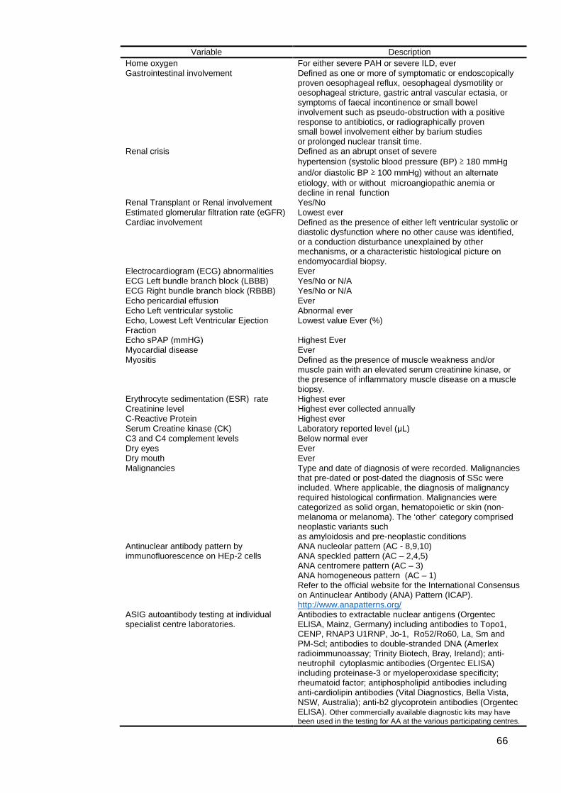
66
Variable Description Home oxygen For either severe PAH or severe ILD, ever Gastrointestinal involvement Defined as one or more of symptomatic or endoscopically
proven oesophageal reflux, oesophageal dysmotility or oesophageal stricture, gastric antral vascular ectasia, or symptoms of faecal incontinence or small bowel involvement such as pseudo-obstruction with a positive response to antibiotics, or radiographically proven small bowel involvement either by barium studies or prolonged nuclear transit time.
Renal crisis
Defined as an abrupt onset of severe hypertension (systolic blood pressure (BP) ≥ 180 mmHg and/or diastolic BP ≥ 100 mmHg) without an alternate etiology, with or without microangiopathic anemia or decline in renal function
Renal Transplant or Renal involvement Yes/No Estimated glomerular filtration rate (eGFR) Lowest ever Cardiac involvement Defined as the presence of either left ventricular systolic or
diastolic dysfunction where no other cause was identified, or a conduction disturbance unexplained by other mechanisms, or a characteristic histological picture on endomyocardial biopsy.
Electrocardiogram (ECG) abnormalities Ever ECG Left bundle branch block (LBBB) Yes/No or N/A ECG Right bundle branch block (RBBB) Yes/No or N/A Echo pericardial effusion Ever Echo Left ventricular systolic Abnormal ever Echo, Lowest Left Ventricular Ejection Fraction
Lowest value Ever (%)
Echo sPAP (mmHG) Highest Ever Myocardial disease Ever Myositis
Defined as the presence of muscle weakness and/or muscle pain with an elevated serum creatinine kinase, or the presence of inflammatory muscle disease on a muscle biopsy.
Erythrocyte sedimentation (ESR) rate Highest ever Creatinine level Highest ever collected annually C-Reactive Protein Highest ever Serum Creatine kinase (CK) Laboratory reported level (μL) C3 and C4 complement levels Below normal ever Dry eyes Ever Dry mouth Ever Malignancies Type and date of diagnosis of were recorded. Malignancies
that pre-dated or post-dated the diagnosis of SSc were included. Where applicable, the diagnosis of malignancy required histological confirmation. Malignancies were categorized as solid organ, hematopoietic or skin (non-melanoma or melanoma). The ‘other’ category comprised neoplastic variants such as amyloidosis and pre-neoplastic conditions
Antinuclear antibody pattern by immunofluorescence on HEp-2 cells
ANA nucleolar pattern (AC - 8,9,10) ANA speckled pattern (AC – 2,4,5) ANA centromere pattern (AC – 3) ANA homogeneous pattern (AC – 1) Refer to the official website for the International Consensus on Antinuclear Antibody (ANA) Pattern (ICAP). http://www.anapatterns.org/
ASIG autoantibody testing at individual specialist centre laboratories.
Antibodies to extractable nuclear antigens (Orgentec ELISA, Mainz, Germany) including antibodies to Topo1, CENP, RNAP3 U1RNP, Jo-1, Ro52/Ro60, La, Sm and PM-Scl; antibodies to double-stranded DNA (Amerlex radioimmunoassay; Trinity Biotech, Bray, Ireland); anti-neutrophil cytoplasmic antibodies (Orgentec ELISA) including proteinase-3 or myeloperoxidase specificity; rheumatoid factor; antiphospholipid antibodies including anti-cardiolipin antibodies (Vital Diagnostics, Bella Vista, NSW, Australia); anti-b2 glycoprotein antibodies (Orgentec ELISA). Other commercially available diagnostic kits may have been used in the testing for AA at the various participating centres.

67
Autoantibody analysis
Sera were analysed using a commercially available line immunoblot assay
(Systemic Sclerosis [Nucleoli] Profile EuroLine [IgG]; Euroimmun), and analysis was
performed in a single laboratory by a single operator (KAP). Serum aliquots were
stored at -80ºC until the time of testing. The assay was performed according to the
manufacturer’s instructions. The kit contains 13 recombinant antigens: those
expressed in Escherichia coli (RNA polymerase III [RNAP III; subunits RP11 and
RP155], fibrillarin, the 90-kd nucleolar protein NOR- 90 (hUBF), and Th/To) or in
insect cells using the baculovirus system (CENP A, CENP B, Pm/Scl-100, Pm/Scl-
75, Ku, and tripartite motif–containing protein 21 (TRIM-21]/Ro 52) plus PDGFR
expressed in mammalian cells and native topoisomerase I (Topo1 formerly Scl-70)
isolated from calf and rabbit thymus. Sera were analysed at a dilution of 1:101, and
autoantibodies were detected using alkaline phosphatase–labelled antihuman IgG.
The EuroLine flatbed scanner provides semi quantitative results. Readings obtained
with a signal intensity of +, ++, and +++ were defined as positive and were allocated
scores that equated to +1, +2, and +3, respectively. Borderline signals or no signal,
representing signal intensities of <11 intensity units, were defined as negative and
were scored as 0. Appropriate positive and negative controls were included in each
run. All scores were then used in the principal component analysis. Separate
studies have found good correlation between the signal intensity in the immunoblot
assay and autoantibody titres/concentrations measured in alternative assays (81,
116, 144, 200).
Initial ASIG ANA Detection The initial presence of ANA was determined by Australian pathology laboratories
that participate in the National Association of Testing Authorities (Australia)
accredited by the Royal College of Pathologists, Australia. The cut off value
distinguishing a negative or positive value were all determined locally according to
established principals (289). The presence of mitotic spindle or mitotic staining is
most commonly reported as negative at local labs, however some may report it as
positive staining.
68
U1RNP Anti-U1RNP antibody is an important AA in the SSc spectrum and is not available
on the EuroLine (Euroimmun) SSc immunoblot. Previous testing for U1RNP by
independent laboratories from ASIG specialist centres had been completed.
Following the SSc immunoblot, selected patient results were examined for the
following reasons:
i. Speckled ANA.
ii. Negative results in the SSc line blot but with a previous U1RNP positive test
result.
iii. Positive monospecific for TRIM21/Ro52 SSc blot result.
iv. Sufficient sera were available for testing.
These sera were then analysed using the EuroLine (Euroimmun) ANA Profile 5
(IgG) to ascertain if any were U1RNP positive to compare to the ASIG database. All
samples were analysed in a single laboratory by a single operator (KAP). Serum
aliquots were stored at -80ºC until the time of testing. The assay was performed
according to the manufacturer’s instructions. The kit contains 18 different antigens
(IgG class), however the antigens of interest were RNP/Sm, native U1RNP purified
by affinity chromatography from calf and rabbit thymus, RNP 70, -A, -C; recombinant
U1RNP proteins. The corresponding cDNA is expressed in insect cells using a
baculovirus vector or in E.coli (RNP 70). Sera were analysed at a dilution of 1:101,
and autoantibodies were detected using alkaline phosphatase–labelled antihuman
IgG. The EuroLine flatbed scanner provides semi quantitative results. Readings
obtained with a signal intensity of +, ++, and +++ were defined as positive and were
allocated scores that equated to +1, +2, and +3, respectively. Borderline signals or
no signal, representing signal intensities of <11 intensity units, were defined as
negative and were scored as 0. Appropriate positive and negative controls were
included in each run. All positive scores were then used as supplementary
individuals in the principal component analysis.
U1RNP positive patients were compared with all other patients in the ASCS cohort
and also with patients within Cluster 4. However, in accord with the literature where
U1RNP confers an ‘overlap’ variant, patients are analysed on an AA
positive/negative status.
ANA and EuroLine Blot Negative Where testing indicated a negative Immunoblot (SSc and ANA 5) result coupled with
a negative ANA result by ASIG associated independent laboratories, further testing
on these specific patient sera were performed. This supplementary testing was
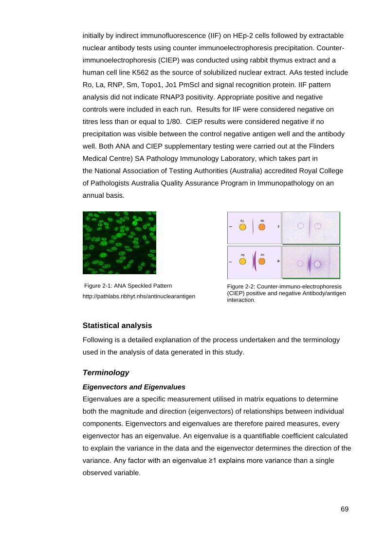
69
initially by indirect immunofluorescence (IIF) on HEp-2 cells followed by extractable
nuclear antibody tests using counter immunoelectrophoresis precipitation. Counter-
immunoelectrophoresis (CIEP) was conducted using rabbit thymus extract and a
human cell line K562 as the source of solubilized nuclear extract. AAs tested include
Ro, La, RNP, Sm, Topo1, Jo1 PmScl and signal recognition protein. IIF pattern
analysis did not indicate RNAP3 positivity. Appropriate positive and negative
controls were included in each run. Results for IIF were considered negative on
titres less than or equal to 1/80. CIEP results were considered negative if no
precipitation was visible between the control negative antigen well and the antibody
well. Both ANA and CIEP supplementary testing were carried out at the Flinders
Medical Centre) SA Pathology Immunology Laboratory, which takes part in
the National Association of Testing Authorities (Australia) accredited Royal College
of Pathologists Australia Quality Assurance Program in Immunopathology on an
annual basis.
Figure 2-1: ANA Speckled Pattern
http://pathlabs.ribhyt.nhs/antinuclearantigen
Figure 2-2: Counter-immuno-electrophoresis (CIEP) positive and negative Antibody/antigen interaction.
Statistical analysis
Following is a detailed explanation of the process undertaken and the terminology
used in the analysis of data generated in this study.
Terminology
Eigenvectors and Eigenvalues Eigenvalues are a specific measurement utilised in matrix equations to determine
both the magnitude and direction (eigenvectors) of relationships between individual
components. Eigenvectors and eigenvalues are therefore paired measures, every
eigenvector has an eigenvalue. An eigenvalue is a quantifiable coefficient calculated
to explain the variance in the data and the eigenvector determines the direction of the
variance. Any factor with an eigenvalue ≥1 explains more variance than a single
observed variable.
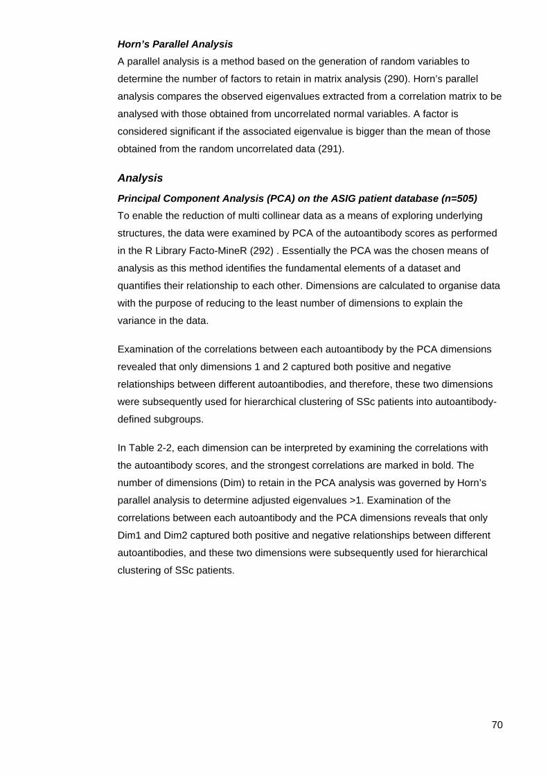
70
Horn’s Parallel Analysis A parallel analysis is a method based on the generation of random variables to
determine the number of factors to retain in matrix analysis (290). Horn’s parallel
analysis compares the observed eigenvalues extracted from a correlation matrix to be
analysed with those obtained from uncorrelated normal variables. A factor is
considered significant if the associated eigenvalue is bigger than the mean of those
obtained from the random uncorrelated data (291).
Analysis
Principal Component Analysis (PCA) on the ASIG patient database (n=505) To enable the reduction of multi collinear data as a means of exploring underlying
structures, the data were examined by PCA of the autoantibody scores as performed
in the R Library Facto-MineR (292) . Essentially the PCA was the chosen means of
analysis as this method identifies the fundamental elements of a dataset and
quantifies their relationship to each other. Dimensions are calculated to organise data
with the purpose of reducing to the least number of dimensions to explain the
variance in the data.
Examination of the correlations between each autoantibody by the PCA dimensions
revealed that only dimensions 1 and 2 captured both positive and negative
relationships between different autoantibodies, and therefore, these two dimensions
were subsequently used for hierarchical clustering of SSc patients into autoantibody-
defined subgroups.
In Table 2-2, each dimension can be interpreted by examining the correlations with
the autoantibody scores, and the strongest correlations are marked in bold. The
number of dimensions (Dim) to retain in the PCA analysis was governed by Horn’s
parallel analysis to determine adjusted eigenvalues >1. Examination of the
correlations between each autoantibody and the PCA dimensions reveals that only
Dim1 and Dim2 captured both positive and negative relationships between different
autoantibodies, and these two dimensions were subsequently used for hierarchical
clustering of SSc patients.
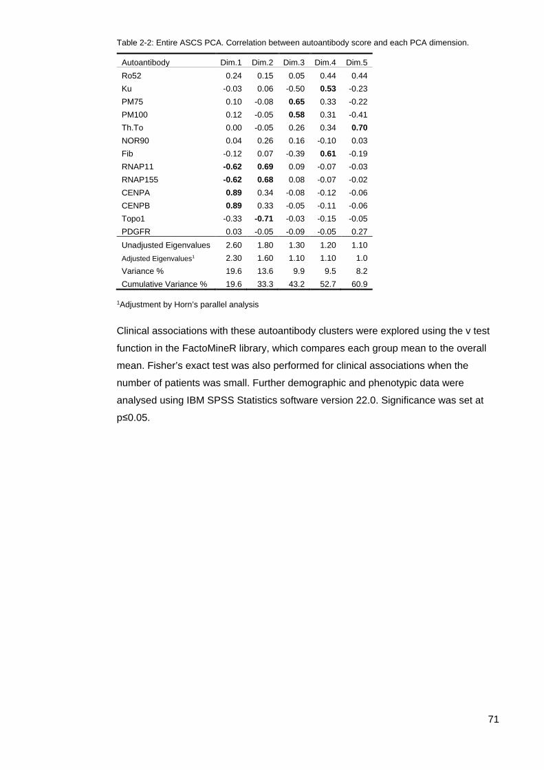
71
Table 2-2: Entire ASCS PCA. Correlation between autoantibody score and each PCA dimension.
Autoantibody Dim.1 Dim.2 Dim.3 Dim.4 Dim.5 Ro52 0.24 0.15 0.05 0.44 0.44 Ku -0.03 0.06 -0.50 0.53 -0.23 PM75 0.10 -0.08 0.65 0.33 -0.22 PM100 0.12 -0.05 0.58 0.31 -0.41 Th.To 0.00 -0.05 0.26 0.34 0.70 NOR90 0.04 0.26 0.16 -0.10 0.03 Fib -0.12 0.07 -0.39 0.61 -0.19 RNAP11 -0.62 0.69 0.09 -0.07 -0.03 RNAP155 -0.62 0.68 0.08 -0.07 -0.02 CENPA 0.89 0.34 -0.08 -0.12 -0.06 CENPB 0.89 0.33 -0.05 -0.11 -0.06 Topo1 -0.33 -0.71 -0.03 -0.15 -0.05 PDGFR 0.03 -0.05 -0.09 -0.05 0.27 Unadjusted Eigenvalues 2.60 1.80 1.30 1.20 1.10 Adjusted Eigenvalues1 2.30 1.60 1.10 1.10 1.0 Variance % 19.6 13.6 9.9 9.5 8.2 Cumulative Variance % 19.6 33.3 43.2 52.7 60.9
1Adjustment by Horn’s parallel analysis
Clinical associations with these autoantibody clusters were explored using the v test
function in the FactoMineR library, which compares each group mean to the overall
mean. Fisher’s exact test was also performed for clinical associations when the
number of patients was small. Further demographic and phenotypic data were
analysed using IBM SPSS Statistics software version 22.0. Significance was set at
p≤0.05.

72
The Cluster 4 PCA The second PCA used IBM SPSS Version 22.0 (Armonk, NY) software as R was not
available. A detailed description of the PCA process using SPSS follows.
Kaiser-Meyer-Olkin (KMO) and Bartlett’s Measure of Sampling Adequacy The KMO and Bartlett’s test is a statistic that indicates the proportion of variance in
the data that might be caused by underlying factors. The KMO and Bartlett’s Test
(p=0.006) established that 46.6% of the data were influenced by underlying factors.
Scree Plot The scree plot utilises eigenvalues associated in descending order against
components. Scree plots assist in the assessment of those components that explain
the most variability in the data. Eigenvalues ranged from 1.679 in component 1 to
1.442 in component 2 to 1.143 in component 3.The decision where to ‘cut’ the data
was at the third component with an eigenvalue of 1.143 (adjusted by Horn’s parallel
analysis). The scree plot for this PCA is visualised in Figure 2-3.
Figure 2-3: Cluster 4 Scree plot associated with the Supplementary PCA of Cluster 4
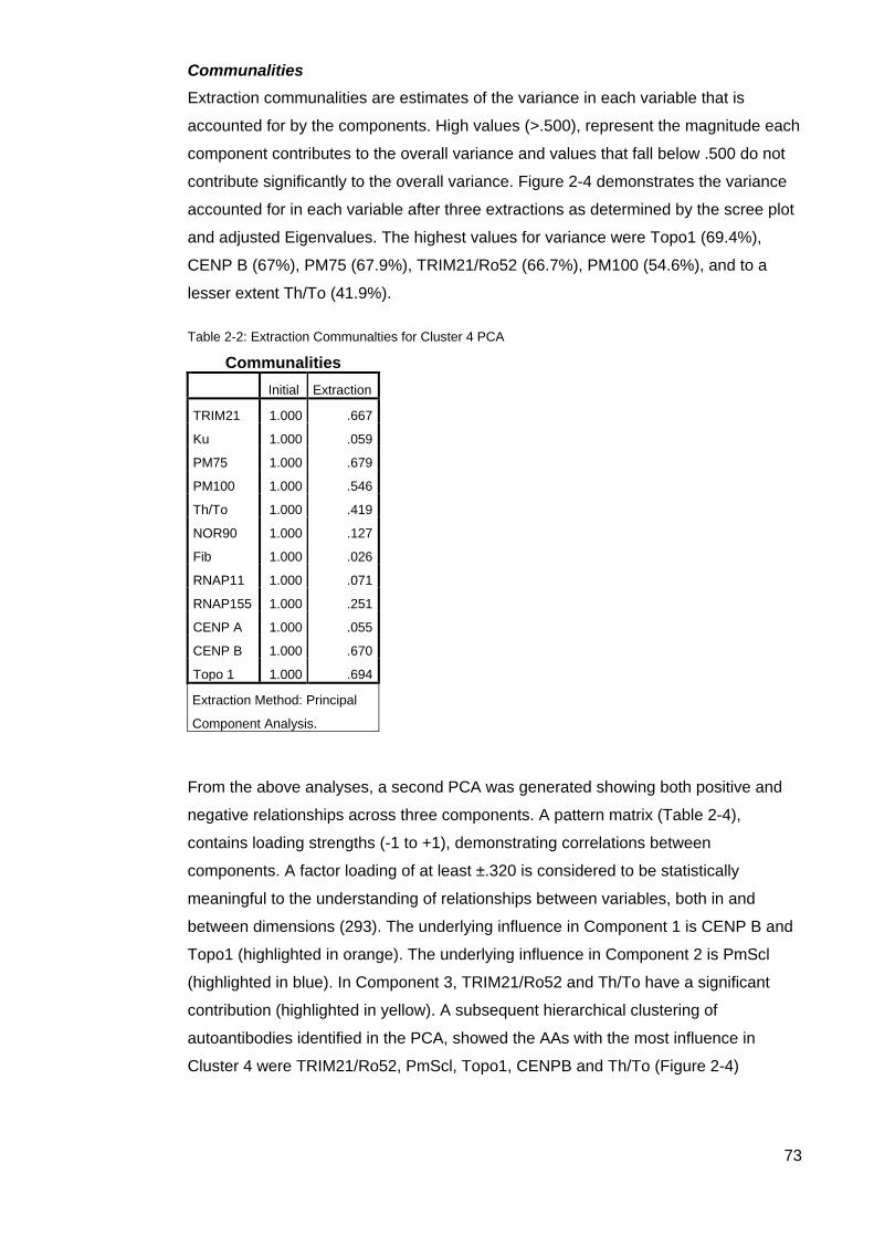
73
Communalities Extraction communalities are estimates of the variance in each variable that is
accounted for by the components. High values (>.500), represent the magnitude each
component contributes to the overall variance and values that fall below .500 do not
contribute significantly to the overall variance. Figure 2-4 demonstrates the variance
accounted for in each variable after three extractions as determined by the scree plot
and adjusted Eigenvalues. The highest values for variance were Topo1 (69.4%),
CENP B (67%), PM75 (67.9%), TRIM21/Ro52 (66.7%), PM100 (54.6%), and to a
lesser extent Th/To (41.9%).
Table 2-2: Extraction Communalties for Cluster 4 PCA
Communalities
Initial Extraction
TRIM21 1.000 .667
Ku 1.000 .059
PM75 1.000 .679
PM100 1.000 .546
Th/To 1.000 .419
NOR90 1.000 .127
Fib 1.000 .026
RNAP11 1.000 .071
RNAP155 1.000 .251
CENP A 1.000 .055
CENP B 1.000 .670
Topo 1 1.000 .694
Extraction Method: Principal
Component Analysis.
From the above analyses, a second PCA was generated showing both positive and
negative relationships across three components. A pattern matrix (Table 2-4),
contains loading strengths (-1 to +1), demonstrating correlations between
components. A factor loading of at least ±.320 is considered to be statistically
meaningful to the understanding of relationships between variables, both in and
between dimensions (293). The underlying influence in Component 1 is CENP B and
Topo1 (highlighted in orange). The underlying influence in Component 2 is PmScl
(highlighted in blue). In Component 3, TRIM21/Ro52 and Th/To have a significant
contribution (highlighted in yellow). A subsequent hierarchical clustering of
autoantibodies identified in the PCA, showed the AAs with the most influence in
Cluster 4 were TRIM21/Ro52, PmScl, Topo1, CENPB and Th/To (Figure 2-4)
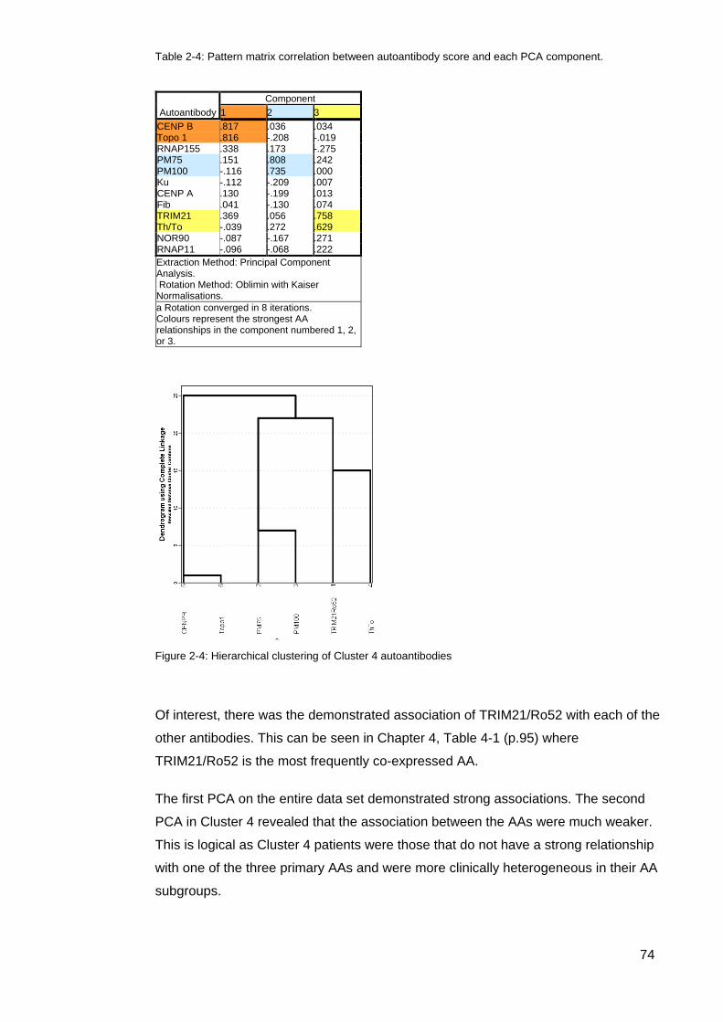
74
Table 2-4: Pattern matrix correlation between autoantibody score and each PCA component.
Figure 2-4: Hierarchical clustering of Cluster 4 autoantibodies
Of interest, there was the demonstrated association of TRIM21/Ro52 with each of the
other antibodies. This can be seen in Chapter 4, Table 4-1 (p.95) where
TRIM21/Ro52 is the most frequently co-expressed AA.
The first PCA on the entire data set demonstrated strong associations. The second
PCA in Cluster 4 revealed that the association between the AAs were much weaker.
This is logical as Cluster 4 patients were those that do not have a strong relationship
with one of the three primary AAs and were more clinically heterogeneous in their AA
subgroups.
Autoantibody Component
1 2 3 CENP B .817 .036 .034 Topo 1 .816 -.208 -.019 RNAP155 .338 .173 -.275 PM75 .151 .808 .242 PM100 -.116 .735 .000 Ku -.112 -.209 .007 CENP A .130 -.199 .013 Fib .041 -.130 .074 TRIM21 .369 .056 .758 Th/To -.039 .272 .629 NOR90 -.087 -.167 .271 RNAP11 -.096 -.068 .222 Extraction Method: Principal Component Analysis. Rotation Method: Oblimin with Kaiser Normalisations. a Rotation converged in 8 iterations. Colours represent the strongest AA relationships in the component numbered 1, 2, or 3.

75
Clinical associations with Autoantibodies Where numbers permitted, statistical analysis was used to explore any clinical
associations with the SSc associated AAs. Statistical analyses were undertaken to
explore the clinical features of any patients testing positive to one of the three primary
AA. To limit bias, only patients monospecific for the less specific or rarer disease
associated AA were tested. Fisher’s Exact method was used for discreet nominal
variables. Continuous variables were assessed with ANOVA, using Levene’s test for
homogeneity of variances. To protect against a Type 1 error, if homogeneity of
variances were violated, a Mann-Whitney U test was applied and this is annotated
where undertaken. Results p≤ 0.05 are considered statistically significant. In some
instances trends were reported where they were found to be significant in other
cohorts and are reported where p<0.09.
ANA, ENA and EuroLine blot negative Data were analysed using IBM SPSS Statistics Version 22.0 (Armonk, NY). Clinical
manifestations were dependent variables and ANA status was the independent
variable. Continuous variables were assessed with ANOVA. Due to the small and
unequal sample size of the AA negative subgroup compared to the AA positive
subgroup, bootstrapping, the Levene’s test for equality of variances, Mann Whitney U
test and Kruskal-Wallis tests were used. For categorical variables a Chi Square
independence test, bootstrapping and Fishers Exact methods were employed, again
to compare AA negative and AA positive groups. Significance was set at p≤0.05.
Comparison of laboratory methods – LIA, IIF and IP Cohen’s Kappa statistic (kappa or κ) is frequently used to test interrater reliability on
qualitative data. The importance of interrater reliability is that it represents the extent
to which the data collected in the study are correct representations of the variables
measured (294).The kappa statistic ranges between −1 to +1. A rating of 0 represents
the amount of agreement that can be expected from random chance, and 1
represents perfect agreement between the methods of rating. While kappa values
below 0 are possible, they are unlikely in practice (294). The kappa statistic is a
standardized value and thus is interpreted the same across multiple studies (294).
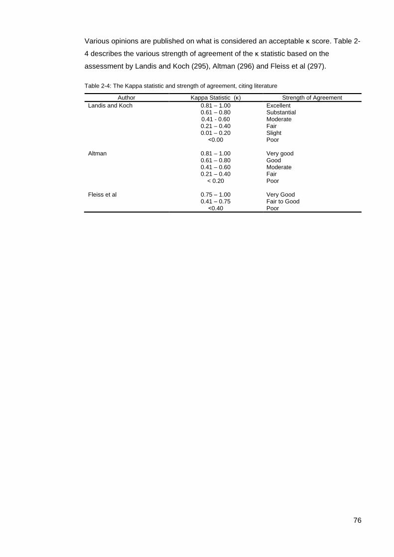
76
Various opinions are published on what is considered an acceptable κ score. Table 2-
4 describes the various strength of agreement of the κ statistic based on the
assessment by Landis and Koch (295), Altman (296) and Fleiss et al (297).
Table 2-4: The Kappa statistic and strength of agreement, citing literature
Author Kappa Statistic (κ) Strength of Agreement Landis and Koch 0.81 – 1.00 Excellent 0.61 – 0.80 Substantial 0.41 - 0.60 Moderate 0.21 – 0.40 Fair 0.01 – 0.20 Slight ˂0.00 Poor Altman 0.81 – 1.00 Very good 0.61 – 0.80 Good 0.41 – 0.60 Moderate 0.21 – 0.40 Fair < 0.20 Poor Fleiss et al 0.75 – 1.00 Very Good 0.41 – 0.75 Fair to Good ˂0.40 Poor
77
CHAPTER 3 INTERPRETATION OF AN EXTENDED AUTOANTIBODY PROFILE
IN A WELL CHARACTERISED AUSTRALIAN SYSTEMIC SCLEROSIS (SCLERODERMA) COHORT UTILISING PRINCIPAL
COMPONENT ANALYSIS.
Introduction
Systemic sclerosis is a heterogeneous disorder with well documented genetic (298-
301) and geographic variation (302). Historically, stratification of patients with early
disease has been difficult due to the absence of definitive laboratory markers and the
problems inherent with a purely clinical sub-classification in a condition that fluctuates
with time. In 1980, the ACR ‘Preliminary criteria for the classification of systemic
sclerosis’ (SSc) (286) were published. Since then there has been continuing
discussion about whether this classification tool should be expanded to include all
patients with variant subsets (42) who fall within the Scleroderma Spectrum Disorders
(53, 303). Initial validation studies of 2013 ACR/EULAR classification criteria for SSc
(43) show improved accuracy but do not encompass sub- classification nor do they
provide prognostic information. Improved biomarkers for the sub classification of SSc
subsets are sorely needed.
Serum AAs are found in up to 95% of patients with SSc (65) and carry greater weight
in the newly proposed ACR/EULAR classification system. CENP, Topo1 and RNAP3
closely reflect patterns of organ involvement and disease progression (65), and with
rare exceptions they appear to be mutually exclusive at this level of AA analysis.
Other serum AAs are known to be associated with SSc (22, 76, 304) but their clinical
associations and frequencies are less well defined. Until recently, many of these
rarer AAs were only available in the research setting and required labour intensive
laboratory analyses. The advent of new, less labour-intensive technologies, such as
the line immunoassay (LIA) means multiple AAs, including rarer AAs, can now be
tested in routine clinical practice. The clinical significance of these rarer AAs remains
uncertain, as does the significance of multiple AA positivity. We report on the clinical
and serological associations of ten AAs in a large, well characterised multi-centre
study of SSc patients utilising LIA. The results demonstrate that the use of an
extended AA provides useful prognostic information at the time of diagnosis and
confirms the utility of AAs in the SSc spectrum of disorders. Autoantibody profiling
will, we predict, have added potential in allowing sub-classification and stratification of
patients into clinical trials.
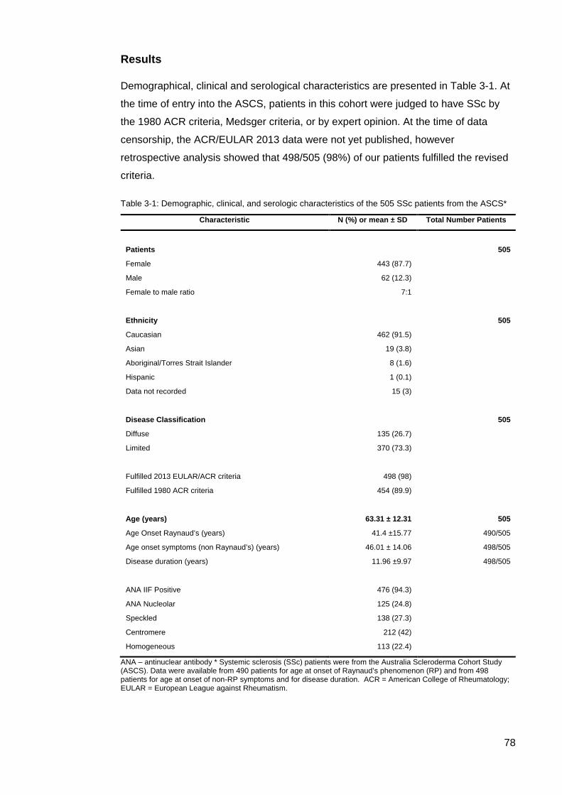
78
Results
Demographical, clinical and serological characteristics are presented in Table 3-1. At
the time of entry into the ASCS, patients in this cohort were judged to have SSc by
the 1980 ACR criteria, Medsger criteria, or by expert opinion. At the time of data
censorship, the ACR/EULAR 2013 data were not yet published, however
retrospective analysis showed that 498/505 (98%) of our patients fulfilled the revised
criteria.
Table 3-1: Demographic, clinical, and serologic characteristics of the 505 SSc patients from the ASCS*
Characteristic N (%) or mean ± SD Total Number Patients
Patients 505
Female 443 (87.7)
Male 62 (12.3)
Female to male ratio 7:1
Ethnicity 505
Caucasian 462 (91.5)
Asian 19 (3.8)
Aboriginal/Torres Strait Islander 8 (1.6)
Hispanic 1 (0.1)
Data not recorded 15 (3)
Disease Classification 505
Diffuse 135 (26.7)
Limited 370 (73.3)
Fulfilled 2013 EULAR/ACR criteria 498 (98)
Fulfilled 1980 ACR criteria 454 (89.9)
Age (years) 63.31 ± 12.31 505
Age Onset Raynaud’s (years) 41.4 ±15.77 490/505
Age onset symptoms (non Raynaud’s) (years) 46.01 ± 14.06 498/505
Disease duration (years) 11.96 ±9.97 498/505
ANA IIF Positive 476 (94.3)
ANA Nucleolar 125 (24.8)
Speckled 138 (27.3)
Centromere 212 (42)
Homogeneous 113 (22.4)
ANA – antinuclear antibody * Systemic sclerosis (SSc) patients were from the Australia Scleroderma Cohort Study (ASCS). Data were available from 490 patients for age at onset of Raynaud’s phenomenon (RP) and from 498 patients for age at onset of non-RP symptoms and for disease duration. ACR = American College of Rheumatology; EULAR = European League against Rheumatism.
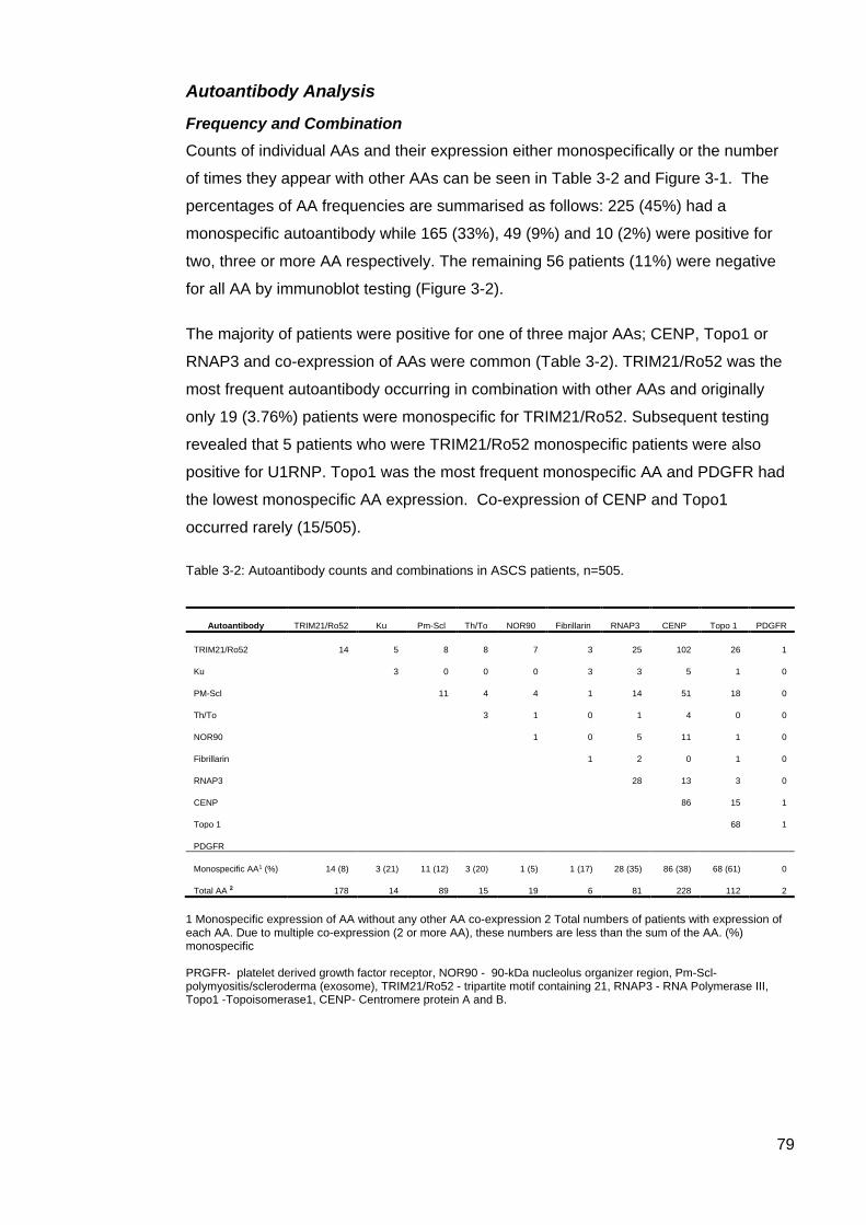
79
Autoantibody Analysis
Frequency and Combination Counts of individual AAs and their expression either monospecifically or the number
of times they appear with other AAs can be seen in Table 3-2 and Figure 3-1. The
percentages of AA frequencies are summarised as follows: 225 (45%) had a
monospecific autoantibody while 165 (33%), 49 (9%) and 10 (2%) were positive for
two, three or more AA respectively. The remaining 56 patients (11%) were negative
for all AA by immunoblot testing (Figure 3-2).
The majority of patients were positive for one of three major AAs; CENP, Topo1 or
RNAP3 and co-expression of AAs were common (Table 3-2). TRIM21/Ro52 was the
most frequent autoantibody occurring in combination with other AAs and originally
only 19 (3.76%) patients were monospecific for TRIM21/Ro52. Subsequent testing
revealed that 5 patients who were TRIM21/Ro52 monospecific patients were also
positive for U1RNP. Topo1 was the most frequent monospecific AA and PDGFR had
the lowest monospecific AA expression. Co-expression of CENP and Topo1
occurred rarely (15/505).
Table 3-2: Autoantibody counts and combinations in ASCS patients, n=505.
1 Monospecific expression of AA without any other AA co-expression 2 Total numbers of patients with expression of each AA. Due to multiple co-expression (2 or more AA), these numbers are less than the sum of the AA. (%) monospecific PRGFR- platelet derived growth factor receptor, NOR90 - 90-kDa nucleolus organizer region, Pm-Scl- polymyositis/scleroderma (exosome), TRIM21/Ro52 - tripartite motif containing 21, RNAP3 - RNA Polymerase III, Topo1 -Topoisomerase1, CENP- Centromere protein A and B.
Autoantibody TRIM21/Ro52 Ku Pm-Scl Th/To NOR90 Fibrillarin RNAP3 CENP Topo 1 PDGFR
TRIM21/Ro52 14 5 8 8 7 3 25 102 26 1
Ku 3 0 0 0 3 3 5 1 0
PM-Scl 11 4 4 1 14 51 18 0
Th/To 3 1 0 1 4 0 0
NOR90 1 0 5 11 1 0
Fibrillarin 1 2 0 1 0
RNAP3 28 13 3 0
CENP 86 15 1
Topo 1 68 1
PDGFR
Monospecific AA1 (%) 14 (8) 3 (21) 11 (12) 3 (20) 1 (5) 1 (17) 28 (35) 86 (38) 68 (61) 0
Total AA 2 178 14 89 15 19 6 81 228 112 2
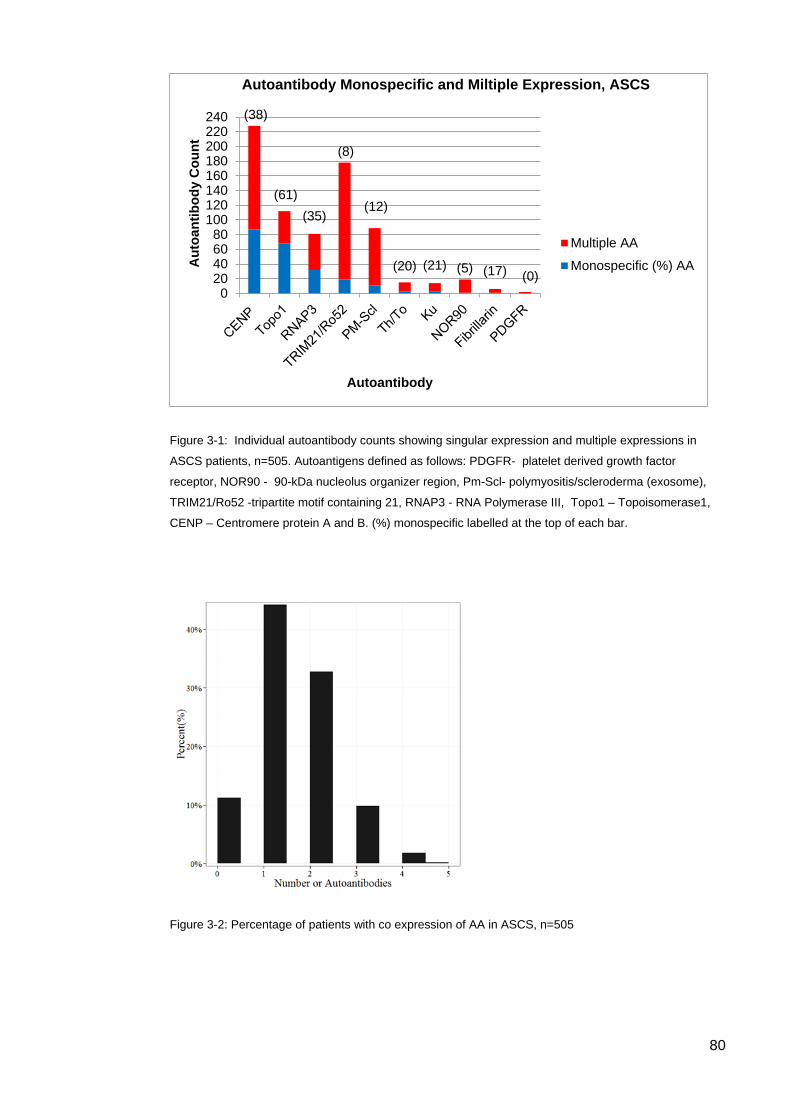
80
Figure 3-1: Individual autoantibody counts showing singular expression and multiple expressions in
ASCS patients, n=505. Autoantigens defined as follows: PDGFR- platelet derived growth factor
receptor, NOR90 - 90-kDa nucleolus organizer region, Pm-Scl- polymyositis/scleroderma (exosome),
TRIM21/Ro52 -tripartite motif containing 21, RNAP3 - RNA Polymerase III, Topo1 – Topoisomerase1,
CENP – Centromere protein A and B. (%) monospecific labelled at the top of each bar.
Figure 3-2: Percentage of patients with co expression of AA in ASCS, n=505
(38)
(61)(35)
(8)
(12)
(20) (21) (5) (17) (0)0
20406080
100120140160180200220240
Aut
oant
ibod
y C
ount
Autoantibody
Autoantibody Monospecific and Miltiple Expression, ASCS
Multiple AAMonospecific (%) AA
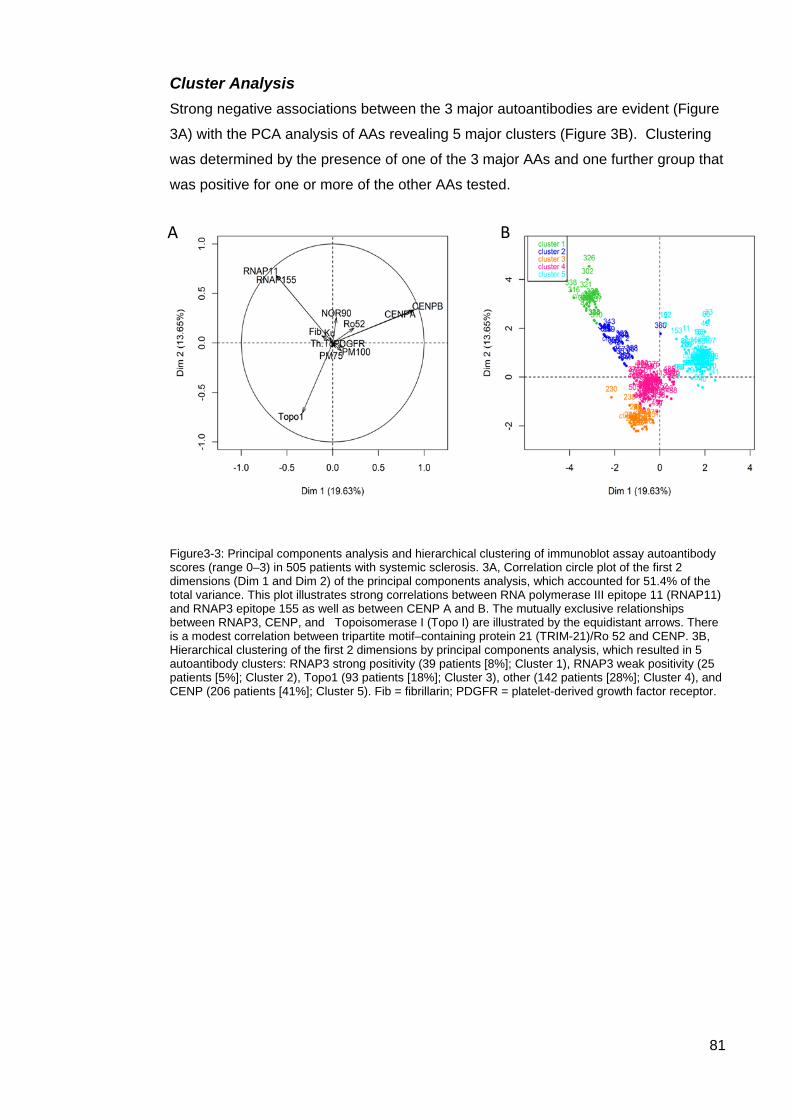
81
Cluster Analysis Strong negative associations between the 3 major autoantibodies are evident (Figure
3A) with the PCA analysis of AAs revealing 5 major clusters (Figure 3B). Clustering
was determined by the presence of one of the 3 major AAs and one further group that
was positive for one or more of the other AAs tested.
Figure3-3: Principal components analysis and hierarchical clustering of immunoblot assay autoantibody scores (range 0–3) in 505 patients with systemic sclerosis. 3A, Correlation circle plot of the first 2 dimensions (Dim 1 and Dim 2) of the principal components analysis, which accounted for 51.4% of the total variance. This plot illustrates strong correlations between RNA polymerase III epitope 11 (RNAP11) and RNAP3 epitope 155 as well as between CENP A and B. The mutually exclusive relationships between RNAP3, CENP, and Topoisomerase I (Topo I) are illustrated by the equidistant arrows. There is a modest correlation between tripartite motif–containing protein 21 (TRIM-21)/Ro 52 and CENP. 3B, Hierarchical clustering of the first 2 dimensions by principal components analysis, which resulted in 5 autoantibody clusters: RNAP3 strong positivity (39 patients [8%]; Cluster 1), RNAP3 weak positivity (25 patients [5%]; Cluster 2), Topo1 (93 patients [18%]; Cluster 3), other (142 patients [28%]; Cluster 4), and CENP (206 patients [41%]; Cluster 5). Fib = fibrillarin; PDGFR = platelet-derived growth factor receptor.
B A
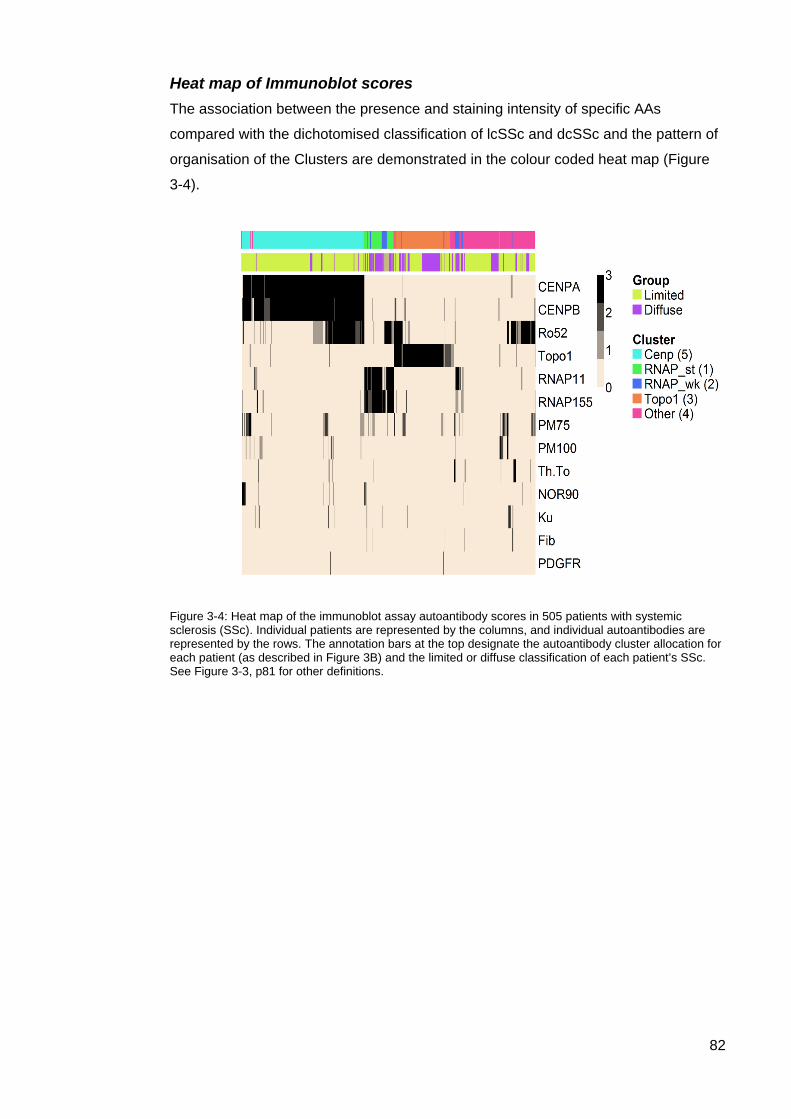
82
Heat map of Immunoblot scores The association between the presence and staining intensity of specific AAs
compared with the dichotomised classification of lcSSc and dcSSc and the pattern of
organisation of the Clusters are demonstrated in the colour coded heat map (Figure
3-4).
Figure 3-4: Heat map of the immunoblot assay autoantibody scores in 505 patients with systemic sclerosis (SSc). Individual patients are represented by the columns, and individual autoantibodies are represented by the rows. The annotation bars at the top designate the autoantibody cluster allocation for each patient (as described in Figure 3B) and the limited or diffuse classification of each patient’s SSc. See Figure 3-3, p81 for other definitions.
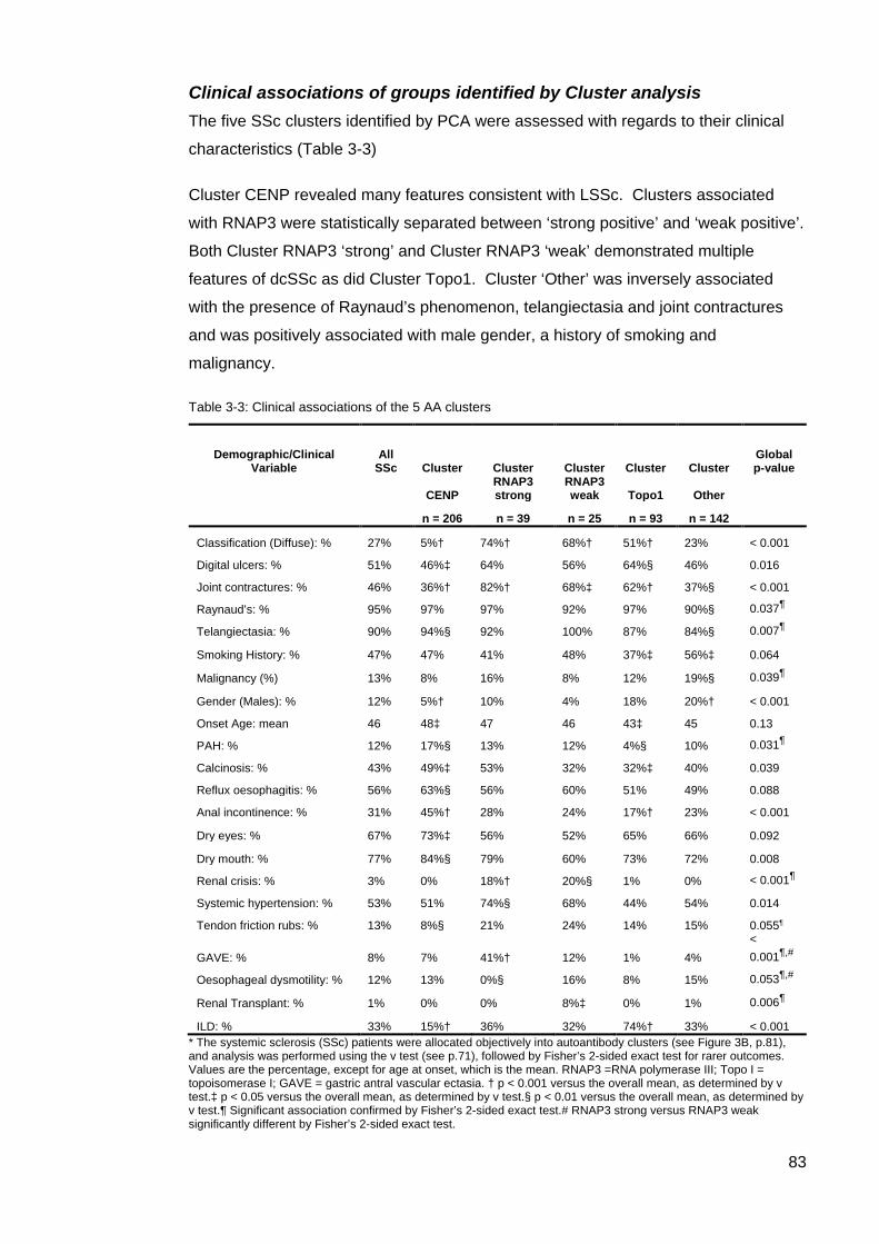
83
Clinical associations of groups identified by Cluster analysis The five SSc clusters identified by PCA were assessed with regards to their clinical
characteristics (Table 3-3)
Cluster CENP revealed many features consistent with LSSc. Clusters associated
with RNAP3 were statistically separated between ‘strong positive’ and ‘weak positive’.
Both Cluster RNAP3 ‘strong’ and Cluster RNAP3 ‘weak’ demonstrated multiple
features of dcSSc as did Cluster Topo1. Cluster ‘Other’ was inversely associated
with the presence of Raynaud’s phenomenon, telangiectasia and joint contractures
and was positively associated with male gender, a history of smoking and
malignancy.
Table 3-3: Clinical associations of the 5 AA clusters
Demographic/Clinical Variable
All SSc Cluster Cluster Cluster Cluster Cluster
Global p-value
CENP RNAP3 strong
RNAP3 weak Topo1 Other
n = 206 n = 39 n = 25 n = 93 n = 142 Classification (Diffuse): % 27% 5%† 74%† 68%† 51%† 23% < 0.001
Digital ulcers: % 51% 46%‡ 64% 56% 64%§ 46% 0.016
Joint contractures: % 46% 36%† 82%† 68%‡ 62%† 37%§ < 0.001
Raynaud’s: % 95% 97% 97% 92% 97% 90%§ 0.037¶
Telangiectasia: % 90% 94%§ 92% 100% 87% 84%§ 0.007¶
Smoking History: % 47% 47% 41% 48% 37%‡ 56%‡ 0.064
Malignancy (%) 13% 8% 16% 8% 12% 19%§ 0.039¶
Gender (Males): % 12% 5%† 10% 4% 18% 20%† < 0.001
Onset Age: mean 46 48‡ 47 46 43‡ 45 0.13
PAH: % 12% 17%§ 13% 12% 4%§ 10% 0.031¶
Calcinosis: % 43% 49%‡ 53% 32% 32%‡ 40% 0.039
Reflux oesophagitis: % 56% 63%§ 56% 60% 51% 49% 0.088
Anal incontinence: % 31% 45%† 28% 24% 17%† 23% < 0.001
Dry eyes: % 67% 73%‡ 56% 52% 65% 66% 0.092
Dry mouth: % 77% 84%§ 79% 60% 73% 72% 0.008
Renal crisis: % 3% 0% 18%† 20%§ 1% 0% < 0.001¶
Systemic hypertension: % 53% 51% 74%§ 68% 44% 54% 0.014
Tendon friction rubs: % 13% 8%§ 21% 24% 14% 15% 0.055¶
GAVE: % 8% 7% 41%† 12% 1% 4% < 0.001¶,#
Oesophageal dysmotility: % 12% 13% 0%§ 16% 8% 15% 0.053¶,#
Renal Transplant: % 1% 0% 0% 8%‡ 0% 1% 0.006¶
ILD: % 33% 15%† 36% 32% 74%† 33% < 0.001 * The systemic sclerosis (SSc) patients were allocated objectively into autoantibody clusters (see Figure 3B, p.81), and analysis was performed using the v test (see p.71), followed by Fisher’s 2-sided exact test for rarer outcomes. Values are the percentage, except for age at onset, which is the mean. RNAP3 =RNA polymerase III; Topo I = topoisomerase I; GAVE = gastric antral vascular ectasia. † p < 0.001 versus the overall mean, as determined by v test.‡ p < 0.05 versus the overall mean, as determined by v test.§ p < 0.01 versus the overall mean, as determined by v test.¶ Significant association confirmed by Fisher’s 2-sided exact test.# RNAP3 strong versus RNAP3 weak significantly different by Fisher’s 2-sided exact test.
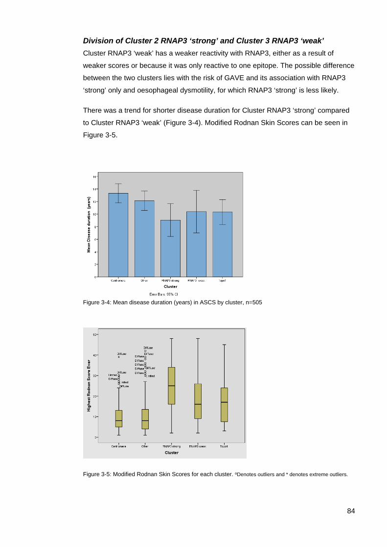
84
Division of Cluster 2 RNAP3 ‘strong’ and Cluster 3 RNAP3 ‘weak’ Cluster RNAP3 ‘weak’ has a weaker reactivity with RNAP3, either as a result of
weaker scores or because it was only reactive to one epitope. The possible difference
between the two clusters lies with the risk of GAVE and its association with RNAP3
‘strong’ only and oesophageal dysmotility, for which RNAP3 ‘strong’ is less likely.
There was a trend for shorter disease duration for Cluster RNAP3 ‘strong’ compared
to Cluster RNAP3 ‘weak’ (Figure 3-4). Modified Rodnan Skin Scores can be seen in
Figure 3-5.
Figure 3-4: Mean disease duration (years) in ASCS by cluster, n=505
Figure 3-5: Modified Rodnan Skin Scores for each cluster. ºDenotes outliers and * denotes extreme outliers.
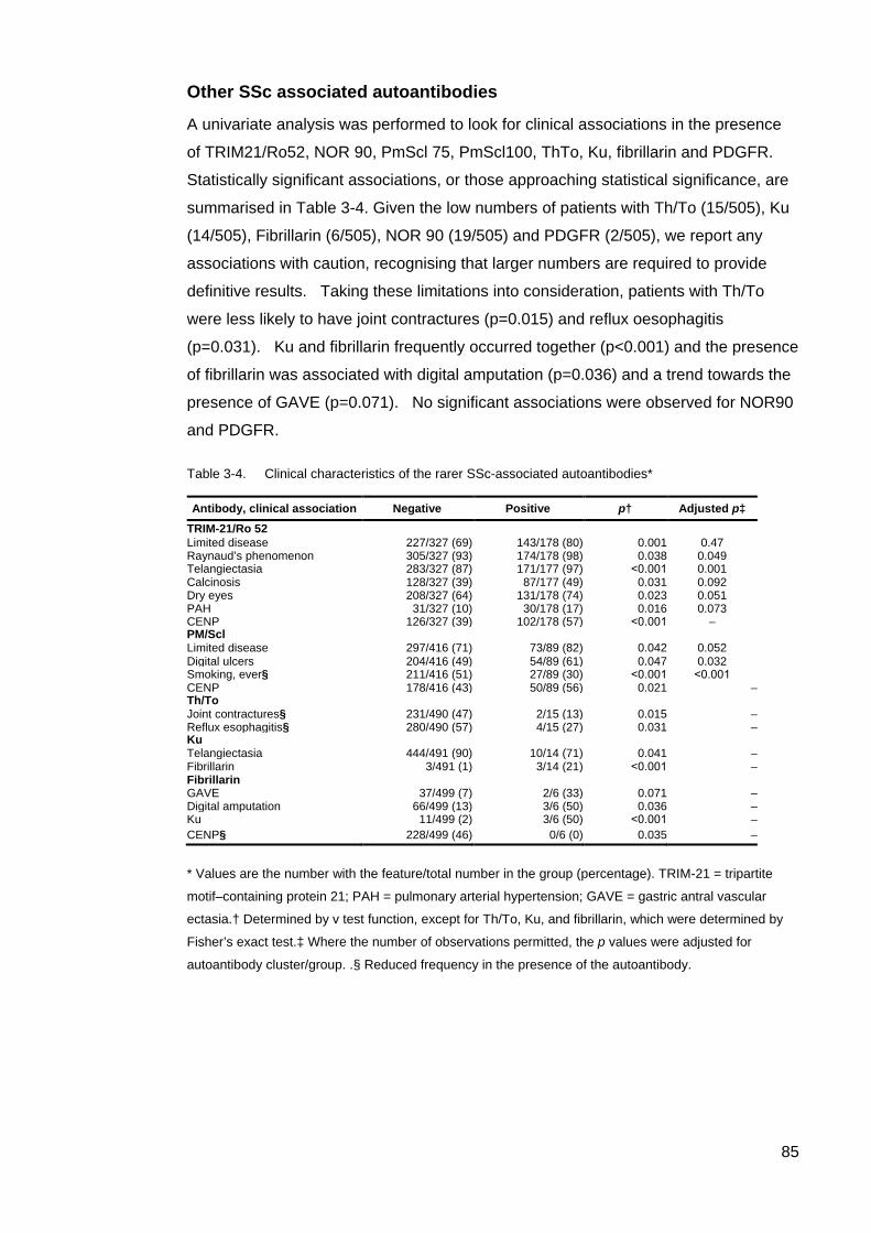
85
Other SSc associated autoantibodies
A univariate analysis was performed to look for clinical associations in the presence
of TRIM21/Ro52, NOR 90, PmScl 75, PmScl100, ThTo, Ku, fibrillarin and PDGFR.
Statistically significant associations, or those approaching statistical significance, are
summarised in Table 3-4. Given the low numbers of patients with Th/To (15/505), Ku
(14/505), Fibrillarin (6/505), NOR 90 (19/505) and PDGFR (2/505), we report any
associations with caution, recognising that larger numbers are required to provide
definitive results. Taking these limitations into consideration, patients with Th/To
were less likely to have joint contractures (p=0.015) and reflux oesophagitis
(p=0.031). Ku and fibrillarin frequently occurred together (p<0.001) and the presence
of fibrillarin was associated with digital amputation (p=0.036) and a trend towards the
presence of GAVE (p=0.071). No significant associations were observed for NOR90
and PDGFR.
Table 3-4. Clinical characteristics of the rarer SSc-associated autoantibodies*
* Values are the number with the feature/total number in the group (percentage). TRIM-21 = tripartite
motif–containing protein 21; PAH = pulmonary arterial hypertension; GAVE = gastric antral vascular
ectasia.† Determined by v test function, except for Th/To, Ku, and fibrillarin, which were determined by
Fisher’s exact test.‡ Where the number of observations permitted, the p values were adjusted for
autoantibody cluster/group. .§ Reduced frequency in the presence of the autoantibody.
Antibody, clinical association Negative Positive p† Adjusted p‡ TRIM-21/Ro 52 Limited disease 227/327 (69) 143/178 (80) 0.001 0.47 Raynaud’s phenomenon 305/327 (93) 174/178 (98) 0.038 0.049 Telangiectasia 283/327 (87) 171/177 (97) ˂0.001 0.001 Calcinosis 128/327 (39) 87/177 (49) 0.031 0.092 Dry eyes 208/327 (64) 131/178 (74) 0.023 0.051 PAH 31/327 (10) 30/178 (17) 0.016 0.073 CENP 126/327 (39) 102/178 (57) ˂0.001 – PM/Scl Limited disease
297/416 (71)
73/89 (82)
0.042
0.052
Digital ulcers 204/416 (49) 54/89 (61) 0.047 0.032 Smoking, ever§ 211/416 (51) 27/89 (30) ˂0.001 ˂0.001 CENP 178/416 (43) 50/89 (56) 0.021 – Th/To Joint contractures§ 231/490 (47) 2/15 (13) 0.015 – Reflux esophagitis§ 280/490 (57) 4/15 (27) 0.031 – Ku Telangiectasia
444/491 (90)
10/14 (71)
0.041
–
Fibrillarin 3/491 (1) 3/14 (21) ˂0.001 – Fibrillarin GAVE 37/499 (7) 2/6 (33) 0.071 – Digital amputation 66/499 (13) 3/6 (50) 0.036 – Ku 11/499 (2) 3/6 (50) ˂0.001 – CENP§ 228/499 (46) 0/6 (0) 0.035 –

86
TRIM21/Ro52 and PmScl75/100 were the most common of the other SSc associated
AA with 35.3% and 17.6% of patients testing positive for these AAs respectively.
While their presence did not contribute to sub-classification of patients, the clinical
associations for TRIM21/Ro52 and PmScl75/100 are of interest and are summarised
in Table 3-4. The presence of telangiectasia was strongly associated with
TRIM21/Ro52 (p = 0.01) while adjusted data show a trend towards the presence of
dry eyes, PAH and calcinosis. There was no evidence of an association with ILD in
this cohort (p=0.30).
PmScl75/100 was associated with a history of digital ulcers (p=0.032) and patients
expressing this AA show a trend towards the presence of lSSc (p=0.052).
PmScl75/100 was more commonly identified in those who had no history of smoking,
the reason for this is uncertain and may relate to as yet unidentified confounding
factors. Both TRIM21/Ro52 and PMScl75/100 were more commonly seen in the
presence of CENP.
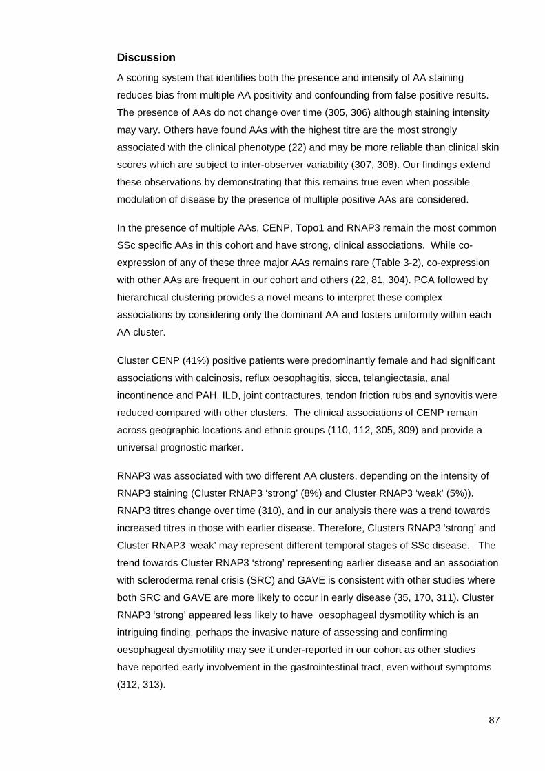
87
Discussion
A scoring system that identifies both the presence and intensity of AA staining
reduces bias from multiple AA positivity and confounding from false positive results.
The presence of AAs do not change over time (305, 306) although staining intensity
may vary. Others have found AAs with the highest titre are the most strongly
associated with the clinical phenotype (22) and may be more reliable than clinical skin
scores which are subject to inter-observer variability (307, 308). Our findings extend
these observations by demonstrating that this remains true even when possible
modulation of disease by the presence of multiple positive AAs are considered.
In the presence of multiple AAs, CENP, Topo1 and RNAP3 remain the most common
SSc specific AAs in this cohort and have strong, clinical associations. While co-
expression of any of these three major AAs remains rare (Table 3-2), co-expression
with other AAs are frequent in our cohort and others (22, 81, 304). PCA followed by
hierarchical clustering provides a novel means to interpret these complex
associations by considering only the dominant AA and fosters uniformity within each
AA cluster.
Cluster CENP (41%) positive patients were predominantly female and had significant
associations with calcinosis, reflux oesophagitis, sicca, telangiectasia, anal
incontinence and PAH. ILD, joint contractures, tendon friction rubs and synovitis were
reduced compared with other clusters. The clinical associations of CENP remain
across geographic locations and ethnic groups (110, 112, 305, 309) and provide a
universal prognostic marker.
RNAP3 was associated with two different AA clusters, depending on the intensity of
RNAP3 staining (Cluster RNAP3 ‘strong’ (8%) and Cluster RNAP3 ‘weak’ (5%)).
RNAP3 titres change over time (310), and in our analysis there was a trend towards
increased titres in those with earlier disease. Therefore, Clusters RNAP3 ‘strong’ and
Cluster RNAP3 ‘weak’ may represent different temporal stages of SSc disease. The
trend towards Cluster RNAP3 ‘strong’ representing earlier disease and an association
with scleroderma renal crisis (SRC) and GAVE is consistent with other studies where
both SRC and GAVE are more likely to occur in early disease (35, 170, 311). Cluster
RNAP3 ‘strong’ appeared less likely to have oesophageal dysmotility which is an
intriguing finding, perhaps the invasive nature of assessing and confirming
oesophageal dysmotility may see it under-reported in our cohort as other studies
have reported early involvement in the gastrointestinal tract, even without symptoms
(312, 313).
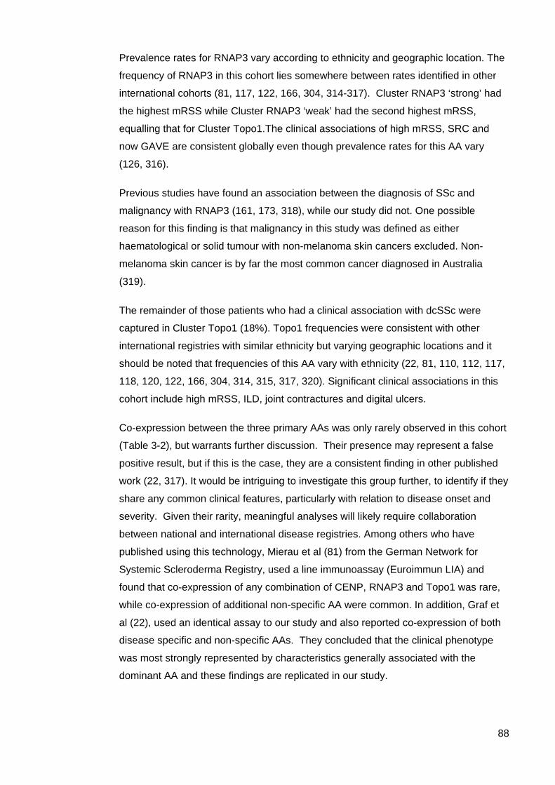
88
Prevalence rates for RNAP3 vary according to ethnicity and geographic location. The
frequency of RNAP3 in this cohort lies somewhere between rates identified in other
international cohorts (81, 117, 122, 166, 304, 314-317). Cluster RNAP3 ‘strong’ had
the highest mRSS while Cluster RNAP3 ‘weak’ had the second highest mRSS,
equalling that for Cluster Topo1.The clinical associations of high mRSS, SRC and
now GAVE are consistent globally even though prevalence rates for this AA vary
(126, 316).
Previous studies have found an association between the diagnosis of SSc and
malignancy with RNAP3 (161, 173, 318), while our study did not. One possible
reason for this finding is that malignancy in this study was defined as either
haematological or solid tumour with non-melanoma skin cancers excluded. Non-
melanoma skin cancer is by far the most common cancer diagnosed in Australia
(319).
The remainder of those patients who had a clinical association with dcSSc were
captured in Cluster Topo1 (18%). Topo1 frequencies were consistent with other
international registries with similar ethnicity but varying geographic locations and it
should be noted that frequencies of this AA vary with ethnicity (22, 81, 110, 112, 117,
118, 120, 122, 166, 304, 314, 315, 317, 320). Significant clinical associations in this
cohort include high mRSS, ILD, joint contractures and digital ulcers.
Co-expression between the three primary AAs was only rarely observed in this cohort
(Table 3-2), but warrants further discussion. Their presence may represent a false
positive result, but if this is the case, they are a consistent finding in other published
work (22, 317). It would be intriguing to investigate this group further, to identify if they
share any common clinical features, particularly with relation to disease onset and
severity. Given their rarity, meaningful analyses will likely require collaboration
between national and international disease registries. Among others who have
published using this technology, Mierau et al (81) from the German Network for
Systemic Scleroderma Registry, used a line immunoassay (Euroimmun LIA) and
found that co-expression of any combination of CENP, RNAP3 and Topo1 was rare,
while co-expression of additional non-specific AA were common. In addition, Graf et
al (22), used an identical assay to our study and also reported co-expression of both
disease specific and non-specific AAs. They concluded that the clinical phenotype
was most strongly represented by characteristics generally associated with the
dominant AA and these findings are replicated in our study.
89
Cluster 4 ‘Other’ (28%) captures the remaining patients in our cohort. This cluster
includes patients who (i) do not express CENP, Topo1 or RNAP3, (ii) may have a low
score for one of the primary SSc AAs coupled with one or more of the SSc associated
AAs or (iii) they may have either monospecificity or multispecificity for the SSc
associated AAs.
Positive associations in Cluster 4 ‘Other’ include male gender, a history of malignancy
and smoking. Negative associations include telangiectasia, Raynaud’s phenomenon
and joint contractures. The clinical associations of this group are intriguing and
warrant further investigation. U1RNP was not available on the SSc immunoblot;
however in separate testing of all patients for this AA, all those classified with MCTD
mapped to Cluster ‘Other’ (data not shown). If U1RNP had been included on the
immunoblot, it may have formed a separate cluster with its own specific clinical
associations. It is likely that Cluster 4 ‘Other’ identifies more than one disease group.
The rarer SSc associated AAs were also assessed in this study but they were not
found to significantly contribute to sub-classification with PCA. Only a small number of
patients tested positive for NOR90, PDGFR, Ku, Fibrillarin and ThTo (to hPOP1
epitope only) and it is likely that large multinational studies will be necessary to
determine if they have significant clinical associations.
Where statistical power has permitted, both TRIM21/Ro52 and Pm-Scl were found to
have more meaningful results. TRIM21/Ro52 is a common AA and its presence in
SSc has been reported elsewhere, with varying clinical associations (22, 81, 304,
321). Of particular significance with regards to disease prognostication have been
reports of associations with overlap disease and ILD (304). In this cohort, the
presence of TRIM21/Ro52 was associated with telangiectasia and Raynaud’s
phenomenon, with a trend towards the presence of dry eyes, calcinosis and limited
disease. These clinical features are consistent with others who have reported
associations with limited disease (22, 81) and sicca features (81). The trend towards
PAH is intriguing and has not been reported elsewhere although direct comparison
between the groups is difficult as right heart catheterisation is not readily available in
all cohorts. The CSRG registry used a surrogate marker obtained from
echocardiogram (pulmonary arterial hypertension) while Mierau et al did not provide a
detailed definition of pulmonary HT (81). Differing methods used to detect
TRIM21/Ro52 may also account for variations between cohorts. However, it is
possible that the use of right heart catheter may improve the specificity of any findings
in relation to PAH. This could be clarified by review in a larger group.
90
Unlike Hudson et al (304), we did not find an association between TRIM21/Ro52 and
ILD although the definition for ILD between groups varied significantly. In our cohort,
ILD was defined on the basis of radiological findings, while Hudson et al used a
recently published algorithm for ILD which included patients with typical clinical
findings in the absence of radiological findings.
The presence of PmScl75/100 in SSc has been associated with limited and overlap
disease, skeletal muscle disease, calcinosis, improved survival (81, 223, 227) and
more recently, improved prognosis in ILD (322). Techniques used to detect these AA
varied considerably between studies and so once again, this must be taken into
account when interpreting findings between cohorts. In this cohort, PmScl75/100 was
associated with digital ulcers and there was a trend towards an increased presence in
limited disease. No significant associations were observed with a history of myositis
or elevated CK (unpublished data).
Autoantibody classification of scleroderma versus traditional classification Traditionally, SSc is classified according to the extent of skin fibrosis. Limited
cutaneous SSc and dcSSc have well-established differences in their presentation,
AAs and outcomes. However, the dermatological changes are a dynamic process and
hence early labelling of a patient with limited disease may need to be modified later
during the disease course.
The difficulties in diagnosis and sub classification of SSc based on skin involvement
have direct relevance in the clinical setting. Virendrakumar Bhavsar et al. recently
reported two cases of RNAP3 associated SRC occurring in the absence of initial skin
involvement (323) presenting a major diagnostic challenge. Furthermore, Cottrell et al
concluded that dichotomous classification based on skin involvement may result in
misclassifying an intermediate group of patients who exhibit a unique AA profile,
disease course and clinical outcome (283).
In contrast AAs are a consistent feature of the disease and it is rare for an AA to
disappear, although fluctuations in antibody titre or binding intensity may occur. Our
analysis has shown significant clinical correlations within each cluster in the absence
of any other information apart from the diagnosis of SSc and the presence and
staining intensity of the AA. It seems logical therefore, to propose disease
stratification based on AAs would be simpler to apply and be more consistent
irrespective of stage of disease than the traditional classification system based on the
extent of skin involvement. Furthermore, this cross–sectional study demonstrates
strong disease associations with the AAs and may have important applications in
91
enriching studies of new therapies for certain subset(s) and identifying subjects for
targeted therapies.
Strengths and Limitations There were minimal missing data enabling a comprehensive statistical analysis of the
results of the AA testing with the clinical and demographical variables. Limitations
include the small number of patients with rarer AAs. While the clinical associations
with the AAs in this study were similar to other published work, it is important to
recognize that there may be different associations depending on how the AAs are
identified by various platforms currently in use. Assays targeting a specific peptide AA
sequence do not necessarily reflect the clinical associations of AA identified using
more labour intensive assays that identify the whole protein, or indeed functional
assays. The sensitivity often reported in LIA may result in changes to previously
reported disease associations and therefore it is recommended that the performance
of the newer technologies be assessed in well-characterised SSc registries.
Final comments and conclusion PCA provides a novel means to identify the presence and intensity of scleroderma
associated AAs and to reduce confounding when more than one AA is detected in
patient sera. We have shown that sub-classification based only on the presence of
AAs reveals clinically meaningful disease associations. The dominant AA is the one
that most accurately reflects disease associations, at least when CENP, Topo1 and
RNAP3 are considered. Two separate clusters of RNAP3 were identified with
intriguing differences in clinical associations. We propose that this may relate to the
well-documented fluctuations in RNAP3 over time, with higher titres being observed
earlier in the disease course. Further prospective studies will provide further
clarification in this regard. In our cohort, those who did not map to one of the 4 major
AA associated clusters had intriguing clinical associations. They are likely to
represent more than one disease group and warrant further investigation.
We conclude that the use of specific and associated SSc AAs provides more
meaningful classification of subsets than the currently utilized limited, diffuse and
overlap subdivisions. We also propose that using AA profiling for sub classification
and stratification will improve disease management and prognostication and the
identification of patients for clinical trials.
92
CHAPTER 4 CLUSTER 4, ‘OTHER’, EXPLORING THE CLINICAL UTILITY OF
SYSTEMIC SCLEROSIS PRIMARY AND ASSOCIATED AUTOANTIBODIES
Introduction
In Chapter 3 an examination was conducted on the clinical and serological features of
SSc-specific and associated autoantibodies (AA) and the stratification of these AAs
into discrete ‘Clusters’ directed by a defining or primary SSc AA. The term ‘primary’
AA is used throughout this chapter when referring to Topoisomerase1 (Topo1),
Centromere A and/or B (CENP A and/or CENP B) and RNA Polymerase III (RNAP3)
as these AAs are considered to be specific for SSc (43). The clusters determined by
PCA considered both the type of AA and the intensity of staining measured by the
Euroimmun line immunoblot assay (LIA).
Among 505 patients with available sera, we found that the majority (72%) could be
grouped according to an AA profile that was largely determined by the presence of
one of the primary SSc associated AA. Furthermore, when these groupings were
assessed according to their clinical characteristics, CENP A/B was found to associate
with clinical features known to occur in lcSSc and RNAP3 and Topo1 with clinical
features seen in dcSSc, independent of any clinical assessment. These findings
suggest that these AAs may be used as sole criterions to sub classify SSc, a finding
that has particular relevance when considering early disease prognostication and
assessing patients for their suitability in clinical trials.
There remained 28% of patients who were not clearly linked to one of the primary AA
groupings identified using cluster analysis (i.e. Clusters 1, 2, 3, & 5). For the most
part, these patients did not have one of the three primary SSc AAs or if they did, it
was of a lower staining intensity, or was present in combination with other SSc
associated AAs. U1RNP testing was not available on initial immunoblot analysis and
may have provided further explanation for patients without a detectable specific SSc
associated AA. We were particularly interested to examine this cohort further as
patients presenting with mixed or less specific AA profiles may offer a greater
diagnostic and prognostic challenge to the clinician. The total number of patients in
Cluster 4 is relatively small (n=142) meaning that care must be taken with analysis to
limit bias. Therefore we decided to review AA groupings in the Cluster 4 cohort as a
whole before evaluating any unique or defining disease features according to the
dominant AA profile. Where numbers permitted, statistical analysis was used to

93
assess results.
Specific aims for this study were;
i. To further characterise the specific AA profile/s identified by the Euroimmun
Line Immunoassay (LIA) and to identify any unique features between patients
within this Cluster with a less homogeneous AA profile and to determine if
variance in Cluster 4 is due to identifiable, underlying factors.
ii. To assess any unique clinical features associated with AA profiling in this
cohort among Cluster 4 patients and among the SSc cohort as a whole.
iii. To explore if patients in Cluster 4, who were positive for one or more of the
three primary AAs, differed clinically to patients positive for these AA but
assigned by PCA to other Clusters (discussed in Chapter 3).
iv. To explore the clinical characteristics of Cluster 4 patients who are positive for
both CENP and Topo1.
v. To assess this well characterised SSc cohort (see Chapter 2, pp. 64-66), for
U1RNP and to compare clinical features among those patients whose sera
contain U1RNP with a randomly selected cohort of patients with MCTD who
are enrolled in the ASIG database
An initial analysis was carried out on patients that have the three primary AAs; CENP,
Topo1 and RNAP3. Following that, analyses on the AAs that feature strongly in this
Cluster; TRIM21/Ro52, PmScl and U1RNP were performed.
Methods
Please refer to Chapter 2, Methodology, (pp.64-66), for details regarding the patient
population, Scleroderma Euroline assay and U1RNP testing. We had additional AA
sera and clinical data on 21 patients identified to have MCTD in the ASIG database
and these patients were used to provide a comparator group for any other patients
that may test positive for U1RNP on further testing in Cluster 4.
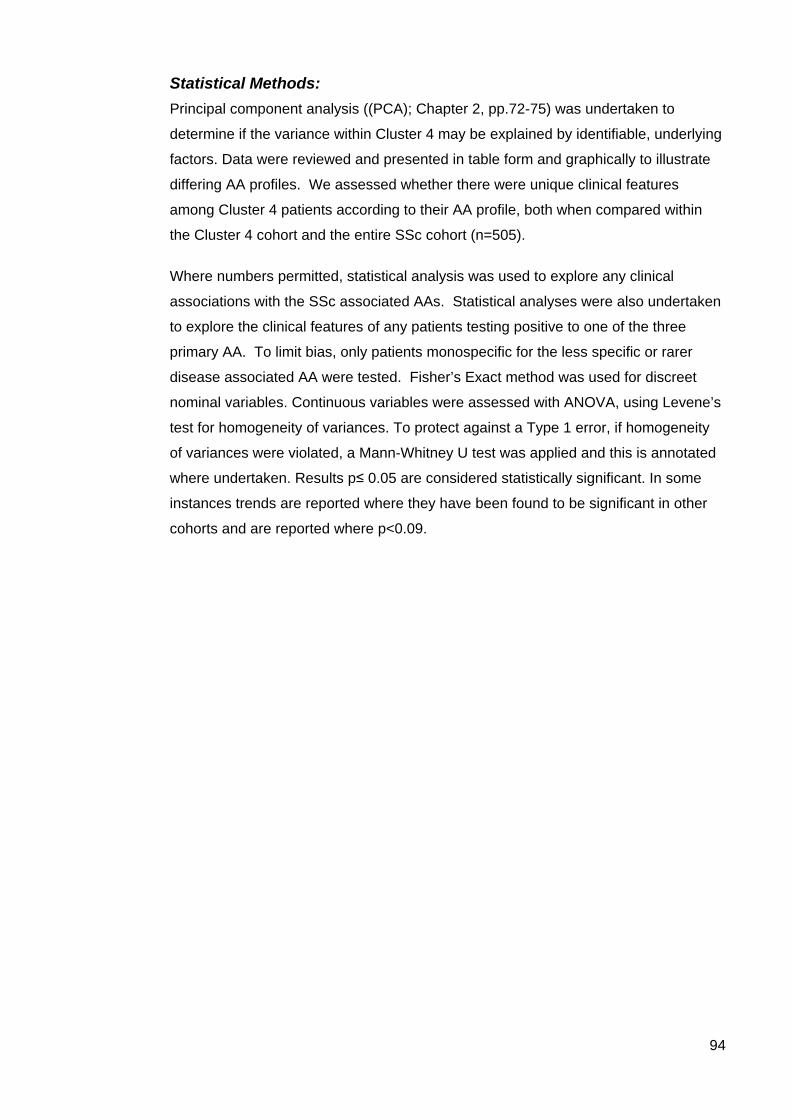
94
Statistical Methods: Principal component analysis ((PCA); Chapter 2, pp.72-75) was undertaken to
determine if the variance within Cluster 4 may be explained by identifiable, underlying
factors. Data were reviewed and presented in table form and graphically to illustrate
differing AA profiles. We assessed whether there were unique clinical features
among Cluster 4 patients according to their AA profile, both when compared within
the Cluster 4 cohort and the entire SSc cohort (n=505).
Where numbers permitted, statistical analysis was used to explore any clinical
associations with the SSc associated AAs. Statistical analyses were also undertaken
to explore the clinical features of any patients testing positive to one of the three
primary AA. To limit bias, only patients monospecific for the less specific or rarer
disease associated AA were tested. Fisher’s Exact method was used for discreet
nominal variables. Continuous variables were assessed with ANOVA, using Levene’s
test for homogeneity of variances. To protect against a Type 1 error, if homogeneity
of variances were violated, a Mann-Whitney U test was applied and this is annotated
where undertaken. Results p≤ 0.05 are considered statistically significant. In some
instances trends are reported where they have been found to be significant in other
cohorts and are reported where p<0.09.
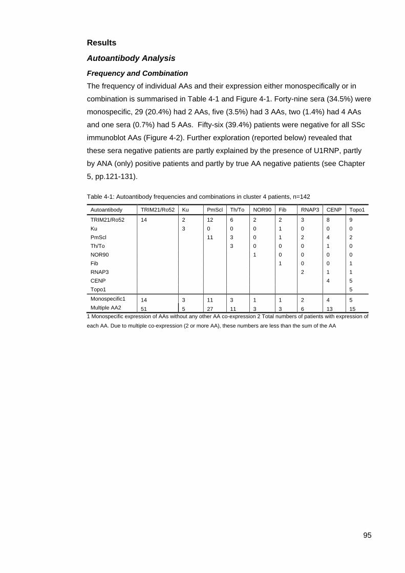
95
Results
Autoantibody Analysis
Frequency and Combination The frequency of individual AAs and their expression either monospecifically or in
combination is summarised in Table 4-1 and Figure 4-1. Forty-nine sera (34.5%) were
monospecific, 29 (20.4%) had 2 AAs, five (3.5%) had 3 AAs, two (1.4%) had 4 AAs
and one sera (0.7%) had 5 AAs. Fifty-six (39.4%) patients were negative for all SSc
immunoblot AAs (Figure 4-2). Further exploration (reported below) revealed that
these sera negative patients are partly explained by the presence of U1RNP, partly
by ANA (only) positive patients and partly by true AA negative patients (see Chapter
5, pp.121-131).
Table 4-1: Autoantibody frequencies and combinations in cluster 4 patients, n=142
Autoantibody TRIM21/Ro52 Ku PmScl Th/To NOR90 Fib RNAP3 CENP Topo1
TRIM21/Ro52 14 2 12 6 2 2 3 8 9 Ku 3 0 0 0 1 0 0 0 PmScl 11 3 0 1 2 4 2 Th/To 3 0 0 0 1 0 NOR90 1 0 0 0 0 Fib 1 0 0 1 RNAP3 2 1 1 CENP 4 5 Topo1 5
Monospecific1 14 3 11 3 1 1 2 4 5 Multiple AA2 51 5 27 11 3 3 6 13 15
1 Monospecific expression of AAs without any other AA co-expression 2 Total numbers of patients with expression of
each AA. Due to multiple co-expression (2 or more AA), these numbers are less than the sum of the AA
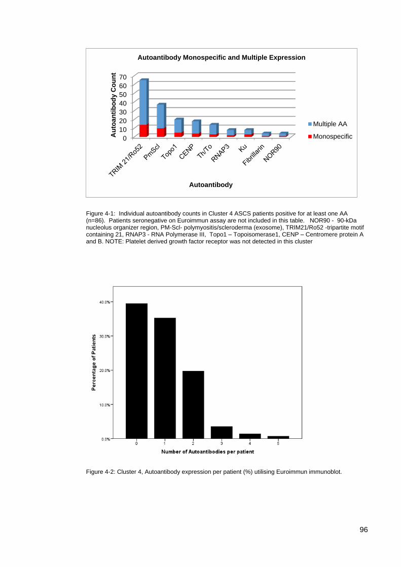
96
Figure 4-1: Individual autoantibody counts in Cluster 4 ASCS patients positive for at least one AA (n=86). Patients seronegative on Euroimmun assay are not included in this table. NOR90 - 90-kDa nucleolus organizer region, PM-Scl- polymyositis/scleroderma (exosome), TRIM21/Ro52 -tripartite motif containing 21, RNAP3 - RNA Polymerase III, Topo1 – Topoisomerase1, CENP – Centromere protein A and B. NOTE: Platelet derived growth factor receptor was not detected in this cluster
Figure 4-2: Cluster 4, Autoantibody expression per patient (%) utilising Euroimmun immunoblot.
010203040506070
Aut
oant
ibod
y C
ount
Autoantibody
Autoantibody Monospecific and Multiple Expression
Multiple AA
Monospecific

97
Supplementary PCA A detailed description of the supplementary PCA can be found in Chapter 2,
Methodology (pp.72-75), however to summarise briefly; a scree plot in conjunction
with Horn’s parallel analysis determined that it was optimal to extract three
components with any additional extractions not contributing meaningfully to the
analysis. The strength of loadings can range from -1 to +1 signifying both positive and
negative relationships between the AAs. Loadings ± .320 are considered to contribute
to the relationship between variables (293). Following the PCA, a hierarchical cluster
analysis revealed that unlike the initial PCA where the first two components
demonstrated that 51.4% of the total variance was attributable to underlying factors,
the relationships in Cluster 4 were much weaker, taking three extractions to show that
46.6% of the total variance was attributable to underlying factors. Nonetheless, using
the data generated from the PCA in a hierarchical cluster analysis it was revealed that
CENPB, Topo1, PmScl, Th/To and TRIM21/Ro52 accounted for the majority of
relationships in Cluster 4. The remaining AAs; RNAP3, Ku, Fibrillarin and NOR90,
were not present in sufficient numbers to determine any statistical significant outcome
and to avoid a Type 1 error, further statistical analyses on these AAs were not done.
Expression of Topo1, CENP and RNAP3 in Cluster 4 The primary SSc AAs, CENP, Topo1 and RNAP3 were detected in this Cluster, either
singularly or in various combinations with differing staining intensities. As these AAs
had generally been captured by other clusters (see Chapter 3, pp.77-84) we reviewed
their presence in this Cluster in detail to understand why they had not been captured
in Clusters 1, 2, 3 or 5. Upon reviewing AA profiling (Table 4-2), it was evident that a
combination of lower staining intensity, single epitope expression and AA co-
expression has determined a place in Cluster 4 for these patients.
Topo1
• Topo1 appears 15 times
• Monospecific ‘+’ 5 times (pts 23, 407, 759, 773, 948)
• Topo1 ‘+++’ co-expressed with TRIM21/Ro52 ‘+++’ and CENP B ‘++’ twice (pt
115, 1050)
• Topo1 ‘++’ co-expressed with TRIM21/Ro52 ‘+++’ once (pt 126)
• Topo1 ‘+’ co-expressed with CENP A ‘+++’, TRIM21/Ro52 ‘+++’ once (pt 328)
• Topo1 ‘+’ co-expressed with CENP B ‘+’ once (pt 266)
• Topo1 ‘+’ co-expressed with TRIM21/Ro52 ‘+’ once (pt 529)
• Topo1 ‘+’ co-expressed with TRIM21/Ro52 ‘++’ once (pt 252)
• Topo1 ‘+’ co-expressed with TRIM21/Ro52 ‘+++’ once (pt 1052)
• Topo1 ‘+’ co-expressed with CENP B ‘+’, RNAP3 155 ‘+’, TRIM21/Ro52 ‘++’,

98
PmScl ‘++’ once (pt 247)
• Topo1 ‘+’ co-expressed with TRIM21/Ro52 ‘+’. PM75 ‘+’, Fib ‘+’ once(pt 1221)
CENP A and B
• Appear 14 times
• CENP A ‘+++’ monospecific twice (pts 99, 851)
• CENP B ‘+’ monospecific twice (pts 194, 855)
• CENP A ‘+++’, co-expressed with TRIM21/Ro52 ‘+++’ and Topo1 ‘+’ once (pt
328)
• CENP A ‘+’ and CENP B ‘++’ co-expressed with PM75 ‘+’ once (pt 728)
• CENP A ‘++’, TRIM21/Ro52 ‘+++’ (pt 858)
• CENP A ‘+’, TRIM21/Ro52 ‘+++’ (pt 123)
• CENP B ‘++’ co-expressed with Topo1 ‘+++”, TRIM21/Ro52 ‘+++’ twice (pts
115, 1050)
• CENP B ‘++’, TRIM21/Ro52 ‘+++’, PM75 ‘++’ (pt 244)
• CENP B ‘+’ co-expressed with Topo1 ‘+’ once (pt 266)
• CENP B ‘+’ co-expressed with Topo1 ‘+’ and TRIM21/Ro52 ‘+++’, RNAP155 ‘+’
PM75 ‘++’ once (pt 247)
• CENP B ‘+’ co-expressed with TRIM21/Ro52 ‘++’, PM75 ‘++’, Th/To ‘+++’ once
(pt 566)
RNAP3
• Appears 6 times
• RP11 ‘+’ monospecific once
• RP155 ‘+’ monospecific once
• RP11 ‘+’ co-expressed with TRIM21/Ro52 ‘+++’ twice
• RP11 ‘+’ co-expressed with PM75 ‘+’ once
• RP155 ‘+’ co-expressed with TRIM21/Ro52 ‘++’ and PM75 ‘++’ once
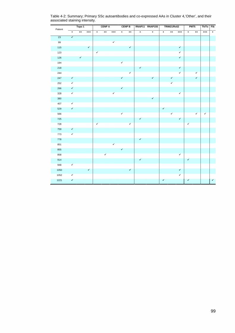
99
Table 4-2: Summary; Primary SSc autoantibodies and co-expressed AAs in Cluster 4,’Other’, and their associated staining intensity.
Patient Topo 1 CENP A CENP B RNAP11 RNAP155 TRIM21/Ro52 PM75 Th/To Fib
+ ++ +++ + ++ +++ + ++ + + + ++ +++ + ++ +++ +
23
99
115
123
126
194
218
244
247
252
266
328
380
407
529
566
725
728
759
773
778
851
855
858
914
948
1050
1052
1221
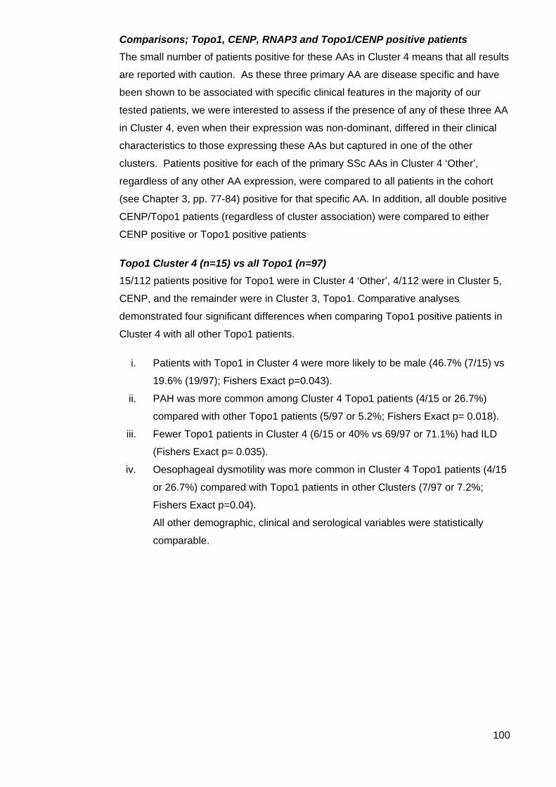
100
Comparisons; Topo1, CENP, RNAP3 and Topo1/CENP positive patients The small number of patients positive for these AAs in Cluster 4 means that all results
are reported with caution. As these three primary AA are disease specific and have
been shown to be associated with specific clinical features in the majority of our
tested patients, we were interested to assess if the presence of any of these three AA
in Cluster 4, even when their expression was non-dominant, differed in their clinical
characteristics to those expressing these AAs but captured in one of the other
clusters. Patients positive for each of the primary SSc AAs in Cluster 4 ‘Other’,
regardless of any other AA expression, were compared to all patients in the cohort
(see Chapter 3, pp. 77-84) positive for that specific AA. In addition, all double positive
CENP/Topo1 patients (regardless of cluster association) were compared to either
CENP positive or Topo1 positive patients
Topo1 Cluster 4 (n=15) vs all Topo1 (n=97) 15/112 patients positive for Topo1 were in Cluster 4 ‘Other’, 4/112 were in Cluster 5,
CENP, and the remainder were in Cluster 3, Topo1. Comparative analyses
demonstrated four significant differences when comparing Topo1 positive patients in
Cluster 4 with all other Topo1 patients.
i. Patients with Topo1 in Cluster 4 were more likely to be male (46.7% (7/15) vs
19.6% (19/97); Fishers Exact p=0.043).
ii. PAH was more common among Cluster 4 Topo1 patients (4/15 or 26.7%)
compared with other Topo1 patients (5/97 or 5.2%; Fishers Exact p= 0.018).
iii. Fewer Topo1 patients in Cluster 4 (6/15 or 40% vs 69/97 or 71.1%) had ILD
(Fishers Exact p= 0.035).
iv. Oesophageal dysmotility was more common in Cluster 4 Topo1 patients (4/15
or 26.7%) compared with Topo1 patients in other Clusters (7/97 or 7.2%;
Fishers Exact p=0.04).
All other demographic, clinical and serological variables were statistically
comparable.
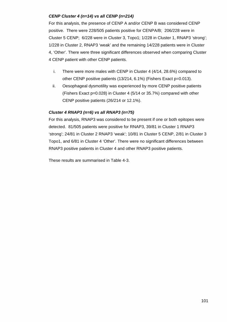
101
CENP Cluster 4 (n=14) vs all CENP (n=214) For this analysis, the presence of CENP A and/or CENP B was considered CENP
positive. There were 228/505 patients positive for CENPA/B; 206/228 were in
Cluster 5 CENP; 6/228 were in Cluster 3, Topo1; 1/228 in Cluster 1, RNAP3 ‘strong’;
1/228 in Cluster 2, RNAP3 ‘weak’ and the remaining 14/228 patients were in Cluster
4, ‘Other’. There were three significant differences observed when comparing Cluster
4 CENP patient with other CENP patients.
i. There were more males with CENP in Cluster 4 (4/14, 28.6%) compared to
other CENP positive patients (13/214, 6.1%) (Fishers Exact p=0.013).
ii. Oesophageal dysmotility was experienced by more CENP positive patients
(Fishers Exact p=0.028) in Cluster 4 (5/14 or 35.7%) compared with other
CENP positive patients (26/214 or 12.1%).
Cluster 4 RNAP3 (n=6) vs all RNAP3 (n=75) For this analysis, RNAP3 was considered to be present if one or both epitopes were
detected. 81/505 patients were positive for RNAP3, 39/81 in Cluster 1 RNAP3
‘strong’; 24/81 in Cluster 2 RNAP3 ‘weak’; 10/81 in Cluster 5 CENP, 2/81 in Cluster 3
Topo1, and 6/81 in Cluster 4 ‘Other’. There were no significant differences between
RNAP3 positive patients in Cluster 4 and other RNAP3 positive patients.
These results are summarised in Table 4-3.
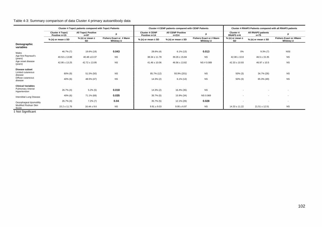
102
Table 4-3: Summary comparison of data Cluster 4 primary autoantibody data
Cluster 4 Topo1 patients compared with Topo1 Patients Cluster 4 CENP patients compared with CENP Patients Cluster 4 RNAP3 Patients compared with all RNAP3 patients
Cluster 4 Topo1 Positive n=15
All Topo1 Positive n=97 p Cluster 4 CENP
Positive n=14 All CENP Positive
n=214 p Cluster 4 RNAP3 n=6
All RNAP3 patients n=75 p
% (n) or mean ± SD % (n) or mean ±
SD Fishers Exact or # Mann
Whitney U % (n) or mean ± SD % (n) or mean ± SD Fishers Exact or # Mann Whitney U
% (n) or mean ± SD % (n) or mean ± SD Fishers Exact or #Mann
Whitney U Demographic variables
Males 46.7% (7) 19.6% (19) 0.043 28.6% (4) 6.1% (13) 0.013 0% 9.3% (7) NS‡
Age first Raynaud’s (years) 40.53 ± 13.88 40.48 ±13.37 NS 38.34 ± 11.78 39.26 ± 15.84 NS 42.08 ± 10.8 46.5 ± 15.35 NS
Age onset disease (years) 42.95 ± 13.25 42.72 ± 13.95 NS 41.46 ± 10.06 46.56 ± 13.82 NS # 0.089 42.33 ± 10.93 46.97 ± 10.5 NS
Disease subset
Limited cutaneous disease 60% (9) 51.5% (50) NS 85.7% (12) 93.9% (201) NS 50% (3) 34.7% (26) NS
Diffuse cutaneous disease 40% (6) 48.5% (47) NS 14.3% (2) 6.1% (13) NS 50% (3) 65.3% (49) NS
Clinical Variables
Pulmonary Arterial Hypertension 26.7% (4) 5.2% (5) 0.018 14.3% (2) 16.4% (35) NS - - -
Interstitial Lung Disease 40% (6) 71.1% (69) 0.035 35.7% (5) 15.9% (34) NS 0.069 - - -
Oesophageal dysmotility 26.7% (4) 7.2% (7) 0.04 35.7% (5) 12.1% (26) 0.028 - - -
Modified Rodnan Skin Score 15.2 ± 11.76 16.44 ± 9.6 NS 9.91 ± 9.03 9.95 ± 6.97 NS 14.33 ± 11.22 21.51 ± 12.51 NS
‡ Not Significant
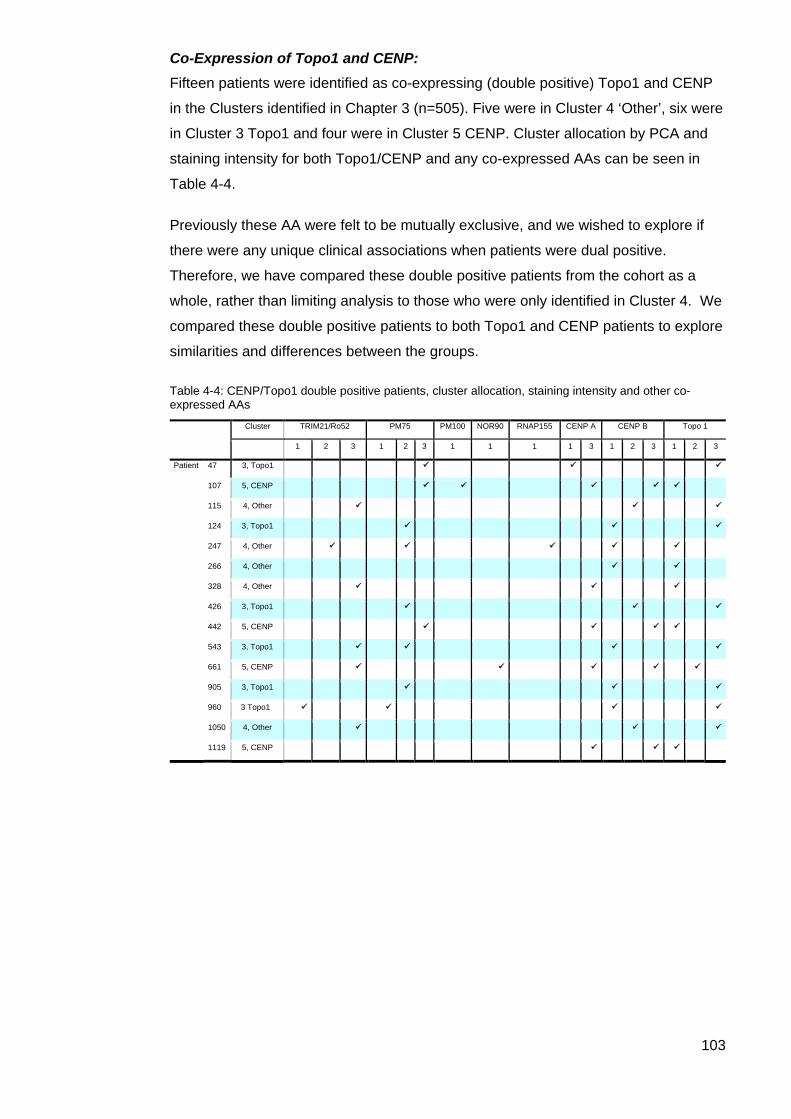
103
Co-Expression of Topo1 and CENP: Fifteen patients were identified as co-expressing (double positive) Topo1 and CENP
in the Clusters identified in Chapter 3 (n=505). Five were in Cluster 4 ‘Other’, six were
in Cluster 3 Topo1 and four were in Cluster 5 CENP. Cluster allocation by PCA and
staining intensity for both Topo1/CENP and any co-expressed AAs can be seen in
Table 4-4.
Previously these AA were felt to be mutually exclusive, and we wished to explore if
there were any unique clinical associations when patients were dual positive.
Therefore, we have compared these double positive patients from the cohort as a
whole, rather than limiting analysis to those who were only identified in Cluster 4. We
compared these double positive patients to both Topo1 and CENP patients to explore
similarities and differences between the groups.
Table 4-4: CENP/Topo1 double positive patients, cluster allocation, staining intensity and other co-expressed AAs
Cluster TRIM21/Ro52 PM75 PM100 NOR90 RNAP155 CENP A CENP B Topo 1
1 2 3 1 2 3 1 1 1 1 3 1 2 3 1 2 3
Patient 47 3, Topo1
107 5, CENP
115 4, Other
124 3, Topo1
247 4, Other
266 4, Other
328 4, Other
426 3, Topo1
442 5, CENP
543 3, Topo1
661 5, CENP
905 3, Topo1
960 3 Topo1
1050 4, Other
1119 5, CENP
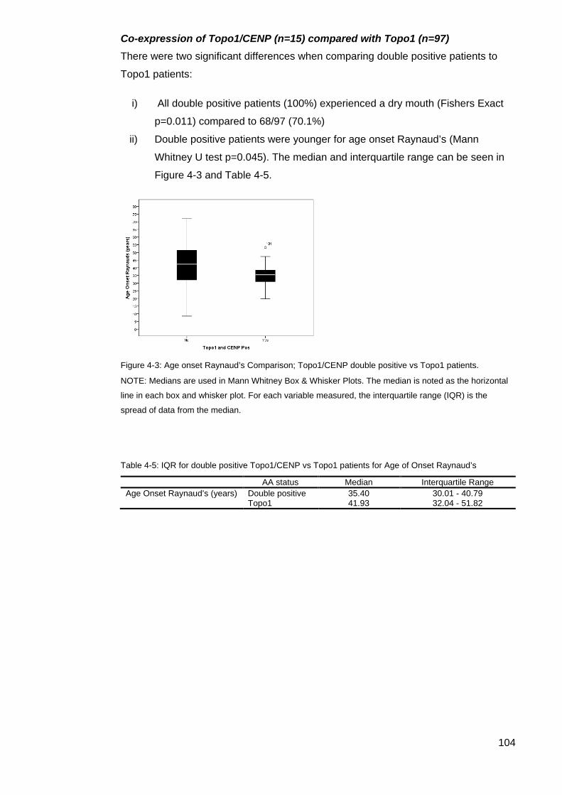
104
Co-expression of Topo1/CENP (n=15) compared with Topo1 (n=97) There were two significant differences when comparing double positive patients to
Topo1 patients:
i) All double positive patients (100%) experienced a dry mouth (Fishers Exact
p=0.011) compared to 68/97 (70.1%)
ii) Double positive patients were younger for age onset Raynaud’s (Mann
Whitney U test p=0.045). The median and interquartile range can be seen in
Figure 4-3 and Table 4-5.
Figure 4-3: Age onset Raynaud’s Comparison; Topo1/CENP double positive vs Topo1 patients.
NOTE: Medians are used in Mann Whitney Box & Whisker Plots. The median is noted as the horizontal
line in each box and whisker plot. For each variable measured, the interquartile range (IQR) is the
spread of data from the median.
Table 4-5: IQR for double positive Topo1/CENP vs Topo1 patients for Age of Onset Raynaud’s
AA status Median Interquartile Range Age Onset Raynaud’s (years) Double positive 35.40 30.01 - 40.79 Topo1 41.93 32.04 - 51.82
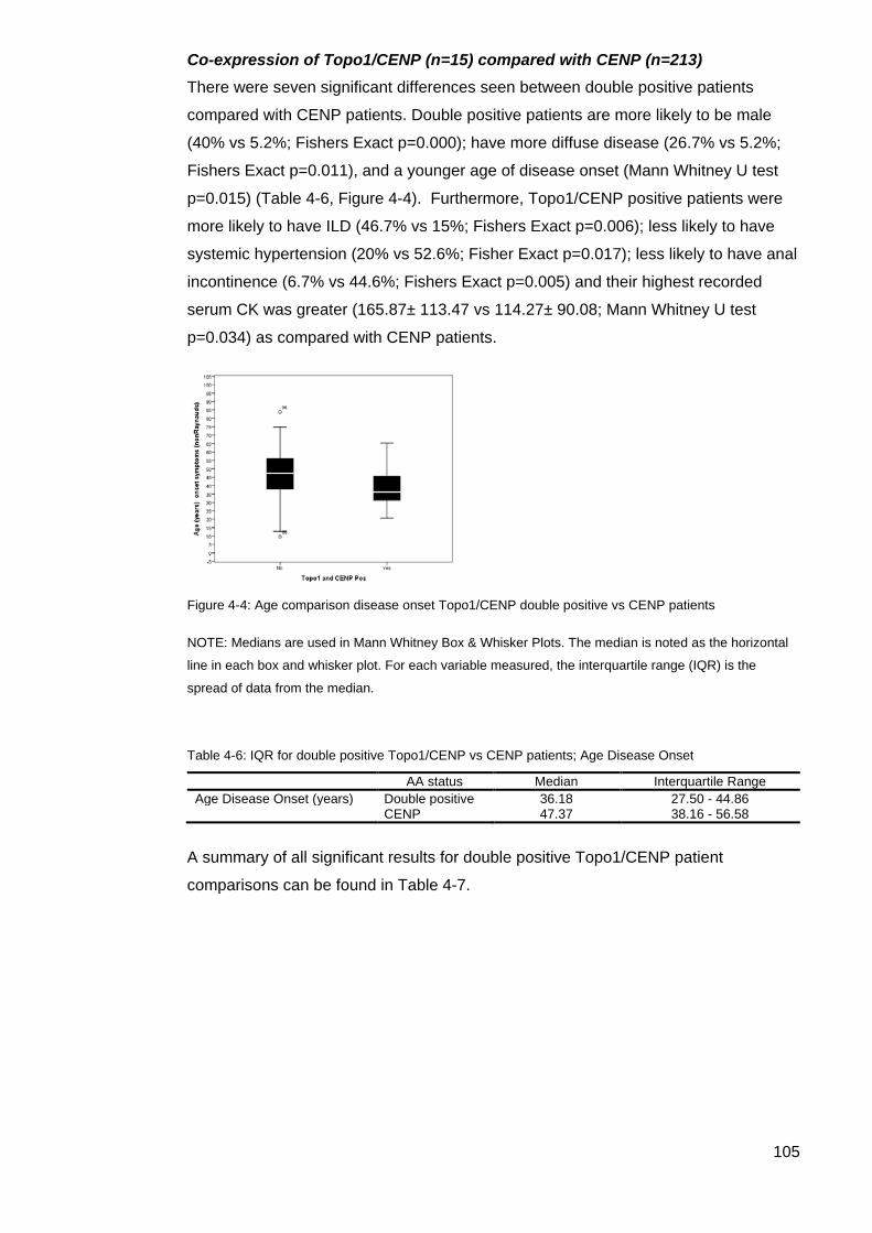
105
Co-expression of Topo1/CENP (n=15) compared with CENP (n=213) There were seven significant differences seen between double positive patients
compared with CENP patients. Double positive patients are more likely to be male
(40% vs 5.2%; Fishers Exact p=0.000); have more diffuse disease (26.7% vs 5.2%;
Fishers Exact p=0.011), and a younger age of disease onset (Mann Whitney U test
p=0.015) (Table 4-6, Figure 4-4). Furthermore, Topo1/CENP positive patients were
more likely to have ILD (46.7% vs 15%; Fishers Exact p=0.006); less likely to have
systemic hypertension (20% vs 52.6%; Fisher Exact p=0.017); less likely to have anal
incontinence (6.7% vs 44.6%; Fishers Exact p=0.005) and their highest recorded
serum CK was greater (165.87± 113.47 vs 114.27± 90.08; Mann Whitney U test
p=0.034) as compared with CENP patients.
Figure 4-4: Age comparison disease onset Topo1/CENP double positive vs CENP patients
NOTE: Medians are used in Mann Whitney Box & Whisker Plots. The median is noted as the horizontal
line in each box and whisker plot. For each variable measured, the interquartile range (IQR) is the
spread of data from the median.
Table 4-6: IQR for double positive Topo1/CENP vs CENP patients; Age Disease Onset
AA status Median Interquartile Range Age Disease Onset (years) Double positive 36.18 27.50 - 44.86 CENP 47.37 38.16 - 56.58
A summary of all significant results for double positive Topo1/CENP patient
comparisons can be found in Table 4-7.
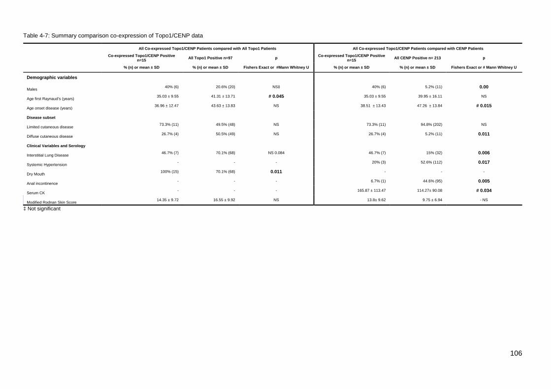
106
Table 4-7: Summary comparison co-expression of Topo1/CENP data
All Co-expressed Topo1/CENP Patients compared with All Topo1 Patients All Co-expressed Topo1/CENP Patients compared with CENP Patients
Co-expressed Topo1/CENP Positive n=15 All Topo1 Positive n=97 p Co-expressed Topo1/CENP Positive
n=15 All CENP Positive n= 213 p
% (n) or mean ± SD % (n) or mean ± SD Fishers Exact or #Mann Whitney U % (n) or mean ± SD % (n) or mean ± SD Fishers Exact or # Mann Whitney U
Demographic variables
Males 40% (6) 20.6% (20) NS‡ 40% (6) 5.2% (11) 0.00
Age first Raynaud’s (years) 35.03 ± 9.55 41.31 ± 13.71 # 0.045 35.03 ± 9.55 39.95 ± 16.11 NS
Age onset disease (years) 36.96 ± 12.47 43.63 ± 13.83 NS 38.51 ± 13.43 47.26 ± 13.84 # 0.015
Disease subset
Limited cutaneous disease 73.3% (11) 49.5% (48) NS 73.3% (11) 94.8% (202) NS
Diffuse cutaneous disease 26.7% (4) 50.5% (49) NS 26.7% (4) 5.2% (11) 0.011
Clinical Variables and Serology
Interstitial Lung Disease 46.7% (7) 70.1% (68) NS 0.084 46.7% (7) 15% (32) 0.006
Systemic Hypertension - - - 20% (3) 52.6% (112) 0.017
Dry Mouth 100% (15) 70.1% (68) 0.011 - - -
Anal incontinence - - - 6.7% (1) 44.6% (95) 0.005
Serum CK - - - 165.87 ± 113.47 114.27± 90.08 # 0.034
Modified Rodnan Skin Score 14.35 ± 9.72 16.55 ± 9.92 NS 13.8± 9.62 9.75 ± 6.94 - NS
‡ Not significant
107
TRIM21/Ro52 Fourteen Cluster 4 patients were monospecific for TRIM21/Ro52 and this was the
most frequently expressed AA either monospecifically or in multiple combinations in
this Cluster. Cluster 4 patients monospecific for TRIM21/Ro52 were compared to the
ASCS cohort excluding patients that co-expressed this AA.
Following this analysis, monospecific TRIM21/Ro52 was then compared to other
patients in Cluster 4, again excluding patients with TRIM21/Ro52 co-expression.
Whole cohort comparison, excluding TRIM21/Ro52 positive patients (n=334) compared with Cluster 4 TRIM21/Ro52 (n=14) There were three significant differences between monospecific TRIM21/Ro52 patients
in Cluster 4 and those negative for TRIM21/Ro52 in the remainder of the cohort.
Monospecific patients experienced first Raynaud’s later (p= 0.044) than their negative
counterparts. They also experienced a higher erythrocyte sedimentation rate (ESR)
(Mann Whitney U p= 0.024) and a higher systolic pulmonary arterial pressure (sPAP)
(mmHG) (43.92+/-10.77 vs 37.48+/-14.32; Mann Whitney U p=0.022)), In this
analysis of monospecific TRIM21/Ro52 patients, there was no increase in PAH
compared with the remainder of the cohort
These results and a comparison of monospecific TRIM21/Ro52 positive and negative
patients within Cluster 4 are summarised in Table 4.7.
Cluster 4 comparison (n=105) with Cluster 4 TRIM21/Ro52 patients (n=14) TRIM21/Ro52 monospecific patients first experienced Raynaud’s at an older age
(p=0.042) than the remainder patients in Cluster 4.
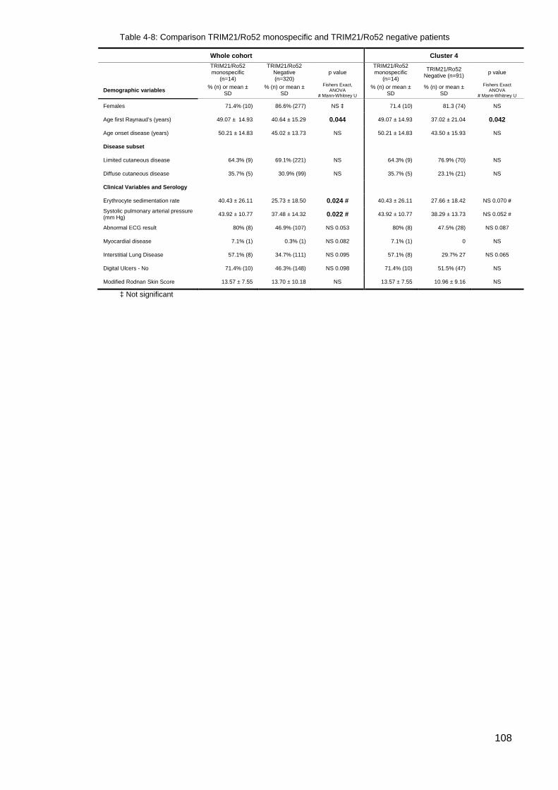
108
Table 4-8: Comparison TRIM21/Ro52 monospecific and TRIM21/Ro52 negative patients
Whole cohort Cluster 4
TRIM21/Ro52 monospecific
(n=14)
TRIM21/Ro52 Negative (n=320)
p value TRIM21/Ro52 monospecific
(n=14)
TRIM21/Ro52 Negative (n=91) p value
Demographic variables % (n) or mean ± SD
% (n) or mean ± SD
Fishers Exact, ANOVA
# Mann-Whitney U
% (n) or mean ± SD
% (n) or mean ± SD
Fishers Exact ANOVA
# Mann-Whitney U
Females 71.4% (10) 86.6% (277) NS ‡ 71.4 (10) 81.3 (74) NS
Age first Raynaud’s (years) 49.07 ± 14.93 40.64 ± 15.29 0.044 49.07 ± 14.93 37.02 ± 21.04 0.042
Age onset disease (years) 50.21 ± 14.83 45.02 ± 13.73 NS 50.21 ± 14.83 43.50 ± 15.93 NS
Disease subset
Limited cutaneous disease 64.3% (9) 69.1% (221) NS 64.3% (9) 76.9% (70) NS
Diffuse cutaneous disease 35.7% (5) 30.9% (99) NS 35.7% (5) 23.1% (21) NS
Clinical Variables and Serology
Erythrocyte sedimentation rate 40.43 ± 26.11 25.73 ± 18.50 0.024 # 40.43 ± 26.11 27.66 ± 18.42 NS 0.070 #
Systolic pulmonary arterial pressure (mm Hg) 43.92 ± 10.77 37.48 ± 14.32 0.022 # 43.92 ± 10.77 38.29 ± 13.73 NS 0.052 #
Abnormal ECG result 80% (8) 46.9% (107) NS 0.053 80% (8) 47.5% (28) NS 0.087
Myocardial disease 7.1% (1) 0.3% (1) NS 0.082 7.1% (1) 0 NS
Interstitial Lung Disease 57.1% (8) 34.7% (111) NS 0.095 57.1% (8) 29.7% 27 NS 0.065
Digital Ulcers - No 71.4% (10) 46.3% (148) NS 0.098 71.4% (10) 51.5% (47) NS
Modified Rodnan Skin Score 13.57 ± 7.55 13.70 ± 10.18 NS 13.57 ± 7.55 10.96 ± 9.16 NS
‡ Not significant
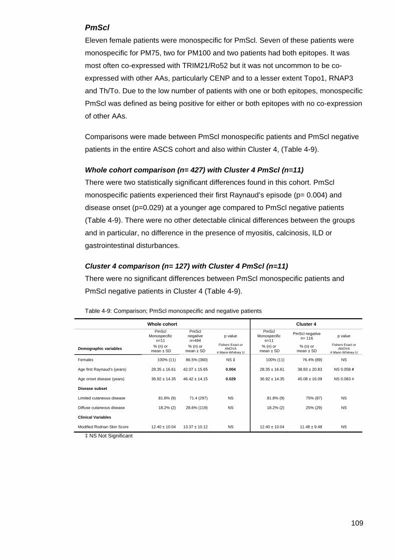
109
PmScl Eleven female patients were monospecific for PmScl. Seven of these patients were
monospecific for PM75, two for PM100 and two patients had both epitopes. It was
most often co-expressed with TRIM21/Ro52 but it was not uncommon to be co-
expressed with other AAs, particularly CENP and to a lesser extent Topo1, RNAP3
and Th/To. Due to the low number of patients with one or both epitopes, monospecific
PmScl was defined as being positive for either or both epitopes with no co-expression
of other AAs.
Comparisons were made between PmScl monospecific patients and PmScl negative
patients in the entire ASCS cohort and also within Cluster 4, (Table 4-9).
Whole cohort comparison (n= 427) with Cluster 4 PmScl (n=11) There were two statistically significant differences found in this cohort. PmScl
monospecific patients experienced their first Raynaud’s episode (p= 0.004) and
disease onset (p=0.029) at a younger age compared to PmScl negative patients
(Table 4-9). There were no other detectable clinical differences between the groups
and in particular, no difference in the presence of myositis, calcinosis, ILD or
gastrointestinal disturbances.
Cluster 4 comparison (n= 127) with Cluster 4 PmScl (n=11) There were no significant differences between PmScl monospecific patients and
PmScl negative patients in Cluster 4 (Table 4-9).
Table 4-9: Comparison; PmScl monospecific and negative patients
Whole cohort Cluster 4
PmScl Monospecific
n=11
PmScl negative n=494
p value PmScl
Monospecific n=11
PmScl negative n= 116 p value
Demographic variables % (n) or mean ± SD
% (n) or mean ± SD
Fishers Exact or ANOVA
# Mann-Whitney U
% (n) or mean ± SD
% (n) or mean ± SD
Fishers Exact or ANOVA
# Mann Whitney U
Females 100% (11) 86.5% (360) NS ‡ 100% (11) 76.4% (89) NS
Age first Raynaud’s (years) 28.35 ± 16.61 42.07 ± 15.65 0.004 28.35 ± 16.61 38.83 ± 20.83 NS 0.058 #
Age onset disease (years) 36.92 ± 14.35 46.42 ± 14.15 0.029 36.92 ± 14.35 45.08 ± 16.09 NS 0.083 #
Disease subset
Limited cutaneous disease 81.8% (9) 71.4 (297) NS 81.8% (9) 75% (87) NS
Diffuse cutaneous disease 18.2% (2) 28.6% (119) NS 18.2% (2) 25% (29) NS
Clinical Variables
Modified Rodnan Skin Score 12.40 ± 10.04 13.37 ± 10.12 NS 12.40 ± 10.04 11.48 ± 9.48 NS
‡ NS Not Significant
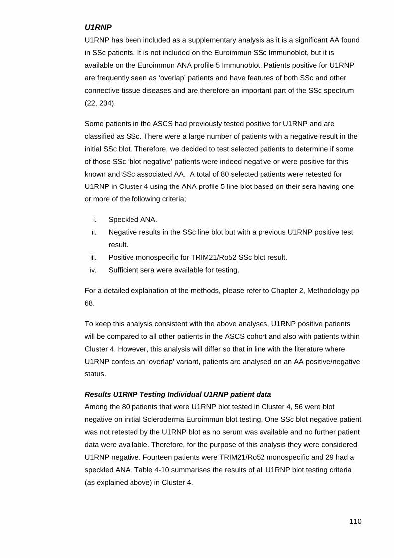
110
U1RNP U1RNP has been included as a supplementary analysis as it is a significant AA found
in SSc patients. It is not included on the Euroimmun SSc Immunoblot, but it is
available on the Euroimmun ANA profile 5 Immunoblot. Patients positive for U1RNP
are frequently seen as ‘overlap’ patients and have features of both SSc and other
connective tissue diseases and are therefore an important part of the SSc spectrum
(22, 234).
Some patients in the ASCS had previously tested positive for U1RNP and are
classified as SSc. There were a large number of patients with a negative result in the
initial SSc blot. Therefore, we decided to test selected patients to determine if some
of those SSc ‘blot negative’ patients were indeed negative or were positive for this
known and SSc associated AA. A total of 80 selected patients were retested for
U1RNP in Cluster 4 using the ANA profile 5 line blot based on their sera having one
or more of the following criteria;
i. Speckled ANA.
ii. Negative results in the SSc line blot but with a previous U1RNP positive test
result.
iii. Positive monospecific for TRIM21/Ro52 SSc blot result.
iv. Sufficient sera were available for testing.
For a detailed explanation of the methods, please refer to Chapter 2, Methodology pp
68.
To keep this analysis consistent with the above analyses, U1RNP positive patients
will be compared to all other patients in the ASCS cohort and also with patients within
Cluster 4. However, this analysis will differ so that in line with the literature where
U1RNP confers an ‘overlap’ variant, patients are analysed on an AA positive/negative
status.
Results U1RNP Testing Individual U1RNP patient data Among the 80 patients that were U1RNP blot tested in Cluster 4, 56 were blot
negative on initial Scleroderma Euroimmun blot testing. One SSc blot negative patient
was not retested by the U1RNP blot as no serum was available and no further patient
data were available. Therefore, for the purpose of this analysis they were considered
U1RNP negative. Fourteen patients were TRIM21/Ro52 monospecific and 29 had a
speckled ANA. Table 4-10 summarises the results of all U1RNP blot testing criteria
(as explained above) in Cluster 4.
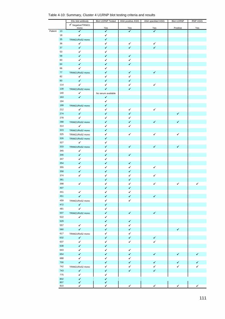
111
Table 4-10: Summary, Cluster 4 U1RNP blot testing criteria and results
SSc blot antibody Blot U1RNP Tested ANA positive ASIG ANA speckled ASIG Blot U1RNP RNP ASIG
Negative/TRIM21 mono Yes Yes Yes Positive Yes
Patient 13 18 35 TRIM21/Ro52 mono 36 37 53 58 60 64 66 77 TRIM21/Ro52 mono 82 83 114 139 TRIM21/Ro52 mono 140 No serum available 163 194 199 TRIM21/Ro52 mono 212 274 278 288 TRIM21/Ro52 mono 314 323 TRIM21/Ro52 mono 325 TRIM21/Ro52 mono 326 TRIM21/Ro52 mono 327 333 TRIM21/Ro52 mono 345 346 347 354 355 358 374 381 398 407 441 451
459 TRIM21/Ro52 mono 472 481 507 TRIM21/Ro52 mono 522 529 557 560 617 TRIM21/Ro52 mono 632 637 638 643 654 688 700 742 TRIM21/Ro52 mono 743 775 802
807 810
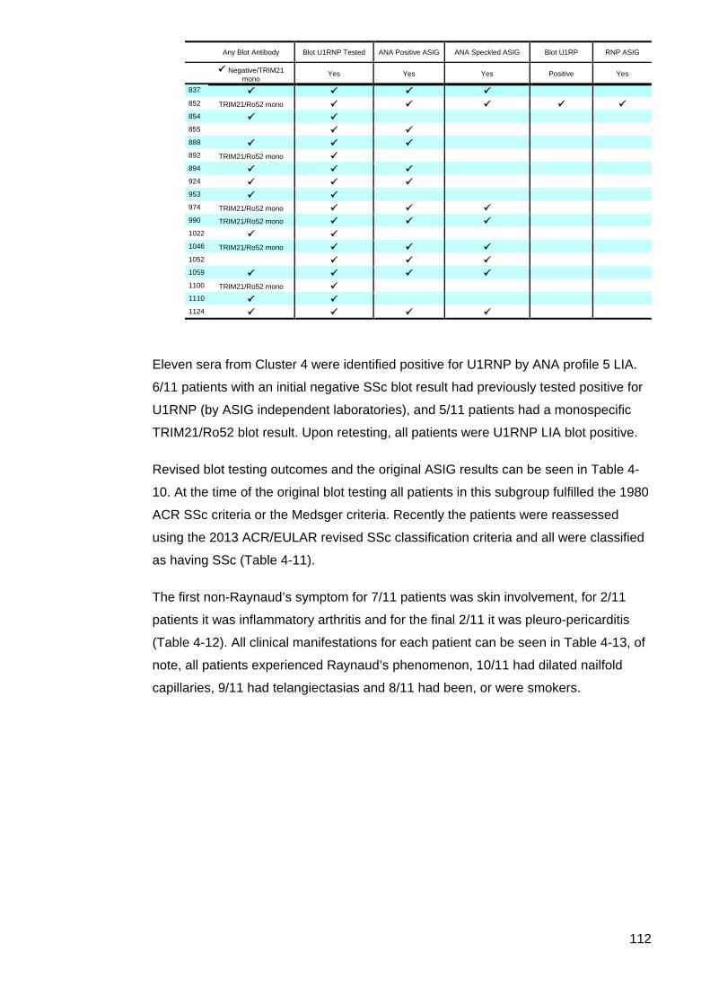
112
Any Blot Antibody Blot U1RNP Tested ANA Positive ASIG ANA Speckled ASIG Blot U1RP RNP ASIG
Negative/TRIM21 mono
Yes Yes Yes Positive Yes
837 852 TRIM21/Ro52 mono 854 855 888 892 TRIM21/Ro52 mono 894 924 953 974 TRIM21/Ro52 mono 990 TRIM21/Ro52 mono 1022 1046 TRIM21/Ro52 mono 1052 1059 1100 TRIM21/Ro52 mono 1110 1124
Eleven sera from Cluster 4 were identified positive for U1RNP by ANA profile 5 LIA.
6/11 patients with an initial negative SSc blot result had previously tested positive for
U1RNP (by ASIG independent laboratories), and 5/11 patients had a monospecific
TRIM21/Ro52 blot result. Upon retesting, all patients were U1RNP LIA blot positive.
Revised blot testing outcomes and the original ASIG results can be seen in Table 4-
10. At the time of the original blot testing all patients in this subgroup fulfilled the 1980
ACR SSc criteria or the Medsger criteria. Recently the patients were reassessed
using the 2013 ACR/EULAR revised SSc classification criteria and all were classified
as having SSc (Table 4-11).
The first non-Raynaud’s symptom for 7/11 patients was skin involvement, for 2/11
patients it was inflammatory arthritis and for the final 2/11 it was pleuro-pericarditis
(Table 4-12). All clinical manifestations for each patient can be seen in Table 4-13, of
note, all patients experienced Raynaud’s phenomenon, 10/11 had dilated nailfold
capillaries, 9/11 had telangiectasias and 8/11 had been, or were smokers.
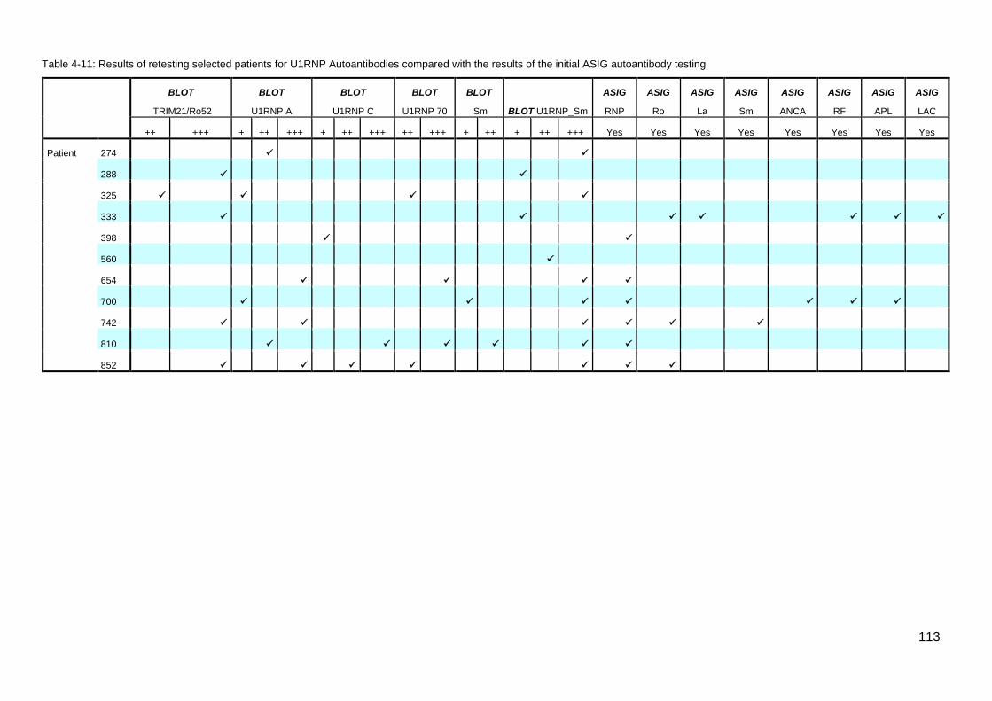
113
Table 4-11: Results of retesting selected patients for U1RNP Autoantibodies compared with the results of the initial ASIG autoantibody testing
BLOT
TRIM21/Ro52
BLOT
U1RNP A
BLOT
U1RNP C
BLOT
U1RNP 70
BLOT
Sm BLOT U1RNP_Sm
ASIG
RNP
ASIG
Ro
ASIG
La
ASIG
Sm
ASIG
ANCA
ASIG
RF
ASIG
APL
ASIG
LAC
++ +++ + ++ +++ + ++ +++ ++ +++ + ++ + ++ +++ Yes Yes Yes Yes Yes Yes Yes Yes
Patient 274
288
325
333
398
560
654
700
742
810
852
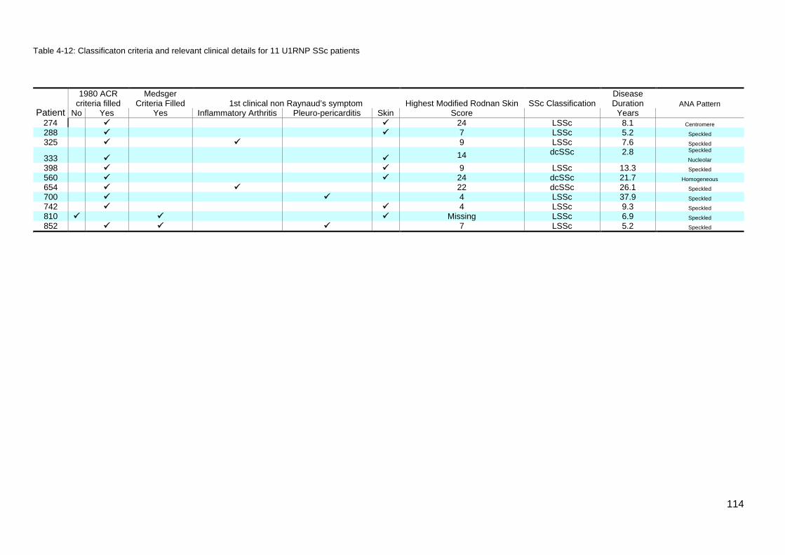
114
Table 4-12: Classificaton criteria and relevant clinical details for 11 U1RNP SSc patients
Patient
1980 ACR criteria filled
Medsger Criteria Filled 1st clinical non Raynaud’s symptom Highest Modified Rodnan Skin SSc Classification
Disease Duration ANA Pattern
No Yes Yes Inflammatory Arthritis Pleuro-pericarditis Skin Score Years
274 24 LSSc 8.1 Centromere 288 7 LSSc 5.2 Speckled 325 9 LSSc 7.6 Speckled
333 14 dcSSc 2.8 Speckled
Nucleolar 398 9 LSSc 13.3 Speckled 560 24 dcSSc 21.7 Homogeneous 654 22 dcSSc 26.1 Speckled 700 4 LSSc 37.9 Speckled 742 4 LSSc 9.3 Speckled 810 Missing LSSc 6.9 Speckled 852 7 LSSc 5.2 Speckled
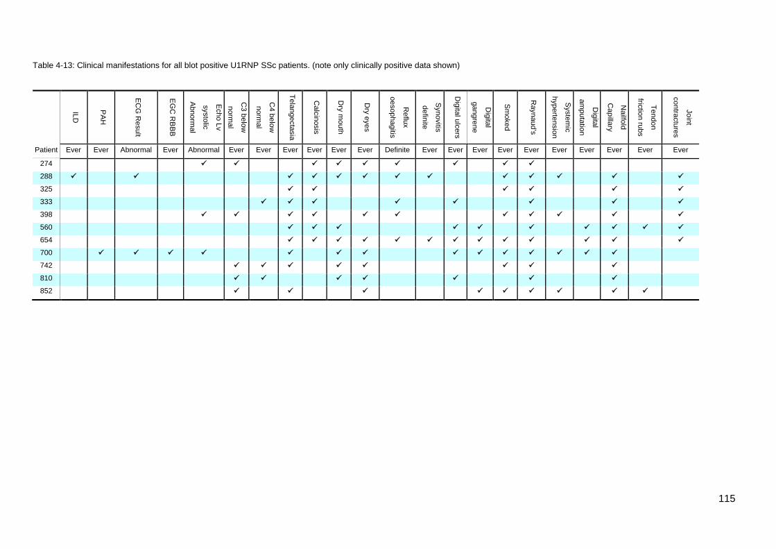
115
Table 4-13: Clinical manifestations for all blot positive U1RNP SSc patients. (note only clinically positive data shown)
Patient
ILD
PAH
ECG
Result
EGC
RBBB
Echo Lv
systolic
Abnormal
C3 below
normal
C4 below
normal
Telangectasia
Calcinosis
Dry m
outh
Dry eyes
Reflux
oesophagitis
Synovitis
definite
Digital ulcers
Digital
gangrene
Smoked
Raynaud’s
Systemic
hypertension
Digital
amputation
Nailfold
Capillary
Tendon
friction rubs
Joint
contractures
Ever Ever Abnormal Ever Abnormal Ever Ever Ever Ever Ever Ever Definite Ever Ever Ever Ever Ever Ever Ever Ever Ever Ever
274
288 325 333 398 560 654 700
742
810
852
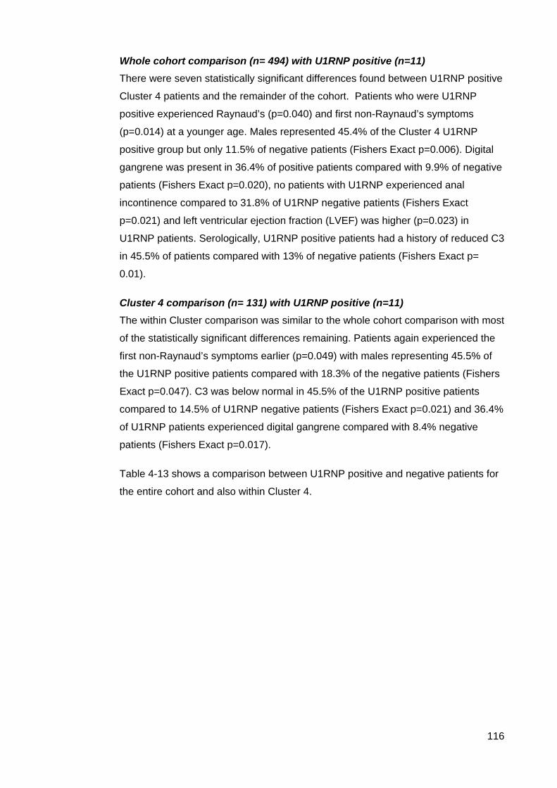
116
Whole cohort comparison (n= 494) with U1RNP positive (n=11) There were seven statistically significant differences found between U1RNP positive
Cluster 4 patients and the remainder of the cohort. Patients who were U1RNP
positive experienced Raynaud’s (p=0.040) and first non-Raynaud’s symptoms
(p=0.014) at a younger age. Males represented 45.4% of the Cluster 4 U1RNP
positive group but only 11.5% of negative patients (Fishers Exact p=0.006). Digital
gangrene was present in 36.4% of positive patients compared with 9.9% of negative
patients (Fishers Exact p=0.020), no patients with U1RNP experienced anal
incontinence compared to 31.8% of U1RNP negative patients (Fishers Exact
p=0.021) and left ventricular ejection fraction (LVEF) was higher (p=0.023) in
U1RNP patients. Serologically, U1RNP positive patients had a history of reduced C3
in 45.5% of patients compared with 13% of negative patients (Fishers Exact p=
0.01).
Cluster 4 comparison (n= 131) with U1RNP positive (n=11) The within Cluster comparison was similar to the whole cohort comparison with most
of the statistically significant differences remaining. Patients again experienced the
first non-Raynaud’s symptoms earlier (p=0.049) with males representing 45.5% of
the U1RNP positive patients compared with 18.3% of the negative patients (Fishers
Exact p=0.047). C3 was below normal in 45.5% of the U1RNP positive patients
compared to 14.5% of U1RNP negative patients (Fishers Exact p=0.021) and 36.4%
of U1RNP patients experienced digital gangrene compared with 8.4% negative
patients (Fishers Exact p=0.017).
Table 4-13 shows a comparison between U1RNP positive and negative patients for
the entire cohort and also within Cluster 4.
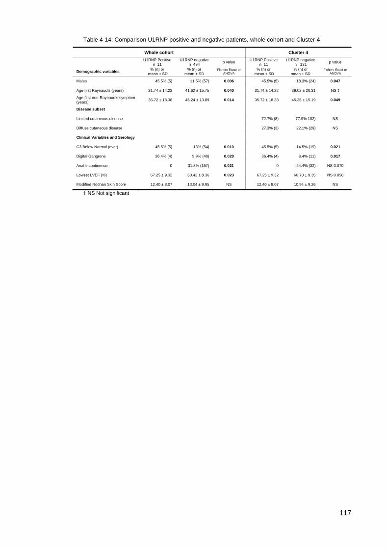
117
Table 4-14: Comparison U1RNP positive and negative patients, whole cohort and Cluster 4
Whole cohort Cluster 4
U1RNP Positive n=11
U1RNP negative n=494 p value U1RNP Positive
n=11 U1RNP negative
n= 131 p value
Demographic variables % (n) or mean ± SD
% (n) or mean ± SD
Fishers Exact or ANOVA
% (n) or mean ± SD
% (n) or mean ± SD
Fishers Exact or ANOVA
Males 45.5% (5) 11.5% (57) 0.006 45.5% (5) 18.3% (24) 0.047
Age first Raynaud’s (years) 31.74 ± 14.22 41.62 ± 15.75 0.040 31.74 ± 14.22 39.02 ± 20.31 NS ‡
Age first non-Raynaud’s symptom (years) 35.72 ± 18.38 46.24 ± 13.89 0.014 35.72 ± 18.38 45.36 ± 15.19 0.049
Disease subset
Limited cutaneous disease 72.7% (8) 77.9% 102) NS
Diffuse cutaneous disease 27.3% (3) 22.1% (29) NS
Clinical Variables and Serology
C3 Below Normal (ever) 45.5% (5) 13% (54) 0.010 45.5% (5) 14.5% (19) 0.021
Digital Gangrene 36.4% (4) 9.9% (40) 0.020 36.4% (4) 8.4% (11) 0.017
Anal Incontinence 0 31.8% (157) 0.021 0 24.4% (32) NS 0.070
Lowest LVEF (%) 67.25 ± 9.32 60.42 ± 8.36 0.023 67.25 ± 9.32 60.70 ± 9.35 NS 0.058
Modified Rodnan Skin Score 12.40 ± 8.07 13.04 ± 9.95 NS 12.40 ± 8.07 10.94 ± 9.26 NS
‡ NS Not significant
118
U1RNP SSc patients compared with U1RNP Mixed Connective Tissue Disease (MCTD) patients. The Australian Scleroderma Cohort Study database includes 21 patients classified
as MCTD where sera were available and who had undergone Euroimmun testing.
These patients’ sera are used in this supplementary, univariate comparative
analysis to compare with the 11 patients that are U1RNP positive and classified as
SSc.
Both subgroups were statistically comparable for all clinical variables with the
exception of two significant differences. MCTD patients had a lower modified
Rodnan Skin Score (7.81± 6.67 vs 16.00 ± 12.94 Mann Whitney U exact p=0.047)
when compared with SSc patients (Figure 4-8).
Figure 4-8: MRSS comparison U1RNP positive SSc
(Median 11.50, IQR 2.5 – 20.5) and MCTD (Median 5, IQR 2.5 – 7.5) patients.
NOTE: Medians are used in Mann Whitney Box & Whisker Plots. The median is noted as the horizontal
line in each box and whisker plot. For each variable measured, the interquartile range (IQR) is the
spread of data from the median.
No U1RNP positive MCTD patients had digital gangrene whereas 4/11 (36.4%) of
U1RNP positive SSc patients experienced digital gangrene (Fishers Exact p=0.009).
However, one of these patients also had anti-phospholipid syndrome which may
have biased results.
119
Discussion
Our initial work demonstrated that in the majority of cases where a primary SSc
associated AA is present, that AA can be used to guide sub classification of disease,
independent of clinical findings. However, SSc patients who lack these primary
disease specific AAs or who express them in a mixed or non-dominant fashion may
present a significant diagnostic challenge to clinicians. Our initial analysis confirmed
that these patients make up a substantial number of those seen in the ASCS
registry. Therefore, we were interested to evaluate this challenging group further to
determine if any AA could provide any further aid to clinical assessment in this
setting. The PCA in a hierarchical cluster analysis revealed that CENP B, Topo1,
PmScl, Th/To and TRIM21/Ro52 accounted for the majority of relationships in
Cluster 4. The remaining AAs; RNAP3, Ku, Fibrillarin and NOR90, were not present
in sufficient numbers to determine any statistically significant outcome. We
assessed those patients with CENP A/B and Topo1 expression, either singly or in
combination who were in Cluster 4; RNAP3 and where numbers permitted, and the
most common SSc associated AAs including TRIM21/Ro52 and Pm-Scl. In
addition, we undertook a sub-analysis on those patients in Cluster 4 who were
subsequently found to have U1RNP and we were able to compare these patients
with a separate cohort of well-characterised MCTD patients within the ASCS cohort.
In line with the results, this discussion will be grouped according to the AAs tested;
firstly the primary AAs; CENP, Topo1 and RNAP3 followed by TRIM21, PmScl and
U1RNP.
Cluster 4 CENP, Topo1 and RNAP3 Although infrequent, these three primary AAs were represented in Cluster 4;
however their presence was atypical, as demonstrated by single epitope expression
(in CENP, RNAP3), lower staining intensity scores, or by co-expression with one or
more AAs of equal or higher intensity staining. There were minimal differences
between the expression of each of these AAs in Cluster 4 when compared with their
expression in the cohort as a whole.
The increased frequency of male gender and oesophageal dysmotility observed in
both the CENP and Topo1 Cluster 4 groups (as compared with expression of these
AAs in the other clusters) is intriguing, but multinational studies are required to direct
further interpretation.
Two further differences were noted in Cluster 4 Topo1 positive patients compared
with Topo1 expression elsewhere. Those in Cluster 4 had reduced frequency of ILD

120
and increased PAH. Among those Cluster 4 Topo1 positive patients that had PAH,
co-expression included Fibrillarin, CENP B, Pm75, and TRIM21/Ro52 with the
former two AAs previously reported to be associated with PAH (19, 205).
Given the frequent co-occurrence of other AAs in these patients, it is likely that they
may have an overlap variant of SSc. While not reaching significance, in Cluster 4
CENP patients (35.7% vs All CENP 15.5%, p= 0.069), that had ILD, Topo1 and
TRIM21/Ro52 were co-expressed and again both of these AAs have been
associated with ILD in this and other cohorts (22, 76, 282).
Coexistence of Topo1 and CENP Topo1 and CENP have previously been thought to be mutually exclusive (129, 246,
324-327). However, with the advent of the newer diagnostic platforms, and the
ability to test for multiple autoantibodies, this paradigm is shifting (324).
Among our 505 patients, 15 tested positive for CENP and Topo1. Perhaps
surprisingly, these patients were dispersed through the Clusters identified in Chapter
3 and this no doubt reflects that in many cases one AA had the dominant staining
pattern. The LIA used to detect these AA has been well-validated and we feel these
findings reflect a true dual positive status rather than laboratory error. We used a
single operator, single platform approach and testing by independent laboratories
had excellent agreement (Topo1 κ=0.837 and CENP κ=0.887) with the results of the
LIA (295-297). Understandably, genetic background, the type of cohort (population
based or registry) and the choice of AA detection method will all have a bearing on
both autoantibodies and clinical associations.
Overall this study found that the Topo1/CENP ‘double positive’ group had more in
common with Topo1 patients than they did with CENP patients. On review, staining
intensity in these dual positive patients was evenly distributed, with 8/15 have
dominant Topo 1 AA expression and the remainder had dominant CENP A or B AA
expression. In further detail, the 15 patients that were double CENP/Topo1 positive
were compared with the 97 other Topo1 patients. There were two significant
differences; double positive patients experienced Raynaud’s younger and they all
had sicca symptoms. In all other regards they were statistically clinically comparable
with Topo1 patients.
In comparing single positive CENP patients with the 15 double positive patients,
more differences were found in the Topo1/CENP group. There were more males,
these patients had a younger age of disease onset, there was more diffuse disease,
ILD was increased and systemic hypertension was decreased, they had less anal
121
incontinence and a higher Serum CK.
The EUSTAR cohort (n=4687) has also recently explored the clinical characteristics
of ‘double positive’ patients and they found that although not statistically significant,
their double positive patients, when compared with single positive Topo1 (control)
patients, also experienced Raynaud’s at a slightly younger age. Overall, they
concluded that their double positive patients were not ‘truly clinically different’ from
their single positive Topo1 positive patients (324). Unlike our cohort, their double
positive sera was more likely to come from female patients and there was a trend for
their double positive patients to have more severe or prevalent involvement of the
musculoskeletal system and pulmonary fibrosis (324) as compared with CENP
positive patients.
Although our double positive patient cohort, like EUSTAR’s, had more in common
with single positive Topo1 patients, the finding of more sicca in our double positive
patients as compared with Topo 1 patients was interesting. A study by Avouac et al
found that ‘the principal cause of sicca syndrome in SSc appears to be glandular
fibrosis, rather than SS lymphocytic sialadenitis’, and was associated with CENP
and limited scleroderma (328). This may provide some evidence of the effect of
CENP on sicca in our cohort, although it may be that our double positives are more
fibrotic compared with CENP patients and the sicca is the result of fibrosis alone and
not the effect of CENP.
One last interesting finding was increased serum CK in our double positive patients
compared with other CENP positive patients. This may relate to the presence of
Topo1 as others have reported that among males, an increased serum CK was
associated with Topo1 and diffuse disease (329).
A German study by Dick et al (327), asked the question if Topo1 and ACA are
regarded as two separate clinical entities and patients express both AAs, do these
patients have both diseases independently? On balance, SSc is more easily
described as a spectrum of disease (45, 330) with many common clinical
manifestations. Autoantibodies and their clinical associations seem to be variations
of the same condition with the AA of the highest titre reflecting the dominant
phenotype in our work and that of others (22). Autoantibodies may influence or
reflect the clinical course but as yet they have not convincingly been shown to be
pathogenic. It may also be that some AAs are dominant over others as a result of
genetic or sex related reasons.
122
Due to the rarity of double positive patients, most studies lack statistical power and
so our results and those of others must be interpreted with caution. In addition,
different assays may have differing sensitivity and specificity profiles; IIF patterns
may be wrongly interpreted or the pattern of an AA with a higher titre may mask the
pattern of a different, weaker AA titre. Allowing for these technical concerns, the
advent of new technologies means detection of double positive patients is likely to
increase and multinational collaborations will allow for further investigation of this
interesting cohort.
In conclusion, the double Topo1/CENP positive patients in this cohort are clinically
more similar to Topo1 patients than CENP patients. It appears that they may have a
more fibrotic phenotype while the presence of CENP may have a modulatory effect
in regards to some clinical characteristics. Larger multinational studies will be
required to see if these characteristics are carried across different SSc cohorts.
123
TRIM21/Ro52 In our cohort, TRIM21/Ro52 patients experienced a higher ESR compared with
other patients and this may reflect a degree of overlap disease. In the German
Network for Systemic Scleroderma Registry, TRIM21/Ro52 was associated with a
higher ESR and Raynaud’s phenomenon (81), and although we did not find a
significant association with Raynaud’s, all patients who were positive for
TRIM21/Ro52 also experienced Raynaud’s.
A higher sPAP, was observed in this monospecific TRIM21/Ro52 cohort but PAH
was not increased and the difference in estimated systolic pulmonary artery
pressure between the groups is not great. Prospective monitoring of these
monospecific patients will help to determine whether the small differences in
pulmonary pressures equates to an increase in the frequency of PAH.
Patients with monospecific TRIM 21/Ro52 in Cluster 4 had an older age of disease
onset and this was also found to be the case in a multinational study (222) that
included not only the ASIG patients, but also patients from the Canadian
Scleroderma Research Group and the American Genetics versus Environment in
Scleroderma Outcome Study (246). TRIM21/Ro52 is the most commonly co-
expressed AA and its role in the ubiquitin process is an important one as well as its
ability to regulate downstream signalling of various pattern recognition receptors
such as the NF-κB, TGF-β and interferon response. Perhaps underlying senescence
changes in cellular elements (331, 332) are involved in the pathogenesis of this
abundantly expressed AA.
In the present study, a trend was observed between the presence of monospecific
TRIM21/Ro52 and ILD (p= 0.09), while in a multinational cohort of SSc patients
from Australia, Canada and the USA, ILD was the only clinical variable significantly
associated with monospecific anti-TRIM21/Ro52 antibodies (282). A Norwegian
study (using ANA Profile 5 Euroline Blot test kit; Euroimmun, Lubeck, Germany),
found that the odds ratio for the presence of TRIM21/Ro52 AAs in lung fibrosis was
4.4 (95% CI 1.8-10.3) in MCTD patients (242). In our initial PCA, no association was
found with ILD and TRIM21/Ro52, but this analysis was on a positive/negative basis
and monospecific analyses were not done. In more recent studies, TRIM21/Ro52
appears in other autoimmune conditions with other AAs such as anti-aminoacyl
transfer RNA synthetase antibodies and anti Jo-1, and these have been associated
with ILD (333). Although there are still many questions regarding autoantibodies, it
may be that different combinations of AAs are protective or act in synergy when
combined with other AAs and so the clinical manifestations may vary from individual
124
to individual notwithstanding genetic background.
PmScl PmScl monospecific patients experienced both Raynaud’s and onset of non
Raynaud’s symptoms at a younger age than PmScl negative patients. Otherwise,
they were statistically comparable to PmScl negative patients. Specifically, no
differences were found for myositis, calcinosis, digital ulcers or gastrointestinal
manifestations, although differences in all of these clinical manifestations have been
reported in the past, generally in the presence of other co-expressed AAs (81, 200,
227, 334) (222) (335) (52). A Canadian led, multinational study (which includes data
from this Australian cohort) investigating monospecific PmScl epitopes found that
although it wasn’t statistically significant, patients that were PM100 positive were
younger at disease onset compared to PM75 and other AA subsets (222). In this
study, there were insufficient numbers to interpret PmScl data when split into its
epitopes.
Patients who are monospecific for PmScl are rare, and the Canadian led
multinational study illustrates how trends observed in a single population can be
further explored in larger cohorts. In this case, the trends we observed in our
Australian cohort were reflected in the multinational study but there is little to
suggest that the presence of monospecific PmScl can be used to predict clinical
disease course.
Th/To In the initial PCA analysis of Cluster 4 patients, Th/To was one of the AA which,
along with TRIM21/Ro52, PmScl and the primary SSc AAs, had most influence in
generating variability in Cluster 4. However, due to the small number of Th/To
monospecific patients in this cluster (n=3) we were not able to explore any clinical
associations. In addition, while the LIA detects AA to the hPop1 antigen (Ag) in the
RNase MRP, recent work has suggested AAs to other more prevalent antigens in
this complex (such as Rpp25 and Rpp38) (184). It will be interesting to explore
clinical associations between the different Th/To Ag targets but as with other rarer
SSc AAs, these questions will need to be explored in larger multinational cohorts.
125
U1RNP - SSc Only 11 U1RNP positive patients were identified in Cluster 4 but 7/11 had high titre
AA present and in 4 cases it was the only AA detected. We wished to explore how
patients’ positive for U1RNP fit within the SSc spectrum of disease.
Given there were a large number of AA negative sera in Cluster 4 and that most
were ANA positive (many with speckled ANAs), we decided that it was important to
test this Cluster, if sera were available, for a common AA that was not included on
the SSc immunoblot. U1RNP patients are also included in the ASIG database and it
seemed that this was the most logical AA to test for given the patients had a
diagnosis of SSc and U1RNP is a known associated SSc AA. Had resources
permitted, the entire 505 patients would have been tested, but as Clusters 1-3 and 5
were highly representative of their dominant Cluster AA and Cluster 4 was more
heterogeneous, we decided it was more prudent to retest this Cluster for U1RNP.
Age demographics U1RNP positive patients (with any disease classification) have consistently been
reported in the literature as having a younger age of onset for both Raynaud’s and
the first non-Raynaud’s symptom (22, 27, 218) and our data is consistent with this
finding.
Male gender It is unclear why there are more males represented in this U1RNP AA subgroup and
Cluster 4 as compared with the SSc cohort as a whole, but perhaps it reflects that
males are more likely to have atypical AA associations in SSc
C3 below normal, ever Hypocomplementemia is a result of increased complement-component consumption
in inflammatory conditions and was proposed by Hudson in 2007 as a marker of
overlap disease in SSc (336). More recently, Esposito et al (337) found that
reduced C3 and C4 are associated with some features of increased SSc disease
activity (digital ulcers amongst others), in patients with overlap disease.
Hypocomplementaemia and overlap disease association was also reported by
Sturfelt and Truedsson who suggested that evidence of complement activation is
related to the dysfunction of regulatory proteins and this could contribute to
complement activation in the inflammatory process and subsequent vascular damage in SSc. They also stated that hypocomplementaemia is preferentially seen in
patients with overlap disease features, especially SLE (338). An increased history of
complement activation in the U1RNP positive Cluster 4 patients may reflect underlying
overlap disease features.
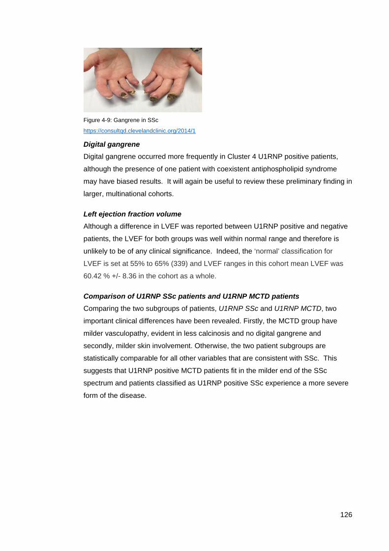
126
Figure 4-9: Gangrene in SSc
https://consultqd.clevelandclinic.org/2014/1
Digital gangrene Digital gangrene occurred more frequently in Cluster 4 U1RNP positive patients,
although the presence of one patient with coexistent antiphospholipid syndrome
may have biased results. It will again be useful to review these preliminary finding in
larger, multinational cohorts.
Left ejection fraction volume Although a difference in LVEF was reported between U1RNP positive and negative
patients, the LVEF for both groups was well within normal range and therefore is
unlikely to be of any clinical significance. Indeed, the ‘normal’ classification for
LVEF is set at 55% to 65% (339) and LVEF ranges in this cohort mean LVEF was
60.42 % +/- 8.36 in the cohort as a whole.
Comparison of U1RNP SSc patients and U1RNP MCTD patients Comparing the two subgroups of patients, U1RNP SSc and U1RNP MCTD, two
important clinical differences have been revealed. Firstly, the MCTD group have
milder vasculopathy, evident in less calcinosis and no digital gangrene and
secondly, milder skin involvement. Otherwise, the two patient subgroups are
statistically comparable for all other variables that are consistent with SSc. This
suggests that U1RNP positive MCTD patients fit in the milder end of the SSc
spectrum and patients classified as U1RNP positive SSc experience a more severe
form of the disease.
127
Conclusion
Patients in Cluster 4 have definite SSc according to Medsger or ACR/EULAR criteria
but lack, or have atypical expression of one of the three primary disease associated
AAs. In this cohort, dual positivity of CENP and Topo1 was more closely associated
with features of diffuse cutaneous rather than limited cutaneous features of SSc.
Sample sizes of monospecific AA expression were small but we were able to further
investigate the clinical associations of monospecific TRIM21/Ro52 and PmScl
expression and generally our findings or trends were consistent with those observed
in other cohorts. Monospecific TRIM21/Ro52 patients developed Raynaud’s
phenomenon later in life, recorded a higher annual ESR and had a higher sPAP
(although this was not associated with an increased frequency of overt PAH) while
PmScl monospecific patients had a younger age of onset compared with tested
ASCS cohort as a whole. These associations have also been assessed in larger
cohorts but there is no indication at this time that monospecific staining of these AA
in our cohort provided additional information that would change prognostication or
disease management.
In addition, we tested selected patients in Cluster 4 for U1RNP and compared this
cohort with a separate cohort in the ASCS with documented MCTD. We found that
while these cohorts were essentially similar, SSc U1RNP positive patients had more
severe skin involvement, vasculopathy and digital gangrene. Truly AA negative
patients also represent an interesting and significant subgroup and will be explored
further in the next chapter.
Cluster 4 patients represent a more heterogeneous SSc group and our explorative
analyses suggest that further subgroups exist within this cohort. Multinational
studies will be invaluable in characterising these patients further.
128
CHAPTER 5 AUTOANTIBODY NEGATIVE SYSTEMIC SCLEROSIS
Introduction
Autoantibodies (AA) are described as a central feature of SSc (13, 65, 340) and yet,
in most cohorts, regardless of geographic location, there are a small percentage (0.2
– 5.8%) diagnosed with SSc who are not only AA negative, they are also anti-
nuclear antibody (ANA) negative (44, 81, 122, 341) . There are a number of
questions that follow in regards to this unusual serological finding;
i. Do these patients truly have SSc? And if so,
ii. What are the clinical manifestations that have led to this diagnosis?
iii. How have they been classified and/or stratified within the SSc spectrum?
iv. What is the prognostic outlook for these patients?
v. Do these patients form a distinct clinical subgroup?
This chapter reports on the clinical associations and demographic data of the AA /
ANA negative patients in this cohort and also how these patients might be stratified
within the SSc spectrum.
Methods
For a detailed explanation of the patient population, definitions of clinical
characteristics, AA testing methodology and statistical analysis, see Chapter 2
Methods, pp. 65-77.
Results
Autoantibody Analysis Among the 505 SSc patients in the Australian Scleroderma Cohort Study (ASCS)
database who had sera available and were initially tested with the SSc immunoblot,
56 (11.08%) had negative results. These patients were identified for further testing
and 19 (3.76%) were subsequently found to remain autoantibody negative on testing
by ANA 5 line blot (which tests for U1RNP) and also ANA negative (by IIF as tested
by regional laboratories and recorded in the ASIG database). These remaining 19
patients were retested by IIF and CIEP for Topo1, U1RNP, Sm, Jo1, PmScl, Ro, La
and signal recognition protein. IIF pattern analysis did not indicate RNAP3 positivity.
Twelve (2.37%) patients were confirmed AA / ANA negative by all methods.
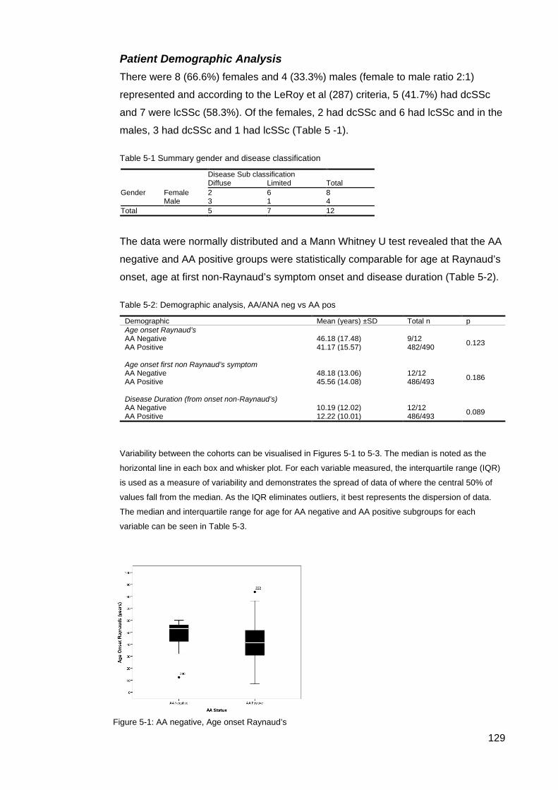
129
Patient Demographic Analysis There were 8 (66.6%) females and 4 (33.3%) males (female to male ratio 2:1)
represented and according to the LeRoy et al (287) criteria, 5 (41.7%) had dcSSc
and 7 were lcSSc (58.3%). Of the females, 2 had dcSSc and 6 had lcSSc and in the
males, 3 had dcSSc and 1 had lcSSc (Table 5 -1).
Table 5-1 Summary gender and disease classification
Disease Sub classification
Total Diffuse Limited Gender Female 2 6 8
Male 3 1 4 Total 5 7 12
The data were normally distributed and a Mann Whitney U test revealed that the AA
negative and AA positive groups were statistically comparable for age at Raynaud’s
onset, age at first non-Raynaud’s symptom onset and disease duration (Table 5-2).
Table 5-2: Demographic analysis, AA/ANA neg vs AA pos
Demographic Mean (years) ±SD Total n p Age onset Raynaud’s AA Negative 46.18 (17.48) 9/12 0.123 AA Positive 41.17 (15.57) 482/490 Age onset first non Raynaud’s symptom AA Negative 48.18 (13.06) 12/12 0.186 AA Positive 45.56 (14.08) 486/493 Disease Duration (from onset non-Raynaud’s) AA Negative 10.19 (12.02) 12/12 0.089 AA Positive 12.22 (10.01) 486/493
Variability between the cohorts can be visualised in Figures 5-1 to 5-3. The median is noted as the
horizontal line in each box and whisker plot. For each variable measured, the interquartile range (IQR)
is used as a measure of variability and demonstrates the spread of data of where the central 50% of
values fall from the median. As the IQR eliminates outliers, it best represents the dispersion of data.
The median and interquartile range for age for AA negative and AA positive subgroups for each
variable can be seen in Table 5-3.
Figure 5-1: AA negative, Age onset Raynaud’s
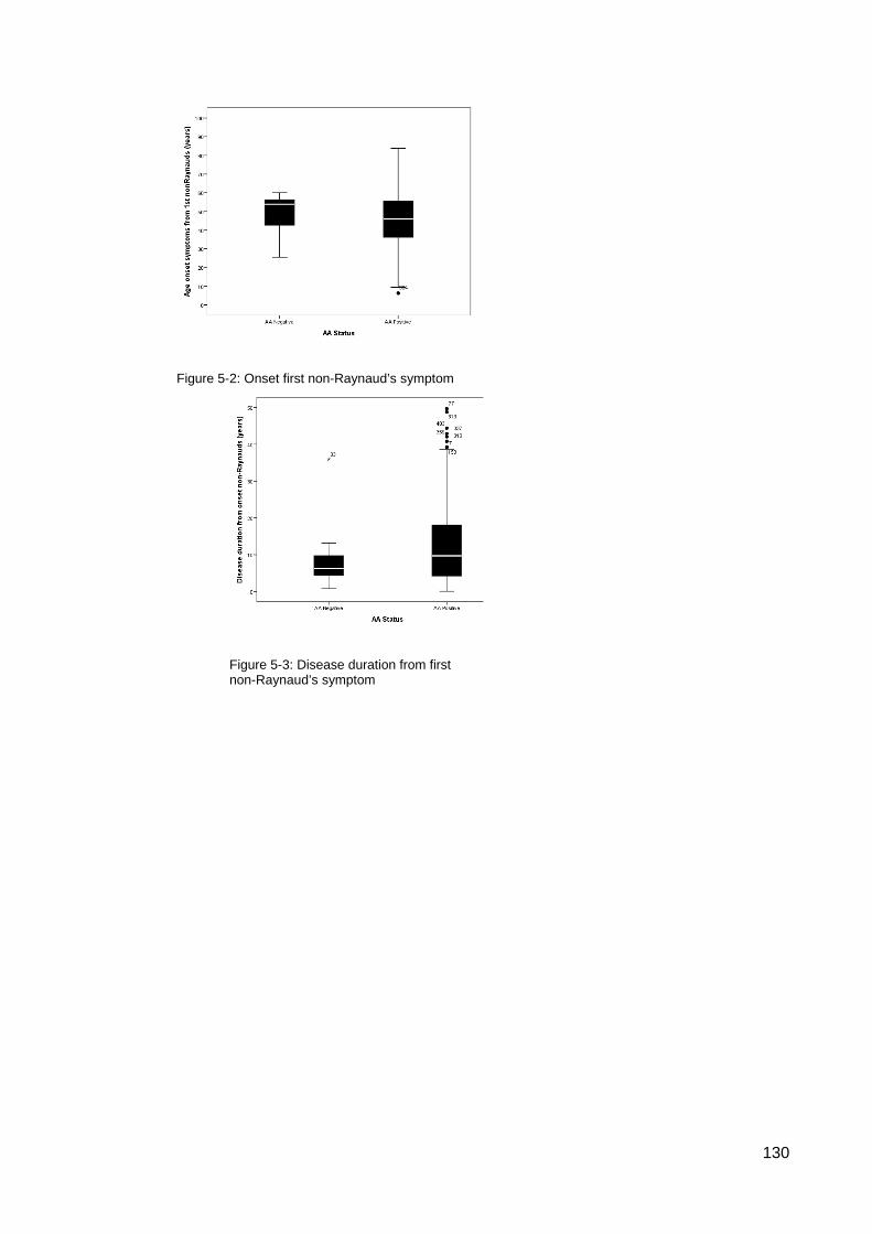
130
Figure 5-2: Onset first non-Raynaud’s symptom
Figure 5-3: Disease duration from first non-Raynaud’s symptom
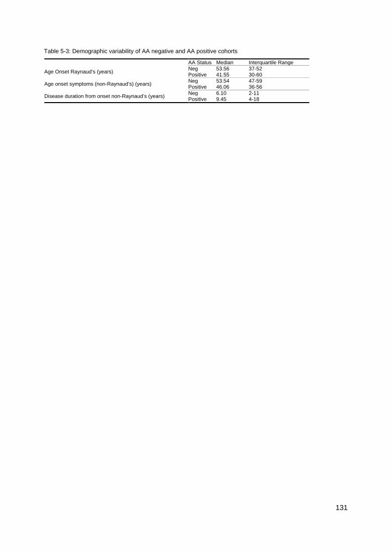
131
Table 5-3: Demographic variability of AA negative and AA positive cohorts
AA Status Median Interquartile Range
Age Onset Raynaud’s (years) Neg 53.56 37-52 Positive 41.55 30-60
Age onset symptoms (non-Raynaud’s) (years) Neg 53.54 47-59 Positive 46.06 36-56
Disease duration from onset non-Raynaud’s (years) Neg 6.10 2-11 Positive 9.45 4-18
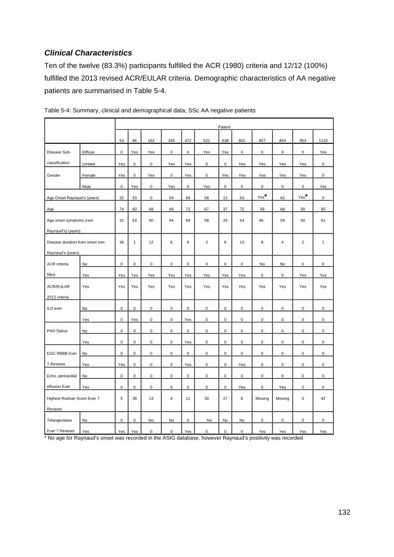
132
Clinical Characteristics Ten of the twelve (83.3%) participants fulfilled the ACR (1980) criteria and 12/12 (100%)
fulfilled the 2013 revised ACR/EULAR criteria. Demographic characteristics of AA negative
patients are summarised in Table 5-4.
Table 5-4: Summary, clinical and demographical data, SSc AA negative patients
Patient
53 66 163 345 472 522 638 802 807 854 953 1110
Disease Sub-
classification
Diffuse 0 Yes Yes 0 0 Yes Yes 0 0 0 0 Yes
Limited Yes 0 0 Yes Yes 0 0 Yes Yes Yes Yes 0
Gender Female Yes 0 Yes 0 Yes 0 Yes Yes Yes Yes Yes 0
Male 0 Yes 0 Yes 0 Yes 0 0 0 0 0 Yes
Age Onset Raynaud’s (years) 32 53 0 54 60 58 12 53 Yes* 62 Yes* 0
Age 74 60 68 66 72 67 37 72 59 68 56 65
Age onset symptoms (non
Raynaud’s) (years)
32 53 50 54 60 58 25 54 46 59 50 61
Disease duration from onset non-
Raynaud’s (years)
36 1 12 6 6 3 6 13 8 4 2 1
ACR criteria
filled
No 0 0 0 0 0 0 0 0 No No 0 0
Yes Yes Yes Yes Yes Yes Yes Yes Yes 0 0 Yes Yes
ACR/EULAR
2013 criteria
Yes Yes Yes Yes Yes Yes Yes Yes Yes Yes Yes Yes Yes
ILD ever No 0 0 0 0 0 0 0 0 0 0 0 0
Yes 0 Yes 0 0 Yes 0 0 0 0 0 0 0
PAH Status No 0 0 0 0 0 0 0 0 0 0 0 0
Yes 0 0 0 0 Yes 0 0 0 0 0 0 0
EGC RBBB Ever
7 Reviews
No 0 0 0 0 0 0 0 0 0 0 0 0
Yes Yes 0 0 0 Yes 0 0 Yes 0 0 0 0
Echo, pericardial
effusion Ever
No 0 0 0 0 0 0 0 0 0 0 0 0
Yes 0 0 0 0 0 0 0 Yes 0 Yes 0 0
Highest Rodnan Score Ever 7
Reviews
5 38 13 4 11 30 27 8 Missing Missing 3 42
Telangectasia
Ever 7 Reviews
No 0 0 No No 0 No No No 0 0 0 0
Yes Yes Yes 0 0 Yes 0 0 0 Yes Yes Yes Yes * No age for Raynaud’s onset was recorded in the ASIG database, however Raynaud’s positivity was recorded
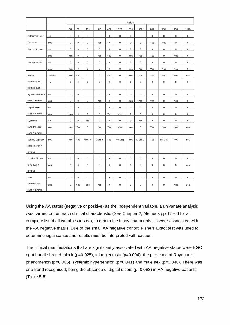
133
Using the AA status (negative or positive) as the independent variable, a univariate analysis
was carried out on each clinical characteristic (See Chapter 2, Methods pp. 65-66 for a
complete list of all variables tested), to determine if any characteristics were associated with
the AA negative status. Due to the small AA negative cohort, Fishers Exact test was used to
determine significance and results must be interpreted with caution.
The clinical manifestations that are significantly associated with AA negative status were EGC
right bundle branch block (p=0.025), telangiectasia (p=0.004), the presence of Raynaud’s
phenomenon (p=0.005), systemic hypertension (p=0.041) and male sex (p=0.048). There was
one trend recognised; being the absence of digital ulcers (p=0.083) in AA negative patients
(Table 5-5)
Patient
53 66 163 345 472 522 638 802 807 854 953 1110
Calcinosis Ever
7 reviews
No 0 0 0 0 0 0 0 0 0 0 0 0
Yes 0 0 0 Yes 0 0 0 0 Yes Yes 0 0
Dry mouth ever No 0 0 0 0 0 0 0 0 0 0 0 0
Yes Yes 0 0 Yes Yes 0 Yes Yes Yes 0 Yes 0
Dry eyes ever No 0 0 0 0 0 0 0 0 0 0 0 0
Yes Yes 0 0 0 0 0 Yes Yes Yes Yes Yes 0
Reflux
oesophagitis
definite ever
Definite Yes Yes 0 0 Yes 0 Yes Yes Yes Yes Yes Yes
No 0 0 0 0 0 0 0 0 0 0 0 0
Synovitis definite
ever 7 reviews
No 0 0 0 0 0 0 0 0 0 0 0 0
Yes 0 0 0 Yes 0 0 Yes Yes Yes 0 Yes 0
Digital ulcers
ever 7 reviews
No 0 0 0 0 0 0 0 0 0 0 0 0
Yes Yes 0 0 0 Yes Yes 0 0 0 0 0 0
Systemic
hypertension
ever 7 reviews
No 0 0 No 0 0 0 0 No 0 0 0 0
Yes Yes Yes 0 Yes Yes Yes Yes 0 Yes Yes Yes Yes
Nailfold capillary
dilation ever 7
reviews
Yes Yes Yes Missing Missing Yes Missing Yes Missing Yes Missing Yes Yes
Tendon friction
rubs ever 7
reviews
No 0 0 0 0 0 0 0 0 0 0 0 0
Yes 0 0 0 0 0 0 0 0 0 0 0 Yes
Joint
contractures
ever 7 reviews
No 0 0 0 0 0 0 0 0 0 0 0 0
Yes 0 Yes Yes Yes 0 0 0 0 0 0 Yes Yes
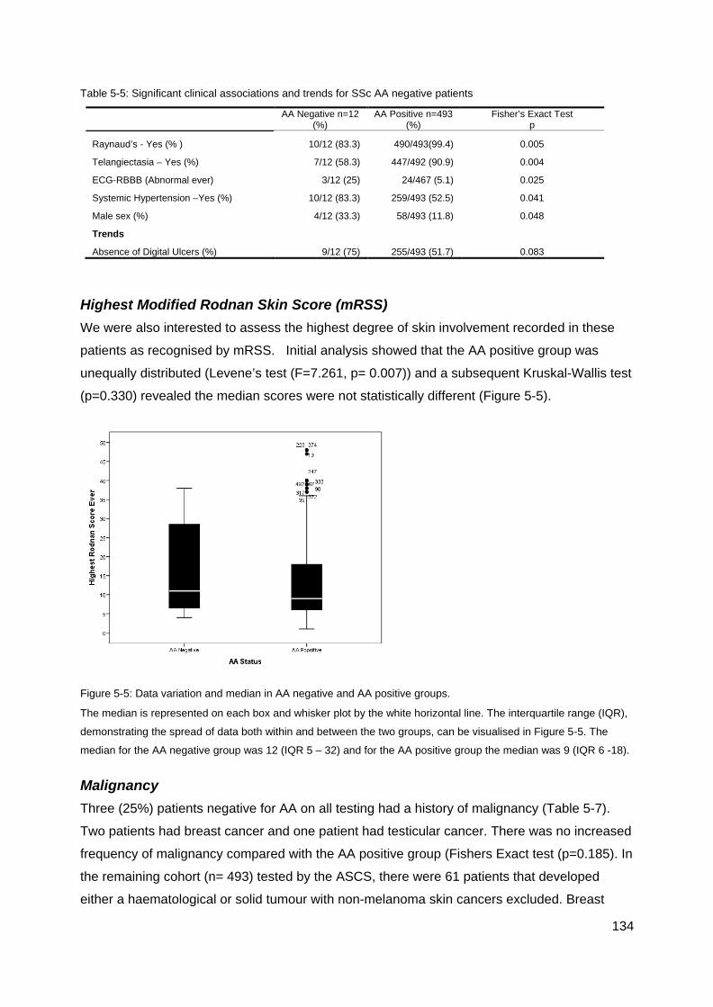
134
Table 5-5: Significant clinical associations and trends for SSc AA negative patients
AA Negative n=12 (%)
AA Positive n=493 (%)
Fisher’s Exact Test p
Raynaud’s - Yes (% ) 10/12 (83.3) 490/493(99.4) 0.005
Telangiectasia – Yes (%) 7/12 (58.3) 447/492 (90.9) 0.004
ECG-RBBB (Abnormal ever) 3/12 (25) 24/467 (5.1) 0.025
Systemic Hypertension –Yes (%) 10/12 (83.3) 259/493 (52.5) 0.041
Male sex (%) 4/12 (33.3) 58/493 (11.8) 0.048
Trends Absence of Digital Ulcers (%) 9/12 (75) 255/493 (51.7) 0.083
Highest Modified Rodnan Skin Score (mRSS) We were also interested to assess the highest degree of skin involvement recorded in these
patients as recognised by mRSS. Initial analysis showed that the AA positive group was
unequally distributed (Levene’s test (F=7.261, p= 0.007)) and a subsequent Kruskal-Wallis test
(p=0.330) revealed the median scores were not statistically different (Figure 5-5).
Figure 5-5: Data variation and median in AA negative and AA positive groups.
The median is represented on each box and whisker plot by the white horizontal line. The interquartile range (IQR),
demonstrating the spread of data both within and between the two groups, can be visualised in Figure 5-5. The
median for the AA negative group was 12 (IQR 5 – 32) and for the AA positive group the median was 9 (IQR 6 -18).
Malignancy Three (25%) patients negative for AA on all testing had a history of malignancy (Table 5-7).
Two patients had breast cancer and one patient had testicular cancer. There was no increased
frequency of malignancy compared with the AA positive group (Fishers Exact test (p=0.185). In
the remaining cohort (n= 493) tested by the ASCS, there were 61 patients that developed
either a haematological or solid tumour with non-melanoma skin cancers excluded. Breast
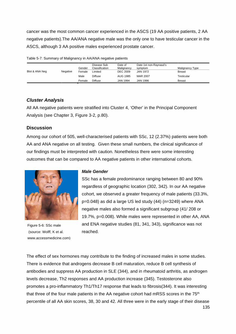
135
cancer was the most common cancer experienced in the ASCS (19 AA positive patients, 2 AA
negative patients).The AA/ANA negative male was the only one to have testicular cancer in the
ASCS, although 3 AA positive males experienced prostate cancer.
Table 5-7: Summary of Malignancy in AA/ANA negative patients
Gender Disease Sub Classification
Date of Malignancy
Date 1st non-Raynaud’s symptom Malignancy Type
Blot & ANA Neg Negative Female Limited DEC 2009 JAN 1972 Breast Male Diffuse AUG 1985 MAR 2007 Testicular Female Diffuse JAN 1994 JAN 1996 Breast
Cluster Analysis All AA negative patients were stratified into Cluster 4, ‘Other’ in the Principal Component
Analysis (see Chapter 3, Figure 3-2, p.80).
Discussion
Among our cohort of 505, well-characterised patients with SSc, 12 (2.37%) patients were both
AA and ANA negative on all testing. Given these small numbers, the clinical significance of
our findings must be interpreted with caution. Nonetheless there were some interesting
outcomes that can be compared to AA negative patients in other international cohorts.
Male Gender SSc has a female predominance ranging between 80 and 90%
regardless of geographic location (302, 342). In our AA negative
cohort, we observed a greater frequency of male patients (33.3%,
p=0.048) as did a large US led study (44) (n=3249) where ANA
negative males also formed a significant subgroup (41/ 208 or
19.7%, p=0.008). While males were represented in other AA, ANA
and ENA negative studies (81, 341, 343), significance was not
reached.
The effect of sex hormones may contribute to the finding of increased males in some studies.
There is evidence that androgens decrease B cell maturation, reduce B cell synthesis of
antibodies and suppress AA production in SLE (344), and in rheumatoid arthritis, as androgen
levels decrease, Th2 responses and AA production increase (345). Testosterone also
promotes a pro-inflammatory Th1/Th17 response that leads to fibrosis(344). It was interesting
that three of the four male patients in the AA negative cohort had mRSS scores in the 75th
percentile of all AA skin scores, 38, 30 and 42. All three were in the early stage of their disease
Figure 5-6: SSc male
(source: Wolff, K et al.
www.accessmedicine.com)
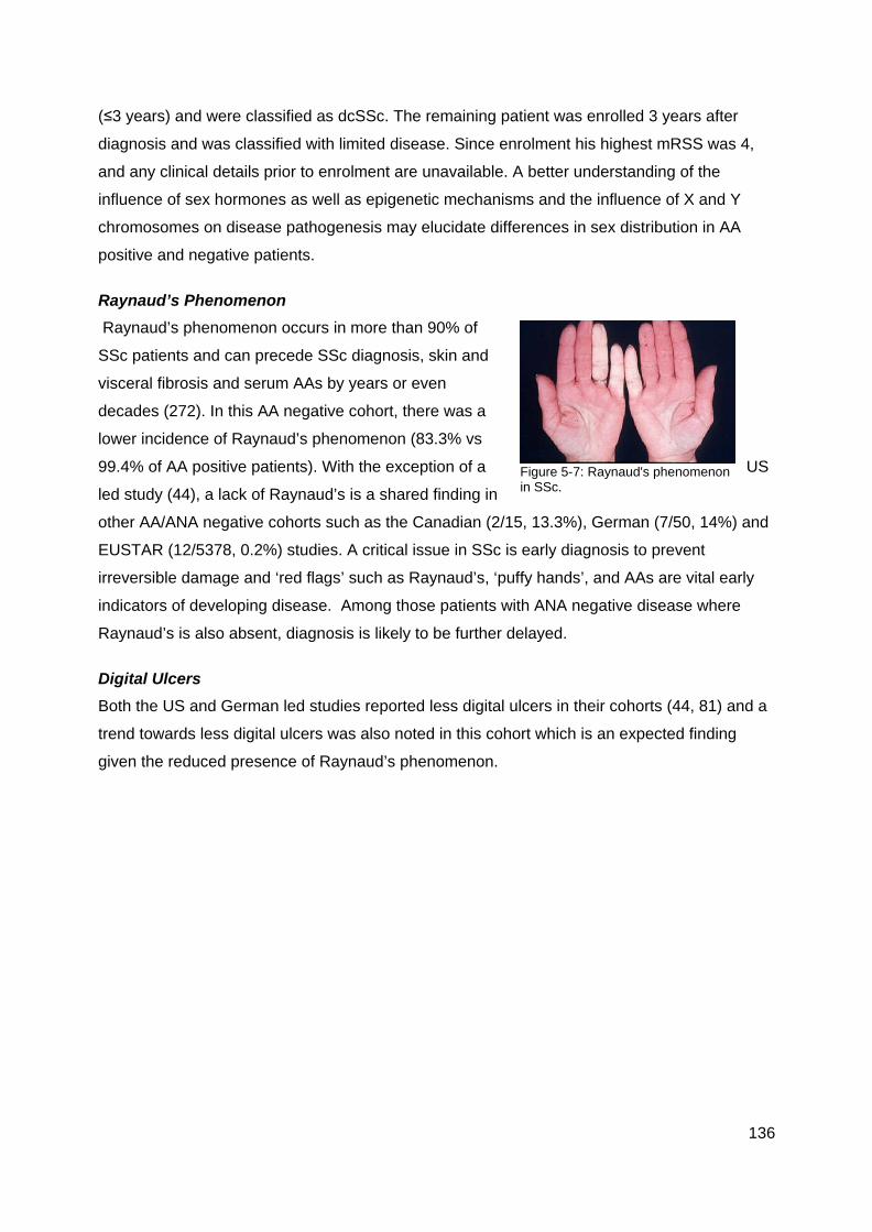
136
(≤3 years) and were classified as dcSSc. The remaining patient was enrolled 3 years after
diagnosis and was classified with limited disease. Since enrolment his highest mRSS was 4,
and any clinical details prior to enrolment are unavailable. A better understanding of the
influence of sex hormones as well as epigenetic mechanisms and the influence of X and Y
chromosomes on disease pathogenesis may elucidate differences in sex distribution in AA
positive and negative patients.
Raynaud’s Phenomenon Raynaud’s phenomenon occurs in more than 90% of
SSc patients and can precede SSc diagnosis, skin and
visceral fibrosis and serum AAs by years or even
decades (272). In this AA negative cohort, there was a
lower incidence of Raynaud’s phenomenon (83.3% vs
99.4% of AA positive patients). With the exception of a US
led study (44), a lack of Raynaud’s is a shared finding in
other AA/ANA negative cohorts such as the Canadian (2/15, 13.3%), German (7/50, 14%) and
EUSTAR (12/5378, 0.2%) studies. A critical issue in SSc is early diagnosis to prevent
irreversible damage and ‘red flags’ such as Raynaud’s, ‘puffy hands’, and AAs are vital early
indicators of developing disease. Among those patients with ANA negative disease where
Raynaud’s is also absent, diagnosis is likely to be further delayed.
Digital Ulcers Both the US and German led studies reported less digital ulcers in their cohorts (44, 81) and a
trend towards less digital ulcers was also noted in this cohort which is an expected finding
given the reduced presence of Raynaud’s phenomenon.
Figure 5-7: Raynaud's phenomenon in SSc.

137
Telangiectasia Telangiectasiae are an important diagnostic indicator in SSc
(142) and are a marker of a more severe vascular
phenotype (346). In this cohort and the large US based cohort
(44), telangiectasias are less frequently found in patients
without circulating AAs or a detectable ANA. This would
suggest a less vasculopathic form of disease (44).
Skin involvement We did not detect any significant difference in the degree of skin involvement in our AA
negative cohort, according to the highest mRSS recorded during each patient’s involvement in
the ASIG cohort. However, we recognise that these patients were not enrolled at the time of
disease onset and so their peak skin involvement may have occurred prior to enrolment in this
study. Inception cohorts will be invaluable to definitively determine if any differences in skin
changes exist between ANA negative and positive patients. Other groups have also explored
this question. Hudson et al reported a trend towards lower mRSS among AA negative patients
and taken together with a lower prevalence of Raynaud’s and telangiectasia, among the AA
negative group they concluded that AA negative patients had milder disease (341). This result
was mirrored somewhat by Hamaguchi et al who found that among AA negative patients in
their Japanese cohort (5%; n= 10/203), there was milder skin involvement and only rarely were
PAH, ILD and digital ulcers detected (122). In contrast, Schneeberger et al reported a trend
towards a higher mRSS and shorter disease duration among AA negative EUSTAR patients
(343).
Right Bundle Branch Block Diagnosis of a right bundle branch block (RBBB) is by
electrocardiogram. It results from a block or delay in conduction of
cardiac impulse through the right branch of the Bundle of His. Blood
supply for this branch is from the septal vessels coming off the left
anterior descending coronary artery (347). In our cohort 3/12 (25%)
of patients experienced a RBBB result. The most common
comorbidity of RBBB is systemic hypertension (347). Systemic
hypertension was a significant cardinal feature of our AA negative
patients with 83.3% experiencing this condition. In other cohorts systemic hypertension or
RBBB are not reported. Only one patient in our AA negative cohort experienced pulmonary
arterial hypertension (PAH) and this low prevalence seems to be the same as the German,
Canadian, US and EUSTAR studies. Therefore we suspect that the higher prevalence of
Figure 5-8: Bundle branch block
Figure 5-9: Telangiectasia in SSc.
138
RBBB may be a spurious finding perhaps influenced by the high prevalence of hypertension in
our ANA negative patients. Hypertension is a common finding in the community and, in the
absence of renal crisis, is not a disease specific feature of SSc. Its presence in our small
cohort may again be a spurious finding and has not been reflected in other published data on
ANA negative SSc.
Malignancy Malignancy, while not reaching statistical significance in the AA negative subgroup, was
present in 3/12 patients and included breast and testicular cancer. In 2/3 cases these
diagnoses were remote form the diagnosis of malignancy. In our AA positive cohort, 61/493
(12.37%) patients had cancer and overall, cancer was found to be significantly associated with
Cluster 4 (p=0.039). Other studies have found a close temporal association with SSc onset
and a diagnosis of cancer, particularly with RNAP3 AAs (161, 164, 175) and also with breast
cancer (348-350). It is noticeable that one AA negative patient was diagnosed with breast
cancer 2 years prior to the SSc onset and Shah et al also found that AA negative patients had
a close temporal relationship between SSc onset and cancer diagnosis and postulated that this
group may express novel tumour antigens that are yet to be identified (173). In light of these
findings it will be interesting to monitor disease associations in the Australian cohort as the
duration of observation increases.
Many studies have investigated breast cancer in SSc and an increased risk or incidence was
found in most (349, 351), although in a meta-analysis of 16 studies and over 7000 patients, the
relationship with breast cancer and SSc was not conclusive (352). Atypical presentations of
cancer have been found particularly in relation to breast cancer with paraneoplastic
scleroderma and ANA negative patients. (353, 354). In the study by Schneeberger et al, four of
the seven patients for whom detailed information was available, had a malignancy (two breast,
one multiple myeloma and one bladder carcinoma) and a fifth patient who died three years
after his initial visit may have had a malignancy but cause of death was not recorded (343).
Despite not finding a statistically increased risk of malignancy in their AA negative cohort,
Schneeberger et al still conclude that a paraneoplastic syndrome should be considered as a
differential diagnosis in AA negative and Raynaud’s negative patients (343). In other SSc
cohorts where studies on AA negative patients have been conducted (US (44)and Canadian
(341)), malignancy information was not available.
While any association between malignancy and AA negative SSc remains uncertain, it was not
evident in our cohort. However, careful screening for malignancy in patients with any form of
atypical presentation, including the absence of detectable AAs seems prudent.
139
Limited survival data is available on AA negative patients and at the time data for this study
were censored (7 June 2013), all patients were still alive, so no survival analysis was possible.
Only the Salazar et al study has reported on survival and they did not find a survival difference
between AA negative and AA positive patients (44). (Figure 5-12). It will be interesting to see
if this finding is reflected in other cohorts as further survival data becomes available.
Conclusion
Among 505 patients with confirmed SSc, only 12 patients were AA negative on all available
testing. Ten of the twelve patients fulfilled the 1980 ACR criteria and retrospectively 12/12
fulfiled the 2013 ACR/EULAR criteria. We reviewed their clinical characteristics and all had
features entirely consistent with clinical disease and in 5 patients skin involvement was
consistent with diffuse disease. Further research in large multinational collaborations is
required to stratify these patients particularly in regards to prognosis and survival.
Our cohort is clinically similar to other published AA negative cohorts. These AA negative
patients have fibrotic features of SSc and are less likely to have the SSc associated
vasculopathy characterised by Raynaud’s phenomenon, telangiectasias, and to a lesser
extent, digital ulcers. These findings and are in accordance with many of the findings of
Hudson et al (341) and Salazar et al (44). Finally, in our cohort AA negative patients, for
reasons yet to be elucidated, are more commonly male.
140
CHAPTER 6 COMPARISON OF METHODS
Introduction
Interpretation and analysis of autoantibodies in this scleroderma cohort have been undertaken
using immunoblot technology. The SSc Euroimmun line blot has been validated in a number
of cohorts and has good agreement with traditional methods of indirect immunofluorescence
(IIF), enzyme linked immunosorbent assay (ELISA) and immunoprecipitation (IP) (144, 196,
355). Line immunoassays (LIA) are reported to be a faster and more practical method of
extractable anti-nuclear (ENA) antigen detection (106, 196). For uniformity, testing was
undertaken by a single operator at a single centre and the LIA analysis had the added benefit
of testing for other rarer, SSc associated AAs that are not generally available in commerical
laboratories.
Different methodologies will have differing sensitivities and specificities, even when targeting
the same autoantigen Furthermore, while most AAs in the setting of SSc are considered to be
constant, there is evidence that some, such as RNAP3, may fluctuate during the disease
course (154). Particular factors to be considered when comparing two or more different testing
methods include:
i) Epitope/vector expression – type of assay
ii) Diagnostic accuracy and reliability - validated in a variety of cohorts with different
genetic and ethnic backgrounds
iii) Sensitivity and specificity
iv) Multiple autoantibody (AA) positivity
v) Expertise in interpreting results
vi) Clinical associations/prognoses with a particular AA or combination of AAs
A more detailed explanation of the Euroimmun line blot is detailed in Chapter 2 and Chapter 1
(pp. 14-17) includes a discussion of the different AA testing platforms available. In general
terms, initial screening for ANAs are carried out using IIF on HEp-2 cells followed by a more
specific method using a variety of platforms. Greater than 100 antigens can be detected using
IIF and it is considered by some the ‘gold standard’ for ANA detection (356), although
interpretation of rarer profiles and mixed patterns requires considerable expertise. Newer
platforms such as the LIA have the ability to test large numbers of AAs using a commercially
viable assay; however, there are concerns that denaturing of proteins may change the
conformation of epitopes. An advantage of a platform such as the immunoblot is that there is

141
an increasing recognition that multiple positive AAs can be detected in a single patient (67).
These results may not have been available in older assays which are perhaps more specific
but may lack in sensitivity. However, it is uncertain whether increased sensitivity aids in clinical
assessment in practical terms.
We were interested to compare the reliability of the Euroimmun LIA in this well characterised
Australian population with other commercially available assays. Where there was discrepancy
between testing, we were also interested to look at the individual results more closely and the
clinical associations of these patients to see whether the test result might support the patient’s
disease sub classification. This chapter will compare results of the (Euroimmun) Line
Immunoassay (LIA), with results obtained by independent laboratories in the Australian
Scleroderma Cohort Study (ASCS).
Comparison of results
There were 505 patients in the SSc cohort and an additional 21 patients that were classified
with Mixed Connective Tissue Disease (MCTD). In addition to the Euroimmun LIA, the results
of independent commercial laboratory testing for ANA staining and AAs to CENP, Topo1 and
RNAP3 were available for the majority of patients. Other rarer AAs were less frequently tested
and for the purposes of this analysis, we limited comparisons to these three major AAs, shown
to be the most clinically significant in our earlier work. All commercially available immunology
laboratories in Australia participate in a Quality Assurance Program which provides feedback
on the accuracy of AA testing. In Australia, the initial screening for ANA is undertaken with IIF
using HEp-2 cells. Identification of CENP AA was by their characteristic IIF staining pattern.
Further characterisations were generally undertaken by ENA with a precipitating antigen for
Topo1 and where available, RNAP3 testing was also performed by ELISA. At the time of data
census (7 June 2013), the majority of regional and central laboratories in Australia were using
ELISA and IP for ENA testing, although with the advent of the newer diagnostic platforms,
there is now greater variation of methods. Results from independent laboratories are referred
to as Australian Scleroderma Interest Group (ASIG) data. LIA data was complete for all
patients in this cohort.

142
Tables 6-1 to 6-4 are a summary of the results from ASIG independent laboratories for SSc
(n=505) and include the supplementary RNP (MCTD) data (n=21) as tested by ASIG
independent laboratories.
Table 6-3: Summary ASIG ANA by IIF
Frequency Percent No 30 5.7
Yes 496 94.3 Total 526 100.0
Table 6-4: Summary ASIG IIF Centromere
Frequency Percent No 310 58.9
Yes 212 40.3 Total 522 99.2
Missing 4 .8 Total 526 100.0
Table 6-5: Summary ASIG ENA Topo1
Frequency Percent No 433 82.3
Yes 87 16.5 Total 520 98.9
Missing 6 1.1 Total 526 100.0
Table 6-6: Summary ASIG RNAP3 (ELISA)
Frequency Percent N/A* 152 28.9
No 301 57.2 Yes 53 10.1 Total 506 96.2
Missing 20 3.8 Total 526 100.0
* NOTE: RNAP3 was physician requested and so not all patients were tested for this AA.
Statistical analysis For the purpose of comparing interrater reliability Cohen’s kappa (κ) statistic, a measure of
agreement between categorical variables, was used. A kappa of 1 indicates perfect
agreement, whereas a kappa of 0 indicates agreement equivalent to chance. Further
information on the κ statistic is found in Chapter 2, Methods (p.76).
Results
CENP The results for the CENP immunoblot data were complete with 4 patients missing from the
ASIG Centromere IIF results. Therefore, 99.2% (501/505) of data were utilised in Cohen’s κ
analysis. The LIA and ASIG data were in agreement on 206 (90.4%) positive results and 267
(97.8%) negative results. Cohen’s κ analysis determined there was excellent or very good
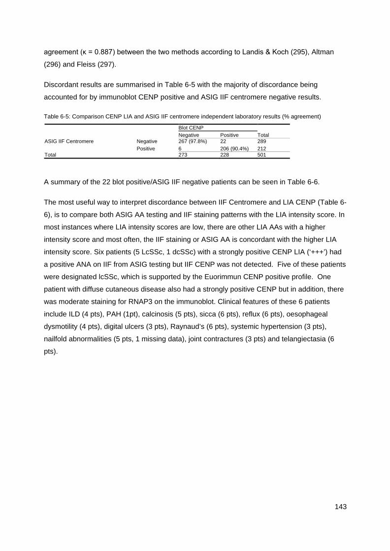
143
agreement (κ = 0.887) between the two methods according to Landis & Koch (295), Altman
(296) and Fleiss (297).
Discordant results are summarised in Table 6-5 with the majority of discordance being
accounted for by immunoblot CENP positive and ASIG IIF centromere negative results.
Table 6-5: Comparison CENP LIA and ASIG IIF centromere independent laboratory results (% agreement)
Blot CENP
Total Negative Positive ASIG IIF Centromere Negative 267 (97.8%) 22 289
Positive 6 206 (90.4%) 212 Total 273 228 501
A summary of the 22 blot positive/ASIG IIF negative patients can be seen in Table 6-6.
The most useful way to interpret discordance between IIF Centromere and LIA CENP (Table 6-
6), is to compare both ASIG AA testing and IIF staining patterns with the LIA intensity score. In
most instances where LIA intensity scores are low, there are other LIA AAs with a higher
intensity score and most often, the IIF staining or ASIG AA is concordant with the higher LIA
intensity score. Six patients (5 LcSSc, 1 dcSSc) with a strongly positive CENP LIA (‘+++’) had
a positive ANA on IIF from ASIG testing but IIF CENP was not detected. Five of these patients
were designated lcSSc, which is supported by the Euorimmun CENP positive profile. One
patient with diffuse cutaneous disease also had a strongly positive CENP but in addition, there
was moderate staining for RNAP3 on the immunoblot. Clinical features of these 6 patients
include ILD (4 pts), PAH (1pt), calcinosis (5 pts), sicca (6 pts), reflux (6 pts), oesophageal
dysmotility (4 pts), digital ulcers (3 pts), Raynaud’s (6 pts), systemic hypertension (3 pts),
nailfold abnormalities (5 pts, 1 missing data), joint contractures (3 pts) and telangiectasia (6
pts).
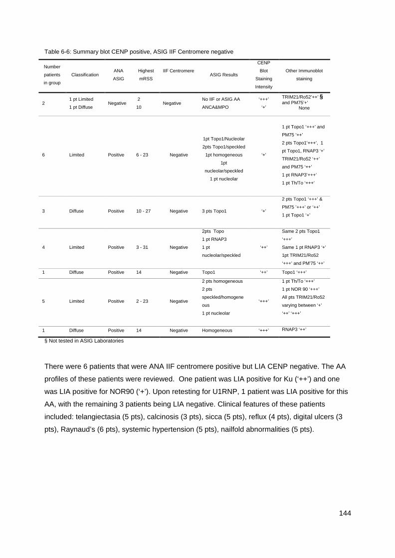
144
Table 6-6: Summary blot CENP positive, ASIG IIF Centromere negative
Number
patients
in group
Classification ANA
ASIG
Highest
mRSS
IIF Centromere ASIG Results
CENP
Blot
Staining
Intensity
Other Immunoblot
staining
2 1 pt Limited
1 pt Diffuse Negative
2
10 Negative
No IIF or ASIG AA
ANCA&MPO
‘+++’
‘+’
TRIM21/Ro52’++’ § and PM75’+’
None
6 Limited Positive 6 - 23 Negative
1pt Topo1/Nucleolar
2pts Topo1/speckled
1pt homogeneous
1pt
nucleolar/speckled
1 pt nucleolar
‘+’
1 pt Topo1 ‘+++’ and
PM75 ‘++’
2 pts Topo1’+++’, 1
pt Topo1, RNAP3 ‘+’
TRIM21/Ro52 ‘++’
and PM75 ‘++’
1 pt RNAP3’+++’
1 pt Th/To ‘+++’
3 Diffuse Positive 10 - 27 Negative 3 pts Topo1 ‘+’
2 pts Topo1 ‘+++’ &
PM75 ’+++’ or ‘++’
1 pt Topo1 ‘+’
4 Limited Positive 3 - 31 Negative
2pts Topo
1 pt RNAP3
1 pt
nucleolar/speckled
‘++’
Same 2 pts Topo1
‘+++’
Same 1 pt RNAP3 ‘+’
1pt TRIM21/Ro52
‘+++’ and PM’75 ‘++’
1 Diffuse Positive 14 Negative Topo1 ‘++’ Topo1 ‘+++’
5 Limited Positive 2 - 23 Negative
2 pts homogeneous
2 pts
speckled/homogene
ous
1 pt nucleolar
‘+++’
1 pt Th/To ‘+++’
1 pt NOR 90 ‘+++’
All pts TRIM21/Ro52
varying between ‘+’
‘++’ ‘+++’
1 Diffuse Positive 14 Negative Homogeneous ‘+++’ RNAP3 ‘++’
§ Not tested in ASIG Laboratories There were 6 patients that were ANA IIF centromere positive but LIA CENP negative. The AA
profiles of these patients were reviewed. One patient was LIA positive for Ku (‘++’) and one
was LIA positive for NOR90 (‘+’). Upon retesting for U1RNP, 1 patient was LIA positive for this
AA, with the remaining 3 patients being LIA negative. Clinical features of these patients
included: telangiectasia (5 pts), calcinosis (3 pts), sicca (5 pts), reflux (4 pts), digital ulcers (3
pts), Raynaud’s (6 pts), systemic hypertension (5 pts), nailfold abnormalities (5 pts).
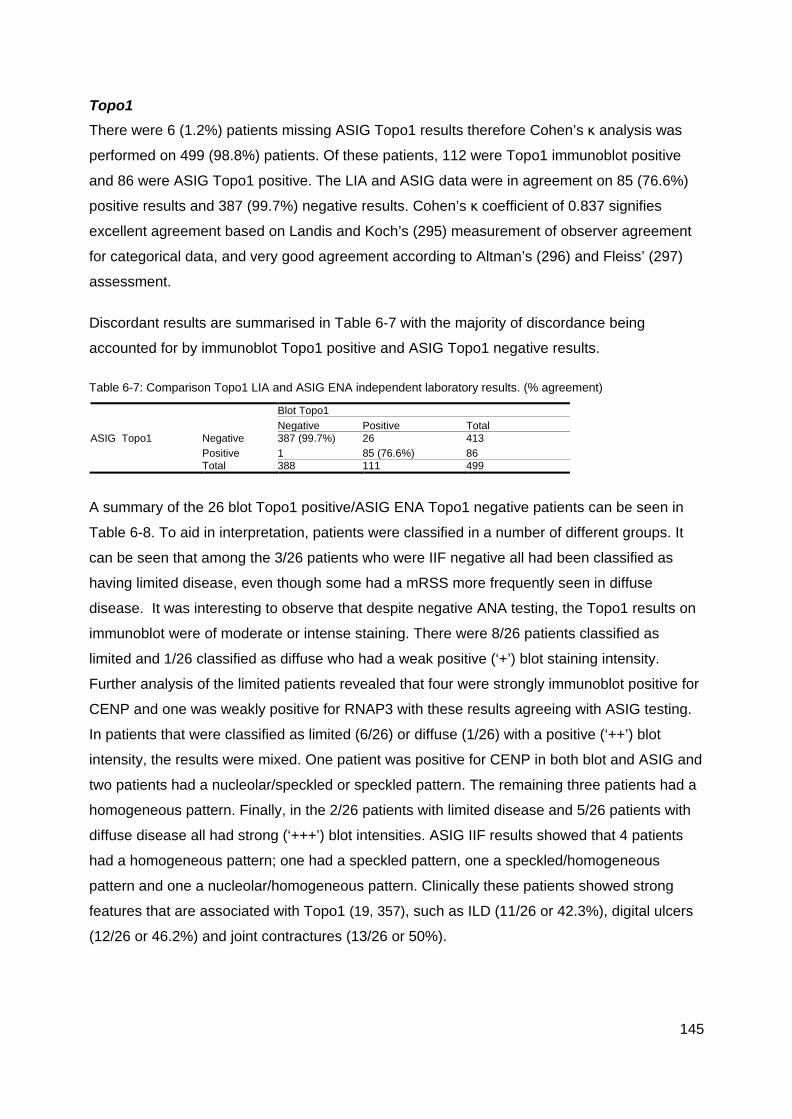
145
Topo1 There were 6 (1.2%) patients missing ASIG Topo1 results therefore Cohen’s κ analysis was
performed on 499 (98.8%) patients. Of these patients, 112 were Topo1 immunoblot positive
and 86 were ASIG Topo1 positive. The LIA and ASIG data were in agreement on 85 (76.6%)
positive results and 387 (99.7%) negative results. Cohen’s κ coefficient of 0.837 signifies
excellent agreement based on Landis and Koch’s (295) measurement of observer agreement
for categorical data, and very good agreement according to Altman’s (296) and Fleiss’ (297)
assessment.
Discordant results are summarised in Table 6-7 with the majority of discordance being
accounted for by immunoblot Topo1 positive and ASIG Topo1 negative results.
Table 6-7: Comparison Topo1 LIA and ASIG ENA independent laboratory results. (% agreement)
Blot Topo1 Negative Positive Total
ASIG Topo1 Negative 387 (99.7%) 26 413 Positive 1 85 (76.6%) 86
Total 388 111 499 A summary of the 26 blot Topo1 positive/ASIG ENA Topo1 negative patients can be seen in
Table 6-8. To aid in interpretation, patients were classified in a number of different groups. It
can be seen that among the 3/26 patients who were IIF negative all had been classified as
having limited disease, even though some had a mRSS more frequently seen in diffuse
disease. It was interesting to observe that despite negative ANA testing, the Topo1 results on
immunoblot were of moderate or intense staining. There were 8/26 patients classified as
limited and 1/26 classified as diffuse who had a weak positive (‘+’) blot staining intensity.
Further analysis of the limited patients revealed that four were strongly immunoblot positive for
CENP and one was weakly positive for RNAP3 with these results agreeing with ASIG testing.
In patients that were classified as limited (6/26) or diffuse (1/26) with a positive (‘++’) blot
intensity, the results were mixed. One patient was positive for CENP in both blot and ASIG and
two patients had a nucleolar/speckled or speckled pattern. The remaining three patients had a
homogeneous pattern. Finally, in the 2/26 patients with limited disease and 5/26 patients with
diffuse disease all had strong (‘+++’) blot intensities. ASIG IIF results showed that 4 patients
had a homogeneous pattern; one had a speckled pattern, one a speckled/homogeneous
pattern and one a nucleolar/homogeneous pattern. Clinically these patients showed strong
features that are associated with Topo1 (19, 357), such as ILD (11/26 or 42.3%), digital ulcers
(12/26 or 46.2%) and joint contractures (13/26 or 50%).
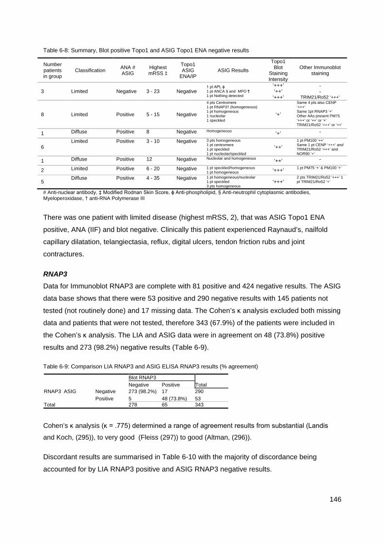
146
Table 6-8: Summary, Blot positive Topo1 and ASIG Topo1 ENA negative results
Number patients in group
Classification ANA # ASIG
Highest mRSS ‡
Topo1 ASIG
ENA/IP ASIG Results
Topo1 Blot
Staining Intensity
Other Immunoblot staining
3 Limited Negative 3 - 23 Negative 1 pt APL ɸ 1 pt ANCA § and MPO ¶ 1 pt Nothing detected
‘+++’ ‘++’
‘+++’
- -
TRIM21/Ro52 ‘+++’
8 Limited Positive 5 - 15 Negative
4 pts Centromere 1 pt RNAP3† (homogeneous) 1 pt homogeneous 1 nucleolar 1 speckled
‘+’
Same 4 pts also CENP ‘+++’ Same 1pt RNAP3 ‘+’ Other AAs present PM75 ‘+++’ or ‘++’ or ‘+’ TRIM21/Ro52 ‘+++’ or ‘++’
1 Diffuse Positive 8 Negative Homogeneous ‘+’ -
6 Limited Positive 3 - 10 Negative 3 pts homogeneous
1 pt centromere 1 pt speckled 1 pt nucleolar/speckled
‘++’ 1 pt PM100 ‘++’ Same 1 pt CENP ‘+++’ and TRIM21/Ro52 ‘+++’ and NOR90 ‘+’
1 Diffuse Positive 12 Negative Nucleolar and homogeneous ‘++’ -
2 Limited Positive 6 - 20 Negative 1 pt speckled/homogeneous 1 pt homogeneous ‘+++’ 1 pt PM75 ‘+’ & PM100 ‘+’
5 Diffuse Positive 4 - 35 Negative 1 pt homogeneous/nucleolar
1 pt speckled 3 pts homogeneous
‘+++’ 2 pts TRIM21/Ro52 ‘+++’ 1 pt TRIM21/Ro52 ‘+’
# Anti-nuclear antibody, ‡ Modified Rodnan Skin Score, ɸ Anti-phospholipid, § Anti-neutrophil cytoplasmic antibodies, Myeloperoxidase, † anti-RNA Polymerase III There was one patient with limited disease (highest mRSS, 2), that was ASIG Topo1 ENA
positive, ANA (IIF) and blot negative. Clinically this patient experienced Raynaud’s, nailfold
capillary dilatation, telangiectasia, reflux, digital ulcers, tendon friction rubs and joint
contractures.
RNAP3 Data for Immunoblot RNAP3 are complete with 81 positive and 424 negative results. The ASIG
data base shows that there were 53 positive and 290 negative results with 145 patients not
tested (not routinely done) and 17 missing data. The Cohen’s κ analysis excluded both missing
data and patients that were not tested, therefore 343 (67.9%) of the patients were included in
the Cohen’s κ analysis. The LIA and ASIG data were in agreement on 48 (73.8%) positive
results and 273 (98.2%) negative results (Table 6-9).
Table 6-9: Comparison LIA RNAP3 and ASIG ELISA RNAP3 results (% agreement)
Blot RNAP3
Total Negative Positive RNAP3 ASIG Negative 273 (98.2%) 17 290
Positive 5 48 (73.8%) 53 Total 278 65 343
Cohen’s κ analysis (κ = .775) determined a range of agreement results from substantial (Landis
and Koch, (295)), to very good (Fleiss (297)) to good (Altman, (296)).
Discordant results are summarised in Table 6-10 with the majority of discordance being
accounted for by LIA RNAP3 positive and ASIG RNAP3 negative results.

147
Table 6-10: Summary blot RNAP3 positive, ASIG ELISA RNAP3 negative
Number patients in group
Classification ANA ASIG
Highest mRSS ASIG Results RNAP3
ASIG
RNAP3 Blot Staining Intensity
Other Immunoblot staining
5 Limited Positive 2 - 23 5 pts IIF Centromere Negative ‘+' 5 pts CENP ‘+++’
1 Diffuse Positive 18 Nucleolar/speckled Negative ‘+' PM75 ‘+’
5 Limited Positive 3 - 18
2 pts IIF Centromere 2 pt speckled nucleolar 1 pt speckled
Negative ‘++'
Same 2 pts CENP ‘+++’ 2 pts also TRIM21/Ro52 ‘+++’ or ‘++’
3 Limited Positive 6 - 12 1 pt IIF Centromere 2 pts speckled Negative ‘+++' Same 1 pt
CENP ‘+++’
3 Diffuse Positive 10 – 37 1 pt nucleolar 2 pts speckled Negative ‘+++'
2 pts also TRIM21/Ro52 ‘+++’ or ‘+’
As previously described, RNAP3 LIA AA intensity scores should be compared with IIF patterns
as well as RNAP3 ELISA. At the time sera were analysed in both central and regional
laboratories, many would not have had RNAP3 ELISA available as a routine test. The higher
LIA intensity scores are concordant with ASIG IIF results and clinical presentation.
Table 6-11 summarises discordant results for ASIG positive and LIA negative results
Table 6-11: Summary RNAP3 ASIG positive blot RNAP3 negative
Number patients in group
Classification RNAP3 Blot Result
Highest mRSS ASIG Results ASIG ANA
pattern Other Immunoblot staining
2 Limited Negative 7 - 11 RNAP3 Speckled 1 pt TRIM21/Ro52 ‘+++’ 1 pt LIA blot negative
1 Limited Negative 2 RNAP3 Speckled/ Nucleolar LIA blot negative
2 Limited Negative 9 2 pts IIF Centromere & RNAP3
Centromere 2 pts LIA CENP ‘+++’
The clinical characteristics of the patients in Table 6-11 are; ILD (2 pts), PAH (1pt), calcinosis
(3 pts), sicca (5 pts), reflux (4 pts), synovitis (3 pts), digital ulcers (4 pts), Raynaud’s, (5 pts),
anal incontinence (4 pts), systemic hypertension (4 pts), nailfold abnormalities, (5 pts) and joint
contractures (2 pts).
148
Discussion
The Euroimmin Scleroderma LIA is a validated, commercially available assay for the detection
of multiple SSc associated AAs. Newer diagnostic technologies such as the immunoblot are
increasingly used in commercial laboratory settings, but in the past, the majority of AA testing
for scleroderma was performed using IIF with HEp-2 cells followed by ELISA for assessment of
ENA, including RNAP3. In Australia, the majority of testing through commercial laboratories at
the time of data censorship (July 2013) was performed using these methods and so we were
interested to compare our LIA findings with the data obtained by ASIG from regional
laboratories. It is important to recognise that the results recorded in the ASCS database were
unlikely to be obtained from sera taken at the same time as the stored sera utilised for the
Euroimmun. While this does not have relevance for the many AAs that are stable over time,
there is some evidence that Topo1 and RNAP3 levels do fluctuate and so this may have
affected interpretation of results.
For this validation study, we limited comparisons to the three primary disease associated AAs
as the ASCS data for Topo1 and CENP/Centromere were largely complete and almost 70% of
data were available for RNAP3, allowing for a good comparative study.
The Cohen’s κ analyses for each AA showed that there was good to excellent agreement
according to Landis & Koch, Altman and Fleiss for CENP and Topo 1 while there was
substantial to very good agreement for RNAP3. Ideally, concordance should be complete but
different testing platforms may not identity the same clinical cohorts, even if they are testing for
the same AA. On closer review of discordant results, we found that IIF patterns on HEp-2 cells
often had a stronger concordance with the LIA blot results rather than ENA or ELISA results.
For example a patient positive for RNAP3 by LIA might have a speckled ANA by IIF even
though ELISA for RNAP3 was negative. In addition, where apparent discordance occurred due
to a positive LIA and negative ELISA, the LIA would also frequently demonstrate additional
dominant staining of another AA identified through testing from regional laboratories. For
example, 4 patients who were LIA blot positive for Topo1 with a staining intensity of ‘+’, initially
appeared discordant with ASIG results showing IIF centromere, but on further investigation on
a case by case basis, these patients were also found to be strongly LIA blot positive for CENP
‘+++’.
While our cohort had insufficient numbers to compare testing platforms for the remaining AA
on the LIA, others have explored validity of different methods with these AA. Some rare AAs
are difficult to test for due to the technical complexity and interpretation of the IP assay and so
and effective LIA for these AA allows testing to extend beyond the research setting. An
149
example of this is anti-U3-RNP/ Fibrillarin testing. Peterson et al compared the Euroimmun LIA
with an IP assay and found that anti-U3-RNP/Fibrillarin IP and LIA results concurred in 98.9 %
of patients, yielding a κ coefficient 0.966 (196) and had good clinical agreement. They
concluded that the LIA is an acceptable and attractive alternative to IP for anti-U3-RNP
detection. The same or similar Euroimmun LIA was used in an Italian (200), Singaporean
(Chinese, Malay, Indian) (144) German (81) and Korean (358) population. Kappa analysis for
the Singaporean cohort between the LIA and ELISA was evaluated by individual AA and
ranged from κ=0.83 (CENP A), κ=0.96 (CENP B), κ= 0.97 (Topo1) to κ=1.0 (Pm-Scl) and
evaluation between LIA and IIF for CENP A and CENP B was κ=0.81 and κ=0.77 respectively.
This study also found good clinical agreement when compared with published data from other
cohorts (144). The Italian study compared sensitivity and specificity and again found that their
results were similar to those found in other studies and concluded that the LIA was ‘a more
rapid and more practical method than IP assays’ (200).
Mierau et al assessed (81) the frequency of disease-associated and other nuclear AA in the
German Network for systemic scleroderma utilising a variety of methods including LIA. They
used a different AA profile (Euroimmun ANA Profile 3) to that tested in our cohort. Some AAs
such as Ku, Th-To, Fibrillarin, RNAP3 NOR90 were tested by other methods and PDGFR was
not tested at all. For those AAs that were in common with the LIA SSc (Nucleoli) profile
(TRIM21/Ro52, Scl-70 [Topo1], PmScl, CENP B), clinical associations were similar to other
published cohorts (81). The Korean study (n=948 SARDs but included only n=25 SSc) used
the same ANA Profile 3 as the German study but the results were in contrast to both the
German and other published results yielding a κ=0.573 for agreement between IIF and the LIA
for SSc AAs and clinical diagnoses. The authors stated a potential limitation of the study were
the clinical diagnoses that were obtained from medical records which may be subject to bias
since diagnostic errors and the effect of past and current treatments could not be taken into
account (358). Again, this highlights the issue that using different testing platforms may not
identity the same clinical cohorts, even if they are testing for the same AA.
Overall, there is substantial agreement between the Immunoblot and the other methods used
by the ASIG independent laboratories when comparing RNAP3, CENP and Topo1 assays.
Other published series have reported clinical associations when LIA is used to detect the
remaining AAs to be similar to AA detected by other methods.
The Immunoblot is more sensitive to AA detection with the added advantage of multiple AA
recognition, although the clinical interpretation of multiple AAs requires further consideration. It
allows for an individual patient profile by testing for rare AAs that can be difficult to obtain by

150
other methods. This initial work has shown that the LIA has a role in AA sub classification and
our comparison with other testing platforms can provide reassurance that extrapolation to other
testing platforms such as ELISA, IP and IIF as long as the AA with the highest titre AA is
considered.
151
CHAPTER 7 CONCLUSIONS AND FUTURE RESEARCH
Conclusion
To conclude this thesis a final summary synthesising results and confirming the testing of
hypotheses is presented below.
Principal Component Analysis Recent advances in diagnostic technologies have allowed commercial laboratories to offer a
wider array of AA testing for scleroderma and other disorders. Interpreting the clinical
relevance of a positive result from these assays can be challenging, not only because of
differing detection methods, but also because multiple AAs may be detected. This thesis has
explored AA expression in scleroderma and uses mathematical modelling to aid interpretation
of complex results.
The Principal components analysis (PCA) provided a novel means to stratify scleroderma
patients according to the presence and intensity of a variety of scleroderma AAs. This method
reduced confounding when more than one AA was detected in patient sera. Multiple AA
expressions were common in this cohort, with an equal number of patients expressing either a
single or multiple AAs.
The AA with the highest titre (the dominant AA) determined individual patient cluster allocation
in the PCA and also correlated with meaningful disease associations. Patients with dominant
primary AAs (CENP, Topo1, RNAP3) made up 4/5 identified clusters and demonstrated clinical
features that are most commonly recognised with these AAs, limited and diffuse disease.
Interestingly, RNAP3 separated into two small, separate clusters depending on the intensity of
staining. In our analyses there was a trend towards higher titres occurring earlier in disease
and it would be interesting to follow a larger cohort of RNAP3 patients prospectively for
changes in titre over time and disease activity.
Fine Specificities The PCA analysis also identified a group of patients (Cluster 4) who did not have one of the
SSc primary AAs as their dominant AA, or had atypical co-expression of other AAs. This group
comprised a significant component of the tested cohort (142/505 patients) and their diverse AA
profile may represent a challenge to the treating clinician looking for confirmation of a clinical
diagnosis. For the most part, these patients did not have one of the three primary AAs and if
they did, it was of a lower staining intensity and often present in combination with other SSc
152
associated AAs. Autoantibodies to CENP, Topo1 (including double positive (CENP/Topo1)
patients), TRIM21/Ro52, PmScl and Th/To were found to have the most substantial influence
on variance in this Cluster. In addition, supplementary testing of selected patients
demonstrated U1RNP in 11 Cluster 4 patients, even though their clinical presentation was
most consistent with SSc rather than MCTD or overlap disease.
Positive associations in Cluster 4 as a whole included male gender, a history of malignancy
and smoking. Negative associations included gender bias and joint contractures. We explored
this cluster further according to their varied AA profiles to see if further information could be
obtained within this subset. We first compared Topo1 Cluster 4 patients with those positive for
Topo1in the entire cohort and repeated a similar analysis for CENP Cluster 4 patients. Some
minor differences were noted in these groups, particularly an increased frequency of PAH and
reduced ILD in the Topo1 Cluster 4 group, which again demonstrates disease associations are
generally most pronounced with the dominant AA.
Co-expression of CENP and Topo1 was rare and these two AA have previously been thought
to be mutually exclusive. Among our 505 patients, 15 patients co-expressed Topo1 and CENP
and were dispersed among Clusters 3-5, usually reflecting the dominant AA staining intensity.
The LIA used to detect these AAs has been well-validated and we feel these findings reflect a
true dual positive status rather than laboratory error. We used a single operator, single
platform approach and testing by independent laboratories had excellent agreement (Topo1
κ=0.837 and CENP κ=0.887) with the results of the LIA. Overall this study found that the
Topo1/CENP ‘double positive’ group had more in common with Topo1 patients than they did
with CENP patients.
Initial investigation of the less disease specific and/or rarer AA in our cohort revealed only a
few specific disease associations. In many cases, numbers of these AAs were small and the
ASCS data has been used in Tri-Nation studies to explore clinical associations of these AAs in
a larger multinational cohort (222, 241). We further hypothesized that the dominant AA would
have the greatest association with the clinical state. We were therefore interested to explore
the clinical associations of those patients in Cluster 4 who were monospecific for scleroderma
AA other than CENP, RNAP3 and Topo1. Sample size meant that meaningful analysis was
only possible for monospecific PmScl, TRIM21/Ro52 and the presence of U1RNP.
Monospecific TRIM21/Ro52 had features of inflammation, myocardial involvement and a
tendency towards ILD that was not evident in the larger cohort.
PmScl was rarely expressed monospecifically as either single or both epitopes. Monospecific
153
patients experienced Raynaud’s and disease onset at a younger age than other SSc patients.
U1 RNP SSc positive patients were more likely to be male, have an earlier disease onset, have
a history of reduced C3 and develop digital gangrene than the remainder of the cohort. In
addition U1RNP SSc positive patients and U1RNP MCTD patients were marginally different
with U1RNP MCTD patients having a milder phenotype with less vasculopathy and skin
involvement. MCTD patients are seen as being within the SSc spectrum of disease. U1RNP
positive SSc patients can develop serious vascular manifestations (digital gangrene), therefore
we believe that this AA should be included in SSc testing panels.
This study demonstrated that where typical AA testing does not find one of the primary SSc
AAs, then sera should be sent to a reference laboratory for further analysis. More effective
analyses of rarer AAs will require multinational collaborations to explore clinical associations,
genetic and environmental influences.
Autoantibody Negative Patients SSc patients that are both AA and ANA negative are very rare and in this cohort there were
only 12/505 patients (2.38%), with all fulfilling the 2013 ACR/EULAR criteria. These patients
emphasize the need for classification criteria for SSc to include provision for those that are AA
negative, even when they have undergone extended AA profiling. Our AA negative cohort was
clinically similar to other published AA negative cohorts in that they have more fibrotic features
and are less likely to have the SSc associated vasculopathy characterised by Raynaud’s
phenomenon, telangiectasias, and to a lesser extent, digital ulcers. Males also featured
prominently in this subset. We did not find an association with malignancy but others have
commented on a temporal association (343) and it seems prudent to screen any patient
presenting atypically for other causes of disease.
Platelet Derived Growth Factor Receptor AA Platelet derived growth factor receptor (PDGFR) was positive in only two patients in our cohort
but deserves mention in this summary as detecting its presence had been an initial aim of this
thesis. Both patients who were PDGFR positive had AAs that were of higher staining intensity
and were stratified to either Cluster 3 Topo or Cluster 5 CENP. There were no clinical
characteristics that were evident in this cohort that could be identified as being unique to
PDGFR. The same was found in other cohorts globally regardless of genetic or geographic
background (76, 116, 144, 200). The LIA might not be the best type of assay to detect this AA
and it may be more useful to replace PDGFR with U1RNP.
154
Line Blot Technology as a Method of Autoantibody Detection The Euroimmun line immunoblot assay (LIA) is an established technology that has been
validated in a variety of cohorts of various ethnic and genetic backgrounds. It compares
favourably with Australian diagnostic laboratory testing. We used a single operator, single
platform approach and testing by independent laboratories had excellent agreement for Topo1
(κ=0.837) and CENP (κ=0.887) and good to substantial agreement for RNAP3 (κ = .775).
Fewer comparisons for the RNAP3 assay were available because many central and regional
laboratories did not have RNAP3 testing routinely available at the time sera were tested for the
ASCS. The lower kappa statistic for RNAP3 may also reflect that these AA titres are known to
change over time as the sera used for this comparison study was not taken at the same time
point.
Where there was discordance among the different testing methods it was most often due to
multiple AA staining, with lower intensity staining being discordant and in these instances the
highest staining intensity scores were most often concordant with ASIG results. The
Immunoblot is more sensitive to AA detection with the added advantage of multiple AA
recognition; although the clinical interpretation of multiple AAs requires further consideration.
This initial work has shown that the LIA has a role in AA sub-classification and our comparison
with other testing platforms can provide reassurance that other testing platforms such as
ELISA, IP and IIF provide comparable results, as long as the AA with the highest titre AA is
considered.
The use of autoantibodies may provide a more meaningful stratification of scleroderma subsets
than the currently used limited, diffuse and overlap classification system, particularly in very
early disease. This may have particular relevance when identifying patients for clinical trials or
guiding monitoring for those more likely to develop organ specific complications.
Future Directions
Patient stratification systems In this study it was evident that the presence and staining intensity of AAs corresponded with
clinical characteristics at least where the three primary AAs (Topo1, CENP and RNAP3) were
concerned. Therefore using AAs as a primary sub classification tool may provide some
benefits over the current limited, diffuse and overlap classification system, particular in very
early disease before clinical features are apparent. However, it is also evident that the extent
of skin fibrosis is important in SSc (26, 283) and variation in the extent of skin involvement
reflects varied clinical phenotype. A sub classification system that uses both AA subset and the
modified Rodnan Skin Score may be more useful in understanding patient subgroups and
155
individual patients. In this way, a utilitarian report of the patient is available for further
prognostication and also how the patient is characterised for treatment.
Further characterisation of patients with monospecific and multiple autoantibody positivity Many patients co-express AAs as was evident in this cohort where half the patients had 2 or
more AAs. Where numbers permitted, we characterised clinical manifestations of monospecific
AA expression. To further characterise the effects of multiple AA expression, a better
understanding of monospecific expression is required, and this requires large multinational
collaborations. Potentially, patients may be further characterised by both their primary AA and
subsequently by their co-expressed AAs.
Titres and staining intensity – high and low, stability and mapping the clinical course Autoantibody titres are thought to remain stable over the course of disease (246); however
there are reports that challenge this finding. Both Topo1 (136) and RNAP3 (157) are reported
to fluctuate in individual patients and the change in titre (increase or decrease) was correlated
with clinical expression of disease. CENP has been reported to be relatively stable over the
disease course (101) but changes in titre in other AAs are unknown. In this study, the AA
staining intensity stratified patients to specific clusters. It would be interesting to take serial AA
titres or staining intensity measures from patient in different clusters over time and map the
clinical course of disease. This would generate an understanding of variability in AA levels and
also potentially identify responders and non-responders to treatment if AA levels follow the
course of disease.
Improve detection methods and explore new epitopes of SSc AAs such as fibrillarin and Th/To. Detection of fibrillarin is reported to be challenging as there is an antigenic complexity to the
C/D box snoRNPs (204) and there is emerging research that there are other Th/To protein
targets (82) that may identify more patients with this AA. At this point, both of these AAs were
relatively rare in this Australian cohort and while this could be attributed to genetic and ethnic
background, there may be other epitopes that may identify more patients that express this AA.
Continuing research to identify new epitopes and assays for diagnostic use is required.
156
Characterising AAs for ANA only positive patients There remained 33 ANA positive patients that did not have a detectable AA in Cluster 4.
Further characterisation of these patients is required to detect specific AA positivity. Some of
these patients may be positive for the new AAs that have been identified in SSc cohorts (see
Chapter 1 Table 1-7 pp.51-52). Clinical characterisation and multinational collaborations are
required to identify patients that are positive for these newer AAs.
Further characterisation of specific subgroups Further characterisation of specific subgroups identified in Cluster 4 such as the AA negative
group and the double positive Topo1/CENP subgroup will give a better understanding of
disease course in these patients.
Cluster Outcomes The ASIG is a relatively new database (2007) and future studies would include survival and
cause of death. This would benefit clinicians in giving advice as to prognostication.
The ageing immune system, cellular senescence and TRIM21/Ro52 Systemic sclerosis is not viewed as a disease of ageing, yet many of the clinical characteristics
have features in common with ageing. Features that are typically viewed in relation to age are
genomic instability and DNA damage, (359) pulmonary and systemic hypertension, renal
impairment, fibrosis, cardio-pulmonary disease and muscle weakness. Older SSc patients, that
is patients who present over the age of 65, have an increased risk of mortality from one of
these features (360).
Ageing is not a passive degenerative process; it is a complex process involving most
physiological systems of the body. The immune system has important physiological and
regulatory responses and it is well documented that many aspects of immune responses are
decreased or increased with ageing resulting in dysregulation (361).
TRIM21/Ro52 patients experience Raynaud’s at an older age and have an older age of
disease onset (241) and it is the most commonly co-expressed autoantibody. It has an
important role in the ubiquitin process and it has the ability to regulate downstream signalling of
various pattern recognition receptors including NF-κβ, TGF-β and interferon response. There
may be underlying senescence mechanisms (331, 332) that are yet to be elucidated that are
connected with this abundantly expressed AA. It would be interesting to investigate the ageing
immune system in SSc in association with the AAs, particularly TRIM21/Ro52.

157
Personalised Medicine Scleroderma, as with most autoimmune conditions, has a suite of biomarkers associated with
various pathologies within the condition (51). Serum autoantibodies, found in >95% (65) of
patients, are correlated with distinct clinical manifestations and have the potential, particularly
in early disease, to aid in predicting disease course. There is potential to utilise AAs in
conjunction with other biomarkers to predict fibrotic, vascular and organ manifestations and
response to treatment. For example a panel of SSc associated AAs could be interpreted in
combination with information obtained from transcriptomics, proteomics, metabolomics,
genomics and epigenomics to provide a detailed and individually personalised assessment of
disease course (13) . Theoretically this will be highly beneficial for patient outcomes as
biomarkers based on precision medicine will refine treatments and therapies for which the
patient is most likely to respond (66).
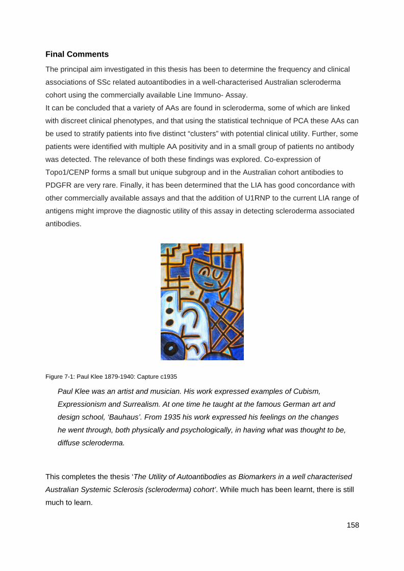
158
Final Comments
The principal aim investigated in this thesis has been to determine the frequency and clinical
associations of SSc related autoantibodies in a well-characterised Australian scleroderma
cohort using the commercially available Line Immuno- Assay.
It can be concluded that a variety of AAs are found in scleroderma, some of which are linked
with discreet clinical phenotypes, and that using the statistical technique of PCA these AAs can
be used to stratify patients into five distinct “clusters” with potential clinical utility. Further, some
patients were identified with multiple AA positivity and in a small group of patients no antibody
was detected. The relevance of both these findings was explored. Co-expression of
Topo1/CENP forms a small but unique subgroup and in the Australian cohort antibodies to
PDGFR are very rare. Finally, it has been determined that the LIA has good concordance with
other commercially available assays and that the addition of U1RNP to the current LIA range of
antigens might improve the diagnostic utility of this assay in detecting scleroderma associated
antibodies.
Figure 7-1: Paul Klee 1879-1940: Capture c1935
Paul Klee was an artist and musician. His work expressed examples of Cubism,
Expressionism and Surrealism. At one time he taught at the famous German art and
design school, ‘Bauhaus’. From 1935 his work expressed his feelings on the changes
he went through, both physically and psychologically, in having what was thought to be,
diffuse scleroderma.
This completes the thesis ‘The Utility of Autoantibodies as Biomarkers in a well characterised
Australian Systemic Sclerosis (scleroderma) cohort’. While much has been learnt, there is still
much to learn.

159
APPENDICES
Thesis publication
i. Patterson, K.A., Roberts-Thomson, P.J., Lester, S., Tan, J.A., Hakendorf, P., Rischmueller,
M., Zochling,J., Sahhar, J., Nash, P., Roddy, J., Hill, C., Nikpour, M., Stevens, W.,
Proudman, D.M., Walker, J.G. Interpretation of an Extended Autoantibody Profile in a
Well-Characterized Australian Systemic Sclerosis (Scleroderma) Cohort Using Principal
Components Analysis. Arthritis and Rheumatology. 2015; 67(12):3234-44
Contributions to other papers from work in this thesis
i. Carolina Mejia Otero, Shervin Assassi, Marie Hudson, Maureen D. Mayes, Rosa Estrada-
Y-Martin, Claudia Pedroza, Tingting W. Mills, Jennifer Walker, Murray Baron, Wendy
Stevens, Susanna M. Proudman, Mandana Nikpour, Sonal Mehra, Mianbo Wang, and
Marvin J. Fritzler; Canadian Scleroderma Research Group (CSRG); Australian
Scleroderma Cohort Study (ASCS); Genetics versus Environment in Scleroderma
Outcome Study (GENISOS) Anti-fibrillarin Antibodies Are Associated with Native-North
American Ethnicity and Poorer Survival in Systemic Sclerosis. Journal of Rheumatology.
Accepted 2016. Manuscript number: 2016-0574.R1
ii. Wodkowski M, Hudson M, Proudman S, Walker J, Stevens W, Nikpour M, Canadian
Scleroderma Research Group (CSRG); Australian Scleroderma Cohort Study (ASCS);
Genetics versus Environment in Scleroderma Outcome Study (GENISOS). Clinical
correlates of monospecific anti-PM75 and anti-PM100 antibodies in a tri-nation cohort of
1574 systemic sclerosis subjects. Autoimmunity. 2015;48(8):542-51
iii. Wodkowski M, Hudson M, Proudman S, Walker J, Stevens W, Nikpour M, Canadian
Scleroderma Research Group (CSRG); Australian Scleroderma Cohort Study (ASCS);
Genetics versus Environment in Scleroderma Outcome Study (GENISOS). Monospecific
anti-Ro52/TRIM21 antibodies in a tri-nation cohort of 1574 systemic sclerosis subjects:
Evidence of an association with interstitial lung disease and worse survival. Clinical and
Experimental Rheumatology. 2015;33:131-5.
iv. Mehra, S., Walker, J.G., Patterson, K.A., Fritzler, M.J. Autoantibodies in Systemic
Sclerosis Autoimmunity Reviews. 2013;12(3):340-54.
160
Contributions to Abstracts from work in this thesis i. Walker JG1, Nikpour M2,3, Huq M3, Patterson K1, Roberts-Thomson PJ1, Proudman
S4,5, Stevens W6, Lester S7,8, Rischmueller M4,8, Zochling J9, Sahhar J10,11, Nash P12,
Roddy J13, Hill C4,8, Hudson M14, Baron M14, Pope J15, Mayes MD16, Assassi S16, Mahler
M17,Fritzler MJ18; Canadian Scleroderma Research Group, Genetics versus
Environment in Scleroderma Outcome Study, Australian Scleroderma Interest Group.
Autoantibodies to the hPOP1 and Rpp25/38 components of the Th/To complex identify
a subgroup of systemic sclerosis (SSc) associated interstitial lung disease (ILD) and
antibodies to hPOP1 are associated with reduced survival. Submitted to American
College Rheumatology Conference, San Diego, California, USA, November 2017.
ii. S Lester, K Patterson, J Walker, JC Charlesworth, J Stankovich, W Stevens, J Sahhar,
P Nash, J Roddy, C Hill, M Nikpour, M Rischmueller, S Proudman, M Brown, J
Zochling. Associations between HLA DRB1 Alleles and Autoantibodies in Systemic
Sclerosis. Internal Medicine Journal. 2014;44:2-2
161
Australian Scleroderma Interest Group Terms of Reference
June 2013
The Steering Committee of the Australian Scleroderma Interest Group (ASIG) first met in
November 2005, and in 2007 it became a special interest group under the auspices of the
Australian Rheumatology Association (ARA), a not-for-profit organisation incorporated as a
company limited by guarantee in Australia. In January 2012, ARA-ASIG was granted an ABN
and registered with the Australian Business Register as an Other Incorporated Entity (ABN:
54 709 736 928). ARA-ASIG is governed by an Executive Committee that operates according
to the Constitution of the ARA.
1. Mission Statement
The ASIG consists of physicians interested in improving the care of patients with
scleroderma or systemic sclerosis (SSc). This will be achieved by clinical research and the
development of guidelines for the investigation, monitoring and management of patients
with SSc in accordance with current best practice. The implementation and effectiveness of
these guidelines in improving survival and quality of life of SSc patients in Australia will be
monitored and recursively modified according to new evidence as it arises from research
activities.
162
2. Background
Scleroderma and mixed connective tissue disease (MCTD) are rare, chronic disease
characterised by autoimmunity, vasculopathy and fibrosis. Approximately one quarter of
SSc/MCTD patients may develop very serious complications - including pulmonary arterial
hypertension (PAH) and/or interstitial lung Disease (ILD). PAH is a disease of lung blood
vessels which become narrowed and reduce blood flow through the lungs. In ILD, the walls of
the air sacs in the lung and the lung tissue become inflamed and if this inflammation
continues then scarring, otherwise known as fibrosis, occurs and the lungs become stiff.
The typical symptoms of PAH and ILD are shortness of breath, dizziness, fainting, and
other symptoms which impact greatly on quality of life and limit daily activities. In the late
stages of PAH, the blood vessel changes in the lungs lead to right-sided heart failure.
PAH and ILD are recognised as the number one cause of death in patients with SSc1. Reports
of the prevalence of SSc and of PAH associated with SSc vary. It has been estimated that in
Australia, the point prevalence of SSc is approximately 2300 patients2 and that SSc PAH in
Australia could be expected to affect approximately 600 patients3, confirmed by a study that
found the prevalence of PAH within the SSc or MCTD population to be 26%4. Furthermore,
there is evidence that PAH is under-diagnosed, with recent studies suggesting more than
13% of patients followed up in a community rheumatology setting had undiagnosed PAH
prior to formal assessment4.
The disease process in the pulmonary arteries which leads to PAH can occur over several
years. The signs and symptoms of this disease are often hard to distinguish from other
conditions, particularly in the early stages. This frequently delays the formal diagnosis of
PAH which is made by right heart catheterisation. As well as the condition often being silent
until late in the disease, it may not be detected by less-invasive investigations such as
echocardiogram (ECHO) if there is no minor leakiness of the tricuspid valve on the right
side of the heart (approximately 25% of people) or if the technicians performing the
investigations do not have the specific skills required for detecting PAH.
163
There is now effective treatment available5-8 which can reduce the severity of the
complications of this disease if provided early in the illness, so timely and ongoing
s creening for SSc patients is currently recommended by the British Thoracic Society9,
American College of Chest Physicians10 and the Royal Free Hospital Connective Tissue
Disease Clinic.
Prior to the commencement of the ASIG screening programs in 2007, screening was not
consistently offered in Australia, according to an agreed protocol.
3. Scope
The ASIG will, as appropriate, undertake projects that assist in meeting the mission of the
group. The findings of the projects will be reported to the ARA distributed to patient groups
published in relevant peer-reviewed journals
4. Specific issues to be addressed
The ASIG successfully achieved the priority areas identified for the period of 2007 to 2012:
The development of the Australian Scleroderma Screening Program (ASSP).
The ASSP utilised a screening protocol developed by the group, based on international best
practice. Screening commenced at designated centres around Australia that were
established according to the standards of the protocol and overseen by a member of ASIG.
All Australian physicians treating patients with SSc/MCTD have been invited to refer their
patients for routine annual screening, with the referring physician retaining responsibility for
the ongoing management of their own patients. As the screening is considered part of
routine care, this priority area will continue indefinitely; however the ASSP Protocol will be
reviewed periodically to ensure that it continues to reflect international best practice.
The implementation of the ASSP Research Project – the Australian Scleroderma Cohort
Study (ASCS)
The aim of the research project is to collate de-identified data collected from the screening
centres for investigating whether there are predisposing factors or markers which can be
used to predict the risk of developing pulmonary complications of SSc. Ethics approval was
granted at each participating centre and data collected into an online database from
December 2007. Aggregated de-identified data have been analysed and used in national and
international presentations.
164
The research outcomes are being used to develop and review guidelines for providing best
care for patients. The analysis of the data will also be used to inform subsequent
modifications of the ASSP Protocol.
Ongoing priority areas for the period 2010 to 2012 were as follows:
i) Annual screening of all patients diagnosed with SSc or MCTD remained a priority for
the ASIG.
More than 1300 patients had elected to participate in a screening program during by end
2012, Screening is ongoing for most of these patients. Additional centres have joined the
collaboration as listed in point 6.0. New centres are welcome to join at any time or
physicians may refer patients for screening services at existing centres.
ii) The ASCS
Most of the screened patients have also consented to participate in the ASIG research
project, ASCS. An electronic database was established in December 2007 to collect
aggregated de-identified data and a blood sample storage arrangement commenced with
the Arthritis Research Laboratory in Adelaide in 2008. Patients consented to the research
have been enrolled consecutively. While further recruitment into the research project is an
option, the collaboration has identified the need to focus on the collection of detailed
longitudinal data on existing research participants.
Detailed data collection on existing consented patients is a priority for the research project.
a) The exploration of collaboration with international groups. Collaborations are being
explored or have been established with groups in Canada, the US and the UK.
b) Data analysis and publication. Several publications are in preparation, have been
submitted and/or have been accepted for publication in peer-reviewed journals.
Many more are in the pipeline.
c) Extending the funding sources beyond pharmaceutical companies. Applications for
competitive funding continue to be made with project grant applications to NHMRC
and Arthritis Australia. Successful applications have also been made to industry.
165
Ongoing priority areas for the period 2012 to 2013 are as follows: a) Ensuring participation in screening a large proportion of SSc patients that is
representative of the Australian population
b) It has been recognised that not all states are well-represented in the ASCS and that
efforts to support the sustainable expansion of existing centres and/or develop new
centres is a priority.
c) Blood research priority – collecting samples and storing data
d) Establishment and improvement of blood sample collection and processing at each
centre.
e) Development of the ASIG blood biobank
Attracting research funds • NHMRC
• Sponsorship form pharmaceutical companies
• Donations
Formulating strategies to assist smaller sites in data collection and follow-up
5. Desired outcomes/outputs
The aims of ASIG are:
• Development and ongoing refinement of guidelines for the screening of patients
with SSc/MCTD
• Establish the prevalence of PAH in Australian patients
• Improve the outcomes for patients with complications of SSc through early detection
• Contribute to epidemiological research into predisposing factors for serious
complications
• Contribute to an understanding of the most effective treatment therapies
• Set up a process for linking data nationally that could be used for future research
projects
• Maintain a multidisciplinary collaboration for the benefit of SSc research.

166
6. Persons involved
The ASIG collaboration consists of the following members:
• A/Prof Susanna Proudman, Chair Executive Committee (2007) and Dr Jenny Walker
(2009), Royal Adelaide Hospital, SA
• Dr Wendy Stevens, St Vincent’s Hospital, VIC – Secretary (2007)
• Dr Janet Roddy and Dr Madelynn Chan, Royal Perth Hospital, WA (2007)
• A Prof Catherine Hill, Queen Elizabeth Hospital, SA (2007)
• A/Prof Peter Nash, Sunshine Coast Rheumatology, QLD (2007)
• A/Prof Peter Youssef, Royal Prince Alfred, NSW – Chair Scientific Committee (2007),
and Dr Tamara Corte, Respiratory Physician (2011)
• A/Prof Allan Sturgess, St George, NSW (2007)
• A/Prof Les Schrieber, Royal North Shore, NSW - Treasurer (2008)
• Dr Gabor Major, John Hunter, NSW (2008)
• A/Prof Glenn Reeves, John Hunter, NSW (2008)
• A/Prof Kathleen Tymms and Dr Anna Dorai Raj, Canberra Rheumatology, ACT
(2007)
• Dr Joanne Sahhar, Monash Medical Centre, VIC (2007)
• Dr Jane Zochling, Menzie’s Institute, TAS (2007)
• Prof David Celermajer, Royal Prince Alfred, NSW – Cardiologist (2007)
• Dr Mandy Nikpour, St Vincent’s Hospital – Rheumatologist & Epidemiologist (2009)
• Ms Sue Lester and Dr Maureen Rischmueller, The Queen Elizabeth Hospital, SA –
Biobank (year of first involvement 2007)
Previous members of the collaboration who contributed to the development of ASIG:
• Assoc Prof Eli Gabbay, Royal Perth Hospital, WA – Respiratory Physician
Previous members of the collaboration who contributed data:
• Dr Fiona Kermeen, Prince Charles Hospital, QLD (2007 – 2009)
Physicians interested in quality improvement and/or research in SSc are still encouraged to
join ASIG, regardless of whether they are members of the ARA or not. Membership of the
ASIG Executive Committee is open to physicians who set up a screening centre. If the
number of screening centres increases above 15, the number of members of the executive
committee will be limited whilst still ensuring equal representation by geographical area. In the
case of an issue going to vote, each centre will have one voting right.

167
All physicians with an interest in SSc/MCTD (mostly but not limited to rheumatologists or
immunologists) will be eligible to set up ASSP screening centres, providing they agree to the
Terms of Reference, ASSP Protocol, and Research Protocol; and have the collaboration of a
cardiologist and respiratory physician.
Members of ASIG provide their time voluntarily to the group.
7. Project Administration
ASIG Executive Committee The ASIG is managed by an Executive Committee (EC) consisting of the Chair, Treasurer,
Secretary (office bearers) and one member from each of the participating centres.
Decisions of the EC will be made according to a vote at scheduled meetings or by email
between meetings. A quorum is defined as at least two office bearers and at least two
members (total of five). The roles of the EC are to
• Set the strategic direction of ASIG and to provide Governance by developing
and monitoring prioriy areas.
• Identify strategies for achieving prioriy areas.
• Administer the group’s finances, ensuring a quarterly report is submitted to the ARA.
• Identify funding opportunities.
• Ensure that ASIG operates within the constitution of the ARA.
• Oversee the Project Officer.
The EC receives operational and administrative support from the Project Officer (PO) who
reports to the EC at each meeting. The PO has regular meetings with the ASIG office
bearers. The PO also provides support to members at each of the ASIG centres in achieving
the aims. The ASIG EC meets by teleconference every two to three months throughout the
year as required. Teleconferences are generally held in the evening, with members using a
dial-in facility charged to the group. Some decisions can be made via email between meetings
providing a consensus is reached.
A face to face meeting occurs once a year in conjunction with the ARA Annual Scientific
Meeting.

168
8. ASIG Committees
Scientific Committee In 2008 a Scientific Committee was established to:
• Devise research strategies for ASIG using ASSP data and patient samples
• Review proposals from other members of ASIG and provide approval before the
projects can commence
• Assist with applications for funding to continue the research activities long term
The committee’s main goals are:
• To publish research findings related to scleroderma and mixed connective tissue
disease
• To mentor other early researchers in this field
• To engage and supervise PhD students or Fellows in the Scleroderma field.
A quorum is defined as at least four members.
Database Committee In 2009, a Database Committee was established to:
• Oversee the maintenance of the existing ASIG clinical database
• To consider improvements to the clinical database A quorum is defined as at least
three members.
Finance Team In 2012 a Finance Team was established to:
• Approve payments within the ASIG budget
The members of the Finance team have access to on-line banking through CommBiz and are
the chair, Susanna Proudman, the treasurer Les Schrieber, Wendy Stevens and Jo Sahhar.
An annual budget is prepared by the PO and approved by the EC at the face to face meeting
at the ARA Annual Scientific Meeting in May. Approved budget items will be processed
through on-line payment or by credit card with approval from two members of the Finance
team. Unbudgeted items of more than $5 000 will need to be approved by the Executive
before arrangements are made for them to go ahead.
169
9. Resources
Funding for the ASIG’s primary operations for 2015 and 2016 will be covered by an
unrestricted educational grant from Actelion Australia.
Other sources of funding will continue to be explored to enable expansion of the priority
areas and to continue the work beyond 2015.
10. Intellectual property and ownership of data
The screening protocol and all instruments contained within it are the property of ASIG
(unless otherwise referenced) and cannot be used for purposes other than those associated
with the ASSP and ASCS, without written permission from the ASIG executive. ASIG requests
that publications arising from the ASIG database reflect the aims of the group and therefore:
• Acknowledge the ASIG collaboration – the group and/or individuals who have
contributed to the establishment and ongoing maintenance of the research project,
including where relevant, the Biobank.
• Be of a standard that contributes to the successful outcomes of the group to ensure
future funding of ASIG for the ongoing maintenance of the database.
In addition, ASIG has developed authorship guidelines (see separate document).
Data from individual centres Centres are encouraged to analyse data from their own patients obtained from the ASSP
database for publication purposes. The ASIG request that in such publications, references to
ASIG follow these guidelines.
Posters and oral presentations There is no need to seek permission for data used in the submission of posters and oral
presentations however it is expected that acknowledgement be made that the data were
collected from the ASIG database as well as a copy of the abstract sent to the project officer
on submission and acceptance.
Manuscripts With the intention of submission for publication, the ASIG scientific committee would like to be
given the opportunity to see the manuscript before submission as any work arising from the
ASSP (or a component of it) directly or indirectly represents ASIG on an international level. In
reviewing the manuscript, ASIG would like the opportunity to provide constructive comment
to the authors for their consideration. The final version should be sent to the committee
170
prior to submission. ASIG would expect to be acknowledged but has the option to request no
acknowledgement if the manuscript is deemed to not meet the standards set by the scientific
committee.
In all instances the ASIG committee encourage authors to contact the committee regarding
their publication if they would like input or comment from the multidisciplinary experts
available. Authorship, including the inclusion of members of ASIG, should comply with current
guidelines eg…The choice of authors should be determined early in the planning of the
project, or at least early in the preparation of the manuscript.
Aggregated data from all centres Projects requiring analysis of patient data from other centres obtained from the database
require permission from the ASIG scientific committee following a submission of a written
research proposal on the relevant ASIG form.
Posters and oral presentations All posters and oral presentations should list a representative from each centre and
acknowledge “Australian Scleroderma Interest Group (ASIG), St Vincent’s Hospital IT Dept
an unrestricted educational grants from Actelion Australia, CSL Biotherapies, Bayer, Pfizer.”
(to be modified to include any donors of funds used for the study in question). Where there
is a limit on number of authors, the centres should be rotated. A copy of the abstract should
be sent to the project officer on submission who should then be informed regarding its
acceptance or not.
Manuscripts With the intention of submission for publication, ASIG requests that manuscripts be sent to
the project officer for perusal by the scientific committee prior to submission as any work
arising from the ASSP (or a component of it) directly or indirectly represents ASIG on an
international level. In reviewing manuscripts with aggregated data, the scientific committee
would like the opportunity to contribute constructively and expects to advise regarding
appropriate selection of ASIG executive members for authorship, based on a significant
contribution to the development phase of ASSP or direct involvement in the study being
submitted for publication. Should the manuscript be deemed to not meet the standards set by
the scientific committee it will be recommended that it not be submitted for publication.
171
References 1. Mukerjee D, StGeorge D, Colerio B, Knight C, Denton C, Davar J, et al. Prevalence and
outcome in systemic sclerosis associated pulmonary arterial hypertension: application of a
registry approach. Ann Rheum Dis. 2003; 63: 1088-93.
2. Englert H, Small-McMahon J, Davis K, O'Connor H, Chambers P, Brook P. Systemic
sclerosis prevalence and mortality in Sydney 1974-88. Internal Medicine Journal. 1999; 29: 42-
50.
3. Badesch D, Tapson V, McGoon M, Brundage B, Rubin L, Wigley F, et al. Continuous
intravenous epoprostenol for pulmonary hypertension due to the scleroderma spectrum of
disease. A randomized, controlled trial. Ann Intern Med. 2000; 132: 425-34.
4. Wigley F, Lima J, Mayes M, McLain D, Chapin JL, Ward-Able C. The prevalence of
undiagnosed pulmonary arterial hypertension in subjects with connective tissue disease at
the secondary health care level of community-based rheumatologists (the UNCOVER study).
Arthritis & Rheumatism. 2005; 52: 2125-32.
5. Joglekar A, Tsai F, McCloskey D, Wilson J, Seibold J, Riley D. Bosentan in Pulmonary
Arterial Hypertension Secondary to Scleroderma. Journal of Rheumatology. 2006; 33: 61-68.
6. Olschewski H, Simonneau G, Galie N, Higenbottam T, Naeije R, Rubin L, et al. Inhaled Iloprost
for Severe Pulmonary Hypertension. N Engl J Med. 2002; 347: 322-29.
7. Rubin LJ, Badesch DB, Barst RJ, Galie N, Black CM, Keogh A, et al. Bosentan Therapy for
Pulmonary Arterial Hypertension. N Engl J Med. 2002; 346: 896-903.
8. Williams M, Das C, Handler C, Akram M, Davar J, Denton C, et al. Systemic sclerosis
associated pulmonary hypertension: improved survival in the current era. Heart. 2006; 92: 926-
32.
9. British Cardiac Society Guidelines and Medical Practice Committee. Recommendations on
the management of pulmonary hypertension in clinical practice. Heart. 2001; 86: 1i-13.
10. McLaughlin VV, Archer SL, Badesch DB, Barst RJ, Farber HW, Lindner JR, et al. ACCP/AHA
2009 expert consensus document on pulmonary hypertension: a report of the American College
of Cardiology Foundation Task Force on Expert Consensus Documents and the American Heart
Association: developed in collaboration with the American College of Chest Physicians, American
Thoracic Society, Inc., and the Pulmonary Hypertension Association. Circulation 2009;119:2250-
94.

172
Rules for authorship of the Australian Scleroderma Interest Group This authorship policy pertains to all publications or abstracts produced using data from
more than one ASIG site obtained from the ASIG central database or using sera or
biologic specimens from either the central ASIG bio-bank or from more than one ASIG
site. This is an official document of the ASIG and was approved/amended on the date at
the bottom.
The screening protocol and all instruments contained within it are the property of ASIG
(unless otherwise referenced) and cannot be used for purposes other than those
associated with the ASSP and ASCS, without written permission from the ASIG
executive. ASIG requests that publications arising from the ASIG database reflect the aims
of the group and therefore:
• Acknowledge the ASIG collaboration – the group and/or individuals who have
contributed to the establishment and ongoing maintenance of the research project.
• Be of a standard that contributes to the successful outcomes of the group to
ensure future funding of ASIG for the ongoing maintenance of the database.
• Acknowledge the contribution of members of ASIG who have contributed patients
to the publication by listing them as authors whenever possible within the
guidelines of the journal.
Therefore ASIG has developed the following guidelines for authorship. A written research
proposal for projects using patient data from multiple ASIG sites must be submitted to
the Scientific Committee for approval before they can proceed (see request for data and
data transfer agreement forms).
These guidelines do not replace the usual processes for determining authorship based
on a substantial contribution to a publication. Such a contribution includes listing as
authors, individuals responsible for processing and handling of patient samples for
publications based on analysis of these samples. It is the responsibility of the lead author
to ensure all individuals making such a substantial contribution are included as authors.
Publications or abstracts written by a recruiting rheumatologist using only his/her own data.
The policy outlined below does not apply to publications or abstracts written by a
recruiting rheumatologist using only his/her own data extracted from the central
database. In that case, the investigator will determine authorship policy and whether the
ASIG Chair or nominated senior member of ASIG will be mentioned as an author to
recognize the fact that the existence of the database, data quality control and data
cleaning procedures are under his/her supervision and these affect the quality of the data
173
used by the individual investigator. The position of the ASIG director or nominated senior
member of ASIG in the authorship list will be at the discretion of the principal author in
this situation. If not included in the authorship, it is expected that acknowledgement be
made that the data were collected from the ASIG database.
For posters and oral presentations, a copy of the abstract should be sent to the project officer on submission and when known, advice be forwarded concerning acceptance or not.
For manuscripts, the manuscript should be sent to the ASIG Scientific Committee for
constructive comment for the authors’ consideration. The final version should be sent to
the committee prior to submission and when known, advice be forwarded concerning
acceptance or not.
ASIG has the option to request no acknowledgement if the manuscript is deemed to not
meet the standards set by the Scientific Committee.
General authorship policy for publications using multi-site data within the ASIG.
The primary investigator for the study shall be considered to be the lead author and will
be responsible for determining which authors are to be listed and the order of all other
named authors except for those mentioned below.
The first and second (or more) author(s) shall be the investigator(s) who have performed
most of the work on the particular publication. These names will be determined by the
lead author
The Chair of ASIG or nominated senior member of ASIG is a co-author on all manuscripts
with his/her name placement a matter of discussion and negotiation with the lead author
PRIOR TO submission of the manuscript for publication, unless the Chair of ASIG is the
senior or first author
Other named authors will include any person(s) who has contributed substantially to the
publication and these names will be determined by the lead author of the study.
If the journal permits, all members of ASIG who have contributed patients to the
publication should be listed individually as authors in the same order as described in
section C.4. If the journal does not permit every contributing member to be listed as an
author, those members who have contributed patients as described in C.1-3. will be listed
as authors in the same order as described in section C.4. If the journal does not permit all
these authors to be listed, the name “Australian Scleroderma Interest Group” will be
included on the authorship line, just preceding the senior author. An indicator such as an *

174
will be placed next to the group name and, if the journal permits, the names of the
members of the group will be listed on the front page of the publication. If the journal
does not permit, they will be listed at the end of the publication. If the group name is
listed, the corresponding author will make every effort, the publication so permitting, to
ensure that all group members will be listed on the NLM pubmed database permitting
that name to be searched and associated with this publication on pubmed.
For posters and oral presentations, all members of ASIG who have contributed patients to
the publication should be listed individually as authors in the same order as described in
section C.4. If there is a limit on the number of authors, the centres should be rotated. A
copy of the abstract should be sent to the project officer on submission and when known,
advice be forwarded concerning acceptance or not.
Manuscript should be sent to the ASIG Scientific Committee and to all the listed authors
for constructive comment for the authors’ consideration. The final version should be sent
to the committee prior to submission and when known, advice be forwarded concerning
acceptance or not.
Advice concerning acknowledgement of financial support (both industry and competitive
grant funding) should be sought from the Scientific Committee.
Names to be included under the rubric of the ASIG
The specific names to be included under the rubric of the “Australian Scleroderma Interest
Group” will be according to the following rules:
• The author must be a recruiting rheumatologist according to the by-laws of ASIG.
• The author must have contributed complete patient data for a minimum of 5
patients whose data are used in the study.
• The date of visit of the last patient for whom there is complete data in the
database entered by an author must be within one (1) year of first submission of
the article for consideration for publication. If a recriting rheumatologist has stopped
submitting data, then one year after the last data entered he/she will no longer be
listed as an author, however, his/her patient information in the database will continue to
be used for studies.
• The order of the author list of recruiting ASIG rheumatologists shall be according to
the total number of patients recruited with the highest recruiter being named first
and the lowest named last. The list will be revised no less frequently than once every 6
months
• Authorship policy for publications for which data has been obtained from other
databases as well.
175
These rules are meant to apply to situations in which ASIG has collaborated with one or
more other research groups and the study has involved merging data from more than one
source, one of which is ASIG data. The principal investigator(s) of the project and the
Directors of each group that contributed data, including the ASIG, must agree on the
order of authorship before the ASIG will agree to provide data for the project. The first
and second (or more) authors shall be the investigators who have performed most of
the work on the particular publication. The primary investigator for the study shall be
considered to be the lead author for the study and will be responsible for signing an
agreement with the ASIG director determining which other authors are to be listed, and the
order of all other named authors. The senior author, who will be listed as the last
author, will be at the discretion of the principal investigator. The Chair of ASIG or
nominated senior member of ASIG, unless he/she has been listed as first or second
author in the list of authors, or he/she has made other specific arrangements with the
principal investigator for the study, shall be listed as an author. If other group directors are
to be similarly listed, then the order of group directors shall be such that the group that
contributed the data from the largest number of subjects will be listed first, the next most
data second etc. Other named authors will include any person(s) who has contributed
substantially to the publication. These names will be determined by the principal
investigator of the study.
In addition, the name “Australian Scleroderma Interest Group” will be included on the
authorship line. If other group names are also to be included, then the order of group
names shall be such that the group that contributed the data from the largest numbers of
patients will be listed first, the next most data second etc…
An indicator such as an * will be placed next to the group name and, if the journal permits,
the names of the members of the group will be listed on the front page of the publication.
If the journal does not permit they will be listed at the end of the publication. If the
journal permits, instead of listing “Australian Scleroderma Interest Group” as an author
all names to be included under the rubric of the “Australian Scleroderma Interest Group”
may be listed individually as authors in the same order as described in section C. If the
group name is listed, the submitter of the publication will make every effort, the
publication so permitting, to ensure that all group members will be listed on the NLM
pubmed database permitting that name to be searched and associated with this
publication on pubmed.
A copy of the abstract should be sent to the project officer on submission and when
known, advice be forwarded concerning acceptance or not.
For posters and oral presentations, a copy of the abstract should be sent to the project

176
officer on submission and when known, advice be forwarded concerning acceptance or
not.
Manuscripts should be sent to the ASIG Scientific Committee for constructive comment for
the authors’ consideration. The final version should be sent to the committee prior to
submission and when known, advice be forwarded concerning acceptance or not.
Advice concerning acknowledgement of financial support (both industry and competitive
grant funding) should be sought from the Scientific Committee.
Date: June 2013
177
REFERENCE
1. Desbois AC, Cacoub P. Systemic sclerosis: An update in 2016. Autoimmunity Reviews. 2016;15(5):417-26. 2. Assassi S, Radstake TR, Mayes MD, Martin J. Genetics of scleroderma: implications for personalized medicine? BMC Med. 2013;11:9. 3. Luo Y, Wang Y, Wang Q, Xiao R, Lu Q. Systemic sclerosis: Genetics and epigenetics. Journal of Autoimmunity. 2013;41:161-7. 4. Agarwal SK, Tan FK, Arnett FC. Genetics and Genomic Studies in Scleroderma (Systemic Sclerosis). Rheumatic Disease Clinics of North America. 2008;34(1):17-40. 5. Domsic RT, Medsger TA. Autoantibodies and Their Role in Scleroderma Clinical Care. Current Treatment Options in Rheumatology. 2016;2(3):239-51. 6. Ellis JA, Kemp AS, Ponsonby AL. Gene-environment interaction in autoimmune disease. Expert Rev Mol Med. 2014;16:e4. 7. Mora GF. Systemic sclerosis: environmental factors. J Rheumatol. 2009;36(11):2383-96. 8. Tan FK. Systemic sclerosis: The susceptible host (genetics and environment). Rheumatic Disease Clinics of North America. 2003;29(2):211-37. 9. Luo Y, Wang Y, Shu Y, Lu Q, Xiao R. Epigenetic mechanisms: An emerging role in pathogenesis and its therapeutic potential in systemic sclerosis. Int J Biochem Cell Biol. 2015. 10. Roberts-Thomson PJ, Walker JG, Lu TY, Esterman A, Hakendorf P, Smith MD, et al. Scleroderma in South Australia: further epidemiological observations supporting a stochastic explanation. Intern Med J. 2006;36(8):489-97. 11. Russo PA, Lester S, Roberts-Thomson PJ. Systemic sclerosis, birth order and parity. Int J Rheum Dis. 2014;17(5):557-61. 12. Alba MA, Velasco C, Simeón CP, Fonollosa V, Trapiella L, Egurbide MV, et al. Early- versus Late-Onset Systemic Sclerosis: Differences in Clinical Presentation and Outcome in 1037 Patients. Medicine. 2014;93(2):73-81. 13. Allanore Y, Simms R, Distler O, Trojanowska M, Pope J, Denton CP, et al. Systemic sclerosis. Nature reviews Disease primers. 2015;1:15002. 14. (PCDS) PCDS. Systemic Sclerosis, images 2nd Floor, Titan Court, 3 Bishop Square, Hatfield, AL10 9NA. UK: Primary Care Dermatological Society (PCDS); 2016 [cited 2016 12 September 2016]. SSc clinical images]. Available from: http://www.pcds.org.uk/clinical-guidance/systemic-sclerosis#!prettyPhoto. 15. Toledano C, Rabhi S, Kettaneh A, Fabre B, Fardet L, Tiev KP, et al. Localized scleroderma: A series of 52 patients. European Journal of Internal Medicine. 2009;20(3):331-6. 16. Roberts-Thomson PJ, Walker JG. Stochastic processes in the aetiopathogenesis of scleroderma. Internal Medicine Journal. 2012;42(3):235-42. 17. Rubio-Rivas M, Royo C, Simeón CP, Corbella X, Fonollosa V. Mortality and survival in systemic sclerosis: Systematic review and meta-analysis. Seminars in Arthritis and Rheumatism. 2014;44(2):208-19. 18. Angelis A, Tordrup D, Kanavos P. Socio-economic burden of rare diseases: A systematic review of cost of illness evidence. Health Policy.119(7):964-79. 19. Steen VD. Autoantibodies in Systemic Sclerosis. Seminars in Arthritis and Rheumatism. 2005;35(1):35-42. 20. Kuwana M, Kaburaki J, Okano Y, Tojo T, Homma M. Clinical and Prognostic Associations Based on Serum Antinuclear Antibodies in Japanese Patients with Systemic Sclerosis. Arthritis Rheum. 1994;37(1):75-83. 21. Hashimoto A, Tejima S, Tono T, Suzuki M, Tanaka S, Matsui T, et al. Predictors of survival and causes of death in Japanese patients with systemic sclerosis. Journal of Rheumatology. 2011;38(9):1931-9. 22. Graf SW, Hakendorf P, Lester S, Patterson K, Walker JG, Smith MD, et al. South Australian Scleroderma Register: autoantibodies as predictive biomarkers of phenotype and
178
outcome. Int J Rheum Dis. 2012;15(1):102-9. 23. Hissaria P, Lester S, Hakendorf P, Woodman R, Patterson K, Hill C, et al. Survival in scleroderma: results from the population-based South Australian Register. Internal Medicine Journal. 2011;41(5):381-90. 24. Vanthuyne M, Smith V, De Langhe E, Van Praet J, Arat S, Depresseux G, et al. The Belgian Systemic Sclerosis Cohort: Correlations between disease severity scores, cutaneous subsets, and autoantibody profile. Journal of Rheumatology. 2012;39(11):2127-33. 25. Nihtyanova SI DC. Autoantibodies as predictive tools in systemic sclerosis. Nature ReviewsRheumatology. 2010;6(2):112-6. 26. Srivastava N, Hudson M, Tatibouet S, Wang M, Baron M, Fritzler MJ. Thinking outside the box—The associations with cutaneous involvement and autoantibody status in systemic sclerosis are not always what we expect. Seminars in Arthritis and Rheumatism. 2015;45(2):184-9. 27. Sobanski V, Giovannelli J, Lynch BM, Schreiber BE, Nihtyanova SI, Harvey J, et al. Characteristics and Survival of Anti–U1 RNP Antibody–Positive Patients With Connective Tissue Disease–Associated Pulmonary Arterial Hypertension. Arthritis rheumatol. 2016;68(2):484-93. 28. Thakkar V, Stevens WM, Prior D, Moore OA, Byron J, Liew D, et al. N-terminal pro-brain natriuretic peptide in a novel screening algorithm for pulmonary arterial hypertension in systemic sclerosis: a case-control study. Arthritis Res Ther. 2012;14(3):1-10. 29. Steen VD, Medsger TA. Changes in causes of death in systemic sclerosis, 1972–2002. Annals of the Rheumatic Diseases. 2007;66(7):940-4. 30. Al-Dhaher FF, Pope JE, Ouimet JM. Determinants of Morbidity and Mortality of Systemic Sclerosis in Canada. Seminars in Arthritis and Rheumatism. 2010;39(4):269-77. 31. Steele R, Hudson M, Lo E, Baron M, The Canadian Scleroderma Research G. Clinical decision rule to predict the presence of interstitial lung disease in systemic sclerosis. Arthritis Care Res (Hoboken). 2012;64(4):519-24. 32. Bose N, Chiesa-Vottero A, Chatterjee S. Scleroderma renal crisis. Seminars in Arthritis and Rheumatism. 2015;44(6):687-94. 33. Emilie S, Goulvestre C, Bérezné A, Pagnoux C, Guillevin L, Mouthon L. Anti-RNA polymerase III antibodies are associated with scleroderma renal crisis in a French cohort. Scandinavian Journal of Rheumatology. 2011;40(5):404-6. 34. Motegi SI, Toki S, Yamada K, Uchiyama A, Ishikawa O. Demographic and clinical features of systemic sclerosis patients with anti-RNA polymerase III antibodies. Journal of Dermatology. 2015;42(2):189-92. 35. Mouthon L, Bussone G, Berezne A, Noel LH, Guillevin L. Scleroderma renal crisis. J Rheumatol. 2014;41(6):1040-8. 36. Hamaguchi Y, Kodera M, Matsushita T, Hasegawa M, Inaba Y, Usuda T, et al. Clinical and Immunologic Predictors of Scleroderma Renal Crisis in Japanese Systemic Sclerosis Patients With Anti–RNA Polymerase III Autoantibodies. Arthritis rheumatol. 2015;67(4):1045-52. 37. Steen VD. Kidney involvement in systemic sclerosis. La Presse Médicale. 2014;43(10, Part 2):e305-e14. 38. Abbott KC, Trespalacios FC, Welch PG, Agodoa LYC. Scleroderma at end stage renal disease in the United States: Patient characteristics and survival. Journal of Nephrology. 2002;15(3):236-40. 39. Tamm M, Gratwohl A, Tichelli A, Perruchoud AP, Tyndall A. Autologous haemopoietic stem cell transplantation in a patient with severe pulmonary hypertension complicating connective tissue disease. Ann Rheum Dis. 1996;55(10):779-80. 40. Henes JC, Wirths S, Kötter I. Autologous stem cell transplantation in systemic sclerosis. Zeitschrift für Rheumatologie. 2016:1-7. 41. Farge D, Labopin M, Tyndall A, Fassas A, Mancardi GL, Van Laar J, et al. Autologous hematopoietic stem cell transplantation for autoimmune diseases: an observational study on 12 years’ experience from the European Group for Blood and Marrow Transplantation Working Party on Autoimmune Diseases. Haematologica. 2010;95(2):284-92. 42. LeRoy EC, Black C, Fleischmajer R, Jablonska S, Krieg T, Medsger TA, Jr., et al. Scleroderma (systemic sclerosis): classification, subsets and pathogenesis. J Rheumatol.
179
1988;15(2):202-5. 43. van den Hoogen F, Khanna D, Fransen J, Johnson SR, Baron M, Tyndall A, et al. 2013 classification criteria for systemic sclerosis: an American College of Rheumatology/European League against Rheumatism collaborative initiative. Arthritis Rheum. 2013;65(11):2737-47. 44. Salazar GA, Assassi S, Wigley F, Hummers L, Varga J, Hinchcliff M, et al. Antinuclear antibody-negative systemic sclerosis. Seminars in Arthritis and Rheumatism. 2015;44(6):680-6. 45. Varga J, Hinchcliff M. Systemic sclerosis: Beyond limited and diffuse subsets? Nature Reviews Rheumatology. 2014;10(4):200-2. 46. LeRoy EC, Medsger TA, Jr. Criteria for the classification of early systemic sclerosis. J Rheumatol. 2001;28(7):1573-6. 47. Fett N. Scleroderma: nomenclature, etiology, pathogenesis, prognosis, and treatments: facts and controversies. Clin Dermatol. 2013;31(4):432-7. 48. Walker JG, Pope J, Baron M, LeClercq S, Hudson M, Taillefer S, et al. The development of systemic sclerosis classification criteria. Clinical Rheumatology. 2007;26(9):1401-9. 49. Wollheim FA. Classification of systemic sclerosis. Visions and reality. Rheumatology (Oxford). 2005;44(10):1212-6. 50. Scolnik M, Catoggio LJ, Lancioni E, Sabelli MR, Saucedo CM, Marin J, et al. Are There Clinical Differences in Limited Systemic Sclerosis according to Extension of Skin Involvement? Int J Rheumatol. 2014;2014:716358. 51. Ligon C, Hummers LK. Biomarkers in Scleroderma: Progressing from Association to Clinical Utility. Curr Rheumatol Rep. 2016;18(3):17. 52. Srivastava N, Hudson M, Tatibouet S, Wang M, Baron M, Fritzler MJ, et al. Thinking outside the box--The associations with cutaneous involvement and autoantibody status in systemic sclerosis are not always what we expect. Semin Arthritis Rheum. 2015;45(2):184-9. 53. Maricq HR, Valter I. A working classification of scleroderma spectrum disorders: a proposal and the results of testing on a sample of patients. Clin Exp Rheumatol. 2004;22(3 Suppl 33):S5-13. 54. Nadashkevich O, Davis P, Fritzler MJ. A proposal of criteria for the classification of systemic sclerosis. Medical Science Monitor. 2004;10(11):CR615-CR21. 55. Martyanov V, Whitfield ML. Molecular stratification and precision medicine in systemic sclerosis from genomic and proteomic data. Current Opinion in Rheumatology. 2016;28(1):83-8. 56. Allanore Y, Distler O. Systemic sclerosis in 2014: Advances in cohort enrichment shape future of trial design. Nat Rev Rheumatol. 2015;11(2):72-4. 57. Dobrota R, Mihai C, Distler O. Personalized Medicine in Systemic Sclerosis: Facts and Promises. Current Rheumatology Reports. 2014;16(6):1-10. 58. Leask A. Toward Personalized Medicine in Scleroderma: Classification of Scleroderma Patients into Stable “Inflammatory” and “Fibrotic” Subgroups. Journal of Investigative Dermatology. 2012;132(5):1329-31. 59. Mohan C, Assassi S. Biomarkers in rheumatic diseases: How can they facilitate diagnosis and assessment of disease activity? BMJ (Online). 2015;351. 60. Bossini-Castillo L, Lopez-Isac E, Martin J. Immunogenetics of systemic sclerosis: Defining heritability, functional variants and shared-autoimmunity pathways. J Autoimmun. 2015;64:53-65. 61. Assassi S, Mayes MD. What does global gene expression profiling tell us about the pathogenesis of systemic sclerosis? Current Opinion in Rheumatology. 2013;25(6):686-91. 62. Rodriguez-Reyna TS, Mercado-Velazquez P, Yu N, Alosco S, Ohashi M, Lebedeva T, et al. HLA Class I and II Blocks Are Associated to Susceptibility, Clinical Subtypes and Autoantibodies in Mexican Systemic Sclerosis (SSc) Patients. PLoS One. 2015;10(5):e0126727. 63. Biomarkers and surrogate endpoints: preferred definitions and conceptual framework. Clin Pharmacol Ther. 2001;69(3):89-95. 64. Strimbu K, Tavel JA. What are biomarkers? Current opinion in HIV and AIDS. 2010;5(6):463-6.
180
65. Walker JG, Fritzler MJ. Update on autoantibodies in systemic sclerosis. Curr Opin Rheumatol. 2007;19(6):580-91. 66. Bossini-Castillo L, López-Isac E, Mayes MD, Martín J. Genetics of systemic sclerosis. Semin Immunopathol. 2015;37(5):443-51. 67. Ligon CB, Wigley FM. Editorial: Scleroderma: Bringing a Disease From Black-and-White Into Technicolor. Arthritis rheumatol. 2015;67(12):3101-3. 68. Pollard KM. Environment, autoantibodies, and autoimmunity. Frontiers in immunology. 2015;6(60):60. 69. Conrad K, Andrade LEC, Chan EKL, Mahler M, Meroni PL, Pruijn GJM, et al. From autoantibody research to standardized diagnostic assays in the management of human diseases – report of the 12th Dresden Symposium on Autoantibodies. Lupus. 2016;25(8):787-96. 70. Mahler M, Meroni PL, Bossuyt X, Fritzler MJ. Current concepts and future directions for the assessment of autoantibodies to cellular antigens referred to as anti-nuclear antibodies. Journal of immunology research. 2014;2014. 71. Fritzler MJ. Advances and applications of multiplexed diagnostic technologies in autoimmune diseases. Lupus. 2006;15(7):422-7. 72. Damoiseaux J, von Mühlen CA, Garcia-De La Torre I, Carballo OG, de Melo Cruvinel W, Francescantonio PLC, et al. International consensus on ANA patterns (ICAP): the bumpy road towards a consensus on reporting ANA results. Autoimmunity Highlights. 2016;7(1):1. 73. Mahler M, You D, Baron M, Taillefer SS, Hudson M, Fritzler MJ. Anti-centromere antibodies in a large cohort of systemic sclerosis patients: Comparison between immunofluorescence, CENP-A and CENP-B ELISA. Clinica Chimica Acta. 2011;412(21–22):1937-43. 74. Manivannan S, Li W, Akbar S, Wang R, Zhang J, McKenna SJ. An automated pattern recognition system for classifying indirect immunofluorescence images of HEp-2 cells and specimens. Pattern Recognition. 2016;51:12-26. 75. Agmon-Levin N, Damoiseaux J, Kallenberg C, Sack U, Witte T, Herold M, et al. International recommendations for the assessment of autoantibodies to cellular antigens referred to as anti-nuclear antibodies. Ann Rheum Dis. 2014;73(1):17-23. 76. Mehra S, Walker J, Patterson K, Fritzler MJ. Autoantibodies in systemic sclerosis. Autoimmunity Reviews. 2013;12(3):340-54. 77. Fritzler MJ, Fritzler ML. Microbead-based technologies in diagnostic autoantibody detection. Expert Opinion on Medical Diagnostics. 2009;3(1):81-9. 78. Op De Beéck K, Vermeersch P, Verschueren P, Westhovens R, Mariën G, Blockmans D, et al. Antinuclear antibody detection by automated multiplex immunoassay in untreated patients at the time of diagnosis. Autoimmunity Reviews. 2012;12(2):137-43. 79. Satoh T, Ishikawa O, Ihn H, Endo H, Kawaguchi Y, Sasaki T, et al. Clinical usefulness of anti-RNA polymerase III antibody measurement by enzyme-linked immunosorbent assay. Rheumatology. 2009;48(12):1570-4. 80. Tighe PJ, Ryder RR, Todd I, Fairclough LC. ELISA in the multiplex era: Potentials and pitfalls. PROTEOMICS – Clinical Applications. 2015;9(3-4):406-22. 81. Mierau R, Moinzadeh P, Riemekasten G, Melchers I, Meurer M, Reichenberger F, et al. Frequency of disease-associated and other nuclear autoantibodies in patients of the German Network for Systemic Scleroderma: correlation with characteristic clinical features. Arthritis Res Ther. 2011;13(5):R172. 82. Mahler M, Fritzler MJ, Satoh M. Autoantibodies to the mitochondrial RNA processing (MRP) complex also known as Th/To autoantigen. Autoimmun Rev. 2015;14(3):254-7. 83. Mahler M, Satoh M, Hudson M, Baron M, Chan JY, Chan EK, et al. Autoantibodies to the Rpp25 component of the Th/To complex are the most common antibodies in patients with systemic sclerosis without antibodies detectable by widely available commercial tests. J Rheumatol. 2014;41(7):1334-43. 84. Brooks WH. Autoimmune disorders result from loss of epigenetic control following chromosome damage. Medical hypotheses. 2005;64(3):590-8. 85. Roberts-Thomson PJ, Male DA, Walker JG, Cox SR, Shen X, Smith MD, et al. Genomic instability in scleroderma. Asian Pacific journal of allergy and immunology / launched by the Allergy and Immunology Society of Thailand. 2004;22(2-3):153-8.
181
86. Fenech M, Kirsch-Volders M, Natarajan AT, Surralles J, Crott JW, Parry J, et al. Molecular mechanisms of micronucleus, nucleoplasmic bridge and nuclear bud formation in mammalian and human cells. Mutagenesis. 2011;26(1):125-32. 87. Russo A, Pacchierotti F, Cimini D, Ganem NJ, Genesca A, Natarajan AT, et al. Genomic instability: Crossing pathways at the origin of structural and numerical chromosome changes. Environ Mol Mutagen. 2015;56(7):563-80. 88. Moroi Y, Peebles C, Fritzler MJ, Steigerwald J, Tan EM. Autoantibody to centromere (kinetochore) in scleroderma sera. Proc Natl Acad Sci U S A. 1980;77(3):1627-31. 89. Falk SJ, Guo LY, Sekulic N, Smoak EM, Mani T, Logsdon GA, et al. Chromosomes. CENP-C reshapes and stabilizes CENP-A nucleosomes at the centromere. Science. 2015;348(6235):699-703. 90. Mahler M, Mierau R, Schlumberger W, Bluthner M. A population of autoantibodies against a centromere-associated protein A major epitope motif cross-reacts with related cryptic epitopes on other nuclear autoantigens and on the Epstein-Barr nuclear antigen 1. J Mol Med (Berl). 2001;79(12):722-31. 91. Cooke CA, Bernat RL, Earnshaw WC. CENP-B: a major human centromere protein located beneath the kinetochore. J Cell Biol. 1990;110(5):1475-88. 92. Masumoto H, Masukata H, Muro Y, Nozaki N, Okazaki T. A human centromere antigen (CENP-B) interacts with a short specific sequence in alphoid DNA, a human centromeric satellite. J Cell Biol. 1989;109(5):1963-73. 93. Fritzler MJ, Rattner JB, Luft LM, Edworthy SM, Casiano CA, Peebles C, et al. Historical perspectives on the discovery and elucidation of autoantibodies to centromere proteins (CENP) and the emerging importance of antibodies to CENP-F. Autoimmun Rev. 2011;10(4):194-200. 94. Okada T, Ohzeki J, Nakano M, Yoda K, Brinkley WR, Larionov V, et al. CENP-B controls centromere formation depending on the chromatin context. Cell. 2007;131(7):1287-300. 95. Earnshaw WC. Discovering centromere proteins: from cold white hands to the A, B, C of CENPs. Nature reviews Molecular cell biology. 2015;16(7):443-9. 96. Earnshaw WC, Machlin PS, Bordwell BJ, Rothfield NF, Cleveland DW. Analysis of anticentromere autoantibodies using cloned autoantigen CENP-B. Proc Natl Acad Sci U S A. 1987;84(14):4979-83. 97. Mahler M, Maes L, Blockmans D, Westhovens R, Bossuyt X, Riemekasten G, et al. Clinical and serological evaluation of a novel CENP-A peptide based ELISA. Arthritis Res Ther. 2010;12(3):1-14. 98. Akbarali Y, Matousek-Ronck J, Hunt L, Staudt L, Reichlin M, Guthridge JM, et al. Fine specificity mapping of autoantigens targeted by anti-centromere autoantibodies. Journal of Autoimmunity. 2006;27(4):272-80. 99. Perosa F, Prete M, Di Lernia G, Ostuni C, Favoino E, Valentini G. Anti-centromere protein A antibodies in systemic sclerosis: Significance and origin. Autoimmun Rev. 2016;15(1):102-9. 100. Koenig M, Dieudé M, Senécal J-L. Predictive value of antinuclear autoantibodies: The lessons of the systemic sclerosis autoantibodies. Autoimmunity Reviews. 2008;7(8):588-93. 101. Tramposch HD, Douglas Smith C, Senecal J-L, Rothfield N. A long-term longitudinal study of anticentromere antibodies. Arthritis Rheum. 1984;27(2):121-4. 102. Stahnke G, Meier E, Scanarini M, Northemann W. Eukaryotic Expression of Recombinant Human Centromere Autoantigen and its use in a Novel ELISA for Diagnosis of CREST Syndrome. Journal of Autoimmunity. 1994;7(1):107-18. 103. Rothfield N, Whitaker D, Bordwell B, Weiner E, Senecal JL, Earnshaw W. Detection of anticentromere antibodies using cloned autoantigen cenp-Bl. Arthritis Rheum. 1987;30(12):1416-9. 104. HANKE K, BECKER MO, BRUECKNER CS, MEYER W, JANSSEN A, SCHLUMBERGER W, et al. Anticentromere-A and Anticentromere-B Antibodies Show High Concordance and Similar Clinical Associations in Patients with Systemic Sclerosis. The Journal of Rheumatology. 2010;37(12):2548-52. 105. Mahler M, Meroni P-L, Bossuyt X, Fritzler MJ. Current Concepts and Future Directions for the Assessment of Autoantibodies to Cellular Antigens Referred to as Anti-
182
Nuclear Antibodies. Journal of immunology research. 2014;2014:18. 106. Villalta D, Imbastaro T, Di Giovanni S, Lauriti C, Gabini M, Turi MC, et al. Diagnostic accuracy and predictive value of extended autoantibody profile in systemic sclerosis. Autoimmun Rev. 2012;12(2):114-20. 107. Roberts-Thomson P. Letter to the Editor. Rheumatology International. 2007;28(2):197-8. 108. Reveille JD, Solomon DH, American College of Rheumatology Ad Hoc Committee of Immunologic Testing G. Evidence-based guidelines for the use of immunologic tests: anticentromere, Scl-70, and nucleolar antibodies. Arthritis Rheum. 2003;49(3):399-412. 109. Liberal R, Grant CR, Sakkas L, Bizzaro N, Bogdanos DP. Diagnostic and clinical significance of anti-centromere antibodies in primary biliary cirrhosis. Clinics and Research in Hepatology and Gastroenterology. 2013;37(6):572-85. 110. Krzyszczak ME, Li Y, Ross SJ, Ceribelli A, Chan EK, Bubb MR, et al. Gender and ethnicity differences in the prevalence of scleroderma-related autoantibodies. Clin Rheumatol. 2011;30(10):1333-9. 111. Hudson M, Mahler M, Pope J, You D, Tatibouet S, Steele R, et al. Clinical correlates of CENP-A and CENP-B antibodies in a large cohort of patients with systemic sclerosis. J Rheumatol. 2012;39(4):787-94. 112. Steen V, Domsic RT, Lucas M, Fertig N, Medsger TA, Jr. A clinical and serologic comparison of African American and Caucasian patients with systemic sclerosis. Arthritis Rheum. 2012;64(9):2986-94. 113. Wang J, Assassi S, Guo G, Tu W, Wu W, Yang L, et al. Clinical and serological features of systemic sclerosis in a Chinese cohort. Clinical Rheumatology. 2012;32(5):617-21. 114. Low AHL, Teng GG, Law WG, Ng SC, Santosa A, Chan G, et al. Disease characteristics of the Singapore systemic sclerosis cohort. Proceedings of Singapore Healthcare. 2013;22(1):8-14. 115. Pakunpanya K, Verasertniyom O, Vanichapuntu M, Pisitkun P, Totemchokchyakarn K, Nantiruj K, et al. Incidence and clinical correlation of anticentromere antibody in Thai patients. Clinical Rheumatology. 2005;25(3):325-8. 116. Chang WS, Schollum J, White DH, Solanki KK. A cross-sectional study of autoantibody profiles in the Waikato systemic sclerosis cohort, New Zealand. Clin Rheumatol. 2015;34(11):1921-7. 117. Rodriguez-Reyna TS, Hinojosa-Azaola A, Martinez-Reyes C, Nunez-Alvarez CA, Torrico-Lavayen R, Garcia-Hernandez JL, et al. Distinctive autoantibody profile in Mexican Mestizo systemic sclerosis patients. Autoimmunity. 2011;44(7):576-84. 118. Sujau I, Ng CT, Sthaneshwar P, Sockalingam S, Cheah TE, Yahya F, et al. Clinical and autoantibody profile in systemic sclerosis: baseline characteristics from a West Malaysian cohort. International Journal of Rheumatic Diseases. 2015;18(4):459-65. 119. Pakunpanya K, Verasertniyom O, Vanichapuntu M, Pisitkun P, Totemchokchyakarn K, Nantiruj K, et al. Incidence and clinical correlation of anticentromere antibody in Thai patients. Clin Rheumatol. 2006;25(3):325-8. 120. Hashimoto A, Endo H, Kondo H, Hirohata S. Clinical features of 405 Japanese patients with systemic sclerosis. Modern rheumatology / the Japan Rheumatism Association. 2012;22(2):272-9. 121. Pradhan V, Rajadhyaksha A, Nadkar M, Pandit P, Surve P, Lecerf M, et al. Clinical and autoimmune profile of scleroderma patients from Western India. Int J Rheumatol. 2014;2014:983781. 122. Hamaguchi Y, Hasegawa M, Fujimoto M, Matsushita T, Komura K, Kaji K, et al. The clinical relevance of serum antinuclear antibodies in Japanese patients with systemic sclerosis. The British journal of dermatology. 2008;158(3):487-95. 123. Avouac J, Sordet C, Depinay C, Ardizonne M, Vacher-Lavenu MC, Sibilia J, et al. Systemic sclerosis-associated Sjögren's syndrome and relationship to the limited cutaneous subtype: Results of a prospective study of sicca syndrome in 133 consecutive patients. Arthritis and Rheumatism. 2006;54(7):2243-9. 124. Tailor R, Gupta A, Herrick A, Kwartz J. Ocular Manifestations of Scleroderma. Survey of Ophthalmology. 2009;54(2):292-304. 125. Bartosik I, Andréasson K, Starck M, Scheja A, Hesselstrand R. Vascular events are
183
risk factors for anal incontinence in systemic sclerosis: A study of morphology and functional properties measured by anal endosonography and manometry. Scandinavian Journal of Rheumatology. 2014;43(5):391-7. 126. Domsic RT. Scleroderma: the role of serum autoantibodies in defining specific clinical phenotypes and organ system involvement. Curr Opin Rheumatol. 2014;26(6):646-52. 127. Carey JF, Schultz SJ, Sisson L, Fazzio TG, Champoux JJ. DNA relaxation by human topoisomerase I occurs in the closed clamp conformation of the protein. Proc Natl Acad Sci U S A. 2003;100(10):5640-5. 128. Mahler M, Silverman ED, Schulte-Pelkum J, Fritzler MJ. Anti-Scl-70 (topo-I) antibodies in SLE: Myth or reality? Autoimmunity Reviews. 2010;9(11):756-60. 129. Piantoni S, Franceschini F, Fredi M, Andreoli L, Tincani A. Chapter 29 - Topoisomerase I (SCL 70) Autoantibodies A2 - Shoenfeld, Yehuda. In: Meroni PL, Gershwin ME, editors. Autoantibodies (Third Edition). San Diego: Elsevier; 2014. p. 239-45. 130. Douvas AS, Achten M, Tan EM. Identification of a nuclear protein (Scl-70) as a unique target of human antinuclear antibodies in scleroderma. The Journal of biological chemistry. 1979;254(20):10514-22. 131. Shero JH, Bordwell B, Rothfield NF, Earnshaw WC. High titers of autoantibodies to topoisomerase I (Scl-70) in sera from scleroderma patients. Science. 1986;231(4739):737-40. 132. Guldner HH, Szostecki C, Vosberg HP, Lakomek HJ, Penner E, Bautz FA. Scl 70 autoantibodies from scleroderma patients recognize a 95 kDa protein identified as DNA topoisomerase I. Chromosoma. 1986;94(2):132-8. 133. Hénault J, Robitaille G, Senécal JL, Raymond Y. DNA topoisomerase I binding to fibroblasts induces monocyte adhesion and activation in the presence of anti-topoisomerase I autoantibodies from systemic sclerosis patients. Arthritis and Rheumatism. 2006;54(3):963-73. 134. Senécal JL, Hénault J, Raymond Y. The pathogenic role of autoantibodies to nuclear autoantigens in systemic sclerosis (Scleroderma). Journal of Rheumatology. 2005;32(9):1643-9. 135. Günther J, Rademacher J, van Laar JM, Siegert E, Riemekasten G. Functional autoantibodies in systemic sclerosis. Semin Immunopathol. 2015;37(5):529-42. 136. Kuwana M, Kaburaki J, Mimori T, Kawakami Y, Tojo T. Longitudinal analysis of autoantibody response to topoisomerase I in systemic sclerosis. Arthritis and Rheumatism. 2000;43(5):1074-84. 137. Conrad K, Schößler W, Hiepe F, Fritzler MJ. Autoantibodies in systemic autoimmune diseases. 3 ed. Lengerich, Germany.: Pabst Science Publishers; 2015 138. Tamby MC, Servettaz A, Guilpain P, Tamas N, Berezné A, Batteux F, et al. Immunoblotting on HEp-2 cells increases the detection of antitopoisomerase 1 antibodies in patients with systemic sclerosis. Clinical Immunology. 2007;123(1):82-8. 139. Bonroy C, Smith V, Van Steendam K, Van Praet J, Deforce D, Devreese K, et al. Fluoroenzymeimmunoassay to detect systemic sclerosis-associated antibodies: Diagnostic performance and correlation with conventional techniques. Clinical and Experimental Rheumatology. 2012;30(5):748-55. 140. Elicha Gussin HA, Ignat GP, Varga J, Teodorescu M. Anti–topoisomerase I (Anti–Scl-70) antibodies in patients with systemic lupus erythematosus. Arthritis Rheum. 2001;44(2):376-83. 141. Meyer OC, Fertig N, Lucas M, Somogyi N, Medsger TA. Disease subsets, antinuclear antibody profile, and clinical features in 127 French and 247 US adult patients with systemic sclerosis. The Journal of Rheumatology. 2007;34(1):104-9. 142. Hudson M, Fritzler MJ, Baron M. Systemic sclerosis: Establishing diagnostic criteria. Medicine. 2010;89(3):159-65. 143. Arnett FC, Howard RF, Tan F, Moulds JM, Bias WB, Durban E, et al. Increased prevalence of systemic sclerosis in a native American tribe in Oklahoma: Association with an Amerindian HLA haplotype. Arthritis and Rheumatism. 1996;39(8):1362-70. 144. Low AHL, Wong S, Thumboo J, Ng SC, Lim JY, Ng X, et al. Evaluation of a new multi-parallel line immunoassay for systemic sclerosis-associated antibodies in an Asian
184
population. Rheumatology. 2012;51(8):1465-70. 145. Rodriguez-Reyna TS, Hinojosa-Azaola A, Martinez-Reyes C, Nuez-Alvarez CA, Torrico-Lavayen R, Garca-Hernndez JL, et al. Distinctive autoantibody profile in Mexican Mestizo systemic sclerosis patients. Autoimmunity. 2011;44(7):576-84. 146. Canella D, Praz V, Reina JH, Cousin P, Hernandez N. Defining the RNA polymerase III transcriptome: Genome-wide localization of the RNA polymerase III transcription machinery in human cells. Genome Research. 2010;20(6):710-21. 147. Massone S, Vassallo I, Castelnuovo M, Fiorino G, Gatta E, Robello M, et al. RNA polymerase III drives alternative splicing of the potassium channel-interacting protein contributing to brain complexity and neurodegeneration. J Cell Biol. 2011;193(5):851-66. 148. White RJ. RNA polymerases I and III, non-coding RNAs and cancer. Trends in genetics : TIG. 2008;24(12):622-9. 149. Marshall L, White RJ. Non-coding RNA production by RNA polymerase III is implicated in cancer. Nature reviews Cancer. 2008;8(12):911-4. 150. Kuwana M, Kaburaki J, Mimori T, Tojo T, Homma M. Autoantibody reactive with three classes of RNA polymerases in sera from patients with systemic sclerosis. The Journal of clinical investigation. 1993;91(4):1399-404. 151. Chang M, Wang RJ, Yangco DT, Sharp GC, Komatireddy GR, Hoffman RW. Analysis of Autoantibodies against RNA Polymerases using Immunoaffinity-Purifed RNA Polymerase I, II, and III Antigen in an Enzyme-Linked Immunosorbent Assay. Clinical Immunology and Immunopathology. 1998;89(1):71-8. 152. Satoh M, Ajmani AK, Ogasawara T, Langdon JJ, Hirakata M, Wang J, et al. Autoantibodies to RNA polymerase II are common in systemic lupus erythematosus and overlap syndrome. Specific recognition of the phosphorylated (IIO) form by a subset of human sera. The Journal of clinical investigation. 1994;94(5):1981-9. 153. Kuwana M, Kimura K, Kawakami Y. Identification of an immunodominant epitope on RNA polymerase III recognized by systemic sclerosis sera: Application to enzyme-linked immunosorbent assay. Arthritis Rheum. 2002;46(10):2742-7. 154. Nihtyanova SI, Parker JC, Black CM, Bunn CC, Denton CP. A longitudinal study of anti-RNA polymerase III antibody levels in systemic sclerosis. Rheumatology. 2009;48(10):1218-21. 155. Bunn CC, Denton CP, Shi-Wen X, Knight C, Black CM. Anti-RNA polymerases and other autoantibody specificities in systemic sclerosis. Rheumatology. 1998;37(1):15-20. 156. Yamasaki Y, Honkanen-Scott M, Hernandez L, Ikeda K, Barker T, Bubb MR, et al. Nucleolar staining cannot be used as a screening test for the scleroderma marker anti-RNA polymerase I/III antibodies. Arthritis Rheum. 2006;54(9):3051-6. 157. Kuwana M, Okano Y, Pandey JP, Silver RM, Fertig N, Medsger TA. Enzyme-linked immunosorbent assay for detection of Anti–RNA polymerase III antibody: Analytical accuracy and clinical associations in systemic sclerosis. Arthritis Rheum. 2005;52(8):2425-32. 158. Parker JC, Burlingame RW, Webb TT, Bunn CC. Anti-RNA polymerase III antibodies in patients with systemic sclerosis detected by indirect immunofluorescence and ELISA. Rheumatology. 2008;47(7):976-9. 159. Steen V, Domsic RT, Lucas M, Fertig N, Medsger TA. A CLINICAL AND SEROLOGIC COMPARISON OF AFRICAN-AMERICAN AND CAUCASIAN PATIENTS WITH SYSTEMIC SCLEROSIS. Arthritis and rheumatism. 2012;64(9):2986-94. 160. Okano Y, Steen VD, Medsger Jr TA. Autoantibody reactive with RNA polymerase III in systemic sclerosis. Ann Intern Med. 1993;119(10):1005-13. 161. Nikpour M, Hissaria P, Byron J, Sahhar J, Micallef M, Paspaliaris W, et al. Prevalence, correlates and clinical usefulness of antibodies to RNA polymerase III in systemic sclerosis: a cross-sectional analysis of data from an Australian cohort. Arthritis Res Ther. 2011;13(6):R211. 162. Hudson M, Pope J, Mahler M, Tatibouet S, Steele R, Baron M, et al. Clinical significance of antibodies to Ro52/TRIM21 in systemic sclerosis. Arthritis Res Ther. 2012;14(2):R50. 163. Sobanski V, Dauchet L, Lefèvre G, Lambert M, Morell-Dubois S, Sy T, et al. Prevalence of Anti–RNA Polymerase III Antibodies in Systemic Sclerosis: New Data From a French Cohort and a Systematic Review and Meta-Analysis. Arthritis rheumatol.

185
2014;66(2):407-17. 164. Airo P, Ceribelli A, Cavazzana I, Taraborelli M, Zingarelli S, Franceschini F. Malignancies in Italian patients with systemic sclerosis positive for anti-RNA polymerase III antibodies. J Rheumatol. 2011;38(7):1329-34. 165. Morozzi G, Bellisai F, Fineschi I, Scaccia F, Pucci G, Simpatico A, et al. Prevalence of anti-histone antibodies, their clinical significance and correlation with other autoantibodies in a cohort of Italian scleroderma patients. Autoimmunity Highlights. 2011;2(1):29-33. 166. Wang J, Assassi S, Guo G, Tu W, Wu W, Yang L, et al. Clinical and serological features of systemic sclerosis in a Chinese cohort. Clin Rheumatol. 2013;32(5):617-21. 167. Santiago M, Baron M, Hudson M, Burlingame RW, Fritzler MJ. Antibodies to RNA polymerase III in systemic sclerosis detected by ELISA. J Rheumatol. 2007;34(7):1528-34. 168. Bardoni A, Rossi P, Salvini R, Bobbio-Pallavicini F, Caporali R, Montecucco C. Autoantibodies to RNA-polymerases in Italian patients with systemic sclerosis. Clin Exp Rheumatol. 2003;21(3):301-6. 169. Cavazzana I, Angela C, Paolo A, Stefania Z, Angela T, Franco F. Anti-RNA polymerase III antibodies: A marker of systemic sclerosis with rapid onset and skin thickening progression. Autoimmunity Reviews. 2009;8(7):580-4. 170. Ingraham KM, O'Brien MS, Shenin M, Derk CT, Steen VD. Gastric antral vascular ectasia in systemic sclerosis: demographics and disease predictors. J Rheumatol. 2010;37(3):603-7. 171. Burgess MA, Domsic RT, Medsger TA, Lucas MR, Fasanella KE. Gastric Antral Vascular Ectasia in Scleroderma: A Single Center Experience. Gastroenterology. 2011;140(5, Supplement 1):S-739. 172. Ghrénassia E, Avouac J, Khanna D, Derk CT, Distler O, Suliman YA, et al. Prevalence, Correlates and Outcomes of Gastric Antral Vascular Ectasia in Systemic Sclerosis: A EUSTAR Case-control Study. The Journal of Rheumatology. 2014;41(1):99-105. 173. Shah AA, Rosen A, Hummers L, Wigley F, Casciola-Rosen L. Close temporal relationship between onset of cancer and scleroderma in patients with RNA polymerase I/III antibodies. Arthritis Rheum. 2010;62(9):2787-95. 174. Saigusa R, Asano Y, Nakamura K, Miura S, Ichimura Y, Takahashi T, et al. Association of anti-RNA polymerase III antibody and malignancy in Japanese patients with systemic sclerosis. The Journal of dermatology. 2015;42(5):524-7. 175. Moinzadeh P, Fonseca C, Hellmich M, Shah AA, Chighizola C, Denton CP, et al. Association of anti-RNA polymerase III autoantibodies and cancer in scleroderma. Arthritis Res Ther. 2014;16(1):1-10. 176. Joseph CG, Darrah E, Shah AA, Skora AD, Casciola-Rosen LA, Wigley FM, et al. Association of the Autoimmune Disease Scleroderma with an Immunologic Response to Cancer. Science. 2014;343(6167):152-7. 177. Shah AA, Hummers LK, Casciola-Rosen L, Visvanathan K, Rosen A, Wigley FM. Examination of autoantibody status and clinical features associated with cancer risk and cancer-associated scleroderma. Arthritis rheumatol. 2015;67(4):1053-61. 178. Shah AA, Casciola-Rosen L. Cancer and scleroderma: a paraneoplastic disease with implications for malignancy screening. Current Opinion in Rheumatology. 2015;27(6):563-70. 179. Mattijssen S, Welting TJM, Pruijn GJM. RNase MRP and disease. Wiley Interdisciplinary Reviews - RNA. 2010;1(1):102-16. 180. Walker SC, Marvin MC, Engelke DR. Eukaryote RNase P and RNase MRP. Protein Reviews2010. p. 173-202. 181. Pluk H, van Eenennaam H, Rutjes SA, Pruijn GJ, van Venrooij WJ. RNA-protein interactions in the human RNase MRP ribonucleoprotein complex. Rna. 1999;5(4):512-24. 182. Kuwana M, Kimura K, Hirakata M, Kawakami Y, Ikeda Y. Differences in autoantibody response to Th/To between systemic sclerosis and other autoimmune diseases. Annals of the Rheumatic Diseases. 2002;61(9):842-6. 183. Van Eenennaam H, Vogelzangs JHP, Lugtenberg D, Van Den Hoogen FHJ, Van Venrooij WJ, Pruijn GJM. Identity of the RNase MRP– and RNase P–associated Th/To autoantigen. Arthritis Rheum. 2002;46(12):3266-72.
186
184. Mahler M, Gascon C, Patel S, Ceribelli A, Fritzler MJ, Swart A, et al. Rpp25 is a major target of autoantibodies to the Th/To complex as measured by a novel chemiluminescent assay. Arthritis Res Ther. 2013;15(2):1-9. 185. Ceribelli A, Satoh M, Chan EKL. A new immunoprecipitation-real time quantitative PCR assay for anti-Th/To and anti-U3RNP antibody detection in systemic sclerosis. Arthritis Res Ther. 2012;14(3):R128-R. 186. Ceribelli A, Cavazzana I, Franceschini F, Airo P, Tincani A, Cattaneo R, et al. Anti-Th/To are common antinucleolar autoantibodies in Italian patients with scleroderma. J Rheumatol. 2010;37(10):2071-5. 187. Bonroy C, Van Praet J, Smith V, Van Steendam K, Mimori T, Deschepper E, et al. Optimization and diagnostic performance of a single multiparameter lineblot in the serological workup of systemic sclerosis. J Immunol Methods. 2012;379(1-2):53-60. 188. Falkner D, Wilson J, Medsger TA, Jr., Morel PA. HLA and clinical associations in systemic sclerosis patients with anti-Th/To antibodies. Arthritis Rheum. 1998;41(1):74-80. 189. Gunduz OH, Fertig N, Lucas M, Medsger TA, Jr. Systemic sclerosis with renal crisis and pulmonary hypertension: a report of eleven cases. Arthritis Rheum. 2001;44(7):1663-6. 190. Mitri GM, Lucas M, Fertig N, Steen VD, Medsger TA, Jr. A comparison between anti-Th/To- and anticentromere antibody-positive systemic sclerosis patients with limited cutaneous involvement. Arthritis Rheum. 2003;48(1):203-9. 191. Fischer A, Pfalzgraf FJ, Feghali-Bostwick CA, Wright TM, Curran-Everett D, West SG, et al. Anti-th/to-positivity in a cohort of patients with idiopathic pulmonary fibrosis. J Rheumatol. 2006;33(8):1600-5. 192. Wassarman KM, Steitz JA. The low-abundance U11 and U12 small nuclear ribonucleoproteins (snRNPs) interact to form a two-snRNP complex. Molecular and Cellular Biology. 1992;12(3):1276-85. 193. Fertig N, Domsic RT, Rodriguez-Reyna T, Kuwana M, Lucas M, Medsger TA, et al. Anti–U11/U12 RNP antibodies in systemic sclerosis: A new serologic marker associated with pulmonary fibrosis. Arthritis Care Res (Hoboken). 2009;61(7):958-65. 194. Hernandez-Verdun D, Roussel P, Thiry M, Sirri V, Lafontaine DLJ. The nucleolus: structure/function relationship in RNA metabolism. Wiley Interdisciplinary Reviews: RNA. 2010;1(3):415-31. 195. Hernandez-Verdun D. Structural Organization of the Nucleolus as a Consequence of the Dynamics of Ribosome Biogenesis. In: Olson JMO, editor. The Nucleolus. New York, NY: Springer New York; 2011. p. 3-28. 196. Peterson LK, Jaskowski TD, Mayes MD, Tebo AE. Detection of anti-U3-RNP/fibrillarin IgG antibodies by line immunoblot assay has comparable clinical significance to immunoprecipitation testing in systemic sclerosis. Immunologic Research. 2016;64(2):483-8. 197. Pollard KM, Hultman P. Chapter 38 - Fibrillarin Autoantibodies A2 - Shoenfeld, Yehuda. In: Meroni PL, Gershwin ME, editors. Autoantibodies (Third Edition). San Diego: Elsevier; 2014. p. 319-25. 198. Mahler M, Blüthner M, Pollard KM. Advances in B-cell epitope analysis of autoantigens in connective tissue diseases. Clinical Immunology. 2003;107(2):65-79. 199. Van Eenennaam H, Vogelzangs JHP, Bisschops L, Te Boome LCJ, Seelig HP, Renz M, et al. Autoantibodies against small nucleolar ribonucleoprotein complexes and their clinical associations. Clin Exp Immunol. 2002;130(3):532-40. 200. Villalta D, Imbastaro T, Di Giovanni S, Lauriti C, Gabini M, Turi MC, et al. Diagnostic accuracy and predictive value of extended autoantibody profile in systemic sclerosis. Autoimmunity Reviews. 2012;12(2):114-20. 201. Yang JM, Hildebrandt B, Luderschmidt C, Pollard KM. Human scleroderma sera contain autoantibodies to protein components specific to the U3 small nucleolar RNP complex. Arthritis Rheum. 2003;48(1):210-7. 202. Sharif S, Fritzler MJ, Mayes MD, Gonzalez EB, McNearney TA, Draeger H, et al. Anti-fibrillarin antibody in African American patients with systemic sclerosis: Immunogenetics, clinical features, and survival analysis (The Journal of Rheumatology (2011)). Journal of Rheumatology. 2011;38(7):1534. 203. SHARIF R, FRITZLER MJ, MAYES MD, GONZALEZ EB, McNEARNEY TA, DRAEGER H, et al. Anti-Fibrillarin Antibody in African American Patients with Systemic
187
Sclerosis: Immunogenetics, Clinical Features, and Survival Analysis. The Journal of Rheumatology. 2011;38(8):1622-30. 204. Arnett FC, Reveille JD, Goldstein R, Pollard KM, Leaird K, Smith EA, et al. Autoantibodies to fibrillarin in systemic sclerosis (scleroderma). An immunogenetic, serologic, and clinical analysis. Arthritis Rheum. 1996;39(7):1151-60. 205. Aggarwal R, Lucas M, Fertig N, Oddis CV, Medsger TA. Anti–U3 RNP autoantibodies in systemic sclerosis. Arthritis Rheum. 2009;60(4):1112-8. 206. Matera AGaW, Z. . A day in the life of the spliceosome. . Nature ReviewsMolecular Cell Biology. 2014;15(2):108-21. 207. Haustein U-F. MCTD – Mixed Connective Tissue Disease. JDDG: Journal der Deutschen Dermatologischen Gesellschaft. 2005;3(2):97-104. 208. Yamane K, Ihn H, Kubo M, Kuwana M, Asano Y, Yazawa N, et al. Anti-U1RNP antibodies in patients with localized scleroderma. Archives of Dermatological Research. 2001;293(9):455-9. 209. López-Longo FJ, Rodríguez-Mahou M, Escalona-Monge M, González ,C.M., Monteagudo I, Carreño-Péréz L. . Simultaneous identification of various antinuclear antibodies using an automated multiparameter line immunoassay system. . Lupus 2003;12(8):623-9. 210. Wang G, Gao X, Han J, Pan J, Huang H. Protein-chip for autoantibodies profile detection. Shengwu Gongcheng Xuebao/Chinese Journal of Biotechnology. 2008;24(8):1496-504. 211. Koenig M, Fritzler MJ, Targoff IN, Troyanov Y, Senécal J-L. Heterogeneity of autoantibodies in 100 patients with autoimmune myositis: insights into clinical features and outcomes. Arthritis Res Ther. 2007;9(4):1-13. 212. Zhou XD, Yi L, Guo XJ, Chen E, Zou HJ, Jin L, et al. Association of HLA-DQB1*0501 with Scleroderma and its Clinical Features in Chinese Population. International Journal of Immunopathology and Pharmacology. 2013;26(3):747-51. 213. Mosca M. Mixed connective tissue diseases: new aspects of clinical picture, prognosis and pathogenesis. The Israel Medical Association journal : IMAJ. 2014;16(11):725-6. 214. P. V. Mixed connective tissue disease. Lupus 2006 04;15(3): 132-7. 215. Yolanda F. Mixed Connective Tissue Disease (MCTD) – A Coming of Age. Current Rheumatology Reviews. 2012;8(1):20-9. 216. Shirai Y, Yasuoka H, Okano Y, Takeuchi T, Satoh T, Kuwana M. Clinical characteristics and survival of Japanese patients with connective tissue disease and pulmonary arterial hypertension: a single-centre cohort. Rheumatology. 2012;51(10):1846-54. 217. Satoh M, Krzyszczak ME, Li Y, Ceribelli A, Ross SJ, Chan EK, et al. Frequent coexistence of anti-topoisomerase I and anti-U1RNP autoantibodies in African American patients associated with mild skin involvement: a retrospective clinical study. Arthritis Res Ther. 2011;13(3):1-6. 218. Choi MY, Fritzler MJ. Progress in understanding the diagnostic and pathogenic role of autoantibodies associated with systemic sclerosis. Current Opinion in Rheumatology. 2016;Publish Ahead of Print. 219. Mahler M, Raijmakers R. Novel aspects of autoantibodies to the PM/Scl complex: Clinical, genetic and diagnostic insights. Autoimmunity Reviews. 2007;6(7):432-7. 220. Staals RH, Pruijn GJ. The human exosome and disease. Adv Exp Med Biol. 2010;702:132-42. 221. Wolfe JF, Adelstein E, Sharp GC. Antinuclear antibody with distinct specificity for polymyositis. The Journal of clinical investigation. 1977;59(1):176-8. 222. Wodkowski M, Hudson M, Proudman S, Walker J, Stevens W, Nikpour M, et al. Clinical correlates of monospecific anti-PM75 and anti-PM100 antibodies in a tri-nation cohort of 1574 systemic sclerosis subjects. Autoimmunity. 2015;48(8):542-51. 223. D'Aoust J, Hudson M, Tatibouet S, Wick J, Canadian Scleroderma Research G, Mahler M, et al. Clinical and serologic correlates of anti-PM/Scl antibodies in systemic sclerosis: a multicenter study of 763 patients. Arthritis rheumatol. 2014;66(6):1608-15. 224. Marguerie C, Bunn CC, Copier J, Bernstein RM, Gilroy JM, Black CM, et al. The clinical and immunogenetic features of patients with autoantibodies to the nucleolar antigen
188
PM-Scl. Medicine (Baltimore). 1992;71(6):327-36. 225. Oddis CV, Okano Y, Rudert WA, Trucco M, Duquesnoy RJ, Medsger TA, Jr. Serum autoantibody to the nucleolar antigen PM-Scl. Clinical and immunogenetic associations. Arthritis Rheum. 1992;35(10):1211-7. 226. Hanke K, Bruckner CS, Dahnrich C, Huscher D, Komorowski L, Meyer W, et al. Antibodies against PM/Scl-75 and PM/Scl-100 are independent markers for different subsets of systemic sclerosis patients. Arthritis Res Ther. 2009;11(1):R22. 227. Koschik RW, 2nd, Fertig N, Lucas MR, Domsic RT, Medsger TA, Jr. Anti-PM-Scl antibody in patients with systemic sclerosis. Clin Exp Rheumatol. 2012;30(2 Suppl 71):S12-6. 228. Mahler M, Swart A, Wu J, Szmyrka-Kaczmarek M, Senécal J-L, Troyanov Y, et al. Clinical and serological associations of autoantibodies to the Ku70/Ku80 heterodimer determined by a novel chemiluminescent immunoassay. Lupus. 2016;25(8):889-96. 229. Zhuang H, Li Y, Yang L-J, Satoh M, Reeves WH. Chapter 24 - Ku and Ki Autoantibodies A2 - Shoenfeld, Yehuda. In: Meroni PL, Gershwin ME, editors. Autoantibodies (Third Edition). San Diego: Elsevier; 2014. p. 203-9. 230. Salamunić I, Pauković Sekulić B, Galetović A, Tandara L, Kaliterna DM. Comparative analysis of multiplex AtheNA Multi-Lyte ANA test system and conventional laboratory methods to detect autoantibodies. Biochemia Medica. 2008;18(1):88-98. 231. Lakota K, Thallinger GG, Sodin-Semrl S, Rozman B, Ambrozic A, Tomsic M, et al. International cohort study of 73 anti-Ku-positive patients: association of p70/p80 anti-Ku antibodies with joint/bone features and differentiation of disease populations by using principal-components analysis. Arthritis Res Ther. 2012;14(1):1-8. 232. Mimori T. Clinical significance of anti-Ku autoantibodies - A serologic marker of overlap syndrome? Internal Medicine. 2002;41(12):1096-8. 233. Cavazzana I, Fredi M, Taraborelli M, Quinzanini M, Tincani A, Franceschini F. A subset of systemic sclerosis but not of systemic lupus erythematosus is defined by isolated anti-Ku autoantibodie. Clinical and Experimental Rheumatology. 2013;31(SUPPL.76):S118-S21. 234. Moinzadeh P, Aberer E, Ahmadi-Simab K, Blank N, Distler JH, Fierlbeck G, et al. Disease progression in systemic sclerosis-overlap syndrome is significantly different from limited and diffuse cutaneous systemic sclerosis. Ann Rheum Dis. 2015;74(4):730-7. 235. Ozato K, Shin, D., Chang, T. and Morse, H.C., . TRIM family proteins and their emerging roles in innate immunity. . Nature ReviewsImmunology. 2008; 8(11):849-60. 236. McNab FW, Rajsbaum R, Stoye JP, O’Garra A. Tripartite-motif proteins and innate immune regulation. Current Opinion in Immunology. 2011;23(1):46-56. 237. Foss S, Watkinson R, Sandlie I, James LC, Andersen JT. TRIM21: a cytosolic Fc receptor with broad antibody isotype specificity. Immunological Reviews. 2015;268(1):328-39. 238. Chan EK, Hamel JC, Buyon JP, Tan EM. Molecular definition and sequence motifs of the 52-kD component of human SS-A/Ro autoantigen. The Journal of clinical investigation. 1991;87(1):68-76. 239. Bentow C, Swart A, Wu J, Seaman A, Manfredi M, Infantino M, et al. Clinical performance evaluation of a novel rapid response chemiluminescent immunoassay for the detection of autoantibodies to extractable nuclear antigens. Clinica Chimica Acta. 2013;424:141-7. 240. Schulte-Pelkum J, Fritzler M, Mahler M. Latest update on the Ro/SS-A autoantibody system. Autoimmunity Reviews. 2009;8(7):632-7. 241. Wodkowski M, Hudson M, Proudman S, Walker J, Stevens W, Nikpour M, et al. Monospecific anti-Ro52/TRIM21 antibodies in a tri-nation cohort of 1574 systemic sclerosis subjects: Evidence of an association with interstitial lung disease and worse survival. Clinical and Experimental Rheumatology. 2015;33:131-5. 242. Gunnarsson R, El-Hage F, Aaløkken TM, Reiseter S, Lund MB, Garen T, et al. Associations between anti-Ro52 antibodies and lung fibrosis in mixed connective tissue disease. Rheumatology. 2016;55(1):103-8. 243. McClintock B. The relation of a particular chromosomal element to the development of the nucleoli in Zea mays. Zeitschrift für Zellforschung und Mikroskopische Anatomie. 1934;21(2):294-326.
189
244. Rodriguez-Sanchez JL, Gelpi C, Juarez C, Hardin JA. Anti-NOR 90. A new autoantibody in scleroderma that recognizes a 90-kDa component of the nucleolus-organizing region of chromatin. The Journal of Immunology. 1987;139(8):2579-84. 245. Chan EK, Imai H, Hamel JC, Tan EM. Human autoantibody to RNA polymerase I transcription factor hUBF. Molecular identity of nucleolus organizer region autoantigen NOR-90 and ribosomal RNA transcription upstream binding factor. The Journal of experimental medicine. 1991;174(5):1239-44. 246. Kayser C, Fritzler MJ. Autoantibodies in systemic sclerosis: unanswered questions. Frontiers in immunology. 2015;6:167. 247. Fujii T, Mimori T, Akizuki M. Detection of autoantibodies to nucleolar transcription factor NOR 90/hUBF in sera of patients with rheumatic diseases, by recombinant autoantigen-based assays. Arthritis and Rheumatism. 1996;39(8):1313-8. 248. Appiah-Kubi K, Wang Y, Qian H, Wu M, Yao X, Wu Y, et al. Platelet-derived growth factor receptor/platelet-derived growth factor (PDGFR/PDGF) system is a prognostic and treatment response biomarker with multifarious therapeutic targets in cancers. Tumor Biology. 2016:1-14. 249. Classen J-F, Henrohn D, Rorsman F, Lennartsson J, Lauwerys BR, Wikström G, et al. Lack of evidence of stimulatory autoantibodies to platelet-derived growth factor receptor in patients with systemic sclerosis. Arthritis Rheum. 2009;60(4):1137-44. 250. Loizos N, LaRiccia L, Weiner J, Griffith H, Boin F, Hummers L, et al. Lack of detection of agonist activity by antibodies to platelet-derived growth factor receptor α in a subset of normal and systemic sclerosis patient sera. Arthritis Rheum. 2009;60(4):1145-51. 251. Kurasawa K, Arai S, Owada T, Maezawa R, Kumano K, Fukuda T. Autoantibodies against platelet-derived growth factor receptor alpha in patients with systemic lupus erythematosus. Modern Rheumatology. 2010;20(5):458-65. 252. Svegliati Baroni S, Santillo M, Bevilacqua F, Luchetti M, Spadoni T, Mancini M, et al. Stimulatory Autoantibodies to the PDGF Receptor in Systemic Sclerosis. New England Journal of Medicine. 2006;354(25):2667-76. 253. Fritzler MJ, Choi MY. Editorial: Are Autoantibodies Involved in the Pathogenesis of Systemic Sclerosis? Arthritis rheumatol. 2016;68(9):2067-70. 254. Moroncini G, Grieco A, Nacci G, Paolini C, Tonnini C, Pozniak KN, et al. Epitope Specificity Determines Pathogenicity and Detectability of Anti–Platelet-Derived Growth Factor Receptor α Autoantibodies in Systemic Sclerosis. Arthritis rheumatol. 2015;67(7):1891-903. 255. Luchetti MM, Moroncini G, Jose Escamez M, Svegliati Baroni S, Spadoni T, Grieco A, et al. Induction of Scleroderma Fibrosis in Skin-Humanized Mice by Administration of Anti−Platelet-Derived Growth Factor Receptor Agonistic Autoantibodies. Arthritis rheumatol. 2016;68(9):2263-73. 256. Meyer W, Janssen, A., Vencovsky, J., Putova, I., Becvar, R. Scheper, T., Komorowski, L., Probst, C., Stoecker, W., Schlumberger, W. A new line blot immunoassay for the parallel detection of 12 systemic sclerosis (SSc) specific autoantibodies. EliA Journal. 2010. 257. Li X, Jane Mcneilage L, Whittingham S. Autoantibodies to the major nucleolar phosphoprotein B23 define a novel subset of patients with anticardiolipin antibodies. Arthritis Rheum. 1989;32(9):1165-9. 258. Ayer LM, Senecal JL, Martin L, Dixon GH, Fritzler MJ. Antibodies to high mobility group proteins in systemic sclerosis. Journal of Rheumatology. 1994;21(11):2071-5. 259. McKeon FD, Tuffanelli DL, Fukuyama K, Kirschner MW. Autoimmune response directed against conserved determinants of nuclear envelope proteins in a patient with linear scleroderma. Proceedings of the National Academy of Sciences. 1983;80(14):4374-8. 260. Corallo C, Franci B, Lucani B, Montella A, Chirico C, Gonnelli S, et al. From microvasculature to fibroblasts: Contribution of anti-endothelial cell antibodies in systemic sclerosis. International Journal of Immunopathology and Pharmacology. 2015;28(1):93-103. 261. Chizzolini C, Raschi E, Rezzonico R, Testoni C, Mallone R, Gabrielli A, et al. Autoantibodies to fibroblasts induce a proadhesive and proinflammatory fibroblast phenotype in patients with systemic sclerosis. Arthritis Rheum. 2002;46(6):1602-13. 262. Akiyama Y, Ogawa F, Iwata Y, Komura K, Hara T, Muroi E, et al. Autoantibody against activating transcription factor-2 in patients with systemic sclerosis. Clinical and
190
Experimental Rheumatology. 2009;27(5):751-7. 263. Grader-Beck T, Boin F, von Gunten S, Smith D, Rosen A, Bochner BS. Antibodies recognising sulfated carbohydrates are prevalent in systemic sclerosis and associated with pulmonary vascular disease. Annals of the Rheumatic Diseases. 2011;70(12):2218-24. 264. Riemekasten G, Philippe A, Näther M, Slowinski T, Müller DN, Heidecke H, et al. Involvement of functional autoantibodies against vascular receptors in systemic sclerosis. Annals of the Rheumatic Diseases. 2011;70(3):530-6. 265. Habeeb RA, Mansour HE, Abdeldayem AM, Abo-Shady RA, Hassan IA, Saafan NK, et al. Anti-annexin V antibodies: Association with vascular involvement and disease outcome in patients with Systemic sclerosis. Clinical Medicine Insights: Arthritis and Musculoskeletal Disorders. 2010;3:15-23. 266. Nishijima C, Hayakawa I, Matsushita T, Komura K, Hasegawa M, Takehara K, et al. Autoantibody against matrix metalloproteinase-3 in patients with systemic sclerosis. Clin Exp Immunol. 2004;138(2):357-63. 267. Fritzler MJ, Hart DA, Wilson D, Garcia-De la Torre I, Salazar-Paramo M, Vazquez-Del Mercado M, et al. Antibodies to fibrin bound tissue type plasminogen activator in systemic sclerosis. Journal of Rheumatology. 1995;22(9):1688-93. 268. Iwata Y, Ogawa F, Komura K, Muroi E, Hara T, Shimizu K, et al. Autoantibody against peroxiredoxin I, an antioxidant enzyme, in patients with systemic sclerosis: possible association with oxidative stress. Rheumatology. 2007;46(5):790-5. 269. Mondini M, Vidali M, Andrea MD, Azzimonti B, Airò P, D'Ambrosio R, et al. A novel autoantigen to differentiate limited cutaneous systemic sclerosis from diffuse cutaneous systemic sclerosis: The interferon-inducible gene IFI16. Arthritis Rheum. 2006;54(12):3939-44. 270. Gabrielli A, Svegliati S, Moroncini G, Luchetti M, Tonnini C, Avvedimento EV. Stimulatory autoantibodies to the PDGF receptor: a link to fibrosis in scleroderma and a pathway for novel therapeutic targets. Autoimmun Rev. 2007;7(2):121-6. 271. Valentini G, Marcoccia A, Cuomo G, Vettori S, Iudici M, Bondanini F, et al. Early Systemic Sclerosis: Analysis of the Disease Course in Patients With Marker Autoantibody and/or Capillaroscopic Positivity. Arthritis Care Res (Hoboken). 2014;66(10):1520-7. 272. Koenig M, Joyal F, Fritzler MJ, Roussin A, Abrahamowicz M, Boire G, et al. Autoantibodies and microvascular damage are independent predictive factors for the progression of Raynaud's phenomenon to systemic sclerosis: A twenty-year prospective study of 586 patients, with validation of proposed criteria for early systemic sclerosis. Arthritis Rheum. 2008;58(12):3902-12. 273. Fritzler MJ. Toward a new autoantibody diagnostic orthodoxy: understanding the bad, good and indifferent. Autoimmunity Highlights. 2012;3(2):51-8. 274. Hasegawa M, Imura-Kumada S, Matsushita T, Hamaguchi Y, Fujimoto M, Takehara K. Anti-topoisomerase I antibody levels as serum markers of skin sclerosis in systemic sclerosis. The Journal of dermatology. 2013;40(2):89-93. 275. Sato S, Hamaguchi Y, Hasegawa M, Takehara K. Clinical significance of anti‐topoisomerase I antibody levels determined by ELISA in systemic sclerosis. Rheumatology. 2001;40(10):1135-40. 276. Harvey, Butts, Rands, Patel, McHugh. Clinical and serological associations with anti-RNA polymerase antibodies in systemic sclerosis. Clin Exp Immunol. 1999;117(2):395-402. 277. Steen VD, Powell DL, Medsger TA. Clinical correlations and prognosis based on serum autoantibodies in patients with systemic sclerosis. Arthritis Rheum. 1988;31(2):196-203. 278. Vázquez-Abad D, Rothfield NF. Autoantibodies in Systemic Sclerosis. International Reviews of Immunology. 1995;12(2-4):145-57. 279. Harvey G, Black C, Maddison P, McHugh N. Characterization of antinucleolar antibody reactivity in patients with systemic sclerosis and their relatives. Journal of Rheumatology. 1997;24(3):477-84. 280. Silverman GJ, Srikrishnan R, Germar K, Goodyear CS, Andrews KA, Ginzler EM, et al. Genetic imprinting of autoantibody repertoires in systemic lupus erythematosus patients. Clin Exp Immunol. 2008;153(1):102-16. 281. Derksen VFAM, Ajeganova S, Trouw LA, van der Helm-van Mil AHM, Hafström I,
191
Huizinga TWJ, et al. Rheumatoid arthritis phenotype at presentation differs depending on the number of autoantibodies present. Annals of the Rheumatic Diseases. 2016. 282. Hudson M, Pope J, Mahler M, Tatibouet S, Steele R, Baron M, et al. Clinical significance of antibodies to Ro52/TRIM21 in systemic sclerosis. Arthritis Res Ther. 2012;14(2):1-9. 283. Cottrell TR, Wise RA, Wigley FM, Boin F. The degree of skin involvement identifies distinct lung disease outcomes and survival in systemic sclerosis. Annals of the Rheumatic Diseases. 2014;73(6):1060-6. 284. Council NHaMR. National Statement on Ethical Conduct in Human Research In: Department AGs, editor. Canberra, ACT. Australia: Australian Government; 2007. 285. Association WM. Declaration of Helsinki. Journal American Medical Association (JAMA). 1964 310(20):2191-4. 286. Preliminary criteria for the classification of systemic sclerosis (scleroderma). Subcommittee for scleroderma criteria of the American Rheumatism Association Diagnostic and Therapeutic Criteria Committee. Arthritis Rheum. 1980;23(5):581-90. 287. Carwile LeRoy E, Black C, Fleischmajer R, Jablonska S, Krieg T, Medsger Jr TA, et al. Scleroderma (systemic sclerosis): Classification, subsets and pathogenesis. Journal of Rheumatology. 1988;15(2):202-5. 288. Sharp GC, Irvin WS, Tan EM, Gould RG, Holman HR. Mixed connective tissue disease--an apparently distinct rheumatic disease syndrome associated with a specific antibody to an extractable nuclear antigen (ENA). The American journal of medicine. 1972;52(2):148-59. 289. Roberts-Thomson PJ, Nikoloutsopoulos T, Cox S, Walker JG, Gordon TP. Antinuclear antibody testing in a regional immunopathology laboratory. Immunol Cell Biol. 2003;81(5):409-12. 290. Ledesma RD, Valero-Mora P. Determining the number of factors to retain in EFA: An easy-to-use computer program for carrying out Parallel Analysis. Practical Assessment, Research and Evaluation. 2007;12(2). 291. Horn JL. A rationale and test for the number of factors in factor analysis. Psychometrika. 1965;30(2):179-85. 292. Husson F JJ, Le S,Mazet J. . Multivariate exploratory data analysis and data mining with R: an R package for exploratory data analysis. FactoMineR package version 1.25. 2013. Available from: http://factominer.free.fr. 293. Yong AG, Pearce S. A beginner’s guide to factor analysis: Focusing on exploratory factor analysis. Tutorials in Quantitative Methods for Psychology. 2013;9(2):79-94. 294. McHugh ML. Interrater reliability: the kappa statistic. Biochemia Medica. 2012;22(3):276-82. 295. Landis JR, Koch GG. The measurement of observer agreement for categorical data. Biometrics. 1977;33(1):159-74. 296. Altman DG. Practical Statistics for Medical Research. London: Chapman & Hall; 1999. 297. Fleiss J, Levin, B., Paik, M. Statistical Methods for Rates and Proportions. 3rd ed. New York: Wiley and Sons; 2003. 298. Martin JE, Bossini-Castillo L, Martin J. Unraveling the genetic component of systemic sclerosis. Hum Genet. 2012;131(7):1023-37. 299. Dieude P, Boileau C, Allanore Y. Immunogenetics of systemic sclerosis. Autoimmun Rev. 2011;10(5):282-90. 300. Haustein UF. Systemic sclerosis-scleroderma. Dermatol Online J. 2002;8(1):3. 301. Roberts-Thomson PJ, Walker JG. Stochastic processes in the aetiopathogenesis of scleroderma. Intern Med J. 2012;42(3):235-42. 302. Mayes MD. Scleroderma epidemiology. Rheum Dis Clin North Am. 2003;29(2):239-54. 303. Varga J, Hinchcliff M. Connective tissue diseases: Systemic sclerosis: beyond limited and diffuse subsets? Nat Rev Rheumatol. 2014;10(4):200-2. 304. Hudson M, Pope J, Mahler M, Tatibouet S, Steele R, Baron M, et al. Clinical significance of antibodies to Ro52/TRIM21 in systemic sclerosis. Arthritis Res Ther. 2012;14(2):R50. 305. Steen VD. Autoantibodies in systemic sclerosis. Semin Arthritis Rheum.
192
2005;35(1):35-42. 306. Mehra S, Hudson M, Mahler M, Baron M, Fritzler M. Is there an association between Ro52/TRIM21 antibodies and rheumatoid factor in systemic sclerosis? Rheumatol Int. 2013. 307. Hanitsch LG, Burmester GR, Witt C, Hunzelmann N, Genth E, Krieg T, et al. Skin sclerosis is only of limited value to identify SSc patients with severe manifestations--an analysis of a distinct patient subgroup of the German Systemic Sclerosis Network (DNSS) Register. Rheumatology (Oxford). 2009;48(1):70-3. 308. Miller CS. Skin-deep diagnosis: affective bias and zebra retreat complicating the diagnosis of systemic sclerosis. Am J Med Sci. 2013;345(1):53-6. 309. Hudson M, Mahler M, Pope J, You D, Tatibouet S, Steele R, et al. Clinical correlates of CENP-A and CENP-B antibodies in a large cohort of patients with systemic sclerosis. J Rheumatol. 2012;39(4):787-94. 310. Nihtyanova SI, Parker JC, Black CM, Bunn CC, Denton CP. A longitudinal study of anti-RNA polymerase III antibody levels in systemic sclerosis. Rheumatology (Oxford). 2009;48(10):1218-21. 311. Hung EW, Mayes MD, Sharif R, Assassi S, Machicao VI, Hosing C, et al. Gastric antral vascular ectasia and its clinical correlates in patients with early diffuse systemic sclerosis in the SCOT trial. J Rheumatol. 2013;40(4):455-60. 312. Lepri G, Guiducci S, Bellando-Randone S, Giani I, Bruni C, Blagojevic J, et al. Evidence for oesophageal and anorectal involvement in very early systemic sclerosis (VEDOSS): report from a single VEDOSS/EUSTAR centre. Ann Rheum Dis. 2014. 313. Thonhofer R, Siegel C, Trummer M, Graninger W. Early endoscopy in systemic sclerosis without gastrointestinal symptoms. Rheumatol Int. 2012;32(1):165-8. 314. Meyer OC, Fertig N, Lucas M, Somogyi N, Medsger TA, Jr. Disease subsets, antinuclear antibody profile, and clinical features in 127 French and 247 US adult patients with systemic sclerosis. J Rheumatol. 2007;34(1):104-9. 315. Vanthuyne M, Smith V, De Langhe E, Van Praet J, Arat S, Depresseux G, et al. The Belgian Systemic Sclerosis Cohort: correlations between disease severity scores, cutaneous subsets, and autoantibody profile. J Rheumatol. 2012;39(11):2127-33. 316. Sobanski V, Dauchet L, Lefevre G, Lambert M, Morell-Dubois S, Sy T, et al. Prevalence of anti-RNA polymerase III antibodies in systemic sclerosis: New data from a French cohort and a systematic review and meta-analysis. Arthritis & rheumatology. 2014;66(2):407-17. 317. Low AH, Wong S, Thumboo J, Ng SC, Lim JY, Ng X, et al. Evaluation of a new multi-parallel line immunoassay for systemic sclerosis-associated antibodies in an Asian population. Rheumatology (Oxford). 2012;51(8):1465-70. 318. Moinzadeh P, Fonseca C, Hellmich M, Shah AA, Chighizola C, Denton CP, et al. Association of anti-RNA polymerase III autoantibodies and cancer in scleroderma. Arthritis Res Ther. 2014;16(1):R53. 319. (AIHW) AIoHW. Cancer Australia & AIHW 2008. Non-melanoma skin cancer: general practice consultations, hospitalisation and mortality.
. 2008. 320. Le Guern V, Mahr A, Mouthon L, Jeanneret D, Carzon M, Guillevin L. Prevalence of systemic sclerosis in a French multi-ethnic county. Rheumatology (Oxford). 2004;43(9):1129-37. 321. Sanchez-Montalva A, Fernandez-Luque A, Simeon CP, Fonollosa-Pla V, Marin A, Guillen A, et al. Anti-SSA/Ro52 autoantibodies in scleroderma: results of an observational, cross-sectional study. Clin Exp Rheumatol. 2014;32(6 Suppl 86):S-177-82. 322. Ghillani P, Andre C, Toly C, Rouquette AM, Bengoufa D, Nicaise P, et al. Clinical significance of anti-Ro52 (TRIM21) antibodies non-associated with anti-SSA 60kDa antibodies: results of a multicentric study. Autoimmun Rev. 2011;10(9):509-13. 323. Virendrakumar Bhavsar S, Carmona R. Anti-RNA Polymerase III Antibodies in the Diagnosis of Scleroderma Renal Crisis in the Absence of Skin Disease. J Clin Rheumatol. 2014;20(7):379-82. 324. Heijnen IAFM, Foocharoen C, Bannert B, Carreira PE, Caporali R, Smith V, et al. Clinical significance of coexisting antitopoisomerase I and anticentromere antibodies in patients with systemic sclerosis: A EUSTAR group-based study. Clinical and Experimental
193
Rheumatology. 2013;31(SUPPL.76):S96-S102. 325. Jarzabek-Chorzelska M, Blaszczyk M, Kolacinska-Strasz Z, Jablonska S, Chorzelski T, Maul GG. Are ACA and Scl 70 antibodies mutually exclusive? British Journal of Dermatology. 1990;122(2):201-8. 326. Kikuchi M, Kikuchi M, Inagaki T. Bibliographical Study of the Concurrent Existence of Anticentromere and Antitopoisomerase I Antibodies. Clinical Rheumatology. 2000;19(6):435-41. 327. Dick T, Mierau R, Bartz-Bazzanella P, Alavi M, Stoyanova-Scholz M, Kindler J, et al. Coexistence of antitopoisomerase I and anticentromere antibodies in patients with systemic sclerosis. (Extended Report). Annals of the Rheumatic Diseases. 2002 2002/02//:121+. 328. Avouac J, Sordet C, Depinay C, Ardizonne M, Vacher-Lavenu MC, Sibilia J, et al. Systemic sclerosis–associated Sjögren's syndrome and relationship to the limited cutaneous subtype: Results of a prospective study of sicca syndrome in 133 consecutive patients. Arthritis Rheum. 2006;54(7):2243-9. 329. Widuchowska M, Głowacka M, Kopeć-Mędrek M, Kotulska A, Trzaska-Sobczak M, Kucharz EJ. Postępująca twardzina układowa o niepomyślnym przebiegu u mężczyzn. Reumatologia/Rheumatology. 2010;48(1):45-8. 330. van Laar JM, Varga J. The immunopathology of systemic sclerosis. Semin Immunopathol. 2015;37(5):439-41. 331. Sandner P, Berger P, Zenzmaier C. The Potential of sGC Modulators for the Treatment of Age-Related Fibrosis: A Mini-Review. Gerontology. 2016. 332. Spagnolo P, Cordier J-F, Cottin V. Connective tissue diseases, multimorbidity and the ageing lung. European Respiratory Journal. 2016;47(5):1535-58. 333. Bauhammer J, Blank N, Max R, Lorenz H-M, Wagner U, Krause D, et al. Rituximab in the Treatment of Jo1 Antibody–associated Antisynthetase Syndrome: Anti-Ro52 Positivity as a Marker for Severity and Treatment Response. The Journal of Rheumatology. 2016;43(8):1566-74. 334. Hanke K, Brückner CS, Dähnrich C, Huscher D, Komorowski L, Meyer W, et al. Antibodies against PM/Scl-75 and PM/Scl-100 are independent markers for different subsets of systemic sclerosis patients. Arthritis Res Ther. 2009;11(1):R22. 335. Marguerie C, Bunn CC, Copier J, Bernstein RM, Gilroy JM, Black CM, et al. The clinical and immunogenetic features of patients with autoantibodies to the nucleolar antigen PM-Scl. Medicine (United States). 1992;71(6):327-36. 336. Hudson M, Walker JG, Fritzler M, Taillefer S, Baron M. Hypocomplementemia in systemic sclerosis--clinical and serological correlations. The Journal of Rheumatology. 2007;34(11):2218-23. 337. Esposito J, Brown Z, Stevens W, Sahhar J, Rabusa C, Zochling J, et al. The association of low complement with disease activity in systemic sclerosis: a prospective cohort study. Arthritis Res Ther. 2016;18(1):246. 338. Sturfelt GaT, L. Complement in the immunopathogenesis of rheumatic disease. Nature ReviewsRheumatology. 2012; 8(8):458-68. 339. Fonarow GC, Hsu JJ. Left Ventricular Ejection Fraction: What Is “Normal”?∗. JACC: Heart Failure. 2016;4(6):511-3. 340. Van Praet J, Smith V, De Keyser F. Serological markers in systemic sclerosis. Tijdschrift voor Geneeskunde. 2007;63(19):910-6. 341. Hudson M, Satoh M, Chan JY, Tatibouet S, Mehra S, Baron M, et al. Prevalence and clinical profiles of 'autoantibody-negative' systemic sclerosis subjects. Clin Exp Rheumatol. 2014;32(6 Suppl 86):S-127-32. 342. Kanaan SB, Onat OE, Balandraud N, Martin GV, Nelson JL, Azzouz DF, et al. Evaluation of X Chromosome Inactivation with Respect to HLA Genetic Susceptibility in Rheumatoid Arthritis and Systemic Sclerosis. PLoS One. 2016;11(6):e0158550. 343. Schneeberger D, Tyndall A, Kay J, Søndergaard KH, Carreira PE, Morgiel E, et al. Systemic sclerosis without antinuclear antibodies or Raynaud's phenomenon: a multicentre study in the prospective EULAR Scleroderma Trials and Research (EUSTAR) database. Rheumatology. 2013;52(3):560-7. 344. Fairweather D, Petri MA, Coronado MJ, Cooperr LT. Autoimmune heart disease: role of sex hormones and autoantibodies in disease pathogenesis. Expert Review of Clinical Immunology. 2012;8:269+.
194
345. Olsen NJ, Kovacs WJ. Hormones, pregnancy, and rheumatoid arthritis. Journal of Gender-Specific Medicine. 2002;5(4):28-37. 346. Hurabielle C, Avouac J, Lepri G, de Risi T, Kahan A, Allanore Y. Skin Telangiectasia and the Identification of a Subset of Systemic Sclerosis Patients With Severe Vascular Disease. Arthritis Care Res (Hoboken). 2016;68(7):1021-7. 347. Arham A, Bhardwaj R, Jain A, Dar I, Jain S, Warden B, et al. Comorbidities of Chronic Complete Right Bundle Branch Block and Correlations With Coronary Angiographic Findings. The American journal of the medical sciences. 2016;351(1):97-100. 348. Scope A, Sadetzki S, Sidi Y, Barzilai A, Trau H, Kaufman B, et al. Breast cancer and scleroderma. Skinmed. 2006;5(1):18-24. 349. Launay D, Le Berre R, Hatron P-Y, Peyrat J-P, Hachulla E, Devulder B, et al. Association between systemic sclerosis and breast cancer: eight new cases and review of the literature. Clinical Rheumatology. 2004;23(6):516-22. 350. FORBES AM, WOODROW JC, VERBOV JL, GRAHAM RM. Carcinoma of Brease and Scleroderma: Four further cases and a literature review. Rheumatology. 1989;28(1):65-9. 351. Lu TY, Hill CL, Pontifex EK, Roberts-Thomson PJ. Breast cancer and systemic sclerosis: a clinical description of 21 patients in a population-based cohort study. Rheumatol Int. 2008;28(9):895-9. 352. Bonifazi M, Tramacere I, Pomponio G, Gabrielli B, Avvedimento EV, La Vecchia C, et al. Systemic sclerosis (scleroderma) and cancer risk: systematic review and meta-analysis of observational studies. Rheumatology. 2013;52(1):143-54. 353. Reynolds TD, Knights SE. Recurrent metastatic breast cancer presenting with paraneoplastic scleroderma. BMJ case reports. 2014;2014:bcr2014203575. 354. Racanelli V, Prete M, Minoia C, Favoino E, Perosa F. Rheumatic disorders as paraneoplastic syndromes. Autoimmunity Reviews. 2008;7(5):352-8. 355. Jeon YL, Kim MH, Lee WI, Kang SY. Comparison of indirect immunofluorescence and line immunoassay for autoantibody detection. Clinical and Experimental Rheumatology. 2013;31(1):84-90. 356. Meroni PL, Schur PH. ANA screening: an old test with new recommendations. Annals of the Rheumatic Diseases. 2010;69(8):1420-2. 357. Wangkaew S, Euathrongchit J, Wattanawittawas P, Kasitanon N, Louthrenoo W. Incidence and predictors of interstitial lung disease (ILD) in Thai patients with early systemic sclerosis: Inception cohort study. Modern Rheumatology. 2016;26(4):588-93. 358. Lee SA, Kahng J, Kim Y, Park YJ, Han K, Kwok SK, et al. Comparative study of immunofluorescent antinuclear antibody test and line immunoassay detecting 15 specific autoantibodies in patients with systemic rheumatic disease. J Clin Lab Anal. 2012;26(4):307-14. 359. Martelli Palomino G, Bassi CL, Wastowski IJ, Xavier DJ, Lucisano-Valim YM, Crispim JCO, et al. Patients with systemic sclerosis present increased DNA damage differentially associated with DNA repair gene polymorphisms. Journal of Rheumatology. 2014;41(3):458-65. 360. Pérez-Bocanegra C, Solans-Laqué R, Simeón-Aznar CP, Campillo M, Fonollosa-Pla V, Vilardell-Tarrés M. Age-related survival and clinical features in systemic sclerosis patients older or younger than 65 at diagnosis. Rheumatology. 2010;49(6):1112-7. 361. Fulop T, Witkowski JM, Le Page A, Fortin C, Pawelec G, Larbi A. Intracellular signalling pathways: targets to reverse immunosenescence. Clin Exp Immunol. 2017;187(1):35-43.