K V 10.1 K + -channel plasma membrane discrete domain partitioning and its functional correlation in...
11
K V 10.1 K + -channel plasma membrane discrete domain partitioning and its functional correlation in neurons ☆ Aura M. Jiménez-Garduño a,b , Miso Mitkovski c , Ioannis K. Alexopoulos c , Araceli Sánchez b , Walter Stühmer b , Luis A. Pardo b, ⁎, Alicia Ortega a, ⁎⁎ a Biochemistry Department, Medicine Faculty, National Autonomous University of Mexico (UNAM), Av. Universidad #3000, 04510 Coyoacán, Mexico City, Mexico b Molecular Biology and Neuronal Signaling Department (MBNS), Max Planck Institute for Experimental Medicine (MPI-EM), Hermann-Rein-Str. 3, 37075 Göttingen, Germany c Light Microscopy Facility, Max Planck Institute for Experimental Medicine (MPI-EM), Hermann-Rein-Str. 3, 37075 Göttingen, Germany abstract article info Article history: Received 15 April 2013 Received in revised form 27 October 2013 Accepted 5 November 2013 Available online 22 November 2013 Keywords: K V 10.1 Eag1 Cholesterol Membrane Cyclodextrin K V 10.1 potassium channels are implicated in a variety of cellular processes including cell proliferation and tumour progression. Their expression in over 70% of human tumours makes them an attractive diagnostic and therapeutic target. Although their physiological role in the central nervous system is not yet fully understood, advances in their precise cell localization will contribute to the understanding of their interactions and function. We have determined the plasma membrane (PM) distribution of the K V 10.1 protein in an enriched mouse brain PM fraction and its association with cholesterol- and sphingolipid-rich domains. We show that the K V 10.1 channel has two different populations in a 3:2 ratio, one associated to and another excluded from Detergent Re- sistant Membranes (DRMs). This distribution of K V 10.1 in isolated PM is cholesterol- and cytoskeleton-dependent since alteration of those factors changes the relationship to 1:4. In transfected HEK-293 cells with a mutant unable to bind Ca 2+ /CaM to K V 10.1 protein, Kv10.1 distribution in DRM/non-DRM is 1:4. Mean current density was doubled in the cholesterol-depleted cells, without any noticeable effects on other parameters. These results demonstrate that recruitment of the K V 10.1 channel to the DRM fractions involves its functional regulation. © 2013 Elsevier B.V. All rights reserved. 1. Introduction The human ether à-go-go1 protein (Eag1, K V 10.1, encoded by KCNH1) is a voltage-gated potassium channel and is a member of the EAG family. It is expressed almost exclusively in brain tissue and is in- volved in cell excitability [1,2], memory processes [1,3] and cell proliferation [4]. The function of K V 10.1 in the central nervous system (CNS) is not yet clear, but its biophysical properties have been studied in heterologous systems. These properties, including delayed rectifica- tion and low activation voltage [5], as well as its extreme long cytoplas- mic regions compared to other non-EAG K V channels [6], and its expression in highly proliferative tumour tissues suggest that K V 10.1 might be involved in non-canonical K + channel activities. Several intra- cellular protein interactions, including its regulation by calmodulin [7], calcium-calmodulin kinase II (CaMKII) [8], rabaptin [9] and cortactin [10], as well as its effect on the hypoxia inducible factor (HIF) expression in heterologous systems [11], imply an involvement with intracellular signalling processes and with cell cycle-related events [12]. Recently, the use of imaging techniques indicated that synaptic K V 10.1 localises to the presynaptic membrane [13]; nevertheless, a more detailed de- scription of its PM localisation at this and other locations is still lacking. The discovery of different lipid phases-like, known as lipid rafts [14], within the PM of eukaryotic cells has changed the understanding of membrane protein dynamics and their interaction with intra- and ex- tracellular proteins [15–19]. Discrete membrane domain formation is promoted not only by the lipid composition but also by the specific localised protein content. The transmembrane protein domains, the cytoplasmic regions interacting with the cytoskeleton and the co- expression and interactions with different proteins have also been shown to participate in the formation of discrete PM domains [20–22]. A higher content of sphingolipids and cholesterol confers to those Biochimica et Biophysica Acta 1838 (2014) 921–931 Abbreviations: PM, plasma membrane; DRM, detergent resistant membrane; Ca-CaM, calcium calmodulin; CaMKII, calcium calmodulin kinase II; HIF, hypoxia inducible factor; SNARE, Soluble NSF Attachment Protein; HERG, human EAG related gene, HEK, human embryonic kidney; GalCer, galactosylceramide; GM1, monosialotetrahexosylganglioside1; TCA, trichloroacetic acid; Cav-1, caveolin-1; Flot-2, flotilin-2; TfR, transferrin receptor; GM130, Golgi marker 130; TRAPα, translocon associated protein alpha; MBP, myelin basic protein; PLP, proteolipid protein; Lat-A, latrunculin A; BTX, bungarotoxin; RSA, rela- tive specific activity; Gfap, glial fibrillary acidic protein; Alf1, allograft inflammatory fac- tor1; Olig2, oligodendrocyte transcription factor2, Mtap1, microtubule associated protein1; Syn, synaptophysin; LPR-1, lipoprotein receptor-1 ☆ This is an open-access article distributed under the terms of the Creative Commons Attribution-NonCommercial-No Derivative Works License, which permits non-commercial use, distribution, and reproduction in any medium, provided the original author and source are credited. ⁎ Correspondence to: L.A. Pardo, MBNS, Max-Planck-Institut fürExperimentelle Medizin, Hermann-Rein-Str. 3, 37075 Göttingen, Germany. Tel.: +49 551 3899643; fax: +49 551 3899646. ⁎⁎ Correspondence to: A. Ortega, Departamento de Bioquímica, Facultad de Medicina, Universidad Nacional Autónoma de México, Cd. Universitaria, Av. Universidad #3000, CP 04510, Coyoacán, México- City, Mexico. Tel.: +52 55 56232511; fax: +52 55 56162419. E-mail addresses: [email protected] (L.A. Pardo), [email protected] (A. Ortega). 0005-2736/$ – see front matter © 2013 Elsevier B.V. All rights reserved. http://dx.doi.org/10.1016/j.bbamem.2013.11.007 Contents lists available at ScienceDirect Biochimica et Biophysica Acta journal homepage: www.elsevier.com/locate/bbamem
Transcript of K V 10.1 K + -channel plasma membrane discrete domain partitioning and its functional correlation in...
Biochimica et Biophysica Acta 1838 (2014) 921–931
Contents lists available at ScienceDirect
Biochimica et Biophysica Acta
j ourna l homepage: www.e lsev ie r .com/ locate /bbamem
KV10.1 K+-channel plasma membrane discrete domain partitioning andits functional correlation in neurons☆
Aura M. Jiménez-Garduño a,b, Miso Mitkovski c, Ioannis K. Alexopoulos c, Araceli Sánchez b, Walter Stühmer b,Luis A. Pardo b,⁎, Alicia Ortega a,⁎⁎a Biochemistry Department, Medicine Faculty, National Autonomous University of Mexico (UNAM), Av. Universidad #3000, 04510 Coyoacán, Mexico City, Mexicob Molecular Biology and Neuronal Signaling Department (MBNS), Max Planck Institute for Experimental Medicine (MPI-EM), Hermann-Rein-Str. 3, 37075 Göttingen, Germanyc Light Microscopy Facility, Max Planck Institute for Experimental Medicine (MPI-EM), Hermann-Rein-Str. 3, 37075 Göttingen, Germany
Abbreviations: PM, plasma membrane; DRM, detergencalcium calmodulin; CaMKII, calcium calmodulin kinase ISNARE, Soluble NSF Attachment Protein; HERG, humanembryonic kidney; GalCer, galactosylceramide; GM1,monTCA, trichloroacetic acid; Cav-1, caveolin-1; Flot-2, flotilGM130, Golgi marker 130; TRAPα, translocon associatebasic protein; PLP, proteolipid protein; Lat-A, latrunculin Ative specific activity; Gfap, glial fibrillary acidic protein; Ator1; Olig2, oligodendrocyte transcription factor2, Mprotein1; Syn, synaptophysin; LPR-1, lipoprotein receptor☆ This is an open-access article distributed under the tAttribution-NonCommercial-No Derivative Works License,use, distribution, and reproduction in any medium, provideare credited.⁎ Correspondence to: L.A. Pardo, MBNS, Max-Planck-Ins
Hermann-Rein-Str. 3, 37075 Göttingen, Germany. Tel.: +3899646.⁎⁎ Correspondence to: A. Ortega, Departamento de BioUniversidad Nacional Autónoma de México, Cd. Universit04510, Coyoacán, México- City, Mexico. Tel.: +52 55 562
E-mail addresses: [email protected] (L.A. Pardo), aort
0005-2736/$ – see front matter © 2013 Elsevier B.V. All rhttp://dx.doi.org/10.1016/j.bbamem.2013.11.007
a b s t r a c t
a r t i c l e i n f oArticle history:Received 15 April 2013Received in revised form 27 October 2013Accepted 5 November 2013Available online 22 November 2013
Keywords:KV10.1Eag1CholesterolMembraneCyclodextrin
KV10.1 potassium channels are implicated in a variety of cellular processes including cell proliferation andtumour progression. Their expression in over 70% of human tumours makes them an attractive diagnostic andtherapeutic target. Although their physiological role in the central nervous system is not yet fully understood,advances in their precise cell localization will contribute to the understanding of their interactions and function.We have determined the plasmamembrane (PM) distribution of the KV10.1 protein in an enriched mouse brainPM fraction and its association with cholesterol- and sphingolipid-rich domains. We show that the KV10.1channel has two different populations in a 3:2 ratio, one associated to and another excluded from Detergent Re-sistantMembranes (DRMs). This distribution of KV10.1 in isolated PM is cholesterol- and cytoskeleton-dependentsince alteration of those factors changes the relationship to 1:4. In transfectedHEK-293 cellswith amutant unableto bind Ca2+/CaM to KV10.1 protein, Kv10.1 distribution in DRM/non-DRM is 1:4. Mean current density wasdoubled in the cholesterol-depleted cells, without any noticeable effects on other parameters. These resultsdemonstrate that recruitment of the KV10.1 channel to the DRM fractions involves its functional regulation.
© 2013 Elsevier B.V. All rights reserved.
1. Introduction
The human ether à-go-go1 protein (Eag1, KV10.1, encoded byKCNH1) is a voltage-gated potassium channel and is a member of theEAG family. It is expressed almost exclusively in brain tissue and is in-volved in cell excitability [1,2], memory processes [1,3] and cell
t resistant membrane; Ca-CaM,I; HIF, hypoxia inducible factor;EAG related gene, HEK, humanosialotetrahexosylganglioside1;in-2; TfR, transferrin receptor;d protein alpha; MBP, myelin; BTX, bungarotoxin; RSA, rela-lf1, allograft inflammatory fac-tap1, microtubule associated-1erms of the Creative Commonswhich permits non-commerciald the original author and source
titut fürExperimentelle Medizin,49 551 3899643; fax: +49 551
química, Facultad de Medicina,aria, Av. Universidad #3000, CP32511; fax: +52 55 [email protected] (A. Ortega).
ights reserved.
proliferation [4]. The function of KV10.1 in the central nervous system(CNS) is not yet clear, but its biophysical properties have been studiedin heterologous systems. These properties, including delayed rectifica-tion and low activation voltage [5], as well as its extreme long cytoplas-mic regions compared to other non-EAG KV channels [6], and itsexpression in highly proliferative tumour tissues suggest that KV10.1might be involved in non-canonical K+ channel activities. Several intra-cellular protein interactions, including its regulation by calmodulin [7],calcium-calmodulin kinase II (CaMKII) [8], rabaptin [9] and cortactin[10], aswell as its effect on thehypoxia inducible factor (HIF) expressionin heterologous systems [11], imply an involvement with intracellularsignalling processes and with cell cycle-related events [12]. Recently,the use of imaging techniques indicated that synaptic KV10.1 localisesto the presynaptic membrane [13]; nevertheless, a more detailed de-scription of its PM localisation at this and other locations is still lacking.
The discovery of different lipid phases-like, known as lipid rafts [14],within the PM of eukaryotic cells has changed the understanding ofmembrane protein dynamics and their interaction with intra- and ex-tracellular proteins [15–19]. Discrete membrane domain formation ispromoted not only by the lipid composition but also by the specificlocalised protein content. The transmembrane protein domains, thecytoplasmic regions interacting with the cytoskeleton and the co-expression and interactions with different proteins have also beenshown to participate in the formation of discrete PM domains [20–22].A higher content of sphingolipids and cholesterol confers to those

922 A.M. Jiménez-Garduño et al. / Biochimica et Biophysica Acta 1838 (2014) 921–931
regions a more hydrophobic, less dynamic environment [23,24] and re-sistance to non-ionic detergent extraction. This property is the basis ofthe concept of DRM [25,26]. Functional studies have confirmed that thelipid compartmentalisation in the PM affects the organisation and func-tion of proteins involved in several forms of signalling processes. Exam-ples include the immunological synapse, where the tyrosine kinase Lckfound in lipid rafts is activated by the phosphatase CD45 localised innon-rafts [17]; the lateral force transmission of skeletal muscle wherethe interaction of the membrane protein β-dystroglycan with and thecostameric protein dystrophin is dependent on membrane cholesterolcontent in lipid rafts [16]; the neurological synapse, wheremost proteinsof the Soluble NSF Attachment Protein (SNARE) complex have beenshown to be in cholesterol rich domains [27]; and signalling cascades,in which the human EAG-related gene channel (HERG, member of theEAG family) localises to caveolae and participates in a transmembranesignalling complex [15]. In addition to HERG, other voltage-gated ionchannels, including Kv2.1, Kv1.5 and BKCa, are located in PM subdomains,where their restricted PM localisation influences not only conductance-related functions but also signalling processes [15,28–30]. BecauseKV10.1 is not detected in any other normal tissue but has been describedas amarker inmany tumour tissues [31], the question arises as towheth-er KV10.1 has different functions within the same cell, depending on itspartitioning in the lipid subdomains of the plasma membrane.
The aim of this study was to examine the subdomain distribution ofthe KV10.1-channel in the neural PM isolated from brain tissue as wellas in transfected human embryonic kidney cells. We examined the ef-fects of partial membrane cholesterol depletion, membrane cytoskele-ton detachment and Ca2+/CaM binding inhibition on the partitioningof KV10.1 within the different domains. We also investigated the effectof MβCD treatment on the K+ currents. We show two different KV10.1populations in themembrane, one of which is dependent onmembranecholesterol concentration, actin submembranal cytoskeleton integrityand Ca2+/CaM binding.
2. Materials and methods
2.1. Experimental models
2.1.1. MiceAll animal experiments were performed in compliance with ap-
proved animal policies of the Max Planck Institute of ExperimentalMedicine and the State of Lower Saxony. Adult male wild-type C57BL/6J mice were used for the isolation of brains.
2.1.2. CellsThe HEK-293 cell line was obtained from DSMZ (DSMZ ACC 305,
Germany) and maintained according to the instructions of the supplier.Stable transfectants with pTracer-KV10.1 [32] and pTracer-KV10.1-BBS[33] were grown in DMEM/nutrient mixture F-12 with Glutamax-I(Invitrogen) supplemented with 10% foetal calf serum and selectedwith Zeocin (Calya, 0.3 mg/ml in culture medium). Glial cell primarycultures were obtained from 8 to 10 isolated P0 mice cortices thatwere dissected (both hemispheres) and maintained in HEPES–DMEM.The cells were dissociated by Trypsin–EDTA digestion at 37 °C for15 min. The isolated cells were washed three times with DMEM andhomogenised through an 18-gauge syringe. The dissociated cells werecollected by centrifugation for 5 min at 500 ×g and resuspended infresh DMEM. The cells were maintained in 10 ml flasks for 2 weeksunder standard incubator parameters and then harvested and/orsubcultured when they reached 80–90% confluence. The HEK-293 cellstransiently transfected with the C7-hEag1 mutant (F714S/F717S) weremaintained under the same conditions. The C7 mutant is unable tobind one Ca2+/CaM molecule in the C-terminal of KV10.1 and shows aresistance to intracellular calcium inhibition [7].
2.2. RT-PCR of glial cells
The RNA from the cultured cells was isolated using RNeasy (Qiagen,Hilden, Germany) and was reverse transcribed using Super Script(Invitrogen). Real-time PCR was performed using the TaqMan systemin a Light Cycler Detector as previously described for KV10.1 detection[31]. For the glial and neuronal markers, primers were selected usingthe Universal Probe Library (Roche) and detected with SYBR Green.
2.3. Plasma membrane isolation
The plasma membranes from the brain tissue were isolated using amodification of the protocol described by Schindler and Nothwang[34]. Briefly, isolated fresh or frozen (−80 °C) brains were homogenisedwith a Teflon pestle homogeniser attached to a drill in Buffer A (15 mMTris–HCl, 320 mM sucrose and EDTA-free protease inhibitor, pH7.8) andcentrifuged three times to eliminate nuclei and debris (3000 ×g, 10 min,4 °C). The supernatant was layered over 850 mMsucrose in Buffer A andcentrifuged for 30 min at 70,000 ×g, at 4 °C. The interface between320 mM and 850 mM sucrose, which corresponds to myelin cloud[35], was discarded, and the remaining sample was centrifuged for30 min at 20,000 ×g, at 4 °C to eliminate the mitochondria and possibleresidual myelin. The supernatant was then centrifuged for 1 h at100,000 ×g at 4 °C to sediment the microsomes. The microsomes wereresuspended in Buffer A with 250 mM sucrose and used for the wheatgerm agglutinin (WGA) binding method of the two-phase affinity sys-tem as previously described [34]. To evaluate the final product, enzymeactivity assays [36] and Western blotting were performed. The plasmamembranes from the cultured cells were isolated using the protocol de-scribed by Yao et al. [37].
2.4. Cholesterol depletion and cytoskeleton detachment
Methyl-β-cyclodextrin (MβCD, Sigma-Aldrich) was used to extractthe cholesterol from isolated crude membranes or living cells. The puri-fied PM from the brain tissue was incubated with 30 mMMβCD for 1 hat 37 °C with agitation and then washed twice with PBS (pH 7.4). Thepellet of the last wash step underwent the DRM isolation protocol. Inthe case of the HEK-KV10.1 cells, the cells were washed with PBS,trypsinised and divided into 1 ml aliquots of 5–7 million cells eachand then incubated with 10 mM MβCD in serum-free medium for30 min at 37 °C with agitation. The control samples were treated inthe same manner substituting PBS for the MβCD. The pellets were sub-sequently frozen at−80 °C or used directly for the PM and DRM isola-tion procedures.
2.4.1. Actin cytoskeleton detachmentSodium carbonate (Na2CO3) was used to detach actin from the iso-
lated brain PM. A 500 μl aliquot of PM was incubated with 20 ml of150 mM Na2CO3 for 5 min on ice and then washed once with PBS. Thepelletwas resuspended in PBS. A 100 μl aliquotwas reserved and the re-mainder of the sample was incubated with 1% Triton X-100 for isolationof the DRM. For the HEK-KV10.1 cells, a similar procedure in which 1 μlof 10 μM Latrunculin A (Lat A, Sigma) dissolved in DMSO replaced theMβCD. The cells were incubated for 1 h at 37 °Cwith agitation. The con-trol samples were incubated with 1 μl DMSO alone.
2.5. Isolation of detergent-resistant membrane (DRM)
Unless otherwise specified, the tissue or cell PMwas resuspendedin PBS (pH 7.4), incubated in 1% Triton X-100 for 30 min at 4 °C andthen diluted in an equal volume of an 80% sucrose solution. A discon-tinuous gradient formed by the addition of 30% and 5% sucrose solu-tions was then centrifuged for 18 h at 200,000 ×g, at 4 °C. Ninefractions were recollected from the top to the bottom. The proteincontent was determined using the bicinchoninic acid (BCA) protein
923A.M. Jiménez-Garduño et al. / Biochimica et Biophysica Acta 1838 (2014) 921–931
assay (Pierce), and the fractions were analysed for the presence ofmonosialotetrahexosylganglioside (GM1) using a dot-blot. For theWestern blot analysis, the fractions were pooled into three samples:DRM (fractions 2–4), and non-DRM (fractions 5–6 and 7–9). Thesame amount of protein of each pooled sample was precipitatedusing trichloroacetic acid (TCA) [38]. The precipitated proteinswere then resuspended in a loading buffer.
2.6. Cholesterol determination
For cholesterol concentration measurement the Cholesterol/Cholesteryl Ester Quantitation Kit (Biovision) was used according tothe manufacturer's instructions. Absorbance measurements were per-formed at 570 nm in a 96-well plate. Cholesterol concentration isexpressed in mg cholesterol/mg protein.
2.7. Immunoblotting and densitometry
Protein extracts were separated by gradient SDS-PAGE (either 3–8%Tris–acetate gel or 4–12% Bis–Tris gel, NuPAGE, Invitrogen) and trans-ferred to nitrocellulose membranes (Amersham). For dot-blot assays,3 μl with the same amount of protein concentration of each fractionwas put onto a nitrocellulose membrane and let dry for 15 min. Mem-branes from either Western blot or dot-blot were blocked at least30 min with 0.1% casein (Roche Applied Science), 5% non-fat dry milk(Bio-Rad) or 5% BSA (Sigma) and incubated with the corresponding an-tibody or toxin. Peroxidase conjugated cholera toxin (1:50,000, Sigma),O1 pentameric antibody against galactosylceramide (GalCer) (1:50; agift from Prof. K.A. Nave, Max-Planck-Institute of Experimental Medi-cine, Göttingen), Caveolin-1 (Cav-1; 1:300, Polyclonal, Santa Cruz),Flotillin-2 (Flot-2; 1:1000, monoclonal, BD Bioscience), KV10.1(1:1500, Polyclonal, 9391; [39]), Transferrin Receptor (TfR; 1:1000,monoclonal, BD Bioscience), Golgi marker (GM130; 1:500, BD),Translocon associated protein alpha subunit (TRAPα; 1:1000, Abcam),Myelin Basic Protein (MBP; 1:1000, Abcam), Proteolipid Protein (PLP,3F4; 1:250, a gift from Prof. K.A. Nave, Max-Planck-Institute of Experi-mentalMedicine, Göttingen) and Actin (Abcam; 1:1000). After washingand incubation with the corresponding peroxidase-labelled secondaryantibody (Invitrogen) if needed, blots were developed using MilliporeImmobilon system. Signals forWestern blot and dot-blot were detectedin a Bio-Rad Chemi-Doc luminescence detection system. Densitometryanalysis was performed with ImageJ.
2.8. Electrophysiology
Cells were grown for 24–72 h on poly-L-lysine-coated glass cover-slips. For MβCD experiments, cells were incubated in serum-freemedium with 30 mMMβCD at room temperature just before the mea-surements. Macroscopic currents were recorded in the whole-cell con-figuration of the patch-clamp technique [40] using an EPC-9 amplifier(HEKA). Patch pipettes with a tip resistance of 2–5 MΩ were madefrom Corning #0010 capillary glass (WPI). Series resistance wascompensated by 70%. Internal solution contained (in mM); 100 KCl,45 N-Methyl-D-Glutamine, 10 1,1-bis(O-aminophenoxy)ethane-N,N,N′,N′-tetraacetic acid (BAPTA) tetrapotassium salt, 10 HEPES/HCl,pH 7.35 and the external solution contained (in mM); 160 NaCl, 2.5KCl, 2 CaCl2, 1 MgCl2, 8 glucose, 10 HEPES/NaOH, pH 7.4. The holdingpotential for all experiments was−70 mV. Electrophysiological exper-iments were carried out at room temperature. Data processing andcurve fitting were performed with Igor Pro (WaveMetrics). Whereapplicable, data are presented as the mean ± standard error.
2.9. Affinity cytochemistry and confocal microscopy
For imaging experiments, HEK-293 stably transfected with pTracer-KV10.1-BBS was used. Cells were grown on 12 mm fibronectin coated
coverslips for 48 h and then treatedwith different drugs. For cholesteroldepletion cells were incubated with 5 mMMβCD for 30 min at 37 °C orwith PBS. For actin detachment, incubationwith 1 μMof Lat A or 1 μl/mlDMSO was performed. After each treatment cells were washed threetimes with TBS (pH 7.4), incubated with Cholera Toxin-FITC (CT-FITC,Sigma, 1 μl/ml) for 5 min at RT, then briefly suspended in TBS and incu-bated with Bungarotoxin-Alexa Fluor® 555 (BTX-AF555, Invitrogen,3 μl/ml) for 15 min at RT, washed three times, fixed with 10% formalinfor 10 min at 4 °C, washed 5 times and mounted on slides withProLong-DAPI (Molecular Probes). Slides were kept overnight at 4 °Cand were observed in a Leica SP5 confocal microscope. The image anal-ysis was performed as follows: A Leica SP5 confocal laser-scanning mi-croscope (CLSM, Leica, Mannheim) equipped with hybrid detectorswas used to record representative fields of view occupied by cells con-taining the FITC and BTX-AF555 signals. Care was taken to ensure iden-tical acquisition settings. A macro was generated for the ImageJsoftware package “Fiji” [41] and applied to quantify fluorescent signaloriginating from the differently treated cells. Herein, a region of interest(ROI) was drawn manually for each cell, which delineated the lower,planar cell membrane, but excluded cell edges, where the membranecurls upward and ismore difficult to quantify due to out-of-focus signal.A binary image of the respective FITC and BTX-AF555 channelswas gen-erated by applying the “MidGrey” local thresholdingmethod with a 15-pixel radius [41]. The binary images were then added to generate animage in which the area covered by the overlapping signals could bemeasured and normalised to the overall ROI area in order to obtainthe percent overlapping area. The means were then calculated from91 CTRL and 92 MβCD-treated cells.
2.10. Statistical analysis
For the statistical analysis we used Excel and StatPlus. For all densi-tometric and microscopy image analyses ImageJ programme was usedand a non-paired two-tailed t test was performed. In the case of currentrecording, analysis was performedwith the Igor programme and a non-paired one-tailed t test was used.
3. Results
3.1. Purified plasma membranes
Plasma membranes from brain tissue were isolated using a modifi-cation of a previously described differential centrifugation and a discon-tinuous sucrose gradient procedure [34]. The major modification wasthe addition of a sequential centrifugation step for myelin extractionbased on classical isolation methods [42]. Fig. 1 shows the characterisa-tion of the isolated PM. Fig. 1A shows a dot blot for GalCer in the isolatedPM compared with the initial homogenate, where only 50% of GalCerwas removed. However, after myelin extraction, 94% of the GalCer wasremoved. Fig. 1B shows that the content of the two major myelin pro-teins, MBP and PLP, diminished by 35% and 37%, respectively, in the iso-lated PM relative to the initial homogenate. To further analyse thepurityof the final PM fraction, assays of the three major membrane enzymesselective for intracellular membrane sources were performed. Fig. 1Cshows that the Relative Specific Activity (RSA) of alkaline phosphataseincreased 5-fold in the PM fraction compared with the initial homoge-nate. The RSA of succinate dehydrogenase was 0.19 ± 0.36 and forNADPH cytochrome c reductase was 0.47 ± 0.08; the RSA was lessthan one in both cases. Fig. 1D showsWestern blots for GM130, a mark-er for the Golgi membranes, and TRAPα, a marker for the endoplasmicreticulum, in the different membrane fractions. GM130 was absentfrom the PM fraction, and the TRAPα content in this fraction was re-duced to 5% of the signal observed in the initial homogenate. Furtheranalysis of the KV10.1 membrane distribution was performed in anenriched PM fraction with minimal or no contribution from myelinand inner membranes.

Fig. 1. Characterization of PM fractions isolated from the brain. A) Dot-blot against GalCerin fractions obtainedwith the original protocol (withoutmyelin extraction) and ourmod-ified protocol (withmyelin extraction); H: total homogenate, M:microsomes, PM: Plasmamembrane fraction. B)Western blot detection of two of the main myelin protein markersMBP (Myelin Binding Protein) and PLP (Proteolipid Protein). C) Enzyme activity assays,expressed as the PM relative activity compared to the total homogenate; AP: AlkalinePhosphatase (PM marker; n = 10, mean ± SD = 4.47 ± 4.47), SD: Succinate Dehydro-genase (mitochondriamarker; n=6, 0.48 ± 0.37), NADPHCcR: NADPHCytochrome c Re-ductase (endoplasmic reticulummarker; n = 5; 0.43 ± 0.08). D)Western blot detectionof the Golgi protein GM130 and the reticulum protein TRAPα in the different membranefractions.
924 A.M. Jiménez-Garduño et al. / Biochimica et Biophysica Acta 1838 (2014) 921–931
3.2. KV10.1 is expressed in neurons but not in glia cells
Neuronal KV10.1 expression has been widely described [43,44];however, KV10.1 expression in glia has not previously been studied.Fig. 2 shows the normalised value of the RT-PCR analysis performedfor mouse glia and neuron markers in three different cell cultures. Themarker for astrocytes was Glial fibrillary acidic protein (Gfap); formicroglia, Allograft inflammatory factor-1 (Alf1); for oligodendrocytes,Oligodendrocyte transcription factor-2 (Olig2); and for neurons,Microtubule-associated protein (Mtap1) and synaptophysin (Syn). Theresults showed in three independent cultures of different passagesand different distributions of glia cell types, only traces of neuronalRNA could be detected, and the KV10.1 message was virtually absent(0.012 ± 0.03). These results allow us to interpret the subsequentresults as essentially specific to neurons.
Fig. 2. Characterization of primarymouse glia cell cultures, KV10.1 is not expressed by gliacells. RT-PCR in three separate cultures enriched in different glia cells. Normalized RNAcontent with TfR for three different glia markers (mGfap, mAlf1 and mOlig2), two neuronmarkers (mMtap2 and mSyp) and mKV10.1.
3.3. Lipid and protein distribution in isolated neuronal plasma membranefrom brain tissue
From the purified PM, the DRMs were isolated and characterised.Fig. 3A shows a dot-blot for the ganglioside GM1, which was primarilypresent in fractions F3 and F4 and absent in fractions F8 and F9. Regard-ing the specific protein content of the DRM and non-DRM fractions,Fig. 3B shows that TfR was exclusively detected in the non-DRM frac-tions, while Cav-1 and Flot-2 preferentially localised in the DRM. Inthe case of KV10.1, 61% (n = 8) was found in the DRM and the rest inthe non-DRM. Fig. 3C shows the protein and cholesterol concentrationsof four representative fractions, two corresponding to the DRM (3 and4) and two to the non-DRM fraction (8 and 9). The DRM fractionsaccounted only for 9% of the total PM protein concentration, whereasthe cholesterol content was 3 times higher in F3 than in the total PM:F3 contained 1.279 ± 0.43 mg chol/mg prot. In contrast, the non-DRM fractions contained 71% of the total protein and a lower cholesterolconcentration (fraction 9, 0.167 ± 0.06 mg chol/mg prot). Therefore,there were at least two different populations of KV10.1 within the neu-ronal PM, one that partitioned into the DRM subdomain of the PM,enriched in GM1, cholesterol, Cav-1 and Flot-2, and another populationthat was not associated with the DRM. Co-immunoprecipitation assaysto test for a possible physical interaction between Cav-1 and DRM-KV10.1 were negative (data not shown).
3.4. Neuronal PM-DRM KV10.1 localisation is dependent on the cholesterolconcentration and the integrity of submembrane cytoskeleton
To investigate the factors that determine the distribution of KV10.1in the cholesterol-enriched PM subdomains, we isolated DRM fromPM previously incubated with 30 mMMβCD to deplete the cholesterol.Fig. 4A–D shows the PM and DRM characterisation after cholesterol de-pletion. Fig. 4A shows that the MβCD treatment reduced the total PMcholesterol content by 81%; however, this procedure did not substan-tially alter the GM1 distribution between DRM and non-DRM fractions(Fig. 4B). In terms of protein distribution, the MβCD treatment reducedthe content of Cav-1 and Flot-2 in the DRM fractions and increased theirconcentrations in the non-DRM fractions (Fig. 4C). The extraction ofcholesterol with MβCD reduced the cholesterol concentration in theDRM from 1.279 ± 0.43 to 0.100 ± 0.09 mg cholesterol/mg proteinin F3, but the concentration in non-DRM fractions was not changed(Fig. 4D). Because DRM proteins such as Cav-1 and Flot-2 interactwith the submembrane cytoskeleton proteins in other systems [16],we investigated the importance of the submembrane cytoskeleton in-tegrity on the DRM lipid and protein composition. Fig. 4E–H showsthe effect of a basic pH shock with 150 mM Na2CO3 (pH 11) on theDRM lipid and protein composition. After the Na2CO3 treatment, thePM-associated actin was reduced by 88% (Fig. 4E); the inset in Fig. 4Eshows a Western blot of a representative experiment in which TfRwas used as an internal control. After the pH shock, GM1 was evenlydistributed among the DRM and non-DRM fractions (Fig. 4F). Thistreatment also caused loss of the signals for Cav-1 and Flot-2 inthe DRM fractions and for TfR in the non-DRM fractions (Fig. 4G).As shown in Fig. 4H, cholesterol concentration in the DRM(0.819 ± 0.369 mg chol/mg prot) and the non-DRM (1.095 ±0.570 mg chol/mg prot) fractions was similar to the concentrationin the PM (0.638 ± 0.230 mg chol/mg prot).
Because MβCD and Na2CO3 modified the DRM lipid and proteincomposition, the KV10.1 distribution was studied following treatmentwith these agents. Fig. 5A shows a representative Western blot withan anti-KV10.1 antibody showing changes in the distribution of KV10.1in the DRM and non-DRM fractions. Densitometric quantification ofthe DRM-KV10.1/non-DRM-KV10.1 ratio is shown in Fig. 5B. Under thecontrol conditions, the average ratio was 1.74 ± 0.644 (n = 8). Thisratio was significantly reduced by both cholesterol depletion (ratio0.175 ± 0.130; n = 6) and actin detachment (0.153 ± 0.105; n = 3)
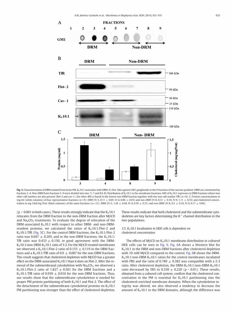
Fig. 3.Characterization of DRM isolated frombrain PM, KV10.1 associateswith DRM. A)Dot- blot against GM1 ganglioside in the 9 fractions of the sucrose gradient. DRM are constituted byfractions 2–4. Non-DRM from fractions 5–9were divided into two: 5–7 and 8,9. B) Distribution of KV10.1 in themembrane fractions; 60% of KV10.1 expresses in DRM fractionswhere twoother raft markers are also present (flot-2 and cav-1), the other 40% is found in the lowest non-DRM fraction together with the non-raft marker TfR (n=8). C) Protein concentrations inmg/ml (white columns) of four representative fractions (n=9): DRM (Fr:3, 0.11 ± 0.05; Fr:4, 0.06 ± 0.03) and non-DRM (Fr:8, 0.31 ± 0.16; Fr:9, 1.11 ± 0.53) and cholesterol concen-tration in mg Chol/mg Prot (black columns) of the same fractions (n=12): DRM (Fr:3, 1.28 ± 0.44; Fr:4, 0.39 ± 0.31) and non-DRM (Fr:8, 0.5 ± 0.19; Fr:9, 0.17 ± 0.06).
925A.M. Jiménez-Garduño et al. / Biochimica et Biophysica Acta 1838 (2014) 921–931
(p b 0.001 in both cases). These results strongly indicate that theKV10.1relocates from the DRM fraction to the non-DRM fraction after MβCDand Na2CO3 treatments. To evaluate the degree of relocation of theDRM-associated KV10.1 with respect to other DRM- and non-DRM-resident proteins, we calculated the ratios of KV10.1/Flot-2 andKV10.1/TfR (Fig. 5C). For the control DRM fractions, the KV10.1/Flot-2ratio was 0.697 ± 0.205, and in the non-DRM fractions, the KV10.1/TfR ratio was 0.433 ± 0.150, in good agreement with the DRM-KV10.1/non-DRM-KV10.1 ratio of 3:2. For theMβCD treatedmembraneswe observed a KV10.1/Flot-2 ratio of 0.155 ± 0.119 in the DRM frac-tions and a KV10.1/TfR ratio of 0.9 ± 0.087 for the non-DRM fractions.This result suggests that cholesterol depletion with MβCD has a greatereffect on the DRM-associated KV10.1 than it does on Flot-2. After the re-moval of the submembranal cytoskeleton with Na2CO3, we observed aKV10.1/Flot-2 ratio of 1.827 ± 0.561 for the DRM fractions and aKV10.1/TfR ratio of 0.939 ± 0.016 for the non-DRM fractions. Thus,our results show that the submembrane cytoskeleton is needed forproper PM protein partitioning of both KV10.1 and Flot-2. The effect ofthe detachment of the submembrane cytoskeletal proteins on KV10.1PM partitioning was stronger than the effect of cholesterol depletion.
These results indicate that both cholesterol and the submembrane cyto-skeleton are key factors determining the K+ channel distribution in thetwo populations.
3.5. KV10.1 localisation in HEK cells is dependent oncholesterol concentration
The effects of MβCD on KV10.1 membrane distribution in culturedHEK cells can be seen in Fig. 6. Fig. 6A shows a Western blot forKV10.1 in the DRM and non-DRM fractions after cholesterol depletionwith 10 mM MβCD compared to the control. Fig. 6B shows the DRM-KV10.1/non-DRM KV10.1 ratios for the control membranes incubatedwith PBS and the ratio of 0.789 ± 0.382 was compatible with a 2:3ratio. After cholesterol depletion, the DRM-KV10.1/non-DRM-KV10.1ratio decreased by 58% to 0.330 ± 0.220 (p b 0.01). These results,obtained from a cultured cell system, confirm that the cholesterol con-centration in the PM is essential for KV10.1 partitioning into thecholesterol-enriched membrane domains. When the cytoskeleton in-tegrity was altered, we also observed a tendency to decrease theamount of KV10.1 in the DRM domains, although the difference was

Fig. 4. Brain DRM characterization after cholesterol depletionwithMβCD (A–D) and after actin cytoskeleton detachmentwith Na2CO3 (E–H). A) Cholesterol (mg/mg Prot) depletion after1 h incubationwith 30 mMofMβCD. 81% of the inicial amount was extracted from the isolated PM (n = 8, control: 0.462 ± 0.1, MβCD: 0.09 ± 0.01, p b 0.01). B) Dot-blot against GM1.C) WB showing the distribution of the three domains markers flot-2, cav-1 and TfR in the different fractions. D) Protein concentrations in mg/ml (white columns) of four representativefractions (n=7): DRM (Fr:3, 0.07 ± 0.05; Fr:4, 0.09 ± 0.07) and non-DRM (Fr:8, 0.4 ± 0.18; Fr:9, 0.99 ± 0.56) and cholesterol concentration inmg chol/mg prot (black columns) of thesame fractions (n=8): DRM (Fr:3, 0.1 ± 0.08; Fr:4, 0.02 ± 0.02) and non-DRM (Fr:8, 0.29 ± 0.13; Fr:9, 0.14 ± 0.07). E) Densitometric analysis of actin detachment (normalized withTfR). Actin decreased in 88%. (n = 5, control: 1.69 ± 0.61, natrium carbonate: 0.21 ± 0.21, pb0.05). Inset: Representative Western blot of isolated PM after treatment with 150 mMof Na2CO3; TfR was used as control. F) Dot-blot against GM1. G)WB showing the distribution of the three domain markers flot-2, cav-1 and TfR in the different fractions. H) Protein con-centrations inmg/ml (white columns) of four representative fractions (n = 3): DRM (Fr:3, 0.13 ± 0.09; Fr:4, 0.15 ± 0.1) and non-DRM(Fr:8, 0.29 ± 0.08; Fr:9, 0.53 ± 0.29) and choles-terol concentration in mg Chol/mg Prot (black columns) of the same fractions (n=8): DRM (Fr:3, 0.82 ± 0.37; Fr:4, 0.34 ± 0.15) and non-DRM (Fr:8, 1.1 ± 0.57; Fr:9, 0.5 ± 0.2).
926 A.M. Jiménez-Garduño et al. / Biochimica et Biophysica Acta 1838 (2014) 921–931
not statistically significant (data not shown). To further investigate theeffect of MβCD on the HEK-KV10.1 cells, we performed confocal micros-copy experiments. Fig. 6C shows representative images of the co-localisation of KV10.1 (red) and CTX (green) after the MβCD treatment.The statistical analysis is shown in Fig. 6D; a significant decrease in thepercent of co-localisation is observed after the MβCD treatment(p b 0.001). In the case of Lat A treatment, the overlapping area showeda significant increase (data not showed). In contrast to the brain mem-branes, the cytoskeleton does not appear to play a key role in the KV10.1membrane distribution in the HEK-KV10.1 cells. We therefore investi-gated whether other factors could be contributing to the partitioningof these channels in the PM of the HEK-KV10.1 cells.
3.6. Ca2+/CaM binding modulates Eag1 DRM partitioning inHEK-KV10.1 cells
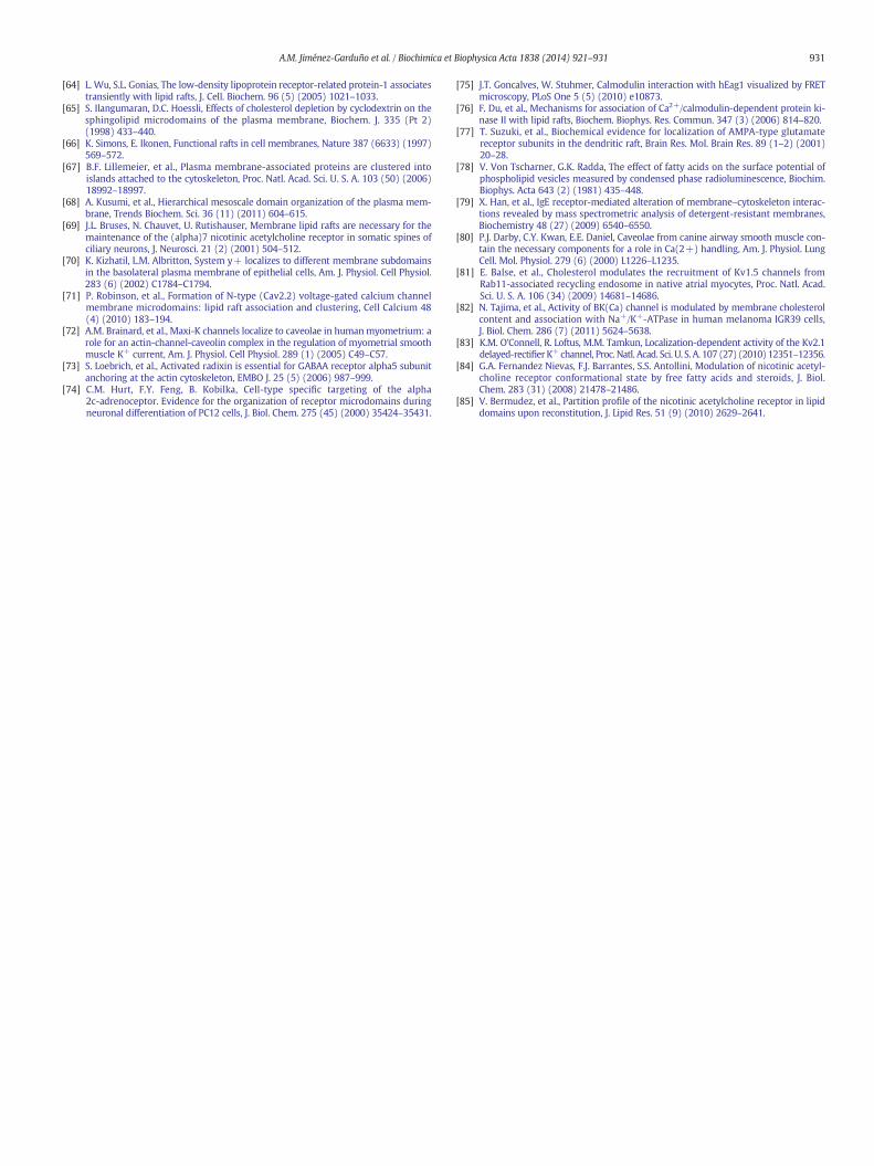
We transiently transfected HEK-293 cells with the C7 mutant (seeMaterials and methods) and isolated the DRM. Fig. 8A and B shows im-ages of a representativeWestern blot and the plotted ratios, respectively.A reduction in the DRM-associated KV10.1 is observed, reflected by the
decrease of the DRM-KV10.1/non-DRM-KV10.1 ratio to 0.233 ± 0.083(p b 0.01). This result strongly suggests that Ca2+/CaM is also a contrib-uting factor for the KV10.1 partitioning to the raft domains, wherepresumably the negative current modulation occurs.
3.7. KV10.1 currents in HEK cells increase after MβCD treatment
To determine the functional correlate of cholesterol depletion, wemeasured KV10.1 currents in whole-cell patch clamp experiments be-fore and after MβCD incubation (Fig. 7). After cholesterol extraction,the current density in response to a depolarisation to +60 mV in-creased by approximately two-fold, from 262.4 ± 25.1 pA/pF (n = 7)to 489.8 ± 79.37 pA/pF (n = 7, p b 0.05); representative recordingsare shown in Fig. 7A and B. In the current–voltage relationship shownin Fig. 7C, it can be appreciated that the increase was already significantin the range from +20 mV to +80 mV. Because the activation timeconstant of KV10.1 shows a characteristic dependence on pre-pulse po-tential [45], we also measured the 20–80% rise time of currents elicitedby a depolarisation to+40 mVafter 5 s prepulses to−120 or−60 mV.Cholesterol extraction did not affect the time required for the channel to

Fig. 5. Brain KV10.1 membrane distribution is affected by cholesterol depletion and actincytoskelton disturbance. A) Western blot against KV10.1 in the different fractions aftertreatment with MβCD and Na2CO3. B) KV10.1 ratio between the amount of KV10.1 inDRM divided by the amount in KV10.1-non-DRM in control (n = 8, 1.74 ± 0.64) andMβCD (n = 6, 0.18 ± 0.13) and Na2CO3 (n = 3, 0.15 ± 0.1) treated membranes. C)KV10.1 percentage normalized with the loading control of each fraction. For DRM the per-centage of flotilin was used (control: n=4, 0.7 ± 0.2; mβcd: n = 5, 0.15β0.12; Na2CO3:n=3, 1.83 ± 0.56), for non-DRM TfR percentage was used. (control: n = 4,0.43 ± 0.15; mβcd: n = 5, 0.9 ± 0.08; sodium carbonate: n = 3, 0.94 ± 0.03).
927A.M. Jiménez-Garduño et al. / Biochimica et Biophysica Acta 1838 (2014) 921–931
reach the open state. These results, together with our previous study[46], suggest that cytoskeleton integrity and cholesterol concentrationparticipate as stabilising factors for KV10.1 currents. Specifically, whenthese factors are disturbed, the currents increase.
4. Discussion
In the present study, we demonstrate that 1) KV10.1 is expressed inneural but not in glial cells ofmice; 2) two populations of KV10.1 partitioninto differentmembrane domains; 3) partitioning to theDRMdepends oncholesterol, the cytoskeleton and Ca2+/CaM; and 4) cholesterol depletionincreases KV10.1 K+ current density in HEK-KV10.1 cells.
4.1. KV10.1 is expressed in neural cells
The expression of KV10.1 in neurons has been demonstrated using insitu hybridisation and immunohistochemistry, but expression in glialcells has not been specifically addressed [13,43,44]. We did not observe
expression of KV10.1 in cultured mouse glia cells. This result is specifi-cally interesting because over 50% of human gliomas express KV10.1[47]. Thus, the presence of KV10.1 appears to be an abnormal acquiredexpression in gliomas, as it is in other solid tumours [31].
4.2. Partitioning of KV10.1 in plasma membrane subdomains
Several proteins, such as caveolins, flotillins, Src, Lck, Rab11, H-Ras,dystroglycans, BK channels, HERG, Kv1.5, Kv1.4 and Kv4.3 are knownto associate with detergent-resistant membrane domains enriched incholesterol and sphingolipids, while other transmembrane proteinssuch as TfR, calnexin,Na+/K+ATPase, K-ras, KV3.2 andKV4.2 are exclud-ed from these membrane domains [19,29,48–59]. Our results indicatethat the KV10.1 present in PM isolated from neurons distributesbetween the DRM and non-DRM fractions, suggesting the existence ofat least two populations of the protein with respect to their affinity tointeract with specific lipids and proteins. Whether KV10.1 partitioninginto the DRM is a stable or a transient state needs to be further investi-gated. KV10.1 thus belongs to the group of K-channels, such as Kir2 [60],KV2.1 [55] and KV4.1 [56], that have also been shown to partition to theDRM aswell as to the non-DRM domains. In order to reside in the DRM,the length of a transmembrane segment must match the thicknessof the cholesterol-enriched membranes [61–63]. This depends on pro-tein sequence, but changes in protein conformation can also inducemismatching with membrane thickness and therefore result in exclu-sion. Such conformational changes can be an effect of protein–proteinand protein–lipid interactions. For example, the kinase PKC-βII associ-ates with DRM, depending on the co-expression of the protein ZAP70in leukaemia cells [22]. The Low Density Lipoprotein Receptor 1 (LPR-1) resides in thenon-DRMdomains in culturedfibroblasts and epithelialcells but is present in the DRM isolated from a smooth muscle cell line[64]. This evidence indicates that the distribution process of membraneproteins involves a more complex regulation than the amino acidsequence alone. The factors involved in this regulation could includepost-translational events (phosphorylation and glycosylation), pro-tein–protein interactions within membrane domains, as the case ofCD45 and Lck associationswithin the immunological synapse [17] or in-teractions betweenmembrane proteins and cytoskeletal proteins as hasbeen shown for the transmission of the lateral force between β-dystroglycan and dystrophin in skeletal muscle [16]. In any case, differ-entmembrane distributions of a protein are incompatible with identicalstructures and conformations.
4.3. KV10.1 PM distribution is dependent on cholesterol, cytoskeletonintegrity and Ca2+/CaM binding
Cholesterol extraction with MβCD revealed a close relationship be-tween the concentration of this lipid and DRM/non-DRM partitioningof KV10.1 in brain and HEK cells. Although MβCD has a direct effect oncholesterol and an indirect effect on glycolipids (GM1) and proteins,there are reports that demonstrate that a fraction of the sphingolipidcore domain remains buoyant after cholesterol depletion [65]. We ob-served the same effect in our experiments (Fig. 5B), in which isolationof GM1-rich fractions was still possible despite the extraction of mem-brane cholesterol. There are several examples that demonstrate thatthe extraction of cholesterol with MβCD varies widely among mem-brane proteins and cell systems. Our results show that 30 mM MβCDreduces the cholesterol content in isolated neural PM by 80%, but only10 mM of MβCD is required to obtain the same effect in culturedHEK-KV10.1 cells. In other tissues, including the skeletal muscle, a10 mM concentration of the same compound reduces the membranecholesterol by only 12% but has a large effect on muscle contractility[16]. Furthermore, different proteins may behave differently afterMβCD treatment. For example, some proteins such as Lck dissociatecompletely from the DRM after cholesterol depletion, while others, in-cluding the antigen Thy-1 [65] and Flot-2, dissociate only partially

Fig. 6. KV10.1membrane distribution is affected by cholesterol depletion in HEK-KV10.1 cells. A)Western blot against KV10.1 in the fractions of isolated PM from HEK- KV10.1 after treat-ment with PBS or 10mMMβCD. B) KV10.1 ratio between KV10.1-DRM and the KV10.1-non-DRM of control and treated membranes (n = 6, p b 0.01). C and E) Merged confocal micros-copy images of HEK-BBS-KV10.1 cellsmarkedwith bungarotoxin-AlexaFluorâ 555 for KV10.1 signal (red) andCT-FITC for GM1 signal (green), treatedwith PBS or 5 mMofMβCD; scalebar:10 μm; Inset: Single channel images for each staining. D and F) Binary masks indicating the location of either single-stained BTX-AF555 (red) or GM1 (green) pixels, as well as areas inwhich both stains are present (yellow). G) Comparison of the percent overlapping area between Kv10.1 and GM1 treated with either PBS (control, n = 91) or MβCD (n = 92) in cellsoriginating from three different experiments (p b 0.0001).
928 A.M. Jiménez-Garduño et al. / Biochimica et Biophysica Acta 1838 (2014) 921–931
even after complete cholesterol extraction. The present study showsthat KV10.1 belongs to the second group. Because no direct interactionof KV10.1 with cav-1 was found, it seemed unlikely that cholesterolalone determined the location of KV10.1 at DRM; therefore, we lookedfor additional regulating factors.
Although it was not evident when the first lipid raft model was pro-posed [66], it is now accepted that in addition to the lipid content, theproteins associated with the PM are important for stabilising the do-main structures. Actin is the key regulator for clustering the proteinsin “islands” independent of the lipid content [67]; thus, the PM-boundcytoskeleton and transmembrane protein pickets form fences dividingthe cell surface into compartments of 40–300 nm [68]. Experimentswith Lat A andCytochalasin B indicate that the actinmeshwork integrityis important for the association of certain proteins including acetylcho-line receptors and anion, calcium, and potassium channels with theDRM [69–72]. In our study, the use of Na2CO3 removed almost 90% ofthe attached actin from the brain tissue PM. We show that in theabsence of the underlying protein structure, neuronal membraneslose their original organisation (Fig. 5E–H), domains are drasticallydisrupted and their detergent resistance is altered. These results are ingood agreement with the mesoscale membrane model, which statesthat the cytoskeleton-defined compartments are the highest hierarchi-cal level of the PM in determining and limiting the behaviour of the lipidphases [68]. However, the role of the cytoskeleton integrity in the asso-ciation of KV10.1 with the DRM in the HEK cells appears to be different;both biochemical and confocal microscopy results strongly indicate thatthe association of KV10.1 with the DRM remains, despite the effects ofLat A on actin polymerisation. Such disparities could be attributed to
the differences in the cytoskeleton structure of the two types of cells.In support to this concept, radixin, a protein that connects the α5-GABAA receptor to the actin cytoskeleton in brain tissue, does not inter-actwith the receptorwhen analysed inHEK-293 transfected cells [73]. Ithas been reported that the targeting of the alpha 2c adrenoreceptordepended not only on the cell type but also on the differentiationstate of a neuroendocrine cell line [74]. The different interactions be-tween KV10.1 and the cytoskeletal proteins that might be taking placein neurons compared to the HEK-KV10.1 cells could account for the dif-ferences observed in the channel dependence on actin integrity for DRMassociation.
Finally, we asked whether another factor besides cholesterol couldcontribute to KV10.1 PM partitioning in HEK-KV10.1 cells. Both the N-and the C-terminal domains of KV10.1 have very well defined bindingsites for Ca2+/CaM and a mutant channel (C7) resistant to the Ca2+-CaM binding effect has already been described [7,75]. We analysed thePM partitioning of KV10.1 when the C7 mutant was transfected toHEK-293 cells, and we observed a reduction in the DRM-associatedKV10.1 (Fig. 8). Although this is the first report where the binding ofCa2+/CaM is explicitly tested as a determining factor for a raft-proteinassociation, a closer examination into the literature provides additionalevidence indicating that this can be a general feature of CaM-bindingproteins. Du and co-workers reported that association of CaMKII mu-tants lacking the CaM binding site (291–301) to raft domains was par-tial or absent [76], compared to the behaviour of wild-type CaMKII[77]. Similarly, only the Ca2+-sensitive adenylyl cyclases partition tomembrane rafts, and the cytosolic domains (CaM binding sites) andnot the transmembrane spans are responsible for their partitioning
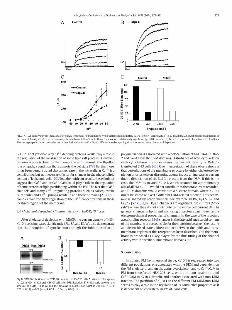
Fig. 7.KV10.1 density current increases afterMβCD treatment. Representativewhole cell recordings inHEK- KV10.1 cells A) control and B) in 30 mMMβCD. C) Graphical representation ofthe current density at different depolarizing stimuli; from+20 mV to +80 mV the increase is statistically significant (p b 0.05, n = 7). D) Time to rise of control and treated cells after a500 ms hyperpolarization pre-pulse and a depolaritzation to +40 mV; no difference in the opening time is observed after cholesterol depletion.
929A.M. Jiménez-Garduño et al. / Biochimica et Biophysica Acta 1838 (2014) 921–931
[21]. It is not yet clear why Ca2+-binding proteins would play a role inthe regulation of the localisation of some lipid raft proteins. However,calcium is able to bind to the membrane and diminish the flip-floprate of lipids, a condition that supports the gel state [78]. Furthermore,it has been demonstrated that an increase in the intracellular Ca2+ is acontributing, but not necessary, factor for changes in the phospholipidcontent of leukaemia cells [79]. Togetherwith our results, these findingssuggest that Ca2+ and/or Ca2+/CaM could play a role in the regulationof some protein or lipid partitioning within the PM. The fact that Ca2+
channels and many Ca2+-regulating proteins such as calsequestrin,calreticulin and Ca2+ pumps reside inside these domains [21,71,80]could explain the tight regulation of the Ca2+ concentrations in thoselocalised regions of the membrane.
4.4. Cholesterol-dependent K+-current density in HEK-KV10.1 cells
After cholesterol depletion with MβCD, the current density of HEK-KV10.1 cells increases significantly (Fig. 8A and B).We also demonstratethat the disruption of cytoskeleton through the inhibition of actin
Fig. 8.DRMdistribution of the C7KV10.1mutant inHEK-293 cells. A)Western blot againstKV10.1 in HEK- KV10.1 and HEK-C7 cells after DRM isolation. B) KV10.1 ratio between theamount of KV10.1 in DRM and the amount in KV10.1-non-DRM in control (n = 9,0.79 ± 0.13) and C7 (n = 4, 0.23 ± 0.04, p b 0.01) cells.
polymerisation is associated with a delocalisation of GM1, KV10.1, flot-2 and cav-1 from the DRM domains. Disturbance of actin cytoskeletonwith cytochalasin B also increases the current density of KV10.1-transfected CHO cells [46]. One interpretation of these observations isthat perturbations of the membrane structure by either cholesterol de-pletion or cytoskeleton-disrupting agents induce an increase in currentdue to dissociation of the KV10.1 protein from the DRM. If this is thecase, the DRM-associated KV10.1, which accounts for approximately60% of all PMKV10.1,would not contribute to the total current recorded,and DRM domains would constitute a discrete domain where KV10.1might be stored or exert a different DRM-related function. This behav-iour is shared by other channels, for example HERG, KV1.5, BK andCaV2.2 [57,71,81,82]. KV2.1 channels are organised into clusters (“cor-rals”) where they do not contribute to the whole cell current [83]. Ingeneral, changes in lipids and anchoring of proteins can influence theelectromechanical properties of channels. In the case of the nicotinicacetylcholine receptor [84], changes in the fatty acid and steroid contentof the membrane are responsible for the transition between the restingand desensitised states. Direct contact between the lipids and trans-membrane regions of this receptor has been described, and the mem-brane is proposed as a key player for the fine-tuning of the channelactivity within specific submembrane domains [85].
5. Conclusion
In isolated PM from neuronal tissue, KV10.1 is segregated into twodifferent populations, one associated with the DRM and dependent onthe PM cholesterol and on the actin–cytoskeleton and to Ca2+/CaM inPM from transfected HEK-293 cells, with a mutant unable to bindCa2+/CaM to Kv10.1 protein, and another associated with non-DRMfraction. The partition of KV10.1 to the different PM DRM/non-DRMseems to play a role in the regulation of its conductive properties as itis dependent on cholesterol in PM of living cells.

930 A.M. Jiménez-Garduño et al. / Biochimica et Biophysica Acta 1838 (2014) 921–931
Acknowledgements
We wish to thank the Consejo Nacional de Ciencia y Tecnología(CONACyT; #267035) in Mexico and the Deutsche AkademischeAustausch Dienst (DAAD; #A/08/74166) in Germany and the DirecciónGeneral del Personal Académico, Universidad Nacional Autónoma deMéxico (UNAM-DGAPA-IN219812) for their financial support to AMJGduring her PhD programme (Programa de Doctorado en CienciasBiomédicas, Universidad Nacional Autónoma de México); Dr. RoserUfartes and Sabine Martin for helpful suggestions on the manuscript;Sabine Klöppner, Barbara Scheufler, Tanja Nilson, Ursula Kutzke, BärbelHeidrich and Victor Díaz for technical support.We thank Pavel Vazquez(PAVNET) for the design of the graphical abstract.
References
[1] W.D. Kaplan, W.E. Trout III, The behavior of four neurological mutants of Drosophila,Genetics 61 (2) (1969) 399–409.
[2] V. Budnik, Y. Zhong, C.F. Wu, Morphological plasticity of motor axons in Drosophilamutants with altered excitability, J. Neurosci. 10 (11) (1990) 3754–3768.
[3] J.E. Engel, C.F. Wu, Genetic dissection of functional contributions of specific potassi-um channel subunits in habituation of an escape circuit in Drosophila, J. Neurosci. 18(6) (1998) 2254–2267.
[4] L.A. Pardo, et al., Oncogenic potential of EAG K(+) channels, EMBO J. 18 (20) (1999)5540–5547.
[5] A. Bruggemann, et al., Ether-a-go-go encodes a voltage-gated channel permeable toK+ and Ca2+ and modulated by cAMP, Nature 365 (6445) (1993) 445–448.
[6] T. Occhiodoro, et al., Cloning of a human ether-a-go-go potassium channelexpressed inmyoblasts at the onset of fusion, FEBS Lett. 434 (1–2) (1998) 177–182.
[7] R. Schonherr, K. Lober, S.H. Heinemann, Inhibition of human ether a go-go potassi-um channels by Ca(2+)/calmodulin, EMBO J. 19 (13) (2000) 3263–3271.
[8] L.C. Griffith, et al., Calcium/calmodulin-dependent protein kinase II and potassiumchannel subunit EAG similarly affect plasticity in Drosophila, Proc. Natl. Acad. Sci.U. S. A. 91 (21) (1994) 10044–10048.
[9] M. Ninkovic, et al., Physical and functional interaction of KV10.1 with Rabaptin-5impacts ion channel trafficking, FEBS Lett. 586 (19) (2012) 3077–3084.
[10] S. Herrmann, et al., Cortactin controls surface expression of the voltage-gated potas-sium channel K(V)10.1, J. Biol. Chem. 287 (53) (2012) 44151–44163.
[11] B.R. Downie, et al., Eag1 expression interferes with hypoxia homeostasis andinduces angiogenesis in tumors, J. Biol. Chem. 283 (52) (2008) 36234–36240.
[12] L.A. Pardo, et al., Cell cycle-related changes in the conducting properties of r-Eag K+
channels, J. Cell Biol. 143 (3) (1998) 767–775.[13] D. Gomez-Varela, et al., Characterization of Eag1 channel lateral mobility in rat hip-
pocampal cultures by single-particle-tracking with quantum dots, PLoS One 5 (1)(2010) e8858.
[14] H.J. Kaiser, et al., Order of lipid phases in model and plasma membranes, Proc. Natl.Acad. Sci. U. S. A. 106 (39) (2009) 16645–16650.
[15] A. Cherubini, et al., Human ether-a-go-go-related gene 1 channels are physicallylinked to beta1 integrins and modulate adhesion-dependent signaling, Mol. Biol.Cell 16 (6) (2005) 2972–2983.
[16] J. Vega-Moreno, et al., Cholesterol depletion uncouples beta-dystroglycans fromdiscrete sarcolemmal domains, reducing the mechanical activity of skeletal muscle,Cell. Physiol. Biochem. 29 (5–6) (2012) 905–918.
[17] C. Irles, et al., CD45 ectodomain controls interaction with GEMs and Lck activity foroptimal TCR signaling, Nat. Immunol. 4 (2) (2003) 189–197.
[18] C. Irles, et al., Plasma membrane subdomain partitioning of Lck in primary human Tlymphocytes, Can. J. Physiol. Pharmacol. 88 (4) (2010) 487–496.
[19] H. Zhao, et al., Neurite outgrowth is dependent on the association of c-Src and lipidrafts, Neurochem. Res. 34 (12) (2009) 2197–2205.
[20] S. Lebreton, S. Paladino, C. Zurzolo, Selective roles for cholesterol and actin in com-partmentalization of different proteins in the Golgi and plasma membrane of polar-ized cells, J. Biol. Chem. 283 (43) (2008) 29545–29553.
[21] A.J. Crossthwaite, et al., The cytosolic domains of Ca2+-sensitive adenylyl cyclasesdictate their targeting to plasma membrane lipid rafts, J. Biol. Chem. 280 (8)(2005) 6380–6391.
[22] C.M. zum Buschenfelde, et al., Recruitment of PKC-betaII to lipid rafts mediatesapoptosis-resistance in chronic lymphocytic leukemia expressing ZAP-70, Leukemia24 (1) (2010) 141–152.
[23] K. Bacia, et al., Fluorescence correlation spectroscopy relates rafts in model and na-tive membranes, Biophys. J. 87 (2) (2004) 1034–1043.
[24] C. Eggeling, et al., Direct observation of the nanoscale dynamics of membrane lipidsin a living cell, Nature 457 (7233) (2009) 1159–1162.
[25] E. London, D.A. Brown, Insolubility of lipids in Triton X-100: physical origin and re-lationship to sphingolipid/cholesterol membrane domains (rafts), Biochim. Biophys.Acta 1508 (1–2) (2000) 182–195.
[26] J. Sot, et al., Detergent-resistant, ceramide-enriched domains in sphingomyelin/ceramide bilayers, Biophys. J. 90 (3) (2006) 903–914.
[27] T. Lang, SNARE proteins and ‘membrane rafts’, J. Physiol. 585 (Pt 3) (2007) 693–698.[28] R.H. Scannevin, et al., Identification of a cytoplasmic domain important in the polar-
ized expression and clustering of the Kv2.1 K+ channel, J. Cell Biol. 135 (6 Pt 1)(1996) 1619–1632.
[29] J.R. Martens, K. O'Connell, M. Tamkun, Targeting of ion channels to membrane mi-crodomains: localization of KV channels to lipid rafts, Trends Pharmacol. Sci. 25(1) (2004) 16–21.
[30] A.K. Weaver, et al., BK channels are linked to inositol 1,4,5-triphosphate receptorsvia lipid rafts: a novel mechanism for coupling [Ca(2+)](i) to ion channel activa-tion, J. Biol. Chem. 282 (43) (2007) 31558–31568.
[31] B. Hemmerlein, et al., Overexpression of Eag1 potassium channels in clinical tu-mours, Mol. Cancer 5 (2006) 41.
[32] R.E. Garcia-Ferreiro, et al., Mechanism of block of hEag1 K+ channels by imipramineand astemizole, J. Gen. Physiol. 124 (4) (2004) 301–317.
[33] T. Kohl, et al., Rapid internalization of the oncogenic K+ channel K(V)10.1, PLoS One6 (10) (2011) e26329.
[34] J. Schindler, H.G. Nothwang, Enrichment of brain plasma membranes by affinitytwo-phase partitioning, Methods Mol. Biol. 528 (2009) 119–126.
[35] J.N. Larocca, W. Norton, Isolation of myelin, Current Protocols in Cell Biology, JohnWiley & Sons, 2007, pp. 1–19, (3.25).
[36] J. Schindler, et al., Proteomic analysis of brain plasmamembranes isolated by affinitytwo-phase partitioning, Mol. Cell. Proteomics 5 (2) (2006) 390–400.
[37] Y. Yao, et al., The differential protein and lipid compositions of noncaveolar lipid mi-crodomains and caveolae, Cell Res. 19 (4) (2009) 497–506.
[38] E. Kirchner, K. Gruhn, Comparative investigations on the precipitation behavior ofvarious protein precipitates in organs and tissues, Nahrung 29 (7) (1985) 703–709.
[39] Y. Chen, et al., Functional K(v)10.1 channels localize to the inner nuclear membrane,PLoS One 6 (5) (2011) e19257.
[40] O.P.Hamill, et al., Improvedpatch-clamp techniques for high-resolution current record-ing from cells and cell-free membrane patches, Pflugers Arch. 391 (2) (1981) 85–100.
[41] J. Schindelin, et al., Fiji: an open-source platform for biological-image analysis, Nat.Methods 9 (7) (2012) 676–682.
[42] H. Jurevics, et al., Normal metabolism but different physical properties of myelinfrom mice deficient in proteolipid protein, J. Neurosci. Res. 71 (6) (2003) 826–834.
[43] M.J. Saganich, E. Machado, B. Rudy, Differential expression of genes encodingsubthreshold-operating voltage-gated K+ channels in brain, J. Neurosci. 21 (13)(2001) 4609–4624.
[44] S. Martin, et al., Eag1 potassium channel immunohistochemistry in the CNS ofadult rat and selected regions of human brain, Neuroscience 155 (3) (2008)833–844.
[45] J. Ludwig, et al., Functional expression of a rat homologue of the voltage gated eithera go-go potassium channel reveals differences in selectivity and activation kineticsbetween the Drosophila channel and its mammalian counterpart, EMBO J. 13 (19)(1994) 4451–4458.
[46] J. Camacho, et al., Cytoskeletal interactions determine the electrophysiological prop-erties of human EAG potassium channels, Pflugers Arch. 441 (2–3) (2000) 167–174.
[47] S. Patt, et al., Expression of ether a go-go potassium channels in human gliomas,Neurosci. Lett. 368 (3) (2004) 249–253.
[48] P. de Diesbach, et al., Differential subcellular membrane recruitment of Src mayspecify its downstream signalling, Exp. Cell Res. 314 (7) (2008) 1465–1479.
[49] L. Rajendran, S. Le Lay, H. Illges, Raft association and lipid droplet targeting offlotillins are independent of caveolin, Biol. Chem. 388 (3) (2007) 307–314.
[50] P.H. Lommerse, et al., Single-molecule diffusion reveals similar mobility for the Lck,H-ras, and K-ras membrane anchors, Biophys. J. 91 (3) (2006) 1090–1097.
[51] H. Niv, et al., Activated K-Ras and H-Ras display different interactions with saturablenonraft sites at the surface of live cells, J. Cell Biol. 157 (5) (2002) 865–872.
[52] L.J. Foster, C.L. De Hoog, M. Mann, Unbiased quantitative proteomics of lipid rafts re-veals high specificity for signaling factors, Proc. Natl. Acad. Sci. U. S. A. 100 (10)(2003) 5813–5818.
[53] M.J. Yu, et al., Large-scale quantitative LC–MS/MS analysis of detergent-resistantmembrane proteins from rat renal collecting duct, Am. J. Physiol. Cell Physiol. 295(3) (2008) C661–C678.
[54] K.M. O'Connell, J.R. Martens, M.M. Tamkun, Localization of ion channels to lipid Raftdomains within the cardiovascular system, Trends Cardiovasc. Med. 14 (2) (2004)37–42.
[55] K.M. O'Connell, M.M. Tamkun, Targeting of voltage-gated potassium channel iso-forms to distinct cell surface microdomains, J. Cell Sci. 118 (Pt 10) (2005)2155–2166.
[56] F. Xia, et al., Targeting of voltage-gated K+ and Ca2+ channels and solubleN-ethylmaleimide-sensitive factor attachment protein receptor proteins tocholesterol-rich lipid rafts in pancreatic alpha-cells: effects on glucagonstimulus-secretion coupling, Endocrinology 148 (5) (2007) 2157–2167.
[57] J. Lin, et al., The regulation of the cardiac potassium channel (HERG) by caveolin-1,Biochem. Cell Biol. 86 (5) (2008) 405–415.
[58] J.R. Martens, et al., Differential targeting of Shaker-like potassium channels to lipidrafts, J. Biol. Chem. 275 (11) (2000) 7443–7446.
[59] R.S. Lam, A.R. Shaw, M. Duszyk, Membrane cholesterol content modulates activationof BK channels in colonic epithelia, Biochim. Biophys. Acta 1667 (2) (2004)241–248.
[60] S. Tikku, et al., Relationship between Kir2.1/Kir2.3 activity and their distributions be-tween cholesterol-rich and cholesterol-poor membrane domains, Am. J. Physiol. CellPhysiol. 293 (1) (2007) C440–C450.
[61] A. Kundu, et al., Transmembrane domain of influenza virus neuraminidase, a type IIprotein, possesses an apical sorting signal in polarized MDCK cells, J. Virol. 70 (9)(1996) 6508–6515.
[62] A.G. Lee, Lipid–protein interactions in biological membranes: a structural perspec-tive, Biochim. Biophys. Acta 1612 (1) (2003) 1–40.
[63] T.J. McIntosh, A. Vidal, S.A. Simon, Sorting of lipids and transmembrane peptidesbetween detergent-soluble bilayers and detergent-resistant rafts, Biophys. J. 85(3) (2003) 1656–1666.
931A.M. Jiménez-Garduño et al. / Biochimica et Biophysica Acta 1838 (2014) 921–931
[64] L. Wu, S.L. Gonias, The low-density lipoprotein receptor-related protein-1 associatestransiently with lipid rafts, J. Cell. Biochem. 96 (5) (2005) 1021–1033.
[65] S. Ilangumaran, D.C. Hoessli, Effects of cholesterol depletion by cyclodextrin on thesphingolipid microdomains of the plasma membrane, Biochem. J. 335 (Pt 2)(1998) 433–440.
[66] K. Simons, E. Ikonen, Functional rafts in cell membranes, Nature 387 (6633) (1997)569–572.
[67] B.F. Lillemeier, et al., Plasma membrane-associated proteins are clustered intoislands attached to the cytoskeleton, Proc. Natl. Acad. Sci. U. S. A. 103 (50) (2006)18992–18997.
[68] A. Kusumi, et al., Hierarchical mesoscale domain organization of the plasma mem-brane, Trends Biochem. Sci. 36 (11) (2011) 604–615.
[69] J.L. Bruses, N. Chauvet, U. Rutishauser, Membrane lipid rafts are necessary for themaintenance of the (alpha)7 nicotinic acetylcholine receptor in somatic spines ofciliary neurons, J. Neurosci. 21 (2) (2001) 504–512.
[70] K. Kizhatil, L.M. Albritton, System y+ localizes to different membrane subdomainsin the basolateral plasma membrane of epithelial cells, Am. J. Physiol. Cell Physiol.283 (6) (2002) C1784–C1794.
[71] P. Robinson, et al., Formation of N-type (Cav2.2) voltage-gated calcium channelmembrane microdomains: lipid raft association and clustering, Cell Calcium 48(4) (2010) 183–194.
[72] A.M. Brainard, et al., Maxi-K channels localize to caveolae in humanmyometrium: arole for an actin-channel-caveolin complex in the regulation of myometrial smoothmuscle K+ current, Am. J. Physiol. Cell Physiol. 289 (1) (2005) C49–C57.
[73] S. Loebrich, et al., Activated radixin is essential for GABAA receptor alpha5 subunitanchoring at the actin cytoskeleton, EMBO J. 25 (5) (2006) 987–999.
[74] C.M. Hurt, F.Y. Feng, B. Kobilka, Cell-type specific targeting of the alpha2c-adrenoceptor. Evidence for the organization of receptor microdomains duringneuronal differentiation of PC12 cells, J. Biol. Chem. 275 (45) (2000) 35424–35431.
[75] J.T. Goncalves, W. Stuhmer, Calmodulin interaction with hEag1 visualized by FRETmicroscopy, PLoS One 5 (5) (2010) e10873.
[76] F. Du, et al., Mechanisms for association of Ca2+/calmodulin-dependent protein ki-nase II with lipid rafts, Biochem. Biophys. Res. Commun. 347 (3) (2006) 814–820.
[77] T. Suzuki, et al., Biochemical evidence for localization of AMPA-type glutamatereceptor subunits in the dendritic raft, Brain Res. Mol. Brain Res. 89 (1–2) (2001)20–28.
[78] V. Von Tscharner, G.K. Radda, The effect of fatty acids on the surface potential ofphospholipid vesicles measured by condensed phase radioluminescence, Biochim.Biophys. Acta 643 (2) (1981) 435–448.
[79] X. Han, et al., IgE receptor-mediated alteration of membrane–cytoskeleton interac-tions revealed by mass spectrometric analysis of detergent-resistant membranes,Biochemistry 48 (27) (2009) 6540–6550.
[80] P.J. Darby, C.Y. Kwan, E.E. Daniel, Caveolae from canine airway smooth muscle con-tain the necessary components for a role in Ca(2+) handling, Am. J. Physiol. LungCell. Mol. Physiol. 279 (6) (2000) L1226–L1235.
[81] E. Balse, et al., Cholesterol modulates the recruitment of Kv1.5 channels fromRab11-associated recycling endosome in native atrial myocytes, Proc. Natl. Acad.Sci. U. S. A. 106 (34) (2009) 14681–14686.
[82] N. Tajima, et al., Activity of BK(Ca) channel is modulated by membrane cholesterolcontent and association with Na+/K+-ATPase in human melanoma IGR39 cells,J. Biol. Chem. 286 (7) (2011) 5624–5638.
[83] K.M. O'Connell, R. Loftus, M.M. Tamkun, Localization-dependent activity of the Kv2.1delayed-rectifierK+channel, Proc. Natl. Acad. Sci. U. S. A. 107 (27) (2010) 12351–12356.
[84] G.A. Fernandez Nievas, F.J. Barrantes, S.S. Antollini, Modulation of nicotinic acetyl-choline receptor conformational state by free fatty acids and steroids, J. Biol.Chem. 283 (31) (2008) 21478–21486.
[85] V. Bermudez, et al., Partition profile of the nicotinic acetylcholine receptor in lipiddomains upon reconstitution, J. Lipid Res. 51 (9) (2010) 2629–2641.