jftLVªh laö Mhö ,yö&33004@99 vlk/kj.k Hkkx II—[k.M 3—mi&[k.M (ii ...
56
96 GI/2018 (1) jftLVªh laö Mhö ,yö&33004@99 REGD. NO. D. L.-33004/99 vlk/kj.k EXTRAORDINARY Hkkx II—[k.M 3—mi&[k.M (ii) PART II—Section 3—Sub-section (ii) izkf/dkj ls izdkf'kr PUBLISHED BY AUTHORITY la- 61] ubZ fnYyh] 'kqØokj] tuojh 5] 2018@ikS"k 15] 1939 No. 61] NEW DELHI, FRIDAY, JANUARY 5, 2018/PAUSHA 15, 1939 Lkkekftd U;k; vkSj vf/kdkfjrk ea=ky; ¼fnO;kaxtu l'kfDrdj.k foHkkx½ vf/klwpuk ubZ fnYyh] 4 tuojh] 2018 dk-vk- 76¼v½-—tcfd fnO;ka xtu l“kfDrdj.k foHkkx] Lkkekftd U;k; vkSj vf/kdkfjrk ea =ky; us fofHkUu fu/kkZ fjr fnO;ka xrkvks a ds izek.ku ds fy, ew Y;ka du ,oa izfØ;k gs rq fn“kk&funs Z“k lq >kus ds fy, lfpo] fnO;ka xtu l“kfDrdj.k foHkkx dh v/;{krk es a rkjh[k 8 tqykbZ ] 2015 ds vkns “k ¼vuqca/k&I½ }kjk ,d fo“ks ’kK lfefr xfBr dh xbZ Fkh( vkSj tcfd fo“ks ’kK lfefr us 10 uoEcj] 2015 dks cSBd dh vkSj ;g fu.kZ; fy;k dh fuEufyf[kr Js f.k;ks a es a 8 mi lfefr;ka LFkkfir dh tk,a % ¼i½ xfrfo’k;d fnO;ka xrk ( ¼ii½ –f’V ckf/krk( ¼iii½ Jo.k ckf/krk ( ¼iv½ fpjdkfyd ra f=dk n“kk,a ( ¼v½ jDr la ca èkh fodkjks a ls çHkkfor O;fDr( ¼vi½ fodkl la ca èkh fodkj( ¼vii½ ekufld :X.krk( vkSj ¼viii½ cgq fnO;ka xrk ( vkSj tcfd fnO;ka xtu l“kfDrdj.k foHkkx }kjk mDr 8 mi&lfefr;ka rkjh[k 21 flrEcj] 2016] 3 vDrw cj] 2016 vkSj 23 tuojh] 2017 ds vkns “kks a }kjk LFkkfir dh xbZ FkhA vkSj tcfd mDr mi&lfefr;ka us foLr`r fopkj&foe“kZ ds ckn viuh fjiks Va s Z izLrqr dh vkSj bu fjiks Vks aZ dks lfpo] fnO;ka xtu l“kfDrdj.k foHkkx dh v/;{krk es a fo“ks ’kK lfefr }kjk tk¡p dh xbZ(
-
Upload
khangminh22 -
Category
Documents
-
view
1 -
download
0
Transcript of jftLVªh laö Mhö ,yö&33004@99 vlk/kj.k Hkkx II—[k.M 3—mi&[k.M (ii ...

96 GI/2018 (1)
jftLVªh laö Mhö ,yö&33004@99 REGD. NO. D. L.-33004/99
vlk/kj.k EXTRAORDINARY
Hkkx II—[k.M 3—mi&[k.M (ii) PART II—Section 3—Sub-section (ii)
izkf/dkj ls izdkf'kr PUBLISHED BY AUTHORITY
la- 61] ubZ fnYyh] 'kqØokj] tuojh 5] 2018@ikS"k 15] 1939 No. 61] NEW DELHI, FRIDAY, JANUARY 5, 2018/PAUSHA 15, 1939
Lkkekftd U;k; vkSj vf/kdkfjrk ea=ky;
¼fnO;kaxtu l'kfDrdj.k foHkkx½
vf/klwpuk
ubZ fnYyh] 4 tuojh] 2018
dk-vk- 76¼v½-—tcfd fnO;kaxtu l“kfDrdj.k foHkkx] Lkkekftd U;k; vkSj vf/kdkfjrk ea=ky; us fofHkUu fu/kkZfjr fnO;kaxrkvksa ds izek.ku ds fy, ewY;kadu ,oa izfØ;k gsrq fn“kk&funsZ“k lq>kus ds fy, lfpo] fnO;kaxtu l“kfDrdj.k foHkkx dh v/;{krk esa rkjh[k 8 tqykbZ] 2015 ds vkns“k ¼vuqca/k&I½ }kjk ,d fo“ks’kK lfefr xfBr dh xbZ Fkh(
vkSj tcfd fo“ks’kK lfefr us 10 uoEcj] 2015 dks cSBd dh vkSj ;g fu.kZ; fy;k dh fuEufyf[kr Jsf.k;ksa esa 8 mi lfefr;ka LFkkfir dh tk,a%
¼i½ xfrfo’k;d fnO;kaxrk (
¼ii½ –f’V ckf/krk(
¼iii½ Jo.k ckf/krk (
¼iv½ fpjdkfyd raf=dk n“kk,a(
¼v½ jDr lacaèkh fodkjksa ls çHkkfor O;fDr(
¼vi½ fodkl lacaèkh fodkj(
¼vii½ ekufld :X.krk( vkSj
¼viii½ cgq fnO;kaxrk (
vkSj tcfd fnO;kaxtu l“kfDrdj.k foHkkx }kjk mDr 8 mi&lfefr;ka rkjh[k 21 flrEcj] 2016] 3 vDrwcj] 2016 vkSj 23 tuojh] 2017 ds vkns“kksa }kjk LFkkfir dh xbZ FkhA
vkSj tcfd mDr mi&lfefr;ka us foLr`r fopkj&foe“kZ ds ckn viuh fjiksVasZ izLrqr dh vkSj bu fjiksVksaZ dks lfpo] fnO;kaxtu l“kfDrdj.k foHkkx dh v/;{krk esa fo“ks’kK lfefr }kjk tk¡p dh xbZ(
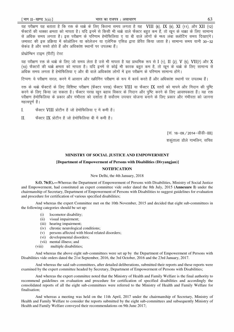
¹Hkkx IIµ[k.M 3(ii)º Hkkjr dk jkti=k % vlk/kj.k 63
;g ijh{k.k ;g crkrk gS fd jä ds Fkôs ds fy, fdruk le; yxrk gS ;g VIII ¼8½] IX ¼9½] XI ¼11½] vkSj XII ¼12½ QSDVjksa dh FkDdk {kerk dks ekirk gSA ;fn buesa ls fdlh Hkh Fkôs okys QSDVj cgqr de gSa] rks [kwu ds Fkôk ds fy, lkekU; ls vfèkd le; yxrk gSA bl ijh{k.k ds ifj.kke gseksfQfy;k , ;k ch okys yksxksa ds e/; yack DyksÇVx le; fn[kk,axsA tekoV dh bl çfØ;k esa dksvksfyu ;k dksystu ;k ,ysfxd ,flM }kjk çsfjr fd;k tkrk gSA lkekU; le; ;kuh 30&32 lsdaM gS vkSj lLrs gksrs gSa vkSj vfèkdka'k LFkkuksa ij miyCèk gSaA
çksFkzksfEcu Vkbe ¼ihVh½ VsLV
;g ijh{k.k jä ds Fkôs ds fy, tks le; ysrk gS mls Hkh ekirk gS ;g çkFkfed :i ls I ¼1½] II ¼2½] V ¼5½] VII¼7½ vkSj X ¼10½ QSDVjksa dh Fkôs {kerk dks ekirk gSA ;fn buesa ls dksà Hkh dkjd cgqr de gS] rks [kwu ds Fkôs ds fy, lkekU; ls vfèkd le; yxrk gS gseksfQfy;k , vkSj ch okys vfèkdka'k yksxksa esa bl ijh{k.k ds ifj.kke lkekU; gksaxsA
fVIi.k% ;s ijh{k.k ljy] djus esa vklku vkSj LØhÇux ijh{k.k ds :i esa dk;Z djrs gSa vkSj vfèkdka'k LFkkuksa ij miyCèk gSaA
jä ds Fkôs QSDVjksa ds fy, fof'k"V ijh{k.k ¼QSDVj ij[k½ QSDVj VIII ;k QSDVj IX Lrjksa dks ekius vkSj funku dh iqf"V djus ds fy, fd;k tk ldrk gSA QSDVj ij[k [kwu cgko fodkj ds funku vkSj iqf"V djus ds fy, vko';drk gSaA ;g jä ijh{k.k gseksfQfy;k ds çdkj vkSj xaHkhjrk dks n'kkZrk gS loksZÙke mipkj ;kstuk cukus ds fy, çdkj vkSj xaHkhjrk dks tkuuk egRoiw.kZ gSA
I- QSDVj VIII çksVhu gS tks gseksfQfy;k , esa deh gSA
II- QSDVj IX çksVhu gS tks gseksfQfy;k ch esa deh gSA
[la- 16&09@2014&MhMh&III]
'kdaqrkyk MkSys xkefyu] lfpo
MINISTRY OF SOCIAL JUSTICE AND EMPOWERMENT
[Department of Empowerment of Persons with Disabilities (Divyangjan)]
NOTIFICATION
New Delhi, the 4th January, 2018
S.O. 76(E).—Whereas the Department of Empowerment of Persons with Disabilities, Ministry of Social Justice
and Empowerment, had constituted an expert committee vide order dated the 8th July, 2015 (Annexure I) under the
chairmanship of Secretary, Department of Empowerment of Persons with Disabilities to suggest guidelines for evaluation
and procedure for certification of various specified disabilities;
And whereas the expert Committee met on the 10th November, 2015 and decided that eight sub-committees in
the following categories should be set up:
(i) locomotor disability;
(ii) visual impairment;
(iii) hearing impairment;
(iv) chronic neurological conditions;
(v) persons affected with blood related disorders;
(vi) developmental disorders;
(vii) mental illness; and
(viii) multiple disabilities;
And whereas the above eight sub-committees were set up by the Department of Empowerment of Persons with
Disabilities vide orders dated the 21st September, 2016, the 3rd October, 2016 and the 23rd January, 2017.
And whereas the said sub-committees, after detailed deliberations, submitted their reports and these reports were
examined by the expert committee headed by Secretary, Department of Empowerment of Persons with Disabilities;
And whereas the expert committee noted that the Ministry of Health and Family Welfare is the final authority to
recommend guidelines on evaluation and procedure for certification of specified disabilities and accordingly the
consolidated reports of all the eight sub-committees were referred to the Ministry of Health and Family Welfare for
finalisation;
And whereas a meeting was held on the 11th April, 2017 under the chairmanship of Secretary, Ministry of
Health and Family Welfare to consider the reports submitted by the eight sub-committees and subsequently Ministry of
Health and Family Welfare conveyed their recommendations on 9th June 2017;
64 THE GAZETTE OF INDIA : EXTRAORDINARY [PART II—SEC. 3(ii)]
Now, therefore, in exercise of powers conferred by Section 56 of the Rights of Persons with Disabilities Act,
2016 (49 of 2016), the Central Government hereby notifies the guidelines for the purpose of assessing the extent of
following specified disabilities in a person after having considered the recommendations of the Ministry of Health and
Family Welfare as provided at Annexure II, namely:-
I. locomotor disability including cerebral palsy, leprosy cured, dwarfism, acid attack victims and
muscular dystrophy;
II. blindness and low-vision;
III. deaf and hard of hearing and speech and language disability;
IV. intellectual disability and specific learning disabilities;
V. mental illness;
VI. chronic neurological conditions;
VII. haemophilia, thalassemia and sickle cell disease; and
VIII. multiple disabilities.
2. The said guidelines for the purpose of assessing disabilities at Annexure II shall supersede the guidelines for
evaluation of various disabilities and procedure for certification vide Government of India, Ministry of Social Justice and
Empowerment notification number 16-18/97-NI I. dated the 1st June 2001 and the guidelines for evaluation and
assessment of mental illness and procedure of certification vide Government of India, Ministry of Social Justice and
Empowerment notification number 16-18/97-NI dated the 18th
February 2002, except as respects things done or omitted
to be done before such supersession.
Note 1:- In terms of Section 57 of the Rights of the Persons with Disabilities Act, 2016 (49 of 2016), the State
Governments or as the case may be, Union Territory Administrators shall designate persons, having requisite
qualifications and experience, as certifying authorities, who shall be competent to issue the certificate of disability and
also notify the jurisdiction within which and the terms and conditions subject to which, the certifying authority shall
perform its certification functions.
Note 2:- The Director General of Health Services, Ministry of Health and Family Welfare, Government of India shall be
the final authority to decide upon cases where any controversy or doubt arises in matters relating to interpretation of the
definitions or classifications or evaluation procedure regarding the said guidelines.
Annexure I
File No. 16-09/2014-DD-III
Government of India
Ministry of Social Justice & Empowerment
Department of Empowerment of Persons with Disabilities
(DD-III Section)
Paryavaran Bhawan, CGO Complex,
Lodhi Road, New Delhi
Dated the 8th
July, 2015
ORDER
Sub:- Constitution of Committee to furnish guidelines for evaluation and certification of 12 newly identified
disabilities in the Rights of Persons with Disabilities Bill.
It has been decided with the approval of Hon’ble Minister (SJ&E) to constitute the Expert Committee to finalise
guidelines for evaluation and certification of 12 newly identified disabilities in the Rights of Persons with Disabilities
Bill, 2014 with the following composition:-
1. Secretary
Department of Empowerment of Persons with Disabilities,
Government of India
Chairman
2. Secretary
Ministry of Health & Family Welfare, Government of India
Member
3. Director
All India Institute of Medical Sciences,
New Delhi
Member
¹Hkkx IIµ[k.M 3(ii)º Hkkjr dk jkti=k % vlk/kj.k 65
4. Director General Health Services
Ministry of Health & Family Welfare
Nirman Bhawan, New Delhi
Member
5. Head of Department
Neurology,
Safdarjung Hospital,
New Delhi
Member
6. Head of Department
Psychiatry
Dr Ram Manohar Lohia Hospital,
New Delhi
Member
7. Head of Department
ENT
Safdarjung Hospital
New Delhi
Member
8. Head of Department
Hemotology
Safdarjung Hospital
New Delhi
Member
9. Head of Department
Ophthalmology
Dr Ram Manohar Lohia Hospital
New Delhi
Member
10. Head of Department
Paediatrics
Safdarjung Hospital
New Delhi
Member
11. Head of Department
PMR
Safdarjung Hospital
New Delhi
Member
12. Director,
Ali Yavar Jung National Institute for the Hearing Handicapped
Mumbai
Member
13. Director
National Institute of Mentally Handicapped
Manovikasnagar, Secunderabad
Member
14. Director
National Institute for Empowerment of Persons with Multiple
Disabilities, Tamil Nadu
Member
66 THE GAZETTE OF INDIA : EXTRAORDINARY [PART II—SEC. 3(ii)]
15. Director
National Institute for the Orthopedically Handicapped, Kolkata
Member
16. Director
National Institute for the Visually Handicapped, Uttarakhand
Member
17. Director
National Institute for Rehabilitation Training and Research, Cuttack
Member
18. Director
Pt Deendayal Upadhaya Institute for Physically Handicapped, New
Delhi
Member
19. Secretary,
Indian Council for Medical Research
Member
20. Joint Secretary
Department of Empowerment of Persons with Disabilities,
Paryavaran Bhawan, CGO Complex, New Delhi
Member
21. Director
Department of Empowerment of Persons with Disabilities,
Paryavaran Bhawan, CGO Complex, New Delhi
Convener
2. The terms of reference for the Committee are as follows:-
(a) The Expert Committee shall:
(i) review existing guidelines for evaluation and certification of various disabilities,
(ii) formulate guidelines for evaluation of newly introduced disabilities in the RPwD Bill, 2014 and
procedure for certification,
(iii) look into the best practices of certification prevailing across the nations.
(b) The Committee may co-opt any other member.
(c) Meetings of the Committee will be held in Delhi as per the convenience of the Chairman.
(d) TA/DA will be borne by the respective organization
(e) The Committee should submit its report within 6 months.
sd/-
(Awanish K. Awasthi)
Joint Secretary to Govt. of India
Tel.No. 24369056
To
1. All Members of the Committee
2. PS to Minister (SJ&E)
3. PS to Secretary (DEPwD)
4. PPS to JS (DEPwD)
5. PA to Director (DEPwD)
¹Hkkx IIµ[k.M 3(ii)º Hkkjr dk jkti=k % vlk/kj.k 67
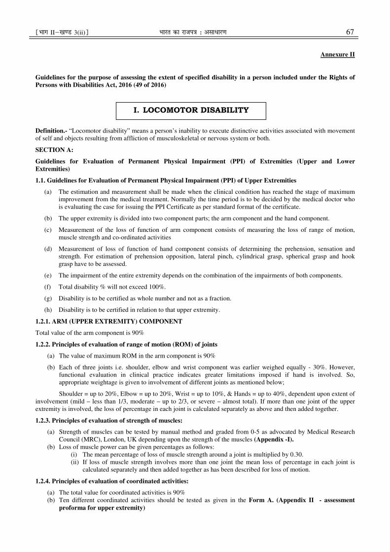
Annexure II
Guidelines for the purpose of assessing the extent of specified disability in a person included under the Rights of
Persons with Disabilities Act, 2016 (49 of 2016)
Definition.- “Locomotor disability” means a person’s inability to execute distinctive activities associated with movement
of self and objects resulting from affliction of musculoskeletal or nervous system or both.
SECTION A:
Guidelines for Evaluation of Permanent Physical Impairment (PPI) of Extremities (Upper and Lower
Extremities)
1.1. Guidelines for Evaluation of Permanent Physical Impairment (PPI) of Upper Extremities
(a) The estimation and measurement shall be made when the clinical condition has reached the stage of maximum
improvement from the medical treatment. Normally the time period is to be decided by the medical doctor who
is evaluating the case for issuing the PPI Certificate as per standard format of the certificate.
(b) The upper extremity is divided into two component parts; the arm component and the hand component.
(c) Measurement of the loss of function of arm component consists of measuring the loss of range of motion,
muscle strength and co-ordinated activities
(d) Measurement of loss of function of hand component consists of determining the prehension, sensation and
strength. For estimation of prehension opposition, lateral pinch, cylindrical grasp, spherical grasp and hook
grasp have to be assessed.
(e) The impairment of the entire extremity depends on the combination of the impairments of both components.
(f) Total disability % will not exceed 100%.
(g) Disability is to be certified as whole number and not as a fraction.
(h) Disability is to be certified in relation to that upper extremity.
1.2.1. ARM (UPPER EXTREMITY) COMPONENT
Total value of the arm component is 90%
1.2.2. Principles of evaluation of range of motion (ROM) of joints
(a) The value of maximum ROM in the arm component is 90%
(b) Each of three joints i.e. shoulder, elbow and wrist component was earlier weighed equally - 30%. However,
functional evaluation in clinical practice indicates greater limitations imposed if hand is involved. So,
appropriate weightage is given to involvement of different joints as mentioned below;
Shoulder = up to 20%, Elbow = up to 20%, Wrist = up to 10%, & Hands = up to 40%, dependent upon extent of
involvement (mild – less than 1/3, moderate – up to 2/3, or severe – almost total). If more than one joint of the upper
extremity is involved, the loss of percentage in each joint is calculated separately as above and then added together.
1.2.3. Principles of evaluation of strength of muscles:
(a) Strength of muscles can be tested by manual method and graded from 0-5 as advocated by Medical Research
Council (MRC), London, UK depending upon the strength of the muscles (Appendix -I).
(b) Loss of muscle power can be given percentages as follows:
(i) The mean percentage of loss of muscle strength around a joint is multiplied by 0.30.
(ii) If loss of muscle strength involves more than one joint the mean loss of percentage in each joint is
calculated separately and then added together as has been described for loss of motion.
1.2.4. Principles of evaluation of coordinated activities:
(a) The total value for coordinated activities is 90%
(b) Ten different coordinated activities should be tested as given in the Form A. (Appendix II - assessment
proforma for upper extremity)
I. LOCOMOTOR DISABILITY
68 THE GAZETTE OF INDIA : EXTRAORDINARY [PART II—SEC. 3(ii)]
(c) Each activity has a value of 9%
(d) Average normal range of different joints for reference is at Appendix III,
1.2.5. Combining values for the Arm Component:
The total value of loss of function of arm component is obtained by combining the value of loss of ROM, muscle strength
and coordinated activities, using the combining formula.
a + b (90-a)
90
where a = higher value and b = lower value
1.3.1. HAND COMPONENT:
(a) Total value of hand component is 90%
(b) The functional impairment of hand is expressed as loss of prehension, loss of sensation and loss of strength.
1.3.2. Principles of evaluation of prehension:
Total value of prehension is 30%
It includes:
(a) Opposition - 8%
Tested against - Index finger - 2%
- Middle finger - 2%
- Ring finger - 2%
- Little finger - 2%
(b) Lateral pinch - 5% - Tested by asking the patient to hold a key between the thumb and lateral side of index
finger.
(c) Cylindrical grasp - 6% Tested for
i. Large object of approx. 4 inches size - 3%
ii. Small object of 1-2 inch size - 3%
(d) Spherical grasp - 6% Tested for
i. Large object of approx. 4 inches size - 3%
ii. Small object of 1-2 inch size - 3%
(e) Hook grasp - 5% -Tested by asking the patient to lift a bag
1.3.3. Principles of Evaluation of sensation:
(a) Total value of sensation in hand is 30%.
(b) It shall be assessed according to the distribution given below:
(i) Complete loss of sensation
Thumb ray 9%
Index finger 6%
Middle finger 5%
Ring finger 5%
Little finger 5%
(ii) Partial loss of sensation: Assessment should be made according to percentage of loss of sensation in
thumb/finger(s).
1.3.4. Principles of Evaluation of strength
(a) Total value of strength is 30%.
(b) It includes:
(i) Grip strength 20%
(ii) Pinch strength 10%

¹Hkkx IIµ[k.M 3(ii)º Hkkjr dk jkti=k % vlk/kj.k 69
Strength of hand should be tested with hand dynamo-meter or by clinical method (grip method). 10% weightage to be
given to persons with involvement of dominant upper extremity (mostly right upper extremity) due to acquired
conditions (diseases/ injuries etc.).
For shortening of upper extremity, addition weightage is as follows:
First 1" - No additional weightage
For each 1" beyond first 1" - 2% additional weightage.
Additional weightage - A total of upto 10% additional weightage can be given to following accompanying factors if they
are continuous and persistent despite treatment.
(i) Deformity
In functional position 3%
In non-functional position 6%
(ii) Pain
Severe (grossly interfering with function) 9%
Moderate (interfering with function) 6%
Mild (slightly interfering with function) 3%
(iii) Loss of sensation
Complete Loss 9%
Partial Loss 6%
(iv) Complications
Superficial complications 3%
Deep complications 6%
Total % of PPI will not exceed 100% in any case.
Disability % is to be certified in relation to that extremity.
Disability % is to be mentioned as whole number, and not as a fraction.
1.3.5. Combining values of hand component:
The final value of loss of function of hand component is obtained by summing up values of loss of prehension, sensation
and strength.
1.3.6. Combining values for the Extremity:
Values of impairment of arm component and impairment of hand component should be added by using combining
formula:
a + b (90-a)/90
where a = higher value and b = lower value.
2. Guidelines for Evaluation of Permanent Physical Impairment in Lower Extremity
The measurement of loss of function in lower extremity is divided into two components, namely, mobility and stability
components.
2.1.1. MOBILITY COMPONENT
Total value of mobility component is 90% which includes range of movement (ROM) and muscle strength.
2.1.2. Principles of Evaluation of Range of Movement:
(a) The value of maximum range of movement in mobility component is 90%
(b) Each of three joints i.e. hip, knee and foot-ankle component was earlier weighed equally - 30%, but functional
evaluation in clinical practice indicates greater limitations imposed if major proximal or middle joints are
involved and, therefore, the appropriate weightage is given to involvement of proximal and middle joints, as
follows:
Hip= up to 35%, Knee= up to 35%, Ankle= up to 20%, dependent upon extent of involvement (mild – less than
1/3, moderate – up to 2/3, or severe – almost total).

70 THE GAZETTE OF INDIA : EXTRAORDINARY [PART II—SEC. 3(ii)]
If more than one joint of the limb is involved the mean loss of ROM in percentage should be calculated in
relation to individual joint separately and then added together to calculate the loss of mobility component in
relation to that particular limb.
2.1.3. Principle of Evaluation of Muscle Strength:
(a) The value for maximum muscle strength in the extremity is 90%.
(b) Strength of muscles can be tested by Manual Method and graded 0-5 depending upon the residual strength in the
muscle group.
(c) Manual muscle strength grading can be given percentage as below:
Numerical Score of Muscle Power Qualitative Score Loss of strength in %
0 Zero 100
1 Trace activity 80
2 Poor 60
3 Fair 40
4 Good 20
5 Normal 0
(d) Mean percentage of muscle strength loss around a joint is multiplied by 0.30 to calculate loss in relation to limb.
(e) If there has been a loss muscle strength involving more than one joint the values are added as has been described
for loss of ROM.
2.1.4. Combining values for mobility component:
The values of loss of ROM and loss of muscle strength should be combined with the help of combining formula: a+b
(90-a)/ 90 where a = higher value, b = lower value.
2.2. Stability Component
(a) Total value of the stability component is 90%
(b) It shall be tested by clinical method as given in Form B (Assessment Proforma for lower extremity) in
Appendix II. There are nine activities, which need to be tested, and each activity has a value of ten per cent
(10%). The percentage valued in relation to each activity depends upon the percentage of loss stability in
relation to each activity.
2.3. Extra Points
Extra points (% of impairment) are given for deformities, pain, contractures, loss of sensations and shortening etc.
For Shortening (true shortening and not apparent shortening)
First 1/2" Nil
Every 1/2" beyond first 1/2" 4%
Maximum extra points for associated problems such as deformity, pain, contractures etc. to be added are 10% (excluding
shortening).
(a) Deformity
In functional position 3%
In non-functional position 6%
(b) Pain
Severe (grossly interfering with function) 9%
Moderate (interfering with function) 6%
Mild (slightly interfering with function) 3%
(c) Loss of sensation
Complete Loss 9%
Partial Loss 6%
(d) Complications
¹Hkkx IIµ[k.M 3(ii)º Hkkjr dk jkti=k % vlk/kj.k 71
Superficial complications 3%
Deep complications 6%
SECTION B:
3. Guidelines for Evaluation of Permanent Physical Impairment of the Spine
Basic guidelines:
3.1. Permanent physical impairment caused by spinal injuries or deformity may change over the years, the certificate
issued in relation to spine may have to be reviewed as per the standard guidelines for disability certification.
3.2. Permanent physical impairment should be awarded in relation to the Spine.
1. TRAUMATIC LESIONS
Cervical Spine Injuries:
No. Cervical Spine Injuries
Percentage of PPI in relation to the
Spine
i. 25% or more compression of one or two adjacent vertebral bodies with
No involvement of posterior elements, No nerve root involvement,
moderate Neck rigidity and persistent Soreness.
20%
ii. Posterior element damage with radiological evidence of moderate
dislocation/subluxation including whiplash injury
A) With fusion healed, No permanent motor or sensory changes
B) Persistent pain with radiologically demonstrable instability.
10%
25%
iii. Severe Dislocation:
a) Fair to good reduction with or without fusion with no residual motor
or sensory involvement
b) Inadequate reduction with fusion and persistent radicular pain
10%
15%
Cervical Intervertebral Disc Lesions:
No. Cervical Intervertebral Disc Lesions Percentage of PPI In relation to
Spine
i. Treated case of disc lesion with persistent pain but no neurological
deficit
10%
ii. Treated case of disc lesion with pain and instability 15%
Thoracic and Thoracolumbar Spine Injuries:
No. Thoracic and Thoracolumbar Spine Injuries Percentage of PPI In relation to
Spine
i. Compression of less than 50% involving one vertebral body with no
neurological manifestation
10%
ii. Compression of more than 50% involving single vertebra or more with
involvement of posterior elements, healed, no neurological
manifestations persistent pain, fusion indicated
20%
iii. Same as (ii) with fusion, pain only on heavy use of back 15%
iv. Radiologically demonstrable instability with fracture or fracture
dislocation with persistent pain
30%
72 THE GAZETTE OF INDIA : EXTRAORDINARY [PART II—SEC. 3(ii)]
Lumbar and Lumbosacral Spine: Fracture
No. Lumbar and/or Lumbosacral Spine Fracture
Percentage of PPI In relation to
Spine
i. Compression of 25% or less of one or two adjacent Vertebral
bodies, No definite pattern, No neurological Deficit
10%
ii. Compression of more than 25% with disruption of Posterior
elements, persistent pain and stiffness, healed with or without
fusion, inability to lift more than 10 kgs.
20%
iii. Radiologically demonstrable instability in low lumbar or
Lumbosacral spine with pain
30%
Intervertebral Disc lesion:
No. Intervertebral Disc lesion Percentage of PPI In relation to
Spine
i. Treated case with persistent pain 10%
ii. Treated case with persistent pain and instability 20%
iii. Treated case with persistent pain and activities of lifting
moderately modified
25%
iv. Treated case with persistent pain and stiffness, aggravated by
heavy lifting necessitating modification of all activities requiring
heavy weight lifting
30%
4. Non Traumatic Lesions:
Scoliosis and/or Kyphoscoliosis:
4.1. Scoliosis is a condition in which an individual's spine has lateral, or side to side curvature. Although scoliosis is a
three-dimensional deformity, on an x-ray, scoliosis curves can often look like a simple “S” or a “C” shape.
4.2. Scoliosis is defined with radiographs that includes a standing x-ray of the entire spine antero-posterior view, as well
as the lateral view. Curve magnitude is measured in degrees using the Cobb method. A straight spine has a curve of 0º;
any curve greater than 10º is considered scoliosis. Between 0ºand 10º is considered "postural asymmetry" which is not
true scoliosis. The lateral radiograph is used to determine the thoracic kyphosis (or roundback appearance) and the
amount of lumbar lordosis (swayback).
4.3. In general, the severity of the scoliosis depends on the degree of the curvature and whether it threatens vital organs,
specifically the lungs and heart. The percentage of PPI shall be as follows:-
Group Cobb Angle % of permanent
impairment
Group 1 10-20 degrees 1 to 5
Group 2 21-30 degrees 6 to 9
Group 3 31-50 degrees 10 to 19
Group 4 51-75 degrees 20 to 29
Group 5 76-100 degrees 30 to 39
Group 6 101-125 degrees 40 to 60
Group 7 126 degrees or greater More than 60
¹Hkkx IIµ[k.M 3(ii)º Hkkjr dk jkti=k % vlk/kj.k 73
4.4. A person with scoliosis or kyphoscoliosis should be assessed for cardiorespiratory limitations if present. Additional
weightage in % of permanent is to be given according to severity of involvement as assessed clinically or relevant
investigations mentioned in the Guidelines under respective section.
4.5. In cases with scoliosis of severe type cardiopulmonary function tests and percentage deviation from normal shall be
assessed by one of the following method whichever seems more reliable clinically at the time of assessment. The value
thus obtained shall be added by combining formula.
(a) Chest Expansion
No. Maximum Chest Expansion % PPI
i. More than 4 cm Nil
ii. 3 cm. to 4 cm. 5
iii. 2 cm. to less than 3 cm 10
iv. 1 cm. to less than 2 cm 15
v. Less than 1 cm. 20
(b) Counting in one breath:
No. Single breath count % PPI
i. More than 40 Nil
ii. 31 to 40 5
iii. 21 to 30 10
iv. 11 to 20 15
v. 5 to 10 20
vi. Less than 5 25
The additional weightage is to be added using combining formula: a+b (90-a)/ 90 (a = higher value, b = lower value).
4.6. Torso Imbalance:
In addition to the above PPI should also be evaluated in relation the torso imbalance. The torso imbalance should be
measured by dropping a plumb line from C7 spine and measuring the distance of plumb line from gluteal crease.
Deviation of Plumb line PPI
Up to 1.5 cm 4%
1.6 – 3.0 cm 8%
3.1 – 5.0 cm 16%
5.1 and above 32%
Head Tilt over C7 spine PPI
Up to 15⁰ 4%
More than 15⁰ 10%
Associated Problems as given below: To be added directly but the total value of PPI in relation to trunk should
not exceed 100%.
(a) Pain
-mildly interfering with ADL* 4%
-moderately restricting ADL 6%
-severely restricting ADL 10%
* ADL - Activities of Daily Living
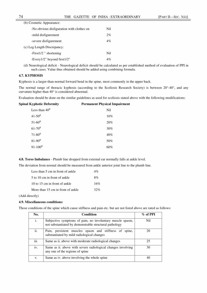
74 THE GAZETTE OF INDIA : EXTRAORDINARY [PART II—SEC. 3(ii)]
(b) Cosmetic Appearance:
-No obvious disfiguration with clothes on Nil
-mild disfigurement 2%
-severe disfigurement 4%
(c) Leg Length Discrepancy:
-First1/2 " shortening Nil
-Every1/2" beyond first1/2" 4%
(d) Neurological deficit - Neurological deficit should be calculated as per established method of evaluation of PPI in
such cases. Value thus obtained should be added using combining formula.
4.7. KYPHOSIS
Kyphosis is a larger-than-normal forward bend in the spine, most commonly in the upper back.
The normal range of thoracic kyphosis (according to the Scoliosis Research Society) is between 20°-40°, and any
curvature higher than 40° is considered abnormal.
Evaluation should be done on the similar guidelines as used for scoliosis stated above with the following modifications:
Spinal Kyphotic Deformity Permanent Physical Impairment
Less than 40⁰ Nil
41-50⁰ 10%
51-60⁰ 20%
61-70⁰ 30%
71-80⁰ 40%
81-90⁰ 50%
91-100⁰ 60%
4.8. Torso Imbalance - Plumb line dropped from external ear normally falls at ankle level.
The deviation from normal should be measured from ankle anterior joint line to the plumb line.
Less than 5 cm in front of ankle 4%
5 to 10 cm in front of ankle 8%
10 to 15 cm in front of ankle 16%
More than 15 cm in front of ankle 32%
(Add directly)
4.9. Miscellaneous conditions:
Those conditions of the spine which cause stiffness and pain etc. but are not listed above are rated as follows:
No. Condition % of PPI
i. Subjective symptoms of pain, no involuntary muscle spasm,
not substantiated by demonstrable structural pathology
Nil
ii. Pain, persistent muscles spasm and stiffness of spine,
substantiated by mild radiological changes
20
iii. Same as ii. above with moderate radiological changes 25
iv. Same as ii. above with severe radiological changes involving
any one of the regions of spine
30
v. Same as iv. above involving the whole spine 40

¹Hkkx IIµ[k.M 3(ii)º Hkkjr dk jkti=k % vlk/kj.k 75
SECTION C:
5. Guidelines for Evaluation of Permanent Physical Impairment in Persons with Amputation (Amputees):
5.1. Basic Guidelines:
(a) In cases of multiple amputees, the % of permanent impairment is to be computed by using the combining
formula: a+b (90-a)/ 90 (a = higher value, b = lower value).
(b) If the stump is unfit for fitting the prosthesis additional weightage of 5% should be added to the value.
(c) Any complication in form of stiffness of proximal joint, neuroma, infection, etc., should be given upto a total of
10% additional weightage.
(d) Involvement of dominant upper limb (right upper limb in majority of individuals) in acquired amputation should
be given 10% additional weightage.
5.2. Upper Limb Amputations:
No. Level of Upper Limb Amputation % of permanent
impairment in relation to
that specific limb
1. Fore-quarter amputation 100
2. Shoulder Disarticulation 90
3. Trans Humeral (Above Elbow) upto upper 1/3 of arm 85
4. Trans Humeral (Above Elbow) upto lower 1/3 of arm 80
5. Elbow disarticulation 75
6. Trans Radial (Below Elbow) upto upper 1/3 of forearm 70
7. Trans Radial (Below Elbow) upto lower 1/3 of forearm 65
8. Wrist disarticulation 60
9. Hand through carpal bones 55
10. Thumb through C.M. or though 1st MC joint 30
11. Thumb disarticulation through metacarpophalangeal Joint or through
proximal phalanx
25
12. Thumb disarticulation through inter phalangeal joint or Through distal
phalanx
15
13. Amputation through Proximal phalanx or Disarticulation through MP joint of
Index finger
Middle finger
Ring finger
Little finger
15
5
3
2
14. Amputation through Middle phalanx or Disarticulation through PIP joint of
Index finger
Middle finger
Ring finger
Little finger
10
4
2
1
15. Amputation through Distal phalanx or disarticulation through DIP joint of
Index finger
Middle finger
Ring finger
Little finger
5
2
1
1
76 THE GAZETTE OF INDIA : EXTRAORDINARY [PART II—SEC. 3(ii)]
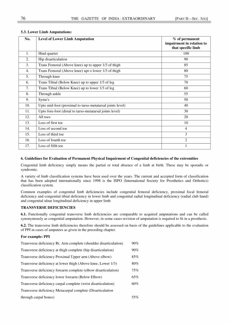
5.3. Lower Limb Amputations:
No. Level of Lower Limb Amputation % of permanent
impairment in relation to
that specific limb
1. Hind quarter 100
2. Hip disarticulation 90
3. Trans Femoral (Above knee) up to upper 1/3 of thigh 85
4. Trans Femoral (Above knee) upt o lower 1/3 of thigh 80
5. Through knee 75
6. Trans Tibial (Below Knee) up to upper 1/3 of leg 70
7. Trans Tibial (Below Knee) up to lower 1/3 of leg 60
8. Through ankle 55
9. Syme's 50
10. Upto mid-foot (proximal to tarso-metatarsal joints level) 40
11. Upto fore-foot (distal to tarso-metatarsal joints level) 30
12. All toes 20
13. Loss of first toe 10
14. Loss of second toe 4
15. Loss of third toe 3
16. Loss of fourth toe 2
17. Loss of fifth toe 1
6. Guidelines for Evaluation of Permanent Physical Impairment of Congenital deficiencies of the extremities
Congenital limb deficiency simply means the partial or total absence of a limb at birth. These may be sporadic or
syndromic.
A variety of limb classification systems have been used over the years. The current and accepted form of classification
that has been adopted internationally since 1998 is the ISPO (International Society for Prosthetics and Orthotics)
classification system.
Common examples of congenital limb deficiencies include congenital femoral deficiency, proximal focal femoral
deficiency and congenital tibial deficiency in lower limb and congenital radial longitudinal deficiency (radial club hand)
and congenital ulnar longitudinal deficiency in upper limb.
TRANSVERSE DEFICIENCIES
6.1. Functionally congenital transverse limb deficiencies are comparable to acquired amputations and can be called
synonymously as congenital amputation. However, in some cases revision of amputation is required to fit in a prosthesis.
6.2. The transverse limb deficiencies therefore should be assessed on basis of the guidelines applicable to the evaluation
of PPI in cases of amputees as given in the preceding chapter.
For example: PPI
Transverse deficiency Rt. Arm complete (shoulder disarticulation) 90%
Transverse deficiency at thigh complete (hip disarticulation) 90%
Transverse deficiency Proximal Upper arm (Above elbow) 85%
Transverse deficiency at lower thigh (Above knee, Lower 1/3) 80%
Transverse deficiency forearm complete (elbow disarticulation) 75%
Transverse deficiency lower forearm (Below Elbow) 65%
Transverse deficiency carpal complete (wrist disarticulation) 60%
Transverse deficiency Metacarpal complete (Disarticulation
through carpal bones) 55%
¹Hkkx IIµ[k.M 3(ii)º Hkkjr dk jkti=k % vlk/kj.k 77
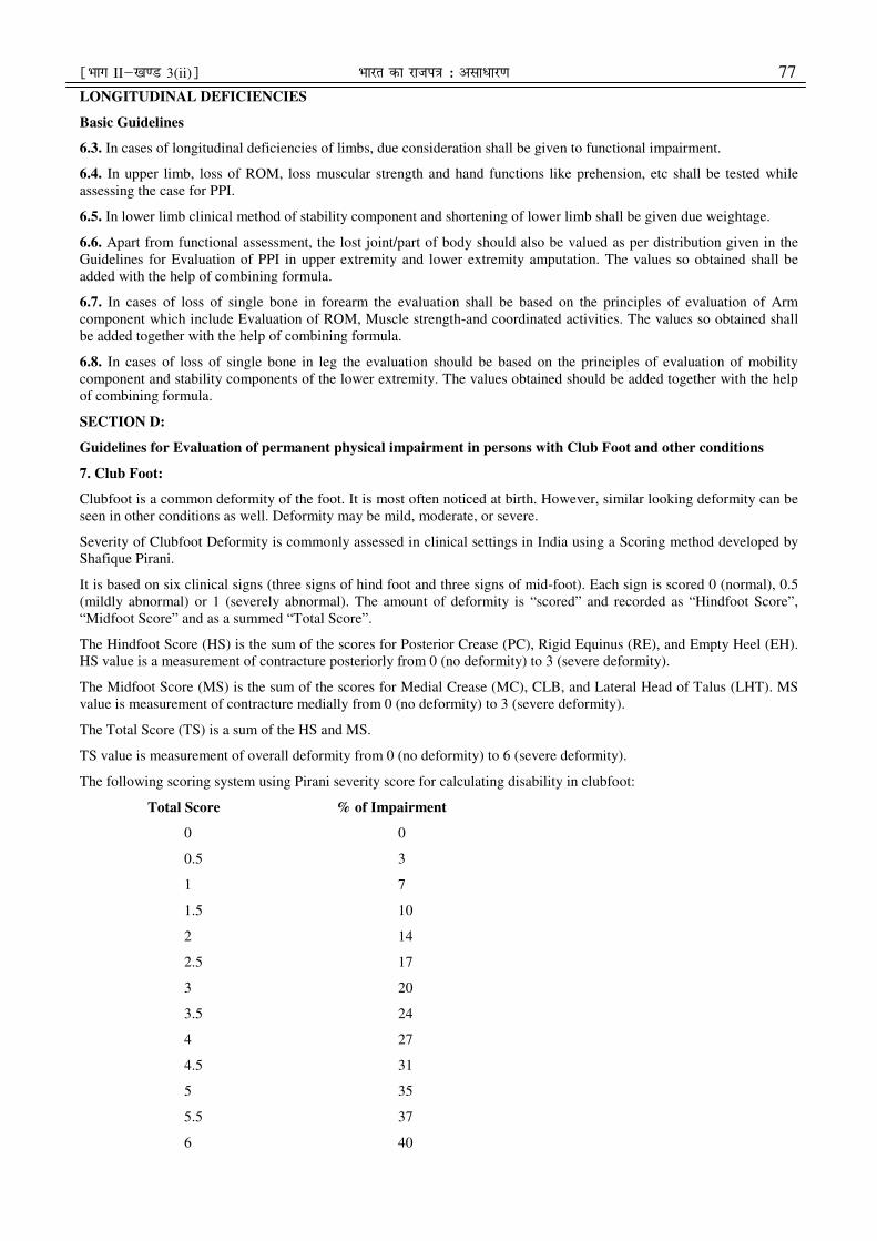
LONGITUDINAL DEFICIENCIES
Basic Guidelines
6.3. In cases of longitudinal deficiencies of limbs, due consideration shall be given to functional impairment.
6.4. In upper limb, loss of ROM, loss muscular strength and hand functions like prehension, etc shall be tested while
assessing the case for PPI.
6.5. In lower limb clinical method of stability component and shortening of lower limb shall be given due weightage.
6.6. Apart from functional assessment, the lost joint/part of body should also be valued as per distribution given in the
Guidelines for Evaluation of PPI in upper extremity and lower extremity amputation. The values so obtained shall be
added with the help of combining formula.
6.7. In cases of loss of single bone in forearm the evaluation shall be based on the principles of evaluation of Arm
component which include Evaluation of ROM, Muscle strength-and coordinated activities. The values so obtained shall
be added together with the help of combining formula.
6.8. In cases of loss of single bone in leg the evaluation should be based on the principles of evaluation of mobility
component and stability components of the lower extremity. The values obtained should be added together with the help
of combining formula.
SECTION D:
Guidelines for Evaluation of permanent physical impairment in persons with Club Foot and other conditions
7. Club Foot:
Clubfoot is a common deformity of the foot. It is most often noticed at birth. However, similar looking deformity can be
seen in other conditions as well. Deformity may be mild, moderate, or severe.
Severity of Clubfoot Deformity is commonly assessed in clinical settings in India using a Scoring method developed by
Shafique Pirani.
It is based on six clinical signs (three signs of hind foot and three signs of mid-foot). Each sign is scored 0 (normal), 0.5
(mildly abnormal) or 1 (severely abnormal). The amount of deformity is “scored” and recorded as “Hindfoot Score”,
“Midfoot Score” and as a summed “Total Score”.
The Hindfoot Score (HS) is the sum of the scores for Posterior Crease (PC), Rigid Equinus (RE), and Empty Heel (EH).
HS value is a measurement of contracture posteriorly from 0 (no deformity) to 3 (severe deformity).
The Midfoot Score (MS) is the sum of the scores for Medial Crease (MC), CLB, and Lateral Head of Talus (LHT). MS
value is measurement of contracture medially from 0 (no deformity) to 3 (severe deformity).
The Total Score (TS) is a sum of the HS and MS.
TS value is measurement of overall deformity from 0 (no deformity) to 6 (severe deformity).
The following scoring system using Pirani severity score for calculating disability in clubfoot:
Total Score % of Impairment
0 0
0.5 3
1 7
1.5 10
2 14
2.5 17
3 20
3.5 24
4 27
4.5 31
5 35
5.5 37
6 40
78 THE GAZETTE OF INDIA : EXTRAORDINARY [PART II—SEC. 3(ii)]
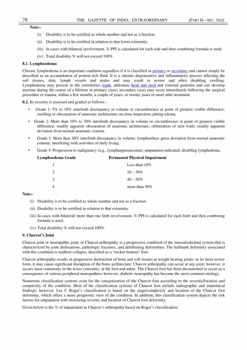
Note:-
(i) Disability is to be certified as whole number and not as a fraction.
(ii) Disability is to be certified in relation to that lower extremity.
(iii) In cases with bilateral involvement, % PPI is calculated for each side and then combining formula is used.
(iv) Total disability % will not exceed 100%.
8.1. Lymphoedema:
Chronic lymphedema is an important condition regardless if it is classified as primary or secondary and cannot simply be
described as an accumulation of protein-rich fluid. It is a chronic degenerative and inflammatory process affecting the
soft tissues, skin, lymph vessels and nodes and may result in severe and often disabling swelling.
Lymphedema may present in the extremities, trunk, abdomen, head and neck and external genitalia and can develop
anytime during the course of a lifetime in primary cases; secondary cases may occur immediately following the surgical
procedure or trauma, within a few months, a couple of years, or twenty years or more after treatment.
8.2. Its severity is assessed and graded as follows:-
• Grade 1: 5% to 10% interlimb discrepancy in volume or circumference at point of greatest visible difference;
swelling or obscuration of anatomic architecture on close inspection; pitting edema.
• Grade 2: More than 10% to 30% interlimb discrepancy in volume or circumference at point of greatest visible
difference; readily apparent obscuration of anatomic architecture; obliteration of skin folds; readily apparent
deviation from normal anatomic contour.
• Grade 3: More than 30% interlimb discrepancy in volume; lymphorrhea; gross deviation from normal anatomic
contour; interfering with activities of daily living.
• Grade 4: Progression to malignancy (e.g., lymphangiosarcoma); amputation indicated; disabling lymphedema.
Lymphoedema Grade Permanent Physical Impairment
1 Less than 10%
2 10 – 39%
3 40 – 50%
4 more than 50%
Note:-
(i) Disability is to be certified as whole number and not as a fraction.
(ii) Disability is to be certified in relation to that extremity.
(iii) In cases with bilateral/ more than one limb involvement, % PPI is calculated for each limb and then combining
formula is used.
(iv) Total disability % will not exceed 100%.
9. Charcot’s Joint
Charcot joint or neuropathic joint, or Charcot arthropathy is a progressive condition of the musculoskeletal system that is
characterized by joint dislocations, pathologic fractures, and debilitating deformities. The hallmark deformity associated
with this condition is midfoot collapse, described as a “rocker-bottom” foot.
Charcot arthropathy results in progressive destruction of bone and soft tissues at weight bearing joints; in its most severe
form, it may cause significant disruption of the bony architecture. Charcot arthropathy can occur at any joint; however, it
occurs most commonly in the lower extremity, at the foot and ankle. The Charcot foot has been documented to occur as a
consequence of various peripheral neuropathies; however, diabetic neuropathy has become the most common etiology.
Numerous classification systems exist for the categorization of the Charcot foot according to the severity/location and
complexity of the condition. Most of the classification systems of Charcot foot include radiographic and anatomical
findings; however, Lee C Roger’s classification is based on the stage/complexity and location of the Charcot foot
deformity, which offers a more prognostic view of the condition. In addition, this classification system depicts the risk
factors for amputation with increasing severity and location of Charcot foot deformity.
Given below is the % of impairment in Charcot’s arthropathy based on Roger’s classification:
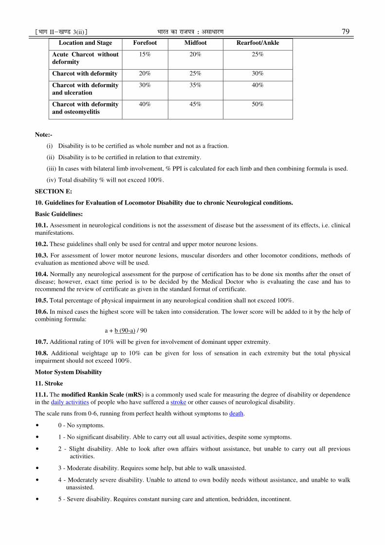
¹Hkkx IIµ[k.M 3(ii)º Hkkjr dk jkti=k % vlk/kj.k 79
Location and Stage Forefoot Midfoot Rearfoot/Ankle
Acute Charcot without
deformity
15% 20% 25%
Charcot with deformity 20% 25% 30%
Charcot with deformity
and ulceration
30% 35% 40%
Charcot with deformity
and osteomyelitis
40% 45% 50%
Note:-
(i) Disability is to be certified as whole number and not as a fraction.
(ii) Disability is to be certified in relation to that extremity.
(iii) In cases with bilateral limb involvement, % PPI is calculated for each limb and then combining formula is used.
(iv) Total disability % will not exceed 100%.
SECTION E:
10. Guidelines for Evaluation of Locomotor Disability due to chronic Neurological conditions.
Basic Guidelines:
10.1. Assessment in neurological conditions is not the assessment of disease but the assessment of its effects, i.e. clinical
manifestations.
10.2. These guidelines shall only be used for central and upper motor neurone lesions.
10.3. For assessment of lower motor neurone lesions, muscular disorders and other locomotor conditions, methods of
evaluation as mentioned above will be used.
10.4. Normally any neurological assessment for the purpose of certification has to be done six months after the onset of
disease; however, exact time period is to be decided by the Medical Doctor who is evaluating the case and has to
recommend the review of certificate as given in the standard format of certificate.
10.5. Total percentage of physical impairment in any neurological condition shall not exceed 100%.
10.6. In mixed cases the highest score will be taken into consideration. The lower score will be added to it by the help of
combining formula:
a + b (90-a) / 90
10.7. Additional rating of 10% will be given for involvement of dominant upper extremity.
10.8. Additional weightage up to 10% can be given for loss of sensation in each extremity but the total physical
impairment should not exceed 100%.
Motor System Disability
11. Stroke
11.1. The modified Rankin Scale (mRS) is a commonly used scale for measuring the degree of disability or dependence
in the daily activities of people who have suffered a stroke or other causes of neurological disability.
The scale runs from 0-6, running from perfect health without symptoms to death.
• 0 - No symptoms.
• 1 - No significant disability. Able to carry out all usual activities, despite some symptoms.
• 2 - Slight disability. Able to look after own affairs without assistance, but unable to carry out all previous
activities.
• 3 - Moderate disability. Requires some help, but able to walk unassisted.
• 4 - Moderately severe disability. Unable to attend to own bodily needs without assistance, and unable to walk
unassisted.
• 5 - Severe disability. Requires constant nursing care and attention, bedridden, incontinent.
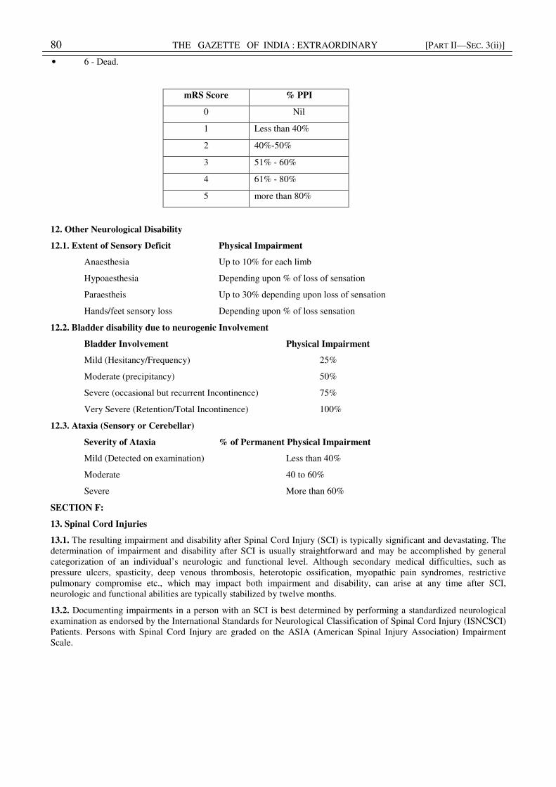
80 THE GAZETTE OF INDIA : EXTRAORDINARY [PART II—SEC. 3(ii)]
• 6 - Dead.
mRS Score % PPI
0 Nil
1 Less than 40%
2 40%-50%
3 51% - 60%
4 61% - 80%
5 more than 80%
12. Other Neurological Disability
12.1. Extent of Sensory Deficit Physical Impairment
Anaesthesia Up to 10% for each limb
Hypoaesthesia Depending upon % of loss of sensation
Paraestheis Up to 30% depending upon loss of sensation
Hands/feet sensory loss Depending upon % of loss sensation
12.2. Bladder disability due to neurogenic Involvement
Bladder Involvement Physical Impairment
Mild (Hesitancy/Frequency) 25%
Moderate (precipitancy) 50%
Severe (occasional but recurrent Incontinence) 75%
Very Severe (Retention/Total Incontinence) 100%
12.3. Ataxia (Sensory or Cerebellar)
Severity of Ataxia % of Permanent Physical Impairment
Mild (Detected on examination) Less than 40%
Moderate 40 to 60%
Severe More than 60%
SECTION F:
13. Spinal Cord Injuries
13.1. The resulting impairment and disability after Spinal Cord Injury (SCI) is typically significant and devastating. The
determination of impairment and disability after SCI is usually straightforward and may be accomplished by general
categorization of an individual’s neurologic and functional level. Although secondary medical difficulties, such as
pressure ulcers, spasticity, deep venous thrombosis, heterotopic ossification, myopathic pain syndromes, restrictive
pulmonary compromise etc., which may impact both impairment and disability, can arise at any time after SCI,
neurologic and functional abilities are typically stabilized by twelve months.
13.2. Documenting impairments in a person with an SCI is best determined by performing a standardized neurological
examination as endorsed by the International Standards for Neurological Classification of Spinal Cord Injury (ISNCSCI)
Patients. Persons with Spinal Cord Injury are graded on the ASIA (American Spinal Injury Association) Impairment
Scale.
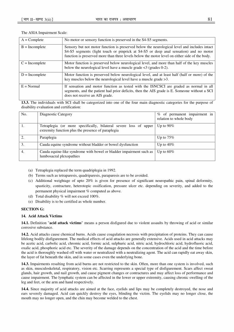
¹Hkkx IIµ[k.M 3(ii)º Hkkjr dk jkti=k % vlk/kj.k 81
The ASIA Impairment Scale:
A = Complete No motor or sensory function is preserved in the S4-S5 segments.
B = Incomplete Sensory but not motor function is preserved below the neurological level and includes intact
S4-S5 segments (light touch or pinprick at S4-S5 or deep anal sensation) and no motor
function is preserved more than three levels below the motor level on either side of the body.
C = Incomplete Motor function is preserved below neurological level, and more than half of the key muscles
below the neurological level have a muscle grade <3 (grades 0-2).
D = Incomplete Motor function is preserved below neurological level, and at least half (half or more) of the
key muscles below the neurological level have a muscle grade >3.
E = Normal If sensation and motor function as tested with the ISNCSCI are graded as normal in all
segments, and the patient had prior deficits, then the AIS grade is E. Someone without a SCI
does not receive an AIS grade.
13.3. The individuals with SCI shall be categorized into one of the four main diagnostic categories for the purpose of
disability evaluation and certification:
No. Diagnostic Category % of permanent impairment in
relation to whole body
1. Tetraplegia (or more specifically, bilateral severe loss of upper
extremity function plus the presence of paraplegia
Up to 90%
2. Paraplegia Up to 75%
3. Cauda equine syndrome without bladder or bowel dysfunction Up to 40%
4. Cauda equine-like syndrome with bowel or bladder impairment such as
lumbosacral plexopathies
Up to 60%
(a) Tetraplegia replaced the term quadriplegia in 1992.
(b) Terms such as tetraparesis, quadriparesis, paraparesis are to be avoided.
(c) Additional weightage of upto 20% is given for presence of significant neuropathic pain, spinal deformity,
spasticity, contracture, heterotopic ossification, pressure ulcer etc. depending on severity, and added to the
permanent physical impairment % computed as above.
(d) Total disability % will not exceed 100%.
(e) Disability is to be certified as whole number.
SECTION G:
14. Acid Attack Victims
14.1. Definition "acid attack victims" means a person disfigured due to violent assaults by throwing of acid or similar
corrosive substance.
14.2. Acid attacks cause chemical burns. Acids cause coagulation necrosis with precipitation of proteins. They can cause
lifelong bodily disfigurement. The medical effects of acid attacks are generally extensive. Acids used in acid attacks may
be acetic acid, carbolic acid, chromic acid, formic acid, sulphuric acid, nitric acid, hydrochloric acid, hydrofluoric acid,
oxalic acid, phosphoric acid etc. The severity of the damage depends on the concentration of the acid and the time before
the acid is thoroughly washed off with water or neutralized with a neutralizing agent. The acid can rapidly eat away skin,
the layer of fat beneath the skin, and in some cases even the underlying bone.
14.3. Impairments resulting from acid burns are not restricted to the skin. Often, more than one system is involved, such
as skin, musculoskeletal, respiratory, vision etc. Scarring represents a special type of disfigurement. Scars affect sweat
glands, hair growth, and nail growth, and cause pigment changes or contractures and may affect loss of performance and
cause impairment. The lymphatic system can be affected in the lower or upper extremity, causing chronic swelling of the
leg and feet, or the arm and hand respectively.
14.4. Since majority of acid attacks are aimed at the face, eyelids and lips may be completely destroyed, the nose and
ears severely damaged. Acid can quickly destroy the eyes, blinding the victim. The eyelids may no longer close, the
mouth may no longer open, and the chin may become welded to the chest.
82 THE GAZETTE OF INDIA : EXTRAORDINARY [PART II—SEC. 3(ii)]
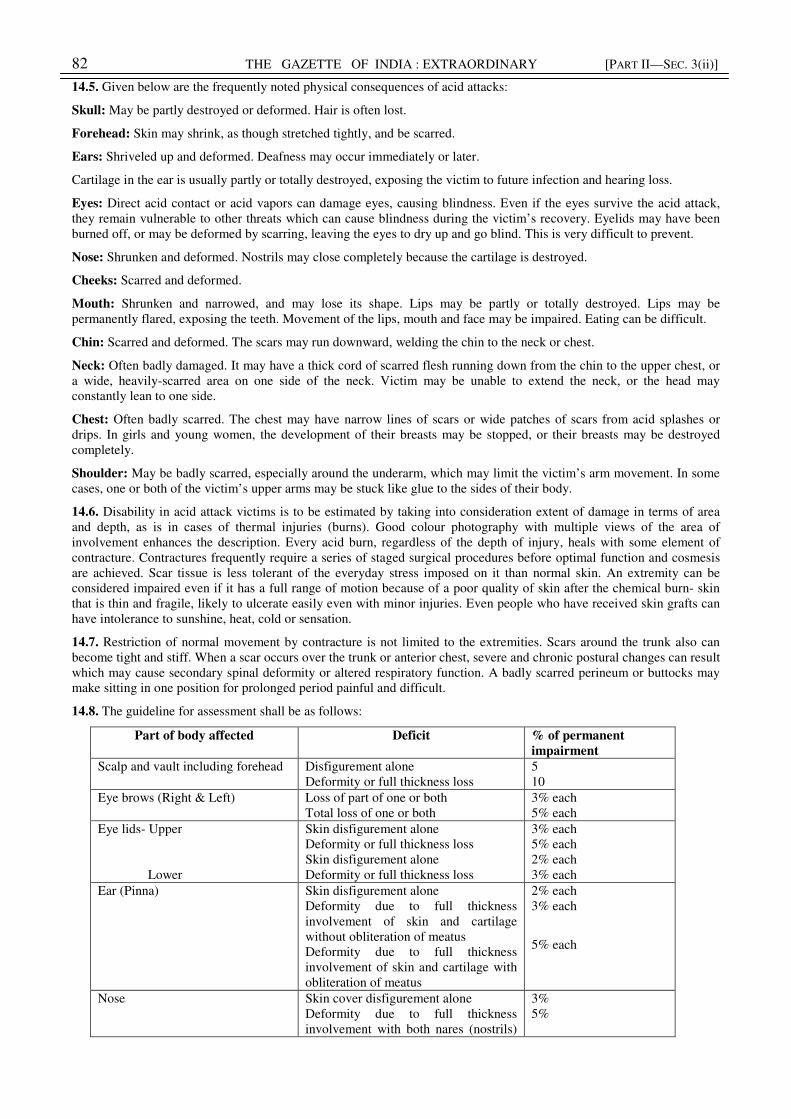
14.5. Given below are the frequently noted physical consequences of acid attacks:
Skull: May be partly destroyed or deformed. Hair is often lost.
Forehead: Skin may shrink, as though stretched tightly, and be scarred.
Ears: Shriveled up and deformed. Deafness may occur immediately or later.
Cartilage in the ear is usually partly or totally destroyed, exposing the victim to future infection and hearing loss.
Eyes: Direct acid contact or acid vapors can damage eyes, causing blindness. Even if the eyes survive the acid attack,
they remain vulnerable to other threats which can cause blindness during the victim’s recovery. Eyelids may have been
burned off, or may be deformed by scarring, leaving the eyes to dry up and go blind. This is very difficult to prevent.
Nose: Shrunken and deformed. Nostrils may close completely because the cartilage is destroyed.
Cheeks: Scarred and deformed.
Mouth: Shrunken and narrowed, and may lose its shape. Lips may be partly or totally destroyed. Lips may be
permanently flared, exposing the teeth. Movement of the lips, mouth and face may be impaired. Eating can be difficult.
Chin: Scarred and deformed. The scars may run downward, welding the chin to the neck or chest.
Neck: Often badly damaged. It may have a thick cord of scarred flesh running down from the chin to the upper chest, or
a wide, heavily-scarred area on one side of the neck. Victim may be unable to extend the neck, or the head may
constantly lean to one side.
Chest: Often badly scarred. The chest may have narrow lines of scars or wide patches of scars from acid splashes or
drips. In girls and young women, the development of their breasts may be stopped, or their breasts may be destroyed
completely.
Shoulder: May be badly scarred, especially around the underarm, which may limit the victim’s arm movement. In some
cases, one or both of the victim’s upper arms may be stuck like glue to the sides of their body.
14.6. Disability in acid attack victims is to be estimated by taking into consideration extent of damage in terms of area
and depth, as is in cases of thermal injuries (burns). Good colour photography with multiple views of the area of
involvement enhances the description. Every acid burn, regardless of the depth of injury, heals with some element of
contracture. Contractures frequently require a series of staged surgical procedures before optimal function and cosmesis
are achieved. Scar tissue is less tolerant of the everyday stress imposed on it than normal skin. An extremity can be
considered impaired even if it has a full range of motion because of a poor quality of skin after the chemical burn- skin
that is thin and fragile, likely to ulcerate easily even with minor injuries. Even people who have received skin grafts can
have intolerance to sunshine, heat, cold or sensation.
14.7. Restriction of normal movement by contracture is not limited to the extremities. Scars around the trunk also can
become tight and stiff. When a scar occurs over the trunk or anterior chest, severe and chronic postural changes can result
which may cause secondary spinal deformity or altered respiratory function. A badly scarred perineum or buttocks may
make sitting in one position for prolonged period painful and difficult.
14.8. The guideline for assessment shall be as follows:
Part of body affected Deficit % of permanent
impairment
Scalp and vault including forehead Disfigurement alone
Deformity or full thickness loss
5
10
Eye brows (Right & Left) Loss of part of one or both
Total loss of one or both
3% each
5% each
Eye lids- Upper
Lower
Skin disfigurement alone
Deformity or full thickness loss
Skin disfigurement alone
Deformity or full thickness loss
3% each
5% each
2% each
3% each
Ear (Pinna) Skin disfigurement alone
Deformity due to full thickness
involvement of skin and cartilage
without obliteration of meatus
Deformity due to full thickness
involvement of skin and cartilage with
obliteration of meatus
2% each
3% each
5% each
Nose Skin cover disfigurement alone
Deformity due to full thickness
involvement with both nares (nostrils)
3%
5%
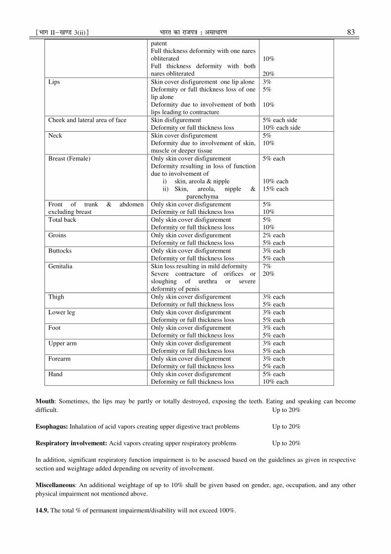
¹Hkkx IIµ[k.M 3(ii)º Hkkjr dk jkti=k % vlk/kj.k 83
patent
Full thickness deformity with one nares
obliterated
Full thickness deformity with both
nares obliterated
10%
20%
Lips Skin cover disfigurement one lip alone
Deformity or full thickness loss of one
lip alone
Deformity due to involvement of both
lips leading to contracture
3%
5%
10%
Cheek and lateral area of face Skin disfigurement
Deformity or full thickness loss
5% each side
10% each side
Neck Skin cover disfigurement
Deformity due to involvement of skin,
muscle or deeper tissue
5%
10%
Breast (Female) Only skin cover disfigurement
Deformity resulting in loss of function
due to involvement of
i) skin, areola & nipple
ii) Skin, areola, nipple &
parenchyma
5% each
10% each
15% each
Front of trunk & abdomen
excluding breast
Only skin cover disfigurement
Deformity or full thickness loss
5%
10%
Total back Only skin cover disfigurement
Deformity or full thickness loss
5%
10%
Groins Only skin cover disfigurement
Deformity or full thickness loss
2% each
5% each
Buttocks Only skin cover disfigurement
Deformity or full thickness loss
3% each
5% each
Genitalia Skin loss resulting in mild deformity
Severe contracture of orifices or
sloughing of urethra or severe
deformity of penis
7%
20%
Thigh Only skin cover disfigurement
Deformity or full thickness loss
3% each
5% each
Lower leg Only skin cover disfigurement
Deformity or full thickness loss
3% each
5% each
Foot Only skin cover disfigurement
Deformity or full thickness loss
3% each
5% each
Upper arm Only skin cover disfigurement
Deformity or full thickness loss
3% each
5% each
Forearm Only skin cover disfigurement
Deformity or full thickness loss
3% each
5% each
Hand Only skin cover disfigurement
Deformity or full thickness loss
5% each
10% each
Mouth: Sometimes, the lips may be partly or totally destroyed, exposing the teeth. Eating and speaking can become
difficult. Up to 20%
Esophagus: Inhalation of acid vapors creating upper digestive tract problems Up to 20%
Respiratory involvement: Acid vapors creating upper respiratory problems Up to 20%
In addition, significant respiratory function impairment is to be assessed based on the guidelines as given in respective
section and weightage added depending on severity of involvement.
Miscellaneous: An additional weightage of up to 10% shall be given based on gender, age, occupation, and any other
physical impairment not mentioned above.
14.9. The total % of permanent impairment/disability will not exceed 100%.
84 THE GAZETTE OF INDIA : EXTRAORDINARY [PART II—SEC. 3(ii)]
SECTION H:
15. Cerebral Palsy affected Persons with disabilities
15.1. Definition- "cerebral palsy" means a group of non-progressive neurological condition affecting body movements
and muscle coordination, caused by damage to one or more specific areas of the brain, usually occurring before, during
or shortly after birth.
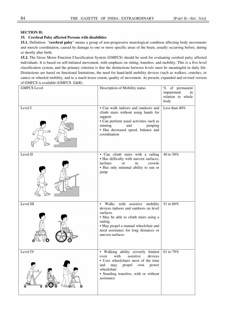
15.2. The Gross Motor Function Classification System (GMFCS) should be used for evaluating cerebral palsy affected
individuals. It is based on self-initiated movement, with emphasis on sitting, transfers, and mobility. This is a five-level
classification system, and the primary criterion is that the distinctions between levels must be meaningful in daily life.
Distinctions are based on functional limitations, the need for hand-held mobility devices (such as walkers, crutches, or
canes) or wheeled mobility, and to a much lesser extent, quality of movement. At present, expanded and revised version
of GMFCS is available (GMFCS- E&R).
GMFCS Level Description of Mobility status % of permanent
impairment in
relation to whole
body
Level I
• Can walk indoors and outdoors and
climb stairs without using hands for
support
• Can perform usual activities such as
running and jumping
• Has decreased speed, balance and
coordination
Less than 40%
Level II
• Can climb stairs with a railing
• Has difficulty with uneven surfaces,
inclines or in crowds
• Has only minimal ability to run or
jump
40 to 50%
Level III
• Walks with assistive mobility
devices indoors and outdoors on level
surfaces
• May be able to climb stairs using a
railing
• May propel a manual wheelchair and
need assistance for long distances or
uneven surfaces
51 to 60%
Level IV
• Walking ability severely limited
even with assistive devices
• Uses wheelchairs most of the time
and may propel own power
wheelchair
• Standing transfers, with or without
assistance
61 to 79%

¹Hkkx IIµ[k.M 3(ii)º Hkkjr dk jkti=k % vlk/kj.k 85
Level V
• Has physical impairments that
restrict voluntary control of movement
• Ability to maintain head and neck
position against gravity restricted
• Impaired in all areas of motor
function
• Cannot sit or stand independently,
even with adaptive equipment
• Cannot independently walk but may
be able to use powered mobility
80% or more
Note:- (i) In a person with cerebral palsy, other than problems of movement or posture, there may be other limitations
such as visual impairment, hearing impairment, speech impairment, epilepsy, mental sub-normality (low IQ) etc. These
are assessed separately as per the guidelines and the final disability % calculated using the combining formula: a+b (90-
a)/ 90 (a = higher value, b = lower value).
(ii) Total permanent physical impairment/disability % will not exceed 100%.
(iii) Disability is to be certified in relation to the whole body.
Manual Ability Classification System (MACS)
15.3. The Manual Ability Classification System (MACS) describes how children with cerebral palsy (CP) use their hands
to handle objects in daily activities. MACS describes five levels. The levels are based on the children’s self-initiated
ability to handle objects and their need for assistance or adaptation to perform manual activities in everyday life.
15.4. MACS can be used for children aged 4–18 years. MACS spans the entire spectrum of functional limitations found
among children with cerebral palsy and covers all sub-diagnoses.
15.5. Level I includes children with minor limitations, while children with severe functional limitations will usually be
found at levels IV and V. MACS levels are stable over time.
15.6. The certifying medical authority needs to know the following to use MACS:
The child’s ability to handle objects in important daily activities, for example during play and leisure, eating and
dressing, is to be considered as per the following scale:-
Level I. Handles objects easily and successfully. At most, limitations in the ease of performing manual asks requiring
speed and accuracy. However, any limitations in manual abilities do not restrict independence in daily activities.
Level II. Handles most objects but with somewhat reduced quality and/or speed of achievement. Certain activities
may be avoided or be achieved with some difficulty; alternative ways of performance might be used but manual abilities
do not usually restrict independence in daily activities.
Level III. Handles objects with difficulty; needs help to prepare and/or modify activities. The performance is slow
and achieved with limited success regarding quality and quantity. Activities are performed independently if they have
been set up or adapted.
Level IV. Handles a limited selection of easily managed objects in adapted situations. Performs parts of activities
with effort and with limited success. Requires continuous support and assistance and/or adapted equipment, for even
partial achievement of the activity.
Level V. Does not handle objects and has severely limited ability to perform even simple actions. Requires total
assistance.
MACS Level Feature % of permanent
impairment
Level I. Handles objects easily and successfully. 20%
Level II. Handles most objects but with somewhat reduced quality
and/or speed of achievement.
30%
Level III. Handles objects with difficulty; needs help to prepare
and/or modify activities.
40%
86 THE GAZETTE OF INDIA : EXTRAORDINARY [PART II—SEC. 3(ii)]
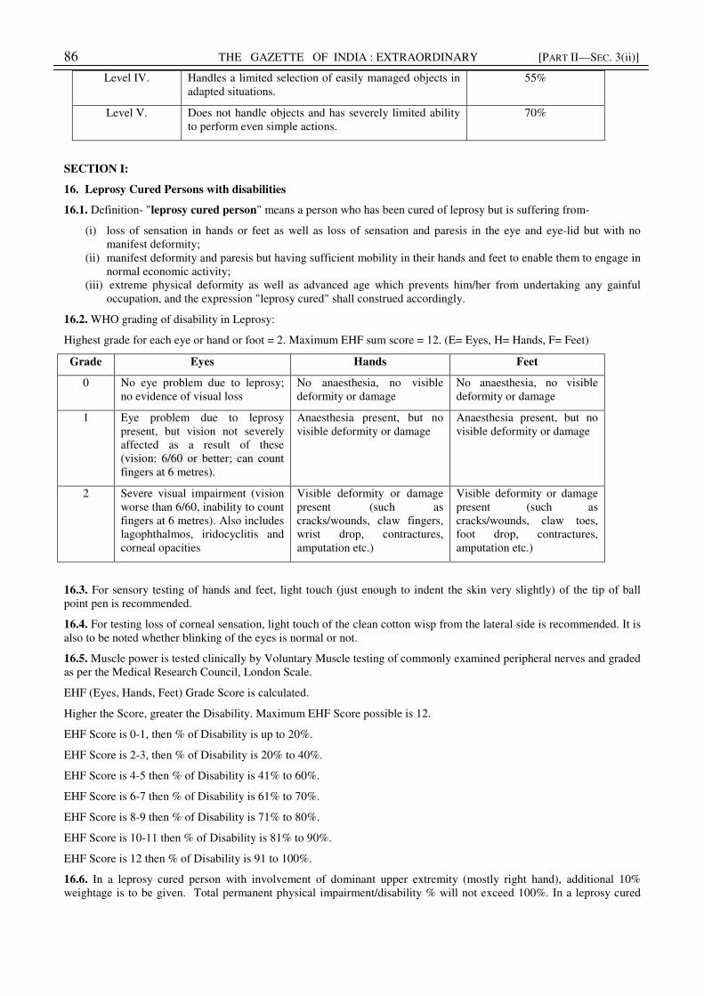
Level IV. Handles a limited selection of easily managed objects in
adapted situations.
55%
Level V. Does not handle objects and has severely limited ability
to perform even simple actions.
70%
SECTION I:
16. Leprosy Cured Persons with disabilities
16.1. Definition- "leprosy cured person" means a person who has been cured of leprosy but is suffering from-
(i) loss of sensation in hands or feet as well as loss of sensation and paresis in the eye and eye-lid but with no
manifest deformity;
(ii) manifest deformity and paresis but having sufficient mobility in their hands and feet to enable them to engage in
normal economic activity;
(iii) extreme physical deformity as well as advanced age which prevents him/her from undertaking any gainful
occupation, and the expression "leprosy cured" shall construed accordingly.
16.2. WHO grading of disability in Leprosy:
Highest grade for each eye or hand or foot = 2. Maximum EHF sum score = 12. (E= Eyes, H= Hands, F= Feet)
Grade Eyes Hands Feet
0 No eye problem due to leprosy;
no evidence of visual loss
No anaesthesia, no visible
deformity or damage
No anaesthesia, no visible
deformity or damage
1 Eye problem due to leprosy
present, but vision not severely
affected as a result of these
(vision: 6/60 or better; can count
fingers at 6 metres).
Anaesthesia present, but no
visible deformity or damage
Anaesthesia present, but no
visible deformity or damage
2 Severe visual impairment (vision
worse than 6/60, inability to count
fingers at 6 metres). Also includes
lagophthalmos, iridocyclitis and
corneal opacities
Visible deformity or damage
present (such as
cracks/wounds, claw fingers,
wrist drop, contractures,
amputation etc.)
Visible deformity or damage
present (such as
cracks/wounds, claw toes,
foot drop, contractures,
amputation etc.)
16.3. For sensory testing of hands and feet, light touch (just enough to indent the skin very slightly) of the tip of ball
point pen is recommended.
16.4. For testing loss of corneal sensation, light touch of the clean cotton wisp from the lateral side is recommended. It is
also to be noted whether blinking of the eyes is normal or not.
16.5. Muscle power is tested clinically by Voluntary Muscle testing of commonly examined peripheral nerves and graded
as per the Medical Research Council, London Scale.
EHF (Eyes, Hands, Feet) Grade Score is calculated.
Higher the Score, greater the Disability. Maximum EHF Score possible is 12.
EHF Score is 0-1, then % of Disability is up to 20%.
EHF Score is 2-3, then % of Disability is 20% to 40%.
EHF Score is 4-5 then % of Disability is 41% to 60%.
EHF Score is 6-7 then % of Disability is 61% to 70%.
EHF Score is 8-9 then % of Disability is 71% to 80%.
EHF Score is 10-11 then % of Disability is 81% to 90%.
EHF Score is 12 then % of Disability is 91 to 100%.
16.6. In a leprosy cured person with involvement of dominant upper extremity (mostly right hand), additional 10%
weightage is to be given. Total permanent physical impairment/disability % will not exceed 100%. In a leprosy cured
¹Hkkx IIµ[k.M 3(ii)º Hkkjr dk jkti=k % vlk/kj.k 87
persons, review may be done after two years, if needed or desired by the affected person, in view of likely worsening of
deformities in some persons.
SECTION J:
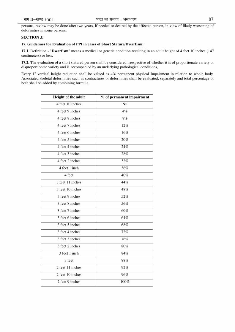
17. Guidelines for Evaluation of PPI in cases of Short Stature/Dwarfism:
17.1. Definition.- "Dwarfism" means a medical or genetic condition resulting in an adult height of 4 feet 10 inches (147
centimeters) or less.
17.2. The evaluation of a short statured person shall be considered irrespective of whether it is of proportionate variety or
disproportionate variety and is accompanied by an underlying pathological conditions,
Every 1" vertical height reduction shall be valued as 4% permanent physical Impairment in relation to whole body.
Associated skeletal deformities such as contractures or deformities shall be evaluated, separately and total percentage of
both shall be added by combining formula.
Height of the adult % of permanent impairment
4 feet 10 inches Nil
4 feet 9 inches 4%
4 feet 8 inches 8%
4 feet 7 inches 12%
4 feet 6 inches 16%
4 feet 5 inches 20%
4 feet 4 inches 24%
4 feet 3 inches 28%
4 feet 2 inches 32%
4 feet 1 inch 36%
4 feet 40%
3 feet 11 inches 44%
3 feet 10 inches 48%
3 feet 9 inches 52%
3 feet 8 inches 56%
3 feet 7 inches 60%
3 feet 6 inches 64%
3 feet 5 inches 68%
3 feet 4 inches 72%
3 feet 3 inches 76%
3 feet 2 inches 80%
3 feet 1 inch 84%
3 feet 88%
2 feet 11 inches 92%
2 feet 10 inches 96%
2 feet 9 inches 100%
88 THE GAZETTE OF INDIA : EXTRAORDINARY [PART II—SEC. 3(ii)]
SECTION K:
18. Muscular Dystrophy
18.1. Definition.- "muscular dystrophy" means a group of hereditary genetic muscle disease that weakens the muscles
that move the human body and persons with multiple dystrophy have incorrect and missing information in their genes,
which prevents them from making the proteins they need for healthy muscles. It is characterised by progressive skeletal
muscle weakness, defects in muscle proteins, and the death of muscle cells and tissue.
18.2. After detailed clinical examination, each of the features namely, weakness, contractures, scoliosis, cardiac or
pulmonary involvement are evaluated and disability is computed based on the criteria for each of these and added to the
locomotor disability component, using the combining formula: a + b (90-a)/ 90 (a = higher value, b = lower value).
Disability is to be expressed in relation to the whole body. Total % of disability will not exceed 100%. Due to
progressive nature of this disease, review may be necessary after a period, such as 2 years or as desired by the patient or
as decided by the disability board.
18.3 Medical Authority and instruments required for certification of locomotor disability
18.3.1 The Medical Superintendent or Chief Medical Officer or Civil Surgeon or any other equivalent authority as
notified by the State Government shall be the head of the certification board for the purpose of certification of locomotor
disability including cerebral palsy, leprosy cured, dwarfism, acid attack victims and muscular dystrophy. The Board shall
comprise of:
(i) Medical Superintendent or Chief Medical Officer or Civil Surgeon
(ii) Specialist in Physical Medicine and Rehabilitation or Specialist in Orthopedics
(iii) One specialist as nominated by Chief Medical Officer as per the condition of the person with disability.
18.3. 2. The most important resource is the knowledge and skill of the Members/Experts involved in the process.
However, a few items listed below may also be required:
a. A measuring tape for measuring – vertical height of the person, degree of chest expansion, shortening of an
extremity, or difference in girth of a limb etc.,
b. Goniometers – small, medium and large, for measuring range of motion at different joints,
c. Hand-held dynamometer,
d. Clean cotton piece for testing corneal sensation,
e. A ball point pen for testing sensory deficit e.g., in leprosy-cured person,
f. X-ray films, e.g., in cases with spinal deformity, amputation, arthritis, club foot, congenital limb deficiency,
fractures etc.
19.1. Definition.- Visual impairment
(a) "blindness" means a condition where a person has any of the following
conditions, after best correction—
(i) total absence of sight; or
(ii) visual acuity less than 3/60 or less than 10/200 (Snellen) in the better eye with best possible correction; or
(iii) limitation of the field of vision subtending an angle of less than 10 degree.
(b) "low-vision" means a condition where a person has any of the following conditions, namely:—
(i) visual acuity not exceeding 6/18 or less than 20/60 upto 3/60 or upto 10/200 (Snellen) in the better eye with
best possible corrections; or
(ii) limitation of the field of vision subtending an angle of less than 40 degree up to 10 degree.
II. VISUAL IMPAIRMENT
¹Hkkx IIµ[k.M 3(ii)º Hkkjr dk jkti=k % vlk/kj.k 89
19.2. Nature of Certificate: The medical authority will decide whether disability certificate should be temporary or
permanent. The disability shall be permanent to be certified. The certificate can be temporary if condition is likely to
worsen and also for specific purposes such as for pursuing education. The need of reassessment, if required, should be
clearly mentioned in the certificate with time frame. In certain cases such as keratoconus, developmental defects,
operated congenital cataract with corneal decompensation, operated congenital glaucoma with hazy cornea etc., the
patient especially can be issued a temporary certificate.
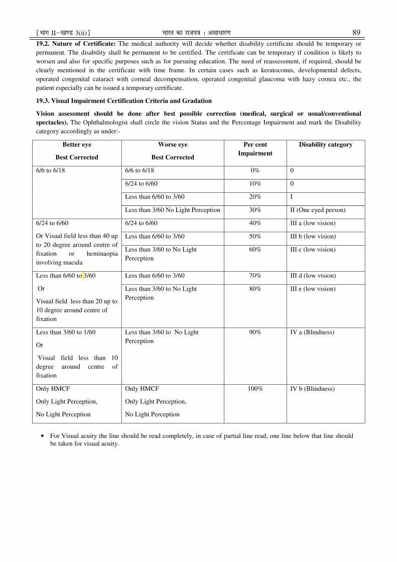
19.3. Visual Impairment Certification Criteria and Gradation
Vision assessment should be done after best possible correction (medical, surgical or usual/conventional
spectacles). The Ophthalmologist shall circle the vision Status and the Percentage Impairment and mark the Disability
category accordingly as under:-
Better eye
Best Corrected
Worse eye
Best Corrected
Per cent
Impairment
Disability category
6/6 to 6/18
6/6 to 6/18 0% 0
6/24 to 6/60 10% 0
Less than 6/60 to 3/60 20% I
Less than 3/60 No Light Perception 30% II (One eyed person)
6/24 to 6/60
Or Visual field less than 40 up
to 20 degree around centre of
fixation or heminaopia
involving macula
6/24 to 6/60 40% III a (low vision)
Less than 6/60 to 3/60 50% III b (low vision)
Less than 3/60 to No Light
Perception
60% III c (low vision)
Less than 6/60 to 3/60
Or
Visual field less than 20 up to
10 degree around centre of
fixation
Less than 6/60 to 3/60 70% III d (low vision)
Less than 3/60 to No Light
Perception
80% III e (low vision)
Less than 3/60 to 1/60
Or
Visual field less than 10
degree around centre of
fixation
Less than 3/60 to No Light
Perception
90% IV a (Blindness)
Only HMCF
Only Light Perception,
No Light Perception
Only HMCF
Only Light Perception,
No Light Perception
100% IV b (Blindness)
• For Visual acuity the line should be read completely, in case of partial line read, one line below that line should
be taken for visual acuity.

90 THE GAZETTE OF INDIA : EXTRAORDINARY [PART II—SEC. 3(ii)]
Matrix Table
Left Eye Vision [Best Corrected Visual Acuity (BCVA)]) R
igh
t E
ye
Vis
ion
[B
est
Co
rrec
ted
Vis
ua
l A
cuit
y (
BC
VA
)]
6/6 to
6/18 6/24 6/36 6/60 3/60 2/60 1/60
HMCF to
PL-
6/6 to 6/18 0% 10% 10% 10% 20% 30% 30% 30%
6/24 10% 40% 40% 40% 50% 60% 60% 60%
6/36 10% 40% 40% 40% 50% 60% 60% 60%
6/60 10% 40% 40% 40% 50% 60% 60% 60%
3/60 20% 50% 50% 50% 70% 80% 80% 80%
2/60 30% 60% 60% 60% 80% 90% 90% 90%
1/60 30% 60% 60% 60% 80% 90% 90% 90%
HMCF to
PL- 30% 60% 60% 60% 80% 90% 90% 100%
• Yellow- Right eye is Better eye Brown- Left eye is better eye
• Percent disability is marked inside the box corresponding to the visual acuity for both eyes
Field of Vision around centre of fixation
Left Eye
<40° to
20°
<20° to
10° <10°
Rig
ht
Ey
e
<40° to
20° 40% 50% 60%
<20° to
10° 50% 70% 80%
<10° 60% 80% 100%
• Yellow- Right eye is Better eye Brown- Left eye is better eye (only better eye Fields to be taken in to
account for determining the %)
19.4. Medical Authority.
The medical authority shall comprise of one ophthalmologist and certificate of disability shall be countersigned by
Medical Superintendent or Chief Medical Officer or Civil Surgeon or any other equivalent authority as notified the State
Government.
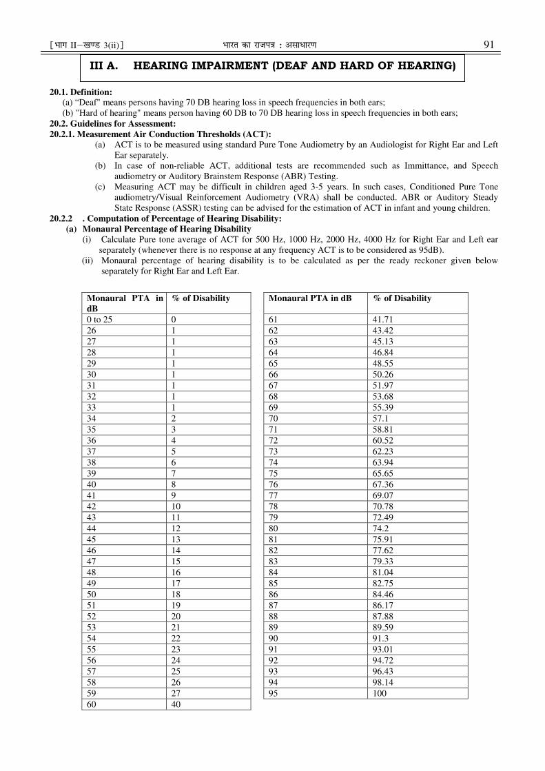
¹Hkkx IIµ[k.M 3(ii)º Hkkjr dk jkti=k % vlk/kj.k 91
20.1. Definition: (a) “Deaf" means persons having 70 DB hearing loss in speech frequencies in both ears;
(b) "Hard of hearing" means person having 60 DB to 70 DB hearing loss in speech frequencies in both ears;
20.2. Guidelines for Assessment:
20.2.1. Measurement Air Conduction Thresholds (ACT):
(a) ACT is to be measured using standard Pure Tone Audiometry by an Audiologist for Right Ear and Left
Ear separately.
(b) In case of non-reliable ACT, additional tests are recommended such as Immittance, and Speech
audiometry or Auditory Brainstem Response (ABR) Testing.
(c) Measuring ACT may be difficult in children aged 3-5 years. In such cases, Conditioned Pure Tone
audiometry/Visual Reinforcement Audiometry (VRA) shall be conducted. ABR or Auditory Steady
State Response (ASSR) testing can be advised for the estimation of ACT in infant and young children.
20.2.2 . Computation of Percentage of Hearing Disability:
(a) Monaural Percentage of Hearing Disability
(i) Calculate Pure tone average of ACT for 500 Hz, 1000 Hz, 2000 Hz, 4000 Hz for Right Ear and Left ear
separately (whenever there is no response at any frequency ACT is to be considered as 95dB).
(ii) Monaural percentage of hearing disability is to be calculated as per the ready reckoner given below
separately for Right Ear and Left Ear.
Monaural PTA in
dB
% of Disability Monaural PTA in dB % of Disability
0 to 25 0 61 41.71
26 1 62 43.42
27 1 63 45.13
28 1 64 46.84
29 1 65 48.55
30 1 66 50.26
31 1 67 51.97
32 1 68 53.68
33 1 69 55.39
34 2 70 57.1
35 3 71 58.81
36 4 72 60.52
37 5 73 62.23
38 6 74 63.94
39 7 75 65.65
40 8 76 67.36
41 9 77 69.07
42 10 78 70.78
43 11 79 72.49
44 12 80 74.2
45 13 81 75.91
46 14 82 77.62
47 15 83 79.33
48 16 84 81.04
49 17 85 82.75
50 18 86 84.46
51 19 87 86.17
52 20 88 87.88
53 21 89 89.59
54 22 90 91.3
55 23 91 93.01
56 24 92 94.72
57 25 93 96.43
58 26 94 98.14
59 27 95 100
60 40
III A. HEARING IMPAIRMENT (DEAF AND HARD OF HEARING)

92 THE GAZETTE OF INDIA : EXTRAORDINARY [PART II—SEC. 3(ii)]
20.2.3. Percentage of Hearing Disability
Percentage of Hearing Disability =
(Better ear % of hearing disability X 5) + (Poorer ear % of hearing disability)
6
20.3.1. Definition: "Speech and language disability" means a permanent disability arising out of conditions such as
laryngectomy or aphasia affecting one or more components of speech and language due to organic or neurological
causes
20.3.2. Conditions affecting Speech Components for which Speech Disability certificate can be issued
• Laryngectomy
• Glossectomy
• Bilateral vocal cord paralysis
• Maxillofacial anomalies
• Dysarthria
• Apraxia of Speech
20.3.3. Computation of Percentage Speech Disability
(a) Speech Intelligibility Test
The verbal output of person should be evaluated using either Perceptual Speech intelligibility rating scale (AYJNISHD,
2003) or Perceptual Rating Scale (SRMC, Chennai) and percentage of Speech Intelligibility Affected (SIA) to be
measured based on score as given below:
Score Percentage of Speech Intelligibility
Affected (SIA)
1 0-15
2 16-30
3 31-39
4 40-55
5 56-75
6 76-89
7 90- 100
(b) Voice Test
Consensus Auditory Perceptual Evaluation of Voice (CAPE-V) orDysphonia Severity Index (DSI) can be used for
measuring percentage of Overall Voice Clarity Affected (OVCA) which includes roughness, breathiness, strain,
pitch and loudness. Average score to be given weighted for the percentage of overall voice clarity affected
Score Percentage of overall voice clarity
affected (OVCA)
1 0-15
2 16-30
3 31-39
4 40-55
5 56-75
6 76-89
7 90-100
III B. SPEECH AND LANGUAGE DISABILITY
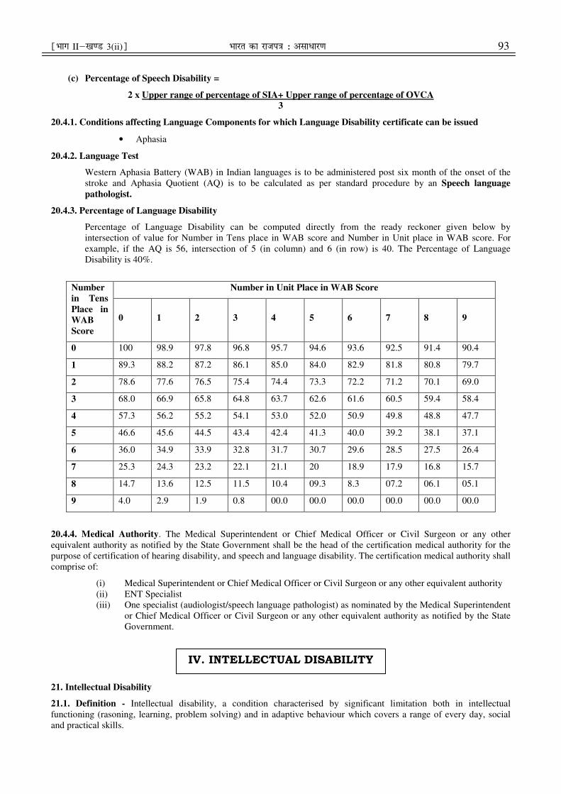
¹Hkkx IIµ[k.M 3(ii)º Hkkjr dk jkti=k % vlk/kj.k 93
(c) Percentage of Speech Disability =
2 x Upper range of percentage of SIA+ Upper range of percentage of OVCA
3
20.4.1. Conditions affecting Language Components for which Language Disability certificate can be issued
• Aphasia
20.4.2. Language Test
Western Aphasia Battery (WAB) in Indian languages is to be administered post six month of the onset of the
stroke and Aphasia Quotient (AQ) is to be calculated as per standard procedure by an Speech language
pathologist.
20.4.3. Percentage of Language Disability
Percentage of Language Disability can be computed directly from the ready reckoner given below by
intersection of value for Number in Tens place in WAB score and Number in Unit place in WAB score. For
example, if the AQ is 56, intersection of 5 (in column) and 6 (in row) is 40. The Percentage of Language
Disability is 40%.
Number
in Tens
Place in
WAB
Score
Number in Unit Place in WAB Score
0 1 2 3 4 5 6 7 8 9
0 100 98.9 97.8 96.8 95.7 94.6 93.6 92.5 91.4 90.4
1 89.3 88.2 87.2 86.1 85.0 84.0 82.9 81.8 80.8 79.7
2 78.6 77.6 76.5 75.4 74.4 73.3 72.2 71.2 70.1 69.0
3 68.0 66.9 65.8 64.8 63.7 62.6 61.6 60.5 59.4 58.4
4 57.3 56.2 55.2 54.1 53.0 52.0 50.9 49.8 48.8 47.7
5 46.6 45.6 44.5 43.4 42.4 41.3 40.0 39.2 38.1 37.1
6 36.0 34.9 33.9 32.8 31.7 30.7 29.6 28.5 27.5 26.4
7 25.3 24.3 23.2 22.1 21.1 20 18.9 17.9 16.8 15.7
8 14.7 13.6 12.5 11.5 10.4 09.3 8.3 07.2 06.1 05.1
9 4.0 2.9 1.9 0.8 00.0 00.0 00.0 00.0 00.0 00.0
20.4.4. Medical Authority. The Medical Superintendent or Chief Medical Officer or Civil Surgeon or any other
equivalent authority as notified by the State Government shall be the head of the certification medical authority for the
purpose of certification of hearing disability, and speech and language disability. The certification medical authority shall
comprise of:
(i) Medical Superintendent or Chief Medical Officer or Civil Surgeon or any other equivalent authority
(ii) ENT Specialist
(iii) One specialist (audiologist/speech language pathologist) as nominated by the Medical Superintendent
or Chief Medical Officer or Civil Surgeon or any other equivalent authority as notified by the State
Government.
21. Intellectual Disability
21.1. Definition - Intellectual disability, a condition characterised by significant limitation both in intellectual
functioning (rasoning, learning, problem solving) and in adaptive behaviour which covers a range of every day, social
and practical skills.
IV. INTELLECTUAL DISABILITY

94 THE GAZETTE OF INDIA : EXTRAORDINARY [PART II—SEC. 3(ii)]
21.2. Screening: Many of these children are on follow-up with pediatricians as developmental delay. Hence, they can be
assessed by pediatricians and screened for associated co-morbidities, viz. hearing/ vision/ locomotor impairments/
epilepsy. Then these children are referred for detailed assessment. (See Figure 1)
21.3. Diagnosis: The screened children will be referred to Child/ clinical psychologists for Adaptive functioning and IQ
testing. The tools that can be used for the same include:
(i) Adaptive functioning: VSMS
(ii) IQ testing: BKT/ MISIC
Based on the above the diagnosis of ID will be confirmed. Based on adaptive functioning assessment,
severity scoring will be done and disability for ID charted.
21.4. Disability calculation: The disability calculation will be done based on VSMS score. The following will be used
for disability calculation:
(i) VSMS score 0-20: Profound Disability-100%
(ii) VSMS score 21-35: Severe Disability-90%
(iii) VSMS score 36-54: Moderate Disability-75%
(iv) VSMS score 55-69: Mild Disability-50%
(v) VSMS score 70-84: Borderline Disability-25%
21.5. Age for certification: The minimum age for certification will be one (01) completed year. Children above one year
and up to the age of 5 years shall be given a diagnosis as Global Developmental Delay (GDD). Children above the age of
5 years shall be given a diagnosis and certificate as Intellectual Disability.
21.6. Medical Authority: The Medical Superintendent or Chief Medical Officer or Civil Surgeon or any other
equivalent authority as notified by the State Government shall be the head of the Medical Board. The Authority shall
comprise of:
(a) The Medical Superintendent or Chief Medical Officer or Civil Surgeon or any other equivalent
authority as notified by the State Government
(b) Pediatrician or Pediatric Neurologist (where available)/ Psychiatrist or Physician (if age
>18years)
(c) Clinical or Rehabilitation Psychologist
(d) Psychiatrist
21.7. Validity of Certificate:
(i) Temporary certificate for children less than 5 years: The certificate will be valid for maximum 3
years/ 5 years age (whichever is earlier).
(ii) For children more than 5 years: The certificate will mention a renewal age. The certificate will
have to be renewed at age of 5 years, 10 years and 18 years. The certificate issued at 18 years
age will be valid lifelong.
22. Specific Learning Disability (SLD):
22.1. Definition.- "specific learning disabilities" means a heterogeneous group of conditions wherein there is a deficit in
processing language, spoken or written, that may manifest itself as a difficulty to comprehend, speak, read, write, spell,
or to do mathematical calculations and includes such conditions as perceptual disabilities, dyslexia, dysgraphia,
dyscalculia, dyspraxia and developmental aphasia;
22.2. Screening.-
(i) The teachers of the public and private school shall carry out the screening in Class III or at eight years
of age, whichever is earlier. The screening test is given in Figure 2. If in the screening shows test three
or more answers are in “frequently” column, then the child should be referred for further assessment.
(ii) Every school (public and private) shall have a screening committee headed by the principal of the
school. After applying the screening test, if an anomaly is detected then, the teacher should bring it to
the notice of principal and screening committee of the school. The teachers shall interview the parents
to assess their involvement and motivation regarding their child’s education. If the parents are
motivated and screening questionnaire suggests SLD, then child should be referred for further
assessment.
(iii) The child shall be referred to pediatrician for SLD assessment by the principal of the school with the
recommendations of the screening committee endorsed.
22.3. Diagnosis: The diagnosis will require a team approach involving a pediatrician and clinical or rehabilitation
psychologist. This would involve three steps:

¹Hkkx IIµ[k.M 3(ii)º Hkkjr dk jkti=k % vlk/kj.k 95
(i) Step 1- Assessment of paediatrician: The paediatrician will do the initial assessment. This will involve
a detailed neurological examination including vision and hearing assessment. It has to be ensured that
the child has normal visual acuity and hearing before proceeding to next step.
(ii) Step 2: IQ Assessment: Child/ clinical psychologist will do the IQ assessment using MISIC or WISC-
III. If the IQ is determined to be >85, then step 3 will be applied.
(iii) Step 3- SLD Assessment: This would involve application of specific psychometric tests for diagnosing
SLD and giving it a severity scale.
22.4. Diagnostic Tool - National Institute for Mental Health and Neurosciences (NIMHANS) battery shall be applied
for diagnostic test for SLD.
22.5. Medical Authority: The Medical Superintendent or Chief Medical Officer or Civil Surgeon or any other equivalent
authority as notified by the State Government shall be head the certification authority. The medical authority will
comprise of:
(a) The Medical Superintendent or Chief Medical Officer or Civil Surgeon or any other equivalent
authority as notified by the State Government
(b) Pediatrician or Pediatric Neurologist (where available)
(c) Clinical or Rehabilitation Psychologist
(d) Occupational therapist or Special Educator or Teacher trained for assessment of SLD.
22.6. Validity of Certificate: The certification will be done for children aged eight years and above only. The child will
have to undergo repeat certification at the age of 14 years and at the age of 18 years. The certificate issued at 18 years
will be valid life-long.
Figure 1. The suggested flow for identification and certification of Children with suspected Intellectual Disability
96 THE GAZETTE OF INDIA : EXTRAORDINARY [PART II—SEC. 3(ii)]
Figure 2. The suggested flow for identification and certification of Children with suspected Specific Learning Disability.
23.1. Definition: "mental illness" means a substantial disorder of thinking, mood, perception, orientation or memory that
grossly impairs judgment, behaviour, capacity to recognise reality or ability to meet the ordinary demands of life, but
does not include retardation which is a condition of arrested or incomplete development of mind of a person, specially
characterised by subnormality of intelligence.
23.2. The examination process will consist of components as required namely, clinical assessment, IDEAS scale
and/or IQ assessment.
23.3. Indian Disability Evaluation and Assessment Scale (IDEAS) administration (see Appendix IV) is to be used for
mental illness.
23.4. In some cases where there is suspicion of intellectual deficits or additional intellectual evaluation is required for
any reason, Standardised IQ test may be carried out. Categories on IQ score will be:
(i) Mild Mental Disabilities: The range of 50 to 69 (standardised IQ test) is indicative of mild
disability.
(ii) Moderate Mental Disability: The IQ is in the range of 35 to 49
(iii) Severe Mental Disability: The IQ is in the range of 20 to 34.
(iv) Profound Mental Disability: The IQ in this category estimated to be under 20.
V. MENTAL ILLNESS
¹Hkkx IIµ[k.M 3(ii)º Hkkjr dk jkti=k % vlk/kj.k 97
23.5. In cases where the mental behavioural condition requires only IDEAS, then only IDEAS can be administered
and degree of disability certified.
23.6. In cases where the mental behavioural condition requires only IQ, then a standardised IQ test shall be used to
certify degree of disability.
23.7. In some cases, only one test may not estimate disability comprehensively. Such a person may have borderline or
normal score on one test with disability score on the other. In such cases both IQ and IDEAS shall be used, the score
indicating more severe disability should be the degree of disability for that person.
24. Medical Authority: The Medical Superintendent or Chief Medical Officer or Civil Surgeon or any other equivalent
authority as notified by the State Government shall be head of the certification authority with the following two other
members:-
(a) Psychiatrist for clinical assessment,
(b) Trained psychologist to administer IQ tests.
25.1. Definition:
Chronic neurological conditions, such as—
(i) "multiple sclerosis" means an inflammatory, nervous system disease in which the myelin sheaths around the axons
of nerve cells of the brain and spinal cord are damaged, leading to demyelination and affecting the ability of nerve
cells in the brain and spinal cord to communicate with each other;
(ii) "parkinson's disease" means a progressive disease of the nervous system marked by tremor, muscular rigidity, and
slow, imprecise movement, chiefly affecting middle-aged and elderly people associated with degeneration of the
basal ganglia of the brain and a deficiency of the neurotransmitter dopamine.
25.2. The disability caused due to chronic neurological conditions such as multiple sclerosis, parkinsons disease is multi
dimensional involving manifestation in muscular skeleton system and also psycho social behaviour. The disability in
musculo-skeletal system on account of these conditions shall be assessed in terms of Section E (para 10-10.8 of
Annexure II) of these guidelines relating to assessment of locomotor disability due to chronic neurological conditions and
the psychosocial disability (mental illness) shall be assessed by using the IDEAS as at Appendix IV. Comprehensive
disability on account of these conditions shall then be calculated by using the formula a + b (90-a).
90
Where “a” will be the higher score and
And “b” will be the lower score. However, the maximum total percentage of multiple disabilities shall
not exceed 100%.
25.3. Neurological conditions which are reversible and without sequel are not certifiable. Only neurological conditions
which are permanent are certifiable. Permanent disability certificate can be issued in irreversible/progressive cases. If
needed in specific cases, a re-evaluation of disability can be done after a period of one year.
25.4. The disability certificate shall mention Chronic Neurological Conditions (name of disease).
25.5. Medical Authority: The Medical Superintendent or Chief Medical Officer or Civil Surgeon or any other
equivalent authority as notified by the State Government shall be the head of the certification authority with the
following two other members:-
(a) Pediatrics for childhood chronic neurological conditions/psychiatrist for mental illness due to chronic
neurological conditions/neurologist for chronic neurological conditions without mental illness
(b) Specialist for certifying locomotor disability
(c) Trained psychologist (clinical or rehabilitation) to administer IQ test
VI. DISABILITY CAUSED DUE TO CHRONIC NEUROLOGICAL CONDITIONS

98 THE GAZETTE OF INDIA : EXTRAORDINARY [PART II—SEC. 3(ii)]
25.6. Standardized IQ test may be carried out if required. Categories on IQ score will be:
(a) Mild Disability: The range of 50 to 69 (standardized IQ test) is indicative of mild disability.
(b) Moderate Disability: The IQ is in the range of 35 to 49.
(c) Severe Disability: The IQ is usually in the range of 20 to 34.
(d) Profound Disability: The IQ in this category estimated is to be lesser than 20.
25.7. In cases where the chronic neurological condition requires only IDEAS, then only IDEAS can be administered and
degree of disability certified.
25.8. In cases where the chronic neurological condition requires only IQ, then a standardized IQ test should be used to
certify degree of disability.
25.9. In some cases, only one test may not estimate disability comprehensively. Such a person may have borderline score
on one test with marked disability score on the other. In such cases both IQ and IDEAS shall be used. The score
indicating more severe disability shall be the degree of disability for that person.
26.1. Definition.- Blood disorder in relation to —
(i) "sickle cell disease" means a hemolytic disorder characterised by chronic anemia, painful events, and various
complications due to associated tissue and organ damage; "hemolytic" refers to the destruction of the cell
membrane of red blood cells resulting in the release of hemoglobin.
(ii) "thalassemia" means a group of inherited disorders characterised by reduced or absent amounts of
haemoglobin.
(iii) "haemophilia" means an inheritable disease, usually affecting only male but transmitted by women to their male
children, characterised by loss or impairment of the normal clotting ability of blood so that a minor wound
may result in fatal bleeding;
26.2. Type of disability certificate - The process of evaluation shall be dynamic and to be reviewed periodically at least
one year interval, as these diseases are progressive in nature. However, in patients with severe disability with score above
80%, permanent certificate shall be issued subject to proof of survival.
26.3. Medical Authority – Medical Authority for certification and evaluation of disability due to blood disorder shall
comprise of the following:-
(a) Chief District Medical Officer or the Chief Medical Officer of the hospital – Chairperson
(b) General physician or paediatrician as the case may be
(c) Orthopaedic surgeon or PMR expert
(d) In case specialities sequel relating to visual abnormality, hearing problem, cerebral dysfunction, respective
specialist.
27. Sickle Cell Disease
27.1. The clinical syndromes resulting from disorders of hemoglobin synthesis are referred to as hemoglobinopathies.
They are grouped in three main categories:
(a) Those owing to structural variants of hemoglobin, such as Sickle cell disease (HbS).
(b) Those owing to the failure to synthesize one or more of the globin chains of hemoglobin at normal rate, as in the
Thalassemias.
(c) Those owing to the failure to complete the normal neonatal switch from fetal hemoglobin (Hb F) to adult
hemoglobin (Hb A). The third category comprises a group of disorders referred to as hereditary presence of fetal
hemoglobin (HPFH).
27.2. Individual can have a combination of two or more of these abnormalities.
28. STRUCTURE VARIANTS:
28.1. Alteration in the structure of hemoglobin are usually brought about by point mutations affecting one or in some
cases two or more bases coding for amino acids of the globin chains. In HbS such a point mutation is caused by the
substitution of valine for glutamic acid in position 6 of β globin chain.
28.2. Hemoglobin variants of clinical significance or genetic significance (e.g. Hbs S, C, Dpunjab
, E and Oarab
) are readily
detectable by electrophoretic and chromatographic techniques.
VII. DISABILITY CAUSED DUE TO BLOOD DISORDER
¹Hkkx IIµ[k.M 3(ii)º Hkkjr dk jkti=k % vlk/kj.k 99
29. Hb S
29.1. The term “sickle cell disease” (SCD) encompasses both homozygous and the compound heterozygous states that
lead to the symptomatic disease as a result of formation of sickle cell red cell, due to presence of Hb S.
29.2. The homozygous state or sickle cell anemia cause both haemolysis and also reduced oxygen affinity of HbS. Sickle
cell anemias refers to specifically to those Individuals having homozygous for the sickle cell disease (HbSS), compound
heterozygous (HbS/β0) thalassaemia.
29.3. The main clinical disability arises from repeated episodes of vaso- occlusive events (called painful crisis), organ
dysfunction, impairment of vision, hearing, anemia, bone disease, pulmonary complications, skin ulcerations, gall
bladder stones and psychological problems.
29.4. Main problem occurs because of easy deformability of RBC under stress (sickling), hypoxia or infection, and RBC
becomes SICKLE SHAPE hence this name.
29.5. The clinical severity of sickle cell anemia is extremely variable. It is partly due to the modifying factors such as
interaction with α Thalassemia or synthesis of HbF and partly to socioeconomic conditions and other factors that
influence general health.
29.6. Sickle cell trait (β genotype AS), heterozygous state, is not associated with hematological abnormalities. In this
group sickling occurs at very high altitude and low oxygen pressure.
30. Other forms of sickle cell disease
(a) Sickle cell/HbC disease: Usually associated with milder form of sickle cell disease.
(b) Sickle β Thalassemia: Gives rise mild sickling disorder.
(c) Interaction of HbS with HbDPunjab
, or HbOArab
or HbOLos Angeles
accompanies with severe sickle cell disease.
(d) Other sickling hemoglobin
31. Detection and Diagnosis of Hemoglobinopathies: - (see Appendix V)
Simple tests are easily available at district hospital like Hb estimation, peripheral blood film examination after adding
sodium metasulite or without it and confirmation by Hb electrophoresis by HPLC from any reference lab or in district
hospital.
32.1. Clinical Presentation
In SCD the disability changes over time and therefore shall be measured longitudinally. It is a chronic disease may lead
to disability as a result of the primary condition as well as due to complications such as pain crisis, acute chest syndrome,
splenic sequestration crisis and rarely even aplastic crises which is due to Parvo B19.
32.2. Fever
Mandatory routine Pneumococcal vaccination and penicillin prophylaxis have reduced the risk of mortality for SCD
children. All children with SCD who have fever (>38.5oC or 101oF) or /and other signs of infection (chills, lethargy,
irritability, poor feeding, vomiting) should be assessed promptly.
32.3. Pain
This is common in all patients with SCD, it may manifest as dactylitis (“hand-foot syndrome”), vasoocclusive pain may
involve the limbs, abdominal viscera, ribs, sternum, vertebrae etc. Pain episodes limit participation in the school,
activities of daily living, and social events. Various studies have confirmed the association of between pain and activity
limitation. Patients with sickle cell anaemia who had frequent (three or more episodes per year) painful crisis, found to
have poor quality of life. Recurrent skeletal disease due to repeated bone infarction, avascular necrosis of femoral head,
and decreased bone density with vertebral disease leading to chronic back pain and nutritional deficiencies are some of
the complications of the SCD that can affect the mobility.
Pain relief needs to be appropriately done, and includes good hydration along with NSAIDS and even opioids may be
needed.
32.4. Acute chest syndrome (ACS)
This is an acute illness characterized by fever and respiratory symptoms, accompanied with a new pulmonary infiltrate
on a chest x ray. Even though the ACS usually is self-limited, it can present with or result in respiratory failure.
Simple transfusions (or rarely exchange transfusions,) decrease the proportion of sickle red cells.
100 THE GAZETTE OF INDIA : EXTRAORDINARY [PART II—SEC. 3(ii)]
32.5. Strokes and transient ischemic attacks (TIAs)
The biggest risk of permanent impairment and disability for individuals with SCD is cognitive and psychomotor
impairment secondary to a stroke. Silent cerebral infarcts can cause specific cognitive deficits, notably in attention and
executive function which are critical for successful academic performance. Language deficits, unrelated to CVA, have
been reported among children with SCD. This could lead to the communication problems in school and the workplace.
Mobility impairment can occur among children as a result of cerebral palsy (which can result from stroke), stroke, and
other aetiologies.
This is a serious condition and such patients should be referred to a higher center to receive evaluation and required
management. Patients who have suffered strokes, TIAs etc. will need transcranial Doppler (TCD), computerized axial
tomography, MRI, or MRI with angiography. Comprehensive management of SCD requires a multi-specialty team,
especially for young children with these complications.
33. Other complications:
Rare complications include leg ulcers, pulmonary hypertension, avascular necrosis head of femur, psychosocial issues
etc. At least an annual review by a hematologist will be necessary for these children, they will need to transit to adult care
for further management as they grow older. Some patients may benefit from allogeneic hematopoietic stem cell
transplant. Sickle cell disease transplant indications are very selective, due to the risks of morbidity associated with the
transplant procedure.
34.1. Indications for allogeneic Hematopoietic stem cell transplant (HSCT) for sickle cell disease:
(a) Stroke or central nervous system event lasting longer than 24 hours, acute chest syndrome with recurrent
hospitalizations or previous exchange transfusions.
(b) Recurrent vaso-occlusive pain (more than 2 episodes per year over several years) or recurrent priapism.
(c) Impaired neuropsychological function with abnormal cerebral MRI scan
(d) Stage I or II sickle lung disease
(e) Sickle nephropathy (moderate or severe proteinuria or a glomerular filtration rate 30 to 50% of the predicted
normal value)
(f) Bilateral proliferative retinopathy with major visual impairment in at least one eye
(g) Osteonecrosis of multiple joint
(h) Red-cell allo-immunization during long-term transfusion therapy
34.2. Transfusions are needed in only special indications. If transfusions needed, then a pre transfusion extended red cell
typing is required [Rh Sub group (Cc, Ee), Kell, Kidd, S/s] as these patients frequently develop delayed Hemolytic
Transfusion Reaction (30% cases) and allo- immunization. Children receiving regular transfusions will need to have
serum ferritin monitoring and chelation therapy.
34.3. The aim of transfusions to reduce Hb S levels to below 30 % prevent strokes in children with high central nervous
system blood flow [evidence from the Stroke Prevention Trial in Sickle Cell Anemia (STOP I)].
34.4. Prevention of complications can be achieved by prescribing Hydroxyurea and judicious use of blood transfusions.
Hydroxyurea decreases crises in patients with severe sickle cell disease.
34.5. Whereas those with sickle cell trait (HbAS), HbS/β+, or HbSC typically have mild to moderate symptoms.
35. The international classification of functions disability and health (ICF), distinguishes functional and structural
impairments from limitations in personal activities and restriction on social participation. The disability changes over
time hence it should be measured longitudinally.
36. Severity Score
0- homozygous sickle cell disease but asymptomatic-but has got mild pallor ( HCT 30) and splenohepatomegaly
and diagnosis confirmed by Hb electrophoresis
1. Sickle cell anemia such as (HbSS), compound heterozygous (HbS/β0) thalassaemia, HbSD, and HbO
arab,
anaemia that is severe and chronic, with persistent haemocrit of 26% or less, and symptomatic, requiring blood
transfusions to maintain the HbS level ≤ 30% and TRANSFUSION DEPENDANT and symptomatic as per
New York Heart Association (NYHA) more than class 2
2. Above plus Painful crisis due to blood clots in blood vessels at least three times in the past five months (vaso-
occlusive crisis or thrombotic crisis).
3. Above plus Hospitalization beyond that of emergency care at least three times in the past 12 months
(could be due to aplastic episodes, haemolytic crisis, strokes, heart problems, kidney failure or pneumonia)
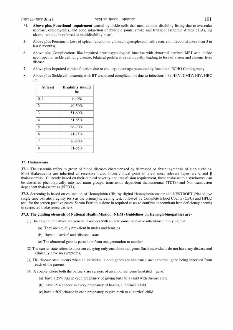
¹Hkkx IIµ[k.M 3(ii)º Hkkjr dk jkti=k % vlk/kj.k 101
*4. Above plus Functional impairment caused by sickle cells that meet another disability listing due to avascular
necrosis, osteomyelitis, and bone infarction of multiple joints, stroke and transient Ischemic Attack (TIA), leg
ulcers. –should be referred to multidisability board
5. Above plus Permanent Loss of spleen function or chronic hypersplenism with recurrent infections( more than 3 in
last 6 months)
6. Above plus Complications like impaired neuropsychological function with abnormal cerebral MRI scan, sickle
nephropathy, sickle cell lung disease, bilateral proliferative retinopathy leading to loss of vision and chronic liver
disease.
7. Above plus Impaired cardiac function due to end organ damage measured by functional ECHO Cardiography
8. Above plus Sickle cell anaemia with BT associated complications due to infections like HBV, CMIV, HIV, HBC
etc.
At level Disability should
be
0, 1 < 40%
2 40-50%
3 51-60%
4 61-65%
5 66-70%
6 71-75%
7 76-80%
8 81-85%
37. Thalassemia
37.1. Thalassaemia refers to group of blood diseases characterized by decreased or absent synthesis of globin chains.
Most thalassaemia are inherited as recessive traits. From clinical point of view most relevant types are α and β
thalassaemias. Currently based on their clinical severity and transfusion requirement, these thalassaemia syndromes can
be classified phenotypically into two main groups; transfusion dependent thalassaemias (TDTs) and Non-transfusion
dependent thalassaemias (NTDTs).
37.2. Screening is based on estimation of Hemoglobin (Hb) by digital Hemoglobinometer and NESTROFT (Naked eye
single tube osmatic fragility test) as the primary screening test, followed by Complete Blood Counts (CBC) and HPLC
test, for the screen positive cases. Serum Ferritin is done in required cases to confirm concomitant iron deficiency anemia
in suspected thalassemia carriers.
37.3. The guiding elements of National Health Mission (NHM) Guidelines on Hemoglobinopathies are-
(1) Haemoglobinopathies are genetic disorders with an autosomal recessive inheritance implying that
(a) They are equally prevalent in males and females
(b) Have a ‘carrier’ and ‘disease’ state
(c) The abnormal gene is passed on from one generation to another
(2) The carrier state refers to a person carrying only one abnormal gene. Such individuals do not have any disease and
clinically have no symptoms,
(3) The disease state occurs when an individual’s both genes are abnormal, one abnormal gene being inherited from
each of the parents.
(4) A couple where both the partners are carriers of an abnormal gene (mutated gene)
(a) have a 25% risk in each pregnancy of giving birth to a child with disease state.
(b) have 25% chance in every pregnancy of having a ‘normal’ child.
(c) have a 50% chance in each pregnancy to give birth to a ‘carrier’ child.
102 THE GAZETTE OF INDIA : EXTRAORDINARY [PART II—SEC. 3(ii)]
Thus, a carrier couple can have ‘normal’, ‘carrier’ or ‘disease’ affected children.
(5) Thalassemia Major, and Thalassemia Intermedia are the major disorders that require lifelong management and
are to be considered for prevention.
(6) Untreated Thalassemia Major is invariably fatal by 2-5 years of age. Commonly Thalassemia Major (TM) is
managed by regular blood transfusions (Packed Red Blood Cells) and iron chelation therapy. Availability of
leuko-depleted packed red blood cells (pRBC) and iron chelators are to be ensured for adequate management
along with facilities for regular monitoring. Adequately treated patients can live a fulfilling life.
(7) It is possible to know whether the child to be born will be affected by disease, or be a carrier or normal by
detecting the mutations of both parents in the fetal tissue. The process is called Prenatal Diagnosis (PND).
Thalassemia Major is a severe and life threatening disease, hence, termination of pregnancy is permitted under
Indian laws.
(8) Newborn screening can detect abnormal hemoglobin variants. On the other hand, thalassemia major is difficult
to detect by newborn screening and can be detected hematologically mostly after 3-6 months of age and
confirmed at one year of age.
(9) Carrier state is asymptomatic, but can be detected by relatively simple blood tests, opening up the possibility of
controlling hemoglobinopathies by preventing birth of affected children.
(10) Cost effective population screening programmes are possible for detection of carriers, as low cost screening tests
with high negative predictive value are available for detection of carriers of β-thalassemia.
37.4. DETECTION AND DIAGNOSIS OF Thalassemia-Appendix VI
(a) -Complete Blood Counts (CBC)
(b) Severe anemia with microcytic hypochromic red cell indices
(Hb<7g/dl; MCV: 50-70fl; MCH: 12-20pg; )
(c) Peripheral blood smears:
(d) RBCs showing anisopoikilocytosis (tear drop cells, target cells), microcytosis hypochromia, and nucleated red
cells markedly increased in relation to degree of anemia
(A) Hemoglobin (HPLC),
HPLC pattern in β-thalassemia: HbA : 0-30% HbF : 70-100% HbA2 : 2-5%
(B) Normal Values:
Hb: 12-17 gm/dl, MCV: 80-100 fl, MCH:27-32 pg, Normocytic Normochromic
HbA: 96-98%, HbF: <2%, HbA2: 2.3-3.3%
(C) Transfusion Regimen: Pre-transfusion Hemoglobin (Hb) should be kept between 9- 10.5 g/dl.
The frequency of transfusions varies from every 2-4 weeks depending on the age, weight of child and other
factors.
(D) Evaluation of Iron Overload:
(i) Serum Ferritin: Serum ferritin reflects the overall iron stores in the body tissues.
(ii) MRI of liver and heart
(iii) Liver Biopsy
The serum ferritin levels shall be assessed after 10 to 15 transfusions and chelation therapy should be initiated when the
serum ferritin value is more than 1000µg/L.
Serum ferritin and Echocardiography should be made available at most of district hospital.
37.5. COMPLICATIONS OF IRON OVERLOAD, MULTIPLE TRANSFUSION AND INDICATIONS OF
SPLENECTOMY
(a) Even with adequate iron chelation patients may go on to develop complications. Iron overload results in toxicity
to the heart, liver, and harms the endocrine system- affecting growth and development.
(b) It can even result in skeletal and bone mineralization problems.
(c) The patients may be affected by transfusion transmitted diseases like hepatitis B, C or HIV.
(d) Toxicity from iron chelation medicines, if occurs may also need to be managed.
(e) Therefore, a multi-specialist team including a pediatrician, cardiologist, gastroenterologist, and endocrinologist
are necessary.
(f) Psychological counseling and support are needed to deal with the consequences of a chronic disease.
(g) Splenectomy is needed only in few cases where hypersplenism is symptomatic and BT requirement exceeds
>250 CC of packed cell RBC/kg/year requirement. Splenectomy is associated with many late complications.
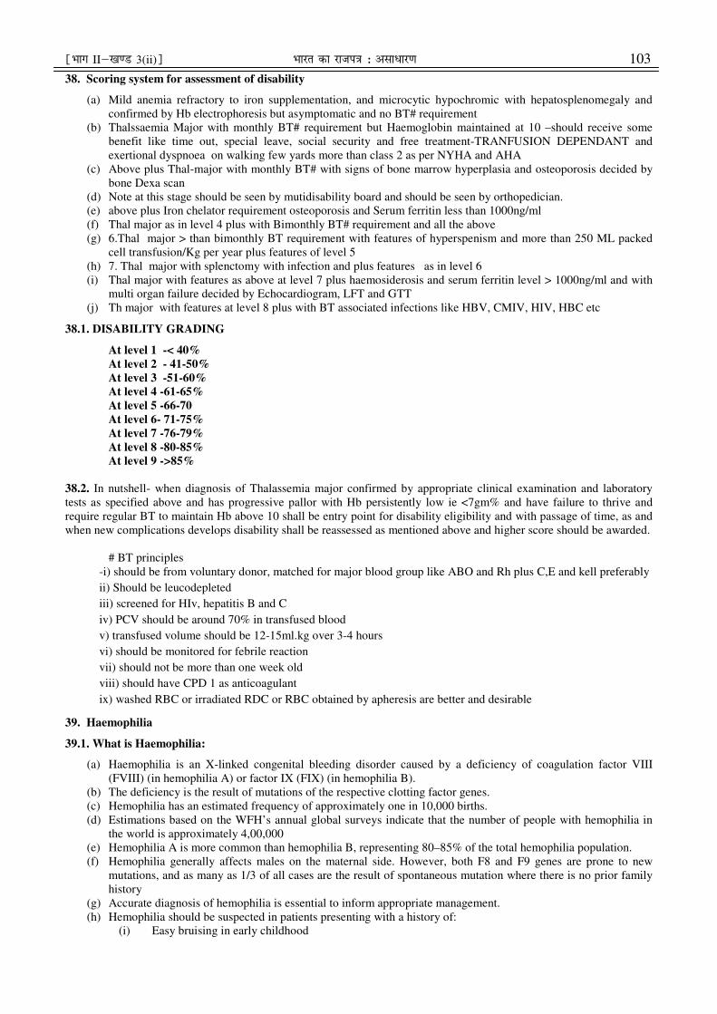
¹Hkkx IIµ[k.M 3(ii)º Hkkjr dk jkti=k % vlk/kj.k 103
38. Scoring system for assessment of disability
(a) Mild anemia refractory to iron supplementation, and microcytic hypochromic with hepatosplenomegaly and
confirmed by Hb electrophoresis but asymptomatic and no BT# requirement
(b) Thalssaemia Major with monthly BT# requirement but Haemoglobin maintained at 10 –should receive some
benefit like time out, special leave, social security and free treatment-TRANFUSION DEPENDANT and
exertional dyspnoea on walking few yards more than class 2 as per NYHA and AHA
(c) Above plus Thal-major with monthly BT# with signs of bone marrow hyperplasia and osteoporosis decided by
bone Dexa scan
(d) Note at this stage should be seen by mutidisability board and should be seen by orthopedician.
(e) above plus Iron chelator requirement osteoporosis and Serum ferritin less than 1000ng/ml
(f) Thal major as in level 4 plus with Bimonthly BT# requirement and all the above
(g) 6.Thal major > than bimonthly BT requirement with features of hyperspenism and more than 250 ML packed
cell transfusion/Kg per year plus features of level 5
(h) 7. Thal major with splenctomy with infection and plus features as in level 6
(i) Thal major with features as above at level 7 plus haemosiderosis and serum ferritin level > 1000ng/ml and with
multi organ failure decided by Echocardiogram, LFT and GTT
(j) Th major with features at level 8 plus with BT associated infections like HBV, CMIV, HIV, HBC etc
38.1. DISABILITY GRADING
At level 1 -< 40%
At level 2 - 41-50%
At level 3 -51-60%
At level 4 -61-65%
At level 5 -66-70
At level 6- 71-75%
At level 7 -76-79%
At level 8 -80-85%
At level 9 ->85%
38.2. In nutshell- when diagnosis of Thalassemia major confirmed by appropriate clinical examination and laboratory
tests as specified above and has progressive pallor with Hb persistently low ie <7gm% and have failure to thrive and
require regular BT to maintain Hb above 10 shall be entry point for disability eligibility and with passage of time, as and
when new complications develops disability shall be reassessed as mentioned above and higher score should be awarded.
# BT principles
-i) should be from voluntary donor, matched for major blood group like ABO and Rh plus C,E and kell preferably
ii) Should be leucodepleted
iii) screened for HIv, hepatitis B and C
iv) PCV should be around 70% in transfused blood
v) transfused volume should be 12-15ml.kg over 3-4 hours
vi) should be monitored for febrile reaction
vii) should not be more than one week old
viii) should have CPD 1 as anticoagulant
ix) washed RBC or irradiated RDC or RBC obtained by apheresis are better and desirable
39. Haemophilia
39.1. What is Haemophilia:
(a) Haemophilia is an X-linked congenital bleeding disorder caused by a deficiency of coagulation factor VIII
(FVIII) (in hemophilia A) or factor IX (FIX) (in hemophilia B).
(b) The deficiency is the result of mutations of the respective clotting factor genes.
(c) Hemophilia has an estimated frequency of approximately one in 10,000 births.
(d) Estimations based on the WFH’s annual global surveys indicate that the number of people with hemophilia in
the world is approximately 4,00,000
(e) Hemophilia A is more common than hemophilia B, representing 80–85% of the total hemophilia population.
(f) Hemophilia generally affects males on the maternal side. However, both F8 and F9 genes are prone to new
mutations, and as many as 1/3 of all cases are the result of spontaneous mutation where there is no prior family
history
(g) Accurate diagnosis of hemophilia is essential to inform appropriate management.
(h) Hemophilia should be suspected in patients presenting with a history of:
(i) Easy bruising in early childhood
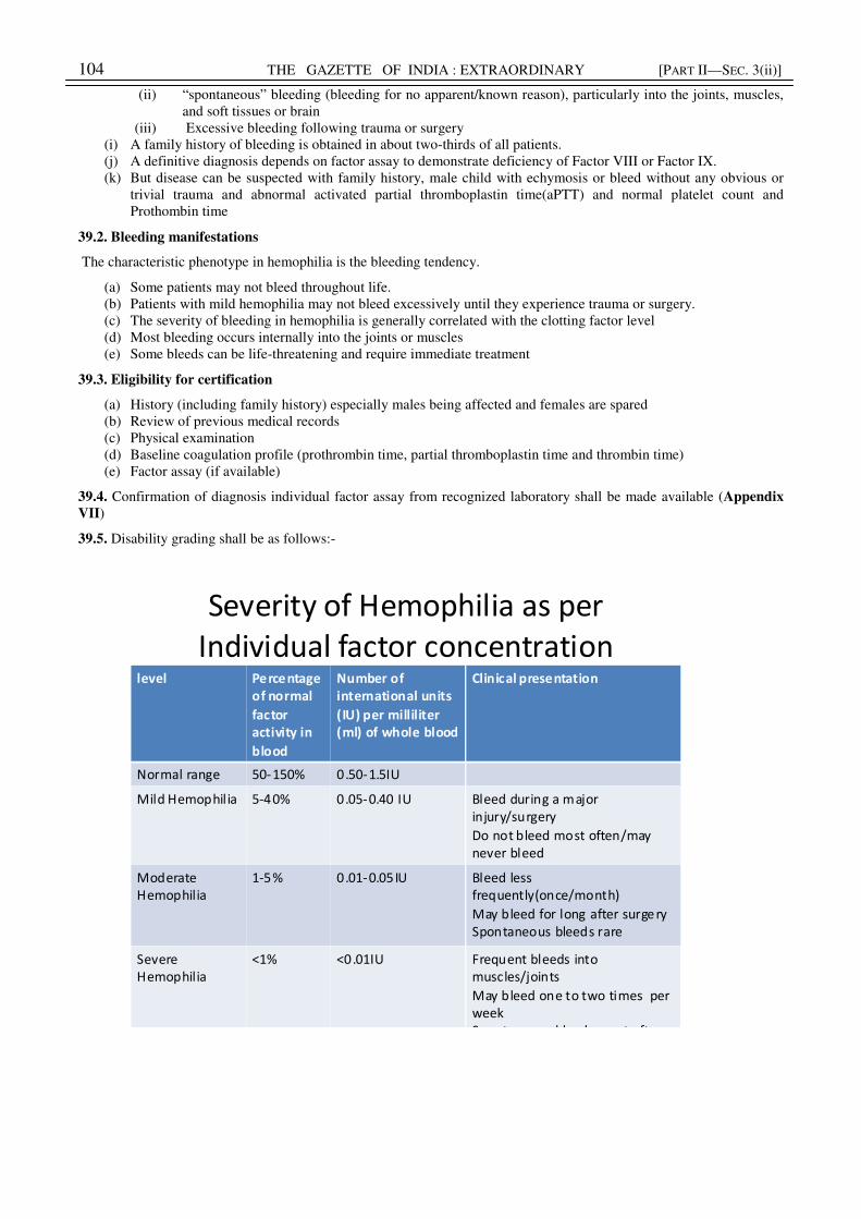
104 THE GAZETTE OF INDIA : EXTRAORDINARY [PART II—SEC. 3(ii)]
(ii) “spontaneous” bleeding (bleeding for no apparent/known reason), particularly into the joints, muscles,
and soft tissues or brain
(iii) Excessive bleeding following trauma or surgery
(i) A family history of bleeding is obtained in about two-thirds of all patients.
(j) A definitive diagnosis depends on factor assay to demonstrate deficiency of Factor VIII or Factor IX.
(k) But disease can be suspected with family history, male child with echymosis or bleed without any obvious or
trivial trauma and abnormal activated partial thromboplastin time(aPTT) and normal platelet count and
Prothombin time
39.2. Bleeding manifestations
The characteristic phenotype in hemophilia is the bleeding tendency.
(a) Some patients may not bleed throughout life.
(b) Patients with mild hemophilia may not bleed excessively until they experience trauma or surgery.
(c) The severity of bleeding in hemophilia is generally correlated with the clotting factor level
(d) Most bleeding occurs internally into the joints or muscles
(e) Some bleeds can be life-threatening and require immediate treatment
39.3. Eligibility for certification
(a) History (including family history) especially males being affected and females are spared
(b) Review of previous medical records
(c) Physical examination
(d) Baseline coagulation profile (prothrombin time, partial thromboplastin time and thrombin time)
(e) Factor assay (if available)
39.4. Confirmation of diagnosis individual factor assay from recognized laboratory shall be made available (Appendix
VII)
39.5. Disability grading shall be as follows:-
Severity of Hemophilia as per
Individual factor concentrationlevel Percentage
of normal
factor
activity in
blood
Number of
international units
(IU) per milliliter
(ml) of whole blood
Clinical presentation
Normal range 50-150% 0.50-1.5IU
Mild Hemophil ia 5-40% 0.05-0.40 IU Bleed during a major
injury/surgery
Do not bleed most often/may
never bleed
Moderate
Hemophilia
1-5% 0.01-0.05IU Bleed less
frequently(once/month)
May bleed for long after surgery
Spontaneous bleeds rare
Severe
Hemophilia
<1% <0.01IU Frequent bleeds into
muscles/joints
May bleed one to two times per
week
Spontaneous bleeds most often
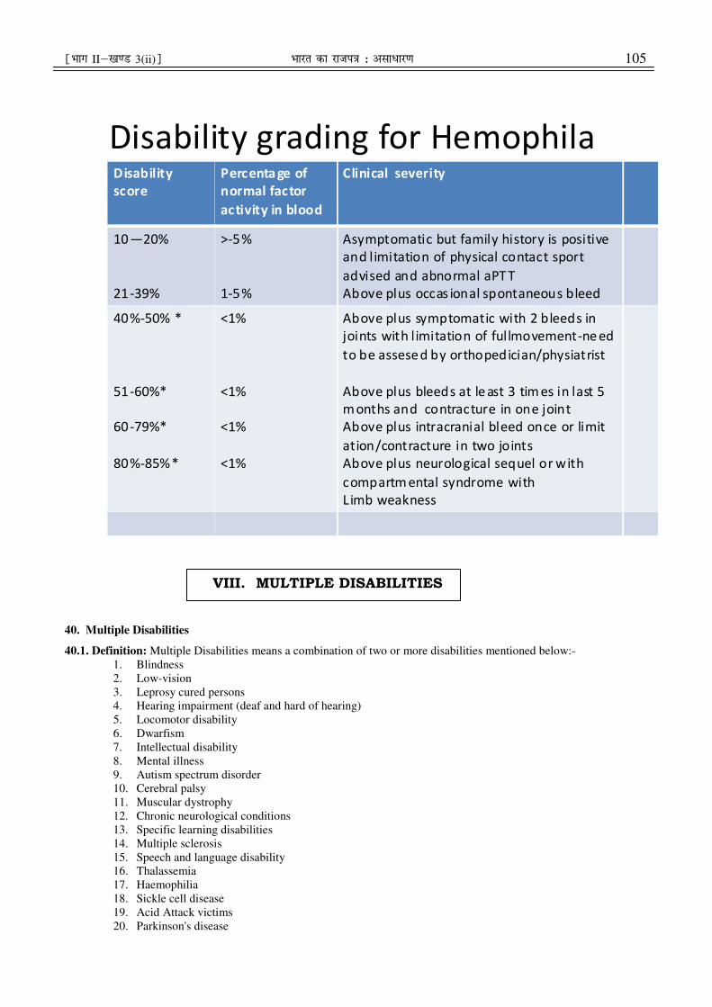
¹Hkkx IIµ[k.M 3(ii)º Hkkjr dk jkti=k % vlk/kj.k 105
40. Multiple Disabilities
40.1. Definition: Multiple Disabilities means a combination of two or more disabilities mentioned below:-
1. Blindness
2. Low-vision
3. Leprosy cured persons
4. Hearing impairment (deaf and hard of hearing)
5. Locomotor disability
6. Dwarfism
7. Intellectual disability
8. Mental illness
9. Autism spectrum disorder
10. Cerebral palsy
11. Muscular dystrophy
12. Chronic neurological conditions
13. Specific learning disabilities
14. Multiple sclerosis
15. Speech and language disability
16. Thalassemia
17. Haemophilia
18. Sickle cell disease
19. Acid Attack victims
20. Parkinson's disease
Disability grading for HemophilaDisability
score
Percenta ge of
normal factor
activity in blood
Clinical severity
10—20%
21-39%
>-5%
1-5%
Asymptomatic but family history is positive
and limitation of physical contact sport
advised and abnormal aPT T
Above plus occas ional spontaneous bleed
40%-50% *
51-60%*
60-79%*
80%-85%*
<1%
<1%
<1%
<1%
Above plus symptomatic with 2 bleeds in
joints with limitation of fullmovement-ne ed
to be assesed by orthopedician/physiatrist
Above plus bleeds at le ast 3 tim es in last 5
m onths and contracture in one joint
Above plus intracranial bleed once or limit
ation/contracture in two joints
Above plus neurological sequel or with
compartm ental syndrome with
Limb weakness
VIII. MULTIPLE DISABILITIES
106 THE GAZETTE OF INDIA : EXTRAORDINARY [PART II—SEC. 3(ii)]
40.2. Guidelines for Assessment:
40.2.1. The guidelines used for every single disability shall be used for assessment of each disability of a person having
multiple disability in the first instance.
40.2.2. Subsequently, in order to arrive at the total percentage of multiple disabilities, the combining formula a + b (90-
a), shall be used where 90
“a” will be the higher score and
“b” will be the lower score. However, the maximum total percentage of multiple disabilities shall not
exceed 100%.
For example, if the percentage of hearing disability is 30% and visual disability is 20%, then by applying the
combining formula given above, the total percentage of multiple disabilities will be calculated as follows:-
30 + 20(90-30) = 43%
90
40.2.3 For certifying more than two disabilities, each disability will be evaluated and the degree of disability will be
calculated by the notified Specialists in the area. Based on the score received for each disability, they will be graded
from the most severe to the least severe. The formula:
a + b (90-a)
90
will be successively applied to subsequent disability till the last disability is covered. This calculation is subject to
maximum of 100%.
For example a person may have disabilities 1, 2 and 3, the score for 1 is the highest equal to (a); score for the second is
equal to (b) (second highest); and score for 3 is (c) the lowest score. According to the above formula:
a + b (90-a) = x
90
(score of disability 1 and 2 = x)
This (x) will become (a) for the purpose of calculation of disability 3 which is C.
x + c (90-x) = y
90
(score of disabilities 1, 2 and 3 = y)
Such calculation will continue till the last disability is covered subject to a maximum of 100%.
41. Medical Authority
The certification medical authority for certifying multiple disability shall comprise of the following:-
(a) The Medical Superintendent or Chief Medical Officer or Civil Surgeon or any other equivalent authority as
notified by the State Government – Chairperson
(b) Specialist required for assessing the disabilities as per the requirement of respective guidelines.
Appendix I
[see paragraph 1.2.3(a)]
Muscle strength grading (Medical Research Council- MRC Scale):
Grade Description
0 No contraction of muscle being tested
1 Flicker or trace of contraction of muscle being tested
2 Active contraction of the muscle with gravity eliminated
3 Active contraction of the muscle against gravity
4 Active contraction of the muscle against gravity and resistance
5 Normal strength
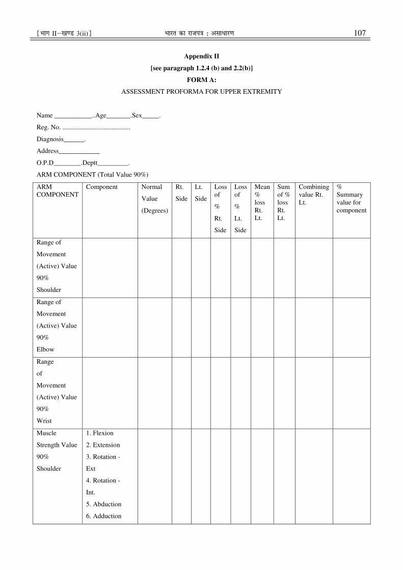
¹Hkkx IIµ[k.M 3(ii)º Hkkjr dk jkti=k % vlk/kj.k 107
Appendix II
[see paragraph 1.2.4 (b) and 2.2(b)]
FORM A:
ASSESSMENT PROFORMA FOR UPPER EXTREMITY
Name ___________..Age_______.Sex_____.
Reg. No. ........................................
Diagnosis______.
Address____________
O.P.D________.Deptt_________.
ARM COMPONENT (Total Value 90%)
ARM
COMPONENT
Component Normal
Value
(Degrees)
Rt.
Side
Lt.
Side
Loss
of
%
Rt.
Side
Loss
of
%
Lt.
Side
Mean
%
loss
Rt.
Lt.
Sum
of %
loss
Rt.
Lt.
Combining
value Rt.
Lt.
%
Summary
value for
component
Range of
Movement
(Active) Value
90%
Shoulder
Range of
Movement
(Active) Value
90%
Elbow
Range
of
Movement
(Active) Value
90%
Wrist
Muscle
Strength Value
90%
Shoulder
1. Flexion
2. Extension
3. Rotation -
Ext
4. Rotation -
Int.
5. Abduction
6. Adduction

108 THE GAZETTE OF INDIA : EXTRAORDINARY [PART II—SEC. 3(ii)]
Muscle
Strength Value
90%
Elbow
1. Flexion
2. Extension
3. Pronation
4. Supination
Muscle
Strength Value
90%
Wrist
1. Dors Flexion
2. Palmar
Flexion
3. Radial
Deviation
4. Ulnardeviatior
Coordinated
Activities
Value 90%
1. Lifting
overhead objects
remove and
placing at the
same place 9%
2. Touching nose
with end of
extremity 9%
3. Eating Indian
Style 9%
4. Combing and
Plaiting 9%
5. Putting on a
shirt/kurta 9%
6. Ablution glass
of water 9%
7. Drinking Glass
of water 9%
8. Buttoning 9%
9 Tie Nara Dhoti
9%
10. Writing 9%
HAND COMPONENT (TOTAL VALUE 90%)
30%
prehension
1. Hand
Component
A.
Opposition
(8%)
B. Lateral
Pinch
(5%)
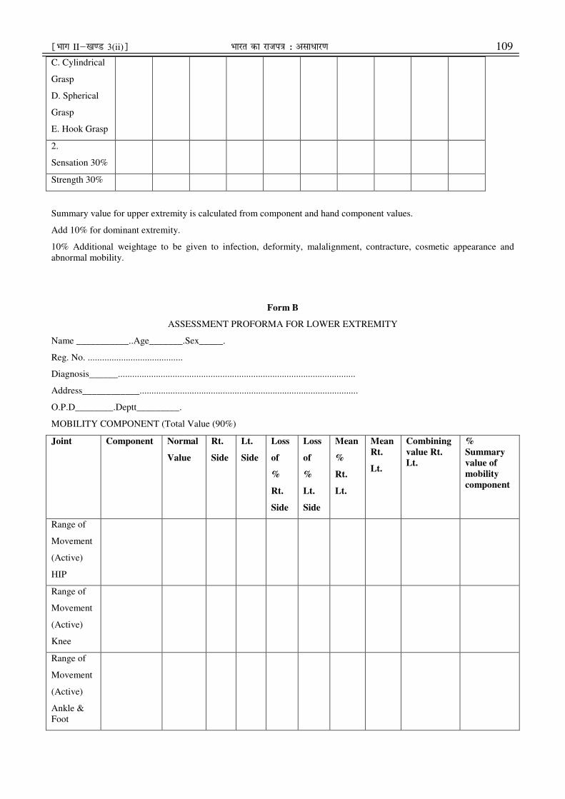
¹Hkkx IIµ[k.M 3(ii)º Hkkjr dk jkti=k % vlk/kj.k 109
C. Cylindrical
Grasp
D. Spherical
Grasp
E. Hook Grasp
2.
Sensation 30%
Strength 30%
Summary value for upper extremity is calculated from component and hand component values.
Add 10% for dominant extremity.
10% Additional weightage to be given to infection, deformity, malalignment, contracture, cosmetic appearance and
abnormal mobility.
Form B
ASSESSMENT PROFORMA FOR LOWER EXTREMITY
Name ___________..Age_______.Sex_____.
Reg. No. ........................................
Diagnosis______....................................................................................................
Address____________............................................................................................
O.P.D________.Deptt_________.
MOBILITY COMPONENT (Total Value (90%)
Joint Component Normal
Value
Rt.
Side
Lt.
Side
Loss
of
%
Rt.
Side
Loss
of
%
Lt.
Side
Mean
%
Rt.
Lt.
Mean
Rt.
Lt.
Combining
value Rt.
Lt.
%
Summary
value of
mobility
component
Range of
Movement
(Active)
HIP
Range of
Movement
(Active)
Knee
Range of
Movement
(Active)
Ankle &
Foot
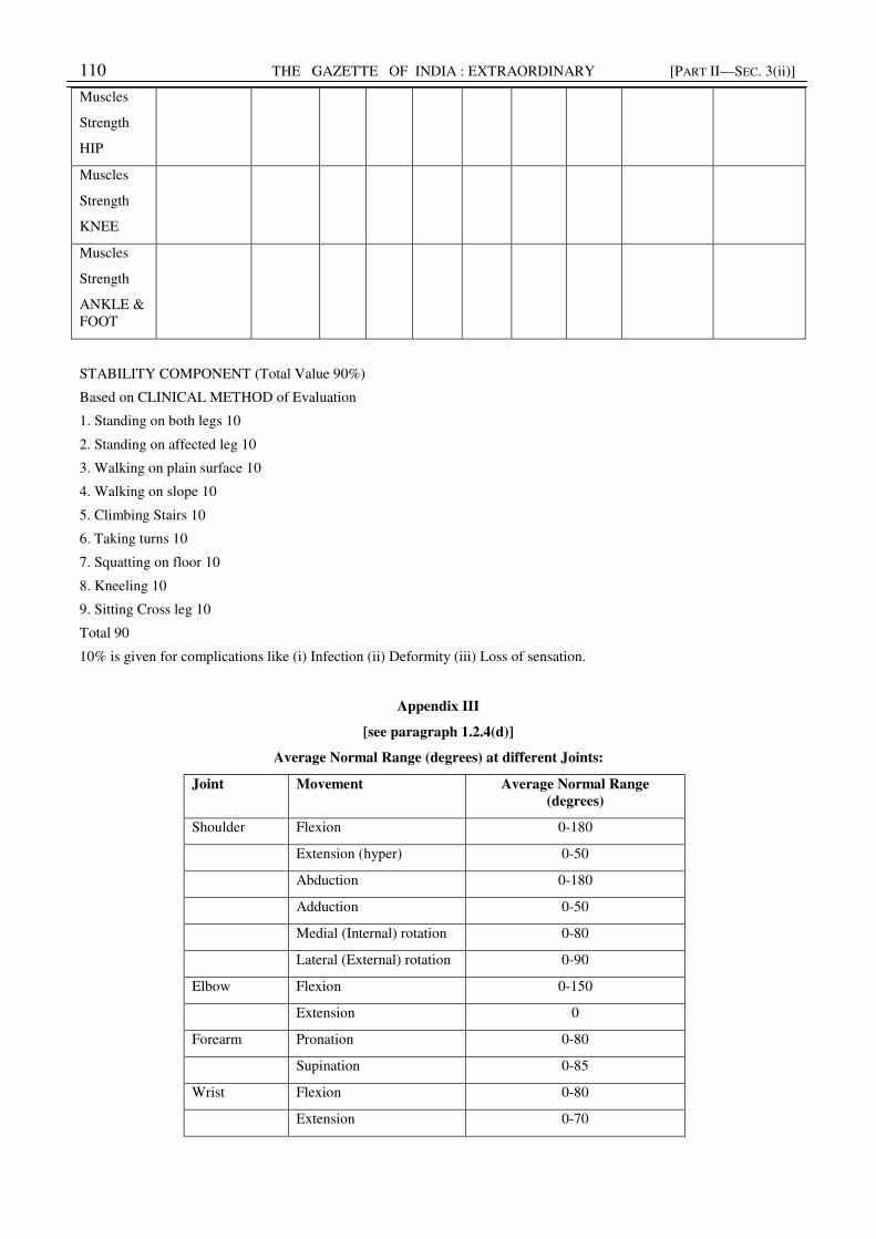
110 THE GAZETTE OF INDIA : EXTRAORDINARY [PART II—SEC. 3(ii)]
Muscles
Strength
HIP
Muscles
Strength
KNEE
Muscles
Strength
ANKLE &
FOOT
STABILITY COMPONENT (Total Value 90%)
Based on CLINICAL METHOD of Evaluation
1. Standing on both legs 10
2. Standing on affected leg 10
3. Walking on plain surface 10
4. Walking on slope 10
5. Climbing Stairs 10
6. Taking turns 10
7. Squatting on floor 10
8. Kneeling 10
9. Sitting Cross leg 10
Total 90
10% is given for complications like (i) Infection (ii) Deformity (iii) Loss of sensation.
Appendix III
[see paragraph 1.2.4(d)]
Average Normal Range (degrees) at different Joints:
Joint Movement Average Normal Range
(degrees)
Shoulder Flexion 0-180
Extension (hyper) 0-50
Abduction 0-180
Adduction 0-50
Medial (Internal) rotation 0-80
Lateral (External) rotation 0-90
Elbow Flexion 0-150
Extension 0
Forearm Pronation 0-80
Supination 0-85
Wrist Flexion 0-80
Extension 0-70
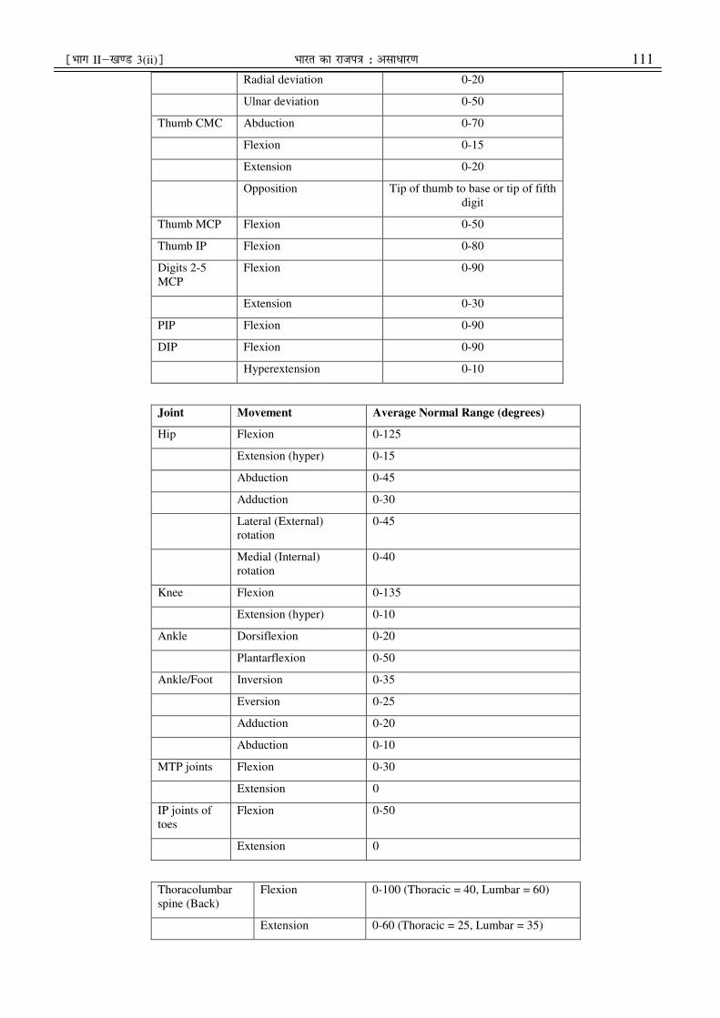
¹Hkkx IIµ[k.M 3(ii)º Hkkjr dk jkti=k % vlk/kj.k 111
Radial deviation 0-20
Ulnar deviation 0-50
Thumb CMC Abduction 0-70
Flexion 0-15
Extension 0-20
Opposition Tip of thumb to base or tip of fifth
digit
Thumb MCP Flexion 0-50
Thumb IP Flexion 0-80
Digits 2-5
MCP
Flexion 0-90
Extension 0-30
PIP Flexion 0-90
DIP Flexion 0-90
Hyperextension 0-10
Joint Movement Average Normal Range (degrees)
Hip Flexion 0-125
Extension (hyper) 0-15
Abduction 0-45
Adduction 0-30
Lateral (External)
rotation
0-45
Medial (Internal)
rotation
0-40
Knee Flexion 0-135
Extension (hyper) 0-10
Ankle Dorsiflexion 0-20
Plantarflexion 0-50
Ankle/Foot Inversion 0-35
Eversion 0-25
Adduction 0-20
Abduction 0-10
MTP joints Flexion 0-30
Extension 0
IP joints of
toes
Flexion 0-50
Extension 0
Thoracolumbar
spine (Back)
Flexion 0-100 (Thoracic = 40, Lumbar = 60)
Extension 0-60 (Thoracic = 25, Lumbar = 35)
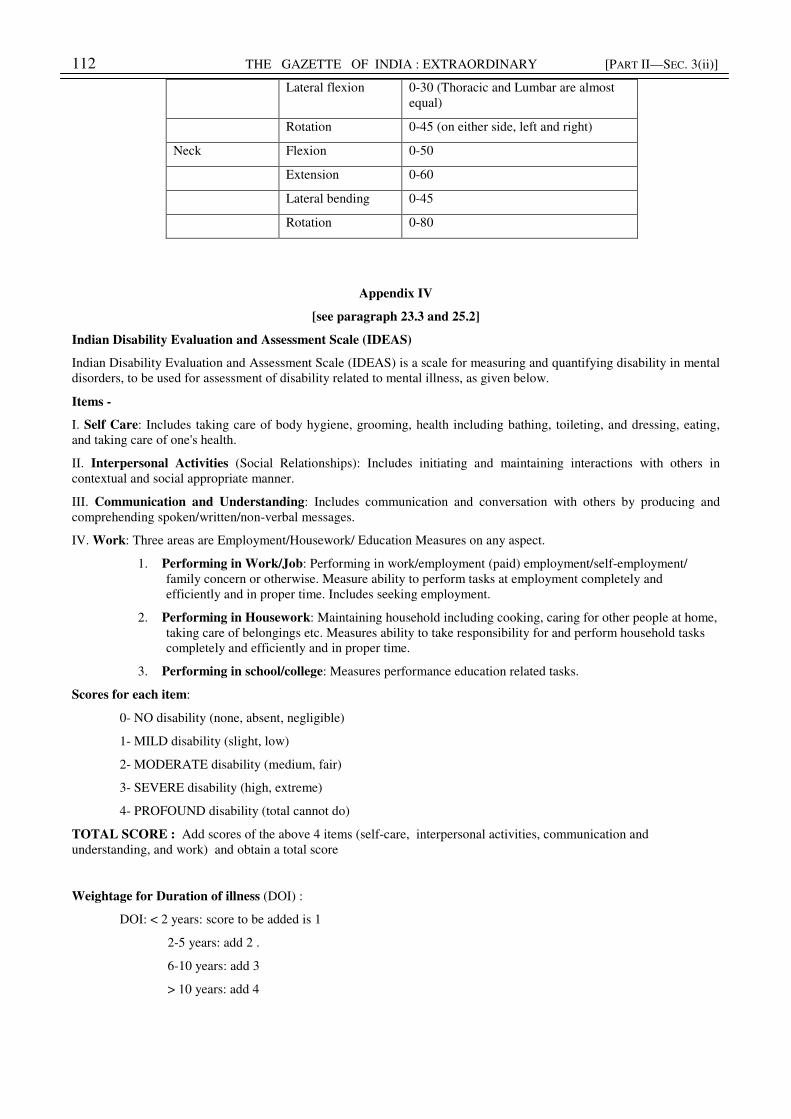
112 THE GAZETTE OF INDIA : EXTRAORDINARY [PART II—SEC. 3(ii)]
Lateral flexion 0-30 (Thoracic and Lumbar are almost
equal)
Rotation 0-45 (on either side, left and right)
Neck Flexion 0-50
Extension 0-60
Lateral bending 0-45
Rotation 0-80
Appendix IV
[see paragraph 23.3 and 25.2]
Indian Disability Evaluation and Assessment Scale (IDEAS)
Indian Disability Evaluation and Assessment Scale (IDEAS) is a scale for measuring and quantifying disability in mental
disorders, to be used for assessment of disability related to mental illness, as given below.
Items -
I. Self Care: Includes taking care of body hygiene, grooming, health including bathing, toileting, and dressing, eating,
and taking care of one's health.
II. Interpersonal Activities (Social Relationships): Includes initiating and maintaining interactions with others in
contextual and social appropriate manner.
III. Communication and Understanding: Includes communication and conversation with others by producing and
comprehending spoken/written/non-verbal messages.
IV. Work: Three areas are Employment/Housework/ Education Measures on any aspect.
1. Performing in Work/Job: Performing in work/employment (paid) employment/self-employment/
family concern or otherwise. Measure ability to perform tasks at employment completely and
efficiently and in proper time. Includes seeking employment.
2. Performing in Housework: Maintaining household including cooking, caring for other people at home,
taking care of belongings etc. Measures ability to take responsibility for and perform household tasks
completely and efficiently and in proper time.
3. Performing in school/college: Measures performance education related tasks.
Scores for each item:
0- NO disability (none, absent, negligible)
1- MILD disability (slight, low)
2- MODERATE disability (medium, fair)
3- SEVERE disability (high, extreme)
4- PROFOUND disability (total cannot do)
TOTAL SCORE : Add scores of the above 4 items (self-care, interpersonal activities, communication and
understanding, and work) and obtain a total score
Weightage for Duration of illness (DOI) :
DOI: < 2 years: score to be added is 1
2-5 years: add 2 .
6-10 years: add 3
> 10 years: add 4
¹Hkkx IIµ[k.M 3(ii)º Hkkjr dk jkti=k % vlk/kj.k 113
Global Disability -
Total Disability score + DOI score = Global Disability Score Percentages:
0 No Disability = 0%
1-6 Mild Disability = < 40 %
7-13 Moderate Disability = 40 - 70 %
14-19 Severe Disability = 71-99%
20 Profound Disability = l00%
Cut off for welfare measures = 40%
Manual for "IDEAS"
In order to score this instrument, information from all possible sources should be obtained. This will include interview of
patient, the care given and case notes when available.
I. SELF CARE: This should be regarded as activity guided by social norms and conventions. The broad areas covered
are
a. Maintenance of personal hygiene and physical health.
b. Eating habits
c. Maintenance of personal belongings and living space
d. Does s/he look after himself, wash his clothes regularly, take a bath and brush his teeth?
e. DOES s/he have regular meals?
f. Does s/he take food of right quality and quantity?
g. What about her/his table manners?
h. Does s/he take care of personal belongings with reasonable standard of cleanliness and orderliness?
0 = No disability: Patient's level and pattern of self-care are normal, within the social cultural and economic
context.
1 = Mild: Mild deterioration in self-care and appearance (not bathing, shaving, changing clothes for the
occasion as expected). Does not have adverse consequences such as hazards to her/his health. No
embarrassment to family.
2 = Moderate: Lack of concern for self-care should be clearly established such as mild deterioration of physical
health, obesity, tooth decay & body odours.
3 = Severe: Decline in self-care should be marked in all areas. Patient wearing torn clothes would only wash if
made to and would only care if told. Evidence of serious hazards to physical health. (Malnutrition, infection,
patient unacceptable in public).
4 = Profound: Total or near total lack of self-care (Example: risk to physical survival, needs feeding, washing,
putting on clothes etc., constant supervision necessary)
II. Inter Personal Activities
Includes patient's response to questions, requests and demands of others, activities or regulating emotions, activities of
initiating, maintaining and terminating interactions and activities of engaging in physical intimacy.
Guiding Questions
a. What is her/his behaviour with others?
b. Is s/he polite?
c. Does s/he respond to questions!
d. Is s/he able to regulate verbal and physical aggression?
e. Is s/he able to act independently in social interactions?
f. How does s/he behave with strangers?
g. Is s/he able to maintain friendship?
h. Does s/he show physical expression of affection and desire?
Scoring
0= No : Patient gets along reasonably well with people, personal relationships. No friction in inter-personal
relationships.
114 THE GAZETTE OF INDIA : EXTRAORDINARY [PART II—SEC. 3(ii)]
1= Mild : Some friction on isolated occasions. Patient known to be nervous or irritable but generally tolerated
by others.
2= Moderate: Factual evidence that pattern of response to people is unhealthy. May be seen or more than few
occasions. Could isolate herself/himself from others and avoid company.
3= Severe: Behaviour in social situations is undesirable and generalized. Causes serious problems in daily
living/or work. Patient is socially ostracized.
4= Profound: Patient in serious and lasting conflict, serious danger to problems of others. Family afraid of
potential consequences.
III. Communication and Understanding
Understanding spoken messages as well as written and non-verbal messages and ability to deduce messages in order to
communicate with others.
1. Questions
a. Does s/he avoid talking to people?
b. When people come home what does s/he do?
c. Does s/he ever visit others?
d. Is s/he able to start, maintain and end a conversation?
e. Does s/he understand body language and emotions of others such as smiling, crying, screaming, etc.,
f. Does s/he indulge in reading and writing?
g. Do you encourage her/him to be more sociable?
Scoring:
0 = No disability: Patient mixes, talks and generally interacts with people as much as can be expected in
her/his socio-cultural context. No evidence of avoiding people.
1= Mild: Patient described as uncommunicative or solitary in social situations. Signs of social anxiety might be
reported.
2= Moderate: A very narrow range of social contacts, evidence of active avoidance of people on some
occasions and interference with performance of social rules causes concern to family.
3= Severe: Evidence of more generalized, active avoidance of contact with people (leave the room when
visitors arrive and would not answer the door or phone).
4= Profound: Hardly has any contacts and actively avoids people nearly all the time. Eg : may lock
herself/himself inside the room. Verbal communication is nil or a bare minimum.
IV. Work
This includes employment, housework and educational performance. Score only one category in case of an overlap.
Employment:
Guiding Questions
a. Is s/he employed/unemployed?
b. If employed, does s/he go to work regularly?
c. Does s/he like his job and coping well with it?
d. Can you rely on her/him financially?
e. If unemployed, does s/he make efforts to find job?
Scoring:
0= No disability: Patient goes to work regularly and output and quality of work performance are within
acceptable levels for the job.
1= Mild: Noticeable decline in patient's ability to work, to cope with it and meet the demands of work. May
threaten to quit.
2= Moderate: Declining work performance, frequent absences, lack of concern about all this. Financial
difficulties foreseen.
3= Severe: Marked decline in work performance, disruptive at work, unwilling to adhere to disciplines of work.
Threat of losing his job.
¹Hkkx IIµ[k.M 3(ii)º Hkkjr dk jkti=k % vlk/kj.k 115
4= Profound: Has been largely absent from work, termination imminent. Unemployed and making no efforts to
find jobs.
In similar ways, housewives should be rated on the amount, regularity and efficiency in which tasks in the following
areas are completed. Consider the amount of help required completing these. Acquiring daily necessities, making, storing
and serving of food, cleaning the house, working with those helping with domestic duties such as maids, cooks etc.,
looking after possessions and valuable in the house.
Students - Assess a score on performance in school/college, regularity, discipline, interest in future studies, behaviour at
the educational institution. Those who had to discontinue education on account of mental disability and unable to
continue further should be given a score of 4.
IDEAS SCORING SHEET
ITEMS 0 1 2 3 4
Self care
Interpersonal Activities
Communication & Understanding
Work
A. TOTAL SCORE
B. DOI SCORE.
GLOBAL SCORE (A+B)
Appendix V
[see paragraph 31]
Screening tests for screening for carriers of haemoglobinopathies for sickle cell cases
NESTROFT Test (Naked Eye Single Tube Red cell Osmotic Fragility Test)
DCIP test (Di-Chlorophenol-Indo-Phenol) Solubility Test
NESTROFT TEST
Nestroft Test: For Beta thalassemia trait, this test has a high specificity and sensitivity and is easy to perform. The
positive test has to be followed by a confirmatory test Sensitivity of 91-100%, specificity of 85.47%. Positive predictive
value of 66% and negative predictive value of 97- 100%.
Screening protocols for Hemoglobinopathies in community settings and public health facilities Initial screening (1
and 2)
Test tube based Turbidity tests in Community settings (one or more tube tests may be included depending on
prevalence)
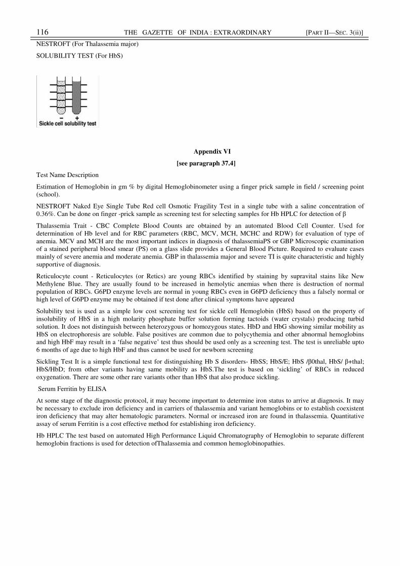
116 THE GAZETTE OF INDIA : EXTRAORDINARY [PART II—SEC. 3(ii)]
NESTROFT (For Thalassemia major)
SOLUBILITY TEST (For HbS)
Appendix VI
[see paragraph 37.4]
Test Name Description
Estimation of Hemoglobin in gm % by digital Hemoglobinometer using a finger prick sample in field / screening point
(school).
NESTROFT Naked Eye Single Tube Red cell Osmotic Fragility Test in a single tube with a saline concentration of
0.36%. Can be done on finger -prick sample as screening test for selecting samples for Hb HPLC for detection of β
Thalassemia Trait - CBC Complete Blood Counts are obtained by an automated Blood Cell Counter. Used for
determination of Hb level and for RBC parameters (RBC, MCV, MCH, MCHC and RDW) for evaluation of type of
anemia. MCV and MCH are the most important indices in diagnosis of thalassemiaPS or GBP Microscopic examination
of a stained peripheral blood smear (PS) on a glass slide provides a General Blood Picture. Required to evaluate cases
mainly of severe anemia and moderate anemia. GBP in thalassemia major and severe TI is quite characteristic and highly
supportive of diagnosis.
Reticulocyte count - Reticulocytes (or Retics) are young RBCs identified by staining by supravital stains like New
Methylene Blue. They are usually found to be increased in hemolytic anemias when there is destruction of normal
population of RBCs. G6PD enzyme levels are normal in young RBCs even in G6PD deficiency thus a falsely normal or
high level of G6PD enzyme may be obtained if test done after clinical symptoms have appeared
Solubility test is used as a simple low cost screening test for sickle cell Hemoglobin (HbS) based on the property of
insolubility of HbS in a high molarity phosphate buffer solution forming tactoids (water crystals) producing turbid
solution. It does not distinguish between heterozygous or homozygous states. HbD and HbG showing similar mobility as
HbS on electrophoresis are soluble. False positives are common due to polycythemia and other abnormal hemoglobins
and high HbF may result in a ‘false negative’ test thus should be used only as a screening test. The test is unreliable upto
6 months of age due to high HbF and thus cannot be used for newborn screening
Sickling Test It is a simple functional test for distinguishing Hb S disorders- HbSS; HbS/E; HbS /β0thal, HbS/ β+thal;
HbS/HbD; from other variants having same mobility as HbS.The test is based on ‘sickling’ of RBCs in reduced
oxygenation. There are some other rare variants other than HbS that also produce sickling.
Serum Ferritin by ELISA
At some stage of the diagnostic protocol, it may become important to determine iron status to arrive at diagnosis. It may
be necessary to exclude iron deficiency and in carriers of thalassemia and variant hemoglobins or to establish coexistent
iron deficiency that may alter hematologic parameters. Normal or increased iron are found in thalassemia. Quantitative
assay of serum Ferritin is a cost effective method for establishing iron deficiency.
Hb HPLC The test based on automated High Performance Liquid Chromatography of Hemoglobin to separate different
hemoglobin fractions is used for detection ofThalassemia and common hemoglobinopathies.
¹Hkkx IIµ[k.M 3(ii)º Hkkjr dk jkti=k % vlk/kj.k 117
Appendix VII
[ see paragraph 39.4.]
Diagnosis of hemophilia
The majority of patients with hemophilia have a known family history of the condition. However, about one-third of
cases occur in the absence of a known family history. Most of these cases without a family history arise due to a
spontaneous mutation in the affected gene. Other cases may be due to the affected gene being passed through a long line
of female carriers.
If there is no known family history of hemophilia, a series of blood tests can identify which part or protein factor of the
blood clotting mechanism is defective if an individual has abnormal bleeding episodes.
Screening Tests
Screening tests are blood tests that show if the blood is clotting properly. Types of screening tests:
The complete blood count in particular the platelet count and bleeding time test should be measured as well as two
indices of blood clotting, the prothrombin time (PT) and activated partial thromboplastin time (aPTT). A normal platelet
count, normal PT, and a prolonged aPTT are characteristic of hemophilia A and hemophilia B.
Complete Blood Count (CBC)
This common test measures the amount of hemoglobin , the size and number of red blood cells and numbers of different
types of white blood cells and platelets found in blood. The CBC is normal in people with hemophilia. However, if a
person with hemophilia has unusually heavy bleeding or bleeds for a long time, the hemoglobin and the red blood cell
count can be low.
Activated Partial Thromboplastin Time (APTT) Test
This test measures how long it takes for blood to clot. It measures the clotting ability of factors VIII (8), IX (9), XI (11),
and XII (12). If any of these clotting factors are too low, it takes longer than normal for the blood to clot. The results of
this test will show a longer clotting time among people with hemophilia A or B. in this process of coagulation is
stimulated by contact kaolin or collagen or ellagic acid. Normal value ie 30-32 seconds and are cheap and available at
most of places
Prothrombin Time (PT) Test
This test also measures the time it takes for blood to clot. It measures primarily the clotting ability of factors I (1), II (2),
V (5), VII (7), and X (10). If any of these factors are too low, it takes longer than normal for the blood to clot. The results
of this test will be normal among most people with hemophilia A and B.
Note these tests are simple, easy to perform and act as screening tests and are available at most of places
Specific tests (factor assay) for the blood clotting factors can then be performed to measure factor VIII or factor IX levels
and confirm the diagnosis. Factor assays are required to diagnose and confirm a bleeding disorder. This blood test
shows the type of hemophilia and the severity. It is important to know the type and severity in order to create the best
treatment plan.
I. Factor VIII is the protein which is lacking in hemophilia A.
II. Factor IX is the protein which is lacking in hemophilia B.
[No. 16-09/2014-DD-III]
SHAKUNTALA DOLEY GAMLIN, Secy.
Uploaded by Dte. of Printing at Government of India Press, Ring Road, Mayapuri, New Delhi-110064
and Published by the Controller of Publications, Delhi-110054.