Isolation of rare circulating tumour cells in cancer patients by microchip technology
11
Isolation of rare circulating tumour cells in cancer patients by microchip technology Sunitha Nagrath 1,* , Lecia V. Sequist 2,* , Shyamala Maheswaran 2 , Daphne W. Bell 2,† , Daniel Irimia 1 , Lindsey Ulkus 2 , Matthew R. Smith 2 , Eunice L. Kwak 2 , Subba Digumarthy 2 , Alona Muzikansky 2 , Paula Ryan 2 , Ulysses J. Balis 1,† , Ronald G. Tompkins 1 , Daniel A. Haber 2 , and Mehmet Toner 1 1 Surgical Services and BioMEMS Resource Center, Massachusetts General Hospital, Harvard Medical School, and Shriners Hospital for Children, Boston, Massachusetts 02114, USA 2 Massachusetts General Hospital Cancer Center, Harvard Medical School, Boston, Massachusetts 02114, USA Abstract Viable tumour-derived epithelial cells (circulating tumour cells or CTCs) have been identified in peripheral blood from cancer patients and are probably the origin of intractable metastatic disease 1–4 . Although extremely rare, CTCs represent a potential alternative to invasive biopsies as a source of tumour tissue for the detection, characterization and monitoring of non-haematologic cancers 5–8 . The ability to identify, isolate, propagate and molecularly characterize CTC subpopulations could further the discovery of cancer stem cell biomarkers and expand the understanding of the biology of metastasis. Current strategies for isolating CTCs are limited to complex analytic approaches that generate very low yield and purity 9 . Here we describe the development of a unique microfluidic platform (the ‘CTC-chip’) capable of efficient and selective separation of viable CTCs from peripheral whole blood samples, mediated by the interaction of target CTCs with antibody (EpCAM)-coated microposts under precisely controlled laminar flow conditions, and without requisite pre-labelling or processing of samples. The CTC-chip successfully identified CTCs in the peripheral blood of patients with metastatic lung, prostate, pancreatic, breast and colon cancer in 115 of 116 (99%) samples, with a range of 5–1,281 CTCs per ml and approximately 50% purity. In addition, CTCs were isolated in 7/7 patients with early- stage prostate cancer. Given the high sensitivity and specificity of the CTC-chip, we tested its potential utility in monitoring response to anti-cancer therapy. In a small cohort of patients with metastatic cancer undergoing systemic treatment, temporal changes in CTC numbers correlated reasonably well with the clinical course of disease as measured by standard radiographic methods. Thus, the CTC-chip provides a new and effective tool for accurate identification and measurement of CTCs in patients with cancer. It has broad implications in advancing both cancer biology Correspondence and requests for materials should be addressed to M.T. ([email protected]). † Present addresses: National Human Genome Research Institute/NIH Cancer Genetics Branch, Bethesda, Maryland 20892, USA (D.W.B.); Department of Pathology, University of Michigan Health System, Ann Arbor, Michigan 48109, USA (U.J.B.). * These authors contributed equally to this paper. Full Methods and any associated references are available in the online version of the paper at www.nature.com/nature. Supplementary Information is linked to the online version of the paper at www.nature.com/nature. Author Contributions S.N., L.V.S., R.G.T., D.A.H. and M.T. designed and conducted the study. S.M., D.W.B. and L.U. performed gene expression analyses; D.I. contributed to the microfluidic system. M.R.S., E.L.K. and P.R. acquired clinical samples. U.J.B. provided input on cytopathology; A.M. performed statistical analysis; and S.D. performed radiology measurements. S.N., L.V.S., D.W.B., S.M., D.I., D.A.H. and M.T. participated in data analysis and writing of the manuscript. Author Information Reprints and permissions information is available at www.nature.com/reprints. The authors declare competing financial interests: details accompany the full-text HTML version of the paper at www.nature.com/nature. NIH Public Access Author Manuscript Nature. Author manuscript; available in PMC 2011 May 10. Published in final edited form as: Nature. 2007 December 20; 450(7173): 1235–1239. doi:10.1038/nature06385. NIH-PA Author Manuscript NIH-PA Author Manuscript NIH-PA Author Manuscript
Transcript of Isolation of rare circulating tumour cells in cancer patients by microchip technology

Isolation of rare circulating tumour cells in cancer patients bymicrochip technology
Sunitha Nagrath1,*, Lecia V. Sequist2,*, Shyamala Maheswaran2, Daphne W. Bell2,†, DanielIrimia1, Lindsey Ulkus2, Matthew R. Smith2, Eunice L. Kwak2, Subba Digumarthy2, AlonaMuzikansky2, Paula Ryan2, Ulysses J. Balis1,†, Ronald G. Tompkins1, Daniel A. Haber2, andMehmet Toner1
1Surgical Services and BioMEMS Resource Center, Massachusetts General Hospital, HarvardMedical School, and Shriners Hospital for Children, Boston, Massachusetts 02114, USA2Massachusetts General Hospital Cancer Center, Harvard Medical School, Boston,Massachusetts 02114, USA
AbstractViable tumour-derived epithelial cells (circulating tumour cells or CTCs) have been identified inperipheral blood from cancer patients and are probably the origin of intractable metastaticdisease1–4. Although extremely rare, CTCs represent a potential alternative to invasive biopsies asa source of tumour tissue for the detection, characterization and monitoring of non-haematologiccancers5–8. The ability to identify, isolate, propagate and molecularly characterize CTCsubpopulations could further the discovery of cancer stem cell biomarkers and expand theunderstanding of the biology of metastasis. Current strategies for isolating CTCs are limited tocomplex analytic approaches that generate very low yield and purity9. Here we describe thedevelopment of a unique microfluidic platform (the ‘CTC-chip’) capable of efficient and selectiveseparation of viable CTCs from peripheral whole blood samples, mediated by the interaction oftarget CTCs with antibody (EpCAM)-coated microposts under precisely controlled laminar flowconditions, and without requisite pre-labelling or processing of samples. The CTC-chipsuccessfully identified CTCs in the peripheral blood of patients with metastatic lung, prostate,pancreatic, breast and colon cancer in 115 of 116 (99%) samples, with a range of 5–1,281 CTCsper ml and approximately 50% purity. In addition, CTCs were isolated in 7/7 patients with early-stage prostate cancer. Given the high sensitivity and specificity of the CTC-chip, we tested itspotential utility in monitoring response to anti-cancer therapy. In a small cohort of patients withmetastatic cancer undergoing systemic treatment, temporal changes in CTC numbers correlatedreasonably well with the clinical course of disease as measured by standard radiographic methods.Thus, the CTC-chip provides a new and effective tool for accurate identification and measurementof CTCs in patients with cancer. It has broad implications in advancing both cancer biology
Correspondence and requests for materials should be addressed to M.T. ([email protected]).†Present addresses: National Human Genome Research Institute/NIH Cancer Genetics Branch, Bethesda, Maryland 20892, USA(D.W.B.); Department of Pathology, University of Michigan Health System, Ann Arbor, Michigan 48109, USA (U.J.B.).*These authors contributed equally to this paper.Full Methods and any associated references are available in the online version of the paper at www.nature.com/nature.Supplementary Information is linked to the online version of the paper at www.nature.com/nature.Author Contributions S.N., L.V.S., R.G.T., D.A.H. and M.T. designed and conducted the study. S.M., D.W.B. and L.U. performedgene expression analyses; D.I. contributed to the microfluidic system. M.R.S., E.L.K. and P.R. acquired clinical samples. U.J.B.provided input on cytopathology; A.M. performed statistical analysis; and S.D. performed radiology measurements. S.N., L.V.S.,D.W.B., S.M., D.I., D.A.H. and M.T. participated in data analysis and writing of the manuscript.Author Information Reprints and permissions information is available at www.nature.com/reprints. The authors declare competingfinancial interests: details accompany the full-text HTML version of the paper at www.nature.com/nature.
NIH Public AccessAuthor ManuscriptNature. Author manuscript; available in PMC 2011 May 10.
Published in final edited form as:Nature. 2007 December 20; 450(7173): 1235–1239. doi:10.1038/nature06385.
NIH
-PA Author Manuscript
NIH
-PA Author Manuscript
NIH
-PA Author Manuscript

research and clinical cancer management, including the detection, diagnosis and monitoring ofcancer10.
CTCs are rare, comprising as few as one cell per 109 haematologic cells in the blood ofpatients with metastatic cancer, hence their isolation presents a tremendous technicalchallenge7,9,11–13. Microfluidic lab-on-a-chip devices provide unique opportunities for cellsorting and rare-cell detection; they have been successfully used for microfluidic flowcytometry14, continuous size-based separation15,16 and chromatographic separation17.Despite their success in manipulating microlitre amounts of simple liquids in microscalechannels14,18,19, they have thus far shown limited capability to deal with the cellular andfluid complexity of large volumes (millilitres) of whole blood samples20–22.
Here we describe the development and application of a microfluidic device (the ‘CTC-chip’)that can efficiently and reproducibly isolate CTCs from the blood of patients with commonepithelial tumours (Fig. 1, and Supplementary Fig. 1). The CTC-chip (Fig. 1b) consists of anarray of microposts (Supplementary Fig. 1c) that are made chemically functional with anti-epithelial-cell-adhesion-molecule (EpCAM, also known as TACSTD1) antibodies. Anti-EpCAM provides the specificity for CTC capture from unfractionated blood becauseEpCAM is frequently overexpressed by carcinomas of lung, colorectal, breast, prostate, headand neck, and hepatic origin, and is absent from haematologic cells23,24.
Two essential parameters that determine the efficiency of cell capture on the CTC-chip are:(1) flow velocity, because it influences the duration of cell–micropost contact; and (2) shearforce, which must be sufficiently low to ensure maximum cell–micropost attachment. Tooptimize these parameters we employed theoretical analyses characterizing the interaction ofcells with microposts distributed within the flow path (Supplementary Figs 1c and 2).Briefly, the simulation indicated an equilateral triangular arrangement, with a 50 μmdistance between microposts and a 50 μm shift after every 3 rows, to be the most efficientgeometric arrangement. For a given volumetric flow rate of 1 ml h−1 through the device, themaximum shear stress experienced by a cell near the micropost surface was estimated to be0.4 dyn cm−2 at θ =68° and the expected maximum velocity was 460 μm s−1
(Supplementary Fig. 2b–d), within the range facilitating maximum cell attachmentaccording to linear shear stress chamber studies (Supplementary Fig. 3). On the basis of thesimulation results, we fabricated a CTC-chip containing an array of 78,000 micropostswithin a 970mm2 surface.
To determine the efficiency of capture, we spiked non-small-cell lung cancer (NSCLC) cells(NCI-H1650) into phosphate buffered saline (PBS) at 100 cells ml−1 and captured thespiked cancer cells using the CTC-chip. NSCLC cells were visually evident about EpCAM-coated microposts, whereas no cancer cells were seen following flow through uncoated posts(Supplementary Fig. 4a–c). The calculated capture efficiency was 65% and decreasedsignificantly at flow rates above 2.5 ml h−1 (Fig. 2a), presumably owing to increased shearstress, consistent with our simulation predictions. The efficiency of capture was notenhanced at flow rates less than 0.75 ml h−1, leading us to select a flow rate of 1–2 ml h−1
for subsequent studies.
To determine the effect of cellular EpCAM expression on efficiency of CTC capture, wecompared capture rates among cancer cell lines with varied EpCAM expression, includingNSCLC NCI-H1650 cells and breast cancer SKBr-3 cells, with >500,000 antigens per cell;prostate cancer PC3-9 cells, with approximately 50,000 antigens per cell; and bladder cancerT-24 cells, with approximately 2,000 antigens per cell25. Each cell line was spiked into PBSat a concentration of 100 cells ml−1 and analysed, resulting in a mean capture yield of >65%in all cases (Fig. 2b). Interestingly T-24 cells with relatively low EpCAM expression were
Nagrath et al. Page 2
Nature. Author manuscript; available in PMC 2011 May 10.
NIH
-PA Author Manuscript
NIH
-PA Author Manuscript
NIH
-PA Author Manuscript
captured as efficiently as high-level antigen-expressing cells. We believe this is due to theaugmented cell–substrate interactions inherent within the CTC-chip.
To evaluate cell capture under more physiological conditions, we conducted a series ofexperiments in which NCI-H1650 cells were spiked into whole blood from healthy donors.Suspensions at concentrations ranging from 50 to 50,000 tumour cells per ml of whole bloodwere analysed, yielding recovery rates of >60% in all cases (Fig. 2c). To assess the potentialsteric hindrance of red blood cells in the flow path, these studies were repeated using lysedblood from healthy donors. Capture rates were comparable under both conditions (r2 = 0.99;Fig. 2c). Similar results were obtained using whole blood and lysed samples from NSCLCpatients (Supplementary Fig. 4d, e). We thus concluded that the CTC-chip does not requireblood-sample pre-processing.
Having optimized the CTC-chip with controlled quantities of cancer-derived cells, we testedits capacity to capture CTCs from whole blood samples donated by cancer patients. A totalof 116 samples from 68 patients with epithelial cancers including NSCLC (n = 55), prostate(n = 26), pancreatic (n = 15), breast (n = 10) and colon (n = 10) were studied. The majorityof patients had metastatic disease; however, 7 of 26 subjects with prostate cancer haduntreated, clinically localized disease with specimens collected before prostatectomy withcurative intent (Supplementary Table 2). The average volume of blood analysed was 2.7 mlper sample (range, 0.9 to 5.1 ml). We also examined samples from 20 healthy individuals(3.0 ± 0.4 ml (mean ± s.d.) of blood per subject) as controls (Supplementary Table 1).
CTCs captured from a group of patient samples were identified using a comprehensiveimage analysis algorithm, consisting of staining with 4,6-diamidino-2-phenylindole (DAPI)for DNA content, and using rhodamine-conjugated anti-cytokeratin (also known asKERSMCR) antibodies for epithelial cells, and fluorescein-conjugated anti-CD45 antibodiesfor haematologic cells (Fig. 2d–k, and Supplementary Fig. 5). Cells captured by anti-EpCAM-coated microposts and staining for cytokeratin were scored as CTCs, whereasCD45-positive cells were scored as contaminating normal haematologic cells. Themorphologic characteristics exhibited by the captured CTCs were consistent with malignantcells, including large cellular size with high nuclear:cytoplasmic ratios and visible nucleoli(Fig. 2d–g). The mean viability of captured cells was 98.5 ± 2.3% (mean ± s.d.), determinedby assessing cell membrane integrity in 10 high-power fields of view per CTC-chip in 4samples obtained from lung (n = 2) and prostate (n = 2) cancer patients.
CTCs were identified in 115 of 116 (>99%) patient samples analysed, with the singlenegative specimen being a small volume sample (0.9 ml) from a patient with colorectalcancer. The number of CTCs isolated ranged from 5 to 1,281 per ml for NSCLC (155 ± 236(mean ± s.d.) CTCs per ml), 16 to 292 (86 ± 78) for metastatic prostate, 25 to 174 (94 ± 63)among localized prostate cancers, 9 to 831 (196 ± 228) for pancreatic, 5 to 176 (79 ± 52) forbreast, and 42 to 375 (121 ± 127) for colorectal cancers (Fig. 3a, b). The identification ofCTCs in subjects with clinically localized prostate cancer at numbers approximating those inmetastatic prostate cancer patients is a novel finding enabled by the high sensitivity of ourtechnique and warrants further study. The average purity of capture, as defined by the ratioof cytokeratin+ to CD45+ cells, was 52% in NSCLC, 49% in metastatic prostate, 53% inlocalized prostate, 53% in pancreatic, 60% in breast, and 67% in colon cancers (Fig. 3c).None of the 20 healthy subjects had any identifiable CTCs (Supplementary Table 1). On thebasis of these results we calculated the sensitivity (99.1%) and specificity (100%) of theCTC-chip across all five cancers. Finally, we evaluated the reproducibility of CTC captureusing split samples and showed high experimental reproducibility (r2= 0.98; SupplementaryFig. 4f).
Nagrath et al. Page 3
Nature. Author manuscript; available in PMC 2011 May 10.
NIH
-PA Author Manuscript
NIH
-PA Author Manuscript
NIH
-PA Author Manuscript

To determine whether captured cells are suitable for subsequent molecular analyses, wetested expression of two tumour-specific markers: prostate-specific antigen (PSA; alsoknown as KLK3) in prostate cancer and thyroid transcription factor-1 (TTF-1; also known asNKX2-1) in lung adenocarcinoma. Specific expression of these markers was evident byimmunostaining (Fig. 4a, b, d, e), and confirmed by direct lysis of captured CTCs on themicrochip and PCR with reverse transcription (RT–PCR) amplification of the individualtranscripts (Fig. 4c, f). Considering the ∼50% purity of captured viable CTCs (two orders ofmagnitude higher than currently available technologies)26, the CTC-chip provides apowerful opportunity for CTC-based molecular analyses.
To demonstrate the unique clinical potential of our approach, we evaluated the ability ofchanges in CTC burden to predict changes in tumour volume in cancer patients undergoinganti-cancer treatments. Patients with advanced epithelial-based malignancies that werecommencing a first or second line systemic treatment regimen were eligible. Blood sampleswere collected at baseline and during subsequent clinic visits; the exact follow-up schedulevaried between patients. Computed tomograms (CT scans) were performed at baseline andat regular intervals according to standard clinical practice. For each CT scan, we calculatedthe sum of the unidimensional size in centimetres of all significant and measurable tumoursites, as per the RECIST standardized system27. All patients with baseline and at least onefollow-up CTC sample and CT scan were analysed, including patients with NSCLC (n=3),colorectal (n=2), pancreatic (n=3) and oesophageal cancer (n=1) (Fig. 3d–i). The absolutenumber of CTCs captured did not necessarily correspond with tumour size, and may beinfluenced by other factors. The correlation between per cent change in CTC quantity andper cent change in tumour size, from first to last measurement, was analysed over all 9patients and yielded a Pearson's correlation coefficient of 0.68 (P=0.03). These results froma small cohort of patients show that CTC quantity correlates reasonably well with clinicalresponse and clinical non-response to treatment (Fig. 3d–i, and Supplementary Fig. 6),indicating that monitoring treatment response using the CTC-chip may ultimately be apowerful tool enabling accurate, early decision-making. The clinical impact of such anapproach could be large, and could enable a decrease in patient exposure to toxicities fromineffective therapies and guide them towards the treatments most active against theirspecific tumour.
Other approaches to enrich or sort CTCs from peripheral blood have been previouslypublished, including flow cytometry28, immunomagnetic beads9, high-throughput optical-imaging systems29, and fibre optic array scanning12. Immunomagnetic-beadpurification1,13,30 is currently the lead technology in the clinical setting, with reportedsuccess in identifying CTCs in a portion of tested patients with lung, prostate, colon, breastand pancreatic cancer30. However, this approach isolates small numbers of CTCs (4 ± 24(mean ± s.d.) per ml in lung; 11 ± 118 in breast; 10 ± 33 in prostate; and 1 ± 2 in bothcolorectal and pancreatic cancers)30 with very low purity (0.01–0.1%)26, and low yield(∼20–60% of patients)30. The level of ‘biological noise’ associated with the low sensitivity,selectivity, and yield of magnetic-bead-based technologies is prohibitive to their capacity tomonitor response to treatment in a dynamic fashion and for early cancer detection. Hence,this method has thus far demonstrated clinical utility only as a gross prognostic tool,classifying patients into high- and low-risk categories1. Conversely, the high sensitivity (1target cell in 1 billion blood cells), selectivity (>47% purity), and yield (99%) of the CTC-chip makes it ideally suited for real-time monitoring of response to cancer therapy.
In addition, the CTC-chip is unique in that it sorts rare cells directly from whole blood in asingle step. From a technical perspective, this is possible because the CTC-chip is the firstmicrofluidic device that can successfully process millilitre volumes of whole blood. Thiscontrasts with magnetic-bead-based systems30 that require multiple ‘bulk’ semi-automated
Nagrath et al. Page 4
Nature. Author manuscript; available in PMC 2011 May 10.
NIH
-PA Author Manuscript
NIH
-PA Author Manuscript
NIH
-PA Author Manuscript

preparatory steps (centrifugation, washing and incubation), resulting in loss and/ordestruction of a significant proportion of the rare cells. In addition to its simplicity, the CTC-chip is readily adaptable for potential use in various clinical scenarios, including changes inthroughput and in the antibody on the microposts, allowing capture of other types of rarecirculating cells. The CTC-chip's one-step nature and versatility make it conducive to point-of-care use and rapid integration into clinical practice.
Finally, the CTC-chip is distinctive in that its gentle nature (maximum shear stress is 0.4 dyncm−2) allows for isolation of viable cells, whereas magnetic-bead-based approaches canonly isolate fixed, non-viable cells30. The stationary nature of the captured cells on fixedmicroposts allows wash-out of non-specifically bound leukocytes, resulting in a 106-foldenrichment, a level of purity that is two orders of magnitude higher than existingtechnologies. The capacity to isolate concentrated, viable CTCs makes the CTC-chip anideal tool for molecular access to rare CTC subpopulations such as metastatic precursor cellsor cancer stem cells.
In summary, the CTC-chip captures large numbers of viable CTCs in a single step fromwhole blood without pre-dilution, pre-labelling, pre-fixation or any other processing steps.The techniques described here and the broader application of microfluidic rare-cell capturetechnology to cancer patients hold significant promise for identifying key biologicaldeterminants of blood-borne metastases, and for providing a robust platform aimed at earlydiagnosis and longitudinal monitoring of cancer.
Methods SummaryThe microfluidic system (Fig. 1a) consists of a microfluidic chip etched in silicon (Fig. 1b),a manifold to enclose the chip (Fig. 1c, and Supplementary Fig. 1b), and a pneumatic pump(Fig. 1a) to establish the flow through the capture module (Fig. 1c). The schematic of themicrofluidic system is depicted in Supplementary Fig. 1b. The dimensions of the chip are25mm × 66 mm, with an active capture area of 19mm × 51 mm. It contains an equilateraltriangular array of microposts, 100 μm tall and 100 μm in diameter with an average 50 μmgap between microposts (Supplementary Fig. 1c). For increased hydrodynamic efficiency,the repeated patterns of equilateral triangular arrays were shifted vertically by 50 μm forevery three rows throughout the chip to maximize the interactions between micropoststructures and cells. This array incorporates 78,000 microposts within a surface area of970mm2. Microposts were fabricated with deep reactive ion etching (DRIE) by Silex. Theblood specimen collection and processing, macro-to-micro coupling, identification andenumeration of CTCs, cell viability and molecular assays are described in Methods.
Supplementary MaterialRefer to Web version on PubMed Central for supplementary material.
AcknowledgmentsWe thank A. Amin for technical assistance in running experiments, O. Hurtado for clean room work, S. Murthy forsurface chemistry, L. Romonosky for cell counting, D. Hyde for digital pictures and D. Poulsen for illustrations.We are also grateful to R. Kapur and his team for technical assistance. The authors acknowledge funding from theNational Institutes of Health (to M.T.), and the Doris Duke Distinguished Clinical Scientist Award (to D.A.H.).
References1. Cristofanilli M, et al. Circulating tumor cells, disease progression, and survival in metastatic breast
cancer. N Engl J Med. 2004; 351:781–791. [PubMed: 15317891]
Nagrath et al. Page 5
Nature. Author manuscript; available in PMC 2011 May 10.
NIH
-PA Author Manuscript
NIH
-PA Author Manuscript
NIH
-PA Author Manuscript

2. Steeg PS. Tumor metastasis: mechanistic insights and clinical challenges. Nature Med. 2006;12:895–904. [PubMed: 16892035]
3. Gupta GP, Massague J. Cancer metastasis: building a framework. Cell. 2006; 127:679–695.[PubMed: 17110329]
4. Reya T, Morrison SJ, Clarke MF, Weissman IL. Stem cells, cancer, and cancer stem cells. Nature.2001; 414:105–111. [PubMed: 11689955]
5. Mocellin S, Hoon D, Ambrosi A, Nitti D, Rossi CR. The prognostic value of circulating tumor cellsin patients with melanoma: a systematic review and meta-analysis. Clin Cancer Res. 2006;12:4605–4613. [PubMed: 16899608]
6. Smerage JB, Hayes DF. The measurement and therapeutic implications of circulating tumour cellsin breast cancer. Br J Cancer. 2006; 94:8–12. [PubMed: 16317435]
7. Rolle A, et al. Increase in number of circulating disseminated epithelial cells after surgery for non-small cell lung cancer monitored by MAINTRAC(R) is a predictor for relapse: A preliminaryreport. World J Surg Oncol. 2005; 3:18. [PubMed: 15801980]
8. Braun S, Marth C. Circulating tumor cells in metastatic breast cancer—toward individualizedtreatment? N Engl J Med. 2004; 351:824–826. [PubMed: 15317898]
9. Zieglschmid V, Hollmann C, Bocher O. Detection of disseminated tumor cells in peripheral blood.Crit Rev Clin Lab Sci. 2005; 42:155–196. [PubMed: 15941083]
10. Bell DW, Haber DA. A blood-based test for epidermal growth factor receptor mutations in lungcancer. Clin Cancer Res. 2006; 12:3875–3877. [PubMed: 16818680]
11. Kahn HJ, et al. Enumeration of circulating tumor cells in the blood of breast cancer patients afterfiltration enrichment: correlation with disease stage. Breast Cancer Res Treat. 2004; 86:237–247.[PubMed: 15567940]
12. Krivacic RT, et al. A rare-cell detector for cancer. Proc Natl Acad Sci USA. 2004; 101:10501–10504. [PubMed: 15249663]
13. Racila E, et al. Detection and characterization of carcinoma cells in the blood. Proc Natl Acad SciUSA. 1998; 95:4589–4594. [PubMed: 9539782]
14. Fu AY, Spence C, Scherer A, Arnold FH, Quake SR. A microfabricated fluorescence-activated cellsorter. Nature Biotechnol. 1999; 17:1109–1111. [PubMed: 10545919]
15. Davis JA, et al. Deterministic hydrodynamics: taking blood apart. Proc Natl Acad Sci USA. 2006;103:14779–14784. [PubMed: 17001005]
16. Huang LR, Cox EC, Austin RH, Sturm JC. Continuous particle separation through deterministiclateral displacement. Science. 2004; 304:987–990. [PubMed: 15143275]
17. Chang WC, Lee LP, Liepmann D. Biomimetic technique for adhesion-based collection andseparation of cells in a microfluidic channel. Lab Chip. 2005; 5:64–73. [PubMed: 15616742]
18. Whitesides GM. The origins and the future of microfluidics. Nature. 2006; 442:368–373.[PubMed: 16871203]
19. Hong JW, Quake SR. Integrated nanoliter systems. Nature Biotechnol. 2003; 21:1179–1183.[PubMed: 14520403]
20. Toner M, Irimia D. Blood-on-a-chip. Annu Rev Biomed Eng. 2005; 7:77–103. [PubMed:16004567]
21. Dittrich PS, Manz A. Lab-on-a-chip: microfluidics in drug discovery. Nature Rev Drug Discov.2006; 5:210–218. [PubMed: 16518374]
22. El-Ali J, Sorger PK, Jensen KF. Cells on chips. Nature. 2006; 442:403–411. [PubMed: 16871208]23. Went PT, et al. Frequent EpCam protein expression in human carcinomas. Hum Pathol. 2004;
35:122–128. [PubMed: 14745734]24. Balzar M, Winter MJ, de Boer CJ, Litvinov SV. The biology of the 17–1A antigen (Ep-CAM). J
Mol Med. 1999; 77:699–712. [PubMed: 10606205]25. Rao CG, et al. Expression of epithelial cell adhesion molecule in carcinoma cells present in blood
and primary and metastatic tumors. Int J Oncol. 2005; 27:49–57. [PubMed: 15942643]26. Smirnov DA, et al. Global gene expression profiling of circulating tumor cells. Cancer Res. 2005;
65:4993–4997. [PubMed: 15958538]
Nagrath et al. Page 6
Nature. Author manuscript; available in PMC 2011 May 10.
NIH
-PA Author Manuscript
NIH
-PA Author Manuscript
NIH
-PA Author Manuscript

27. Therasse P, et al. New guidelines to evaluate the response to treatment in solid tumors. EuropeanOrganization for Research and Treatment of Cancer, National Cancer Institute of the UnitedStates, National Cancer Institute of Canada. J Natl Cancer Inst. 2000; 92:205–216. [PubMed:10655437]
28. Terstappen LW, et al. Flow cytometry–principles and feasibility in transfusion medicine.Enumeration of epithelial derived tumor cells in peripheral blood. Vox Sang. 1998; 74(suppl. 2):269–274. [PubMed: 9704456]
29. Kraeft SK, et al. Reliable and sensitive identification of occult tumor cells using the improved rareevent imaging system. Clin Cancer Res. 2004; 10:3020–3028. [PubMed: 15131038]
30. Allard WJ, et al. Tumor cells circulate in the peripheral blood of all major carcinomas but not inhealthy subjects or patients with nonmalignant diseases. Clin Cancer Res. 2004; 10:6897–6904.[PubMed: 15501967]
31. Drummond JE, Tahir MI. Laminar viscous flow through regular arrays of parallel solid cylinders.Int J Multiphase Flow. 1984; 10:515–539.
32. Murthy SK, Sin A, Tompkins RG, Toner M. Effect of flow and surface conditions on humanlymphocyte isolation using microfluidic chambers. Langmuir. 2004; 20:11649–11655. [PubMed:15595794]
Nagrath et al. Page 7
Nature. Author manuscript; available in PMC 2011 May 10.
NIH
-PA Author Manuscript
NIH
-PA Author Manuscript
NIH
-PA Author Manuscript

Figure 1. Isolation of CTCs from whole blood using a microfluidic devicea, The workstation setup for CTC separation. The sample is continually mixed on a rocker,and pumped through the chip using a pneumatic-pressure-regulated pump. b, The CTC-chipwith microposts etched in silicon. c, Whole blood flowing through the microfluidic device.d, Scanning electron microscope image of a captured NCI-H1650 lung cancer cell spikedinto blood (pseudo coloured red). The inset shows a high magnification view of the cell.
Nagrath et al. Page 8
Nature. Author manuscript; available in PMC 2011 May 10.
NIH
-PA Author Manuscript
NIH
-PA Author Manuscript
NIH
-PA Author Manuscript
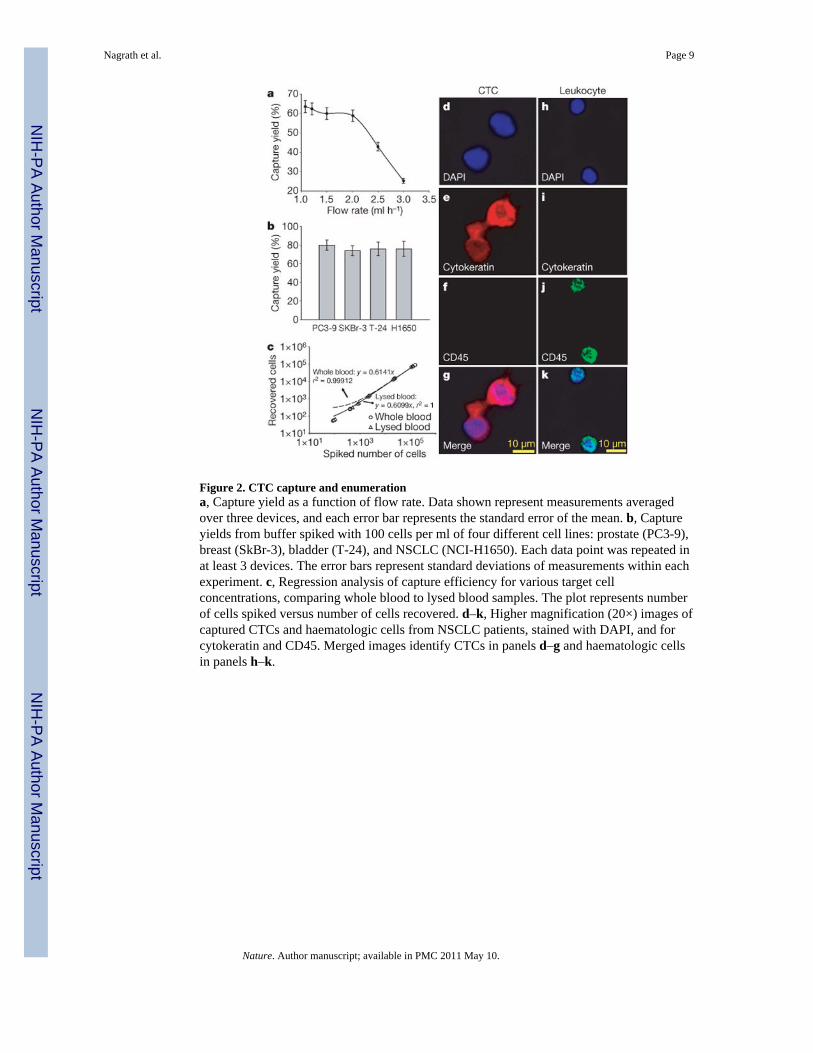
Figure 2. CTC capture and enumerationa, Capture yield as a function of flow rate. Data shown represent measurements averagedover three devices, and each error bar represents the standard error of the mean. b, Captureyields from buffer spiked with 100 cells per ml of four different cell lines: prostate (PC3-9),breast (SkBr-3), bladder (T-24), and NSCLC (NCI-H1650). Each data point was repeated inat least 3 devices. The error bars represent standard deviations of measurements within eachexperiment. c, Regression analysis of capture efficiency for various target cellconcentrations, comparing whole blood to lysed blood samples. The plot represents numberof cells spiked versus number of cells recovered. d–k, Higher magnification (20×) images ofcaptured CTCs and haematologic cells from NSCLC patients, stained with DAPI, and forcytokeratin and CD45. Merged images identify CTCs in panels d–g and haematologic cellsin panels h–k.
Nagrath et al. Page 9
Nature. Author manuscript; available in PMC 2011 May 10.
NIH
-PA Author Manuscript
NIH
-PA Author Manuscript
NIH
-PA Author Manuscript

Figure 3. Enumeration of CTCs from cancer patientsa, Summary of samples and CTC counts per 1 ml of blood in patients with various advancedcancers and localized prostate cancer. b, Frequency of CTCs per 1 ml of blood, bydiagnosis. The box plot presents the median, lower and upper quartiles (25th, 75thpercentiles). Data points that lie outside the 10th and 90th percentiles are shown as outliers.c, Purity of captured CTCs (ratio of CTCs to total nucleated cells), by diagnosis. d–i, SerialCTC assessment. CTC quantity (red), and tumour size (blue) measured as theunidimensional sum of all significant tumour sites on a CT scan, are well correlated over thecourse of anti-cancer treatment for nine patients. Six of them are shown here, for whomdiagnoses and specific therapies were: NSCLC, 1st-line carboplatin, paclitaxel (d); NSCLC,2nd-line pemetrexed (e); colorectal, 1st-line 5FU, irinotecan (f); pancreatic, 1st-linegemcitabine, bevacizumab (g); pancreatic, 1st-line gemcitabine (h); pancreatic, 1st-linegemcitabine, erlotinib (i). Baseline CT scans were before therapy initiation and CTCmeasurements began at or shortly after the first treatment.
Nagrath et al. Page 10
Nature. Author manuscript; available in PMC 2011 May 10.
NIH
-PA Author Manuscript
NIH
-PA Author Manuscript
NIH
-PA Author Manuscript

Figure 4. Characterization of CTCs with tumour-specific molecular markersa, b, CTCs from a prostate cancer patient stained positive for DAPI and PSA expression. c,RT–PCR amplification of PSA transcript is seen in two patients with prostate cancer (PCa),but not in two patients with lung cancer (LuCa), and only in blood fractions enriched forCTCs as opposed to non-enriched fractions (non-CTC). d, e, CTCs from an NSCLC patientstained for DAPI and TTF-1. f, RT–PCR shows expression of TTF-1 in two patients withlung cancer (LuCa), which is absent in two patients with prostate cancer (PCa), and onlywhen RNA was eluted from blood fractions enriched for CTCs as opposed to non-enrichedfractions (non-CTC).
Nagrath et al. Page 11
Nature. Author manuscript; available in PMC 2011 May 10.
NIH
-PA Author Manuscript
NIH
-PA Author Manuscript
NIH
-PA Author Manuscript